Lipase-Catalyzed Acidolysis of Egg-Yolk Phosphatidylcholine ...
Lwc
GVAa
b
c
d
e
Tf
a
ARRAA
KCMLCSP
1
dtcim
GEn
W
1h
The International Journal of Biochemistry & Cell Biology 44 (2012) 1839– 1846
Contents lists available at SciVerse ScienceDirect
The International Journal of Biochemistry& Cell Biology
journa l h o me page: www.elsev ier .com/ locate /b ioce l
ipidomic profiling in Crohn’s disease: Abnormalities in phosphatidylinositols,ith preservation of ceramide, phosphatidylcholine and phosphatidylserine
omposition
avin W. Sewell a, Yusuf A. Hannunb, Xianlin Hand, Grielof Kostere, Jacek Bielawskic,ictoria Gosse, Philip J. Smitha, Farooq Z. Rahmanf, Roser Vegaf, Stuart L. Bloomf,nn P. Walkera, Anthony D. Postlee, Anthony W. Segala,∗
Division of Medicine, UCL, 5 University Street, London, United KingdomDepartment of Medicine, Stony Brook University, Stony Brook, NY, United StatesDepartment of Biochemistry and Molecular Biology, Medical University of South Carolina, Charleston, SC, United StatesSanford-Burnham Medical Research Institute, Orlando, FL, United StatesDivision of Infection, Inflammation & Immunity, University of Southampton, School of Medicine, Southampton General Hospital, Mailpoint 803, South Block,remona Road, Southampton, United KingdomDepartment of Gastroenterology, University College Hospital, 235 Euston Road, London, United Kingdom
r t i c l e i n f o
rticle history:eceived 2 March 2012eceived in revised form 8 June 2012ccepted 12 June 2012vailable online 19 June 2012
eywords:rohn’s diseaseacrophage
ipids
a b s t r a c t
Crohn’s disease is a chronic inflammatory condition largely affecting the terminal ileum and large bowel.A contributing cause is the failure of an adequate acute inflammatory response as a result of impairedsecretion of pro-inflammatory cytokines by macrophages. This defective secretion arises from aberrantvesicle trafficking, misdirecting the cytokines to lysosomal degradation. Aberrant intestinal permeabil-ity is also well-established in Crohn’s disease. Both the disordered vesicle trafficking and increasedbowel permeability could result from abnormal lipid composition. We thus measured the sphingo-and phospholipid composition of macrophages, using mass spectrometry and stable isotope labellingapproaches. Stimulation of macrophages with heat-killed Escherichia coli resulted in three main changes;a significant reduction in the amount of individual ceramide species, an altered composition of phos-
eramidephingolipidhospholipid
phatidylcholine, and an increased rate of phosphatidylcholine synthesis in macrophages. These changeswere observed in macrophages from both healthy control individuals and patients with Crohn’s disease.The only difference detected between control and Crohn’s disease macrophages was a reduced propor-tion of newly-synthesised phosphatidylinositol 16:0/18:1 over a defined time period. Shotgun lipidomicsanalysis of macroscopically non-inflamed ileal biopsies showed a significant decrease in this same lipid
ervati
species with overall pres. Introduction
A number of physiological processes have been shown to beisturbed in Crohn’s disease (CD). A recently described manifes-ation is the failure of acute inflammation, resulting in impaired
learance of bacteria from the tissues. This is due to mistarget-ng of pro-inflammatory mediators to lysosomal degradation inacrophages as a result of aberrant vesicle trafficking (Smith et al.,
Abbreviations: CCT, Phosphocholine cytidylyltransferase; CD, Crohn’s disease;WAS, Genome-wide association study; HC, Healthy control; HkEc, Heat-killedscherichia coli; PA, Phosphatidic acid; PC, Phosphatidylcholine; PI, Phosphatidyli-ositol; PS, Phosphatidylserine; TNF, Tumor necrosis factor.∗ Corresponding author at: Division of Medicine, UCL, 5 University Street, LondonC1E6JJ, United Kingdom. Tel.: +44 02076796175.
E-mail address: [email protected] (A.W. Segal).
357-2725/$ – see front matter. Crown Copyright © 2012 Published by Elsevier Ltd. All rittp://dx.doi.org/10.1016/j.biocel.2012.06.016
on of sphingolipid, phospholipid and cholesterol composition.Crown Copyright © 2012 Published by Elsevier Ltd. All rights reserved.
2009). Autophagy (Cooney et al., 2010) and apoptosis (Palmer et al.,2009) have also been shown to be abnormal in CD, and intestinalpermeability is increased in this condition (Hollander et al., 1986).
Sphingolipids and phospholipids play key roles in the modula-tion of inflammation and immunity (El et al., 2006). Ceramide andceramide-1-phosphate act to reduce tumor necrosis factor (TNF)release (Jozefowski et al., 2010), most likely via post-translationalregulation of TNF and modulation of TNF converting enzyme activ-ity (Rozenova et al., 2010). Furthermore, ceramides have importantroles in the control of autophagy, a process strongly implicated inthe pathogenesis of CD (Barrett et al., 2008). Addition of exoge-nous ceramide induces autophagy (Scarlatti et al., 2004), which may
relate to effects on signalling networks or changes in the biophys-ical membrane properties (Zheng et al., 2006).Phosphatidylcholine (PC) is an important structural componentof all cell membranes, including intracellular vesicles (Howe and
ghts reserved.

1 Biochemistry & Cell Biology 44 (2012) 1839– 1846
Macilurs
lwcc(sfb
it1filggemmFua(
dwplheodpapfl
ntuip(tm
2
2
N1ab
Table 1Demographics of patients. Demographics of patients and healthy controls (HC)included in (A) sphingolipid study, (B) phospholipid studies and (C) shotgunlipidomics study.
A
Unstimulated HkEc stimulated
HC CD HC CD
Number 7 8 7 12M:F 5:2 4:4 5:2 6:6Mean age 44.1 36.2 44.1 39.7Age standard deviation 15.6 12.9 15.6 14.8Age range 23–63 19–63 23–63 19–65Smokers 0 0 0 1TreatmentNo medication 2 45-ASA 6 8
B
Unstimulated HkEc stimulated
HC CD HC CD
Number 10 13 7 8M:F 5:5 6:7 5:2 4:4Mean age 33.5 36.5 34.8 35.1Age standard deviation 9.2 13.7 10.4 11.6Age range 22–55 23–70 22–55 23–61Smokers 1 1 0 1TreatmentNo medication 6 35-ASA 7 5Mean BMI 23.7 23.3 23.3 23.3BMI standard deviation 3.5 3.0 3.8 3.7BMI range 17.8–30.5 19.4–29.4 17.8–30.5 19.4–29.4
C
HC CD
Number 5 5M:F 1:4 3:2Mean age 40.0 33.1Age standard deviation 17.8 10.1Age range 21–54 20–46Smokers 0 0TreatmentNo medication 35-ASA 1Methotrexate 1Previous resection 2Active disease 2
840 G.W. Sewell et al. / The International Journal of
cMaster, 2001). In macrophages, the generation of PC may play role in differentiation (Ecker et al., 2010) and pro-inflammatoryytokine release (Tian et al., 2008). Murine macrophages deficientn phosphocholine cytidylyltransferase (CCT-�) secrete reducedevels of TNF and IL-6 in response to lipopolysaccharide (LPS) stim-lation, as a result of abnormal post-translational processing andetention of these molecules in the Golgi apparatus, similar to theituation observed in CD macrophages.
Phosphatidylinositol (PI) is an important structural phospho-ipid, and also a substrate for lipid kinases and phosphatases,
hich can generate phosphoinositide derivatives (PIPs). PIPs areritical second messenger molecules in pathways involved in theontrol of cytoskeletal re-organisation and membrane traffickingOdorizzi et al., 2000). PI 3-kinase, an enzyme that converts PIpecies to phosphatidylinositol-3,4,5-trisphosphate, is importantor TNF trafficking from the Golgi apparatus to the plasma mem-rane in macrophages (Low et al., 2010).
Various studies have suggested alterations in lipid metabolismn CD. Positive correlations exist between dietary fat consump-ion and the development of CD (Amre et al., 2007; Shoda et al.,996). Genome-wide association studies (GWAS) have identi-ed CD-associated variants in loci containing genes related to
ipid metabolism. Specifically, a locus containing the ORMDL3ene confers susceptibility to CD (Barrett et al., 2008). ORMenes are involved in sphingolipid homeostasis; the ORM proteinsncoded by these genes act as negative regulators of sphingolipidetabolism (Breslow et al., 2010). Furthermore, the recent GWASeta-analysis identified a CD-associated SNP in close proximity to
ADS1 (Franke et al., 2010), which encodes the fatty acid desat-rase 1 enzyme. Genetic variation in this gene is associated withlterations in the fatty acid composition in serum phospholipidsSchaeffer et al., 2006).
Studies conducted on biological samples have demonstratedecreased membrane fluidity in erythrocytes from CD patients,ith concomitant increases in sphingomyelin and reductions inhosphatidylcholine and polyunsaturated acyl chains of phospho-
ipid (Aozaki, 1989). Increased concentrations of lactosylceramideave been reported in bowel biopsies from CD patients (Stevenst al., 1988), although it is possible that these changes were sec-ndary to inflammation. There is somewhat conflicting evidenceescribing fatty acid abnormalities in CD, including in plasma phos-holipids (Esteve-Comas et al., 1992, 1993; Geerling et al., 1999)nd PBMCs (Trebble et al., 2004). Lipids from adipose and lym-hoid tissues contain more saturated but fewer polyunsaturatedatty acids, with preferential depletion of n-6 polyunsaturates inymphoid tissue (Westcott et al., 2005).
Macrophage phospholipid and sphingolipid composition haveot been previously investigated in CD. Given the possibilityhat macrophage sphingolipid or phospholipid composition couldnderlie the defective cytokine secretion from macrophages that
s observed in CD, these molecules were quantified using higherformance liquid chromatography tandem mass spectrometryHPLC-MS). Phospholipid composition and dynamics were inves-igated using stable isotope labelling and electrospray ionisation
ass spectrometry (ESI-MS).
. Materials and methods
.1. Subject recruitment and selection
Patients from University College London Hospitals Foundation
HS Trust fulfilled criteria for the diagnosis of CD (Lennard-Jones,989). All patients in the macrophage studies were between 18nd 75 years of age and had quiescent disease, as determinedy the Harvey-Bradshaw disease activity index of <3 (Harvey andBradshaw, 1980), and were receiving either no treatment or a sta-ble dose (for the preceding 3 months) of 5-aminosalicylates (5-ASA)alone. Healthy volunteers were between 18 and 75 years of age andwere not receiving immunosuppressant medication. Demograph-ics of patients included in these studies are shown (Table 1).
For shotgun lipidomics investigations, ileal biopsies wereobtained from CD patients (n = 5) and control individuals (n = 5).Two of the patients had macroscopic and microscopic evidence ofactive disease; the remaining patients had macroscopic and his-tologic features consistent with quiescent disease. Three patientswere receiving no treatment, one was receiving 5-aminosalicylatesand one was receiving methotrexate. Two patients had previousileal resections; in these cases biopsies from the neoterminal ileumwere obtained. In all cases biopsies were taken from macroscopi-cally non-inflamed bowel.
Ethical approval was obtained from the Joint UCL/UCLH Com-mittees on the Ethics of Human Research (project number 02/0324).
No subject was studied more than once in each of the different setsof experiments.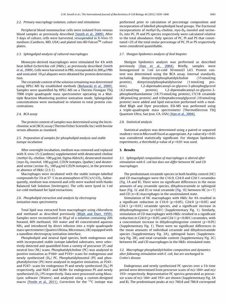
Bioche
2
b5mp
2
heat
uS7tcc
2
cs
2i
w((ao
cqBi
2i
aSbNma
wtnpnparsLm
G.W. Sewell et al. / The International Journal of
.2. Primary macrophage isolation, culture and stimulation
Peripheral blood mononuclear cells were isolated from venouslood samples as previously described (Smith et al., 2009). After
days of culture, cells were harvested, resuspended in X-Vivo-15edium (Cambrex, MD, USA) and plated into BD FalconTM culture
lates.
.3. Sphingolipid analysis of cultured macrophages
Monocyte-derived macrophages were stimulated for 4 h witheat-killed Escherichia coli (HkEc), as previously described (Smitht al., 2009). Cells were harvested in PBS, resuspended in 200 �l PBSnd sonicated. 10 �l aliquots were obtained for protein determina-ion.
The ceramide content of the solution remaining was determinedsing HPLC-MS by established methods (Bielawski et al., 2009).amples were quantified by HPLC-MS on a Thermo Finnigan TSQ000 triple quadrupole mass spectrometer operating in a Mul-iple Reaction Monitoring positive ionisation mode. Sphingolipidoncentrations were normalised in relation to total protein con-entrations.
.4. BCA assay
The protein content of samples was determined using the bicin-honinic acid (BCA) assay (Thermo Fisher Scientific Inc) with bovineerum albumin as standard.
.5. Preparation of samples for phospholipid analysis and stablesotope incubation
After overnight incubation, medium was removed and replacedith X-vivo-15 (Cambrex) supplemented with deuterated choline
methyl-D9-choline, 100 �g/ml, Sigma Aldrich), deuterated inositolmyo-D6-inositol, 100 �g/ml, C/D/N isotopes, Quebec) and deuter-ted serine (serine-D3, 100 �g/ml C/D/N isotopes), in the presencer absence of HkEc (2.5:1).
Macrophages were incubated with the stable isotope-labelledompounds for 3 h at 37 ◦C in an atmosphere of 5% (v/v) CO2. Subse-uently, medium was removed and cells were washed with Hanksalanced Salt Solution (Invitrogen). The cells were lysed in 1 ml
ce-cold methanol for lipid extractions.
.6. Phospholipid extraction and analysis by electrosprayonisation mass spectrometry
Total lipid was extracted from macrophages using chloroformnd methanol as described previously (Bligh and Dyer, 1959).amples were reconstituted in 30 �l of a solution containing 20%utanol, 60% methanol, 16% water and 4% concentrated aqueousH3 and introduced by direct infusion into a triple quadrupoleass spectrometer (Quattro Ultima, Micromass, UK) equipped with
nanoflow electrospray ionisation interface.Phospholipid and neutral lipid species, both endogenous and
ith incorporated stable isotope-labelled substrates, were selec-ively detected and quantified from a variety of precursor (P) andeutral loss (NL) scans. Phosphatidylcholine (PC) was analysed inositive ionisation as P184+ and P193+ scans for endogenous andewly synthesised (D9) PC. Phosphatidylinositol (PI) and phos-hatidylserine (PS) were analysed in negative ionisation, as P241-nd P247- scans for endogenous PI and newly synthesised (D6) PI
espectively, and NL87- and NL90- for endogenous PS and newlyynthesised (D3) PS respectively. Data were processed using Mass-ynx software (Waters) and analysed using a custom-designedacro (Postle et al., 2011). Correction for the 13C isotope wasmistry & Cell Biology 44 (2012) 1839– 1846 1841
performed prior to calculation of percentage composition andincorporation of labelled phospholipid head groups. The fractionalincorporations of methyl-D9-choline, myo-D6-inositol and serine-D3 into PC, PI and PS species respectively were calculated relativeto the total abundance. Only species of PC, PI and PS that consti-tuted >2% of the total molar percentage of PC, PI or PS respectivelywere considered quantifiable.
2.7. Shotgun lipidomics analysis of ileal biopsies
Shotgun lipidomics analysis was performed as describedpreviously (Han et al., 2004). Briefly, samples werehomogenised in 1 ml ice-cold 50 mmol/l LiCl. Protein con-tent was determined using the BCA assay. Internal standards,including dimyristoylphosphatidylcholine (15 nmol/mgprotein), dimyristoylphosphatidylserine (1 nmol/mg pro-tein), 1,2-dipentadecanoyl-sn-glycero-3-phosphoglycerol(4.2 nmol/mg protein), 1,2-dipentadecanoyl-sn-glycero-3-phosphoethanolamine (18.75 nmol/mg protein), 17C18 ceramide(40 pmol/mg protein) and triheptadecenoylglycerol (10 nmol/mgprotein) were added and lipid extraction performed with a mod-ified Bligh and Dyer procedure. ESI-MS was performed usinga triple-quadrupole mass spectrometer (ThermoElectron TSQQuantum Ultra, San Jose, CA, USA) (Han et al., 2004).
2.8. Statistical analysis
Statistical analysis was determined using a paired or unpairedstudent t-test in Microsoft Excel as appropriate. A p-value of p < 0.05was considered statistically significant. For shotgun lipidomicsexperiments, a threshold p-value of p < 0.01 was used.
3. Results
3.1. Sphingolipid composition of macrophages is altered afterstimulation with E. coli but does not differ between HC and CDmacrophages
The predominant ceramide species in both healthy control (HC)and CD macrophages were the C16:0, C24:0 and C24:1 ceramides(Fig. 1A and B). There were no significant differences in the meanamounts of any ceramide species, dihydroceramide or sphingoidbase (Fig. 1C and D) or total ceramide (Fig. 1E) between HC (n = 7)and CD (n = 8) macrophages in the unstimulated state.
Stimulation of HC macrophages with HkEc for 4 h resulted ina significant reduction in C16:0 (p < 0.05), C24:0 (p < 0.05) andC24:1 (p < 0.01) ceramide species, and a significant increase indihydrosphingosine (p < 0.01) (Supplementary Fig. 1). Similarly,stimulation of CD macrophages with HkEc resulted in a significantreduction in C24:0 (p < 0.05) and C24:1 (p < 0.001) ceramides, witha concomitant increase in dihydrosphingosine content (p < 0.05)(Supplementary Fig. 1). There were no significant differences inthe mean amounts of individual ceramide and dihydroceramidespecies (Supplementary Fig. 2A), sphingoid bases (Supplemen-tary Fig. 2B), and total ceramide content (Supplementary Fig. 2C)between HC and CD macrophages in the HkEc stimulated state.
3.2. Macrophage phosphatidylcholine composition and dynamicsalter following stimulation with E. coli, but are unchanged inCrohn’s disease
Endogenous and newly synthesised PC species over a 3 h time
period were determined from precursor scans of m/z 184+ and m/z193+ respectively. Representative PC spectra generated as precur-sor scans of m/z 184+ and 193+ are shown (Supplementary Fig. 3Aand B). The predominant peaks at m/z 760.8 and 786.8 correspond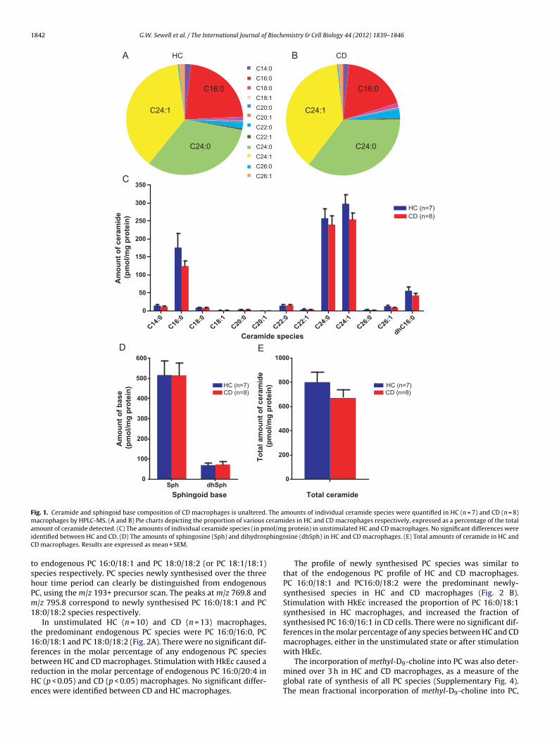
1842 G.W. Sewell et al. / The International Journal of Biochemistry & Cell Biology 44 (2012) 1839– 1846
C14:0
C16:0
C18:0
C18: 1
C20:0
C20: 1
C22:0
C22: 1
C24:0
C24: 1
C26:0
C26: 1
C16:0 C16:0
C24:1 C24:1
C24:0 C24:0
Sph dhSph0
100
200
300
400
500
600
Sphingoid base
Am
ou
nt
of
base
(pm
ol/m
g p
rote
in)
CD (n=8)HC (n=7)
A B
C
D
C14
:0
C16
:0
C18
:0
C18
:1
C20
:0
C20
:1
C22
:0
C22
:1
C24
:0
C24
:1
C26
:0
C26
:1
dhC16
:00
50
100
150
200
250
300
350
HC (n=7)
CD (n=8)
Ceramide species
Am
ou
nt
of
cera
mid
e
(p
mo
l/m
g p
rote
in)
0
200
400
600
800
1000
To
tal am
ou
nt
of
cera
mid
e
(p
mo
l/m
g p
rote
in)
Total ceramide
E
CD (n=8)HC (n=7)
HC CD
Fig. 1. Ceramide and sphingoid base composition of CD macrophages is unaltered. The amounts of individual ceramide species were quantified in HC (n = 7) and CD (n = 8)macrophages by HPLC-MS. (A and B) Pie charts depicting the proportion of various ceramides in HC and CD macrophages respectively, expressed as a percentage of the totala ol/mi phingoC
tshPm1
t1fbrHe
mount of ceramide detected. (C) The amounts of individual ceramide species (in pmdentified between HC and CD. (D) The amounts of sphingosine (Sph) and dihydrosD macrophages. Results are expressed as mean + SEM.
o endogenous PC 16:0/18:1 and PC 18:0/18:2 (or PC 18:1/18:1)pecies respectively. PC species newly synthesised over the threeour time period can clearly be distinguished from endogenousC, using the m/z 193+ precursor scan. The peaks at m/z 769.8 and/z 795.8 correspond to newly synthesised PC 16:0/18:1 and PC
8:0/18:2 species respectively.In unstimulated HC (n = 10) and CD (n = 13) macrophages,
he predominant endogenous PC species were PC 16:0/16:0, PC6:0/18:1 and PC 18:0/18:2 (Fig. 2A). There were no significant dif-erences in the molar percentage of any endogenous PC species
etween HC and CD macrophages. Stimulation with HkEc caused aeduction in the molar percentage of endogenous PC 16:0/20:4 inC (p < 0.05) and CD (p < 0.05) macrophages. No significant differ-nces were identified between CD and HC macrophages.g protein) in unstimulated HC and CD macrophages. No significant differences weresine (dhSph) in HC and CD macrophages. (E) Total amounts of ceramide in HC and
The profile of newly synthesised PC species was similar tothat of the endogenous PC profile of HC and CD macrophages.PC 16:0/18:1 and PC16:0/18:2 were the predominant newly-synthesised species in HC and CD macrophages (Fig. 2 B).Stimulation with HkEc increased the proportion of PC 16:0/18:1synthesised in HC macrophages, and increased the fraction ofsynthesised PC 16:0/16:1 in CD cells. There were no significant dif-ferences in the molar percentage of any species between HC and CDmacrophages, either in the unstimulated state or after stimulationwith HkEc.
The incorporation of methyl-D9-choline into PC was also deter-mined over 3 h in HC and CD macrophages, as a measure of theglobal rate of synthesis of all PC species (Supplementary Fig. 4).The mean fractional incorporation of methyl-D9-choline into PC,
G.W. Sewell et al. / The International Journal of Biochemistry & Cell Biology 44 (2012) 1839– 1846 1843
Endogenous PC
PC16
:0/1
6:1
PC16
:0/1
6:0
PC16
:0a/
18:1
PC16
:0/1
8:2
PC16
:0/1
8:1
PC16
:0a/
20:4
PC16
:0/2
0:4
PC18
:1/1
8:2
PC18
:0/1
8:2
PC18
:0/1
8:1
PC18
:1a/
20:4
PC18
:1/2
0:4
PC18
:0/2
0:4
0
5
10
15
20
25
PC species
Co
mp
osit
ion
(m
ole
%)
Newly synthesised PC
D 9PC
16:0
/16:
1
D 9PC
16:0
/16:
0
D 9PC
16:0
a/18
:1
D 9PC
16:0
/18:
2
D 9PC
16:0
/18:
1
D 9PC
16:0
a/20
:4
D 9PC
16:0
/20:
4
D 9PC
18:1
/18:
2
D 9PC
18:0
/18:
2
D 9PC
18:0
/18:
1
D 9PC
18:1
a/20
:4
D 9PC
18:1
/20:
4
D 9PC
18:0
/20:
4
0
5
10
15
20
25HC Uns (n=10)
CD Uns (n=13)
HC HkEc (n=7)
CD HkEc (n=8)
D9PC specie s
Co
mp
osit
ion
(m
ole
%)
HC Uns (n=10)
CD Uns (n=13)
HC HkEc (n=7)
CD HkEc (n=8)
A
B
*
*
Fig. 2. Composition of endogenous and newly synthesised PC in HC and CDmacrophages, in the presence and absence of HkEc. Composition of (A) endoge-nous PC, consisting of various carbon chain length fatty acid species, and (B) newlysna
tcpaSPb
3b
mrlasi7sTPTuC
t
Endogenous PS
PS16:0
/18:
1
PS16:0
/18:
0
PS18:0
a/18
:1
PS18:0
/18:
2
PS18:0
/18:
1
PS18:0
/20:
4
PS18:0
/20:
3
PS18:0
/22:
6
PS18:0
/22:
5
PS18:0
/22:
4
0
10
20
30
40
50
PS species
Co
mp
osit
ion
(m
ole
%)
Newly synthesised PS
D 3PS16
:0/1
8:1
D 3PS16
:0/1
8:0
D 3PS18
:0a/
18:1
D 3PS18
:0/1
8:2
D 3PS18
:0/1
8:1
D 3PS18
:0/2
0:4
D 3PS18
:0/2
0:3
D 3PS18
:0/2
2:6
D 3PS18
:0/2
2:5
D 3PS18
:0/2
2:4
0
10
20
30
40
50 HC Uns (n=7)
CD Uns (n=9)
HC HkEc (n=6 )
CD HkEc (n=5 )
D3PS species
Co
mp
osit
ion
(m
ole
%)
A
B
HC Uns (n=7)
CD Uns (n=9)
HC HkEc (n=6 )
CD HkEc (n=5 )
Fig. 3. Composition of endogenous and newly synthesised PS in HC and CDmacrophages, in the presence and absence of HkEc. Composition of (A) endoge-nous PS and (B) synthesised (D3) PS, in unstimulated (HC n = 7, CD n = 9) and
ynthesised (D9) PC in unstimulated (HC n = 10, CD n = 13) and HkEc-stimulated (HC = 7, CD n = 8) macrophages, expressed as a molar percentage of total PC. Resultsre mean + SEM. * indicates p < 0.05.
hus rates of PC synthesis, were equivalent between HC and CDells. Stimulation with HkEc increased the mean fractional incor-oration of methyl-D9-choline into PC in HC macrophages (p < 0.01),nd CD macrophages (p < 0.01) compared to unstimulated cells.timulation was therefore associated with an increased rate ofC synthesis; although no significant differences were observedetween HC and CD macrophages.
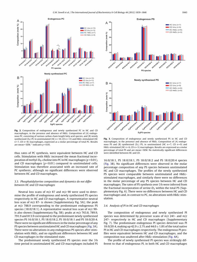
.3. Phosphatidylserine composition and dynamics do not differetween HC and CD macrophages
Neutral loss scans of m/z 87 and m/z 90 were used to deter-ine the profile of endogenous and newly synthesised PS species
espectively in HC and CD macrophages. A representative neutraloss scan of m/z 87- is shown (Supplementary Fig. 5A); the peakt m/z 788.9 corresponding to the predominant endogenous PSpecies (18:0/18:1). A representative neutral loss scan of m/z 90-s also shown (Supplementary Fig. 5B); peaks at m/z 763.8, 789.9,91.9 and 813.9 correspond to the predominant newly synthesisedpecies PS 16:0/18:1, PS 18:0/18:2, PS 18:0/18:1 and PS 18:0/20:4.here were no significant differences in the molar percentage of anyS species between unstimulated HC and CD macrophages (Fig. 3A).here were no alterations in any endogenous PS species after stim-
lation with HkEc, and no significant differences between HC andD macrophages in the stimulated state.The predominant newly synthesised PS species over the 3 hime period in unstimulated HC and CD macrophages included PS
HkEc-stimulated (HC n = 6, CD n = 5) macrophages. Results are expressed as a molarpercentage of total PS and are mean + SEM. No statistically significant differenceswere identified between HC and CD.
16:0/18:1, PS 18:0/18:1, PS 18:0/18:2 and PS 18:0/20:4 species(Fig. 3B). No significant differences were observed in the molarpercentage composition of any PS species between unstimulatedHC and CD macrophages. The profiles of the newly synthesisedPS species were comparable between unstimulated and HkEc-stimulated macrophages, and similarly there were no differencesin the molar percentage of any PS species between HC and CDmacrophages. The rates of PS synthesis over 3 h were inferred fromthe fractional incorporation of serine-D3 within the total PS (Sup-plementary Fig. 6). There were no differences between HC and CDmacrophages and, in contrast to PC, no alterations with HkEc stim-ulation.
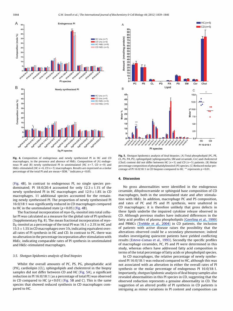
3.4. Analysis of PI in HC and CD macrophages
The composition of endogenous and newly synthesised PIspecies was determined by precursor scans of m/z 241- and m/z247- respectively in HC and CD macrophages (SupplementaryFig. 7). The predominant endogenous PI species detected was PI18:0/20:4, making up 42.9 ± 2.7% and 44.0 ± 1.8% of the total nativePI in HC and CD macrophages respectively. The endogenous PI pro-
files were equivalent between HC and CD macrophages, and thecomposition was unaltered after HkEc stimulation (Fig. 4A).The profile of newly synthesised PI species was strikingly dif-ferent to that of endogenous PI, in both HC and CD macrophages
1844 G.W. Sewell et al. / The International Journal of Biochemistry & Cell Biology 44 (2012) 1839– 1846
PI16:
0/18
:2
PI16:
0/18
:1
PI16:
0/20
:4
PI18:
1/18
:2
PI18:
0/18
:2
PI18:
0/18
:1
PI18:
1/20
:4
PI18:
0/20
:4
PI18:
0/20
:3
PI18:
0/22
:6
PI18:
0/22
:5
PI18:
0/22
:40
5
10
15
20
25
30
35
40
45
50
HC Uns (n=7 )
CD Uns (n=9 )
HC HkEc (n=6 )
CD HkEc (n=5 )
PI species
Co
mp
osit
ion
(m
ole
%)
D 6PI1
6:0/
18:2
D 6PI1
6:0/
18:1
D 6PI1
6:0/
20:4
D 6PI1
8:1/
18:2
D 6PI1
8:0/
18:2
D 6PI1
8:0/
18:1
D 6PI1
8:1/
20:4
D 6PI1
8:0/
20:4
D 6PI1
8:0/
20:3
D 6PI1
8:0/
22:6
D 6PI1
8:0/
22:5
D 6PI1
8:0/
22:4
0
5
10
15
20
25
30
35
40
45
50
D6 PI species
Co
mp
osit
ion
(m
ole
%)
HC Uns (n=7 )
CD Uns (n=9 )
HC HkEc (n=6 )
CD HkEc (n=5 )
A
B
*
Newly synthesised PI
Endogenous PI
Fig. 4. Composition of endogenous and newly synthesised PI in HC and CDmacrophages, in the presence and absence of HkEc. Composition of (A) endoge-nous PI and (B) newly synthesised PI in unstimulated (HC n = 7, CD n = 9) andHp
(dnmi1t
l(D1anHa
3
(srisp
PC PE CL PG PA PS SM Cer Chol05
101520253035404550556065707580859095
HC (n=5 )
CD (n=5)
Lipid
Am
ou
nt
(n
mo
l/m
g p
rote
in)
16:0
/16:
0
16:0
/18:
2
16:0
/18:
1
16:0
/18:
0
16:1
/20:
4
16:0
/20:
4
18:1
/18:
2
18:0
/18:
2
18:0
/18:
1
18:0
/18:
0
16:0
/22:
6
18:1
/20:
4
18:0
/20:
4
18:0
/20:
3
18:0
/20:
2
18:0
/22:
6
18:0
/22:
5
18:0
/22:
40
5
10
15
20
25
30
35
40
45
**
PI species
Mo
lar
%
HC CD0.0
2.5
5.0
7.5
10.0
12.5
15.0**
PI 16:0/18: 1
A
Mo
lar
%
BC
Fig. 5. Shotgun lipidomics analysis of ileal biopsies. (A) Total phospholipid (PC, PE,CL, PG, PA, PS), sphingolipid (sphingomyelin, SM and ceramide, Cer) and cholesterol
revealed abnormalities in this PI species in CD, suggesting that the
kEc-stimulated (HC n = 6, CD n = 5) macrophages. Results are expressed as a molarercentage of the total PI and are mean + SEM. * indicates p < 0.05.
Fig. 4B). In contrast to endogenous PI, no single species pre-ominated; PI 18:0/20:4 accounted for only 12.3 ± 1.1% of theewly synthesised PI in HC macrophages and 12.9 ± 1.8% in CDacrophages. 11 additional species accounted for the remain-
ng newly synthesised PI. The proportion of newly synthesised PI6:0/18:1 was significantly reduced in CD macrophages comparedo HC in the unstimulated state (p < 0.05) (Fig. 4B).
The fractional incorporation of myo-D6-inositol into total cellu-ar PI was calculated as a measure for the global rate of PI synthesisSupplementary Fig. 8). The mean fractional incorporation of myo-6-inositol as a percentage of the total PI was 16.1 ± 2.3% in HC and5.5 ± 1.5% in CD macrophages over 3 h, indicating equivalent over-ll rates of PI synthesis in HC and CD. In contrast to PC, there waso alteration in the percentage incorporation after stimulation withkEc, indicating comparable rates of PI synthesis in unstimulatednd HkEc-stimulated macrophages.
.5. Shotgun lipidomics analysis of ileal biopsies
Whilst the overall amounts of PC, PS, PG, phosphatidic acidPA), cardiolipin (CL), sphingolipids and cholesterol in the biopsyamples did not differ between CD and HC (Fig. 5A), a significanteduction in PI 16:0/18:1 (as a percentage of total PI) was observed
n CD compared to HC (p < 0.01) (Fig. 5B and C). This is the samepecies that showed reduced synthesis in CD macrophages com-ared to HC.(Chol) content did not differ between HC (n = 5) and CD (n = 5) patients. (B) Molarpercentage composition of phosphatidylinositol (PI) species. (C) Reduced molar per-centage of PI 16:0/18:1 in CD biopsies compared to HC. ** represents p < 0.01.
4. Discussion
No gross abnormalities were identified in the endogenousceramide, dihydroceramide or sphingoid base composition of CDmacrophages, both in the unstimulated state and after stimula-tion with HkEc. In addition, macrophage PC and PS composition,and rates of PC and PS and PI synthesis, were unaltered inCD macrophages; it is therefore unlikely that gross defects inthese lipids underlie the impaired cytokine release observed inCD. Although previous studies have indicated differences in thefatty acid profiles of plasma phospholipids (Geerling et al., 1999)and PBMCs (Trebble et al., 2004) in CD patients, the inclusionof patients with active disease raises the possibility that thealterations observed could be a secondary phenomenon; indeedstudies investigating quiescent patients have yielded conflictingresults (Esteve-Comas et al., 1993). Secondly the specific profilesof macrophage ceramides, PC, PS and PI were determined in thisstudy, whereas others have addressed fatty acid composition interms of the total percentage of fatty acids or phospholipid species.
In CD macrophages, the relative percentage of newly synthe-sised PI 16:0/18:1 was reduced compared to HC, although this wasnot associated with an alteration in either the overall rates of PIsynthesis or the molar percentage of endogenous PI 16:0/18:1.Importantly, shotgun lipidomic analysis of ileal biopsy samples also
observed reduction represents a genuine abnormality in CD. Thesuggestion of an altered profile of PI synthesis in CD patients isintriguing as minor variations in PI content and composition can
Bioche
e(kppdc
saactIacaaotn
ottPmpi(ab
arTiDlCdeti
aotsobTmd1ets
tcaepbr
G.W. Sewell et al. / The International Journal of
xert major effects on the physical properties of membrane systemsMulet et al., 2008). It is known that the p110� isoform of PI 3-inase, an enzyme involved in the generation of 3-phosphorylatedhosphatidylinositol derivatives, is important in the TNF secretoryathway (Low et al., 2010). It is therefore possible that altered PIynamics could contribute to the impairment in pro-inflammatoryytokine secretion observed in CD.
We have developed a three-stage model for CD, in which the firsttage is failure of intestinal barrier function followed by impairedcute inflammation (Sewell et al., 2009). Phospholipids such as PIre important components of intestinal mucus as well as cellularomponents of the mucosa, and animal studies indicate a protec-ive role for phospholipids in barrier function (Fabia et al., 1992).t is plausible that abnormalities in the mucosal PI profile couldlter membrane or mucus fluidity, facilitating ingress of luminalontents into the bowel wall directly; alternatively, such alter-tions could enhance susceptibility to damaging emulsifiers suchs bile acids. Although alterations in the phospholipid compositionf intestinal mucus have previously been associated with ulcera-ive colitis rather than CD (Braun et al., 2009), PI composition wasot determined in this study, which warrants further investigation.
The underlying mechanism of the reduced PI 16:0/18:1bserved in CD can only be speculated on at this stage. Given thathe overall rate of PI synthesis is unchanged in CD macrophages,he difference could relate to an altered substrate preference ofI synthase in CD. Alternatively, abnormalities in the fatty acidetabolism pathway could lead to altered fatty acid availability for
hospholipid synthesis. Notably, the recent GWAS meta-analysisdentified a CD-associated SNP in a region containing the FADS1Fatty acid desaturase 1) gene (Franke et al., 2010). This associationdds credence to the hypothesis that fatty acid desaturation maye relevant in the pathogenesis of CD.
Stimulation of macrophages with HkEc was associated withlterations in ceramides and phospholipids, further implicatingoles for these lipids in inflammation and innate immunity. LPS,NF and IL-1� were previously shown to cause a rapid increasen the levels of ceramide in macrophage cell lines (MacKichan andeFranco, 1999). In contrast with this previous work, HkEc stimu-
ation in the present study was associated with a reduction in the16:0, C24:0 and C24:1 ceramides, and a concomitant increase inihydrosphingosine content. This difference could relate to differ-ntial effects of specific Toll-like receptor and HkEc stimulation onhe sphingolipid pathway, or the different time frames investigatedn the two studies.
Alterations in the phospholipid profile of macrophages observedfter stimulation with HkEc included a reduction in the amountf endogenous PC 16:0/20:4. Recent application of lipidomicechnologies to the activation of murine macrophages has demon-trated rapid generation of a wide range of eicosanoids and otherxylipins following mobilisation of arachidonate from major mem-rane phospholipids, including PC and PS (Rouzer et al., 2007).he decreased content of PC 16:0/20:4 in both control and CDacrophages after E. coli activation is consistent with such arachi-
onate mobilisation. Intriguingly, the fractional synthesis of PC6:0/20:4 from D9-choline was unchanged in these activated cells,ven though the rate of total PC synthesis was increased, implyinghere was no apparent deficit of arachidonate availability for PCynthesis under these conditions.
Comparison of patterns of D9-choline incorporation into PC withhe profile of endogenous PC composition indicated a degree ofo-ordinated acyl remodelling, with enhanced initial synthesis ofll three 18:2-containing species. Endogenous PC was relatively
nriched in disaturated and ether-linked PC species, which wereresumably all formed subsequently to initial PC synthesis de novoy a variety of acyl exchange mechanisms. The extent of such acylemodelling was even more apparent for PS and PI synthesis. Themistry & Cell Biology 44 (2012) 1839– 1846 1845
single species PS 18:0/18:1 accounted for over 40% of the total PS,but contributed less than 20% of PS synthesised from serine-D3.PS is formed by headgroup exchange from either PC or PE by theaction of PS synthase (Hermansson et al., 2011), but acyl exchangemechanisms in PS synthesis have not been previously identified.Similarly, PI 18:0/20:4 was >40% of total PI, but only 12% of PIsynthesis from myo-D6-inositol, substantiating previous sugges-tions that the high content of arachidonate in macrophage PI ismaintained not by its synthesis de novo from myo-inositol but bydirect acyl incorporation due to sequential activities of PLA2 andacyltransferases enzymes, proposed from incorporation patternsof D8-arachidonate (Balgoma et al., 2008).
Overall, in spite of the previous differences in cytokine releaseand intestinal barrier function, no major differences in lipid com-position and synthesis were observed between tissue samples fromCD patients and healthy controls, and their responses to bacte-rial stimulation. In contrast to studies of elicited bone marrowor peritoneal macrophages, all samples in our study were fromindividual subjects, and inter-subject differences in lipid nutri-tion could contribute to the variation in observed lipid responses.Nevertheless, the results question the extent to which lipid mobili-sation is obligatorily linked to macrophage activation, and suggest asubtle difference in phosphatidylinositol composition in CD whichcould have an important influence on cytokine release and mucosalbarrier function.
Author contribution
Macrophage experiments were conceived and designed byGWS, AWS and ADP. GWS, JB, APW and YH conducted sphingolipidexperiments. Phospholipid experiments were conducted by GWS,PJS, GK, VG and ADP. The shotgun lipidomics studies were plannedby AWS and XH. FZR, RV and SLB contributed materials to this study.Data were analysed by GS, XH, ADP, GK and JB. GS, ADP and AWSwrote the paper.
Acknowledgements
We thank Tony Futerman, Al Merrill, Daniel Marks and AndrewSmith for helpful discussions, Bu Hayee, and Nuala O’Shea for tak-ing blood in clinics, and Penelope Harrison, Carol McDonald andJonathan Townsend for technical assistance. This work was sup-ported by the Wellcome Trust (AWS) and NIH grant CA097132(YH).
Appendix A. Supplementary data
Supplementary data associated with this article can befound, in the online version, at http://dx.doi.org/10.1016/j.biocel.2012.06.016.
References
Amre DK, D’Souza S, Morgan K, Seidman G, Lambrette P, Grimard G, et al. Imbal-ances in dietary consumption of fatty acids, vegetables, and fruits are associatedwith risk for Crohn’s disease in children. American Journal of Gastroenterology2007;102:2016–25.
Aozaki S. Decreased membrane fluidity in erythrocytes from patients with Crohn’sdisease. Gastroenterologia Japonica 1989;24:246–54.
Balgoma D, Montero O, Balboa MA, Balsinde J. Calcium-independent phospholi-pase A2-mediated formation of 1,2-diarachidonoyl-glycerophosphoinositol inmonocytes. FEBS Journal 2008;275:6180–91.
Barrett JC, Hansoul S, Nicolae DL, Cho JH, Duerr RH, Rioux JD, et al. Genome-wideassociation defines more than 30 distinct susceptibility loci for Crohn’s disease.
Nature Genetics 2008;40:955–62.Bielawski J, Pierce JS, Snider J, Rembiesa B, Szulc ZM, Bielawska A. Comprehen-sive quantitative analysis of bioactive sphingolipids by high-performance liquidchromatography-tandem mass spectrometry. Methods in Molecular Biology2009;579:443–67.
1 Bioch
B
B
B
C
E
E
E
E
F
F
G
H
H
H
H
H
J
L
L
Diseases 2005;11:820–7.
846 G.W. Sewell et al. / The International Journal of
ligh EG, Dyer WJ. A rapid method of total lipid extraction and purification. CanadianJournal of Biochemistry and Physiology 1959;37:911–7.
raun A, Treede I, Gotthardt D, Tietje A, Zahn A, Ruhwald R, et al. Alterations ofphospholipid concentration and species composition of the intestinal mucusbarrier in ulcerative colitis: a clue to pathogenesis. Inflammatory Bowel Diseases2009;15:1705–20.
reslow DK, Collins SR, Bodenmiller B, Aebersold R, Simons K, ShevchenkoA, et al. Orm family proteins mediate sphingolipid homeostasis. Nature2010;463:1048–53.
ooney R, Baker J, Brain O, Danis B, Pichulik T, Allan P, et al. NOD2 stimulationinduces autophagy in dendritic cells influencing bacterial handling and antigenpresentation. Nature Medicine 2010;16:90–7.
cker J, Liebisch G, Englmaier M, Grandl M, Robenek H, Schmitz G. Induction offatty acid synthesis is a key requirement for phagocytic differentiation of humanmonocytes. Proceedings of the National Academy of Sciences of the United Statesof America 2010;107:7817–22.
l AM, Wu BX, Obeid LM, Hannun YA. Bioactive sphingolipids in the modulation ofthe inflammatory response. Pharmacology & Therapeutics 2006;112:171–83.
steve-Comas M, Nunez MC, Fernandez-Banares F, bad-Lacruz A, Gil A, Cabre E, et al.Abnormal plasma polyunsaturated fatty acid pattern in non-active inflamma-tory bowel disease. Gut 1993;34:1370–3.
steve-Comas M, Ramirez M, Fernandez-Banares F, bad-Lacruz A, Gil A, Cabre E,et al. Plasma polyunsaturated fatty acid pattern in active inflammatory boweldisease. Gut 1992;33:1365–9.
abia R, Ar’Rajab A, Willen R, Andersson R, Ahren B, Larsson K, et al. Effects of phos-phatidylcholine and phosphatidylinositol on acetic-acid-induced colitis in therat. Digestion 1992;53:35–44.
ranke A, McGovern DP, Barrett JC, Wang K, Radford-Smith GL, Ahmad T, et al.Genome-wide meta-analysis increases to 71 the number of confirmed Crohn’sdisease susceptibility loci. Nature Genetics 2010;42:1118–25.
eerling BJ, Houwelingen AC, Badart-Smook A, Stockbrugger RW, Brummer RJ. Fatintake and fatty acid profile in plasma phospholipids and adipose tissue inpatients with Crohn’s disease, compared with controls. American Journal ofGastroenterology 1999;94:410–7.
an X, Yang J, Cheng H, Ye H, Gross RW. Toward fingerprinting cellular lipidomesdirectly from biological samples by two-dimensional electrospray ionizationmass spectrometry. Analytical Biochemistry 2004;330:317–31.
arvey RF, Bradshaw JM. A simple index of Crohn’s-disease activity. Lancet 1980;1,514-.
ermansson M, Hokynar K, Somerharju P. Mechanisms of glycerophospholipidhomeostasis in mammalian cells. Progress in Lipid Research 2011;50:240–57.
ollander D, Vadheim CM, Brettholz E, Petersen GM, Delahunty T, Rotter JI. Increasedintestinal permeability in patients with Crohn’s disease and their relatives. Apossible etiologic factor. Annals of Internal Medicine 1986;105:883–5.
owe AG, McMaster CR. Regulation of vesicle trafficking, transcription, and meiosis:lessons learned from yeast regarding the disparate biologies of phosphatidyl-choline. Biochimica et Biophysica Acta 2001;1534:65–77.
ozefowski S, Czerkies M, Lukasik A, Bielawska A, Bielawski J, KwiatkowskaK, et al. Ceramide and ceramide 1-phosphate are negative regulators ofTNF-alpha production induced by lipopolysaccharide. Journal of Immunology2010;185:6960–73.
ennard-Jones JE. Classification of inflammatory bowel disease. Scandinavian Jour-nal of Gastroenterology 1989;170(Suppl.):2–6.
ow PC, Misaki R, Schroder K, Stanley AC, Sweet MJ, Teasdale RD, et al. Phosphoinosi-tide 3-kinase delta regulates membrane fission of Golgi carriers for selectivecytokine secretion. Journal of Cell Biology 2010;190:1053–65.
emistry & Cell Biology 44 (2012) 1839– 1846
MacKichan ML, DeFranco AL. Role of ceramide in lipopolysaccharide (LPS)-inducedsignaling. LPS increases ceramide rather than acting as a structural homolog.Journal of Biological Chemistry 1999;274:1767–75.
Mulet X, Templer RH, Woscholski R, Ces O. Evidence that phosphatidylinositol pro-motes curved membrane interfaces. Langmuir 2008;24:8443–7.
Odorizzi G, Babst M, Emr SD. Phosphoinositide signaling and the regulationof membrane trafficking in yeast. Trends in Biochemical Sciences 2000;25:229–35.
Palmer CD, Rahman FZ, Sewell GW, Ahmed A, Ashcroft M, Bloom SL, et al. Diminishedmacrophage apoptosis and reactive oxygen species generation after phorbolester stimulation in Crohn’s disease. PLoS ONE 2009;4, e7787-.
Postle AD, Henderson NG, Koster G, Clark HW, Hunt AN. Analysis of lung surfac-tant phosphatidylcholine metabolism in transgenic mice using stable isotopes.Chemistry and Physics of Lipids 2011;164:549–55.
Rouzer CA, Ivanova PT, Byrne MO, Brown HA, Marnett LJ. Lipid profiling revealsglycerophospholipid remodeling in zymosan-stimulated macrophages. Bio-chemistry 2007;46:6026–42.
Rozenova KA, Deevska GM, Karakashian AA, Nikolova-Karakashian MN. Stud-ies on the role of acid sphingomyelinase and ceramide in the regulationof tumor necrosis factor alpha (TNFalpha)-converting enzyme activity andTNFalpha secretion in macrophages. Journal of Biological Chemistry 2010;285:21103–13.
Scarlatti F, Bauvy C, Ventruti A, Sala G, Cluzeaud F, Vandewalle A, et al.Ceramide-mediated macroautophagy involves inhibition of protein kinase B andup-regulation of beclin 1. Journal of Biological Chemistry 2004;279:18384–91.
Schaeffer L, Gohlke H, Muller M, Heid IM, Palmer LJ, Kompauer I, et al. Commongenetic variants of the FADS1 FADS2 gene cluster and their reconstructed hap-lotypes are associated with the fatty acid composition in phospholipids. HumanMolecular Genetics 2006;15:1745–56.
Sewell GW, Marks DJ, Segal AW. The immunopathogenesis of Crohn’s disease: athree-stage model. Current Opinion in Immunology 2009;21:506–13.
Shoda R, Matsueda K, Yamato S, Umeda N. Epidemiologic analysis of Crohn diseasein Japan: increased dietary intake of n-6 polyunsaturated fatty acids and animalprotein relates to the increased incidence of Crohn disease in Japan. AmericanJournal of Clinical Nutrition 1996;63:741–5.
Smith AM, Rahman FZ, Hayee B, Graham SJ, Marks DJ, Sewell GW, et al. Disor-dered macrophage cytokine secretion underlies impaired acute inflammationand bacterial clearance in Crohn’s disease. Journal of Experimental Medicine2009;206:1883–97.
Stevens CR, Oberholzer VG, Walker-Smith JA, Phillips AD. Lactosylceramide ininflammatory bowel disease: a biochemical study. Gut 1988;29:580–7.
Tian Y, Pate C, Andreolotti A, Wang L, Tuomanen E, Boyd K, et al. Cytokinesecretion requires phosphatidylcholine synthesis. Journal of Cell Biology2008;181:945–57.
Trebble TM, Arden NK, Wootton SA, Mullee MA, Calder PC, Burdge GC, et al. Periph-eral blood mononuclear cell fatty acid composition and inflammatory mediatorproduction in adult Crohn’s disease. Clinical Nutrition 2004;23:647–55.
Westcott E, Windsor A, Mattacks C, Pond C, Knight S. Fatty acid compositions oflipids in mesenteric adipose tissue and lymphoid cells in patients with andwithout Crohn’s disease and their therapeutic implications. Inflammatory Bowel
Zheng W, Kollmeyer J, Symolon H, Momin A, Munter E, Wang E, et al. Ceramidesand other bioactive sphingolipid backbones in health and disease: lipidomicanalysis, metabolism and roles in membrane structure, dynamics, signaling andautophagy. Biochimica et Biophysica Acta 2006;1758:1864–84.
Copyright © 2022 FDOKUMEN