Phosphatidylcholine-Specific Phospholipase C Activation in Epithelial Ovarian Cancer Cells
Upload
independentCategory
view
0download
0
Journal of Biological Physics 30: 83–96, 2004.© 2004 Kluwer Academic Publishers. Printed in the Netherlands.
83
Interactions of Cyclic AMP and Its DibutyrylAnalogue with a Lipid Layer in the AqueousMixtures of Monoolein Preparation and DioleoylPhosphatidylcholine as Probed by X-RayDiffraction and Raman Spectroscopy
ZITA TALAIKYTE1, JUSTAS BARAUSKAS1,2, GEDIMINAS NIAURA1,∗,IRENA ŠVEDAITE1, EUGENIJUS BUTKUS1, VALDEMARAS RAZUMAS1
and TOMMY NYLANDER2
1Department of Bioelectrochemistry and Biospectroscopy, Institute of Biochemistry, Mokslininku12, LT-2600 Vilnius, Lithuania2Department of Physical Chemistry 1, Center for Chemistry and Chemical Engineering, P.O. Box124, S-221 00 Lund, Sweden(∗Author for correspondence, e-mail: [email protected] )
Abstract. Interactions of adenosine 3’:5’-cyclic monophosphate (cAMP) and N6,2’-O-dibutyryl-adenosine 3’:5’-cyclic monophosphate (dbcAMP) with a lipid layer composed of monoolein-basedpreparation and dioleoyl phosphatidylcholine (DOPC) were investigated by small-angle X-ray dif-fraction (SAXD) and Raman spectroscopy. The reversed hexagonal (HII) MO/DOPC/H2O phase of65:15:20 wt.% composition was selected as a reference system. SAXD revealed that entrapment (atthe expense of water) of 3 wt.% cAMP into the reference system did not change the polymorphicform and structural parameters of the phase. The same content of dbcAMP induced the transitionfrom the HII phase to the reversed bicontinuous cubic phase of space group Ia3d. This transitionis explained by the increase of lipid head-group area due to the penetration of the acylated adeninegroup of dbcAMP into the polar/apolar region of lipid layer. The conclusion is supported by Ramanspectroscopy, showing the disruption/weakening of hydrogen bonding in the MO/DOPC-based mat-rix at the N1- and N3-sites of the dbcAMP adenine ring. As distinct from dbcAMP, cAMP remainsmostly in the water channels of the HII phase, although the phosphate residue of nucleotide interactswith the quaternary ammonium group of DOPC. Both nucleotides increase the population of gaucheisomers in the DOPC choline group.
Key words: Cyclic AMP, dibutyryl cyclic AMP, dioleoyl phosphatidylcholine, liquid-crystallinephases, monoolein, Raman spectroscopy, X-ray diffraction
1. Introduction
Adenosine 3’:5’-cyclic monophosphate (cAMP; Figure 1), one of the most import-ant biological secondary messengers, plays a crucial role in most cellular events,

84 Z. TALAIKYTE ET AL.
Figure 1. Molecular structures of adenosine 3’:5’-cyclic monophosphate (cAMP),N6,2’-O-dibutyryladenosine 3’:5’-cyclic monophosphate (dbcAMP), 1-monooleoyl glyceroland dioleoyl phosphatidylcholine (DOPC).
INTERACTION OF CAMP AND DBCAMP WITH LIPID LAYER 85
and thus has received much attention of numerous investigators. A great num-ber of synthetic analogues of cAMP have been synthesized in an effort to in-crease their biomembrane permeability by introducing lipophylic acyl groups intoamino, sugar, and phosphate moieties. Among them, the most popular syntheticanalogue of cAMP is N6,O2′
-dibutyryladenosine 3’:5’-cyclic monophosphate (db-cAMP; Figure 1), which is widely applied as a molecular tool in biochemicalstudies, and has some therapeutic applications [1].
In an attempt to understand the factors which govern differences in the mem-brane permeability of these two cyclic nucleotides, we have investigated previously[2] the interactions of cAMP and dbcAMP with the lipid bilayer of monoolein(MO; as indicated below, 1-monooleoyl glycerol constitutes the greater part of MO,Figure1) by using small-angle X-ray diffraction (SAXD) and Raman spectroscopymethods. In that study, among several liquid-crystalline phases of aqueous MO [3],the reversed bicontinuous cubic phase of space group type Pn3m had been selectedas a bilayer-based model system for some reasons. Firstly, the Pn3m cubic phase ofMO can coexist with bulk water. Secondly, there is a good ground to claim that non-lamellar (e.g., bicontinuous cubic, reversed hexagonal) structures of lipids performa number of biological roles [4–8].
Thus, in the MO-based model system, we have observed that, distinctly fromcAMP, dbcAMP (at ca. 3 wt.% content) induces the intercubic Pn3m → Ia3dphase transition. Different effects of the MO phase matrix on the Raman shiftsof the adenine and phosphate vibrational modes have enabled us to conclude thatdbcAMP associates with MO bilayer, whereas the cAMP molecules for the mostpart remain in the water channels of the cubic phase [2].
The main motivation of the current study was to apply a lipid layer of morerealistic composition by introducing phospholipids to the MO system. We focusespecially on phosphatidylcholines, since they are among the most abundant phos-pholipids in membranes. It should be mentioned that the effect of cAMP (concen-tration range 10−6–10−2 M) on phospholipids of human erythrocytes membraneshas been studied previously by Raman spectroscopy [9]. It has been determinedthat cAMP slightly changes the packing of the aliphatic chains, somewhat de-creases the all-trans portion of the chains, and, depending on the concentration,increases or decreases the mobility of the polar parts of membrane phospholipids.However, in that work, the possible types of intermolecular interactions have notbeen defined.
In the present study, the difference between the interactions of cAMP and db-cAMP with lipid bilayer again received primary consideration. However, 1,2-di(cis-9-octadecenoyl)-sn-glycero-3-phosphocholine (DOPC, Figure1) is chosen as anadditional component of the MO-based bilayer. DOPC contains a zwiterionic head-group and the same hydrophobic oleoyl chain as MO. Cyclic nucleotides were in-corporated into the MO/DOPC/H2O mixture of 65:15:20 wt.% composition, whichis considered as the reference system, in the quantity of about 3 wt.%. The ref-erence and cAMP- or dbcAMP-containing samples are analyzed by employing
86 Z. TALAIKYTE ET AL.
SAXD and Raman spectroscopy. The lipid-to-water content ratio was selectedon the basis of phase diagram of the 1-monooleoylglycerol/DOPC/D2O systempresented by Gutman et al. [10] and Nilsson et al. [11]. The authors of thosestudies have shown that an isotropic liquid-crystalline sample of 65:15:20 wt.%composition belongs to the cubic phase of body-centered Ia3d or Im3m spacegroup.
In more recent investigations of the aqueous 1-monooleoylglycerol/DOPC mix-ture [12, 13] it has been demonstrated that the fully hydrated (≥ 60 wt.% ofwater) Pn3m phase of 1-monooleoylglycerol can accommodate up to 25 mol%of DOPC. However, the spectroscopic investigation of these MO/DOPC samples iscomplicated since two separate phases, aqueous and lipidic, coexist at the excesswater condition. For this reason, in the present study, the fully hydrated MO/DOPCmixture was ruled out in favour of the above-mentioned MO/DOPC/H2O mixtureof 65:15:20 wt.% composition.
2. Materials and Methods
The mixture of mono- and diglycerides (about 25:1 by weight), denoted as mono-olein (MO), was kindly provided by Danissco Ingredients (Brabrand, Denmark)with the following acyl chain composition (batch TS-ED 173): 90 wt.% oleicacid, 5 wt.% linoleic acid, 2.7 wt.% stearic acid, 1 wt.% palmitic acid, 0.3 wt.%linolenic acid, and 1 wt.% other fatty acids. The same batch of MO was used inall experiments. 1,2-Di(cis-9-octadecenoyl)-sn-glycero-3-phosphocholine (DOPC;purity ca. 99%, from Sigma), sodium salt of adenosine 3’:5’-cyclic monophos-phate (cAMP; purity ca. 98%, from Reanal, Hungary), sodium salt of N6,2’-O-dibutyryladenosine 3’:5’-cyclic monophosphate (dbcAMP; purity ca. 97%, fromSigma) were used as received. The water was distilled and passed through a Milli-Qwater purification system (Millipore S.A., Molsheim, France).
Samples were prepared by weighing of ca. 300 mg of MO/DOPC mixture (13:3by weight) in the 8 mm (i.d.) glass ampoules (Duran�, Schott Glaswerke, Mainz,Germany), melting MO at 40 ◦C, adding appropriate volumes of water or of thecAMP or dbcAMP solutions in water, and gently mixing the content. The ampouleswere flame-sealed, and the samples were then centrifuged several times at 3000 ×g and 25 ◦C before storing them in the dark at 20–23 ◦C for at least 2 weeks toensure equilibration.
Small-angle X-ray diffraction (SAXD) studies were performed at 20 ◦C usinga Kratky compact small angle system equipped with a position sensitive detectorOED 50M (MBraun, Graz, Austria) containing 1024 channels of width 53.1 µm.Cu Kα nickel-filtered radiation (λ = 1.542 Å) was provided by a Seifert ID 3000X-ray generator (Rich. Seifert & Co, Ahrensburg, Germany), operating at 50 kVand 40 mA. Diffraction data were collected for 2–3 h at the sample-to-detectordistance of 277 mm. Temperature control within 0.1 ◦C was achieved by using aPeltier element.

INTERACTION OF CAMP AND DBCAMP WITH LIPID LAYER 87
Raman spectra were recorded by a DFS-24 spectrometer (Leningradskoje Optiko-Mekhanitcheskoje Ob’jedinenije, St. Petersburg, Russian Federation) with samplesin the above-mentioned glass ampoules using a 90◦ scattering geometry. An argonion laser LGN-503 (Lvov, Ukraine) was used for excitation of the samples at the514.5 nm line. The laser plasma lines were attenuated by means of a holographiclaser bandpass filter (Kaiser Optical Systems, Inc., USA). Spectra were recordedat 2-cm−1 resolution and with a laser power of ca. 50 mW. The total integra-tion time was 12–16 s per point, resulting from the summation of 3–4 spectra.The Raman scattering light was analyzed by a f/5.3 double monochromator with1200 lines/mm gratings, and detected by a water cooled (to ca. 10 ◦C) photomul-tiplier and a photon counting system. Elastic scattering was eliminated with aholographic SuperNotch� filter (Kaiser Optical Systems, Inc., USA). Spectro-scopic experiments were performed at 20–23 ◦C. The spectral frequencies werecalibrated with the argon-ion laser plasma lines, and are believed to be accurate to±1 cm−1. The overlapping Raman bands were deconvoluted into the sum of Gaus-sian and Lorentzian shapes. Positions, bandwidths and amplitudes were varied untilgood agreement was achieved between actual and simulated spectra (correlationcoefficients r were no less than 0.999).
3. Results and Discussion
3.1. SMALL-ANGLE X-RAY DIFFRACTION STUDY
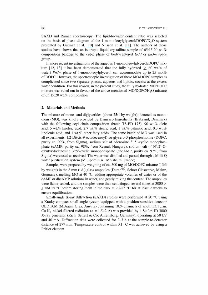
Small-angle X-ray diffraction (SAXD) data of the samples studied are presented inFigure 2.
As distinct from the 1-monooleoylglycerol/DOPC/D2O system of 65:15:20 wt.%composition [10, 11], the diffraction patterns obtained for the reference (MO/DOPC/H2O of 65:15:20 wt.% composition) and cAMP-containing samples are character-istic of a two-dimensional hexagonal lattice with Bragg reflections having a spacingratio 1 (10):
√3 (11): 2 (20) (the Miller indices are given in the brackets). As shown
in Figure 2, two corresponding plots of dhk vs. (h2 + k2 + hk)−1/2 give straightlines passing through the origin and giving hexagonal lattice constants (a0; cylindercenter-center distance) of 57.4 ± 0.2 (value ± S.E. of the linear fit; r2 = 0.9999)and 55.8 ± 0.5 Å (r2 = 0.9997), respectively. In contrast, six Bragg peaks appear inthe SAXD pattern of the dbcAMP-containing sample with spacings having a ratio√
6 (211):√
8 (220):√
14 (321):√
16 (400):√
20 (420):√
22 (332), consistentwith the body-centered cubic lattice of space group Ia3d. A plot of dhkl vs. (h2 + k2
+ l2)−1/2 (Figure 2) results in the unit cell axis a0 of 110.2 ± 0.2 Å (r2 = 0.9999).As already mentioned above, our SAXD result from the reference MO/DOPC/
H2O system differs from that reported on the 1-monooleoylglycerol/DOPC/D2Omixture of the same composition. In the latter system, Gutman et al. [10] andNilsson et al. [11] have detected formation of the reversed hexagonal phase (HII)only at low heavy water content (< 10 wt.%). At higher D2O content, including
88 Z. TALAIKYTE ET AL.
Figure 2. Plot of dhkl vs. (h2 + k2 + l2)−1/2 of the reflections obtained for theMO/DOPC/H2O/dbcAMP sample of 65:15:17:3 wt.% composition (cubic phase of spacegroup Ia3d), and plots of dhk vs. (h2 + k2 + hk)−1/2 of the reflections obtained for the ref-erence MO/DOPC/H2O sample of 65:15:20 wt.% composition and MO/DOPC/ H2O/cAMPsample of 65:15:17:3 wt.% composition (reversed hexagonal phases, HII).
the sample of 65:15:20 wt.% composition, a large area of the bicontinuous cubicphase has been determined.
By analogy to the 1-monooleoylglycerol/DOPC/D2O system, a type II structure(reversed) is assigned to the hexagonal phase of the reference MO/DOPC/H2Osample (Figure 2). We believe that the cubic/HII boundary of our sample is shiftedtowards higher water content because of admixed diglycerides and polyunsaturatedmonoglycerides in the MO preparation (recall that monooleoylglycerol comprises
INTERACTION OF CAMP AND DBCAMP WITH LIPID LAYER 89
only ca. 86 wt.% of the preparation), which promote the formation of reversedmesophases [14–16].
However, the most interesting experimental fact is that entrapment of 3 wt.%dbcAMP into the reference lipidic system induces the HII → Ia3d phase transition,whereas the same quantity of cAMP leaves the mesophase structure and its para-meters practically unchanged. As mentioned in the introduction, these nucleotidesaffect the cubic Pn3m phase of aqueous MO in an analogous fashion [2].
The lipid aggregates and their transformations are often analyzed by using adimensionless lipid shape parameter v/al, where v is the hydrocarbon chain volume,a is the head-group area at the hydrocarbon/water interface, and l is the maximumeffective length of the acyl chain [17]. By applying this concept to the HII → Ia3dphase transition in our system, it appears that dbcAMP decreases v/al since, at thesame hydration, the molecular shape parameter for the HII phase invariably exceedsthe value for the reversed bicontinuous cubic phase [18]. One should anticipate thatthis effect is determined by the molecular interactions between dbcAMP and thecomponents of MO/DOPC layer. Since Raman spectroscopy provides valuable in-formation on the molecular interactions and dynamics, this technique was selectedfor further investigation of the intermolecular features in our systems.
3.2. RAMAN SPECTROSCOPY OF THE AQUEOUS MO/DOPC
LIQUID-CRYSTALLINE PHASES WITH ENTRAPPED CYCLIC NUCLEOTIDES
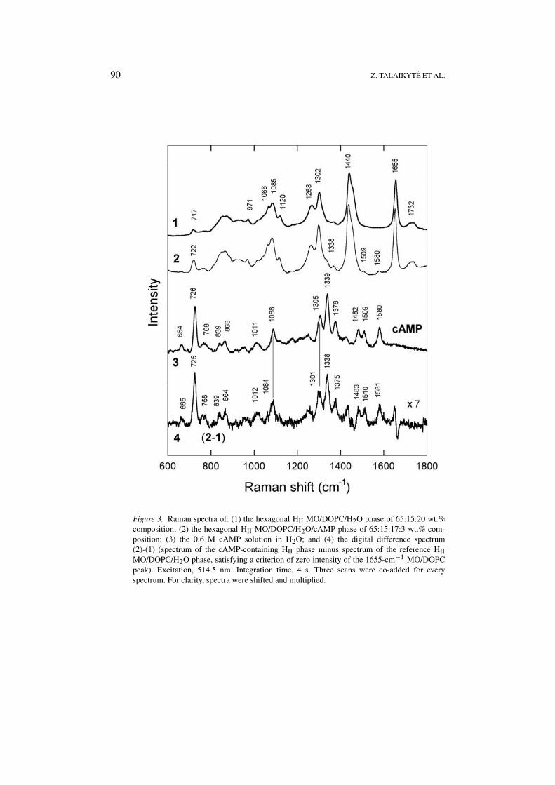
Raman spectra of the above-mentioned pseudoternary and pseudoquarternary liquid-crystalline phases over the wavenumber range of 600–1800 cm−1 are presented inFigures 3 and 4.
Since the MO/DOPC-based phases contain low amounts of cAMP or dbcAMP,only a few bands associated with these cyclic nucleotides can be observed dir-ectly in the Raman spectra. These are: (i) the bands at 1509 and 1580 cm−1,and a shoulder at around 1338 cm−1 in the spectrum of the cAMP-containing HII
phase (Figure 3, spectrum 2), and (ii) the features at 1168, 1355 and 1592 cm−1
in the spectrum of the dbcAMP-containing cubic phase (Figure 4, spectrum 2).Other bands of nucleotides are overlapped by strong features of MO and/or DOPC.However, using the digital difference spectra shown by traces 4 in Figures 3 and4 (the spectrum of the nucleotide-containing sample minus the spectrum of thereference liquid-crystalline phase, satisfying a criterion of zero intensity of theC=C stretching vibration band at 1655 cm−1), we were able to retrieve a numberof bands that are attributable to the entrapped cyclic nucleotides. As it has beenshown previously [2], the comparison of these difference spectra with the spectra ofnucleotides in water (traces 3 in Figures 3 and 4) is particularly useful for detectingthe interactions of nucleotides with model lipid membrane.
Traces 3 and 4 in Figure 3 show that the entrapment of cAMP in the HII phase ofaqueous MO/DOPC mixture leaves the frequencies of the most cAMP vibrationalbands unchanged, with two exceptions. Thus, the bands corresponding to the sym-
90 Z. TALAIKYTE ET AL.
Figure 3. Raman spectra of: (1) the hexagonal HII MO/DOPC/H2O phase of 65:15:20 wt.%composition; (2) the hexagonal HII MO/DOPC/H2O/cAMP phase of 65:15:17:3 wt.% com-position; (3) the 0.6 M cAMP solution in H2O; and (4) the digital difference spectrum(2)-(1) (spectrum of the cAMP-containing HII phase minus spectrum of the reference HIIMO/DOPC/H2O phase, satisfying a criterion of zero intensity of the 1655-cm−1 MO/DOPCpeak). Excitation, 514.5 nm. Integration time, 4 s. Three scans were co-added for everyspectrum. For clarity, spectra were shifted and multiplied.

INTERACTION OF CAMP AND DBCAMP WITH LIPID LAYER 91
Figure 4. Raman spectra of: (1) the reversed hexagonal HII MO/DOPC/H2O phase of65:15:20 wt.% composition; (2) the cubic Ia3d MO/DOPC/H2O/dbcAMP phase of 65:15:17:3wt.% composition; (3) the 0.3 M dbcAMP solution in H2O; and (4) the digital differencespectrum (2)-(1) (spectrum of the dbcAMP-containing cubic phase minus spectrum of thereference HII MO/DOPC/H2O phase, satisfying a criterion of zero intensity of the 1655 cm−1
MO/DOPC peak). Excitation, 514.5 nm. Integration time, 4 s. Three scans were co-added forspectra (1) and (2). Four scans were co-added for spectrum (3). For clarity, spectra were shiftedand multiplied.
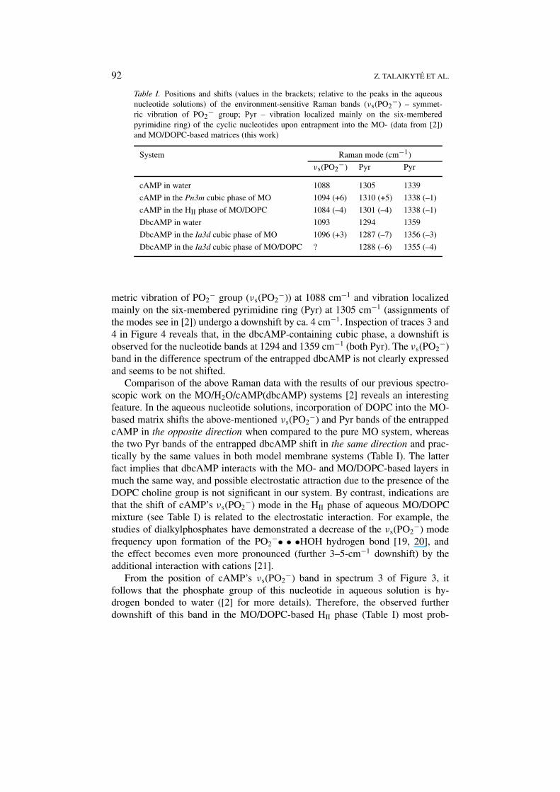
92 Z. TALAIKYTE ET AL.
Table I. Positions and shifts (values in the brackets; relative to the peaks in the aqueousnucleotide solutions) of the environment-sensitive Raman bands (νs(PO2
−) – symmet-ric vibration of PO2
− group; Pyr – vibration localized mainly on the six-memberedpyrimidine ring) of the cyclic nucleotides upon entrapment into the MO- (data from [2])and MO/DOPC-based matrices (this work)
System Raman mode (cm−1)
νs(PO2−) Pyr Pyr
cAMP in water 1088 1305 1339
cAMP in the Pn3m cubic phase of MO 1094 (+6) 1310 (+5) 1338 (–1)
cAMP in the HII phase of MO/DOPC 1084 (–4) 1301 (–4) 1338 (–1)
DbcAMP in water 1093 1294 1359
DbcAMP in the Ia3d cubic phase of MO 1096 (+3) 1287 (–7) 1356 (–3)
DbcAMP in the Ia3d cubic phase of MO/DOPC ? 1288 (–6) 1355 (–4)
metric vibration of PO2− group (νs(PO2
−)) at 1088 cm−1 and vibration localizedmainly on the six-membered pyrimidine ring (Pyr) at 1305 cm−1 (assignments ofthe modes see in [2]) undergo a downshift by ca. 4 cm−1. Inspection of traces 3 and4 in Figure 4 reveals that, in the dbcAMP-containing cubic phase, a downshift isobserved for the nucleotide bands at 1294 and 1359 cm−1 (both Pyr). The νs(PO2
−)band in the difference spectrum of the entrapped dbcAMP is not clearly expressedand seems to be not shifted.
Comparison of the above Raman data with the results of our previous spectro-scopic work on the MO/H2O/cAMP(dbcAMP) systems [2] reveals an interestingfeature. In the aqueous nucleotide solutions, incorporation of DOPC into the MO-based matrix shifts the above-mentioned νs(PO2
−) and Pyr bands of the entrappedcAMP in the opposite direction when compared to the pure MO system, whereasthe two Pyr bands of the entrapped dbcAMP shift in the same direction and prac-tically by the same values in both model membrane systems (Table I). The latterfact implies that dbcAMP interacts with the MO- and MO/DOPC-based layers inmuch the same way, and possible electrostatic attraction due to the presence of theDOPC choline group is not significant in our system. By contrast, indications arethat the shift of cAMP’s νs(PO2
−) mode in the HII phase of aqueous MO/DOPCmixture (see Table I) is related to the electrostatic interaction. For example, thestudies of dialkylphosphates have demonstrated a decrease of the νs(PO2
−) modefrequency upon formation of the PO2
−• • •HOH hydrogen bond [19, 20], andthe effect becomes even more pronounced (further 3–5-cm−1 downshift) by theadditional interaction with cations [21].
From the position of cAMP’s νs(PO2−) band in spectrum 3 of Figure 3, it
follows that the phosphate group of this nucleotide in aqueous solution is hy-drogen bonded to water ([2] for more details). Therefore, the observed furtherdownshift of this band in the MO/DOPC-based HII phase (Table I) most prob-

INTERACTION OF CAMP AND DBCAMP WITH LIPID LAYER 93
ably points to the interaction between the strongly basic quaternary ammoniumgroup of DOPC choline and the phosphate group of cAMP. We cannot rule outthe formation of a complex, in which two charged groups are separated by water –(CH3)3N+• • •O(H)-H• • •−O2P.
Some so-called ‘marker’ bands, which are sensitive to the intermolecular in-teractions at a particular site of the adenine ring, are especially useful for theinterpretation of the frequency shifts observed in the difference spectra of cyclicnucleotides. Intensive cAMP bands at 1305 and 1339 cm−1 in aqueous solution(Figure 3, spectrum 3), arising due to the vibrations localized mainly on the Pyrring (corresponding dbcAMP modes are at 1294 and 1359 cm−1; Figure 4, spec-trum 3), have been shown to be sensitive to the formation/disruption of hydrogenbonds at the N1- and N3-sites (see Figure 1) of adenine, respectively [22]. Follow-ing the interpretations [22], the downshift (∼3 cm−1; Table I) of the 1305-cm−1
cAMP band when going from aqueous solution to the MO/DOPC-based matrix isattributable to the weakening of hydrogen bonding at the N1-site of Pyr, whereasthe entrapped dbcAMP presumably undergoes disruption and/or weakening of hy-drogen bonds at both sites of protonation. The cAMP mode at 1509 cm−1 (Figure 3,spectrum 3), associated with the vibration localized mainly on the five-memberedimidazole (Im) ring (corresponding mode of dbcAMP is at 1493 cm−1; Figure 4,spectrum 3), is the marker band of hydrogen bonding and complexation at the ad-enine N7-site [23, 24]. The intensity of this band, relative to the peak at 1482 cm−1
(Pyr + Im vibrational mode), has been found to decrease with increasing strength ofthe hydrogen bonding interaction at the C6-NH2 site [25]. Both bands retain theirfrequencies and intensity ratio when cAMP is incorporated into the MO/DOPC-based lipid matrix (Figure 3, spectra 3 and 4), indicating that environment ofthe C6-NH2 group of adenine in the lipid matrix is the same as in the aqueoussolution. However these two marker bands are not well resolved in the case ofdbcAMP and were assigned to one broad peak at 1493 cm−1 in the spectrum ofthe aqueous solution of cyclic nucleotide [2]. In the difference spectrum of theentrapped dbcAMP (Figure 4, spectrum 4) this band is considerably weakened.Further systematic Raman studies of dbcAMP dissolved in different solvents arerequired to establish the reliable marker bands sensitive to H-bonding interactionsat the C6-NH- site.
Another interesting feature of the difference spectrum of the entrapped dbcAMP(Figure 4, spectrum 4) is a clearly resolved band at 716 cm−1, which is absent in thespectrum of the aqueous solution (Figure 4, spectrum 3). In the difference spectrumof the entrapped cAMP (Figure 3, spectrum 4), a prominent in-plane vibrationalmode of Pyr + Im is observed at 725 cm−1 that is asymmetric and of somewhathigher intensity relative to the spectrum in the aqueous solution of cAMP (Fig-ure 3, spectrum 3). Deconvolution of this feature in the difference spectrum alsoreveals the presence of weak band at 716 cm−1 (data not shown). The latter band isrepresentative of C-N symmetric stretching mode of the choline segment -O-CH2-CH2-N+(CH3)3 in a gauche conformation [26]. Therefore, there are grounds to

94 Z. TALAIKYTE ET AL.
believe that cyclic nucleotides induce the conformational rearrangement of DOPChead-group by increasing the population of gauche isomers.
The influence of the studied nucleotides on the apolar hydrophobic region ofthe MO/DOPC-based layer might be analyzed by their effects on the parametersof the definite lipid bands. To this end, the difference spectra were obtained bythe subtraction of the nucleotide aqueous solution spectrum from the spectrumof the corresponding pseudoquarternary liquid-crystalline phase. The subtractionwas performed to satisfy a criterion of zero intensity of the 1580 and 1590 cm−1
bands for cAMP and dbcAMP, respectively. We analyzed the obtained differencespectra in the region of 1200–1350 cm−1, containing in-plane =C-H deformation,δ(=CH), methylene twisting, δt (CH2), and methylene deformation, δ(CH2), bands.It has been demonstrated previously that the δt (CH2) mode becomes broader withan increase in the conformational disorder in the hydrocarbon chains of lipids[27, 28], while the δ(CH2) mode is sensitive to the interchain interactions [28].However, no reliable differences between the reference and nucleotide containingsamples in the regions of these two bands were observed. The same is true for theC-H stretching vibration region (2800–3000 cm−1), which is more sensitive to theconformational rearrangements and lateral interaction of the hydrocarbon chainsof lipids. Therefore, all these difference spectra are not shown and discussed ingreater detail.
4. Conclusions
Interactions of two cyclic nucleotides, adenosine 3’:5’-cyclic monophosphate(cAMP) and N6,2’-O-dibutyryladenosine 3’:5’-cyclic monophosphate (dbcAMP),with a lipid layer composed of monoolein (MO) and dioleoyl phosphatidylcholine(DOPC) were studied by small-angle X-ray diffraction (SAXD) and Raman spec-troscopy. The SAXD data revealed that the reversed hexagonal (HII) MO/DOPC/H2O phase of 65:15:20 wt.% composition, which was selected as a reference sys-tem, retained its crystallographic structure and did not change the lattice parametersby the incorporation (at the expense of water) of about 3 wt.% of cAMP, whereasthe same amount of dbcAMP induced the HII → cubic Ia3d phase transition. Basedon the concept of a dimensionless lipid shape parameter (v/al), the latter transitionshould be accompanied by the decrease of v/al. It is conceivable that the partition-ing of the dbcAMP’s acylated adenine group into the MO/DOPC layer could leadto this effect by, for example, increasing the interfacial area of the lipid head-group(a). Indeed, this conclusion was confirmed by the Raman spectroscopic data for theMO/DOPC/H2O/dbcAMP system, which indicated that the hydrogen bonding atthe N1- and N3-sites of the dbcAMP adenine ring was disrupted/weakened. How-ever, when compared to the MO/DOPC/H2O system, the spectroscopic parametersof the lipid hydrocarbon chains in the MO/DOPC/H2O/dbcAMP sample remainedunaltered. This suggests that the penetration of dbcAMP into the lipid layer is ap-parently not deep. According to the present SAXD and Raman spectroscopic data,
INTERACTION OF CAMP AND DBCAMP WITH LIPID LAYER 95
it seems that cAMP remains in the water channels of the HII MO/DOPC/H2O/cAMPphase, although it interacts with the quaternary ammonium group of the DOPCcholine residue through the phosphate moiety. Moreover, as evident from our Ra-man spectroscopy experiments, both cyclic nucleotides exhibit the capacity to in-crease the population of gauche isomers in the DOPC choline group.
References
1. Schwede, F., Maronde, E., Genieser, H.-G. and Jastorff, B.: Cyclic Nucleotide Analogs asBiochemical Tools and Prospective Drugs, Pharmacol. Therapeut. 87 (2000), 199–226.
2. Razumas, V., Niaura, G., Talaikyte Z., Vagonis, A. and Nylander T.: Interactions of CyclicAMP and its Dibutyryl Analogue with Model Membrane: X-ray Diffraction and Raman Spec-troscopic Study using Liquid-Crystalline Phases of Monoolein, Biophys. Chem. 90 (2001),75–87.
3. Qiu, H. and Caffrey, M.: The Phase Diagram of the Monoolein/Water System: Metastabilityand Equilibrium Aspects, Biomaterials 21 (2000), 223–234.
4. Lindblom, G., Wieslander, A., Sjoelund, M. and Wikander, G.: Phase Equilibria of MembraneLipids for Acheoplasma laidlawii: Importance of a Single Lipid Forming Nonlamellar Phases,Biochemistry 25 (1986), 7502–7510.
5. Landh, T.: From Entagled Membranes to Eclectic Morphologies: Cubic Membranes asSubcellular Space Organizers, FEBS Lett. 369 (1995), 13–17.
6. Deng Y. and Landh T.: The Cubic Gyroid-Based Membrane Structure of the Chloroplast inZygnema (Chlorophyceae Zygnematles), Zool. Stud. 37, Suppl. I, (1995), 175–177.
7. Morein, S., Andersson, A-S., Rilfors, L. and Lindblom, G.: Wild-type Escherichia coli Cellsregulate the Membrane Lipid Composition in a ‘Window’ between Gel and Non-LamellarStructures, J. Biol. Chem. 271 (1996), 6801–6809.
8. Deng, Y. and Mieczkowski, M.: Three-Dimensional Periodic Cubic Membrane Structure in theMitochondria of Amoebae Chaos Carolinensis, Protoplasma 203 (1998), 16–25.
9. Li, Y.-M., Wei, M. and Zhao, Y.-F.: Investigations on the Effects of cAMP on Membrane Phos-pholipid of Human Erythrocytes by laser Raman Spectroscopy, Spectroscopy Lett. 32 (1999),197–203.
10. Gutman, H., Arvidson, G., Fontell, K. and Lindblom, G.: 31P and 2H NMR Studies of PhaseEquilibria in the Three Component System: Monoolein Dioleylphosphatidylcholine, In: K.Mittal and L.B. Lindman (eds.), Surfactants in Solutions Vol. 1, Plenum Press, New York,1984, pp. 143–152.
11. Nilsson, A., Holmgren, A. and Lindblom, G.: Fourier-Transform Infrared SpectroscopicStudy of Dioleoylphosphatidylcholine and Monooleoylglycerol in Lamellar and Cubic LiquidCrystals, Biochemistry 30 (1991), 2126–2133.
12. Templer, R.H., Madan, K.H., Warrender, N.A. and Seddon, J.M.: Swollen Lyotropic Cu-bic Phases in Fully Hydrated Mixtures of Monoolein, Dioleoylphosphatidylcholine, andDioleoylphosphatidylethanolamine, Springer Proc. Phys. 66 (1992), 262–265.
13. Cherezow, V., Clogston, J., Misquitta, Y., Abdel-Gawad, W. and Caffrey, M.: Membrane ProteinCrystallization in Meso: Lipid Type-Tailoring of the Cubic Phase, Biophys. J. 83 (2002), 3393–3407.
14. Seddon, J.M.: Structure of the Inverted Hexagonal (HII) Phase and Non-Lamellar PhaseTransition of Lipids, Biochim. Biophys. Acta 1031 (1990), 1–69.
15. Caboi F., Amico G.S., Pitzalis P., Monduzzi, M., Nylander T. and Larsson K.: Addition of Hy-drophilic and Lipophilic Compounds of Biological Relevance to the Monoolein/Water System.I. Phase Behavior, Chem. Phys. Lipids 109 (2001), 47–62.

96 Z. TALAIKYTE ET AL.
16. Borné, J., Nylander, T. and Khan A.: Phase Behavior and Aggregate Formation for the AqueousMonoolein System Mixed with Sodium Oleate and Oleic Acid, Langmuir 17 (2001), 7742–7751.
17. Israelachvili, J.N., Mitchel, D.J. and Ninham, B.W.: Theory of Self-Assembling of Hydro-carbon Amphiphiles into Micelles and Bilayers, J. Chem. Soc., Faraday Trans. 272 (1976),1525–1568.
18. Hyde, S.T., Andersson, S., Larsson, K., Blum, Z., Landh, T., Lidin, S. and Ninham, B.W.:The Language of Shape. The Role of Curvature in Condensed Matter: Physics, Chemistry andBiology, Elsevier, Amsterdam, 1997.
19. Pohle, W., Bohl, M. and Böhlig, H.: Interpretation of the Influence of Hydrogen Bonding onthe Stretching Vibrations of the PO2
− Moiety, J. Mol. Struct. 242 (1990), 333–342.20. Guan, Y. and Thomas, G.J.: Vibrational Analysis of Nucleic Acids. III. Conformation-
Dependent Raman Markers of Phosphodiester Backbone Modeled by Dimethyl Phosphate, J.Mol. Struct. 379 (1996), 31–41.
21. Stangret, J. and Savoie, R.: Vibrational Spectroscopic Study of Metal Ions with DiethylPhosphate, a Model for Biological Systems, Can. J. Chem. 70 (1992), 2875–2883.
22. Fujimoto, N., Toyama, A. and Takeuchi, H.: Binding Modes of Cyclic AMP and Environmentsof Tryptophan Residues in 1:1 and 1:2 Complexes of Cyclic AMP Receptor Protein and CyclicAMP, Biopolymers (Biospectroscopy) 67 (2002), 186–196.
23. Toyama, A., Takeuchi, H. and Harada, I.: Ultraviolet Resonance Raman Spectra of Adenine,Uracil and Thymine Derivatives in Several Solvents. Correlation between Band Frequenciesand Hydrogen-Bonding States of the Nucleic Acid Bases, J. Mol. Struct. 242 (1991), 87–98.
24. Štepánek, J., Kowalewski, J. and Lang J.: Spectroscopic Investigation of Nickel Cation Bindingwith Adenine Mononucleotides: Stability and Structure of the 1:2 Complex with Adenosine5’-Monophosphate, J. Biol. Inorg. Chem. 3 (1998), 543–556.
25. Fujimoto, N., Toyama, A. and Takeuchi, H.: Effects of Hydrogen Bonding on the UV Res-onance Raman Bands of the Adenine Ring and its C8-Deuterated Analog, J. Mol. Struct. 447(1998), 61–69.
26. Harrand, M.: Study of the Configuration of Phospholipids (as Models for Biological Mem-branes) by Raman Spectroscopy: Intensity and Polarization, In: J.P. Alix, L. Bernard and. M.Manfait (eds.), Spectroscopy of Biological Molecules, John Wiley & Sons, Chichester, 1985,pp. 265–270.
27. Larsson, K. and Rand, R.P.: Detection of Changes in the Environment of Hydrocarbon Chainsby Raman Spectroscopy and its Application to Lipid-Protein Systems, Biochem. Biophys. Acta326 (1973), 245–255.
28. Razumas, V., Talaikyte, Z., Barauskas, J., Larsson, K., Miezis, Y. and Nylander, T.: Effects ofDistearoylphosphatidylglycerol and Lysozyme on the Structure of the Monoolein-Water CubicPhase: X-ray Diffraction and Raman Scattering Studies, Chem. Phys. Lipids 84 (1996), 123–138.
Copyright © 2022 FDOKUMEN