Topology of the Shaker Potassium Channel Probed with Hydrophilic Epitope Insertions
9
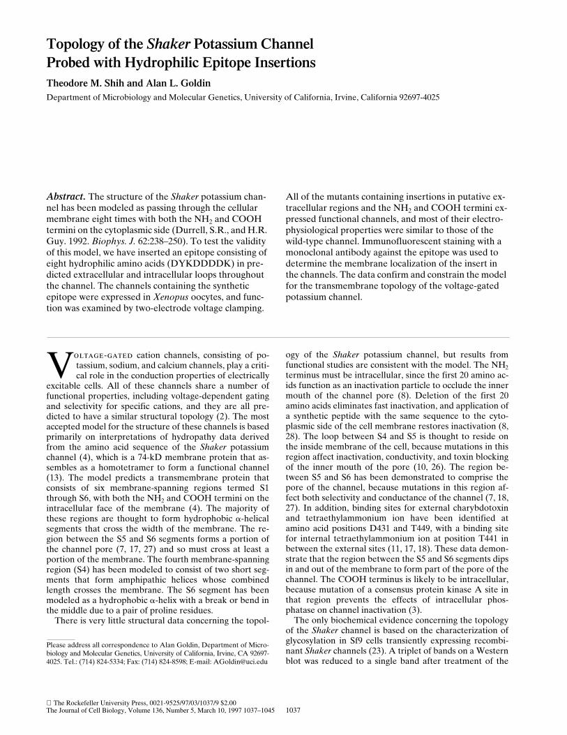
The Rockefeller University Press, 0021-9525/97/03/1037/9 $2.00 The Journal of Cell Biology, Volume 136, Number 5, March 10, 1997 1037–1045 1037 Topology of the Shaker Potassium Channel Probed with Hydrophilic Epitope Insertions Theodore M. Shih and Alan L. Goldin Department of Microbiology and Molecular Genetics, University of California, Irvine, California 92697-4025 Abstract. The structure of the Shaker potassium chan- nel has been modeled as passing through the cellular membrane eight times with both the NH 2 and COOH termini on the cytoplasmic side (Durrell, S.R., and H.R. Guy. 1992. Biophys. J. 62:238–250). To test the validity of this model, we have inserted an epitope consisting of eight hydrophilic amino acids (DYKDDDDK) in pre- dicted extracellular and intracellular loops throughout the channel. The channels containing the synthetic epitope were expressed in Xenopus oocytes, and func- tion was examined by two-electrode voltage clamping. All of the mutants containing insertions in putative ex- tracellular regions and the NH 2 and COOH termini ex- pressed functional channels, and most of their electro- physiological properties were similar to those of the wild-type channel. Immunofluorescent staining with a monoclonal antibody against the epitope was used to determine the membrane localization of the insert in the channels. The data confirm and constrain the model for the transmembrane topology of the voltage-gated potassium channel. V oltage-gated cation channels, consisting of po- tassium, sodium, and calcium channels, play a criti- cal role in the conduction properties of electrically excitable cells. All of these channels share a number of functional properties, including voltage-dependent gating and selectivity for specific cations, and they are all pre- dicted to have a similar structural topology (2). The most accepted model for the structure of these channels is based primarily on interpretations of hydropathy data derived from the amino acid sequence of the Shaker potassium channel (4), which is a 74-kD membrane protein that as- sembles as a homotetramer to form a functional channel (13). The model predicts a transmembrane protein that consists of six membrane-spanning regions termed S1 through S6, with both the NH 2 and COOH termini on the intracellular face of the membrane (4). The majority of these regions are thought to form hydrophobic a-helical segments that cross the width of the membrane. The re- gion between the S5 and S6 segments forms a portion of the channel pore (7, 17, 27) and so must cross at least a portion of the membrane. The fourth membrane-spanning region (S4) has been modeled to consist of two short seg- ments that form amphipathic helices whose combined length crosses the membrane. The S6 segment has been modeled as a hydrophobic a-helix with a break or bend in the middle due to a pair of proline residues. There is very little structural data concerning the topol- ogy of the Shaker potassium channel, but results from functional studies are consistent with the model. The NH 2 terminus must be intracellular, since the first 20 amino ac- ids function as an inactivation particle to occlude the inner mouth of the channel pore (8). Deletion of the first 20 amino acids eliminates fast inactivation, and application of a synthetic peptide with the same sequence to the cyto- plasmic side of the cell membrane restores inactivation (8, 28). The loop between S4 and S5 is thought to reside on the inside membrane of the cell, because mutations in this region affect inactivation, conductivity, and toxin blocking of the inner mouth of the pore (10, 26). The region be- tween S5 and S6 has been demonstrated to comprise the pore of the channel, because mutations in this region af- fect both selectivity and conductance of the channel (7, 18, 27). In addition, binding sites for external charybdotoxin and tetraethylammonium ion have been identified at amino acid positions D431 and T449, with a binding site for internal tetraethylammonium ion at position T441 in between the external sites (11, 17, 18). These data demon- strate that the region between the S5 and S6 segments dips in and out of the membrane to form part of the pore of the channel. The COOH terminus is likely to be intracellular, because mutation of a consensus protein kinase A site in that region prevents the effects of intracellular phos- phatase on channel inactivation (3). The only biochemical evidence concerning the topology of the Shaker channel is based on the characterization of glycosylation in Sf9 cells transiently expressing recombi- nant Shaker channels (23). A triplet of bands on a Western blot was reduced to a single band after treatment of the Please address all correspondence to Alan Goldin, Department of Micro- biology and Molecular Genetics, University of California, Irvine, CA 92697- 4025. Tel.: (714) 824-5334; Fax: (714) 824-8598; E-mail: [email protected]
-
Upload
independent -
Category
Documents
-
view
3 -
download
0
Transcript of Topology of the Shaker Potassium Channel Probed with Hydrophilic Epitope Insertions
The Rockefeller University Press, 0021-9525/97/03/1037/9 $2.00The Journal of Cell Biology, Volume 136, Number 5, March 10, 1997 1037–1045 1037
Topology of the
Shaker
Potassium ChannelProbed with Hydrophilic Epitope Insertions
Theodore M. Shih and Alan L. Goldin
Department of Microbiology and Molecular Genetics, University of California, Irvine, California 92697-4025
Abstract.
The structure of the
Shaker
potassium chan-nel has been modeled as passing through the cellular membrane eight times with both the NH
2
and COOH termini on the cytoplasmic side (Durrell, S.R., and H.R. Guy. 1992.
Biophys. J.
62:238–250). To test the validity of this model, we have inserted an epitope consisting of eight hydrophilic amino acids (DYKDDDDK) in pre-dicted extracellular and intracellular loops throughout the channel. The channels containing the synthetic epitope were expressed in
Xenopus
oocytes, and func-tion was examined by two-electrode voltage clamping.
All of the mutants containing insertions in putative ex-tracellular regions and the NH
2
and COOH termini ex-pressed functional channels, and most of their electro-physiological properties were similar to those of the wild-type channel. Immunofluorescent staining with a monoclonal antibody against the epitope was used to determine the membrane localization of the insert in the channels. The data confirm and constrain the model for the transmembrane topology of the voltage-gated potassium channel.
V
oltage-gated
cation channels, consisting of po-tassium, sodium, and calcium channels, play a criti-cal role in the conduction properties of electrically
excitable cells. All of these channels share a number offunctional properties, including voltage-dependent gatingand selectivity for specific cations, and they are all pre-dicted to have a similar structural topology (2). The mostaccepted model for the structure of these channels is basedprimarily on interpretations of hydropathy data derivedfrom the amino acid sequence of the
Shaker
potassiumchannel (4), which is a 74-kD membrane protein that as-sembles as a homotetramer to form a functional channel(13). The model predicts a transmembrane protein thatconsists of six membrane-spanning regions termed S1through S6, with both the NH
2
and COOH termini on theintracellular face of the membrane (4). The majority ofthese regions are thought to form hydrophobic
a
-helicalsegments that cross the width of the membrane. The re-gion between the S5 and S6 segments forms a portion ofthe channel pore (7, 17, 27) and so must cross at least aportion of the membrane. The fourth membrane-spanningregion (S4) has been modeled to consist of two short seg-ments that form amphipathic helices whose combinedlength crosses the membrane. The S6 segment has beenmodeled as a hydrophobic
a
-helix with a break or bend inthe middle due to a pair of proline residues.
There is very little structural data concerning the topol-
ogy of the
Shaker
potassium channel, but results fromfunctional studies are consistent with the model. The NH
2
terminus must be intracellular, since the first 20 amino ac-ids function as an inactivation particle to occlude the innermouth of the channel pore (8). Deletion of the first 20amino acids eliminates fast inactivation, and application ofa synthetic peptide with the same sequence to the cyto-plasmic side of the cell membrane restores inactivation (8,28). The loop between S4 and S5 is thought to reside onthe inside membrane of the cell, because mutations in thisregion affect inactivation, conductivity, and toxin blockingof the inner mouth of the pore (10, 26). The region be-tween S5 and S6 has been demonstrated to comprise thepore of the channel, because mutations in this region af-fect both selectivity and conductance of the channel (7, 18,27). In addition, binding sites for external charybdotoxinand tetraethylammonium ion have been identified atamino acid positions D431 and T449, with a binding sitefor internal tetraethylammonium ion at position T441 inbetween the external sites (11, 17, 18). These data demon-strate that the region between the S5 and S6 segments dipsin and out of the membrane to form part of the pore of thechannel. The COOH terminus is likely to be intracellular,because mutation of a consensus protein kinase A site inthat region prevents the effects of intracellular phos-phatase on channel inactivation (3).
The only biochemical evidence concerning the topologyof the
Shaker
channel is based on the characterization ofglycosylation in Sf9 cells transiently expressing recombi-nant
Shaker
channels (23). A triplet of bands on a Westernblot was reduced to a single band after treatment of the
Please address all correspondence to Alan Goldin, Department of Micro-biology and Molecular Genetics, University of California, Irvine, CA 92697-4025. Tel.: (714) 824-5334; Fax: (714) 824-8598; E-mail: [email protected]
The Journal of Cell Biology, Volume 136, 1997 1038
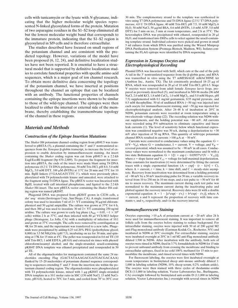
cells with tunicamycin or the lysate with
N
-glycanase, indi-cating that the higher molecular weight species repre-sented N-linked glycosylated forms of the protein. Mutationof two asparagine residues in the S1–S2 loop eliminated allbut the lowest molecular weight band that corresponds tothe immature protein, indicating that the S1–S2 region isglycosylated in Sf9 cells and is therefore extracellular.
The studies described have focused on small regions ofthe potassium channel and are consistent with the pre-dicted topology. However, variations of the model havebeen proposed (6, 12, 24), and definitive localization stud-ies have not been reported. It is essential to have a struc-tural model that is supported by definitive localization stud-ies to correlate functional properties with specific amino acidsequences, which is a major goal of ion channel research.To obtain more definitive data concerning the topologyof the potassium channel, we have inserted at positionsthroughout the channel an epitope that can be localizedwith an antibody. The functional properties of channelscontaining the epitope insertions were shown to be similarto those of the wild-type channel. The epitopes were thenlocalized to either the internal or external side of the mem-brane, thereby establishing the transmembrane topologyof the channel in these regions.
Materials and Methods
Construction of the Epitope Insertion Mutations
The
Shaker
H4 potassium channel–coding region from pH4U5 was trans-ferred to pBSTA (5), a plasmid containing the 5
9
and 3
9
nontranslated se-quences from the
Xenopus
b
-globin transcript, to increase the level of ex-pression to enable detection by immunofluorescent staining. An insertcontaining the
Shaker
coding region was cut from pH4U5 in a 2.2-kbEagI/EcoRI fragment (bp 670–2,889). To prepare the fragment for inser-tion into pBSTA, the ends of the insert were made blunt using T4
DNApolymerase (0.2 U T4 DNA polymerase, 50 mM Tris-HCl, pH 8.0, 5.0 mMMgCl
2
, 5.0 mM DTT, 100
m
M dNTPs) at 11
8
C for 20 min and 75
8
C for 10min. BglII linkers (5
9
GAAGATCTTC 3
9
), which were previously phos-phorylated with T4 polynucleotide kinase and annealed, were attached tothe fragment using T4 DNA ligase. The pBSTA vector was linearized withBglII, treated with calf intestinal phosphatase, and ligated to the 2.2-kb
Shaker
H4 insert. The new pBSTA vector containing the
Shaker
H4 cod-ing region was named pKH4T.
Phagemid DNA was prepared from pKH4T grown in CJ236 cells, a
dut
2
ung
2
strain (Invitrogen Corp., San Diego, CA). A single, transformedcolony was used to inoculate 5 ml of 2
3
YT containing 30
m
g/ml chloram-phenicol and 50
m
g/ml ampicillin. The culture was grown at 37
8
C for 6 h,and then 300
m
l was used to inoculate 30 ml 2
3
YT containing 250 ng/mluridine. The culture was grown to early log phase (A
600
5
0.05,
z
1
3
10
9
cells) within 2 h at 37
8
C, and then infected with 80
m
l VCS-M13 helperphage (Stratagene, La Jolla, CA) with a multiplicity of infection of 10:1and grown at 37
8
C overnight. The cells were removed by centrifugation at4K for 20 min, and the supernatant was transferred to a new tube. Phage-mids were precipitated by adding 0.25 vol 20% PEG (polyethylene glycol;8,000) in 3.5 M NH
4
OAc (pH 7.5), incubating on ice for 30 min, and spin-ning at 17K for 15 min at 4
8
C. The pellet was resuspended in 0.6 ml TE (10mM Tris-HCl, 1 mM EDTA, pH 8.0) and extracted six times with phenol/chloroform/isoamyl alcohol; and the single-stranded, uracil-containingpKH4T DNA template was ethanol precipitated and resuspended in 50
m
l TE.All of the oligonucleotides for mutagenesis contained the same 24 nu-
cleotides encoding Flag (GACTATAAAGACGATGACGACAAA)flanked by 15–20 nucleotides of potassium channel sequence correspond-ing to sequences extending 5
9
and 3
9
from the insertion site within the po-tassium channel coding region. The oligonucleotides were phosphorylatedwith T4 polynucleotide kinase, mixed with 1
m
g pKH4T single-strandedDNA template in a 10:1 molar ratio in SSC (150 mM NaCl, 15 mM NaCi-trate, pH 6.8), heated to 70
8
C for 5 min, and cooled from 70
8
to 30
8
C over
30 min. The complementary strand to the template was synthesized invitro using T7 DNA polymerase and T4 DNA ligase (2.5 U T7 DNA poly-merase, 4.0 U T4 DNA ligase, 40 mM Tris-HCl, pH 7.5, 10 mM MgCl
2
?
6H
2
O, 50 mM NaCl, 50
m
g/ml BSA, 0.6 mM dNTPs, 1.0 mM ATP, 5.0 mMDTT) for 5 min on ice, 5 min at room temperature, and 2 h at 37
8
C. Theheteroduplex DNA was precipitated with ethanol, resuspended in 20
m
lH
2
O, and transformed into DH5
a
cells to select against the uracil-contain-ing template strand. Individual colonies were picked and grown in small5 ml cultures from which DNA was purified using the Wizard MiniprepDNA Purification System (Promega Biotech, Madison, WI). Isolates con-taining the FLAG epitope were identified by DNA sequencing.
Expression in Xenopus Oocytes and Electrophysiological Recording
Plasmid DNA was linearized with SstII, which cuts at the end of the polyA tail in the 3
9
nontranslated sequence from the
b
-globin gene, and RNAwas transcribed in vitro using the T7 mMESSAGE mMACHINE kit(Ambion Inc., Austin, TX). The kit consistently produced 18–25
m
g ofRNA, which was resuspended in 20
m
l of 10 mM Tris-HCl, pH 6.5. StageV oocytes were removed from adult female
Xenopus laevis
frogs, pre-pared as previously described (5), and incubated in ND-96 media (96 mMNaCl, 2.0 mM KCl, 1.8 mM CaCl
2
, 1.0 mM MgCl
2
, and 5.0 mM Hepes, pH7.5) supplemented with 0.1 mg/ml gentamicin, 0.55 mg/ml pyruvate, and0.5 mM theophylline. 50 nl of undiluted RNA (
z
50 ng) was injected intoeach oocyte for immunofluorescent staining, and
z
50 pg was injected forelectrophysiological analysis. After 24–48 h of incubation at 20
8
C inND96, potassium currents were recorded at room temperature using atwo-electrode voltage clamp (22). The recording solution was ND96 with-out supplements, and the holding potential was
2
80 mV. All currentswere recorded using P/4 subtraction to eliminate capacitive and linearleak currents (1). The level of sensitivity below which functional expres-sion was considered negative was 50 nA, during a depolarization to
1
30mV after injection of 50 ng RNA. This quantity of wild-type potassiumchannel RNA resulted in currents
.
100
m
A at
2
50 mV.Currents were converted to conductance values using the equation: G
5
I/(V
2
V
R
), where G
5
conductance, I
5
current, V
5
voltage, and V
R
5
reversal potential, which was assumed to be
2
90 mV in all cases. Conduc-tance values were normalized to the maximum conductance and fit with atwo state Boltzmann equation: G
5
1/[1
1
exp (
2
0.03937*z*(V
2
V
1/2
))],where z
5
slope factor and V
1/2
5
voltage for half-maximal depolarization.Time constants for inactivation (
t
) were determined by fitting the currenttraces with a single exponential function: (A
5
exp[
2
(
t
2
k)/
t
]
1
C),where A
5
current,
t
5
time, k
5
time shift, and C
5
steady-state asymp-tote. Recovery from inactivation was determined from a holding potentialof
2
80 mV by a 50 mV inactivating pulse for 50 ms, a variable recovery in-terval from 10 to 250 ms in 10 ms steps, and a test pulse to
1
50 mV for 40ms to assess recovery. The maximum current during each test pulse wasnormalized to the maximum current during the inactivating pulse andplotted against the recovery interval. Recovery data were fit with a doubleexponential equation: A
5
1
2
[a*exp(
2
t/
t
1
)
1
b*exp(
2
t
/
t
2
)], where A
5
current, a and b represent the proportion of recovery with time con-stants
t
1
and
t
2
, respectively, and t is the recovery interval.
Immunofluorescent Staining
Oocytes expressing
.
10
m
A of potassium current at
2
20 mV after 24 hwere used for immunofluorescent staining. It was important to remove allfollicle cells from the oocytes before staining to reduce background. Forintracellular staining, oocytes were injected with 50 nl of 200
m
g/ml M2anti-Flag monoclonal antibody (Eastman Kodak Co., Rochester, NY) andincubated in ND96 at 20
8
C overnight. For extracellular staining, oocyteswere incubated overnight at 20
8
C in 1 ml M2 anti-Flag monoclonal antibodydiluted 1:500 in ND96. After incubation with the antibody, both sets ofoocytes were rinsed in ND96, fixed in 3.7% formaldehyde in ND96 for 15 minto prevent unbound antibody from crossing the membrane and binding toextracellular epitopes, fixed in ice-cold 100% methanol for 15 min to per-meabilize the membrane, and rinsed several times with ND96.
For fluorescent labeling, the oocytes were first incubated overnight atroom temperature in biotinylated sheep anti–mouse antibody diluted 1:1,000 in labeling solution (ND96, 10% sheep serum, 0.2% sodium azide).The oocytes were then stained with three cycles of rhodamine–avidinDCS (1:1,000 in labeling solution, Vector Laboratories Inc., Burlingame,CA) overnight followed by biotinylated anti-avidin D (1:1,000 in labelingsolution, Vector Laboratories Inc.) overnight with several rinses in ND96

Shih and Goldin
Topology of the Shaker Potassium Channel
1039
after each step. The labeled oocytes were protected from light to preventphotobleaching. The stained oocytes were embedded in Tissue-Tek OCTcompound (Baxter Scientific Products, Salt Lake City, UT) and frozen inblocks at
2
20
8
C. A Reichert Jung Cryocut 1800 cryostat was used to cut14 mm sections from the OCT blocks, which were mounted on slides. Theslides were air dried overnight at room temperature, fixed with a drop ofacetone, and then rehydrated for 10 min in PBS. Glass coverslips weremounted with a drop of Vectashield (Vector Laboratories Inc.) and sealedaround the edges with nail polish. The slides were stored protected fromlight at 48C. The samples were viewed and photographed on a Nikon OP-TIPHOT microscope with epifluorescent illumination at 1603 magnifica-tion using 30 s exposures and Kodak 35 mm Tmax 400 film. Phase contrastimages were exposed for 10 s.
Results
Insertion of the Flag Epitope in Many Regions Does Not Disrupt Channel Function
To experimentally test the proposed transmembrane to-pology of the Shaker potassium channel, the hydrophilicepitope termed Flag (DYKDDDDK) was inserted intoputative extracellular and intracellular regions throughoutthe channel (Fig. 1). The specific sites for insertion weregenerally chosen to be near clusters of charged amino acids inan attempt to avoid transmembrane regions. This strategywas based on the assumption that insertion of the epitope’scharged amino acids into a transmembrane-spanning re-gion would severely alter the structure of the protein, re-sulting in either a nonfunctional channel or profoundchanges in the channel’s electrophysiological properties.In contrast, insertions that resulted in functional channelswould not have significantly altered the normal topologyof the channel. To determine the effects of each insertionon channel function, each channel was expressed in Xeno-pus oocytes, and the electrophysiological properties werecharacterized by two-electrode voltage clamping. Inser-tions of Flag into all of the putative extracellular loops re-sulted in functional channels, as did insertions in either theNH2- or COOH-terminal regions (Figs. 1 a and 2). How-ever, the two putative intracellular loops were very sensi-tive to insertions, and all but one of the insertions withinthese two regions resulted in the complete absence offunctional potassium channels in oocytes (Fig. 1 b). Oneinsertion in the S2–S3 loop (pk300) did result in a low levelof potassium channel activity when expressed in oocytes(Fig. 2 b). However, the level of expression was only z200nA at 130 mV so that we were unable to characterize thismutant in any detail.
Functional Analysis of Potassium Channels Containing the Flag Epitope
It is likely that the eight insertion mutations that resultedin functional channels had not severely altered the trans-membrane topology of the channel protein. To test this hy-pothesis, the electrophysiological properties of the chan-nels were examined in detail. The properties that wereexamined include voltage dependence of conductance, ki-netics of inactivation, and recovery from inactivation. Se-lectivity did not appear to be altered for any of the mutantchannels, based on the observation that the reversal po-tential was consistent for all of the functional channels(data not shown).
The voltage dependence of conductance, which providesan indication of the integrity of the voltage sensing mecha-nism of the channel, was characterized by two parameters.The slope of the curve (z) provides a measure of the volt-age sensitivity of the channel, and the voltage for half-maximal conductance (v1/2) provides a measure of the rela-tive stability of the open and closed states of the channel.All of the mutants demonstrated conductance voltage re-lationships that were comparable to that of the wild-typechannel (Fig. 3 a and Table I). Mutant pk276, with an in-sertion in the putative S1–S2 loop, is the most distinct witha positive shift in v1/2 of z10.5 mV, but this degree of shiftis quite small compared to mutations in the voltage sensorof the channel (14, 15, 21). Therefore, none of the Flag in-sertions appeared to significantly disrupt the structure of
Figure 1. Schematic diagram of the Shaker potassium channelshowing the sites at which Flag epitopes were inserted to result infunctional (a) or nonfunctional (b) channels. The epitopes wereplaced after the amino acids indicated. The position for insertsclose to the surface of the membrane was choosen to be next toclusters of charged residues to avoid disrupting transmembranesegments. (a) Functional insertions were determined by heterolo-gous expression in Xenopus oocytes (Fig. 2). An insertion in theNH2-terminus (pk91) confirms the intracellular location of thisend. The S1–S2 loop is defined by two functional insertions at 252and 276. Some evidence for the S2–S3 loop is provided by a par-tially functional insertion at 300. The minimum length of the S3–S4 loop is established by three insertions at 333, 348, and 356. TheNH2-terminal boundary of the p-region is determined by one in-sertion at 418. Finally, the COOH terminus must begin at least byposition 488. (b) Numerous insertions into the S2–S3 and S4–S5loops resulted in nonfunctional channels. No currents .50 nAwere detected in oocytes injected with 50 ng of RNA for thesemutants, whereas oocytes injected with 50 ng of wild-type RNAproduced currents greater than 100 mA at 250 mV.

The Journal of Cell Biology, Volume 136, 1997 1040
Figure 2. Current recordings from oocytes expressing the Flag insertion mu-tants. (a) 200 pg of RNA encoding the wild-type Shaker and insertion mutantswere injected into oocytes, which were incubated at 208C for 24–48 h before re-cording currents using a two-electrode voltage clamp, as described in Materialsand Methods. The currents were elicited by depolarizations from a holding po-tential of 280 mV to between 230 mV and 130 mV in 10 mV increments. Theslowly activating outward currents for pk91, pk333, and pk418 result from cal-cium activated chloride channels that are endogenously present in oocytes fromsome frogs. (b) 50 ng of RNA encoding insertion mutant pk300 was injected intooocytes. Incubation and recording conditions were as described in a.

Shih and Goldin Topology of the Shaker Potassium Channel 1041
the voltage sensor and other structures associated with ac-tivation of the channel.
Fast inactivation in the potassium channel is thought toinvolve a ball-and-chain type mechanism, in which theNH2 terminus functions to occlude the pore of the channel(8, 28). All of the mutant channels except pk91 appear toinactivate with kinetics comparable to those of the wild-type channel (Fig. 3 b). It is not surprising that pk91 woulddemonstrate slower inactivation, because insertions intothe NH2-terminal region between amino acid 20 and thebeginning of S1 have previously been shown to slow inacti-vation, presumably by lengthening the chain by which theinactivating ball is tethered to the channel. Therefore, in-sertion of 8 amino acids after amino acid 91 would be ex-pected to slow inactivation. None of the other insertionsmarkedly affected the kinetics of channel inactivation.
The kinetics of recovery from fast inactivation providesan indication of the rate at which the inactivating ball is re-leased from occluding the channel pore. A number of themutants demonstrated differences from the wild-type chan-nel with respect to recovery from inactivation (Fig. 3 C).Two of the insertions in the S3–S4 loop accelerated recov-ery (pk348 and pk356), and the insertion in the S5–S6region (pk418) slowed recovery. There is no precedent forthe effects of the S3–S4 insertions, but the S5–S6 insertionis in a region that has previously been shown to be in-volved in slow inactivation of the potassium channel (9). It
is apparent that Flag insertions in many regions of thechannel can affect recovery from inactivation, but all ofthe changes were subtle, suggesting that there was not adramatic change in the transmembrane topology of thechannel.
Localization of the Flag Epitope byImmunofluorescent Staining
Since insertion of the Flag epitope into eight regions of thechannel did not significantly alter channel function, it is
Figure 3. Electrophysiologicalproperties of the Flag inser-tion mutants. (a) Conduc-tance voltage relationshipswere determined by depolar-izations from a holding po-tential of 280 mV to be-tween 280 mV and 1150mV in 10 mV increments.Currents were converted toconductance, as described inMaterials and Methods. Con-ductance values were nor-malized to the maximumconductance, and the aver-age value is plotted againstthe depolarizing potential.The curve represents the bestfit of a two state Boltzmannequation, as described in Ma-terials and Methods. Symbolsrepresent the means, and er-ror bars indicate the standarddeviations for data from fourto eight oocytes. (b) Timeconstants for inactivationwere determined by fittingthe current traces obtainedas in a with a single exponen-
tial function, as described in Materials and Methods. The average time constants are plotted against the depolarizing potential. Symbolsrepresent the means, and error bars indicate the standard deviations for data from four to six oocytes. (c) Recovery from inactivationwas determined by a 50 mV inactivating pulse for 50 ms, a variable recovery interval from 10 to 250 ms, and a test pulse to 150 mV for40 ms to assess recovery, as described in Materials and Methods. The maximum current during each test pulse was normalized to themaximum current during the inactivating pulse, and the average values are plotted against the recovery interval. The smooth curve rep-resents a fit of the average data to a double exponential equation, as described in Materials and Methods. Symbols represent the means,and error bars indicate the standard deviations for data from four to eight oocytes.
Table I. Voltage Dependence of Conductance
Channel Slope (z) V1/2
mV
Wild type 1.7 6 0.1 21.1 6 1.1pk91 1.9 6 0.1 22.5 6 1.8pk252 2.0 6 0.1 6.2 6 1.1pk276 2.1 6 0.2 10.5 6 2.6pk333 2.2 6 0.1 26.9 6 1.9pk348 1.7 6 0.1 24.6 6 2.5pk356 1.8 6 0.1 26.4 6 0.1pk418 1.8 6 0.1 1.7 6 1.7pk488 1.9 6 0.1 25.2 6 1.6
Normalized conductance data for individual recordings were fit with a two state Boltz-mann equation as described in Materials and Methods. The values for the slope (z)and the half-maximal conductance (V1/2) were averaged, and standard deviations werecalculated for data from four to eight oocytes for each channel.

The Journal of Cell Biology, Volume 136, 1997 1042
likely that the channels containing those insertions hadcomparable topology to the wild-type channel. Therefore,localization of the Flag epitope should localize the regionof the channel in which the epitope was inserted. To deter-mine the transmembrane location of each epitope, sepa-rate pools of oocytes were examined for extracellular orintracellular immunofluorescent staining. Extracellular epi-topes were targeted by incubating the intact oocytes in theM2 monoclonal anti-Flag antibody. Intracellular epitopeswere targeted by injecting the M2 antibody into the intactoocytes. The results are shown in Figs. 4 and 5, with extra-cellular staining shown in a (fluorescent photomicrograph)and b (phase contrast photomicrograph) and intracellularstaining shown in c (fluorescent photomicrograph) and d(phase contrast photomicrograph).
The first Flag insertion that was examined was pk91,which contains a Flag epitope in the NH2-terminal region.The intracellular stain of pk91 showed a concentrated bandof fluorescence at the membrane, indicating that the M2antibody localized specifically to the membrane when the
epitope was present on the intracellular face of the mem-brane (Fig. 4). This staining was specific, in that only dif-fuse background staining was observed when pk91 was in-cubated with extracellular M2 antibody. The backgroundlevel of staining was comparable to that observed in unin-jected oocytes (Blank) and oocytes injected with RNA en-coding the wild-type potassium channel that does not con-tain a Flag epitope (Wild Type). The level of backgroundstaining varied among oocytes from different frogs, as canbe seen in the difference in extracellular staining for theBlank versus Wild Type. To control for this variability, eachexperiment included negative controls consisting of blankand wild-type injected oocytes and positive controls con-sisting of pk91 (internal) and pk348 (external) injected oo-cytes. The internal staining of oocytes injected with pk91demonstrates that the NH2 terminus is intracellular, whichis consistent with previous studies indicating that it func-tions as an inactivating particle.
The two Flag insertions within the S1–S2 loop (pk252and pk276) both show positive extracellular staining and
Figure 4. Immunofluorescent staining of control, wild-type, pk91, pk252, and pk276. Schematic diagrams to the left of each row of mi-croscopy images indicate the region in which each Flag insertion is located. Photomicrographs of oocytes labeled on the extracellularside of the membrane are shown in columns a (fluorescence) and b (phase contrast). Photomicrographs of oocytes labeled on the intra-cellular side of the membrane are shown in columns c (fluorescence) and d (phase contrast).

Shih and Goldin Topology of the Shaker Potassium Channel 1043
negative intracellular staining, demonstrating that the S1–S2 loop is on the outer surface of the membrane and con-sists of at least 24 amino acids (Fig. 4). The Flag insertionswithin the S3–S4 loop (pk333, pk348, and pk356) all showpositive extracellular staining and negative intracellularstaining (Fig. 5), indicating that this 23-amino acid stretchis on the outer surface of the membrane. The Flag inser-tion between S5 and the pore region (pk418) similarlyshows positive extracellular staining and negative intracel-lular staining (Fig. 5), indicating an extracellular locationfor this region of the channel. Finally, the Flag insertion inthe COOH-terminus (pk488) shows negative extracellularstaining and positive intracellular staining (Fig. 5), indicat-ing that the COOH-terminus resides on the intracellularface of the membrane. The intensity of specific fluores-cence varied with the different insertion mutations, and itdid not correlate with either the level of functional expres-sion or the localization (external or internal) of the epi-tope. For example, pk91 (internal) and pk348 (external)produced the strongest signals, even though some of the
other insertion mutants expressed comparable current am-plitudes. One possible explanation for the variation in sig-nal intensity is that the epitope in both of these mutants islocated farthest away from the surface of the membranecompared to the other insertions, which might indicatethat accessibility of the epitope to the antibody decreaseswith proximity to the membrane. In summary, these re-sults demonstrate that the S1–S2, S3–S4, and S5–pore re-gions are all located on the extracellular side, and that theNH2- and COOH-termini are both located on the inside ofthe cell.
Discussion
Functional Studies of the Epitope Insertion Mutants
By inserting a synthetic epitope throughout the Shakerpotassium channel and determining the transmembranelocalization of that epitope, we have confirmed and con-strained the structural model of the channel. The epitope
Figure 5. Immunofluorescent staining of pk333, pk348, pk356, pk418, and pk488. Schematic diagrams to the left of each row of micros-copy images indicate the region in which each Flag insertion is located. Photomicrographs of oocytes labeled on the extracellular side ofthe membrane are shown in columns a (fluorescence) and b (phase contrast). Photomicrographs of oocytes labeled on the intracellularside of the membrane are shown in columns c (fluorescence) and d (phase contrast).

The Journal of Cell Biology, Volume 136, 1997 1044
that we inserted (Flag) is extremely hydrophilic, whichshould have ensured that any insertion within the mem-brane would result in a nonfunctional channel. An inser-tion into a transmembrane segment would presumablyforce that segment to the surface, significantly altering thefunctional properties of the channel. It was therefore es-sential to demonstrate that the channels containing theepitope insertions had relatively normal function, to con-clude that the results reflected the normal topology of thechannel.
Some Flag insertions did alter the electrophysiologicalproperties of the potassium channel. Insertion into theNH2-terminal region (pk91) resulted in slower inactivation(Fig. 3 B), but this result is expected based on the ball-and-chain model for potassium channel inactivation (8). Oneinsertion into the S1–S2 loop resulted in a positive shift inthe voltage dependence of activation (Fig. 3 A and Table I),indicating that this mutation stabilizes the channel in theclosed state relative to the open state. Negative charges inthe S2 region have an important role in activation by inter-acting with positive charges in S4 (21) and contribute tothe gating charge of the channel (25) so that a small distor-tion of the normal topology of that region could interferewith activation. However, it is unlikely that this insertionresulted in a major rearrangement of channel topology, be-cause the mutant channel still functions in a manner verysimilar to that of the wild-type channel. In addition, an-other insertion into the S1–S2 loop (pk252) did not alterany of the functional properties that were examined.
Three Flag insertions, pk348 and pk356 in the S3–S4loop and pk418 in the S5–pore region, altered the kineticsof recovery from inactivation. The results with pk418 areconsistent with previous data, in that mutations in the S5–S6 region have been shown to affect the process of C-typepotassium channel inactivation (16). Variations in the S3–S4 segment of L-type calcium channels have been shownto affect the kinetics of activation (19), but an effect on re-covery from inactivation has not previously been ob-served. The observation that pk348 and pk356 acceleraterecovery but that pk333 (which is closer to the S3 end ofthe region) does not, suggests that the effect may be medi-ated through the S4 region. The two insertions may makeit more likely for the S4 helix to return to the closed state,displacing the inactivation particle. However, it is unlikelythat these two insertions caused a major rearrangement ofchannel topology, since all other functional properties aresimilar to those of the wild-type channel.
Insertion of Flag epitopes into two regions of the chan-nel (S2–S3 and S4–S5) eliminated channel activity in oo-cytes. There are a number of possible explanations forthese results. First, these two loops may normally be lo-cated within the membrane, so that the hydrophilic inser-tions would have disrupted the normal topology. If thiswere the case, it is likely that one or more of the putativemembrane-spanning segments (S2, S3, S4, and S5) wouldhave to be located either inside or outside of the mem-brane. However, Flag insertions in all of those segmentsalso resulted in nonfunctional channels (Fig. 1 B). A sec-ond possibility is that the S2–S3 and S4–S5 loops need tomove into the membrane during channel activation, and ahydrophilic epitope prevents that movement. Finally, Flaginsertions into these two small loops (modeled to contain 9
and 12 amino acids) might disrupt the structure of thechannel, preventing either assembly or transport to themembrane in a functional conformation.
Topological Model of the Potassium Channel
The NH2-terminus of the potassium channel has beenmodeled to extend through A225 before the S1 segmentcrosses the membrane (4). The intracellular localization ofthis region was previously demonstrated by the fact thatthe first 20 amino acids function as an inactivating particleto block the channel from the inside of the cell (8, 28). Ourdata with pk91 are in complete agreement with the intra-cellular position of the NH2 terminus. Similarly, the COOH-terminus is predicted to be located on the intracellular faceof the membrane, starting at H486, and our data withpk488 are consistent with that prediction.
The S1–S2 loop has been modeled as an extracellularloop stretching 28 amino acids from L249 to D277 (4). Thetwo asparagine residues (N259 and N263) within this loophave been shown to be glycosylated (23), consistent withits extracellular position. Our data substantiate that local-ization and define the extracellular region as extendingfrom at least F252 to T276. The S3–S4 loop is modeled as a30 amino acid region from A328 to L358. Our results dem-onstrate that this region is extracellular and define the loopas extending from at least E333 to M356. The loop may ex-tend farther, since insertion of a Flag epitope after residueV363 does not disrupt channel function (data not shown).
The topology of the region between the S5 and S6 seg-ments (S424 to K450) has been defined by binding sites forpore blocking toxins. The residues D431 and T449 havebeen shown to be binding sites for externally blockingcharybdotoxin and tetraethylammonium ion (17, 18). Be-tween these two sites, the residue T441 was shown to bethe binding site for internally blocking tetraethylammo-nium ion (11). These results indicated that the S5–S6stretch dipped in and out of the membrane forming thepore of the channel. Consistent with this model, mutationsin this region affect ion conductance and selectivity, prop-erties inherent in the channel pore (7, 18, 27). Our dataverify the extracellular localization of the region betweenS5 and the pore, limiting the boundary of S5 to E418rather than S424, as predicted in the model (4). We wereunable to establish the extent of the external loop betweenthe pore and S6, because an insert near the S6 segment af-ter V454 was nonfunctional. The external staining of pk418demonstrates that the exposed extracellular surface ex-tends from at least E418 to the toxin-binding site at D431.
These results are consistent with the model proposed byDurell and Guy (4) for the structure of the Shaker potas-sium channel. The only regions for which we have notbeen able to obtain data are the putative intracellular S2–S3 and S4–S5 loops. As insertions in these two regionseliminate channel activity, alternative approaches will haveto be used to demonstrate their positions. Both the NH2-and COOH-termini were shown to be intracellular withthe expected boundaries. The S1–S2 and S5–pore loopswere shown to be extracellular and consistent with the lim-its of the model. The S3–S4 loop was also extracellular andconsistent with the model, although the extent of this loopwas six amino acids more than predicted. In summary,

Shih and Goldin Topology of the Shaker Potassium Channel 1045
these data provide an experimental basis for refining themodel of the Shaker potassium channel.
We thank Dr. Rozanne Sandri-Goldin and Dr. Sandra Loughlin for theuse of equipment, Dr. Roderick MacKinnon for the procedure to isolatephagemid DNA, and Drs. Raymond Smith, Michael Pugsley, Kris Kontis,Linda Hall, and Marianne Smith for helpful discussions during the courseof this work.
This work was supported by grants from the National Institutes ofHealth (NS26729) and the National Science Foundation (IBN9221984).A.L. Goldin is an Established Investigator of the American Heart Associ-ation, and T.M. Shih was supported by a University of California Biotech-nology Research and Education Program Training Grant.
Received for publication 30 October 1996 and in revised form 7 January1997.
References
1. Bezanilla, F., and C.M. Armstrong. 1977. Inactivation of the sodium chan-nel–sodium current experiments. J. Gen. Physiol. 70:549–566.
2. Catterall, W.A. 1993. Structure and function of voltage-gated ion channels.Trends Neurosci. 16:500–506.
3. Drain, P., A.E. Dubin, and R.W. Aldrich. 1994. Regulation of Shaker K1
channel inactivation gating by the cAMP-dependent protein kinase. Neu-ron. 12:1097–1109.
4. Durell, S.R., and H.R. Guy. 1992. Atomic scale structure and functionalmodels of voltage-gated potassium channels. Biophys. J. 62:238–250.
5. Goldin, A.L. 1991. Expression of ion channels by injection of mRNA intoXenopus oocytes. Methods Cell Biol. 36:487–509.
6. Greenblatt, R.E., Y. Blatt, and M. Montal. 1985. The structure of the volt-age-sensitive sodium channel inferences derived from computer-aidedanalysis of the Electrophorus electricus channel primary structure. FEBS(Fed. Eur. Biochem. Soc.) Lett. 193:125–134.
7. Hartmann, H.A., G.E. Kirsch, J.A. Drewe, M. Taglialatela, R.H. Joho, andA.M. Brown. 1991. Exchange of conduction pathways between two re-lated K1 channels. Science (Wash. DC). 251:942–944.
8. Hoshi, T., W.N. Zagotta, and R.W. Aldrich. 1990. Biophysical and molecu-lar mechanisms of Shaker potassium channel inactivation. Science (Wash.DC). 250:533–538.
9. Hoshi, T., W.N. Zagotta, and R.W. Aldrich. 1991. Two types of inactivationin Shaker K1 channels: effects of alterations in the carboxy-terminal re-gion. Neuron. 7:547–556.
10. Isacoff, E.Y., Y.N. Jan, and L.Y. Jan. 1991. Putative receptor for the cyto-plasmic inactivation gate in the Shaker K1 channel. Nature (Lond.). 353:86–90.
11. Kirsch, G.E., M. Taglialatela, and A.M. Brown. 1991. Internal and externalTEA block in single cloned K1 channels. Am. J. Physiol. 261:C583–C590.
12. Kosower, E.M. 1985. A structural and dynamic molecular model for the so-dium channel of Electrophorus electricus. FEBS (Fed. Eur. Biochem.Soc.) Lett. 182:234–242.
13. Li, M., Y.N. Jan, and L.Y. Jan. 1992. Specification of subunit assembly bythe hydrophilic amino-terminal domain of the Shaker potassium channel.Science (Wash. DC). 257:1225–1230.
14. Liman, E.R., P. Hess, F. Weaver, and G. Koren. 1991. Voltage-sensing res-idues in the S4 region of a mammalian K1 channel. Nature (Lond.). 353:752–756.
15. Lopez, G.A., Y.N. Jan, and L.Y. Jan. 1991. Hydrophobic substitution muta-tions in the S4 sequence alter voltage-dependent gating in Shaker K1
channels. Neuron. 7:327–336.16. Lopez-Barneo, J., T. Hoshi, S.H. Heinemann, and R.W. Aldrich. 1993. Ef-
fects of external cations and mutations in the pore region on C-type inac-tivation of Shaker potassium channels. Receptors and Channels. 1:61–71.
17. MacKinnon, R., and G. Yellen. 1990. Mutations affecting TEA blockadeand ion permeation in voltage-activated K1 channels. Science (Wash.DC). 250:276–279.
18. MacKinnon, R., L. Heginbotham, and T. Abramson. 1990. Mapping the re-ceptor site for a pore-blocking potassium channel inhibitor. Neuron. 5:767–771.
19. Nakai, J., B.A. Adams, K. Imoto, and K.G. Beam. 1994. Critical roles ofthe S3 segment and S3-S4 linker of repeat I in activation of L-type cal-cium channels. Proc. Natl. Acad. Sci. USA. 91:1014–1018.
20. Papazian, D.M., L.C. Timpe, Y.N. Jan, and L.Y. Jan. 1991. Alteration ofvoltage-dependence of Shaker potassium channel by mutations in the S4sequence. Nature (Lond.). 349:305–310.
21. Papazian, D.M., X.M. Shao, S.-A. Seoh, A.F. Mock, Y. Huang, and D.H.Wainstock. 1995. Electrostatic interactions of S4 voltage sensor in ShakerK1 channel. Neuron. 14:1293–1301.
22. Patton, D.E., J.W. West, W.A. Catterall, and A.L. Goldin. 1993. A peptidesegment critical for sodium channel inactivation functions as an inactiva-tion gate in a potassium channel. Neuron. 11:967–974.
23. Santacruz-Toloza, L., Y. Huang, S.A. John, and D.M. Papazian. 1994. Gly-cosylation of shaker potassium channel protein in insect cell culture andin Xenopus oocytes. Biochemistry. 33:5607–5613.
24. Sawaryn, A., and H. Drouin. 1991. Reevaluation of hydropathy profiles ofvoltage-gated ionic channels. Experientia (Basel). 47:962–964.
25. Seoh, S.-A., D. Sigg, D.M. Papazian, and F. Bezanilla. 1996. Voltage-sens-ing residues in the S2 and S4 segments of the Shaker K1 channel. Neuron.16:1159–1167.
26. Slesinger, P.A., Y.N. Jan, and L.Y. Jan. 1993. The S4-S5 loop contributes tothe ion-selective pore of potassium channels. Neuron. 11:739–749.
27. Yool, A.J., and T.L. Schwarz. 1991. Alteration of ionic selectivity of a K1
channel by mutation of the H5 region. Nature (Lond.). 349:700–704.28. Zagotta, W.N., T. Hoshi, and R.W. Aldrich. 1990. Restoration of inactiva-
tion in mutants of Shaker potassium channels by a peptide derived fromShB. Science (Wash. DC). 250:568–571.