Nuclear insertions of mitochondrial origin: Database updating and usefulness in cancer studies
8
Nuclear insertions of mitochondrial origin: Database updating and usefulness in cancer studies Amanda Ramos a , Elena Barbena a , Ligia Mateiu b , María del Mar González a , Quim Mairal a , Manuela Lima c, d , Rafael Montiel e , Maria Pilar Aluja a , Cristina Santos a, ⁎ a Unitat d'Antropologia Biològica, Departament BABVE, Universitat Autònoma de Barcelona, 08193 Cerdanyola del Vallès, Barcelona, Spain b Laboratory for Molecular Plant Physiology and Biotechnology CGB, University of Antwerpen Groenenborgerlaan, 171 B-2020 Antwerpen, Belgium c IBMC — Institute for Molecular and Cellular Biology, University of Porto, Porto, Portugal d Center of Research in Natural Resources (CIRN), University of the Azores, Ponta Delgada, Portugal e Laboratorio Nacional de Genómica para la Biodiversidad, CINVESTAV-IPN, Irapuato, Mexico abstract article info Article history: Received 10 June 2011 Received in revised form 10 August 2011 Accepted 26 August 2011 Available online 2 September 2011 Keywords: NUMTs Insertions Heteroplasmy Cancer Nuclear insertions of mitochondrial origin (NUMTs) can be useful tools in evolution and population studies. However, due to their similarity to mitochondrial DNA (mtDNA), NUMTs may also be a source of contamina- tion in mtDNA studies. The main goal of this work is to present a database of NUMTs, based on the latest ver- sion of the human genome—GRCh37 draft. A total of 755 insertions were identified. There are 33 paralogous sequences with over 80% sequence similarity and of a greater length than 500 bp. The non-identical positions between paralogous sequences are listed for the first time. As an application example, the described database is used to evaluate the impact of NUMT contamination in cancer studies. The evaluation reveals that 220 po- sitions from 256 with zero hits in the current mtDNA phylogeny could in fact be traced to one or more nucle- ar insertions of mtDNA. This is due to they are located in non-identical positions between mtDNA and nuclear DNA (nDNA). After in silico primer validation of each revised cancer study, risk of co-amplification between mtDNA and nDNA was detected in some cases, whereas in others no risk of amplification was identified. This approach to cancer studies clearly proves the potential of our NUMT database as a valuable new tool to val- idate mtDNA mutations described in different contexts. Moreover, due to the amount of information provided for each nuclear insertion, this database should play an important role in designing evolutionary, phylogenetic and epidemiological studies. © 2011 Elsevier B.V. and Mitochondria Research Society. All rights reserved. 1. Introduction The evolution of eukaryotic cells is linked to the phenomena of en- dosymbiosis and it is widely accepted that DNA transfer occurs be- tween the cellular organelles (mitochondria and chloroplasts) and the nucleus. The general evolutionary tendency is a reduction in the gene content of cellular organelles to avoid genetic redundancy (Kleine et al., 2009). Although the origin of this transfer is unknown (Blanchard and Lynch, 2000), it is accepted that it was an important evolutionary mechanism in the prokaryotic–eukaryotic transition and it appears that it was in the early endosymbiosis when this trans- fer was the most significant. DNA transfer from cellular organelle to the nucleus is a process that remains active (Hazkani-Covo et al., 2010; Hazkani-Covo and Graur, 2007; Ricchetti et al., 2004) and has resulted in the formation of pseudogenes, known as NUMTs (nuclear insertions of mitochondrial origin) or NUPTs (nuclear insertions of chloroplast origin) depending on whether they are of mitochondrial or chloroplast origin (Leister, 2005). These insertions are non- uniformly distributed throughout the genomes and the patterns that enable prediction of the insertion position are currently unknown (Bensasson et al., 2001). The most parsimonious explanation for the origin of specific NUMT integration is the non-homologous end joining (NHEJ) repair mecha- nism. NHEJ is the major mechanism of double-strand break repair (DSBR) in mammalian cells and during this process, NUMTs appear to prevent chromosomal deletions primarily through blunt-end repair (Hazkani-Covo and Covo, 2008). De novo NUMT insertions have been described as being associated to diseases and to the aging process (Caro et al., 2010; Goldin et al., 2004; Turner et al., 2003). However, most reported NUMTs are poly- morphic or fixed into the species. Since the mutation rate is higher in mitochondrial DNA (mtDNA) than in nuclear DNA (nDNA), once an insertion is fixed, it undergoes a different evolutionary process to that of the original mtDNA sequence. Mitochondrion 11 (2011) 946–953 ⁎ Corresponding author at: Unitat d'Antropologia Biològica, Departament Biologia Ani- mal, Biologia Vegetal i Ecologia, Universitat Autònoma de Barcelona, 08193 Cerdanyola del Vallès, Barcelona, Spain. Tel.: +34 935811503; fax: +34 935811321. E-mail address: [email protected] (C. Santos). 1567-7249/$ – see front matter © 2011 Elsevier B.V. and Mitochondria Research Society. All rights reserved. doi:10.1016/j.mito.2011.08.009 Contents lists available at SciVerse ScienceDirect Mitochondrion journal homepage: www.elsevier.com/locate/mito
-
Upload
independent -
Category
Documents
-
view
1 -
download
0
Transcript of Nuclear insertions of mitochondrial origin: Database updating and usefulness in cancer studies
Mitochondrion 11 (2011) 946–953
Contents lists available at SciVerse ScienceDirect
Mitochondrion
j ourna l homepage: www.e lsev ie r .com/ locate /mi to
Nuclear insertions of mitochondrial origin: Database updating and usefulness incancer studies
Amanda Ramos a, Elena Barbena a, Ligia Mateiu b, María del Mar González a, QuimMairal a, Manuela Lima c,d,Rafael Montiel e, Maria Pilar Aluja a, Cristina Santos a,⁎a Unitat d'Antropologia Biològica, Departament BABVE, Universitat Autònoma de Barcelona, 08193 Cerdanyola del Vallès, Barcelona, Spainb Laboratory for Molecular Plant Physiology and Biotechnology CGB, University of Antwerpen Groenenborgerlaan, 171 B-2020 Antwerpen, Belgiumc IBMC — Institute for Molecular and Cellular Biology, University of Porto, Porto, Portugald Center of Research in Natural Resources (CIRN), University of the Azores, Ponta Delgada, Portugale Laboratorio Nacional de Genómica para la Biodiversidad, CINVESTAV-IPN, Irapuato, Mexico
⁎ Corresponding author at: Unitat d'Antropologia Biolòmal, Biologia Vegetal i Ecologia, Universitat Autònoma deBVallès, Barcelona, Spain. Tel.: +34 935811503; fax: +34
E-mail address: [email protected] (C. Santos).
1567-7249/$ – see front matter © 2011 Elsevier B.V. andoi:10.1016/j.mito.2011.08.009
a b s t r a c t
a r t i c l e i n f oArticle history:Received 10 June 2011Received in revised form 10 August 2011Accepted 26 August 2011Available online 2 September 2011
Keywords:NUMTsInsertionsHeteroplasmyCancer
Nuclear insertions of mitochondrial origin (NUMTs) can be useful tools in evolution and population studies.However, due to their similarity to mitochondrial DNA (mtDNA), NUMTs may also be a source of contamina-tion in mtDNA studies. The main goal of this work is to present a database of NUMTs, based on the latest ver-sion of the human genome—GRCh37 draft. A total of 755 insertions were identified. There are 33 paralogoussequences with over 80% sequence similarity and of a greater length than 500 bp. The non-identical positionsbetween paralogous sequences are listed for the first time. As an application example, the described databaseis used to evaluate the impact of NUMT contamination in cancer studies. The evaluation reveals that 220 po-sitions from 256 with zero hits in the current mtDNA phylogeny could in fact be traced to one or more nucle-ar insertions of mtDNA. This is due to they are located in non-identical positions between mtDNA and nuclearDNA (nDNA). After in silico primer validation of each revised cancer study, risk of co-amplification betweenmtDNA and nDNA was detected in some cases, whereas in others no risk of amplification was identified. Thisapproach to cancer studies clearly proves the potential of our NUMT database as a valuable new tool to val-idate mtDNA mutations described in different contexts. Moreover, due to the amount of information providedfor each nuclear insertion, this database should play an important role in designing evolutionary, phylogeneticand epidemiological studies.
© 2011 Elsevier B.V. and Mitochondria Research Society. All rights reserved.
1. Introduction
The evolution of eukaryotic cells is linked to the phenomena of en-dosymbiosis and it is widely accepted that DNA transfer occurs be-tween the cellular organelles (mitochondria and chloroplasts) andthe nucleus. The general evolutionary tendency is a reduction in thegene content of cellular organelles to avoid genetic redundancy(Kleine et al., 2009). Although the origin of this transfer is unknown(Blanchard and Lynch, 2000), it is accepted that it was an importantevolutionary mechanism in the prokaryotic–eukaryotic transitionand it appears that it was in the early endosymbiosis when this trans-fer was the most significant. DNA transfer from cellular organelle tothe nucleus is a process that remains active (Hazkani-Covo et al.,2010; Hazkani-Covo and Graur, 2007; Ricchetti et al., 2004) and has
gica, Departament Biologia Ani-arcelona, 08193 Cerdanyola del935811321.
d Mitochondria Research Society. A
resulted in the formation of pseudogenes, known as NUMTs (nuclearinsertions of mitochondrial origin) or NUPTs (nuclear insertions ofchloroplast origin) depending on whether they are of mitochondrialor chloroplast origin (Leister, 2005). These insertions are non-uniformly distributed throughout the genomes and the patterns thatenable prediction of the insertion position are currently unknown(Bensasson et al., 2001).
The most parsimonious explanation for the origin of specific NUMTintegration is the non-homologous end joining (NHEJ) repair mecha-nism. NHEJ is the major mechanism of double-strand break repair(DSBR) in mammalian cells and during this process, NUMTs appear toprevent chromosomal deletions primarily through blunt-end repair(Hazkani-Covo and Covo, 2008).
De novo NUMT insertions have been described as being associatedto diseases and to the aging process (Caro et al., 2010; Goldin et al.,2004; Turner et al., 2003). However, most reported NUMTs are poly-morphic or fixed into the species.
Since the mutation rate is higher in mitochondrial DNA (mtDNA)than in nuclear DNA (nDNA), once an insertion is fixed, it undergoes adifferent evolutionary process to that of the original mtDNA sequence.
ll rights reserved.

947A. Ramos et al. / Mitochondrion 11 (2011) 946–953
Thus, NUMTs are considered molecular fossils that provide informationabout the mtDNA ancestral state, allowing to root intra-specific mtDNAphylogenetic trees without needing an outgroup species. Moreover,they can be used as tools in the study of the evolutionary history ofpopulations (Ricchetti et al., 2004; Thalmann et al., 2005; Turner et al.,2003) and in the establishment of molecular phylogenies (Hazkani-Covo, 2009; Hazkani-Covo and Graur, 2007; Jensen-Seaman et al.,2009).
Due to the similarity with mtDNA, NUMTs are a potential source ofcontamination in mtDNA studies based on PCR amplification (Parr etal., 2006b; Yao et al., 2008). Although this problem has previouslybeen considered to be muted in consequence of the high copy numberof mtDNA compared with the corresponding nuclear loci, caution ismandatory (Parfait et al., 1998) since amplification of NUMTs is a realproblem for accurate sequence interpretation, and particularly for theinterpretation of mtDNA heteroplasmy (Parr et al., 2006a, 2006b). Parret al. (2006b), in a study performed with rho0 cells, suggested thatone of the most important factors that determine whether a NUMTwill or will not co-amplify withmtDNA are the region of themtDNA tar-geted by the PCR and the number of NUMT copies. Moreover, samples ofancient DNA or any tissue with a lowmtDNA copy number, in both nor-mal and pathological states (such as, for example, tumor samples), alsoseem to be important factors that could determine the co-amplificationof NUMTs (Goios et al., 2008; Parr et al., 2006b). Goios et al. (2008) stat-ed that, in standard sequencing of samples used for population charac-terization, the amplification of NUMTs does not constitute a realproblem, since the mtDNA content in samples is much higher than thenDNA content and the detection of mtDNA heteroplasmy is not a prior-ity. Yao et al. (2008) demonstrated, nevertheless, that co-amplificationof NUMTs can occur even when samples of a standard quality andamount are used.
From the analysis of several studies concerning the prevalence ofNUMTs, a very different number of nuclear insertions of mitochondrialorigin are reported. This discordance can be attributed to different fac-tors: 1) after insertion of a NUMT, it becomes susceptible to sufferingpost-insertional processes, such as duplications, translocations, dele-tions, etc., that alter the initially inserted sequence (Hazkani-Covo etal., 2003, 2010); these post-insertional processes have not been equallyconsidered in different studies; and 2) there are no unified criteria forthe in silico detection of NUMTs. This lack of standardization affectsboth the selection of bioinformatic tools aswell as the criteria for accep-tance or not of the NUMT paralogous sequences (Lascaro et al., 2008).On the other hand, the existing NUMT compilations were based on aprevious version of the Human Genome Reference Sequence, insteadof the current version (GRCh37) and an update is required. This updatewould be useful for population and phylogenetic studies and the pre-vention of mtDNA and nDNA co-amplification.
Themain goal of this study is to present a comprehensive database ofNUMTs, based on the latest version of the human genome — GRCh37draft. Moreover, as an application example, the described database isused to assess the impact of NUMT contamination on cancer studies.
2. Materials and methods
2.1. Updating of insertions of mitochondrial origin database
NUMT detection was performed in silico; the new version of thehuman genome draft (GRCh37) and the criteria for NUMTs detectionproposed by Hazkani-Covo and Graur (2007) were used. The BasicLocal Alignment Search Tool (BLAST), available from the NCBI (NationalCenter for Biotechnology Information: http://www.ncbi.nlm.nih.gov/BLAST/) (Altschul et al., 1990), was used to identify regions of similaritybetween mitochondrial and nuclear genomes. The Human mtDNA Ref-erence Sequence (NC_012920) was compared against the humanRefSeq Genomic database at NCBI. The similar nucleotide sequenceswere found using the RemoteBlast package in Bioperl bundle (Stajich
et al., 2002) with the parameters set to restrict the search to human or-ganism and the E-value of 10−3 (Hazkani-Covo and Graur, 2007).Moreover, regions with less than 20 bp were excluded. The BLAST re-port contains the basic information for each hit and the lists of the iden-tical positions in the high scoring alignment pairs, both in the query andin the hit sequence. An in-house Perl script was written to further pro-cess this information. For each alignment pair, the array of sites listedin the query range was compared against the identical sites found.The same was carried out for positions in the hit range, and finally thesites of interest, namely the non-identical nucleotides, were extracted.
BLAST resultsweremanually inspected to select only the hits obtainedfor the GRCh37 primary reference assembly and no post-insertional pro-cesses were taken into account.
2.2. Applications to cancer
MtDNAmutations previously described in cancer samples, and clas-sified as having zero hits in the mtDNA phylogeny (that are thereforenot polymorphic in human populations) by Santos et al. (2008) wereused to evaluate the impact of NUMT contamination in cancer studies.First, mutations compiled by Santos et al. (2008) were re-evaluatedusing the updated mtDNA phylogeny – mit. Tree build 8 – (Van Ovenand Kayser, 2009) and mutations that had one or more hits in thenew phylogeny were not considered for subsequent analysis.
Mutations detected in cancer, with no hits in themtDNA phylogeny,were then searched in the database of non-identical positions betweenthe paralogous sequences. Furthermore, for those studies in whichmu-tations coincided with non-identical positions between the paralogoussequences, an in silico validation of primers used in each study was per-formed. In brief, the PCR primers were submitted to BLAST. The specificBasic BLAST tool, optimized for highly similar sequences (Megablast),was performed using the Reference Genomic Sequence database forHomo sapiens (refseq_genomic: Genomic sequences from NationalCenter for Biotechnology Information Reference Sequence Project).If both primers showed homology (expectednumber of chancematchesin a random model – E-value – lower than 1) inside the same chromo-some region they were selected as being susceptible to co-amplifymtDNA and nDNA.
3. Results
3.1. Database of nDNA sequences of mitochondrial origin based on theGRCh37 Human Genome Reference Sequence
The NUMT database based on the GRCh37 Human Genome Refer-ence Sequence is reported in reported in Table I (Online SupplementaryMaterial I). Additionally, a BLAST-searchable database of all NUMTs isalso reported in Online Supplementary Material II.
Seven hundred and fifty-five insertions were found to be spreadthroughout the entire genome. For each insertion the following informa-tion is reported: location at the chromosome; length of the chromosome;access number; score and the E-value of thematch; fraction and the per-centage identity; number of gaps; total length of match (alignmentmtDNA/nDNA); the effective length in nDNA and in the mtDNA; thenumber of identical positions; the region in the mtDNA and nDNA; andfinally, matrix of the identical and non-identical positions in mtDNAand nDNA (Fig. 1).
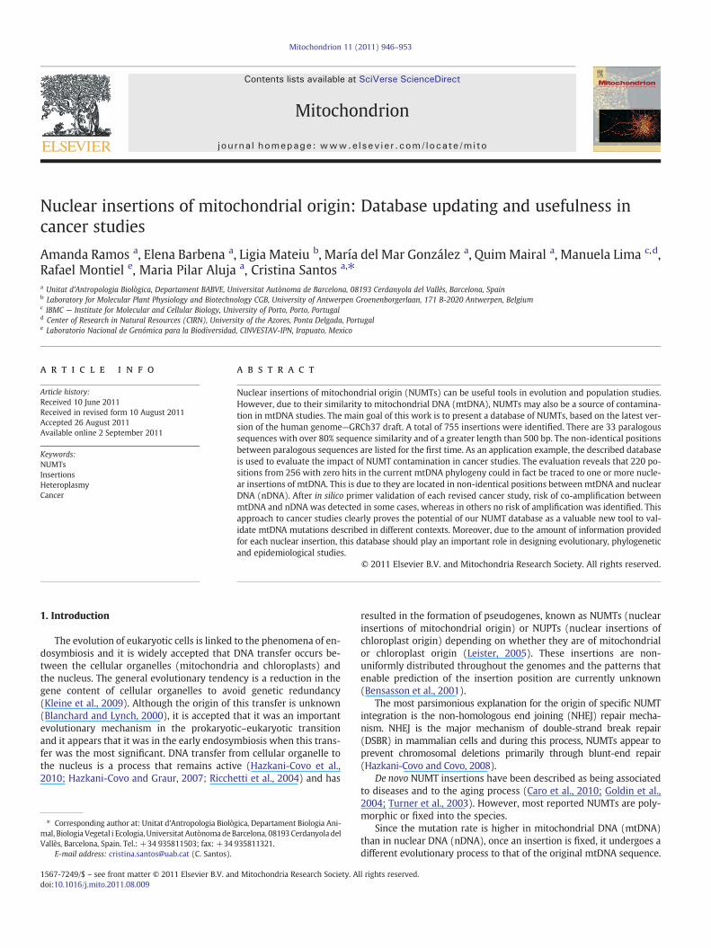
The 755 insertions are distributed throughout the genomewith var-iable frequencies and sizes for each chromosome (Table 1 and Fig. 2). Alarge number of insertions are observed in most chromosomes. Thereare only six chromosomes (14, 15, 16, 18, 20 and Y) that accumulate15 or fewer insertions, representing less than 2% of the total insertionspresent in each.
There is a positive correlation between the number of insertions andchromosome size (Spearman correlation: 0.822 pb0.001) (Fig. 3a).Chromosome 2, the second largest chromosome in relation to the entire

Fig. 1. Alignment between mtDNA (region 12,032–12,340) and a NUMT located in chromosome 1 (region 94,387,537–94,387,832). Examples of identical positions (box _ _ _ _),non-identical position (box ____) and gap region (box ……) are shown. Length in nDNA: 296 bp; length in mtDNA: 309 bp; total length of match: 311 bp.
948 A. Ramos et al. / Mitochondrion 11 (2011) 946–953
genome (7.86%), appears as an outlier. It encompasses 106 insertions(representing 14.04% of total number of insertions), nearly double thenumber of insertions that would be expected (Fig. 3a).
The correlation between the size of insertions and their representa-tion on each chromosome is also positive and significant (Spearmancorrelation: 0.587 p=0.003). However, two outliers, chromosomes 2and 3, were identified (Fig. 3b). As before, chromosome 2 is an outliercontaining the highest number of insertions. But the length of insertionsis quite short, covering only 0.96% of the total length of insertions(Table 1). On the contrary, chromosome 3, which is also one of the larg-est (6.40% in relation to the entire genome) with a relatively high
Table 1Number and length of insertions and their percentages for each chromosome. Length of ea
Chromosome Length chromosome(% in relation to the entire genome)
Number of insertion(% in relation to the
1 249,250,621 (8.05%) 51 (6.75%)2 243,199,373 (7.86%) 106 (14.04%)3 198,022,430 (6.40%) 44 (5.83%)4 191,154,276 (6.17%) 41 (5.43%)5 180,915,260 (5.84%) 35 (4.64%)6 171,115,067 (5.53%) 31 (4.11%)7 159,138,663 (5.14%) 43 (5.70%)8 146,364,022 (4.73%) 47 (6.23%)9 141,213,431 (4.56%) 34 (4.50%)10 135,534,747 (4.38%) 35 (4.64%)11 135,006,516 (4.36%) 31 (4.10%)12 133,851,895 (4.32%) 42 (5.56%)13 115,169,878 (3.72%) 22 (2.91%)14 107,349,540 (3.47%) 9 (1.19%)15 102,531,392 (3.31%) 14 (1.85%)16 90,354,753 (2.92%) 15 (1.99%)17 81,195,210 (2.62%) 23 (3.05%)18 78,077,248 (2.52%) 6 (0.79%)19 59,128,983 (1.91%) 20 (2.65%)20 63,025,520 (2.04%) 9 (1.19%)21 48,129,895 (1.55%) 18 (2.38%)22 51,304,566 (1.66%) 17 (2.25%)X 155,270,560 (5.02%) 50 (6.62%)Y 59,373,566 (1.92%) 12 (1.59%)Total 3,095,677,412 (100%) 755 (100%)
frequency of insertion (representing 5.83%of total number of insertion),presents the highest total length of insertions (17.78%). In both ana-lyses, chromosome 2 appears as an outlier. This would suggest a differ-ential evolution of this chromosome, a hypothesis that deserves furtherinvestigation (Fig. 3b).
The mean percentage of identity between nuclear insertions andmtDNA sequence is 79.2%, ranging from 63.5% to 100%.When the analy-sis of identity is performed for each chromosome (Table 2), the similaritybetween inserted mitochondrial sequences and the original mitochon-drial sequences presents almost the same pattern in all chromosomes,with a mean percentage of identity of ~80% along the chromosomes.
ch chromosome and their percentage are also reported.
stotal number of insertions)
Length of insertions(% in relation to the total length of insertions)
44,798 (8.17%)5268 (0.96%)
97,489 (17.78%)35,103 (6.40%)33,053 (6.03%)11,738 (2.14%)36,328 (6.63%)36,917 (6.73%)28,498 (5.20%)20,926 (3.82%)26,640 (4.86%)8264 (1.51%)
10,691 (1.95%)8899 (1.62%)9395 (1.71%)
15,965 (2.91%)21,990 (4.01%)1334 (0.24%)9318 (1.70%)7471 (1.36%)6753 (1.23%)
23,544 (4.29%)38,843 (7.08%)9025 (1.65%)
548,250 (100%)
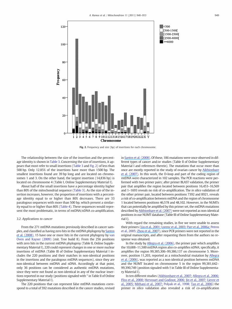
Fig. 2. Frequency and size (bp) of insertions for each chromosome.
949A. Ramos et al. / Mitochondrion 11 (2011) 946–953
The relationship between the size of the insertion and the percent-age identity is shown in Table 3. Concerning the size of insertions, it ap-pears thatmost refer to small insertions (Table 3 and Fig. 2) of less than500 bp. Only 12.85% of the insertions have more than 1500 bp. Thesmallest insertions found are 39 bp long and are located on chromo-somes 1 and 3. On the other hand, the largest insertion (14,836 bp) islocated on chromosome 4 (Table I, Online Supplementary Material I).
About half of the small insertions have a percentage identity higherthan 80% of the mitochondrial sequence (Table 3). As the size of the in-sertion increases, however, the proportion of insertions with a percent-age identity equal to or higher than 80% decreases. There are 33paralogous sequences with more than 500 bp, which present a similar-ity equal to or higher than 80% (Table 4). These sequenceswould repre-sent the most problematic, in terms of mtDNA/nDNA co-amplification.
3.2. Applications to cancer
From the 271mtDNAmutations previously described in cancer sam-ples, and classified as having zero hits in themtDNAphylogenyby Santoset al. (2008), 15 have one or more hits in the current phylogeny by vanOven and Kayser (2009) (mit. Tree build 8). From the 256 positionswith zero hits in the current mtDNA phylogeny (Table II, Online Supple-mentaryMaterial I), 220 could represent changes in one ormore nuclearinsertions of mtDNA (Table III of Online Supplementary Material I in-cludes the 220 positions and their matches in non-identical positionsin the insertions and the paralogous mtDNA sequences), since they arenon-identical between mtDNA and nDNA. Accordingly at that point,only 36 positions can be considered as authentic mtDNA mutations,since they were not found as non-identical in any of the nuclear inser-tions reported in our study (positions signaledwith * in Table II of OnlineSupplementary Material I).
The 220 positions that can represent false mtDNA mutations corre-spond to a total of 592mutations described in the cancer studies, revised
in Santos et al. (2008). Of these, 186mutationswere once observed in dif-ferent types of cancer and/or studies (Table II of Online SupplementaryMaterial I and references therein). The mutations that occur more thanonce are mostly reported in the study of ovarian cancer by Aikhionbareet al. (2007). In this work, the D-loop and part of the coding region ofmtDNA were characterized in 102 samples. The PCR reactions were per-formed with two primer pairs; after primer BLAST validation, the primerpair that amplifies the region located between positions 16,453–16,569and 1–1693 reveals no risk of co-amplification. The in silico validation ofthe other primer pair, located between positions 7392 and 8921, revealsa risk of co-amplification betweenmtDNAand the region of chromosome1 located between positions 46,578 and 48,102. However, in the NUMTsthat can potentially be amplifiedby this primer set, themtDNAmutationsdescribed byAikhionbare et al. (2007)were not reported as non-identicalpositions in ourNUMTdatabase (Table III of Online SupplementaryMate-rial I).
With regard the remaining studies, in five we were unable to assesstheir primers (Liu et al., 2001; Lorenc et al., 2003; Parr et al., 2006a; Petroset al., 2005; Zhou et al., 2007), since PCR primerswere not reported in theoriginal manuscripts, and after requesting them from the authors no re-sponse was obtained.
In the study by Allegra et al. (2006), the primer pair which amplifiesthe 10,688–11,500mtDNA region also co-amplifies nDNA; specifically, itamplifies the region 99,385,306–99,386,137 on chromosome 5. More-over, position 11,203, reported as a mitochondrial mutation by Allegraet al. (2006), was reported as a non-identical position between mtDNAand the NUMT located on chromosome 5 in the region 99,381,642–99,390,749 (position signaled with † in Table III of Online Supplementa-ry Material I).
In ten different studies (Aikhionbare et al., 2007; Allegra et al., 2006;Fliss et al., 2000; Hervouet and Godinot, 2006; Jin et al., 2007; Lievre etal., 2005; Mithani et al., 2007; Polyak et al., 1998; Tan et al., 2006) theprimer in silico validation also revealed a risk of co-amplification

Fig. 3. Correlation between chromosome length and number of insertions (a), and correlation between chromosome and insertion length (b). Coefficient of determination (Sq rlinear) for the two correlations is shown.
950 A. Ramos et al. / Mitochondrion 11 (2011) 946–953
between mtDNA and nDNA. The twenty-seven positions described bythe authors as mtDNA mutations, however, are not reported as non-identical position between mtDNA and the nuclear regions that mightpotentially be co-amplified.
Finally, 123 positions described in 15 studies (Aikhionbare et al., 2007;Fliss et al., 2000; Gasparre et al., 2007; Habano et al., 1999; Hervouet andGodinot, 2006; Jin et al., 2007; Jones et al., 2001; Kassauei et al., 2006;Lievre et al., 2005; Mithani et al., 2007; Nagy et al., 2003; Parrella et al.,2001; Polyak et al., 1998; Tan et al., 2006; Tzen et al., 2007) aremore likely
to be authentic, since the primers used to amplify the regions that encom-pass such mutations did not suggest the possibility of nDNA co-amplification (positions signaled with ** in Table II of Online Supplemen-tary Material I).
4. Discussion
In population and clinical genetics, as well as in the design of evolu-tionary, phylogenetic and epidemiological studies, it is important to

Table2
Percen
tage
iden
tity
forallc
hrom
osom
es.
Chromosom
e1
23
45
67
89
1011
1213
1415
1617
1819
2021
22X
Y
Mea
n78
.76
80.20
79.42
81.24
82.02
80.77
77.67
77.88
78.22
78.31
78.27
83.40
80.61
80.40
79.23
77.37
81.69
84.61
72.92
81.43
75.82
75.74
76.05
79.00
Med
ian
75.73
78.93
79.80
80.43
80.73
81.74
75.83
76.32
77.30
76.98
75.76
8880
.72
79.13
77.65
76.72
80.41
83.96
69.65
77.88
74.05
72.16
73.83
74.57
Minim
um64
.71
65.04
68.14
68.71
66.35
64.69
68.46
67.77
68.02
69.08
66.46
67.79
65.41
73.55
71.83
68.26
68.54
74.08
63.50
69.49
65.59
65.67
64.82
70.15
Max
imum
100
98.48
97.44
96.77
97.67
93.33
97.09
98.81
90.20
9298
.61
98.88
99.22
93.07
95.35
85.45
97.06
95.57
86.59
100
98.75
100
96.30
100
Percen
tiles
2571
.43
74.58
73.16
76.19
76.47
77.78
72.22
72.37
73.97
74.14
74.44
77.58
72.41
74.75
75.15
73.53
77.06
75.89
67.43
72.40
69.09
69.32
69.78
71.38
5075
.73
78.93
79.80
80.43
80.73
81.74
75.83
76.32
77.30
76.98
75.76
8880
.72
79.13
77.65
76.72
80.41
83.96
69.65
77.88
74.04
72.16
73.83
74.57
7584
.03
85.47
83.56
84.86
88.36
8582
.02
82.35
80.71
82.08
81.67
8888
.59
86.02
83.37
81.73
83.42
94.08
79.04
94.10
79.71
80.31
80.95
82.35 Table 3
Frequency and identity of insertions in relation to the size of insertion (bp).
Size (bp) Identity Total Percentage
≥80% b80%
b500 276 245 521 69.00[500–1500] 21 116 137 18.15[1500–2500] 5 43 48 6.36[2500–4000] 3 21 24 3.18≥4000 4 21 25 3.31Total 309 446 755 100
951A. Ramos et al. / Mitochondrion 11 (2011) 946–953
have a complete overview of the total number of NUMTs, their locationand variation. In this study, a NUMT database obtained using the latestversion of the Human Genome — GRCh37 draft is reported. This is, toour knowledge, the first database that is based on this latest version ofthe human genome. Moreover, it is also the first where the non-identical positions betweenmtDNA and nDNA are listed for each NUMT.
In this study, 755 nuclear insertions of mtDNA origin were reported.A comparison of the total number of NUMTs in previously publishedworks clearly shows significant discrepancies. The number of reportedNUMTs ranges from 190 (Lascaro et al., 2008) to 1105 (Tourmen etal., 2002), with reports of 206 (Richly and Leister, 2004), 211 (Ricchettiet al., 2004), 247 (Mishmar et al., 2004), 452 (Hazkani-Covo and Graur,2007), 612 (Woischnik and Moraes, 2002) and 871 (Hazkani-Covo etal., 2010) insertions. The reasons for such discrepancies have been dis-cussed by others previously (Lascaro et al., 2008). According to Lascaroet al. (2008), the incautious usage of bioinformatics methods and theapplication of methods to an as yet not correctly assembled nuclear ge-nome, are the main factors underlying the disagreement observed be-tween studies. In this sense, the Human mtDNA Reference Sequence(NC_012920) was compared against the human RefSeq Genomic data-base at NCBI, using the RemoteBlast package in Bioperl bundle (Stajichet al., 2002) with the parameters set to restrict the search to the humanorganism and the E-value of 10−3. Moreover, regions with less than20 bp were excluded and only hits in the most recent version of theHuman genome draft — GRCh37 primary reference assembly wereconsidered.
Another factor that can induce differences in the number of reportedinsertions is whether or not the post-insertional processes were evalu-ated in order to differentiate original insertions from those that resultfrom processes that alter the initially inserted sequence. In this study,post-insertional processes were not considered, since we aimed to ob-tain an accurate database with all the nuclear insertions and their spe-cific variation (relatively to human mtDNA).
In accordance to other authors [for a review see Hazkani-Covo et al.(2010)], the reported frequency of insertions, aswell as their length anddistribution along the genome is variable. Most insertions have lessthan 500 bp. This agrees with the most parsimonious explanation forthe origin of NUMT which assumes that mtDNA would be used as a“patch” in the double-strand break repair from non-homologous endjoining repair mechanism (Hazkani-Covo et al., 2010; Hazkani-Covoand Covo, 2008; Leister, 2005).
As previously mentioned, due to the similarity withmtDNA, NUMTsare a potential source of contamination in mtDNA studies based on PCRamplification (Parr et al., 2006b; Yao et al., 2008). In this context, largeinsertionswith a high percentage of identitywithmtDNAwould be par-ticularly problematic. In fact, one of these insertions, located on chro-mosome 1, was previously reported to be a source of several errorsand misinterpretations (Yao et al., 2008). We characterized the 33large insertions (with more than 500 bp) that present a percentageidentity greater than 80%. Its size makes it difficult to perform primerdesigns to prevent co-amplification between nuclear insertions andmtDNA (Ramos et al., 2009; Ramos et al., 2011).
This study reports for the first time all identical and non-identicalpositions between the NUMTs and the GRCh37 version of the nuclear
Table 4Paralogous sequences with more than 500 bp and that present a similarity equal or higher than 80%.
Chromosome % Identity nDNA length mtDNA length mtDNA region nDNA region
1 98.53 5844 5845 3911–9755 564,461–570,3041 84.03 517 521 987–1507 142,792,601–142,793,1171 83.87 518 521 987–1507 143,243,224–143,243,7411 83.78 515 519 987–1505 143,344,679–143,345,1932 84.03 516 521 987–1507 95,564,754–95,565,2692 83.95 728 729 6270–6998 50,815,826–50,816,5532 81.78 1350 1349 4854–6202 156,119,971–156,121,3203 95.39 1323 1323 1396–2718 96,336,032–96,337,3543 83.31 623 623 7151–7773 120,440,870–120,441,4923 80.38 1621 1622 1418–3039 40,293,638–40,295,2585 95.05 2114 2121 577–2697 79,945,841–79,947,9545 94.06 5219 5219 10,269–15,487 134,258,999–134,264,2175 88.36 9108 9067 6117–15,183 99,381,642–99,390,7495 87.24 3463 3463 12,662–16,124 93,903,161–93,906,6235 84.54 962 961 577–1537 123,096,499–123,097,4606 90.70 527 527 2408–2934 62,284,008–62,284,5347 83.66 2514 2517 577–3093 142,373,012–142,375,5257 81.27 562 561 2740–3300 141,504,769–141,505,3309 83.85 2517 2517 577–3093 33,656,612–33,659,12810 84.62 533 533 8281–8813 101,817,140–101,817,67211 94.24 2394 2396 577–2972 10,529,434–10,531,82711 81.68 560 568 596–1163 87,524,440–87,524,99914 93.07 1021 1024 5583–6606 32,953,304–32,954,32417 96.17 653 653 6818–7470 51,183,094–51,183,74617 83.42 10,477 10,536 577–11,112 22,021,365–22,031,84117 81.73 621 619 2656–3274 19,504,577–19,505,19717 80.57 1514 1521 577–2097 19,501,874–19,503,38717 80.41 2206 2205 14,365–16,569 22,018,521–22,020,72617 80.24 575 582 2084–2665 19,503,699–19,504,27321 83.78 515 519 987–1505 9,735,524–9,736,038X 93.68 554 554 10,606–11,159 125,606,714–125,607,267X 93.07 749 750 6553–7302 125,605,687–125,606,435Y 83.08 517 521 987–1507 13,290,151–13,290,667
952 A. Ramos et al. / Mitochondrion 11 (2011) 946–953
genome and the mtDNA Reference Sequence. This information can beused as a tool in the authentication ofmtDNAmutations reported in dif-ferent fields. Notwithstanding, it is worth mentioning that, in this com-pilation, the variation of NUMTs was not considered since no studies ofNUMT sequence variation had yet been performed to date. Moreover,themtDNA variationwas only taken into account for humanmtDNA, al-though the original insertion could in fact represent mtDNA sequencefrom very different species.
ThedescribedNUMTdatabasewas used as a tool to assess the impactof NUMT contamination on cancer studies. Several authors detected in-numerable deficiencies in the medical literature related to the analysisand interpretation of mtDNA data [Salas et al. (2005), Yao et al. (2004)among others]. Concerning cancer studies, Salas et al. (2005) showedthe consequences of poor experimental design, both in the misinterpre-tation of the role ofmtDNA in the complex tumoral process, aswell as inthe comparison of results. Also, Yao et al. (2004) showed that re-searchers may still ignore the possibility of a NUMT contribution whena seemingly novelmtDNA sequence is encountered. Using theNUMTda-tabase, we evaluated 256 mtDNA positions that were previouslyreported to be mutated in cancer samples and that present zero hits inthe updated mtDNA phylogeny (Van Oven and Kayser, 2009). Fromthose positions, 220 (identified in 21 studies) could represent changesin one or more nuclear insertions of mtDNA since they are non-identical between mtDNA and nDNA. Thus, we raise the hypothesisthat these changes could represent nuclear variations instead of mtDNAvariation. To test this hypothesis, we further validated in silico the primerpairs used in several original studies if they were accessible in the publi-cation or if they were provided by the authors after request. After primerin silico validation, from a total of 16works for which primers were avail-able, only six studies (Habano et al., 1999; Jones et al., 2001; Kassauei etal., 2006; Nagy et al., 2003; Parrella et al., 2001; Tzen et al., 2007) didnot evidence the possibility of nDNA co-amplification. In the remainingstudies (Table III of Online Supplementary Material I and references
therein), some of the positions reported are located in regions thatcould be the result of co-amplification. However, with the exception ofposition 11,203 reported by Allegra et al. (2006), these positions are notreported as non-identical between the potentially co-amplified mtDNAand nDNA regions. Even so, we must take into account the fact that thevariability of NUMTs is unknown, and that the non-identical position da-tabase was generated using the mtDNA and nDNA Reference Sequences.Thus, the differences between mtDNA and nDNA reported in our data-base are surely underestimated and the above mentioned positionscould be non-authentic mtDNAmutations. On the other hand, it is possi-ble that these changes actually represent authentic mtDNA mutations;however, this hypothesis seems extremely difficult to test.
Using cancer studies as an example application, we aimed to high-light the applicability of the described NUMT database as a new toolto validate in silico mtDNA mutations described in different contexts;it is clear that there is a need to follow standardized protocols thatavoid co-amplification between mitochondrial and nuclear paralogoussequences. Moreover, and due to the amount of information providedfor each nuclear insertion, this database could be applied as a tool to de-sign evolutionary, phylogenetic and epidemiological studies.
To our knowledge, this is the first work to report the non-identicalpositions between the mtDNA and nDNA. These positions are the basisfor the application to cancer studies reported in this work, and knowl-edge of their distribution is useful for the authentication of mtDNA mu-tations reported in different kinds of study, especially those occurring inheteroplasmy.
Supplementary data associated with this article can be found, inthe online version, at doi:10.1016/j.mito.2011.08.009.
Acknowledgments
This work was supported by MEC (project: CGL2009-08205) andby the Generalitat de Catalunya (Ref. 2009 SGR 566).

953A. Ramos et al. / Mitochondrion 11 (2011) 946–953
We are in debt with Dr. Czarnecka, Dr Liu and Dr. Maitra, the corre-sponding authors of cancer studies for supplying the information neces-sary for the analysis.
References
Aikhionbare, F.O., Mehrabi, S., Kumaresan, K., Zavareh, M., Olatinwo, M., Odunsi, K.,Partridge, E., 2007. Mitochondrial DNA sequence variants in epithelial ovariantumor subtypes and stages. J. Carcinog. 6, 1.
Allegra, E., Garozzo, A., Lombardo, N., De Clemente, M., Carey, T.E., 2006. Mutations andpolymorphisms in mitochondrial DNA in head and neck cancer cell lines. ActaOtorhinolaryngol. Ital. 26, 185–190.
Altschul, S.F., Gish, W., Miller, W., Myers, E.W., Lipman, D.J., 1990. Basic local alignmentsearch tool. J. Mol. Biol. 215, 403–410.
Bensasson, D., Zhang, D., Hartl, D.L., Hewitt, G.M., 2001. Mitochondrial pseudogenes:evolution's misplaced witnesses. Trends Ecol. Evol. 16, 314–321.
Blanchard, J.L., Lynch, M., 2000. Organellar genes: why do they end up in the nucleus?Trends Genet. 16, 315–320.
Caro, P., Gomez, J., Arduini, A., Gonzalez-Sanchez, M., Gonzalez-Garcia, M., Borras, C.,Vina, J., Puertas, M.J., Sastre, J., Barja, G., 2010. Mitochondrial DNA sequences arepresent inside nuclear DNA in rat tissues and increase with age. Mitochondrion10, 479–486.
Fliss, M.S., Usadel, H., Caballero, O.L., Wu, L., Buta, M.R., Eleff, S.M., Jen, J., Sidransky, D.,2000. Facile detection of mitochondrial DNAmutations in tumors and bodily fluids.Science 287, 2017–2019.
Gasparre, G., Porcelli, A.M., Bonora, E., Pennisi, L.F., Toller, M., Iommarini, L., Ghelli, A.,Moretti, M., Betts, C.M., Martinelli, G.N., Ceroni, A.R., Curcio, F., Carelli, V., Rugolo,M., Tallini, G., Romeo, G., 2007. Disruptive mitochondrial DNA mutations in com-plex I subunits are markers of oncocytic phenotype in thyroid tumors. Proc. Natl.Acad. Sci. U.S.A. 104, 9001–9006.
Goios, A., Prieto, L., Amorim, A., Pereira, L., 2008. Specificity of mtDNA-directed PCR-influence of NUclear MTDNA insertion (NUMT) contamination in routine samplesand techniques. Int. J. Legal Med. 122, 341–345.
Goldin, E., Stahl, S., Cooney, A.M., Kaneski, C.R., Gupta, S., Brady, R.O., Ellis, J.R., Schiffmann,R., 2004. Transfer of a mitochondrial DNA fragment to MCOLN1 causes an inheritedcase of mucolipidosis IV. Hum. Mutat. 24, 460–465.
Habano, W., Sugai, T., Yoshida, T., Nakamura, S., 1999. Mitochondrial gene mutation,but not large-scale deletion, is a feature of colorectal carcinomas with mitochon-drial microsatellite instability. Int. J. Cancer 83, 625–629.
Hazkani-Covo, E., 2009. Mitochondrial insertions into primate nuclear genomes sug-gest the use of numts as a tool for phylogeny. Mol. Biol. Evol. 26, 2175–2179.
Hazkani-Covo, E., Covo, S., 2008. Numt-mediated double-strand break repair mitigatesdeletions during primate genome evolution. PLoS Genet. 4, e1000237.
Hazkani-Covo, E., Graur, D., 2007. A comparative analysis of numt evolution in humanand chimpanzee. Mol. Biol. Evol. 24, 13–18.
Hazkani-Covo, E., Sorek, R., Graur, D., 2003. Evolutionary dynamics of large numts inthe human genome: rarity of independent insertions and abundance of post-insertion duplications. J. Mol. Evol. 56, 169–174.
Hazkani-Covo, E., Zeller, R.M., Martin, W., 2010. Molecular poltergeists: mitochondrialDNA copies (numts) in sequenced nuclear genomes. PLoS Genet. 6, e1000834.
Hervouet, E., Godinot, C., 2006. Mitochondrial disorders in renal tumors. Mitochondri-on 6, 105–117.
Jensen-Seaman, M.I., Wildschutte, J.H., Soto-Calderón, I.D., Anthony, N.M., 2009. A com-parative approach shows differences in patterns of numt insertion during homi-noid evolution. J. Mol. Evol. 68.
Jin, X., Zhang, J., Gao, Y., Ding, K., Wang, N., Zhou, D., Jen, J., Cheng, S., 2007. Relationshipbetween mitochondrial DNA mutations and clinical characteristics in human lungcancer. Mitochondrion 7, 347–353.
Jones, J.B., Song, J.J., Hempen, P.M., Parmigiani, G., Hruban, R.H., Kern, S.E., 2001. Detec-tion of mitochondrial DNA mutations in pancreatic cancer offers a “mass”-ive ad-vantage over detection of nuclear DNA mutations. Cancer Res. 61, 1299–1304.
Kassauei, K., Habbe, N., Mullendore, M.E., Karikari, C.A., Maitra, A., Feldmann, G., 2006.Mitochondrial DNA mutations in pancreatic cancer. Int. J. Gastrointest. Cancer 37,57–64.
Kleine, T., Maier, U.G., Leister, D., 2009. DNA transfer from organelles to the nucleus:the idiosyncratic genetics of endosymbiosis. Annu. Rev. Plant Biol. 60, 115–138.
Lascaro, D., Castellana, S., Gasparre, G., Romeo, G., Saccone, C., Attimonelli, M., 2008.The RHNumtS compilation: features and bioinformatics approaches to locate andquantify Human NumtS. BMC Genomics 9, 267.
Leister, D., 2005. Origin, evolution and genetic effects of nuclear insertions of organelleDNA. Trends Genet. 21, 655–663.
Lievre, A., Chapusot, C., Bouvier, A.M., Zinzindohoue, F., Piard, F., Roignot, P., Arnould, L.,Beaune, P., Faivre, J., Laurent-Puig, P., 2005. Clinical value of mitochondrial muta-tions in colorectal cancer. J. Clin. Oncol. 23, 3517–3525.
Liu, V.W., Shi, H.H., Cheung, A.N., Chiu, P.M., Leung, T.W., Nagley, P., Wong, L.C., Ngan, H.Y.,2001. High incidence of somatic mitochondrial DNA mutations in human ovariancarcinomas. Cancer Res. 61, 5998–6001.
Lorenc, A., Bryk, J., Golik, P., Kupryjanczyk, J., Ostrowski, J., Pronicki, M., Semczuk, A.,Szolkowska, M., Bartnik, E., 2003. Homoplasmic MELAS A3243G mtDNA mutationin a colon cancer sample. Mitochondrion 3, 119–124.
Mishmar, D., Ruiz-Pesini, E., Brandon, M., Wallace, D.C., 2004. Mitochondrial DNA-likesequences in the nucleus (NUMTs): insights into our African origins and the mech-anism of foreign DNA integration. Hum. Mutat. 23, 125–133.
Mithani, S.K., Taube, J.M., Zhou, S., Smith, I.M., Koch, W.M., Westra, W.H., Califano, J.A.,2007. Mitochondrial mutations are a late event in the progression of head and necksquamous cell cancer. Clin. Cancer Res. 13, 4331–4335.
Nagy, A., Wilhelm, M., Kovacs, G., 2003. Mutations of mtDNA in renal cell tumours aris-ing in end-stage renal disease. J. Pathol. 199, 237–242.
Parfait, B., Rustin, P., Munnich, A., Rotig, A., 1998. Co-amplification of nuclear pseudo-genes and assessment of heteroplasmy of mitochondrial DNA mutations. Biochem.Biophys. Res. Commun. 247, 57–59.
Parr, R.L., Dakubo, G.D., Crandall, K.A., Maki, J., Reguly, B., Aguirre, A.,Wittock, R., Robinson,K., Alexander, J.S., Birch-Machin,M.A., Abdel-Malak,M., Froberg,M.K., Diamandis, E.P.,Thayer, R.E., 2006a. Somatic mitochondrial DNA mutations in prostate cancer andnormal appearing adjacent glands in comparison to age-matched prostate sampleswithout malignant histology. J. Mol. Diagn. 8, 312–319.
Parr, R.L., Maki, J., Reguly, B., Dakubo, G.D., Aguirre, A., Wittock, R., Robinson, K., Jakupciak,J.P., Thayer, R.E., 2006b. The pseudo-mitochondrial genome influences mistakes inheteroplasmy interpretation. BMC Genomics 7, 185.
Parrella, P., Xiao, Y., Fliss, M., Sanchez-Cespedes, M., Mazzarelli, P., Rinaldi, M., Nicol, T.,Gabrielson, E., Cuomo, C., Cohen, D., Pandit, S., Spencer, M., Rabitti, C., Fazio, V.M.,Sidransky, D., 2001. Detection of mitochondrial DNA mutations in primary breastcancer and fine-needle aspirates. Cancer Res. 61, 7623–7626.
Petros, J.A., Baumann, A.K., Ruiz-Pesini, E., Amin, M.B., Sun, C.Q., Hall, J., Lim, S., Issa, M.M., Flanders, W.D., Hosseini, S.H., Marshall, F.F., Wallace, D.C., 2005. mtDNA muta-tions increase tumorigenicity in prostate cancer. Proc. Natl. Acad. Sci. U.S.A. 102,719–724.
Polyak, K., Li, Y., Zhu, H., Lengauer, C., Willson, J.K., Markowitz, S.D., Trush, M.A., Kinzler,K.W., Vogelstein, B., 1998. Somatic mutations of the mitochondrial genome inhuman colorectal tumours. Nat. Genet. 20, 291–293.
Ramos, A., Santos, C., Alvarez, L., Nogués, R., Aluja, M.P., 2009. Human mitochondrialDNA complete amplification and sequencing: a new validated primer set that pre-vents nuclear DNA sequences of mitochondrial origin co-amplification. Electro-phoresis 30, 1–7.
Ramos, A., Santos, C., Barbena, E., Mateiu, L., Alvarez, L., Nogues, R., Aluja, M.P., 2011.Validated primer set that prevents nuclear DNA sequences of mitochondrial originco-amplification: a revision based on the New Human Genome Reference Se-quence (GRCh37). Electrophoresis 32, 782–783.
Ricchetti, M., Tekaia, F., Dujon, B., 2004. Continued colonization of the human genomeby mitochondrial DNA. PLoS Biol. 2, E273.
Richly, E., Leister, D., 2004. NUMTs in sequenced eukaryotic genomes. Mol. Biol. Evol.21, 1081–1084.
Salas, A., Carracedo, A., Macaulay, V., Richards, M., Bandelt, H.J., 2005. A practical guideto mitochondrial DNA error prevention in clinical, forensic, and population genet-ics. Biochem. Biophys. Res. Commun. 335, 891–899.
Santos, C., Martínez, M., Lima, M., Hao, Y.-J., Simoes, N., Montiel, R., 2008. MitochondrialDNA mutations in cancer: a review. Curr. Top. Med. Chem. 8, 1351–1366.
Stajich, J., Block, D., Boulez, K., Brenner, S., Chervitz, S., Dagdigian, C., Fuellen, G., Gilbert,J., Korf, I., Lapp, H., Lehväslaiho, H., Matsalla, C., Mungall, C., Osborne, B., Pocock, M.,Schattner, P., Senger, M., Stein, L., Stupka, E., Wilkinson, M., Birney, E., 2002. TheBioPerl toolkit: Perl modules for the life sciences. Genome Res. 12, 1611–1618.
Tan, D.J., Chang, J., Liu, L.L., Bai, R.K., Wang, Y.F., Yeh, K.T., Wong, L.J., 2006. Significanceof somatic mutations and content alteration of mitochondrial DNA in esophagealcancer. BMC Cancer 6, 93.
Thalmann, O., Serre, D., Hofreiter, M., Lukas, D., Eriksson, J., Vigilant, L., 2005. Nuclearinsertions help and hinder inference of the evolutionary history of gorillamtDNA. Mol. Ecol. 14, 179–188.
Tourmen, Y., Baris, O., Dessen, P., Jacques, C., Malthiery, Y., Reynier, P., 2002. Structureand chromosomal distribution of human mitochondrial pseudogenes. Genomics80, 71–77.
Turner, C., Killoran, C., Thomas, N.S.T., Rosenberg, M., Chuzhanova, N.A., Johnston, J.,Kemel, Y., Cooper, D.N., Biesecker, L.G., 2003. Human genetic disease caused byde novo mitochondrial-nuclear DNA transfer. Hum. Genet. 112, 303–309 (112,303–309).
Tzen, C.Y., Mau, B.L., Wu, T.Y., 2007. ND4 mutation in transitional cell carcinoma: doesmitochondrial mutation occur before tumorigenesis? Mitochondrion 7, 273–278.
Van Oven, M., Kayser, M., 2009. Updated comprehensive phylogenetic tree of globalhuman mitochondrial DNA variation. Hum. Mutat. 30, E386–E394.
Woischnik, M., Moraes, C.T., 2002. Pattern of organization of human mitochondrialpseudogenes in the nuclear genome. Genome Res. 12, 885–893.
Yao, Y.G., Bravi, C.M., Bandelt, H.J., 2004. A call for mtDNA data quality control in foren-sic science. Forensic Sci. Int. 141, 1–6.
Yao, Y.G., Kong, Q.P., Salas, A., Bandelt, H.J., 2008. Pseudomitochondrial genome hauntsdisease studies. J. Med. Genet. 45, 769–772.
Zhou, S., Kachhap, S., Sun, W., Wu, G., Chuang, A., Poeta, L., Grumbine, L., Mithani, S.K.,Chatterjee, A., Koch, W., Westra, W.H., Maitra, A., Glazer, C., Carducci, M., Sidransky,D., McFate, T., Verma, A., Califano, J.A., 2007. Frequency and phenotypic implica-tions of mitochondrial DNA mutations in human squamous cell cancers of thehead and neck. Proc. Natl. Acad. Sci. U.S.A. 104, 7540–7545.