Continuing analysis of microRNA origins: Formation from transposable element insertions and...
9
RESEARCH PAPER www.landesbioscience.com Mobile Genetic Elements e27755-1 Mobile Genetic Elements 3, e27755; November/December 2013; © 2013 Landes Bioscience RESEARCH PAPER Introduction MicroRNAs (miRs) are short (approximately 20 nucleotide) noncoding RNAs (ncRNAs) involved in the regulation of gene expression. 1 Functioning much like small interfering RNAs (siR- NAs), miRs bind to complimentary messenger RNA (mRNA) resulting in the repression of translation. 2,3 MiRs are initially transcribed in the nucleus as portions of larger precursor mol- ecules called pri-miRs. These initial transcripts are processed by Drosha to generate ~70 nucleotide stem loops (pre-miRs) that are exported into the cytosol where Dicer ultimately trims these dsRNA pre-miRs into functional single stranded miRs. 2,4 Following this, complementary sequence association between a miR and mRNA target leads to inhibited translation of the mRNA molecule. 2 To date, our principal obstacle to deciphering miR function has proven to be our inability to accurately predict miR asso- ciation with target mRNAs. This is principally the result of the capacity of miRs to associate with mRNAs bearing imperfect sequence complementarities. 5 Although accurately determining which mRNAs a miR targets has been an extremely active area of research, no universal model of target prediction has been widely adopted. Unlike siRNAs which associate with targets through perfect complementarity, miR association requires as little as seven complementary nucleotides. 6 Usually located at the 5′ end of a miR, these seven complementary nucleotides are known as “seeds” and their complement in target mRNAs are known as “seed matches.” 7 This complementarity between seeds and seed matches is the core requirement for the majority of currently utilized miR target prediction algorithms after which programs *Correspondence to: Glen M Borchert; Email: [email protected] Submitted: 12/05/2013; Revised: 01/03/2014; Accepted: 01/07/2014 Citation: Roberts JT, Cooper EA, Favreau CJ, Howell JS, Lane LG, Mills JE, Newman DC, Perry TJ, Russell ME, Wallace BM, et al. Continuing analysis of microRNA origins: Formation from transposable element insertions and noncoding RNA mutations. Mobile Genetic Elements 2014; 3:e27755; http://dx.doi.org/10.4161/ mge.27755 Continuing analysis of microRNA origins Formation from transposable element insertions and noncoding RNA mutations Justin T Roberts, Elvera A Cooper, Connor J Favreau, Jacob S Howell, Lee G Lane, James E Mills, Derrick C Newman, Tabitha J Perry, Meaghan E Russell, Brittany M Wallace, and Glen M Borchert* Department of Biological Sciences, University of South Alabama; Mobile, AL USA Keywords: LINE, microRNA, miR, miRNA, noncoding RNA, repetitive, retrotransposon, SINE, transposable, transposon Abbreviations: Ago, argonaute; bp, base pair; LINE, long interspersed repeated element; LTR, long terminal repeat; miR, microRNA; mRNA, messenger RNAs; ncRNA, noncoding RNA; nt, nucleotide; OrBId, origin based identification of microRNA targets algorithm; ORF, open reading frame; RISC, RNA-Induced Silencing Complex; RNAi, RNA interference; rRNA, ribosomal RNA; scaRNA, small Cajal body-specific RNA; SINE, short interspersed repeated elements; siRNA, small interfering RNA; snoRNA, small nucleolar RNA; snRNA, spliceosomal RNA; sRNA, short regulatory RNA; TE, transposable element; tmRNA, transfer messenger RNA; tRNA, transfer RNA; UTR, untranslated region MicroRNAs (miRs) are small noncoding RNAs that typically act as regulators of gene expression by base pairing with the 3′ UTR of messenger RNAs (mRNAs) and either repressing their translation or initiating degradation. As of this writ- ing over 24,500 distinct miRs have been identified, but the functions of the vast majority of these remain undescribed. This paper represents a summary of our in depth analysis of the genomic origins of miR loci, detailing the formation of 1,213 of the 7,321 recently identified miRs and thereby bringing the total number of miR loci with defined molecular origin to 3,605. Interestingly, our analyses also identify evidence for a second, novel mechanism of miR locus generation through describing the formation of 273 miR loci from mutations to other forms of noncoding RNAs. Importantly, several independent investigations of the genomic origins of miR loci have now supported the hypothesis that miR hairpins are formed by the adjacent genomic insertion of two complementary transposable elements (TEs) into opposing strands. While our results agree that subsequent transcription over such TE interfaces leads to the formation of the majority of functional miR loci, we now also find evidence suggesting that a subset of miR loci were actually formed by an alternative mechanism—point mutations in other structurally complex, noncoding RNAs (e.g., tRNAs and snoRNAs).
Transcript of Continuing analysis of microRNA origins: Formation from transposable element insertions and...
ReseaRch PaPeR
www.landesbioscience.com Mobile Genetic elements e27755-1
Mobile Genetic elements 3, e27755; November/December 2013; © 2013 Landes Bioscience
ReseaRch PaPeR
Introduction
MicroRNAs (miRs) are short (approximately 20 nucleotide) noncoding RNAs (ncRNAs) involved in the regulation of gene expression.1 Functioning much like small interfering RNAs (siR-NAs), miRs bind to complimentary messenger RNA (mRNA) resulting in the repression of translation.2,3 MiRs are initially transcribed in the nucleus as portions of larger precursor mol-ecules called pri-miRs. These initial transcripts are processed by Drosha to generate ~70 nucleotide stem loops (pre-miRs) that are exported into the cytosol where Dicer ultimately trims these dsRNA pre-miRs into functional single stranded miRs.2,4 Following this, complementary sequence association between a miR and mRNA target leads to inhibited translation of the mRNA molecule.2
To date, our principal obstacle to deciphering miR function has proven to be our inability to accurately predict miR asso-ciation with target mRNAs. This is principally the result of the capacity of miRs to associate with mRNAs bearing imperfect sequence complementarities.5 Although accurately determining which mRNAs a miR targets has been an extremely active area of research, no universal model of target prediction has been widely adopted. Unlike siRNAs which associate with targets through perfect complementarity, miR association requires as little as seven complementary nucleotides.6 Usually located at the 5′ end of a miR, these seven complementary nucleotides are known as “seeds” and their complement in target mRNAs are known as “seed matches.”7 This complementarity between seeds and seed matches is the core requirement for the majority of currently utilized miR target prediction algorithms after which programs
*Correspondence to: Glen M Borchert; Email: [email protected]: 12/05/2013; Revised: 01/03/2014; Accepted: 01/07/2014Citation: Roberts JT, Cooper EA, Favreau CJ, Howell JS, Lane LG, Mills JE, Newman DC, Perry TJ, Russell ME, Wallace BM, et al. Continuing analysis of microRNA origins: Formation from transposable element insertions and noncoding RNA mutations. Mobile Genetic Elements 2014; 3:e27755; http://dx.doi.org/10.4161/mge.27755
Continuing analysis of microRNA originsFormation from transposable element insertions
and noncoding RNA mutationsJustin T Roberts, elvera a cooper, connor J Favreau, Jacob s howell, Lee G Lane, James e Mills, Derrick c Newman,
Tabitha J Perry, Meaghan e Russell, Brittany M Wallace, and Glen M Borchert*
Department of Biological sciences, University of south alabama; Mobile, aL Usa
Keywords: LINE, microRNA, miR, miRNA, noncoding RNA, repetitive, retrotransposon, SINE, transposable, transposon
Abbreviations: Ago, argonaute; bp, base pair; LINE, long interspersed repeated element; LTR, long terminal repeat; miR, microRNA; mRNA, messenger RNAs; ncRNA, noncoding RNA; nt, nucleotide; OrBId, origin based identification of microRNA targets algorithm; ORF, open reading frame; RISC, RNA-Induced Silencing Complex; RNAi, RNA interference; rRNA, ribosomal RNA; scaRNA, small Cajal body-specific RNA; SINE, short interspersed repeated elements; siRNA, small
interfering RNA; snoRNA, small nucleolar RNA; snRNA, spliceosomal RNA; sRNA, short regulatory RNA; TE, transposable element; tmRNA, transfer messenger RNA; tRNA, transfer RNA; UTR, untranslated region
MicroRNas (miRs) are small noncoding RNas that typically act as regulators of gene expression by base pairing with the 3′ UTR of messenger RNas (mRNas) and either repressing their translation or initiating degradation. as of this writ-ing over 24,500 distinct miRs have been identified, but the functions of the vast majority of these remain undescribed. This paper represents a summary of our in depth analysis of the genomic origins of miR loci, detailing the formation of 1,213 of the 7,321 recently identified miRs and thereby bringing the total number of miR loci with defined molecular origin to 3,605. Interestingly, our analyses also identify evidence for a second, novel mechanism of miR locus generation through describing the formation of 273 miR loci from mutations to other forms of noncoding RNas. Importantly, several independent investigations of the genomic origins of miR loci have now supported the hypothesis that miR hairpins are formed by the adjacent genomic insertion of two complementary transposable elements (Tes) into opposing strands. While our results agree that subsequent transcription over such Te interfaces leads to the formation of the majority of functional miR loci, we now also find evidence suggesting that a subset of miR loci were actually formed by an alternative mechanism—point mutations in other structurally complex, noncoding RNas (e.g., tRNas and snoRNas).
e27755-2 Mobile Genetic elements Volume 3
largely differ in how much they weight target sequence conserva-tion across species and how they score additional complementar-ity between the mRNA and the remainder of the miR.8-15
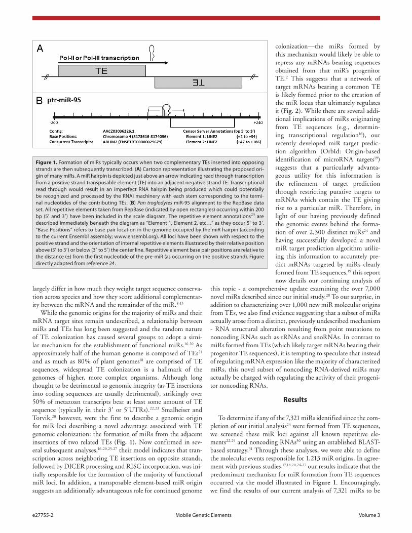
While the genomic origins for the majority of miRs and their mRNA target sites remain undescribed, a relationship between miRs and TEs has long been suggested and the random nature of TE colonization has caused several groups to adopt a simi-lar mechanism for the establishment of functional miRs.16-20 As approximately half of the human genome is composed of TEs21 and as much as 80% of plant genomes18 are comprised of TE sequences, widespread TE colonization is a hallmark of the genomes of higher, more complex organisms. Although long thought to be detrimental to genomic integrity (as TE insertions into coding sequences are usually detrimental), strikingly over 50% of metazoan transcripts bear at least some amount of TE sequence (typically in their 3′ or 5′UTRs).22,23 Smalheiser and Torvik,20 however, were the first to describe a genomic origin for miR loci describing a novel advantage associated with TE genomic colonization: the formation of miRs from the adjacent insertions of two related TEs (Fig. 1). Now confirmed in sev-eral subsequent analyses,16-20,25-27 their model indicates that tran-scription across neighboring TE insertions on opposite strands, followed by DICER processing and RISC incorporation, was ini-tially responsible for the formation of the majority of functional miR loci. In addition, a transposable element-based miR origin suggests an additionally advantageous role for continued genome
Figure 1. Formation of miRs typically occurs when two complementary Tes inserted into opposing strands are then subsequently transcribed. (A) cartoon representation illustrating the proposed ori-gin of many miRs. a miR hairpin is depicted just above an arrow indicating read through transcription from a positive strand transposable element (Te) into an adjacent negative strand Te. Transcriptional read through would result in an imperfect RNa hairpin being produced which could potentially be recognized and processed by the RNai machinery with each stem corresponding to the termi-nal nucleotides of the contributing Tes. (B) Pan troglodytes miR-95 alignment to the RepBase data set. all repetitive elements taken from RepBase (indicated by open rectangles) occurring within 200 bp (5′ and 3′) have been included in the scale diagram. The repetitive element annotations23 are described immediately beneath the diagram as “element 1, element 2, etc…” as they occur 5′ to 3′. “Base Positions” refers to base pair location in the genome occupied by the miR hairpin (according to the current ensembl assembly; www.ensembl.org). all loci have been shown with respect to the positive strand and the orientation of internal repetitive elements illustrated by their relative position above (5′ to 3′) or below (3′ to 5′) the center line. Repetitive element base pair positions are relative to the distance (±) from the first nucleotide of the pre-miR (as occurring on the positive strand). Figure directly adapted from reference 24.
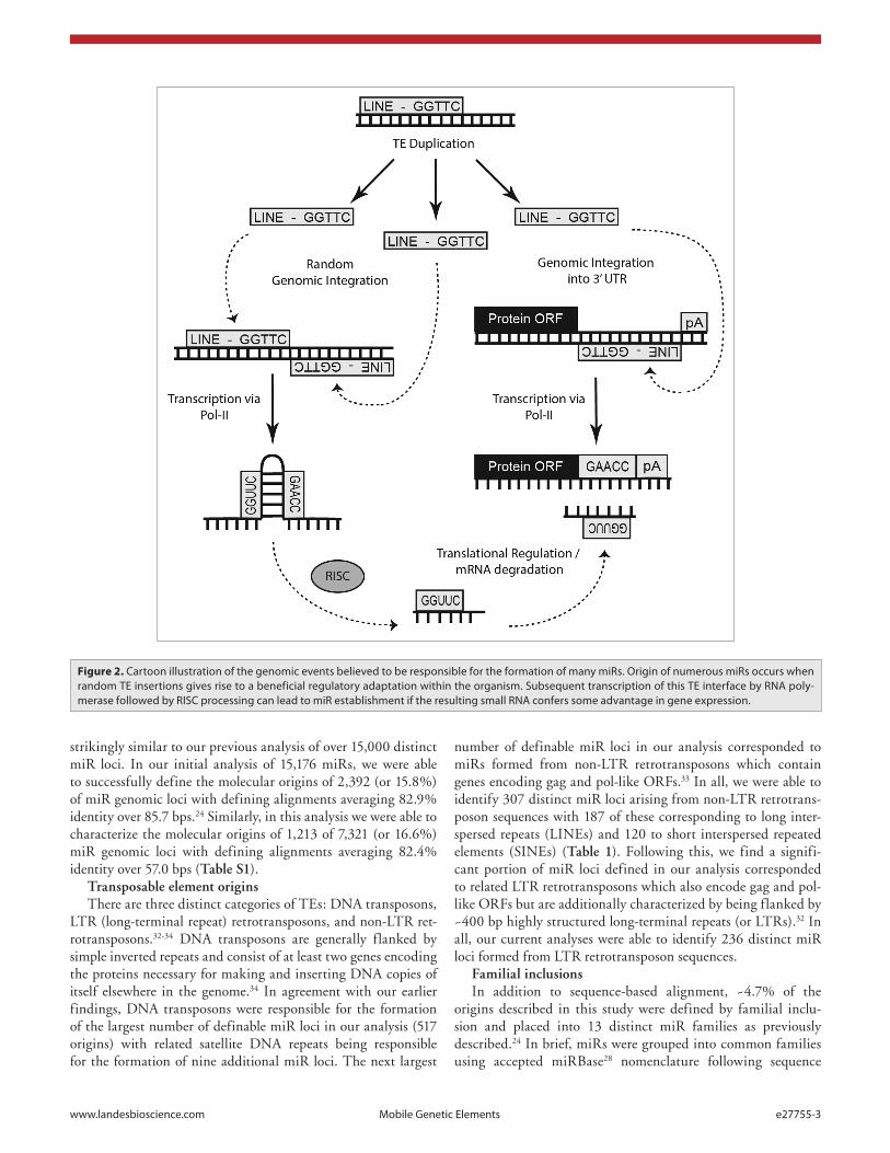
colonization—the miRs formed by this mechanism would likely be able to repress any mRNAs bearing sequences obtained from that miR’s progenitor TE.2 This suggests that a network of target mRNAs bearing a common TE is likely formed prior to the creation of the miR locus that ultimately regulates it (Fig. 2). While there are several addi-tional implications of miRs originating from TE sequences (e.g., determin-ing transcriptional regulation16), our recently developed miR target predic-tion algorithm (OrbId: Origin-based identification of microRNA targets19) suggests that a particularly advanta-geous utility for this information is the refinement of target prediction through restricting putative targets to mRNAs which contain the TE giving rise to a particular miR. Therefore, in light of our having previously defined the genomic events behind the forma-tion of over 2,300 distinct miRs24 and having successfully developed a novel miR target prediction algorithm utiliz-ing this information to accurately pre-dict mRNAs targeted by miRs clearly formed from TE sequences,19 this report now details our continuing analysis of
this topic - a comprehensive update examining the over 7,000 novel miRs described since our initial study.28 To our surprise, in addition to characterizing over 1,000 new miR molecular origins from TEs, we also find evidence suggesting that a subset of miRs actually arose from a distinct, previously undescribed mechanism - RNA structural alteration resulting from point mutations to noncoding RNAs such as tRNAs and snoRNAs. In contrast to miRs formed from TEs (which likely target mRNAs bearing their progenitor TE sequences), it is tempting to speculate that instead of regulating mRNA expression like the majority of characterized miRs, this novel subset of noncoding RNA-derived miRs may actually be charged with regulating the activity of their progeni-tor noncoding RNAs.
Results
To determine if any of the 7,321 miRs identified since the com-pletion of our initial analysis24 were formed from TE sequences, we screened these miR loci against all known repetitive ele-ments22,29 and noncoding RNAs30 using an established BLAST-based strategy.31 Through these analyses, we were able to define the molecular events responsible for 1,213 miR origins. In agree-ment with previous studies,17,18,20,24-27 our results indicate that the predominant mechanism for miR formation from TE sequences occurred via the model illustrated in Figure 1. Encouragingly, we find the results of our current analysis of 7,321 miRs to be
www.landesbioscience.com Mobile Genetic elements e27755-3
strikingly similar to our previous analysis of over 15,000 distinct miR loci. In our initial analysis of 15,176 miRs, we were able to successfully define the molecular origins of 2,392 (or 15.8%) of miR genomic loci with defining alignments averaging 82.9% identity over 85.7 bps.24 Similarly, in this analysis we were able to characterize the molecular origins of 1,213 of 7,321 (or 16.6%) miR genomic loci with defining alignments averaging 82.4% identity over 57.0 bps (Table S1).
Transposable element originsThere are three distinct categories of TEs: DNA transposons,
LTR (long-terminal repeat) retrotransposons, and non-LTR ret-rotransposons.32-34 DNA transposons are generally flanked by simple inverted repeats and consist of at least two genes encoding the proteins necessary for making and inserting DNA copies of itself elsewhere in the genome.34 In agreement with our earlier findings, DNA transposons were responsible for the formation of the largest number of definable miR loci in our analysis (517 origins) with related satellite DNA repeats being responsible for the formation of nine additional miR loci. The next largest
number of definable miR loci in our analysis corresponded to miRs formed from non-LTR retrotransposons which contain genes encoding gag and pol-like ORFs.33 In all, we were able to identify 307 distinct miR loci arising from non-LTR retrotrans-poson sequences with 187 of these corresponding to long inter-spersed repeats (LINEs) and 120 to short interspersed repeated elements (SINEs) (Table 1). Following this, we find a signifi-cant portion of miR loci defined in our analysis corresponded to related LTR retrotransposons which also encode gag and pol-like ORFs but are additionally characterized by being flanked by ~400 bp highly structured long-terminal repeats (or LTRs).32 In all, our current analyses were able to identify 236 distinct miR loci formed from LTR retrotransposon sequences.
Familial inclusionsIn addition to sequence-based alignment, ~4.7% of the
origins described in this study were defined by familial inclu-sion and placed into 13 distinct miR families as previously described.24 In brief, miRs were grouped into common families using accepted miRBase28 nomenclature following sequence
Figure 2. cartoon illustration of the genomic events believed to be responsible for the formation of many miRs. Origin of numerous miRs occurs when random Te insertions gives rise to a beneficial regulatory adaptation within the organism. subsequent transcription of this Te interface by RNa poly-merase followed by RIsc processing can lead to miR establishment if the resulting small RNa confers some advantage in gene expression.
e27755-4 Mobile Genetic elements Volume 3
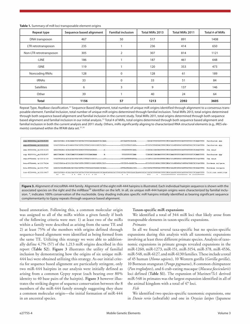
based annotation. Following this, a common molecular origin was assigned to all of the miRs within a given family if both of the following criteria were met: 1) at least two of the miRs within a family were described as arising from the same TE and 2) at least 75% of the members with origins defined through sequence-based alignment were identified as being formed from the same TE. Utilizing this strategy we were able to addition-ally define 4.7% (57) of the 1,213 miR origins described in this report (Table S2). Figure 3 illustrates the utility of familial inclusion by demonstrating how the origins of six unique miR-444 loci were obtained utilizing this strategy. As our initial crite-ria for sequence based alignment are particularly stringent, only two miR-444 hairpins in our analysis were initially defined as arising from a common Gypsy repeat (each bearing over 80% identity to 40 base pairs of the hairpin). Figure 3 however illus-trates the striking degree of sequence conservation between the 8 members of the miR-444 family strongly suggesting they share a common molecular origin—the initial formation of miR-444 in an ancestral species.
Taxon-specific miR expansionsWe identified a total of 344 miR loci that likely arose from
transposable elements in taxon-specific expansions.PrimatesIn all we found several taxa-specific but no species-specific
expansions during this analysis with all taxonomic expansions involving at least three different primate species. Analysis of taxo-nomic expansions in primate groups revealed expansions in the miR-1260, miR-1273, miR-151, miR-3154, miR-378, miR-4536, miR-548, miR-6127, and miR-6130 families. These include a total of 45 human (Homo sapiens), 10 Western gorilla (Gorilla gorilla), 10 Bornean orangutan (Pongo pygmaeus), 8 common chimpanzee (Pan troglodytes), and 6 crab-eating macaque (Macaca fascicularis) loci defined (Table S1). The expansion of Mariner/Tc1 derived miR-548 in primates was the largest expansion identified in all of the animal kingdom with a total of 47 loci.
FishWe identified two species-specific taxonomic expansions, one
in Danio rerio (zebrafish) and one in Oryzias latipes (Japanese
Table 1. summary of miR loci transposable element origins
Repeat type Sequence based alignment Familial inclusion Total MiRs 2013 Total MiRs 2011 Total # of MiRs
DNa transposon 467 50 517 891 1408
LTR retrotransposon 235 1 236 414 650
Non-LTR retrotransposon 305 2 307 814 1121
-LINe 186 1 187 461 648
-sINe 119 1 120 353 473
Noncoding RNas 128 0 128 61 189
tRNas 33 0 33 51 84
satellites 6 3 9 137 146
Other 39 1 40 24 64
Total 1156 57 1213 2392 3605
Repeat Type, RepBase classification.22 sequence Based alignment, total number of unique miR origins identified through alignment to a consensus trans-posable element. Familial Inclusion, total number of unique miR origins determined through familial inclusion. Total MiRs 2013, total origins determined through both sequence based alignment and familial inclusion in the current study. Total MiRs 2011, total origins determined through both sequence based alignment and familial inclusion in our initial analysis.24 Total # of MiRs, total origins determined through both sequence based alignment and familial inclusion in both the current analysis and 2011 study. Others, miRs significantly aligning to characterized RNa structural elements (e.g., IRes ele-ments) contained within the RFaM data set.35,36
Figure 3. alignment of microRNa-444 family. alignment of the eight miR-444 hairpins is illustrated. each individual hairpin sequence is shown with the associated species on the right and the miRBase28 identifier on the left. In all, six unique miR-444 hairpin origins were characterized by familial inclu-sion. *, indicates 100% conservation of the nucleotide. Grey shading indicates specific miR hairpins initially identified as bearing significant sequence complementarity to Gypsy repeats through sequence based alignment.
www.landesbioscience.com Mobile Genetic elements e27755-5
killifish). Our analyses identify 12 distinct miR-812 loci in the Japanese killifish genome arising from a variety of sources. The three distinct miR-2190 in the zebrafish, however, were found to have arisen from Atlantys-2-I-OS Gypsy Oryza elements (Table S1).
Mice/RatA total of five taxonomic expansions were found in the mouse/
rat grouping, with a total of 42 distinct hits. Our sequenced base alignments for the mouse (Mus musculus) show that the nine miR-467 loci were formed from TSEEEEBEII Mariner/Tc 1 ele-ments, and the ten miR-669 loci was formed from CR1-18-HM CR1 Hydra elements. Four possible origins for MIR-466 were also uncovered for Mus musculus and two for the rat Ratticus nor-vicus (Table S1).
PlantsOur analysis was able to define 201 loci across 31 distinct miR
families in plant genomes. The largest taxonomic expansion iden-tified in plants was for miR-156. There were 19 definitions across 13 species. The miR-5565 and miR-5568 families were popu-lated solely by Sorghum bicolor. Thirteen definitions were found across these two families, with nine definitions corresponding to Helitron-N11-SBi Helitron Sorghum elements and four cor-responding to STOWAWAY22-SB DNA elements. Twenty-nine definitions for Medicago truncatula were found across six miR families (miR-5298, miR-5291, miR-5272, miR-5741, miR-2592, and miR-2590). ShaMUDRAV2-MT MuDR Medicago, RF00028; Intron-gpI; AY098639.1/6-303, and MtPH-M-I-Ia Harbinger Medicago elements were common origins across these families (Table S1).
OtherFour loci from the miR-2284 family in the bull (Bos taurus)
genome were identified and three loci from the miR-3015 family in the pea aphid (Acrthosiphon pisum) genome apparently arising from DNA3-4-AP DNA elements (Table S1).
Non-transposable element originsAlthough ~87% of our annotations identified through
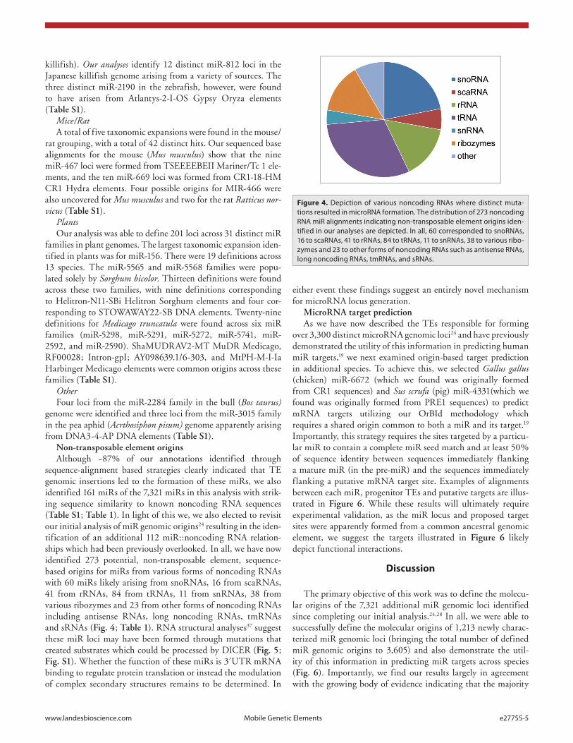
sequence-alignment based strategies clearly indicated that TE genomic insertions led to the formation of these miRs, we also identified 161 miRs of the 7,321 miRs in this analysis with strik-ing sequence similarity to known noncoding RNA sequences (Table S1; Table 1). In light of this we, we also elected to revisit our initial analysis of miR genomic origins24 resulting in the iden-tification of an additional 112 miR::noncoding RNA relation-ships which had been previously overlooked. In all, we have now identified 273 potential, non-transposable element, sequence-based origins for miRs from various forms of noncoding RNAs with 60 miRs likely arising from snoRNAs, 16 from scaRNAs, 41 from rRNAs, 84 from tRNAs, 11 from snRNAs, 38 from various ribozymes and 23 from other forms of noncoding RNAs including antisense RNAs, long noncoding RNAs, tmRNAs and sRNAs (Fig. 4; Table 1). RNA structural analyses37 suggest these miR loci may have been formed through mutations that created substrates which could be processed by DICER (Fig. 5; Fig. S1). Whether the function of these miRs is 3′UTR mRNA binding to regulate protein translation or instead the modulation of complex secondary structures remains to be determined. In
either event these findings suggest an entirely novel mechanism for microRNA locus generation.
MicroRNA target predictionAs we have now described the TEs responsible for forming
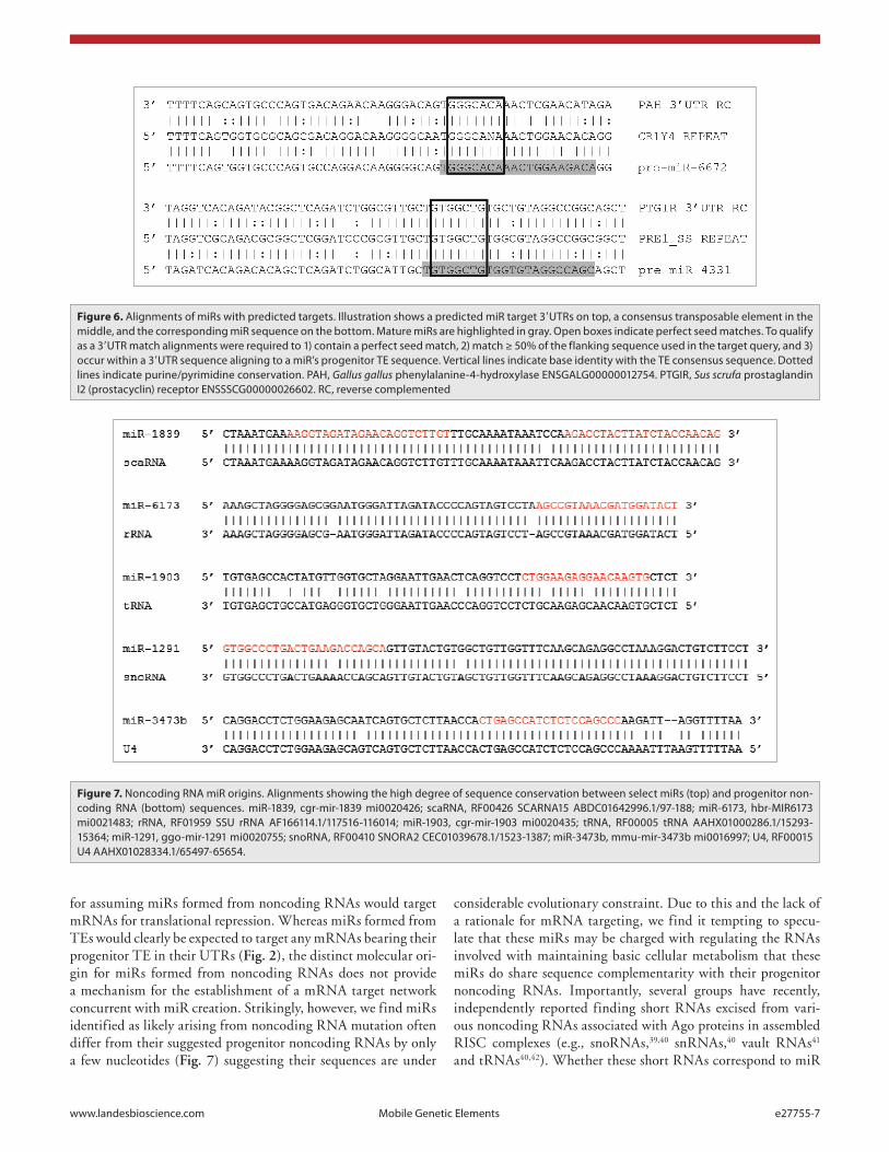
over 3,300 distinct microRNA genomic loci24 and have previously demonstrated the utility of this information in predicting human miR targets,19 we next examined origin-based target prediction in additional species. To achieve this, we selected Gallus gallus (chicken) miR-6672 (which we found was originally formed from CR1 sequences) and Sus scrufa (pig) miR-4331(which we found was originally formed from PRE1 sequences) to predict mRNA targets utilizing our OrBId methodology which requires a shared origin common to both a miR and its target.19 Importantly, this strategy requires the sites targeted by a particu-lar miR to contain a complete miR seed match and at least 50% of sequence identity between sequences immediately flanking a mature miR (in the pre-miR) and the sequences immediately flanking a putative mRNA target site. Examples of alignments between each miR, progenitor TEs and putative targets are illus-trated in Figure 6. While these results will ultimately require experimental validation, as the miR locus and proposed target sites were apparently formed from a common ancestral genomic element, we suggest the targets illustrated in Figure 6 likely depict functional interactions.
Discussion
The primary objective of this work was to define the molecu-lar origins of the 7,321 additional miR genomic loci identified since completing our initial analysis.24,28 In all, we were able to successfully define the molecular origins of 1,213 newly charac-terized miR genomic loci (bringing the total number of defined miR genomic origins to 3,605) and also demonstrate the util-ity of this information in predicting miR targets across species (Fig. 6). Importantly, we find our results largely in agreement with the growing body of evidence indicating that the majority
Figure 4. Depiction of various noncoding RNas where distinct muta-tions resulted in microRNa formation. The distribution of 273 noncoding RNa miR alignments indicating non-transposable element origins iden-tified in our analyses are depicted. In all, 60 corresponded to snoRNas, 16 to scaRNas, 41 to rRNas, 84 to tRNas, 11 to snRNas, 38 to various ribo-zymes and 23 to other forms of noncoding RNas such as antisense RNas, long noncoding RNas, tmRNas, and sRNas.
e27755-6 Mobile Genetic elements Volume 3
of functional miRs were created by mobile element transposi-tion events17,18,20,24-27 and suggest that future informatic analyses will continue to characterize additional miR-TE relationships as miR discovery remains ongoing (as evidenced by the 7,321 miR characterized in the two years since our initial analysis24,28). In addition, using our current computational methodology, we con-sistently find we are able to characterize the genomic origins of ~15% of miR loci. That said, we suggest this represents a marked underestimate of the percentage of miR loci formed from TEs as: 1) the high degree of stringency we required for our alignments resulted in our discarding thousands of likely origins likely repre-senting miRs whose nonessential sequences have simply degener-ated more than younger miR loci having been formed earlier in evolutionary time, and 2) our ability to identify transposable ele-ments will also improve as the RepBase22,23,29 data set is updated with novel TEs.
Excitingly, in addition to characterizing the origins of over 1,000 new miR loci from transposable element sequences, we have now also identified 273 miRs likely formed from noncoding RNA mutations. Interestingly, despite apparently having arisen through entirely distinct mechanisms, a preliminary analysis of the genomic distributions of, and available chromatin interactome data on,38 these miRs find no significant differences between noncoding RNA-derived miRs, TE-derived miRs or miR distri-butions as a whole. We speculate that point mutations to noncod-ing RNA secondary structures resulted in the formation of stable hairpins suitable for processing by the RNAi machinery. Indeed, mfold37 RNA structural analyses support this hypothesis as we find single point mutations to noncoding RNAs (e.g., tRNAs) can result in conformational changes that create substrates potentially processed by DICER (Fig. 5; Fig. S1). Unlike miRs formed from TE sequences, however, we see no clear rationale
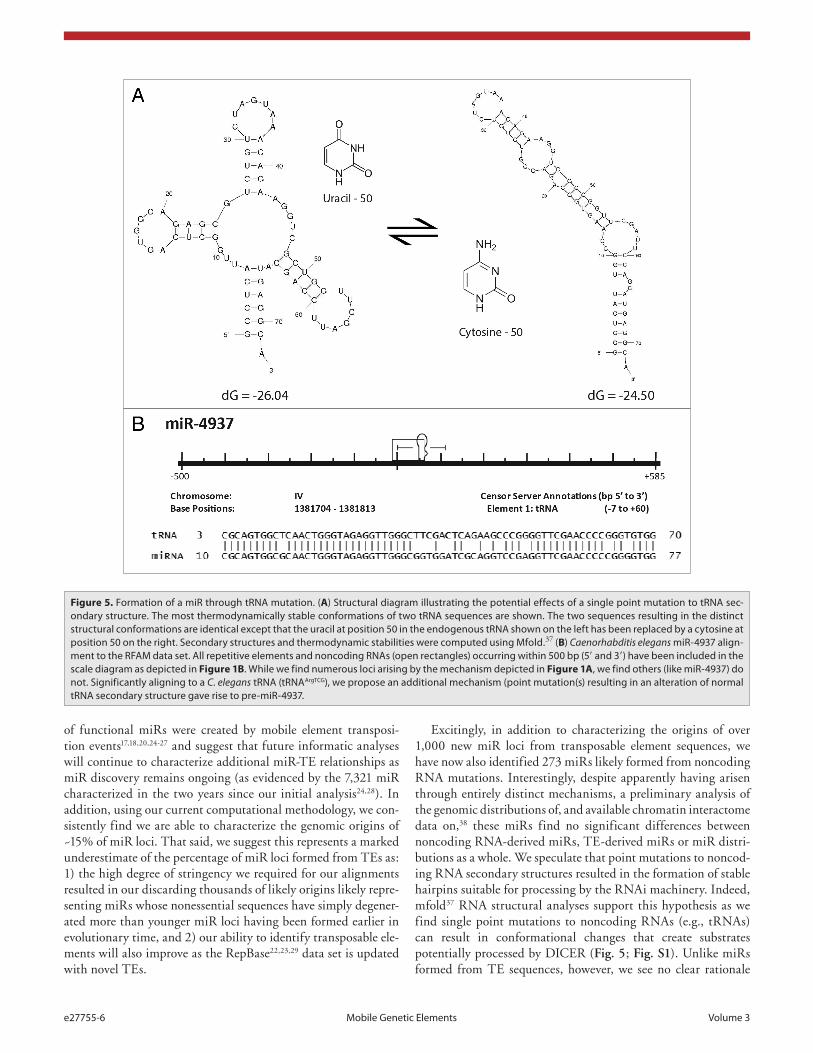
Figure 5. Formation of a miR through tRNa mutation. (A) structural diagram illustrating the potential effects of a single point mutation to tRNa sec-ondary structure. The most thermodynamically stable conformations of two tRNa sequences are shown. The two sequences resulting in the distinct structural conformations are identical except that the uracil at position 50 in the endogenous tRNa shown on the left has been replaced by a cytosine at position 50 on the right. secondary structures and thermodynamic stabilities were computed using Mfold.37 (B) Caenorhabditis elegans miR-4937 align-ment to the RFaM data set. all repetitive elements and noncoding RNas (open rectangles) occurring within 500 bp (5′ and 3′) have been included in the scale diagram as depicted in Figure 1B. While we find numerous loci arising by the mechanism depicted in Figure 1A, we find others (like miR-4937) do not. significantly aligning to a C. elegans tRNa (tRNaargTcG), we propose an additional mechanism (point mutation(s) resulting in an alteration of normal tRNa secondary structure gave rise to pre-miR-4937.
www.landesbioscience.com Mobile Genetic elements e27755-7
for assuming miRs formed from noncoding RNAs would target mRNAs for translational repression. Whereas miRs formed from TEs would clearly be expected to target any mRNAs bearing their progenitor TE in their UTRs (Fig. 2), the distinct molecular ori-gin for miRs formed from noncoding RNAs does not provide a mechanism for the establishment of a mRNA target network concurrent with miR creation. Strikingly, however, we find miRs identified as likely arising from noncoding RNA mutation often differ from their suggested progenitor noncoding RNAs by only a few nucleotides (Fig. 7) suggesting their sequences are under
considerable evolutionary constraint. Due to this and the lack of a rationale for mRNA targeting, we find it tempting to specu-late that these miRs may be charged with regulating the RNAs involved with maintaining basic cellular metabolism that these miRs do share sequence complementarity with their progenitor noncoding RNAs. Importantly, several groups have recently, independently reported finding short RNAs excised from vari-ous noncoding RNAs associated with Ago proteins in assembled RISC complexes (e.g., snoRNAs,39,40 snRNAs,40 vault RNAs41 and tRNAs40,42). Whether these short RNAs correspond to miR
Figure 7. Noncoding RNa miR origins. alignments showing the high degree of sequence conservation between select miRs (top) and progenitor non-coding RNa (bottom) sequences. miR-1839, cgr-mir-1839 mi0020426; scaRNa, RF00426 scaRNa15 aBDc01642996.1/97-188; miR-6173, hbr-MIR6173 mi0021483; rRNa, RF01959 ssU rRNa aF166114.1/117516-116014; miR-1903, cgr-mir-1903 mi0020435; tRNa, RF00005 tRNa aahX01000286.1/15293-15364; miR-1291, ggo-mir-1291 mi0020755; snoRNa, RF00410 sNORa2 cec01039678.1/1523-1387; miR-3473b, mmu-mir-3473b mi0016997; U4, RF00015 U4 aahX01028334.1/65497-65654.
Figure 6. alignments of miRs with predicted targets. Illustration shows a predicted miR target 3′UTRs on top, a consensus transposable element in the middle, and the corresponding miR sequence on the bottom. Mature miRs are highlighted in gray. Open boxes indicate perfect seed matches. To qualify as a 3′UTR match alignments were required to 1) contain a perfect seed match, 2) match ≥ 50% of the flanking sequence used in the target query, and 3) occur within a 3′UTR sequence aligning to a miR’s progenitor Te sequence. Vertical lines indicate base identity with the Te consensus sequence. Dotted lines indicate purine/pyrimidine conservation. Pah, Gallus gallus phenylalanine-4-hydroxylase eNsGaLG00000012754. PTGIR, Sus scrufa prostaglandin I2 (prostacyclin) receptor eNssscG00000026602. Rc, reverse complemented
e27755-8 Mobile Genetic elements Volume 3
precursors formed through mutation as we have suggested in this work, or if instead some percentage of noncoding RNAs are at times processed by the RNAi machinery to participate in nor-mal cellular regulation will ultimately require further examina-tion. That said, RISC complexes carrying these noncoding RNA pieces likely target sequences through the same means as those bearing their traditional miR counterparts—through sequence complementarity. If so, we suggest simple association of these ~20 nt RNAs with their ~100 nt corresponding progenitor full-length noncoding RNAs would likely function by rendering the targeted ncRNAs inactive through disruption of their essential structural motifs similar to several characterized riboswitches (reviewed in refs. 43-45). However, whether the function of these miRs is to regulate protein translation through mRNA 3′UTR binding or instead noncoding regulation through conformational modula-tion due to miR association will for now remain undetermined. In either event, the relationships between these miRs and related noncoding RNAs identified in this work suggest a second, pre-viously undescribed mechanism potentially responsible for miR locus formation.
Materials and Methods
MiR and 3′UTR sequence retrievalFASTA files containing complete sets of mature and stem
loop miR sequences were obtained from miRBase28 (http://www.mirbase.org/). Full sets of ENSEMBL 3′UTR sequences and miR loci flanking genomic sequences were obtained using the Biomart utility46 (www.ensembl.org/biomart/martview).
Screening miR loci for repetitive originsAs a control, all annotations and alignment analyses were
run identically in parallel by two distinct individuals and then compared for verification. FASTA files containing individual miR stem loops as well as miRs with 500 nucleotides of flanking sequence both upstream and downstream (when available) were aligned against the full RepBase22 annotated repetitive elements data set, the RFAM noncoding RNA collection35 (www.sanger.
ac.uk/Software/Rfam), and tRNAscan-SE47 (lowelab.cse.ucsc.edu/GtRNAdb) using stand-alone BLAST31 (BLASTN 2.2.15 with -FF, -W7, -e.1 flags). Alignments recognized as positive relationships were strictly defined as ≥ 80% identity to at least 40 nt or ≥ 70% identity to at least 50 nt of a miR hairpin. The highest scoring alignment for each pre-miR (averaging 82.4% identity over 57.0 nts) was taken to correspond to initial miR ori-gins. Upon completion of sequence based origin characterization, miRs were sorted into familial clusters using miRBase nomencla-ture.28 Common familial origins were defined if: 1) the same TE scored the best alignment to multiple miR family members and 2) at least 75% of all family members produced significant align-ments to the best scoring TE.
MiR target predictionMiR hairpin sequences were screened against the correspond-
ing set of 3′ UTRs currently available in Ensembl Biomart46 for that species using BLASTN31 2.2.15 with -FF,, -S2, -W7 flags. Putative targets were required to 1) contain perfect miR seed matches, 2) contain ≥ 50% identity to at least 50 base pairs of flanking sequences, and 3) be contained within 3′UTR sequences annotated as being the same TE as the miR’s progenitor TE by RepBase.29
RNA structural conformation modelingSecondary structures and thermodynamic stabilities were
computed using Mfold.37 The most thermodynamically stable conformation of individual miRNA sequences were determined when unaltered as well as with single nucleotide changes at posi-tions differing from identified progenitors.
Disclosure of Potential Conflicts of Interest
We, the authors, declare no financial or nonfinancial conflicts of interest.
Acknowledgments
This work was funded by the Department of Biology, the College of Arts and Sciences at the University of South Alabama.
References1. Lee RC, Feinbaum RL, Ambros V. The C. elegans
heterochronic gene lin-4 encodes small RNAs with antisense complementarity to lin-14. Cell 1993; 75:843-54; PMID:8252621; http://dx.doi.org/10.1016/0092-8674(93)90529-Y
2. Hutvágner G, Zamore PD. A microRNA in a multi-ple-turnover RNAi enzyme complex. Science 2002; 297:2056-60; PMID:12154197; http://dx.doi.org/10.1126/science.1073827
3. Fire A, Xu S, Montgomery MK, Kostas SA, Driver SE, Mello CC. Potent and specific genetic interfer-ence by double-stranded RNA in Caenorhabditis elegans. Nature 1998; 391:806-11; PMID:9486653; http://dx.doi.org/10.1038/35888
4. Cai X, Hagedorn CH, Cullen BR. Human microR-NAs are processed from capped, polyadenylated transcripts that can also function as mRNAs. RNA 2004; 10:1957-66; PMID:15525708; http://dx.doi.org/10.1261/rna.7135204
5. Smalheiser NR, Torvik VI. Complications in mam-malian microRNA target prediction. Methods Mol Biol 2006; 342:115-27; PMID:16957371
6. Zeng Y, Wagner EJ, Cullen BR. Both natural and designed micro RNAs can inhibit the expression of cognate mRNAs when expressed in human cells. Mol Cell 2002; 9:1327-33; PMID:12086629; http://dx.doi.org/10.1016/S1097-2765(02)00541-5
7. Lai EC. Micro RNAs are complementary to 3′ UTR sequence motifs that mediate negative post-tran-scriptional regulation. Nat Genet 2002; 30:363-4; PMID:11896390; http://dx.doi.org/10.1038/ng865
8. Burgler C, Macdonald PM. Prediction and verifi-cation of microRNA targets by MovingTargets, a highly adaptable prediction method. BMC Genomics 2005; 6:88; PMID:15943864; http://dx.doi.org/10.1186/1471-2164-6-88
9. Krüger J, Rehmsmeier M. RNAhybrid: microRNA target prediction easy, fast and flexible. Nucleic Acids Res 2006; 34:W451-4; PMID:16845047; http://dx.doi.org/10.1093/nar/gkl243
10. Lewis BP, Shih IH, Jones-Rhoades MW, Bartel DP, Burge CB. Prediction of mammalian microRNA targets. Cell 2003; 115:787-98; PMID:14697198; http://dx.doi.org/10.1016/S0092-8674(03)01018-3
11. Nam JW, Shin KR, Han J, Lee Y, Kim VN, Zhang BT. Human microRNA prediction through a probabilistic co-learning model of sequence and structure. Nucleic Acids Res 2005; 33:3570-81; PMID:15987789; http://dx.doi.org/10.1093/nar/gki668
12. Rehmsmeier M, Steffen P, Hochsmann M, Giegerich R. Fast and effective prediction of microRNA/target duplexes. RNA 2004; 10:1507-17; PMID:15383676; http://dx.doi.org/10.1261/rna.5248604
13. Rhoades MW, Reinhart BJ, Lim LP, Burge CB, Bartel B, Bartel DP. Prediction of plant microRNA targets. Cell 2002; 110:513-20; PMID:12202040; http://dx.doi.org/10.1016/S0092-8674(02)00863-2
14. Saetrom O, Snøve O Jr., Saetrom P. Weighted sequence motifs as an improved seeding step in microRNA target prediction algorithms. RNA 2005; 11:995-1003; PMID:15928346; http://dx.doi.org/10.1261/rna.7290705
15. Wang X, Wang X. Systematic identification of microRNA functions by combining target predic-tion and expression profiling. Nucleic Acids Res 2006; 34:1646-52; PMID:16549876; http://dx.doi.org/10.1093/nar/gkl068
http://www.ncbi.nlm.nih.gov/entrez/query.fcgi?cmd=Retrieve&db=PubMed&list_uids=8252621&dopt=Abstract
www.landesbioscience.com Mobile Genetic elements e27755-9
16. Borchert GM, Lanier W, Davidson BL. RNA poly-merase III transcribes human microRNAs. Nat Struct Mol Biol 2006; 13:1097-101; PMID:17099701; http://dx.doi.org/10.1038/nsmb1167
17. Devor EJ, Peek AS, Lanier W, Samollow PB. Marsupial-specific microRNAs evolved from marsupial-specific transposable elements. Gene 2009; 448:187-91; PMID:19577616; http://dx.doi.org/10.1016/j.gene.2009.06.019
18. Diao XM, Lisch D. Mutator transposon in maize and MULEs in the plant genome. Yi Chuan Xue Bao 2006; 33:477-87; PMID:16800377; http://dx.doi.org/10.1016/S0379-4172(06)60075-9
19. Filshtein TJ, Mackenzie CO, Dale MD, Dela-Cruz PS, Ernst DM, Frankenberger EA, He C, Heath KL, Jones AS, Jones DK, et al. OrbId: Origin-based iden-tification of microRNA targets. Mob Genet Elements 2012; 2:184-92; PMID:23087843; http://dx.doi.org/10.4161/mge.21617
20. Smalheiser NR, Torvik VI. Mammalian microR-NAs derived from genomic repeats. Trends Genet 2005; 21:322-6; PMID:15922829; http://dx.doi.org/10.1016/j.tig.2005.04.008
21. Venter JC, Adams MD, Myers EW, Li PW, Mural RJ, Sutton GG, Smith HO, Yandell M, Evans CA, Holt RA, et al. The sequence of the human genome. Science 2001; 291:1304-51; PMID:11181995; http://dx.doi.org/10.1126/science.1058040
22. Jurka J, Kapitonov VV, Pavlicek A, Klonowski P, Kohany O, Walichiewicz J. Repbase Update, a data-base of eukaryotic repetitive elements. Cytogenet Genome Res 2005; 110:462-7; PMID:16093699; http://dx.doi.org/10.1159/000084979
23. Tempel S, Jurka M, Jurka J. VisualRepbase: an interface for the study of occurrences of trans-posable element families. BMC Bioinformatics 2008; 9:345; PMID:18710569; http://dx.doi.org/10.1186/1471-2105-9-345
24. Borchert GM, Holton NW, Williams JD, Hernan WL, Bishop IP, Dembosky JA, Elste JE, Gregoire NS, Kim JA, Koehler WW, et al. Comprehensive analysis of microRNA genomic loci identifies pervasive repet-itive-element origins. Mob Genet Elements 2011; 1:8-17; PMID:22016841; http://dx.doi.org/10.4161/mge.1.1.15766
25. Piriyapongsa J, Jordan IK. A family of human microRNA genes from miniature inverted-repeat transposable elements. PLoS One 2007; 2:e203; PMID:17301878; http://dx.doi.org/10.1371/journal.pone.0000203
26. Yan Y, Zhang Y, Yang K, Sun Z, Fu Y, Chen X, Fang R. Small RNAs from MITE-derived stem-loop precursors regulate abscisic acid signal-ing and abiotic stress responses in rice. Plant J 2011; 65:820-8; PMID:21251104; http://dx.doi.org/10.1111/j.1365-313X.2010.04467.x
27. Yao C, Zhao B, Li W, Li Y, Qin W, Huang B, Jin Y. Cloning of novel repeat-associated small RNAs derived from hairpin precursors in Oryza sativa. Acta Biochim Biophys Sin (Shanghai) 2007; 39:829-34; PMID:17989873; http://dx.doi.org/10.1111/j.1745-7270.2007.00346.x
28. Kozomara A, Griffiths-Jones S. miRBase: inte-grating microRNA annotation and deep-sequenc-ing data. Nucleic Acids Res 2011; 39:D152-7; PMID:21037258; http://dx.doi.org/10.1093/nar/gkq1027
29. Kohany O, Gentles AJ, Hankus L, Jurka J. Annotation, submission and screening of repetitive elements in Repbase: RepbaseSubmitter and Censor. BMC Bioinformatics 2006; 7:474; PMID:17064419; http://dx.doi.org/10.1186/1471-2105-7-474
30. Burge SW, Daub J, Eberhardt R, Tate J, Barquist L, Nawrocki EP, Eddy SR, Gardner PP, Bateman A. Rfam 11.0: 10 years of RNA families. Nucleic Acids Res 2013; 41:D226-32; PMID:23125362; http://dx.doi.org/10.1093/nar/gks1005
31. Altschul SF, Madden TL, Schäffer AA, Zhang J, Zhang Z, Miller W, Lipman DJ. Gapped BLAST and PSI-BLAST: a new generation of protein database search programs. Nucleic Acids Res 1997; 25:3389-402; PMID:9254694; http://dx.doi.org/10.1093/nar/25.17.3389
32. Bushman FD. Targeting survival: integration site selection by retroviruses and LTR-retrotransposons. Cell 2003; 115:135-8; PMID:14567911; http://dx.doi.org/10.1016/S0092-8674(03)00760-8
33. Konkel MK, Batzer MA. A mobile threat to genome stability: The impact of non-LTR retrotranspo-sons upon the human genome. Semin Cancer Biol 2010; 20:211-21; PMID:20307669; http://dx.doi.org/10.1016/j.semcancer.2010.03.001
34. Ni J, Clark KJ, Fahrenkrug SC, Ekker SC. Transposon tools hopping in vertebrates. Brief Funct Genomic Proteomic 2008; 7:444-53; PMID:19109308; http://dx.doi.org/10.1093/bfgp/eln049
35. Griffiths-Jones S, Moxon S, Marshall M, Khanna A, Eddy SR, Bateman A. Rfam: annotating non-cod-ing RNAs in complete genomes. Nucleic Acids Res 2005; 33:D121-4; PMID:15608160; http://dx.doi.org/10.1093/nar/gki081
36. Griffiths-Jones S, Bateman A, Marshall M, Khanna A, Eddy SR. Rfam: an RNA family database. Nucleic Acids Res 2003; 31:439-41; PMID:12520045; http://dx.doi.org/10.1093/nar/gkg006
37. Zuker M. Mfold web server for nucleic acid fold-ing and hybridization prediction. Nucleic Acids Res 2003; 31:3406-15; PMID:12824337; http://dx.doi.org/10.1093/nar/gkg595
38. Chen D, Fu LY, Zhang Z, Li G, Zhang H, Jiang L, Harrison AP, Shanahan HP, Klukas C, Zhang HY, et al. Dissecting the chromatin interactome of microRNA genes. Nucleic Acids Res 2013; In press; PMID:24357409.
39. Ender C, Krek A, Friedländer MR, Beitzinger M, Weinmann L, Chen W, Pfeffer S, Rajewsky N, Meister G. A human snoRNA with microRNA-like func-tions. Mol Cell 2008; 32:519-28; PMID:19026782; http://dx.doi.org/10.1016/j.molcel.2008.10.017
40. Burroughs AM, Ando Y, de Hoon MJ, Tomaru Y, Suzuki H, Hayashizaki Y, Daub CO. Deep-sequencing of human Argonaute-associated small RNAs provides insight into miRNA sorting and reveals Argonaute association with RNA frag-ments of diverse origin. RNA Biol 2011; 8:158-77; PMID:21282978; http://dx.doi.org/10.4161/rna.8.1.14300
41. Persson H, Kvist A, Vallon-Christersson J, Medstrand P, Borg A, Rovira C. The non-coding RNA of the multidrug resistance-linked vault particle encodes multiple regulatory small RNAs. Nat Cell Biol 2009; 11:1268-71; PMID:19749744; http://dx.doi.org/10.1038/ncb1972
42. Haussecker D, Huang Y, Lau A, Parameswaran P, Fire AZ, Kay MA. Human tRNA-derived small RNAs in the global regulation of RNA silencing. RNA 2010; 16:673-95; PMID:20181738; http://dx.doi.org/10.1261/rna.2000810
43. Henkin TM. Riboswitch RNAs: using RNA to sense cellular metabolism. Genes Dev 2008; 22:3383-90; PMID:19141470; http://dx.doi.org/10.1101/gad.1747308
44. Breaker RR. Riboswitches and the RNA world. Cold Spring Harb Perspect Biol 2012; 4:4; PMID:21106649; http://dx.doi.org/10.1101/cshper-spect.a003566
45. Montange RK, Batey RT. Riboswitches: emerg-ing themes in RNA structure and function. Annu Rev Biophys 2008; 37:117-33; PMID:18573075; h t t p : / / d x . d o i . o r g / 1 0 . 1 1 4 6 / a n n u r e v .biophys.37.032807.130000
46. Durinck S, Moreau Y, Kasprzyk A, Davis S, De Moor B, Brazma A, Huber W. BioMart and Bioconductor: a powerful link between biological databases and microarray data analysis. Bioinformatics 2005; 21:3439-40; PMID:16082012; http://dx.doi.org/10.1093/bioinformatics/bti525
47. Chan PP, Lowe TM. GtRNAdb: a database of trans-fer RNA genes detected in genomic sequence. Nucleic Acids Res 2009; 37:D93-7; PMID:18984615; http://dx.doi.org/10.1093/nar/gkn787