Protein tyrosine kinase 6 regulates mammary gland tumorigenesis in mouse models
Upload
independentCategory
view
0download
0
A noncoding RNA is a potential marker of cell fateduring mammary gland developmentMelanie R. Ginger*, Amy N. Shore†, Alejandro Contreras*, Monique Rijnkels‡, Jonathan Miller§,Maria F. Gonzalez-Rimbau*, and Jeffrey M. Rosen*¶
Departments of *Molecular and Cellular Biology and §Biochemistry and Molecular Biology and †Program in Developmental Biology, Baylor College ofMedicine, 1 Baylor Plaza, Houston, TX 77030; and ‡U.S. Department of Agriculture�Agricultural Research Services Children’s Nutrition Research Center,Department of Pediatrics, Baylor College of Medicine, 1100 Bates Street, Houston, TX 77030
Communicated by Huda Y. Zoghbi, Baylor College of Medicine, Houston, TX, February 14, 2006 (received for review April 28, 2005)
PINC is a large, alternatively spliced, developmentally regulated,noncoding RNA expressed in the regressed terminal ductal lobularunit-like structures of the parous mammary gland. Previous studieshave shown that this population of cells possesses not onlyprogenitor-like qualities (the ability to proliferate and repopulatea mammary gland) and the ability to survive developmentallyprogrammed cell death but also the inhibition of carcinogen-induced proliferation. Here we report that PINC expression istemporally and spatially regulated in response to developmentalstimuli in vivo and that PINC RNA is localized to distinct foci ineither the nucleus or the cytoplasm in a cell-cycle-specific manner.Loss-of-function experiments suggest that PINC performs dualroles in cell survival and regulation of cell-cycle progression,suggesting that PINC may contribute to the developmentallymediated changes previously observed in the terminal ductallobular unit-like structures of the parous gland. This is one of thefirst reports describing the functional properties of a large, devel-opmentally regulated, mammalian, noncoding RNA.
parity � terminal ductal lobular unit
Noncoding RNAs (ncRNAs) constitute an important and ex-panding family of regulatory molecules. However, unlike their
orthodox cousins, the coding RNAs, which were hitherto thoughtto comprise the majority of transcriptional output in eukaryotes,their functions have been largely unexplored, in part because untilrecently their roles in gene regulation have not been fully appre-ciated. Moreover, ncRNAs are often present in low abundance;thus, their presence has for the large part evaded detection (1).
However, several findings demonstrate that ncRNAs constitutea large and significant proportion of the transcriptional output ofcomplex organisms, thus drawing attention to their importance aspotential regulatory molecules (1–8). In many cases, the expressionof ncRNAs appears to require finely tuned transcriptional andregulatory events (such as genomic imprinting or transcription fromconverging promoters), which in numerous cases include the re-cruitment of spliceosomal machinery and nuclear export (1, 9–12).Moreover, many of these ncRNAs are expressed in a definedtemporal–spatial window, thus limiting their activities to a specificstage of development or particular tissue type (see Y. Hayashizaki’sresponse to ref. 13) (1, 14, 15). These observations have led to thehypothesis that ncRNAs act by imposing a higher tier of regulationon a limited pool of structural and catalytic proteins (1, 16). Thishypothesis is of specific relevance to the processes of cell and tissuespecification and can thus be expected to have a major impact onour understanding of epigenetic processes guiding cell fate.
We previously reported the isolation of GB7, a novel ncRNAidentified during a screen for developmentally regulated, differen-tially expressed genes in the rodent mammary gland (17). We havenow designated GB7 as PINC for pregnancy-induced ncRNA,because it is induced by both pregnancy and hormonal stimulationof the mammary gland. Remarkably, PINC remains persistentlyelevated in cells in the regressed terminal ductal lobular unit-likestructures of the mammary gland 4 weeks after withdrawal of
hormonal stimuli. Interestingly, this population of cells has alsobeen shown to possess not only progenitor-like qualities but also theability to survive cell death during mammary gland involution andinhibition of carcinogen-induced proliferation (18–20). The cells ofthese terminal ductal lobular unit-like structures also retain thesignature of a developmentally mediated change in cell fate andpartial commitment to secretory differentiation (18). Previously, weproposed that pregnancy causes a change in the molecular pathwaysgoverning cell fate in the parous mammary gland (17, 20, 21). Oneplausible mechanism for how this effect might occur is by epigeneticchanges induced during pregnancy affecting the regulatory circuitscontrolling proliferation, survival, and gene expression (17, 22).Such changes restrict the fate of these cells long after the initialinductive event has been removed (23). The aforementioned de-velopmental observations and the noncoding properties of GB7(PINC) lead us to speculate that its expression comprises part of anepigenetic memory affecting cell fate decisions during development(20). Herein we characterize in detail PINC expression duringmammary gland development and cell-cycle progression in mam-mary epithelial cells, and we propose a functional role for PINC inthe regulation of intracellular pathways affecting proliferation andsurvival in the mammary gland. To our knowledge, this is one of thefirst reports describing the functional properties of a large, devel-opmentally regulated mammalian ncRNA.
ResultsComparative Analysis of the PINC Locus. Rat GB7 (PINC) is analternatively spliced ncRNA with a full-length transcript of 6.3 kb(GenBank accession no. AY035343; PINCA) (17) and severalshorter transcripts ranging in size from 1.2 to 4.8 kb (PINCB–PINCG) (Fig. 1A). Sequence analysis revealed multiple stop codonsand multiple short predicted ORFs (�200 nt) in all three frames ofthese additional transcripts (Table 1, which is published as sup-porting information on the PNAS web site), strongly suggesting thatthese transcripts are also noncoding. Despite this observation, allseven transcripts are spliced and polyadenylated and contain aconserved polyadenylation signal (AATAAA) located 15–33 ntfrom the polyA tail. These features, polyadenylation and splicing,provide strong evidence that PINC is the product of RNA poly-merase II-mediated transcription and is most likely subject to 5�capping as well (9).
To determine whether PINC contains any conserved featuresthat might provide clues to its function, we performed homologysearches against the mouse EST and nucleotide databases. Thesesearches identified two related mouse EST clones (GenBank
Conflict of interest statement: No conflicts declared.
Freely available online through the PNAS open access option.
Abbreviations: ncRNA, noncoding RNA; miRNA, microRNA; siRNA, small interfering RNA.
Data deposition: The sequences reported in this paper have been deposited in the GenBankdatabase (accession nos. AK021318, AK024261, AY035343, BE377788, DQ059755,DQ059756, DQ080210, DQ080211, DQ099682, DQ099683, DQ105700, and DQ105701).
¶To whom correspondence should be addressed. E-mail: [email protected].
© 2006 by The National Academy of Sciences of the USA
www.pnas.org�cgi�doi�10.1073�pnas.0600745103 PNAS � April 11, 2006 � vol. 103 � no. 15 � 5781–5786
CELL
BIO
LOG
Y

accession nos. DQ059755 and DQ059756), which appear to repre-sent alternatively spliced orthologs of rat PINC. The mouse PINCgene locus is depicted in Fig. 1B. DQ059755 and DQ059756 are1,630 and 1,017 nt in length, respectively (including polyA� tails),and are hereafter referred to as mPINC�1.6 and mPINC�1.0. Bothtranscripts are alternatively spliced and differ from one another bythe sequence of their terminal exons and also by the inclusion of anadditional exon in mPINC�1.0; however, neither contains a sizeableORF (�200 nt; Table 1) and are likely noncoding. Intriguingly, bothclones are derived from different mammary gland libraries, thusproviding independent evidence for the expression of PINC in themouse mammary gland. In addition, the mouse PINC locus over-laps with an independent transcriptional unit, RIKEN clone
D530049I02 (GenBank accession no. AK021318). mPINC andD530049I02 are encoded on opposite strands but do not share exonsequences, suggesting that they match the criteria of a ‘‘nonan-tisense bidirectional transcriptional locus’’ and thus are unlikely toregulate one another directly.
BLAST analyses comparing the full-length PINC sequence togenome assemblies and whole-genome sequence databases identi-fied regions in the genomes of the mouse, human, chimpanzee, dog,cow, and opossum that are syntenic with the rat PINC locus. Thecorresponding sequences were obtained through the University ofCalifornia at Santa Cruz genome browser (http:��genome.ucsc.edu) and aligned by using MULTIPIPMAKER (http:��pipmaker.bx.psu.edu�pipmaker; Fig. 1C). The multispecies alignment showedthe presence of two highly conserved regions (boxed regions in Fig.1C) present in the genomes of all seven species. Interestingly, thesehighly conserved regions correspond, in part, to the unique 3�-terminal regions of mPINC�1.0 and mPINC�1.6, suggesting thatconserved functional elements might be encoded in the exons ofthese two transcripts. In addition, some of this conserved sequencealso falls within introns, suggesting that introns may also containfunctional sequence elements. A very high level of homology wasobserved in the 5� flanking region of PINC in most species,suggesting the presence of conserved regulatory elements compris-ing a proximal promoter. No significant homology was detectedbetween the rat and chicken sequences for the PINC locus, althoughthe regions that flank the PINC locus are syntenic and showhomologies. In addition, rat PINC has no significant homology withany sequences within the fugu, zebrafish, or Xenopus genomes,suggesting that PINC might be a mammalian-specific gene. Toaddress the question of whether PINC contains a short conservedORF, we mapped ORFs encoding peptides of �50 aa from rat andmouse (Fig. 6, which is published as supporting information on thePNAS web site). However, none of these putative ORFs containeda homologous methionine codon in humans or other species.
To determine whether PINC encodes a microRNA (miRNA),we performed additional alignments with the rat, mouse, andhuman genomic sequences and identified several areas of extendedevolutionary conservation (corresponding to �70% homology)over the entire PINC genomic locus (data not shown). Rat se-quences comprising these regions were then subjected to secondarystructure analysis to identify conserved hairpin structures typical ofmiRNA precursors. Although two potential precursors with signif-icant similarity (E � 0.1 over a �50-nt span of homology) to thecorresponding region of the human genome were identified, neitherof the corresponding human sequences was predicted to form stablehairpin structures, suggesting that PINC does not encode a miRNA(Craig Burglar and Paul MacDonald, personal communication).
Expression of PINC. PINC was originally identified from a screen forgenes that are persistently up-regulated for at least 28 days afterpregnancy or after 21 days of treatment with estrogen (E) andprogesterone (P) (17). To determine whether PINC is a directtarget of E and P or, instead, a marker of alveologenesis, 42-day-oldintact rats were treated with E and P for either 2 days (acutetreatment) or 7 days (to promote alveologenesis), and PINC RNAlevels were examined by quantitative real-time PCR. Mammaryglands from 18-day pregnant rats were used as positive control forPINC expression. PINC was not detected after acute treatment withE�P. However, 7-day treatment with either E�P or P alone led toa marked induction of PINC expression, relative to control age-matched virgin glands; treatment with E alone resulted in low butdetectable levels of PINC. These data indicate that hormonaltreatment (E�P or P alone) sufficient to stimulate modest alveolarbudding (Fig. 7, which is published as supporting information on thePNAS web site) induces PINC expression.
We also examined the PINC expression profile at different stagesof mammary gland development (Fig. 2B). In the mammary gland,PINC expression was induced during pregnancy (18 days pregnant)
Fig. 1. Overview of the PINC genomic region. (A) Diagrammatic represen-tation of the rat PINC transcriptional locus showing the major 6.3-kb full-length transcript PINC A (GenBank accession no. AY035343) and alternatetranscripts B (GenBank accession no. DQ099683), C (GenBank accession no.DQ099682), D (GenBank accession no. DQ105700), E (GenBank accession no.DQ080210), F (GenBank accession no. DQ105701), and G (GenBank accessionno. DQ080211). Sizes of the transcripts are indicated. Transcripts initiate fromthe minus (�) strand and are presented relative to the plus (�) strand of therat genome. (B) Diagrammatic representation of the mouse PINC transcrip-tional locus showing the two transcripts (mPINC�1.6 and mPINC�1.0) identifiedfrom the mouse EST database. These transcripts contain unique 3�-terminalexons indicated by two boxed regions. The position of an additional EST clone(GenBank accession no. BE377788) is also indicated. All three transcriptsinitiate from the minus (�) strand and are presented relative to the plus (�)strand. The position of an independent overlapping transcriptional unit iden-tified as RIKEN clone D530049I02 (GenBank accession no. AK021318) is indi-cated. AK021318 is transcribed from the plus (�) strand. (C) Graphical outputfrom a MULTIPIPMAKER alignment of the PINC region and flanking sequencesfrom mouse (assembly May 2004), rat (assembly June 2003), human (assemblyMay 2004), chimpanzee, dog (assembly July 2004), cow, and opossum ge-nomes using the rat genomic sequence as reference sequence. The transcrip-tional orientation and position of exons of the main rat transcript (GenBankaccession no. AY035343) are indicated above the plot. Genomic featuresindicated by the colored underlay include conserved exons (light purple),introns (light yellow), alternative exons expressed in the mouse (light blue),and putative proximal promoter (light pink). The exon of an independent,overlapping gene (GenBank accession no. AK024261) is indicated by lightorange.
5782 � www.pnas.org�cgi�doi�10.1073�pnas.0600745103 Ginger et al.
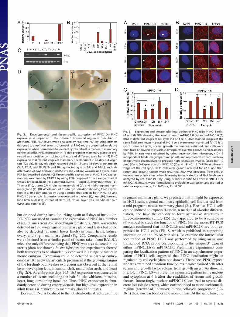
but dropped during lactation, rising again at 5 days of involution.RT-PCR was used to examine the expression of PINC in a numberof adult tissues from 96-day-old virgin female rats. PINC was readilydetected in 12-days-pregnant mammary gland and testes but couldalso be detected (at much lower levels) in brain, heart, kidney,ovary, and virgin mammary gland (Fig. 2C). Comparable resultswere obtained from a similar panel of tissues taken from BALB�cmice, the only difference being that PINC was also detected in theuterus (data not shown). In situ hybridization experiments showedboth transcripts to be abundantly expressed in a range of tissues inmouse embryos. Expression could be detected as early as embry-onic day 10.5 and was particularly prominent at the growing marginsof the forelimb bud; weaker expression was observed at the retinallayer, developing lens, intranasal cleft, mandibular arch, and heart(Fig. 2D). At embryonic days 14.5–16.5 expression was detected ina number of tissues including the hair follicle, whiskers, intestine,heart, lung, developing bone, etc. Thus, PINC expression is abun-dantly detected during embryogenesis, but high-level expression inadult tissues is restricted to mammary gland and testes.
Because PINC is localized to the lobuloalveolar structures of the
pregnant mammary gland, we predicted that it might be expressedin HC11 cells, a clonal mammary epithelial cell line derived froma mid-pregnant mouse mammary gland (24). Because HC11 cellscan be induced to express �-casein, a marker of alveolar differen-tiation, and have the capacity to form acinar-like structures inthree-dimensional culture (25) they appeared to be a suitable invitro model to study the function of PINC. Furthermore, RT-PCRanalysis confirmed that mPINC�1.6 and mPINC�1.0 are both ex-pressed in HC11 cells (Fig. 8, which is published as supportinginformation on the PNAS web site). To examine the intracellularlocalization of PINC, FISH was performed by using an in vitrotranscribed RNA probe corresponding to the unique 3� exon ofeither mPINC�1.6 or mPINC�1.0. Preliminary experiments com-paring the localization pattern of PINC in an asynchronous popu-lation of HC11 cells suggested that PINC localization might beregulated by cell cycle (data not shown). Therefore, PINC expres-sion was examined at various time points in synchronized cells afterserum and growth factor release from growth arrest. As shown inFig. 3A, mPINC�1.0 was present in a punctate pattern in the nucleusand cytoplasm at 6 h after the readdition of serum and growthfactors. Interestingly, nuclear mPINC�1.0 localized to several dis-crete foci (single arrow), which corresponded to more euchromaticregions (arrowhead); however, during cell-cycle progression (12–16 h) these nuclear foci became more diffuse. At the same time, the
Fig. 2. Developmental and tissue-specific expression of PINC. (A) PINCexpression in response to the different hormonal regimens described inMethods. PINC RNA levels were analyzed by real-time PCR by using primersdesigned to amplify all seven isoforms of rat PINC and are presented as relativeexpression when normalized to levels of cytokeratin 8 (a marker of mammaryepithelial cells). PINC expression in 18-day-pregnant mammary glands is pre-sented as a positive control (note the use of different scale bars). (B) PINCexpression at different stages of mammary development in 42-day-old virginrats (42d vir), 96-day-old virgin rats (96d vir), 5-, 12-, and 18-days-pregnant rats(5dP, 12dP, and 18dP), 2- and 10-days-lactating rats (2dL and 10dL), and ratsafter 5 and 28 days of involution (5d inv and 28d inv) was assessed by real-timePCR (as described above). (C) Tissue-specific expression of PINC. PINC expres-sion was examined by RT-PCR by using RNA prepared from a range of adulttissues: brain (B), heart (H), kidney (K), liver (Li), lung (Lu), ovary (O), testes (Te),Thymus (Th), uterus (U), virgin mammary gland (V), and mid-pregnant mam-mary gland (P). (D) Whole-mount in situ hybridization showing PINC expres-sion in a 10.5-day embryo by using a probe that detects both PINC�1.6 andPINC�1.0 transcripts. Expression was detected in the lens (L), heart (H), fore andhind limb buds (LB), intranasal cleft (IC), retinal layer (RL), mandibular arch(MA), and somites (S).
Fig. 3. Expression and intracellular localization of PINC RNA in HC11 cells.(A and B) FISH showing the localization of mPINC�1.0 (A) and mPINC�1.6 (B)RNA at different stages of cell cycle in HC11 cells. DAPI-stained images of thesame field are shown in parallel. HC11 cells were growth-arrested for 72 h tosynchronize cell cycle; normal growth medium was returned, and cells werethen fixed on coverslips at various time points over the next 24 h and examinedby FISH. Images were obtained by using deconvolution microscopy (10–12independent fields imaged per time point), and representative captured rawimages were deconvolved to produce high-resolution images. (Scale bar: 10�m.) (C and D) Expression of mPINC�1.0 (C) and mPINC�1.6 (D) RNA at differentstages of the cell cycle. HC11 cells were growth-arrested for 72 h, and thenserum and growth factors were returned. RNA was prepared from cells atvarious time points after cell-cycle reentry (as indicated), and RNA levels wereanalyzed by real-time PCR by using primers specific to either mPINC�1.0 ormPINC�1.6. Results were normalized to cyclophilin expression and plotted asrelative expression. *, P � 0.05; **, P � 0.005.
Ginger et al. PNAS � April 11, 2006 � vol. 103 � no. 15 � 5783
CELL
BIO
LOG
Y

cytoplasmic mPINC�1.0 signal increased, although mPINC�1.0 re-mained associated primarily with the perinuclear region (doublearrows). By 24 h after reentry into the cell cycle, the nuclear signaldiminished and the majority of the signal was localized to thecytoplasm. In contrast, the majority of mPINC�1.6 signal waslocalized to punctate granular foci within both the nucleus andcytoplasm throughout cell-cycle progression (Fig. 3B). Thus, itappears that localization of each transcript is regulated differen-tially and that PINC�1.0 localization is regulated in a cell-cycle-specific manner. Last, PINC signal was abrogated by treatmentbefore fixation with Triton X-100-containing CSK buffer, suggest-ing that PINC is not strongly associated with the nuclear andcytoplasmic matrix (Fig. 9, which is published as supporting infor-mation on the PNAS web site).
To determine whether PINC expression varies quantitatively atdifferent stages of the cell cycle, real-time PCR analysis wasperformed with RNA isolated from HC11 cells at various timepoints after cell-cycle reentry. In contrast to the FISH analysis,which strongly suggests cell-cycle-dependent regulation ofPINC�1.0 localization, total RNA PINC�1.0 levels do not varysignificantly in cycling cells. However, they do show a significantdecrease as the cells reenter the cell cycle (6-h time point; P � 0.05)and continue to remain low at 12 h after cell-cycle reentry (P �0.005). mPINC�1.6 levels, on the other hand, fell significantly (P �0.05) between 6 and 12 h, after readdition of normal growth media,and then slowly recovered by 24 h to the levels observed at the 6-htime point (Fig. 3D). Interestingly, mPINC�1.6 levels do not appearto vary greatly between the 6-h time point and in growth-arrestedcells, suggesting that there are no major changes in mPINC�1.6expression upon the reinitiation of cell cycle. Previous studies usingflow cytometry suggest that 53% of HC11 cells have enteredS-phase by 14 h after reinitiation of the cell cycle (26); thus, thedecrease in PINC expression occurred just before S-phase entry.
Loss-of-Function Experiments. To probe PINC’s function in greaterdetail, we performed knockdown experiments using several in vitrotranscribed small interfering RNAs (siRNAs) specific to eithermPINC�1.6 or mPINC�1.0 (Fig. 4A). Exponentially growing HC11swere transfected with both experimental and control siRNAs,growth-arrested, and analyzed for DNA replication by immuno-fluorescence detection of BrdU incorporation 6 h after cell-cyclereentry. As expected from previous flow cytometry analyses (26),the number of BrdU-positive cells was extremely low in both themock-transfected cells and those transfected with an unrelatedoligo (Fig. 4B). Similar numbers were observed with cells trans-fected with an oligo against mPINC�1.0 (1.0A). Surprisingly, cellstransfected with an oligo against mPINC�1.6 (1.6A) displayed a16-fold increase in the number of BrdU-positive cells, suggestingthat knocking down mPINC�1.6 facilitated the G1–S-phase transi-tion (Fig. 4C). Similar results were obtained with two additionalindependent oligos against mPINC�1.6 (P � 0.001) and also by flowcytometry analysis by using the same antibody against BrdU (datanot shown).
Because PINC expression was increased during mammary glandinvolution, when tissue remodeling and apoptosis is occurring, itwas of interest to examine the effect of PINC depletion on cellsurvival. Therefore, HC11s were transfected with siRNA againstmPINC�1.6, mPINC�1.0, or the unrelated oligo and then serum-starved for a period of 12 h before TUNEL analysis (Fig. 5A).Normal HC11s are relatively resistant to the effects of serumwithdrawal, as exemplified by the limited number of TUNEL-positive cells detected in mock-transfected HC11s. Cells transfectedwith siRNA against mPINC�1.6 (1.6A) or the unrelated oligo wereequally unaffected by serum withdrawal. However, cells transfectedwith siRNAs against mPINC�1.0 (1.0A, 1.0B, and 1.0C) showed amarked increase in the number of TUNEL-positive cells. Thiseffect was observed with four independent siRNAs againstmPINC�1.0 (1.0A, 1.0B, 1.0C, and 1.0D, the results with three of
which are shown in Fig. 5A) leading to a 7- to 13-fold increase inTUNEL-positive cells compared with the negative control. Similarresults were obtained by immunofluorescence using an antibodyagainst the cleaved form of caspase-3 (Fig. 5B).
DiscussionPINC is an alternatively spliced, polyadenylated, mRNA-likencRNA persistently expressed in the terminal ductal lobularunit-like structures of the parous rat mammary gland (17).Comparative sequence analysis revealed conserved sequences inthe genomes of at least seven mammalian species, leading to theidentification of putative orthologs of rat PINC as well as twohighly conserved regions with potential functional importance(Fig. 1). Independent evidence from the mouse EST databaserevealed that at least one of these orthologous genes is expressed,leading to two alternatively spliced isoforms in the mouse(mPINC�1.6 and mPINC�1.0). Although it lacks an apparentprotein coding function, its conserved syntenic location and the
Fig. 4. Knocking down mPINC�1.6 alters cell-cycle progression. (A) siRNAknockdown strategy. siRNAs were designed to target sequences in the unique3�-terminal exon of mPINC�1.0 and mPINC�1.6 (illustrated diagrammatically)to allow transcript-specific knockdown of each mPINC isoform. (B) Immuno-fluorescent detection of BrdU incorporation in HC11s transfected with siRNAsagainst mPINC�1.0 (1.0 A), mPINC�1.6 (1.6 A), or an unrelated negative controloligo and mock-transfected cells. Transfected cells were growth-arrested for72 h, and then normal growth medium was returned for 6 hours to reinitiatecell cycle. HC11s were labeled with BrdU for 45 min before fixation, and BrdUincorporation was measured by immunofluorescence by using a FITC-conjugated antibody against BrdU. (C) BrdU incorporation was measured byimmunofluorescence (as described in B) and calculated as the percentage ofBrdU-positive cells relative to total DAPI-stained nuclei averaged over sixindependent microscope fields. Knocking down mPINC�1.6 (1.6A) resulted ina 16-fold increase in the number of BrdU-positive cells compared with cellstransfected with negative control siRNA. The percentage of BrdU-positive cellstransfected with siRNA against mPINC�1.0 (1.0A) was similar to that of thecontrol cells. *, P � 0.002.
5784 � www.pnas.org�cgi�doi�10.1073�pnas.0600745103 Ginger et al.

degree of conservation between these orthologous genes sug-gests that evolutionary constraints have prevented the diver-gence of functional elements within this gene. Perhaps thegreatest clue to its function lies in the identification of tworegions of high evolutionary conservation that encompass se-quences specific to the unique 3�-terminal exons of eithermPINC �1.0 or mPINC �1.6. RNA interference knockdownexperiments using siRNAs specific to either of these exons (andthereby specific to either mPINC�1.0 or mPINC�1.6) point todistinct roles for mPINC�1.0 and mPINC�1.6 in cell survival andregulation of cell-cycle progression, respectively (Figs. 4 and 5),suggesting that an appropriate balance of the two transcripts isimportant for the maintenance of these cells. Thus, it is temptingto speculate that these conserved elements are central to thefunction of PINC. These elements perhaps contain binding sitesfor RNA-binding proteins, transcription factors, or even local-ization signals that target PINC to a particular cytoplasmiclocality.
In HC11 cells intracellular localization of PINC is regulated in acell-cycle- and transcript-specific manner, with distinct foci ob-served in both the nucleus and cytoplasm at different time pointsduring the cell cycle (Fig. 3). At the 6-h time point mPINC�1.0 signalwas observed as discrete euchromatic nuclear foci. These focibecame more diffuse and gradually disappeared as the cell cycleprogressed, correlating with a shift to a more cytoplasmic localiza-tion. These observations suggest a possible nuclear function formPINC�1.0, as well as a role in shuttling or signal transductionbetween the nucleus and cytoplasm. mPINC�1.6, on the other hand,is localized as discrete foci (suggestive of ribonucleoprotein parti-cles) throughout the nucleus and cytoplasm, possibly implying aninvolvement with intracellular trafficking. Changes in the levels oftotal PINC RNA suggest that the RNA is relatively unstable or thatits stability is regulated in a temporal fashion by its association withRNA-binding proteins.
Based on the known properties of other ncRNAs, what are themost likely functions of PINC? Because PINC does not appear toencode a conserved precursor structure, typical of a ‘‘classical’’miRNA (27), this type of function seems unlikely. However, recentevidence demonstrating the presence of primate-specific miRNAs(28) suggests that not all miRNAs are conserved; hence, thisfunction cannot be ruled out entirely. PINC does not seem tocomprise part of an imprinted locus, or at least is not located in agenomic location referenced by any database of imprinted loci
(www.mgu.har.mrc.ac.uk�research�imprinting�imprin-intro.html).Likewise, PINC does not appear to be part of a sense–antisensepair, but this categorization also depends on the completeness ofpublicly available databases. Evidence of splicing precludes thepossibility that PINC is an expressed pseudogene. Other roles forncRNAs, based on sequence-directed nucleic acid recognition,have also been proposed (1, 29–33), and it is possible that sequence(or even structural recognition) plays a role in the function of PINC.
The observation that PINC is present in discrete foci in thenucleus (Fig. 3) suggests it might play a role in guiding DNAmethylation, transcriptional modulation, or chromatin modificationin the nucleus. However, because the PINC RNA signal wasabolished by CSK extraction it is likely that it is associated withsoluble proteins that can move between the two compartmentsrather than with the nuclear and cytoplasmic matrix directly. Basedon the current evidence and the limited number of known mech-anisms for ncRNA function, we foresee a number of potential rolesfor PINC in transcriptional and�or chromatin modulation as alocalized RNA or as part of a cytoplasmic signal transductionpathway. We propose that the conserved domains present in the3�-terminal exons of mPINC�1.6 and mPINC�1.0 confer discretefunctions, and these functions might be mediated by an associationwith RNA-binding proteins or by sequence-directed nucleic acidrecognition.
Function for PINC in the Mammary Gland. We have shown that thelevels of mPINC�1.6 decrease before S-phase entry. Conversely,knocking down mPINC�1.6 leads to premature S-phase entry inHC11 cells. These observations suggest that PINC may play a rolein regulating cell-cycle progression and that PINC levels may needto be reduced to facilitate S-phase entry. Knocking downmPINC�1.0, on the other hand, predisposes HC11 cells to apoptosis,suggesting that mPINC�1.0 is a cell survival factor.
PINC is up-regulated during alveolar development in the ratmammary gland, but not by short-term treatment with E and P,suggesting that, although it may not be a direct target of thesehormones, PINC can serve as a marker of partially committedalveolar cell fate. Previous studies from our laboratory have shownthat PINC expression is enriched in a specific population ofmammary epithelial cells localized to the terminal ductal lobularunit-like structures of the involuted mammary gland (17). A similarpopulation of cells (20) have also been shown to possess thecapability not only to survive involution-mediated cell death (19)but also to resist further proliferative stimuli (such as exposure tochemical carcinogen) (34). These observations and the comple-mentary functions of mPINC�1.0 and mPINC�1.6 (modulating cellsurvival and cell-cycle regulation) suggest that PINC is more thanmerely a marker of alveologenesis. We postulate that PINC,therefore, may play a role in modulating cell fate in this specificpopulation of mammary epithelial cells and, as such, forms part ofthe ‘‘functional memory’’ or ‘‘epigenetic imprint’’ imposed by parity(17, 20, 22). Such epigenetic marks are important for preserving thecell’s identity and for preventing cell-lineage aberrations leading tocancer (23). To our knowledge, this is one of the first studies todemonstrate a function for a large, developmentally regulated,mammalian ncRNA.
MethodsClones and Sequence Analysis. All rat clones were obtained from arat mammary cDNA library as described previously (17). ESTclones (GenBank accession nos. DQ059755 and DQ059756) wereobtained from Invitrogen. Sequencing was performed by the Se-quencing Core at the Baylor College of Medicine.
The following computational tools were used: REPEATMASKER(www.repeatmasker.org), GENSCAN (http:��genes.mit.edu�GENSCAN.html), BLAST (www.ncbi.nlm.nih.gov�blast), andMULTIPIPMAKER. For visual representation of the PINC transcrip-
Fig. 5. Knocking down mPINC�1.0 induces apoptosis in serum-free condi-tions. (A) The percentage of TUNEL-positive cells in HC11s transfected withsiRNA against mPINC�1.0 (siRNAs 1.0A, 1.0B and 1.0C), mPINC�1.6 (1.6A),negative control siRNA, or mock-transfected siRNA was calculated relative tototal number of DAPI-stained nuclei. **, P � 0.0005; *, P � 0.003. (B) HC11swere transfected with siRNA against mPINC�1.0 (1.0A) or a negative controloligo, maintained under normal growth conditions for 48 h after transfection,and then placed in serum-free medium for 3 h before fixation. Caspase-3activation was examined by immunofluorescent detection of cleavedcaspase-3, and the percentage of cleaved caspase-3-positive cells was calcu-lated relative to the total number of DAPI-stained nuclei. **, P � 0.0005; *, P �0.003. Similar results were obtained with three independent siRNAs againstmPINC�1.0 (data not shown).
Ginger et al. PNAS � April 11, 2006 � vol. 103 � no. 15 � 5785
CELL
BIO
LOG
Y

tional locus, we used the University of California at Santa Cruzgenome browser.
Animals. Wistar–Furth rats and BALB�c mice were purchased fromHarlan Sprague–Dawley. Animals were housed in an approvedfacility using Association for Assessment of Laboratory AnimalCare guidelines. All experiments were performed in accordancewith the National Institutes of Health’s Guide for the Care and Useof Laboratory Animals using protocols approved by the BaylorCollege of Medicine Subcommittee for Animal Use.
Hormonal Manipulations and Developmental Time Points. Forty-two-day-old virgin rats were treated with a priming dose of estradiolbenzoate (Sigma) (2.5 �g per animal) as described previously (17),then divided into four groups and treated with 200 �g of E (n � 3),20 mg of P (n � 3), 200 �g of E and 20 mg P (E�P; n � 3), or vehiclealone [age-matched virgin (AMV); n � 4] for a period of 7 days.Hormones were delivered by s.c. implantation of silastic capsule asdescribed (35). For developmental time points, inguinal mammaryglands were excised from timed pregnant rats at 6, 12, and 18 daysof pregnancy, from 2- and 10-day lactating dams, and from rats after10 days of lactation and 5 and 28 days of forced involution. Inaddition, inguinal mammary glands were obtained from 45-day-old(immature) virgin and 96-day-old (mature) virgin rats. In all casesthe right gland was flash-frozen for RNA, and the left gland wasfixed in 10% neutral-buffered formalin (24 h at 4°C) and stained asdescribed previously (36) to ensure that they displayed morpho-logical features appropriate for each stage of development.
Tissue Culture and RNA Interference Knockdown Experiments. HC11cells (24), a clonal cell line of COMMA-D, were routinely main-tained as previously described (37). For experiments requiringsynchronization, normal growth medium was replaced with growtharrest medium (RPMI medium 1640 supplemented with 0.1%bovine calf serum). Double-stranded siRNA against mPINC�1.0and mPINC�1.6, as well as an unrelated nonspecific control siRNA
(Ambion), were prepared by using the Ambion siRNA Construc-tion kit. Transfection was performed by using 100 nM (finalconcentration) siRNA and siPORT Amine transfection reagent(Ambion) according to the manufacturer’s instructions. Mocktransfections were performed by using siPORT Amine alone (nosiRNA). Transfection efficiency was monitored by using Cy3-labeled tracer siRNA, and knockdown was monitored by in situhybridization (see Fig. 10, which is published as supporting infor-mation on the PNAS web site).
Proliferation and Apoptosis Assays. Proliferation was monitored byBrdU staining as described (38). Apoptosis was detected byTUNEL staining (37) or by immunofluorescence using an antibodyspecific to the cleaved (active) form of caspase-3 (Santa CruzBiotechnology).
Real-Time PCR. Quantitative RT-PCR with SYBR green detection(see Supporting Materials and Methods, which is published assupporting information on the PNAS web site) was performed asdescribed (39), and expression levels for rat PINC, mPINC1.6, andmPINC1.0 normalized to either cyclophilin or cytokeratin 8 levels(as indicated in the text).
In Situ Hybridization and FISH. In situ hybridization was performedby using digoxigenin-labeled complementary RNA probes andparaformaldehyde-fixed embryos or cells. For embryos, hybridiza-tion signals were detected by using an alkaline phosphatase-conjugated antibody against digoxigenin (Roche). For FISH, hy-bridization signals were detected by fluorescence (as described inSupporting Materials and Methods).
We thank Paul MacDonald and Craig Burglar for performing themiRNA prediction analysis, Daniel Medina for helpful discussions andcritical reading of the manuscript, and Panagiota Karagianni and Jung-soon Lee for work that contributed indirectly to this study. This researchwas supported by U.S. Department of Defense Breast Cancer ResearchProgram Grant DAMD17-00-1-0137 and National Institutes of HealthGrant CA 64255.
1. Mattick, J. S. (2003) BioEssays 25, 930–939.2. Okazaki, Y., Furuno, M., Kasukawa, T., Adachi, J., Bono, H., Kondo, S., Nikaido,
I., Osato, N., Saito, R., Suzuki, H., et al. (2002) Nature 420, 563–573.3. Kampa, D., Cheng, J., Kapranov, P., Yamanaka, M., Brubaker, S., Cawley, S.,
Drenkow, J., Piccolboni, A., Bekiranov, S., Helt, G., et al. (2004) Genome Res. 14,331–342.
4. Rodriguez, A., Griffiths-Jones, S., Ashurst, J. L. & Bradley, A. (2004) GenomeRes. 14, 1902–1910.
5. Waterston, R. H., Lindblad-Toh, K., Birney, E., Rogers, J., Abril, J. F., Agarwal,P., Agarwala, R., Ainscough, R., Alexandersson, M., An, P., et al. (2002) Nature420, 520–562.
6. Claverie, J. M. (2005) Science 309, 1529–1530.7. Mattick, J. S. (2005) Science 309, 1527–1528.8. Willingham, A. T., Orth, A. P., Batalov, S., Peters, E. C., Wen, B. G., Aza-Blanc,
P., Hogenesch, J. B. & Schultz, P. G. (2005) Science 309, 1570–1573.9. Numata, K., Kanai, A., Saito, R., Kondo, S., Adachi, J., Wilming, L. G., Hume,
D. A., Hayashizaki, Y. & Tomita, M. (2003) Genome Res. 13, 1301–1306.10. Kiyosawa, H., Yamanaka, I., Osato, N., Kondo, S. & Hayashizaki, Y. (2003)
Genome Res. 13, 1324–1334.11. Nikaido, I., Saito, C., Mizuno, Y., Meguro, M., Bono, H., Kadomura, M., Kono,
T., Morris, G. A., Lyons, P. A., Oshimura, M., et al. (2003) Genome Res. 13,1402–1409.
12. Suzuki, M. & Hayashizaki, Y. (2004) BioEssays 26, 833–843.13. Wang, J., Zhang, J., Zheng, H., Li, J., Liu, D., Li, H., Samudrala, R., Yu, J. &
Wong, G. K. (2004) Nature 431, discussion following p. 757.14. Bono, H., Yagi, K., Kasukawa, T., Nikaido, I., Tominaga, N., Miki, R., Mizuno,
Y., Tomaru, Y., Goto, H., Nitanda, H., et al. (2003) Genome Res. 13, 1318–1323.15. Cawley, S., Bekiranov, S., Ng, H. H., Kapranov, P., Sekinger, E. A., Kampa, D.,
Piccolboni, A., Sementchenko, V., Cheng, J., Williams, A. J., et al. (2004) Cell 116,499–509.
16. Mattick, J. S. (2001) EMBO Rep. 2, 986–991.17. Ginger, M. R., Gonzalez-Rimbau, M. F., Gay, J. P. & Rosen, J. M. (2001) Mol.
Endocrinol. 15, 1993–2009.18. Wagner, K. U., Boulanger, C. A., Henry, M. D., Sgagias, M., Hennighausen, L.
& Smith, G. H. (2002) Development (Cambridge, U.K.) 129, 1377–1386.19. Sivaraman, L., Conneely, O. M., Medina, D. & O’Malley, B. W. (2001) Proc. Natl.
Acad. Sci. USA 98, 12379–12384.
20. Ginger, M. R. & Rosen, J. M. (2003) Breast Cancer Res. 5, 192–197.21. Medina, D., Sivaraman, L., Hilsenbeck, S. G., Conneely, O., Ginger, M., Rosen,
J. & Omalle, B. W. (2001) Ann. N.Y. Acad. Sci. 952, 23–35.22. Chodosh, L. A., D’Cruz, C. M., Gardner, H. P., Ha, S. I., Marquis, S. T., Rajan,
J. V., Stairs, D. B., Wang, J. Y. & Wang, M. (1999) Cancer Res. 59, 1765–1771.23. Muller, C. & Leutz, A. (2001) Curr. Opin. Genet. Dev. 11, 167–174.24. Ball, R. K., Friis, R. R., Schoenenberger, C. A., Doppler, W. & Groner, B. (1988)
EMBO J. 7, 2089–2095.25. Xian, W., Schwertfeger, K. L., Vargo-Gogola, T. & Rosen, J. M. (2005) J. Cell
Biol. 171, 663–673.26. Dearth, L. R., Hutt, J., Sattler, A., Gigliotti, A. & DeWille, J. (2001) J. Cell
Biochem. 82, 357–370.27. Ambros, V., Bartel, B., Bartel, D. P., Burge, C. B., Carrington, J. C., Chen, X.,
Dreyfuss, G., Eddy, S. R., Griffiths-Jones, S., Marshall, M., et al. (2003) RNA 9,277–279.
28. Bentwich, I., Avniel, A., Karov, Y., Aharonov, R., Gilad, S., Barad, O., Barzilai,A., Einat, P., Einav, U., Meiri, E., et al. (2005) Nat. Genet. 37, 766–770.
29. Storz, G. (2002) Science 296, 1260–1263.30. Eddy, S. R. (2001) Nat. Rev. Genet. 2, 919–929.31. Kawasaki, H. & Taira, K. (2004) Nature 431, 211–217.32. Tufarelli, C., Stanley, J. A., Garrick, D., Sharpe, J. A., Ayyub, H., Wood, W. G.
& Higgs, D. R. (2003) Nat. Genet. 34, 157–165.33. Kuwabara, T., Hsieh, J., Nakashima, K., Taira, K. & Gage, F. H. (2004) Cell 116,
779–793.34. Sivaraman, L., Stephens, L. C., Markaverich, B. M., Clark, J. A., Krnacik, S.,
Conneely, O. M., O’Malley, B. W. & Medina, D. (1998) Carcinogenesis 19,1573–1581.
35. Guzman, R. C., Yang, J., Rajkumar, L., Thordarson, G., Chen, X. & Nandi, S.(1999) Proc. Natl. Acad. Sci. USA 96, 2520–2525.
36. Williams, J. M. & Daniel, C. W. (1983) Dev. Biol. 97, 274–290.37. Tepera, S. B., McCrea, P. D. & Rosen, J. M. (2003) J. Cell Sci. 116, 1137–1149.38. Seagroves, T. N., Lydon, J. P., Hovey, R. C., Vonderhaar, B. K. & Rosen, J. M.
(2000) Mol. Endocrinol. 14, 359–368.39. Yuen, T., Zhang, W., Ebersole, B. J. & Sealfon, S. C. (2002) Methods Enzymol.
345, 556–569.
5786 � www.pnas.org�cgi�doi�10.1073�pnas.0600745103 Ginger et al.
Copyright © 2022 FDOKUMEN