Induction of apoptosis in mammary gland by a pure anti-estrogen ICI 182780
Upload
khangminh22Category
view
0download
0
•
, ,
"
, •
"
• •
RESEARCH BULLETIN 537 DECEMBER, 1953
UNIVERSITY OF MISSOURI COLLEGE OF AGRlCULTURE AGRICULTURAL EXPERlMENT STATION
J. H. Lonr-c:lI, D""I"
The Mammary Gland Spreading Factor .
J. R , ElliOTT AND C HARLES W . T URNER
(PubJindon au{horited De<:embc:r 27, 19H)
COLUMBIA, MISSOURI

TABLE OF CONTENT& Introduction ... .. . ... . . .. . . ........ . .......... 3 Previous Work .......... . .... . ......... . ... . . 5
I. Spreading Factors ........................ 5 11. Growth of Mammary in Different Species .. 7 III. Hormonal Control 0 fMammary Gland
Growth . . . ........ .. . . .... . ....... 9 Methods and Materials .. . ............... . ..... 16
1. Assay for the Mammary Gland Spreading Factor ............. . ........ . . ... . 16
II. Methods of Inducing and Determining Pregnancy .... . . .. . . . ... . . .... . .. .. 19
III. Methods of Castration and Injection ..... 20 Results ............................... . ... . . 21
I. Mammary Gland Spreading Factor in Normal Pregnane Animals .......... 21
II . Effects 0 f Hormones on Spreading Factor . ... . .. . . . ....... . ..... ..... 27
III. Characteristics of Spreading Factor ..... . . 38 D iscussion ...... . . . . . . ... . . .. . . .. . ....... .. .. 42 Summary ....... ...... .............. .. .. . .... 4:; Bibliography ... .... . . . . .. .. . . . . . . .. . .. . ... . . 46
Report on Department of Dairy Husbandry Rese:uch ProjeCt No. 28
Entitled Hormone-Enzyme Interrelations in the Mammuy Gland

The Mammary Gland Spreading Factor
J. R. ELUOTT AND CHARLES W . T URNER
I NTR O D UCTION The past 25 yeus have setn a. period of intensive investigation of the
role of hormones in stimub.dng growth of m:unmary g lands and in the initiation and rrWnten2nce of milk secretion. While a numbtt of species differences in regud to the role of pituitary and ovarian hormones in mammary gland growth remain to be expi2ined, great strides have been made in undersu.nding the growth and function of this tissue.
Researchers, in recent years, have shifted attendon to a study ofhormone action at the celJ levd. Evidence is besinnins: to appeu that indiC2te5 hormones rruy pby:m important part in st1ID.u1:anng enzyme systems present in mammary epitheliil cells. Thus. resorch in the field of hormoneenzyme rebtions may provide answers to several questions which have arisen eoncerning mechanisms of mammary gl:tnd growth.
In looking at the mammary gland from the mndpoint of embryology, twO ty~ of tissue can be recognized, the epithelial secretory tissue, which is of ectOdermal oris-in, and the connC(tive tissue, which is of mesodermal origin. The connective tissue serves to support the secretory tissue.
Most rapid growth of the mammuy gland occurs during the first onehalf to two-ihirds of presnancy fo r it is then that the end and side buds rapidly ~necrate the Guner of fatty conneCtive tissue. This can be Sttn in the rapidity of division and increased numbers of cells in the alveoli. Increase in size of the mammary gland during the larter put of pregnancy is primarily due to an increase in amount of secretion in the lumen as secretory activity is initiated; however, an increase in the size of the mammary gland capihries after mid-term is a conrributing hctor.
Rapid increase in size and amount of mammary Sttretory tissue during pregnancy ruses the question whether part of the ht cells pres~t at the beginning of pregnancy are nor repl:tced. T he hypothesis is suggested that if such a replacement takes place, a lirase or esterase may be present in the growing gland, aiding the epithelia tissue in its penem.cion into the farry pad.
The mesodermal &try pad, through which the mammary gland sproUts advance, consists of loose connective tissue in which fat ceUs are tightly packed Fat cdls and connective tissue are hdd together by collagenous and dastic fibers which run in all directions around the mass of cells. These fiIxrs ue embedded in a jelly-like amorphous material, the ground substance, which cements fibers and cells in place.
Penetration of the mesenchyme by tbe m ammary ducts and the more rapid advance through the for midable barrier of the htty pad during pregnancy has attracted little comment or speculation from phYSIologists . .R.ather, they Sem'1 to have taken this remubble phenomenon for granted. It is sug-

gested (hat some mechanism other than pressure may be responsible for the penetration of the gland into the fatty pad.
Apparently, no one has suggested, heretOfore, the presence of a chemical agent 10 cells of the mammary gland which might erode connective tissue in front of the growing sproutS and buds, thus making possible their forward progress.
The theory that a chemical agent or enzyme aids in forward growth of the mammary gland was given imp<=rus· by (he discovery of spreading factors which attack and liquify the ground substance of connective tissue. These factors, which enhance the spread of material through the dermis, were found (0 break down certain mucopolysaccharides by their hydrolytic ucion; thus, they were named mucopolysaccharases. Perhaps the most importam of this group of enzymes is hyaluronidase, which acts on hyaluronic acid.
Preliminary investigacion has shown that a spreading factor is, in faa, present in mammary glands of albino ·rats. These data indicate that the amount of spreading factor increases during the first part of pregnancy at about the same rate as growth of mammary secretOry tissue. Further, dedine in amoum of spreading factor agrees with decline in numbers of mitot-ic figures and leveling off of numbers of epithelial cells found in the alveoli, which occurs durin~ the latter part of pregnancy. This study also has shown that in ovariectonuzed rats with incact pinlitary and adrenals, production of the mammary spreading factor can be stimulated to variable degrees by eStrogen alone; although with removal of the adrenals the power of estrogen to quse production of the spreading factor was essentially lost. Progesterone, alone, also was shown to have the power to cause elaboration Ot activation of the mammary spreading factor, even in the absence of adrenals. However, as in hormone produced mammary gland growth, a combination of estrogen and ptogesterone produced the greatest amounts of the spreading factor; amountS which were comparable to thac obtained from normal
, pregnane rats. -Since the mammary gland complex consists not only of the growing
gland, but also of the fatty connective tissue pad, it has been difficult to obtain a quantitative measure of the extent of gland growth. If the elaborarion or activation of the mammary spreading faCtor during the growth phase is related to the extent of growth of epithelial cells, then an estimation of the amount of spreading facto r should provide an indication of mammary gland growth.
•
• •
••
,
Preliminary srudy with the pregnant cae was so encouraging that observations on the presence of a mammary spreading factor were extended to .. other species of experimental animals.
Further investigation seemed desirable on the role that the pituitary, ovarian, and other hormones play in the elaboration or activation of the mammary spreading factor. The amounts of spreading factor produced by various types, quantities, and lengths of hormone injected also need to be related, if possible, to the amount of mammary gland growth.
While chemistry of the mammary gland spreading factor was not srudied extensively, some of its general properties were investigated.
,

•
REsEAROI BULLETIN 537
PREVIOUS WORK I. Spreading Factors
,
Discovery of diffusing or spreading factors was first made by DuranReynals in 1928, when he found that a testicular extract increased the infecting power of vaccine viruses. The following ynr, he confirmed this o~ servation (Duf2Il-Reynals, 1929). McClean (19;0) also reported that testicular extracts increased dermal permeability to vaccines. A number of investigations followed which revealed that certain bacteria (Duran.Reynals, 1933; and Mcdean, 1936), snake venoms (Duran-Reynals, 1939) and leach extractS (Claude, 1937) also contained spreading factors. Later, the occurrence of spreading factor in extracts of spleen and eye from animal bodies was reported (Meyer tt at. , 1940) .
At about the same time, chemical investigations dealing with isolation and characterization of hyaluronic acid were undertaken. After hyaluronic acid was definitely isolated and characterized, it was demonstrated that certain bacteria hydrolyzed this compound with the liberation of reducing sugars (Meyertt al. , 1936, 1937a, and 1937b).
Investigations culminating in reports by Chain and Duthie (1939 and 1940) showed that a large numbe-r of the spreading factors were an enzyme, hyaluronidase, which has for its substrate the mucopolysaccharide, hyaluronic acid. This acid is prescot in the ground subseance of connecdve tissue (McClean, 1933; and Duran.Reynals, 1936).
Chemistry and Methods of ASSllY for H yaluronidase and its Substrates.It is now fairly cercain that hyaluronidase represents a complex of several enzymes which brings about enzymatic degradation of hyalutonic acid in several steps. Investigations by Madinaveitia and Quibell (1940), and East tt aJ. (1941), have shown that glucosidic linkages were nOt opened during the short interval of depolymerizarion. Meyer tt at. (1941) found the pH activity curve for enzymatic hydrolysis showed tWO peaks, at 4.4 and 5.8. Further, they demonstrated that a testiculu enzyme produced only 65 percent hydrolysis of the $ubsuate while incubation of the digest with pneumococus enzyme completed the reaction. Hahn (1945 ) founa evidence for the existence of a hyaluronidase complex.
Princiral substrate of hyaluronidase hyaluronic acid, is a straight chain polymer 0 high molecular weight, composed ofN-acetylglucosamine and glucuronic acid in which both glucosidi linkages are of the 13 configuration (Meyer and Palmer, 1935). While it originaUy had been assumed that the end products of testicular enzyme action were disacchuides and those of crude testicular and baCterial enzyme action were monosaccharides (Meyer,l947), it now has been shown (Hahn, 1947, and Meyertt al. 1952) that the end product of hyaluronidase action is a disaccharide and that the monosaccharides are produced by g -glucwonidase which is a conraminant. The exaCt strucrure of hyaluronic acid still has not been established (Kaye and Stacey, 1950; and J eanloz and Forchielli, 19~1).

6 MISSOURI A GRICULT URAL EXPERIMENT STATION
Several in vitro methods of hyaluronidase assay have been perfected. One of the earliest was the mucin dot prevention method described by McClean (1943). This involves the degradation of dot produced by the addition of proteio to a solution of hyaluronic acid. T his method is limited in application by virtue of its inaccuracy. One of the mOSt commonly used assay procedures is the viscosimecric method which is based on discovery that the decrease in viscosity is inversly proportional to enzyme concentratioo. Meyer and Palmer (1936) first deveJo~d this method. Modifications have been made by Madinavdda and Q uibell (1940), Mcdean and H ale (1941), and H aas (1946).
The turbidity reduction method h:1s recently acquired the mOSt widespread usage. Based on original observations of Kass and Seastone (1944), in which it was shown that native hyaluronidate loses ies turbidity during depolymerizacion, this method has since been modified by l eonard eJ ai. (1946), Dorfman and Ort (1948), W arren et at. (1948), and Tolksdorf et ai. (1949). T his method has the advantage of using only small amounts of the substrate but, as the units of enzyme are defined in cerms of the assay method, it suffers from the same difficul ty that made the viscosimetric method of assay hard to evaluate.
" H yaluronidate-Inactive" Sp readin g FaCto rs, their Possible Modes o f Action and Assay. In addition to hyaluronidase, chere are many ocher spreading factors. Some of these spreading factors may act in conjunction with hyaluronidase, while others are independent of it. Materials that have been found to give dermal spreading, but have no effect in vitro upon the substrate hyaluronidate include: peptones (Aylward, 1937, and D unnReynals, 1942); lecithins (Aylward, 1937, and Duran-Reynals, 1942); kallikreins (Christensen, 1938, and Madenaveitia, 1939); extracts from certain organs (Claude and Duran-Reynals, 1934, Boyland and McClean, 1935, and D uran·Reynals, 1942); human urine fractions (Christensen, 1938); factors in venoms (Madinaveitia, 1939; and D uran-Reynals, 1942); and bacteria (Blundell, 1942; Burnet and Stone, 1948a and 1948b; and Gar:lOfi, 1933.
In 1950, Elliott and Turner demonstrated that a spreading.factor was present in mammary glands taken from female albino rats, pregnant from 3 to 18 days. Conceocration of the spreading factor was found to increase until about the twelfth day when a gradual d~cline began. Glands from normal male and female rats failed to exhibit any spreading effect. T urbidimetric assays of the mammary gland extracts showed the active spreading factor did not hydrolyze the substrate hyaluronidate. Tests showed that while the spreading factor W:1$ still active after heating to 60°C. for 10 minutes, it was inactive after heating to 70°C. for 10 minutes. Further investigation of (he spreading factor (Elliott and Turner, 1951a) showed the mammary spreading factor had no ability to cause pelvic relaxation in mature female guinea pigs, unlike the ovarian hormone, relaxin. Experiments con· ductea by ElGort and T urner (1951b) gave evidence for elaboration or ac-
,
,
•
•
,

REsEARCH B ULLETfN 537 7
dvation of th~ spreading factor in mammary glands in which growth was sdmulat~d by hormon~s.
Thec~:a.r~ s~v~C2l possibl~ exr.lanations why the~ spr~ading factors ar~ hyaluronida~ inactive. One possibility, pointed out by Hadinian 2nd Piri~ (1948), is that inst~ad of a single homogenous polysaccharide, hyaluronic acid, there may be a f2mily of hyaluronic acids which vary gre2.c1y in particle size. Hecbter (1948) stated thar, since the information on d~rmal hyaluronidate W2S so severly Iimit~d , it was not unreasonable to consider the possibility th2t a dermal hyaluronidat~ was bound (0 protein or some structural e1~ment in the skin. H obby and associat~s (194 1) considered some of the spreading agents as acting to liberate "bound" hyalUIonidat~ from the slcin. Meyer et ai. (1941 ) also believ~d that c~rra in factors may comain a modified form of hyaluronidase which can be activated under in vi1l0 conditions. Duran-Reynals (1942). however, has pr~s~O[ed the ide2. that these "hya!uronida~-inacrive" spr~ading hctors m2y act on some component of the dermis other dun hyaluronidate, possibly chondroicin sulfuric acid, collap, or other unidentified constituents.
ASS2Y Methods for Spreading Factors. As these factors have been shown nOt to act in vitro several bio-assay methods have been developed by which their pow~r to enhance spreading has been demonstrated. The firs t of these is an assay proposed by Bacharch, Chance and M idcileron (1940). H ere an amount of substanc~, who~ spreading activity is to be measured. is injected along with a dye into the slcin of 2 shaved white nbbit and (he incrase in bleb size mer a cerWn time is noted. H umphrey (1943) proposed practiC211y the same type of assay using (he guinea pig instead of the rabbit. Modifications of these assays used. in measuring the amount of mammary gland spreading factor will be described in detail under the section on methods and materi2Is.
II. Growth of the Ma:mmuy Gland in Diffeunt Species During Pregnancy
Since this investigation involves the possible correlation between the amount of mammary gland spreading &.Ctor and growth of mamm2C)' gland tissue. A shore review is presented on the mammary growth phase in different spe<:ies during pregn2ncy.
The Albino Rat. Development o f the albino rat mamm2ry gland during its twenty-one-d2Y gestation period is generally considered to be divided into two partS. Growth of glandular tissue occurs during the first 12 to 1:5 days, with secretion being initiated slowly and increasing during the remainder of pregnancy. Roberts (1921) concluded the gland reached its maximum growth in 12 days by showing the 2bsence of boch cell division 2Qd incre2Se in number of cells 2bout the 2JvCOJi after this period. He further showed that v2cuolization of che cytoplasm, which is indiative of secretory 2ctiviry, incre2.sed from the thirteenth day of pregnancy. W eather·

8 MISSOURI AGRICULTURAL EXPERlMENT STATION
ford (1929) also found that th~ amount of epithelial tissue reached its peak by mid-pregnancy. .
Jeffers (193~) noted freguent mitosis of alveolar cells up to the fifteenth clay of pregnancy. Reece and Warbrinon (1950), in a similar study, reported that mitotic activity in the mammaryl.land was greatest ae 10 days of pregnancy. In a rl!l:ent stud:r on nucleic ad conccmf1ltion in the rrummary gland during pregnancy an bccacion, Kirkham and Turner (1953) found that the desoxypentose nucleic acid (which is supposedly constant for the various cells and, therefore, reAects cell numbers) increased markedly to mid-pregnancy, but only slightly in the latter part of pregnancy and during lactation. In contrase, the pentose nucleic acid (which has been proposed to be closely r relared to protein synthesis and, therefore, indicating cellular activity) increased throughout pregnancy and lactation, reaching a maximum value at 21 to 22 days of lactation.
The Albino Mouse. The next animal to be employed was the albino mouse. This animal has the same period of pregnancy as the rat so it is not surprising to find that the periods of mammary gland growth and secretion for the twO are apptoximately the same.
Turner and Gomez (1933) concluded that the lobule-alveolar system was pracrically complete by che middle of pregnancy, after which secretion of the mammarr epithelial cells started. Cole (1933) described g rowth as continuing unti the 12th day of pregnancy; however, he did not mention the secretory phase. Bradbury (1932) noted that no marked changes occurred in the gland during the first three to four days after pregnancy, indicating >1
the initial Sclmulus inducing mammary gland g rowth during gestation was coincident with zygme implantation.
The Guinea Pig. The guinea pig was selected as an experimental animal to give one species with a longer gestation period. les length of pregnancy was reported by Ibsen (1928) as about 68 days, while Nelson (1933) reported a range of 64 to 68 days.
In a study of mammary gland growth during pregnancy, condition of the gland at che onset of pregnancy is important, since animals having been pregnant show a more advanced stage of mammary development than nulliparous animals. This is especially true in the guinea pig where the gestation period is long. Growth due co an earlier pregnancy might be confused with new growth occurring during the pregnancy under study.
According ro $rockard and Papanicobou (1917), the normal estrus cycle in guinea pigs is approximately 15 to 17 days. Due to the length of ovarian hormone action. mammary $land growth during this period should be noted separately from that occurnng during pregnancy. Locb and Hesselberg (1917a and 1917b) concluded that pregnancy in the guinea pig did not induce proliferation of the mammary gland to a much greater degree than did the hctoes active during the latter parr of the normal estrus cycle, unaccompanied by pregnancy. Proliferation of the mammary gland during preg-

,
,
REsEARCH BUllETIN 537 9
nancy became regular only at a period of time which exceeded the duration of the normal sexual cycle.
Turner and Gomez (1933) presented observations obtained from studies on a series of mammary glands from primiparous female guinea pigs. Their earli,est stage, exami~ed at 15 days, sho~ed that the gland~ still consisted mamly of a branchmg duct system With very few alveoh. By the twentieth day they found a greatly extended gland with a larger duct system and the beginning of a well defined lobule-alveolar system. Both of these types of development could be distinguished in the 25-and 30-day animals but the lobule-alveolar development was much more advanced. In the 33-day animal they reported duct growth could no longer be distinguished and the alveoli appeared to be formed throughout most of the gland. The 47-day stage showea greatly enlarged lobule-alveolar development and upon sectioning the pithelial cells of the alveoli were found to contain a secretion. Gradual increase in the diameter of the lumina of the alveoli was noted from this stage, but even at 57 days the lumina were not greatly distended with secretion. The most advanced stage examined was 64 days, at which time they noted chac the alveolar lumina were only slightly larger.
The Rabbit . Development of the mammary ghnd of the rabbit has been described by l ane-Claypan and Starling (1906) Ancel and Bouin (1911), Shil (1912), and Hammond and Marshall (1914). All reJX>rted that, following conception, growth of the duer system continued, after which the lobule.alveolar system was rapidly formed. T hey reported that the first 12 to 15 days of growth during pregnancy were comparable to the growth resulting fro m pseudo-pregnancy. Most students were of the opinion that growth of glandular tissue took place before the mid-point of pregn:mcy, following which the secretory activity of the epithelial cells began .. loeb and Smith (1936) stated chac they believed growth was completed in 15 to 18 days, but mitosis was still noted as late as the twemy-fourth day. .
Summary. W hile there may be some variation, reports on individual species indicate chat growth of the mammary gland is completed during the first half to twa-thirds of pregnancy, Secretion is slowly initiated after growth is complete.
III. Hormonal Control of Mammary Gland Growth W ork on hormonal control of mammary gland growth has been cov
ered in a number of reviews by T urner (1932, 1939 and 1950), Riddle (19-40), Folley and Malpress (1948) and Folley (1952) . Points of special significance bearing on the problem make it necessary for a short review.
Ovarian Hormones and Mammary Gland Growth. Afrer physiologists had discarded the idea of neural control of the mammary gland, the the concept of hormonal control gained popularity almost immediately. As mammaty gland growth seemed so closely associated with pregnancy, attention began to focus on transplants of ovary. placenta, and fetus as possible

10 MISSOUlU AGRICULTURAL ExPElUMENT STATION
sources of hormones. E:uly attempts at linking these organs to hormonal control of the mammary ghnd were only partially successful. It was not until easy accurate methods of 2ssay of estrogen and progesterone were discovered t h2C conclusive experimentation was possibfe. The assay methods, together with isolation of active preparations of estrogen and progesterone, led to a great deal of experimentll.uon on mammary gland growth with the~ hormones. Aclministution of estrogen in physiological doses was found to cause development of t he mammary duct system ( Laquer, 1928; Turner and Schultze, 1931). Injection of like doses of progesterone apparently had no stimubtin$ effect on the mammary gland (Corner. 1930). H owever, experiment~mon, following the announcement by Turner and Frank (1931) that simultaneous injections of estrogen and progesterone produced alveolar as well as duct ,growth , led to the conclusion that while the mammary duct system grows directly in response to treatment with eSfrogen, the combination of both estro~n and progesterone is necessuy for alveolar development.
Thus, while esuogen and progestrone adminism.tion stimulated the lobule·alveolar system to an extent com parable co that observed at mid· pregnancy in the normal animal, the amountS employed of progesterone alone had no growth promoting effects on the mammary ~land. Apparently, the ineffectiveness otprogesterone was in the quantity injected, as Gudner .and H ill (1936) w~ able to indua: duct growth with luger amounts of progesterone. Further, it has been shown by Selye (19403 and 1940b) in rats and by H artman and Speert ( 1941) in monkeys that su fficiently large amounts of progestttone will evoke both duct and alveolar growth in castI2te animals. Mixner and Turner (1941) reporeed that large quantities of the steriod, pre$neninolone, produced alveolar development in castrate female mice. Expenments by Mixner and Turner in 1943 revealed that alveoli could be developed in ovariectomized, virgin female mice with progesterone alone, but about 6 times as much progesterone was required to secure a unit of alveolar respon~ as when estrogen was given simuluneously.
Response of the mammary gland co ovarian hormone treatment is not uniform. In the guinea pig (Tumtt and Gomez, 1934a; Lyons and Penehan, 1936, Nelson, 1937; and Lewis and Turner, 1942) and monkey (Turner and Allen. 1933; Gardntt and van W agenen, 1938; and Speen, 1940), it has hero shown that estrogen brings about alveolar as well as duct growth.
In the dog, the exact opposite appears to be the crse. Turntt and Gomez (1934b), G2rdntt (1941) and Trentin tI aI. ( 19~2) ru.ve shown that estrogen either has no effect on mammary duct growth in do~ or it ca.uses its inhibition when given in large qw.nticies. Even growth III the norma1 animal varies greatly between species. The mammary gland of the male mouse remains es~nciilly a rudimentary duct system throughout normal life (Turner and Gomez, 1933 ), though strain differences are Known to occur. By conerast. the normal male rat shows rather extensive mammary gl2nd growth (T urner 2nd Schultze, 1931). Invesdg:uions to be discussed later make it

•
•
'.
•
REsEARCH BULLETIN 537 11
appear thac the differences in mammary gland growth observed between species may be due to production of ovarian honnones by organs other than the ovary or the in viw conversion of similar steroid hormones to proges-cerone.
Pituitary and Mammary Gland Growth. Perhaps the most successful rype of experiments proving that the pituitary was involved in mammary gland growth consisted of the implantation of pituitary tissue or the adminiscration of pituitary extracts to castrate or hypophysectomized animals. Following this treatment glands were examined for signs of growth. Experiments of chis type by Selye and Collip (1936) , G omez and T urner (1937), and Nelson (1938) gave the firsc indication that pituitary preparations directly stimulate mammary gland growth . Experiments also revealed a relationship of the pituitary co the estrogen effect on mam mary gland growth. ReportS by Nelson (1938), Nathanson et at. (1939), and Astwood (1941) indicated that animals treated with estrogen, in addition to pituitary extractS, showed better mammary gland growth thAn animals where the pituitary preparations were administered alone. Furtber experiments along this line by Gomez and T urner (1938), Lewis et al. (1939), and Reece ana leonard (1939) provided additional evidence for the theory that an anterior pituitary faCtor was involved in the induction of mammary gland growth.
Examination of the mammary glands for signs of g rowth follo wing treatment of hypophysectomized animals with estrogen and progesterone was another type of experiment frequently cried. Thesc experiments generally supported the theory of the importance of the pituitary and subordinate roles of the ovarian hormones. A few research workers reported some mammary gland growth could be obcained in hypophysectomized animals with ovarian hormones (R uinen, 1932; de J ongb , 1933; Freud and de J ongb, 1935; and Asdell and Seidenstein, 1935), while ochers found slight or no mammary gland development on treatment of hypophysectomized animals with estrogen and progesterone (Selye, et al. , 1935; Rero= et aI., 1936; Gomez and T urner, 1936 and 1937; Selye, 1940; and Reece and Leonard, 1941) .
A third group found th at in the absence of the pituitary, animals responded to treatment with ovarian hormones by ei ther limited growth or growth when the animals were created shordy after hypophyseaom y (Nelson, 1936; Leona.rd, 1943 ; and Smithcors and Leonard, 1943).
Differ~nces in r~sults have been explained partially on th e basis of incomplete removal of the pituitary.
Experiments by G omez tt at. (1937) showed that after complete hypophysectomy, estrogen h ad no effect on mammary gland growth; however, if as little as 2 percent of the pituitary remained in the animal, mammary gland growth response was fair. T herefore, interpretation of mammary growth responses to ovarian hormone treatment of hypophysectomized animals should be considered valid only after careful histological study has revealed that no pituitary residue remained in the animal at the termination
12 MISSOURI AGRICULTURAL EXPERIMENT STATION
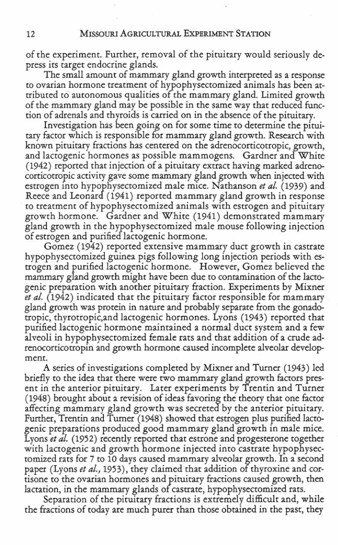
of (he experiment. Further, removal of the pituitary would seriously depress its target endocrine glands.
The small amount of mammary gland growth interpreted as a response to ovarian hormone treatment ofhypophysenomized animals has been attributed to autonomous qualities of the mammary gland. Limited growth of the mammary gland may be possible in the same way that reduced funcdon of aclrenals and thyroids is carried on in the absence of the pituitary.
Investigation has been going on for some time to determine the pituicary factor which is responsible for mammary gland growth. Research with known pituitary fractions has cencered on the adrenocorricorropic,!fOwth, and lactogenic hormones as possible mammogens. Gardner an White (1942) reponed thar injection of a pituirary extract having marked adrenoconicorropic aaiviry gave some mammary shnd growrh wben injeaed with estrogen Into hypophysectomized male mlee. Nathanson et al. (1939) and Reece and leonard (1941 ) reporced mammary gland growth in response to treatment of hypophysectomized animals with esrrogen and pituitary growth hormone. Gardner and White (1941) demonstrated mammary gland growth in the hypophysectomized male mouse following injection of estrogen and purified lactogenic hormone.
Gomez (1942) reponed extensive mammary duct growth in castrate hypophysectomized guinea pigs following long injection periods with es· trogen and purified lactogenic hormone. However, Gomez believed the mammary gland growth might have ~n due to contamination of the laaogenic preparation with another pituitary fraction. Experiments by Mixner et at. (1942) indicated that the pituitary factor responsible for mammary gland growth was protein in namre and probably separate from rhe gonadotropic, thyrorropic,and lactogenic hormones. Lyons (1943) reported that purified lactogenic hormone maintained a normal duct system and a few alveoli in hypophysectomized female rats and that addition of a crude ad· renocorticotropin and growth hormone caused incomplete alveolar development.
A series of investigations completed by Mixner and Turner (1943) led briefly to the idea that there were twO mammary gland growth fanors pres· ent in the anterior pituitary. Later experiments by 'frentin and Turner (1948) brought about a revision of ideas favoring the rheory that one faaor affecting mammary gland growth was secreted by the anterior pituitary. Further, Trenrin and Turner (1948) showed that estrogen plus purified lactogenic preparations produced good mammary gland growth in male mice. Lyons et al. (1952) recently reporced that estrone and progesterone together with lactogenic and growth hormone injected into castrate hypophysec. tomized rats for 7 to 10 days caused mammary alveolar growth. In a second paper (Lyons tt aI., 1953), they claimed that addition of thyroxine and cor· tisone to the ovarian hormones and pituitary fraaions caused growth, chen Iaaation, in the mammary glands of castrate, hypophysectomized rats.
Separation of the pituitary fraaions is extremely difficult and, while the fractions of today are much purer than those obtained in the past, they

REsEARCH B ULLETIN B7 13
may still contain sevc=ru componc=ncs as shown by various mc=chods of idc=ntification. Thus, whik it appc=ars chat thc= pituitary fraction rich in thc= lactOgenic hormonc= is capable of stimulacing growth of thc= mammary gland, the fact chat lactogenesis and mamogenesis arc due to the same pituit:ay facror has nor been demonstr2ted ( Astwood, 19~3).
Androgens and Mammary Gland G rowth . In addition to effeCts of the ovarian and pituica.ry hormones, it has been known for some time thar androgens may playa role in mammary gland growth. Sclye t t ai. ( 1936) first reported that testOsterone benzoate caused slight development of mammary glands in immature male and female !2CS. Nelson and Gallagher (19-36) aho reported that injection of the various andco$ens, including androsterone, producc=d growth of the du~ts and lobules In castrate virgin rars. Other rc=pons by McEuen tJ al. (1936), Astwood tt al. (1937), Bottomley and Folley (1938), Fo llc=y tl al. (1939), Lewis tlal. ( lx39), Noble (1939), Rc=ece and Mixner (1939), Van Heuverswyn tl al. (1939), Forbes (1942), and Mixner and Turner (1943) showed that androgen injection in a number of species caused some mammary g land growth in castrate animals but had no effect on mammary gland growth in hypophysectomizc=d animals. Arecc=nt papc=r by De Graff t l ai. (1950) prC=S(:nte=<! c=xperimc=ntal data which lc=d them co believe that testosterone, in sufficiently large quantities, was capable of stimulating mammary dc=vdopment excc=c=ding that caused by estradiol benzoate and progcs[(:rone, but that physiologinl injeCtions of testosceronc= wc=rc= without c=ffecc on the mammary gland.
Adrenal Cortical H ormones and Mammary Gland Growth. The adrc=nal is anmher gland which influences mammary gland growth. Van Hc=uverswyn tt ai. (1939) noted that desoxyconicosterone stimula.ced mammi:fy duct growth in imm:Hutc= male micc=. It has been shown that desoxycorticostc=ronc= will c=vokc= alveoi2r growth of the mammary g land in thc= monkey (Spec=rc, 1940), mouse (Mixner and Turnc=r, 1942) and guinc=a pig (Nelson tl aJ., 1943).
Treatment of hypophyS«tomizc=d anima.1s complicate=<! undc=rscanding of the adrenals' rok in mammary gland growth, for Gardner (1940) rc=ported obtaining mammary growth in. hypophysectomized male mice as a rc=sult of creatment with desoxycorticosteronc=. Further, Chamarro (1940) reportc=d that injections of desoxycorticosterone acc=tatc= caused alveolar growth in hypophysectomized male rats while Nelson (1941) observe=<! mammary development in cascr-ate, hypophysc=ctomized nts m:ated with adreno tropin. Ex pc=rimc=nts by Leonard and Reece (1942) again pointed out doubts concerning completeness of hypophysectomy in such c=xperiments when they found that rats with pituitary rc=sidues showc=d growth of the mammary gland in response to desoxycorticosterone acetate injection while completely hypophysectomize=<! animals failed to respond to the same treatment.
A paper by Mixner and Turner (1943) rc=portc=d that dc=soxycorticosterone possessed roughly onc=-thitd the activity of progesterone in causing

14 MISSOURI AGRICULTURAL ExPERIMENT STATION
alveolar growth in C2SmltC femaJe mice. Cowie and Folley (1944 and 1947), following this up, found little evidence for mammary ghnd regression after adrenalectomy. They noted [h:lt anterior pituitary extC1l.CtS were srill cap:abJe of producing mammary growth in adrenalectomized rats. Further, Cowie and Folley (1947) stated (hu alveolu development in untreated castr:l.re r.iH$ may be due to action of progesterone secreted by the adren2.1 cortex. Trentin and Turner (1948) concluded that, in certain species, mammary alveolar response to esu ogen 2Pparcd (0 be dependent upon the 2biliry of estrogen to stimul:ue the adrenal cortex, which, in rurn, secreted steroids either identical with or re~mbling progesterone in ability co synergize with estrogen in stimulation of mammary s:land development.
T hese theories were supported by investigations of Zarrow t t al. ( 19~O), who were able to show th at desoxycorticosterone acetate was converted to progesterone in the chimpanzee.
A recent papa- by Smith and Braverman (l93~) reports that, while desoxycorticosterone acetate exerted a duct stimulating effect on the m:unmary gland in immature castnte ats, it alone or in connection with low levels of progesterone had no effea on lobule alveolar growth. A high level of desoxycort icosterone acetate, injected together with estradiol, caused only slight alveolar proliferation. These :l.Uthors also St:l.ted they believed the progesterone-like act ion of desoxycorticosterone acetate might be due, in part, to conversion of the adrenal compound to progesterone, since it closely resembles progesterone structurally.
Relaxin and Mammary Gland Growth. The phenomena of relantion and separation of the pelvic bones during pregn~cy has been known for some time (Duncan, ISn). Observation of this rdax:l.Cion has bttn mentioned by other investigators, bur Whitley ( 1911) was the nrst to report that t he separation of pubic bones was a gradual process. It remained for Todd (1923) ~d Karavau. (1926) to describe the ~atomical and histological aspeas of relaxation.
The normal condition of the pubic symphysis in the virgin female involves collagenous ligaments connecting bOnes of the right and left side, anterior and posterior to the symphyses. An interpubic d eft is present between the opposin$ articular surfaces. Amerior and posterior to this deft, there is tissue contlnuity between the cart ilaginous ca.ps of the right and left p'ubes. The first cha.nge during pregnancy is seen as the articular hyaline ca.rrJiage begins to proliferate and a slight separation of the bones is noticeable. As the interpubic gap continues to widen, IiAaments connecting the bones lengthen by prOliferation of cells at the ends. D uring the final few days before panuritlon, an intensive process of resorption of the symphysial ends of the pubic bones mes place. I t has been noted also that cell com· position of the li$amenrs changes somewha.c during this phase of mitosis. Thus, as the medial ends of the bones a.re "ea.!en awa.y," the ga.p widens and the ligaments lengthen. In this way, the passage through whIch the ferus must crave! during birth is enla.rged. Following parturition, the gap betWeen
>
•
,
• , ,
,
•
•
•
,

REsEARCH BULLETIN '37 " the p.u?ic bones closes rapidly, but does nOt completely tecum to the virgin condmon.
Hisaw (1926) was able to produce changes in the pelvic ligaments of virgin guinea pigs by subcutaneous injection of blood serum from pregnant rabbitS and guinea pigs. These changes proved to be identical with those observed during normal pregnancy. Hisaw (1929 and 1930) was later able to isolate and iaentify the active material as the hormone relaxin. It has been shown that relaxin, a non-steroid hormone, is E~esent in the corpus luteum (Hisaw tt al., 1925 and 1927) and placenu (H isaw, 1929) dunng pregnancy. Studies on the mouse (Gardner, 1936; Hall and Newton, 1946; ancfHall, 1947), and guinea pig (Ruth, 1937) have shown that histological changes which bring about relaxation during late pregnancy are due to relaxin.
Van der Meer (1950) presented evidence to supporr the idea that connective tissue of the symphysis pubis may be changed in such a way that normal or increased muscle tone or static forces in the pelvis cause the relaxation. He suggested further that certain histological changes might be due to increased capillary or tissue permeability brought about by relaxin, the relaxin having an effect on the connective tissue similar to hyaluronidase or spreading hctors. Van der Meer did not test relaxin for itS a.bility co change permeability; be did, bowever, test a number of fa. ctors, including hyaluronidase, for their ability to cause relaxa.tion. In no case did any orthe &'CtOrs cause relaxation.
In 1945, Hamolsky a.nd Spurow first tested tbe effect of rela.xin on mammary gland growth. They found that relaxin, together with estradiol benzoa.te a.nd progesterone, gave !>trter growth and lobulation in immature castrate female ra.ts than did the estradiol benzoate and progesterone, alone or in combina.tion.
Garrett a.nd Taima$e (1950 a.nd 1953) tested the effeCts of relaxin on mammary glands of gumea pigs and rabbits. In guinea pigs, where it bas ~n shown that estrogen, alone, is capable of producing full lobule-alveoia.r growth, they concludeo chat addition of relaxin to estrogen caused a guantiutive increase in degree of gla.nd growth. In the rabbit, where both estrogen and pro~cerone a.re necessary for lobule-alveolar growth, the addition of relaxin to estrogen caused a greacer development of the duct system but nOt alveola.r growtn. They believe, therefore, that relaxin actS as a potentiator of estrogen in the case of the guinea pig and that it sensitizes che mammary gJa.nd oT the rabbit so that the action of estrogen is more wide-sprea.d on the duct system.
A recent investigation by Trentin (1951) gave a.dditional support to the idea that relaxin does noc repJa.ce progesterone in mammary development in tbe mouse. This auchor found litrle or no increase in the percentage of positive mammary alveoJa.r responses in caStrate female mice treated with estrogen, progesterone, and relaxin, compa.red to those treated with estrogen and progesterone.

16 MISSOURI AGRlCULTUR.AL ExPERIMENT STATION
While it has ~n proven that a number of endocrine glands and hotmanes affect mammary $Lmd growth, one hypothesis explains the main mammary development In the following way. N orma12utonomous secrecion which is observed in a number of the: endocrine glands of hypophysectomized animals may account for a part of mammary gland growth abs<:rved in the hypophysectomized animal and may be responsible for the increased duct growth following estrogen treatment. Increased duct growth, :mribuced to eStrogen treatment of C1Str:lt(: anim:us, may be due also to in· creased utilization of an anterior pituitary factor, normally secreted, which causes mammuy glmd growth. Increased amounts of this anterior p ituitary neeor are believed co be secreted in response to me presence of progesterone.
Thus, at sexua..l maturity, or when estrogen is administered, the estrogen lIctS directly on the mllmmary gland, incrosing the va..scu1uity:and permeabi.li ty ofbJood vessels in the fatty pads. The anterior pituitary facto r thus reaches t he glands in increasi ng concentration. T he autonomous growth caracity of rhe mammary gland is stimulated a..l so by rhe increased amount 0 nutrients supplied by the blood. As a result, the duct system is stimulated to g rowth. During pregnancy or when progesterone is given, increased senerion of an anterior pituituy factor in response to progesterone brings about a rapid extension of the ducts and alveolar growth of the mammary $:Iand.
VariabIlity in response to the administration of ovarian hormones may be accounted for by tne fact that in some species estrogen stimulates the production of progesterone by the adrenals. Since adrenal compounds, such liS desoxycorticosterone, have ~n shown to be converted to progesterone in the animal body, the adrenals may ~ an important source of progesterone in the: absence of the corpus luteum. .
METHODS AND MATERIALS 1. Assay for the Mammary Gland
Spreading FaCtor In removing mammary ~lands for assay, animals were skinned, leaving
the glands attached to the skm. The blood was squeezed out, then the fatty pads containing the mammary ~Iands were carefully dissected, free of all lymph nodes and other tissue. SInce it was sometimes impossible to assay the gl:ands immediately, some were held at _l :5 °C until th lS could be done.
To excr:act the sproding factor from mammary gl:ands, a 0.1 M sodium acetate buffer, p H 6.0, with 0.1:5 M sodium chloride was used. With the first experimenta..l animal, the albin? ra~. 2:5 mt of the b.uffer was em,?loved. Since this amount of buffer gave a dIlUtion of the spreading factor which fell within li mits of the assay method, the amount of buffer use~ to ext~act mammary glands of the other animals was kept at the same r:atlO of weIght of gland per millil iter of buffer. W eight of the mam~ary gland of the normal female ra t being approximately 4 grams, the rano 6.2 :5 ml. for 1 gram

REsEARCH BULLETIN 537 17
of manunary tissue was used for the other experimental anima1s. The normal albino mouse was found to have a mammary gland weighing about 0.8 gram so 5 ml. of buffer was used for extraction.
Guinea pigs had mammary glands averaging 4.5 grams, so 30 ml. of buffer was employed. Mammary glands of normal female rabbits varied greatly; however, the average weight appeared to be around 28 grams. Thus, 175 mi. of buffer was used to extract their glands. Mammary glands of the three larger animals were homogenized in the Waring blender, while the mouse glands were homogenized in a Potter homogenizer. The homogenates were then centrifuged for 20 minutes at 2,000 r.p.m. The aqueous layer was then pipened off for assay, avoiding, as far as possible, the inclusion of any fraction of the fat layer which formed on top, or the tissue debris which centrifuged to the bottom.
Mammary gland extracts were diluted ten times with buffer solution and 0.2 mI. of the diluted extracts was taken up in a 1 ml. tuberculin syringe, along with 0.1 mL of2 percent T-1824 (Evan Blue) dye. After thoroughly mixing, the extract and dye were injected intradermally with a 24-gauge needle into a shaved white rabbit. Injections produced blebs on the rabbit's skin which were measured immediately after injection and 30 minutes later. The areas of spread were measured, by length and width, to the nearest millimeter. The area of the initial bleb was subtracted from the final area to give the net area of spread in millimeters sguared. Practically all assays were run in duplicate and the average reported. .
Making injections with a slow steady pressure on the syringe plunger was important to obtain a uniform bleb and to avoid, as much as possible, the interference of pressure effects upon the amount of actual spread in the rabbit's skin. Age of the rabbit also was extremely important. BeSt results were obtained with rabbits weighing between 4 and 6 pounds. Condition of the skin in rabbits of this size was most satisfactory. If the rabbits were more mature, the skin was tOO tough, while smaller rabbits had thin skins which were'easily punctured. It was difficult to get satisfactory results out· side the weight range indicated.
To determine the reproducibility of results by this assay method, several checks were made. One rabbi t received 10 injections of buffer and 10 injections of mammary gland extract from a castrate female rat treated 10 days with 1 ug. estradiol benzoate and 5 mg. progesterone daily. Areas of spread from the buffer ranged from 34.6 CO 59.7 millimeters sguared with the average of the 10 being 49.6. Areas of spread from the mammary extracts ranged from 174.4 to 226.3 millimeters squared with the average of the 10 being 204.0. The buffer and mammary gland extracts were preserved and, over a period of time, were injected into 10 different rabbits. Potency of the mammary gland extract apparently was unchanged. Areas of spread from buffer ranged for 34.6 to 59.7 millimeters sguared with an average of

l8 MISSOURI AGRlCULTURAL E XPERIMENT STATION
44.6. Area of spread from mammary gland extracts ranged from 159.5 to 226.3 millimeters squared with the average being 192.7 (Table 1).
TABLE I --VARIABILITY OF AMOUNT OF INDUCED INTRADERMAL SPREADING IN THE SAME AND DIFFERENT RABBITS
Trial No.
1 2 3 4 , 6 7 8
• 10
Aug.
Rabbit No.
1 2 , 4 , 6 7 6
• 10
Aug.
Assays on Same Rabbit in mm2
Buffer SOlution
47.1 47 .1 34 .6 47 .1 34.6 59 .7 47.1 '47.1 34.6 47.1
44.6 .. 12.3
Mammary Gland Extracts·
176.0 191.7 192.5 174.4 209.0 226.3 226.3 209 .0 226.3 209.0
204.0 .. 25.2 Assays on 10 Different Rabbits
Buffer SOlution
47 .1 59.7 47 .1 34.6 47.1 47.1 59.7 47.1 47. 1 59.7
49.6 + 12 .8
Mammary Gland Extract*
160 .3 159.5 226.3 159 .5 209.0 209.0 176.0 191. 7 226.3 209.0
192.7 .. 33 .4
*From castr ate female rat received 1 ug. estradiol benzoate .. 5 mg. progesterone dally lor 10 days.
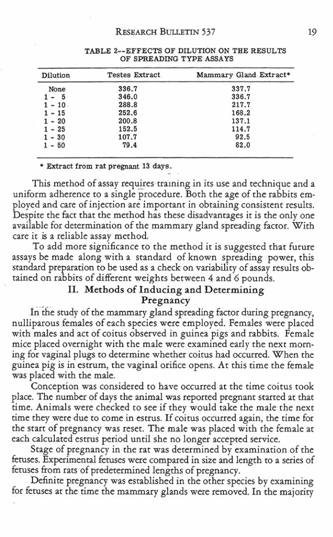
To obtain a better understanding of the meaning of differences in areas of spread from different amounts of spreading factor, several extraCtS were assayed at various dilutions. Starting with straight extracts of testis and mammary glands from a 13-<1ay pregnant rat, assays were made on dilutions of 1 to ~, 1 to 10, 1 to 1~, 1 to 20, 1 to 2~, 1 to 30 and 1 to ~O. It was found that higher concentrations, dilution of 1 to 10 or less, produced blebs which would not exceed an area larger than 24 x 27 millimeters, indicating that this was the upper limit of t he assay. It was the extent to which the extraCtS would spread, regardless of amount of spreading factor they connined. As the extracts were diluted in series below a 1 to 10 dilution, the areas of spread became progressively sm:a1ler. However, even extracts diluted 1 to ~o produced spreading in the rabbit's skin (Table 2) .
Dilution
No" I - 5 1 - 10 1 - 15 1 - 20 1 - 25 1 - 30 1 - 50
RESEARCH BUll..ETIN 537
TABLE 2 __ EFFECTS OF DILUTION ON THE RESUL TS OF SPREADrnO TYPE ASSAYS
Testes Enract
336.7 346.0 288.8 252.6 200.8 152.5 107.7
79 .4
Mammary Gland Extract*
337.7 336.7 217.7 168.2 137.1 114. 7 92.5 82.0
• Extract from rat pregnant 13 days .
19
This method of assay re9uireS'Craming in its use and technique and a uniform adherence to a single procedure. Both the age of the rabbies em· played and care of injection are important in obtaining consistent results. Despite the fact that the method has these disadvamages it is the only one available for determinacion of the mammary gland spreading factor. With care it is a reliable assay method.
To add more significance to the method it is suggested cllat future assays be made along with a standard of known spreading power, this standard preparation to be used as a check on variability of assay results obtained on rabbits of different weights between 4 and 6 pounds.
II. Methods of IndUCing and D etermining Pregnancy
in-the study of the mammary gland spreading factor during pregnancy, nulliparous females of each species were employed. Females were placed with males and act of coitus observed in guinea pigs and rabbits. Female mice placed overnight with the male were examined early the next morn· ing for vaginal plugs to determine whether coitus had occurred. When ehe guinea pig is in estrum, the vaginal orifice opens. At this eime the female was placed wieh the male.
Conception was considered to have occurred at the time coitus cook place. The number of days the animal was reported pregnant started ae that time. Animals were checked to see if they would take the male the next time they were due to come in estrus. If coitus occurred again, the time for ehe stare of pregnancy was reset. The male was placed with the female at each calculated estrus period until she no longer accepted service.
Stage of pregnancy in the rat was determined by examination of the fetuses. Experimental feruses were compared in size and length to a series of fetuses from rats of predetermined lengths of pregnancy.
Definite pregnancy was established in the other species by examining for fetuses at the time the mammary glands were removed. In the majority

20 MISSOURI AGRICULTIrkAL ExPERIMENT STATION
of cases, anim21s were pregnant ; those which were nor were used as nonpregnant control s.
HI. Methods of Castration 2nd Injection in Experimental Animals
Male 2nd female Wisea! rats from the University of Missouri Colony were employed as one experimental animal and female albino mice from the White Rose Mousery at Billings, Mo., were used as the other. Females of both species wefe castrated by removal of the ovaries through a venrnl midline incision. Testes of the male ratS were removed. through an incision in each scro tum. Following C2stration, a lO-day resting period was allowed for anim:als to recover from (he shock of opeflltion and (or effects of naNnl gonadal hormones to wear off.
If only ustrne animals were to be used, treatment was started on the eleventh day. In seJeaed experiments, C2srrate·adrenalectornized t2tS were used. Castrare animals were adrenalectomized for these experiments on the eleventh day following castr2tion and t~tment was started about five days following the last operation. The castrate-adrenalectomized animals were maintained on 1 percent sodium chloride in the drinking water.
Trea.cmems involved daily injections of a number of hormones, general· ly for a period of 10 days. A number of experiments were carried on which called for longer periods of in jection. Injections were made subcutaneously over the shoulder and back, crying as far as possible (0 keep skin irtitation at a minimum. The day following the last injection, animals were sacrificed and mammary glands removed as previously described.
The estradiol benzoate and progesterone used in the treacmenc were dissolved in olive oil. T he testosterone used was dissolved in 60 percent alcohol. Relaxin was already in solution and was used as received. Pituitary extracts and whole pituitaries were dissolved or suspended in distilled water. All of the materials used to take up the hormones were assayed for theif :lbility to cause production of the mammary gland spreading factor. In no case did the mammary glands from CaStrace female racs injected with olive oil, alcohol, or distilled water give any indication of the presence of the spreading factor (Table 3).
TABLE 3--EFFECTS OF HORMONE SOLVENTS ON THE AMOUNT OF MAMMARY GLAND SPREADING FACTOR IN RATS
M.ammary Extractll f rom Castrate Rats Treated with·
0.2 ml Olive Oil 0.2 ml 60% Alcohol 0.2 DlstiUed Hz<)
No. of Animals Average Areas ot Spread mm2
" 5F
" • Da.lly Treatment tor 10 Days F • Cutrate Females
47. 1 =.12.6 49.6! 12.7 45.9! 12.S
•
•
•
,
, ,

RESULTS I. The Mammary Gland Spreading Factor in
Normal Pregnant Animals In a nwnber of spt:cies, it has been shown that growth of the mammary
gland occurs chiefly during the first one-half to two-thirds of pregnancy. The duct system first, and later the lobul~·alveolaI system penetrates into a fatty pad of connective tissue. The theory had been proposed that the forward progress of the growing gland would be facilitated if cells of the endbuds of the ducts, and later the growing side-branches which form the alveoli, produced a spreading factor which would cause the cementing substance of the connective tissue to liquify.
A preliminary investigation by Elliott and Turner (1950) demoosmced that such a spreading faeCQr was present in che mammary glands of pregnant cats and that it could be extracted and assayed. This bulletin reports on an extension of their investigation to determine if the mammary gland spread· ing factOr in the rat was present in other species of experimental animals. during pregnancy. In addition, these experiments were designed to help detetmine whether or not the f.attern of increase in amounts of mammary gland spreading factor paralle ed the pattern of growth in the mammary gland during pregnancy; thus indicating that the spread ing factor does play a pan in extension of the growing mammary gland into the fatty pad.
TABLE 4- - ASSAY OF SPREADING FACTOR IN RAT MAMMARY EXTRACTS
Type of Extract
pH 6 BuIfer Testes Extract Norma.! F ema.!e· 1 day pregnant· 2 days pregnant· 3 days pregnant" 4 days pregnant" 5 days pregnant· 6 days pregnant" 7 days pregnant-6 days pregnant-9 days pregnant -
10 days pregnant· II days pregnant· 12 days pregnant. 13 days pregnant · 14 days pregnant" 15 days pregnant" 16 days pregnant" 17 days pregnant" 16 days pregnant" 19 day5 pregnant" 20 days pregnant· 21 days pregnant-
_____ .M--'-==mary gland extracts.
Number of Animals
42 42 42 14
" 13 18 23
" " 24 28 22 23
" 25 22 23 20 17 23
" 13 16
Area of Spread !11m2
55.4.13.6 310.4:; 30.4 47.7+11 .2 51.6:; 12.9 52.6+13.0 69.9 + 14.6 83.5 + 16.3
110.9 + 16.4 146.4 + 19.3 196.7+20.2 229 .9:; 21.3 234.3 + 23.1 238.2 + 23.2 249.0 + 23.7 260.2 + 24.9 223.8 + 21.1 198.5 + 20.1 160.9 + 19.8 131.8 + 18.'1 107.0 + 18.2 84.0 + 16.5 79.7 + 15.3 55.9 -; 13.8 51.9 -; 13.0

22 MISSOURI AGRICULTURAL ExPERIMENT STATION
The Albino Rat. The preliminary report (Elliott and Turner, 1950) showed chat the mammary glands from non-pregnant animals contained no spreading faeror, while the mammary glands taken from a large number of rats at various Stages of pregnancy showed measurable amounts of spreading factor, except during the first and laSt cwo days. The amount of spreading factor increased with each clay of pregnancy until a maximum was reached on about the twelfth day, following which there was a decline in amount of spreading factor during the remainder of pregnancy (Table 4).
As previously shown, growth of the mammary gland during pregnancy is considered to be largely completed in from 12 to 14 days in the rat. Following chis period the amount of cellular division slows down greatly and milk secretion is initiated. As indicated by data in the present investigation, the amount of spreading factor in the mammary gland increased until it reached a peak near the time when growth is considered to be largely com· pleted after which the amount of spreading factor decreased. Since the num· ber of animals used in this investigation was by far the largest for a single species, results show dearly the rise and decline in amount of spreading factor at various stages of pregnancy (Figure 1).
0 0
'" 0
N " Z N Z
0 00 «N w 0:
0 "-" <fl_
u-°0 «~ w 0: «0 ,. ,.
" ,. ,. ,. 0
0 2
,. ,. ;' >
. > '. ,. ; '
., • 3 ,~ 2' 3 2'
2' 9 ~
'" ,. ,. ,
3 4 5 6 7 6 9 10 11 12 13 14 15 16 17 18 19 20 21 NUMBER OF DAYS RATS PREGNANT
Fig. 1. The amount of mammary gland spreading factor in the rat increases during pregnancy, reaching a peak on the 12th day. The rise in amount of ~preading factor parallels the growth phase of mammary gland growth. FollowlDg. the peak, the amount of spreading faeto r declines rapidly as gland growth declines and seaetory activity i~ iniciued.
•
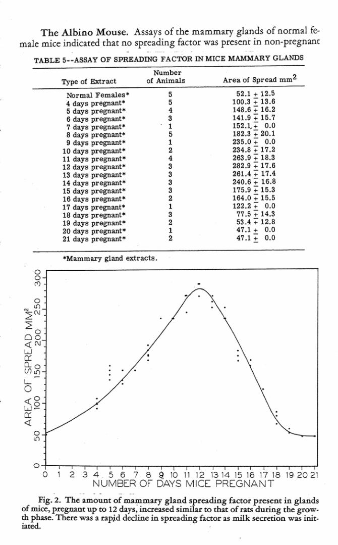
Tbe Albino Mouse. Assays of the mammary glands of normal female mice indicated that no spreading f.lCtor was present in non-pregnant
TABLE 5--ASSAY OF SPREADING FACTOR IN MIC E MAMMARY GLANDS
Type of Extract
tbrmal Females ' 4 days pregnant ' 5 days pregnant· 6 days pr egnant· 7 clays pregnant· 8 days pregnant· 9 days pregnant·
10 days prepant· 11 days pregnant· 12 days pr egnant' 13 days pregnant· 14 days prepant· 15 days pr egnant· 16 days pregDant· 17 days pregnant· 18 days pr egnant· 19 days pregnant· 20 days pr egnant· 21 days pregnant"
Number of Animals
5 5
• 3 1 5 1 , • 3 3 3 3 , 1 3 , 1 ,
"Mammary gLand extracts.
Area 01 Spread mm2
52.1 .. 12.5 100.3:; 13.6 148.6:; 16.2 141.9:; 15.7 152.1..+ 0.0 182.3:; 20.1 235.0" 0.0 234.8 + 17. 2 263.9" 18.3 282 .9 + 17.6 261.4 " 17 .4 240.6 .. 16.8 175 .9 + 15.3 164.0:; 15.5 122.2:; 0.0
77.5 :; 14.3 53 .4 +12.8 47.1 + 0.0 47.1'+ 0.0
g r------------------------------------------, '"
OjO~'1 ~2~3-,4--5c-6C-C7-,8-,Q--tOr-trt-tT2-,13-,,4--t~5-tr6-tT7-,t8-,t9--2~O-2~t NUMBER or DAYS MICE PREGNAN T
. . . ~ig. 2. The amount of ma':llmary gland spreading factor present in glands
of ml(e, p~gn:ant up to 12 days, increased similar to that of cats during the grow. ~ ph.ue. There wu a rapjd decline in spreading f1lctor 2.$ milk $«l'etioo. was iait . • ",l

24 MISSOURI AORICULTIfRAL ExPERIMENT STAnON
animals. First evidence of the presence of the spreading factor was observed on the fourth day of pregnancy. At this time, mammary glands contained some spreadiag factor. The amount gradually increased with advancing pregnancy until a peak was reached on the twelfth day, after which it gradually declined. No spreading factor was detectable in the mammary gland extracts from mice pregnant 19, 20 or 21 days (Table 5).
As pointed out in an orlier section, the period of growth of the mammary gland during gestuion is approximately the same for the mouse and rat. ThcedoIe, it is not surprising to find that the amounts of mammary gland spreading factor in mice and rats parallel each other during the various stages of pregnancy.
While few mice were employed in this investigation, the curve showing amounts of spreading factor in their mammary glands (Figure 2) during pregriancy follows the same pattern as that shown by the tats.
The Guine2 Pig. As in srudies with the two previous species, mammary glands from non-pregnant guinea pigs showed no spreading factor.
TABLE 6- -A$SAY OF SPREADING FACTOR FROM GUINEA PIG MAMMARY GLANDS
Type of Elrtract
Norm:ll. F emales· 5 days pregnant· 7 days pregnant·
10 days pregnant" 13 days pregnant· 15 days pregnant" 18 days pregnant" 20 days pregnant" 22 days pregnant" 25 days pregnant" 28 days pregnant· 30 days pr egnant· 33 days pregnant" 35 days pregnant" 36 days pregnant" 36 days pregnant" 40 days pregnant" 41 days pregnant" 43 days pregnant" 45 days pregnant" 47 days pregnant" 50 daYs pregnant" 53 daye pregnant" 55 days pregnant" 58 days pregnant" 60 days pregnant" 62 days pregnant" 66 days pregnant '" 67 days pregnant"
Nu.mber of AnimalS ,
1 , , , 3 , 3 , 3 3 3 , 3 1 3 , 1 , 1 3 3 , , 1 3 , 1 1
-Mammary gland e:rtracts.
Area of Spread mm2
50.3 + 12.9 53.4:;: 0.0 61.4 -; 13.9 76.0:;: 14.4 93.3 :;: 14.9 64.7:;: 14.7
122.1:;: 15.1 141.9:;: 15.3 171.9:;: 15.4 200.6;- 15.6 220.1 ;- 16.0 243.5:;: 16.5 256.8 ;- 16.3 266.2 -; .16.8 252.3 -; '0.0-270.3 -; .l'6.3 303.3 -; 17.1 327.3 -; 0.0 303.1 +n.o 296.6 -; 0.0 252.5 -; 16.3 261.3 -; 16 .5 209.1 -; 15.6 189.0 -; 15.1 115.1 -; 0 .0 82.9 -; 13.2 59.9 + 12.7 53.4 -; 0.0 53.4 -; 0.0

REsEARCH BULLETIN 537 25
Mammary glands from guinea pigs pregnant ~ and 7 days also showed no spreading factor. Extracts of glands from animals pregn~nc 10, 13, and. l~ days indicated the presence of small amounts of the spreading factor. Dunng (he rest of early pregnancy, there was an increase in amount of spreading factor in the mammary glands until a peak was reached on about the fortyfirst day, after which the amounc declined. By the sixtieth day of pregnancy, very little spreading factor could be detected in the mammary gland an~ on the sixcy.seoond day, assays showed none. Mammacy glands from cwo guinea pigs at parturition, pregnant 66 and 67 days, showed that no spreading factor was presc=nc (Table 6).
Here, again, the amoum of spreading factor parallels gtowth of the mam· mary gland during pregnancy (Figure 3). As in the case of growth, little
~.-------------------------------,
g
o~'""''''~~~~~~~~~ " ~ 10 15 2025 3 0 3~ 40 "5 ~o 55 ~o 05 NUMBER or DAYS GUINEA PIGS PREGNAN T
Fig.~. In the guinea pig, wi th a long period of pregnancy, rise in the amount of spreading betor preSent in mammary glana , was gradual up to the peak at 40 to 45 days. The subse<luent decline in spreading {:letor with cessniOD of growth and initiation of lactation was fairly upid .
spreading factor activity is present before the fifteenth day. After l~ days both increase gradually. The peak amoum of spreading factor was observed on the fony-first day of pregnancy. While the growth period of the mammary gland during pregnancy is not known, the two-thirds point of pregnancy, probably the time of maximum growth, would fall becw~n 40 and 45 days. Thus, the maximum spreading factor activity and maximum mammary gland growth during pregnancy occurred at about the same time in the guinea pig. Decline in amounts of the spreading factor after the fonyfirst day also agrees with the decrease in numbers of mitotic figures known to occur in mammary glands during this period of pregnancy.

26 MISSOURI AGRICULTUltAL ExPERIMENT STATION
The: Rabbit. Assays of mammary glands of non-pregnant rabbits showed that no spreading factor W:lS present. Mammary glands from the
TABLE 7.·ASSAY OF THE SPREADING FACTOR IN RABBIT MAMMARY GLANDS
Type of Extract
Normal Females" -4 days pregnant· 7 days pregnant · 9 days pregnant ·
10 days pregnant" 11 days pregnant· 12 days pregnant · 13 days pregnant· U days pregnant" 15 days pregnant · 16 days pregnant" 17 days pr egnant" 18 days pregnant-19 days pr egnant" 20 days pregnant" 21 days pregnant" 22 days pregnant" 23 days pregnant-25 days pregnant" 28 days pregnant-
"Mammary gland extracts
Number of-An.tmals
3 1 2 2 2 1 2 2 2 2 1 2 2 2 2 2 2 , 2 1
Area of Spread 10m2
51.3 + 12.9 66.0:; 0.0
107.5+14.3 129.7" 14.4 159.9"+ 15.3 152.0:; 0.0 164.0:; 15.1 184.3"+ n.s 239.1" 16.9 230.3 .. 16.3 252.6:; 0.0 270.4:; 16.8 305.7+1 7.4 284.5 :; 17.0 270.5:; 16.8 221.8:; 17.1 176.0:; 1$.2 1$4.3 :; 0.0 114.$:; 14.3
53.4 :; 0.0
FigA. The rise and decline in amount of mammary gland spreading faeror in the pregnant rabbit, in relation to $rowth and the initiation of milk secretion, follows the pau ern of the other species.
•

RESEARCH BULLETIN 537 27
earliest stage of four days of pregnancy showed link or no spreading factor, the next stage, 7 days, showed that spreading factor was present. The amount of mammary gland spreading facto r increased to a peak on the eighteenth day of pregnancy. after which there was a gradual decline. By the twenty-eighth day of pregnancy no spteading factor was detectable in the mammary gland extracts (Table 7).
Increase in amounts, a peak at the eighteenth day, and a decline in amounts of spreading factor during pregnancy (Figure 4) check with the pattern of mammary gland growth occurring in the pregnane rabbits (Figure 4).
Summary. Investigation of four species of experimental animals has shown that the increases in amounts and times at which peak amounts of the spreading faccQt'were observed in the glands during pregnancy closely parallel the growth phase of mammary gland development. D uring the latter one-third of pregnancy, when cells of the mammary gland begin secretion, amounts of the spreading fac tor decline. It would appear that elaboration or activation of the spreading factor practically ceases as secretory activiry begins.
11. Effects of Hormones on the Mammary Gland Spreading Factor
It has been shown that a number of hormones separately or together" stimulate growth of the mammary gland. Among the more important of these hormones is estrogen which has been shown to cause duct growth, an estrogen and progesterone synetgism which has been shown to cause lobulealveolar growth, and an anterior pituitary hormone which has been called mammogen (Turner, 1950). Effects of these various hormones on the elaboration or activation of the mammary gland spreading factor were studied.
The Ovarian H ormones. In a preliminary study (Elliott and Turner, 1951b) . various levels of the ovarian hormones were injected daily either separately or together into groups of fi ve castrate ratS for 10-<lay periods.
Mammary gland extracts from castrate females injected with 1 ug. of estradiol benzoate showed little spreading facror. When the estradiol benzoate level was raised to 3 ug., the extracts indicated fair levels of the spreading factor. Mammary gland extracts from castrate male and female rats trea.ted with 5 ug. of estradiol benzoate indicated rhe presence of more spreading facror (Table 8).
These data indicate that estrogen can cause production of the active mammary gland spreading factor the same as it has been shown to induce mammary duct growrh. Ir would appear that this hormone, which initiates extension of che mammary duct system into the fatty pad, causes elaboration or activation of the spreading factor which may.help in the forward extension of these ducts through the connective tissue barrier.

28 MlSSOURl AGRICULTURAL EXPERIMENT STATION
With progesterone alone the amount of spreading factor in the mammary gland extracts was small at the 1 mg. level in castrate females. An increase in the amount of progesterone to 3 mg. caused a higher level of spreading facror to be presenc in the mammary extracts of castrate females , while a 5 mg. level of progesterone injected into castrate males, castrate females and castrate adrenalectomized females showed a furchcr increase and indicated fair amounts of the spreading factor to be present in the extracts. Ten mg. of progesterone increased the amount of spreading factOr in castnte adrenalectomized females, as well as castrate males and females giving the highest level of mammary gland spreading factor obtained from any of the progesterone injected groups (Table 8).
TABLE S--EFFECTS OF ESTROGEN AND PROGESTERONE ON AMOUNTS OF SPREADING FACTOR IN RATS·
Mammary Elrtracts ~m Rats Treated With:
1 ug estradiol benzoate 3 ug estradiol benzoate 5 ug estradiol benzoate 5 ug estradiol benzoate 5 ug estradiol benzoate
1 mg progesterone 3 mg progesterone 5 mg progesterone 5 mg progesterone 5 mg progesterone
10 mg progesterone 10 mg progesterone 10 mg progesterone
1 ug E. B. + 1 mgprog. 1 ug E. B. + 3 mg prog. 1 ug E . B. + 5 mg pro,. 1 ug E. B. + 5 mg prog. 3 ug E. B. + 1 mg prog. 3 ug E. B ... 3 mg prog. 3 u, E. B. + 5 mg prog. 5 ug E. B . .. 1 mg prog. 5 ug E. B ... 3 mg prog. S ug E. B ... 5 mg prog.
*DaUy treatment lor 10 days M '" Castrate Male Adren. '" Adrenalectomized
No. of Animals and Sell;
5F 5 F 5 F 5M 5 F Adren.
5F 5F 5 F 5M 5 F Adren. SF 5 M 5 F Adren.
5 F 5 F 5 F 5 F Adren. SF 5 F 5 F 5 F 5 F 5 F
Area 01 Spread mm2 + S. D.
12.3 .. 12.3 '55.5; 16.3 176.0; 14.6 171.0; 13.7 79.9; 12.6
12.3 .. 12A 97.4 ; 13.3
114.1; 14.8 10<1.6; 15.2 107.4; 13.9 191.3" 16.3 184.2; IS. 1 193A" 11.4
60.2 .. 11.9 168A" 13 .2 208.6" 14.6 212.3; 13 .1 8l.6" 12.5
160A ; 13.1 179.6;14.1 129.1; 13.8 141.2 .. 16.9 US.O" 14:7
F _ Castra.te Fe male Prog . • Progesterone E. B. '" Estradiol benzoate
This investigation sbowed that progesterone also was effective in ClUSing elaboration or activ:aion of the mammary gland spreading factor, indiClting that the spreading facror is involved also in mammary lobule-alveolar development, possibly by breaking down the connective tissue bar· rier before the growing buds.

REsEARCH BUllETIN 537 29
The combination of 1 ug. of estradiol benzoate with 1, 3 and 5 mg. of progesterone injected into castrate males and females gave increasingly larger amounts of mammary gland spreading factOr. When the amount of estradiol benzoate was raised to 3 ug. in combination with 1, 3 and 5 mg. of progesterone the responses were not quite as great but showed the same graded effect. Raising the estradiol benzoate level to 5 ug. with the above amountS of progesterone gave uniformly smaller amounts of the spreading faeror in mammary gland extractS (Table 8).
Combinations of estrogen and progesterone follow the same pattern of effect on the spreading factor that has been shown on mammary gland growth. The ratio of optimum synergism between estrogen and progesterone is 1 to 3000-5000 on a gravimetric basis as measured by the elaboration or activation of the spreading factor.
Since the combination of 1 ug. of estradiol benzoate and 5 mg. of progesterone daily for 10 days gave the greatest amountS of the mammary spreading factor in treated animals, it was decided to try this same combina· tion of estrogen and progesterone for longer periods of time. These ovarian hormones were injected daily into separate groups of five castrate female racs foc periods of 12, 15, 20, 25 and 30 days. The amounts of spreading factor in the mammary gland extracts of gtoups receiving hormones for 12 and 15 days was larger than in those which received hormones for only 10 days. The groups which received hormone treatment for 15 days showed the greatest amount of mammary spreading factOr. The groups of rats which received hormone treatment for 20-, 25- and 30-day periods showed progressively smaller amounts of the spreading factor in the mammary gland extracts (Table 9; Figure 5).
These data indicate that the combination.of estrogen and progesterone, which causes growth of the mammary lobule-alveolar system comparable co thac observed in 'pregnancy, also stimulates the elaboration or activation of the mammary gland spreading factor to a degree comparable to that observed at the peak of pregnancy (Compare Table 9 wich Table 4).
With contmued injection of these hormones, growth of the mammary glands gradually subsides (Gardner, 1941). Gradual decline in elaboration or activation of the spreading factor, under similar hormone treatment, further supports the hypothesis that this factor is elaborated or activated only in the growing mammary gland cells.
If the amount of spreading factor that can be extracted from a mammary gland is related to the number of growing cells present in the gland, a considerable difference would be expected in the amount of spreading factor thac could be extracted from glands of animals in which duct growth had and had nOt taken place previous to castration, since the numbers of cells in the animals with duct growth would be much larger than those without.

30 MISSOURI AGRICULTURAL EXPERIMENT STATION
TABLE 9--SPREADING FACTOR IN HORMONALLY TREATED RATS
Mammary Extracts From Rats Treated for
No. of Animals
md "'" Area of Spread
mm2 .. S. D.
Effect of Length of Treatment Time on Amount of Spreading Factor
12 days 15 days 20 days 25 days 30 days
in Hormonally Treated Rats* 5F 5 F 5 F 5F 5 F
212.5 .. lS.3 249.3:; 16.2 182.0:; 15.6 122.4:; 16.1 83.7"+ 14.1
EFFECT OF SIZE OF MAMMARY GLAND ON AMOUNT OF SPREAD.
lZ days 12 days
ING FACTOR IN HORMONALLY TREATED RATS-
51F 5 MF
• 1 ug estradiol benzoate .. 5 mg progesterone dally F " Castrate female
IF .. Female eastrate at 40 - 50 grams weight MF .. Female castrate at 175 grams weight
160.8 .. 19.1 207.4:; 17.3
~,---------------------,
>
Fig.5. When groups of ots were admiDistered esuogen :ilod progesterone in the optimal synergistic proportions for 30 d:.l.ys, it was observed that the amount of spreading Hcror present incrased until the 15th day followed by a gr:td. wl decline. Since lactation is not a factor in this ase, the decline is interpreted as:lll indication of the declining growth response of the mammary glana with continued hormone injection.
•
RtsSAROI B ULLETIN 537 ;1
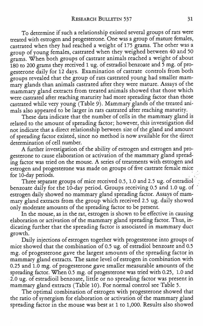
To det~mine if sucb a relationsbip existed several groups of rars were treated with estrogen and progesterone. One was a group of macure females, castrated when tbey had reacbed a weight of In g rams. T he ocher was a group of young females, castrated when chey weigbed between 40 and 50 grams. When botb groups of castrate animals reached a weight of about 180 to 200 grams they received 1 ug. of estr2.diol benzoace and 5 mg. of progesterone daily fo r 12 days. Examination of castrate controls from both groups revealed that the g roup of rats castrated young had smaller mammary glands than animals castC2ted after they were mature. Assays of the mammary gland extracts from treaced animal s showed that those which were castrated after reaching maturity had more spreading factOr than those castrated while very young (Table 9). Mammary glands of the treated animals also appeared to be larger in rats castrated afeer reaching maturity.
These data indicate that the number of ceUs in the mammary gland is related to the amount of spre2ding factor; however, this investigation did nOt indicate that a direa relationship between size of the gland and amount of spreading factOr existed, since no method is now avaiJable for the direa determination of cell number.
A further investigation of the ability of estrogen and estrogen and progesterone to cause elaboration or activation of the mammary gland spreading factor was tried on the mouse. A series of tte3tments with estrogen and estrogen and progesterone was made on groups of five castrace female mice for lO-day periods. . . '.
Three separate groups of mice received 0.5, 1.0 and 2.5 ug. of estradiol ' benzoate daily for che 100day period. Groups receiving 0.5 and 1.0 ug. of escrogen daily showed no mammary gland spreading factor. Assays of mammary gland extracts from the group which received 2.5 ug. daily showed only moderate amounts of the spreading factor to be present.
In the mouse, as in the Illt, estrogen is shown co be eff'eaive in causing elaboration or aCtivation of the mammary gland spreading factor. Thus, indicating further that the spreading factor is associated in mammary duct growth.
Daily injeCtions of escrogen together with progesterone into groups of mice showed thac the combination of 0.5 ug. of estradiol benzoate and 0.5 mg. of progesterone gave the largest amounu of the spreading factor in mammary gland extracts. The same level of estrogen in combination with 0.2~ and 1.0 mg. of progesterone gave smaller masurable amounts of the spreading faecor. When 0.5 mg. of pro~terone was tried with 0.25, 1.0 and 2.0 ug. of esu adioiJ benzoate, little or no spreading faCtor was present in mamrmry gland extracts (Table 10). For normal concrol see Table ~.
The 0rtimal combination of estrogen with progesterone showed that the C2tio 0 syn~gism for elaboration or activation of the mammary gland spreading factor in the mouse was best at 1 to 1,000. Results also showed
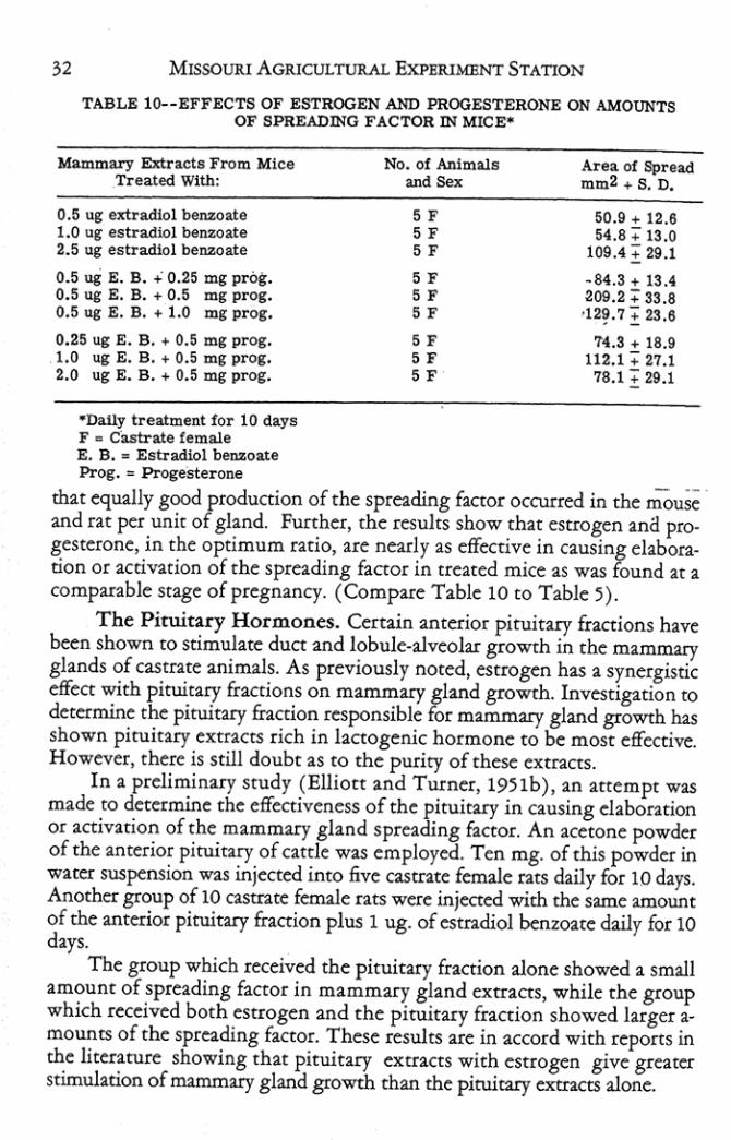
32 MISSOURI AGRICULTlJRAL ExPERIMENT STATION
TABLE 10-·EFFECTS OF ESTROGEN AND PROGESTERONE ON AMOUNTS OF SPREADlNQ FACTOR m MICE~
Mammary Elrtraetl From Mice Tl"eated With:
O.~ UJ extradiol benzoate 1.0 1.1, estradiol Mn'ZOate 2.& ug estradiol benzoate
0.5 u.i E. B . ..: 0.25 mg prot. 0.5 iii E. B. + 0.5 mg prog. 0.5 ug E. B ... 1.0 IIlIiI: prog.
0.25 ug E. 8 ... 0.5 ms prog. ' .0 ug E. B ... 0.5 mg prog. 2.0 ug E. B ... 0.5 mg prog.
. Da.lly treatment for 10 days F a Castrate female E. B. " Estradiol bem:oate Prog. " Progesterone
No. of Animals Area of Spread
'""'" mm2 ... S. D.
SF 50.9 ... 12.6 S F U.8:; 13 .0 SF 109.4 -; 29.1
SF .84.3 ... 13.4 S F 209.2" 33.8 SF · 12?7~23.8
SF 74 .S .. 18.9 SF 112.1 :; 27.1 SF 78. 1 +" 29.1
that e<Jtlally good production of the SpIe2d.ing factor occurred in the mou~ and rat per unit of gland. Further, the results show that estrogen :lna progesterone, in the optimum ratio, are nearly as effective in causing ehboracion or ll.ctivation of the spreading factor in (reared mice as Wll.S found at a comparable stage of pregnll.ncy. (Compare T:able 10 (0 Table ).
The Pituit2.cy Hormones. Certain :anterior piruit2.ry fractions have been shown to stimulate duct 2nd lobule-alveolar growth in the mammary glands of castrate animals. As previously noted, estrogen has a synergistic effect with pituitary fractions on mammary gl:and growth. Investigation to determine the pituiury fraction responsible for m:ammary gland growth has shown pituit:ary extr:acts rich in lactogenic hormone to be most effective. However, there is still doubt:as to the purity of these extracts.
In a preliminary study (Elliott :and Turner, 19)1b), an attempt was m2de to determine the effectiveness of the pituiury in C2using elaboration or activation of the mammary gl:and spreading faCtor. An :a.cetone powder of the anteriot pituiury of c:attle was employed. Ten mg. of this powder in water suspension W2S injected into five C2Stnte female rats daily for 10 days. Another group of 10 C2Stnte fenu.le rats were injected with the same amount of the anterior pituitary mction plus 1 ug. of estradiol benzoate daily for 10 days.
The group which received the pituit2ry fraction alone showed :a. small amount of spreading f:actor in m:ammuy gl2nd extr:acts, while the group which received botb estrogen :and [he pituitary fnction showed brgtt:amounts of the spreading factor. T hese results :are in accord with reportS in the literature showing that pituitary extracts with estrogen give greater stimulation of ma.mmary gland growth than the pituitary extnctS alone.
REsEARCH BULLETIN :537 33
Another series of investigoaions was undertaken to determine the piNitary fraction responsible for ela~oration or ~ctivation of the mammary gland spreading factor. At the same time, expenments were conducted to determine if an active pituitary fraerion was produced by estrogen or estrogen and progesterone treatment of castrate animals.
Groups of five cascrate female mice were injected daily with purified pituitary fractions or crude extracts from hormonally treated rats for 10-day periods. Groups which received pituitary fractions containing 3 mg. (60 I. U.) oflactogenic hormone daily showed good elaboration or activation of the mammary gland spreading factor. Other groups which received separate pituitary fractions rich in growth hormone, thyrotropic hotmone, gonadotropic hormone and melanaphore factor in the amount of 3 mg. daily, showed little or no spreading factor in the mammary gland extracts.
A group of mice which received 05 ug. of estradiol benzoate plus 20 mg. daily of anterior pituitary powder from normal ratS showed no mammary gland spreading factor. A group which received, daily, 0.:5 ug. of estradiol benzoate, plus 20 mg. of pituitary powder from castrate female rats treated with I ug. of escradiol benzoate daily for 10 days, also showed no mammary gland spreading factor. However, assays of mammary glands from a group of mice which received, daily, 0.5 ug. of estradiol benzoate, in addition to 10 mg. of pituitary powder from castrate female rats created with 1 ug. of estradiol benzoate and 3 mg. of progesterone daily for 10 days, showed measurable amounts of the spreading factor to be present (Table 11).
These data show that of the purified pituitary fractions, only the one rich in lactogenic hormone was effective in eausing elaboration or activation of the mammary gland spreadng factor. Pituitary fractions rich in growth hormone, gonadotropic (FSH) hormone, thyrotropic hormone and melanaphore factor were ineffective in causing elaboration or activation of the spreading faeror. Assays of the crude pituitary powder showed that only the one from castrate ratS receiving estrogen and progesterone treatment had any power to cause production of the mammary spreading factor.
These investigations indicate that a pituitary fraction rich in lactogenic hormone can cause good e1aboraton or activation of the mammary gland
I spreading factor just as it has been shown to induce mammary gland growth. However, there is still the possibility that the mammogenic action of this pituitary substance is caused by a contaminant which is a separate fraction from the lactOgenic hormone. Investigation of the crude pituirary fractions makes it appear that treatment with estrogen and progeSterone may have caused secretion of the active pituitary hormone in caStrate female rats because the pituitary extracts from these rats caused elaboration or activation of the mammary gland spreading facror when injecred inco mice. These data agree with findi ngs that pituitary extracts from animals treated with estrogen and progesterone stimulated growth of the mammary gland.

TABLE It- -EFFECTS OF PIT UITARY EXTRACTS ON THE AMOUNT OF SPREAD_ ING FACTOR IN MICE AND RATS*
Mammary Extracts From Mice Treated With:
3 mg Lactogenic Hormone~ 3 mg Lactogenic Horrtlone 3 IIlg Growth Hormone! 3 109 Growth Hor~ne2 3 mg Gonadotropin (FSH) 3 mg ThyrotroPlnl 3 IIlg Melanophore Factor l
10 mg A. P. extract 1 ug E. B. + 10 mg A. P . extract 0.5 iii E. B. + 20 mg A. P . extract3 0.5 ug E. B. + 20 mg Pitultar y4 0 .5 ug E. B. + 10 mg Pituitary5
- Oally Treatment for 10 days, Lactogenic Hormone, 3 mg. = 60 I. U. FM = Castrate female mice FR z Castr ate female rats 1 • Armour Pituitary Fr actions 2 • Squibb Pituitary Fractions
No. of Animals ~d 50><
5 FM 5 F M 5 FM 5 FM 5 FM 5 F M 5 FM
5 FR 10 FR
5 FM 5 FM 5 FM
S * Pituitary extr acts f rom normal female rats
Area of Spread mm2 + S. D.
215.0 + 11 .9 202.4 :; 24.S 65.2:; 19.7 68.0 + 26.7 57.3 + 13 .0 57.2:; 12.6 57.7! 13.0
114 .7 + 16 .5 192.5 + 28.3 53.5+13 .0 67.9 + 19 .6
125 .1 + 36 .2
4 • Pituitary extracts f rom castr ate female r ats getting 1 ug. estrogen daily lor 10 days.
5. Pituitary extracts from casttate female rats getting 1 ug. estr ogen + 5 mg. progesterone dally for 10 days.
Effect on H yp ophysectomized Rats. In a previous section it was pointed out that a number of investigators found the ovarian hormones to be ineffective and an anterior pituitary hormone effective in stimulating mammary gland growth in hypophysectomized animals. To check the effects of these hormones on the elaboration or activation of the mammary gland spreading factor in hypophysectomized animals, groups of mature castrate and castrate· hypophysectomized male Sprague-Dawley rats were injected with hormones for lQ-day periods.
One group of six castrate rats which received 0.:5 ug. of estradiol benzoate showed no spreading factor in the mammary gland extracts. Assays of the mammary gland extractS from another group of six castrate rats which received 1 ug. of estradiol benzoate, 5 mg. of progesterone and 60 I. U. (3 mg.) o(a pituitary fraction rich lactogenic hormone showed large amounts of the factor to be present.
A group of fi'le castrate-hypophysectomized rats which received 1 ug. ofestradiol benzoate and 60 I. U. (3 mg.) ofa pituitary fraction rich in lactogenic hormone also showed large amounts of spreading nctor in the mammary gland extracts. However, a group of six casrrate-hypophyseetD
. mized ratS which received 1 ug. of estradiol benzoate and :5 mg. of progesterone showed no spreading nCtOr in the mammary gland extracts. Assays of a final group of five castrate-hypophysectomized rats which received 1 ug. of estradiol benzoate, :5 mg. of progesterone and 60 I. U. (3 mg.) of

REsEARCH BULLETIN 537 31
a pituitary fracdon rich in the lactogenic hormone showed large amounts of the spreading facror in the mammary gland extracts, (Table 12).
TABLE 12--EFFECTS OF HORMONE TREATMENT ON AMOUNTS OF SPREADING FACTOR IN HYPOPHYSECTOMIZED RATS.
Mainmary Extracts From Rats Treated With:
0.5 ug. Estradiol Benzoate 1.0 ug. E. B. , 5 mg. Pr0'Y
+ 60 1, U. LactogeniC H. 1.0 ug. E. B. + 60 I. U.
Lactogenic Hormonel 1.0 ug. E. B. + 5 mg. Prog. 1.0 ug. E. B. , 5 mg. Prog.
60 I. U. LactogeniC H.
·Daily treatment for 10 days M a Castrate Sprague-Dawley rats
No. of Animals
"'" So.
'M 'M 5 HM , HM
5 HM
lIM = Castrate_hypophysectomized Sprague-Dawl ey rats 1 "' Squibb pituitary fraction
Area of Spread 1l1.Il).2 + S. D.
45.0 + 10.4
198.0 + 37.2
200.6 + 23.7 ' 45.0"+ 10.4
206.4 + 34 .6
Here again it can be noted that hormone treatment elicits a pattern of response of the spreading factor similar ro those reported in stimulating mammary gland growth. Estrogen and progeSterone, which are ineffective in stimulating mammary gland growth in hypophysectomized animals, also are ineffective in causing the elaboration or activation of the spreading fac.ror in hypophysectomized rats. Combinations of the piruitary fraction rich in lactogenic hormone with estrogen or estrogen and progesterone are as effective in castrate-hy}X)physectornized rats as in the castrate rats in causing production of an active spreading factor.
These results are consistent with results obtained by other investigators in stimulating mammary gland growth. It also can be seen from these data that progesterone does not aid estrogen and the fraction rich in lactogenic hormone in causing elaboration or activation of the spreading factor.
These data are believed to provide further evidence for the theory that one of the chid functions of estrogen is to act locally upon the mammary gland, increasing the vascularity and permeability of capillary blood vessels. Primary function of progesterone in mammary gland growth, on the other hand, is to stimulate secretion of the mammogenic hormone of the pituitary. In the absence of the pituitary it is ineffective. In hypophysectOmized animals, progesterone does not synergize with estrogen when the mammogenic hormone is administered.
The Androgens. In determining the effects of androgens on the elaboration or activation of the mammary gland spreading factor, treatment with a level of testosterone known ro stimulate mammary gland growth was

36 MISSOURI AGRICULTURAL EXPERIMENT STATION
employed (Elliott and Turner, 19~ 1 b). Groups of five castrate males and females were injected daily with 1 mg. of testosro=:rone for 10 days. Mammary gland extracts from boch of these groups indicated only fair amounts of the spreading factor to be prest:nt (Table 13).
TABLE l3_·EFFECTS OF TESTOSTERONE, CORTISONE AND DESOXYCORTIC· OSTERONE ON AMOUNTS OF SPREADING FACTOR IN RATS-
Mammary Extracts From Ralll Treated With:
1 mg Testosterone 1 IIlg Testosterone-1 ag. E. B. + 1.5 mg. Cortisone
Acetate 1 ug. E. B. + 2.5 mg. desoxy
corticosterone acetate
*Oa.Ily Treatment for 10 days E. B ... Estradiol benzoate
F .. Castrate fema!e M z Castrate male
No. of Animals
"'" SOx 5F 5M
5F
5F
Area of Spread mm2 + S. D.
144.6 + 23.7 159.5'+ 26.3
103.6 + 18.9
169.7 + 22.4
As'has seen shown in the case of mammary gland growth, testosterone can cause elaboration or activation of the mammary gland spreading faccor when administered at a high level. However, since the majority of the Stu
dies deal with female hormones, no further investigations with effects of male hormones on the mammary gland spreading factor were made.
Adrenal Cortical Hormones. It has been shown experimentally that estrogen can stimulate duet and some lobule-alveolar growth in a number of animals with intact adrenals. Ability of estrogen to cause this growth was greatly reduced when adrenals were removed (Trentin and Turner, 1948). It has also been shown that estrogen can stimulate adrenal cortical activity. In view of these facts. it has been postulated that growth of the mammary gland in response to estrogen treatment, may be due to the ability of estrOgen to cause the adrenal cortex to produce steroids which are either identical with or closely resemble progesterone in its ability to synergize with estrogen in stimulating such growth. Moreover, it has been proven that the adr~nal compound desoxycorticosterone can be converced to progesterone in VIVO.
As previously reported (Elliott and Turner, 19S1b), the ability of estrogen to cause elaboration or activation of the mammary gland spreading factor is better in animals with intact adrenals, indicating that pare of the effect of estrogen is due to ability of estrogen to cause production of an adrenal compound which synergizes with estrogen to cause elaboration or activation of the spreading factor. Thus, it would appear that part of the spreading factor and mammary gland growth produced in response to estrogen is mediated in the same manner, i.e., by way of the adrenal.

,
REsEARCH BUllETIN 537 37
In continuing investigation of the role that the adrenal cortex plays, it was decided that several cortical compounds known to stimulate mammary gland growth should be tested for their effect on the spreading factor. One group of five castrate female rats received 1 ug. of estradiol benzoate plus 1.5 mg. of cortisone acetate daily for 10 days. A second group of five castrate female rats received 1 ug. of estradiol benzoate plus 3 mg. of desoxycorticosterone acetate daily for 10 days. Assays showed measu rable amounts of spreading factor to be present in the mammary gland extractS of both groups. The grour which received desoxycorticosterone acetate showed larger amounts a the spreading factor (Table 12).
These data indicate that adrenal compounds which stimulate mammary gland growth can cause elaboration or activation of the spreading factor. This investigation also adds support to the idea that adrenal cortical compounds produced in response to the presence of estrogen may account for part of the mammary gland growth in escrogen treated animals.
Rdaxio. It has been shown experimentally that relaxin may act as a potentiator of estrogen in inducing mammary gland growth. However, investigations have shown that relaxin does not aid or act as a substitute for progesterone in inducing mammary gland growth. Preliminary investigations (Elliot[ and Turner, 19:511), indicated that relaxin was not the mammary gland spreading factor and that the mammary gland spreading factor did not cause pelvic rdaxation in the guinea pig.
The present investigations were designed to show the effect of relaxin on the elaboration or activation of the mammary gland spreading factor when administered in connection with estrogen and progesterone.
Groups of five castrate female rats were injected daily with various levels of relaxin together with a number of combinations of estradiol benzoate and progesterone, as well as various levels of the individual steroid hormones, for 10 day periods.
Assays on mammary gland extracts from groups of animals which received 25 guinea pig units of relaxin in addition to 1 and 5 ug. of estradiol benzoate, 5 mg. of progesterone, and 1 ug. ofestradiol benzoate plus 5 mg. of progesterone failed to show more spreading factor than was obtained from the same levels of estrogen and progesterone alone. Mammary gland extracts of animals which received 100 guinea pig units of relaxin in addition to 1 ug. of estradiol benzoate showed more spreading factor than those from animals which received the estrogen alone. However, 100 guinea pig units of relaxin tOgether with 1 ug. of estradiol benzoate and:5 mg. of progesterone failed to cause more spreading factor in mammary gland extracts than the same levels of estrogen and progesterone without relaxin.
Results of experiments in which 1 ug. of estradiol benzoate was injected tOgether with 200 guinea pig units of relaxin, daily, showed that the addition of this level of reLt.xin co estrogen caused a large increase in the amount of the spreading f2ctor over the estrogen alone. Relaxin, alone, even

38 MISSOURI AGRICULTURAL EXPERIMENT STATION
at this high level, failed to cause elaboration or activation of the mammary gland spreading factor (T able 13).
Experimental evidence seems to show th:l.t relaxin is ineffective alone and as an aid to progesterone or estrogen and progesterone in causing elaboration or activation of the mammary gland spreading facror. Relaxin in progressively higher levels, however, seems to cause estrogen to be more effecdve in producing the mammary gland spreading factor. The same pattern of response has been shown with relaxin in mammary gland growth by some ocher investigators. It, therefore, appears that relaxin in sufficiently high dosage may synergize with esuogen in stimulating production of the mammary gland spreading factor, but it has no syn~gistic effect when added to combinations of estrogen and progesterone.
Summary. These investig:uions have shown that various combina· tions of eStrogen, progesterone, anterior pituitary fractions, testosterone, adrenal cortical hormones and relaxin are aU effective to a-greater or lesser degree in causing elaboration at activation of the mammary gland spreading factor. These data are believed to indicate that the hormones which stimu· late variable degrees of duct and lobule-alveolar growth of the mammary gland also stimulate production of the mammary gland spreading factor to comparable degrees.
lll.CharaCteristics of the Mammary Gland Spreading Factor
The research was designed, primarily, to investigate physiology of the mammary gland -spreading factor; however, a few facts concerning its char· acteristics were investigated. In a preliminary report (Elliott and Turner, 1950), it was shown that the mammary gland spreading factor did not hy· drolyze the substrate hyaluronidate, and, therefore, probably was not identical with the spreading factor, hyaluronidase. It was further shown in studies of itS enzymatic characteristics that the mammary gl:md spreading factor differed from hyaluronidase in its inactivation by heat.
In investigating the possibility that t he mammary gland spreading fac· tor was hyaluronidase, a turbidimetri'C method of assay for hyaluronidase was employed (Tolksdorf et al., 1949). Several preliminary assays conducted with purified hyaluronidase indicated the reliability of potency of the enzy· me preparation and the reproducibility of the method. Assays were made on mammary gland extracts from rats pregnant 6, 7,8,9, 10 and 11 days. In no case did any of these mammary gland extracts give indication of hydrolyz· ing the substrate hyaluronidate (Table 14). . Since it was deemed necessary that the enzymatic charaeter of the mammary gland spreading factor be checked, a number of assays were made to determine irs inactivation temperature. Testicular extracts containing hyal-

• REs£A.RO! BULLETIN 537 39
TABLB H __ EFFECTS OF RELAXIN ON THE AMOUNT OF SPREADING FACTOR IN RATS" .,
Mammary Elrtract~ From Rats No. of Anlmale Area ot Spread . Tr'eaied With; ... "" mm2 .. S. D • ,
lUi. E. B ... 25 O. P. lJ. Relu1n " 8e.4 ~ 14.1
5 ",. E. B ... 25 O. P. U. ....... " lIiIl.7 ~ 18.5 5 mg. Prof ... 25 G. P. (1.
Relax1n " 114.7 .. 14.5 1 Ui. E. B ... 5 m,. Pro, ...
• 25 G. P. U. Reloin " 20$,0 .. 16.~ • 1 ul. E. B ... tOO G. P. U. Relaxin " 11 2.0 .. 15.1
1 1If. E. B ... 5 m&:. Prof ... • 100 G. P. U. Relutn
1 III. E. 8 ... 200 G. P. U. .., 207 .. .. 111.3
Relaxl.n " l Si.7 .. 26.4 200 O. P. U. Relaxin " 53.8:;: 12.8
· Daily Treatment tor 10 day. Prog .• Progesterone E. B . • Estradiol benzoate F • Castrate femaln
• TABLE GLAND EXTRACTS
Tnt "bUrial ..... Byaluronlda:n° >00 75.5 87.5 95.0 n.1l 88.0 .,., 17.0 Hyaluronldue· . 00 '1'5.0 88.0' 98.0 !iZ.S 89.0 ".0 78.0 • lfyaluronldueo ' 00 14.5 88.0 94.5 92.0 8'1.5 .,., 17.0 HyalllronLdu. · .00 74.5 8$ .Il 98.0 9205 87.5 " .0 78.5 Hyaluron.ldue· · . 00 74 .0 86.5 97.0 84..0 96.0 87.0 80.0 Hyll\lronldue' • .00 74.5 S7.0 97.5 .... ~1.0 86.5 7~.$ '.0 HyalllrOftidu.'* .00 ?S.$ SU n.O ".0 ~0.5 811.0 SO.$ '.0 !lJalllroaidu.· • '00 74.0 SU n.o U.$ 90.$ 87.0 80.$ '.0 £xt:I'II,et.1I d., Gtand· ·· '00 72.5 .... 72.$ " .. 72.5 ".0 72.5 0.0 Eldra,et._11 d., Glam' " . 00 12.0 115.11 ?S.O "., ?S .O 72.5 12.5 0.0
• ~a,et..7 day Gland'" . 00 11.5 112.5 72.0 72.0 72.5 77.5 72.5 '.0 Ii:xtr&e:t·7 day Gland. '. . 00 11.0 82.0 71.5 72.0 72.0 72.0 71.5 ••• Eldraet·8 d., Oland'" .00 72.0 84.S 72.5 72.5 12.0 72.5 ?S.O '.0 Extract · ; day Oland'" . 00 72.0 82.0 71.0 72.11 71 .0 71.5 71.5 0.' Enra,et..l0 day Oland'" ' 00 72.Q 82.5 72.0 ' 71 .5 71. 5 71.5 72.5 0.0 Eldraet.lO d., .Oland· ·· ' 00 12.0 82.0 71.5 71.5 12.5 72.0 72.0 '.0 Enra,et.. l1 day Gland' " . 00 12.5 84.5 ?S.O 72.5 72.5 73.0 73.0 0.' E:rtra,et.·11 day Qt..IId· '· . 00 73.0 85.0 72.5 72.5 72.5 72.0 72.5 0.0
• ' 3 TIlrbtd1ty Reclucine Ullita/ =l. _. - .5 ml $\Ibabt ... .5 ml Buffer **' TIlrbtd1ty Reduclllg UlliUl / =l. _n
•. 25 ml Sllbatauo .. . 15 =l Buffer " ' Day. prtpt.nt 1'IIba m • .5 ml Sllbetat ... .5 m1 test material
1'IIbe IV '. .5 ml !ubt;tat ... .1 m1 BWfer * .4 101 teat MaJ.. "' ... V - .5 ml SllbIWe .. .2 m1 Buffer ... 3 ml tellt M.t. _n_
.5 Illi Subltate .. .3 m1 Buffer ... 2 mllellt Ma t . Tuba vn. .5 Illi Substal., .. • 4 ... 1 BlltJer ... 1 ml tn t Mat .
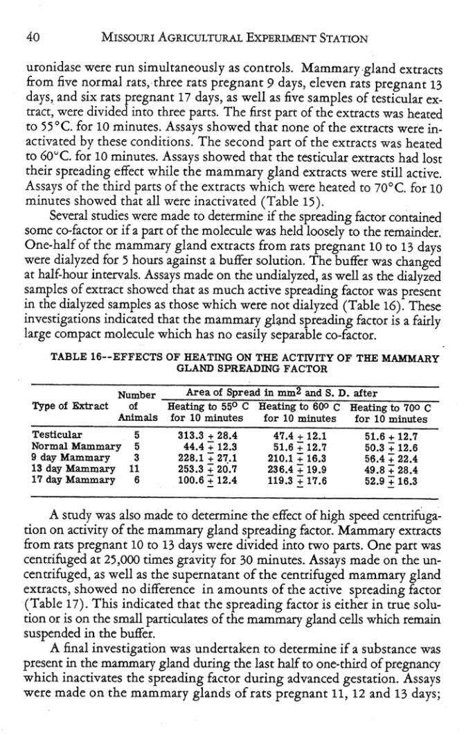
40 MISSOURI AGRICULTURAL ExPERIMENT STATION
uronid2Se were run simultaneously as controls. M ammary .gland exttlas from five nortn2.1 rau, three nts pregnant 9 days, eleven cats pregnant 13 days, and six tats pregnant 17 days, as well as five samples of testicular extract, were divided into three parts. The first pan of the excncts was heated to ~5°C. for 10 minutes. Assays showed that none of the excnctS were in· activated by these conditions. The second p:m of the extncts was heated co 6O~C. for 10 minutes. Assays showed that the testicular exU:lcrs had lost their spreading effect: while the mammary gland extracts were still active. Assays of the third parts of the extracts which were heated to 70°C. for 10 minutes showed that ill were inactivated (Table 15).
Several studies were made to determine if the spreading factor contained some co-factor or if a part of che molecule was held loosely to the remainder. One-half of the mammary ghnd extracts from r:m pregnant 10 to 13 days were dialyzed for :> hours against a buffer solution. The buffer was changed at half·hout intervals. Assays made on the undialyzed, as well as the dia.lyzed samples of extraCt showed that as much active spreading factor was present in the dialyzed samples as those which were nOt dialyzed (Table 16). These investigations indicated that the mammary gland spreading factor is a fairly large comput molecule which has no easily separable co-factor.
TABLE 16--EFFECTS OF HEATING ON THE ACTIVITY OF THE MAMMARY GLAND SPREADING FACTOR
Number Type of Extract of
AnImals
Testicular 5 Normal Mammaq 5 9 day Mammary 3 13 day Mammary 11 17 day Mammary 6
Area of Spread In mm2 and S. D. after Heating to 550 C Heating to 600 C Heating to 700 C for 10 minutes for 10 mlIlutes for 10 minutes
S13.3 + 28.4 44.4 + 12.3
226.1 + 27.1 253.3 + 20.7 100.6 + 12.4
47.4 + 12.1 51.6 -; 12.7
210.1 -; 16.3 236.4 -; 19.9 119.3 + 17.6
51.8 + 12.7 50.3:; 12.8 56.4:; 22.4 49.8 + 28.4 52.9 + 16.3
A srudy was also made to determine the effect of high speed centrifuga. tion on activity of the mammary gland spreading bctor. Mammary exrncts from rats pregnant 10 to 13 days were divided into cwo partS. One puc was centrifuged at 25,000 times gravity for 30 minutes. Assays made on the uncentrifuged, as well as the supernatant of the centrifuged mammary gland extracts, showed no difference in amounts of the active spreading baor (Table 17). This indicated that the spreading factor is either in true solution or is on the small particulates of the mammary ghnd cells whicb remain suspended in the buffer.
A final investigation was undertaken to determine if a substance was present in the mammary gland during the last half to one·third of pregnancy which inactivates the spreading factor during advanced gestation. Assays were made on tbe mamm:uy glands of rats pregnant 11, 12 and 13 days;

REsEARCH B ULLETIN H7 41
TABLE n·-EFFECTS OF DIALYSIS AND HIGH SPEED CENTRIFUGATION ON ACTIVITY OF MAMMARY GLAND SPREADING FACTOR m RATS
Type of Extract
10 clay M ammary 10 day M ammary dlalyud 10 clay M ammary cenb'Uuied 13 day M ammary 13 day M ammary dialyzed IS day M ammary centrUl.lged
Number of
Animal,
1 I 1 I 1 1
Area of Spread mm2
228.6 236.4 221.6 207.6 21 7 •• 229.7
. then these solutions without the fat were used to extnct the mammary glands from ntS pregnant 19 and 20 days. The assays showed that the active spreading factor present in mammary gland extracts from the rats at midpregnancy was unaffected by extncting the mammary glands from rats of late pregn:lnCY. This indicates that no inactivating substances are present in the mammary gland dueing the larrer pact of pregnancy (Table 18).
TABLE 18··EFFECTS OF MAMMARY GLAND EXTRACTS F ROM RATS m LATE PREGNANCY ON SPREADING F ACTOR IN EX TRACTS FROM
RATS m MID· PREGNANCY
Type of Extract
11 clay Mammary 12 day M ammary 13 day M alJUllary 11 day + 19 day M ammary 12 day + 19 day M ammary 13 day + 20 day M ammazy
No. of AnImals
1 1 I 1 1 1
Area of Spread mm2
22'1 .4 258.6 234.9 236.'1 25 1,4 226.3
Sllmmuy. The mammary gland spreading factor is a heat coaguJabJe substance, possibly a protein, fairly stable under normal conditions. It apparently is not the same as the spreading factor hyaluronidase. It has been shown that dialysis or high speed centrifugation does nOt change the spread. ing activiry of the extract. This indicates th:at the spreading factor is io true solution or is attached to the smaller particulates in the mammary gland cells which remain suspended in the buffer. It further indicates that the spreading faeror is a fairly large molecule which has no easily separable cofaeror.
In addition, it was shown that exua.ccs of mammary glands t2ken from t2ts in bte pregnancy had no diminishing effect on me spreading facror present in extracts of mammary glands taken from nts at mid'pregn:ll0CY, indio cating that no :mti-spreading factor is present in the mammary gl:lnd during late pregnancy at a time when the spreading factor was shown to I?e very
42 MISSOURI AGRICULTURAL EXPERIMENT STATION
low. In other words, the decline in the amount of spreading factor in the latter st2ges of pregnancy is nOt due to the production of a factor which in· activates the enyzme. The enzyme is thus [dated to the growth phase of mammary gland development and becomes inactive as secretory activity is induced.
DISCUSSION The rapid advance of the mammary gland ducts and lobule-alveolar
system through the connective tissue barrier during pregnancy, Or when certain hormones are used to induce mammary gland growth, has previously been taken for granted. With the discovery of spreading [wors which are known to break down the ground substance of the connective tissue, it was posrulated that such a spreading factor might be produced by growing cells of the advancing mammary gland.
Inv~stigations on th~ albino rat, albino mous~, guinea pig and rabbit hav~ shown that such a spreading factor is pr~scnt in the mammary glands of these speci~s during pr~gnancy. It was also noted that the general pat· tern of amounts of spreading faeror found in the mammary glands during various stages of ptegnancy follows the mammary growth pattern known to occur in each of these species. In view of these results, it seems possible that a mammary gland spreading faeror is present in all mammalian speci~s and that the spreading factor is associated with or involved in mammary gland growth into the connectiv~ tissue of the fatty pad.
A srudy of hormon~s which have been proven to induce both mammary duct and lobule-alveolar growth has revealed that these hormones are effective in causing production of the aerive mammary gland spreading fac· tor.
While estrogen and progest~rone. separately, caused elaboration or activation of the spreading factor; the correct combination of these two hor· mones gave amounts of the spreading factor comparable to those extracted from the mammary glands of normal pregnant animals. Prolonged treat· \ menr with estrogen and progesterone in the rat caused an increased production of the spreading factor for about 1S days, after which the amount of spreading factor produced declined despit~ continued treatment. Gradual decline of mammary gland growth after a certain period of time, despit~ COntinued hormone treatment, has been recognized for some time. The fact thac production of che mammary gland spreading factOr follows chis same pattern is believed co indicate that only the growing cells of the mammary gland elaborate or activate the spr~ading factor.
The elaboration or activation of greater amounts of the spreading factor and the larger size of the mammary gland of castrate rats with previously de· veloped duct systems, compared to chose without such syst~ms, strongly supportS the hypothesis that the number of c~lls in the mammary gland,

REsEARCH BULLETIN 537 43
especially of growing cells, is related to the production of active spreading faCtOr.
Unfortunately, there is no available method by which the number of epithelial cells in the growing mammary gland can be determined. Kirkham and Turner (1953 have shown that desoxypentose nucleic acid increases up to mid· pregnancy in the rat. This nucleic acid is believed to be a relatively conStant constituent of the cell and, therefore, the increase in amount of this acid is believed to indicate the multiplication of cells at this time. While a quantitative comparison oif the number of epithelial cells in the growing mammary gland with the amount of spreading £actor which can be extracted cannot be made, the results which have been obtained in chis srudy are believed to give strong suppOrt to such a theory.
With furcher increase in our knowledge of the biochemistry of this enzyme system and as more quantitative methods of assay are developed, the determination of the amount of mammary gland spreading factOr may serve both as a method of assay of the hormones involved in the growth of the mammary gland and as a quantitative measure of the extent and varia· tion in the number of epithelial cells present in the mammary gland, i.e., the potential functional capacity of the mammary gland.
Good elaboration or activation of the mammary gland spreading factOr resulted from treatment of castrate female rats with pituitary fractions rich in lactogenic hormone. Assays of mammary gland extracts from rats treated with pituitary fractions rich in growth hormone, gonadotropic hormone (FSH ), thyrotropic hormone and the melanophore factor failed to show the presence of the spreading factor. Thus, if as postulated, the presence of the spreading factot indicated that mammary gland growth had occurred, these data would indicate that the lactogenic fraction of the pituitary is the only fraction which possesses mammogenic activity. Due to the possibility that more than one substance is present in this pituitary fraction it is possible that the mammogenic action is due to a contaminant and nOt the laerogenic hormone.
During the second half of pregnancy, it has been shown, there is a gradual decline in the amount of mammary gland spreading factor. It is at this time that the lactogenic hormone is believed to increase, reaching a maximum follOwing parturition. If the lactogenic hormone were the factor Stimulating growth of the mammary gland one would expect that the spreading factor would continue to be present at a high level during the second half of pregnancy and early lactation.
Production of the active spreading factor by a whole pituitary powder from rars treated with estrogen and progesterone adds further weight to the suggestion that the mammogenic hormone is produced in the anterior pituitary in response to estrogen and progeSterone administration in experimental animals.

44 MISSOURI AGRICULTURAL EXPERIMENT STATION
Elaboration or acdvation of the mammary gland spreading factor in response co treatment with testosterone, cortisone, desoxycorticosterone, the diminished effect of estrogen in the absence of the adrenal, and che increased effect of estrogen in combination with relaxin also indicate that the spreading factor is associated with or involved in duct and lobule-alveolar growth of the mammary gland.
I n examin ing all data from the animals in normal pregnancy and animals under various types of hormone treatment, it appears that the spread· ing factor is either elaborated or activated in the gtowing mammary gland cells by hormones which initiate both duct and lobule-alveolar growth of the mammary glands. The spreading factor may then break down the connective tissue in some manner allowing the rapid forward growth of the mammary ducts and lobes.
Heat stability studies of the mammary gland spreading factor show that it is inactivated after heating at 70 Q C. for 10 minutes, which suggests the possibility that the factor is an enzyme. However, assays reveal that it does not act on the substrate hyaluronidase so that it apparently is nor identical with the spreading factor hyaluronidase.
Dialysis and high speed centrifugation of mammary gland spreading factor extracts indicated that the spreading factor is either in true solution or is associated with the smaller particulates in the mammary epithelial cells. Further, the results signify that the spreading factor is a fairly large molecule without an easily separable co-factor.
Extraction of the mammary glands ftom rats in late pregnancy (which showed no spreading faeror) with the extract solution of rats in mid-pregnancy (which showed large amounts of the spreading factor) failed to diminish the spreading activity of the latter extracts. Apparently, there is no antispreading factor present in mammary glands of rats during late pregnancy.
This study indicates that a spreading factor is elaborated or activated in the mammary gland during the growth phase of pregnancy. It also has been shown that a number of hormones, which are known to induce duct and lobule-alveolar growth of the mammary gland, cause elaboration or activation of the mammary gland spreading factor. T he theories that the amount of spreading factor is related to the numbers of epithelial cells in the growing mammary gland and that the spreading factor aids in the forward progress of the mammary gland through the connective tissue barder are also presented.

REsEARCH B UllETIN 537 45
SUMMARY 1. Investigation has revealed the presence of a spreading factor in the
mammary glands of four species of experimental animals during pregnancy. The four species were the albino rat, the albino mouse, the guinea pig and the rabbit. The amount of spreading factor present in the glands was found to increase during early pregnancy until a peak was reached in from half to
two-thirds of pregnancy, afrer which it declined until none was present at parturition. The spreading facto r was not present in the mammary glands of normal or castrate animals of any of the species investigated. The spreading factor was assayed by measuring the extent of intradermal spreading achieved when it was injected with T-1824 (Evans Blue) dye into a shaved white rabbit.
2. The injection of a number of hormones, which induce both duct and lobule-alveolar growth of the mammary glands, has shown them to be effective in causing elaboration or activation of the mammary gland spreading factor. Estrogen and progesterone, separately or together, pituitary extracts, particularly a fraction rich in lactogenic hormone, testosterone, desoxycorticosterone acetate, cortisone acetate and relaxin were all shown to be effective co a greater or lesser degree in causing or helping to cause elaboration or :l.Ctivation of the mammary gland spreading facror.
3. Prolonged treatment of castrate female rats with the optimum combination of estrogen and progesterone (a ratio of 1:5000) showed that the amount of mammary gland spreading facro r increased until a peak was reached at 15 days' treatment, after which the amount declined.
4. Estrogen and progesterone treatment of castrate rats caused the elaboration or activation of greater amouncs of the mammary gland spreading factor in the rats with rudimencary duct systems.
5. Tests revealed that the mammary gland spreading factor did not act on the substrate hyaluronic acid and it is probable that this s,preading factor is not hyaluronidase.
6. Investigation showed that the mammary gland spreading factor was inactivated by heating to 70°C. for 10 minutes.
7. Dialysis and high speed centrifugation of mammary gland extracts containing large amounts of active spreading aCtivity of the extracts.
8. Extracting the mammary glands of rats in late pregnancy with the extract solutions of mammary glands from rats in mid-pregnancy failed to inactivate the spreading factor in the extracts of the glands from the rats in mid-pregnancy.
9. The theory is presented that the amount of spreading factor is related to the numbers of epithelial cells in the mammary gland. As more quantitative methods of assay are developed, it is believed that the determination of the amount of mammary gland spreading factor may serve both as a method of assay of hormones involved in mammary gland growth and as a quantitative m~asure ofthe numbers of growing epithelial cells present in the mammary gland.

46 MISSOURl AGRICULTURAL ExPERIMENT STATION
BIBLIOGRAPHY Ancel, P. and Bouin, P. 1911. Recherches sur lC"5 fonct ions du corps Jaune Gemtiff. II
Sur de dccerminC"5me development de It Glilnde Mllmmairc au (ours de b gesh.rion.]. til PhYI;()/, II Path. Gm. 13 :3 1.
AsdeIJ, S. A. and Seidenncin, N. R. 1935. Theelin and progc$fin injection on uterus :and ma~mary glands of ovariectomized and hypophy~ctomized rabbits. Pr«. 5«. Exptr. Bid. and Mtd. 32:93l.
AstwOOd, E. B. 1941. The (cgub,don of corpus lutcum function by hypophyscalluteouophin. EnthrrinCIbJgy 28:309.
AstwOOd, E, S , 1953. TestS for luteotrophin. In Wolstenholme and Cameron Bioassay of Anterior Pituitary and Adrenocortical Hormones. eiba Foundation Colloqllia on EmitxrinDkgy Volume V,
Asrwood, E. B., Geschickttt, C F. :1nd R:ausch, E. D. 19n. Development of the m:lmmuy gltnd of the nt. A study of normal. experimental and pathologic changes ;tru:\ their endocrine relationships. Am.]. Arlat. 61 :373.
Aylward, F. X. 1937. Chemiol nature of the Reynals sprea.ding hctor from mamm1iian testicle. Pr«. SIX. Exjwr. Bid and M,d 36:411-
Bach:arach, A. L. , Olance, M. R. A. U1d Middleton, T . R. 1940. The biologica.l assay of testicular diffusing factor. Biocbnn.. J. }f:I464.
BottOmley, A. C. 3nd Folley, S. J. 1938. The effect of androgenic substances on the growth of the ~t wd munmary gland in the immature m1ie guinea pig. Proc. &yo Soc., Lom/(;n, B. 126:224.
Boylwd, F. and McI.(1n, D. A. 1935. A faetor in malign3nt tissue which increase the pc:rmea.bility of the dermis.]. Path. and &«t. 41 :553.
Blundell , G . P. 1942. ScreptoCOCCUS hemolytieus: A study of virulence. y.w J BioI.. Mea. 14:3n.
Br:l.dbury,]. T. 1932. Study of the endo<:rine f3crors influencing mammuy development wd secretion in the mouse. Prrx. SIX. Exptr. BiDI. and Mtd. 30:21 2.
Burnet, F. M. and Srone, ]. D. 1948;1.. Desquamation of intes tinal epithelium inllitrrl by vibro cho1erae filtr:l.tes: Ch:aracteriution of mucinasc md tissue disimegr:ating enzymes. AWlr.j. Exptr. Bioi. and M tti. Sci. 2':219.
Burnet, F. M. ;1.nd Stone,]. D. 1948. The receptor destroying enzyme of V. cholerae. Awtr. J Exptr. Bioi. and M td. Sri. 25 :227.
Ch1in, E. and Duthie, E. S. 199. A mucolytic enzyme in testis eX(f1cts" Nallirt 144:977. Chain, E. 1nd Duthie, E. S. 1940. Identity of hy1iuronidue and spre3ding factor. Bril.
I Exptr. Palh. 21:324. Ol;llllorro, 1...1940. L'Action de]a desoxycorticosterone sur 11 m3mill et sur l'appareii
sexual secondaire de la souris male hypophysectomisee. Comp. RInd. SIX. di Bioi. 133:546.
Christen..sen,j. F. 1938. Kallikrein;15 a Reyn;1.ls haor. Nalllrt 142:36. CiuVlfi, E. 1933. Influenza dell' ure~no sulla vdoci~ d.i 1SSOCbimento ddl' and·lossilu
differia. Sptrimmtais, Anh. Bioi. 87:471. Claude, A. 19}7. Spreading properties of le;1.ch extracts and the formation of lymph.
J. f.xpIr. M,d. 66:n3. Claude, A. 1nd Dunn-Reynals, F. 1934. O n the existenee of a f.a CtOt inCl"C:1s ing tissue
pame;tbility in org.ms o ther th3n tes ticle.]. Exptr. Mid. 60:457 . Cole, H. A. 1 9~3. The mammuy gland of the mouse during oeStrus cycle, ptegn3ncy
and lam.non. Proc. &yo S«. B., London. 114: 136. Comer, G. W . 1930. The bormond control of lactation. I. Non-effect of the corpus
luteum. 11. Positive 1ction of extCllas of the hypophysis. Am. J. PbylioL 95:43. Cowie, A. T . and Folley, S. J. 1944. Effects of adrenalectomy and anteriot pitui tary in
jectiions on rrumltW}' development. Nallm 154:307.

•
•
REsEARCH B ULLETIN :537 47
Cowie, A. T. and Folley, S. J. 1947. The role of the adreru.l COrtex in nummary develop-menr and its relation to the mammogenic action of the anterior pituitary. Endrxrin-o/Qgy 40:274. .
DeGra.af, H.)., de )ongh, S. E. and Paesi, F.). A. 1950. On the development of the mammary gbnd in the rat. III. Aaa. PhysioL Pha1"!'1JlWJL Netrl. 1 :290.
De)ongh, S. E. 1933. Fortsen:ung von uncersuchungen uber den angriffspunkt der menformonwirkung auf de munma. Acl.1. BrtfI. Nmi. 3:99.
Dorfman, A. and O cc, M. L. 1948. A turbidimetric method for the assay ofhyaluronidasc.j. Bioi. Chtm. 172:367.
Duncan, ) . M. 1875. On the pelvic articulations in parturition. Mech:mism of na(urill and morbid parturition, Edinburgh, pp. 144 .
Duran-Reynills" F. 1928. Exaltation de I'aetivite' def virus vaccinal par les extraits des e=ins or~nes. OJmp. Rtnd. S()C. Bioi. 99:6.
Duran-Reyna!s, F. 1929. The effeet of excraetS of certain organs from normal and immunited animals on the infeeting power of nccine virus.). i?'f". Mid. 50:327.
Duran.Reynals, F. 1933. Studies on a. certain spreading faetor existing in Mcteria. and its significance for ba.eteria.i invasivene$S.)' Expw. Mtd. ~8:141.
Duran.Reynals, F. 1936. The inva.sion of the body by animal poisons. Sdmet 83:286. Duran-Reynais, F. 1939. A spreading betor in cernin snake venoms and its relation to
their mode of action.). EX/>". Mtd. 69:69. Duran-Reynals, F. 1942. Tissue permeability and the spreading faetors in infection.
&a. Rfti. 6:197. East, M. E., Madinaveicia,). and Todd, A. R. 1941. T he B-glucosaminase acdvity of
testicular extra.cts and its bearing on the problem of diffusing factors. BibChtm.J. ":872.
Elliott, ) . R. and Tumer, C. W. 1950. Observation of a spreading bctot present in mammary gland extr:lCts. PrIX. Soc. ExJ-. BioL and Mtd. n :384.
Elliott,). R. and Turner, C. W. 1951. Mammary spreading bctor and relaxin. PrIK. Soc. Exptr. Bioi. and M ed. 77:33.
Elliott,). R. and Turner, C. W. 1951. Some hormones involved in the elaboration or aetivation of the mammary spreading factor. PrIX. 50(. Exper. Biql. and Md. 77:;20.
Folley. S.). 1952. LaCTation. In Marshall Physiology of Reproduction, Longmans, Green and Co., London, Volume II.
Folley, S.)., Guthkelch, A. N . and Zuckerman, S. 1939. T he mammary gland of che rhesus monkey under normal and experimental conditions. PrIX. Roy. Soc. , London, B.126:469.
Folley, S. ). and Malpress, F. H. 1948. Chapler XV in Pincus and Thimann, The Hormones. Volume I. Academic Press Inc., New York.
Fo~, T. R. 1942. Factors of age in the rate of absorption of, and in mammary stimu· lation by, tl:SIOSrc:rone monopropionate pellets in rats. En&xrinokJgy 30:765.
Freud, T. and de ) ongh, S. E. 1935. The effect of progestin upon the mammary gland o(the rat. Ana. BrtfI. Nterl. ':47. .
Gardner, W. U. 1936. &xual dimorphism of che pelvis of the mouse, the effect of eStrogenic hormones upon the pelvis and upon the development of scrotal hernia. Am.. 1. Anat. '9:4~9.
Gardner, w . u. 1940. Growrh of the mammary glands in hypophysectomized mice. Prix. 50(. Exper. Bioi. and Mid. 4':835.
Gardner, W . U. 1941. Inhibition of ma.mmary gland growth by luge amounts of estrogen. Etuiomno1og) 31 :124.
Gardner, W. U. and Hill, R.. T. 1936. Effect of progestin upon the mammary glands of the mouse. Proc. SIX. Expn-. Biol. and MM. 34 :71 8.

48 M ISSOURI ACRICULTlJRAL ExPERL\(ENT STATION
Gudner, w. U. and VWl Wtgcncn. G. 1938. Expcrimenw development of tilt rnatnJTUry gland ofrbe monkey. bui<lcrin,logy 22:164.
Gucfncr, W. U. :and White, A. 1941. Mammary gltnd gfOwth in hypophysectomized male mice receiving eStrogen :lnd proberin. Pm. S«. Exptr. Bia[ and Mtd. 48:590.
Gardner, W . U. and White, /1.,1942. Mammary growth in hypophyseaomi~ed rove mice. Anal. RK.82:414.
Garrett, F. A. and Talmage, R. V. 1950. Influence of relaxin on m:.l.mmary development. Anal. RK. 108:523.
G:mcn , F. A. Uld T1.llIl2ge, R. V. 19B. The influence of relaxin on mammary developm..~t in gWnCi pig1 and rabbits.]. EndtJ. 8:336-
Gomez, £. T. 1942. Mammary gland growth in hypophysectomized wtnte guinea pigs. End«rinokKJ31 :613.
Gome:z, E. T. and Tumer, C W. 19~. Non·dfects of emogenic hormones on mammary gland of hypophysectomized guinCl pig, Prrx. 5«. f!.xptr, Bioi. and Mtd. 34:320.
Gomez, E. T. and Turner, C W. 1937. Hypophysectomy and replacement therapy in reo lation to the growth and secretory activity of the mammary glmd. MD. Agr. Exptr. S~. Rtf. BilL 2~9.
Gomn, E. T. and Turner, C W. 1938. Further evicknce for a mammogenic hormone in me aneerior pituituy. Prrx. 5«. Expw. BiDi. and Mtd. 37:607.
Gomez, E. T., Turner, C W ., <nrdner, W . U.:md Hill, R. T. 1937. Oesuogenie tre:l.t· ment ofhypophyseaorni:ed male mice. PrtX. StK. Exptr. BiDi. and Md. 36:287.
Hw, E. 1946. On the rne<hanisrn of invasion I. Antinvuin I, an enzyme in plasma.]. BiDI. Cbtm. 163;63.
Hadidim, Z. and Pirie, N. W . 1948. The prepantion and some properties of hyaluronic 1(id from human umbilical cord. Bi()Chtm. /: 42 :260.
Hahn, L 19-". On the.cheE!'isuy of giucuromdase and acetylglucosarninidase (Title tnns.) A(~ftI. IVnIt Ml1ftral GHH. 21A:1.
Hahn, L 1947. Struetute of N.uctylgluconmine as related ro breakdown (Title tnns.) BiodN1II. Z:318.
Hall, K. ~~7 . The e1fem of pregnancy and reluin on the histology of the pubic sym· physlS In the mouse. J. EI'Ifi4. ':174.
Hall , K. and Newton, W. H. 1946. The nOfmd course of separation of the pubes in pregnant mice.]. PbyliDi. 104:346.
Hammond,]. and Marshall, F. H. A. 1914. The functional correlation between the ovu· ies, uterus and m3tllmary glands in the rabbit, with observations on the oestrus cy. de. Prot. Roy. SIx'., I.Andott 8., 76:206.
H:unoisky, M. and Sparrow, R. C 19·H. Influence of relaxin on mamnury development in sexually imrm.rure female rats. hrK. 5«. Expw. BirJ. and Mta. 60:8.
Hartman, C G. and Specrt, H. 1941. Action of progesterone on the genital organs of the unpdmcd rhesus monkey. EnJ«ri~gy 29:639.
Hcch[Cf, D. 1948. Reconstruction of the dermal burier to fluid diffusion following ad· ministration ofhyalUlonidase. PrtX. Sf)(. Exptr. Bioi. a"d MId. 67:343.
Hisaw, F. L. 1926. Exp:crimental relaxation of the pubic ligaments of the guinea pig. Prrx. S«. Exptr. BioJ. a"d Mra. 23:661.
Hisaw, F. L. 1927. Presence of relaxin in the corpus lutcum. Anal. Rtc. 37:126. Hisaw, F. L. 1929. The cOfJlus lutcum bormone. I Experimental relaxation of me pelvic
ligaments of the guinea pig.). PbysiDi. ZDoI. 2:59. Hisaw, F. L. 1930. Some aspects of the hormone of the corpus lureum. Am.]. Phys;DI.
42:574. Hisaw, F. L. and Leon:IJ:d, S. L. 192' . The relation of relaxin to the corpusluteum.).
Exptr. ZDtJ/. 42 :411.

REsBARCH B ULLETIN 537 49
Hobby, G. 1., Dlwson, M. H ., Meyer, K. lind Ch1ffee, E. 1941. The rel1tionship be· fWCCll spreading &cror and hyaluronidase.). Exptr. Mid. '3.43.
Hwnphrey,). H . 1943 . Studies on diffusing bctors. (II. A new biologia.1 asS1Y of diffus· ing bcto(5 in guinC1 pigs. Biodmn.]. 37: 177.
Ibsen, H. L 1928. Prenlw growth in guinea pigs with specw refcrcoce to environment· 11 bcto(5 lffeCting weight at birth.]. Exptr. ZtJci. '1 :' 1.
Jeanloz, R . w. :and Forchielli, E. 1951. Srudies on hYlluronic acid and relued $ubst:mces. IV. Periodue oxidltion.]. Bioi. Chtm. 190:537.
Jeffe(5, K . R. 193'. Cytology of the mammary gland of che albino £lit. I. Pregnlncy, Iacu.don 2nd involution. Am.]. Anat. '6:257.
Kass, E. H . rod Seastone, C. V. 1944. The role of the muooid polYS1<.:<.:huide (hyalu. ronic acid) in the viruJene of group A hemolytic strept:ococo.]. Exf-.. Med 79:319.
Ku2wt2, S. 1925. On the morphological ch:mge of the symphsis PUboLSChilidrica in the guinC1 pig during pregnancy. Mitth. d. Md .. Aw' 211 /Vijc 8:21.
lC1ye, M. A. G . and Sc-accy, M. 19'0. Observations on the chemistry of hyaluronic acid. Bimm.]. 46, XIII.
Kirkham, W. R. lind Turner, C. W . 1953. Nucleic ldds of the mlmmuy gland. Prot. 5«. Exptr. BiO/. and Mtti.83:123.
l.:me.Cl1ypon, J. E.:md Sruling, E. H . 1906. An experimenral inquiry into the nctors which determine the growth and tcrivity of the mammary ghnch. PrOt. [(Py. Soc., l..Jmdon B. n:,0,.
Laqueur, E. 1928. Uber weibliches sexull.hocmone, Menformon. V. Uber den femin· surenden einfluss des menformons auf de unenrwidreite brustdruse. Dtutub. Med. WochtnKhr.53:867.
Lavine, M. D. 1950. Muco.lyric :activity of the thyroid $1and.J. Endo. 6:288.. Lcon:ud, S. L 1943. Stimulation of mamrru.ry gbnds In hypophysectomized L'2ts byes.
trOgen ltId testOsterone. Endocrin~gy 32:229. Leonard, S. 1., Pednun, P . L. lind Kuurok, R. 1946. A turbidimetric method for de
termining hYli Juronid1Se in semen lind tissue extncts. Endtxrinokgy 39:261. Leonard, S. L:md Reece, R. P. 1942. Failure of steroid hormones to induce mammary
growth in hypophysecromized nts. E.nti«rindtm ~:32. Lewis, A. A. lind T urner, C. W . 1941 . Effect of sdlbenrol on lactogenic content of pi
tuitary :md m1JTlmary glanch of fem..ale nts. PrtK. 5«. Exper. BiO/. and Mtti. 48:439. Lewis, A. A. and Turner, C. W . 1942. Growth of the male guinea pig mammary gland
with diethylstilbesterol. Etuiomnokgy 30:585. , Lewis, A. A. , Turner, C. W. and G omez, E. T . 1939. The biological assay of the rrum
mogenk duct growth fliCtOr of the anterior piruirary. End«rinoiogy 24:157. Loeb, L and Hessdbets;, C. 1917. The: cyclic ch:mges in the mamm1ty gland under nor
malltld pilthologiC:1l condit ions. I. T he changes in the non·pregnam guina pig. ). ExJwr. Mtd. 2' :28'. .
Loeb, L lind Hesselbers;, C 1917. The cyclic changes in the mammary gland under normal lind patholoSI<.:a1 conditions. II. The chlnges in the pregnant guinel pig, the effect of futein inJections lind the cort"darion between cycle of the uterus:and oVllries ltId the cycle of the mammary gbnd.]. Exf". Mtti.. 2,:}O,.
loeb, L.. lind Smith, M. G. 1936. The effect of hysterectomy on tbe dun.tion of ljfe:md retrogression of the corporl lu tea lind on secondary sex orgms in the nbbit. Am. I Anal. 58:1.
Lyons, W.R. 190. Lobule-alveol:u mlmmuy growth induced in hypophysectomized rats by injections of ovarian and hypophysial hormones. EsJay in Biology, 317. Uni. versiry of ClJjfornia Press.
50 MISSOURI AGRICULTURAL ExPERIMENT STATION
Lyons, W. R. , Li, C. H ., Coi(, R. D. and Johnson, B. A. 1953. Some hormones required by the mamm1t}' gland in in development and function . Program /1J1' the Ent:hcrint SlXitly 18.
Lyons, W. R., Li, C. H. and Johnson, R. E. 19'2. The enhmcing effectS of somatotropin on the mam;lry growth induced in rats with estrin, progestin and lIl2mmotropin. Program for lIN F.tui!Krim SlXi,ty 4'.
Lyons, W. R. and Pencharl, R. I. 1936. RC1lctions of ffiaffiffillry glands of normal and hypophysecromized male guine1 pigs to (enule sex hormones. Proc. SI)(, ExJi". Bid. aid Med. 33:'89.
M:u:linavt: itia,j. 1939. Studies on diffusing Uetors. II. Coml';uison of diffusing factors from different sources and preparation of concentrates from bull testicle. Bilxhtm. ].33:347.
Madlruvc:ida, f. and Quibell, T. H. H. 1940. Studies on diffusing factors. VI. The action of testicular extracts on the viscosity of vitreous humor prepantions. BiQ(htm,]. ~:62~.
McClean, D.]. 1930. The influence of testicular exrracrs on dermal permeability and response to vaccine virus.]. Palh. and Ball. 33:104~.
McClean, D. J. 1933. The influence on tissue permeability of a subsnoce extncted from mammafim tcstis. BioL Rtv. Cambridge Phil. Soc. 8:34'.
McClean, D. J.1936. A &.etO! in culture filmtes of cemin pathogenic bacteria which increasc:s tfie permeability of tissues.]. Path. and Baa. 42:477.
McClean, D.]. 1943. Diffusing &.ctors. II. Metbods of assay of hyaluronidase and their correlation with skin diffusing activity_ Biockm. J. 37:169_
McClean, D. J. and Hale, C. W. 1941. Studies on diffusing factors. The hyaluronidase activity of restkular exuaCtS, bacterial cultural filtr:oIte, and other agents tbat increase tissue permeabiliry. BifXhtm.].3~ :159.
McEuen, C. S., Selye, H. and .Collip, J. B. 1936, Effects of the testis on the mammary gbnd. hoc, Soc. Exptr. Bioi and Mid. 36:213.
Meyer, K. 1947. T he biological significance of hyaluronic acid and hyaluronidase. Pby. siol. RtliJ. 27:33'.
Meyer, K. and Chaffee, E. 1941. The mucopolysac(harides of skin.]. Bioi. Chrm. 138: 49l.
Meyer, K., Cuffee, E., Hobby, G. L. :md Dawson, M. H. 1941. Hyaluronidase ofbacrerial and animal origin.]. Exptf". M tli 73:309.
MtyeI, K., Dubos, R. and Smyth, E. M. 1937. 1be hydrolysis of the po!ysa.ccharide acids of vitreous humor, of umbilical cord, and of srreptooxOlS by the autolyrk enzyme ofpneumococcus.J. Bioi. CImn. 118:71.
Meyer, K., Hobby, G. 1., Chaffee, E. and Dawson, M. H. 1940. The hydrolysis ofhyalu. rook acid by bacterial enzymes.]. Expn-. Med. 71 :137.
Meyer, K. and Rappon, M. M_ 19~2. Hyaluronidase. In recent adv=Ce5 in enzymology. Ac:tdemi( press. .
Meyer, K. and Palmer,). W. 193~. On rhe nature of oOllar fluids. Am. J-OpthdlmaL 19:8~9, •
Meyer, K., and Palmer,). W . 1936. On glycoproteins. II. The polysaccharides of vitreous humor and of umbilieal cords.). BioI. Ckm. u4:689.
Mixner. J. P., Lewis, A. A. and Turner, C. W. 1940. Evidence for the presence of a secomf m=ogenic (Lobule-Alveolar) factor in the anterior pituitary. ETJd«rinf)logy 27:888_
Mixner,). P. and Turner, C. W. 1941. BiOlo$ic:a.! :assay of the mammogenic lobulealveolar growth nccor of the anterior pitum.ry. Endorrinoiop 29:324.
Mixner,). P. and Tumer, C. W. 1942. Role of estrogen in the stimulation of mammary looule-:a.!veo!ar growth by progesterone and by the m:ammogenic lobule-alveolar growth nCtOt of the Interior pituir:ary. End«rinology 30:~91,
, •
, ,
I
•
, •
•
REsEARCH BlJllETIN :;37
Mixner,j. P. and Turner, C. W. 1943. The tn;ltnmogenic hormones of die ;lnrerior picuiwy. II. The 10bulNIveolu growth f;laor. M(). Agr. Extm. Sla. RJJ. Buf. 378.
Nuhanson, I. T., ShlW, D. T. and Franseen, C. C. 1939. Effect or growth complex and esrndiol on mlmDUr}' growth of hypophyseaomiled rat. PTrx. SIX. Expr. Bi()l. and Md. 42:6~2 .
Nelson, W. O. 1933. RecirrOC;1.l relatiOll$hip between ovuies and :mterior hypophysis as naors in control 0 bttation. PrIX. SIX. Exper. Br()l. and Mtd. 30; 9B.
Nelson, W. O. 1936. Endocrine control of the mammary gl:md. Phytitli. I(nJs. 16:488. Nelson, W. O. 1937. Studies on the physiology oflacmion. VI. The endocrine influ·
ences concerned in the development and function of the mammary gland in the guinea pig. Am.]. Anal. 6o:34l.
Nehon, W. O. 1938. Effect of pituitary impbnts on thc mammary glands of hypophy. sea:Omiled rats. Anaf. R«.72 ;117.
Nelson, W . O. 1941. The relation of the adrenal cortex to the production of sex hor· mones. Twenly-fifth Annual Meeting and Scitntifo 5IJSiM ()f AlslXialion f()1" th, Sludy ()flnternal SemtilmS. M;ly 2, 3, p. 24.
Nelson, W. O. and Gallaghcr, T. F. 1936. Some effectS of androgeniC subStlnces in the nt. Sdma84:230.
Nelson, R. L. 1939. Direct gynaecogenk and indirect: oestrogenic ;laion of resterone propiOnlte in fema.le rars.} Emu. 1 :184.
Reece, R. P. and Leonard, S. L. 1939. Further evidence for a ma.mmogenic factor in the tar hypophysis. Prtx. SIX. Exptr. Bioi. and MM. 42:200.
Reece, R. P. and Leonml, S. L. 1941. Effect of escrogen, gonadotropins and growth hormone on m;lmrouy glands of hypophysectomized tars. End«rin()logy. 29:297.
Reece, R. P. and Leonud, S. L. 1942. Lobule-;llveolar growth of mammuy glands of hypophysectomized fem;J.]e tats. PrIX. SIX. ExjMr. Bio£ and Mtd. 49:660.
Reece, R. P. and Mixner, j. P. 1939. Effect of teSCOSterOM on pituituy ;lnd mammuy gland. Prrx. SIX. Expr. Bioi. and Mtd. 40:66.
Reece, R. P., Turner, C. W . and Hill , R. T. 1936. Mammary ghnd development in the hypophysectom1led albino tat. PrIX. SIX. Exp. Bioi. and Mid. 34:204.
Reece, R. P. and Warbritton, P. 19~0. Personaf communiC;l.tion to C. W. Turner. Riddle, O. 1940. Lactogenic:md mammogenic hormones.} Am. Med. A.ts1X. 11~:2276. RobertS, F. L. 1921. ChangC5 in the IIl2mmary gl;lnd of the ;J.]bino {;It during the second
h;J.]f of pregnancy. T he Graduate School of the University of Minnesota. Papers from the M;lYO Foundation for Educ~tion:md Reseuch and the Medic;J.] SchooL Volume 1, W. B. Saunders Co., Phibdelph~ and London.
Ruinen, F. H. 1932. Uber den angriffspunkr der munmawirkung ,-on menforrnon. Am:. BTtfJ. Nttrl. 2:161.
Ruth, E. B. 1937. Metamorphosis of the pubic symphysis. Anat. ~. 67:409. ScIye, H. 194Oa. Effea of chronic progesrerone overdosage on the female accessory or
gans of normal, ovuiecromhed ;lnd hypophysectomized rats. Anal. Rp.78:2'3. Sclye, H. 194Ob. Activity of progeSterOne in spayed females nOf pretreated with estrin.
Prr;c. SIX. Expr. Bioi. and M td. 43:343. Sclye, H. and Collip,]. B. 1936. Fundamental factors in the interpretation of stimuli
influencing endoCrine glands. End«ri1UJiogy 20:667. Selye, H., Collip,]. B. and Thompson, D. L. 193~. Effect of oestrin on ovaries:md
adrcnili. Prr:x. S()(. Exp#. Bioi. and M td. 32;1377. Shil, L. 1912. Research on the mammary gland during the v;lriouS stages of its evolu
tion;l.J1d their derermill;l.tion. Thm Nancy 263 (Title trans. ) Smith. T. C. and Braverm;ln, L. E. 19'3. The urion of desoxycorticosterone acetate on
the ITWlllllll}' glands of the immature ovareaomhed rat. EndlXrinology ~2:311. Smithcors,]. F. and Leonard, S. L 1943. Limited effectS of certain steroid" hormones on
mammary gl;lnds ofhypophyseaom1led tatS. Prr;c. SIX. Expw. Bioi. and Mtd. ":109 . .

" MISSOUlU AGRICULTURAl. ExPElUMENT STATION
Spccn, H . 19'10. Gynerogc:nic action of deso)tycorticosrcrone in the rhesus monkey. JobnJ HopltitIJ H(lJp. BuL 67:198-
Speerr, H . 1940. Mode of ution of estrogens on the mammary gland Samu 92:416. Speerr. H . 19(1. Cyclic Changes in the rnammuy gb.nd of the rhesus monkey. Slfrg.
0"K. ObsUJ. 73:388. Srockard, C. R. :and Pa~colaou. 1917. The existence of a ty,PiaJ oestrous cydc in the
guinea pig with a study of in histological and physiolo81ClJ Changes. Am. J. Alia'. 22: 22~ .
Todd, T. W . 1923. The pubic symphysis of the guinea pig in rebtion to pregnancy and parrurition. Am. J. Allar. 31:H),
Tolbdorf, S., McCreidy, M. H., McCullagh, D. R. and Schwenk, E. 1949. The [\lrbidimetric usay ofhyaduronidase.j. Lab. CJiIl. Mill. 34:74.
TrQlrin,j. J. 19~ 1 . Relaxin and nummuy srOwth in the mouse. Pr'«, 5«. Exptr. BioJ. .miM",. 18:9.
Trenton,). J. and Turner, C. W . 19of8. The experimenul development of [he mammary gland with special reference to the ineenet:ion of the piC\lituy and ovuian hormones. M,. Ar. ~. St.,. Re. BuL <118.
Tumer, C. W. 19~2. The Mammary Glands. In Allen, E., Sex and Internal Seen::tions. The W illi2ms and Wilkens Co., Baltimore.
Turner, C. W . 19~9. Chapter XI. In ..... llen, Danforth and Doisey, Sex and Inrernal Secretions. The Williams and Wtlkeru Co., Baltimore (2nd. edition).
Turner, C. W . 19'0. Mammogenic hormone. In Emmen5 Hormone ..... 551yS. Academic Pn::ss, Inc., N. Y.
Turner, C. W. and ..... lIen, E. 19H. The norma.! and e)(perirnenul development of the m:unmuy gland of the monkey, (Maccacus rhesus). An"l. 1m. 55 (Supp!. ) SO.
Turner, C. W . and Frank, ...... H. 19}o. The effect of the escrus producing hOIm()ne on the growth of the mammaty gland. M,. Ar.~. Suo RJs. BilL 14'.
Turner, C. W . and Frank, A. H. 1931. The re lation between the estrus.producing hor· mone and a corpus luteum extract on the growth of the mammary gland. Sama 7}:295.
Tumer,C. W . and Gomez, E. T. 19H. The normll development of the mammary gland of the m2le and female lIbino mouse. M o. $. ~. St.,. hi. Bul. 182.
Turner, C. W . and Gomez, E. T. 19H. The normal deveJopmcnt of the mammary glll'ld of the rru.le and female guinea pig. M o. Agr. Exptr. S/.,. Rn. Bill. 19-'.
Turner, C. W . and Gomez, E. T. 19H. The e.xperimenuJ development of the mammary gland. I. The male and female albino mouse. II. The male and female guinea pig. ~. Ap. Expw. St ... IUs. Bill. 206.
Turner,C. W. and Gomez, E. T. 1934b. The normal development ofehe mammary gland. II. The male and female dog. M,. Ar. bptr. Sf ... &S. Bill. 207.
Turner, C. W . and Schultze, A. B. 19~1. A sNdy of the causes of the norJru.l development of the: ID.2mnury gl1nds of the albino fat. M,. :exp,r. Su. Rts. BilL 157.
Van do: Mccr, C. 19'0. &pcrimems on mechanism of action of rebxin. Aa... End«ri_ Iogi(.(l. <1:32'.
Van Hcuvmwyn,j., Follc:y, s.]. and Gardner, W. H. 1939. Munmuy growth in male mice rca:iving androgens, estrogens, and desoxyconicostctone acc:ute. PrH. 5«. bptr. Bid. tiM Mld. 41:389.
Warren, G. H., Turso,j. G. and Levin, N. R. 1948, A modified turbidimc:ttic method for the assay ofhyaJutonidase. E/ultxrin"hgy 4~:48.
Weathcdotd, H. L. 1929. A cytological sNdy of thc mammary ~land, Golgi appantu5, trophongium and Other cytoplasmic canaliculi, miuochondria. ...11'11. J. Anal. 44:199.
Whitley, W. P. J 1911. Duration of ge:su.tion in the: guinea pig. Brit. Mtd.]. 2:1446. Zarrow, M. X. , Hisaw, F. L and Bryans, F. 19'0. Conversion of desoxycorticosterone:
acetatc to progesterone in "ilJO. EntkcrinlJit;gy 46:40~.
• , • • •
! ~ i
r
, , •
•• •
,
j
t
Copyright © 2022 FDOKUMEN