Production of Monoclonal Antibodies Against Canine Leukocytes

Hindawi Publishing CorporationVeterinary Medicine InternationalVolume 2012, Article ID 274608, 7 pagesdoi:10.1155/2012/274608
Review Article
Canine Mammary Mixed Tumours: A Review
Geovanni Dantas Cassali,1 Angelica Cavalheiro Bertagnolli,2 Enio Ferreira,1
Karine Araujo Damasceno,1 Conrado de Oliveira Gamba,1 and Cecılia Bonolo de Campos1
1 Laboratorio de Patologia Comparada, Departamento de Patologia Geral, Instituto de Ciencia Biologicas,Universidade Federal de Minas Gerais, Avenida Antonio Carlos 6627, 31270-901 Belo Horizonte, MG, Brazil
2 Fepagro Saude Animal, Instituto de Pesquisas Veterinarias Desiderio Finamor (IPVDF), 92990-000 Eldorado do Sul, RS, Brazil
Correspondence should be addressed to Geovanni Dantas Cassali, [email protected]
Received 8 March 2012; Revised 16 August 2012; Accepted 25 September 2012
Academic Editor: Giuliano Bettini
Copyright © 2012 Geovanni Dantas Cassali et al. This is an open access article distributed under the Creative CommonsAttribution License, which permits unrestricted use, distribution, and reproduction in any medium, provided the original work isproperly cited.
Mammary mixed tumours are the most frequent neoplasias in female dogs. In humans, mixed tumours are frequently found in thesalivary glands and are known as pleomorphic adenomas. In addition to their histomorphologic similarities, mixed tumours andpleomorphic adenomas have the potential to become malignant and give rise to carcinomas in mixed tumours and carcinomasex-pleomorphic adenoma, respectively. The factors associated with malignant transformation are still poorly known in the caseof canine mixed tumours. However, this form of neoplasia tends to be associated with a better prognosis than other malignanthistological types. This paper discusses the main features associated with female canine mammary mixed tumours.
1. Introduction
Mammary tumours are the most frequent neoplasia infemale dogs; therefore, these tumours represent a seriousproblem in veterinary medicine [1]. Mixed tumours are oneof the most common tumour types in the female caninemammary glands. These tumours exhibit a complex his-tological pattern because they comprise elements from theepithelium and the mesenchyme and have the capacityto undergo malignant transformation, thereby giving risemainly to carcinomas and less frequently carcinosarcomasand sarcomas in mixed tumours [2, 3].
Defining the origin of the several cellular elementsinvolved in mixed tumours, as well as the factors contribut-ing to malignant transformation is important in understand-ing the behaviour and evolution of this type of neoplasia.However, these components of mixed tumours still remainto be elucidate.
This paper discusses the main features associated with theclinical-epidemiological characteristics, histogenesis, malig-nant transformation, and comparative aspects of femalecanine mammary mixed tumours.
2. Definition/Morphology
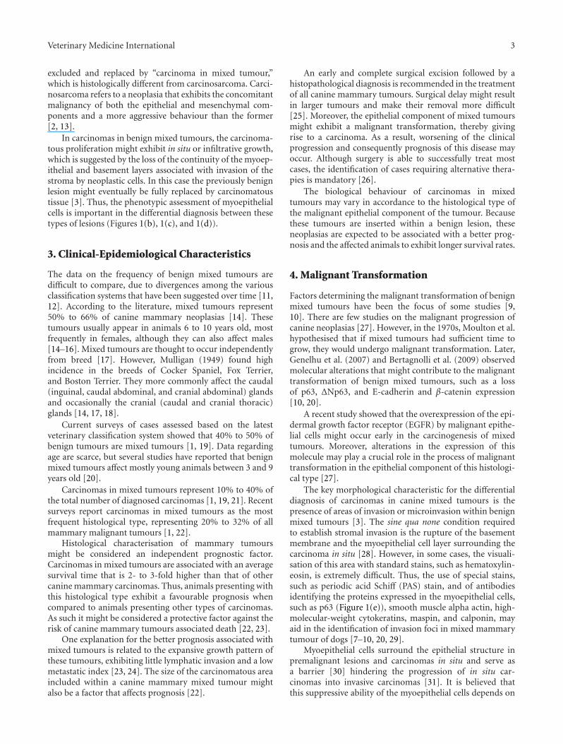
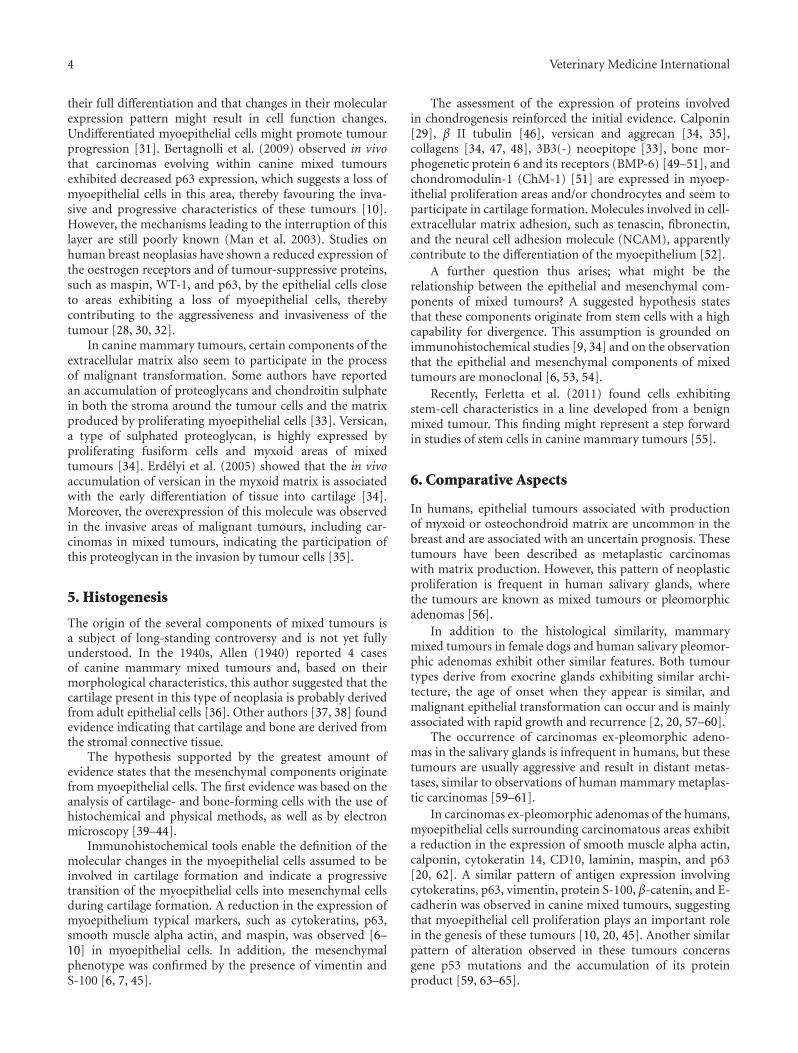
Benign mixed tumours are characterised by the presenceof benign epithelial elements (ductal and/or acinar andmyoepithelial cells) and mesenchymal cells with cartilageand/or bone formation eventually combined with myxoidfibrous tissue [2] (Figure 1(a)).
The proliferating myoepithelial cells may exhibit afusiform or stellate appearance, and these cells are oftenenveloped within an abundant extracellular matrix (myxoidmatrix). The cartilage tissue is characterised by nodulesor plaques of different sizes, including low or moderatenumbers of chondrocytes and chondroblasts rarely exhibit-ing cellular morphological alterations. When bone tissue isinvolved, it comprises osteoid matrix-forming osteoclastsand mineralised bone. Certain cases also exhibit bone mar-row, including haematopoietic and adipose tissue [4, 5].
A certain degree of pleomorphism and atypia is generallyfound in these tumours; therefore, the differential diagnosisis often difficult, especially regarding carcinomas in benignmixed tumours. The use of special staining techniques inorder to analyse the integrity of the basement membrane

2 Veterinary Medicine International
(a) (b)
(c)
(d)
(e)
Figure 1: (a) Benign mixed tumor in canine mammary gland presenting chondroid and myeloid metaplasia. HE, 10x. (b) Ductal in situcarcinoma in benign mixed tumor in canine mammary gland presenting myoepithelial cells producing myxoid matrix. HE, 40x. (c)Carcinoma in benign mixed tumor in canine mammary gland presenting in situ carcinomatous areas and myoepithelial cell proliferationproducing myxoid matrix. HE, 20x. (d) Carcinoma in benign mixed tumor in canine mammary gland presenting invasive areas in theadjacent stroma (arrow). HE, 40x. (e) Carcinoma in benign mixed tumor in canine mammary gland presenting absence of myoepithelialcells confirmed through negative p63 expression (arrow) in stromal invasive areas. Immunohistochemical stain with Mayer’s haematoxylincounterstain, 60x.
allows for a better decision on the benign or malignantnature of this type of tumour [6–10].
According to the developing system of classification ofcarcinomas in mixed tumours, these carcinomas are char-acterised by a focal or nodular development of malignancywithin a primarily benign mixed tumour [2].
Initially, the term “malignant mixed tumour” wasapplied to carcinomas arising in the context of benign mixed
tumours. However, several authors used this same term formixed tumours in which one or both (epithelial or mes-enchymal) components were malignant [11, 12]. The termcarcinosarcoma was used as synonym of malignant mixedtumour, even in cases without malignant transformation ofone of the two cellular components [11].
In the classification scheme proposed by Misdorp et al.(1999), the expression “malignant mixed tumour” was
Veterinary Medicine International 3
excluded and replaced by “carcinoma in mixed tumour,”which is histologically different from carcinosarcoma. Carci-nosarcoma refers to a neoplasia that exhibits the concomitantmalignancy of both the epithelial and mesenchymal com-ponents and a more aggressive behaviour than the former[2, 13].
In carcinomas in benign mixed tumours, the carcinoma-tous proliferation might exhibit in situ or infiltrative growth,which is suggested by the loss of the continuity of the myoep-ithelial and basement layers associated with invasion of thestroma by neoplastic cells. In this case the previously benignlesion might eventually be fully replaced by carcinomatoustissue [3]. Thus, the phenotypic assessment of myoepithelialcells is important in the differential diagnosis between thesetypes of lesions (Figures 1(b), 1(c), and 1(d)).
3. Clinical-Epidemiological Characteristics
The data on the frequency of benign mixed tumours aredifficult to compare, due to divergences among the variousclassification systems that have been suggested over time [11,12]. According to the literature, mixed tumours represent50% to 66% of canine mammary neoplasias [14]. Thesetumours usually appear in animals 6 to 10 years old, mostfrequently in females, although they can also affect males[14–16]. Mixed tumours are thought to occur independentlyfrom breed [17]. However, Mulligan (1949) found highincidence in the breeds of Cocker Spaniel, Fox Terrier,and Boston Terrier. They more commonly affect the caudal(inguinal, caudal abdominal, and cranial abdominal) glandsand occasionally the cranial (caudal and cranial thoracic)glands [14, 17, 18].
Current surveys of cases assessed based on the latestveterinary classification system showed that 40% to 50% ofbenign tumours are mixed tumours [1, 19]. Data regardingage are scarce, but several studies have reported that benignmixed tumours affect mostly young animals between 3 and 9years old [20].
Carcinomas in mixed tumours represent 10% to 40% ofthe total number of diagnosed carcinomas [1, 19, 21]. Recentsurveys report carcinomas in mixed tumours as the mostfrequent histological type, representing 20% to 32% of allmammary malignant tumours [1, 22].
Histological characterisation of mammary tumoursmight be considered an independent prognostic factor.Carcinomas in mixed tumours are associated with an averagesurvival time that is 2- to 3-fold higher than that of othercanine mammary carcinomas. Thus, animals presenting withthis histological type exhibit a favourable prognosis whencompared to animals presenting other types of carcinomas.As such it might be considered a protective factor against therisk of canine mammary tumours associated death [22, 23].
One explanation for the better prognosis associated withmixed tumours is related to the expansive growth pattern ofthese tumours, exhibiting little lymphatic invasion and a lowmetastatic index [23, 24]. The size of the carcinomatous areaincluded within a canine mammary mixed tumour mightalso be a factor that affects prognosis [22].
An early and complete surgical excision followed by ahistopathological diagnosis is recommended in the treatmentof all canine mammary tumours. Surgical delay might resultin larger tumours and make their removal more difficult[25]. Moreover, the epithelial component of mixed tumoursmight exhibit a malignant transformation, thereby givingrise to a carcinoma. As a result, worsening of the clinicalprogression and consequently prognosis of this disease mayoccur. Although surgery is able to successfully treat mostcases, the identification of cases requiring alternative thera-pies is mandatory [26].
The biological behaviour of carcinomas in mixedtumours may vary in accordance to the histological type ofthe malignant epithelial component of the tumour. Becausethese tumours are inserted within a benign lesion, theseneoplasias are expected to be associated with a better prog-nosis and the affected animals to exhibit longer survival rates.
4. Malignant Transformation
Factors determining the malignant transformation of benignmixed tumours have been the focus of some studies [9,10]. There are few studies on the malignant progression ofcanine neoplasias [27]. However, in the 1970s, Moulton et al.hypothesised that if mixed tumours had sufficient time togrow, they would undergo malignant transformation. Later,Genelhu et al. (2007) and Bertagnolli et al. (2009) observedmolecular alterations that might contribute to the malignanttransformation of benign mixed tumours, such as a lossof p63, ΔNp63, and E-cadherin and β-catenin expression[10, 20].
A recent study showed that the overexpression of the epi-dermal growth factor receptor (EGFR) by malignant epithe-lial cells might occur early in the carcinogenesis of mixedtumours. Moreover, alterations in the expression of thismolecule may play a crucial role in the process of malignanttransformation in the epithelial component of this histologi-cal type [27].
The key morphological characteristic for the differentialdiagnosis of carcinomas in canine mixed tumours is thepresence of areas of invasion or microinvasion within benignmixed tumours [3]. The sine qua none condition requiredto establish stromal invasion is the rupture of the basementmembrane and the myoepithelial cell layer surrounding thecarcinoma in situ [28]. However, in some cases, the visuali-sation of this area with standard stains, such as hematoxylin-eosin, is extremely difficult. Thus, the use of special stains,such as periodic acid Schiff (PAS) stain, and of antibodiesidentifying the proteins expressed in the myoepithelial cells,such as p63 (Figure 1(e)), smooth muscle alpha actin, high-molecular-weight cytokeratins, maspin, and calponin, mayaid in the identification of invasion foci in mixed mammarytumour of dogs [7–10, 20, 29].
Myoepithelial cells surround the epithelial structure inpremalignant lesions and carcinomas in situ and serve asa barrier [30] hindering the progression of in situ car-cinomas into invasive carcinomas [31]. It is believed thatthis suppressive ability of the myoepithelial cells depends on
4 Veterinary Medicine International
their full differentiation and that changes in their molecularexpression pattern might result in cell function changes.Undifferentiated myoepithelial cells might promote tumourprogression [31]. Bertagnolli et al. (2009) observed in vivothat carcinomas evolving within canine mixed tumoursexhibited decreased p63 expression, which suggests a loss ofmyoepithelial cells in this area, thereby favouring the inva-sive and progressive characteristics of these tumours [10].However, the mechanisms leading to the interruption of thislayer are still poorly known (Man et al. 2003). Studies onhuman breast neoplasias have shown a reduced expression ofthe oestrogen receptors and of tumour-suppressive proteins,such as maspin, WT-1, and p63, by the epithelial cells closeto areas exhibiting a loss of myoepithelial cells, therebycontributing to the aggressiveness and invasiveness of thetumour [28, 30, 32].
In canine mammary tumours, certain components of theextracellular matrix also seem to participate in the processof malignant transformation. Some authors have reportedan accumulation of proteoglycans and chondroitin sulphatein both the stroma around the tumour cells and the matrixproduced by proliferating myoepithelial cells [33]. Versican,a type of sulphated proteoglycan, is highly expressed byproliferating fusiform cells and myxoid areas of mixedtumours [34]. Erdelyi et al. (2005) showed that the in vivoaccumulation of versican in the myxoid matrix is associatedwith the early differentiation of tissue into cartilage [34].Moreover, the overexpression of this molecule was observedin the invasive areas of malignant tumours, including car-cinomas in mixed tumours, indicating the participation ofthis proteoglycan in the invasion by tumour cells [35].
5. Histogenesis
The origin of the several components of mixed tumours isa subject of long-standing controversy and is not yet fullyunderstood. In the 1940s, Allen (1940) reported 4 casesof canine mammary mixed tumours and, based on theirmorphological characteristics, this author suggested that thecartilage present in this type of neoplasia is probably derivedfrom adult epithelial cells [36]. Other authors [37, 38] foundevidence indicating that cartilage and bone are derived fromthe stromal connective tissue.
The hypothesis supported by the greatest amount ofevidence states that the mesenchymal components originatefrom myoepithelial cells. The first evidence was based on theanalysis of cartilage- and bone-forming cells with the use ofhistochemical and physical methods, as well as by electronmicroscopy [39–44].
Immunohistochemical tools enable the definition of themolecular changes in the myoepithelial cells assumed to beinvolved in cartilage formation and indicate a progressivetransition of the myoepithelial cells into mesenchymal cellsduring cartilage formation. A reduction in the expression ofmyoepithelium typical markers, such as cytokeratins, p63,smooth muscle alpha actin, and maspin, was observed [6–10] in myoepithelial cells. In addition, the mesenchymalphenotype was confirmed by the presence of vimentin andS-100 [6, 7, 45].
The assessment of the expression of proteins involvedin chondrogenesis reinforced the initial evidence. Calponin[29], β II tubulin [46], versican and aggrecan [34, 35],collagens [34, 47, 48], 3B3(-) neoepitope [33], bone mor-phogenetic protein 6 and its receptors (BMP-6) [49–51], andchondromodulin-1 (ChM-1) [51] are expressed in myoep-ithelial proliferation areas and/or chondrocytes and seem toparticipate in cartilage formation. Molecules involved in cell-extracellular matrix adhesion, such as tenascin, fibronectin,and the neural cell adhesion molecule (NCAM), apparentlycontribute to the differentiation of the myoepithelium [52].
A further question thus arises; what might be therelationship between the epithelial and mesenchymal com-ponents of mixed tumours? A suggested hypothesis statesthat these components originate from stem cells with a highcapability for divergence. This assumption is grounded onimmunohistochemical studies [9, 34] and on the observationthat the epithelial and mesenchymal components of mixedtumours are monoclonal [6, 53, 54].
Recently, Ferletta et al. (2011) found cells exhibitingstem-cell characteristics in a line developed from a benignmixed tumour. This finding might represent a step forwardin studies of stem cells in canine mammary tumours [55].
6. Comparative Aspects
In humans, epithelial tumours associated with productionof myxoid or osteochondroid matrix are uncommon in thebreast and are associated with an uncertain prognosis. Thesetumours have been described as metaplastic carcinomaswith matrix production. However, this pattern of neoplasticproliferation is frequent in human salivary glands, wherethe tumours are known as mixed tumours or pleomorphicadenomas [56].
In addition to the histological similarity, mammarymixed tumours in female dogs and human salivary pleomor-phic adenomas exhibit other similar features. Both tumourtypes derive from exocrine glands exhibiting similar archi-tecture, the age of onset when they appear is similar, andmalignant epithelial transformation can occur and is mainlyassociated with rapid growth and recurrence [2, 20, 57–60].
The occurrence of carcinomas ex-pleomorphic adeno-mas in the salivary glands is infrequent in humans, but thesetumours are usually aggressive and result in distant metas-tases, similar to observations of human mammary metaplas-tic carcinomas [59–61].
In carcinomas ex-pleomorphic adenomas of the humans,myoepithelial cells surrounding carcinomatous areas exhibita reduction in the expression of smooth muscle alpha actin,calponin, cytokeratin 14, CD10, laminin, maspin, and p63[20, 62]. A similar pattern of antigen expression involvingcytokeratins, p63, vimentin, protein S-100, β-catenin, and E-cadherin was observed in canine mixed tumours, suggestingthat myoepithelial cell proliferation plays an important rolein the genesis of these tumours [10, 20, 45]. Another similarpattern of alteration observed in these tumours concernsgene p53 mutations and the accumulation of its proteinproduct [59, 63–65].

Veterinary Medicine International 5
Alterations in other proteins involved in the regulation ofthe cell cycle, such as p21 and c-myc [66], and growth factorreceptors, such as HER-2 and EGFR [67–69], and a decreaseof adhesion molecules, such as E-cadherin and β-catenin,and oestrogen and progesterone receptors [20, 69, 70], havealso been observed in both human and canine tumours.
Current comparative studies suggest that the matrix-producing glandular tumours observed in canine and humanmammary glands and human salivary glands exhibit thesame tumourigenic characteristics. Defining the prognosticand predictive similarities among these tumours mightprovide better information on the clinical behaviour of thesetumours and support the use of a spontaneous canine modelin studies of human carcinomas.
7. Conclusions and Perspectives
Regarding clinical behaviour, mammary benign mixedtumours occur frequently in dogs and are usually associatedwith a good prognosis. However, divergences in nomencla-ture and histological classification over time make it difficultto analyse data on relapses, malignant transformation, andbiology of these tumours. Studies focusing on clinical fea-tures, malignant transformation, histogenesis, and epithelial-mesenchymal interactions might provide new informationrequired to elucidate the clinical and biological behaviour ofthis type of tumour in the veterinary medicine setting.
From a comparative perspective, canine mammarymixed tumours and human pleomorphic adenomas of thesalivary gland exhibit morphological and molecular similar-ities, suggesting a similarity in the pathogenic mechanismsinvolved in malignant transformation and histogenesis.
Acknowledgments
This work has been financially supported by “Fundacao deAmparo a Pesquisa de Minas Gerais” (FAPEMIG), “ConselhoNacional de Desenvolvimento Cientıfico e Tecnologico”(CNPq), and “Coordenacao de Aperfeicoamento de Pessoalde Nıvel Superior” (CAPES), Brazil.
References
[1] G. D. Cassali, B. M. Melo, N. Madureira et al., “Mammarygland diagnosis of the laboratory of comparative pathology—UFMG, from 2000 to 2008,” in Proceedings of the World SmallAnimal Veterinary Association, vol. 14, p. 173, Sao Paulo,Brazil, 2009, Clınica Veterinaria-supplement.
[2] W. Misdorp, R. W. Else, and E. Hellmen, Histological Classi-fication of Mammary Tumors of the Dog and the Cat, WorldHealth Organization, Geneva, Switzerland, 1999.
[3] G. D. Cassali, G. E. Lavalle, A. B. De Nardi et al., “Consensusfor the diagnosis, prognosis and treatment of canine mam-mary tumors,” Brazilian Journal of Veterinary Pathology, vol.4, no. 2, pp. 153–180, 2011.
[4] F. Grandi, M. M. Colodel, L. N. Monteiro, J. R. V. Leao,and N. S. Rocha, “Extramedullary hematopoiesis in a case ofbenign mixed mammary tumor in a female dog: cytologicaland histopathological assessment,” BMC Veterinary Research,vol. 6, article 45, 2010.
[5] P. A. Auler, A. C. Bertagnolli, E. Ferreira et al., “Myeloid meta-plasia in canine mixed mammary tumors: occurrence andcharacterization,” Veterinary Quarterly, vol. 31, no. 4, pp. 173–177, 2011.
[6] F. Gartner, M. Geraldes, G. Cassali, A. Rema, and F. Schmitt,“DNA measurement and immunohistochemical characteri-zation of epithelial and mesenchymal cells in canine mixedmammary tumours: putative evidence for a common histoge-nesis,” The Veterinary Journal, vol. 158, no. 1, pp. 39–47, 1999.
[7] A. Gama, A. Alves, F. Gartner, and F. Schmitt, “p63: a novelmyoepithelial cell marker in canine mammary tissues,” Veteri-nary Pathology, vol. 40, no. 4, pp. 412–420, 2003.
[8] A. E. De Los Monteros, M. Y. Millan, G. A. Ramırez, J. Ordas,C. Reymundo, and J. Martın De Las Mulas, “Expression ofmaspin in mammary gland tumors of the dog,” VeterinaryPathology, vol. 42, no. 3, pp. 250–257, 2005.
[9] L. N. Z. Ramalho, A. Ribeiro-Silva, G. D. Cassali, and S.Zucoloto, “The expression of p63 and cytokeratin 5 in mixedtumors of the canine mammary gland provides new insightsinto the histogenesis of these neoplasms,” Veterinary Pathol-ogy, vol. 43, no. 4, pp. 424–429, 2006.
[10] A. C. Bertagnolli, G. D. Cassali, M. C. L. S. Genelhu, F. A.Costa, J. F. C. Oliveira, and P. B. D. Goncalves, “Immunohis-tochemical expression of p63 and δNp63 in mixed tumors ofcanine mammary glands and its relation with p53 expression,”Veterinary Pathology, vol. 46, no. 3, pp. 407–415, 2009.
[11] J. F. Hampe and W. Misdorp, “Tumours and dysplasias of themammary gland,” Bulletin of the World Health Organization,vol. 50, no. 1-2, pp. 111–133, 1974.
[12] J. E. Moulton, “Tumors of the mammary gland,” in Tumors inDomestic Animals, pp. 518–552, University of California Press,Berkeley, Calif, USA, 3rd edition, 1990.
[13] S. A. Benjamin, A. C. Lee, and W. J. Saunders, “Classificationand behavior of canine mammary epithelial neoplasms basedon life-span observations in Beagles,” Veterinary Pathology,vol. 36, no. 5, pp. 423–436, 1999.
[14] A. G. Jabara, “Canine mixed tumours,” The Australian Veteri-nary Journal, vol. 36, no. 5, pp. 212–221, 1960.
[15] G. Sittner, “Mammamischtumor bei einem mannlichen Hundund seine Histogenese,” Archiv fur Wissenschaftliche undPraktische Tierheilkunde, no. 74, pp. 406–410, 1939.
[16] E. Cotchin, “Some glandular tumours of the dog,” Proceedingsof the Royal Society of Medicine, vol. 40, no. 11, pp. 636–638,1947.
[17] K. Nieberle, “Zur Kenntnis der sog. Mammamischgeschwulstedes Hundes,” Journal of Cancer Research And Clinical Oncology,vol. 39, no. 1, pp. 113–127, 1933.
[18] R. M. Mulligan, Neoplasms of the Dog, Willians and Wilkins,Baltimore, Md, USA, 1949.
[19] H. G. Richards, P. E. McNeil, H. Thompson, and S. W. J. Reid,“An epidemiological analysis of a canine-biopsies databasecompiled by a diagnostic histopathology service,” PreventiveVeterinary Medicine, vol. 51, no. 1-2, pp. 125–136, 2001.
[20] M. C. L. S. Genelhu, S. V. Cardoso, H. Gobbi, and G. D. Cassali,“A comparative study between mixed-type tumours fromhuman salivary and canine mammary glands,” BMC Cancer,vol. 7, article 218, 2007.
[21] W. A. Priester, “Occurrence of mammary neoplasms in bitchesin relation to breed, age, tumour type, and geographical regionfrom which reported,” Journal of Small Animal Practice, vol. 20,no. 1, pp. 1–11, 1979.

6 Veterinary Medicine International
[22] M. F. Cavalcanti, Fatores prognosticos na abordagem clınica ehistopatologica dos carcinomas mamarios de cadelas: estadia-mento TNM e sistema de Nottingham [M.S. thesis], FederalUniversity of Minas Gerais, Belo Horizonte, Brazil, 2006.
[23] T. Yamagami, T. Kobayashi, K. Takahashi, and M. Sugiyama,“Prognosis for canine malignant mammary tumors based onTNM and histologic classification,” The Journal of VeterinaryMedical Science, vol. 58, no. 11, pp. 1079–1083, 1996.
[24] W. Misdorp, E. Cotchin, J. F. Hampe, A. G. Jabara, and J.von Sandersleben, “Canine malignant mammary tumours. II.Adenocarcinomas, solid carcinomas and spindle cell carcino-mas,” Veterinary Pathology, vol. 9, no. 6, pp. 447–470, 1972.
[25] W. Misdorp, “Tumors of the mammary gland,” in Tumours inDomestic Animals, pp. 575–606, Iowa State Press, Ames, Iowa,USA, 4th edition, 2002.
[26] D. E. Bostock, “The prognosis following the surgical excisionof canine mammary neoplasms,” European Journal of Cancerand Clinical Oncology, vol. 11, no. 6, pp. 389–396, 1975.
[27] A. C. Bertagnolli, E. Ferreira, E. J. Dias, and G. D. Cassali,“Canine mammary mixed tumours: immunohistochemicalexpressions of EGFR and HER-2,” Australian VeterinaryJournal, vol. 89, no. 8, pp. 312–317, 2011.
[28] Y. G. Man, L. Tai, R. Barner et al., “Cell clusters overlyingfocally disrupted mammary myoepithelial cell layers andadjacent cells within the same duct display different immuno-histochemical and genetic features: implications for tumorprogression and invasion,” Breast Cancer Research, vol. 5, no.6, pp. R231–R241, 2003.
[29] A. E. Los de Monteros, M. Y. Millan, J. Ordas, L. Carrasco, C.Reymundo, and J. Martın Las de Mulas, “Immunolocalizationof the smooth muscle-specific protein calponin in complexand mixed tumors of the mammary gland of the dog:assessment of the morphogenetic role of the myoepithelium,”Veterinary Pathology, vol. 39, no. 2, pp. 247–256, 2002.
[30] Y. G. Man and Q. X. A. Sang, “The significance of focalmyoepithelial cell layer disruptions in human breast tumorinvasion: a paradigm shift from the ”protease-centered” hypo-thesis,” Experimental Cell Research, vol. 301, no. 2, pp. 103–118, 2004.
[31] T. Gudjonsson, L. Rønnov-Jessen, R. Villadsen, F. Rank, M.J. Bissell, and O. W. Petersen, “Normal and tumor-derivedmyoepithelial cells differ in their ability to interact with lumi-nal breast epithelial cells for polarity and basement membranedeposition,” Journal of Cell Science, vol. 115, no. 1, pp. 39–50,2002.
[32] Z. Xu, W. Wang, C. X. Deng, and Y. G. Man, “Aberrant p63and WT-1 expression in myoepithelial cells of pregnancy-associated breast cancer: implications for tumor aggressive-ness and invasiveness,” International Journal of BiologicalSciences, vol. 5, no. 1, pp. 82–96, 2009.
[33] U. Hinrichs, G. R. Rutteman, and H. Nederbragt, “Stromalaccumulation of chondroitin sulphate in mammary tumoursof dogs,” British Journal of Cancer, vol. 80, no. 9, pp. 1359–1365, 1999.
[34] I. Erdelyi, D. H. M. Nieskens, J. E. Van Dijk, L. Vass, and H.Nederbragt, “Immunohistochemical evaluation of versican,in relation to chondroitin sulphate, in canine mammarytumours,” Histology and Histopathology, vol. 18, no. 4, pp.1067–1080, 2003.
[35] I. Erdelyi, A. J. A. M. Van Asten, J. E. Van Dijk, and H. Neder-bragt, “Expression of versican in relation to chondrogenesis-related extracellular matrix components in canine mammarytumors,” Histochemistry and Cell Biology, vol. 124, no. 2, pp.139–149, 2005.
[36] A. C. Allen, “So-called mixed tumors of the mammary glandof dog and man,” Archives of Pathology, vol. 29, pp. 589–624,1940.
[37] C. Huggins and P. V. Moulder, “Studies of the mammarytumours of dog. I. Lactation and the influence of ovariectomyand suprarenalectomy thereon,” The Journal of ExperimentalMedicine, vol. 80, no. 5, pp. 441–454, 1944.
[38] F. Bloom, Pathology of the Dog and Cat: The GenitourinarySystem With Clinical Considerations, American VeterinaryPublications, 1954.
[39] E. Cotchin, “Mammary neoplasms of the bitch,” The Journalof Comparative Pathology and Therapeutics, vol. 68, pp. 1–22,1958.
[40] S. Erichsen, “A histochemical study of mixed tumors of thecanine mammary gland,” Acta Pathology and MicrobiologyScandinavica, vol. 36, pp. 490–502, 1955.
[41] J. V. Hurley and A. G. Jabara, “Properties of “cartilage” incanine mammary tumors,” Archives of Pathology, vol. 77, pp.343–347, 1964.
[42] L. T. Pulley, “Ultrastructural and histochemical demon-stration of myoepithelium in mixed tumors of the caninemammary gland,” American Journal of Veterinary Research,vol. 34, no. 12, pp. 1513–1522, 1973.
[43] S. Tateyama and E. Cotchin, “Alkaline phosphatase reactionof canine mammary mixed tumours: a light and electronmicroscopic study,” Research in Veterinary Science, vol. 23, no.3, pp. 356–364, 1977.
[44] S. Tateyama and E. Cotchin, “Electron microscopic obser-vations on canine mixed mammary tumors, with specialreference to cytoplasmic filamentous components,” AmericanJournal of Veterinary Research, vol. 39, no. 9, pp. 1494–1501,1978.
[45] E. Destexhe, L. Lespagnard, M. Degeyter, R. Heymann,and F. Coignoul, “Immunohistochemical identification ofmyoepithelial, epithelial, and connective tissue cells in caninemammary tumors,” Veterinary Pathology, vol. 30, no. 2, pp.146–154, 1993.
[46] K. Arai, H. Nakano, M. Shibutani, M. Naoi, and H. Matsuda,“Expression of class II β-tubulin by proliferative myoepithelialcells in canine mammary mixed tumors,” Veterinary Pathology,vol. 40, no. 6, pp. 670–676, 2003.
[47] K. Arai, K. Uehara, and Y. Nagai, “Expression of type II andtype XI collagens in canine mammary mixed tumors anddemonstration of collagen production by tumor cells in col-lagen gel culture,” Japanese Journal of Cancer Research, vol. 80,no. 9, pp. 840–847, 1989.
[48] K. Arai, K. Uehara, and Y. Nagai, “Simultaneous expression oftype IX collagen and an inhibin-related antigen in proliferativemyoepithelial cells with pleomorphic adenoma of caninemammary glands,” Japanese Journal of Cancer Research, vol.86, no. 6, pp. 577–584, 1995.
[49] S. Tateyama, K. Uchida, T. Hidaka, M. Hirao, and R. Yam-aguchi, “Expression of bone morphogenetic protein-6 (BMP-6) in myoepithelial cells in canine mammary gland tumors,”Veterinary Pathology, vol. 38, no. 6, pp. 703–709, 2001.
[50] T. Akiyoshi, K. Uchida, and S. Tateyama, “Expression of bonemorphogenetic protein-6 and bone morphogenetic proteinreceptors in myoepithelial cells of canine mammary glandtumors,” Veterinary Pathology, vol. 41, no. 2, pp. 154–163,2004.
[51] A. Kawabata, K. Okano, K. Uchida, R. Yamaguchi, T. Hayashi,and S. Tateyama, “Co-localization of chondromodulin-I(ChM-I) and bone morphogenetic protein-6 (BMP-6) inmyoepithelial cells of canine mammary tumors,” The Journal

Veterinary Medicine International 7
of Veterinary Medical Science, vol. 67, no. 11, pp. 1097–1102,2005.
[52] K. Arai, M. Naoi, and K. Uehara, “Immunohistochemicalexamination of neural cell adhesion molecule (NCAM),tenascin and fibronectin on the development of cartilaginoustissue in canine mammary mixed tumors,” The Journal ofVeterinary Medical Science, vol. 56, no. 4, pp. 809–811, 1994.
[53] G. D. Cassali, A. C. Bertagnolli, F. Gartner, and F. Schmitt,“Canine mammary tumours: a quantitative DNA study usingstatic cytometry,” Revista Espanola de Patologia, vol. 44, no. 4,pp. 195–201, 2011.
[54] A. C. Bertagnolli, P. Soares, B. van Asch et al., “An assessmentof the clonality of the components of canine mixed mammarytumours by mitochondrial DNA analysis,” The VeterinaryJournal, vol. 182, no. 2, pp. 269–274, 2009.
[55] M. Ferletta, J. Grawe, and E. Hellmen, “Canine mammarytumors contain cancer stem-like cells and form spheroids withan embryonic stem cell signature,” The International Journal ofDevelopmental Biology, vol. 55, pp. 791–799, 2011.
[56] M. L. Voz, W. J. Van de Ven, and K. Kas, “First insights intothe molecular basis of pleomorphic adenomas of the salivaryglands,” Advances in Dental Research, vol. 14, no. 1, pp. 81–83,2000.
[57] P. L. Auclair and G. L. Ellis, “Atypical features in salivary glandmixed tumors: their relationship to malignant transforma-tion,” Modern Pathology, vol. 9, no. 6, pp. 652–657, 1996.
[58] E. Ferreira, A. C. Bertagnolli, M. F. Cavalcanti, F. C. Schmitt,and G. D. Cassali, “The relationship between tumour size andexpression of prognostic markers in benign and malignantcanine mammary tumours,” Veterinary and ComparativeOncology, vol. 7, no. 4, pp. 230–235, 2009.
[59] J. E. Lewis, K. D. Olsen, and T. J. Sebo, “Carcinoma ex pleo-morphic adenoma: pathologic analysis of 73 cases,” HumanPathology, vol. 32, no. 6, pp. 596–604, 2001.
[60] E. S. Wargotz and H. J. Norris, “Metaplastic carcinomas ofthe breast. I. Matrix-producing carcinoma,” Human Pathology,vol. 20, no. 7, pp. 628–635, 1989.
[61] V. A. Livolsi and K. H. Perzin, “Malignant mixed tumorsarising in salivary glands. I. Carcinoma arising in benignmixed tumors: a clinicopathologic study,” Cancer, vol. 39, no.5, pp. 2209–2230, 1977.
[62] V. C. de Araujo, A. Altemani, C. Furuse, M. T. Martins, andN. S. de Araujo, “Immunoprofile of reactive salivary myoepi-thelial cells in intraductal areas of carcinoma ex-pleomorphicadenoma,” Oral Oncology, vol. 42, no. 10, pp. 1011–1016, 2006.
[63] C. Chhieng, M. Cranor, M. E. Lesser, and P. P. Rosen,“Metaplastic carcinoma of the breast with osteocartilaginousheterologous elements,” American Journal of Surgical Pathol-ogy, vol. 22, no. 2, pp. 188–194, 1998.
[64] Y. Yamamoto, Y. Kishimoto, I. I. Wistuba et al., “DNA analysisat p53 locus in carcinomas arising from pleomorphic adeno-mas of salivary glands: comparison of molecular study and p53immunostaining,” Pathology International, vol. 48, no. 4, pp.265–272, 1998.
[65] J. S. Morris, C. Nixon, O. J. A. King, I. M. Morgan, andA. W. Philbey, “Expression of TopBP1 in canine mammaryneoplasia in relation to histological type, Ki67, ERα and p53,”The Veterinary Journal, vol. 179, no. 3, pp. 422–429, 2009.
[66] H. Deguchi, H. Hamano, and Y. Hayashi, “c-myc, ras p21 andp53 expression in pleomorphic adenoma and its malignantform of the human salivary glands,” Acta Pathologica Japonica,vol. 43, no. 7-8, pp. 413–422, 1993.
[67] S. Di Palma, A. Skalova, T. Vanıeek, R. H. W. Simpson, I.Starek, and I. Leivo, “Non-invasive (intracapsular) carcinoma
ex pleomorphic adenoma: recognition of focal carcinoma byHER-2/neu and MIB1 immunohistochemistry,” Histopathol-ogy, vol. 46, no. 2, pp. 144–152, 2005.
[68] S. Matsubayashi and T. Yoshihara, “Carcinoma ex pleomor-phic adenoma of the salivary gland: an immunohistochemicalstudy,” European Archives of Oto-Rhino-Laryngology, vol. 264,no. 7, pp. 789–795, 2007.
[69] G. Gauchotte, L. Coffinet, E. Schmitt et al., “Salivary glandanlage tumor: a clinicopathological study of two cases,” Fetal& Pediatric Pathology, vol. 30, no. 2, pp. 116–123, 2011.
[70] R. F. do Prado, A. Consolaro, and L. A. Taveira, “Expressionof betacatenin in carcinoma in pleomorphic adenoma, pleo-morphic adenoma and normal salivary gland: an immuno-histochemical study,” Medicina Oral, Patologıa Oral y CirugıaBucal., vol. 11, no. 3, pp. E247–E251, 2006.
Copyright © 2022 FDOKUMEN