Polymorphism and Methylation of Four Genes Expressed in Salivary
Upload
independentCategory
view
5download
0
Virchows Archiv A Pathol Anat (1992) 421:217 222 Virchows Archiv A Pathologicai Anatomy and Histopathoiogy �9 Springer-Verlag 1992
Tenascin in salivary gland tumours Ylermi Soini 1, Paavo Piiiikk51, Ismo Virtanen 2, and Veli-Pekka Lehto 1
1 Department of Pathology, University of Oulu, Oulu, Finland 2 Department of Anatomy, University of Helsinki, Helsinki, Finland
Received November 10, 1991 / Received a�9 revision March 14, 1992 / Accepted March 16, 1992
Summary. The distribution of tenascin immunoreactivity was analysed in salivary gland tissue and in various be- nign and malignant tumours of the salivary gland. In the non-neoplastic tissue, tenascin was seen in the areas of basement membranes of the ductal epithelium. No immunoreactivity could be observed in the serous or mucous glands. In pleomorphic adenomas, tenascin im- munoreactivity could be seen in the stromal compart- ment. It was more pronounced in the dense stromal areas and chondroid elements than in the myxoid area. In Warthin's tumours, strong tenascin immunoreactivity could be observed in the basement membrane zone of the epithelial component. In the lymphatic component, faint reticular staining could be seen. In adenoid cystic carcinomas, acinic cell tumours and mucoepidermoid carcinomas, tenascin showed a linear stromal distribu- tion. No intracytoplasmic immunoreactivity could be seen in any of the cases. The widespread tenascin positi- vity in salivary gland tumours suggests that tenascin may play a role in the induction and progression of salivary gland tumours, presumably by interfering with the nor- mal parenchymal-mesenchymal interaction.
Key words: Tenascin - Salivary gland tumours - Immu- nohistochemistry
Introduction
Tenascin is a six-armed extracellular matrix glycoprotein with a molecular weight of 1,900 kDa (Erickson and Bourdon 1989; Chiquet-Ehrismann 1990). It was dis- covered independently in several laboratories and, con- sequently, is known by various names, such as hexabra- chion, cytotactin, myotendineous antigen, GP250 and J-1 protein (Erickson and Lightner 1988). The subunits of tenascin show heterogeneity and, in SDS-polyacryl- amide gel electrophoresis, three separate tenascin sub-
units can be found with molecular weights of 240 kDa, 200 kDa and 190 kDa (Chiquet-Ehrismann et al. 1986).
Tenascin is found in embryonic tissues (Chiquet and Famborough 1984; Crossin et al. 1986; Chuong et al. 1987), especially in areas of epithelial-mesenchymal junc- tions, and in developing brain tissue. It probably has a role in epithelial-mesenchymal induction and cell mi- gration (Chiquet-Ehrismann et al. 1986; Crossin et al. 1986; Chuong et al. 1987; Erickson and Bourdon 1989; Chiquet-Ehrismann 1990). Tenascin is synthesized by glial cells and fibroblasts (Erickson and Bourdon 1989). By in situ hybridization, tenascin synthesis has also been shown in embryonic kidney and lung mesenchymal cells and in lung epithelial cells (Koch et al. 1991 ; Prieto et al. 1990).
In adult tissues tenascin can be found in healing wounds, in dense connective tissue, such as tendons and ligaments, in cartilaginous tissue and in smooth muscle (Erickson and Bourdon 1989; Chiquet-Ehrismann 1990; Chuong and Chen 1991). Tenascin has also been ob- served in the basement membranes of different epithelia of ectodermal and endodermal origin (Natali et al. 1991).
In malignant neoplasms, stromal tenascin immunore- activity has been found in various tumours such as glio- mas, sarcomas, melanomas and carcinomas (Bourdon et al. 1983; Erickson and Bourdon 1989; Natali et al. 1990, 1991; Vollmer et al. 1990; Howeedy et al. 1990). It is also been detected in some benign tumours such as naevocellular naevi and benign breast tumours (How- eedy et al. 1990; Natali et al. 1991).
In this study we used immunohistochemical tech- niques to investigate the distribution of tenascin in sali- vary gland tissue and neoplasms. We used two mono- clonal antibodies, one of which detects tenascin also in formalin-fixed, paraffin-embedded specimens.
Matcrials and methods
Correspondence to: Y. Soini, Department of Pathology, University Thirty-five cases of benign and malignant salivary gland tumours of Oulu, Kajaanintie 52 D, SF-90220 Oulu, Finland were selected from the files of the Department of Pathology, Oulu
218
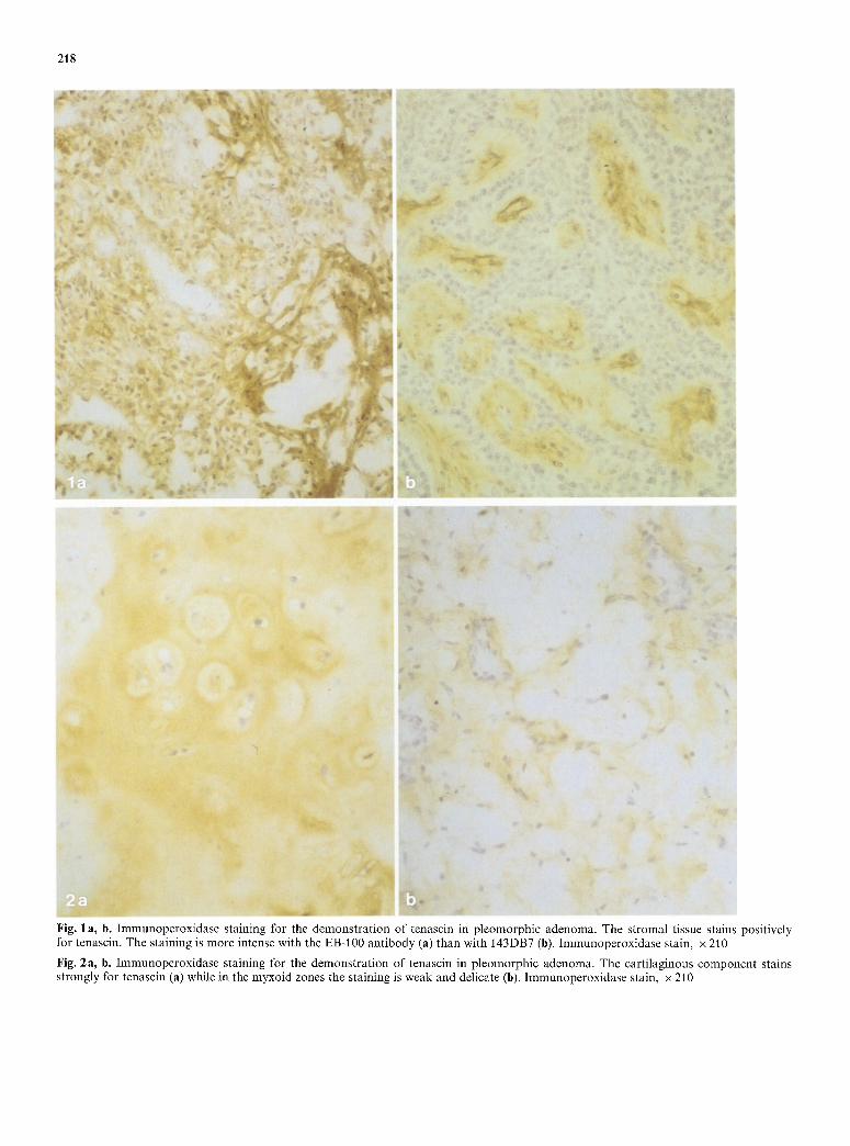
Fig. 1 a, b. Immunoperoxidase staining for the demonstration of tenascin in pleomorphic adenoma. The stromal tissue stains positively for tenascin. The staining is more intense with the EB-100 antibody (a) than with 143DB7 (b). Immunoperoxidase stain, x 210
Fig. 2a, b. Immunoperoxidase staining for the demonstration of tenascin in pleomorphic adenoma. The cartilaginous component stains strongly for tenascin (a) while in the myxoid zones the staining is weak and delicate (b). Immunoperoxidase stain, x 210

219
Fig. 3. Immunoperoxidase staining for the demonstrat ion of tenascin in Warthin's tumour. The areas of the basement membranes stain positively. Delicate reticular staining can be observed in the lymphatic areas. Immunoperoxidase stain, x210
Fig. 4. Immunoperoxidase staining for the demonstration of tenascin in mucoepidermoid carcinoma. The stromal tissue stains strongly for tenascin while the tumour cells are nega- tive. Immunoperoxidase stain, x 155
Fig. 5. immunoperoxidase staining for the demonstrat ion of tenascin in acinic cell tumour. Delicate positive fibrous strands can be observed in the stroma. Immunoperoxidase stain, x 210
University Central Hospital. The cases were from the years 1985- 1990 and the material was fixed in neutral formalin and embedded in paraffin. Some of the material (7 cases) was obtained fresh from the operating theatre and stored in liquid nitrogen. The tumours were classified according to the WHO International classification of salivary gland tumours (Seifert 1991). The diagnosis of all the cases was based on light microscopic examination using conven- tional haematoxylin and eosin stain.
The material consisted of 16 pleomorphic adenomas, 6 adeno-
lymphomas, 6 mucoepidermoid carcinomas, 3 acinic cell tumours, 3 adenoid cystic carcinomas and 1 pleomorphic sarcoma of the salivary gland. Frequently, the specimens also contained non-neo- plastic elements. Eight cases of non-neoplastic salivary gland tissue were also included (4 normal, 4 chronic inflammations of the sali- vary gland).
We aimed at producing a monoclonal antibody that also detects tenascin efficiently in formalin-fixed material. For that purpose, the mAb 143DB7 was raised as follows. First, tenascin was purified

220
from the culture medium of human embryonal fibroblasts by using affinity purification with a well-characterized anti-tenascin hybri- doma clone 100EB2 (Howeedy et al. 1990). For that purpose, im- munoglobulin fraction of 100EB2 was purified from the ascites fluid by using protein G column (Pharmacia, Uppsala, Sweden) in FPLC. It was then coupled to cyanogen bromide-activated Se- pharose 4B (Pharmacia) according to the manufacturer's instruc- tions. Culture medium of human embryonal fibroblasts was then passed over the column. After extensive washing, the bound tenas- cin was eluted with 3-[(Cholamidopropyl)-dimethyl ammonio]-l- propanesulfonate (CHAPS) buffer, p l i 11, and used for immuniza- tion. Three days after the last immunization of BALB/c micc, the hybridoma production was initiated by standard techniques (K6hler and Milstein 1975). The hybridomas were screened, first, by using purified tenascin in enzyme-linked immunoassay, and then by immunohistochemistry on pepsin-treated paraffin sections of selected formalin-fixed tumours. The hybridoma culture 143BD7 was cloned by using standard techniques.
The specificity of mAb 143DB7 was shown by immunoblotting of whole human cultured fibroblasts. For immunoblotting, the cells were lysed in electrophoresis sample buffer, boiled and analysed in SDS-polyacrylamide gel electrophoresis, carried out according to Laemmli (1970), by using 6.5% stab gels and reducing condi- tions. The transfer of the electrophoretically separated polypeptides onto nitrocellulose paper was carried out according to the method of Towbin et al. (1979). To visualize the polypeptide bands, strips of the nitrocellulose sheets were cut and stained with amido black. The immunodetection was done by incubating the nitrocellulose sheets first with mAb 143DB7 and then with peroxidase-coupled rabbit anti-mouse IgG (Dakopatts, Copenhagen, Denmark). 3'-3' Diaminobenzidine was used as a chromogen. In immunoblotting, two distinct bands of 250,000 and 190,000 daltons were revealed. No polypeptide bands were revealed in experiments in which the primary antibody was either omitted or replaced by antibodies to proteins not produced by fibroblasts (hot shown).
For paraffin sections, 5-gm-thick sections were deparaffinized in xylene and rehydrated in graded alcohol. The endogenous perox- idase was consumed by treating the sections with 0.3% hydrogen peroxidase in absolute methanol for 30 rein. Prior to the immuno- staining, the sections were treated with 0.4% pepsin (Merck, Darm- stadt, FRG; 10 units/mg) for 30 rein at 37 ~ C. Monoclonal mouse antibody (143DB7) to human tenascin was used as the primary antibody. For the immunostaining, the avidin-biotin complex (ABC) method was used (Hsu et al. 1981). The sections were first incubated overnight with the primary antibody at 4 ~ C, followed by biotinylated rabbit anti-mouse secondary antibody (1 : 200) and the ABC complex (Dakopatts). The colour was developed with diaminobenzidine, whereafter the sections were mounted in an aqueous medium. The sections were counterstained with a light haematoxylin stain. Negative control consisted of substituting PBS (140 ml sodium chloride, 0.01 M phosphate buffet, p l i 7.2) for the primary antibody.
For frozen sections, 5-gm-thick sections were cut from the spec- imens. They were air-dried for 1 h and were then fixed in acetone for 10 min at - 2 2 ~ C. The endogenous peroxidase was consumed by treating the sections with 0.3% hydrogen peroxidase in metha- no1 for 30 rein. A monoclonal mouse antibody (EB-100) to human tenascin, with a dilution of 1 : 5, was used as the primary antibody (Howeedy et al. 1990). The sections were incubated with the prima- ry antibody overnight at 4 ~ C, followed by the biotinylated second- ary rabbit anti-mouse antibody (1:200) and the ABC complex (Dakopatts).
The peroxidase reaction was developed with diaminobenzidine, whereafter the sections were mounted in an aqueous medium. The sections were counterstained lightly with haematoxylin. Negative controls were as described previously.
Results
In the n o r m a l sa l ivary gland, tenascin i m m u n o r e a c t i v i t y was seen in the b a s e m e n t m e m b r a n e s o f the ducts and in the s m o o t h muscle cells in the wal ls o f the small a r te r - les. The mucous and serous g lands were negative. The f inding was s imi lar in the chron ica l ly in f lamed sa l ivary g land tissue.
In p l e o m o r p h i c adenomas , l inear immunoreac t i v i t y cou ld be obse rved in the s t roma l t issue in all cases (Figs. 1, 2). A l so the f ibrot ic capsule s ta ined posi t ive. In the dense, f ib ro t ic areas, the immunoreac t i v i t y was s t ronger than in the m y x o i d areas (Fig. 2b) . Some myx- oid is lands r e m a i n e d negat ive for the s taining. Also the ca r t i l ag inous c o m p o n e n t s ta ined posi t ive for tenascin (Fig. 2a). Often, a u n i f o r m d i s t r ibu t ion o f the s ta in ing was seen over the ca r t i l ag inous area. Occas iona l ly , how- ever, non- reac t ive s t romal zones cou ld be seen a r o u n d ind iv idua l chondrocy tes . N o in t race l lu la r tenascin was seen in t u m o u r cells.
In War th in ' s t u m o u r s (Fig. 3), tenascin i m m u n o r e a c - t ivi ty was seen in the areas o f the basemen t m e m b r a n e s benea th the epi the l ia l cell c o m p o n e n t . In some areas , pos i t iv i ty also occu r red in the f ib rous cores o f the papi l - lary pro jec t ions . In the l ympha t i c c o m p o n e n t , i m m u n o - reac t iv i ty was conf ined to thin re t icu la r fibres. Also the t u m o u r capsule s ta ined posi t ive.
The s t r o m a o f all m a l i g n a n t sa l ivary g land t u mour s was posi t ive for tenascin. The s ta in ing was weaker in a de no id cystic c a r c inoma s and in acinic cell t u m o u r than in m u c o e p i d e r m o i d ca rc inomas . In a de no id cystic carci- n o m a s and m u c o e p i d e r m o i d ca rc inomas , a l inear tenas- cin i m m u n o r e a c t i v i t y was observed, which somet imes concen t r a t ed a r o u n d the t u m o u r cell i s lands (Fig. 4). In acinic cell t umour s , fa int ly posi t ive l inear s t rands could be obse rved (Fig. 5). N o i n t r a c y t o p l a s m i c s ta in ing for tenasc in was seen in t u m o u r cells.
There was no difference be tween the results ob t a ined with the two tenasc in ant ibodies . The intensi ty o f the i m m u n o s t a i n i n g was s t ronger , however , in the EB-100 s ta ined fresh f rozen mater ia l . The con t ro l s ta ining was negat ive in all cases.
Discussion
In this inves t iga t ion we ana lysed the d i s t r ibu t ion o f ten- ascin in benign and m a l i g n a n t sa l ivary g land t u m o u r s and in non-neop la s t i c sa l ivary gland. The i m m u n o s t a i n - ing was car r ied ou t by using two dif ferent an t ibodies , EB-100 and 143DB7, b o t h o f which w o r k well on fresh, f rozen sect ions and the la t te r also on formal in- f ixed , p a r a f t � 9 mater ia l . The s ta in ing results wi th the two an t ibod ies were ident ical . However , the immu- nos ta in ing o f fresh f rozen ma te r i a l wi th EB-100 yie lded the mos t in tense s ta in ing react ion . This p r o b a b l y de- pends no t only on the difference in the reac t iv i ty be tween the an t ibodies , bu t also on the be t te r p rese rva t ion o f the tenascin an t igen in fresh f rozen mate r i a l a n d / o r the fact t ha t the an t igenic de t e rminan t s are pa r t ly h idden in fo rmal in - f ixed mater ia l .
221
In benign salivary gland tumours, stromal positivity was observed both in pleomorphic adenomas and in adenolymphomas. In pleomorphic adenomas, there was a strong stromal immunoreactivi ty in the dense, fibrotic s t roma as well as in the cartilaginous component of the tumour. In the myxoid zones the staining was less in- tense, suggesting that there is a lower concentration of tenascin in the myxoid zones. Interestingly, pleomorphic adenomas contain fibronectin (Caselitz et al. 1988) and chondroitin sulphate proteoglycans (Harrison and Auger 1991), which both bind tenascin (Chiquet Ehr- ismann et al. 1991; Erickson and Lightner 1988). Thus the lower degree of staining for tenascin in the myxoid zone could also be a reflection of a lower concentration of these substances in this region.
A diffuse distribution of tenascin could also be seen in the chondroid compar tment in pleomorphic ad- enomas. Tenascin is present in embryonic chrondroid tissue, where it is distributed throughout the tissue, while in adult tissue it is concentrated mainly in the perichon- drium (Erickson and Lightner 1988). It may even by possible that the chondrogenic differentiation in pleo- morphic adenomas is induced by tenascin. This is sup- ported by the observation that tenascin is associated with chondrogenic differentiation in vivo (Chiquet-Ehr- ismann 1990).
In Warthin's tumours, as in normal salivary gland, a strong tenascin immunoreactivity was seen in the base- ment membrane areas beneath the epithelial component . This is in line with previous studies which have shown tenascin in the basement membranes of different epithe- lia of ectodermal and endodermal origin (Natali et al. 1991). Synthesis of tenascin bas also been reported in the epithelial cells of developing lung buds (Koch et al. 1991). A receptor for tenascin has been described that belongs to a family of integrin molecules (Bourdon and Ruoslahti 1989). The epithelial cells of salivary gland ducts, as well as other epithelia, might harbour such surface receptors and tenascin might thus function as a structural protein in the ductal units to retain the integ- rity and orientation of the epithelial cells.
In malignant salivary gland tumours, stromal tenas- cin immunoreactivi ty could be seen in ail cases. This is in accordance with previous results which indicate stromal tenascin immunoreactivity in various malignant neoplasms (Bourdon et al. 1983; Erickson and Bourdon 1989; Natali et al. 1990; Vollmer et al. 1990; Howeedy et al. 1990). In the present study, however, there were differences in the intensity of tenascin staining between different types of tumours.
The tissue distribution of tenascin is reminiscent of that of fibronectin, which is also found in the stromal areas, while laminin and type IV collagen are mainly found in the basement membrane areas of these tumours (Caselitz et al. 1988). This similarity in tissue distribution may be due to the fact that the 190 kDa isoform of tenascin is able to attach to fibronectin (Chiquet-Ehr- ismann et al. 1991).
In tumour tissue tenascin is considered to be synthe- sized by stromal fibroblasts which are stimulated by cy- tokines such as transforming growth factor-beta, which
is produced by the tumour cells (Pearson et al. 1988; Chiquet-Ehrismann 1990). Tenascin contains epidermal growth factor-like repeats (Jones et al. 1988) and it has been suggested that tenascin might induce tumour growth by an autocrine mechanism (Engel 1989). The growth promot ion induced by tenascin may also play a part in the maintenance and progression of both be- nign and malignant salivary gland tumours.
References
Bourdon MA, Ruoslahti E (1989) Tenascin mediates cell attach- ment through an RGD-dependent receptor. J Cell Biol 108:1149-1155
Bourdon MA, Wikstrand CJ, Furthmayr H, Matthews T J, Bigner DD (1983) Human glioma-mesenchymal extracellular matrix antigen defined by monoclonal antibody. Cancer Res 43 : 2796- 2805
Caselitz J, Schmitt P, Seifert G, Wustrow J, Schuppan D (1988) Basal membrane associated substances in human salivary glands and salivary gland tumours. Pathol Res Pract 183:386 394
Chiquet M, Fambrough DM (1984) Chick myotendineous antigen. I.A. Monoclonal antibody as a marker for tendon and muscle morphogenesis. J Cell Biol 98:1937 1946
Chiquet-Ehrismann R (1990) What distinguishes tenascin from fi- bronectin? FASEB J 4:2598-2604
Chiquet~Ehrismann R, Mackle EJ, Pearson CA, Sakakura T (1986) Tenascin: an extracellular matrix protein involved in tissue in- teractions during fetal development and oncogenesis. Cell 47:131 139
Chiquet-Ehrismann R, Matsuoko Y, Hofer U, Spring J, Bernasconi C, Chiquet M (1991) Tenascin variants: differential binding to fibronectin and distinct distribution in cell cultures and tis- sues. Cell Regulation 2:927-938
Chuong C-M, Chen H-M (1991) Enhanced expression of neural cell adhesion molecules and tenascin (cytotactin) during wound healing. Am J Pathol 138:427-440
Chuong C-M, Crossin KL, Edelman GM (1987) Sequential expres- sion and differentiation function of multiple adhesion molecules during the formation of cerebellar cortical layers. J Cell Biol 104:331-342
Crossin KL, Hoffman S, Grumet M, Thiery J-P, Edehnan GM (1986) Site-restricted expression of cytotactin during develop- ment of the chicken embryo. J Cell Biol 102:1917-1930
Engel (1989) EGF-like domains in extracellular matrix proteins: localized signals for growth and differentiation? FEBS Lett 251 : 1-7
Erickson HP, Bourdon MA (1989) Tenascin: an extracellular ma- trix protein prominent in specialized embryonic tissues and tu- mors. Annu Rev Cell Biol 5:71 92
Erickson HP, Lightner VA (1988) Hexabrachion protein (tenascin, cytotactin, brachionectin) in connective tissue, embryonic brain and tumors. Adv Cell Biol 2:55-90
Harrison JD, Auger DW (1991) Mucosubstance histochemistry of pleomorphic adenoma of parotid and submandibular salivary glands of man: light and electron microscopy. Histochem J 23:293 303
Howeedy AA, Virtanen I, Laitinen L, Gould NS, Koukoulis GK, Gould VE (1990) Differential distribution of tenascin in the normal, hyperplastic and neoplastic breast. Lab Invest 63 : 798 806
Hsu S-M, Raine L, Fanger H (1981) Use of avidin-biotin-peroxi- dase complex (ABC) in immunoperoxidase techniques: a com- parison between ABC and unlabelled antibody (PAP) proce- dures. J Histochem Cytochem 29 : 577 580
Jones FS, Burgoon MP, Hoffman S, Crossin KL, Cunningham BA, Edelman GM (1988) A cDNA clone for cytotactin contains sequences similar to epidermal growth factor-like repeats and

222
segments of fibronectin and fibrinogen. Proc Natl Acad Sci USA 85:2186 2190
Koch M, Wehrle-Haller B, Baumgartner S, Swing J, Brubacher D, Chiquet M (1991) Epithelial synthesis of tenascin at tips of growing bronchi and graded accumulation in basement mem- brane and mesenchyme. Exp Cell Res 194:297 300
K6hler G, Milstein C (1975) Continuous cultures of fused cells secreting antibody of predefined specificity. Nature 256:495 497
Laemmli VK (1970) Cleavage of structural proteins during the assembly of the head of bacteriophage T4. Nature 227 : 680~685
Natali PG, Nicotra MR, Bartolazzi A, Mottolese M, Coscia N, Bigotti A, Zardi L (1990) Expression and production of tenas- cin in benign and malignant lesions of melanocytic lineage. Int J Cancer 46 : 586 590
Natali PG, Nicotra MR, Borri C, Castellani P, Risso AM, Zardi L (1991) Comparative analysis of the expression of the extracel- lular matrix protein tenascin in normal human fetal, adult and tumor tissues. Int J Cancer 37:811-816
Pearson CA, Pearson D, Shibahara S, Hofsteenge J, Chiquet Ehr- ismann R (1988) Tenascin: cDNA cloning and induction of TGF-beta. EMBO J 7:2977-2981
Prieto AL, Jones FS, Cunningham BA, Crossin KL, Edelman GM (1990) Localization during development of alternatively spliced forms of cytotactin mRNA by in situ hybridization. J Cell Biol 111 : 685-698
Seifert G (1991) World Health Organization (WHO) International histological classification of tumours. Histological typing of sal- ivary gland tumours, 2nd edn. Springer, Berlin Heidelberg New York
Towbin H, Staehelin T, Gordon J (1979) Electrophoretic transfer of proteins from polyacrylamide gels to nitrocellulose sheets: procedure and some applications. Proc Natl Acad Sci USA 76:4350-4354
Vollmer G, Siegal GP, Chiquet-Ehrismann R, Lightner VA, Arn- holdt H, Knuppen R (1990) Tenascin expression in the human endometrium and in endometrial adenocarcinomas. Lab Invest 2:725-730
Copyright © 2022 FDOKUMEN