a retrospective analysis of a series of forty orbital tumours - Via ...
13
124 Copyright © 2021 Via Medica, ISSN 2450–7873, e-ISSN: 2450–9930 orIgINal paper DoI: 10.5603/oJ.2021.0023 CorreSpoNDINg author: girish Menon, Department of Neurosurgery, KMC, Manipal, 576104, tel: 0820 2923059; e-mail: [email protected] IntroductIon e orbit encases the ocular globe with its ap- pendages. Tumours of the orbit include a broad spectrum of pathologies [1–3]. Its unique anatomi- cal outline and its proximity to the cranial cavity make surgical approaches to the orbit a challeng- ing pursuit [4]. Different surgical corridors have been described to access the orbit, each customised this article is available in open access under Creative Common attribution-Non-Commercial-No Derivatives 4.0 International (CC BY-NC-ND 4.0) license, allowing to download articles and share them with others as long as they credit the authors and the publisher, but without permission to change them in any way or use them commercially Optimising surgical approaches to the orbit — a retrospective analysis of a series of forty orbital tumours Sudha Menon 1 , Vyjayanth reddy 2 , Arya ravindran Poduval 2 , Girish Menon 2 1 Department of Ophthalmology, Manipal Academy of Higher Education, Manipal, India 2 Department of Neurosurgery, Manipal Academy of Higher Education, Manipal, India ABStrAct BAckGround: is study aimed to analyse the influence of location, pathology, and approach on the extent of resection of orbital tumours. MAterIAl And MethodS: We have conducted a retrospective analysis of the clinico-radiological features of all orbital tumours operated between January 2012 and December 2019. Frontotemporal craniotomy with orbitotomy was performed for almost all lesions, except those limited to the lateral compartment (lateral orbitotomy), superior compartment (mini-craniotomy), and inferior medial compartment (transconjunctival/endoscopy). Statistical Ana- lysis System (SAS) version 9.4 was used for analysis, and a p-value < 0.05 was considered to be statistically significant. e study was approved by the Institutional Review Board (3/10/2020). reSultS: Our study group comprised forty patients with a mean age of 40.89 years ± 19 years. Of the forty tumo- urs, 21 (52.5%) cases were primary tumours of the orbit, and 19 (47.5%) were secondary tumours. e majority, 27 (67.5%), of the tumours were extraconal in location. e two most common presenting symptoms were proptosis (36/40; 90%) and visual blurring (30/40; 75%). e majority of the patients could be successfully operated through a fronto-tempero-orbital approach with or without removal of zygoma (28/40; 70%). Meningioma was the most common histology (45%), followed by schwannoma and lacrimal gland tumours (10%). Primary orbital tumours (15/21; 71.6%) and intraconal tumours (9/13; 69.23%)were associated with greater rates of gross total removal (GTR). GTR in sphenoorbital meningiomas was poor (2/17; 11.8%). concluSIon: Intraconal primary benign orbital tumours have a better chance of total gross removal. A fronto- tempero-orbital approach with or without removal of zygoma provides adequate exposure for the majority of the tumours. key wordS: orbit; orbital tumours; craniotomy; lateral orbitotomy Ophthalmol J 2021; Vol. 6, 124–136
-
Upload
khangminh22 -
Category
Documents
-
view
1 -
download
0
Transcript of a retrospective analysis of a series of forty orbital tumours - Via ...

124 Copyright © 2021 Via Medica, ISSN 2450–7873, e-ISSN: 2450–9930
orIgINal paperDoI: 10.5603/oJ.2021.0023
CorreSpoNDINg author: girish Menon, Department of Neurosurgery, KMC, Manipal, 576104, tel: 0820 2923059; e-mail: [email protected]
IntroductIonThe orbit encases the ocular globe with its ap-
pendages. Tumours of the orbit include a broad spectrum of pathologies [1–3]. Its unique anatomi-
cal outline and its proximity to the cranial cavity make surgical approaches to the orbit a challeng-ing pursuit [4]. Different surgical corridors have been described to access the orbit, each customised
this article is available in open access under Creative Common attribution-Non-Commercial-No Derivatives 4.0 International (CC BY-NC-ND 4.0) license, allowing to download articles and share them with others as long as they credit the authors and the publisher, but without permission to change them in any way or use them commercially
Optimising surgical approaches to the orbit — a retrospective analysis of a series of forty
orbital tumoursSudha Menon 1, Vyjayanth reddy2, Arya ravindran Poduval2, Girish Menon 2
1Department of Ophthalmology, Manipal Academy of Higher Education, Manipal, India2Department of Neurosurgery, Manipal Academy of Higher Education, Manipal, India
ABStrAct
BAckGround: This study aimed to analyse the influence of location, pathology, and approach on the extent of resection of orbital tumours.MAterIAl And MethodS: We have conducted a retrospective analysis of the clinico-radiological features of all orbital tumours operated between January 2012 and December 2019. Frontotemporal craniotomy with orbitotomy was performed for almost all lesions, except those limited to the lateral compartment (lateral orbitotomy), superior compartment (mini-craniotomy), and inferior medial compartment (transconjunctival/endoscopy). Statistical Ana-lysis System (SAS) version 9.4 was used for analysis, and a p-value < 0.05 was considered to be statistically significant. The study was approved by the Institutional Review Board (3/10/2020).reSultS: Our study group comprised forty patients with a mean age of 40.89 years ± 19 years. Of the forty tumo-urs, 21 (52.5%) cases were primary tumours of the orbit, and 19 (47.5%) were secondary tumours. The majority, 27 (67.5%), of the tumours were extraconal in location. The two most common presenting symptoms were proptosis (36/40; 90%) and visual blurring (30/40; 75%). The majority of the patients could be successfully operated through a fronto-tempero-orbital approach with or without removal of zygoma (28/40; 70%). Meningioma was the most common histology (45%), followed by schwannoma and lacrimal gland tumours (10%). Primary orbital tumours (15/21; 71.6%) and intraconal tumours (9/13; 69.23%)were associated with greater rates of gross total removal (GTR). GTR in sphenoorbital meningiomas was poor (2/17; 11.8%). concluSIon: Intraconal primary benign orbital tumours have a better chance of total gross removal. A fronto-tempero-orbital approach with or without removal of zygoma provides adequate exposure for the majority of the tumours.
key wordS: orbit; orbital tumours; craniotomy; lateral orbitotomy
Ophthalmol J 2021; Vol. 6, 124–136
Sudha Menon et al. Surgery for orbtital tumours
125www.journals.viamedica.pl/ophthalmology_journal
based on the location of the tumour and its pathol-ogy. The primary guiding principle is the location of the lesion within the orbit and its relation to the globe and neurovascular structures. Successful management of orbital tumours involves a multi-disciplinary approach involving ophthalmologists, neurosurgeons, endonasal surgeons, and plastic surgeons. Although orbital tumours are not un-common, Indian literature on surgical management of orbital tumours is limited. This study aims to analyse and highlight the challenges involved in the surgical management of a single institute series of forty patients with orbital tumours
MAterIAl And MethodSRetrospective analysis of the clinico-radiological
features of all orbital tumours operated in a sin-gle institute between January 2012 and December 2019. A detailed ophthalmological and neurologi-cal assessment was done preoperatively in all the patients. Visual acuity (VA) was evaluated using the Snellen chart, and visual field (VF) was measured using the Humphrey automated perimetry or the Goldmann perimetry. The exophthalmos index as described by Scarone was used to measure the ex-ophthalmos radiologically [5]. Our surgical policy was to perform a frontotemporal craniotomy with orbitotomy (FTO) for all lesions in the superior compartment (intra- and extraconal), those reach-ing the orbital apex, and those with intracranial extension. An extended fronto-temperozygoma-tico-orbital craniotomy (FTZO) was reserved for lesions with extensive intracranial extensions. Simi-larly, a minimal approach in the form of suprabrow mini-fronto-orbital craniotomy was used if the le-sion was restricted to the superior compartment alone. Lesions limited to the lateral compartment were approached through a lateral orbitotomy. Medial orbital lesions, especially in the inferior compartment, were approached preferably either through a transconjunctival approach or through an endoscopic approach. The extent of removal was graded as GTR (90–100%), subtotal removal (STR) (25–90%), partial removal/biopsy. All patients un-derwent a serial visual examination in the postop-erative period and a repeat magnetic resonance im-aging (MRI) at three months. Influence of location, pathology, and approach to the extent of resection was analysed using Statistical Analysis System (SAS) version 9.4). A p-value < 0.05 was considered statis-tically significant. The study was initiated after get-
ting approval from the Institutional Review Board (310/2020) and following strict adherence to the guidelines of the Declaration of Helsinki.
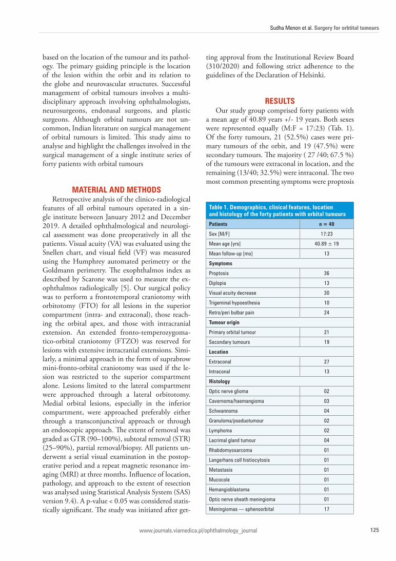
reSultSOur study group comprised forty patients with
a mean age of 40.89 years +/- 19 years. Both sexes were represented equally (M:F = 17:23) (Tab. 1). Of the forty tumours, 21 (52.5%) cases were pri-mary tumours of the orbit, and 19 (47.5%) were secondary tumours. The majority ( 27 /40; 67.5 %) of the tumours were extraconal in location, and the remaining (13/40; 32.5%) were intraconal. The two most common presenting symptoms were proptosis
table 1. demographics, clinical features, location and histology of the forty patients with orbital tumours
Patients n = 40
Sex [M/F] 17:23
Mean age [yrs] 40.89 ± 19
Mean follow-up [mo] 13
Symptoms
proptosis 36
Diplopia 13
Visual acuity decrease 30
trigeminal hypoesthesia 10
retro/peri bulbar pain 24
tumour origin
primary orbital tumour 21
Secondary tumours 19
location
extraconal 27
Intraconal 13
histology
optic nerve glioma 02
Cavernoma/haemangioma 03
Schwannoma 04
granuloma/pseduotumour 02
lymphoma 02
lacrimal gland tumour 04
rhabdomyosarcoma 01
langerhans cell histiocytosis 01
Metastasis 01
Mucocole 01
hemangioblastoma 01
optic nerve sheath meningioma 01
Meningiomas — sphenoorbital 17
oPhthAlMoloGy JournAl 2021, Vol. 6
126 www.journals.viamedica.pl/ophthalmology_journal
(36/40; 90%) and visual blurring (30/40; 75%). Retro-/peribulbar pain (24/40; 60%), diplopia (13/40; 32.5%) paresthesias in the trigeminal dis-tribution (10/40; 25 %), were the other presenting symptoms.
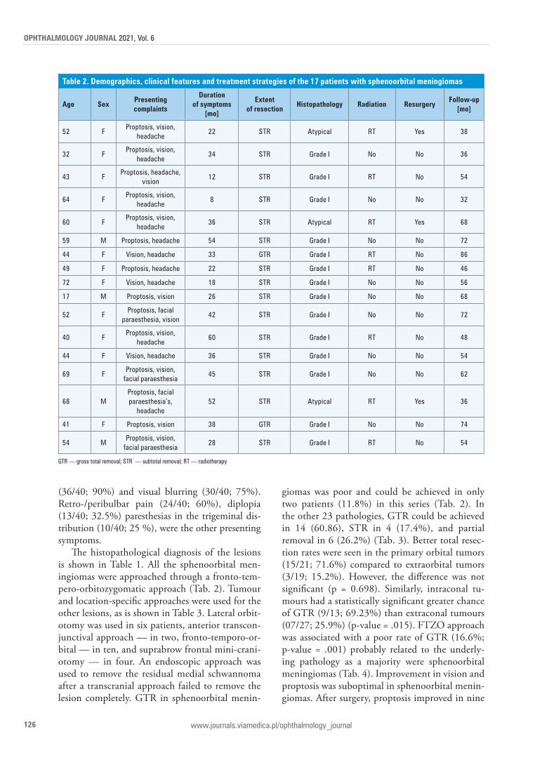
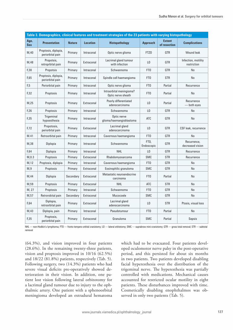
The histopathological diagnosis of the lesions is shown in Table 1. All the sphenoorbital men-ingiomas were approached through a fronto-tem-pero-orbitozygomatic approach (Tab. 2). Tumour and location-specific approaches were used for the other lesions, as is shown in Table 3. Lateral orbit-otomy was used in six patients, anterior transcon-junctival approach — in two, fronto-temporo-or-bital — in ten, and suprabrow frontal mini-crani-otomy — in four. An endoscopic approach was used to remove the residual medial schwannoma after a transcranial approach failed to remove the lesion completely. GTR in sphenoorbital menin-
giomas was poor and could be achieved in only two patients (11.8%) in this series (Tab. 2). In the other 23 pathologies, GTR could be achieved in 14 (60.86), STR in 4 (17.4%), and partial removal in 6 (26.2%) (Tab. 3). Better total resec-tion rates were seen in the primary orbital tumors (15/21; 71.6%) compared to extraorbital tumors (3/19; 15.2%). However, the difference was not significant (p = 0.698). Similarly, intraconal tu-mours had a statistically significant greater chance of GTR (9/13; 69.23%) than extraconal tumours (07/27; 25.9%) (p-value = .015). FTZO approach was associated with a poor rate of GTR (16.6%; p-value = .001) probably related to the underly-ing pathology as a majority were sphenoorbital meningiomas (Tab. 4). Improvement in vision and proptosis was suboptimal in sphenoorbital menin-giomas. After surgery, proptosis improved in nine
table 2. demographics, clinical features and treatment strategies of the 17 patients with sphenoorbital meningiomas
Age SexPresenting complaints
duration of symptoms
[mo]
extent of resection
histopathology radiation resurgeryFollow-up
[mo]
52 Fproptosis, vision,
headache22 Str atypical rt Yes 38
32 Fproptosis, vision,
headache34 Str grade I No No 36
43 Fproptosis, headache,
vision 12 Str grade I rt No 54
64 Fproptosis, vision,
headache8 Str grade I No No 32
60 Fproptosis, vision,
headache36 Str atypical rt Yes 68
59 M proptosis, headache 54 Str grade I No No 72
44 F Vision, headache 33 gtr grade I rt No 86
49 F proptosis, headache 22 Str grade I rt No 46
72 F Vision, headache 18 Str grade I No No 56
17 M proptosis, vision 26 Str grade I No No 68
52 Fproptosis, facial
paraesthesia, vision42 Str grade I No No 72
40 Fproptosis, vision,
headache60 Str grade I rt No 48
44 F Vision, headache 36 Str grade I No No 54
69 Fproptosis, vision,
facial paraesthesia45 Str grade I No No 62
68 Mproptosis, facial paraesthesia’s,
headache52 Str atypical rt Yes 36
41 F proptosis, vision 38 gtr grade I No No 74
54 Mproptosis, vision,
facial paraesthesia28 Str grade I rt No 54
gtr — gross total removal; Str — subtotal removal; rt — radiotherapy
Sudha Menon et al. Surgery for orbtital tumours
127www.journals.viamedica.pl/ophthalmology_journal
(64.3%), and vision improved in four patients (28.6%). In the remaining twenty-three patients, vision and proptosis improved in 10/16 (62.5%) and 18/22 (81.8%) patients, respectively (Tab. 5). Following surgery, two (14.3%) patients who had severe visual deficits pre-operatively showed de-terioration in their vision. In addition, one pa-tient lost vision following lateral orbitotomy for a lacrimal gland tumour due to injury to the oph-thalmic artery. One patient with a sphenoorbital meningioma developed an extradural hematoma
which had to be evacuated. Four patients devel-oped oculomotor nerve palsy in the post-operative period, and this persisted for about six months in two patients. Two patients developed disabling facial hyperesthesia over the distribution of the trigeminal nerve. The hyperesthesia was partially controlled with medications. Mechanical causes accounted for restricted ocular motility in eight patients. These disturbances improved with time. Cosmetically disabling enophthalmos was ob-served in only two patients (Tab. 5).
table 3. demographics, clinical features and treatment strategies of the 23 patients with varying histopathology
Age, Sex
Presentation nature location histopathology Approachextent
of resectioncomplications
M,40proptosis, diplopia,
periorbital painprimary Intraconal optic nerve glioma FtZo gtr Wound leak
M,48propotsis,
retroprbital painprimary extraconal
lacrimal gland tumour with infection
lo gtrInfection, motility
restriction
F,38 propotsis primary Intraconal Schwannoma Fto gtr No
F,65proptosis, diplopia,
periorbital painprimary Intraconal Spindle cell haemangioma Fto Str No
F,5 periorbital pain primary Intraconal optic nerve glioma Fto partial recurrence
F,32 proptosis primary IntraconalIntraorbital meningioma?
optic nerve sheath Fto partial No
M,25 proptosis primary extraconalpoorly differentiated
adenocarcinomalo partial
recurrence — both eyes
F,26 proptosis primary Intraconal Schwannoma lo gtr No
F,35trigeminal
hypoesthesiaprimary Intraconal
optic nerve glioma/haemangioblastoma
atC gtr No
F,72propotosis,
periorbital painprimary extraconal
lacrimal gland adenocarcinoma
lo gtr CSF leak, recurrence
M.41 retroorbital pain primary Intraconal Cavernous haemangioma Fto gtr No
M,38 Diplopia primary Intraconal SchwannomaFto,
endoscopicgtr
recurrence, decreased vision
F,64 Diplopia primary Intraconal Nhl lo gtr recurrence
M,0.3 proptosis primary extraconal rhabdomyosarcoma SMC Str recurrence
M,12 proptosis, diplopia primary Intraconal Cavernous haemangioma Fto gtr No
M,9 proptosis primary extraconal eosinophilic granuloma SMC gtr No
M,44 Diplopia Secondary extraconalMetastatic neuroendocrine
carcinomaFto partial No
M,59 proptosis primary extraconal Nhl atC Str No
M, 27 proptosis primary Intraconal Schwannoma Fto gtr No
M,57 retrorobital pain Secondary extraconal Mucocoele SMC gtr No
F,64Diplopia,
retroorbital painprimary extraconal
lacrimal gland adenocarcinoma
lo Str ptosis, visual loss
M,43 Diplopia, pain primary Intraconal pseudotumour Fto partial No
F,35proptosis,
periorbital pain primary extraconal granuloma SMC partial Sepsis
Nhl — non-hodkin’s lymphoma; Fto — fronto-tempero-orbital craniotomy; lo — lateral orbitotomy; SMC — suprabrow mini-craniotomy; gtr — gross total removal; Str — subtotal removal
oPhthAlMoloGy JournAl 2021, Vol. 6
128 www.journals.viamedica.pl/ophthalmology_journal
dIScuSSIonrelevant anatomy
Orbital anatomy is complex, and a thorough knowledge of orbital anatomy is a prerequisite in the surgical management of orbital tumours [6–9]. The bony orbit is pyramidal and has an approximate volume of 30 cm3. The bony margins of the orbit are made up of seven bones, namely — frontal, ethmoid, lacrimal, sphenoid, zygomatic, palatine, and maxilla. The orbit has a roof, lateral wall, floor, and medial wall, and the apex is directed posteriorly. The optic canal and superior orbital fissure form the apex of the pyramid, which is tilted medially. The lateral walls of the orbits make a right angle with each other. The periorbita lines the orbital bones and
merges with the dura at the optic canal and near the superior orbital fissure. Eight muscles within the or-bit control the movements of the globe and eyelids.
The recti and the superior oblique arise from the tendinous ring called the annulus of Zinn and the adjacent bone at the apex of the orbit and in-sert into the sclera. The levator palpebrae superioris originates at the orbital apex, forms an aponeurosis, and inserts into the upper eyelid. The Optic nerve leaves the orbit through the optic canal. The an-nulus of Zinn divides the superior orbital fissure into three compartments through which the other orbital nerves enter the orbit. The significant blood supply to the orbit is through the ophthalmic artery, a branch of the supraclinoid portion of the ICA. This vessel follows the optic nerve in the optic canal and orbit. The ophthalmic artery crosses lateral to the optic nerve at the optic canal, and once inside the orbit, it passes above the optic nerve. At this crossing point, it usually gives rise to the central retinal artery. It is vital to avoid blind coagulation in the orbital apex as inadvertent coagulation of the retinal artery results in vision loss [7, 10]. The superior ophthalmic vein originates in the supe-ronasal quadrant and passes through the superior orbital fissure to drain the cavernous sinus. The supratrochlear, supraorbital, angular, maxillary, and retromandibular veins also drain the orbit.
Incidence of orbital tumoursDifferent studies quote varying prevalence of
orbital lesions types [1, 3, 11, 12]. Primary orbital tumors are sporadic with an overall incidence of less than 1 per 100,000/year. Cavernous hemangiomas, lymphoid tumours, and meningiomas account
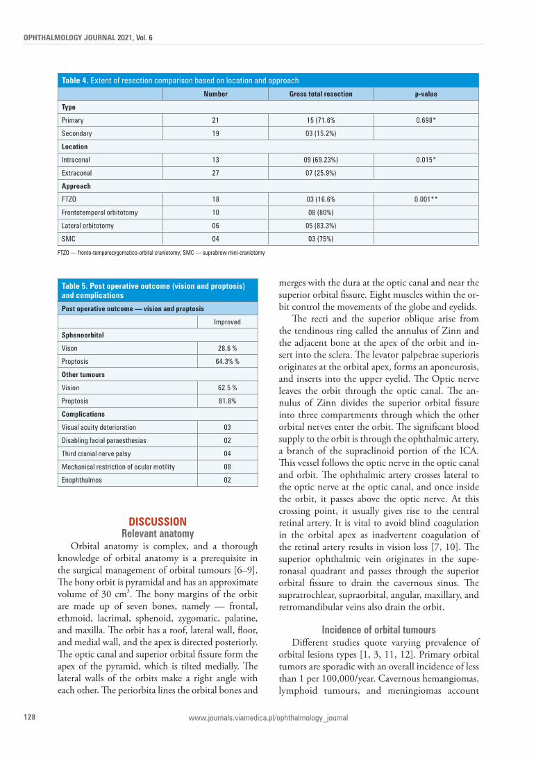
table 4. extent of resection comparison based on location and approach
number Gross total resection p-value
type
primary 21 15 (71.6% 0.698*
Secondary 19 03 (15.2%)
location
Intraconal 13 09 (69.23%) 0.015*
extraconal 27 07 (25.9%)
Approach
FtZo 18 03 (16.6% 0.001**
Frontotemporal orbitotomy 10 08 (80%)
lateral orbitotomy 06 05 (83.3%)
SMC 04 03 (75%)
FtZo — fronto-temperozygomatico-orbital craniotomy; SMC — suprabrow mini-craniotomy
table 5. Post operative outcome (vision and proptosis) and complications
Post operative outcome — vision and proptosis
Improved
Sphenoorbital
Vison 28.6 %
proptosis 64.3% %
other tumours
Vision 62.5 %
proptosis 81.8%
complications
Visual acuity deterioration 03
Disabling facial paraesthesias 02
third cranial nerve palsy 04
Mechanical restriction of ocular motility 08
enophthalmos 02
Sudha Menon et al. Surgery for orbtital tumours
129www.journals.viamedica.pl/ophthalmology_journal
for the most common primary orbital tumors in adults. In contrast, dermoid cysts, optic nerve glio-mas, and rhabdomyosarcoma are the more common tumors in children. Meningiomas and sinonasal car-cinomas are common tumours that extend into the orbit [1, 2, 13]. In one of the largest published series of orbital tumours, Bonovolanta et al. ana-lysed 2,173 primary orbital tumours, 220 secondary orbital tumours, and 87 metastatic tumours in his clinical series spread over 35 years [1]. The dermoid cyst was the most frequently (14%) encountered orbital tumor in their series, followed by cavern-ous hemangioma (218 cases, 9% of all cases). The most common malignant orbital tumor in his study was non-Hodgkin lymphoma, which accounted for nearly 12% of all cases [1]. Orbital tumours can be classified either based on their location or their tissue of origin (Tab. 6). Table 6 summarizes the orbital tumours based on their compartments. The commonly seen orbital tumours in our series are presented in Figures 1–8.
clinical presentationClinical presentation of orbital tumours var-
ies depending on the location of the tumour [6].
Proptosis and diplopia are the most frequent initial symptoms of an orbital mass [12, 14]. A decrease in visual acuity as a presenting symptom is less com-mon and indicates that tumour is situated at the orbital apex or close to the optic nerve [1, 2, 13]. Intraconal tumors tend to cause early visual loss, impairment of ocular motility, and axial proptosis from early direct compression of the optic nerve and impingement on extraocular muscles. Extraconal tumors cause proptosis as an early manifestation, displacement of the globe, and diplopia due to me-chanical restriction of extraocular muscles move-ment [15–17].
Imaging of orbital tumoursMagnetic resonance imaging (MRI), computed
tomography (CT), ultrasound (US), fluoro-2-de-oxy-D-glucose positron emission tomography CT (FDG-PET CT), and MRI positron emission to-mography (PET) [18–20] can be helpful in the detailed evaluation of orbital lesions. The imaging modality of choice in orbital lesions is the MRI. CT scan is reserved for lesions in which a bony pathology is suspected [18, 21]. Recommended MRI includes fat suppressed multiplanar orbital
table 6. classification of orbital tumours
Based on their location /compartment Based on the tissue of origin
extraconal
outside the extra-ocular muscle cone
these tumours can be sub periosteal or extra periostealPrimary tumours
primary orbital tumours are those which originate from structures within the orbitIntraconal
the intraconal space also can be further subdivided in relation to the optic nerve into medial, central, and lateral orbital spaces
Intracanalicular
Within the optic canal Secondary tumours
Secondary tumours refer to tumours involving the orbital cavity by local extension or distant metastases from other parts of the body
Anterior or preseptal compartment
Formed by the orbital septum and lid
Multicompartmental
lesions straddling multiple compartments
orbital tumours — classification
extraconal Intraconal Septal/eyelid Multicompartmental
lacrimal gland tumours
Dermoid cyst
orbital wall meningiomas
rhabdomyosarcoma
Mucocoele
Fibrous dysplasia
Sarcoid
lymphangioma
globe and tumours from globe
optic nerve sheath meningiomas
optic nerve gliomas
orbital Schwannomas
Cavernous hemangiomas
Capillary haemangioma
orbital varix
pediatric capillary hemangioma
SCC
BCC
Melanoma
lymphoma
plasmacytoma
Metastasis
pseudotumour
orbital plexiform neurofibroma
SCC — squamos cell carcinoma; BCC — basal cell carcinoma
oPhthAlMoloGy JournAl 2021, Vol. 6
130 www.journals.viamedica.pl/ophthalmology_journal
cuts with a 2–3 mm slice thickness with contrast administration. Ultrasound (US) with Doppler is a non-invasive and cost-effective adjunct due to the superficial location of the globe and its cystic contents. Advanced imaging techniques such as diffusion-weighted imaging (DWI), diffusion ten-sor imaging (DTI), FDG-PET-CT, and MRI-PET
are newer modalities that supplement information acquired through MRI or CT scans. Apparent diffu-sion coefficient (ADC) values on echo-planar DWI help to differentiate between benign and malig-nant orbital masses. DTI can be used to map optic nerve fibres and assess whether the tumour displaces or infiltrates the nerve fibres. FDG-PET CT and
A B c
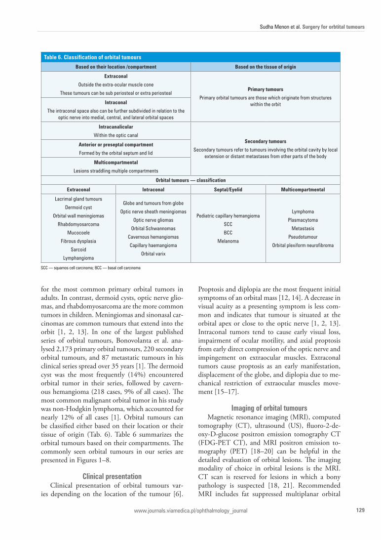
FIGure 1. axial MrI contrast images of three different patients (A, B, c) with diffuse sphenoorbital meningiomas. Note the extension into the cavernous sinus in the second (B) and the temporal fossa in the third (c) patient
FIGure 2. A–d. MrI axial and sagittal images of the orbit showing a retinal hemangioblastoma. the lesion shows altered signal intensity lesions isointense to hyperintense on t1 (A, c) and heterogeneously hyperintense on t2 (B) and showing significant post-contrast enhancement (d) noted in both globes
AB
c d
Sudha Menon et al. Surgery for orbtital tumours
131www.journals.viamedica.pl/ophthalmology_journal
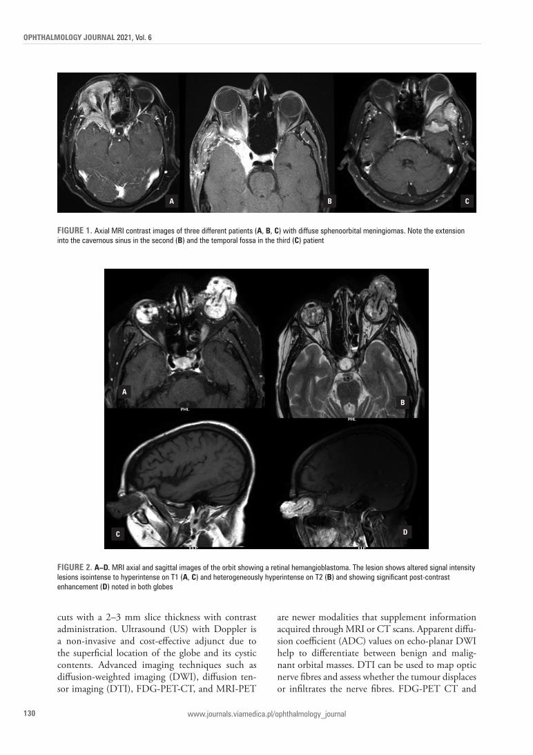
FIGure 3. MrI plain axial t1 (A) images show a well-delineated lesion placed superomedial to the orbit, hypointense on t1, hyperintense on t2 (B), and no contrast enhancement. Biopsy of this lesion in this 3-year-old child proved it to be an embryonal rhabdomyosarcoma
A B c
FIGure 4. MrI orbit axial t2 (A), axial t1 (B), and contrast (c) images showing a diffuse infiltrating lymphoma involving all the compartments of the orbit
A B c
FIGure 5. MrI orbit axial t1 (A) showing an isointense lesion in the lateral compartment in the lacrimal fossa, which remains isointense on t2 (B) and does not shows moderate contrast enhancement. Biopsy was suggestive of a lacrimal gland adenoma
A B c
oPhthAlMoloGy JournAl 2021, Vol. 6
132 www.journals.viamedica.pl/ophthalmology_journal
MRI-PET provides functional information regard-ing tumour metabolism and is a part of standard practice in the pre-operative and post-treatment as-sessment of orbital malignancies. MRI-PET is supe-rior to CT-PET in terms of reduced radiation [22].
Management of orbital tumoursSurgery remains the treatment of choice for
most symptomatic orbital tumours. The aim of the surgery is gross total removal (GTR) of the tumour with preservation of function, especially
A B c
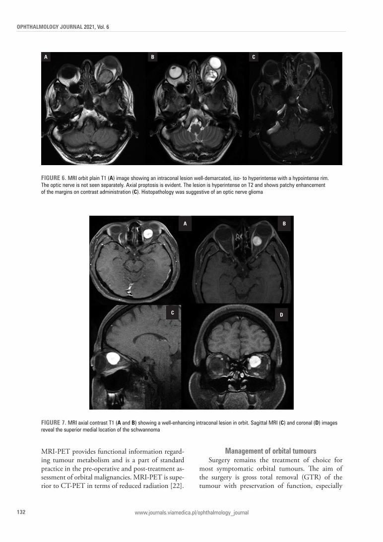
FIGure 6. MrI orbit plain t1 (A) image showing an intraconal lesion well-demarcated, iso- to hyperintense with a hypointense rim. the optic nerve is not seen separately. axial proptosis is evident. the lesion is hyperintense on t2 and shows patchy enhancement of the margins on contrast administration (c). histopathology was suggestive of an optic nerve glioma
A B
c d
FIGure 7. MrI axial contrast t1 (A and B) showing a well-enhancing intraconal lesion in orbit. Sagittal MrI (c) and coronal (d) images reveal the superior medial location of the schwannoma
Sudha Menon et al. Surgery for orbtital tumours
133www.journals.viamedica.pl/ophthalmology_journal
vision. However, for few lesions like optic nerve sheath meningioma, and optic nerve glioma, radi-cal surgery would involve a risk of significant visual deterioration. In such instances, surgery is often delayed. Preservation of vision is preferential over exophthalmos correction. In such cases, surgery is planned if there is a risk of tumour progression in-tracranially or to the opposite normal nerve.
For all other tumours, the choice of a given approach depends on the location and extension of the tumour and its biological behaviour. Of these, compartmental location is the most critical criteria to finalise the approach. Paluzzi et al. were the first to suggest that the orbit should be com-pared to a clock and that the surgical approach to the orbit should vary based on the tumor position [23]. Approaches to the orbit can be essentially divided as transcranial, transorbital, or endonasal and combined.
transcranial approachesWalter Dandy, in 1922, was the first to use
a transcranial fronto-temporal approach for an orbital apex tumour [24]. Subsequently, the ap-proach went through further refinements and modifications. These include:• Frazier’s proposal of an additional removal of the
supraciliary arch [25];
• the pterional approach proposed by Nafzigger [26] and modified by Yasergill [27)];
• mini-pterional approach proposed by Figueiredo et al. (28);
• Pellerin et al.’s and Hakuba et al.’s description of an orbitozygomatic approach [29];
• supraorbital approach suggested by Ormond et al. [30].The frontotemporal/pterional approach com-
bined with orbital roof removal and liberal drilling of the sphenoid bone provides an excellent view of the superior and lateral aspects of the posterior orbit, better access to the optic canal, the superior orbital fissure and the anterior temporal fossa, and better accessibility of the orbital apex [8, 31]. The orbitozygomatic variant provides an added mul-tidirectional exposure of the anterior and middle cranial fossae, the upper clivus, and the region of the basilar apex. Tumours confined to the superior compartment of the orbit are better accessed by the supraorbital mini-craniotomy approach [32–35].
The majority of the lesions in our series were sphenoorbital meningiomas, and they were ap-proached through an extended FTZO approach. For most of the other lesions, we preferred a fronto-temporal craniotomy combined with orbitotomy as it provides optimal exposure of almost three-quar-ters of the globe. A suprabrow mini-craniotomy
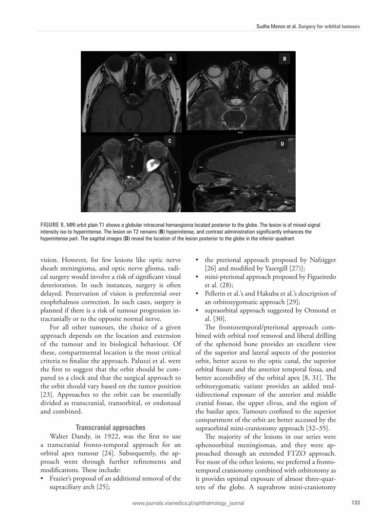
FIGure 8. MrI orbit plain t1 shows a globular intraconal hemangioma located posterior to the globe. the lesion is of mixed-signal intensity iso-to hyperintense. the lesion on t2 remains (B) hyperintense, and contrast administration significantly enhances the hyperintense part. the sagittal images (d) reveal the location of the lesion posterior to the globe in the inferior quadrant
A B
c d
oPhthAlMoloGy JournAl 2021, Vol. 6
134 www.journals.viamedica.pl/ophthalmology_journal
was reserved only for small lesions restricted to the superior compartment. Our policy was to err on the side of a larger flap which would provide greater exposure. We have observed that almost always, after incising the periorbita, the fat starts prolaps-ing out. In spite of adequate retraction, it becomes difficult to approach deeply located tumours. Small exposures compound the challenge. Locating the tumour is often challenging, and we have had to use the intraoperative ultrasound to localize the lesion after opening even for large tumours.
transorbital approachesThe indications for a lateral orbital approach in-
clude: laterally placed extraconal tumors, intraconal tumours lateral or inferior to the optic nerve, lesions in the orbital apex, middle fossa, and cavernous sinus [23, 36]. In 1889 Krönlein first described the lateral orbitotomy, and several modifications have been proposed since then [37]. When combined with a small pterional craniotomy, it provides bet-ter access to lesions situated superior and lateral to the optic nerve in orbit [24, 37]. The total lateral orbitotomy involves additional removal of the su-perior orbital rim up to the superior orbital notch and the body of the zygoma. Tumors affecting the orbital apex, superior and inferior orbital fissures, and pterygopalatine are better accessed through this approach [37–40]. Our surgical approach is limited to a Kronliens hockey stick incision and removal of the zygomatic arch and lateral wall of the orbit. This, combined with liberal drilling of the lateral wall posteriorly up to the apex, provides excellent exposure to the entire lateral compartment.
anterior medial micro-orbitotomy and transconjunctival approach
First described by Galbraith and Sullivan, an-terior medial orbitotomy is helpful for anterior and medial tumours in the intraconal region of the anterior two-thirds of the orbital cavity [41]. The transconjunctival approach is preferred for surgical treatment of tumors located in the in-feromedial, basal, and lateral regions of the an-terior orbit. This approach is less traumatic and cosmetically more superior as it obviates the need for resection of the bone. However, the transcon-junctival approach is not preferred for medium- and large-sized tumors as the surgical corridor is limited. The endoscopic endonasal approach is fast replacing this approach to access medially and inferiorly located orbital disease.
We feel that this approach is best suited for le-sions in the anterior quarter of the globe, and adopt-ing this approach for posteriorly located lesions is cumbersome. Our experience with this approach is limited to two cases, both intraocular tumours with exophytic components.
extended endonasal approaches (eeas)The three main endonasal approaches to the
orbit are:1. The medial–inferior extraconal approach.2. The trans maxillary extraconal approach.3. The medial intraconal approach.
Together, these approaches provide excellent exposure for the safe removal of most intraconal and extraconal tumors that lie medial and inferior to the optic nerve. Endoscopic approaches have almost become the standard of care for medially posteriorly located orbital lesions. Total endoscopic removal of such orbital lesions has a significant learning curve even for established endoscopic sur-geons [42–44].
extent of resectionOur patient cohort was disproportionate with
a relatively large number of sphenoorbital meningi-omas where GTR is extremely difficult due to the invasive nature of the disease. We achieved a nearly 80% GTR with FTO, 83% with Lateral orbitoto-my, and 75% with SMC for the other lesions. It was the primary nature of the lesion which influenced the GTR rather than the approach. Lesions, where GTR was not possible, were diffuse with poor mar-gins. We have observed that the majority of the or-bital tumours can be successfully removed through either an FTC or lateral orbitotomy. The best tech-nique to remove the tumours in the medial com-partment is an endoscopic approach, especially if they are inferiorly and posteriorly situated. Another interesting observation in our series was that intra-conal lesions had a better rate of GTR compared to extraconal lesions. Intraconal lesions are more likely to be well-circumscribed (e.g., neurofibroma) than extraconal lesions, which could explain this obser-vation. Primary tumours similarly had a better rate of GTR, again related to the intrinsic pathology of the lesion.
concluSIonOrbital tumours comprise a rare and challenging
spectrum of pathologies that are best tackled by an
Sudha Menon et al. Surgery for orbtital tumours
135www.journals.viamedica.pl/ophthalmology_journal
interdisciplinary approach. Thorough knowledge of anatomy and the combined experience of a neuro-surgeon, ophthalmologist, and endoscopic surgeon is a prerequisite for the successful management of orbital tumours. The majority of the orbital lesions can be effectively operated through one of the three following approaches — fronto-temporo-orbital ap-proach, a lateral orbitotomy, or an endoscopic ap-proach. Intraconal primary benign orbital tumours have a better chance of GTR. The nature and pa-thology of the lesion influence the extent of resec-tion rather than the location of the lesion.
AcknowledgmentsNone declared.
Financial disclosuresNone declared.
FundingNone declared.
Conflicts of interestNone declared.
reFerenceS1. Bonavolontà g, Strianese D, grassi p, et al. an analysis of 2,480 space-
occupying lesions of the orbit from 1976 to 2011. ophthalmic plast re-constr Surg. 2013; 29(2): 79–86, doi: 10.1097/Iop.0b013e31827a7622, indexed in pubmed: 23470516.
2. hasegawa M. Differential Diagnosis and pathogenesis for orbital tumors. Japan J Neurosurg. 2017; 26(6): 419–429, doi: 10.7887/jcns.26.419.
3. Shinder r, al-Zubidi N, esmaeli B. Survey of orbital tumors at a com-prehensive cancer center in the united States. head Neck. 2011; 33(5): 610–614, doi: 10.1002/hed.21498, indexed in pubmed: 20824811.
4. park hJ, Yang Sh, Kim IlS, et al. Surgical treatment of orbital tumors at a single institution. J Korean Neurosurg Soc. 2008; 44(3): 146–150, doi: 10.3340/jkns.2008.44.3.146, indexed in pubmed: 19096665.
5. Scarone p, leclerq D, héran F, et al. long-term results with exophthalmos in a surgical series of 30 sphenoorbital meningi-omas. Clinical article. J Neurosurg. 2009; 111(5): 1069–1077, doi: 10.3171/2009.1.JNS081263, indexed in pubmed: 19267523.
6. Kannan S, hasegawa M, Yamada Y, et al. tumors of the orbit: Case report and review of Surgical Corridors and Current options. asian J Neurosurg. 2019; 14(3): 678–685, doi: 10.4103/ajns.aJNS_51_19, indexed in pubmed: 31497084.
7. Martins C, Costa e Silva Ie, Campero a, et al. Microsurgical anatomy of the orbit: the rule of seven. anat res Int. 2011; 2011: 468727, doi: 10.1155/2011/468727, indexed in pubmed: 22567293.
8. Maroon JC, Kennerdell JS. Surgical approaches to the orbit. Indications and techniques. J Neurosurg. 1984; 60(6): 1226–1235, doi: 10.3171/jns.1984.60.6.1226, indexed in pubmed: 6726367.
9. Kennerdell JS, Maroon JC, Malton Ml. Surgical approaches to orbital tumors. Clin plast Surg. 1988; 15(2): 273–282, indexed in pubmed: 3349739.
10. Campbell aa, grob Sr, Yoon MK. Novel Surgical approaches to the orbit. Middle east afr J ophthalmol. 2015; 22(4): 435–441, doi: 10.4103/0974-9233.164613, indexed in pubmed: 26692713.
11. ohtsuka K, hashimoto M, Suzuki Y. a review of 244 orbital tumors in Japanese patients during a 21-year period: origins and locations. Jpn
J ophthalmol. 2005; 49(1): 49–55, doi: 10.1007/s10384-004-0147-y, indexed in pubmed: 15692775.
12. Shields Ja, Shields Cl, Scartozzi r. Survey of 1264 patients with orbital tumors and simulating lesions: the 2002 Montgomery lec-ture, part 1. ophthalmology. 2004; 111(5): 997–1008, doi: 10.1016/j.ophtha.2003.01.002, indexed in pubmed: 15121380.
13. pförtner r, Mohr C, Daamen J, et al. orbital tumors: operative and therapeutic strategies. Facial plast Surg. 2014; 30(5): 570–577, doi: 10.1055/s-0034-1395210, indexed in pubmed: 25397713.
14. Montano N, lauretti l, D’alessandris Qg, et al. orbital tumors: report of 70 Surgically treated Cases. World Neurosurg. 2018; 119: e449–e458, doi: 10.1016/j.wneu.2018.07.181, indexed in pubmed: 30071324.
15. rootman J. Diseases of the orbit. 2nd ed. lippincott Williams & Wilkins, philadelphia 2003: 579.
16. alkatan hM, al Marek F, elkhamary S. Demographics of pediatric orbital lesions: a tertiary eye Center experience in Saudi arabia. J epi-demiol glob health. 2019; 9(1): 3–10, doi: 10.2991/jegh.k.181224.001, indexed in pubmed: 30932383.
17. garrity Ja, henderson JW, Cameron JD. henderson’s orbital tumors. 4th ed . lippincott Williams & Wilkins, philadelphia : 404.
18. purohit BS, Vargas MI, ailianou a, et al. orbital tumours and tumour-like lesions: exploring the armamentarium of multiparametric imaging. Insights Imaging. 2016; 7(1): 43–68, doi: 10.1007/s13244-015-0443-8, indexed in pubmed: 26518678.
19. goh pS, gi Mt, Charlton a, et al. review of orbital imaging. eur J radiol. 2008; 66(3): 387–395, doi: 10.1016/j.ejrad.2008.03.031, indexed in pubmed: 18501542.
20. razek aa, elkhamary S, Mousa a. Differentiation between benign and malignant orbital tumors at 3-t diffusion Mr-imaging. Neuroradiology. 2011; 53(7): 517–522, doi: 10.1007/s00234-011-0838-2, indexed in pubmed: 21286695.
21. hasegawa M, Fujisawa h, hayashi Y, et al. Ct arteriography for orbital tumors: diagnostic and surgical value. J Clin Neurosci. 2005; 12(5): 548–552, doi: 10.1016/j.jocn.2004.08.012, indexed in pubmed: 15982890.
22. Miyamoto J, tatsuzawa K, owada K, et al. usefulness and limitations of fluorine-18-fluorodeoxyglucose positron emission tomography for the detection of malignancy of orbital tumors. Neurol Med Chir (tokyo). 2008; 48(11): 495–9; discussion 499, doi: 10.2176/nmc.48.495, indexed in pubmed: 19029776.
23. paluzzi a, gardner pa, Fernandez-Miranda JC, et al. „round-the-Clock” Surgical access to the orbit. J Neurol Surg B Skull Base. 2015; 76(1): 12–24, doi: 10.1055/s-0033-1360580, indexed in pubmed: 25685644.
24. Dandy W. prechiasmal Intracranial tumors of the optic Nerves. am J ophthalmol. 1922; 5(3): 169–188, doi: 10.1016/s0002-9394(22)90261-2.
25. Khan aM, Varvares Ma. traditional approaches to the orbit. otolaryngol Clin North am. 2006; 39(5): 895–909, vi, doi: 10.1016/j.otc.2006.08.008, indexed in pubmed: 16982253.
26. Naffziger h. pathologic changes in the orbit in progressive exophthalmos. arch ophthalmol. 1933; 9(1): 1, doi: 10.1001/ar-chopht.1933.00830010004001.
27. Yaşargil Mg, reichman MV, Kubik S. preservation of the frontotem-poral branch of the facial nerve using the interfascial temporalis flap for pterional craniotomy. technical article. J Neurosurg. 1987; 67(3): 463–466, doi: 10.3171/jns.1987.67.3.0463, indexed in pubmed: 3612281.
28. Figueiredo eg, Zabramski JM, Deshmukh p, et al. anatomical and quantitative description of the transcavernous approach to interpe-duncular and prepontine cisterns. technical note. J Neurosurg. 2006; 104(6): 957–964, doi: 10.3171/jns.2006.104.6.957, indexed in pubmed: 16776341.
29. pellerin p, lesoin F, Dhellemmes p, et al. usefulness of the orbit-ofrontomalar approach associated with bone reconstruction for frontotemporosphenoid meningiomas. Neurosurgery. 1984; 15(5): 715–718, doi: 10.1227/00006123-198411000-00016, indexed in pubmed: 6504290.
30. ormond Dr, hadjipanayis Cg. the Supraorbital Keyhole Craniotomy through an eyebrow Incision: Its origins and evolution. Minim Invasive
oPhthAlMoloGy JournAl 2021, Vol. 6
136 www.journals.viamedica.pl/ophthalmology_journal
Surg. 2013; 2013: 296469, doi: 10.1155/2013/296469, indexed in pubmed: 23936644.
31. humphrey C, Kriet J. Surgical approaches to the orbit. operat tech otolaryngol-head Neck Surg. 2008; 19(2): 132–139, doi: 10.1016/j.otot.2008.07.002.
32. da Silva Sa, Yamaki VN, Solla DJ, et al. pterional, pretemporal, and orbitozygomatic approaches: anatomic and Comparative Study. World Neurosurg. 2019; 121: e398–e403, doi: 10.1016/j.wneu.2018.09.120, indexed in pubmed: 30266695.
33. reisch r, perneczky a, Filippi r. Surgical technique of the supraorbital key-hole craniotomy. Surg Neurol. 2003; 59(3): 223–227, doi: 10.1016/s0090-3019(02)01037-6, indexed in pubmed: 12681560.
34. Srinivasan a, Bilyk Jr. transcranial approaches to the or-bit. Int ophthalmol Clin. 2018; 58(2): 101–110, doi: 10.1097/IIo.0000000000000224, indexed in pubmed: 29517647.
35. hamby WB. pterional approach to the orbits for decompression or tumor removal. J Neurosurg. 1964; 21: 15–18, doi: 10.3171/jns.1964.21.1.0015, indexed in pubmed: 14110353.
36. abou-al-Shaar h, Krisht KM, Cohen Ma, et al. Cranio-orbital and orbi-tocranial approaches to orbital and Intracranial Disease: eye-opening approaches for Neurosurgeons. Front Surg. 2020; 7: 1, doi: 10.3389/fsurg.2020.00001, indexed in pubmed: 32118028.
37. Stallard hB. lateral orbitotomy (Krönlein’s operation). Br J ophthalmol. 1946; 30(4): 250–251, indexed in pubmed: 18170216.
38. Kim JW, Yates BS, goldberg ra. total lateral orbitotomy. orbit. 2009; 28(6): 320–327, doi: 10.3109/01676830903334028, indexed in pubmed: 19929652.
39. Nemet a, Martin p. the lateral triangle flap--a new approach for lateral orbi-totomy. orbit. 2007; 26(2): 89–95, doi: 10.1080/01676830600974571, indexed in pubmed: 17613854.
40. Chabot JD, gardner pa, Stefko St, et al. lateral orbitotomy approach for lesions Involving the Middle Fossa: a retrospective review of thirteen patients. Neurosurgery. 2017; 80(2): 309–322, doi: 10.1093/neuros/nyw045, indexed in pubmed: 28175869.
41. Sullivan gl. Caveat chirurgicus. trans am ophthalmol Soc. 1972; 70: 328–336, indexed in pubmed: 4663672.
42. Signorelli F, anile C, rigante M, et al. endoscopic treatment of orbital tumors. World J Clin Cases. 2015; 3(3): 270–274, doi: 10.12998/wjcc.v3.i3.270, indexed in pubmed: 25789299.
43. Campbell pg, Yadla S, rosen M, et al. endoscopic transnasal cryo-assisted removal of an orbital cavernous hemangioma: a technical note. Minim Invasive Neurosurg. 2011; 54(1): 41–43, doi: 10.1055/s-0030-1270465, indexed in pubmed: 21509724.
44. Koutourousiou M, gardner pa, Stefko St, et al. Combined endoscopic endonasal transorbital approach with transconjunctival-medial orbit-otomy for excisional biopsy of the optic nerve: technical note. J Neurol Surg rep. 2012; 73(1): 52–56, doi: 10.1055/s-0032-1323156, indexed in pubmed: 23946927.