Hypercholesterolemia Impaired Sperm Functionality in Rabbits
Upload
independentCategory
view
0download
0
British Joumal of Cancer (1998) 77(12), 2104-2111© 1998 Cancer Research Campaign
Electrochemotherapy on liver tumours in rabbits
LH Ramirez', S Orlowski2, D An'*, G Bindoula3, R Dzodic3t, P Ardouin4, C Bognel5, J Belehradek Jr', J-N Munck3and LM Mir4
'UMR 1772 CNRS, Institut Gustave-Roussy, F-94805 Villejuif, France; 2SBPM/DBCM and URA 2096 CNRS, CEA-Saclay, F-91191 Gif sur Yvette, France;Departments of 3Medecine and 4Service Commun d'Experimentation Animale and 5Anatomopathologie, Institut Gustave-Roussy, F-94805 Villejuif, France
Summary Electrochemotherapy (ECT) is a new therapeutic approach combining the effects of a low-permeant cytotoxic drug, bleomycin(BLM), administered i.v. and cell-permeabilizing electric pulses (EPs) locally delivered to tumours. The transient permeabilization of the cellmembrane by the EPs allows free access of BLM to its intracellular targets, largely enhancing BLM's cytotoxic effects. ECT efficacy has beenproved so far on transplanted subcutaneous murine tumours and on subcutaneous metastases in humans. Here, we present the first study ofthe effects of ECT on tumours transplanted to livers in rabbits. We used a recently developed EP applicator consisting of an array of paralleland equidistant needles to be inserted in tissues. Effects of EPs alone or of ECT were assessed by histological analysis, tumour growth ratesand survival of the treated animals. A transient blood hypoperfusion was seen in the electropulsed areas, with or without BLM, related to EP-dependent vasoconstriction but this had no major effects on cell survival. Long-term effects depended on the presence of BLM at the time ofEP delivery. Almost complete tumour necrosis was observed after ECT, resulting from both BLM direct cytotoxic effects on electro-permeabilized tumour cells and indirect effects on the tumour vessels. A large reduction in tumour growth rate and significantly longer survivaltimes were scored in comparison with control rabbits. Moreover, ECT of liver tumours was well tolerated and devoid of systemic side-effects.When ECT was associated with a local interleukin 2-based immunotherapy, increased local anti-tumour effectiveness as well as a largedecrease in the number of metastases were observed. Thus, ECT could become a novel treatment modality for liver tumours and other solidinternal malignancies.
Keywords: electrochemotherapy; bleomycin; electric pulse; liver tumour; immunotherapy; interleukin 2
Bleomycin (BLM), a non-permeant cytotoxic drug largely used inclinical oncology (Sikic, 1988; Mir et al, 1996), is a relativelylarge hydrophilic molecule that is carried into the cells by a low-efficiency mechanism (Pron et al, 1994). Consequently, very smallamounts of BLM enter intact cells, limiting its cytotoxic effects(Orlowski et al, 1988; Poddevin et al, 1991). Brief and intenseelectric pulses (EPs) delivered in vitro or in vivo are known toinduce changes in the plasma membrane of the pulsed cells,resulting in a transient and reversible cell permeabilization (Changet al, 1992; Orlowski and Mir, 1993). One of the most innovativeand promising biomedical applications of electropermeabilizationis the therapeutic use of this technique to incorporate cytotoxicdrugs into tumour cells (Mir et al, 1995a). In vitro cell electroper-meabilization enhances BLM influx into electropulsed cells andthus greatly potentiates BLM cytotoxicity (Orlowski et al, 1988;Poddevin et al, 1991). Furthermore, in vivo experiments ontumour-bearing mice demonstrated that BLM potent anti-tumoureffects were obtained by delivering transcutaneous EPs to subcuta-neous tumours by means of external electrodes (Mir et al, 1991).The effectiveness of this approach has been proved on a largevariety of murine subcutaneous tumours (Belehradek J et al, 1991;Sersa et al, 1994; Heller et al, 1995) and on spontaneous soft tissuesarcomas in cats (Mir et al, 1997). The usefulness of this method indeeply located tumours has also been reported on brain-implanted
Received 27 June 1997Revised 13 November 1997Accepted 27 November 1997
Correspondence to: LM Mir, URA 147 CNRS-lnstitut Gustave-Roussy, 39,rue Camille-Desmoulins, F-94805 Villejuif Cedex, France
gliomas in rats using two needles stereotaxically inserted at eachside of the tumour (Salford et al, 1993). We have termed this newtherapeutic principle, which combines the systemic administrationof BLM with local permeabilizing EPs, electrochemotherapy(ECT) (Mir et al, 1991, 1995a). ECT provokes a transient localperitumoral oedema followed by the rapid disappearance of thetreated tumour. We have previously shown the role of the host'simmunological response in achieving cures induced by ECT inmurine tumour models (Mir et al, 1992). Moreover, the combina-tion of ECT with an interleukin 2 (IL-2)-based immunotherapy,used to stimulate the host's immune system, led to increased localanti-tumour effects and, even more important, revealed systemicanti-tumour effects (Mir et al, 1 995b).
Clinical use of ECT alone has already shown good tolerance andresponse rates on subcutaneous permeation nodules of heavilypretreated patients with head and neck squamous carcinoma(Belehradek M et al, 1993; Domenge et al, 1996), as well as oncutaneous or subcutaneous metastatic melanomas and on basal cellcarcinomas (Heller et al, 1996; Mir et al, 1998). To apply this ther-apeutic strategy to other malignancies, such as visceral tumours, itappeared necessary to investigate the functional and histologicaleffects of ECT on an experimental model of internal tumour.Factors such as the absence of the skin barrier could have an influ-ence on the ECT application conditions or effects. In particular, ourbasic studies on tissue electropermeabilization demonstrated theimportance of the geometry of the field lines, obviously related tothe electrodes used (Belehradek J et al, 1994).
Present addresses: *Department of Thoracic Surgery, Xian Dong Hospital, Liling,412200, Hunan, People's Republic of China. 'Department of Surgery, Institute ofOncology and Radiology of Serbia, 11000 Beograd, Yougoslavia
2104
Electrochemotherapy on liver tumours in rabbits 2105
To approach simultaneously the treatment of internal deeptumours and of large and thick tumour nodules, for which mice arenot suitable, we treated VX2 tumours transplanted in the livers ofrabbits (Miller et al, 1987). We used a recently developed devicefor EP delivery that consists in parallel and equidistant needleelectrodes forming a centred hexagonal array (Mir et al, 1997). Oninsertion in the tissues, this needle configuration divides theoverall tumour volume in small unit volumes. Thus, even in thecase of large tumours, ECT will not require too high voltages to bedelivered between each pair of neighbouring needle electrodes asthe voltage necessary to obtain cell permeabilization is propor-tional to the distance separating the electrodes. Moreover, theneedles allow the deepest parts of thick tumours to be reached. Theaim of our work in rabbits was to determine the ECT effectivenesson experimental liver tumours. We report here that (a) a transientblood hypoperfusion was seen on the electropulsed areas, with orwithout BLM; (b) in the presence of BLM, almost completetumour cell death was observed, due to a combination of directBLM cytotoxicity and indirect vascular effects; (c) ECT caused aclear reduction in the tumour growth rate, significantly increasedsurvival times as well as cure achievement; (d) when an IL-2-based immunotherapy was associated with ECT, the local anti-tumour effectiveness was increased and the number of metastaseswas largely decreased.
MATERIALS AND METHODS
Animals and tumours
New Zealand white rabbits (Elevage Scientifique des Dombes,Romans, France) were maintained under standard conditions witha laboratory diet and water ad libitum. All procedures were carriedout under general i.v. anaesthesia using ketamine hydrochloride(Ketamine, Parke Davis, Courbevoie, France) and xylazine 2%(Rompun, Bayer, Puteaux, France). The experiments wereconducted in accordance with European Council directives andFrench legislation concerning animal welfare. The VX2 carcinomawas maintained by serial passages in the liver in carrier rabbits, aspreviously described (Munck et al, 1993). Hepatic implantation ofVX2 carcinoma cells was accomplished through a small mediansubxyphoid incision. A tumour was removed from one animal,minced in NCTC 109 medium (Eurobio, Paris, France) and filteredthrough cotton gauze. Samples of I07 VX2 cells in 100 gl ofmedium were injected with a 30-gauge needle under the hepaticcapsule, resulting in the development of a localized hepatic tumour2 weeks later (diameter 6-18 mm). To comply with the majorconstraint of the last series of experiments (generation of singletumours, in the absence of other small tumour nodules growingbeside the main tumour due to tumorigeneic cell spreading at thetime of VX2 cell inoculation) small tumour fragments of 2-4 mm3were transplanted under the hepatic capsule. Two weeks later, thesetumours reached diameters between 6 and 11 mm.
Therapeutical procedures
ElectrochemotherapyThe rabbits were anaesthetized and a subxyphoid incision wasperformed to expose liver tumours. Bleomycin (Laboratoire RogerBellon, Neuilly-sur-Seine, France) dissolved in sterile 0.9% sodiumchloride, was injected as a bolus i.v. dose of 0.5 orI mg kg-'. This dose is not the maximum tolerated dose of BLM
but the dose used in previous preclinical trials in mice and in cats(Mir et al, 1992, 1995b, 1997), and similar to that used in clinicaltrials (Belehradek M et al, 1993; Domenge et al, 1996). The electriccomponent of the treatment by ECT consisted of eight square-waved EPs of 100 ts length delivered between 4 and 12 min afterthe end of the BLM infusion. In accordance with previous ex vivostudies that determined the permeabilizing threshold for tumourtissues (Belehradek J et al, 1994), the electric field intensity appliedwas 850 V cm-'. The electric signal delivered was checked througha digital storage oscilloscope (Hitachi, Tokyo, Japan). Two genera-tors delivering the same type of EPs were used for the EP delivery(a) a Jouan PS 15 electropulsator (Nantes, France) connected to twostainless-steel plates or to two acupuncture needles fixed 6 mmapart and held by an insulating template, delivering the run of eightsquare pulses at the frequency of I Hz; the metal plate electrodeswere placed on the surface of the abdominal organs, whereas theneedle electrodes were positioned and inserted into the normaltissues and the tumours; after each run, the needle electrodesassembly was repositioned into the tumour along different direc-tions (mean = 11 runs) to ensure coverage of the whole tumourvolume; (b) a CELTEM MKO generator (Antony, France)connected to an electrode assembly consisting of seven equidistantand parallel needles (5 cm length) arranged in a centred hexagonalarray, defining 12 electrode pairs separated by 6 mm; for each run,the square pulses, delivered by the generator at a frequency of 8 Hz,were distributed successively to each electrode pair by an inte-grated switch in order to deliver eight pulses (four of each polarity)between every pair of needles at a final frequency of 0.67 Hz, witha total duration of 12 s for the entire run; after each run, the needleelectrode assembly was repositioned into the tumour to ensurecoverage of the whole tumour volume by the application of six runs(Mir et al, 1997). All the tumour treatments were performed withneedles. The same effects were observed with either the two- or theseven-needle devices, provided that the coverage of the wholetumour volume was ensured by multiple positioning of the needleswithin the tumour. Obviously, the seven-needle device was moreconvenient, particularly for the treatment of the largest tumours.
ImmunotherapyIL-2 gene-transfected Chinese hamster CHO(IL-2) cells (Ferraraet al, 1987) were routinely maintained in vitro in MEM culturemedium supplemented with 8% fetal calf serum and antibiotics. Invitro they secrete 3500 IL-2 units of the Biological ResponseModifiers Program per millilitre, 72 h and 0.8 x 10⁵ initiallyseeded cells. For rabbit treatment, 30 x 106 CHO(IL-2) cells,resuspended in 200-300 ,ul of MEM without serum, were injectedintratumorally, either in the absence of any other treatment or incombination with ECT, within 10 min after the delivery of the EPto the tumours.
ControlsControl rabbits, without treatment, were laparotomized 2 weeksafter VX2 tumour implantation and their tumours measured. Aftersurgery, they were maintained and followed up like treated animals.Other control groups included rabbits receiving either (a) only EPs,or (b) only BLM, or (c) only injection of CHO(IL-2) cells.
Effects on normal tissues
The effects of ECT were studied in vivo during EP application, andsubsequently by sequential histological analysis. The peroperative
British Journal of Cancer (1998) 77(12), 2104-21110 Cancer Research C-ampaign 1998

2106 LH Ramirez et al
A
B
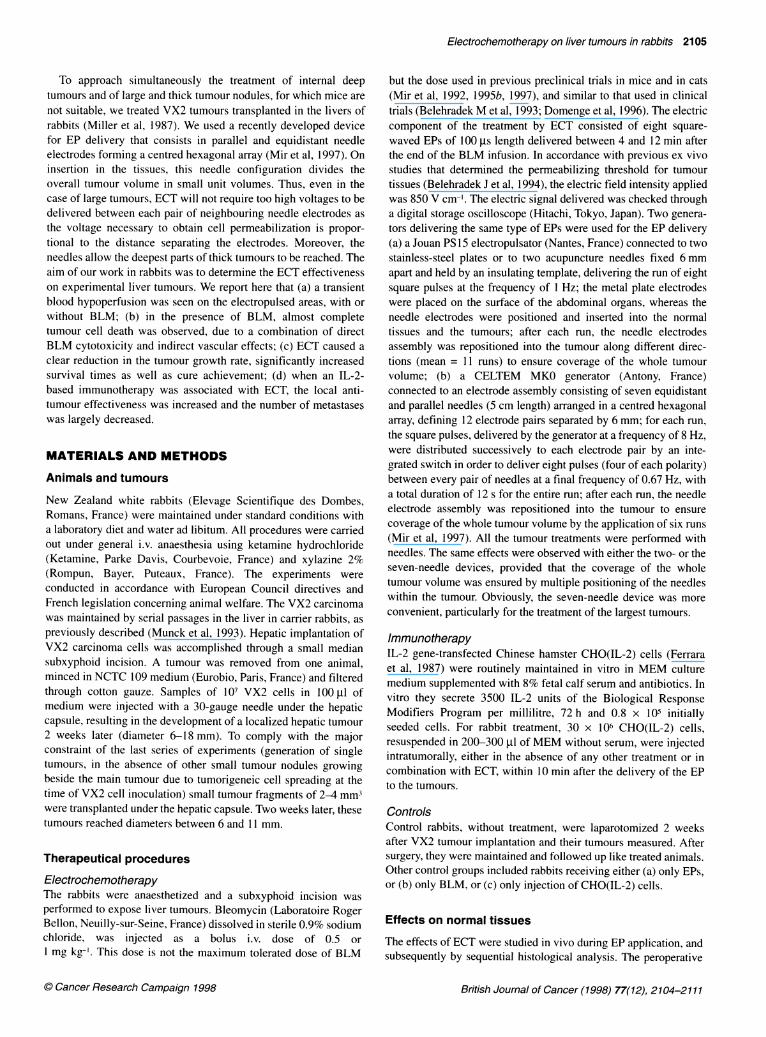
Figure 1 Histological observations of VX2 tumours after ECT. ECTconsisted of square-waved EPs of 850 V cm-' electric field intensity and of100 ,s length, applied using the PS15 electropulsator from 4 min after theBLM i.v. (0.5 mg kg-') injection onwards. (A) At day 2 after ECT. Tumournecrosis with isolated cell nuclei, associated with inflammatory cell infiltrationaround the necrosed area. Scale bar, 100 gm. (B) At day 9 after ECT.Tumour necrosis associating fibrotic reaction, and marked vascular lesionswith endothelium alterations and intraluminal thrombus. Scale bar, 200 ,um.(C) At day 30 after ECT. Massive tumour necrosis with total disappearance oftissue organization. Scale bar, 500,m
vascular effects were assessed by the distribution of fluoresceineither before or after the EP delivery. EPs, alone or in combinationwith BLM, were applied on the left lobe of the liver of healthy (non-tumour-implanted) rabbits and immediately after this treatment, thedye was injected i.v. (0.02 mg in 1 ml) and the livers illuminated
with a Woods light. The same experiment was performed on controlrabbits without treatment, and in both cases staining of the liverswas observed.
Histological analysis of tissues was performed on rabbits killed10 min and 1 h later, to study early alterations, especially vascularlesions. Late changes in normal tissues were also assessed bymacroscopic and microscopic analysis of samples of livers,kidneys, pancreas and spleens obtained 2 days after the treatment.
Samples were fixed in Bouin's fixative. Paraffin-embeddedsections were stained with haemalun-eosin.
Anti-tumour efficacy and survival
The evolutive changes in tumour response were analysed by histo-logical examinations up to 30 days after treatment. Short-termanti-tumour efficacy of ECT was tested by comparing the tumour
growth rates in four groups of rabbits. The VX2 tumour is a
rapidly growing and aggressive carcinoma, and consequently theevaluation end point for the anti-tumour effects was fixed at 9 daysafter the treatment. Each experimental group was composed of sixrabbits receiving either (a) no treatment or (b) i.v. BLM alone or
(c) EPs alone or (d) ECT. Tumour volumes (V) on the treatmentday were measured through a subxyphoid incision using calipersto determine the three largest perpendicular diameters a, b and c,and then applying the formula: V = rtabc/6. All tumours weremeasured at the treatment time and 9 days later when the rabbitswere killed. The tumour growth rate for each animal was estab-lished from the ratio of the tumour volumes at day 9 to those at day0 by [(Vd9/Vdo) -1] x 100. In the four experimental groups, histo-logical confirmation of tumour response and the determination ofthe percentage of tumour necrosis were performed by two inde-pendent observers. To accurately estimate the anti-tumour effectsof ECT, a tumour growth score was established taking into accountthe former tumour growth rate corrected by the estimated necrosisrate of each individual tumour at day 9.
In survival experiments, the local response of the implantedtumour as well as the number and the location of metastases weredetermined at the time of the rabbit's death. To perform necropsyunder good conditions immediately after death, animals werekilled when their general status was very low, which was estimatedfrom a daily follow-up consisting in weight determination and inbehaviour observation.
Anti-tumour effects were statistically compared using the non-parametric Mann-Whitney test for tumour growth rates (Figure 2),and the Mantel-Haenzel log-rank test for survival times(Figure 3). Statistical comparisons in Tables 1 and 2 were madeusing contingency tables analysis and exact X2 Fischer's test.Significance was assumed for tests at P < 0.05.
RESULTS
Effects of bleomycin alone
BLM alone, at the doses used (0.5 mg kg-'), did not induce anyhistological change in healthy tissues compared with the controls.Tumours treated by i.v. BLM alone at the same dose showed histo-logical aspects indistinguishable from native non treated tumours.In both cases, necrotic areas reached approximately 10% of thetumour volume 9 days after the treatment. These results could beexpected as the BLM doses used were far below the maximumtolerated dose.
British Journal of Cancer (1998) 77(12), 2104-2111 0 Cancer Research Campaign 1998

Electrochemotherapy on liver tumours in rabbits 2107
E
.2
1 2 3 4 1 2 3 4
Figure 2 Tumour growth rates and tumour growth scores determined 9days after the treatments. At the time of the treatment, tumour volumes weredetermined as described in Materials and methods. Experimental groups: 1,ECT; 2, EPs alone; 3, BLM i.v. (1 mg kg-') alone; 4, untreated controls. ECTconsisted of square-waved EPs of 850 V cm-1 electric field intensity and of100 ps length, applied using the MKO electropulsator from 4 min after theBLM i.v. (1 mg kg-') injection onwards. Nine days later, rabbits (six in eachgroup) were killed. Tumours were removed, measured and processed forhistological determinations of the necrosis percentage. (A) Relative tumourvolume increases according to the ratio of the macroscopically measurablevolume at day 9 to the macroscopically measurable volume at the day of thetreatment. The value of the ECT group is statistically different (P < 0.05) fromthe values of the three other groups. (B) Relative increases when tumourvolumes at day 9 are corrected by the histologically determined percentageof necrosis. The value of the ECT group is statistically different (P < 0.02)from the values of the three other groups
Table 1 Survival experiments using rabbits with VX2 carcinoma in the livergenerated by tumour fragment transplantations
Treatment Per cent of Median survivalcured rabbits (days)
None 0 50Electric pulses 0 51IL-2-secreting cells 0 45ECT 30 82ECT and IL-2-secreting cells 40 80
Long-term survivors and median survival times were determined in groups offive rabbits, except in the ECT group, which consisted of ten rabbits. ECTwas performed as in Figure 2. Rabbits were considered to be cured if theysurvival more than 250 days after treatment without any sign of disease.
Effects of electric pulses alone
In tissues submitted to EPs alone, immediate reactions wereobserved as colour modifications restricted to the electropulsedareas. On the spleen, we noticed an immediate reduction in thevolume of the pulsed area. When EPs were applied on thepancreas, mesenteric vessel branches showed a reduction of theirvascular diameter, with perivascular oedema. After EP applicationto the liver, fluorescein was injected and illumination with aWoods light revealed no dye coloration of the pulsed volume,confirming the absence of blood flow. In all cases, blood flowbreakdown was observed transiently, over 15-20 min. After thistime, tissues progressively recovered their initial colour andconsistence, and no bleeding was ultimately observed at the needleinsertion sites. It is noteworthy that the application of EPs on
_60-
c 40-
20-
00 20 40 60 80 100
Days after tumour cell inoculation
Figure 3 Survival curves of rabbits with VX2 carcinoma in the livergenerated by the injection of tumour cell suspensions. The nine controlrabbits (0) had VX2 carcinoma but received no treatment. The eight treatedrabbits (0) received ECT 2 weeks after VX2 cell injection in the liver. ECTwas performed as in Figure 2 and rabbit survival (in days after tumour cellinoculation) was checked daily. The difference between the two curves isstatistically significant according to the log-rank test, with P < 0.02
normal tissues and tumours did not induce distant side-effects andwas fairly well tolerated locally.The immediate histological analysis (10 min and 1 h after appli-
cation of EPs) showed markedly congestive tissues and interstitialoedema. The presence of fibrin deposits between the vascularlayers as well as the loose perivascular oedema observed onmesenteric vessels confirmed an enhanced vascular permeability.No evidence of cell damage or change in tissue organization wasobserved except a sparsely distributed endothelial damage onpulsed areas: arteries were distorted, showing thrombus andperivascular oedema with an eosinophil inflammatory infiltrate.Two days after EP delivery, the liver, pancreas, kidney and
spleen showed minor local necrosis and vascular damage. Theselesions were focal, limited to the electrode contact sites, and treat-ment did not induce diffuse organ damage or distant side-effects.
At day 9 after EPs alone, percentages of necrosis in the pulsedtumours were estimated from 10% to 20-25%, depending on thenumber of EP runs. These should be compared with the sponta-neous necrosis of untreated tumours, which was 10-15%. Lesionswere sparsely distributed with minimal vascular damage, and localnecrosis and fibrosis were seen just at the electrode applicationsite. EP delivery did not induce diffuse organ damage or distantside effects.
Effects of electrochemotherapy
Histological analysesIn the presence of BLM, the early vascular effects of EPs detectedin the absence of BLM were also present, without histologicaldifferences, and the macroscopic aspects of each treated area werealso indistinguishable. However, the treatment by ECT or EPsalone induced quite different evolutive changes with characteristichistological patterns.Two days after ECT, examination of the tumours revealed
severe lesions consisting in a polygonal necrotic lesion corre-sponding to the geometry of the liver area crossed by permeabi-lizing electric field intensities. Dead cells were surrounded by aneosinophil inflammatory infiltrate, and the structures of pulsed
British Journal of Cancer (1998) 77(12), 2104-2111. Cancer Research Campaign 1998

2108 LH Ramirez et al
Table 2 Local and systemic tumour progression at the death of rabbits with VX2 carcinoma in livers generated by tumour fragment transplantations
Treatment Per cent of rabbits with Average number of Per cent of rabbits withlocal regression of the countable metastases massive lung
treated tumour invasion bymicrometastases
None 0 27 60Electric pulses 0 16 60IL-2-secreting cells 0 58 0ECT 50 18 50ECT and IL-2-secreting cells 80 3 0
Local progression of liver tumours and the presence of metastases at necropsy were determined in groups of five rabbits, except in the ECT group,which consisted of ten rabbits. ECT was performed as in Figure 2. Local tumour response corresponded to the absence of live tumour tissue at thesite of the treated tumour. For evaluation of the metastatic dissemination, we distinguished, on one hand, the well-limited and macroscopicallycountable secondary tumour nodules on the various organs inspected during the necropsy and, on the other hand, the massive lung infiltration bymicrometastases.
tissues were distorted, showing isolated nuclei and considerablevascular damage (Figure IA). Nine days after ECT, we observeda massive necrotic area, with 100% necrosis in some cases,surrounded by fibrosis. Arteries were severely damaged showingan endothelium detached from the basal membrane, with intra-mural fibrin clotting and thrombosis (Figure IB). Thirty days afterECT, total disappearance of tissue structures was observed and ahyaline reaction on infarction areas, with no viable tumourresidues (Figure IC). Lesions after ECT and EPs alone were thusquantitatively and qualitatively different. However, application ofECT did not provoke distant side-effects.
Effects on tumour growthECT clearly reduced the liver tumour growth rates when comparedwith treatments by EPs alone, i.v. BLM alone (1 mg kg-'), or toabsolute controls without any treatment (Figure 2). The precisemeasurement of tumour limits after treatments was made difficultbecause the normal liver tissue surrounding the tumour, which wasalso electropermeabilized, showed an intense fibrosis. Thus, ECTefficacy was probably underestimated by these macroscopicmeasurements of apparent tumour volume. Nevertheless, tumourgrowth rates determined at day 9 showed significant differencesbetween the ECT group (relative tumour volume increase 280%)and any other group (relative tumour volume increases in the range790-890%) (Figure 2A). After histological assessment of thetumour responses in terms of percentage of cell necrosis, thetumour growth score was determined as it better reflected theshort-term effects of ECT. The differences in the tumour growthscores were much greater, with a net decrease of the viable tumourvolume after ECT (Figure 2B).
Survival experimentsWhen compared with the control non-treated animals (meansurvival of 38 ± 4 days after tumour cell inoculation), a significantincrease in lifespan of the animals treated by ECT was obtained(mean survival of 60 ± 7 days) (Figure 3). However, no cure wasobtained. We wondered whether cure absence was related to theinitial procedure for tumour transplantation based on tumour cellinjections under the hepatic capsule. Indeed, the generation of aliver tumour by this procedure could induce, besides the largertumour treated by ECT, small nodules. These nodules could be dueto cell spreading from the inoculation site at the time of cell injec-tion and would not be detectable at the treatment time.
Thus, to obtain a unique well-defined tumour at the treatmenttime, we repeated the experiments with a modified protocol fortumour transplantation using small VX2 tumour fragments graftedinto the left lobe of rabbit livers. Delivery of EPs alone did notmodify the median survival time compared with the non-treatedrabbits (treatment by BLM alone was not performed as we havealready extensively documented that BLM, at the doses used forECT, never modifies tumour evolution). All the animals in thesetwo groups died before 98 days after treatment, with a mediansurvival time of about 50 days. In contrast, in the ECT group, threeout of ten rabbits survived for more than 250 days and wereconsidered as cured, and the whole group had a median survivaltime of 82 days (Table 1).To quantify more precisely the long-term local and systemic
anti-tumour effects of the treatments, we systematically performednecropsy after the rabbits died. The two control groups, no treat-ment or EP alone, never showed local regression of the primaryliver tumour (Table 2). Furthermore, they displayed a largenumber of visceral metastases and a high frequency of massivelung metastatic spreading (Table 2). After treatment with ECT,50% of the rabbits showed a local regression of the primary livertumour, but the number of visceral metastases and the frequency ofmassive lung invasion were similar to that of the two controlgroups (Table 2). The difference in the number of local regressionsin the ECT-treated group (ten rabbits) vs the absolute control andthe EPs alone control (5 + 5 rabbits that had exactly the samebehaviour) is statistically significant (P < 0.05).
Effects of electrochemotherapy combined withimmunotherapy
The anti-tumour effects of the combination of ECT with an IL-2-based immunotherapy was assayed by performing two otherexperimental groups: (a) the local administration of histoincom-patible IL-2 secreting cells alone; and (b) the combination of ECTwith the administration of these cells. The control group receivingthe immunotherapy alone showed a median survival time similarto that of the two other control groups (no treatment or EPs alone)(Table 1). However, two rabbits of the immunotherapy alonecontrol group displayed a very long survival time (131 and 236days) due to slow tumour evolution. When combined with ECT,the immunotherapy increased neither the percentage of long-termsurvivors nor the median survival time observed with ECT alone.
British Journal of Cancer (1998) 77(12), 2104-2111 0 Cancer Research Campaign 1998

Electrochemotherapy on liver tumours in rabbits 2109
Therefore, the administration of these cells, alone or combined,did not result in modifications of animal survival. Taken together,the rabbits treated by ECT on the one hand (10 + 5 rabbits) and therabbits not treated by ECT (5 + 5 + 5 rabbits) on the other hand, thedifference in the number of cures obtained, attributable to the ECT,is statistically significant (P < 0.05).The rabbits treated by the IL-2-secreting cells alone showed no
local regression of the primary liver tumour and an increasednumber of visceral metastases (Table 2). However, no massivelung metastatic spread was observed (Table 2). In fact, this groupwas heterogeneous: three rabbits had short survival times andexhibited a very large number of metastases, one rabbit had a
moderately increased survival time and a tumour mass resultingfrom the confluence of a large number of peritoneal metastases,and one rabbit had a very long survival related to the slow growthof the transplanted tumour. This rabbit showed a few small metas-tases, and the non-implanted lobes of the liver were still free oftumour at the necropsy. In the case of combination of ECT with theIL-2-based immunotherapy, not only was the frequency of localregression of the primary liver tumour increased, but the numberof visceral metastases was largely decreased, and no rabbitdisplayed a massive metastatic spreading in the lungs (Table 2).
The difference in the number of local regressions in the ECTplus immunotherapy-treated group (five rabbits) vs its strictcontrol, i.e. the immunotherapy alone-treated group (five rabbits),is statistically significant (P < 0.05) as in the comparison betweenECT alone and its strict controls (see above). Finally, the differ-ence in the number of local regressions in the ECT-treated animals(10 + 5 rabbits) and in the rabbits not treated by ECT (5 + 5 + 5rabbits) is highly statistically significant (P < 0.001).
There is a large difference in the average number of visceralmetastases between, on the one hand, the absolute control, the EPalone and the ECT groups, and, on the other hand, the grouptreated by the combination of ECT and immunotherapy. However,the statistical comparison is hampered by the large dispersion inthe number of metastases in each experimental group. Moreover,each experimental group consisted of a limited number of animalsbecause of the constraints inherent to this experimental model,both in the treatment and in the follow-up of the rabbits.
DISCUSSION
Safety of the EP application using needle electrodes
The application of EPs alone on normal tissues or on tumoursappears to be safe. Only a transient local hypoperfusion of theelectropulsed area was observed, followed by blood flow recoveryseveral minutes later. No distant effects of the EPs were detectedbeyond the treated sites, except the reduction in the diameter ofmesenteric vessel branches and a slight oedema after EP deliveryto the pancreas. This good tolerance probably results from thevirtual absence of thermal effects because of the extremely shortduration of the EPs delivered. The applied electric currents couldinduce local pH changes, but any in vivo influence of this effect islimited to the contact surface of the electrodes. The absence ofbleeding even with the use of needles deeply inserted several timesinto the liver parenchyma is another aspect of the safety of EPdelivery. This absence could be due to the transient local hypoper-fusion as well as to a type of electrocoagulation at the needle inser-tion point related to the very high current density at the surface ofthe needle electrodes. This electrocoagulation would be consistent
with the observation that the treatment of tumours by EPs alonedoes not induce an increase in the metastatic dissemination oftumour cells (Table 2), as could be feared with the insertion of'passive' needles into tumours.
Direct bleomycin cytotoxic effects
In this study, we confirm that ECT can be extended to the treat-ment of internal tumours including those located in visceralorgans. Moreover, increase in rabbit survival and even cures wereobtained. However, in the initial tumour inoculation procedure(the inoculation of tumour cells under the hepatic capsule, prone toproduce tumour cell spreading at the time of tumour transplan-tion), ECT did not result in long-term survivors. When tumourswere prepared by transplantation of small tumour fragments (aprocedure supposed to be almost free of tumour cell detrimentaldissemination at the time of tumour transplantation), ECT resultedin obtaining 50% (five out of ten) of local complete responses and30% (three out of ten) disease-free long-term survivors. Therefore,as previously shown in other preclinical models (Belehradek J etal, 1991; Mir et al, 1991, 1997; Salford et al, 1993), ECT is an effi-cient local treatment. Moreover, the ECT-treated rabbits exhibiteda very good general tolerance.As previously demonstrated in vitro on cell suspensions and in
vivo on murine tumours, the basal mechanism of ECT efficacy isBLM electroloading into tumour cells (Poddevin et al, 1991;Belehradek J et al, 1994). On one hand, tumour cell permeabiliza-tion induces high cytotoxicity only when BLM is present, whichmeans that ECT efficacy depends on the presence of BLM withinthe whole electropulsed tumour volume. This is clearly illustratedin Figure 2. The fact that a very limited number ofBLM moleculesinternalized in the cytosol is sufficient to induce cell death indi-cates that moderate non-toxic BLM i.v. doses should be sufficient.As a matter of fact, in the reported experiments, BLM i.v. doseswere far below the maximum tolerated dose. On the other hand,BLM induces high anti-tumour effects only when tumour cells arepermeabilized. The fact that massive tumour cell death wasobserved as early as 48 h after ECT (in agreement with previous invitro cell death studies by Tounekti et al, 1993) and the extent ofthe residual fibrosis of the ECT-treated tumours show that theintratumoral application of EPs by the needle electrodes used isefficient in permeabilizing the tumour cells. However, we previ-ously reported ex vivo experiments showing that a fraction of thecell content of an electropulsed tumour fragment is not electroper-meabilized. The large decrease observed in tumour growth ratesafter ECT, as compared with EPs alone or BLM alone, indicatesthe implication of additional anti-tumour mechanisms, such asvascular effects and immunological effects.
Vascular effects
In this study we observed new effects of EPs on living tissues andwe distinguished early and late vascular effects of EPs, which areprobably related to different physiopathological origins.
Early effects were detected whether BLM was present or not, andthey were transient and fully reversible. Our observations supportthe hypothesis that EPs provoked a vascular closure of vesselsafferent to the electropulsed area, as well as an immediate localoedema. The local closure of arterial vascular supply of the pulsedareas, that can explain, at least partly, the absence of bleeding, prob-ably involved the muscularis propria layer of arterioles causing a
British Journal of Cancer (1998) 77(12), 2104-21110 Cancer Research Campaign 1998

2110 LH Ramirez et al
steady constriction of electropulsed vessels. Evidence for this isfurnished by (a) the dramatic reduction in the size of the spleensafter EP application, (b) the reduced diameter of mesenteric vesselbranches after EP delivery on the pancreas, (c) the absence of fluo-rescence of the treated area consecutive to the application of EPsand fluorescein injection. However, venous occlusion was probablyalso present and could also explain the absence of bleeding afterseveral EP runs. This venous occlusion would depend on other EPeffects at the sinusoidal or capillary levels, such as an enhancedvascular permeability that could cause the local congestion andoedema. The presence of fibrin deposits between the vascular layersas well as the perivascular oedema observed on mesenteric vesselsconfirm the possibility of an enhanced vascular permeability.The late vascular effects observed in this study should be recog-
nized as specific damage due to the presence of BLM as greatdifferences were noticed between tissues submitted to ECT or toEPs alone. We hypothesize that this was the consequence of BLMelectroloading into the endothelial cells constituting the electro-pulsed volume. Indeed, the endothelial cells are submitted to thehighest BLM exposure after the BLM i.v. administration, and theycan be permeabilized in the same way (and in fact even moreeasily because of the geometry of the current lines following thevascular axes that have the highest conductivities) as all other cellsin tissues, either healthy, stromal or tumour cells. This hypothesisis supported by the intense vascular lesions revealed by ourextensive histological analysis of the ECT-treated tumours.Furthermore, the large infarction areas observed 9 days after ECTdemonstrate a vascular toxicity, only detectable when BLM waspresent. Thus, beside the massive killing of the tumour cellsdirectly caused by BLM cytotoxicity on tumour cells, ECT-induced vascular effects appear to constitute an additional anti-tumour mechanism of this therapy. However, we are not yet able toquantify the role of the vascular effects in the overall anti-tumourresponse, but it will possibly provide an additional target forspecific modulations to improve the anti-tumour effect of ECT.
Immunological aspects
As immunological markers of the VX2 tumours are not wellknown, we did not perform detailed analyses of the immunologicalresponses in the treated rabbits. However, based on our previousexperience using an IL-2-based immunotherapy that gave systemicanti-tumour effects after ECT in murine models (Mir et al, 1995b;S Orlowski et al, manuscript in preparation), this immunotherapywas combined with ECT in rabbits to investigate the potentialsystemic effects of that combination. The usual protocols in micecomprised three injections of CHO(IL-2) cells in the peritumoraloedema at days 1, 2 and 3 or 5 after ECT. As in our liver tumourmodel, tumours were accessible only after a subxyphoid incisionin anaesthetized rabbits, we performed one single intratumoralinjection within the 10 min after the ECT.
The intratumoral injection of CHO(IL-2) cells in the absence ofECT led to an increase in the number of metastases at necropsy infour out of five rabbits. This could be due (a) to a possible transientincrease in the intratumoral fluid pressure resulting from the intra-tumoral injection of 200-300 pl of medium with CHO(IL-2) cellsand (b) to the existence of the hole created by the needle used toinject the cells. Thus, a possible release of tumour cells couldexplain the observed increase in metastases generation. However, inthe fifth rabbit of this group, the number of metastases was largelydecreased and the non-implanted liver lobes and the lungs were
completely protected from metastatic spreading. This shows thepotentialities of the administered immunotherapy, even if, alone, it isinsufficient to control the growth of the transplanted tumour.
After ECT, in spite of the fact that the immunotherapy protocolconsisted of only one injection, the anti-tumour effects of thecombined therapy were clearly superior to those of the ECT alone(Table 2), in particular when considering the control of themetastatic dissemination. Thus, as previously observed in mice(Mir et al, 1995b), this IL-2-based immunotherapy administeredafter the ECT gives better local anti-tumour effects than the ECTalone, as well as distant systemic anti-tumour effects. Our previousresults in mice showed that after the combination of ECT andxenogeneic IL-2-secreting cells, distant anti-tumour effects areachieved by the generation of CD4+ and CD8+ cells (Mir et al,1995b). A similar situation in rabbits could explain the observedreduction in the number of metastases. Indeed, the massive tumourcell lysis due to the ECT, acting both as a 'tumour mass debulkingagent' and as an 'immune system activating factor', could allowpotential tumour-specific antigen release in the inflammatoryresponse context revealed by the peritumoral oedema, i.e. in thepresence of antigen-presenting cells such as macrophages. In thiscontext, the prolonged presence of the IL-2, continously releasedby the injected cells, could result in the reconstitution of an effec-tive immune response able to override tumour anergy mechanisms.
In conclusion, ECT, in the absence of concomitant immuno-therapy, is a local anti-tumour treatment. Its efficacy involves thedirect cytotoxic effect of the bleomycin entering the electroperme-abilized cells, as well as vascular and local immune effects. Thecombination of an IL-2-based local immunotherapy can turn ECTinto a systemic anti-tumour treatment. Altogether, our resultsprovide evidence that ECT is applicable for the treatment of largeand of deep tumours.
ACKNOWLEDGEMENTS
We thank Dr Julie Gehl for a careful reading of the manuscript, MrsB Leon for her excellent technical assistance, Mrs A Rouches andMr F Herant for rabbit maintenance, and Mr A Boyd for linguisticrevision of the manuscript. This work was supported by the CentreNational de la Recherche Scientifique and the Institut Gustave-Roussy, by grants from the Association pour la Recherche contre leCancer and the Institut Electricite Sante, and by a grant from thegovernment of the People's Republic of China to DA.
REFERENCES
Belehradek J Jr, Orlowski S, Poddevin B, Paoletti C and Mir LM (1991)Electrochemotherapy of spontaneous mammary tumours in mice. Eur J Cancer27: 73-76
Belehradek J Jr, Orlowski S, Ramirez LH, Pron G, Poddevin B and Mir LM (1994)Electropermeabilization of cells in tissues assessed by the qualitative andquantitative electroloading of bleomycin. Biochim Biophys Acta 1190: 155-163
Belehradek M, Domenge C, Luboinski B, Orlowski S, Belehradek J Jr and Mir LM(1993) Electrochemotherapy, a new antitumor treatment: first clinical phaseI-II trial. Cancer 72: 3694-3700
Chang DC, Chassy BM, Saunders JA and Sowers AE (1992) Guide toElectroporation and Electrofusion. Academic Press: San Diego
Domenge C, Orlowski S, Luboinski B, De Baere T, Schwaab G, Belehradek J Jr andMir LM (1996) Antitumor electrochemotherapy: new advances in the clinicalprotocol. Cancer 77: 956-963
Ferrara P, Pecceu F, Marchese E, Vita N, Roskam W and Lupker J (1987)Characterization of recombinant glycosylated human interleukin-2 produced bya recombinant plasmid transformed CHO cell line. FEBS Lett 226: 47-52
British Journal of Cancer (1998) 77(12), 2104-2111 C Cancer Research Campaign 1998

Electrochemotherapy on liver tumours in rabbits 2111
Heller R, Jaroszeski M, Leo-Messina J, Perrot R, Van Voorhis N, Reintgen D andGilbert R (1995) Treatment of B 16 melanoma with the combination ofelectroporation and chemotherapy. Bioelectrochem Bioenerg 36: 83-87
Heller R, Jaroszeski MJ, Glass LF, Messina JL, Rapaport DP, Deconti RC, FenskeNA, Gilbert RA, Mir LM and Reintgen DS (1996) Phase I/II trial for thetreatment of cutaneous and subcutaneous tumors using electrochemotherapy.Cancer 77: 964-971
Miller DL, O'Leary TJ and Girton M (1987) Distribution of iodized oil within theliver after hepatic arterial injection. Radiology 162: 849-852
Mir LM, Orlowski S, Belehradek J Jr and Paoletti C (1991) Electrochemotherapy:potentiation of antitumour effect of bleomycin by local electric pulses. Eur JCancer 27: 68-72
Mir LM, Orlowski S, Poddevin B and Belehradek J Jr (1992) Electrochemotherapytumor treatment is improved by interleukin-2 stimulation of host's defenses.Eur Cyrokine Netw 3: 331-334
Mir LM, Orlowski S, Belehradek Jr J, Teissie J, Rols MP, Serga G, Miklavcit D,Gilbert R and Heller R (I 995a) Biomedical applications of electric pulses withspecial emphasis on antitumor electrochemotherapy. Bioelectrochem Bioenerg38: 203-207
Mir LM, Roth C, Orlowski S, Quintin-Colonna F, Fradelizi D, Belehradek Jr J andKourilsky P (1 995b) Systemic antitumor effects of electrochemotherapycombined with histoincompatible cells secreting interleukin-2. J Immunother17: 30-38
Mir LM, Tounekti 0 and Orlowski S (1996) Bleomycin: revival of an old drug. GenPharmacol 27: 745-748
Mir LM, Devauchelle P, Quintin-Colonna F, Delisle F, Doliger S, Fradelizi D,Belehradek J Jr and Orlowski S (1997) First clinical trial of cat softtissue sarcomas treatment by electrochemotherapy. Br J Cancer 76:1617-1622
Mir LM, Glass LF, Sersa G, Teissie J, Domenge C, Miklavcic D, Jaroszeski MJ,Orlowski S, Reintgen DS, Rudolf Z, Belehradek M, Gilbert R, Rols M-P,
Belehradek Jr J, Bachaud JM, DeConti R, Stabuc B, Cemazar, Coninx P andHeller R (1998) Effective treatment of cutaneous and subcutaneous malignanttumours by electrochemotherapy. Br J Cancer (in press)
Munck JN, Riggi M, Rougier P, Chabot GG, Ramirez LH, Zhao Z, Bognel C,Ardouin P, Herait P and Gouyette A (1993) Pharmacokinetic andpharmacodynamic advantages of pirarubucin over adriamycin after intraarterialhepatic administration in the rabbit VX2 tumor model. Cancer Res 53:1550-1554
Orlowski S and Mir LM (1993) Cell electropermeabilization: a new tool forbiochemical and pharmacological studies. Biochim Biophys Acta 1154: 51-63
Orlowski S, Belehradek J Jr, Paoletti C and Mir LM (1988) Transientelectropermeabilization of cells in culture: increase of the cytotoxicity ofanticancer drugs. Biochem Pharmacol 37: 4727-4733
Poddevin B, Orlowski S, Belehradek J Jr and Mir LM (1991) Very high cytotoxicityof bleomycin introduced into the cytosol of cells in culture. BiochemPharmacol 42(suppl.): 67-75
Pron G, Belehradek J Jr, Orlowski S and Mir LM (1994) Involvement of membranebleomycin-binding sites in bleomycin cytotoxicity. Biochem Pharmacol 48:301-310
Salford LG, Persson BRR, Brun A, Ceberg CP, Kongstad CP and Mir LM (1993) Anew brain tumour therapy combining bleomycin with in vivoelectropermeabilization. Biochem Biophys Res Commun 194: 938-943
Serla G, Cemazar M, Miklavcic D and Mir LM (1994) Electrochemotherapy:variable anti-tumor effect on different tumor models. Bioelectrochem Bioenerg35: 23-27
Sikic B (1988) Bleomycin. In Handbook of Chemotherapy in Clinical Oncology,Droz JP, Cvitkovic E, Armand JP and Khoury S (eds), pp. 143-145. FIIS:Paris
Tounekti 0, Pron G, Belehradek J Jr and Mir LM (1993) Bleomycin, an apoptosis-mimetic drug that induces two types of cell death depending on the number ofmolecules intemalized. Cancer Res 53: 5462-5469
C) Cancer Research Campaign 1998 British Joural of Cancer (1998) 77(12), 2104-2111
Copyright © 2022 FDOKUMEN