Liver & Pancreas - Nature
20
272A ANNUAL MEETING ABSTRACTS (including HE, PAS and Trichrome stains). Using Dako Autostainer, representative renal sections were immunohistochemically stained for CD3 and CD68 to identify T lymphocytes and monocytes, respectively. Results: Statistically, cases with grade Ib or higher rejection (n = 11) had a significantly higher percentage of CD68 positive monocytes (66.9 ± 6.4 %∗) than the cases with Ia rejection (n = 11, 40.0 ± 5.2 %, ∗p<0.05 vs Ia rejection, using student t test), regardless of the status of Campath-1H induction. Conversely, the percent of CD3 positive T lymphocytes from the cases with grade Ib or higher rejection was significantly lower (27.2 ± 7.1 %∗), when compared to the cases with Ia rejection (56.4 ± 5.8 %). Furthermore cases of acute rejection, following Campath-1H induction, appear to demonstrate a “pure form” of monocytic acute rejection, whereas monocytes were mixed with many other types of inflammatory cells in the cases of acute rejection in the absence of Campath-1H induction. In addition with Campath-1H induction, the cases of monocyte- predominant acute rejection were found to uniformly exhibit a good response to corticosteroid treatment. Conclusion: Our findings suggest that monocyte-predominate acute rejection is often a severe form of acute rejection either with or without Campath- 1H induction, although monocytic acute rejection, following Campath-1H induction, appears to be more obvious on the CD68 stained section. Liver & Pancreas 1258 Histologic Abnormalities Are Common in Protocol Liver Allograft Biopsies from Patients with Normal Liver Function Tests SC Abraham, AM Krasinskas. Mayo Clinic, Rochester, MN; University of Pittsburgh, Pittsburgh, PA. Background: The utility of protocol liver allograft biopsies remains controversial, particularly in patients with normal liver function tests (LFTs). However, evaluation of these biopsies provides an opportunity to examine the types and severity of liver disease that occur in livers with normal clinical and laboratory function. Design: We studied 165 protocol allograft biopsies taken from 100 liver transplant patients at the time of normal LFTs at 3-8 mos (n=36), 1 yr (n=52), 2-3 yrs (n=54), and 4-5 yrs (n=23). Biopsies were classified as normal, minimal/nonspecific changes (e.g., nonaggressive portal or lobular inflammation, steatosis <10%), mild steatosis (10- 33%), fatty liver disease (steatosis >33% or active steatohepatitis), recurrent primary liver disease, and transplant-related disease (portal-based rejection or central venulitis, an inflammatory pattern encompassing perivenular hepatocyte dropout, mononuclear inflammation, and pigment-laden macrophages). Results: In these 100 patients, 165 (42%) of a total of 394 protocol biopsies were performed at the time of normal LFTs. 121 (73%) biopsies were normal or showed minimal changes. 44 (27%) showed histologic abnormalities that included mild steatosis (n=9), fatty liver disease (n=10; steatosis range 25-50%, grade 1/3 steatohepatitis in 7, stage 1/4 fibrosis in 1), recurrent primary biliary cirrhosis (n=8; all stage 1/4), recurrent hepatitis C (n=6; grade 0/4 in 1, grade 1/4 in 5, stage 0/4 in 4, stage 1/4 in 1, and stage 2/4 in 1), recurrent sarcoidosis (n=1), Ito cell hyperplasia (n=3), central venulitis (n=10; with mild zone 3 fibrosis or central vein obliteration in 5 and central- portal bridging fibrosis in 1), and mild acute portal rejection (n=2). We judged the histologic changes to be of clinical significance in 19 (11.5%) cases. Conclusions: Despite normal clinical and laboratory liver function, a significant fraction of protocol allograft biopsies show histologic (27%) and clinically significant (11.5%) abnormalities. Common liver diseases that occur in the absence of clinical or laboratory dysfunction include steatohepatitic liver disease, low-grade/low-stage hepatitis C and primary biliary cirrhosis, and central venulitis (including cases with early or advanced fibrosis). These results 1) support the performance of protocol biopsies to assess the status of the allograft, and 2) provide insight into the types and severity of liver disease that can smolder in transplant (and probably also native) livers with apparent normal function. 1259 Will the Real Mucinous Carcinoma of the Pancreas Please Stand Up?: A Reappraisal of the Terminology, Classification and Differential Diagnosis of “Mucinous” Carcinomas in the Ampullo-Pancreatobiliary Region VN Adsay, F Khanani, O Basturk, A Andea, JD Cheng. Wayne State University, MI. Background: The definition of “mucinous carcinoma” varies from organ to organ. In the pancreas, most tumor types are of ductal nature, and thereby form mucin. This study investigates the types of tumors of the ampullo-pancreatobiliary region that are referred to as mucinous by pathologists, and attempts to formulate an approach for a more accurate conceptualization of these neoplasms. Design: Pathology material from 65 pancreatic specimens with 1° or 2° carcinomas, which had been designated as “mucinous” in a database of 908 cases, was reviewed. The cases were classified by an algorithmic approach based on the distribution of mucin and presence or absence of invasion (inv). Each case was designated a specific clinicopathologic entity according to current concepts. Results: I. Inv ca with intracellular and/or intraluminal mucin (n=33). A. Without a pre-inv mass. 1. Inv ductal. (n=11). 2. Inv ductal: morphologic variants (8 foamy gland, and 2 signet ring). 3. CBD ca (n=2). 4. Secondary from GI-tract (n=3), and ovary (n=1). These were all highly aggressive neoplasia. B. With a pre- invasive mass. 5. Inv ductal associated with IPMN (n=3), or MCN (=2). 6. Intestinal type of ampulla/duodenum (n=1) with adenoma. These were relatively less aggressive. II. Inv ca with stromal mucin deposition (n=25). A. Cells confined to (floating within) mucin 7. Colloid ca (n=8), 4 with IPMNs. These had indolent behavior. B. Non-mucinous type inv present. 8. Mixed mucinous (n=7): Colloid admixed with intestinal type. This group had moderately aggressive behavior. 9. Mucinous ductal (n=10):Usual ductal ca with abundant stromal mucin deposition. These were highly aggressive. III. Intraductal and cystic (non-invasive) ca with intraluminal (and cytoplasmic) mucin (n=7). A. Without ovarian stroma. 10. IPM ca (in-situ), n=5: 4 intestinal and 1 pancreatobiliary type papillae. B. With ovarian stroma. 11. Mucinous non-invasive cystadenoca (in-situ), n=2. These were indolent, although 1 from each behaved aggressively. Conclusions: In the pancreas, the term “mucinous carcinoma” has been applied to a plethora of neoplastic entities, ranging from in-situ to inv to secondary tumors. Pancreatic carcinomas with mucin formation should be classified as one of the specific entities described above, as each has different clinicopathologic characteristics and outcomes. An algorithmic approach based on the distribution of mucin and invasiveness of the tumor is useful in the differential diagnosis. 1260 Ampullary Carcinoma: Role of DNA Mismatch Repair Gene Defects in Pathogenesis NP Agaram, J Shia, DS Klimstra. Memorial Sloan-Kettering Cancer Center, New York, NY. Background: While microsatellite instability (MSI) secondary to DNA mismatch repair (MMR) gene abnormality is known to be the underlying molecular defect in about 15% colorectal carcinomas, the frequency and significance of MSI in ampullary carcinomas remain to be defined. We and others have previously shown that immunohistochemistry (IHC) using antibodies against the MLH1 and MSH2 proteins is a simple and specific method for detecting MMR defects, and in the case of colorectal carcinoma, certain morphological features, such as tumor infiltrating lymphocytes (TIL), poor differentiation, and medullary type and mucinous histology, also carry a significant predictive value in identifying MMR abnormalities. The purpose of this study was therefore to evaluate the frequency of MMR abnormality in ampullary carcinomas using IHC, and to investigate the presence or absence of any association of morphological features with MMR defects in these tumors. Design: Pathologic review and IHC with anti-MLH1 (clone G168-728, PharMingen) and anti-MSH2(clone FE11, Oncogene Research Products) antibodies were performed on a series of 60 ampullary carcinomas treated at Memorial Sloan-Kettering Cancer Center from 1986 to 1994. Morphological features analyzed included tumor differentiation, histological subtypes (medullary, mucinous and signet ring cell), and number of TILs per 10 high power field. Results: All 60 tumors were adenocarcinomas, 45 moderately differentiated, 8 poorly differentiated and 7 mucinous. No tumors showed typical medullary carcinoma morphology. Focal presence of signet ring cells was noted in only 1 tumor. TILs were noted in 39 tumors, ranging from 1/10 hpf to 62/10 hpf. All 54 cases that were tested by IHC showed positive staining for MSH2, and 53 of 54 showed positive staining for MLH1. Of the positive cases, the staining intensity was scored as weak for MSH2 in 5 tumors and for MLH1 in 18 tumors, whereas the remaining positive cases showed moderate to strong intensity. No significant correlation was detected between any of the morphologic features and the staining intensity. The one MLH1-negative tumor was a moderately differentiated adenocarcinoma with no apparent TILs. Conclusions: DNA mismatch repair gene defect does not appear to play a significant role in the pathogenesis of ampullary carcinoma, and morphological features that are shown to be associated with microsatellite instability in colorectal tumors do not carry the same implication in ampullary carcinomas. 1261 p53, Ki-67, VEGF and EGFR Expression in Liver Transplant Patients with Hepatocellular Carcinoma (HCC) with and without Subsequent HCC Recurrence V Alagiozian-Angelova, S Cotler, D Mehta, A Kajdacsy-Balla, G Guzman. Univ. of Illinois at Chicago, Chicago, IL. Background: Recurrence is the main factor influencing the prognosis of HCC. The aim of this study is to determine the expression of p53, Ki-67, VEGF, and EGFR in explanted livers with HCC and correlate the expression pattern with tumor recurrence. Design: The study population consisted of 23 liver transplant recipients at UICMC who had HCC of the liver explant. The subjects were divided into two groups: 1. Non-recurrent: HCC patients with at least 1 year recurrence-free survival (n=14), and 2. Recurrent: HCC patients with known recurrence within the first year post- transplant (n=9). We performed standard immunohistochemical staining for p53, Ki- 67, VEGF and EGFR on the cirrhotic liver and the tumor. Results were independently analyzed by two observers. Nuclear staining for p53 was interpreted as positive. Ki- 67 was interpreted as percent positive nuclear stain per 100 cells. A diffuse cytoplasmic staining for VEGF was interpreted as positive. A membranous staining for EGFR was interpreted as positive. For both VEGF and EGFR, the percentage of positive cells were multiplied by the intensity of staining (grade 0=no staining to 3+=strongest intensity of staining) to provide an index. Results: While p53 expression in the tumor of the non-recurrent vs. recurrent group did not show any significant difference (χ² test), a negative p53 in the cirrhotic liver correlated significantly with absence of HCC recurrence (p≤0.05 χ² test). High percentage of Ki-67 expression (>10%) correlated significantly with HCC recurrence when observed both in the tumor (p≤0.05 χ² test) or the cirrhotic liver (p≤0.025 χ² test). A high VEGF index in the cirrhotic liver was not seen in any of the cases that recurred. While VEGF index was stronger in the tumor compared to the cirrhotic liver in the recurrent group (p≤0.008 Wilcoxon paired test), there was no difference in the VEGF index of the cirrhotic liver compared to tumor in the non-recurrent group (p≤0.25 Wilcoxon paired test). EGFR indices were higher in the tumor compared to the cirrhotic liver in both the recurrent (p≤ 0.008 Wilcoxon matched-pairs signed- ranks test) and non-recurrent group (p≤0.014 Wilcoxon matched-pairs signed-ranks test).
-
Upload
khangminh22 -
Category
Documents
-
view
1 -
download
0
Transcript of Liver & Pancreas - Nature

272A ANNUAL MEETING ABSTRACTS
(including HE, PAS and Trichrome stains). Using Dako Autostainer, representativerenal sections were immunohistochemically stained for CD3 and CD68 to identify Tlymphocytes and monocytes, respectively. Results: Statistically, cases with grade Ibor higher rejection (n = 11) had a significantly higher percentage of CD68 positivemonocytes (66.9 ± 6.4 %∗) than the cases with Ia rejection (n = 11, 40.0 ± 5.2 %,∗p<0.05 vs Ia rejection, using student t test), regardless of the status of Campath-1Hinduction. Conversely, the percent of CD3 positive T lymphocytes from the caseswith grade Ib or higher rejection was significantly lower (27.2 ± 7.1 %∗), whencompared to the cases with Ia rejection (56.4 ± 5.8 %). Furthermore cases of acuterejection, following Campath-1H induction, appear to demonstrate a “pure form” ofmonocytic acute rejection, whereas monocytes were mixed with many other types ofinflammatory cells in the cases of acute rejection in the absence of Campath-1Hinduction. In addition with Campath-1H induction, the cases of monocyte-predominant acute rejection were found to uniformly exhibit a good response tocorticosteroid treatment. Conclusion: Our findings suggest that monocyte-predominateacute rejection is often a severe form of acute rejection either with or without Campath-1H induction, although monocytic acute rejection, following Campath-1H induction,appears to be more obvious on the CD68 stained section.
Liver & Pancreas
1258 Histologic Abnormalities Are Common in Protocol Liver AllograftBiopsies from Patients with Normal Liver Function TestsSC Abraham, AM Krasinskas. Mayo Clinic, Rochester, MN; University of Pittsburgh,Pittsburgh, PA.Background: The utility of protocol liver allograft biopsies remains controversial,particularly in patients with normal liver function tests (LFTs). However, evaluationof these biopsies provides an opportunity to examine the types and severity of liverdisease that occur in livers with normal clinical and laboratory function.Design: We studied 165 protocol allograft biopsies taken from 100 liver transplantpatients at the time of normal LFTs at 3-8 mos (n=36), 1 yr (n=52), 2-3 yrs (n=54), and4-5 yrs (n=23). Biopsies were classified as normal, minimal/nonspecific changes (e.g.,nonaggressive portal or lobular inflammation, steatosis <10%), mild steatosis (10-33%), fatty liver disease (steatosis >33% or active steatohepatitis), recurrent primaryliver disease, and transplant-related disease (portal-based rejection or central venulitis,an inflammatory pattern encompassing perivenular hepatocyte dropout, mononuclearinflammation, and pigment-laden macrophages).Results: In these 100 patients, 165 (42%) of a total of 394 protocol biopsies wereperformed at the time of normal LFTs. 121 (73%) biopsies were normal or showedminimal changes. 44 (27%) showed histologic abnormalities that included mild steatosis(n=9), fatty liver disease (n=10; steatosis range 25-50%, grade 1/3 steatohepatitis in7, stage 1/4 fibrosis in 1), recurrent primary biliary cirrhosis (n=8; all stage 1/4),recurrent hepatitis C (n=6; grade 0/4 in 1, grade 1/4 in 5, stage 0/4 in 4, stage 1/4 in 1,and stage 2/4 in 1), recurrent sarcoidosis (n=1), Ito cell hyperplasia (n=3), centralvenulitis (n=10; with mild zone 3 fibrosis or central vein obliteration in 5 and central-portal bridging fibrosis in 1), and mild acute portal rejection (n=2). We judged thehistologic changes to be of clinical significance in 19 (11.5%) cases.Conclusions: Despite normal clinical and laboratory liver function, a significantfraction of protocol allograft biopsies show histologic (27%) and clinically significant(11.5%) abnormalities. Common liver diseases that occur in the absence of clinical orlaboratory dysfunction include steatohepatitic liver disease, low-grade/low-stagehepatitis C and primary biliary cirrhosis, and central venulitis (including cases withearly or advanced fibrosis). These results 1) support the performance of protocolbiopsies to assess the status of the allograft, and 2) provide insight into the types andseverity of liver disease that can smolder in transplant (and probably also native)livers with apparent normal function.
1259 Will the Real Mucinous Carcinoma of the Pancreas Please StandUp?: A Reappraisal of the Terminology, Classification and DifferentialDiagnosis of “Mucinous” Carcinomas in the Ampullo-PancreatobiliaryRegionVN Adsay, F Khanani, O Basturk, A Andea, JD Cheng. Wayne State University, MI.Background: The definition of “mucinous carcinoma” varies from organ to organ. Inthe pancreas, most tumor types are of ductal nature, and thereby form mucin. Thisstudy investigates the types of tumors of the ampullo-pancreatobiliary region thatare referred to as mucinous by pathologists, and attempts to formulate an approachfor a more accurate conceptualization of these neoplasms.Design: Pathology material from 65 pancreatic specimens with 1° or 2° carcinomas,which had been designated as “mucinous” in a database of 908 cases, was reviewed.The cases were classified by an algorithmic approach based on the distribution ofmucin and presence or absence of invasion (inv). Each case was designated a specificclinicopathologic entity according to current concepts.Results: I. Inv ca with intracellular and/or intraluminal mucin (n=33). A.Without a pre-inv mass. 1. Inv ductal. (n=11). 2. Inv ductal: morphologic variants(8 foamy gland, and 2 signet ring). 3. CBD ca (n=2). 4. Secondary from GI-tract(n=3), and ovary (n=1). These were all highly aggressive neoplasia. B. With a pre-invasive mass. 5. Inv ductal associated with IPMN (n=3), or MCN (=2). 6. Intestinaltype of ampulla/duodenum (n=1) with adenoma. These were relatively less aggressive.II. Inv ca with stromal mucin deposition (n=25). A. Cells confined to (floatingwithin) mucin 7. Colloid ca (n=8), 4 with IPMNs. These had indolent behavior. B.Non-mucinous type inv present. 8. Mixed mucinous (n=7): Colloid admixed withintestinal type. This group had moderately aggressive behavior. 9. Mucinous ductal(n=10):Usual ductal ca with abundant stromal mucin deposition. These were highly
aggressive. III. Intraductal and cystic (non-invasive) ca with intraluminal (andcytoplasmic) mucin (n=7). A. Without ovarian stroma. 10. IPM ca (in-situ), n=5:4 intestinal and 1 pancreatobiliary type papillae. B. With ovarian stroma. 11.Mucinous non-invasive cystadenoca (in-situ), n=2. These were indolent, although 1from each behaved aggressively.Conclusions: In the pancreas, the term “mucinous carcinoma” has been applied to aplethora of neoplastic entities, ranging from in-situ to inv to secondary tumors.Pancreatic carcinomas with mucin formation should be classified as one of the specificentities described above, as each has different clinicopathologic characteristics andoutcomes. An algorithmic approach based on the distribution of mucin and invasivenessof the tumor is useful in the differential diagnosis.
1260 Ampullary Carcinoma: Role of DNA Mismatch Repair GeneDefects in PathogenesisNP Agaram, J Shia, DS Klimstra. Memorial Sloan-Kettering Cancer Center, NewYork, NY.Background: While microsatellite instability (MSI) secondary to DNA mismatchrepair (MMR) gene abnormality is known to be the underlying molecular defect inabout 15% colorectal carcinomas, the frequency and significance of MSI in ampullarycarcinomas remain to be defined. We and others have previously shown thatimmunohistochemistry (IHC) using antibodies against the MLH1 and MSH2 proteinsis a simple and specific method for detecting MMR defects, and in the case ofcolorectal carcinoma, certain morphological features, such as tumor infiltratinglymphocytes (TIL), poor differentiation, and medullary type and mucinous histology,also carry a significant predictive value in identifying MMR abnormalities. Thepurpose of this study was therefore to evaluate the frequency of MMR abnormalityin ampullary carcinomas using IHC, and to investigate the presence or absence of anyassociation of morphological features with MMR defects in these tumors.Design: Pathologic review and IHC with anti-MLH1 (clone G168-728, PharMingen)and anti-MSH2(clone FE11, Oncogene Research Products) antibodies were performedon a series of 60 ampullary carcinomas treated at Memorial Sloan-Kettering CancerCenter from 1986 to 1994. Morphological features analyzed included tumordifferentiation, histological subtypes (medullary, mucinous and signet ring cell), andnumber of TILs per 10 high power field.Results: All 60 tumors were adenocarcinomas, 45 moderately differentiated, 8 poorlydifferentiated and 7 mucinous. No tumors showed typical medullary carcinomamorphology. Focal presence of signet ring cells was noted in only 1 tumor. TILs werenoted in 39 tumors, ranging from 1/10 hpf to 62/10 hpf. All 54 cases that were testedby IHC showed positive staining for MSH2, and 53 of 54 showed positive stainingfor MLH1. Of the positive cases, the staining intensity was scored as weak forMSH2 in 5 tumors and for MLH1 in 18 tumors, whereas the remaining positive casesshowed moderate to strong intensity. No significant correlation was detected betweenany of the morphologic features and the staining intensity. The one MLH1-negativetumor was a moderately differentiated adenocarcinoma with no apparent TILs.Conclusions: DNA mismatch repair gene defect does not appear to play a significantrole in the pathogenesis of ampullary carcinoma, and morphological features that areshown to be associated with microsatellite instability in colorectal tumors do notcarry the same implication in ampullary carcinomas.
1261 p53, Ki-67, VEGF and EGFR Expression in Liver TransplantPatients with Hepatocellular Carcinoma (HCC) with and withoutSubsequent HCC RecurrenceV Alagiozian-Angelova, S Cotler, D Mehta, A Kajdacsy-Balla, G Guzman. Univ. ofIllinois at Chicago, Chicago, IL.Background: Recurrence is the main factor influencing the prognosis of HCC. Theaim of this study is to determine the expression of p53, Ki-67, VEGF, and EGFR inexplanted livers with HCC and correlate the expression pattern with tumor recurrence.Design: The study population consisted of 23 liver transplant recipients at UICMCwho had HCC of the liver explant. The subjects were divided into two groups: 1.Non-recurrent: HCC patients with at least 1 year recurrence-free survival (n=14),and 2. Recurrent: HCC patients with known recurrence within the first year post-transplant (n=9). We performed standard immunohistochemical staining for p53, Ki-67, VEGF and EGFR on the cirrhotic liver and the tumor. Results were independentlyanalyzed by two observers. Nuclear staining for p53 was interpreted as positive. Ki-67 was interpreted as percent positive nuclear stain per 100 cells. A diffuse cytoplasmicstaining for VEGF was interpreted as positive. A membranous staining for EGFRwas interpreted as positive. For both VEGF and EGFR, the percentage of positivecells were multiplied by the intensity of staining (grade 0=no staining to 3+=strongestintensity of staining) to provide an index.Results: While p53 expression in the tumor of the non-recurrent vs. recurrent groupdid not show any significant difference (χ² test), a negative p53 in the cirrhotic livercorrelated significantly with absence of HCC recurrence (p≤0.05 χ² test). Highpercentage of Ki-67 expression (>10%) correlated significantly with HCC recurrencewhen observed both in the tumor (p≤0.05 χ² test) or the cirrhotic liver (p≤0.025 χ²test). A high VEGF index in the cirrhotic liver was not seen in any of the cases thatrecurred. While VEGF index was stronger in the tumor compared to the cirrhotic liverin the recurrent group (p≤0.008 Wilcoxon paired test), there was no difference in theVEGF index of the cirrhotic liver compared to tumor in the non-recurrent group(p≤0.25 Wilcoxon paired test). EGFR indices were higher in the tumor compared tothe cirrhotic liver in both the recurrent (p≤ 0.008 Wilcoxon matched-pairs signed-ranks test) and non-recurrent group (p≤0.014 Wilcoxon matched-pairs signed-rankstest).

ANNUAL MEETING ABSTRACTS 273A
Conclusions: Our results suggest that P-53, Ki-67, VEGF and EGFR expression inadjacent cirrhotic livers and HCC may provide a means to identify patients who arehigh risk for recurrence and therefore may need more aggressive antineoplastic therapyat the initial diagnosis.
1262 Intestinal and Oncocytic Variants of Pancreatic IntraepithelialNeoplasia (PanIN): A Morphologic and Immunohistochemical StudyJ Albores-Saavedra, J Wu, T Crook, RH Amirkhan, L Jones, RH Hruban. LSUHSC,Shreveport, LA; Dallas Veterans Affairs Medical Center, Dallas, TX; The JohnsHopkins Medical Institutions, Baltimore, MD.Background: PanIN is currently considered the precursor of the ordinary invasiveductal carcinoma of the pancreas. Data in favor of this conclusion include: a) highgrade Pan-IN is found in up to 60% of pancreata harboring invasive ductal carcinoma;b) Both PanIN and invasive ductal carcinoma share many genetic alterations; c) Thecytologic features of high grade PanIN lesions are similar to those of invasive ductalcarcinomas; d) PanIN, like invasive ductal carcinoma, expresses MUC-1 and is MUC-2 negative. PanIN lesions with intestinal or oncocytic phenotype have not beendescribed.Design: Two pancreatic mucinous carcinomas and three intraductal papillary oncocyticcarcinomas (IPOC) that showed foci of stromal invasion were examined histologicallyand by immunohistochemistry. Immunostains for MUC-1, MUC-2, MIB-1, P53and DPC4 were also evaluated.Results: The pancreas adjacent to the mucinous carcinomas showed small ducts (<5mm) with PanIN lesions. The ducts were lined by pseudostratified columnar cellswith intestinal phenotype and variable degrees of dysplasia. These PanIN lesionsexpressed both membranous and cytoplasmic MUC-2, but were MUC-1 and P53negative. Approximately 35% of PanIN intestinal cells labeled with MIB-1 andnearly all expressed DPC4. The pancreas adjacent to the three IOPCs showed smallducts with Pan-IN lesions that vary from conventional PanIN-1 to PanIN-3 withoncocytic features. Some small ducts were lined by dysplastic goblet cells. OncocyticPanIN lesions labeled focally with MUC-2 and MUC-1. They expressed DPC4,were P53 negative and showed a low MIB-1 labeling index (< 5% of cells). SimilarMUC-2 and MUC-1 reactivities were observed in the IOPCs.Conclusions: PanIN-3 lesions cytologically similar to colonic tubular adenomas andwith an immunophenotype (MUC-2 positive and MUC-1 negative) different from thatof conventional PanIN were seen in association with pancreatic mucinous carcinomasthat arose in the tail of the pancreas of two adult men. PanIN oncocytic lesions associatedwith IOPCs showed an immunophenotype (focal co-expression of MUC-2 and MUC-1 mucin) different from that of conventional PanIN. We believe that these intestinal andoncocytic PanIN lesions are unusual but distinctive variants of PanIN, which mayrepresent cancer precursors.
1263 Papillary Carcinomas of the Gallbladder: Analysis of Non-Invasiveand Invasive TypesJ Albores-Saavedra, M Tuck, KS Carrick, DE Henson. LSUHSC, Shreveport, LA;UT Southwestern Medical Center, Dallas, TX; George Washington University CancerInstitute, Washington, DC.Background: Although papillary carcinomas have been recognized as distinctmorphological variants of gallbladder neoplasms they have been categorized as asingle group despite the recognition of non-invasive and invasive types. As a resultthe biologic behavior of each type is unknown.Design: The clinical and morphologic features of 16 non-invasive papillary carcinomas(> 1 cm) of the gallbladder were analyzed and their clinical behavior compared withthat of 370 invasive papillary carcinomas recorded in the Survey Epidemiology andEnd Results (SEER) Program of the National Cancer Institute from 1973 to 2001.Hematoxylin-eosin stained sections were available for review in the 16 non-invasivepapillary carcinomas. The number of slides examined varied form 3 to 16 with anaverage of 7.Results: The 16 patients with non-invasive papillary carcinomas included 11 womenand 5 men from 34 to 83 years (mean age 61 years). Thirteen patients had cholelithiasis.Laparoscopic cholecystectomy was performed on 12 patients and opencholecystectomy in 4. The tumors measured from 1.3 to 8.6 cm and were well tomoderately differentiated. Fourteen non-invasive papillary carcinomas showed biliaryphenotype and two intestinal phenotype. Follow-up was obtained in 11 patients, 6were asymptomatic, 5 to 11 years after surgery; two were symptom free, 9 monthsto 4 years following cholecystectomy and 3 died of unrelated causes, 2 to 3 yearsafter surgery. There were 370 cases of invasive papillary carcinomas recorded inSEER. The ten year relative survival rate for 225 patients with papillary carcinomasconfined to the gallbladder wall was 50%, while the ten year relative survival rate for83 patients with papillary carcinomas that had spread to the lymph nodes was 10 %.The stage for the remaining 62 cases was unknown.Conclusions: Non-invasive papillary carcinomas of the gallbladder, regardless ofsize, cell phenotype and degree of differentiation do not metastasize and a simplecholecystectomy appears to be a curative procedure. In contrast, invasive papillarycarcinomas metastasize and are associated with poor prognosis (ten year relativesurvival rate for tumors confined to the wall was 50 %, while the ten year relativesurvival rate for tumors with lymph node metastasis was 10 %). The separation ofpapillary carcinomas into non-invasive and invasive types is fully justified.
1264 Immunohistochemistry as a Helpful Adjunct in the DifferentialDiagnosis of Intraabdominal Carcinomatosis Originating from thePancreas Versus OvaryR Ali-Fehmi, O Basturk, JD Cheng, I Khalifeh, S Bandyopadhyay, E Silva, H Nassar,NV Adsay. Wayne State University, MI; MD Anderson Cancer Center, TX.Background: Ovarian surface-epithelial (OV) and pancreatobiliary (PB) carcinomasare similar in their propensity for abdominal carcinomatosis. Our recent study disclosedthe high number of misdiagnoses in distinguishing these tumors on biopsies from theomentum or peritoneum. Although the application of morphologic criteria establishedin our previous study appeared to improve the diagnostic accuracy, it still needs to becomplemented by other diagnostic studies.Design: Immunohistochemical stains for WT1, CDX2, MUC5AC, CK7 and CK20were performed on confirmed examples of OV (n=21) and PB (n=21) adenocarcinomasmetastatic to omentum or peritoneum, in order to determine their applicability indistinguishing these two tumor types in these locations.Results: The percentage of cases that showed labeling with these markers is shownin the table.In addition to the individual markers, when they were analyzed in combination,WT1-/MUC5AC+ phenotype had 100% specificity for PB while WT1+/MUC5AC-had 96% specificity for OV.Conclusions: Immunohistochemistry can be a valuable adjunct to the morphologiccriteria we recently identified as useful in distinguishing OV from PB carcinomas inomental or peritoneal biopsies, which often proves to be a challenging differentialdiagnosis. While expression of WT1 is highly in favor of OV over PB, the positivityof MUC5AC would point towards PB. These two markers are highly specificespecially if used in combination. If present, CK20, and less reliably, CDX2 wouldalso be more compatible with a diagnosis of PB. Based on these findings, a panelcomposed of WT1, MUC5AC and CK20 would be advisable in this differentialdiagnosis.
WT1 MUC5AC CK20 CDX2 CK7PB 10 76 33 24 100OV 90 10 0 5 95
1265 Adenocarcinoma Progression Analysis in Pancreatic IntraductalPapillary Mucinous Neoplasms Using Tissue MicroarraysA Andea, L Tang, A Suriawinata, A Hoos, C Ferrone, J Metcalf, D Klimstra. MedicalUniversity of South Carolina, Charleston, SC; Memorial Sloan-Kettering CancerCenter, New York, NY.Background: Intraductal papillary mucinous neoplasms (IPMN) appear to progressfrom IPM adenoma (IPMA) to borderline IPMN (IPMB) to carcinoma in situ (IPMC)and eventually to invasive carcinoma, either tubular (TA) or colloid (CC) types. Thegenetic events associated with IPMN progression have not been entirely establishedbut are thought to differ from those involved in the development of DA from PanINlesions.Design: We analyzed by immunohistochemistry the expression pattern of 13 proteinsimplicated in pancreatic carcinogenesis in IMPN lesions of different grades as well asin IPMNs associated with invasive carcinoma (TA and CC) and compared these tothe expression profile of conventional DAs in an attempt to identify molecularmarkers associated with progression and invasive phenotype in IPMNs. The markersanalyzed were epithelial apomucins (MUC1, MUC2, MUC5, MUC6), tumorsuppressor genes (p53, DPC4, CDX-2), inhibitors of apoptosis (BCL2, survivin),cell adhesion proteins (mesothelin, galectin-3), maspin and B72.3. Three tissuemicroarray blocks were constructed containing IPMN lesions of various grades,some with associated TA and CC as well as conventional DA lesions (343 cores).Results: There was a gradual increase in the expression of MUC1, MUC2, MUC5,maspin, mesothelin, B72.3, p53, survivin and CDX2 along the adenoma-carcinomaaxis. This was associated with a progressive decrease in staining for MUC6, DPC4and BCL2. Some abnormalities appeared to occur early, at the IPMA stage (e.g.MUC5 expression) whereas others manifested at IPMC or TA level (e.g. expressionof MUC1, MUC2, mesothelin, and p53 or loss of DPC4 and MUC6). A significantdifference was found between non-invasive IPMCs (increased expression of MUC2;decreased expression of MUC1, MUC6, mesothelin, and p53; retained DPC4) andIPMCs associated with invasive TA (increased expression of MUC1, MUC6, andmesothelin; decreased expression of MUC2; some gain in p53 expression and loss ofDPC4), the latter being similar to the expression profile of conventional DA.Conclusions: The results provide further evidence of an adenoma-carcinomaprogression pathway in IPMNs. Moreover, the phenotypic difference between IPMCassociated with TA (that has a profile similar to conventional DA in the non-invasivecomponent) and non-invasive IPMC supports the existence of 2 separate pathwaysof carcinogenesis in IPMNs and could provide a tool to predict their behavior.
1266 Preneoplasia and Neoplasia in Hepatitis C Cirrhosis-A Study ofLiver ExplantsA Arslan, M Facciuto, G Ramaswamy. Westchester Medical Center, Valhalla, NY.Background: 200 million people world wide including 3 million in the U.S. areinfected with the hepatitis C virus. It is responsible for 40% of end stage liver disease,60% of hepatocellular carcinomas and 30% of all liver transplants in the U.S.The aim of our study was to specifically analyse explanted livers due to hepatitis Ccirrhosis and evaluate these explants for the presence of clinically/radiologicallyundetected (incidental) hepatocellular carcinoma (HCC); as well preneoplastic changesincluding macroregenerative nodules (MRN), small cell change (SCC)and large cellchange(LgCC) .We did a comparative analyses of preneoplastic changes in the hepatitis C positiveexplants having HCC and those without tumor.
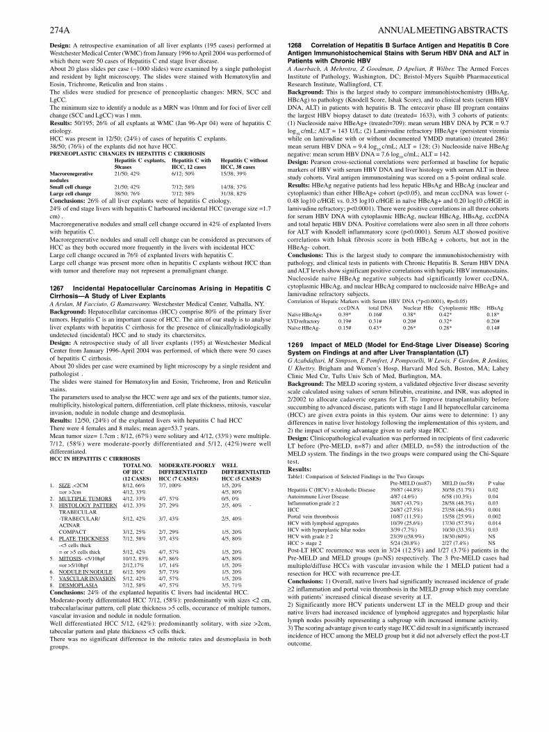
274A ANNUAL MEETING ABSTRACTS
Design: A retrospective examination of all liver explants (195 cases) performed atWestchester Medical Center (WMC) from January 1996 to April 2004 was performed ofwhich there were 50 cases of Hepatitis C end stage liver disease.About 20 glass slides per case (∼1000 slides) were examined by a single pathologistand resident by light microscopy. The slides were stained with Hematoxylin andEosin, Trichrome, Reticulin and Iron stains .The slides were studied for presence of preneoplastic changes: MRN, SCC andLgCC.The minimum size to identify a nodule as a MRN was 10mm and for foci of liver cellchange (SCC and LgCC) was 1 mm.Results: 50/195; 26% of all explants at WMC (Jan 96-Apr 04) were of hepatitis Cetiology.HCC was present in 12/50; (24%) of cases of hepatitis C explants.38/50; (76%) of the explants did not have HCC.PRENEOPLASTIC CHANGES IN HEPATITIS C CIRRHOSIS
Hepatitis C explants, Hepatitis C with Hepatitis C without50cases HCC, 12 cases HCC, 38 cases
Macrorenegerative 21/50; 42% 6/12; 50% 15/38; 39%nodulesSmall cell change 21/50; 42% 7/12; 58% 14/38; 37%Large cell change 38/50; 76% 7/12; 58% 31/38; 82%Conclusions: 26% of all liver explants were of hepatitis C etiology.24% of end stage livers with hepatitis C harboured incidental HCC (average size =1.7cm) .Macroregenerative nodules and small cell change occured in 42% of explanted liverswith hepatitis C.Macroregenerative nodules and small cell change can be considered as precursors ofHCC as they both occured more frequently in the livers with incidental HCCLarge cell change occured in 76% of explanted livers with hepatitis C.Large cell change was present more often in hepatitis C explants without HCC thanwith tumor and therefore may not represent a premalignant change.
1267 Incidental Hepatocellular Carcinomas Arising in Hepatitis CCirrhosis—A Study of Liver ExplantsA Arslan, M Facciuto, G Ramaswamy. Westchester Medical Center, Valhalla, NY.Background: Hepatocellular carcinomas (HCC) comprise 80% of the primary livertumors. Hepatitis C is an important cause of HCC. The aim of our study is to analyseliver explants with hepatitis C cirrhosis for the presence of clinically/radiologicallyundetected (incidental) HCC and to study its charctersitcs.Design: A retrospective study of all liver explants (195) at Westchester MedicalCenter from January 1996-April 2004 was performed, of which there were 50 casesof hepatitis C cirrhosis.About 20 slides per case were examined by light microscopy by a single resident andpathologist .The slides were stained for Hematoxylin and Eosin, Trichrome, Iron and Reticulinstains.The parameters used to analyse the HCC were age and sex of the patients, tumor size,multiplicity, histological pattern, differentiation, cell plate thickness, mitosis, vascularinvasion, nodule in nodule change and desmoplasia.Results: 12/50, (24%) of the explanted livers with hepatitis C had HCCThere were 4 females and 8 males; mean age=53.7 years.Mean tumor size= 1.7cm ; 8/12, (67%) were solitary and 4/12, (33%) were multiple.7/12, (58%) were moderate-poorly differentiated and 5/12, (42%)were welldifferentiated.HCC IN HEPATITIS C CIRRHOSIS
TOTAL NO. MODERATE-POORLY WELLOF HCC DIFFERENTIATED DIFFERENTIATED(12 CASES) HCC (7 CASES) HCC (5 CASES)
1. SIZE ,<2CM 8/12, 66% 7/7, 100% 1/5, 20%=or >2cm 4/12, 33% 4/5, 80%
2. MULTIPLE TUMORS 4/12, 33% 4/7, 57% 0/5, 0%3. HISTOLOGY PATTERN 4/12, 33% 2/7, 29% 2/5, 40% -
TRABECULAR-TRABECULAR/ 5/12, 42% 3/7, 43% 2/5, 40%ACINARCOMPACT 3/12, 25% 2/7, 29% 1/5, 20%
4. PLATE THICKNESS 7/12, 58% 3/7, 43% 4/5, 80%-<5 cells thick= or >5 cells thick 5/12, 42% 4/7, 57% 1/5, 20%
5. MITOSIS- <5/10hpf 10/12, 83% 6/7, 86% 4/5, 80%=or >5/10hpf 2/12,17% 1/7, 14% 1/5, 20%
6. NODULE IN NODULE 6/12, 50% 5/7, 73% 1/5, 20%7. VASCULAR INVASION 5/12, 42% 4/7, 57% 1/5, 20%8. DESMOPLASIA 7/12, 58% 4/7, 57% 3/5, 71%Conclusions: 24% of the explanted hepatitis C livers had incidental HCC.Moderate-poorly differentiated HCC 7/12, (58%): predominantly with sizes <2 cm,trabecular/acinar pattern, cell plate thickness >5 cells, occurance of multiple tumors,vascular invasion and nodule in nodule formation.Well differentiated HCC 5/12, (42%): predominantly solitary, with size >2cm,tabecular pattern and plate thickness <5 cells thick.There was no significant difference in the mitotic rates and desmoplasia in bothgroups.
1268 Correlation of Hepatitis B Surface Antigen and Hepatitis B CoreAntigen Immunohistochemical Stains with Serum HBV DNA and ALT inPatients with Chronic HBVA Auerbach, A Mehrotra, Z Goodman, D Apelian, R Wilber. The Armed ForcesInstitute of Pathology, Washington, DC; Bristol-Myers Squibb PharmaceuticalResearch Institute, Wallingford, CT.Background: This is the largest study to compare immunohistochemistry (HBsAg,HBcAg) to pathology (Knodell Score, Ishak Score), and to clinical tests (serum HBVDNA, ALT) in patients with hepatitis B. The entecavir phase III program containsthe largest HBV biopsy dataset to date (treated= 1633), with 3 cohorts of patients:(1) Nucleoside naive HBeAg+ (treated=709): mean serum HBV DNA by PCR = 9.7log
10 c/mL; ALT = 143 U/L; (2) Lamivudine refractory HBeAg+ (persistent viremia
while on lamivudine with or without documented YMDD mutation) (treated 286):mean serum HBV DNA = 9.4 log
10 c/mL; ALT = 128; (3) Nucleoside naive HBeAg
negative: mean serum HBV DNA = 7.6 log10
c/mL; ALT = 142.Design: Pearson cross-sectional correlations were performed at baseline for hepaticmarkers of HBV with serum HBV DNA and liver histology with serum ALT in threestudy cohorts. Viral antigen immunostaining was scored on a 5-point ordinal scale.Results: HBeAg negative patients had less hepatic HBsAg and HBcAg (nuclear andcytoplasmic) than either HBeAg+ cohort (p<0.05), and mean cccDNA was lower (-0.48 log10 c/HGE vs. 0.35 log10 c/HGE in naïve HBeAg+ and 0.20 log10 c/HGE inlamivudine refractory; p<0.0001). There were positive correlations in all three cohortsfor serum HBV DNA with cytoplasmic HBcAg, nuclear HBcAg, HBsAg, cccDNAand total hepatic HBV DNA. Positive correlations were also seen in all three cohortsfor ALT with Knodell inflammatory score (p<0.0001). Serum ALT showed positivecorrelations with Ishak fibrosis score in both HBeAg + cohorts, but not in theHBeAg- cohort.Conclusions: This is the largest study to compare the immunohistochemistry withpathology, and clinical tests in patients with Chronic Hepatitis B. Serum HBV DNAand ALT levels show significant positive correlations with hepatic HBV immunostains.Nucleoside naive HBeAg negative subjects had significantly lower cccDNA,cytoplasmic HBcAg, and nuclear HBcAg compared to nucleoside naive HBeAg+ andlamivudine refractory subjects.Correlation of Hepatic Markers with Serum HBV DNA (*p<0.0001), #p<0.05)
cccDNA total DNA Nuclear HBc Cytoplasmic HBc HBsAgNaïve HBeAg+ 0.39* 0.16# 0.38* 0.42* 0.18*LVD refractory 0.19# 0.31# 0.20# 0.32* 0.20#Naïve HBeAg- 0.15# 0.43* 0.26* 0.28* 0.14#
1269 Impact of MELD (Model for End-Stage Liver Disease) ScoringSystem on Findings at and after Liver Transplantation (LT)G Azabdaftari, M Simpson, E Pomfret, J Pomposelli, W Lewis, F Gordon, R Jenkins,U Khettry. Brigham and Women’s Hosp, Harvard Med Sch, Boston, MA; LaheyClinic Med Ctr, Tufts Univ Sch of Med, Burlington, MA.Background: The MELD scoring system, a validated objective liver disease severityscale calculated using values of serum bilirubin, creatinine, and INR, was adopted in2/2002 to allocate cadaveric organs for LT. To improve transplantability beforesuccumbing to advanced disease, patients with stage I and II hepatocellular carcinoma(HCC) are given extra points in this system. Our aims were to determine: 1) anydifferences in native liver histology following the implementation of this system, and2) the impact of scoring advantage given to early stage HCC.Design: Clinicopathological evaluation was performed in recipients of first cadavericLT before (Pre-MELD, n=87) and after (MELD, n=58) the introduction of theMELD system. The findings in the two groups were compared using the Chi-Squaretest.Results:Table1: Comparison of Selected Findings in the Two Groups
Pre-MELD (n=87) MELD (n=58) P valueHepatitis C (HCV) ± Alcoholic Disease 39/87 (44.8%) 30/58 (51.7%) 0.02Autoimmune Liver Disease 4/87 (4.6%) 6/58 (10.3%) 0.04Inflammation grade ≥ 2 38/87 (43.7%) 28/58 (48.3%) 0.03HCC 24/87 (27.5%) 27/58 (46.5%) 0.001Portal vein thrombosis 10/87 (11.5%) 15/58 (25.9%) 0.002HCV with lymphoid aggregates 10/39 (25.6%) 17/30 (57.5%) 0.014HCV with hyperplastic hilar nodes 3/39 (7.7%) 10/30 (33.3%) 0.03HCV with grade ≥ 2 23/39 ((58.9%) 18/30 (60%) NSHCC > stage 2 5/24 (20.8%) 2/27 (7.4%) NSPost-LT HCC recurrence was seen in 3/24 (12.5%) and 1/27 (3.7%) patients in thePre-MELD and MELD groups (p=NS) respectively. The 3 Pre-MELD cases hadmultiple/diffuse HCCs with vascular invasion while the 1 MELD patient had aresection for HCC with recurrence pre-LT.Conclusions: 1) Overall, native livers had significantly increased incidence of grade≥2 inflammation and portal vein thrombosis in the MELD group which may correlatewith patients’ increased clinical disease severity at LT.2) Significantly more HCV patients underwent LT in the MELD group and theirnative livers had increased incidence of lymphoid aggregates and hyperplastic hilarlymph nodes possibly representing a subgroup with increased immune activity.3) The scoring advantage given to early stage HCC did result in a significantly increasedincidence of HCC among the MELD group but it did not adversely effect the post-LToutcome.

ANNUAL MEETING ABSTRACTS 275A
1270 Two Major Types of Intraductal Papillary Mucinous Neoplasm (IPMN)of the Pancreas: Their Distinct Histological and Mucin Phenotypic FeaturesS Ban, Y Naitoh, T Sakurai, M Kuroda, M Shimizu. Saitama Medical School, Iruma,Saitama, Japan; Kyoto University Hospital, Kyoto, Japan; Fujita Health UniversitySchool of Medicine, Toyoake, Aichi, Japan.Background: IPMNs have been recognized as a distinct clinicopathological entityfor the last two decades. Some histologic variations are observed in the type ofepithelium of IPMNs, and several kinds of categorization of IPMNs have beenproposed by different researchers. However, this issue has not been fully determineduntil now.Design: Sixty cases of surgically resected IPMNs from three institutions between1983 and 2003 were included in this study. Histological assessment and mucinimmunohistochemistry for MUC1, MUC2, MUC5AC, and MUC6 were performedfor each lesion to establish a major categorization of IPMNs. The results ofimmunohistochemistry were evaluated semi-quantitatively (score 0 to 5).Results: Two major types of IPMNs were noted on the basis of the histological featuresof epithelium, one showing a feature similar to gastric foveolar epithelium (Gastrictype, 38 cases) and the other with a feature similar to that of colorectal villous adenoma(Intestinal type, 15 cases). Other minor types included 2 cases with both Gastric typeand Intestinal type areas, and 5 cases showing an oncocytic feature. These two majortypes were distinct in several respects. Compared to the Gastric type, the Intestinaltype showed the following features: 1) they mostly expressed MUC2 as well as MUC5AC(mean score of MUC2: Intestinal type 4.20±1.37 vs. Gastric type 0.39±1.05, p<0.05);2) they more frequently involved the main pancreatic duct, causing severe atrophy ofthe surrounding pancreatic tissue (p<0.05); 3) they had a higher histologic grade(p<0.05); 4) they were frequently associated with nodular growth (p<0.05) and mucouslake formation (p<0.05); 5) they were less frequently associated with pyloric gland-like structures (p<0.05) and PanIN-like complex lesions (p<0.05); and 6) they wereaccompanied with mucinous carcinoma (2 cases).Conclusions: Our results indicate that the classification of IPMNs into the Gastrictype and Intestinal type is regarded as a reasonable major categorization from thehistological and mucin phenotypical point of view. This categorization suggests thatat least two different forms of histogenesis and behavior are present in IPMNs.
1271 Intraductal and Papillary Variants of Pancreatic Acinar CellCarcinoma: A New Addition to the Challenging Differential Diagnosis ofIntraductal NeoplasmsO Basturk, G Zamboni, D Klimstra, A Andea, NS Kamel, P Capelli, JD Cheng, NVAdsay. Wayne State University, MI; Verona University, Italy; Memorial SloanKettering Cancer Center, NY.Background: The recognition and differential diagnosis of pancreatic intraductalneoplasms (IN) have gained importance in the past few years, as the incidence ofthese tumors (especially IPMNs) have risen to >10% of pancreatic resections, andtheir significance as precursors of invasive cancer is better appreciated. Acinar cellcarcinomas (ACCs) are typically solid tumors; however, we have recently encounteredseveral examples that fall in the differential diagnosis of IN.Design: 7 ACCs with either intraductal growth and/or a papillary/papillocysticpattern that could be mistaken for IN were identified in the authors’ files, and theirclinicopathologic features were studied.Results: Clinical: M/F=4/3; mean age=59; mean tm size= 4.8 cm (as opposed to 10cm in conventional ACCs). Only 1 patient had metastasis at the time of diagnosis (asopposed to 50% in usual ACCs). Pathology: In 5 cases, the tumors had nodulargrowth of sheet-forming acinar cells, some of which were within ducts, as evidencedby the polypoid nature of the process, partial ductal lining, and presence of smalltributary ducts in the walls. In 3 cases, the tumor had papillary and/or papillocysticpatterns, at least focally. All had cystic dilatation of the ducts. No mucin was identified.All expressed trypsin. Markers of ductal differentiation were either absent or veryfocal. A minor endocrine component was present in 3. The main histologic findingsthat distinguished these tumors from IPMNs were: more compact nature of thenodules (rather than villous or arborizing papillae); cuboidal cells; prominent nucleoli,overall basophilia of the cytoplasm, apical granules; intraluminal crystals or pale,acidophilic secretions (enzymatic condensations); and lack of mucin.Conclusions: Some ACCs show intraductal growth or exhibit papillary patterns,which can mimic IN, especially IPMNs. In such cases, attention to morphologicdetails described above, and immunohistochemistry are helpful. An endocrinecomponent was present in 3/7 cases. The tumors were relatively small and, metastasisat presentation was less common than typically seen in ACCs (1/7 vs 50%).
1272 Pathologic Findings in Gallbladders Resected during MorbidObesity OperationsO Basturk, G Altinok, L Budev, A Hildebrandt, JD Cheng, NV Adsay. Wayne StateUniversity, MI.Background: Pathologic changes in the gallbladder (GB) are highly population-dependent. The differential findings aid in understanding the pathogenesis of gallbladderdisease and potentially, in developing preventive strategies. Elective cholecystectomiesperformed during bypass or banding operations for morbid obesity help exposeanother population that has not been studied previously.Design: 58 elective cholecystectomies performed during bypass or banding operationswere reviewed. 30 of these were obtained after the initiation of this study, and fromthese, 3 blocks each containing 3 different sections were submitted. In the remaining28, only one block per case, each with 3 sections was available. Pathology reportswere reviewed for stones or any other macroscopic findings. The slides were evaluatedfor chronic changes (inflammation/fibrosis), cholesterolosis, metaplasia (pyloric andintestinal), Aschoff-Rokitansky (A-R) sinuses, and dysplasia. Each parameter wasgraded none, minimal, mild, moderate or marked. The findings were contrasted with theranges presented in the literature for other risk groups.
Results: Most patients were female (55/58) and the mean age was 40 (range, 26-58).Cholelithiasis was very common (87.9% vs 10-40%). Chronic changes (inflammation/fibrosis) of mild or higher degree were seen in 72% (vs 90% of gallstone cases). A-Rsinuses, however, were prominent in only 10% of the cases. Cholesterolosis wasdetected in 34% (vs 8-39%); adenomyomatous changes in 3% (vs 0.6-8.7%); pyloric-type metaplasia in 55% (vs 50-75%); intestinal metaplasia in 9% (2-25%). Dysplasticchanges were seen in 13.7% (vs 3.3 in Australia and 13-16% in cancer endemic areassuch as Chili and Mexico). One patient had porcelain gallbladder.Conclusions: Morbidly obese patients undergoing elective cholecystectomy form arelevant group in which to study GB pathology. Not surprisingly, the frequency ofcholelithiasis is very high (87%) in this group, as compared to other risk groups(highest reported, 40%). Most appear to be cholesterol stones, as expected. Some ofthe chronic changes associated with gallstones such as A-R sinuses, however, maynot be as high as anticipated, possibly because of the relatively younger age group.The frequency of dysplasia noted in this group may be as high as the incidence reportedin cancer-endemic areas; however, such comparison is difficult because of the possibledifferences in the criteria applied.
1273 The Immunohistochemical Characterization of EGFR Expression inPancreatic Adenocarcinoma- A Tissue Microarray Based StudyA Bhardwaj, JW Nash, C Barbacioru, WM Smith, WL Frankel. Ohio State University,Columbus, OH.Background: Over-expression of epidermal growth factor receptor (EGFR) hasbeen demonstrated in pancreatic carcinoma. Therapies targeted at EGFR have beenshown to inhibit the growth of pancreatic carcinoma either alone or in combinationwith chemotherapy and radiation. We evaluated EGFR expression in pancreatic tissuemicroarrays. Additionally, we studied the correlation between EGFR expression andtumor size, grade and lymph node status in the patients with pancreaticadenocarcinoma.Design: Seventy-two cases of pancreatic adenocarcinoma and 18 cases of chronicpancreatitis were retrieved from the archival files. Carcinomas were graded as well,moderately, or poorly differentiated using WHO criteria and the tumor size andlymph node status (positive or negative) were noted in each tumor case. Tissue coresfrom formalin-fixed, paraffin embedded donor blocks (2 cores per block) were arrayed tocreate a tissue microarray of cores measuring 2.0 mm each. Sections were stained withEGFR (Dakocytomation California, Inc) and 2 pathologists determined intensity ofmembranous staining and percentage of tumor cells showing immunoreactivity. Stainingwas considered positive in cases with greater than or equal to 1% membranous staining.Positive and negative controls stained appropriately.Results: EGFR expression was present in 48 of 72 (67%) cases of pancreaticadenocarcinoma and 7 of 18 (39%) cases of chronic pancreatitis. We did not find astatistically significant correlation between the intensity or extent of EGFR expressionand tumor size, tumor grade or the lymph node status of the patients.Conclusions: EGFR targeted therapies may be useful for the treatment of advancedpancreatic carcinoma, and the immunohistochemical analysis of EGFR staining mayhave a role in identifying pancreatic cancer patients who can benefit from EGFRinhibitors. EGFR expression did not correlate with the commonly used prognosticindicators for pancreatic carcinoma that were evaluated in this study. Long termfollow-up is necessary to determine if EGFR expression correlates with patientoutcome and whether patients with tumors expressing EGFR benefit from EGFRinhibitors.
1274 Comparison of Cytokeratin 7 and 20 Staining in Whole TissueBlock and Mock-Radiologically Obtained Needle Cores Biopsies: APotential Cause of False-Positive Results Due to Limited Tissue SamplesD Bosler, NS Goldstein. William Beaumont Hospital, Royal Oak, MI.Background: Radiologically obtained thin-needle core biopsies are frequently usedto evaluate the primary site of metastatic adenocarcinoma. Cytokeratin 7 and 20 aretwo antibodies that are commonly used for this purpose. Most authors have used 1%- 10% staining cut-point for a positive result. We evaluated the effect of limited tissuesamples on CK 7/20 positive/ negative results by comparing staining of metastaticcolorectal adenocarcinoma in whole tissue blocks and simulated needle core biopsyspecimens.Design: One tissue block with ample adenocarcinoma from 25 metastatic colorectaladenocarcinoma hepatic resection specimens was stained with CK 7 and CK 20.Extent of staining was scored 0%, 1= 1%-5%, 2= 6%-25%, 3= 26%-50%, 4= 51%-75%,5= >75%. Two paper cut-outs (6 mm and 8 mm) of radiologic needle corebiopsies were overlaid over the slide and the proportion of stained cells in the mock-needle core was evaluated. Length of viable adenocarcinoma in each mock needle corewas recorded.Results: The mean percentage of CK 7/CK 20 staining in whole tissue blocks was1.0/ 4.2. [CK 7 (-)/ CK 20 (+)]. For CK 7, mean staining in the mock needle coresincreased 1.2 over the corresponding whole block staining in 13 cases (52%), shiftingthem from CK 7 (-) to CK 7 (+). For CK 20, staining in needle cores decreased a meanof 1.7 in 11 cases. The mean length of viable, non-necrotic adenocarcinoma in themock needle cores was 1.2 mm (range, 0.6 mm 2.9 mm).Conclusions: The limited tissue in needle cores shifted the proportion of CK 7 stainedcells sufficiently to reclassify 52% of the cases from CK 7 (-) to CK 7 (+) which wouldalter the differential diagnosis list of primary sites to include pancreaticobiliary andgastric as more likely sites than the colon. No CK 7 (-) to (+) shifts would have occurredif a 25%-50% staining positivity cut-point was used. Positive/ negative staining cut-points should be increased to 25%-50% stained cells in needle core biopsies to avoidfalse-positive interpretations due to limited tissue samples. Focal staining with CK 7

276A ANNUAL MEETING ABSTRACTS
or 20 occurs in most neoplasms. Extent of staining should be interpreted in the contextof the amount of tissue available for evaluation, and higher thresholds should beconsidered for smaller specimens. Scoring the extent of staining in needle core biopsiesis further hampered by the limited length (mean 1.2 mm) of viable adenocarcinoma in theneedle core.
1275 EGFR Expression and Gene Copy Number in FibrolamellarHepatocellular CarcinomaAF Buckley, LJ Burgart, S Kakar. UCSF Medical Center, San Francisco, CA; MayoClinic, Rochester, MN.Background: Increased expression of epidermal growth factor receptor (EGFR), atransmembrane tyrosine kinase, is associated with tumor progression in manycarcinomas. Both small-molecule tyrosine kinase inhibitors and anti-EGFR antibodieshave shown promise in treating some of these tumors. Fibrolamellar hepatocellularcarcinoma (FL-HCC) is a potentially aggressive neoplasm that occurs in young patientswith no history of cirrhosis. This study examines the expression and gene copy numberof EGFR in FL-HCC, with a view to using EGFR antagonists to treat advanced tumors.Design: Formalin-fixed, paraffin-embedded FL-HCC (n=13) sections were stained witha monoclonal antibody against EGFR (Clone 2-18C9) according to manufacturer’sinstructions, using the EGFR pharmDx kit (DAKO, Carpinteria, CA). Cell membranestaining was recorded as absent, 1+ (weak), 2+ (moderate) or 3+ (strong). Forfluorescence in situ hybridization (FISH) analysis, Spectrum Orange- labeled EGFRprobe (Vysis, Downer’s Grove, IL) and Spectrum Green-labeled probe against thecentromeric region of chromosome 7 (CEP 7) were hybridized to 5 µm sections, andcounterstained with 2’-6’-diamidino-2’-phenylindole (DAPI). EGFR and CEP 7signals were counted in 50 tumor nuclei per case as well as 300 normal hepatocytenuclei. The EGFR to CEP 7 signal ratio was calculated for each case.Results: All cases had typical FL-HCC features including polygonal cells with granularcytoplasm, prominent nucleoli and lamellar fibrosis. 12/13 (92%) FL-HCC tumorsstained diffusely positive (3+) with anti-EGFR antibody. Normal hepatocytes werenegative or showed 1+ staining. FISH was informative in 10 cases, all of whichshowed extra EGFR gene copy numbers (mean 3.69; range 3.13-5.0). EGFR wasoverexpressed in all these cases. The mean number of EGFR signals per cell in FL-HCC was double that of normal hepatocytes (3.69 vs. 1.80); the mean EGFR:CEP 7ratio in tumor cells was 1.05.Conclusions: EGFR is strongly overexpressed on the cell membrane in almost all casesof FL-HCC. EGFR gene copy number in FL-HCC is double that of normal hepatocytes.Similar gains are observed in chromosome 7, indicating that the extra EGFR gene copiesare due to aneuploidy rather than gene amplification. The strong expression of EGFRin FL-HCC tumors suggests that they may respond to treatment with EGFR anatagonists.Since FL-HCC arises in young patients with non-cirrhotic livers, a response to treatmentcould have a significant impact on patient survival.
1276 Hepatocellular Carcinoma: Therapeutic Intervention and Outcome -A Pathology PerspectiveN Buza, F Regenstein, S Dash, S Haque. National Institute of Oncology, Budapest,Hungary; Tulane University Health Sciences Center, New Orleans, LA.Background: Hepatocellular carcinoma (HCC) is a fatal disease unless detected andtreated early. Unfortunately only occasional cases are detected early enough forcurative surgical resection, vast majority of tumors are unresectable. Recently localablative (LA) techniques (transarterial chemoembolization , percutaneous ethanolinjection and radiofrequency ablation) are being used in selected cases as an adjuncttherapy preceding resection or orthotopic liver transplantation (OLT) in an attemptat cure or longer survival. The purpose of our study was to correlate therapeuticinterventions (OLT and LA), histologic findings and outcome.Design: From the files of Tulane University Health Sciences Center between 1999and 2004, 37 patients with HCC were identified who had undergone either 1) ablativeprocedure prior to resection or while awaiting OLT and 2) HCC found in explantedlivers after OLT. Two pathologists graded histologic parameters. Review was madeof records.Results: 29 males and 8 females, all with cirrhosis due to: 26 HCV, 1HBV, 1hemochromatosis, 9 steatohepatitis. At time of study 23 patients were alive withoutrecurrent HCC. Two patients treated with ablation alone died soon after therapy.
Resection OLT Ablation + OLT Ablation onlyTotal # of cases 8 15 12 2Age (mean, range) 54 (42-71) 55 (47-66) 52 (47-63) 59 (49-69)# of tumor / case 2 (1->5) 2.1 (1->5) 2.8 (1->5) 5.5 (1-10)(mean, range)Tumor size cm 3.6(0.3-9.0) 2.1(0.2-4.5) 2.8(0.7-8.8) 15.5 (11-20)(mean, range)Histologic grade (mean) 2.4 2.6 2.3 3
VASCULAR INVASION 3 8 5 0(# of cases) AbsentOccasional 3 4 5 0Extensive 2 3 2 2Area of necrosis 14% 18,6% 46% 27,5%(mean, range) (0-85%) (0-90%) (0-100%) (25-30%)OUTCOME Alive 3 12 8 0(# of patients)Survival in mth 14 (9-23) 35.6 (2-68) 25.2 (1-61)(mean, range)DOD (# of patients) 5 3 4 2Survival in mth 12 (0-38) 22 (0-42) 8.25 (2-21) 1.5 (1-2)(mean, range)
Conclusions: In our series of patients 1) OLT with or without ablation appeared to bethe best therapeutic option for HCC. We found that patients treated with OLT hadsignificantly better survival than those treated with resection. 2) Ablation alone didnot improve survival. 3) Long term follow up is needed to see if ablation prior to OLToffers further survival benefits.
1277 Differential Expression of Mucins, MIB-1 and p53 in Mucinous Tumorsof the PancreasG Cai, A Simsir, H Yee, L Chiriboga, P Kefalides, J Cangiarella. New York University- Bellevue Hospital Center, New York, NY; New York University School of Medicine,New York, NY.Background: Mucins are high molecular weight glycoproteins that are produced byvarious epithelial cells including those found in the pancreas. Previous studies havedemonstrated differential expression of mucin subtypes in pancreatic andgastrointestinal neoplasms. Although pancreatic tumors display overt histologicalheterogeneity, many share a common feature, extracellular mucin accumulation. Themucin-producing tumors in the pancreas include ductal adenocarcinoma (DCA),mucinous cystic neoplasm (MCN) and intraductal papillary mucinous tumor (IPMT).The aim of this study is to investigate expression of different subtypes of mucins inpancreatic mucinous tumors, in conjunction with the expression of MIB-1, a proliferationmarker, and p53, a tumor suppressor gene to explore their potential diagnostic value indistinguishing these entities.Design: Thirty-six cases including DCA (n = 6), serous cystadenoma (SCA, n = 8),mucinous cystadenoma (MCA, n = 6), mucinous cystic adenocarcinoma (MCCA, n= 6), IPMT (n = 5) and solid pseudopapillary tumor (SPT, n = 5) were retrieved.Representative foci from each case were combined to make a tissue microarray.Immunohistochemical studies were performed on microarray sections using theantibodies against MUC1, MUC2, MUC5AC, MUC6, MIB-1 and p53. Membranousstaining for mucins, and nuclear staining for MIB-1 and p53 were quantitated as apercentage of immunoreactive neoplastic epithelial cells /total # neoplastic epithelialcells.Results:Expression pattern of mucins, MIB-1 and P53 in mucinous pancreatic neoplasms
MUC1 MUC2 MUC5AC MUC6 MIB-1 P53SCA, n=8 54±28 0 0 57±24 1.5±1.0 0MCA, n=6 17±28 0 58±42 25±27 4.2±4.5 0MCCA, n=6 40±31 6±10 35±31 27±22 30±13 25±29IPMT, n=5 4±10 66±43 54±33 26±36 13±8 0.3±0.6DCA, n=6 91±2 0 15±8 7.5±4.2 37±8 58±12SPT, n=5 0 0 0 0 4.8±3.3 1.4±2.1Conclusions: SCA showed immunostaining for MUC1 and MUC6 but not forMUC2 and MUC5AC. All mucins except MUC2 were expressed in MCN. OnlyIPMT displayed significant immunoreactivity for MUC2. DCA predominantlyexpressed MUC1. No mucins were detected in SPT. MCCA differed from MCA inhigher MIB-1 and p53 immunoreactivity. DCA also showed increased MIB-1 andp53 expression. These results indicate that mucins were expressed differentially inpancreatic neoplasms and combining expression of mucin with MIB-1 and p53 maybe helpful in the differential diagnoses of pancreatic neoplasms.
1278 Expression of Two Novel Tumor Markers, Maspin and Tubulinbeta Polypeptide (TUBB), in Biliary CancersD Cao, K Kassaui, P Argani, C Neumann, L Ho, JL Abbruzzese, M Ouellette, AMaitra. The Johns Hopkins Hospital, Baltimore, MD; MD Anderson Cancer Center,Houston, TX.Background: Biliary tract cancers (BTCs) are uncommon neoplasms, usuallyassociated with a dismal prognosis. Identification of novel tumor markers in BTCs isa prerequisite for the development of effective diagnostic and therapeutic strategies.Maspin, a serine protease inhibitor, is upregulated in pancreatic and breast cancers,and detection of circulating maspin mRNA or protein is reported to be a valuableadjunct for early detection and relapse monitoring protocols. Tubulin beta polypeptide(TUBB) is a cytoskeletal constituent that is also overexpressed in many solid cancers,and is associated with chemoresistance to microtubule stabilizing agents like paclitaxel.A recently developed TUBB immunohistochemical assay permits identification ofTUBB-negative cases that might benefit from paclitaxel therapy. Here, we examinethe expression of maspin and TUBB, two novel tumor markers with diagnostic andtherapeutic potential, respectively, in BTCs.Design: Tissue microarrays (TMAs) were generated from archival specimens of 34BTCs, including 19 gallbladder, 4 intrahepatic, and 11 extrahepatic cancers. Eachcase was represented by 4 cores on the TMAs, to exclude tissue heterogeneity. Bothmaspin and TUBB are expressed in the cytoplasmic compartment, and immunolabelingwas scored as negative (expression in < 5% neoplastic cells), focal (5-25% expression)and diffusely positive (>25% expression).Results: Normal biliary epithelial cores did not label with either maspin or TUBB.Expression of maspin and TUBB in biliary tract cancers stratified by site is summarizedin Table 1.Conclusions: Both maspin and TUBB are frequently overexpressed in BTCs,although there are site-specific variations. Maspin overexpression supports the useof circulating maspin mRNA or protein as a diagnostic tool in BTCs. TUBBoverexpression suggests a possible explanation for resistance to microtubule-stabilizingagents in these cancers, and immunolabeling for TUBB can be used as guide forselection of patients who might benefit from this form of therapy.

ANNUAL MEETING ABSTRACTS 277A
Site Maspin TUBBNegative Focally + Diffusely + Negative Focally + Diffusely +
Gallbladder 6(32%) 1(5%) 12 (63%) 5 (26%) 4 (21%) 10(53%) (N =19)Intrahepatic 0 0 4(100%) 0 1(25%) 3(75%)bile duct (N=4)Extrahepatic 1(9%) 0 10(91%) 0 2(18%) 9(82%)bile duct (N=11)Total 7 (21%) 1(3%) 26 (76%) 5(15%) 7(21%) 22(64%)(N =34)
1279 Ultrasensitive Detection of KRAS Mutations in Bile and Serum fromPatients with Biliary Cancer Using LigAmp TechnologyA Chandrasekharan, C Shi, PJ Thuluvath, II Wistuba, CA Karikari, P Argani, MGGoggins, JR Eshleman, A Maitra. Johns Hopkins University, Baltimore, MD; UTMD Anderson Cancer Center, Houston, TX.Background: Biliary cancer is a lethal disease, and early detection efforts are neededto ameliorate the dismal prognosis. Mutations of the KRAS gene, specifically atcodon 12, are one of the most common genetic aberrations in this cancer. An ultra-sensitive technology – LigAmp - has been described (Shi et al, Nature Methods, 2004)for the detection of single base pair mutations in clinical samples. LigAmp has asensitivity of detecting a mutant population with a sensitivity of 1:10,000 wild-typecells. We utilized LigAmp to detect KRASG12D mutations in patients with a variety ofneoplastic and non-neoplastic biliary diseases.Design: In LigAmp, a mutation specific 5’ oligonucleotide and a generic 3’oligonucleotide (both tagged with M13 “tails”) are ligated using Pfu ligase, followedby amplification using M13 primers. The 5’ oligonucleotide also has an upstreamunrelated bacterial gene sequence (e.g., lacZ), and a specific flurophor-labeled probeto the latter can be utilized to generate cycle threshold (Ct) values for the mutantDNA of interest in the sample. Serially diluted positive control and negative controlcell lines in each run provide relative quantification of mutant KRAS levels.Oligonucleotides specific to the KRASG12D mutation were designed. DNA was extractedfrom 119 samples, including 10 biliary cancer xenografts, 54 archival biliary cancers,44 bile samples, and 11 serum samples. Of the 44 bile samples, 16 were from patientswith biliary cancers, and 28 from a variety of non-neoplastic pancreato-biliarydisorders; all 11 serum samples were from patients with biliary cancer.Results: KRASG12D mutations were detected in 10/10 (100%) biliary xenografts and52/54 (96%) archival cancers. 13/16 (81%) neoplastic bile samples and only 6/28(21%) non-neoplastic bile samples harbored mutant KRAS DNA (P=0.0003); thelatter included chronic pancreatitis and primary sclerosing cholangitis, both conditionswhere this mutation has been reported. KRASG12D mutations were also detected in 6/11 (55%) serum samples from biliary cancer patients.Conclusions: KRASG12D mutations are present in the majority of biliary cancers, andare detectable in bile and serum using LigAmp. This technology has the potential forearly detection of biliary cancer as well as for disease monitoring post-therapy.
1280 Under-Expression of JUNB Gene in Hepatocellular CarcinomaJH Chang, YS Chang, KT Yeh, JG Chang. Chang-Hua Christian Hospital, Chang-Hua, Taiwan; China Medical College, Taichung, Taiwan.Background: JUNB is a major component of Activator Protein-1 (AP-1)transcriptional factor complex, which consists of JUN (JUNB, C-JUN, and JUND),FOS (C-FOS, FOSB, FRA-1and FRA-2), and ATF families. The JUNB gene is animmediate early transcriptional factor, which can be activated by polypeptide growthfactors, cytokines and various chemical agents in a variety of cell types (includingmyeloid, lymphoid, liver, neuron, fibroblast, and epidermal lineages). JUNB has alsobeen implicated in the activation of P16 gene expression during cell cycle control andcell senescence. Targeted disruption of the JUNB locus in mice caused early embryonicdemise. Promoter methylation leading to JUNB under-expression has been reportedin chronic myelocytic leukemia. This study is to determine the molecular profile ofJUNB in human HCC and explore its role in the tumorigenesis of HCC.Design: Thirty cases of resected primary hepatocellular carcinoma and paired non-cancerous tissue were collected. The molecular profile of JUNB was analyzed byreal-time quantitative reverse transcription-polymerase chain reaction (RT-PCR),mutational analysis and methylation specific PCR. Immunohistochemical study forJUNB, P16 and cyclin D1 expression was accompanied.Results: RT-PCR revealed a significant down-regulation of JUNB in all HCC cases(p<0.001). The mutational analysis demonstrated no mutation in the coding region inany of the HCC. Methylation specific PCR disclosed no promoter methylation ofJUNB. Immunohistochemical study showed under-expression of JUNB in most ofHCC, correlated well with decreased expression of P16, but was inversely proportionalto the expression of cyclin D1.Conclusions: Down-regulation of JUNB in HCC was not caused by genomic mutationor epigenetic silencing (promoter methylation). The under-expression of JUNBcoincided with under-expression of P16. The mechanisms of under-expression ofJUNB and its potential role in the development of HCC remain to be explored.
1281 Identification of a Gene Expression Signature That DifferentiatesHepatocellular Adenoma from Well Differentiated HepatocellularCarcinomaZE Chen, KG Crone, MA Watson, JD Pfeifer, HL Wang. Washington University, St.Louis, MO.Background: It is often difficult to distinguish hepatocellular adenoma (HCA) fromwell differentiated hepatocellular carcinoma (WDHCC) when limited needle biopsytissue is microscopically evaluated. The aim of this study was to identify differentiatinggene expression patterns that may lead to the discovery of useful diagnostic markers.
Design: Gene expression profile analysis using Affymetrix U133Plus2 GeneChipmicroarrays was performed on 6 HCA and 8 WDHCC specimens. The SignificanceAnalysis of Microarrays (SAM) algorithm was utilized to identify genes whoseexpression differed significantly between these two types of tumors. Patterns of geneexpression were validated by quantitative RT-PCR on an independent set of 9 HCA and9 HCC RNAs. Immunohistochemistry was performed on additional 28 HCAs and 32HCCs using commercially available antibodies against 5 proteins.Results: We identified 79 genes whose expression levels were significantly differentbetween HCA and WDHCC. These included 73 genes overexpressed by HCAs and6 overexpressed by WDHCCs. The identity of 8 genes chosen for further analysis isshown in the Table. By RT-PCR, 7 of them (with the exception of PRX1) demonstratedconcordant average expression differences between HCAs and HCCs. The expressionpattern of these genes correctly classified an independent set of 18 tumors usingunsupervised cluster analysis. Immunostains confirmed that HCAs expressedsignificantly higher levels of IGF-II, TMOD1, CLU and ER proteins and a lowerlevel of PCNA protein comparing to HCCs.Gene (Abbreviation) Microarray* RT-PCR*Insulin-like growth factor-II (IGF-II) 17.0 32.8 (P<0.0001)Tropomodulin 1 (TMOD1) 9.5 1.7 (P=0.0493)Hsp40 homolog, member C1 (DNAJ) 8.7 4.5 (P<0.0001)Clusterin (CLU) 4.9 3.6 (P<0.0001)Estrogen receptor (ER) 3.8 2.9 (P<0.0001)Paired related homeobox 1 (PRX1) -4.0 2.2 (P=0.0899)Epithelial V-like antigen 1 (EVA1) -2.4 -2.0 (P=0.0332)Proliferating cell nuclear antigen (PCNA) -2.3 -1.7 (P=0.0151)*Fold change comparing HCA with HCC.Conclusions: Our results demonstrate significant molecular differences betweenHCA and WDHCC, despite morphological similarities. Furthermore, we haveidentified a unique set of genes whose expression pattern can differentiate betweenthese two entities, suggesting the possibility of future development of ancillarymolecular or immunohistochemical diagnostic methods.
1282 Plasma Cell Infiltrate May Predict the Severity of Acute CellularRejection in Liver AllograftsW Chu, PE Swanson, MP Upton, MM Yeh. University of Washington Medical Center,Seattle, WA.Background: Acute cellular rejection is one of the most common complications inallograft livers. Multiple episodes of acute cellular rejection may lead to chronicrejection and eventual graft failure. Typical inflammatory cells in acute cellular liverallograft rejection include a mixture of lymphocytes, neutrophils, and eosinophils.Increased plasma cells have been observed in renal allografts (plasma cell-rich allograftrejection) and are associated with poor graft survival, but it is not known if theamount of plasma cells are associated with the severity of acute cellular rejection inallograft livers.Design: We studied 61 cases of acute cellular rejection from 2000 to 2004, andcompared plasma cell numbers in the portal regions in four groups, including focalmild, mild, moderate, and severe acute cellular rejection. Histopathologic grading ofacute cellular rejection is based on Banff criteria. In order to standardize the assessmentof plasma cells in portal areas with variable intensities of inflammation, the number ofplasma cells as a percentage of total inflammatory infiltrate was calculated: totalnumber of plasma cells and total number of portal inflammatory cells includingmononuclear cells, neutrophils and eosinophils were serially counted in the 5 mostcellular portal areas; the mean percentage of plasma cells from those 5 portal areas isshown in table.Results: The percentage of plasma cells in portal infiltrate increases with severity ofrejection. Most cases of focal mild rejection do not contain a single plasma cell in theportal infiltrate (0-2.5%), while all severe rejection cases have plasma cells (2-30%).The difference in average percentage of plasma cells in the portal areas among the fourgroups is statistically significant by Anova analysis (p<0.0001). Plasma cell infiltrationcan occur either early (9 days post OLT) or late (3 years post OLT).Conclusions: Plasma cells are seen in acute cellular rejection of liver allografts, and therelative number of plasma cells increase when the severity of rejection increases. Thisfinding suggests an important role for plasma cells in the immunological response ofliver allograft rejection, and indicates that their presence in the portal areas may be areliable histologic indicator for more severe acute cellular rejection.Mean percentage of plasma cells in portal infiltrate of liver allograft rejectionGroups Mean PercentageFocal mild (n=15) 0.33Mild (n=25) 0.79Moderate (n=17) 5.56Severe (n=4) 14.00P<0.0001 by ANOVA
1283 Analysis of Molecular Alterations and Differentiation Pathwaysin Intraductal Oncocytic Papillary Neoplasm of the PancreasSM Chung, RH Hruban, C Iacobuzio-Donahue, NV Adsay, SY Zee, DS Klimstra.Memorial Sloan-Kettering Cancer Center, New York, NY; Johns Hopkins UniverstiyHospitals, Baltimore, MD; Karmanos Cancer Institute and Wayne State University,Detroit, MI; Albert Einstein School of Medicine, New York, NY.Background: Intraductal oncocytic papillary neoplasms (IOPNs) and intraductalpapillary mucinous neoplasms (IPMNs) of the pancreas share features such asintraductal papillary growth, mucin secretion, and indolent behavior compared topancreatic ductal adenocarcinoma (DA). IPMNs have different morphologic typesof papillae, including gastric (IPMN-G), intestinal (IPMN-I) and pancreatobiliary(IPMN-PB). However, IOPNs are distinct, with granular, oncocytic cytoplasmcontaining intraepithelial lumina and cytoplasmic mucin vacuoles. The molecular anddifferentiation patterns of IOPNs have not been well studied.

278A ANNUAL MEETING ABSTRACTS
Design: Immunohistochemical stains were performed (Table 1) on 19 IOPNs and 7IPMN-G, 7 IPMN-I and 6 IPMN-PB. Ligase detection reaction was used to detectmutations in codons 12 and 13 of the K-ras gene in 8 IOPNs and 26 IPMNs.Results:Table 1: # of cases and staining resultStain IOPNs (19) IPMN-G (7) IPMN-I (7) IPMN-PB (6)p53¹ 4 P 0 P 2 P 3 Pp16² 12 A 5 A 5 A 3 ASmad4² 0 A 0 A 0 A 0 Aß catenin¹ 0 P 0 P 0 P 0 Pmesothelin¹ 16 P 0 P 1 P, 1 FP 3 Pclaudin4¹ 0 P 1 P 6 P 1 PMUC1¹ 11 P 0 P 1 P 4 PMUC2¹ 4 P 1 P 6 P 1 P, 1 FPHepPar1¹ 12 P 0 P 4 P 1 FPCDX2¹ 2 P 1 P 6 P 1 FP¹ P = >25% staining, FP = 10-25% staining, ² A= absence of staining in neoplasm withexpression in normal tissueK-ras mutation detection: 0/8 IOPNs, 18/26 IPMNs.Conclusions: 21% of IOPNs and 25% of IPMNs showed p53 staining, 78% ofwhich showed high grade dysplasia. No cases showed evidence of Smad4 or ß cateninabnormalities. 63% of IOPNs and 65% of IPMNs lacked p16 staining. Thus IOPNs andIPMNs have similar changes in these genetic markers. However, IOPNs lacked K-rasmutations, unlike IPMNs and DA. In addition, 84% of IOPNs versus 25% of IPMNsexpressed mesothelin. Only IPMN-I consistently expressed claudin4. IOPNs alsoshowed distinct differentiation patterns from IPMNs, such as expression of MUC1, amarker of an aggressive phenotype, and less expression of MUC2, a marker of a moreindolent pathway. Most IOPNs showed no intestinal differentiation (CDX2 negative),in contrast to IPMN-I. HepPar1 staining in IOPNs is interesting but unexplained.Thus, IOPNs and IPMNs are related tumors, but these data support divergentdifferentiation.
1284 Expression of ZO-1 in Pancreatic Intraepithelial Neoplasia (PanIN)T Cibull, S Badve, A Thomas, R Saxena. Indiana University, Indianapolis, IN.Background: Zonula occludens-1 (ZO-1) is a member of the MAGUK (membrane-associated guanylate kinase homologue) protein family found in the cytoplasmicplaques of tight junctions; these are the most apical junctions between adjacent cellsand they divide the cell membrane into apical and baso-lateral domains. ZO-1 playsan important role in organization of proteins within the tight junctional complex andis thus an important marker of polarity. Since increasing grades of epithelial dysplasiaare characterised by loss of polarity, we studied expression of ZO-1 in PanIN toelucidate whether this expression changes with grade of dysplasia.Design: We searched the database of the Department of Pathology to identifyspecimens of Whipple’s resection with adenocarcinoma. The slides from these caseswere examined to identify and classify PanIN lesions by already published criteria(Hruban, 2001). Four micron thick sections were cut and stained for ZO-1 (prediluted,Zymed) by a standard immunohistochemical protocol following digestion byproteinase K for 5 minutes. The staining pattern in the lesions was characterised asapical (dot-like positivity at the apical portion of the cell membrane between adjacentcells) or cytoplasmic (with or concurrent apical staining). The intensity was gradedas 1+ (weak/absent), 2+ or 3+ (positive).Results: There were 19 cases of Whipple’s resection for pancreatic adenocarcinoma(12 men, 7 women; age 40 - 79 years, mean 56 years). There were 11 lesions of PanIN1A, 14 of PanIN 1B, 8 of PanIN 2 and 9 of PanIN 3. The staining pattern is shownin[Table1]: ZO-1 staining in PanINPanIN Grade No of lesions Apical Posivity Cytoplasmic positivityPanIN 1A 11 11 1PanIN 1B 14 14 1PanIN 2 8 6 5PanIN 3 9 4 9Cytoplasmic staining in the 2 PanIN 1 lesions was of 2+ intensity. Cytoplasmicstaining in PanIN2 lesions was 3+ in 4 lesions and 2+ in 1 lesion. Cytoplasmicstaining in PanIN 3 was 3+ in 8 lesions and 2+ in 1 lesion.Conclusions: 1. Apical staining of ZO-1 was maintained in PanIN 1, but was lost inhigher grades of PanIn, being absent in 25% of PanIN 2 and 60% of PanIN 3.2. Cytoplasmic localization of ZO-1 was rare in PanIN1A and PanIN1B but commonin higher grades of PanIN, with 60% incidence in PanIN 2 and 100% incidence inPanIN 3.3. Abnormal localization of ZO-1 to the cytoplasm occurs in high grade PanINs, andmay be helpful in the diagnosis of high grade lesions.Reference: Hruban RH et al. Intraepithelial neoplasia: A new nomenclature andclassification system for pancreatic duct lesions. Am J Surg Pathol 2001; 25: 579.
1285 The Expression of Proxisome Proliferator Activated ReceptorGamma (PPAR γγγγγ) in Ductal Pancreatic Carcinoma: A Correlative Studywith Angiogenic Factors (VEGF and CD 34)Y Dancer, C Heard, R Shi, J Albores-Saavedra. Louisiana State University HealthScience Center, Shreveport, LA.Background: PPARs are ligand-activated transcription factors belonging to the nuclearreceptor superfamily. Ligand activation of PPARγ decreases VEGF production bytumor cells. In ductal pancreatic carcinoma, the VEGF expression and the microvesseldensity (MVD) are closely correlated, and both are markers of prognostic relevance.It has been reported that ligand activation of PPARγ results in growth inhibition anddifferentiation of pancreatic cancer cell lines.Design: In this study, we analyzed PPARγ expression in ductal pancreatic carcinomasand correlated it with tumor size, tumor grade and angiogenetic factors such as VEGFand microvessel density (MVD). Histologic sections from 28 pancreatic ductal
carcinomas were stained with antibodies against PPARγ, VEGF (Santa Cruz, CA) andCD 34 (Ventana). Interpretation of the results was done by three pathologists as follows:The expression of PPARγ was graded as positive when there was more than 5 percentof nuclear staining. The scores of the extent (0-3) and the intensity of staining (0-4)with VEGF antibodies were summed. Intratumoral microvessel density (MVD) wasrecorded by counting CD-34-positive vessels in the highest vascularized area in fourhigh power fields. Cases were analyzed according to PPARγ expression; positive vs.negative groups. Chi -Square and Student T Tests were used for statistical analysis.Results: Of twenty-eight carcinomas, sixteen (57.2%) stained for PPARγ, and showedmoderate to strong nuclear staining. The mean ages were 63 ± 8.39 vs. 64.14 ± 15.37(p>0.05). Tumor sizes were 3.56 ±1.01 vs. 2.64 ±1.00 (p>0.05). Twenty-six (92 %)carcinomas stained for VEGF, and showed moderate to strong diffuse cytoplasmicstaining in tumor cells. All of the PPARγ positive cases showed less VEGF expressionthan tumors with negative PPARγ expression (2.31±0.47 vs. 2.91±1.16, p =0.012).Tumors with positive PPARγ showed significantly less MVD than tumors withnegative PPARγ expression (43.62±13.25 vs. 134.66±47.7, p<0.0001).Conclusions: The expression of PPARγ correlates inversely with expression of VEGFand MVD. There was no significant correlation between PPARγ expression andclinicopathologic data. Our findings suggest that the PPARγ signaling pathway maybe responsible for tumor angiogenesis in human ductal pancreatic carcinoma. Ligandactivation of PPARγ appears to decrease VEGF production by tumor cells and haveboth direct and indirect antiangiogenic effects.
1286 Comparison of PPAR-alpha Immunostaining in Steatohepatitis andNormal Liver BiopsiesRD DeHaan, SO Sanderson. Mayo Clinic, Rochester, MN.Background: Peroxisome proliferator-activated receptor-α (PPARα) is a memberof the nuclear hormone receptor superfamily and is a key transcriptional regulator ofthe genes involved in hepatic β-oxidation of fatty acids (FA). Recently, studies inrodent models have shown that PPARα may play a role in the pathogenesis of non-alcoholic fatty liver disease (NALFD). Mice deficient in PPARα and those deficientin both PPARα and PPARα-regulated FA oxidation enzymes exhibit severe steatosisand steatohepatitis under fasting conditions. Reduced PPARα expression has beendemonstrated in mouse models of NAFLD and administration of PPARα agonists tosuch mice has been shown to both prevent the development of, and reverse existingsteatohepatitis. It remains to be established if abnormalities in PPARα levels contributeto steatohepatitis in humans. In the present study, we compared the staining patternof PPARα in normal liver biopsies to that of liver biopsies showing steatohepatitis.Design: 22 cases of steatohepatitis and 28 cases of normal liver were selected.Formalin-fixed paraffin embedded biopsies were stained with antibodies againstPPARα (Santa Cruz, clone H-98) using a biotin free detection system(DakoCytomation EnVision+ System, HRP). The character and distribution of PPARαstaining was recorded for each case. The presence of steatosis and any associatednecroinflammatory activity was evaluated on H&E stained sections. Medical recordswere reviewed to confirm the clinical diagnosis.Results: All cases showed similar mild, homogeneous, cytoplasmic and nuclear stainingfor PPARα. Additional coarse granular cytoplasmic staining was present in 31 casesin a predominantly zone 3 distribution. 27 of these 31 cases showed no histologicnecroinflammatory activity and absent to mild steatosis. Of the 22 cases with confirmedsteatohepatitis, 18 lacked coarse granular cytoplasmic staining. In regards tosteatohepatitis, the absence of coarse granular cytoplasmic PPARα staining had apositive predictive value of 95%, with an overall sensitivity and specificity of 82%and 96% respectively.Conclusions: In our series, nearly all normal liver biopsies showed coarse cytoplasmicPPARα staining in a predominantly zone 3 distribution. This pattern of staining wastypically not observed in confirmed cases of steatohepatitis. The apparent differenceobserved in PPARα immunostaining may reflect a lower quantity or altered nature ofhuman PPARα in cases of steatohepatitis and may offer some insight into the overallpathogenesis of fatty liver disease.
1287 Demonstration of Metallothionein in Hepatocellular Carcinomasand Hepatocellular Adenomas - A Molecular and ImmunohistochemicalAnalysisCC Gable, K Ghoshal, S Majumder, ST Jacob, ML Prasad, WL Frankel. The OhioState University, Columbus, OH.Background: The metallothioneins (MT) are a family of cysteine-rich, low molecularweight proteins that exist as 4 different, yet highly homologous isoforms within thehuman body. Among their most important roles are those of detoxification of heavymetals and the scavenging of hydroxyl free radicals. MT-I and MT-IIA are the 2isoforms induced in a variety of stresses in all cell types. MT are down-regulated inhepatocellular carcinomas (HCC). We examined the differential expression of MT inHCC and hepatocellular adenomas (HA) at the RNA level and analyzedimmunohistochemical expression to confirm the molecular findings.Design: Ten fresh frozen human HCC and 2 HA and matched controls (adjacent non-tumor liver) were analyzed for expression of MT-I (in HCC cases only) and MT-IIAat the RNA level by RT-PCR with primers specific for MT-IIA and common to allfive variants (MT-I). In addition, 34 HCC and 7 HA were retrieved from the archivalfiles. All cases were reviewed for confirmation of the diagnosis. Formalin-fixed, paraffin-embedded sections were cut and stained with antibodies against MT. MT stainingwas graded 0 to 2+ and the percentage of cells expressing MT in the tumor andadjacent non-tumor liver was scored by two pathologists.Results: Tissue RNA levels of MT-I and MT-IIA were significantly decreased in all10 HCC and MT-IIA was decreased in HA compared to the matched controls.Immunohistochemical analysis showed that of the 27 cases of HCC with adjacent controlliver, 24 (89%) showed a decrease in MT expression (and 10 of these cases showed no

ANNUAL MEETING ABSTRACTS 279A
expression). Of the 7 cases of HCC without matched controls, 6 (86%) showed no MTexpression. Overall, the majority of the adjacent control livers showed 2+ or diffuse 1+expression while HCC showed 0, or focal 1 or 2+ staining with no expression in 16 ofthe 34 cases. Of the 5 HA with adjacent control liver, 4 (80%) displayed a decrease ineither the intensity or percentage of MT expression when compared to the adjacentliver. All but one of the control adjacent livers showed 2+ staining in nearly 100% ofcells while the HA showed 1+ staining or a decreased percentage of cells with 2+staining.Conclusions: The decreased RNA levels of MT-I and MT-IIA in HCC and HAindicate transcriptional downregulation of MTs in the tumors. We confirmed asignificant decrease in MT at the protein level using immunohistochemistry.
1288 Association of E-Cadherin, Matrix Metalloproteinases, and TissueInhibitors of Metalloproteinases in the Progression of HepatocellularCarcinomaZ Gao, M Tretiakova, W Liu, J Hart. University of Calgary, Calgary, AB, Canada;University of Chicago, Chicago, IL.Background: Molecular markers can provide additional information to traditionalhistomorphological evaluation for the assessment of tumor progression and predictingthe likelihood of invasion and metastasis in various types of malignancies. We studiedthe association of E-Cadherin (E-C), matrix metalloproteinases (MMPs), and thetissue inhibitors of metalloproteinases (TIMPs) with the development and progressionof hepatocellular carcinoma.Design: Tissue arrays including 6 normal liver (NL), 13 cirrhotic liver (CL), 39macro-regenerative nodule (MRN), 15 dysplastic nodule (DN), 22 grade Ihepatocellular carcinoma (HCC-I), 42 grade II HCC (HCC-II), 6 grade III HCC(HCC-III) and 9 metastatic HCC (HCC-M) were stained immnuohistochemicallywith antibodies against MMP1, MMP2, MMP3, MMP6, MMP7, MMP9, TIMP1,TIMP2, TIMP3 and E-C. The intensity of staining for each antibody was scoredmanually by two pathologists as well as automatically using the ChromavisionAutomated Cellular Imaging System (ACIS).Results: (1) Compared with NL, CL had lower E-C and TIMP1, but significantlyhigher MMP3 and MMP7; MRN had lower E-C and MMP6; DN had significantlyhigher MMP3 and TIMP2; (2) The histomorphological progression from HCC-I toHCC-III is associated with gradual decrease of MMP2, MMP3, MMP7, E-C, TIMP2and TIMP3, but gradual increase of MMP9; (3) HCC-M is associated with significantlylower tissue expression of MMP2, MMP3, TIMP1, 2, 3 and E-C.Conclusions: E-Cadherin over expression and lower level of MMPs in HCC-I andHCC-II explains the rarity of extrahepatic metastasis in low grade HCC. MMPs 2, 3,7 and E-C can be used as markers for tumor progression. TIMP1, 2, 3, E-C andMMP9 are markers useful in predicting metastatic potential.
1289 C-Kit Expression in Hepatoblastoma - Evidence of Stem – LikeCell in Histogenesis?A Gupta, PM Chou. Northwestern University, Feinberg School of Medicine, Chicago,IL; Childrens Memorial Hospital, Feinberg School of Medicine, Chicago, IL.Background: Hepatoblastomas are the most common pediatric liver tumor. Due tothe wide range of epithelial or mesenchymal differentiation, these tumors have beensuggested to arise from pluripotent stem cells. The hepatic stem cells or stem-likecells have been shown to express C-Kit among various markers. Due to the implicationthat these stem-like cells are involved in the histogenesis of hepatoblastomas, wehypothesized that C-Kit may be expressed in various types of hepatoblastomas.Design: Selected paraffin blocks from hepatoblastomas were obtained from 20 patientsfrom our surgical pathology files. These sections were evaluated for expression of C-Kit using routine immunohistochemical analysis with a polyclonal rabbit antiserum(A4502, Dako, California).Tumor cells were evaluated for membrane and cytoplasmicimmunoreactivityResults: Histologically, all twenty hepatoblastomas were typed into either anembryonal, fetal, or mixed component. C- Kit expression was strongly positive inthirteen of the twenty cases (65%). This immunoreactivity was specifically presentin the membrane of the fetal and the cytoplasm of the embryonal component of thesetumors. In contrast to the this, the mesenchymal component and adjacent normalliver in all twenty cases were negative.Conclusions: In conclusion, to our knowledge this is the first study identifiying theprevalence of C-Kit expression in hepatoblastomas supporting the hypothesis thatthese tumors arise from pluripotent stem-like cells.These findings may have therapeuticimplications with a Kit-selective tyrosine kinase inhibitor(Glivec) in the future. Morecases should be studied to confirm our findings.
1290 Analysis of Expression of CDX2 Protein in Metastatic ColorectalCarcinoma in the Liver and Primary CholangiocarcinomasR Gupta, M Abecassis, P Kashireddy, MS Rao. The Feinberg School of Medicine,Northwestern University, Chicago, IL.Background: CDX2 is a transcription factor and plays an important role in thedevelopment and maintenance of the differentiated state of intestinal epithelium.CDX2 protein is considered as colon-specific marker. CDX2 protein expression isincreasingly used in the diagnosis of primary and metastatic colorectal carcinomasince this protein is expressed in more than 90% of colorectal carcinomas. However,we, and others have shown that CDX2 protein is also expressed in adenocarcinomasarising in Barrett’s esophagus and in the stomach arising in the background of intestinalmetaplasia. One of the common sites that colorectal carcinomas metastasize is theliver and these tumors can histologically mimic cholangiocarcinomas and the distinctionbetween these two types of tumors may be difficult. Commonly used cytokeratin markers(7, 19 and 20) are not often helpful in differentiating cholangiocarcinomas from metastaticcolorectal carcinomas.
Design: Formalin-fixed, paraffin embedded sections from 15 metastatic colorectalcarcinomas and 10 cholangiocarcinomas were stained immunohistochemically forCDX2 protein using monoclonal antibodies after antigen retreival in citrate buffer.Immunostain was evaluated based on the percentage of cells showing nuclear stain.Results: Normal bile ducts (inter lobular and septal) were negative for CDX2 protein.One hundred percent of metastatic colorectal carcinomas showed strong nuclear stainin more than 50% of tumor cells. Three of ten cholangiocarcinomas showed CDX2immunoreactivity. However the staining was strong and diffuse in only one tumorand in the other two tumors the nuclear staining was focal (15 to 20% of cells).Conclusions: Results of this study indicate that CDX2 protein expression can beseen in a small percentage of cholangiocarcinomas. One should exercise caution whenusing CDX2 stain in small liver biopsies for differentiating cholangiocarcinomas frommetastatic colorectal carcinomas.
1291 Oncocytic Pancreatic Endocrine Neoplasms: A Clinicopathologicand Immunohistochemical Analysis of 21 CasesS Hussain, A Arwini, R Chetty, DS Klimstra. Memorial Sloan-Kettering Cancer Center,New York, NY; University Health Network, University of Toronto, Toronto, ON,Canada.Background: Pancreatic endocrine neoplasms (PEN) are rare tumors that can exhibitunusual growth patterns, one of which is the oncocytic variant. They can mimichepatocellular carcinoma histologically, especially when metastatic to the liver. Wedescribe the clinical, pathological, and immunohistochemical features of 21 cases.Design: 16 cases of PEN with oncocytic features were retrieved out of 237 PEN casesfrom the pathology files at Memorial Sloan-Kettering Cancer Center (1968-2003) andfive additional cases came from the University of Toronto. Pathological data were takenfrom the slides and the pathology reports. Immunohistochemical stains were performedon selected paraffin blocks. Follow-up information was obtained from a prospectiveclinical database and from patients’ charts. Electron microscopy was performed onthree cases. Comparison and survival analysis were done using the Kaplan-Meier methodbetween the oncocytic PENs and conventional PENs, which have been previouslyreported.Results: There were 11 females and 10 males between 37-70 yrs (mean = 54). Thetumors were 2.5-12.5 cm, with 13 cases in the tail of the pancreas, 7 in the head of thepancreas, and one unknown (liver metastasis only). Histologic examination showed>20% of the tumor cells were polygonal, with abundant granular eosinophiliccytoplasm, round vesicular nuclei, and occasionally prominent nucleoli. The cellswere arranged in trabecular, gyriform and nested patterns. 8/17 evaluable cases hadlymph node metastases. At follow-up, five patients (5/16) died of disease after amean of 38 mos. Three patients (3/16) had no evidence of disease after a mean of 19mos . Immunohistochemical results were as follows:IHC Stain Number of cases positive %111.3 (mitochondrial antigen) 16/16 100Synaptophysin 18/19 95Chromogranin 17/20 85CEA (polyclonal)* 16/21 76CK19 16/21 76CD10 8/21 38Hepatocyte paraffin 1 2/21 10Trypsin 1/16 5Chymotrypsin 1/16 5AFP 0/21 0*cell surface, “pseudocanalicular” pattern in 48%The nuclear proliferative activity, determined by positive reaction for Ki-67, waslow(<=5%)Conclusions: Oncocytic PENs are important to recognize to avoid misdiagnosis.They may be confused for hepatocellular carcinomas due to a pseudocanalicularpattern of staining for pCEA. The clinical outcome of these tumors is not significantlydifferent from that of the conventional PENs.
1292 Concordant Loss MTAP and p16/CDKN2A Expression inPancreatic Intraepithelial Neoplasia: Evidence of Homozygous Deletionin a Non-Invasive Precursor LesionS Hustinx, A Maitra, LM Leoni, R Ashfaq, MG Goggins, C Iacobuzio-Donahue, SEKern, RH Hruban. Johns Hopkins University, Baltimore, MD; Academic MedicalCenter, Amsterdam, Netherlands; Salmedix, Inc, San Diego, CA; University of TexasSouthwestern Medical Center, Dallas, TX.Background: The p16INK4A gene on chromosome 9p21 is inactivated in >90% ofinvasive pancreatic cancers, either by homozygous deletion (40%), intragenic mutation(40%), or promoter methylation (10-15%). Immunohistochemical loss of p16expression does not provide information on the mechanism of p16INK4A geneinactivation. The methylthioadenosine phosphorylase gene (MTAP) gene resides onchromosome 9p21, and is frequently contained within p16INK4A homozygousdeletions. Concordant loss of both p16INK4A and MTAP gene expression can thereforebe used as a surrogate marker for p16INK4A homozygous deletion.Design: We immunolabeled a series of 27 pancreatic intraepithelial neoplasia (PanIN)lesions and four infiltrating adenocarcinomas using monoclonal anti-p16 (clone 16P07,Neomarkers, Inc) and anti-MTAP (clone 6.9, Salmedix, Inc.) antibodies. Completeloss of labeling (nuclear for p16, cytoplasmic for MTAP) was considered negative,while any labeling was considered positiveResults: 1/9 (11%) PanIN-3 lesions and 1/4 (25%) invasive ductal adenocarcinomasdid not label for the MTAP protein (the negative PanIN-3 and cancer were from thesame specimen). The loss of MTAP labeling was diffuse in the infiltratingadenocarcinoma, and focal in the PanIN-3. MTAP labeling was lost in most of theepithelium, with an abrupt transition to a region with intact expression that preciselycoincided with loss of p16 (Figure). The morphology of MTAP negative epitheliumwas similar to that with intact MTAP expression.

280A ANNUAL MEETING ABSTRACTS
Conclusions: Immunolabeling for p16 and MTAP expression provides a tool to evaluatetissues with intact morphology for the mechanism of p16INK4A gene inactivation. Theconcordant loss of both proteins in a PanIN-3 lesion demonstrates that homozygousdeletions of the p16INK4A tumor suppressor gene can also occur in non-invasiveprecursor lesions.
1293 Differentially Expressed Genes in Pancreatic Ductal AdenocarcinomasIdentified through Serial Analysis of Gene Expression: An UpdateSR Hustinx, D Cao, RH Hruban, N Sato, ST Martin, D Sudhir, C Iacobuzio-Donahue,SE Kern, MG Goggins, A Pandey, A Maitra. Johns Hopkins University, Baltimore,MD; Academic Medical Center, Amsterdam, Netherlands; Institute of Bioinformatics,Barcelona, India.Background: Global gene expression platforms, such as serial analysis of geneexpression (SAGE), are powerful tools for the discovery of novel tumor markers forpancreatic adenocarcinoma. The publicly available on-line SAGE libraries of normaland neoplastic tissues (http://www.ncbi.nlm.nih.gov/SAGE/). have recently beenexpanded; in addition, a more complete annotation of the human genome and betterbicomputational techniques have substantially enhanced the assignment ofdifferentially expressed SAGE “tags” to human genes.Design: SAGE libraries generated from 6 pancreatic cancers were compared to SAGElibraries generated from 11 non-neoplastic tissues, and differentially expressed tagsidentified using X-Profiler program.Results: Compared to normal tissue libraries, we identified 453 SAGE tags asdifferentially expressed in pancreatic cancer, including 395 that mapped to knowngenes and 58 “uncharacterized” tags. Of the 395 SAGE tags assigned to known genes,223 were overexpressed in pancreatic cancer, and 172 were underexpressed. In orderto map the 58 uncharacterized differentially expressed SAGE tags onto the humangenome, we used a novel biocomputational tool, TAGmapper (http://www.ibioinformatics.org/tagmapper), and identified 16 additional differentiallyexpressed genes. The differential expression of 7 of the genes, involved in multiplecellular processes such as signal transduction (MIC-1), differentiation (DMBT1 andNeugrin), immune response (CD74), inflammation (CXCL2), cell cycle (CEB1) andenzymatic activity (Kallikrein 6), was confirmed by either immunohistochemicallabeling of tissue microarrays (Kallikrein 6, CD74, DMBT1) or by RT-PCR (CEB1,Neugrin, MIC1, CXCL2). Neugrin was one of the genes whose previouslyuncharacterized SAGE tag was correctly assigned using TAGMapper, validating itsutility.Conclusions: Our results indicate that revisiting updated and expanded SAGEdatabases can be used to identify additional differentially expressed genes in pancreaticcancer. The new bioinformatic technique TAGmapper is a potential power tool todiscover tumor markers through assigning uncharacterized SAGE tags.
1294 Homozygous Deletion of the MTAP Gene in InvasiveAdenocarcinoma of the Pancreas and in Periampullary Cancer: APotential New Target for TherapySR Hustinx, RH Hruban, LM Leoni, P Argani, R Ashfaq, MG Goggins, SE Kern, AMaitra. Johns Hopkins University, Baltimore, MD; Academic Medical Center,Amsterdam, Netherlands; Salmedix, Inc., San Diego, CA; University of TexasSouthwestern Medical Center, Dallas, TX.Background: Methylthioadenosine phosphorylase (MTAP) plays an importantrole in the salvage pathway for the synthesis of adenosine. Novel chemotherapeuticstrategies exploiting the selective loss of MTAP function in cancers have beenproposed. The MTAP gene, on chromosome 9p21, is frequently included withinhomozygous deletions of the p16INK4A/CDKN2A gene. Biallelic deletions of the p16INK4A/CDKN2A gene are found in 40 % of pancreatic cancers, suggesting that the MTAPgene may be frequently inactivated in pancreatic cancer and that selected patientsmay benefit from therapies targeting this loss.Design: We immunolabeled six pancreatic cancer xenografts with known p16INK4A/CDKN2A and MTAP deletion status, and a series of 320 microarrayed archival infiltratingpancreatic adenocarcinomas, 35 biliary adenocarcinomas, 54 ampullary cancers, and35 non-invasive intraductal papillary mucinous neoplasms using a monoclonal antibodyagainst MTAP protein (clone 6.9, Salmedix, Inc) and two separate antibodies againstp16 (Clone 16P07, Neomarkers, and Clone 16P04, Cell Marque Corp). Completeloss of labeling was considered “negative,” while any labeling (p16: nuclear; MTAP:cytoplasmic) was considered “positive”.Results: There was complete concordance between the two p16 antibodies. Loss ofexpression of both MTAP and p16 was observed in 3/6 xenografted pancreaticcancers with homozygous deletions that encompassed both the MTAP and p16INK4A/CDKN2A genes; loss of only p16 was seen in the other three xenografts.Immunolabeling for MTAP was lost in 91 of the 300 (30%) evaluable pancreaticcancers, 9 of 54 (17%) ampullary cancers, 4 of 33 (12%) biliary cancers, and in 1 of 35(3%) IPMNs. All neoplasms with loss of MTAP labeling also demonstrated loss of p16labeling.
Conclusions: These results suggest that MTAP expression is lost in ∼30% of infiltratingpancreatic cancers and in a lower percentage of other periampullary neoplasms, thatthis loss is the result of homozygous deletions encompassing both the MTAP andp16INK4A/CDKN2A genes. Thus, pancreatic cancer is a promising cancer type in whichto explore novel chemotherapeutic strategies to exploit the selective loss of MTAPfunction.
1295 Histologic Variation of Grade and Stage of Non-Alcoholic Fatty LiverDisease in Liver Biopsies Obtained from Obese Patients during GastricBypass SurgeryER Jacobson, DJ Janiec, L Spaulding, H Blaszyk. University of Vermont College ofMedicine, Burlington, VT; Walter Reed Army Medical Center, Washington, DC.Background: Sampling error regarding disease grade and stage has been ascribed toneedle liver biopsies in patients with chronic liver disease. Although several studiesevaluating sampling error in liver biopsies exist, none have investigated thisphenomenon in patients with non-alcoholic fatty liver disease (NAFLD). This studyaims to determine the rate and extent of sampling error in liver biopsies obtained frompatients undergoing Roux-en-Y gastric bypass surgery (RYGBP) for morbid obesity.Design: Ten morbidly obese patients underwent simultaneous liver biopsies fromthe right and left hepatic lobes during an open examination preceding RYGBP. Thebiopsies were subsequently randomly evaluated and then blindly re-evaluated by aliver pathologist. Percentage of fat and degrees of inflammatory activity and fibrosiswere determined and semi-quantitatively scored for each sample using a four-tieredgrading and staging system.Results: Four patients (40%) had a difference of one grade and five patients (50%)had a difference of at least one stage between the right and left lobes. No two-gradedifferences were found in any of the patients and only one patient had a two-stagedifference. Differences in grading and staging were seen predominantly in paired sampleswith significantly different biopsy sizes and number of portal tracts. Blinded histologicre-evaluation did not result in grading or staging scores that differed by more than onein any of the biopsies.Conclusions: Liver biopsy samples taken from the right and left hepatic lobesdiffered in histopathologic grading or staging in 50% of the NAFLD patients, however,differences of more than one stage or grade were uncommon. Obtaining an adequatelysized biopsy (> 1.5 cm in length with > 10 portal tracts) greatly reduces samplingerror. Although obtaining two simultaneous liver biopsies may reduce sampling error,cost and risk-benefit issues pertaining to additional liver biopsies should be considered.
1296 Diffuse Biliary Casts Causing Hepatic Allograft Failure:Clinicopathological PerspectiveS Jakate, J Scudiere, A Savo, F Dodson. Rush University Medical Center, Chicago,IL.Background: Diffuse biliary cast formation in hepatic allograft is an uncommon andpoorly understood phenomenon that often leads to graft failure and re-transplantation.Described in up to 18% of orthotopic liver transplants (OLT) in the older literature(pre-1979), limited attention to bile cast syndrome is accorded in modern literatureprobably due to its much lower incidence. Several causative mechanisms are suspectedwhich include ischemia, acute cellular rejection, infection and biliary obstruction. Wereviewed clinicopathological findings in 6 patients with hepatic allograft failure dueto diffuse bile casts to look for causes and associated conditions.Design: Between 1997 and mid-2004, 473 OLT were performed in our institution ofwhich 52 (11%) were re-transplants due to allograft failure. 6 of these graft failures(12% of all failed grafts, and 1% of total transplants) were solely due to diffuse bilecasts. The following aspects from these 6 cases were reviewed: original disease thatcaused liver failure, histological evidence of acute cellular rejection, ischemia orcholestasis in the pre-failure biopsies from the graft, sepsis, radiological detection ofbile casts by ultrasound and/or CT, duration of graft survival, donor liver type andperiod of ischemia and recurrence of bile casts in the new graft.Results: Native diseases were HCV (3/6), HBV (1/6), PBC (1/6) and confluentnecrosis (1/6). Biopsies 1 to 6 weeks before graft failure showed cholestasis withincreasingly larger bile plugs in all 6 cases, cellular rejection in only 2/6 cases and nosign of zone 3 ischemic change in any case. None had sepsis clinically. Radiologically(ultrasound and/or CT), none showed evidence of bile cast formation. Graft failureoccurred in 17 to 315 days (mean 172 days) after transplant. All grafts were fromheart-beating donors with <10 hours of cold ischemia. And there was no recurrence ofthis phenomenon in the new grafts (follow-up 2 months to 7 years).Conclusions: Diffuse bile cast formation is an uncommon cause of allograft failure.The causative mechanisms and clinicopathological associations remain unknown. Itoccurs at an average of six months after transplant and shows no definite relationshipwith native liver disease, donor liver ischemia, graft ischemia or cellular rejection anddoes not predispose to similar cast formation in the new grafts. Imaging studies suchas ultrasound and CT fail to detect the bile casts. Enlarging bile plugs in the graftbiopsies may be the best way of predicting impending failure due to bile casts.
1297 Liver Cell Steatosis and Apoptosis in Experimental Fatty LiverDisease. A Mechanism towards Increased Liver InjuryC Kalogeropoulou, I Tsota, P Zabakis, T Kourelis, T Petsas, AC Tsamandas. Univ. ofPatras, Patras, Greece.Background: Liver cell steatosis seems to have a generally benign prognosis, eitherbecause most hepatocytes are not significantly damaged, or mechanisms that replaceinjured hepatocytes are induced. Apoptosis has been linked to liver cell depletion andensuing liver fibrosis. This study assesses the effect of liver cell steatosis and apoptosison the disease severity, in experimental non-alcoholic (NAFLD) and alcoholic (AFLD)fatty liver disease.

ANNUAL MEETING ABSTRACTS 281A
Design: The study included 50 male C57Bl/6 wild type mice aged 7-8 weeks: 25 micewere ob/ob (group A-NAFLD) and 25 their lean littermates (group B-AFLD). Ten micewere used as controls. In group A, mice received standard mouse chow ad libitum. Ingroup B, mice were fed with liquid diets containing ethanol, for 5 w. Ethanolconcentration was gradually increased by 1%, every 3 days, until reaching 4% ethanol(vol/vol). Controls were fed with equal volumes of similar liquid diet in which dextrinsubstituted ethanol. All mice were sacrificed at 5 w. SGPT values were measured inblood samples. Steatosis (% of hepatocytes affected) was graded as follows: 0 (<5%), 1(5%-30%), 2 (31%-70%), 3 (>70%). Liver tissues were evaluated for I) Bax, Bcl-2,TNFα, and caspase-3 mRNA (RT-PCR) and protein (Western blot), II) liver stellate cellactivation (immunostain for αSMA) and III) apoptosis (TUNEL method). Resultswere expressed following morphometric analysis.Results: Mice of groups A and B, displayed higher SGPT values compared tocontrols (p<0.001 respectively). A direct correlation between liver cell apoptosis anddegree of steatosis was recorded (group A: r =0.58, p <0.01, group B: r =0.73, p<0.01). Increased steatosis was associated with: I) decreased Bcl-2 mRNA levels(group A: r =-0.34 p<0.05, group B: r =-0.61 p<0 .01), II) increased Bax/Bcl-2mRNA and protein ratio (group A: r=0.63 and 0.74 p<0.01, group B: r=0.59 and 0.38p<0.01 and p<0.05), III) active caspase-3 (group A: r=0.64, group B: r=0.71-p<0.01).In group B (AFLD) a direct correlation was revealed between I) apoptosis andstellate cell activation (r =0.46, p<0.05) and II) TNFα levels and active caspase-3(r=0.68, p<0.01).Conclusions: In cases of experimental fatty liver disease, lievr cell steatosis maycontribute to hepatocytic injury. Further research is warranted in order to clarify themolecular pathways responsible for the proapoptotic effect of steatosis and whetherthis increase in apoptosis contributes directly to progression of liver injury in casesof fatty liver disease.
1298 Increased Bax/Bcl-2 Ratio Upregulates Caspase-3 and IncreasesApoptosis in an Experimental Model of Acute Liver FailureC Kalogeropoulou, I Tsota, P Zabakis, T Kourelis, T Petsas, AC Tsamandas. Univ. ofPatras, Greece.Background: Apoptosis has been linked to liver cell depletion however, its regulatingmechanisms have not been fully elucidated. Bax/Bcl-2 ratio has been considered asthe best regulator of apoptosis. This study investigates the alterations of Bax/Bcl-2ratio in relation to changes in the apoptosis co-ordination enzyme, caspase-3, in anexperimental model of acute liver failure.Design: The study comprised 120 Wistar rats that received simultaneously allyl-alcohol (intraperitoneally 0.05ml/kg) and carbon tetrachloride (rhinogastric 1.9ml/kg). Rats were sacrificed 2, 4, 6, 12, 24, 48, 81, and 153 hr, after chemicals institution.SGPT values were measured in blood samples. Liver tissues were evaluated for a)Bax and Bcl-2 mRNA (semiquantitative real time PCR assay), b) Bax and Bcl-2protein levels (Western blot), and distribution (immunohistochemistry), c) caspase-3 activity (substrate cleavage assay), d) immunohistochemical expression of antigensCK7, HEPAR, CD68, αSMA and e) apoptosis (TUNEL method). Results wereexpressed following image analysis and the bax/bcl-2 ratio was calculated.Results: The death rate of animals was 80% within 48 hrs after chemical institution.Liver sections developed combined periportal and pericentral necrosis; it developedat 2 hr and the peak was at 48 hr. Liver regeneration originated from zone 2 andaccomplished (at 153 hr) mainly by non-necrotic mature hepatocyte proliferation;most of them were HEPAR(+) and less CK7(+). Bax mRNA was significantly increasedand Bcl-2 mRNA decreased towards 48 hr (+252% and -61%). Similar results wererecorded for bax and Bcl-2 proteins (+342% and -25% by 48hr). Bax/Bcl-2 ratioreached the peak at 48 hr and thereafter decreased. Apoptosis reached the peak 48 hr.Apoptotic bodies were sequestrated in the adjacent hepatocytes and sinusoidal cells.Double stain with TUNEL and antigen CD68 or αSMA, revealed that apoptoticbodies were phagocytosed by Kupffer cells or incorporated in stellate cells respectively.The Bax/Bcl-2 ratios were correlated with up-regulated caspase-3 activity (r=0.734and 0.610-p<0.01), apoptosis (r=0.553 and 0.362 p<0.01 and p<0.05), and SGPTvalues (r=0.574 and r=0.603, p<0.01)Conclusions: This study provides evidence that the changes in the Bax/Bcl-2 ratiomay contribute to caspase-3 activation and increase of liver apoptosis in experimentalmodel of acute liver failure. These results may have prognostic and therapeuticimplications in acute liver failure.
1299 Resistance of Fatty Acyl-CoA Oxidase Null Mice to Methionine-Choline Deficient Diet-Induced SteatohepatitisP Kashireddy, R Gupta, MS Rao. Feinberg School of Medicine, NorthwesternUniversity, Chicago, IL.Background: Fatty acyl-CoA oxidase (AOX) is the first and rate-limiting enzyme ofthe peroxisomal β-oxidation system. Mice with disrupted AOX gene (AOX nullmice) develop marked steatohepatitis during the first 2-4 months of life, followed bydecreased fatty change. Wild type mice fed methionine-choline deficient diet (MCDD)for 4 weeks also develops marked steatohepatitis. The histology of steatohepatitisthat develop in young AOX null mice (AOXNM) and MCDD mice are comparable.However the effect of MCDD in AOXNM has not been studied. In this study, weinvestigated the possibility of enhanced liver cell damage by MCDD in AOXNMthrough synergistic mechanisms.Design: Male AOXNM aged 5-6 months were fed MCDD (8 mice) or control diet(4 mice) for 4 weeks. In addition, male wild type mice of same age were also fedMCDD or control diet (4 mice in each group) for 4 weeks. At the time of sacrificeblood was collected for estimation of serum alanine transaminase (ALT) andtriglycerides. Portions of liver were processed for light microscopy and the remainderwas snap-frozen for triglyceride (TG) estimation.
Results: H&E-stained sections of liver from wild type mice fed MCDD showed markedsteatohepatitis, whereas livers from mice fed control diet were normal. Livers fromAOXNM fed normal diet showed mild fatty change and groups of hepatocytes withmarked peroxisome proliferation. Histology of livers from AOXNM maintained onMCDD was comparable to AOXNM fed normal diet and lacked steatohepatitis. In wildtype mice fed MCDD, serum ALT levels (139±10U/L) and liver triglyceride levels(368±16mg/g) increased several fold over the controls (ALT 57±6 and TG 17±1).Whereas, serum TG levels in MCDD fed wild type mice were 34±2, compared to 106±3in wild type fed control diet. In AOXNM fed MCDD the liver TG levels were markedlylower compared to wild type mice fed MCDD (53 mg/g liver in AOX versus 368 mg/gliver in wild type). TG levels in the liver of AOXNM fed control diet was higher thanin wild type controls (53 versus 17mg/g). Interestingly the serum ALT levels in wildtype and AOXNM fed MCDD and AOXNM on control diet was comparable.Conclusions: The results of this study show that AOXNM are resistant to MCDD-induced steatohepatitis. This resistance of AOXNM to MCDD is most likely due toincreased levels of peroxisome proliferator activated receptor α (PPARα) in liverscaused by accumulation of long chain fatty acids. Increased PPARα upregulate thefatty acid oxidation system resulting in decreased hepatic fatty acid content.
1300 Telomere Shortening in Hepatocellular Carcinomas Arising inCirrhotic Livers in Patients Infected with Hepatitis C VirusP Kashireddy, DM Weinrach, KL Wang, MS Rao. The Feinberg School of Medicine,Northwestern University, Chicago, IL.Background: Telomere shortening is considered as one of the molecular causes ofgenetic instability, a common finding in many types of malignant neoplasms. It is alsoshown that telomere shortening plays an important role in initiation and progressionphases of tumor development. Oxidative DNA damage appears to be one of thecauses of telomere shortening. Oxidative stress is implicated in causation of severalhuman diseases including cancer. Increased incidence of hepatocellular carcinomas(HCC) in United States has been attributed to chronic hepatitis C (HCV) infection. Thepathogenesis of HCV-associated HCC is not understood. One of the postulatedmechanisms is oxidative stress resulting from unrelenting inflammation. The purposeof this study is to evaluate telomere shortening in HCC developing in HCV-associatedcirrhosis to determine whether telomere shortening contributes to either developmentof cirrhosis or HCC.Design: This study included 5 explants from patients that underwent liver transplantfor end-stage liver disease caused by chronic HCV infection. All these livers showedcirrhosis and HCC. Formalin-fixed and paraffin embedded sections containing HCCand cirrhotic regenerative nodules were hybridized with a Cy3-labeled telomere-specific peptide nucleic acid probe, and counterstained with 4-6-diamidino-2-phenylindole. In addition, sections from 5 normal livers were also processed forTelomere Fluorescent in Situ Hybridization (TELI-FISH). Visualization andphotography were performed using Zeiss-axiophot epifluorescence microscope.Images were taken at x100 magnification and photographs were prepared with theuse of photoshop 5.0. Telomere signals were counted in 15 to 20 nuclei in HCC,regenerative nodules and normal liver in each case.Results: Telomere fluorescent signals were easily identified as discrete bright redspots within each nucleus. The mean number of telomere signals in normal liver,regenerative nodules and HCC were 9.0 (range 8-10), 10.2 (range 9 to 12.8), and 1.2(range 0-2.2), respectively. Telomere signals were significantly less in HCC.Conclusions: The results of this are interesting in two respects: 1) There is nodifference in the telomere signals between control livers and regenerative nodulessuggesting telomere shortening is not associated with cirrhosis development in HCVinfected patients; and 2) Telomere shortening in HCC but not in regenerative nodulesalso indicates that telomere dysfunction is a late event in the development of HCC.
1301 Villous-Intestinal Differentiation and Progression to ColloidCarcinoma, Characterstic of a Major Subset of IPMNs, Are Not Featuresof Mucinous Cystic NeoplasmsI Khalifeh, O Basturk, G Zamboni, DS Klimstra, W Frankel, RH Hruban, J Cheng,NV Adsay. Wayne State University, MI; University of Verona, Italy; Memorial Sloan-Kettering Cancer Center, NY; Ohio State University, OH; Johns Hopkins University,MD.Background: A distinct subtype of intraductal papillary mucinous neoplasm (IPMN)characterized by intestinal differentiation and diffuse expression of the intestinalmarkers MUC2 and CDX2 has recently been recognized. The invasive carcinomas(IC) arising from these intestinal-type IPMNs are mostly of colloid (CC) type. Thishas led to the hypothesis of an intestinal pathway of carcinogenesis in IPMNs. Thepresence or absence of an intestinal pathway in mucinous cystic neoplasms of thepancreas has not been examined.Design: All MCNs (defined to have ovarian-type stroma) in the authors files werereviewed to identify those with florid papillary nodules (PN) and/or IC. Morphologicand immunophenotypic characteristics of the PNs and the type of IC wereinvestigated.Results: Pattern of PNs. PNs were identified in 13 cases and were composed ofcomplex papillae lined by 1-3 cell-layers of cuboidal-columnar cells with prominentnucleoli. In addition, in 3 of these, there were also smaller polypoid intraluminalgrowths composed of tubular units lined by foveolar-like epithelium. In some foci,the epithelium resembled intestinal absorptive cells or that of gallbladder; however,none had a villous adenoma pattern. Immunohistochemically, rare MUC2+ gobletcells (<1%) were identified in 5/13 cases studied, in accordance with the findings ofLuttges and Kloppel. Similarly, scanty nuclear labeling with CDX2 (<1%) was notedin 2/13. MUC1 was expressed diffusely in 10/12. Type of IC. All 8 ICs identified wereof the tubular type. No CC type IC was identified.

282A ANNUAL MEETING ABSTRACTS
Conclusions: Diffuse intestinal differentiation characterized by MUC2/CDX2expressing villous-intestinal nodules seen in many IPMNs is not a feature of MCNs.In contrast, the papillary nodules in MCNs commonly express MUC1. Colloid typeinvasive carcinoma, which constitutes >60% of invasive carcinomas in IPMNs, is notseen in association with MCNs, if defined to exhibit ovarian-type stroma. Thesefindings provide an additional argument for the distinction of these two entities, aswell as for the importance of using ovarian-type stroma as a required criterion for MCN.
1302 The Distinction of Mixed Mucinous Carcinomas of Ampullo-Pancreatobiliary Region from Colloid Carcinoma: A ClinocopathologicAnalysis of 25 CasesF Khanani, O Basturk, S Khayyata, JD Cheng, A Andea, L Fathallah, NV Adsay.Wayne State University, MI.Background: In the breast where mucinous carcinomas have been well characterized,it has been documented that “mixed mucinous” (MM) carcinomas have significantlyworse prognosis than colloid carcinomas (CC). This issue has yet to be resolved inthe ampullo-pancreatobiliary (A-PB) region.Design: All invasive ca with prominent extracellular mucin formation identified inthe authors’ institution were analyzed. Colloid pattern was defined strictly as in thebreast (pools of mucin that contain scanty, detached ca cells, most of which arefloating within the mucin), and tumors that comprised >90% of this pattern wasdesignated as CC. Tumors with areas in which more cells were clinging to the mucin-stroma interface, and those with non-mucinous cellular infiltration into the stromawere classified as MM. MM ca in which the non-mucinous component had intestinalfeatures were classified as MM-int, and those with ordinary ductal ca pattern asMM-du.Results: 8 cases were classified as CC, 7 as MM-Int and 10 as MM-Du. The mainclinical and pathologic characteristics are documented in Table 1.Conclusions: MM ca of the A-PB region are distinct from CCs bothimmunophenotypically and clinically. MUC2 expression may be seen in any mucin-forming tumor of this region including some MM-ductal ca (in contrast with the non-mucinous examples of this tumor that typically lack MUC2). MUC1 expression, onthe other hand, is a reliable discriminator of MMs from CC (100% vs 0%). Incontrast, lack of CDX2 expression distinguishes MM-Du. Overall, the survival rateof CC is much better than that of MM. MM-Du cases behave like ordinary ductal ca.MM-Int group appeared to have intermediary behavior with a median and 5-y surv28 mos and 50% (122 and 71% in CC); however, the difference did not reach statisticalsignificance. Of note, in 4 MM-Int non-invasive neoplasm constituted the majorityof the tumor.TYPE CC (n=8) MMIT (n=7) MMDT (n=10)MEAN AGE YRS 64 60 69MEAN SIZE TM (CM) 5.2 5.5 3.5MEAN SIZE OF INV TM (CM) 4.5 2.5 3.5SEX M/F 5/3 6/1 5/5LN + CASES 3/7 4/7 6/7MEADIAN SURVIVAL IN MONTHS 122 28 7MUC1 0% 100% 100%MUC2 100% 80% 50%CDX2 76% 60% 0%
1303 Invasive Micropapillary Carcinoma of the Ampullo-Pancreatobiliary RegionSH Khayyata, O Basturk, F Khanani, H Nassar, J Cheng, NV Adsay. Wayne StateUniversity, MI.Background: Originally described as a distinctive type of invasive carcinoma in thebreast, the micropapillary pattern (MP) is being increasingly recognized as a separateentity in other organs including the urinary bladder; however, it has not yet beendocumented in the pancreas (PAN) or peri-ampullary (P-AM) region.Design: 313 PAN and 38 P-AM carcinomas were reviewed to investigate MP in thislocation
Results: 7 P-AM AND 5 PAN cases (3 %) were composed predominantly (>50%) ofMP ca. In addition, in 3 P-AM carcinomas of the NOS type, there were rare foci oflymphovascular invasion with MP-like features in the mucosa and the submucosa,which were not regarded as MP ca. Pathology: MP ca were characterized by small,round-to-ovoid micropapillary clusters (without fibrovascular cores), which were lyingwithin clefts. The micropapillary units were closely packed, with much less interveningstroma than is typically seen in carcinomas of this region. The cells had a fair amountof acidophilic cytoplasm, and moderate degree of nuclear atypia. In 9 cases, there wasabundant inflammation composed of PMNs, which were both intraepithelial, focallyforming microabcesses, and stromal. PMNs were focal in 3 cases in which MP componentwas less abundant. The concentration of the PMNs in and around the tumor cells, alongwith the lack of any specific history or pathologic evidence of prior instrumentationindicated that the presence of PMNs was a primary process. Immunohistochemistry:The neoplastic cells expressed galectin-3 and e-cadherin (molecules implicated inabnormalities of tumor cell- stroma adhesion) in 8/8 and 6/8 cases, respectively. Reversalof cell-polarity was observed by MUC1 in all 8 cases tested, which showed labelingin the stroma-facing surfaces of the MP clusters, also confirming that the “clefts” are notmerely processing artefact, but indeed a true biologic alteration. Clinical: M/F=9/3 ;mean age 66; and mean tm size 3.3 cm. LN metastasis was detected in 7/11 cases. Mediansurvival was 8 months (all were resected), which qualifies the group to be graded as G3(poorly differentiated).Conclusions: Invasive MP ca described as a distinct entity in the breast and urinarybladder does also occur in the P-AM/PAN, and constitute 3 % of the tumors in thisregion. MP in this location, however, is commonly associated with abundantneutrophilic infiltrates. MP ca appears to be more common in P-AM than in PAN.Further studies are warranted to determine the mechanisms of MP development, andthe neutrophilic chemotaxis specific to this tumor.
1304 Allelotype Analysis of Extrahepatic and IntrahepaticCholangiocarcinomaAM Krasinskas, L Wang, A Bakker, JL Hunt. University of Pittsburgh MedicalCenter, Pittsburgh, PA.Background: The molecular pathogenesis of extrahepatic cholangiocarcinoma (ECC)has not been well characterized. Allelic imbalance has been studied in intrahepaticcholangiocarcinoma (ICC) and comparisons have been made to allelic imbalance inhepatocellular carcinoma. Since ECC is associated with chronic inflammatory biliarydiseases, it may have a different pathogenesis than ICC. This study compares patternsof allelic imbalance between ICC and ECC at loci that have been implicated in biliarytumor carcinogenesis.Design: 16 cholangiocarcinomas (8 ECC and 8 ICC) were obtained from archivedspecimens over a 22-month period. Clinicopathologic data were obtained from thesurgical pathology reports. DNA was extracted after microdissection from formalin-fixed normal and tumor tissue. PCR was performed using fluorescently labeled primersfor polymorphic microsatellite makers at the following loci: APC at 5q23 (D5S.1384,D5S.659), p53 at 17p13 (D17S.516, D17S.768, D17S.1884), DCC at 18q21(D18S.364, D18S.487, D18S.1119), p21 at 1q36 (D1S.407) and p16 at 9p21(D9S.251). Capillary electrophoresis was used to obtain the peak heights from thealleles in informative cases, and these were compared in a ratio between the tumor andnormal sample. Ratios that were <0.7 were indicative of allelic imbalance (loss ofheterozygosity). Fractional allelic loss (FAL) was calculated as the number of lociwith allelic imbalance/total number of informative loci.Results: The mean tumor size was 3.5 cm for ECC and 7.5 cm for ICC. All tumorswere moderately differentiated and there were no differences between TNMclassifications. Similar FALs for individual loci were seen in both ECC and ICC on 1q(both 67%), 5q (64%, 60%), 9p (both 67%) and 17p (50% and 63%)(overall, p≤0.68).Allelic imbalance of 18q was less frequent in ECC compared to ICC (22% and 52%,p≤0.08). Overall FAL for ECC ranged from 0 to 63% (mean 46%) and for ICC rangedfrom 29% to 100% (mean 60%)(p≤0.28).Conclusions: Both ECC and ICC exhibit frequent allelic imbalance. ECC tends toexhibit less frequent allelic imbalance of 18q (DCC) and lower overall FAL than ICC.These data suggest a potential difference in tumorigenesis between ECC and ICC.
1305 Prevalence and Significance of Central Venulitis in Adult LiverAllograftsAM Krasinskas, SC Abraham. University of Pittsburgh Medical Center, Pittsburgh,PA; Mayo Clinic, Rochester, MN.Background: In the post-liver transplant (OLT) setting, central venulitis (CV) canbe associated with portal-based acute rejection (AR), de novo autoimmune hepatitis(AIH) or can occur in isolation (ICV). Because protocol allograft biopsies are notperformed in many centers, the prevalence and significance of CV (particularly inadults) has not been well defined. We studied adult OLT patients with protocolbiopsies and long-term histologic and clinical follow-up, which allowed us to estimatethe prevalence and study the natural history of CV.Design: The study population included 100 consecutive adult OLT patients whomet the following criteria: 1) OLT was not performed for primary disease that couldrecur in zone 3, such as hepatitis C or AIH; 2) protocol liver biopsies were taken; and3) long-term (≥3 yrs) histologic follow-up was available. CV was defined ascentrilobular hepatocyte dropout and hemorrhage with mononuclear inflammation.All allograft biopsies were reviewed for CV and, when present, the following featureswere scored: 1) portal AR; 2) zone 3 fibrosis; 3) ductopenia; and 4) de novo AIH.Results: Mean age at OLT was 51 yrs (range, 17-71 yrs); 67 were males and 33females. A total of 572 allograft biopsies were reviewed (mean, 5.7 per patient) andhad been obtained from 4 days to 7.25 yrs post-OLT (mean, 1.4 yrs). CV was seenin 36 (36%) patients: 8 had CV+AR, 15 had ICV with AR at an earlier time point, and13 had ICV without ever having AR. ICV tended to occur later (mean, 492 days) than

ANNUAL MEETING ABSTRACTS 283A
CV+AR (mean, 279 days), p≤.06. In our population, ICV was generally not treated withincreased immunosuppression but CV+AR was. On follow-up, 71% of repeat biopsiesfollowing CV+AR showed resolved AR and persistent CV; in 7 patients ARsubsequently recurred. Nine (25%) patients with CV developed zone 3 fibrosis(including 5 with bridging), 3 (8%) developed ductopenia, and 3 (8%) developed denovo AIH. These adverse outcomes were more common in severe CV than mild CV(p≤.05). One patient with ductopenia required re-transplantation at 3 months, but all36 were alive at clinical follow-up within the past year.Conclusions: Central venulitis occurs in 36% of adult OLT patients and tends topersist after AR has resolved. ICV occurs in 28% of cases and tends to occur laterthan CV+AR. In a significant minority of patients, CV is associated with zone 3fibrosis (25%) (including bridging fibrosis in 14%), ductopenia (8%) and de novoAIH (8%). Although CV does not appear to impact patient survival, it may contributeto increased morbidity.
1306 Clinicopathologic Findings and Outcome for 12 Liver Transplantswith Unusual Donor DiseasesM Krishna, S Pungpapong, SC Abraham, AP Keaveny, RC Dickson, RE Nakhleh.Mayo Clinic, Jacksonville, FL; Mayo Clinic, Rochester, MN.Background: This report describes 12 liver transplant (LT) cases with uncommondonor liver disease, a majority of which were undetected prior to LT. The casesconsisted of donor amyloidosis (AM), schistosomiasis (SCH), iron overload (IO)and alpha-1-antitrypsin deficiency (A1AT).Design: All LT cases with donor AM, SCH, IO and A1AT were identified. Slides andpatient charts were reviewed with the aim of identifying posttransplant findings,clinical course and outcome.Results: There were 6 cases of donor AM (5 vascular, transthyretin/hereditary; 1sinusoidal and portal interstitial, type unknown), 3 with SCH, 2 with IO and 1 withhomozygous A1AT. Four of 6 donor livers with AM were used as part of dominotransplantation; in all other cases the donor disease was unrecognized prior to transplant.The findings were present in the donor biopsies performed at the time of LT in all cases,and absent in explants. There were 9 male and 3 female allograft recipients with agesranging from 29-71 years. Indications for LT were endstage HCV (4 cases;1 with EtOH,1 with HCC), EtOH cirrhosis (2 cases, 1 with HCC), cryptogenic cirrhosis (2), and 1case each of PBC, PSC with cholangiocarcinoma (CC), choledochal cyst/CC, andallograft loss due to biliary necrosis/chronic rejection, respectively. Follow-up biopsieswere performed in 11 cases at 1-508 weeks post-LT. Of the 6 cases with AM 2 sufferedgraft loss due to hepatic artery thrombosis and stenosis at 1 and 18 weeks respectively,both with vascular transthyretin AM, the first case also having interlobular arteritis.One patient died at 53 weeks due to recurrent HCC. The remaining 3 patients had stablegraft function at 3-59 weeks. All cases of SCH were confirmed with donor serology,treated with praziquantel after LT, and had stable graft function at 8-72 weeks. IO waspersistent in the 2 recipients at 8 and 112 weeks; the latter case also had stage 3recurrent HCV and underwent multiple phlebotomies beginning at 116 weeks. The 1case of donor A1AT had stable graft function, but died at 740 weeks due to nontransplant-related cause.Conclusions: (1) The role of donor vascular transthyretin amyloidosis in hepaticarterial complications is unclear, and needs further study. (2) With treatment donorliver SCH does not appear to impact LT outcome. (3) Donor IO tends to persist andmay exacerbate the progression of fibrosis. (4) Donor A1AT may not impact outcomepost LT.
1307 Ovarian-Type Stroma in Hepatobiliary Mucinous Cystadenomaand Pancreatic Mucinous Cystic Neoplasms: An ImmunohistochemicalStudyMM Lam, PE Swanson, MP Upton, MM Yeh. University of Washington, Seattle, WA.Background: Hepatobiliary mucinous cystadenomas (HBCs) and pancreaticmucinous cystic neoplasms (PMCNs) arise almost exclusively in women, and theirpattern of cell differentiation is similar to ovarian MCNs. HBCs and PMCNs comprisemucin-producing epithelial cells lining an ovarian-type stroma that expresses estrogenreceptor (ER) and progesterone receptor (PR). Inhibin, secreted by sex cord stromalcells, has been described in pancreatic and ovarian MCNs, but not in HBCs.Furthermore, over-expression of hepatocyte growth factor (HGF) and its receptor,c-met, is involved in a variety of tumors, including ovarian cancers derived fromsurface epithelium. In this study, we compared the immunohistochemical expressionof HGF, c-met, inhibin, ER and PR in PMCNs and HBCs to further understand theirmorphology and pathogenesis.Design: Archived cases of 8 HBCs, 10 PMCNs and 6 simple liver cysts werestudied immunohistochemically with antibodies to HGF, c-met, inhibin, ER and PR.Stromal density was semi-quantitatively graded from 0 to 3. The percentages of ERand PR positive stromal cells, staining intensity of HGF and c-met in epithelial cells,and inhibin reactivity in stromal cells were semi-quantitatively graded from 0 to 4.Results: All of our cases of HBCs and PMCNs were found in women (mean ages of46 vs 55, respectively, p>0.05). All HBCs had abundant stroma, while PMCNstromal density was more variable (3 vs 1.8, respectively, p<0.05).Immunohistochemical Results (Mean Score)
ER PR Inhibin HGF c-metHepatobry Mucinous Cystadenoma (HBCs); n=8 3.5 3.5 2.8 1 3.9Pancreatic MCNs (PMCNs); n=10 1.2 0.9 1.4 0.6 3.7Simple Liver Cysts; n=6 0 0 0 0.3 2.7Stromal cells in HBCs had greater ER/PR staining and more consistent focal strongreactivity to inhibin when compared to PMCNs (p<0.05 in all three). Simple livercysts lacked ovarian-type stroma and were negative for ER, PR and inhibin. HGFexpression was not significantly different among the epithelia of HBCs, PMCNs andsimple liver cysts, but c-met expression in the epithelium of HBCs and PMCNs wassignificantly stronger than in simple liver cysts (p<0.05).
Conclusions: HBCs and PMCNs are histologically similar, but careful comparisonreveals significant differences in stromal density and reactivity to ER, PR and inhibin,indicating different microenvironments. Additionally, c-met is up-regulated in HBCsand PMCNs compared to simple liver cysts, suggesting that this receptor is activatedin the pathogenesis of HBCs and PMCNs.
1308 Overexpression of beta-Catenin Is Associated with the Progressionof Human Hepatocarcinogenesis Independent of GSK-3 betaJ Liang, M Losada, L Barisoni, L Chiriboga, H Yee, B West. New York UniversityMedical Center, New York, NY.Background: Overexpression of ß-catenin has been demonstrated in colon carcinomas,melanomas and human hepatocellular carcinoma (HCC). ß-catenin is regulated byprotein complexes, including glycogen synthase kinase-3 ß (GSK-3 ß). When ß -catenin is phosphorylated by GSK-3 ß, it is targeted for ubiquitination and proteasome-mediated degradation. On the other hand, phosphorylation of GSK-3 ß (phospho-GSK-3 ß ) results in inactivation of GSK-3ß and stabilization of ß-catenin. ß-cateninbinds to transcription factors, translocates to the nucleus and activates the transcriptionof a variety of target genes. There is conflicting data on the association of phospho-GSK-3 ß with the development of HCC in recent studies. In this study, we analyzedthe expression of ß-catenin and GSK-3ß in low-grade and high-grade dysplasticnodules (LDN, HDN) and HCC.Design: A total of 33 liver specimens were selected, including 10 LDN, 4 HDNand19 HCC. Formalin-fixed, paraffin-embedded sections were immunostained withantibodies to ß-catenin, GSK-3 ß, phospho-GSK-3 ß (Ser9) and CK19.Immunoreactivity was scored based on intensity (0=none, 1= weak, 2=strong) anddistribution (0=none, 1=focal, 2= diffuse).Results: The immunoscores of ß-catenin are summarized in Table 1.1): There is increased cytoplasmic but decreased membranous expression of ß-cateninin HCC both in intensity and distribution compared with LDN and HDN. There is nosignificant difference in ß-catenin expression between LDN and HDN. There is noconvincing ß-catenin nuclear staining. 2). Expression of either GSK-3 ß or phospho-GSK-3 ß is not detected in hepatocytes in any lesions. 3). In contrast to normal bileducts, CK19-stained proliferative bile ducts are also stained strongly with bothGSK-3 ß and phospho-GSK-3 ß in a diffuse cytoplasmic staining pattern.Conclusions: 1). Assuming the validity of a progression model ofhepatocarcinogenesis: LDN-HDN-HCC, overexpression of beta-catenin is associatedwith the progression of human hepatocarcinogenesis independent of GSK-3 beta. 2).GSK-3 ß can be used as a marker for identification of proliferative bile ducts.Table 1: Immunoscores of ß-catenin in LDN, HDN and HCC
Membranous staining Cytoplasmic stainingIntensity Distribution Intensity Distribution
LDN (n=10) 1.1 1.6 0.8 1.1HDN (n=4) 1.2 1.5 0.9 1.2HCC (n=19) 0.7 1.2 1.7 1.8
1309 Expression of Pancreatic Duodenal Transcription Factor 1 (PDX-1) and CDX-2 in Mucinous Cystadenocarcinomas of the Pancreas andOvaryX Lin, C Deng, JF Silverman, M Tung, YL Liu. Allegheny General Hospital, Pittsburgh,PA.Background: Pancreatic mucinous cystadenocarcinomas (MCA) is an uncommonmalignancy histologically similar to ovarian MCA due to the presence of mucinousepithelium and surrounding ovarian type stroma. PDX-1, a pancreatic-duodenaltranscription factor, has recently been reported to be expressed in the pancreaticendocrine neoplasms and adenocarcinomas. CDX-2, an intestinal transcription factor,has also been reported to be expressed in some of pancreatic adenocarcinomas.However, the diagnostic value of PDX-1 and CDX-2 expression in pancreaticmucinous cystadenocarcinoma has not been studied.Design: 12 pancreatic MCAs and 22 ovary MCAs were retrieved from the hospitalcomputer system. Immunostaining for PDX-1 and CDX-2 together with cytokeratinsCK7 and CK20 were performed on an automated immunostainer with appropriatepositive and negative controls. The statistical analysis was performed with Chi-square test.Results:Immunohistochemical results of PDX-1, CDX-2, CK7 and CK20 in MCAsPrimary No. PDX-1 CDX-2 CK7 CK20Pancreas 12 100.0%* 66.7%* 83.3% 100.0%*Ovary 22 54.5% 9.1% 100.0% 54.5%*: P < 0.01.Conclusions: PDX-1 and CDX-2 positivity is more frequently seen in pancreaticMCAs than in histological similar ovarian MCAs. However, expression of PDX-1 inall pancreatic MCAs and ovarian MCAs with positive CK20 suggest that someovarian MCAs may share a similar histogenesis with pancreatic MCAs. In the working-up of metastatic MCA of unknown origin, negative staining for PDX-1 and CK20 aremore consistent with ovarian MCAs.
1310 The Effect of Sindbis Viral Vectors on the N-Nitroso-Bis(2-Oxopropyl)Amine(BOP) Induced Hamster Pancreatic DuctalAdenocarcinoma and CholangiocarcinomaQ Liu, AB West, S Merali, B Levin, D Meruelo, A Pellicer. NYU School of Medicine,New York, NY.Background: Sindbis viral vectors have the ability to infect mammalian tumor cellsthrough a laminin receptor, which is substantially upregulated in numerous humantumors including pancreatic cancer.

284A ANNUAL MEETING ABSTRACTS
Design: This study was done to evaluate the suitability of a BOP induced hamsterpancreatic cancer model for cancer gene therapy with Sindbis viral vectors. 20 femaleSyrian golden hamsters were used. 18 of the hamsters were injected s.c. 70 mg BOP/perkg of body weight, followed by 3 injection of 20 mg BOP/per kg with 3 cycles ofcholine-deficient diet, DL-ethionine and L-methionine in 10 weeks. 2 hamsters werenot injected with BOP and used as negative controls. 10 weeks after initiation, plasmalevels of S-adenosyl-L-methionine (AdoMet, SAM) were determined. 9 BOP injectedhamsters received daily i.p. injections of Sindbis vectors for 8 weeks and the other 9BOP injected hamsters were given PBS buffer. Blood was drawn at 4 weeks after theSindbis and PBS injection started. The hamsters were sacrificed at 8 weeks after theSindbis and PBS injection and organs including pancreas and liver were harvested forhistology and molecular studies.Results: The levels of AdoMet were increased in all hamsters at 10 week after BOPinitiation (5.1-10.9 µM, normal: <5 µM). The levels of AdoMet slightly decreased inBOP injected hamsters treated with Sindbis vector at four weeks after the injection(5.12 vs 6.55 µM). At autopsy, no pancreatic tumors were identified in pancreas ongross examination. However, microscopic examination revealed pancreatic ductaladenocarcinomas/Pancreatic intraepithelial neoplasia III(PanIN III) in 3 out of 9 BOPinjected hamsters treated with Sindbis vector compared to 7 out of 9 BOP injectedhamsters treated with PBS. The livers of BOP injected animals were extensivelycirrhotic on both macroscopic and microscopic examination. Cholangiocarcinomaswere found in 7 out of 9 BOP injected hamsters treated with Sindbis vector and 8 outof 9 BOP injected hamsters treated with PBS.Conclusions: Sindbis viral vectors appeared to decrease the manifestation of BOPinduced pancreatic ductal adenocarcinomas but not that of BOP inducedcholangiocarcinomas. Immunohistochemistry and molecular studies would be usedto determine the laminin receptor levels in hamster pancreas and liver, which mayexplain the difference between pancreas and liver in response to the treatment withSindbis viral vectors.
1311 The Tubulin Beta-4 Polypeptide (TUBB4), a Marker of Resistance toTaxanes, Is Overexpressed in Pancreatic Intraepithelial Neoplasia andPancreatic Ductal AdenocarcinomaA Maitra, D Cao, K Lee, A Itami, RH Hruban, M Ouellette. The Johns HopkinsHospital, Baltimore, MD; Eppley Institute for Research in Cancer, Omaha, NE.Background: Alteration in beta-tubulin isotype selection has emerged as an importantmechanism of taxane resistance in breast, prostate, lung and ovarian cancers. Expressionof the tubulin beta-4 polypeptide (TUBB4; also known as class III beta-tubulin) hasbeen associated with resistance to paclitaxel and taxotere, two commonly usedmicrotubule-stabilizing anticancer agents. Pancreatic ductal adenocarcinomas (PDA)demonstrate limited responsiveness to paclitaxel, and the recent availability of animmunohistochemical assay can permit identification of TUBB4-negative cases aspotential candidates for therapy with this agent. In this study, we analyzed TUBB4expression in a series of archival PDAs; in addition, we examined TUBB4 labeling inPancreatic Intraepithelial Neoplasia (PanIN), in order to determine whether abnormalmicrotubule dynamics are a feature of precursor lesions of PDA.Design: Tissue microarrays (TMAs) were generated from archival specimens of 49PanINs (PanIN1 19, PanIN2 22 and PanIN3 8) and 60 PDAs. Each adenocarcinomawas represented with up to 4 cores on TMAs, to exclude tissue heterogeneity.Immunolabeling with anti-TUBB4 was scored as negative (expression in < 5% lesionalcells), focal (5-25% expression) and diffusely positive (>25% expression).Results: No TUBB4 labeling was evident in non-neoplastic pancreatic ductalepithelium, including both inter- and intra-lobar ductal epithelium. In contrast, thevast majority (47/60 or 78%) of PDAs demonstrated either diffuse (43/60) or focal(4/60) TUBB4 overexpression. TUBB4 was also progressively overexpressed in thehistologic spectrum of non-invasive precursor lesions, including 3/16 (19%) PanIN-1, 5/17 (29%) PanIN-2, and 5/8 (63%) PanIN-3 lesions.Conclusions: TUBB4 is overexpressed in most PDAs, possibly accounting for thesuboptimal response to microtubule-stabilizing agents. Nevertheless, TUBB4immunohistochemical labeling can help identify subsets of patients who might benefitfrom such therapy. The upregulation of TUBB4 in non-invasive precursor lesions ofPDA suggests that microtubule dysfunction is an early feature in the multistagepathogenesis of this cancer.
1312 Steatosis in Human Immunodeficiency Virus and Hepatitis C VirusCo-Infected PatientsMP Murray, KM Marks, AH Talal, RM Gulick, MJ Glesby, LM Petrovic. Weill MedicalCollege Cornell University, New York, NY.Background: Hepatic steatosis is a common finding in liver biopsies from patientswith hepatitis C virus (HCV)-related liver disease. The prevalence, severity andimpact of steatosis are poorly understood in HIV/HCV co-infected patients. Whethersteatosis is associated with more extensive liver fibrosis, host factors such as diabetes,body mass index (BMI), and antiretroviral therapy (ART) is currently the topic ofgreat interest.Design: Liver biopsies from HIV/HCV patients (n=106) were evaluated and gradedfor inflammation, steatosis/steatohepatitis, and staged for fibrosis. The inflammationand fibrosis were graded/staged based on the modified Scheuer’s grading/staging systemfor chronic hepatitis. Steatosis/steatohepatitis were assessed based on the criteriaproposed by Brunt et al. Patient records were retrospectively reviewed to elucidaterisk factors for steatosis.Results: Steatosis was present in 56% of biopsies, with grade 2-3/3 in 7%. Theoverall fibrosis stage was higher in patients with steatosis compared to the groupwithout steatosis (p=0.06). Steatosis was also associated with more advancedpericellular fibrosis (p=0.03). Necroinflammatory grade was not associated with thepresence of steatosis. The median duration since HIV diagnosis was 10 yrs; median
CD4 was 413 cells/ul at time of biopsy. 58% had an AIDS diagnosis, 88% had historyof ART and 67% were on ART at biopsy. Of pts with available HCV genotypes, 92(87%) were 1a or b, 5 were 2a or 2b, and 4 were 3a. Of 104 pts with available HCVviral loads, 63 (59%) had >1,000,000 copies/ml. Median BMI was 26.0 and wassimilar in men and women; 10 (10%)of patients were diabetic. On univariate analysis,steatosis was positively associated with BMI (odds ratio [OR]=1.12 per kg/m2, 95%confidence interval [CI]=[1.002-1.25], p=0.04), nadir CD4 count (OR=1.13 per 50cells/ul, CI=[1.00-1.27], p=0.04), and log HIV RNA (OR=1.49, CI=[0.99-2.24], p=0.05).Alcohol use and duration of ART use were not associated with the presence of steatosis.On multivariate analysis, steatosis was associated with HDL (OR=0.75, CI=[0.56-1.00], p=0.05), and BMI (OR=1.15, CI=[1.00-1.32], p=0.04).Conclusions: Our results suggest that in HIV/HCV co-infected patients, steatosis iscommon, and when present is associated with more advanced fibrosis, includingpericellular fibrosis. Steatosis was also associated with higher BMI.
1313 Double Immunohistochemical Staining with MUC4/P53 Is Usefulin the Distinction of Pancreatic Adenocarcinoma from ChronicPancreatitis- A Tissue Microarray Based StudyJW Nash, A Bhardwaj, P Wen, C Barbacioru, S Jones, WL Frankel. Ohio StateUniversity, Columbus, OH.Background: Several immunohistochemical stains have been proposed as useful inthe distinction of pancreatic adenocarcinoma (PA) from chronic pancreatitis (CP)such as maspin, MUC4, p53 and Smad4. We compared these stains and determinedif a double stain with MUC4/p53 improved the specificity and sensitivity for thedistinction of PA from CP.Design: Seventy-two cases of PA and 18 cases of CP were retrieved from the archivalfiles. Carcinomas were graded as well, moderately, or poorly differentiated using WHOcriteria. Tissue cores from formalin-fixed, paraffin embedded donor blocks (2 cores perblock) were arrayed to create a tissue microarray of cores measuring 2.0 mm each. Sectionswere stained with antibodies against maspin, MUC4, p53, and Smad4. Additionally,a two color, double stain for MUC4 and p53 was developed and evaluated. Five percentor greater staining in either of the cores was interpreted as positive, and the intensity(0,1,2,3) and percentage of tumor cells staining were evaluated by 2 pathologists.Sensitivity, specificity and correlation with tumor differentiation were determined.Results: The sensitivity for distinction of PA from CP was 90% with maspin, 77% withMUC4, 63% with Smad4, and 60% with p53; while the specificity was 88% with p53and Smad4, 78% with MUC4 and 67% with maspin. A two color, double stain wasdeveloped using MUC4 and p53 since these antibodies showed staining limited toeither the cytoplasm or nucleus, respectively, avoiding interference of interpretationbetween the stains. When MUC4 and p53 were combined, and positive staining foreither considered a positive result, the sensitivity for detecting PA increased to 95%but specificity fell slightly to 73%. When immunoreactivity for both antibodies wasnecessary for a positive, the sensitivity fell to 39% but the specificity was 100%. Nocorrelation was found between intensity or extent of staining with any of the stains anddegree of tumor differentiation.Conclusions: The single most sensitive immunohistochemical stain for the distinctionof PA from CP was maspin, while the most specific stains were p53 and Smad4.Sensitivity with the double stain for MUC4/p53 was greater than any of the individualstains while specificity remained high. The double immunohistochemical stain forMUC4/p53 can be a useful diagnostic tool in conjunction with the H&E stainedsection for the diagnosis of PA particularly when limited tumor is available for theperformance of multiple stains.
1314 Hepatitis C Recurrence and Acute Cellular Rejection FollowingOrthotopic Liver Transplantation (OLTX): A Histopathologic PreliminaryReport on the “Hepatitis C 3” TrialGJ Netto, C Fasola, M Abecassis, the Hepatitis 3 Trial Group. Baylor UniversityMed Ctr, Dallas; Emory University; Northwestern University.Background: The primary objectives of the Hepatitis C 3 trial are to compare therates of hepatitis C (HCV) recurrence, acute cellular rejection (ACR), and treatmentfailure (death, graft loss or >1 dose of corticosteroid for presumptive rejection)among three immunosuppressive Rx regimens.Design: The trial is an open-label, prospective, randomized multicenter study involvingadult patients receiving OLTX for end stage HCV liver disease randomized to threeRx Arms : arm 1: tacrolimus (TAC) and steroids (Pred); arm 2: TAC, mycophenolatemofetil (MMF) and Pred; and arm 3: TAC, MMF and daclizumab (steroid free arm).A total of 312 pts from 17 institutions will be randomized at a ratio of 1:1: 2. Perprotocol, liver biopsies are obtained at days 1, 90, 365 and 730. All biopsies arereviewed by a central pathologist. All hepatitis C recurrences and rejections werebiopsy documented. HCV recurrence is evaluated according to Batts and Ludwigschema (Batts et al. Am J Surg Pathol 1995: p1409). ACR is evaluated using the 1997Banff schema (Demetris et al. Hepatology 1997: p658). Primary composite endpoints are defined as a recurrent HCV of stage ≥ 2 during the first year or grade ≥3 atany point post transplantation and or an ACR of Banff global grade ≥2 with RAIscore ≥4 . The current abstract summarizes the histologic findings of 276 biopsiesobtained from 125 pts with a F/U range of (6-382 days) post OLTX..Results: HCV recurrence: Histologic HCV recurrence as defined above was establishedin 46/125 pts (36.8%). 23 (50%) of HCV recurrences were diagnosed on or beforeday 90 post OLTX (18.4 % three months recurrence rate). The shortest and longestdurations to HCV recurrence were 40 and 371 days post OLTX respectively.ACR: Histologic ACR as defined above was established in 12/125 pts (9.6%). Onlyone pt had two episodes of ACR with the second episode being severe (Banff globalgrade 3). The RAI scores range was 4-7. The majority of ACR’s occurred in the firstmonth (70 %) with the earliest and latest ACR occurring on days 3 and 70 respectively.

ANNUAL MEETING ABSTRACTS 285A
Conclusions: At three months post OLTX, 36.8 % HCV recurrence rate and 9.6% ACRincidence is histologically demonstrated in 125 pts with complete data and histologyreported to the study center in the Hepatitis C 3 trial. Pts accrual has been completedin March 2004. An updated report on histologic findings in all pts completing 12months F/U will be presented.
1315 Evaluation of Interobserver Agreement in Hepatitis C Grading/Stagingand ACR Grading: The “HCV 3” Multiinstitutional Trial ExperienceGJ Netto, G Klintmalm, The Hepatitis C 3 Trial Group. Baylor University MedicalCtr., Dallas, TX.Background: Primary objectives of the Hepatitis C (HCV) 3 trial are to compare therates of hepatitis C recurrence, acute cellular rejection (ACR), and Rx failure amongthree immunosuppressive regimens. By design, clinical management decisions arebased on local pathologist’s interpretations of liver Bx’s. Therefore, demonstratingacceptable degree of interobservers agreement between local and central pathologistis crucial.Design: HCV 3 Trial: The trial is an open-label, prospective, randomized multicenter(17 institutions) study involving 312 pts undergoing OLTX for end stage HCV. Ptsare randomized to 3 Rx Arms. Liver Bx’s are obtained at days 1, 90, 365 and 730. Forfinal data analysis, all Bx’s are reviewed by a Cpath. HCV recurrence is evaluatedaccording to Batts and Ludwig schema (Batts et al. Am J Surg Pathol 95). ACR isevaluated using the 1997 Banff schema (Demetris et al. Hepatology 97). Primarycomposite end points are defined as a recurrent HCV of stage≥ 2 during the first yearor grade ≥3 at any point or an ACR of Banff global grade≥ 2 with RAI score≥ 4 .Interobserver Agreement: H&E sections (two sets of identical recuts) from 11 liverbiopsies (5 ACR and 6 HCV) were sent by the central pathologist to 16 local pathologistsfrom 12 institutions. For each slide, pathologists (including central pathologist)completed an ACR/HCV worksheet designed for HCV 3 trial data collection illustratingthe Banff/Batts & Ludwig critiria. Statistical analysis was performed on raw ACR/HCVdata as well as on ACR/HCV data grouped according to the clinically significant“primary endpoints” cut-off’s.Results: Statistically significant agreement was found among all participatingpathologists. Agreement was stronger for HCV grading/staging compared to ACRgrading. Agreement was relatively stronger when data were grouped based on endpoint cut off’s.Variable (Raw) Kendall C.C. Kappa p valueHCV Grade 0.72 0.30 0.0001HCV Stage 0.85 0.33 0.0001ACR global 0.56 0.37 0.0001ACR RAI 0.57 0.15 0.0001
Variable (End Points Grouping) Kappa p valueHCV Grade 0.76 0.0001HCV Stage 0.76 0.0001ACR global 0.62 0.0001ACR RAI 0.62 0.0001Conclusions: An acceptable Interobserver agreement in grading and staging of HCVand in grading ACR is found among 17 hepatopathologists participating in HCV 3trial.
1316 Copper Stain in Chronic Biliary Disease: An Aid To Diagnosis?H Qureshi, SC Abraham, TC Smyrk. Mayo Clinic, Rochester, MN.Background: Stainable copper accumulates in the hepatocytes of patients withchronic biliary disease, and can be detected with a histochemical stain. We studiedbiopsies from patients with chronic biliary disease and controls in order to assess therelationship stainable copper, diagnosis, and stage of disease.Design: Pathology files were searched for cases demonstrating all stages of primarybiliary cirrhosis (PBC), primary sclerosing cholangitis (PSC), sarcoidosis, chronichepatitis C and steatohepatitis. A series of biopsies showing acute panacinar cholestatichepatitis was also identified. We applied a rhodanine stain to all biopsies, and scoredstainable copper as follows: no copper = 0; copper in rare periportal hepatocytes =1, patchy periportal staining = 2; circumferential periportal staining = 3; staining innon-periportal hepatocytes = 4. All H and E slides were reviewed to confirm diagnosisand stage of disease.Results: Stainable copper was detected in the chronic biliary diseases with theprevalence and mean score shown in Table 1. Copper was not detected on 18 cases ofsteatohepatitis or 8 cases of acute panacinar cholestatic hepatitis. Three of 15 biopsieswith sarcoidosis had stainable copper; all had duct damage and at least stage 2 disease.Two of 21 biopsies with chronic hepatitis C had stainable copper; both were cirrhotic.Conclusions: Copper accumulates in the liver of patients with chroinic biliarydisease. Both the prevalence and extent of positive staining increase with increasingstage of disease, reaching 100% in stage 4 disease. While the prevalence of stainablecopper is lower in stage 1 disease (30% for PBC and 47% for PSC), a positive resultadds support for the diagnosis.Table 1: Prevalence and (mean score) of copperStage PBC PSC1 6/20 (0.4) 8/17 (0.8)2 14/16 (1.3) 10/16 (1.0)3 9/9 (2.2) 21/24 (2.7)4 6/6 (3.2) 15/15 (3.3)
1317 Stellate Cell Activation in Liver Biopsies with Hepatitis C Comparedwith Hepatitis C/Diabetes MellitusM Rivera, F Ahmed, LM Petrovic. NYPH-Weill Cornell Medical Center, New York,NY.Background: Hepatits C virus (HCV) infection is a major cause of chronic liverdisease. Steatosis is a common finding both in HCV and in diabetes mellitus (DM).Hepatic stellate cell activation, manifested by the expression of smooth muscle actin(SMA), is a major factor responsible for fibrosis and its progression in chronic liverdisease. The aim of this study was to assess and compare the degree of stellate cellactivation in liver biopsies of patients with HCV alone and in patients with HCV/DM.Design: A total of 69 liver biopsies were evaluated: HCV (n=39), and HCV/DM (n=30).All biopsies were immunostained with the antibody for smooth muscle actin (SMA).A grading system on a scale of 0-3 was applied reflecting the percentage of stellate cellactivation which occurred in each zone of the hepatic acinus. Statistical analysis (t-testand Wilcoxon rank-sum test) was implemented to determine if stellate cell activationdiffered at a given stage of fibrosis, grade of inflammation and steatosis in comparing theaforementioned groups.Results: The mean stage of fibrosis for the HCV/DM and HCV group were 2.8 and1.6 respectively (p<.0001). The mean grade of inflammation for the HCV/DM groupand HCV group were 1.9 and 1.5 respectively (p=0.03). The mean grade of steatosisfor the HCV/DM and HCV group were 1.1 and 0.5 respectively (p=0.01). Theaverage grade of stellate cell activation for all zones for the HCV/DM group and HCVgroup was 1.3 and 1.2 respectively (p=.39). However, in both diagnostic groupscombined, a significant stellate cell activation was observed in the zone 1 of the acinusand portal tracts for higher stages (>2) vs lower stages (≤2) of fibrosis (p<.0001). Inaddition, a statistically significant increase in stellate cell activation was observed inzone 1 for the HCV/DM group (mean=1.5) as compared to the HCV group (mean=1.2)regardless of stage of fibrosis (p=.02).Conclusions: The results suggest that the activated stellate cells are predominantlypresent in zone 1 of the acinus, which correlates with advanced stages (>2) of fibrosis.Zone 1 stellate cell activation is significantly increased in the HCV/DM group ascompared to the HCV group.
1318 Lymphatic Vessel Density and VEGF-C Expression Correlate withMalignant Behavior in Human Pancreatic Endocrine TumorsL Rubbia-Brandt, B Terris, E Giostra, B Dousset, P Morel, MS Pepper. GenevaUniversity Hospital, Geneva, Switzerland; Hopital Cochin, Paris, France.Background: Metastatic dissemination of tumor cells to regional lymph nodes is acommon early feature of many human cancers including pancreatic adenocarcinoma.In contrast, lymph node metastasis is more variably observed in pancreatic endocrinetumors.Design: The objective of this study was to assess the lymphatic system of humanpancreatic endocrine tumors and to correlate this to clinical behavior.Immunohistochemistry was performed using antibodies to two recently identifiedmarkers of lymphatic endothelium, namely LYVE-1 and podoplanin, and to thelymphangiogenic factor, vascular endothelial growth factor-C (VEGF-C).Results: As has previously been reported, we observed that in the normal pancreas,islets of Langerhans are devoid of intra-islet lymphatics, but that lymphatics arepresent in connective tissue in association with ducts and blood vessels. We foundthat both benign and malignant pancreatic endocrine tumors contain intra-tumorallymphatic vessels. Lymphatic vessel density was related to the size of the tumor inbenign tumors, and to the presence of liver metastasis but not to lymph node metastasisin malignant tumors. VEGF-C was expressed in tumor cells: 4/19 (21 %) of benigntumors were positive while of 6/9 (67%) of borderline tumors and 9/11 (82%) ofcarcinomas were positive.Conclusions: These findings strongly suggest that lymphangiogenesis occurs inpancreatic endocrine tumors and that lymphatic invasion and the development ofmetastases are associated with VEGF-C expression.
1319 Maspin Is Overexpressed in the Majority of Pancreatic DuctalAdenocarcinomas and Pancreatic Intraepithelial Neoplasia (PanIN)Lesions: A Potential Biomarker for Early DetectionS Salaria, M Cronin, E Kim, Q Zhang, A Maitra, C Neumann, RH Hruban, MGGoggins, JL Abbruzzese, L Ho. Johns Hopkins University, Baltimore, MD; U.T. M.D. Anderson Cancer Center, Houston, TX.Background: Maspin, a serine protease inhibitor, is downregulated in breast andprostate cancers. However, we recently identified Maspin as a differentiallyoverexpressed gene in pancreatic adenocarcinomas using suppression subtractionhybridization. The pattern of Maspin expression in the multistage progression ofpancreas cancer has not been systematically examined. Therefore, Maspinimmunolabeling was performed in a large series of archival pancreatic adenocarcinomasand Pancreatic Intraepithelial Neoplasia (PanIN) using a high-throughput tissuemicroarray (TMA) approach.Design: TMAs were constructed from 300 archival pancreatic cancer specimens andmatched non-neoplastic pancreatic tissues. Multiple (2-4) cores were representedfrom each cancer to exclude effects of heterogeneous antigen expression. A PanINTMA comprised of 37 lesions of all histologic grades was also constructed. Monoclonalanti-Maspin was used for immunohistochemical labeling, and a four-tiered scoringscheme based on intensity was adopted: negative (0), 1+, 2+, and 3+. In cases withheterogeneous expression, the predominant staining pattern was assigned. PanIN coreswere scored as positive (+) or negative (-) only. Maspin scores were correlated with avariety of clinicopathological parameters, including tumor size, differentiation, stage,and patient survival.

286A ANNUAL MEETING ABSTRACTS
Results: Of the 300 primary pancreatic cancer specimens, Maspin expression was 3+ in101 (34%), 2+ in 105 (35%), and 1+ in 73 (24%); 21 (7%) cases lacked Maspin expression.Of the 37 PanIN lesions, 22 (60%) were positive, and 15 (40%) lacked Maspin expression.By histologic grade, 13/27 (48%) low-grade PanIN (1A and 1B) and 9/10 (90%) high-grade PanIN (2 and 3) expressed Maspin. Non-neoplastic pancreatic parenchyma,including ductal epithelium, acini, and islets, did not demonstrate Maspin expression.Maspin expression in the 300 pancreatic cancers demonstrated no significant correlationwith tumor size, differentiation, metastases, or survival.Conclusions: Maspin is overexpressed in the vast majority of infiltratingadenocarcinomas of the pancreas but not in normal pancreatic tissues. Maspinexpression in non-invasive precursor lesions of pancreatic cancer signifies itsimportance as a potential early detection biomarker for patients at risk for developingthis deadly disease.
1320 Immunohistochemical Labeling for Deoxycytidine Kinase (dCK)in Pancreatic Cancer Correlates with Response to GemcitabineV Sebastiani, S Witzel, F Ricci, D Laheru, C Iacobuzio-Donahue. The Johns HopkinsHospital, Baltimore, MD.Background: Deoxycytidine kinase (dCK) is an enzyme involved in thephosphorylation of 2’-deoxycytidine to its active form, as well as the phosphorylationof many deoxynucleoside analogues, such as gemcitabine, used in anticancerchemotherapy. Deficiency of dCK protein, due to a mutation or a deletion of thegene, has been reported as a mechanism of resistance to these molecules and failure ofchemotherapy. The aim of our study was to evaluate dCK immunohistochemicalexpression in human pancreatic cancers, and to correlate that expression with thegenetic status of the gene.Design: Thirty-six cases of surgically resected, Gemcitabine-naïve pancreatic cancerwere collected. IHC for dCK was performed using polyclonal anti-human dCKantibody at a 1:200 dilution following standard techniques. The slides were scoredblindly by two of the authors (V.S. and C.I.D.) with agreement in all cases. For nineteenpatients for which the primary carcinoma was immunostained, the complete treatmenthistory was also obtained. Clinical responses and immunostaining patterns werecompared using a Fisher exact test.Results: Thirty-one cases were conventional infiltrating duct adenocarcinoma, 2were acinar cell carcinomas, 1 was mucin producing carcinomas, 1 mucinous cysticadenocarcinoma and 1 microcystic cystoadenocarcinoma. dCK protein was alwaysdetectable within the cytoplasm of normal acinar cells and islets, as well as in thenuclei of normal lymphocytes and stromal cells. Among infiltrating carcinomas, dCKwas expressed in 29 (80.5 %) cases. Ten cases (34.5%) showed nuclear staining only,4 cases (13.8%) cytoplasmic staining only, and 15 cases (51.7%) showed both patterns.Seven cases (19)% were negative for dCK protein within the neoplastic epithelium.Strong intensity was detected in 15 of the 29 positive cases (51.7%). For 19 patients,dCK immunostaining patterns were correlated with the observed clinical response,including CA19-9 levels pre and post treatment. Complete lack of dCKimmunostaining was correlated with failure of Gemcitabine treatment and diseaseprogression, whereas strong positive dCK showed a trend with stable disease (p<0.1).Conclusions: dCK is expressed in the majority of pancreatic cancers and showsdifferent patterns of cellular localization (cytoplasm versus nucleus) and a range ofexpression intensities that may have biologic significance. The pretreatmentmeasurements of dCK protein levels in paraffin-embedded tissues might be of usefulpredictive value in the clinical setting.
1321 Analysis of MSI and Cdx2 Expression in Adenocarcinomas ofthe Ampulla of VaterF Sessa, F Franzi, C Zampatti, D Furlan, C Capella. University of Insubria, Varese,Italy; Ospedale di Busto Arsizio, Busto Arsizio, Varese, Italy.Background: Microsatellite instability (MSI), an indicator of mutation of mismatchrepair genes, has been reported in a variety of malignant tumors. The role of MSI inthe development of adenocarcinomas (AC) arising from the ampulla of Vater (AV) hasnot yet been well established because only a few studies have been done on this topic.Design: Seventy nine surgically resected (Whipple procedure) invasive AC, 44 fromthe AV and 35 from head of pancreas (HP) were immunostained for hMLH1, hMSH2and Cdx2 using avidin-biotin-complex method, with positive and negative control.The MSI status of AC of AV was established by a fluorescent pentaplex PCR at Bat-26, Bat-25, NR-21, NR-22, NR-24 mononucleotide markers using an automatedDNA sequencer (Applied Biosystems 310).Results: According to Cubilla and Fitzgerald (1980) we classified the AC of AV asperiampullary (11/44), intra-ampullary (26/44) and mixed (7/44). Twenty eight wereof intestinal type (Cdx2 positive) and 16 were of pancreatobiliary type (Cdx2 negative).Cdx2 was detected in 12/26 (46%) intra-ampullary ACs and in 16/18 (89%) of theother types with a p=0,0089. MSI was detected in 6 cases; 1 with hMLH1 andhMSH2 nuclear positivity and 5 without hMLH1 nuclear positivity; all cases wereCdx2 positive and classified as of the intestinal type. Three out of six MSI AV-ACwere intra-ampullary, 2 were periampullary AC, and 1 was of mixed type.Histologically one case had mucinous pattern, 3 were ACs with a minor mucinouscomponent and 2 were poorly differentiated, medullary-type AC. No MSI wasfound in the HP ductal carcinomas and Cdx2 was detected only in 2 of them.Conclusions: This study indicates that MSI is involved in the development ofsubset of ampullary carcinoma of intestinal type showing the same histologic featuresof MSI colorectal cancer(mucinous pattern or rich peritumoral lymphocitic infiltrate).Periampullary and mixed AC of the AV are from intestinal mucosa. Pancreatobiliary ACof the AV, like pancreatic cancer do not express MSI. Cdx2 can be useful to distinguishAC of AV of the intestinal type from HP carcinoma.
1322 The Expression of PDGF-C in Non-Alcoholic Steatohepatitis: A Trendof Increase as Fibrosis Progresses and a Pattern of Centrilobular AccentuationAJ Shaw, JS Campbell, N Fausto, J Dai, MM Yeh. University of Washington, Seattle,WA.Background: Non-alcoholic steatohepatitis (NASH) can progress to fibrosis, cirrhosisand hepatocellular carcinoma. However, the exact mechanisms leading to fibrosisremain largely unknown. Platelet-derived growth factor (PDGF) especially PDGF-Bproduced by activated hepatic stellate cell (HSC) is well known to perpetuate liverfibrosis. PDGF-C is a novel member of the PDGF family that has been implicated inthe development of fibrosis in a few organs, including cardiac fibrosis. We examine theexpression of PGDF-C in NASH and its relationship to activated HSC.Design: 46 consecutive NASH cases were retrieved from our archive during a periodfrom 2000 to 2001. All were core biopsy specimens. Routine stains including H/E,trichrome, and PAS with diastase were reviewed. The liver biopsies were graded andstaged using the Brunt criteria. Double immunohistochemical stains for PGDF-C andsmooth muscle actin (SMA) were performed to evaluate for the cells expressingPGCF-C and the presence of activated HSC. The SMA and PDGF stains weresemiquantitatively scored from 0 to 3.Results: The staining of PDGF-C is mainly seen in the cytoplasm of sinusoidalendothelial cells and there is no staining in normal liver. The staining of SMA is notedin the activated HSC. Double staining of PDGF-C and SMA are noted in someactivated HSC. The scores of single PDGF-C as well as SMA stainings are significantlygreater in the centrilobular than in periportal regions, when compared at the samestage or across stages (p<0.05). The scores of PDGF-C staining show a trend ofincrease as the stages increase, both in central and periportal regions (p<0.05,respectively). The expression of PDGF-C in periportal region shows a trend ofincrease when the grades increase (p<0.05). The scores of PDGF-C staining inperiportal region increase when the amount of fat increases (p<0.05).Conclusions: The expression of PDGF-C and SMA shows a centrilobular accentuation,reflecting the typical pattern of injury and fibrosis seen in NASH. There is a trend ofincrease in PDGF-C expression as fibrosis progresses. These findings suggest PDGF-C may play an important role in the progression of hepatic fibrosis in NASH.Mean scores of immunohistochemical stainingStage SMA SMA PDGF-C PDGF-C Double Double
portal central portal central portal central0 0.71 1.4 0.29 0.64 0 0.291 0.42 1.3 0.47 0.94 0.28 0.532 0.77 1.6 0.73 1.3 0.19 0.53/4 0.3 0.7 0.8 1.6 0.15 1.1
1323 Detection of Lymphatic Invasion in Pancreatic Cancers Basedon Immunohistochemistry of D2-40Y Shimizu, S Ban, T Mitsuhashi, F Ogawa, T Hirose, M Shimizu. Saitama MedicalSchool, Iruma, Saitama, Japan.Background: Lymphatic invasion is an important microscopic finding when examiningsurgical specimens of pancreatic cancer. Recently, a new monoclonal antibody D2-40has been reported as a marker of lymphatic endothelium. Several studies have beendone for the evaluation of lymphatic invasion in various cancer cases; however, noneof them have paid specific attention to cases of pancreatic cancer. In this presentstudy, we performed immunohistochemical staining with D2-40 in cases of pancreaticcancers to evaluate its usefulness to detect lymphatic invasion.Design: Forty cases of invasive pancreatic cancer (25 cases with lymph node (LN)metastases, 15 without LN metastases) were investigated. A representative sectionwas selected in each case, and the detection rate of lymphatic invasion was comparedbetween routine hematoxylin and eosin (HE)-stained slides and immunohistochemical-stained slides using D2-40 (Signet Laboratories Inc.). Slides were stained with both3,3’-diaminobenzidine (DAB) and alkaline phosphatase (APase)-Fast Red as thechromogen, and the immunostaining results were also compared.Results: By using immunohistochemical staining, 10 out of 40 cases (25%) werenoted to have lymphatic invasion. These included 7 of 25 cases with lymph nodemetastases and 3 of 15 cases with negative lymph node involvement. On the otherhand, lymphatic invasion was observed in only 4 cases by HE-stained slides alone.These 4 cases showed lymph node metastases as well. As for the chromogen, theAPase-Fast Red detection method was superior to the DAB detection method. Inother words, the former enabled clearer visualization of the lymphatic endotheliumthan the latter in this study.Conclusions: Our findings indicate that D2-40 is useful to detect lymphatic invasionin cases of pancreatic cancers, especially by using the APase-Fast Red rather thanDAB as the chromogen. As shown above, lymphatic invasion may be overlookedwhen only HE stain is used, even for positive LN cases.
1324 Lipid-Rich Pancreatic Endocrine Neoplasms (PENs):Characterization of a Clinicopathologically Distinct VariantRS Singh, O Basturk, G Zamboni, DS Klimstra, R Chetty, S La Rosa, JD Cheng, CVCapella, NV Adsay. Wayne State University, MI; Verona University, Italy; MemorialSlaoan-Kettering Cancer Center, NY; University of Insubria, Italy; University ofToronto, Canada.Background: Most PENs show the typical endocrine morphology; however, alipid-rich pattern, which is typically a diagnostic problem in biopsies, has beenreported mostly as individual cases. Some have been included in the rare clear-cellvariant, associated with von Hippel-Lindau (VHL). The histogenesis,clinicopathologic characteristics and significance of this lipid-rich pattern have notbeen unraveled.Design: 11 PENs characterized by foamy/microvesicular cytoplasm were analyzed.

ANNUAL MEETING ABSTRACTS 287A
Results: Histopathology: The cells had abundant foamy/microvesicular cytoplasm. Insome cases, the nuclei were obscured by the vesicles, and the endocrine chromatinpattern was not as evident. The growth pattern was relatively diffuse with vaguecompartmentalization of the cells by a delicate vasculature; prominent nesting wasnoted in only 4 cases. Pathology reports indicated substantial diagnostic challengein these cases: On biopsies, 1 case was misdiagnosed as adrenocortical neoplasm, 2ndas RCC and a 3rd as solid-pseudopapillary tumor. All were chromogranin andsynaptophysin positive. EM: (in 3 cases) confirmed the cytoplasmic vesicles to belipid vacuoles. Neuroendocrine granules were evident. Clinical: As in ordinary PENs,there appeared to be 2 distinct subsets: Familial/Syndromic group (1 VHL, 1 MEN-1 and 1 glucagonoma) was seen mainly in younger adults (mean age: 43), whereas thenon-syndromic/nonfamilial cases (n=8) were mostly in elderly males (mean age: 65vs 58 in ordinary PENs). Pathogenesis: IHChemically, markers implicated in VHL-associated neoplasia including HIF1-α, inhibin and melan-A (in clear cell PENs) andMUC6 (in serous adenomas) were mostly negative in this group (1/10, 0/10, 0/10 and0/10, respectively).Conclusions: Lipid-rich pattern reminiscent of adrenal cortex identifies aclinicopathologically distinct subset of PENs. It presents a diagnostic challenge forsurgical pathologists, especially in biopsies. EM supports the name lipid-rich forthis variant. The accumulation of lipid in syndromic patients (usually younger age)maybe an intrinsic biologic defect, whereas in the non-syndromic cases (mostlyelderly males) the process may be senescence-related. The findings also suggest thatthe pathogenesis of lipid-rich tumors may be different than the VHL-associated clearcell variants of PENs.
1325 Prevalence of Pancreatic Intraepithelial Neoplasia in Non-DuctalType Neoplasms of the PancreasEB Stelow, DJ Chute, CA Moskaluk. University of Virginia, Charlottesville, VA.Background: Pancreatic intraepithelial neoplasia (PanIN) is currently considered aprecursor lesion for the development pancreatic ductal adenocarcinoma. Precursorlesions for other pancreatic neoplasms, however, remain obscure. We undertook thisstudy to investigate the prevalence of PanIN in less common forms of pancreatic neoplasiato (1) assess whether the identification of PanIN might assist with the diagnosis ofparticular tumors and (2) possibly shed light on the etiology of these less commonneoplasms.Design: The surgical pathology files at the University of Virginia (UVa) from 7/1/91to 8/1/04 were searched for all cases of pancreatic mucinous cystic neoplasms (MCNs),serous cystadenomas (SCs), solid pseudopapillary tumors (SPTs), acinar cellcarcinomas (ACCs), and pancreatic endocrine neoplasms (PENs). All slides fromthese cases were reviewed and assessed for PanIN and sub-typed according to thecurrent PanIN classification system.Results: From 7/1/91 to 8/1/04 there were 14 MCNs, 12 SCs, 3 SPTs, 2 ACCs, and23 PENs reviewed at UVa. Of the MCNs, 13 (93%) showed PanIN (grade 1a/b (6);grade 2 (6); grade 3 (1)). Of the SCs, 10 (83%) showed PanIN (grade 1a/b (2); grade2 (7); grade 3 (1)). Of the SPTs, 2 (67%) showed PanIN (grade 1a/b (1); grade 2(1)).Of the ACCs, 2 (100%) showed PanIN (grade 1a/b (1); grade 3 (1)). Of the PENs, 16(70%) showed PanIN (grade 1a/b (3); grade 2 (10); grade 3 (3)). No tumor typeshowed a statistically higher prevalence of PanIN.Conclusions: Despite the fact that PanIN is considered a precursor lesion for thedevelopment of pancreatic ductal adenocarcinoma, it is found in a high percentage ofpancreata that harbor other primary pancreatic neoplasms. When identified, it should,therefore, not be used to assist in the diagnosis of a specific pancreatic neoplasm.Whether it plays any role in the development of pancreatic neoplasms other thanpancreatic ductal adenocarcinoma will require further investigation as no particulartumor type appears to be more associated with its presence.
1326 Reticulin, Copper, and CD34 Staining in the Diagnosis of FocalNodular HyperplasiaZL Tabatabai, LD Ferrell. University of California San Francisco, San Francisco,CA.Background: The diagnosis of focal nodular hyperplasia (FNH) and its differentiationfrom cirrhosis (cir) with features of chronic biliary tract disease (CBD) can be difficult,particularly in small biopsy samples. We undertook this study to assess the utility ofreticulin (retic), copper, and CD34 staining in the diagnosis of FNH and its distinctionfrom cirrhosis with features of CBD.Design: Formalin-fixed, paraffin-embedded tissues of native livers from 10 cases ofchronic obstructive biliary tract disease (5 each of primary biliary cirrhosis andprimary sclerosing cholangitis), 10 cases of chronic hepatitis C cirrhosis (hepC cir),and 14 cases of FNH were stained with Retic, Copper, CD34, and H&E. The stainswere examined and analyzed in a blinded fashion by two pathologists. The Retic stainwas assessed for cell plate thickening and pseudoacinar pattern. The Copper stainwas evaluated for staining of hepatocytes at periphery of nodules and the percentageof staining (0%, 1-25%, 25-50%, >50%), as well as associated cholestasis on thecorresponding H&E. The CD34 stain was assessed for the presence or absence ofperiportal staining and periportal staining with intranodular extension.Results: Cell plates were lined by Retic with at least focal thickening of 4-6 cells in100% of all cases. A pseudoacinar pattern was also present at least focally in 100%of all cases. Copper staining was positive in 50%, 90%, and 100% of cases of FNH,hepC cir, and CBD cases, respectively. Of the positive cases, no FNH cases showed>25% staining, whereas 33% of hepC cir and 80% of CBD cases showed >25%staining. Of the cases that were positive, histologic cholestasis was absent in 86%,11%, and 10% of FNH, hepC cir, and CBD cases, respectively. CD34 showed periportalstaining with intraportal extension in 100%, 60%, and 0% of FNH, HepC cir, and CBDcases, respectively, and showed only periportal staining in 40% and 100% of HepC cirand CBD cases, respectively.
Conclusions: Thickened plates and pseudoacinar patterns may be seen on Retic stainin FNH, hepC cir, and CBD and may present a pitfall in diagnosis for well-differentiatedhepatocellular carcinoma, particularly in small biopsy samples. Although copperstaining may be seen in FNH, it is less commonly associated with cholestasis andshows less staining when compared to cirrhosis of CBD. CD34 staining patterndifferences may be related to lack of remodeling of blood flow patterns due to thepredominant portal-based damage in CBD and may be useful in distinguishing FNHfrom cirrhosis of CBD, but not from hepC cir.
1327 Correlation of Laboratory Values, Genotypes and Liver Histology inHepatitis CSS Talwalkar, L Zhang, J Snyder, C Maclein, MB Ray. Univ. of Louisville, Louisville,KY.Background: More than one million new cases of hepatitis C virus (HCV) arereported annually. Among the 6 genotypes, the most common, type I, has beenassociated with the highest mortalities, chronicity and drug resistance. However, therelationship between liver function tests, genotypes and liver histology has not beenstudied. We try to evaluate if any corelation exists.Design: 73 patients with a diagnosis of hepatitis C were included in the study. Liverfunctions tests, hepatitis C viral loads (VL) and genotypes (G) were obtained fromthe patient records. Mean liver enzymes (MLE): AST, ALT, Alk.PO4 were calculated.The cohort was then divided based on the G, which were compared with MLE (Table1) followed by seperation into two groups based on VL. The liver biopsies, donesimultaneously, from all these patients were graded and based on portal/lobularinflammation and stage of fibrosis, without any prior knowledge of the laboratorydata.Results: Of 73 cases, 64 were G I, 4 G II, 4 G III and 1 G IV. MLE for G I weresignificantly higher compared to other Gs (p<0.05) (Table 1). 35 of 73 had VL >500,000 with mild to moderate elevation of mean AST (58-74 U/L) and ALT (70-82U/L). 25 of these 35 cases had stage 3-4 fibrosis. 18 of the 25 cases with advancedfibrosis had VL > 500,000; their portal and lobular grade was 1-2 and mean AST andALT were 63 ± 9.5 and 56 ± 10.2. All of these cases were genotype I (Table 2). Predominantlymphocytic infiltrates in the peri-portal areas, without piecemeal/bridging necrosis,with occasional lymphoid follicles were seen in 13 of these 18 patients which wasdistinctly absent in all the cases with VL < 500,000 and/or with stage 1-2 fibrosisConclusions: Type I is associated with higher VL and higher MLEs compared to othergenotypes. VL > 500,000 increase the risk of progression to cirrhosis but do notsignificantly affect the liver functions. Follow-up of these patients with repeat biopsiesand liver functions is required for accurate staging of disease progression and responseto treatment.Table 1:Correlation of HCV genotypes with Mean Liver Enzymes (MLEs)Mean Liver Enzymes Genotype I Genotype II Genotype III Genotype IVAST (10-47 U/L) 105 62 57 62ALT (20-60 U/L) 89 37 43 45Alk.PO4 (38-126 U/L) 115 80 87 99
Table 2: Correlation of Viral loads, MLEs and Histologic GradingViral Loads (IU/ml) Mean AST Mean ALT Mean Alk.PO4 Stage 3-4 (n=25)<500,000 90 ± 10.6 62 ± 8.6 105 ± 12.7 7>500,000 63 ± 9.5 56 ± 10.2 83 ± 10.1 18
1328 ααααα –Methylacyl Coenzyme A Racemase (P504S Antibody)Expression in Hepatocellular CarcinomaK Taylor, D Lawson, G Cotsonis, A Fields, C Cohen. Emory University School ofMedicine, Atlanta, GA; Emory University School of Public Health, Atlanta, GA.Background: α -Methylacyl CoA racemase (AMACR-P504S) is a mitochondrialand peroxisomal enzyme involved in the metabolism of branched-chain fatty acid andbile acid intermediates. AMACR overexpression has been demonstrated in prostaticcarcinoma and high grade prostatic intraepithelial neoplasia as well as in lung (14%),colon (45-70%), breast (3-15%), and endometrial (7%) carcinomas. Expression ofAMACR may be of prognostic significance and therapeutic relevance. Because of itsinvolvement in the metabolism of bile acid intermediates, we investigated AMACRexpression in hepatocellular carcinoma (HCC), correlating results with prognosticfactors and outcome.Design: 89 HCC, using tissue microarray (TMA) technology, were immunostainedfor AMACR using rabbit monoclonal AMACR antibody (1/40) (Zeta Corp) andHRP labeled polymer conjugated secondary antibody (Envision, Dako Corp). Visually,expression was evaluated in the cytoplasm as intensity (0-3+) and percentage ofpositive cells scored on a 4-tiered system with less than 10%=negative, 11-25%=1,26-50%=2, 51-75%=3, and 76-100%=4. The TMA was also evaluated by a computer-assisted imaging technique, the Automated Cellular Imaging System (ACIS) (SanJuan Capistrano, CA), with results given as intensity, percentage, and score. Prognosticparameters and follow up were obtained with IRB approval.Results: Frequency of AMACR expression in HCC was 72% (58/82). There was astatistically significant correlation between the visual and image cytometric quantitation(p value <0.0001). No statistical correlation was noted of AMACR expression(assessed visually and by automation), with prognostic parameters (nuclear grade,stage, histologic grade, mitotic rate, cirrhosis, tumor necrosis, metastatic disease), oroutcome (local recurrence, overall and disease-free survival) with a mean follow up of27.19 months (range 1.5-83 months). AMACR expression was noted in 21/51 (29%)normal/cirrhotic livers.Conclusions: ACIS provides an easy, quick and objective means to quantitate AMACRoverexpression in HCC and TMA’s. Results correlate significantly with visual scoring.The 72% frequency of AMACR expression in HCC is comparable to the 81% reported.However, the lack of a statistically significant correlation between prognosticparameters and outcome suggests that, while AMACR is present in a high percentageof HCC, it does not appear to directly have an affect on outcome.

288A ANNUAL MEETING ABSTRACTS
1329 Activated Caspase 3 Expression in Hepatocellular CarcinomaK Taylor, D Lawson, G Cotsonis, A Fields, C Cohen. Emory University School ofMedicine, Atlanta, GA; Emory University School of Public Health, Atlanta, GA.Background: Caspase 3 is a downstream effector cysteine protease in the apoptoticpathway. It has previously been shown to be expressed in 52% of hepatocellularcarcinoma (HCC) by immunohistochemistry. Nuclear survivin, present in 42% HCC,correlated with poor prognostic survival parameters. We studied Caspase 3 expressionin HCC and related it to survivin overexpression, prognostic parameters, and outcome.Design: Paraffin-embedded sections of 88 HCC specimens, using tissue (TMA)microarray technology, were immunostained for activated Caspase 3 after prior steamantigen retrieval using polyclonal activated Caspase 3 (1/100) (Cell Signaling, Beverly,MA) and HRP labeled polymer conjugated secondary antibody (Envision, DakoCorp, Carpinteria, CA). Tonsillar tissue was used as the positive control.Immunostaining was evaluated in the cytoplasm and nucleus of HCC and Kupffercells, according to intensity (0-3+) and percentage of cells stained. Prognosticparameters and follow up were obtained with IRB approval. Mean follow up timewas 27.19 months (range 1.5-83 months).Results: Caspase 3 was present in 80 of 88 (91%) HCC, nuclear positive in 17 of 88(19%) and cytoplasm positive in 39 of 88 (44%). In addition, Caspase was presentin 55 of 88 (62%) Kupffer cells. In our previous study, survivin was present in 30 of72 (42%) HCC and cytoplasmic survivin in 60 of 72 (83%). Nuclear Caspase 3expression significantly correlated with cytoplasmic survivin expression (p=0.0306).Caspase 3 nuclear intensity significantly correlated with nuclear grade (p=0.0145)and tended to correlate with histologic grade (p=0.0769). Neither nuclear norcytoplasmic Caspase expression correlated with clinicopathologic/prognosticparameters (gender, cirrhosis, histologic type, microvascular invasion, necrosis, stage,lymph node staging) or outcome (local recurrence, metastatic disease, overall or diseasefree survival).Conclusions: Surprisingly, activated Caspase 3 correlates with poor prognosticparameters (survivin expression, histologic grade, nuclear grade). Unlike survivin,which suppresses apoptosis and is associated with worse outcome (short disease freesurvival), caspase 3 has no relation to outcome. As a reflection of apoptosis, Caspase3 is expected to be associated with improved survival. This association is not confirmedin our data.
1330 Hepatic Angiomyolipomas Share a Similar Gene Expression Profilewith Hepatic Stellate CellsM Torbenson, R Kannangai, M Rojkind, J Sicklick, AM Diehl. Johns Hopkins,Baltimore, MD; George Washington University, Washington, DC; Duke UniversityMedical Center, Durham, NC.Background: Angiomyolipomas of the liver are rare neoplasms composed of largeepithelioid cells with intermixed fat and blood vessels. Hepatic Angiomyolipomashave no clear normal-cell counterpart in the liver. However, angiomyolipomas havesome similarities to liver stellate cells as both express neural crest derived markerssuch as HMB 45. Stellate cells in the liver can be quiescent, activated, or have amyofibroblastic phenotype depending on their state of activation.Design: To further explore the similarities between hepatic AML and stellate cells,gene expression of a hepatic angiomyolipoma was studied by cDNA microarrayanalysis. Real time PCR was used to confirm gene expression. Quiescent and activatedstellate cells were obtained from mice fed normal and half choline deficient diets. Toexamine stellate cells with a myofibroblastic phenotype, stellate cell lines CFSC-8Band CFSC-5H were used. Gene expression of known markers of activated stellatecells was compared between the angiomyolipoma, activated primary mouse stellatecells, and rat stellate cell lines. Next, 5 novel genes from the AML were selectedbecause they were not previously known to be markers of stellate cells and expressionwas measured by real time PCR in the activated mouse stellate cells and inmyofibroblastic stellate cell lines. Finally, expression levels of 10 novel genes weredetermined in 5 cirrhotic and 5 non-cirrhotic human livers.Results: In the hepatic AML, gene expression microarray studies and subsequentconfirmation by real time PCR demonstrated over expression of known markers ofactivated stellate cells including TGF-beta, smooth muscle actin, and collagen. Inaddition, 3/5 novel markers that were identified in the AML, including RRAD, CTSK,and NIBAN, were also found to be over expressed in activated (compared to quiescentor myofibroblastic) stellate cells. Of 10 novel genes over-expressed in AML, 9 werealso over-expressed in cirrhotic human livers vs. non-cirrhotic livers.Conclusions: Hepatic angiomyolipomas share a similar gene expression profile andmay differentiate towards activated stellate cells.
1331 EGFR Is Phosphorylated at Ty845 in Hepatocellular CarcinomaM Torbenson, R Kannangai, F Sahin. Johns Hopkins, Baltimore, MD.Background: Epidermal growth factor receptor (EGFR) is frequently over-expressedin carcinomas from many organ systems including a significant proportion ofhepatocellular carcinomas (HCC). Recent clinical trials of inhibitors of EGFR havebeen disappointing despite their clear efficacy in cell line and animal studies. Thereasons for failure are unclear but may be partly related to our limited understandingof EGFR signaling in tumors. EGFR is known to be activated by phosphorylation atdifferent tyrosine (Tyr) sites, leading to subsequent activation of different pathways.Cell line and animal studies have shown non-neoplastic hepatocytes signal primarilythrough STAT-3 and MAPK. Other benign tissues also signal through STAT-5 andAKT. Essentially nothing is known about EGFR signaling in primary HCC.Design: We investigated the site of EGFR phosphorylation by western blot in 18HCCs. Subsequent pathway activation was investigated by tissue arrays in 79 HCCsusing antibodies that recognize phosphorylated (or activated) proteins.Results: 14/18 HCCs had detectable EFGR by western blotting and 13/14 showedphosphorylation at Tyr845. Phosphorylation at Tyr845 is known to be mediated by
Src and to subsequently signal through STAT-1. In contrast, no EGFR phosphorylationwas detected at Tyr998, Tyr1045, or Tyr1068, which signal through other pathwaysincluding STAT-3 and MAPK. Tissue array studies correlated well with these findingsdemonstrating no correlation between EFGR over expression (27% of cases) andSTAT-3 nuclear positivity (15%), AKT (4%), MAPK (3%), or STAT-5 (3%)positivity, all p > 0.05.Conclusions: In HCC, EGFR is phosphorylated at Tyr845 and signals primarily throughthe STAT-1 pathway, in contrast to normal and regenerating liver which signals primarilythrough the STAT-3 (stimulatory) and AKT (inhibitory) pathways
1332 The Role of Matrix Metalloproteinases and Their Inhibitors in theMetastatic Potential of Primary Hepatic TumorsM Tretiakova, Z Gao, W Liu, C Gong, J Hart. University of Chicago, Chicago, IL;University of Calgary, Calgary, AB, Canada.Background: The clinical behavior in terms of risk of local invasion, metastasis andmortality rate differ among cholangiocarcinoma, hepatocellular carcinoma arising fromcirrhotic liver and hepatocellular carcinoma arising from non-cirrhotic liver. Thisstudy was designed to explore the expression of E-Cadherin (E-C), matrixmetalloproteinases (MMPs), and the tissue inhibitors of metalloproteinase (TIMPs)in tumor and surrounding liver tissue in these groups.Design: Tissue arrays were constructed utilizing tissues from 6 normal livers (NL),tumor and adjacent background non-neoplastic liver from 24 hepatocellular carcinomasarising from cirrhotic liver (HCC-C, CL), 13 hepatocellular carcinomas arising fromnon-cirrhotic liver (HCC-NC, NCL), and 6 cholangiocarcinoma (CCA, CCA-L). Theywere stained immnuohistochemically with antibodies against MMP1, MMP2, MMP3,MMP6, MMP7, MMP9, TIMP1, TIMP2, TIMP3 and E-C. The intensity of staining wasscored manually by two pathologists as well as automatically using the ChromavisionAutomated Cellular Imaging System (ACIS).Results:
NL CL NCL CCA-L HCC-C HCC-NC CCAMMP1 0.5 2.8 2.0 2.5 1.5 1.7 0.0MMP2 2.5 2.7 2.9 3.0 2.4 2.6 0.2MMP3 1.0 1.4 0.9 2.0 0.0 0.2 0.0MMP6 3.0 2.4 2.9 1.5 2.1 2.8 1.5MMP7 3.0 2.8 2.8 3.0 2.2 1.9 2.0MMP9 2.7 2.1 2.3 2.5 1.5 1.2 1.7E-C 2.3 0.9 1.5 1.0 1.2 2.5 1.8TIMP1 3.0 1.6 2.7 2.5 1.4 2.1 1.2TIMP2 1.5 0.9 0.6 1.0 0.1 0.1 0.7TIMP3 2.3 1.2 1.8 0.0 0.0 0.0 0.0Conclusions: In comparison to NCL and NL, the CL showed significant lowerexpression of E-C, TIMP1 and TIMP3. The CCA-L demonstrated a significantlyhigher level of MMP3, and lower level of E-C and TIMP3. The expression of theseproteins represents a more favorable environment for tumor invasion and metastasisin HCC-C and CCA. HCC-NC has the highest E-Cadherin expression which isconsistent with the very low risk of metastases in these tumors. In contrast, CCA hasthe highest level of TIMP2 expression, consistent with its high risk of metastasis.
1333 The Potential Effect of Liver Cell Steatosis and Apoptosis on theProgression of the Disease in Cases of Chronic Hepatitis B and CAC Tsamandas, K Thomopoulos, G Theocharis, I Syrrokosta, D Dimitropoulou, TPetsas, C Karatza, C Gogos. Univ. of Patras, Greece.Background: In cases of chronic hepatitis C, steatosis is an increasingly recognizedfactor influencing the progression of fibrosis. On the other hand, apoptosis has beenlinked to liver cell depletion and ensuing liver fibrosis. This study investigates thepresence of liver cell steatosis and apoptosis and their relation with disease severity,in cases of chronic hepatitis B (HBV) and C (HCV).Design: The study included 210 serial liver biopsies from 210 patients with HBV(n=101) and HCV (n=109). HAI score ranged from 3/18-13/18, and fibrosis stagefrom 1-6 (17HBV/32HCV). Steatosis (% of hepatocytes affected) was graded asfollows: 0 (<5%), 1 (5%-30%), 2 (31%-70%), 3 (>70%). Liver tissues were evaluatedfor I) Bax, Bcl-2, TNFα, and caspase-3 mRNA (RT-PCR) and protein (Westernblot), II) myofibroblasts activation (immunostain for αSMA) and III) apoptosis(TUNEL method). Results were expressed following morphometric analysis.Results: In both groups (HBV+HCV), cases with steatosis had higher HAI score(p<0.001) and higher fibrosis stage (p<0.001) than those without steatosis. A directcorrelation between liver cell apoptosis and steatosis was recorded (HBV: r =0.56,p<0.01, HCV: r =0.61, p<0.01). Cox analysis revealed that only hepatic steatosiswas an independent predictor of apoptosis (OR, 1.33; CI, 1.06–1.66, p=0.015).Increased steatosis was associated with: I) decreased Bcl-2 mRNA levels (HBV: r =-0.29 p=0.03, HCV: r =-0.53 p=0.006), II) increased Bax/Bcl-2 mRNA and proteinratio (HBV: r=0.59 and 0.55 p<0.01, HCV: r=0.66 and 0.41 p<0.01 and p<0.05) andIII) active caspase-3 (HBV: r=0.58, HCV: r=0.79-p<0.01). In the presence of steatosis,increasing apoptosis was associated with activation of myofibroblasts and advancedfibrosis (HBV: r =0.37 and r=0.41, p<0.05, HCV: r =0.45 and r=0.32, p<0.05).However the later correlations could not be demonstrated in the absence of steatosis,implying that superimposed steatosis makes liver with HBV or HCV more vulnerableto injury. In HCV cases, TNFα levels were correlated with active caspase-3 (r=0.64,p=0 .004).Conclusions: This study shows that liver cell steatosis and apoptosis contribute tothe disease progression in cases of chronic hepatitis B and C. Further research iswarranted in order to clarify the molecular pathways responsible for the proapoptoticeffect of steatosis and whether this increase in apoptosis contributes directly tofibrogenesis.

ANNUAL MEETING ABSTRACTS 289A
1334 Hepatic Progenitor Cell Activation in Human Chronic Liver Diseasesand the Relation to Hepatocellular Carcinoma DevelopmentAC Tsamandas, D Dimitropoulou, I Syrrokosta, T Kourelis, D Bonikos, C Gogos.Univ. of Patras, Greece.Background: This study investigates the potential role of hepatic progenitor cells(HPC) activation and their relation to HCC future development in patients withchronic liver disease (CLD).Design: The study included 52 liver biopsies obtained from 52 patients with CLDassigned in 2 groups (26 each). Group A: chronic hepatitis B (HBV-A1-n=9), chronichepatitis C (HCV-A2-n=5), genetic hemochromatosis (GH-A3-n=5), and alcoholicliver disease (ALD-A4-n=7). These 26 patients developed HCC (confirmed by liverbiopsy) in a 1-5 (median 3) year period after the time of the initial biopsy. None ofthese 26 patients had HCC at the first time. Group B: 26 biopsies: HBV (B1-n=9),HCV (B2-n=5), GH (B3-n=5) and ALD (B4-n=7). Patients of group B did notdevelop HCC at the same time period. Liver biopsies were matched for inflammationgrade and fibrosis stage: subgroup A1 to B1, A2-B2, A3-B3, A4-B4. Paraffin sectionswere subjected to a) immunohistochemistry using anti-CK7, anti-LCA and anti-CD34 antibodies and b) in-situ hybridization for alpha fetoprotein mRNA detection.Cells with morphologic features of HPC (Roskams T et al Hepatology 39:1739,2004) that were CK7+/AFP+ and LCA(-)/CD34(-) were scored. In addition, weperformed gene analysis for AFPmRNA, on microdissected tissue samples removedfrom liver sections. Microdissection targets included areas with HPC expression, asthis was described above. Microdissected tissue was amplified with PCR.Results: The table lists the results for CK7 and AFPmRNA expression. Similarresults were obtained from AFP gene analysis. The numbers of HPC were directlycorrelated with i) inflammation degree (p<0.01) and ii) fibrosis stage (p<0.01).Table: CK7 and AFPmRNA expression in 52 CLD cases
CK7 AFPmRNAHBV A1 59.2±2.3* 55.8±1.5¤
B1 31.4±3.2* 29.3±1.1¤HCV A2 56.7±1.8** 50.3±1.4•
B2 27.2±2.1** 24.3±1.7•GH A3 60.3±3.4ℵ 54.7±2.3•
B3 32.1±1.8ℵ 29.3±1.1•ALD A4 64.2±4.1∂ 61.3±1.5∝
B4 34.3±3.2∂ 31.2±2.4∝*, **, ℵ, ∂, ¤, •, •, ∝:p<0.01Conclusions: The study shows that HPC are frequently present in CLD cases andtheir numbers are directly related to the severity of the disease. The fact that biopsiesof group A (cases that developed HCC in the future) showed significantly higherHPC numbers, supports the hypothesis that HPC proliferation is associated withincreased risk for hepatocellular carcinoma. Further studies are needed to determine ifthe increased expression of HPC in CLD cases predicts the risk for future developmentof HCC in such cases.
1335 p62 as a Reliable Marker for Mallory Bodies in NonalcoholicSteatohepatitis (NASH)G-H Wang, A Mehrotra, JP Ong, ZM Younossi, Z Goodman. Armed Forces Instituteof Pathology, Washington, DC; Inova Fairfax Hospital, Falls Church, VA.Background: The finding of Mallory bodies (MB) in a liver biopsy is of great valuein the diagnosis of NASH. The presence of MB and/or zone 3 pericellular fibrosis,although not essential for diagnosis, help to distinguish NASH from steatosis. MBoften can be identified in H&E stains, but they may be difficult to find. In such casesimmunostains may be helpful. Stains for cytokeratin and ubiquitin have been used fora number of years. Recently, p62, a ubiquitin binding protein and functional componentin TNF-a and IL-1 signaling cascade, was found to be a major constituent of MB. Thepresent study was to determine the value of immunostaining for p62 in recognition ofMB and as an adjunct in the diagnosis of NASH.Design: Liver biopsies from 103 patients undergoing bariatric surgery were obtainedfor studies of fatty liver disease. These were classified histologically as NASH (n=28)or steatosis (n=75) based on H&E and Masson trichrome stains. Another 17 casespreviously diagnosed as NASH and 10 cases of steatosis from the files of AFIP werealso included for a total of 130 biopsies – 45 NASH and 85 steatosis. Each biopsywas stained with anti-p62 (RDI) and anti-ubiquitin (Dako). The cases were thenreclassified as NASH or steatosis with non-specific inflammation based on theidentification of MB decorated by p62 and/or ubiquitin antibodies.Results: Both ubiquitin and p62 decorated MB, when present. Small MB notidentifiable by H&E could be visualized. There was also background staining ofhepatocyte cytoplasm and nuclei that was more prevalent with ubiquitin than p62.Kupffer cells and portal lymphocytes were frequently decorated by p62. Fifteen ofthe 45 NASH biopsies (33%) had MB recognizable on H&E stain. Immunostainingfor p62 detected MB in an additional 3, while 2 of the 3 were also positive forubiquitin. All 3 had already been classified as NASH based on the presence of steatosis,ballooning degeneration and zone 3 pericellular fibrosis. None of the 85 cases ofsteatosis was reclassified as NASH after immunostaining.Conclusions: Immunostaining for MB with antiserum to p62 generally has goodagreement with results of H&E and ubiquitin, but p62 stained MB are often easier torecognized because ubiquitin tends to be diffusely present to a greater extent in thehepatocyte cytoplasm and nuclei. A stepwise approach H&E and trichrome stains,followed by immunostaining of the doubtful samples, may increase the diagnostic yieldfor NASH.
1336 Expression of BNIP3, a Pro-Apoptotic Protein, Is Suppressed duringHepatic CarcinogenesisR Wilcox, K Tracy, B Dibling, M Tretiakova, K Macleod, J Hart. University ofChicago.Background: Data from cell kinetic studies suggest that suppression of apoptosismay be more important than increased cell proliferation during the process of hepaticcarcinogenesis. However, the molecular mechanisms involved in the anti-apoptoticprocess have not been elucidated. BNIP3 is a hypoxia-induced, pro-apoptotic memberof the BCL-2 gene family that is strongly expressed during hepatic morphogenesis.The expression of BNIP3 protein was chosen because it is suppressed byretinoblastoma (Rb) protein.* We have shown in previous cDNA microarray studiesthat Rb expression is up-regulated during hepatic carcinogenesis (Am J Pathol 2003;162:991-1000).Design: Paraffin embedded samples from normal liver tissue (n=4), cirrhotic liver(n=11), macroregenerative nodules (n=56), hepatocellular carcinoma (n=52) andcholangiocarcinoma (n=2) were collected. Three tissue microarray blocks constructedwith 1.5mm cores were subjected to immunohistochemical study using a 1:50 dilutionof monoclonal mouse antibody to BNIP3 (Abcam) incubated for one hour at roomtemperature following antigen unmasking in citrate buffer. Intensity of reactivity wasscored on a 0-3 scale.Results: Granular cytoplasmic reactivity was present diffusely in hepatocytes.There was no reactivity in bile duct epithelium, endothelium, Kupffer cells,inflammatory cells or stroma. There was a stepwise decrease in staining intensityfrom normal (X = 2.5) and cirrhotic (X = 2.64) liver to macroregenerative nodule (X =2.20) to hepatocellular carcinoma (X = 1.87). The difference in mean intensity ofreactivity in cirrhotic samples (X=2.64 ± 0.51) versus hepatocellular carcinoma(X=1.87 ±0.66) was statistically significant (p<0.01). There was no reactivity incholangiocarcinoma.Conclusions: The incremental decrease in BNIP3 suggests that suppression ofapoptosis is an important aspect in the development of hepatocellular carcinoma.Furthermore, the seemingly paradoxical finding of increased expression of Rb, a tumorsuppressor gene, during hepatic carcinogenesis, might be explained by its role insuppressing BNIP3.*Dibling BC, Tracy K, Spike BT, Van den Hout C, KF MacLeod. The RB tumor suppressorprotects cells from hypoxia-induced necrosis through repression of BNip3. Inpreparation (2004).
1337 Can Alpha-Methylacyl-COA Racemase (AMACR/P504s) Be Utilized toEasily Differentiate Hepatocellular Carcinoma (HCC) from Cirrhosis?SJ Wu, S Cotler, A Kajdacsy-Balla, G Guzman. Univ. of Illinois at Chicago, Chicago,IL.Background: Immunohistochemistry with anti-AMACR/P504S was recently foundto be useful for detecting adenocarcinoma of prostate, HCC, and renal cell carcinoma.AMACR was noted to be expressed in normal tissues from the nervous system,colon, kidney, breast, and pancreas, blood elements, and hepatocytes. Distinctionbetween HCC and cirrhosis may be fraught with difficulty. Our aim was to determinewhether AMACR (P504S) is differentially expressed in HCC and cirrhosis.Design: The study material consisted of paraffin blocks of primary HCCs andadjacent cirrhotic liver from 20 patients who underwent liver transplantation.Immunohistochemical stain with Anti-AMACR was performed. Positive AMACRstaining was defined as cytoplasmic or parabasal granular staining which can be easilyobserved at low power magnification (<100×). We compared the cytoplasmicdistribution, pattern, and intensity of AMACR immunostain in HCC and the cirrhoticliver.Results: 20 out of 20 HCC (100%) stained positively for AMACR. In 16 out of 20,the AMACR positivity comprised a majority of the tumor (75%), while the remaining4 out of 20 showed < 25% staining. Lack of staining with AMACR was observed inthe clear cell type of HCC. Areas of fibrosis, bile duct epithelium, inflammatory cells,vessels, and fat were negative for AMACR stain. The staining features for HCC weredistinctively different when compared to that seen in cirrhosis. Cytologically HCCdisplayed a strong clumpy granular diffuse or parabasal staining whereas the cirrhoticliver showed a weak diffuse finely granular cytoplasmic positivity. These patternswere easily discernible even at low power magnification. Architecturally, thepseudoglandular or trabecular types of HCC showed a characteristic pattern withAMACR immunostain. In these variants of HCC, the cluster or ribbon-likearrangement of cells imparted a “flower bouquet” distribution of the granules. In thecirrhotic liver, the narrow liver cell plates were highlighted by the negative staining ofsinusoidal/fibrous components, allowing for immediate recognition of it’s benignarchitecture. The dysplastic hepatocytes showed clumpy granular positivity, similarto that seen in malignant hepatocytes, although these lacked the architecture of HCC.Conclusions: We conclude that AMACR (P504S) may be effectively utilized toseparate HCC versus cirrhosis through a distinctive difference in the pattern ofstaining of benign and malignant hepatocytes.
1338 Arterialization of Central Zones in Alcoholic CirrhosisA Yabes, LD Ferrell. Univ of California, San Francisco, CA.Background: Fibrosis of the centrizonal region is a typical feature of alcoholiccirrhosis. We have anecdotally noted small arteries within centrizonal scars that canlead to mistaken identification of scarred central zones as portal tracts, and so resultin errors in diagnosis of the etiology of the cirrhosis. In this study, we aim to confirmthe location and determine the frequency of this under-recognized finding.Design: 17 explanted livers with history and histology of alcoholic cirrhosis werereviewed by H&E stain for centrizonal arteries. Cases showing this possible feature

290A ANNUAL MEETING ABSTRACTS
were then studied with orcein to identify the dark, densely-stained elastic fibers of theoriginal portal zones and to identify any copper deposits, CD34 to identify endothelium,and SMA and calponin to identify muscular components of the vessel walls.Results: By H&E, 12 of the 17 livers (71%) showed arteries in probable centrizonalregions. Orcein identified original portal zones by the presence of darker, denserelastic fibers (3-4+ staining) in portal tract connective tissue in all cases. Interlobularbile ducts were noted near the hepatic arteries in the portal zones. In contrast, scarredcentrizonal regions showed only 1-2+ orcein staining of small, delicate fibers. Ductswere not routinely present near the artery in the centrizonal region and no copperdeposits were noted in adjacent hepatocytes to suggest ductopenic portal zones.However, ductular metaplasia was present typically near the edges of the scarredcentral zone. SMA and calponin demonstrated smooth muscle and CD34 confirmedan endothelial lining in these vessels. CD34 also showed sinusoidal staining of variabledegrees, a phenomenon known as “capillarization”, or “arterialization”, of sinusoids.50% of cases showed diffuse and patchy CD34 staining in nodules, while 42% hadonly patchy staining, and 8% had minimal staining. The most diffuse staining occurredin the smaller nodules.Conclusions: Centrizonal arterialization is a common, but under-recognized, featurein alcoholic cirrhosis which may lead to errors in correctly identifying original portaltracts. The problem is compounded by bile ductular metaplasia adjacent to thecentrizonal scars to further mimic portal zones. Orcein stain could be useful todifferentiate portal zones from scars in this setting. We propose that our finding ofincreased sinusoidal capillarization of nodules supports the concept that this additionalarterial blood flow from the centrizonal region contributes to the abnormalhemodynamics and metabolic function of the nodules of alcoholic cirrhosis.
1339 Tissue Transglutaminase II: A Biomarker of Pancreatic DuctalAdenocarcinomaG-Y Yang, HP Dilworth, R Ashfaq, RH Hruban, A Maitra. Johns Hopkins MedicalInstitutions, Baltimore, MD; UT Southwestern Medical Center, Dallas, TX.Background: Multiple global gene expression platforms (cDNA, oligonucleotide,and SAGE) have demonstrated that transglutaminase 2 (TGase) transcripts areoverexpressed in infiltrating pancreatic ductal adenocarcinomas. Biochemically, TGasemediates the transamidation of a post-translational modification of proteinsincorporated with a GTP-binding protein. TGase is known to be associated with cellmigration, extracellular matrix assembly, cell apoptosis, and differentiation. TGaseprotein is only minimally expressed in non-neoplastic pancreatic epithelial cells, butthe expression of TGase in infiltrating cancer and precursor lesions (PancreaticIntraepithelial Neoplasia or PanIN) has never been systematically documented. Inthe present study, the expression pattern of TGase was systemically analyzedimmunohistochemically in pancreatic adenocarcinoma and in PanIN lesions.Design: Sixty-nine cases of pancreatic ductal adenocarcinoma with different gradesof PanIN lesions were analyzed. Paraffin sections were labeled with a polyclonalantibody to TGase (Biomeda, CA) using heat-treated antigen retrieval and appropriatepositive and negative controls. The avidin-biotin-peroxidase method was used in alllabeling. Cases were scored as positive if cytoplasmic labeling was seen.Results: 54 of 69 (78%) primary infiltrating pancreatic ductal adenocarcinomaexhibited positive labeling. 19 of 50 (38%) PanIN lesions showed positive labeling;while, all of 19 positive labeling PanIN lesions were PanIN-3. Furthermore, 19/24(80%) PanIN-3 lesions were positive. PanIN-1b and -2 were all negative. In PanIN-3, the focal positive labeling was mainly identified in the lesions adjacent to invasivecarcinoma. In addition, angiogenic endothelial cells surrounding carcinomas alsoexhibited positive labeling, but not in endothelial cells remote to tumor. All non-neoplastic pancreatic tissues were negative.Conclusions: Transglutaminase II immunostaining profiles of non-neoplastic, PanINand malignant pancreatic lesions appear to be significantly different, and suggest thatTGase may be a malignant and an early invasive biomarker for pancreatic carcinomaand PanIN-3 lesions. In addition, this marker may aid in studying tumor angiogenesis.Our results suggest that transglutaminase II may prove to be a valuable ancillary testin the evaluation of pancreatic carcinogenesis.
1340 Correlation between Hereditary Hemochromatosis Genotypesand PhenotypesMM Yearsley, I Aguilera, WL Frankel, TW Prior. The Ohio State University MedicalCenter, Columbus, OH.Background: A small percentage of patients heterozygotes for the HFE geneaccumulate sufficient hepatic iron to meet accepted criteria for expressed hereditaryhemochromatosis. The aim of this study was to review the genotypes on the non-282HFE homozygous patients evaluated for Hereditary Hemochromatosis at ourmolecular laboratory and to establish if there is any clinical and histological differencesamong genotypes.Design: We retrospectively reviewed the clinical and laboratory information of 126cases received for hereditary hemochromatosis screening at the molecular laboratory,without knowledge of the genotype. Molecular screening was performed by polymerasechain reaction. 11 liver biopsies were available for review. All biosies were staged andgraded for chronic hepatitis according to Batts and Ludwig. Iron stains were reviewedand hepatocyte iron deposition was graded 0-4.Results: Genotypes were present, as follows: H63D/wild type (wt), 23 cases (18.2%);C282Y/wt, 18 cases (14.2%); C282Y/H63D, 7 cases (5.5%); H63D/H63D, 2 cases(1.5%); C282Y/C282Y, 3 cases (2.3%) and wt/wt, 73 cases (57.9%). All groups werepredominantly composed of caucasian males. Increased iron, transferrin and ironsaturation, as well as increased AST were seen more frequently in H63D/wt and C282Y/C282Y than in the other groups. Higher stage of fibrosis was noted in the liver biopsies
of C282Y/C282Y, C282Y/wt and C282Y/H63D groups than in the H63D/wt group,except for one case with concomitant Alpha-1-antitrypsin deficiency. Inflammatoryactivity (grade) was similar in all groups. Iron deposition was seen in hepatocytes inall groups, and there were no significant differences.Conclusions: Abnormal iron studies and liver function tests are seen in non-C282Yhomozygous patients; however, biochemical differences cannot be used to separatethe various genotype groups. Iron overload and liver fibrosis are seen in homozygotesas well as heterozygotes, but appear to be lower in the H63D/wt group. The role ofexogenous factors such as Alpha-1-antitrypsin deficiency and alcohol consumptionshould be considered as genetic modifiers. Additional larger studies are recommendedto further evaluate these findings.Liver biopsy findings in genotype groupsGenotype Number of cases Average Grade Average Stage Average Hepatocyte
ironC282Y/wt 5 3.2 2.6 2H63D/wt 4 1.5 1 1.5C282Y/H63D 1 1 3 2C282Y/C282Y 1 2 3 2
1341 Monocyte Recruitment and Modulation of Angiogenesis inHepatocellular CarcinomaLM Yerian, MW Lingen. Cleveland Clinic Foundation, Cleveland, OH; University ofChicago, Chicago, IL.Background: Angiogenesis is a critical, early, and poorly understood event duringhepatocellular carcinogenesis. Chronic inflammation is a predisposing factor for thedevelopment of hepatocellular carcinoma (HCC), and inflammation is known toincite angiogenesis in wound-healing and in tumors of other organs. The role ofinflammation in angiogenesis in HCC is not known. Our previous studies demonstratedthat CD68 positive macrophage populations are increased in dysplastic nodules(DN) and HCC when compared to adjacent cirrhotic liver in patients with chronichepatitis C. We hypothesized that HCC can recruit monocytes which stimulateangiogenesis.Design: Normal peripheral blood monocytes were isolated and cultured. Using amodified Boyden chamber, monocyte migration in response to serum-free conditionedmedia (CM) from a HCC cell line (PLC/PRF/5) was assessed. Monocytes werecultured in isolation and in a transwell co-culture system with the HCC cell line. CMwas collected. Endothelial cell migration assays were performed with CM frommonocytes and from monocytes co-cultured with the HCC cell line to assess for proteinsthat stimulate angiogenesis.Results: Monocyte migration was stimulated in response to HCC CM.
CM collected from pure monocyte cultures did not stimulate endothelial cell migration.CM collected from monocytes co-cultured with the HCC cell-line, on the other hand,strongly stimulated endothelial cell migration.
Conclusions: Proteins produced by HCC are capable of inducing migration ofmonocytes. These findings suggest that the prominent macrophage populationspreviously identified by immunohistochemical staining are recruited by the HCC.Furthermore, monocytes co-cultured with HCC produced potent stimulators ofangiogenesis. Identification of the specific proteins responsible for stimulation ofangiogenesis may provide a new therapeutic modality.

ANNUAL MEETING ABSTRACTS 291A
1342 Expression of MUC1, MUC2, MUC5AC and CDX2 in Carcinomas of theGallbladder and Extrahepatic Bile DuctH Zhang, O Basturk, M Othman, JD Cheng, S H. Khayyata, A Maitra, R Hruban, NVAdsay. Wayne State University, MI; The John Hopkins University, MD.Background: Alteration or aberrant expression of MUC and CDX2 genes has beenfound in a variety of malignant neoplasms and is believed to play important roles inthe tumorigenesis in these organs. The expression profile of these MUC genes incarcinomas of the gallbladder (GB) and extrahepatic bile ducts (EHB) is rarely studied,and no data has been published on CDX2 expression in these neoplasms.Design: The expression profile of MUC1, MUC2, MUC5AC and CDX2 wereinvestigated immunohistochemically in a total of 33 cases of carcinomas arising fromGB (20 cases) or EHB (13 cases). Labeling of more than 10% of the cells was regardedas expression, as previously done in other studies.Results: MUC1 was commonly expressed in both invasive (88%) and in-situ (60%)carcinoma of the GB and EHB. MUC5AC was expressed in approximately half ofthe cases (50% invasive and 67% in-situ carcinoma). In contrast, expression of MUC2and CDX2 was rare in invasive carcinoma (2/26 for each), and slightly higher in the in-situ carcinoma (30 and 22%, respectively). These MUC2/CDX2 expressing in-situlesions displayed intestinal morphology; however, the invasive carcinomas appearedto be ordinary carcinomas (without obvious intestinal features). No MUC1, MUC2,MUC5AC or CDX2 expression was detected in the 2 cases of small cell carcinoma.Conclusions: MUC and CDX2 expression profiles in GB and EHB carcinomas aresimilar to that of pancreatic ductal adenocarcinoma, supporting the close kinshipbetween these foregut carcinomas. MUC1, the so-called mammary type mucin, whichhas been implicated in tumor aggressiveness, is expressed in most cases, in keepingwith the rapidly fatal outcome of these tumors. In contrast, MUC2 and CDX2, “keymolecules in intestinal programming” with well-established tumor suppressor activity,are present in less than 10% of the cases. The unusual cases that express these twomarkers do not appear to be morphologically different. On the other hand, MUC2/CDX2 expression is slightly more common in the in-situ lesions, which typicallyexhibit intestinal morphology, in keeping with the intestinal metaplasia sometimesobserved to precede biliary carcinomas.
1343 Histologic Evaluation of Steatohepatitis and Fibrosis (Stage) inHepatitis CL Zhang, S Talwalkar, C McClain, MB Ray. U of L, Louisville, KY.Background: Hepatitis C virus is a major cause of chronic liver disease in the world.Although viral loads, genotypes and coinfections with hepatitis B or HIV play acritical role in chronicity, the relationship of steatohepatitis with increased liverfibrosis is not well understood. Recent studies demonstrated that superimposed non-alcoholic steatohepatitis (NASH) might influence fibrosis in hepatitis C(hep C). Thepresent study evaluates degree of steatohepatitis in hep C patients with/without riskfactors of NASH and correlates the results with the degree of liver fibrosis (stage).Design: Liver biopsies from 75 patients (average age of 46.3) with hep C wereincluded in the study. Twenty-one of 75 (28%) patients had single or multiple riskfactors (diabetes mellitus, hyperlipidemia, obesity, metabolic syndrome ormedications) for NASH. Three of the 21 (14.3%) had an additional history ofalcoholism. Five histologic parameters were evaluated on a semi-quantitative scaleand expressed in numerical scores (0-10): % of fatty change (0=0, 1-33=1, 34-67=2and >67=3), lipogranulomas (absent=0, present=1), glycogenated nuclei (GN)(absent=0, occasional=1, several=2, many=3), ballooning degeneration (BD) ofhepatocytes (absent=0, present=1) & Mallory bodies (absent=0, occasional=1,several=2). Fibrosis was scored (0-4) based on standard grading criteria.Results: Of the 21 patients with risk factors for NASH, 16 (14+2, 76.2%) had highersteatohepatitis scores (>3) compared to 25 (21+4) patients with only hep C (25 of54, 46.3%). Twelve of these 21 (59%) had higher stage of fibrosis (scores 3-4) versusonly 19 of 54 (33%) without NASH (Table1). Overall, in both groups of patients, ofthe 5 histologic parameters only GN & BD are correlated directly with higher stageof fibrosis (scores 3-4) (P<0.05) (Table 2).Conclusions: 1). Hep C patients with risk factors for NASH are more prone todevelop cirrhosis. 2) GN and BD of hepatocytes in hep C are related to fibrogenesisindependent of risk factors for NASHScoring of Steatohepatitis & Staging of Fibrosis in Hep C Patients +/- Risks for NASHSteatohepatitis scores Hep C (n=54) Hep C + Risks for NASH(n=21)0-2 29/54(53.7%) 5/21(23.8%)3-5 21/54(38.9%) 14/21(66.7%)6-10 4/54(7.4%) 2/21(9.5%)Fibrosis 0-2 35/54(64.8%) 9/21(42.5%)Fibrosis 3-4 19/54(33.2%) 12/21(58.5%)
Direct Correlation between GN & BD with FibrosisScores Stage 0-2 Stage 3-4GN = 0 (n=35) 23/35(65.7%) 12/35(34.3%)GN = 1(n=29) 17/29(58.6%) 12/29(41.4%)GN = 2-3(n=11) 4/11(36.4%) 7/11(63.6%)BD = 0 (n=61) 40/61(65.6%) 21/61(34.4%)BD = 1(n=14) 4/14(28.5%) 10/14(71.4%)
Neuropathology
1344 Hemispheric Extraventricular Glioneurocytoma: AClinicopathological Review with Immunohistochemical ProfileD El Demellawy, M Sur, J Provias. McMaster University, Hamilton, ON, Canada.Background: Tumors expressing concomitant neuronal and glial differentiation inthe same tumor cells are classified under the umbrella of glioneurocytic tumors (GNC).Tumors in this category are not recognized in the WHO classification 2000. There areno definite morphologic criteria to assess grade and prognosis of these tumors.Design: We evaluated 10 cases of GNC diagnosed in our department from 2003-2004with emphasis on clinico-pathological features, immunohistochemical profile andprognosis.Results: The 10 cases of GNC showed a male to female ratio of 2:3 with age range of 23-59 years. The most common complaint was seizure. 9 cases had a tumor in the frontallobe and one in the temporal lobe. 8 cases had hypodense, non-enhancing lesions, oneof which showed a cystic component and one showed calcification. Two cases showedenhancing hyperintense lesions on T2 imaging. All cases were morphologically lowgrade tumors. Endovascular proliferation was present in 5 cases which was notinterpreted as high grade and may be analogous to what is seen in pilocytic astrocytoma.3 cases also showed ganglionic differentiation and 5 cases had minigemistocyticmorphology. Immunohistochemically, all cases showed varying degrees of positivityfor glial and neuronal markers in the same tumor cells. Synaptophysin and NSE weremost sensitive in identifying neurocytic differentiation. Calretinin was a good markerfor mature ganglion cells and cells showing early neuronal differentiation but notundifferentiated neurocytes. CD56 was a sensitive but non-specific marker for glialdifferentiation similar to S-100 and GFAP. Vimentin, CD57, neurofilament, alpha-synuclein and beta-tubulin were non-contributory.Conclusions: GNC are a distinct entity and not rare tumors. From our study itshows that these tumors are composed of single cells with divergent glial and neurocyticdifferentiation. Immunohistochemistry is an essential tool to show co-expression ofglial and neurocytic differentiation. Our findings show that synaptophysin andcalretinin are the best markers for neurocytic differentiation and GFAP, S-100 andCD56 for glial differentiation. Some of the GNC may show atypical features such asincreased mitosis and endovascular proliferation but prognostic and gradingimplications are still unclear. Specific morphologic criteria for grading and prognosisneeds long term follow up of larger series of such tumors.
1345 Changes in the Internal Granular Cell Layer of the Cerebellum inProgressive Multifocal Leukoencephalopathy in 3 Patients with AIDSH Ghaffar, RJ Hicks, LA Moral. Baystate Medical Center/Tufts University Schoolof Medicine, Springfield, MA.Background: Progressive multifocal leukoencephalopathy (PML) is a disease of thecentral nervous system caused by infection with JC polyomavirus (JCV) inimmunosupressed hosts. Demyelination, viral inclusions and astrocyte pleomorphismare characteristically seen in PML, the former resulting from lytic JCV infection ofoligodendrocytes and the latter reflecting restrictive JCV infection of astrocytes.Cellular alterations in the internal granular cell layer (IGCL) of the cerebellum havealso been described, and productive infection of granule cell neurons by JCV wasrecently demonstrated in a HIV-positive patient with PML. Here we study thehistopathologic changes in the cerebellar IGCL of 3 PML cases and assess the utilityof in situ hybridization (ISH) and immunohistochemistry (IHC) in identifying JCV-infected cells at this level.Design: Formalin-fixed tissue samples from 3 HIV-positive patients with neurologicdysfunction and imaging studies demonstrating demyelination in the cerebellum wereassessed, 1 at autopsy and 2 by open biopsy. PML was confirmed histologically inall 3 cases. JCV ISH was performed using a hybridized biotinylated DNA probe.Single and double-label IHC were performed using antibodies specific for the VP-1capsid of the JC virus and MAP-2.Results: Morphologic, ISH and IHC changes of PML were present in the cerebellarwhite matter of all 3 cases. Marked neuronal loss in the IGCL of the cerebellum wasfeatured in 2 cases. In 1 case, the cellularity of the IGCL was unaltered but enlargedovoid nuclei with glassy chromatin were focally noted. There was no significantdifference between JCV ISH and IHC staining of the IGCL. In 2 cases, JCV ISH andsingle-label IHC were positive in a moderate to high number of cells that appeared torepresent granule cell neurons, and double-label IHC revealed VP-1 immunoreactivityin MAP-2 positive cells. In 1 case with pronounced neuronal loss, JCV ISH and IHCwere negative in the IGCL.Conclusions: Our results support that JCV can infect IGCL neurons and suggestthat this may lead to neuronal loss. Enlarged ovoid nuclei with glassy chromatin in theIGCL are the result of JCV infection. ISH and double-label IHC are similarly usefulin demonstrating infection of cerebellar granule cell neurons, including morphologicalynormal ones. In cases with severe neuronal depletion, enlarged nuclei with glassychromatin are not found in the IGCL and ISH or IHC for JCV may be negative.
1346 Immunoreactivity for Claudin-1 Can Help DistinguishMeningiomas from Potential Histologic MimicsHP Hahn, EA Bundock, JL Hornick. Brigham & Women’s Hospital, Harvard MedicalSchool, Boston, MA.Background: The diversity of histologic variants of meningioma can occasionallymake the distinction between meningiomas and other tumors difficult. Ultrastructuralstudies have shown that a distinguishing feature of meningothelial cells andmeningiomas is the presence of cell adhesion complexes similar to epithelialdesmosomes. Claudin-1 is a recently identified component of tight junctions, whoseexpression is believed to be important in maintaining the blood-brain barrier. The