Genitourinary - Nature
74
190A ANNUAL MEETING ABSTRACTS Sensitivity, specificity, positive/negative predictive value, and the odds ratio of the 2 markers were summarized in Table 1. There was no difference in p16 expression between the DG and control group. Table 1. Summary of sensitivity, specificity, PPV, NPV, and OR. Sensitivity (%) Specificity (%) PPV (%) NPV (%) OR beta-catenin 92.3 76.9 80.0 90.9 40.0 c-met 69.2 61.5 64.3 66.7 3.6 Either 100.0 53.8 68.4 100.0 13.9* Both 61.5 92.3 88.9 70.6 19.2 NPV, negative predictive value; PPV, positive predictive value; OR, odds ratio. *, estimated OR. Conclusions: The combination of c-met and beta-catenin provides a relatively specific panel to identify patients with BE who are at increased risk for future development of dysplasia. 783 Microfibrillar Associated Protein 5 (MFAP 5): A Marker for Desmoplasia That Facilitates the Distinction between an Invasive Component and Pseudoinvasion in Colonic Adenomas L Zhao, T Antic, S-Y Xiao, C VanSlambrouck, J Hart. University of Chicago, Chicago, IL. Background: Distinction between invasive adenocarcinoma and submucosal displacement of adenomatous mucosa in colonic polyps can be problematic in clinical practice. Recent microarray studies have demonstrated that tumor stroma exhibits reproducible gene-expression changes compared to normal stromal tissue. MFAP5, a 25- kD glycoprotein that is involved in elastic microfibril assembly, has been demonstrated to be significantly down regulated in tumorous stroma [Jia et al. Cancer Res 2011]. The aim of this study is to confirm the reduced expression of MFAP5 in tumor stroma by immunohistochemistry and evaluate the utility of MFAP5 as a diagnostic marker. Design: An immunohistochemical stain for MFAP 5 (Sigma) was first performed on ten invasive colon cancer resection specimens to compare the its expression pattern in invasive tumor and in the surrounding lamina propria and submucosa. Then a total of 24 diagnostically challenging adenomatous polypectomy specimens (6 with an invasive focus and 18 with pseudoinvasion) were used to evaluate the diagnostic value of MFAP5. Results: In all 10 colon cancer resection cases there was no reactivity in the desmoplastic stroma surrounding the invasive component, while the uninvolved lamina propria and submucosa exhibited strong diffuse reactivity in stromal cells. In 16 out of 18 polypectomy specimens with pseudoinvasion, strong reactivity was preserved in the stroma surrounding the displaced adenomatous mucosa (left panel of figure below). Lack of staining in the stroma surrounding small foci of invasive tumor was observed in all 6 malignant polyps (right panel of figure below). Conclusions: MFAP5 is a useful marker to demonstrate tumor associated desmoplastic stromal proliferation. It facilitates the distinction between pseudoinvasion and true invasive cancer in colonic adenomatous polyps with high sensitivity and specificity. 784 ALK Status in Esophageal Adenocarcinoma and Squamous Carcinoma Studied by DNA Microarray, FISH and Immunohistochemistry Z Zhou, S Bandla, J Ye, L Li, T Godfrey, N Wang. University of Rochester Medical Center, Rochester, NY. Background: The anaplastic lymphoma kinase (ALK) gene encodes a tyrosine kinase receptor that belongs to the insulin receptor superfamily. The 3’-tyrosine kinase domain is fused to a variety of partners such as NPM-ALK or EML4-ALK leading to express fusion proteins that play oncogenic roles in a variety of malignancies. Using mass spectroscopy alone, TPM4-ALK fusion protein was detected in esophageal squamous cell carcinoma (SCC). To verify the possible role of ALK in SCC and esophageal adenocarcinoma (EAC), we use different approaches to study ALK gene arrangement, amplification or expression. Design: Genomic DNA from 116 EAC (95 M and 21 F) fresh tissue was analyzed for copy number aberrations using Affymetrix SNP 6.0 arrays. Tissue microarrays constructed at the University of Rochester between 1997 and 2005 included squamous mucosa (SE), EAC and SCC. ALK amplification and rearrangement were detected by FISH and ALK expression was tested by immunohistochemistry (IHC). Results: By genomic analysis, ALK amplification was found in 7% (8/116) EAC frozen tissue cases by SNP analysis which was confirmed by FISH test in TMA cases (7%; 5/74). However, no ALK expression was identified either in EAC (0/112) or in SCC (0/33) by IHC. No gene rearrangement of ALK was detected in either EAC (0/74) or SCC (0/33) by FISH. No difference of overall survival time was observed between amplified and non-amplified groups in EAC patients. Conclusions: ALK amplification was present in 7% of EAC cases, but ALK rearrangement and expression was not present. No ALK amplification, rearrangement and expression were detected in SCC. ALK amplification is not associated with EAC patients’ overall survival. 785 Mitosis-Specific Marker PHH3 Immunostain Is a More Sensitive and Efficient Method To Evaluate the Mitotic Activity in Gastrointestinal Stromal Tumor (GIST) S Zhu, F Lin, ZE Chen. Geisinger Medical Center, Danville, PA. Background: Mitotic activity is an important prognostic factor in GIST. The accurate identification of mitotic figures on the H&E stained slides could be challenging due to processing artifact, degeneration, apoptosis, or lymphocytic infiltration. Mitosis-specific marker PHH3 was proven as a sensitive and reliable method to assess the mitotic activity in various tumors. The aim of the current study was to compare the PHH3-stained mitotic counts and the time for counting on immunostained slides with the mitotic counts and the time for counting on H&E stained slides in GIST. Design: Immunohistochemical stain for PHH3 and routine H&E staining were performed on 45 cases (41 non-malignant and 4 malignant at the time of sampling) of GIST. The mitotic counts were assessed on both immunostaining slides and H&E stained slides. The PHH3-stained mitotic counts were compared to the mitotic counts on the H&E stained slides. The time to count the mitosis by two methods was recorded too. Results: For 41 non-malignant GIST cases, the mean mitotic count on the H&E stained slides was 1.9 per 50 high power fields and the mean PHH-stained mitotic count was 4.9 (p < 0.001) per 50 high power fields. For 4 malignant GIST cases, the mean mitotic count on the H&E stained slides was 47.5 per 50 high power fields and the mean PHH-stained mitotic count was 90 per 50 high power fields. The mean time to count the mitotic figures for 50 high power fields on the H&E stained slides was 2 minutes and 50 seconds. The mean time to count the PHH3-stained mitotic figures for 50 high power fields was 40 seconds (p < 0.001). Conclusions: PHH3 immunostain is a more sensitive and efficient method to evaluate the mitotic activity in GIST. The malignant GISTs demonstrate significant higher mitotic counts by both PHH3 immunostain and H&E stained slides. 786 Etiology and Histomorphology of Esophagogastric Junction Intramucosal Adenocarcinoma (IMC): In Comparison to Esophageal IMC H Zhu, Z Li, H Xie, TW Rice, LA Rybicki, JR Goldblum, X Liu. Cleveland Clinic Foundation, Cleveland, OH; Second Military Medical University, Shanghai, China. Background: While it is clear Barrett Esophagus (BE) predisposes to esophageal adenocarcinoma, controversy exists regarding the etiology of esophagogastric junction (EGJ) adenocarcinoma. The aim of this study is to evaluate the clinical and pathologic features of EGJ IMC and compare them to IMC of the distal esophagus, as these tumors are smaller than more deeply invasive ones and allow for a better comparison of the surrounding mucosal changes which are often obscured by larger tumors. Design: 149 cases of IMC from an esophagectomy database (1983 – 2010) were identified by reviewing medical charts and slides from these resection specimens. Clinicopathologic features were compared between IMC arising in the esophagus versus EGJ. Results: Of these 149 cases; 54 cases (36.2%) were EGJ and 95 (63.7%) were esophageal IMC. Mean age and gender were not significantly different between these two groups [(61.8 yrs vs. 64.7, p=0.12; 92.6% male vs. 83.2%, p=0.14)]. Hiatal hernia was present in 87.5% and 90.5% of EGJ and esophageal IMC (p=0.51); hiatal hernia length was shorter in patients with EGJ IMC [median: 3 cm vs. 4 cm, p = 0.007)]. Compared with esophageal IMC, intestinal metaplasia (IM) in the esophagus was absent in 8 of 54 EGJ IMC (14.8% vs. 0%, p<0.001) and lack of dysplasia in IM of the esophagus was noted in patients with EGJ IMC (18.8% vs. 4.2%, p=0.012). Tumor histomorphologic features including macroscopic abnormalities, focality, size, depth of invasion, grade, and lymphovascular invasion were similar. Both EGJ and esophageal IMC had low rates of nodal metastasis (0% and 1.1%). Of the 8 EGJ IMC patients without esophageal IM, 3 had long-standing GERD, 3 had cardia/EGJ IM associated with chronic gastritis (2) and reflux (1), 1 had a clinical history of familial adenomatous polyposis (FAP), and 1 had no apparent gastric or esophageal diseases. Conclusions: Similar to esophageal IMC, most EGJ IMC arise in patients with reflux disease and intestinal metaplasia with hiatal hernia, but hiatal hernias were shorter compared with esophageal IMC patients. About 7.5% EGJ IMC occurred in patients without GERD and may be associated with H. pylori infection or genetic predilection. Our study supports the current view that EGJ and esophageal IMC in the USA share similar etiology, tumor morphology and clinical behavior. Genitourinary 787 “More Cocktails, More Cancer”; Do Pathologists Who Use Immunohistochemistry More Frequently on Prostate Biopsies, Diagnose Prostate Cancer More Frequently? S Al Diffalha, W Roquiz, GA Barkan, EM Wojcik, MM Picken, SE Pambuccian. Loyola University Chicago, Maywood, IL. Background: Atypical small acinar proliferations (ASAP), found in 1.5-5% of all prostate biopsies, are very small foci of atypical glands (usually <10 glands and <0.5 mm), which show no definite histologic features of malignancy and cannot be reproducibly classified as benign or malignant based on routine histology. Immunohistochemical (IHC) stains for basal cells (p63, HMWCK) and alpha-Methylacyl-CoA Racemase
-
Upload
khangminh22 -
Category
Documents
-
view
2 -
download
0
Transcript of Genitourinary - Nature
190A ANNUAL MEETING ABSTRACTS Sensitivity, specificity, positive/negative predictive value, and the odds ratio of the 2 markers were summarized in Table 1. There was no difference in p16 expression between the DG and control group.Table 1. Summary of sensitivity, specificity, PPV, NPV, and OR. Sensitivity (%) Specificity (%) PPV (%) NPV (%) ORbeta-catenin 92.3 76.9 80.0 90.9 40.0c-met 69.2 61.5 64.3 66.7 3.6Either 100.0 53.8 68.4 100.0 13.9*Both 61.5 92.3 88.9 70.6 19.2NPV, negative predictive value; PPV, positive predictive value; OR, odds ratio. *, estimated OR.Conclusions: The combination of c-met and beta-catenin provides a relatively specific panel to identify patients with BE who are at increased risk for future development of dysplasia.
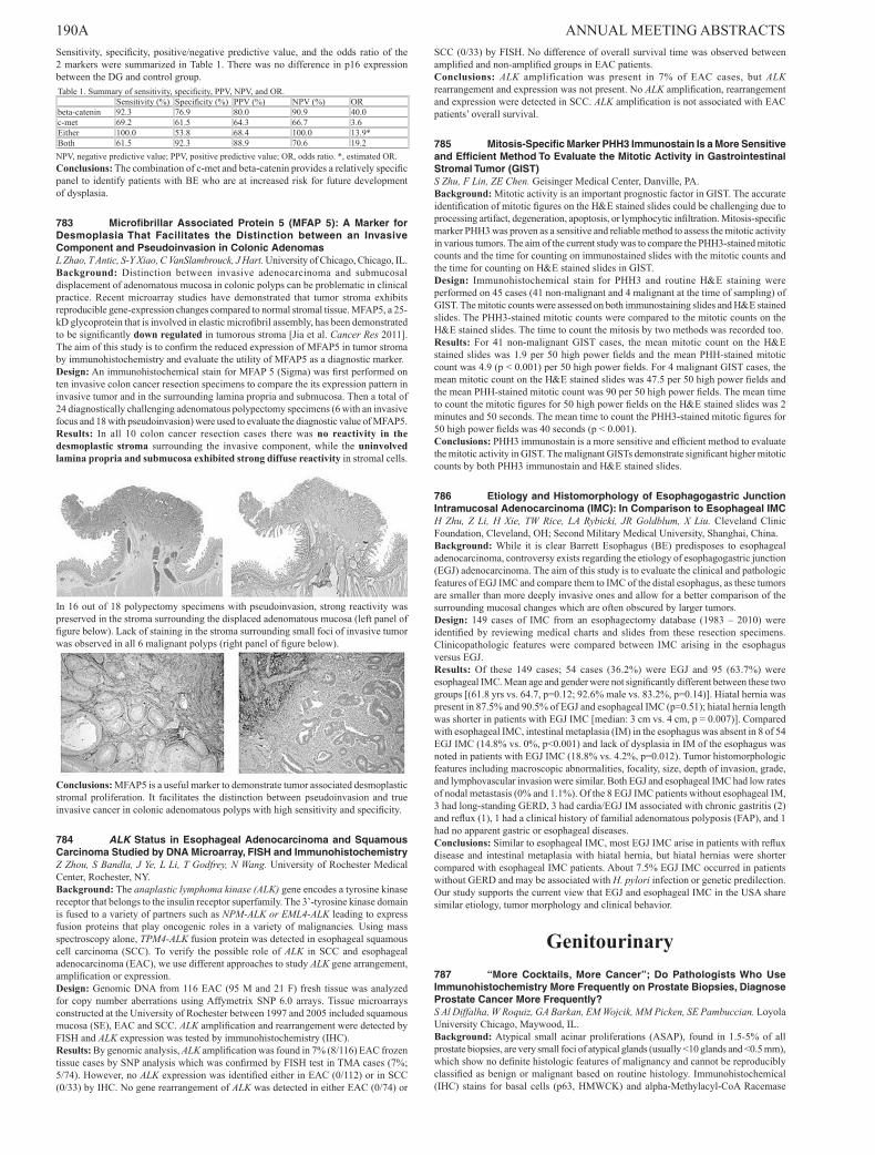
783 Microfibrillar Associated Protein 5 (MFAP 5): A Marker for Desmoplasia That Facilitates the Distinction between an Invasive Component and Pseudoinvasion in Colonic AdenomasL Zhao, T Antic, S-Y Xiao, C VanSlambrouck, J Hart. University of Chicago, Chicago, IL.Background: Distinction between invasive adenocarcinoma and submucosal displacement of adenomatous mucosa in colonic polyps can be problematic in clinical practice. Recent microarray studies have demonstrated that tumor stroma exhibits reproducible gene-expression changes compared to normal stromal tissue. MFAP5, a 25-kD glycoprotein that is involved in elastic microfibril assembly, has been demonstrated to be significantly down regulated in tumorous stroma [Jia et al. Cancer Res 2011]. The aim of this study is to confirm the reduced expression of MFAP5 in tumor stroma by immunohistochemistry and evaluate the utility of MFAP5 as a diagnostic marker.Design: An immunohistochemical stain for MFAP 5 (Sigma) was first performed on ten invasive colon cancer resection specimens to compare the its expression pattern in invasive tumor and in the surrounding lamina propria and submucosa. Then a total of 24 diagnostically challenging adenomatous polypectomy specimens (6 with an invasive focus and 18 with pseudoinvasion) were used to evaluate the diagnostic value of MFAP5.Results: In all 10 colon cancer resection cases there was no reactivity in the desmoplastic stroma surrounding the invasive component, while the uninvolved lamina propria and submucosa exhibited strong diffuse reactivity in stromal cells.
In 16 out of 18 polypectomy specimens with pseudoinvasion, strong reactivity was preserved in the stroma surrounding the displaced adenomatous mucosa (left panel of figure below). Lack of staining in the stroma surrounding small foci of invasive tumor was observed in all 6 malignant polyps (right panel of figure below).
Conclusions: MFAP5 is a useful marker to demonstrate tumor associated desmoplastic stromal proliferation. It facilitates the distinction between pseudoinvasion and true invasive cancer in colonic adenomatous polyps with high sensitivity and specificity.
784 ALK Status in Esophageal Adenocarcinoma and Squamous Carcinoma Studied by DNA Microarray, FISH and ImmunohistochemistryZ Zhou, S Bandla, J Ye, L Li, T Godfrey, N Wang. University of Rochester Medical Center, Rochester, NY.Background: The anaplastic lymphoma kinase (ALK) gene encodes a tyrosine kinase receptor that belongs to the insulin receptor superfamily. The 3’-tyrosine kinase domain is fused to a variety of partners such as NPM-ALK or EML4-ALK leading to express fusion proteins that play oncogenic roles in a variety of malignancies. Using mass spectroscopy alone, TPM4-ALK fusion protein was detected in esophageal squamous cell carcinoma (SCC). To verify the possible role of ALK in SCC and esophageal adenocarcinoma (EAC), we use different approaches to study ALK gene arrangement, amplification or expression.Design: Genomic DNA from 116 EAC (95 M and 21 F) fresh tissue was analyzed for copy number aberrations using Affymetrix SNP 6.0 arrays. Tissue microarrays constructed at the University of Rochester between 1997 and 2005 included squamous mucosa (SE), EAC and SCC. ALK amplification and rearrangement were detected by FISH and ALK expression was tested by immunohistochemistry (IHC).Results: By genomic analysis, ALK amplification was found in 7% (8/116) EAC frozen tissue cases by SNP analysis which was confirmed by FISH test in TMA cases (7%; 5/74). However, no ALK expression was identified either in EAC (0/112) or in SCC (0/33) by IHC. No gene rearrangement of ALK was detected in either EAC (0/74) or
SCC (0/33) by FISH. No difference of overall survival time was observed between amplified and non-amplified groups in EAC patients.Conclusions: ALK amplification was present in 7% of EAC cases, but ALK rearrangement and expression was not present. No ALK amplification, rearrangement and expression were detected in SCC. ALK amplification is not associated with EAC patients’ overall survival.
785 Mitosis-Specific Marker PHH3 Immunostain Is a More Sensitive and Efficient Method To Evaluate the Mitotic Activity in Gastrointestinal Stromal Tumor (GIST)S Zhu, F Lin, ZE Chen. Geisinger Medical Center, Danville, PA.Background: Mitotic activity is an important prognostic factor in GIST. The accurate identification of mitotic figures on the H&E stained slides could be challenging due to processing artifact, degeneration, apoptosis, or lymphocytic infiltration. Mitosis-specific marker PHH3 was proven as a sensitive and reliable method to assess the mitotic activity in various tumors. The aim of the current study was to compare the PHH3-stained mitotic counts and the time for counting on immunostained slides with the mitotic counts and the time for counting on H&E stained slides in GIST.Design: Immunohistochemical stain for PHH3 and routine H&E staining were performed on 45 cases (41 non-malignant and 4 malignant at the time of sampling) of GIST. The mitotic counts were assessed on both immunostaining slides and H&E stained slides. The PHH3-stained mitotic counts were compared to the mitotic counts on the H&E stained slides. The time to count the mitosis by two methods was recorded too.Results: For 41 non-malignant GIST cases, the mean mitotic count on the H&E stained slides was 1.9 per 50 high power fields and the mean PHH-stained mitotic count was 4.9 (p < 0.001) per 50 high power fields. For 4 malignant GIST cases, the mean mitotic count on the H&E stained slides was 47.5 per 50 high power fields and the mean PHH-stained mitotic count was 90 per 50 high power fields. The mean time to count the mitotic figures for 50 high power fields on the H&E stained slides was 2 minutes and 50 seconds. The mean time to count the PHH3-stained mitotic figures for 50 high power fields was 40 seconds (p < 0.001).Conclusions: PHH3 immunostain is a more sensitive and efficient method to evaluate the mitotic activity in GIST. The malignant GISTs demonstrate significant higher mitotic counts by both PHH3 immunostain and H&E stained slides.
786 Etiology and Histomorphology of Esophagogastric Junction Intramucosal Adenocarcinoma (IMC): In Comparison to Esophageal IMCH Zhu, Z Li, H Xie, TW Rice, LA Rybicki, JR Goldblum, X Liu. Cleveland Clinic Foundation, Cleveland, OH; Second Military Medical University, Shanghai, China.Background: While it is clear Barrett Esophagus (BE) predisposes to esophageal adenocarcinoma, controversy exists regarding the etiology of esophagogastric junction (EGJ) adenocarcinoma. The aim of this study is to evaluate the clinical and pathologic features of EGJ IMC and compare them to IMC of the distal esophagus, as these tumors are smaller than more deeply invasive ones and allow for a better comparison of the surrounding mucosal changes which are often obscured by larger tumors.Design: 149 cases of IMC from an esophagectomy database (1983 – 2010) were identified by reviewing medical charts and slides from these resection specimens. Clinicopathologic features were compared between IMC arising in the esophagus versus EGJ.Results: Of these 149 cases; 54 cases (36.2%) were EGJ and 95 (63.7%) were esophageal IMC. Mean age and gender were not significantly different between these two groups [(61.8 yrs vs. 64.7, p=0.12; 92.6% male vs. 83.2%, p=0.14)]. Hiatal hernia was present in 87.5% and 90.5% of EGJ and esophageal IMC (p=0.51); hiatal hernia length was shorter in patients with EGJ IMC [median: 3 cm vs. 4 cm, p = 0.007)]. Compared with esophageal IMC, intestinal metaplasia (IM) in the esophagus was absent in 8 of 54 EGJ IMC (14.8% vs. 0%, p<0.001) and lack of dysplasia in IM of the esophagus was noted in patients with EGJ IMC (18.8% vs. 4.2%, p=0.012). Tumor histomorphologic features including macroscopic abnormalities, focality, size, depth of invasion, grade, and lymphovascular invasion were similar. Both EGJ and esophageal IMC had low rates of nodal metastasis (0% and 1.1%). Of the 8 EGJ IMC patients without esophageal IM, 3 had long-standing GERD, 3 had cardia/EGJ IM associated with chronic gastritis (2) and reflux (1), 1 had a clinical history of familial adenomatous polyposis (FAP), and 1 had no apparent gastric or esophageal diseases.Conclusions: Similar to esophageal IMC, most EGJ IMC arise in patients with reflux disease and intestinal metaplasia with hiatal hernia, but hiatal hernias were shorter compared with esophageal IMC patients. About 7.5% EGJ IMC occurred in patients without GERD and may be associated with H. pylori infection or genetic predilection. Our study supports the current view that EGJ and esophageal IMC in the USA share similar etiology, tumor morphology and clinical behavior.
Genitourinary787 “More Cocktails, More Cancer”; Do Pathologists Who Use Immunohistochemistry More Frequently on Prostate Biopsies, Diagnose Prostate Cancer More Frequently?S Al Diffalha, W Roquiz, GA Barkan, EM Wojcik, MM Picken, SE Pambuccian. Loyola University Chicago, Maywood, IL.Background: Atypical small acinar proliferations (ASAP), found in 1.5-5% of all prostate biopsies, are very small foci of atypical glands (usually <10 glands and <0.5 mm), which show no definite histologic features of malignancy and cannot be reproducibly classified as benign or malignant based on routine histology. Immunohistochemical (IHC) stains for basal cells (p63, HMWCK) and alpha-Methylacyl-CoA Racemase

ANNUAL MEETING ABSTRACTS 191A(AMACR), ideally combined into a cocktail, can be helpful in ASAP to differentiate prostate carcinoma (PC) from its benign mimics. The aim of this study was to determine the pathologists’ frequency of use of IHC stains to resolve ASAP in routine practice, and the impact of IHC use on their diagnostic rates of PC and ASAP.Design: We performed a retrospective review of all prostatic needle biopsies diagnosed from 1/1/2006 to 9/20/2012, recording data on the sign-out pathologist, diagnosis, and use IHC (p63/34βE12/AMACR cocktail) for each individually labeled biopsy site, which was considered a biopsy unit (BU). Each pathologist’s % IHC use, % ASAP and % PC diagnosis was calculated. Comparisons between groups were made using χ2 or Fisher’s exact test. p<0.05 was considered significant.Results: During the study period, 12510 BU (each composed of 1-3 cores) from 2085 men were diagnosed by 12 pathologists (average 1043, range 270-3282 per pathologist). IHC was used in an average 2.5% of BU (range 0-7%). ASAP was diagnosed in 1.7% (range 0-4.6%) and PC in 14.6% (range 6.6-17.5%) of BU. 5 pathologists who used IHC ≥1% were defined as “high users” (HU) and 7 pathologists who used IHC <1% as “low users” (LU). The HU diagnosed more BU (8112 vs. 4398) and had a higher % PC (15.47% vs. 12.96%, p<0.0001), a higher % PC diagnosed after IHC (78/1255, 6.2% vs. 2/570, 0.4%, p<0.0001), a narrower range of variation of %PC (13.96%-17.51% vs. 6.60 -16.93 %) and a higher %ASAP (2.4% vs. 0.3%, p<0.0001) than LU. Overall IHC “resolved” ASAP to either benign or malignant in 42.7% (individual pathologists’ range 0-90%, HU mean 43.37%, LU mean 25%).Conclusions: Our results suggest that pathologists have different thresholds for regarding a focus as atypical, have different IHC ordering behavior, and may use IHC differently (i.e. some to confirm PC, others to rule it out). Pathologists who use IHC more frequently, diagnose PC more frequently and have less variation in the frequency of PC diagnoses than pathologists who use IHC less frequently. These results suggest the need for more explicit guidelines for the use and interpretation of IHC in ASAP.
788 Recurrent Novel and Potentially Targetable Genetic Alterations in High Grade, Invasive Urothelial Carcinoma of the BladderHA Al-Ahmadie, G Iyer, PH Kim, JP Sfakianos, Y-B Chen, A Gopalan, SW Fine, SK Tickoo, BH Bochner, DF Bajorin, VE Reuter, MF Berger, DB Solit. Memorial Sloan-Kettering Cancer Center, New York, NY.Background: A significant proportion of urothelial carcinoma (UC) will present at an advanced stage or will develop into advanced or metastatic disease after repeated recurrences. By then, such patients will have limited therapeutic options. This underscores the importance of identifying novel therapeutic approaches to managing these patients. With the advent of new small molecules targeting specific molecular aberrations, we sought to characterize a cohort of high grade UC using targeted genetic analysis to define the prevalence of known cancer genes and identify potential targets for therapy.Design: We analyzed tumor and matched normal samples from 50 radical cystectomies resected for high grade, invasive UC. All samples were analyzed using a targeted, deep-sequencing assay designed to identify point mutations, indels, and copy number alterations in 275 cancer-associated genes.Results: Somatic mutations in TP53 were the most common alteration, present in 29 (58%) samples. Nine samples contained mutations in RB (18%). Alterations in the PI3K/AKT/mTOR pathway were also observed (9 PIK3CA mutations, 3 MTOR, 3 PTEN, 2 AKT). These mutations were mostly non-overlapping. Non-overlapping mutations were present in FGFR3 (8 tumors), ERBB2 (6 tumors) and BRAF (3 tumors). Mutations in chromatin remodeling genes were also common including 18 (36%) tumors with mutations in KDM6A, 16 (32%) with mutations in ARID1A, and 11 (22%) with mutations in MLL2. Overall, 36% of all analyzed tumors harbored potentially targetable alterations in genes such as ERBB2, BRAF, and FGFR3.Conclusions: High grade, invasive urothelial carcinoma is a genetically heterogeneous disease. In addition to common alterations in TP53, mutations in the genes involved in chromatin remodelling are relatively common, particularly KDM6A, ARID1A and MLL2. Some of the identified alterations have been successfully targeted in other solid tumors and may offer a novel therapeutic option for patients with advanced bladder cancer. Further clinical correlation is needed to determine the prognostic significance of these genetic events.
789 E-Cadherin (CDH1) Is Frequently Mutated in Urothelial Carcinoma with Signet-Ring Cell and Plasmacytoid MorphologyH Al-Ahmadie, G Iyer, R Mehra, Y Chen, A Gopalan, SW Fine, SK Tickoo, MF Berger, DF Bajorin, G Dalbagni, DB Solit, VE Reuter. Memorial Sloan-Kettering Cancer Center, New York, NY.Background: A minority (<1%) of urothelial carcinomas (UC) exhibit a diffuse growth pattern without significant stromal reaction to the invasive tumor. Such tumors have been referred to as signet-ring cell carcinoma (SRC), plasmacytoid carcinoma (PC), and/or lymphoma-like carcinoma. They typically present at advanced stage and have been associated with significantly worse cancer specific survival than classical UC. Tumors possessing this morphology have also been observed in other organs such as diffuse gastric cancer and lobular breast carcinoma. In both of these locations, inactivating mutations in E-cadherin (CDH1) have been reported in the majority of cases with loss of E-cadherin expression by immunohistochemistry (IHC). We analyzed the mutation status of CDH1 in a cohort of urothelial SRC and PC by Sanger sequencing and compared the results to E-cadherin expression by IHC.Design: Twenty cases of primary bladder SRC and/or PC with matched normal tissue were selected. DNA was extracted from formalin-fixed paraffin-embedded tissue and all coding exons of CDH1 were sequenced. IHC for E-cadherin expression was performed for all cases. For comparison, we assessed CDH1 status in a separate cohort of 50 high grade UC without PC/SRC morphology.
Results: Mutations in CDH1 were present in 15 of 20 SRC and or PC cases (75%). All mutations were confirmed to be somatic. PC morphology was present in 16 (80%) cases and SRC morphology in 10 (50%) cases. Both morphologies were present in 8 (40%) cases. In tumors with CDH1 mutations, E-cadherin expression was retained as a diffuse membranous expression in 6 (40%) cases, completely lost in 4 (27%) cases and decreased (weak cytoplasmic staining) in 5 (33%) other cases. In the 5 cases with PC/SRC morphology but without mutation in CDH1, E-cadherin expression was completely lost in the tumor. Only 1 of the 50 UC cases without SRC or PC morphology harbored a CDH1 mutation.Conclusions: Mutations in CDH1 are frequently present in urothelial carcinoma with signet-ring cell and plasmacytoid morphology and appear to be specific to this distinct subset. The functional role of these aberrations has yet to be defined in this aggressive subtype of UC. The presence of mutations does not always correlate with loss of E-cadherin expression by IHC. Clinical correlations with patient outcome and both mutation status and loss of expression of E-cadherin are ongoing in a larger cohort.
790 Long-Term Clinical Outcome of Urothelial Papilloma – Which Patients Need Follow-Up?S Al-Bashir, C Wang, A Yilmaz, T Bismar, K Trpkov. Calgary Laboratory Services and University of Calgary, Calgary, AB, Canada.Background: Urothelial papilloma (UP) of the urinary tract is a benign urothelial neoplasm characterized by delicate fibrovascular cores covered by normal urothelium. However, only few larger studies have been published using the contemporary WHO definition and its biologic behavior has not been examined in studies with long-term follow-up.Design: We retrieved all consecutive cases with a diagnosis of UP from our institutional information system during a 10-year period (01/2000 to 12/2009), diagnosed using the WHO definition. All cases were identified in a regional centralized uropathology setting and the follow-up was obtained through search from pathology and clinical electronic databases. Patients with a previous history of higher grade urothelial neoplasm, diagnostic ambiguity or external consult cases were not included in the study.Results: The study cohort comprised of 41 de novo (‘primary’) UP. The average patient age was 57 years (median 56, range, 30-84), with a male:female ratio of 1.9:1. 13 (31%) patients were below the age of 50. The mean patient follow-up was 81 months (median 76; range, 3–127 months). In 37 (90.2%) patients no recurrent neoplasms were documented, including progression to low grade urothelial carcinoma (UC), high grade UC or carcinoma in situ. In 4 patients, subsequent urothelial neoplasms have been diagnosed. Three male patients (7.3%) had a diagnosis of subsequent UP at 1, 31, and 43 months after the initial diagnosis of UP. One of these patients had additional progression to papillary urothelial neoplasm of low malignant potential (PUNLMP) 17 months after the second UP (48 months after the initial UP). The patient with a subsequent UP at 1 month after the initial diagnosis likely represented an incomplete initial resection. The average age of these 3 patients was 62 years (individual age 50, 59 and 77 years). One additional male patient (age, 76 years) had a subsequent diagnosis of PUNLMP 76 months after the initial UP. Both patients diagnosed with PUNLMP presented with hematuria during the follow-up.Conclusions: Absence of progression on long-term follow-up in patients diagnosed with de novo UP argues strongly against the need of continuous surveillance for patients in which: 1.) the diagnosis is established using strict diagnostic criteria, 2.) the completeness of the resection can be ascertained and 3.) when no previous or synchronous urothelial malignancies are documented. A small percentage of patients, typically male and above the age of 50, who present with subsequent hematuria after the initial UP, require follow-up and repeat cystoscopy.
791 Proviral Integration Site Moloney Murine Leukemia Virus (PIM) Expression in Urothelial CarcinomaDJ Albertson, JJ Bearss, DJ Bearss, S Tripp, T Liu. University of Utah, Salt Lake City, UT; Huntsman Cancer Institute, Salt Lake City, UT; ARUP Institute for Clinical and Experimental Pathology, Salt Lake City, UT; Brigham Young University, Provo, UT.Background: The Proviral integration site Moloney murine leukemia virus (PIM) family is a group of proteins belonging to the calcium dependent protein kinases with serine/threonine kinase activity. Many studies have demonstrated overexpression of this family in hematologic and epithelial malignancies. The aim of the investigation was to evaluate PIM expression (PIM-1, 2, 3) in urothelial carcinoma and to assess for expression that may contribute to disease progression and serve as a site for PIM kinase targeted therapy.Design: Seventy-two cases of urothelial carcinoma were included in this retrospective study of surgical biopsy and resection specimens from the University of Utah Department of Pathology (retrieved from 2008-2011). Tissue was stained with commercially available antibodies against PIM-1, PIM-2, and PIM-3. Cases were divided into three groups (invasive high grade urothelial carcinoma (n=49), non-invasive high grade urothelial carcinoma/carcinoma in situ (n=16), and non-invasive low grade urothelial carcinoma (n=7)). Individual cases were then given a score (0-4) based upon a percentage of cells staining positive for each antibody (<5%=0; 5-25%=1; 26-50%=2; 51-75%=3; >75%=4). A score of 2 or greater was considered a positive finding.
192A ANNUAL MEETING ABSTRACTS
Results: Increased PIM-1, PIM-2 and PIM-3 expression was seen in a significant number of urothelial carcinoma cases when compared to adjacent bladder tissue.PIM-1,2,3 Kinase Expression PIM-1 PIM-2 PIM-3low grade non-invasive urothelial carcinoma 29%(2/7) 43%(3/7) 86%(6/7)high grade non-invasive urothelial carcinoma 44%(7/16) 50%(8/16) 44%(7/16)high grade invasive urothelial carcinoma 10%(5/49) 27%(13/49) 18%(9/49)
Conclusions: PIM-1, PIM-2 and PIM-3 expression is present in a high percentage of non-invasive low and high grade urothelial carcinoma and a lower percentage of invasive high grade urothelial carcinoma. These findings suggest the possibility of PIM-kinase inhibition as a target in a significant number of individuals with urothelial carcinoma.
792 Human Papillomavirus Is Not an Etiologic Agent of Urothelial Inverted PapillomasRE Alexander, A Lopez-Beltran, R Montironi, GT MacLennan, E Comperat, L Cheng. Indiana University School of Medicine, Indianapolis, IN; Cordoba University, Cordoba, Spain; Polytechnic University of the Marche Region, United Hospitals, Ancona, Italy; Case Western Reserve University, Cleveland, OH; Groupe Hospitalier Pitié-Salpêtrière, Paris, France.Background: Inverted papilloma of the urinary bladder is rare, accounting for less than 1% of all bladder neoplasms. Though it was long believed to be a pre-malignant neoplasm, there is a growing consensus that it is a benign lesion with limited potential for recurrence. Its etiology has been a matter of controversy, and recently it has been suggested that human papillomavirus (HPV) may play a role in its development. To date, this hypothesis has not been adequately substantiated. The goal of this study was to evaluate a number of inverted papillomas of the urinary bladder to investigate a possible etiologic role of HPV, employing an in-situ hybridization technique.Design: We studied urinary bladder inverted papillomas from 27 patients ranging from 35-78 years of age (average age 61 years), the great majority of whom were male (M:F ratio of 11:1). HPV in situ hybridization was performed on all cases using Inform HPV II Family 6 Probe (detecting HPV genotypes 6 and 11) and HPV III Family 16 Probe (detecting HPV genotypes 16, 18, 31, 33, 35, 39, 45, 51, 52, 56, 58, and 66) (Ventana Medical Systems, Inc, Tucson, AZ). The cases were then assessed for nuclear expression pattern. A control slide using uterine cervix with established HPV was prepared for each run. p16 immunostaining was performed with the anti-p16 mouse monoclonal antibody using the CINtec Histology kit (CINtec Histology, Westborough, MA, USA).Results: In all 27 cases, no HPV was detected with either the low-risk or high-risk probes. All controls reacted appropriately and confirmed the lack of HPV within the studied population of inverted papillomas. Immunoreactivity to p16 was detected in 11 of 27 (41%) cases. In six of these cases, staining was seen in greater than 50% of the inverted papilloma tissue; the remaining five cases demonstrated only focal reactivity.Conclusions: Our study provides no support for the premise that HPV is an etiologic agent in the development of inverted papilloma of the urinary bladder. In addition, correlation of HPV-ISH with p16 immunostaining shows that p16 is not a reliable marker of HPV infection status in inverted papillomas and should not be used as a surrogate marker to determine HPV status in these lesions.
793 EGFR and ALK Mutational Events Are Not Present in Primary Adenocarcinoma of the Urinary BladderRE Alexander, R Montironi, A Lopez-Beltran, M Wang, KM Post, JD Shen, AK Arnold, SA Bilbo, S Zhang, X Wang, SR Williamson, MO Koch, N Hahn, GT MacLennan, E Comperat, L Cheng. Indiana University School of Medicine, Indianapolis, IN; Polytechnic University of the Marche Region, Ancona, Italy; Cordoba University, Cordoba, Spain; Case Western Reserve University, Cleveland, OH; Groupe Hospitalier Pitié-Salpêtrière, Paris, France.Background: Mutations in the epidermal growth factor receptor (EGFR) have been shown to play an important role in driving cancer progression in many organs. Therapies targeting the mutant EGFR have allowed for more personalized treatment options in these cancers. Much effort has been expended in identifying additional molecular alterations that will lead to targeted therapies in patients with specific EGFR mutations. Rearrangements involving ALK, most notably the EML4-ALK rearrangement, have shown considerable clinical utility in the treatment of certain lung cancers. The goal
of this study was to determine whether EGFR mutations or ALK rearrangements are present in primary adenocarcinoma of the urinary bladder.Design: 23 cases of primary bladder adenocarcinoma were identified. Clinical histories and hematoxylin and eosin (H&E) slides of each case were reviewed to confirm origination within the urinary bladder. DNA was extracted from formalin-fixed paraffin embedded tissue in all 23 cases. For EGFR analysis, PCR amplified products were analyzed on the Q24 Pyrosequencer with Qiagen EGFR Pyro® kits (Qiagen, Inc., Valencia, CA). All cases were analyzed via (FISH) using Vysis ALK Break Apart FISH Probes (Vysis, Downers Grove, IL) for detection of ALK chromosomal translocation.Results: None of the twenty-three cases examined showed mutational events in EGFR or ALK rearrangements by FISH. Control tissues for both EGFR mutational analysis and ALK FISH exhibited the expected molecular alterations.Conclusions: EGFR mutations and ALK rearrangements do not appear to be involved in the development of primary adenocarcinoma of the urinary bladder. Use of targeted therapies based upon these molecular alterations is unlikely to yield significant clinical benefit. Pursuit of other molecular alterations is necessary to determine the precise molecular events that lead to the development of this cancer and may provide targets for future treatment.
794 Aggressive Variants of Chromophobe Renal Cell Carcinoma: Importance of Recognition and GradingM Alimchandani, C Neira de Paz, S Williams, K Lara-Otero, WM Linehan, MJ Merino. National Institutes of Health, Bethesda, MD.Background: Chromophobe renal cell carcinoma (ChRCC) comprises 5% of all renal cell carcinomas (RCCs) and has been known to have a better prognosis. It has been reported that tumor grading rather than Fuhrman nuclear grading is more appropriate to predict biologic behavior and prognosis. In this study we evaluate 97 cases of ChRCC and provide clinico-pathological and molecular support for tumor grading and for a better understanding and treatment of this rare neoplasm.Design: Ninety five nephrectomy specimens and 2 core biopsies obtained from 97 patients diagnosed as RCC, Chromophobe type, were reviewed at the NIH from 1999 – 2012. Normal and tumor tissue were microdissected for loss of heterozygosity (LOH) analysis using polymorphic markers for chromosomes 3p25, 1p35-36 and 1q42-43. IHC was performed for (MIB1), p53, CK7, CKAE1/AE3, CD10, CD117, SMA on selected specimens. In tumors with spindle (sarcomatoid) differentiation, both elements were studied.Results: Males and females were affected equally and range in age from 15-82 years. Three distinct morphologies were encountered, and graded according to Paner (2010). Seventy-five cases (77%) were designated Grade 1 (G1) histology and consisted of large polygonal cells with granular eosinophilic cytoplasm, prominent cell borders and irregular nuclei with the characteristic perinuclear halo. The Grade 2 (G2) intermediate morphology consisted of 15 cases (16%) with marked atypia of nuclei and lipidized clear cytoplasm. Seven cases (7%) displayed sarcomatoid transformation with a spindle cell component that varied in cellularity and pleomorphism, Grade 3 (G3). Metastasis occurred in 2/ 75 G1 tumors, 7/ 15 G2 tumors and 4/7 G3 tumors. Metastasis involved lymph nodes (either epithelial or sarcomatoid), liver and bones. One patient had massive retroperitoneal lymphangitic carcinomatosis. Genetic studies of selected G3 cases showed LOH in chromosomes 1p and 1q in tumor cells of epithelial morphology while tumor cells of spindle cell morphology displayed LOH in chromosomes 3p in addition of 1p and 1q. Survival correlates with tumor grading.Conclusions: Despite the favorable prognosis of ChRCC, it is important to realize that aggressive variants of chromophobe can occur with potential to metastasize and poor prognosis. Grading of tumors assist in the recognition of these aggressive forms. The finding of additional genetic changes in the sarcomatoid elements suggest tumor progression.
795 Comparison of Immunohistochemical Profiles of Papillary Renal Cell Carcinoma SubtypesA Alomari, H Kluger, K Haines, D Singh, ML Prasad, AJ Adeniran. Yale School of Medicine, New Haven, CT.Background: Papillary renal cell carcinoma (PRCC) is characterized morphologically by the presence of fibrovascular cores, lined by papillae of malignant epithelial cells. It is further sub-classified into 2 types based on cell type. Type 2 tumors are generally believed to have a poorer prognosis than type 1 tumors. This prognostic difference has led some to believe that some of the type 2 tumors may actually represent a separate entity from PRCC. This study was designed to compare the immunohistochemical profiles in both subtypes of PRCC.Design: A tissue microarray block with 124 cases of PRCC was constructed. The tumors were stained with the following diagnostic antibodies: CK7, EMA, CD10, CAIX, vimentin, CK903, C-kit, racemase, PAX-8, PAX-2 and CEA. A stain was deemed to be positive if it demonstrated at least weak intensity in at least 10% of the tumor cells. Intensity was scored as 1+, 2+ and 3+ for weak, moderate and strong intensity, respectively. A score of 0 was given for negative staining.

ANNUAL MEETING ABSTRACTS 193AResults: Results of the immunohistochemical stains are listed in Table 1.Table 1: Results of Immunohistochemical Stains Type 1 Type 2 MixedCK7 62/67 34/44 14/14CD10 33/63 33/44 9/14EMA 55/62 30/43 14/14CAIX 6/56 7/43 4/14Vimentin 8/62 2/43 1/14CK903 16/57 9/43 3/14C-kit 1/59 0/44 0/14Racemase 60/61 35/42 14/14PAX-8 48/57 38/42 11/13PAX-2 27/61 22/41 8/13CEA 4/57 3/43 1/14
The best immunohistochemical profile for both subtypes is: CK7: positive, EMA: positive, racemase: positive, PAX-8: positive, CD10: variable, PAX-2: variable, CAIX: negative, vimentin: negative and CK903: negative. There was no correlation between the staining pattern and tumor characteristics such as tumor type, location, nuclear grade, and presence of desmoplasia.Conclusions: The immunohistochemical profiles of both types 1 and 2 PRCC are essentially the same. There is no prognostic significance to any of the immunostains. The similar immunohistochemical profile confirms that PRCC is one entity with divergent histologic features. The 2 subtypes can only be differentiated based purely on morphologic features.
796 Papillary Renal Cell Carcinoma: Clinicopathologic Study of 144 Cases with Emphasis on SubtypingA Alomari, H Kluger, K Haines, D Singh, AJ Adeniran. Yale School of Medicine, New Haven, CT.Background: Papillary renal cell carcinoma (PRCC), a morphologically and genetically distinct subtype of RCC, is morphologically separated into two subtypes for therapeutic and prognostic purposes. In spite of multiple studies, many clinicopathologic issues about PRCC remain vague because the cohorts were small. Our study was designed to review and analyze the clinicopathologic features associated with Type 1 versus Type 2 PRCC.Design: Our pathology archives were searched for all nephrectomies and kidney tumor biopsies performed between 1985 and 2011, with the final pathologic diagnosis of PRCC. Slides and pathology reports were reviewed. The diagnosis of PRCC was confirmed in each case and subtypes were determined based on established morphologic criteria. Follow-up data was obtained from the clinical database.Results: We identified a total of 144 cases (74 type 1, 46 type 2 and 24 mixed) – 29 female and 115 male. Mean age was 56.0 years for type 1 and 59.0 years for type 2 (range, 23-88 years). Mean tumor size: 3.6 cm type 1 and 4.6 cm type 2 (p=0.1). Race distribution: 88 white, 45 black, 12 others. Ten patients had metastases (2 type 1, 6 type 2 and 2 mixed). None of the tumors showed sarcomatoid differentiation. Desmoplasia was present in 9% of cases while microscopic scar was identified in 45% of cases. Type 1 tumors were more likely to have nuclear grade 2 and less while type 2 tumors were more likely to have nuclear grade 3 and above (p<0.001). Type 1 tumors mostly presented at stage 1 while type 2 tumors were more likely to present at higher stages (p=0.05). Perinephric fat invasion (p=0.004), microvascular angiolymphatic invasion and main renal vein invasion were more likely in type 2 tumors. There was no significant association between tumor type and renal sinus fat invasion or invasion of muscular branches of renal vein. Follow up information was available in 134 patients: 11 alive with disease, 117 alive no disease, 1 dead of disease, 5 dead of other causes. Median follow up time was 58.5 months (range, 1-272 months).Conclusions: Type 2 tumors are larger. Sarcomatoid differentiation is a rarity in both subtypes. Type 2 tumors have higher nuclear grades and present at higher stages than type 1 tumors. Type 1 PRCC appears to have better clinical outcomes than type 2. Based on long follow-up data, both subtypes appear to have excellent prognosis when diagnosed at early stage.
797 Revisiting the Atypical or So-Called “Anaplastic” Seminoma with Emphasis on Diffuse AtypiaI Alvarado-Cabrero, N Hernandez-Toriz, GP Paner. Mexican Oncology Hospital, IMSS, Mexico City, Mexico; University of Chicago, Chicago, IL.Background: A subset of seminoma may exhibit greater degree of cellular pleomorphism and increased mitotic activity. There is controversy in the literature regarding the clinical relevance of this histologic finding. Some authors suggested this change to represent an early step in transformation toward a more aggressive phenotype because of the higher stage presentation, while others suggested not to designate them as atypical because there is no need for a different therapeutic approach.Design: We present the clinicopathological features of 20 atypical seminomas with diffuse cellular atypia (present in >50% area) identified from retrospectively reviewed histology slides of 1,010 tumoral orchiectomies (1999-2011) from a national oncology institution. Seminomas with focal cellular atypia or fixation artifacts were not included.Results: Patient age ranged from 31 to 46 years old (mean 36 years). Tumor involved the right (7/19), left (10/19) and both (2/19) testis. Foci of atypical seminomas were characterized by solid growth of crowded cells containing larger, irregular nuclei, with darker chromatin and increased mitotic activity. These areas were devoid of papillary, tubular or glandular architectures. The “higher grade”-appearing seminoma cells occasionally transitioned or blended imperceptibly with solid seminoma foci. No other germ cell tumor component was present. Stage of 19 cases at presentation was pT1 (15), pT2 (4); N0 (17), N1 (1), N3 (1); and M0 (19). Serum tumor markers in 20 cases were S0 (3), S1 (16) and S2 (1). Eleven patients were treated with radiotherapy and 3 patients (with pT2, N1 and N3) received bleomycin, etoposide and platinum (BEP)
chemotherapy. Follow-up on 19 patients (48-96 months, median 60 months) showed all patients were alive with no evidence of disease.Conclusions: This study shows that presence of the so-called “anaplastic” features even when involving >50% area of seminoma does not predict a high stage presentation and poorer outcome. Thus, presence of these atypical features may not necessitate separation from seminoma, including when choosing the type of therapy for the patient.
798 Clinicopathological Analysis of Choriocarcinoma as Pure and Predominant Component of Testicular Germ Cell TumorI Alvarado-Cabrero, N Hernandez-Toriz, GP Paner. Mexican Oncology Hospital, IMSS, Mexico City, Mexico; University of Chicago, Chicago, IL.Background: Although well recognized in textbooks including the 2004 WHO “blue book”, there is still a limited body of clinicopathological information regarding pure choriocarcinoma (CC) of the testis due to its rarity, including when it occurs as the predominant component of a mixed germ cell tumor (GCT). Herein, we present, to our knowledge, the largest surgical pathology series of pure CC and mixed GCT with predominant CC component of the testis.Design: A comprehensive review of histology slides from 1,010 tumoral orchiectomies from the surgical pathology files (1999-2011) of a national oncology institution was performed. Six (0.6%) pure CC and 9 (0.9%) mixed GCT with predominant CC component were identified, which formed the basis of this study.Results: Patients’ age ranged from 21 to 39 years old (mean 28 years). All tumors were unilateral and involved the right (9/15) and left (6/15) testis. Mean tumor size was 5.7 cm (range 1.5 to 8 cm). The 9 predominant CC comprised 50-95% (7 tumors with >80% CC) of mixed GCT, that included 7/9 combined with 1 and 2/9 combined with 2 other GCT components that included teratoma (5/9), seminoma (3/9), yolk sac tumor (YST) (2/9) and embryonal carcinoma (1/9). CCs were often hemorrhagic cystic tumor comprised of syncitio- and cyto- and variable intermediate trophoblastic cells. No case of monophasic CC was identified in our review. Lymphovascular invasion was identified in all 15 cases, that was often multifocal. Spermatic cord and tunica vaginalis were involved by tumor in 5/15 and 1/12 cases, respectively. Serum tumor markers for 15 patients were: S1 (1), S2 (1), S3 (13), marked by high serum βHCG elevation. Stage for 15 cases was: pT2 (10), pT3 (5); NX (1), N1 (4), N2 (5), N3 (5); and M1a (2) and M1b (13). Distant metastasis mostly involved the lung (11) and liver (10). Chemotherapy (mostly bleomycin, etoposide, platinum or BEP) was administered to 14 patients. On follow-up, all 6 pure CC patients were dead of disease (DOD) (6-14 months, median 9.5 months). Follow up of 8 mixed GCT with predominant CC patients (10-72 months, median 27 months) showed 5 DOD, 1 alive with disease and 2 alive with no disease; the later were the only 2 patients with M1a disease on presentation.Conclusions: This series confirms the proclivity for high stage presentation including presence of distant metastasis, hematogenous spread, and poor outcome of testicular CC. Mixed GCT with predominant CC component has similar tendency for high stage presentation and aggressive behavior to pure CC.
799 Sub-Classification of Extracapsular Extension in Prostate Cancer (PCA): Does It Matter? Prognostic Significance from a Study of 675 Patients at a Large Academic InstitutionM Amin, JB Nelson, E Woldemichael, AV Parwani, R Dhir. University of Pittsburgh Medical Center, Pittsburgh, PA; Unversity of Pittsburgh Medical Center, Pittsburgh, PA.Background: The finding of extracapsular extension (ECE) in a radical prostatectomy (RP) specimen is a poor prognostic factor. ECE has been divided into ‘focal’ (<0.8 mm) and ‘established’ (≥0.8 mm). Our reporting format also uses a third category, ‘multifocal,’ defined as more than one ECE focus, with each focus <0.8 mm. We examined the significance of this third category of ECE for predicting biochemical recurrence rate (BCRR) following RP.Design: We examined follow-up data on 675 men with stage pT3a/pT3b PCA, from a cohort of 2691 men. Patients were separated into groups based on 12 variables, including Gleason scores, volume of prostatic involvement (≤25%, >25%), tertiary grade 5, seminal vesicle invasion, perineural invasion, angiolymphatic invasion, positive lymph nodes and positive margins. The BCRR was calculated for each group, adjusting for these variables. Biochemical recurrence (BCR) was defined as a post-operative PSA ≥0.2 ng/mL or initiation of any adjuvant therapy.Results: Of 675 patients with stage pT3a/pT3b disease, 324 (48%) had focal ECE, 93 (14%) had multifocal ECE and 258 (38%) had established ECE. 205 (30.4%) had BCR. The median follow-up time was 49.84 months. BCRRs adjusted for the 12 variables are summarized in Table 1.Biochemical Recurrence (BCR) Rates of Prostate Cancer
Variable # Patients # with BCR BCR Rate (Focal ECE)
BCR Rate (Multi-focal ECE)
BCR Rate (Established ECE)
GS 8-10 153 93 0.1 0.18 0.33GS 4+3=7 169 61 0.1 0.11 0.15GS 3+4=7 317 47 0.05 0.04 0.05GS ≤6 178 82 0.14 - -Tertiary grade 5 190 58 0.05 0.08 0.17PI ≤25% 496 118 0.08 0.05 0.11PI >25% 178 82 0.09 0.19 0.21Seminal vesicle invasion 134 85 0.1 0.19 0.34Perineural invasion 669 204 0.08 0.09 0.15Angolymphatic invasion 59 41 0.1 0.22 0.37(+) Lymph nodes 62 44 0.06 0.23 0.4(+) Margins 136 59 0.08 0.13 0.21PI: prostatic involvement. GS: Gleason score. ECE: extracapsular extension. Conclusions: Although we see differences in BCRRs in patients according to the subtype of ECE, they are only significant with the presence of one or more of the following: Gleason score 8 or higher; tertiary grade 5 cancer, angiolymphatic invasion, positive

194A ANNUAL MEETING ABSTRACTS lymph nodes or positive margins. No significant differences in BCRRs were seen in patients with Gleason score 7 or lower cancer. Multifocal ECE provides additional prognostic significance, with outcomes intermediate between focal and established ECE, when the Gleason score is 8 or higher or concomitant negative prognostic factors are present.
800 Comparative Gene Expression and miRNA Expression Patterns in Chromophobe Renal Cell Carcinoma (ChRCC) and Oncocytoma (OC)M Amin, AV Parwani, S Roy, S Bastacky, W LaFramboise, R Dhir. University of Pittsburgh Medical Center, Pittsburgh, PA.Background: ChRCC and OC are distinct neoplasms arising from the intercalated duct of collecting tubules. Significant differences in clinical outcome warrant a definitive diagnosis that may be challenging due to overlapping morphologic and immunophenotypic features. We compared mRNA and microRNA transcriptional profiles of both tumors using high density expression arrays.Design: Total RNA was purified from microdissected specimens using the Qiagen miRNeasy kit with spectrophotometric absorption: 260/280 >1.8 and RIN value >8.0. RNA (1ug) was labeled with Hy3 for hybridization on miRCURY LNA arrays (Exiqon, Woburn, MA) for 18 hours followed by stringent wash and scan with probe readout classification against the Sanger miRBase (v.11.0). Message RNA (500ng) underwent cDNA synthesis and in vitro transcription using Ambion WT Expression assays (Ambion Inc, Austin, TX) followed by fragmentation and hybridization on Human Exon 1.0 ST arrays (Affymetrix Corp.,Santa Clara, CA) for 18 hours. Arrays were washed, stained and scanned (Affymetrix Fluidics Station 450, Scanner 3000) after hybridization. Signal intensity was calculated by Microarray Suite (v.5.0). Statistical comparisons (ANOVA) were performed (Partek Genomics Suite, St. Louis, MO) with false discovery rate:q value=0.1, -2.0>fold-change>2.0 and removal of comparisons within 5% of background signal intensity.Results: 856 and 38 mRNA transcripts had significantly altered expression in ChRCC and oncocytoma, respectively. ChRCC exclusively harbored altered expression of 785 transcripts. Conversely, there were no statistically significant alterations in mRNA expression unique to OC. Further analysis revealed 60 tumor suppressor genes and 30 oncogenes with significantly altered expression in ChRCC. Preliminary pathway analysis segregated the above genes into the following signaling pathways: MAP Kinase, Insulin, Chemokine, Wnt, Hedgehog, Notch, VEGF, PPAR, ERBB and TGF-beta. miRNA expression profiling revealed 153 statistically significant changes specific to ChRCC with no changes in OC expression. Analysis of common miRNAs altered in both tumor types compared to normal tissue demonstrated that miRNA200c and miRNA141 were overexpressed in ChRCC and underexpressed in OC.Conclusions: Assessment of differential mRNA and miRNA expression in ChRCC and OC provides novel markers for discrimination of both tumors. Absence of statistically significant alterations in mRNA expression in OC supports its accepted benign nature. Identifying pathways with aberrant activity in ChRCC has implications for targeted therapy.
801 When Worlds Collide: A Series of Genitourinary Collision TumorsW Anani, S Roy, M Amin, U Rao, L Pantanowitz, A Parwani. University of Pittsburgh Medical Center, Pittsburgh, PA.Background: Primary genitourinary neoplasms as well as metastasis from other sites to the genitourinary tract are common. In contrast, collision tumors, characterized by coexistence of phenotypically and genotypically distinct tumors at the same site, are distinctly rare in the genitourinary tract and pose a diagnostic challenge. The goal of this study is to present a series of such cases from a single institution highlighting the unusual clinicopathologic features of these tumors.Design: Nine cases were retrospectively identified from our surgical pathology files and included internal and consultation cases (2006-2012). All tumors were identified by H&E and immunohistochemistry as distinct primary neoplasms. In select cases, the final diagnosis was substantiated by fluorescence in-situ hybridization.Results: The study included 9 patients, 8 males and 1 female ranging in age from 34-84 years (mean 66.3 years). Collision tumors composed 7 of the 8 cases with the site of the collision as follows: kidney (6), bladder (2), and pelvis (1). All but one of the collision tumors involved two malignant neoplasms.Collision Tumor CasesCase Primary Tumor Secondary Tumor Location Age Sex
1 Chromophobe Renal Cell Carcinoma Neuroendocrine/Carcinoid Tumor Kidney 34 M
2 Clear Cell Renal Cell Carcinoma Pulmonary Adenocarcinoma Kidney 57 M
3 Clear Cell Renal Cell Carcinoma
Poorly Differentiated Carcinoma of Urothelial Origin Kidney 64 M
4 Angiomyolipoma Mammary Carcinoma Kidney 67 F
5 Clear Cell Renal Cell Carcinoma Chromophobe Renal Cell Carcinoma Kidney 76 M
6 Chromophobe Renal Cell Carcinoma Papillary Renal Cell Carcinoma Kidney 81 M
7 Pleomorphic Liposarcoma Prostatic Adenocarcinoma Pelvis 70 M
8 Urothelial Carcinoma Prostatic Adenocarcinoma Bladder 73 M9 Urothelial Carcinoma Prostatic Adenocarcinoma Bladder 84 M
Conclusions: To our knowledge, this is the largest report of collision neoplasms of genitourinary origin. Diagnosis of collision tumors in the genitourinary tract is a perplexing task and awareness about these rare entities, thorough sampling of the tumor mass and appropriate use of ancillary techniques are recommendations for avoiding incorrect diagnosis, and pathologic staging.
802 Influence of Histologic Criteria and Confounding Factors (CFs) in Staging Equivocal Cases for Microscopic Perivesical Tissue Invasion (MPVI or pT3a): An Interobserver Study among Expert GU PathologistsV Ananthanarayanan, Y Pan, M Kocherginsky, M Tretiakova, MB Amin, L Cheng, JI Epstein, DJ Grignon, DE Hansel, RE Jimenez, JK McKenney, R Montironi, E Oliva, AO Osunkoya, P Rao, VE Reuter, JY Ro, SS Shen, JR Srigley, T Tsuzuki, JL Yao, T Antic, M Haber, JB Taxy, GP Paner. University of Chicago, Chicago, IL; Emory University, Atlanta, GA; Cedars-Sinai Medical Center, Los Angeles, CA; Indiana University, Indianapolis, IN; Johns Hopkins Hospital, Baltimore, MD; Cleveland Clinic, Cleveland, OH; Mayo Clinic, Rochester, MN; Polytechnic University of the Marche Region, Ancona, Italy; Massachusetts General Hospital, Boston, MA; MD Anderson Cancer Center, Houston, TX; Memorial Sloan-Kettering Cancer Center, New York, NY; Methodist Hospital, Houston, TX; McMaster University, Hamilton, ON, Canada; Nagoya Daini Red Cross Hospital, Nagoya, Japan; University of Rochester, Rochester, NY.Background: Current oncology guidelines consider giving adjuvant chemotherapy for bladder cancer with at least MPVI (>pT3a). The boundary of muscularis propria (MP) vs. perivesical tissue (PVT) is commonly ill defined and may influence the interpretation of MPVI.Design: 20 sets of images including 19 equivocal for MPVI with CFs were sent to 17 expert GU pathologists. CFs included “histoanatomic” (HA) defined by irregular MP-PVT border and “tumor-related” (TR) such as fibrosis/desmoplasia, dense inflammation, tumor at edge of outermost MP bundle, and lymphovascular invasion (LVI). Cases were grouped by CFs: I - HA (7/19), II - HA+TR (7/19) and III - TR (5/19) factors.Results: Criteria used for MPVI varied among pathologists: A (3/17); B (9/17); C (4/17).
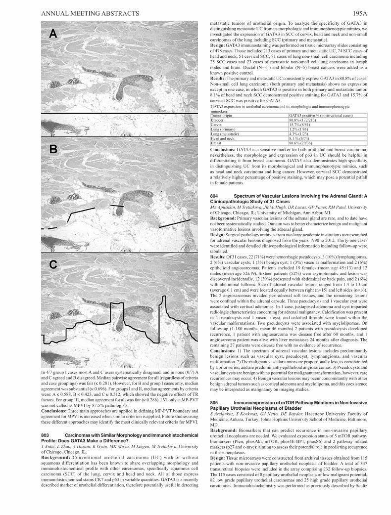
ANNUAL MEETING ABSTRACTS 195A
In 4/7 group I cases most A and C users systematically disagreed, and in none (0/7) A and C agreed and B disagreed. Median pairwise agreement for all (regardless of criteria and case groupings) was fair (κ 0.281). However, for B and group I cases only, median agreement was substantial (κ 0.696). For groups I and II, median agreements by criteria were: A κ 0.588, B κ 0.423, and C κ 0.512, which showed the negative effects of TR factors. For group III, median agreement for all was fair (κ 0.286). LVI only at MP-PVT was not called as MPVI by 87.5% pathologists.Conclusions: Three main approaches are applied in defining MP-PVT boundary and agreement for MPVI is increased when similar criterion is applied. Future studies using these different approaches may identify the most clinically relevant criteria for MPVI.
803 Carcinomas with Similar Morphology and Immunohistochemical Profile: Does GATA3 Make a Difference?T Antic, L Zhao, A Husain, K Gwin, MK Mirza, M Lingen, M Tretiakova. University of Chicago, Chicago, IL.Background: Conventional urothelial carcinoma (UC) with or without squamous differentiation has been known to share overlapping morphology and immunohistochemical profile with other carcinomas, specifically squamous cell carcinoma (SCC) of the lung, cervix and head and neck. All of those express immunohistochemical stains CK7 and p63 in variable quantities. GATA3 is a recently described marker of urothelial differentiation, therefore potentially useful in detecting
metastatic tumors of urothelial origin. To analyze the specificity of GATA3 in distinguishing metastatic UC from its morphologic and immunophenotypic mimics, we investigated the expression of GATA3 in SCC of cervix, head and neck and non-small carcinomas of the lung including SCC (primary and metastatic).Design: GATA3 immunostaining was performed on tissue microarray slides consisting of 478 cases. Those included 213 cases of primary and metastatic UC, 74 SCC cases of head and neck, 51 cervical SCC, 81 cases of lung non-small cell carcinoma including 25 SCC cases and 23 cases of metastatic non-small cell lung carcinoma in lymph nodes and brain. Ductal (N=31) and lobular (N=5) breast cancers were added as a known positive control.Results: The primary and metastatic UC consistently express GATA3 in 80.8% of cases. Non-small cell lung carcinoma (both primary and metastasis) shows no expression except in one case, in which GATA3 is positive in both primary and metastatic tumor. 8.1% of head and neck SCC demonstrated positive staining for GATA3 and 15.7% of cervical SCC was positive for GATA3.GATA3 expression in urothelial carcinoma and its morphologic and immunophenotypic mimickersTumor origin GATA3 positive % (positive/total cases)Bladder 80.8% (172/213)Cervix 15.7% (8/51)Lung (primary) 1.2% (1/81)Lung (metastatic) 4.3% (1/23)Head and neck 8.1 % (6/74)Breast 80.6% (29/36)
Conclusions: GATA3 is a sensitive marker for both urothelial and breast carcinoma; nevertheless, the morphology and expression of p63 in UC should be helpful in differentiating it from breast carcinoma. GATA3 also demonstrates high specificity in distinguishing UC from its morphological and immunophenotypic mimics, such as head and neck carcinoma and lung cancer. However, cervical SCC demonstrated a relatively higher percentage of positive staining, which may pose a potential pitfall in female patients.
804 Spectrum of Vascular Lesions Involving the Adrenal Gland: A Clinicopathologic Study of 31 CasesMA Apushkin, M Tretiakova, JB McHugh, DR Lucas, GP Paner, RM Patel. University of Chicago, Chicago, IL; University of Michigan, Ann Arbor, MI.Background: Primary vascular lesions of the adrenal gland are rare, and to date have not been systematically studied. Our aim was to better characterize benign and malignant vasoformative lesions involving the adrenal gland.Design: Surgical pathology archives from two large academic institutions were searched for adrenal vascular lesions diagnosed from the years 1990 to 2012. Thirty-one cases were identified and detailed clinicopathological information including follow-up were tabulated.Results: Of 31 cases, 22 (71%) were hemorrhagic pseudocysts, 3 (10%) lymphangiomas, 2 (6%) vascular cysts, 1 (3%) benign cyst, 1 (3%) vascular malformation and 2 (6%) epithelioid angiosarcomas. Patients included 19 females (mean age 45±13) and 12 males (mean age 52±19). Sixteen patients (52%) were asymptomatic and lesion was discovered incidentally, 12 (39%) presented with abdominal or back pain, and 2 (6%) with abdominal fullness. Size of adrenal vascular lesions ranged from 1.4 to 13 cm (average 6.1 cm) and were located equally between right (n=15) and left sides (n=16). The 2 angiosarcomas invaded peri-adrenal soft tissues, and the remaining lesions were confined within the adrenal capsule. Three pseudocysts and 1 vascular cyst were associated with cortical adenomas. In 1 case, juxtaposed adenoma and cyst imparted radiologic characteristics concerning for adrenal malignancy. Calcification was present in 4 pseudocysts and 1 vascular cyst, and calcified thrombi were found within the vascular malformations. Two pseudocysts were associated with myelolipomas. On follow-up (1-180 months, mean 46 months) 2 patients with pseudocysts developed recurrence, 1 patient with angiosarcoma was disease free after 60 months, and 1 angiosarcoma patient was alive with liver metastases 24 months after diagnosis. The remaining 27 patients were disease free with no evidence of recurrence.Conclusions: 1) The spectrum of adrenal vascular lesions includes predominantly benign lesions such as vascular cyst, pseudocyst, lymphangioma, and vascular malformation. 2) The malignant vascular tumors are proportionally less, as corroborated by a prior series, and are predominantly epithelioid angiosarcomas. 3) Pseudocysts and vascular cysts are benign with no potential for malignant transformation, however, rare recurrences may occur. 4) Benign vascular lesions may occur concomitantly with other benign adrenal tumors such as cortical adenoma and myelolipoma, and this coexistence may be interpreted as malignancy on imaging studies.
805 Immunoexpression of mTOR Pathway Members in Non-Invasive Papillary Urothelial Neoplasms of BladderS Arslankoz, S Korkmaz, GJ Netto, DE Baydar. Hacettepe University Faculty of Medicine, Ankara, Turkey; Johns Hopkins University School of Medicine, Baltimore, MD.Background: Biomarkers that can predict recurrence in non-invasive papillary urothelial neoplasms are needed. We evaluated expression status of 5 mTOR pathway biomarkers (Pten, phosAkt, mTOR, phos4E-BP1, phosS6) and 2 pathway related markers (p27 and c-myc); aiming to assess their potential role in predicting recurrence in these neoplasms.Design: Tissue microarrays were constructed from archival tissues obtained from 115 patients with non-invasive papillary urothelial neoplasia of bladder. A total of 347 transurethral biopsies were included in the array comprising 232 follow-up biopsies. The 115 cases consisted of 8 papillary urothelial neoplasia of low malignant potential, 82 low grade papillary urothelial carcinomas and 25 high grade papillary urothelial carcinomas. Immunohistochemistry was performed as previously described by Scultz

196A ANNUAL MEETING ABSTRACTS et al. Cancer 2010. Markers were evaluated for pattern, percentage and intensity of staining. Tumor expression levels were correlated with clinicopathologic parameters. Paired non-neoplastic and neoplastic urothelium were also compared for differences in expression.Results: Mean follow-up period was 56,60 months (13-258 months). 57/115 tumors (49,6%) had recurrences. Overall, lower phos4E-BP1, p27, mTOR, Pten and phosS6 and higher c-myc expression levels were found in neoplastic urothelium compared to non-neoplastic urothelium. The differences were statistically significant for phos4E-BP1 (p=0,005) and phosS6 (p= 0,003). None of the assessed markers were predictive of recurrence.Conclusions: Our finding of mTOR pathway members phos4E-BP1 and phosS6 alterations in non-invasive urothelial neoplasms of bladder supports a role in pathogenesis. mTOR markers were not predictive of recurrence in our cohort.
806 Renal Oncocytic Tumors with Hybrid Features Occurring in a Sporadic Setting: A Clinicopathologic Study of 22 CasesD Atherton, K Sircar, P Tamboli, P Rao. MD Anderson Cancer Center, Houston, TX.Background: Renal oncocytic tumors that show histologic features that overlap between renal oncocytoma (RO) and chromophobe renal cell carcinoma (ChRCC) comprise a rare subgroup of tumors that have been widely referred to as “hybrid oncocytic tumors” (HOT). Although originally described in patients with Birt-Hogg Dube (BHD) syndrome, these tumors are most frequently seen in the sporadic setting.Design: We did a retrospective search of our databases (2005-2012) using search terms “hybrid tumor”, “oncocytosis” and “Birt-Hogg Dube”. Final analysis included 21 primary and one metastatic case. All available pathologic material was reviewed and clinical information was acquired from the medical record.Results: Tumors were from 15 men 7 women. No patient had a diagnosis or showed stigmata suggestive of BHD syndrome. Tumor size ranged from 1.1 to 25 cm (mean 5.5). Six patients presented with more than 1 tumor in the resected kidney wherein the dominant tumor was associated with areas of renal oncocytosis. Six tumors were associated with perinephric/renal sinus fat invasion (pT3=6, pT1=15). One additional tumor showed involvement of a large vessel within the renal sinus. Most cases showed true hybrid morphology and had areas within the same tumor that resembled both RO and ChRCC. In most cases areas resembling RO predominated and gradually transitioned to areas resembling ChRCC. The single case of a metastatic tumor within the liver occurred in a patient with a remote history of RO. The liver lesion histologically closely resembled a RO but showed diffuse CK7 staining thus resulting in the diagnosis of HOT. Immunohistochemical stains were performed in 20 cases. All cases were negative for vimentin. The majority of cases (15/20) showed only focal staining for CK7, which was helpful in the distinction from ChRCC. In 2 instances, tumors resembled RO histologically but were classified as HOT’s due to diffuse staining with CK7. Follow up information of greater than 6 months was available in 15 cases (range 6-59 mos; mean 25 mos). None of the cases with primary resections resulted in recurrence or metastasis.Conclusions: Renal hybrid oncocytic tumors are rare renal neoplasms which occur in both the sporadic and syndromic setting. Tumors have histologic features that overlap between RO and ChRCC and may pose a diagnostic dilemma for the pathologist. Tumors generally follow an indolent clinical course even in the setting of locally advanced tumor at diagnosis. There is a definite potential for metastasis which makes accurate classification of these neoplasms critical.
807 The Significance of Midline Crossing in Organ Confined pT2c Prostate CarcinomaA Bartakova, PH Sweet, AS Shabaik. UCSD, San Diego, CA; ULB, Brussels, Belgium.Background: In the seventh edition of the American Joint Committee on Cancer (AJCC) cancer staging manual, organ confined prostatic adenocarcinoma (PCa) pT2 is substaged into pT2a in which carcinoma involves one half or less of one side, pT2b, with more than one half of one side, and pT2c with both sides involvement. Frequently, pT2c cases consist of two unrelated tumor nodules with one nodule in each side. In this study we investigated the difference in outcome in pT2c cases in which a dominant tumor nodule crossed the midline to involve both sides of the prostate, against cases that involved both sides with two separate unrelated nodules.Design: We reviewed the slides of 65 cases of pT2c PCa resected in 2006-2007. We divided the cases in two groups: those where the tumor crossed the midline (group 1), and those presenting with bilateral foci of independent tumors without histologic evidence of crossing of the midline (group 2). We compared the pathologic and clinical findings and the outcome of both groups.Results: Our results are presented in table 1:Table 1: Cases review GR 1: 49 cases GR 2: 16 casesAGE (y) Mean 61.3 59.3 Median [Range] 61.0 [42-74] 58.5 [41-76]PRE-OP PSA (ng/ml) Mean 5.6 5.1 Median [Range] 5.1 [0.02-23] 4.5 [0.9-12.5]GLAND VOLUME (cc) Mean 68.8 85.7 Median [Range] 68.4 [31.5-109] 64.0 [27.5-216]GLEASON SCORE Mean 6.7 6.8 Median [Range] 6 [6-9] 7 [6-9]TUMOR VOLUME (%) 13.4 4.0POST-OP PSA (ng/ml) Mean 0.04 0.05 Median,[Range] 0.01 [0.01-0,06] 0.02 [0.01-0.42]RFS 5y (%) 87.8 93.8GS 5y (%) 93.9 100Group 1: large bilateral tumor, crossing the midline; Group 2: bilateral foci of independent tumors; PSA: prostate-specific antigen; RFS: recurrence-free survival; GS: global survivalConclusions: This pilot study found a difference in global and recurrence-free survival between group 1 and group 2 pT2c PCa. Although this may be attributed to chance
because of the small sample size, it merits a larger series to assess whether there is a statistically significant difference. Interestingly, if this difference is born out, it would suggest that the biologic behavior is indeed different for these two types of presentation and pT2a or pT2b categories may suit better to cases that are pT2c because of bilateral separate nodules. Indeed, our recurrence-free survival for group 2 patients is comparable to pT2a-pT2b data published previously (Caso et al., 2010). Alternatively the organ-confined tumors may better be subdivided into two subclasses rather than three based on crossing of the midline by the index tumor.
808 Transcriptional (mRNA) Profiling in Clear Cell Renal Cell Carcinoma (CCRCC): Comparison of CCRCC to Non-Neoplastic Renal TissueSI Bastacky, ML Bastacky, S Roy, AV Parwani, R Dhir, MA Lyons-Weiler, CM Sciulli, M Krill-Burger, M Amin, WA LaFramboise. University of Pittsburgh Medical Center, Pittsburgh, PA.Background: CCRCC is the most common primary renal tumor, with a characteristic morphology and immunophenotype. Prior studies have found consistent chromosomal mutations, e.g. 3p deletions, and dysregulation of hypoxic inducible factor-1 (HIF-1) leading to transcription of HIF-1 inducible genes. The study aim was to characterize the mRNA expression profile of CCRCC using a high density mRNA array to identify a transcript signature and important cancer-related transcripts in CCRCC.Design: RNA was purified from macrodissected specimens (nl=6, sporadic CCRCC=5) using the Qiagen miRNeasy kit (Valencia, CA) with spectrophotometric absorption: 260/280 >1.8 (NanoDrop, Wilmington, DE) and RIN value >8.0 (Bioanalyzer 2100, Agilent Technologies, Santa Clara, CA). mRNA (500ng) underwent cDNA synthesis and in vitro transcription using the Ambion WT Expression assay (Ambion Inc, Austin, TX) followed by fragmentation and hybridization on Human Exon 1.0 ST arrays (Affymetrix Corp.,Santa Clara, CA). Washing, staining and scanning of arrays was performed (Fluidics Station 450, Scanner 3000) after hybridization and signal intensity calculated by Microarray Suite version 5.0. Statistical comparisons (ANOVA) were performed (Partek Genomics Suite, St. Louis, MO) with false discovery rate: q value=0.05 and -2.0>fold-change>2.0.Results: 415 CCRCC transcripts were significantly increased (n=191; 2 to 22x) or decreased (n=224; -2 to -92x)). 137 transcripts involved cancer-related genes. Altered tumor suppressor gene (n=14) and oncogene (n=10) transcripts were seen, with a partial list with greatest fold changes including: EGF variant(-17.5x), SCL481(-10x), MAL(-9.5x), GPC3(-7x), PTGS2(-6.5x), RARB(-2.3x) and CA-IX(10x), IGFBP3(6x), C3(4.6x), VEGF-A(4.6x), MDM2(2.5x). HIF-1α/1β, and VHL transcripts were not significantly altered. 21 transcripts were found in 13 different cancer signaling pathways (e.g. pancreas, bladder, ovary, lung (non-small cell and small cell), melanoma, breast, thyroid, other) with a mean+SD (range) of transcripts per pathway of 4.7+2.3 (1-9) (Ingenuity database).Conclusions: CCRCC showed many mRNA transcript alterations, including increases and decreases of important oncogene and tumor suppressor transcripts. A subset of these transcript alterations is shared amongst a diverse group of other organ cancer signaling pathways, suggesting that CCRCC involves dysfunction of multiple growth regulatory pathways. The absence of significant HIF-1 transcript alterations is consistent with the understanding that HIF-1 is dysregulated at the protein level.
809 SIX1 Expression Correlates with Tumor Grade of Urothelial CarcinomaRC Batiste, R Rong, L-P Wang, Z Bing. Hospital of the University of Pennsylvania, Philadelphia, PA.Background: Six1 is a homeobox transcription factor belonging to the evolutionarily-conserved Six1–Eya–Dach transcriptional regulatory network that is critical during embryonic development. Six1 requires interaction with the Eya family of proteins to activate the transcription of genes involved in nephrogenesis, neurogenesis and myogenesis. Mutations in either Six1 or Eya2 can lead to branchio-oto-renal syndrome. Mutations in either gene can disrupt individual protein function, Six1–DNA binding or Six1–Eya2 binding. While both genes are commonly expressed during development, their overexpression has been observed in various neoplastic states. SIX1 is aberrantly expressed in numerous cancers including those arising from breast, ovary, cervix, and liver, as well as Wilms tumor and alveolar rhabdomyosarcoma. Eya2 is required to mediate the pro-metastatic functions of Six1.Design: The following is an immunohistochemical study of Six1 and Eya2 expression in high grade and low grade urothelial carcinoma. In the appropriate setting, the expression of Six1 and Eya2 in invasive carcinoma has also been investigated. Tumor cell nuclear staining intensity is scored in a blinded fashion using the Allred scoring system.Results: Eighteen cases of high grade and 10 cases of low grade urothelial carcinoma were retrieved and stained for Six1 and Eya2. Twelve out of the eighteen cases of high grade urotehlial carcinoma had invasive components. The scores for non-invasive and invasive high grade urothelial carcinoma for Six1 were 6.3 +/- 2.4 and 5.9 +/- 2.3 respectively. There was no statistical difference between these two groups. The average score for low grade urothelial carcinoma for Six1 was 1.9 +/-2.5. There were significant statistical differences between low grade and high grade non-invasive and invasive carcinomas (p<0.05). Eya2 showed diffuse moderate to strong nuclear staining in both low grade and high grade noninvasive and invasive urothelial carcinoma. There were no statistical differences in the Eya2 staining among the different groups.Conclusions: Six1 expression appears to correlate with urothelial carcinoma grade, with higher expression in the high grade tumors in both invasive and noninvasive components. There is no difference in Eya2 expression between low and high grade urothelial carcinoma.

ANNUAL MEETING ABSTRACTS 197A810 Fuhrman Grading in Renal Cell Carcinoma: Comparison between Core Needle Biopsies and Surgical ResectionsJA Bennett, LA Wilkinson, CS Abendroth, EE Frauenhoffer, B Han. Penn State Hershey Medical Center, Hershey, PA.Background: With increased use of computed tomography (CT) imaging, many renal cell carcinomas are being detected incidentally. Fine needle aspiration (FNA) is often used for confirmation of the diagnosis. Recently, surgeons have requested Fuhrman grading of the lesion, resulting in a core needle biopsy being performed simultaneously with FNA. We undertook a study to determine whether Fuhrman grading of biopsies correlates with that of the nephrectomy specimen.Design: The pathology database was searched for patients who had both a biopsy and nephrectomy performed. The cases were randomly blinded and reviewed by three staff pathologists. Biopsies and nephrectomies were scored according to standardized Fuhrman grading criteria. The results were tabulated to determine whether Fuhrman grading of biopsy correlated with that of the nephrectomy.Results: The average Fuhrman grade ranged from 1.00-2.67 for the biopsies and from 1.33-3.67 for the nephrectomies. In general, the biopsy underestimated the Fuhrman grade when compared to the nephrectomy by approximately one grade. In 4/19 cases the Fuhrman grade was higher in the biopsy than in the nephrectomy. There was interobserver agreement in 6/19 cases (kappa = 0.578) for both biopsies and nephrectomies. Comparison between the average Fuhrman grades for biopsies versus nephrectomies resulted in a p-value of 0.004. Reclassification of the Fuhrman grade into low grade (1-2) versus high grade (3-4) showed agreement in 14/19 cases (kappa = 0.825) for biopsies and 12/19 cases (kappa = 0.754) for nephrectomies. Comparison of biopsies versus nephrectomies in the two-tiered scoring system resulted in a p-value of 0.083.Conclusions: This study illustrates the degree of interobserver variability present when using the traditional Fuhrman grading system. When using the two-tiered system, interobserver variability is improved. The likelihood of an increase in the Fuhrman grade from biopsy to nephrectomy is common, but less so when utilizing the two-tiered system. The clinical utility of a prenephrectomy biopsy is highest when attempting to provide basic diagnostic information. Interpreting Fuhrman grade on limited material is clearly fraught with limitations; however, with the advent of less invasive surgical approaches including radioablation and partial nephrectomy, the biopsy grade may play an increased role in refining clinical management. From the evidence gathered here, both interobserver variability and biopsy to surgical consistency is improved when utilizing the two-tiered grading system.
811 The Effects of Chemotherapy on Spermatocytic SeminomaDM Berney, W Ansell, J Shamash. St Bartholomew’s Hospital, Queen Mary University of London, London, United Kingdom.Background: Spermatocytic seminoma is a rare testicular tumor, which ususally receives no treatment beyond excision. Rare cases of metastasis are reported in the literature, usually but not inevitably associated with sarcomatoid change. We present two cases of spermatocytic seminoma without sarcomatoid change which were treated with chemotherapy prior to excision and examine the possible effects of standard germ cell chemotherapy on these tumors.Design: The files of Bartshealth hospitals were searched for cases of spermatocytic seminoma and reviewed for any which had received chemotherapy.Results: 25 cases of spermatocytic seminoma were reviewed of which two had received chemotherapy after mutli-disciplinary discussion. A 30 year old male presented with a right testicular lump, which was initially diagnosed as a non-seminomatous germ cell tumor. He received 1 cycle of adjuvant BEP chemotherapy. On supra-regional review the diagnosis was changed to spermatocytic seminoma, and a second round of chemotherapy was not given. On histology, the tumour was over 6cm in size, and showed typical features for spermatocytic seminoma, though vascular invasion was seen. Immunochemistry for OCT3/4 was negative. On routine follow up he represented with haemoptysis and a single 4cm lung mass was seen on imaging. This was completely excised and showed features identical to the primary tumour. He remains well after 1 year of further follow up and recurrence free. A 47 year old man presented with a 5cm right and 1cm left testicular lump. Excision of the right testicle showed a spermatocytic seminoma. In order to preserve endocrine function, he underwent 2 cycles of BEP chemotherapy to shrink the second tumour and facilitate a partial orchidectomy. However no response was seen on imaging and he underwent a left orchidectomy. This showed a spermatocytic seminoma with anaplastic features, but no necrosis or other features of regression. He remains tumor free after 4 years.Conclusions: These rare cases demonstrate that BEP chemotherapy appears to have little effect on the growth or prevention of recurrence of spermatocytic seminoma. Both tumors showed a high mitotic index with no necrosis and features completely different from those expected with typical post-chemotherapy germ cell tumors. The highly unusual metastatic case showed only a single metastasis and appears well after surgery. We suggest that this may represent an embolic phenomenon rather than true metastasis. The chemoresistance of these tumors suggests that other modalities should be preferred in the treatment of these exceptional cases.
812 Reporting Guidelines for Prostate Biopsies – A Survey of 266 European PathologistsD Berney, F Algaba, P Camparo, E Comperat, D Griffiths, G Kristiansen, A Lopez-Beltran, R Montironi, M Varma, L Egevad. St Bartholomew’ Hospital, London, United Kingdom; Fundacio Puigvert-University Autonomous, Barcelona, Spain; Centre de Pathologie Amiens Picardie, Paris, France; Hopital La Pitié-Salpetrière, Paris, France; University Hospital of Wales, Cardiff, United Kingdom; University of Bonn, Bonn, Germany; Cordoba University Medical School, Cordoba, United Kingdom; Polytechnic University of the Marche Region, Ancona, United Kingdom; Karolinska Institutet, Stockholm, Sweden.Background: It is not known how European uropathologists currently report histopathological features of cancer such as core length, tumour length, peri-neural invasion and non-tumour associated features such as inflammation and hyperplasia in needle biopsies.Design: A web-based survey was distributed among 661 members of the European Network of Uropathology (ENUP).Results: Complete replies were received from 266 pathologists in 22 European countries. Total core lengths were reported by 64%, the majority were measured on wet specimens by laboratory technicians. The numbers of cores positive for cancer was given by 79% while 14% would report biopsies separately without summarizing numbers. Linear cancer extent was reported by 81%, most often given in mm for each core (53%) followed by estimation of percentage of cancer in each core (40%). Total extent of cancer in all cores was given in mm (22%), as an estimated percentage (25%) or by calculating exact percentages from core lengths (17%). A gap of benign tissue between separate cancer foci in a single core would always be subtracted by 48% while even a wide gap would be included in tumour extent by 27%. Peri-neural invasion was reported by 97%. Fat invasion by tumor was interpreted as extraprostatic extension by 81%. Chronic and active/acute inflammation was always reported by 32% and 56% but only if pronounced by 54% and 39%, respectively. While most (79%) would never diagnose benign prostatic hyperplasia on needle biopsy, 21% would attempt to make this diagnosis (13% sometimes, 8% often).Conclusions: Reporting practices for prostate biopsies are variable among European pathologists. In line with international guidelines, the vast majority report the number of biopsies positive for cancer and linear extent but the methodologies used for this vary widely. Despite uncertain clinical relevance, most pathologists report inflammation, at least if pronounced, and some even attempt to diagnose benign prostatic hyperplasia in needle biopsies. The great variation in methodologies used suggests a need for further international consensus, in order for retrospective data to be comparable between different institutions.
813 Do European Pathologists Follow ISUP 2005 Gleason Grading Guidelines? A Web Based SurveyDM Berney, F Algaba, P Camparo, E Comperat, D Griffiths, G Kristiansen, A Lopez-Beltran, R Montironi, M Varma, L Egevad. St Bartholomew’s Hospital, London, United Kingdom; Fundacio Puigvert-University Autonomous, Barcelona, Spain; Centre de Pathologie Amiens Picardie, Paris, France; Hopital La Pitié-Salpetrière, Paris, France; University Hospital of Wales, Cardiff, United Kingdom; University of Bonn, Bonn, Germany; Cordoba University Medical School, Cordoba, Spain; Polytechnic University of the Marche Region, Ancona, Italy; Karolinska Institutet, Stockholm, Sweden.Background: The Gleason grading system underwent a major revision at the International Society of Urological Pathology consensus conference in 2005 (ISUP 2005). It is not known how European uropathologists have adopted or interpreted its recommendations.Design: A web-based survey was distributed among 661 members of the European Network of Uropathology (ENUP).Results: Complete replies were received from 266 pathologists in 22 countries. All respondents used Gleason grading and 89% claimed to follow ISUP 2005 recommendations for pattern interpretation and reporting. Most often a Gleason pattern (GP) 3 would be assigned to regular and smoothly rounded cribriform glands (51%), a GP 4 to irregular cribriform glands (98%), poorly formed glands (84%) and glomeruloid glands (86%) and a GP 5 to cribriform glands with comedonecrosis (86%). A Gleason score (GS) 2-3, 4 and 5 would never be given by 98%, 94% and 70%. Clusters of single cells or solid strands of cancer seen already at 20x magnification were required by 72% to diagnose GP 5. Necrosis would be diagnosed as GP 5 by 62% while 38% thought necrosis by itself would be insufficient. Any amount of a secondary pattern of higher grade would be included in the GS by 58%, while others required this pattern to be identified already at medium to low power or to comprise 5% or more of the tumor. Similary, a tertiary GP of higher grade on needle biopsies was included in the GS by only 58%. Among those who embedded biopsy cores separately, only 56% would give a GS for each core/slide examined. Among those who sometimes blocked multiple biopsy cores, 46% would only give an overall GS for the case. A majority (68%) would give a concluding GS for the case and among them the most common method was to give a global GS (77%).Conclusions: European pathologists generally claim to follow ISUP 2005 recommendations for Gleason grading, but misinterpretation is widespread, with a low threshold for diagnosing secondary GP 4 and not including tertiary GP of higher grade in the GS. Only slightly more than half would give a GS for each core/slide examined when embedded separately. Clarity in teaching ISUP 2005 recommendations is necessary to avoid misinterpretations.

198A ANNUAL MEETING ABSTRACTS 814 Long Term Follow-Up (F/U) in Patients with Initial PIN and Benign Prostate BiopsiesZ Biljetina, J Zhang, A Yilmaz, K Trpkov. Calgary Laboratory Services and University of Calgary, Calgary, Canada.Background: There are limited long term F/U data on PCa features on biopsy and RP after initial PIN or benign prostate biopsy.Design: We identified 161 patients with initial PIN and 85 patients with initial benign prostate biopsies who underwent repeat biopsies in our institution. All patients had extended (10 core, mean) biopsies. We compared the preoperative findings in patients with and without PCa on F/U biopsy and we evaluated the PCa biopsy and radical prostatectomy (RP) findings after initial PIN and benign biopsies. Patients characteristics between the groups were compared using the Wilcoxon rank-sum test for continuous variables and Chi-square test for categorical variables.Results: We found 43 (26.7%) Pca after PIN and 19 (22.3%) Pca after benign initial biopsy, during a mean F/U of 8.9 and 9.3 years (medians, 11.4 and 11.1 years), respectively. Pca was diagnosed after 1.5 and 2.5 years (p<0.001, mean) after initial PIN vs. benign biopsy, and after 1.9 vs. 1.5 biopsies (NS, mean), respectively. Patients with Pca on F/U in PIN and benign groups differed significantly only in age (63.8 vs. 59.4 years, p=0.032) and PSA ∆ (2.7 vs. 3.3 ng/ml; p=0.026). Patients with initial PIN with Pca on F/U vs. no Pca on F/U, were older (63.8 vs. 60.8 years, p=0.039) and had higher PSA (8.6 vs. 6.7 ng/ml; p=0.041), but did not show other significant differences, including the number of PIN biopsy sites. Patients with initial benign biopsy with Pca on F/U vs. no Pca on F/U, had smaller gland volume (45.4 vs. 63.7cc, p=0.012), but had no other significant differences. Pca on F/U biopsy after PIN was 1 or 2 core positive in 96% (mean total Pca 2.4%). 95% Pca biopsies after PIN were Gleason score (GS)6 (84%) or GS3+4 (11%). Of 22 (51%) patients with RP after initial PIN biopsy, 95% had GS6 (86%) or GS3+4 (9%) and all had node negative pT2 disease. Pca on F/U biopsy after initial benign biopsy was 1 or 2 core positive in 79% (mean total Pca 2.3%). 84% of Pca biopsies after initial benign biopsy were GS6 (58%) or GS 3+4 (26%). In 13 (68%) patients who had RP in this group, 85% had GS6 (54%) or GS3+4 (31%) and all had node negative pT2 disease. 91% and 69% of RP showed ≤5% Pca volume after initial PIN and benign biopsy, respectively.Conclusions: Great majority of patients diagnosed with PCa on extended biopsy after initial PIN or benign biopsy are candidates for active surveillance because they show favourable PCa findings on biopsy and RP on long term F/U. Preoperative findings, including number of PIN sites, did not sufficiently discriminate patients who develop PCa on F/U after initial benign and PIN biopsy.
815 Genitourinary Malakoplakia: Clinicopathologic Features and Ultrastructural Findings of 15 CasesA Billis, L Meirelles, LLLL Freitas, AA Tavares, FF Carvalho, JPU Fontenele, LGF Cortes. School of Medicine, University of Campinas (Unicamp), Campinas, SP, Brazil.Background: Malakoplakia is an uncommon disease that affects many body tissues but most frequently involves the urinary tract, followed in order by the genital tract, gastrointestinal tract and retroperitoneum. Microscopically, is characterized by presence of large histiocytes (von Hansemann cells) admixed with intracellular and extracellular “target-like” Michaelis-Gutmann bodies. Electron microscopy has shown the origin of the bodies in phagolysosomes and the various stages of development. We describe the clinicopathologic features of 15 cases and the ultrastructural findings.Design: The study comprised 3 cases from the kidney one in a transplant (2 females and 1 male); 3 cases from urinary bladder one associated with urothelial carcinoma (all female patients); 5 cases from the prostate one in a patient with kidney transplant; and, 4 cases from the testis. In the microscopic study we evaluated the frequency of von Hansemann cells, Michaelis-Gutmann bodies, granulomatous reaction, lymphocytes, plasmocytes, neutrophils, eosinophils, and fibrosis. The electron microscopy study was based on 3 cases fixed in Karnovsky.Results: In none of the cases the diagnosis was suspected previously to the biopsy or surgical resection. Michaelis-Gutmann bodies were scant in 1 case from the testis, and 3 cases from the prostate showing granulomatous reaction; were in moderate number in 1 case from the testis and 1 from the kidney showing predominantly neutrophilic (exsudative) inflammation; and, very numerous in cases showing exclusively histiocytes (von Hansemann cells). Michaelis-Gutmann bodies showed strong positivity for PAS and von Kossa, however less consistently and with less intensity some bodies also stained with Perls, Grocott, Ziehl-Neelsen or mucicarmin. In the electron microscopy study we documented the origin of the Michaelis-Gutmann bodies in the phagolysosomes, the various stages of development, formation of membranous whorls, and presence of bacilli.Conclusions: The frequency of Michaelis-Gutmann bodies is associated with the type of inflammation: when exclusively histiocytic (von Hansemann cells) they are very numerous and in granulomatous reactions scant. In the latter, the proper identification of the Hansemann cells and the targetoid Michaelis-Gutmann bodies using PAS and von Kossa stains is important for the diagnosis considering that other stains, albeit less consistently and with less intensity may also stain the bodies. The 2 transplant associated cases favor an immunossupression effect in the possible defect of histiocytes to gram-negative coliform bacteria.
816 The Value of the 2005 International Society of Urological Pathology (ISUP) Modified Gleason Grading System as Predictor of Biochemical Recurrence Following Radical ProstatectomyA Billis, L Meirelles, LLLL Freitas, LBE Costa, JFL Bonfitto, BL Diniz, PH Poletto, LA Magna. School of Medicine, University of Campinas (Unicamp), Campinas, SP, Brazil.Background: Since the 2005 consensus, a limited number of studies have validated the prognostic value of the modified Gleason grading system (MGS). We compared the biochemical recurrence (BR) following radical prostatectomy (RP) of two
chronologically different groups of patients: needle biopsies graded before and after the modified Gleason grading system.Design: Group 1 comprised 197 consecutive patients submitted to radical prostatectomy with needle prostatic biopsies (NPB) graded according to the standard Gleason system in the period 1997/2004; and Group 2, 176 consecutive patients with NPB graded according to the MGS in the period 2005/2011. Three prognostic factors were analyzed: Gleason biopsy score, preoperative serum PSA, and clinical stage (T1c/T2). Time to biochemical recurrence following RP was analyzed with the Kaplan-Meier product-limit analysis using the log-rank test for comparison between the groups and prediction of time to BR using univariate and multivariate Cox proportional hazards model.Results: There was no difference between the groups related to age and race. The mean number of cores was 8 and 11 for Groups 1 and 2, respectively. Group 1 had significantly more patients in clinical stage T2 (p<0.01), higher preoperative PSA (p=0.03), and a higher total percentage of length of cancer in mm in all cores (p=0.03). The mean Gleason score was 6.3 and 6.5 for Groups 1 and 2, respectively. Time to BR was significantly shorter in Group 2 (log-rank, p=0.01). In univariate analysis only Gleason biopsy grading in Group 2 was significantly predictive of time to BR (hazard ratio 2.21, confidence interval, 1.26-3.90, p=0.01). In multivariate analysis including preoperative PSA and clinical stage, only Gleason grading in Group 2 was independent predictor of BR (hazard ratio 2.21, confidence interval 1.26-3.90, p=0.01).Conclusions: The patients in Group 2 (2005/2011) reflect the time-related changes: higher clinical stage T1c, lower preoperative serum PSA, and lower total percentage of length of cancer in all cores. However, using the modified Gleason system, the Gleason score was higher in Group 2. Only in Group 2, Gleason grading significantly predicted time of BR in univariate analysis and was independent predictor in multivariate analysis. The results favor that the 2005 ISUP revision is a refinement of the Gleason grading.
817 Is Race Predictive of Biochemical Recurrence Following Radical Prostatectomy? A Study in a Latin-American CountryA Billis, L Meirelles, LLLL Freitas, AKS Uejo, H Covre, MVB Mota, LA Magna, LO Reis, U Ferreira. School of Medicine, University of Campinas (Unicamp), Campinas, SP, Brazil.Background: It is controversial whether race is an independent predictor of biochemical recurrence following radical prostatectomy (RP). The aim of this study was to compare African-Brazilians vs. Whites in clinicopathological features and prostate-specific antigen (PSA) recurrence after surgery from a public and equal-access medical care. This is the first study from a South-American country.Design: From 400 RP a mean of 32 paraffin blocks were processed from prostate surgical specimens step-sectioned at 3 to 5mm intervals from 322 (80.5%) Whites and 78 (19.5%) African-Brazilians submitted to RP from 1997 to 2010. This racial proportion is similar to the population of the South of Brazil. The clinicopathological features studied were: age, weight of the prostate, preoperative PSA, clinical stage (T1c/T2), surgical margin status, Gleason score on needle prostatic biopsy, Gleason score on RP, tumor extent on RP using a point-count semiquantitative method, and seminal vesicle invasion. Time to biochemical recurrence following RP was analyzed with the Kaplan-Meier product-limit analysis using the log-rank test for comparison between the groups and prediction of time to biochemical recurrence using univariate and multivariate Cox proportional hazards model.Results: No significant difference was found comparing African-Brazilians vs. Whites related to age (p=0.622), weight of the prostate (p=0.782), preoperative PSA (p=0.085), clinical stage (p=0.314), surgical margin status (p=0.613), Gleason score on needle prostatic biopsy (p=0.375), Gleason score on RP (p=0.471), tumor extent on RP (p=0.535), and seminal vesicle invasion (p=0.676). There was no statistically significant difference to time of biochemical recurrence comparing the two groups (log-rank, p=0.572). Using Cox proportional hazards model, race was not predictive of time to biochemical recurrence neither in univariate analysis (p=0.575) nor in multivariate analysis (p=0.605). In our cohort of patients preoperative PSA (p=0.015), Gleason score on needle biopsies (p=0.048), positive surgical margins (p<0.001), and seminal vesicle invasion (p=0.012) were independent predictors of biochemical recurrence.Conclusions: Race in Brazil (African-Brazilians vs Whites) seems to have no influence on clinicopathological features and biochemical recurrence following RP.
818 The Association between Apoptosis and Androgen Receptor in Benign Prostatic Hyperplasia, Prostatic Intraepithelial Neoplasia and Prostate Carcinoma*
S Bircan, S Baspinar, KK Bozkurt, K Bozkurt, FN Kapucuoglu, A Kosar. Suleyman Demirel University School of Medicine, Isparta, Turkey.Background: The aim of this study was to investigate the relationship of apoptosis-regulating genes and Androgen Receptor (AR) expression with histopathological features in benign prostate hyperplasia (BPH), high grade prostatic intraepithelial neoplasia (HGPIN) and prostate adenocarcinomas (PC).Design: The study included 30 BPH, 40 HGPIN and 114 primer PCs including 82 radical prostatectomy. They were re-reviewed for Gleason scoring and pT staging. HGPINs were obtained from the specimens of the cases with or without PC. Immunohistochemistry were performed on the tissue microarray (TMA) sections for FasLigand, caspase-3, APAF-1, TRADD, NF-kB, PTEN, Bcl-2, Bax, AR, and evaluated based on the percentage of the staining cells.Results: The mean expressions in BPH, HGPIN, PC for FasLigand were 5.91, 17.17, 4.10; for Caspase-3 were 7.74, 96.50, 62.57; for APAF-1 were 0.00, 57.12, 33.71; for TRADD were 60.33, 89.87, 88.14; for NF-kB were 99.66, 97.62, 90.15; for PTEN were 90, 1.20, 49.39; for Bcl-2 were 0.08, 97.75, 79.59; for Bax were 0.13, 8.55, 0.92; for AR were 71.98, 33.58, 36.77. There were significant differences in the mean expressions of all markers examined between the groups by Anova test (p<0.05). Significant correlations were found between NF-kB and Bax (p<0.0001,r=-0.947),

ANNUAL MEETING ABSTRACTS 199Aand TRADD and PTEN, AR (p=0.018,r=0.435;p=0.019,r=0.434) in BPH, and PTEN and AR (p=0.001,r=0.490) in HGPIN. In PCs, PTEN expression positively correlated with FasLigand, NF-kB and Bax (p=0.040,r=0.210;p=0.009,r=0.256; p=0.014,r=0.247), and Bcl-2 also inversely correlated with PTEN, Bax (p<0.0001,r=-0.546;p=0.018,r=-0.237). Caspase-3 expression showed positively correlation with APAF-1, TRADD, NF-kB and AR (p=0.019,r=0.237;p<0.0001,r=0.402;p=0.003,r=0.297;p<0.0001,r=0.501, respectively). TRADD expression related with NF-kB, PTEN, APAF-1 and AR(p<0.0001,r=0.583;p=0.003,r=0.298;p=0.007,r=0.261;p=0.002,r=0.303). Gleason score of the PCs positively correlated with caspase-3, PTEN, Bax and AR (p<0.05), and pT stages inversely related with PTEN (p=0.034,r=0.240).Conclusions: According to the results, different type of apoptosis-related markers are involved during the tumorogenesis from BPH to HGPIN and PC. The overexpression of Caspase-3, Bcl-2, APAF-1 and TRADD and downregulation of PTEN and AR seem to be related with increasing malignant transformation.*This study was supported by the Turkish Association for Cancer Research and Control, Terry Fox Foundation.
819 Differentially Expressed MicroRNA in Urothelial Carcinoma, Adenocarcinoma, and Squamous Cell Carcinoma of the Urinary BladderMLZ Bissonnette, M Kocherginsky, GD Steinberg, KP White, GP Paner. University of Chicago, Chicago, IL.Background: The vast majority (90%) of bladder carcinomas are urothelial carcinomas (UroCA). Of the uncommon histological types of bladder carcinoma, squamous cell carcinoma (SCC) (up to 5% in Western countries) and adenocarcinoma (AdenoCA) (2%) are the most common, which usually present at a higher stage. Currently, the prognostic significance and response to treatment of SCC and AdenoCA compared to UroCA is still not clear. In this study, we analyzed the miRNA expression of these 3 tumor types and normal urothelium to determine if there is genomic distinction in addition to the morphologic differences among these entities.Design: Archival formalin-fixed paraffin embedded material of 9 invasive primary bladder carcinomas and 3 normal urothelium specimens were selected. The carcinoma cases included 3 pure AdenoCAs (enteric type, non-urachal), 3 pure keratinizing SCCs, and 3 conventional UroCAs. The normal urothelium specimens were selected from cystectomies with no prior history or evidence of UroCA. RNA was extracted, and the level of miRNA expression was determined for each sample using an 806 human miRNA code set with the nCounter miRNA Expression Assay (NanoString, Seattle, WA).Results: Total RNA was extracted for all 12 cases. Of the 806 unique human miRNAs in the assay expression code set, 763 were expressed in all 12 samples. Individual miRNA expression among the 3 tumor groups (AdenoCA, SCC, and UroCA) differed, and miRNAs showing significant mean expression differences are listed in Table 1 (adjusted p<0.10).Mean MiRNA Expression Comparison of Tumor SubtypesmiRNA SCC AdenoCA UroCA p-value Adjusted
p-valuemiR-194 3.47 11.87 4.12 6.68E-07 0.00050miR-192 4.20 10.98 4.90 1.87E-06 0.00071miR-215 2.92 8.87 3.55 0.00013 0.033miR-10a 5.52 8.43 5.45 0.00029 0.053miR-132 3.99 6.14 3.01 0.00035 0.053miR-27a 4.49 6.66 0.35 0.00060 0.075miR-375 1.72 8.78 4.02 0.00068 0.075miR-590 4.21 6.10 4.69 0.00089 0.085miR-214 3.94 6.87 3.81 0.0011 0.097
Conclusions: Our study suggests that primary AdenoCA, keratinizing SCC, and conventional UroCA are genomically as well morphologically distinct. This study also shows AdenoCAs have higher expression of the differentially expressed miRNAs and suggests it is more distinct from UroCA than SCC. These differences in miRNA expression may suggest a genomic basis to aid in determination of the biology of these tumors.
820 Accuracy of an Oligonucleotide-Based Fluorescence In Situ Hybridization (FISH) Urine Assay: Prospective Study of 389 Patients Monitored for Recurrence of Bladder CancerDG Bostwick, D Hossain. Bostwick Laboratories, Inc., Orlando, FL.Background: Current methods of fluorescence in situ hybridization (FISH) testing for bladder cancer in urine samples are slow and expensive, are BAC clone-based, and include one chromosome (loss of 9p21) that is rarely informative by itself. Oligonucleotide-based FISH probes have recently become available that have a shorter hybridization time (10 minutes vs. 16 hours), smaller probe size (25-35 nucleotides vs. 190kb), require less specialized equipment, and do not require sample cell density evaluation. However, these two FISH-based methods have not been previously compared.Design: We prospectively enrolled 389 patients who had a prior history of bladder cancer and an ordered cystoscopy/biopsy for monitoring for recurrence. Each urine sample was tested by split-sample method for oligonucleotide-based FISH (aneuploidy for chromosomes 3, 6, 7, and 20), cytology, and UroVysionTM FISH (aneuploidy for chromosomes 3, 7, 17, and loss of 9p21), and the results were compared to cystoscopy/biopsy findings.Results: Of the subjects included in the study, 336 gave informative results by oligonucleotide-based FISH, 352 by cytology, and 172 for UroVysionTM; overall hybridization efficiency for oligonucleotide-based FISH was 98.3%. Sensitivity and specificity was 52.9%, 21.2%, and 71%, and 84.7%, 93.4%, and 65.8% for oligonucleotide-based FISH, cytology, and UroVysionTM FISH, respectively. Results of cancer cases were similar for the two methods of FISH analysis (p=0.20).
Conclusions: Oligonucleotide-based FISH and UroVysionTM FISH appear to have similar predictive accuracy for monitoring recurrence of bladder cancer. However, the oligonucleotide-based method has multiple technical advantages.
821 PTEN Losses by Fluorescence In Situ Hybridization (FISH) – but Not Immunohistochemistry – Correlate with Higher Gleason Grade: Prospective Study of 2261 Prostate Needle Biopsies in a Community Practice SettingDG Bostwick, H Singh, D Hossain. Bostwick Laboratories, Inc., Orlando, FL; Bostwick Laboratories, Inc., Glen Allen, VA.Background: Genomic deletion of the phosphatase and tensin homolog deleted on chromosome 10 (PTEN) tumor suppressor gene is a significant predictor of aggressiveness in prostate cancer. The correlation of protein expression (immunohistochemistry) and gene expression (fluorescence in situ hybridization (FISH)) with Gleason grade and patient age is uncertain. We evaluated the correlation of these two methods of PTEN analysis with Gleason score and patient age in a prospective study of a large series of biopsy cores from patients in a community urology setting.Design: PTEN deletion was assayed prospectively in needle biopsy specimens with prostate cancer from 2261 patients in a large national reference laboratory serving community urologists. A single core with the highest Gleason score was chosen for analysis from each case. Immunohistochemistry was used in patients from New York state (n= 415) owing to regulatory restrictions, whereas samples from patients in the remainder of the country were assayed for PTEN deletions by FISH (n= 1846).Results: PTEN deletion by immunohistochemistry was observed in prostate cancer samples from 110 of 415 (26.5%) patients, significantly more than PTEN deletion by FISH observed in 334 of 1846 cancers (total, 21.1%; hemizygous, 217 [11.8%]; homozygous, 117 [9.3%])(p < 0.0005). PTEN deletion by immunohistochemistry did not correlate with Gleason score (Gleason 6, 7, 8, and 9 in 23.9%, 31%, 25%, and 36.4% cases; p=0.51), whereas PTEN deletion by FISH showed a significant positive correlation (11.8%, 33.0%, 28.2%, and 45.3%, respectively; p, 0.0001). Neither method of PTEN deletion determination was correlated with patient age (both p > 0.1).Conclusions: The frequency of PTEN deletion in prostate cancer varied from about 12 to 45%, depending on method of determination and Gleason score. There was no correlation with patient age by either method. Given the likely greater impact of tissue fixation on protein overexpression compared with gene expression, PTEN deletion by FISH may be more accurate.
822 Pseudoangiomatous Stromal Hyperplasia (PASH) of Prostatic Stroma Is Immunohistochemically Similar to Mammary PASHNO Bullock, R Cartun, R Muller, A Ricci, Jr.. Hartford Hospital, Hartford, CT.Background: Spindle cell proliferations derived from specialized prostate stroma include hyperplastic nodules, stromal tumors of uncertain malignant potential and stromal sarcomas. Some hyperplastic stromal nodules bear a striking resemblance to pseudoangiomatous stromal hyperplasia (PASH) of the breast. The current study further explores this histologic similarity.Design: Our institution’s Surgical Pathology database was queried for cases of prostate core biopsies (from June 2010 – May 2012) that specifically noted “stromal hyperplasia” in the diagnostic text field. A total of 13 cases, upon histologic review, showed features of mammary-like PASH. These cases were studied by immunohistochemistry (IHC) for CD34, desmin and hormone receptor (ER/PR) expression with antibodies known to react with mammary PASH.Results: All 13 cases showed histologic features resembling mammary PASH, usually restricted to a portion of the core volume. Delicate inter-anastomosing cords of spindle cells with scant cytoplasm simulated blood vessels, but contained no erythrocytes. IHC studies showed CD34 positivity in 12/13 cases and ER & PR were positive in 11/13 & 13/13, respectively. Desmin was generally positive, however, in contrast to mammary PASH where smooth muscle tissue is sparse, the dense fibromuscular component in native prostate stroma confounded IHC interpretation.Conclusions: PASH-like proliferations of the prostate, as illustrated here, resemble the lesion coined by Rosen as “pseudoangiomatous hyperplasia of mammary stroma”. Subsequent to its initial description it was found that mammary PASH frequently exhibited hormone receptor positivity, particularly PR. This study presents histologic and IHC data that formally characterize this prostate stromal proliferation and indeed supports the concept of prostatic PASH as closely analogous to mammary PASH.
823 DeltaNp63 Is a Protective Marker of Progression in HGT1 Bladder CancerM Castillo-Martin, JM Gaya Sopena, N Gladoun, O Karni-Schmidt, C Gonzalez, F Algaba, J Palou Redorta, C Cordon-Cardo. Mount Sinai School of Medicine, Mount Sinai Medical Center, New York, NY; Fundacio Puigvert, Barcelona, Spain.Background: High grade T1 (HGT1) bladder cancer represents a heterogeneous disease with very different outcomes depending on progression to muscle invasive disease (MID). Even though some clinical and molecular risk factors have been claimed to predict progression, these are not specific enough to define which patients should be treated immediately with radical cystectomy. It is therefore critical to identify markers that can help clinicians provide individualized risk-stratified decision-making. We present DeltaNp63 (∆Np63) expression as a protective individual marker of clinical HGT1 (cHGT1) tumor progression.Design: Immunohistochemical ∆Np63 expression was analyzed in 135 cHGT1 tumors included in two TMAs from two different institutions and correlated with clinical outcome. We evaluated patients’ outcome considering clinical progression to MID or radical cystectomy as end points. Almost all of the patients in this series had undergone a re-TUR and cases with early progression (<3 months) were excluded in order to avoid understaging.

200A ANNUAL MEETING ABSTRACTS Results: From the 135 tumors analyzed, 45 cases (33.3%) expressed ∆Np63 whereas 90 cases (66.7%) showed ∆Np63 loss. Twenty of these patients (14.8%) progressed to MID during a median follow-up of 62.1 months (range: 3.0 – 173.0 months). Progression rate was 22.2% in tumors with ∆Np63 loss, whereas none of the patients with a ∆Np63-positive phenotype tumor progressed (p<0.001).
Therefore, the 20 patients who progressed to MID were characterized by a ∆Np63-negative tumor phenotype.Conclusions: Expression of ∆Np63 has been found to be a favorable prognostic factor in cHGT1. This marker has the capacity to identify patients with no risk of progression who could benefit from a conservative management with TURBT+BCG-maintenance, avoiding overtreatment of immediate radical cystectomy.
824 Detailed Histologic Evaluation of Renal Cell Carcinoma (RCC) Subtypes in 138 Metastatic Tumors with Emphasis on Homology with Primary SiteEE Chang, DJ Luthringer, M de Peralta-Venturina, B Balzer, S Wang, JM Wu, MB Amin. Cedars-Sinai Medical Center, Los Angeles, CA.Background: RCC is known for its propensity to metastasize, and even present initially at a metastatic site. Accurate recognition of distinct subtypes of RCC, especially when metastatic, is important as therapies differ between them. Detailed histologic information about metastatic RCC is scarce.Design: We reviewed 136 metastatic tumors in 103 patients. In 63 metastases in which 45 primaries were available for comparison, the aim was to understand the differences in morphology between the two. In metastasis-cases only (73), the aim was to determine if we could subtype the RCC based on the histology of the metastasis alone.Results: The primaries consisted of 28 clear cell RCC (CCRCC), 9 non-CCRCC (2 papillary, 2 chromophobe, 2 collecting duct, 1 TFE3-associated; 1 ACKD-associated, 1 sarcomatoid), and 8 unclassified. For 26/37(70%) of metastatic CCRCC, there was overall concordance of histologic pattern with the primary or the metastasis could be diagnosed as a clear cell carcinoma based on histology alone. However in the primary tumor, clear cell histology predominated over eosinophilic histology (2.2:1), whereas in metastatic cases the ratio was reversed (1:2). The discordant CCRCC metastatic tumors more commonly showed solid growth pattern (8/11) in which tumor cells were predominantly eosinophilic (average 95%). In 73 metastases without primaries, 8 tumors could not be subtyped, due to mostly solid and/or papillary architecture. 64 of metastases-only cases were CCRCC based on sinusoidal architecture and/or clear cell features; these metastases showed increased clear:eosinophilic histology (1.2:1). Rhabdoid morphology was seen in multiple metastatic sites. Only 24% of nonCCRCC and 12.5% of unclassified RCC were concordant. In tumors with discordant histology, the metastases frequently exhibited solid or papillary architecture (18/20) with mainly eosinophilic cells (95%).Conclusions: 1) When determining the subtype of RCC at a metastatic site, correlation with the histology of the primary, if available, is necessary. 2) The histology of the metastasis in CCRCC and non-CCRCC may be discordant with the primary tumor due to greater proportions of “higher grade” components/dedifferentiation (solid appearance, eosinophilic cytoplasm, rhabdoid morphology), which may not always be present in the primary. 3) Given our experience, in the era of targeted therapies, ancillary discriminatory immunohistochemical markers or molecular modalities are still needed for accurate subtyping of RCC at metastatic sites.
825 Laminin ß1 and ß2 Expression in Vasculature of Clear Cell Renal Cell CarcinomaEE Chang, DJ Luthringer, M dePeralta-Venturina, N Babaidorabad, AV Ljubimov, JY Ljubimova, MB Amin. Cedars-Sinai Medical Center, Los Angeles, CA.Background: Renal cell carcinomas respond to anti-angiogenic treatment modalities. Various isoforms of the basement membrane (BM) protein laminin (Ln) have been implicated in tumor angiogenesis, aggressiveness, and invasion. Recently developed Ln-targeted therapies hold promise in treatment of vascular tumors such as clear cell renal cell carcinoma (CCRCC). Greater understanding of Ln synthesis and distribution is needed, and this study investigates expression of two important subunits of Ln, β1 and β2, in BM of the CCRCC vasculature on paraffin sections.Design: Representative sections of 20 cases of formalin-fixed paraffin embedded CCRCC were selected for immunohistochemical (IHC) staining with monoclonal antibodies for Lnβ1 (clone DG10 from Abcam (Cambridge, MA) at 1:100) and Lnβ2 (clone C4 from Santa Cruz Biotechnology (Santa Cruz, CA) at 1:3,000 dilution) chains.
Positivity of Lnβ1 for tubules and Lnβ2 for glomeruli as well as vasculature within non-neoplastic kidney served as internal controls. The tumor sinusoidal vasculature and intra- and peritumoral small vessels were evaluated semiquantitatively for negative (0), weak(+1), or strong(+2) expression compared to control and focal, patchy, or diffuse distribution. These characteristics were compared with Fuhrman nuclear grade (NG).Results: Lnβ1 and Lnβ2 were positive respectively in 95% (19/20) and 100%(20/20) of cases (1 NG1, 14 NG2, 5 NG3). Lnβ1 was variable with no identifiable pattern with respect to NG. For NG1 and NG2, Lnβ2 was +2 for all 15/15, and the majority (14/15) showed a diffuse pattern (1 patchy). For NG3, Lnβ2 expression in 4/5 was patchy (1/5 diffuse), and variable +1 to +2 intensity was noted (4/5). In 10/20 cases (1 NG1; 7 NG2, 3 NG3), slit-like small capillaries, usually at the periphery of the tumor or within the connective tissue between tumors nests were present and expressed only Lnβ1.Conclusions: Lnβ1 and Lnβ2 synthesis and distribution appear modified in CCRCC. The sinusoidal vasculature of CCRCC is positive for both Lnβ1 and Lnβ2. Although consistently present, Lnβ1 expression was highly variable by intensity and distribution. Lnβ2 expression was consistently strong and diffuse, with decreased intensity and distribution for higher grade tumors. The significance of the small capillaries expressing only Lnβ1 remains uncertain. These changes may potentially be exploited for novel evolving targeted Ln therapies, and additional investigation is warranted.
826 Immunohistochemical Expression of ARID1A in Penile Squamous Cell CarcinomasA Chaux, N Gonzalez-Roibon, SF Faraj, E Munari, I-M Shih, AL Cubilla, GJ Netto. Johns Hopkins Hospital, Baltimore, MD; Norte University, Asuncion, Paraguay; Instituto de Patologia e Investigacion, Asuncion, Paraguay.Background: ARID1A, a member of the chromatin remodeling genes family, has been suggested as a novel tumor suppressor gene in gynecologic malignancies. However, its role in penile cancer has yet to be determined. This study assesses the immunohistochemical expression of ARID1A in invasive penile squamous cell carcinomas (SCC).Design: 112 cases of formalin-fixed paraffin-embedded penile SCC from Paraguay were used to build 4 tissue microarrays (TMA). Each tumor was sampled 3–12 times. Histologic subtyping and grading was done using recently published criteria (Hum Pathol 2012;43:771). ARID1A expression was evaluated by immunohistochemistry using a polyclonal rabbit anti-ARID1A (BAF250A) antibody (HPA005456, Sigma-Aldrich, St Louis, MO). An H score is calculated in each spot as the sum of expression intensity (0-3) X extent (0-100). Median H score per case was used for statistical analysis.Results: Distribution of subtypes was as follows: usual SCC, 48 cases; warty-basaloid carcinoma, 25 cases; warty carcinoma, 16 cases; basaloid carcinoma, 11 cases; papillary carcinoma, 9 cases; and 1 case each of sarcomatoid, verrucous, and usual-verrucous (hybrid) carcinomas. Distribution of histologic grades vas as follows: grade 1, 5 cases; grade 2, 30 cases; grade 3, 77 cases. Human papillomavirus (HPV) DNA was detected by PCR in 20 of 52 (38%) analyzed samples. ARID1A expression was observed in all cases, ranging from 3% to 100% of tumor cells (median, 95%). In 96 cases (86%) ARID1A expression was observed in 90% or more of tumor cells. ARID1A expression was not associated with histologic subtype (P=0.79) or HPV status (P=0.18). However, the Kruskal-Wallis test yielded a P=0.054 for the association between ARID1A and histologic grade. The Cuzick test showed that this trend was significant (P=0.026).Conclusions: ARID1A is expressed in penile SCC, in most cases at high levels. A significant trend was found between histologic grade and ARID1a expression, with higher ARID1A expression at higher histologic grades. The implications of our findings for penile cancer oncogenesis and prognosis merits further investigation.
827 Strong Association of Insulin-Like Growth Factor 1 Receptor (IGF1R) Expression and Histologic Grade in Penile Squamous Cell CarcinomasA Chaux, E Munari, SF Faraj, N Gonzalez-Roibon, R Sharma, AL Cubilla, GJ Netto. Johns Hopkins Hospital, Baltimore, MD; Norte University, Asuncion, Paraguay; Instituto de Patologia e Investigacion, Asuncion, Paraguay.Background: The insulin-like growth factor-1 receptor (IGF1R) is a tyrosine kinase receptor that plays a key role in cell growth and transformation. IGF1R is overexpressed in several solid tumors. Trials of IGFR1 inhibitors are underway. IGF1R expression status has not been determined in invasive penile squamous cell carcinomas (SCC). This study evaluates IGF1R immunoexpression in SCC.Design: Tissue microarrays (TMA) were constructed from FFPE tissues of 112 penile SCC from Paraguay. Each tumor was sampled 3–12 times. Histologic subtyping and grading was done using recently published criteria (Hum Pathol 2012;43:771). Membranous IGF1R expression was evaluated by immunohistochemistry (G11, Ventana Medical Systems). H score was calculated in each spot [Σ (stain intensity X extent)] and a median score per tumor was obtained. Proportions were compared using the Fisher’s exact test. H Scores were compared using the Kruskal-Wallis test. Logistic regression was used for multivariate analyses.Results: Distribution of subtypes was as follows: usual SCC, 48 cases; warty-basaloid carcinoma, 25 cases; warty carcinoma, 16 cases; basaloid carcinoma, 11 cases; papillary carcinoma, 9 cases; and 1 case each of sarcomatoid, verrucous, and usual-verrucous (hybrid) carcinomas. Distribution of histologic grades vas as follows: grade 1, 5 cases; grade 2, 30 cases; grade 3, 77 cases. Human papillomavirus (HPV) DNA was detected by PCR in 20 of 52 (38%) analyzed samples. IGF1R expression was not associated with HPV status (P=0.13) or histologic subtype (P=0.07). Using >10% extent expression as a cut-off for IGF1R overexpression, 64 cases (57%) demonstrated IGF1R overexpression. Overexpression was strongly associated with histologic grade: 11/35 (31%) of low-grade

ANNUAL MEETING ABSTRACTS 201A(grades 1 and 2) Vs 53/77 (69%) of high-grade (grade 3) penile SCC (P<0.001). The association between IGF1R overexpression and high grade remained strong (P=0.001) when adjusting for histologic subtype.Conclusions: IGF1R is overexpressed in 57% of penile SCC showing a strong association with high grade regardless of histologic subtype. Our results suggest that immunohistochemical expression level of IGF1R may have prognostic relevance and could point to a potential role for IGF1R inhibitors in treating penile SCC.
828 Cumulative Prostate Cancer Length in Prostate Biopsy Cores Improves Prediction of Clinically Insignificant Cancer at Radical Prostatectomy in Patients Eligible for Active SurveillanceD Chen, SM Falzarano, J McKenney, C Przybycin, J Reynolds, A Roma, E Klein, C Magi-Galluzzi. Cleveland Clinic, Cleveland, OH.Background: Although the rationale for active surveillance (AS) in patients with prostate cancer (PCA) is well established, eligibility criteria vary significantly across institutions. We evaluated if cumulative cancer length divided by number of biopsy cores (CCL/core) could improve the prediction of insignificant cancer (IC) at radical prostatectomy (RP) in a population eligible for AS.Design: We searched our database for consecutive patients diagnosed with PCA on extended (≥10 cores) biopsies (Bx) performed between 2010 and 2011. Patients with prostate-specific antigen (PSA) <20 ng/mL, clinical (cT) stage ≤2a, and highest Bx Gleason score (BxGS) ≤7 with <3 positive cores who underwent RP were included in the study. Preoperative features {age, PSA, cT stage, BxGS, D’Amico risk class (low [L] vs. intermediate [I]), # of positive cores, and max % of Bx core involvement} were recorded in an IRB approved database. CCL/core was calculated and presence or absence of IC (organ-confined, volume <0.5 mL, RP GS≤6) at RP was recorded.Results: 188 patients met the inclusion criteria. GS was 6 in 61% and 7 in 39% of cases. IC was found in 46 (24%) patients, 43 BxGS6 and 3 BxGS7. Preoperative characteristics are summarized in table I. On univariate analysis BxGS (p<0.0001), D’Amico risk class (p<0.0001), 1 vs. 2 positive Bx cores (p=0.002), max % Bx involvement <50 (p<0.0001) and CCL/core (p<0.0001) were significantly associated to IC. All IC had max % Bx involvement <50. Significant variables were combined in multivariate models to predict IC at RP. The best resulting model [BxGS (6 vs. 7), D’Amico risk (L vs. I), # of positive cores (1 vs. 2), max % Bx involvement ≤10%, and CCL/core ≤0.10] could predict IC with a p value of <0.0001 and AUC = 0.82. The only independently significant variable within the model was CCL/core ≤0.10 mm (p<0.03).Conclusions: CCL/core ≤0.10 mm improves prediction of IC at RP in patients eligible for AS and is a useful parameter for candidate selection in addition to BxGS, D’Amico risk class, number of positive Bx cores, and max % Bx involvement.Table 1Age, mean (±SD), years 60±6PSA, median (range), ng/mL 4.7 (0.6-17.0)cT (%)1c 89%2a 11%GS (%)6 61%7 39%D Amico (%)L 58%I 42%#Bx cores taken, mean (±SD) 14 (±3)#Bx cores + (%)1 54%2 46%Max % Bx involvement (%)Median (range) 15 (2.5-95)<50 84%≥50 16%CCL/core (mm)Median (range) 0.17 (0.02-1.14)<0.1 (%) 35%≥0.1 (%) 65%
829 A Simple Method To Obtain a High Percentage of Fresh Prostate Cancer Tissue from Radical Prostatectomy Specimens without Compromising the Evaluation of Tumor Grade, Stage and Margin StatusJ-Y Chen, MKT Win, S-J Chang, L-Y Yang, M-P Tsai, Y-L Chen, C-M Liu, Y-F Cheng, A-Y Chuang. Koo Foundation Sun Yat-Sen Cancer Center, Taipei, Taiwan.Background: Compromising the pathologic evaluation of radical prostatectomy specimens (RPs) during the process of fresh tumor tissue procurement is a major concern. We designed a simple method that achieves a high yield of tumor procurement without compromising the pathologic evaluation.Design: Immediately after arrival in the pathology department, the prostate was oriented, measured and weighed. A single transverse section of 3-5 mm was made. If there was palpable tumor, the section was taken from that particular area. The capsular area (a distance of 5mm from the prostate surface) was removed and submitted for pathologic examination. The central portion was divided into four quadrants and one piece of fresh tissue (1-2 cm) was taken from each quadrant for tissue harvest. An H&E stained frozen section of each harvested tissue was done for confirming the presence of prostate cancer in tumor repository and Gleason grade for pathologic examination. The remaining prostate tissue, seminal vesicles and vas deferens were totally submitted for pathologic examination.Results: From 2007 to 2009, a total of 408 fresh tissue fragments were procured from 102 RPs (pT2: 22.5%, pT3a: 52.9%, pT3b: 23.5%, pT4: 1%). There were 6 prostate cancers with previous hormone and/or radiotherapy effect. The yield rate of fresh tumor tissue was 85.3% (87/102). The percentages of tumor area reaching a value of >=25%,
>= 50% and >=75% in at least one harvested tissue fragment in these 87 RPs were 80.5%, 57.5% and 34.5%, respectively. Extraprostatic extension was detected in 79 RPs, four (3.9%) of which occurred only at the capsular area from the tumor harvest section. Positive margins were detected in 44 RPs, one of which involved only the capsular area from the tumor harvest section.Conclusions: Our method is simple and can achieve a high yield (85.3%) for fresh prostate cancer tissue procurement from RPs without compromising pathologic evaluation for clinical patient care. The percentage of procured tumor bulk is remarkable since a yield of >=50% tumor in any single tissue harvest was achieved in approximately 60% RPs. 3.9% of patients would be understaged and 1% of patients would have a false negative margin if the entire capsule had not been submitted for comprehensive pathologic evaluation.
830 Tuberous Sclerosis-Associated Renal Cell Carcinoma: Morphologic Spectrum and Molecular CharacterizationY-B Chen, A Gopalan, H Al-Ahmadie, SW Fine, H Won, AR Brannon, MF Berger, VE Reuter, SK Tickoo. Memorial Sloan-Kettering Cancer Center, New York, NY.Background: Renal cell carcinoma (RCC) is a rare presentation of tuberous sclerosis complex (TSC), seen in only 2-4% patients. The reported histology of TSC-related renal epithelial tumors has been quite heterogeneous, including clear cell, chromophobe, unclassified RCC and oncocytoma etc. It remains unclear if these tumors are histologically and molecularly similar to their sporadic counterparts.Design: We identified 34 renal epithelial tumors resected from 6 TSC patients. H&E slides of all cases were reviewed. DNA samples from 3 tumors and matched normal controls were analyzed by a targeted next-generation exome sequencing platform.Results: The 6 TSC patients were 4 men and 2 women. Mean age at first nephrectomy was 24.2 yr (12-43). The number of tumors resected from each individual ranged from 1 to 15. Two patients had bilateral nephrectomies. Multiple angiomyolipomas, often microscopic, were found in the adjacent kidney of all cases. The feature common to all 34 epithelial tumors was intricate, branching fibrovascular septations similar to that seen in clear cell RCC. However, many other features were distinctive. Cytology ranged from exclusively clear cells with voluminous cytoplasm traversed by cobweb-like strands to purely eosinophilic cytoplasm with variably-sized pink granules. 15 of 34 (44%) showed admixed clear and eosinophilic cells, 15 (44%) had predominantly pink cells with focal, minimal cytoplasmic clearing, 3 tumors were entirely clear and 1 entirely eosinophilic. Architecturally they had often admixed nests, tubules, solid sheets, and variable papillary areas (16; 47%). Nuclei were high grade in all tumors (Fuhrman grade 3). Psammomatous calcifications were seen in 10 (29%). Scattered stromal histiocytes and lymphocytes were found in some tumors. Many tumors, or areas within the same tumor, bore superficial resemblance to clear cell, papillary or chromophobe RCC. Molecular study of 3 tumors with different cytology (clear, mixed, and eosinophilic) showed that complete loss of TSC1/2 by germline and somatic mutations was the main molecular event detected in all. No classic molecular alterations of sporadic clear cell, papillary or chromophobe RCC were identified. All 3 tumors had diffuse and strong immunoreactivity to pS6 and p4EBP1, suggesting mTOR pathway activation.Conclusions: TSC-associated renal epithelial neoplasms, although morphologically diverse, show unique histologic features that can distinguish them from common sporadic renal tumors. Molecular features of these tumors are quite dissimilar from sporadic RCCs, supporting their distinction from the sporadic RCCs.
831 Collecting Duct Carcinoma of Kidney: Molecular Heterogeneity Revealed by Targeted Deep SequencingY-B Chen, A Gopalan, H Al-Ahmadie, SW Fine, H Won, AR Brannon, MF Berger, VE Reuter, SK Tickoo. Memorial Sloan-Kettering Cancer Center, New York, NY.Background: The diagnosis of collecting duct carcinoma (CDC), a rare subtype of renal cell carcinoma, is challenging; the histologic criteria proposed for CDC are partially based on exclusion and identify a group of high-grade adenocarcinomas with a spectrum of morphologic features. Molecular alterations in CDC are poorly understood.Design: We conducted molecular characterization of 7 renal tumors that fulfilled the WHO and published diagnostic criteria for CDC. Renal medullary carcinoma was not included. DNA from frozen or formalin-fixed-paraffin embedded tissues of tumor and matched normal was analyzed by a targeted next-generation exome sequencing platform that evaluates mutations and copy number alterations in 230 cancer related genes.Results: The study cohort included 6 men and 1 woman. Mean age was 47.2 years (33-60). Mean tumor size was 6.7 cm (2.5-11.7). All tumors were pT3, 4 of 7 had regional lymph node metastasis at time of surgery, and all developed systemic metastases. Five died from kidney cancer (mean time to death 28 months). In one case, sequencing discovered germline FH deletion in addition to somatic FH mutation, conferring a diagnosis of hereditary leiomyomatous renal cell carcinoma syndrome (HLRCC). This 38 year-old man did not have family history or concurrent cutaneous lesions, and histologic features of the tumor were not classical for HLRCC. Among the remaining 6 cases of CDC, one tumor contained a missense somatic mutation of VHL with unclear functional impact; another had a somatic TSC1 frameshift mutation; no mutations of MET, FLCN or SDHB were found. Copy number data revealed heterogenous chromosomal alteration patterns: 2 of 6 tumors harbored aberrations involving large regions of multiple chromosomes, some of which were shared (i.e. gain of 1q, loss of 11q). In contrast, the remaining 4 had either focal gains/losses, or showed no significant copy number alterations compared to normal. 22q loss (2 of 4), 10p15 gain (2 of 4) were among the focal alterations seen. No trisomy 7 or 17 was detected. Except focal 3p 21 and 3p25 losses in one tumor, no other 3p loss was identified. The amplified or lost regions suggested a role for genes regulating cell growth/proliferation, metabolism and DNA damage responses in the pathogenesis of CDC.

202A ANNUAL MEETING ABSTRACTS Conclusions: Our study suggests that histologically defined CDC is a molecularly heterogeneous group of tumors, with genetic alterations mostly distinct from other subtypes of RCC. Whether CDC represents a heterogeneous group of tumors rather than a single entity remains to be investigated.
832 Association of Fatty Acid Synthase Polymorphisms and Expression with Prostate Cancer Recurrence after Radical ProstatectomyJ Cheng, RD Payne Ondracek, KA Kasza, B Xu, JR Marshall. Roswell Park Cancer Institute, Buffalo, NY.Background: Fatty acid synthase (FAS), selectively overexpressed in prostate cancer cells, may contribute to the aggressiveness of prostate cancer (PCA). Constitutional genetic variation of the FAS gene and the expression levels of FAS protein in tumor cells could be a predictor for outcomes after radical prostatectomy (RP). This study evaluates the associations of single nucleotide polymorphisms (SNPs) and the expression of the FAS gene with post-prostatectomy prostate cancer recurrence.Design: Seven tagging SNPs that represent the entire set of FAS gene SNPs were genotyped in 643 Caucasian men who had RP at our institute from 1993 to 2005. FAS protein expression was assessed by IHC staining. Treatment outcome was evaluated by a range of end points, including post treatment PSA > 0.2 ng/ml, PSA doubling time < 6 months, NCCN failure, AUA failure, metastatic PCA and PCA-specific mortality. Cox proportional hazards analyses were used to evaluate the associations between each tagging SNP and each endpoint. Bivariate associations with outcomes were considered for each tagging SNP and the associations were controlled for well-established risk factors, including age at surgery, body mass index (BMI), pathologic stage and pathologic grade. The associations of FAS protein staining with each tagging SNP and each endpoint are currently under investigation.Results: FAS SNPs were generally not associated with the clinical and pathological risk factors. The minor variant T allele of SNP rs4246444 was associated with higher risk of at least 1 detectable PSA (adjusted HR=1.27, P=0.042), at least 2 detectable PSAs (adjusted HR=1.4, P=0.011) and AUA failure (adjusted HR=1.42, P=0.036). The minor variant A allele of SNP rs1127678 was associated with decreased risk of NCCN failure (adjusted HR=0.72, P=0.052) and marginally associated with decreased metastasis risk after RP (adjusted HR=0.43, P=0.081).Conclusions: Our results suggest that FAS genetic variation may be a new independent predictor of the recurrence of PCA after RP.
833 The Morphological and Immunohistochemical Spectrum of Papillary Renal Cell Carcinoma (PRCC): A Study of 132 Type 1, Type 2 and ‘Mixed’ TumorsM Chevarie-Davis, C Wu, S Tanguay, M Latour, F Brimo. McGill University, Montreal, QC, Canada; Université de Montréal, Montreal, QC, Canada.Background: PRCCs are classically divided into type 1 and type 2 tumors, according to the 1997 description by Delahunt, et al. However, many routine cases do not fulfill all the criteria for either type and display cytoplasmic or nuclear features of both types. We herein describe the clinical, morphological and immunohistochemical (IHC) features of a large series of PRCCs in order to better characterize the frequency and nature of those ‘mixed’ tumors.Design: 132 consecutive cases of PRCC from 2 institutions were reviewed and classified as either type 1, type 2 or ‘mixed’. Using tissue microarrays of 95 cases, IHC evaluation of CK7, EMA, TopoIIa, napsin and AMACR was performed using the H-score (staining intensity X % of positive cells). Areas of different nuclear grades within the ‘mixed’ cases were represented.Results: The frequency of type 1, type 2, and ‘mixed’ PRCC was 25%, 28%, and 47%, respectively, with the detection of the latter being unrelated to the extent of tumor sampling. The two main categories of ‘mixed’ tumors were: 1) cases with nuclear grade 1-2 but no basophilic cytoplasm (type A), and 2) cases with predominantly type 1 histology admixed with areas showing nuclear grade 3-4 with mostly eosinophilic or foamy cytoplasm (type B). Overall, the pathological stage of ‘mixed’ cases showed concordance with type 1 tumors. At the IHC level, using the only two markers that showed significant staining differences between type 1 and 2 tumors (CK7, EMA), mixed type ‘A’ cases were similar to type 1. Although the high-nuclear grade areas of mixed ‘B’ tumors showed some staining differences from their low-nuclear grade counterpart, their IHC profile was closer to type 1 than type 2 tumors (See table). Type 1 Type 2 Mixed ‘A’ Mixed ‘B’ Low grade
areasHigh grade areas
Age (mean) 61 64 65 61Size (mean) 3.5 5.3 2.5 3.8Sections per cm 1.7 1.8 2.1 1.9Stage, pT1-2/pT3-4 100% 73% 92% 98%CK7 (H-score), with 95% CI 213 (165-261) 67 (32-103) 219 (152-286) 256 (226-286) 170 (131-208)EMA (H-score), with 95% CI 225 (180-270) 125 (83-168) 225 (173-278) 224 (182-265) 182 (149-216)
Conclusions: ‘Mixed’ PRCCs are very frequent in routine practice. We demonstrate that variations in cytoplasmic quality and/or presence of high grade nuclear areas in tumors otherwise displaying features of type 1 PRCCs are of similar stage and IHC profile as those with classic type 1 histology, suggesting that the spectrum of type 1 PRCCs might be wider than originally described.
834 ERG-Immunopositivity in Prostatic Adenocarcinoma Is Unrelated to Aggressive Local Tumor Characteristics or to Biochemical Recurrence: A Study of 454 CasesM Chevarie-Davis, S Chevalier, E Scarlata, A Dragomir, S Tanguay, W Kassouf, A Aprikian, F Brimo. McGill University, Montreal, QC, Canada.Background: ERG mutations are among the most frequent genetic anomalies in prostate adenocarcinomas (PCa). While positive immunostaining for ERG was shown to highly correlate with ERG fusion status, the clinical and prognostic significance of a positive ERG stain in PCa remains undetermined. Numerous related studies have yielded contradictory results with some showing a clear association with adverse prognosis, others showing a favourable association, and some being unable to demonstrate any relation between ERG and clinico-pathological parameters. We herein evaluate the significance of ERG immunostaining in a large cohort of PCa from radical prostatectomies (RP) with long term follow-up, and we also compare ERG status in peripheral zone PCa vs transitional zone PCa.Design: Eight tissue microarrays (TMA) including duplicate cores of 454 consecutive PCa (from RPs) from our institution were evaluated using ERG immunostain and constituted the main study set. A separate set of 59 cases of incidental PCa detected on trans-urethral resection of prostate (TURP) with Gleason score (GS) of 6 was also included. Staining intensity was graded using a four-tiered system (0-1-2-3). ERG status and staining intensity were correlated with various clinical and pathological parameters including biochemical recurrence (BCR).Results: Pathologic (T)-stage of the main cohort was T2, T2+, T3A and T3B in 59%, 13%, 23% and 5%, respectively. The final GS was 6, 7 and 8-10 in 29%, 63% and 8%, respectively. Of the 307 patients with available clinical follow-up (mean follow-up of 5.5 years), 27% recurred after a mean time of 2.8 years post-RP. ERG positivity was detected in 33% of TMA cases. In these cases, although ERG-positivity was significantly associated with younger age at presentation (p = 0.0018) and lower PSA values (p=0.03), it showed no association with final GS nor pathologic stage. On multivariate analysis, BCR was only associated with final GS (p=0.001) and elevated PSA levels (p=0.04) and was unrelated to either ERG-positivity or its staining intensity. In comparison, ERG positivity in incidental PCa detected on TURP was only 5%.Conclusions: ERG mutations are much more common in peripheral zone PCa in comparison to those of transitional zone. In our hands, ERG positivity was unrelated either to aggressive local tumor characteristics or to a worse outcome.
835 Lineage Relationships of Gleason Patterns in Gleason Score 7 Prostate CancerJC Cheville, IV Kovtun, SJ Murphy, SH Johnson, S Zarei, F Kosari, W Sukov, EJ Parilla Castellar, RJ Karnes, G Vasmatzis. Mayo Clinic, Rochester, MN.Background: The presence of Gleason pattern 3 and 4 (Gleason score 7) portends a significantly more aggressive tumor than Gleason pattern 3 alone (Gleason score 6), and therefore it is critical to understand the genomic relationship of Gleason pattern 3 to 4. We performed next generation DNA sequencing on Gleason score 7 prostate cancer to better define this relationship.Design: In this study, we utilized laser capture microdissection and whole genome amplification to perform massively parallel mate-pair sequencing from Gleason pattern 3 and adjacent Gleason pattern 4 in 14 Gleason score 7 prostate cancers. Laser capture microdissection was used to separately collect and isolate Gleason patterns, and an DNA amplification process was performed prior to next generation sequencing. The use of the mate-pair sequencing protocol allowed for the identification of large chromosomal alterations within the Gleason patterns, and bioinformatics analysis used a unique binary indexing algorithm to map sequence data to the human genome. In prostate cancer with TMPRSS2-ERG, PCR was performed in both Gleason patterns within the same tumor to identify the specific breakpoints.Results: Analysis of the landscape of large chromosomal alterations identified 10 to 140 breakpoints per tumor. Recurrent chromosomal translocations were uncommon, and there was genetic heterogeneity with over 1000 unique breakpoints within the set of 14 tumors. The number of breakpoints differed significantly between tumors, and between Gleason patterns within the same tumor. However, common breakpoints were identified between Gleason patterns in the same tumor. Fusion of TMPRSS2 and ERG and loss of PTEN were the most recurrent abnormalities, and were present in eight and four cases, respectively. Hierarchical clustering analysis revealed that Gleason pattern 3 had greater similarity to its partner Gleason pattern 4 than to Gleason pattern 3 from other patients. In cases with TMPRSS2-ERG, Gleason pattern 3 and Gleason pattern 4 shared the identical breakpoints.Conclusions: Our analysis confirms the genetic heterogeneity of prostate cancer both within the same tumor as well as between different patient’s tumors. In addition, we show directly that Gleason pattern 3 and Gleason pattern 4 share chromosomal alterations with identical breakpoints indicating the clonal relationship of Gleason patterns within the same tumor.
836 Korean Nomogram for Prediction of Biochemical Recurrence of Clinically Localized Prostate Cancer in Koreans with Emphasis on International Variation of Prostate CancerYM Cho, SJ Jung, N Cho, M-J Kim, MWW Kattan, C Yu, JY Ro. University of Ulsan College of Medicine, Asan Medical Center, Seoul, Republic of Korea; Inje University Paik Hospital, Pusan, Republic of Korea; Yonsei University, Seoul, Republic of Korea; Cleveland Clinic, Cleveland, OH; Methodist Hospital and Weill Medical College of Cornell University, Houston, TX.Background: Korean prostate cancer has been rising rapidly in incidence with aggressive clinicopathologic features compared to Western countries. This study aimed to summarize clinicopathologic features of clinically localized Korean prostate cancer
ANNUAL MEETING ABSTRACTS 203Acases and to provide a predictive nomogram for biochemical recurrence (BCR)-free survival based on their prognostic factors.Design: A nationwide multicenter study was designed on 730 clinically localized Korean prostate cancer cases that had undergone radical prostatectomy. The Cox proportional hazards model was applied on 550 cases from four heavy volume institutions to define clinicopathologic prognostic factors and to develop the Korean nomogram, which was validated through internal validation, external validation using a separate 295 cases, and head-to-head comparison to updated Kattan nomogram.Results: Among 730 cases, BCR was developed in 257 cases (35.2%) during 57.6 months of mean follow-up. A significant proportion of the cases had aggressive pathologic features with Gleason score (GS) ≥ 8 in 202 cases (27.7%) and pathologic tumor stage (pT) 3 in 297 cases (40.7%). On a multivariable analysis, preoperative PSA, pT, GS, and LVI as well remained as independent prognostic factors and used to develop Korean nomogram in conjunction with age and surgical margin status. The Korean nomogram performed well in predicting BCR-free 5 and 10 year survivals on internal validation (c-statistics: 0.70 and 0.67, respectively). On external validation, the Korean nomogram showed better calibration than the updated Kattan nomogram albeit with lower discrimination power.Conclusions: In addition to preoperative PSA, pT, and GS, LVI was an independent prognostic factor for BCR in clinically localized Korean prostate cancer. The better performance of the Korean nomogram for Korean prostate cancer patients suggests that international variation on clinicopathologic factors should be reflected in a predictive nomogram.
837 Does Frozen Section Analysis of Surgical Margins during Laparoscopic Radical Prostatectomy Reduce the Risk of Biochemical Recurrence?B Choy, J Gordetsky, H Miyamoto. University of Rochester, Rochester, NY.Background: Positive surgical margin (SM) in radical prostatectomy (RP) specimens is known to correlate with a high risk of biochemical recurrence. Intraoperative frozen section analysis (FSA) of SMs during RP has been often used to prevent incomplete tumor resection. However, studies have questioned the utility of FSA citing low sensitivity and low positive predictive value. Thus, it remains unanswered whether intraoperative FSA contributes to preventing biochemical recurrence after RP.Design: We retrospectively reviewed the data from a consecutive series of patients who underwent robot-assisted laparoscopic RP with (n=1086) or without (n=1421) intraoperative FSA performed at our institution between 2004 and 2011. Of the 2631 patients identified from our surgical pathology electronic database, 124 were lost to follow-up and were excluded. A biochemical recurrence was regarded as a postoperative prostate-specific antigen (PSA) level of ≥0.1 ng/mL with a confirmatory increase or immediately receiving salvage treatment.Results: A total of 99 (9%) and 159 (11%) patients with and without FSA, respectively, had PSA recurrence during the mean follow-up of 24.5 months (range: 2-89). There was no significant difference in biochemical recurrence between all cases with versus without FSA (P=0.5804). Nonetheless, FSA tended to improve outcomes in patients with biopsy Gleason score of 7 (P=0.0633), but not of ≤6 (P=0.4663), ≤7 (P=0.1266), ≥7 (P=0.1607), or ≥8 (P=0.5858). In addition, patients with atypical (n=59) or positive (n=34) FSA had a significantly (P=0.0004) higher risk of biochemical recurrence, compared to those with benign FSA (n=993). No impact of FSA on recurrence was seen in other subgroups of patients with different Gleason scores on RP, pT stages, lymph node involvements, estimated cancer volumes, and FSA sites. Final positive SM in RP was strongly associated with recurrence (P<0.0001).Conclusions: Overall, FSA during RP did not dramatically reduce the rate of biochemical recurrence. However, it may particularly be beneficial to a select group of patients who are diagnosed with Gleason score 7 prostate cancer on their biopsies.
838 Urachal Adenocarcinoma Is Associated with Microsatellite Instability and KRAS MutationsA Cimic, J Sirintrapun, J Woo. Wake Forest University, Winston Salem, NC.Background: Urachal adenocarcinoma (UAC) is a rare tumor of the urinary bladder which can show intestinal, mucinous, and signet-ring cell (SRC) histology. The morphology is similar to that of colorectal adenocarcinoma (CAC). With mucinous and SRC morphology in CAC, the association is more with microsatellite instability (MSI). KRAS activating mutations are frequently seen in sporadic CAC, while BRAF has been reported in both sporadic and a subset with MSI. What is not known is whether UAC in its morphologic similarity to CAC can show immunohistochemical features of MSI along with KRAS and BRAF activating mutations.Design: All cases of UAC (n=7) from our institution were selected. Analysis included 1) immunohistochemistry for microsatellite markers (MLH1, MSH2, MSH6, and PMS2), 2) KRAS mutation analysis for codons 12 and 13 of the KRAS gene, and 3) BRAF mutation analysis for codon 600 of the BRAF gene.Results: All cases were adenocarcinoma. Six showed mucinous (muc) histology with four of those with additional signet-ring cell (SRC) histology. Two of those four cases had focal (f) SRC presence. Three cases had mucinous carcinoma peritonei (MCP). Three cases showed MSI; one with MSH2 and MSH6 loss and two with PMS2 loss. Of the remaining four cases, KRAS mutations of codon 12 were present in three. No cases showed a BRAF mutation at codon 600.
Patient Age Histology MSI KRAS BRAF MCP1 71 int Present (PMS2 loss) - - -2 18 muc/SRC (f) Negative + (p.G12S) - Present3 42 muc Present (MSH2 & MSH6 loss) - - -4 54 muc/SRC Negative - - -5 58 muc Negative + (p.G12C) - -6 39 muc/SRC Present (PMS2 loss) - - Present7 59 muc/SRC (f) Negative + (p.G12V) - Present
Conclusions: Like CAC, UAC can show KRAS activating mutations along with MSI and both appear mutually exclusive. Because the vast majority of our cases of UAC had mucinous histology, it was difficult to predict KRAS mutations and MSI on morphologic grounds. That stated, our three cases with KRAS mutations were mucinous histology. In our series, KRAS and MSI comprised the majority (6/7) of cases. Thus in addition to similar morphologies between CAC and UAC, our findings suggest the necessity for a CAC approach to ancillary testing for UAC. With CAC, mutations of KRAS and BRAF suggest resistance to anti-EGFR therapy. With a number of our UAC cases showing KRAS mutations, this also hints at resistance to anti-EGFR therapy. Treatment currently is non-standardized for advanced UAC and none of our patients underwent anti-EGFR therapy to correlate clinical outcome.
839 Effect of New AJCC pT4 Exclusion of Intraurethral Prostatic Stromal Invasion (IPSI) in Bladder Cancer: Stage in Bladder Proper Remains Predictive of Mortality in Non-pT4 Categories with IPSIJA Cohn, AR Patel, R Miocinovic, JB Taxy, GD Steinberg, DE Hansel, GP Paner. University of Chicago, Chicago, IL; Cleveland Clinic, Cleveland, OH.Background: In 2010, the AJCC reclassified T staging for bladder cancer (BC) to exclude subepithelial or intraurethral prostatic stromal invasion (IPSI) from pT4a, reserving pT4a for tumors extending into prostate by direct invasion through the bladder wall. Recently, our group validated the prognostic difference between the new pT4a vs. <pT2 with IPSI (J Urol 2013). However, the prognostic impact of IPSI particularly in other non-pT4 categories remains unclear.Design: BC patients with prostatic stromal invasion on RC from the two institutions (12/1994-10/2011 and 3/2004-3/2010) were included. Variant histology and IPSI with extra-prostatic extension were excluded. Patients were divided based on bladder stage into: <pT2+IPSI, pT2+IPSI, pT3+IPSI, and pT4a.Results: 145 BC patients that included 97 with IPSI and 48 with transmural prostatic invasion (pT4a) were identified. Of patients with IPSI, 26 (27%) had bladder stage <pT2, 23 (24%) pT2 and 48 (49%) pT3. Median follow-up was 12 months (IQR 4.8-26.3 months). Increased bladder stage was associated with increased risk of positive surgical margins (p<0.001) and lymph node positivity (LNP) (p<0.001). Median overall survival (OS) was 90.4 (16.4-NR), 53.4 (14.7-NR), 14.0 (6.0-56.2), and 9.7 (6.4-26.0) months (p<0.001, Figure 1) and median cancer-specific survival (CSS) not reached (22.4-NR), 62.4 (32.5-NR), 21.0 (14.0-NR), and 18.2 (7.9-66.0) months in the bladder stage <pT2, pT2, pT3, and pT4a groups, respectively (p=0.001). On multivariate, age [HR 1.04 (95% CI 1.02-1.07); p=0.001], LNP [HR 1.87 (1.11-3.16); p=0.02] and increased bladder stage [HR 1.32 (1.03-1.69); p=0.03] were associated with increased risk of all-cause mortality. Increased risk of cancer-specific death was associated with age [HR 1.04 (1.01-1.08); p=0.008], ASA class >2 [HR 1.99 (1.09-3.62); p=0.02] and bladder stage (HR 1.38 (1.01-1.89); p=0.041].
Conclusions: Bladder pT stage predicts decreased OS and CSS in BC with IPSI. These findings support that if IPSI is identified, stage should follow the BC pT status in bladder proper. However, it is unclear if the lowest pT categories (<pT2) will be similar with and without concomitant IPSI.
840 Somatic-Type Cancers in Patients with Testicular Germ Cell Tumors: Relationship to Yolk Sac Tumor and Utility of New MarkersCD Cole, TM Ulbright. Indiana University School of Medicine, Indianapolis, IN.Background: Patients with testicular germ cell tumors (TGCT) occasionally develop neoplasms that resemble tumors of somatic lineage. When these are found in metastatic sites it may be difficult to distinguish them from a newly developed tumor. We investigated the utility of several immunohistochemical (IHC) stains, especially SALL4, a marker that recognizes GCTs with high specificity and sensitivity, in the recognition of these “somatic-type germ cell-derived cancers” (STGCDC).

204A ANNUAL MEETING ABSTRACTS Design: We selected 38 cases of STGCDCs of various types from our files from 1990-2010. The patients were young men (mean, 37.4 years; range, 18-57) with known TGCTs who had excision of STGCDCs from typical metastatic sites after chemotherapy. The H&E slides were reviewed and the tumors initially classified based on morphology and previously performed IHC stains. Immunostaining for SALL4 was performed in all cases and scored by adding intensity (0-3) and extent (0-3). In selected cases, stains for glypican 3 (GPC3), AFP, CK7, AE1/3 and EMA were also performed. Based on the results of the latter, 6 previously diagnosed sarcomas were reclassified as sarcomatoid yolk sac tumors (YST), 4 adenocarcinomas as glandular YSTs, and 1 clear cell carcinoma as solid YST.Results: The results are summarized in the following table (# positive cases / # total stained cases), with the mean staining scores for SALL4 positive cases provided in parentheses.Tumor SALL4 GPC3 AFP CK7 EMA AE1/3Adenocarcinoma / Carcinoma, NOS (n=15) 8/15 (3.9) 1/3 0/6 1/4 4/6 2/2Low-grade mucinous neoplasm (n=3) / Mucinous carcinoma (n=2) 1/5 (3.0) 0/3 0/3 1/3 3/3
Glandular yolk sac tumor* (n=4) 3/4 (5.3) 3/3 2/3 0/3 1/3 Solid yolk sac tumor† (n=1) 1/1 (6.0) 1/1 0/1 1/1 1/1 Sarcomatoid yolk sac tumor‡ (n=6) 5/6 (4.6) 5/5 0/6 0/5 0/3 6/6Teratoma with intraepithelial carcinoma (n=1) 1/1 (5.0) Myxoid/myofibroblastic sarcoma (n=1) 0/1 0/1 0/1 0/1 0/1 0/1Embryonal rhabdomyosarcoma (n=4) 1/4 (3.0)
Malignant glioma (n=1) 0/1 Total (n=38) 20/38 *Originally diagnosed as adenocarcinoma; †Originally diagnosed as clear cell carcinoma; ‡Originally diagnosed as sarcomaConclusions: SALL4 is a helpful marker in the recognition of epithelial STGCDCs but not for STGCDCs showing specific forms of sarcomatous differentiation. Application of newly available markers, but not AFP, clarifies that a significant subset of apparent somatic-type cancers in patients with TGCTs are unusual, chemoresistant forms of YST. SALL4 shows particular positivity in these cases, with comparatively higher mean staining scores.
841 Expression of CD30, oct3/4 and SOX-2 in Embryonal Carcinoma (EC) Component of Multi-Relapsed or Chemotherapy (CT) Refractory Germ-Cell Tumor (GCT)M Colecchia, N Nicola, S Roberto, N Andrea, P Biagio. Fondazione IRCCS Istituto Nazionale dei Tumori di Milano, Milano, Italy.Background: CD30, Oct3/4 and SOX-2 are consistently expressed in embryonal carcinoma (EC). Our aim was to examine changes in the expression of this markers in EC after chemotherapy (CT) either in 1st-line or in the salvage setting.Design: We retrieved paraffin-embedded tumor samples and clinical data of all Institutional patients who had persistence of non-teratoma viable cells after ≥ 1 platinum-based CT for advanced disease. The presence of any EC component was required and assessed by morphology and immunohistochemical staining. The entire set was re-assessed for CD30, oct3/4 and SOX-2 staining by 2 blinded referral pathologists. CD30-positivity was defined as a >80% membranous staining with moderate-to-strong intensity. Oct3/4 and Sox-2 positivity was defined as at least 50% nuclear staining with moderate-to intense staining. Clinical data (included histology of primary tumor) treatment setting and type of surgery were available.Results: In the time-frame 12/1990-04/2012, a total of 245 cases with pure EC or mixed GCT residuals were treated at our Institute. Among them, 51 (retroperitoneal or mediastinal nodes, lung metastasis and liver metastasis) had complete clinical data. Tumor tissue was not available in six cases for SOX-2 and in one case for Oct3/4. Where avaible the three immunostainings were performed 35/51 (68,6%) preserved CD30 positivity, 40/50 (80%) showed Oct3/4 positivity while 29/45 (64,4%) showed SOX-2 positivity. Four cases were negative for all the immunostains.Conclusions: As result of multiple line of platinum-based CT, EC cells showed variable loss of expression of CD30, Oct3/3 and SOX-2. This finding could represent tumor differentiation after CT and a putative explanatory mechanism in the development of chemoresistance.
842 A Consensus Study on Invasiveness of Bladder Cancer Using Virtual Microscopy and Heatmaps (pT1 European Network of Uropathology (ENUP) Study)E Comperat, L Egevad, A Lopez-Beltran, F Algaba, M Amin, J Epstein, H Hamberg, C Hulsbergen-Van de Kaa, G Kristiansen, R Montironi, C-C Pan, J Sykes, K Treurniet, P Camparo, T van der Kwast. Hopital La Pitié Sampetriere, Université Paris VI, Paris, France; Karolinska University, Stockholm, Sweden; Faculty of Medicine, University of Cordoba, Cordoba, Spain; Fundacio Puigvert, Barcelona, Spain; Cedars-Sinai Medical Center, Los Angeles, CA; Johns Hopkins Hospital, Baltimore, MD; Uppsala University Hospital, Uppsala, Sweden; Radboud University Nijmegen Medical Centre, Nijmegen, Netherlands; University Hospital Bonn, Bonn, Germany; Polytechnic University of the Marche Region, Ancona, Italy; Taipei Veterans General Hospital, Taipei, Taiwan; Ontario Cancer Institute, Toronto, Canada; Leiden University Medical Center, Leiden, Netherlands; Laboratoire de Pathologie, Amiens, France; University Health Network, Toronto, Canada.Background: Distinction between pTa and pT1 non-muscle invasive bladder cancer (NMIBC) is of big clinical relevance. Literature demonstrated up- and downstaging of NMIBC after central pathological review. The ENUP initiated a study to improve correct staging of NMIBC.
Design: Twenty-five cases of NMIBC initially reported as pT1 were selected by 3 genitourinary pathologists(GUP) based on potential uncertainty for stromal invasion. All slides were digitized and reviewed independently by a panel of eight GUP. GUP had to stage NMIBC and annotate invasive areas on digital slides if present. Annotations were reviewed and heatmaps showing prevalence of stromal invasion were constructed for each case. Kappa statistics was performed to determine agreement among the 8 GUP.Results: Full agreement was obtained in 11 of 25 cases (44%) and majority consensus (6-7 of 8) in 18 / 25 cases (72%). Weighted kappas for interobserver reproducibilitywere 0.42 - 0.6, mean 0.49. There were 7 discordant cases (28 %). The constructed heatmaps were used to identify areas contentious with regard to invasion. After review of the 7 discordant cases by the three expert GUP consensus was obtained for 6 of the 7 cases.Conclusions: ]The interobserver agreement among the 8 GUP was moderate (mean kappa 0.49), consensus among three expert GUP could be reached in almost all cases. Heatmaps allowed us to identify cases that can be used as a teaching set of images for standardization of histologic criteria for invasiveness of NMIBC.
843 IMP3, a Prognostic Biomarker for Disease Progression in Superficial Urothelial CarcinomaKM Cornejo, Z Jiang, C Owens. University of Massachusetts Medical School, Worcester, MA.Background: The majority of urothelial carcinomas (UC) are superficial which includes the tumor, node and metastasis (TNM) categories of Ta (non-invasive papillary carcinoma), Tis (flat carcinoma in situ) and T1 (tumor invading the lamina propria) on initial biopsy. However, the biological behavior varies from relatively indolent to overtly aggressive. Treatment is based upon tumor stage and histologic grade with the majority of low grade and stage lesions treated with local resection and close follow-up and/or intravesical therapy, versus high grade and stage lesions are treated with more aggressive therapy such as cystectomy or radiation with chemotherapy. Tumor grade and stage alone is limited in predicting disease progression. Therefore, a biomarker that can identify superficial UC with a high probability of progressing to muscle invasive disease (T2-T4) would be clinically useful as more aggressive therapy or closer observation may be initiated. IMP3, an oncofetal protein has been found to predict aggressive behavior in certain malignant neoplasms. The aim of this study was to investigate the expression of IMP3 in superficial UC to determine whether it can serve as a prognostic biomarker to predict progression to muscle invasive disease.Design: A total of 81 cases (biopsy n=81), including 50 non-invasive papillary carcinoma (Ta), 9 flat UC in situ (Tis) and 22 invasive UC to the lamina propria (T1) were obtained from the surgical pathology files of a tertiary Medical Center between 2003-2009. Follow-up ranged from 4 to 107 months with a mean of 38.6 months. Disease progression was defined as muscle invasive disease (T2-T4) from a superficial UC (Ta, Tis or T1) on initial diagnostic biopsy.Results: Twenty-seven of 81 (33%) superficial UC expressed IMP3. IMP3 positivity was detected in 6 of 50 (12%) Ta, 5 of 9 (56%) Tis and 16 of 22 (73%) T1 UC cases. IMP3 expression was detected in 8 of 11 (73%) cases with disease progression which was statistically significant (P<0.01). In addition, the 2 cases with metastatic disease were both IMP3 positive. Of the 81 cases, 12 were treated with cystectomy or radiation with chemotherapy, of which 10 (83%) cases expressed IMP3.Conclusions: Our findings indicate that IMP3 can be used as a prognostic marker at the time of initial diagnosis to identify a subgroup of patients with superficial UC with a high potential of progressing to muscle invasive disease. Therefore, these patients may benefit from early intensive therapy or stringent monitoring.
844 Clinicopathologic Characteristics and Overall Survival in Cystectomy Patients with Bladder Cancer Involving the Lower Gastrointestinal TractAM D’Souza, GS Phillips, KS Pohar, DL Zynger. Ohio State University Medical Center, Columbus, OH.Background: Involvement of the gastrointestinal (GI) tract by bladder cancer is rare and documented in only a few case reports with no prognostic information available. The aim of this study was to characterize pathologic staging and analyze survival in patients with histologically proven bladder cancer involving the lower GI tract.Design: Cystectomy pathology reports from our institution from 2006-2011 were reviewed to identify those with lower GI involvement. All slides were subsequently reviewed by a genitourinary pathologist and re-staged based on the American Joint Committee on Cancer 7th edition Cancer Staging Manual, for which GI involvement is not a criterion. Overall survival (OS) was analyzed using Kaplan-Meier curves and Cox proportional hazard regression models.Results: The study reviewed 476 cystectomy pathology reports and identified 12 (3%) with lower GI involved by bladder cancer. Location of tumor occurred with similar frequency in the colon, rectum, and small bowel. Half were a variant of urothelial carcinoma (50%) and the remaining were pure urothelial (33%) or pure squamous cell (17%) carcinoma. Heterogeneity in pathologic staging based upon presence of tumor in the GI tract was observed. 27% had increased pT classification and 9% had increased pM classification based upon GI involvement. Lower GI involvement was a negative predictor of survival, with a 1.5 year OS of 25% versus 62% without GI involvement (P < .001), similar to our pT4 patients (OS 26%). In node negative patients, there was a significantly worse 1.5 year OS of 25% with GI involvement compared to 72% in those without tumor in the GI tract (P = .005). At last follow-up (mean survival in remaining living=3.5 years), 2 of the 12 patients were alive.Conclusions: We provide the first description of patients with bladder cancer in the lower GI tract. Lower GI involvement is a strong negative predictor of survival and behaves comparable to pT4 tumors. There is heterogeneity in pathological staging based on presence of tumor in the GI tract. We recommend that pathologists adhere to the current pT staging guidelines, in which GI involvement is not a criterion, until further

ANNUAL MEETING ABSTRACTS 205Aresearch illustrates if and how it should be incorporated. Although outcome is poor, our data reveals survivors of bladder cancer with lower GI involvement and therefore, complete resection should be a consideration.
845 Comparative Study of Chromosomal Imbalances, Histological and Clinical Features in a Series of 86 Clear Cell Renal Cell CarcinomasJ Dagher, F Dugay, G Verhoest, F Cabillic, S Jaillard, C Henry, Y Arlot-Bonnemains, K Bensalah, C Vigneau, N Rioux-Leclercq, M-A Belaud-Rotureau. CHU Pontchaillou, Rennes, France; Faculty of Medicine, Rennes, France.Background: Clear cell renal cell carcinoma (ccRCC) is the most common type of renal cancer. The aim of this study was to define specific chromosomal imbalances in ccRCC which could be related to clinical or histological criteria of malignancy.Design: Tumors and karyotypes of 86 patients who underwent nephrectomy for ccRCC were analyzed from April 2009 till July 2012. Cytogenetic results were correlated with clinical and histological data.Results: The mean number of chromosomal aberrations per case was 3.7. This number was significantly higher (5.5, p<0.05) in Fuhrman grade 4 cases (F4) than in the other grades although there was a trend but not significant variation between F2 (2.3) and F3 (3.3) cases. The results were similar considering separately the mean number of chromosomal losses (F4: 4.7; F3: 2.1; F2: 1.8) and the mean number of chromosomal gains (F4: 2.5; F3: 1.4; F2: 1.0). The mean number of genomic losses was always more frequent than gains (p<0.05). The most frequently observed abnormalities were chromosomal losses in 3p (50%), and Y (36%). Compared to the F2 group, the F4 cases had a distinct pattern with more frequent losses of chromosomes 4, 7, 9, 13, 14, 18, 20, 21, 22, Y and more frequent gains of chromosomes 12, 16 and 20 (p<0.05). Only a loss of the chromosome Y was more frequent in the F3 than the F2 cases. A loss of chromosomes 9 or Y was associated with a higher Fuhrman grade (F3 or F4, p<0.05). Sarcomatoid and necrosis features were both associated with losses of chromosomes 4, 7, 9, and 22. Separately, sarcomatoid features were related to losses of chromosome 14 and gains of 16 and 20 (p<0.05). Necrosis was also associated with a loss of chromosome 18. T stage varied with losses of chromosomes 4, 18, and Y; N stage with losses of chromosomes 4, 7 and 22, and gains of 16 (p<0.05). Renal fat invasion and microscopic vascular invasion were respectively associated with a chromosome 13 loss and a chromosome 12 gain (p<0.05). The number of chromosomal imbalances showed a relation with higher Fuhrman grades starting 4 imbalances, and with sarcomatoid features for more than 5 imbalances.Conclusions: A significant relation exists in ccRCC between cytogenetic specific chromosomal imbalances and specific histological prognostic factors. Further studies considering the relation with patients’ survival would crystallize these observations.
846 Gleason Inflation in Sweden 1998-2011. A Registry Study of 97168 MenD Danneman, L Drevin, P Stattin, L Egevad. Karolinska Institutet, Stockholm, Sweden; Uppsala University Hospital, Uppsala, Sweden; Memorial Sloan-Kettering Cancer Center, New York, NY.Background: In recent years there has been a shift upwards of how Gleason grading of prostate cancer is applied. At an International Society of Urological Pathology (ISUP) consensus meeting in 2005 recommendations were issued that might contribute to this trend. There has been a concurrent shift towards lower stages at diagnosis, which may mask some of the upgrading. It has clinical relevance to know the scale of these changes in the pathology community.Design: All newly diagnosed prostate cancers in Sweden are reported to the National Prostate Cancer Registry (NPCR). In 97168 men with a primary diagnosis of prostate cancer on needle biopsy from 1998-2011, Gleason score (GS), clinical T stage (cT) and serum PSA (S-PSA) at diagnosis were analyzed.Results: A GS, cT and S-PSA was reported to NPCR in 97.0%, 98.9% and 99.4% of cases. Before and after 2005, GS 7-10 was diagnosed in 51.8% and 57.4%, respectively (p <0.001). After standardization for cT and S-PSA with 1998 as baseline these tumors increased from 58.5% to 71.6%. Newly diagnosed cancers were stage T1c in 19.7% in 1998 and 50.8% in 2011, while median S-PSA was 20.5 ng/ml and 8.9 ng/ml, respectively. Among low-risk tumors (stage T1c and S-PSA 4-10 ng/ml) GS 7-10 increased from 16.3% 1998 to 39.9% 2011 with a mean of 19.2% and 32.8% before and after 2005 (p <0.001). Among high-risk tumors (stage T3 and S-PSA 20-50 ng/ml) GS 7-10 increased from 64.8% 1998 to 93.9% 2011 with a mean of 77.9% and 90.4% before and after 2005 (p <0.001). A GS 2-5 was reported in 19.6% 1998 and in 1.0% 2011. Notably, the use of GS 5 decreased sharply after 2005 and GS 2-4 was almost entirely abandoned. There were small variations in grading practice among the 6 healthcare regions of Sweden. In all regions % GS 7-10 increased from 1998-2004 to 2006-2011.Conclusions: Over the past 14 years there has been a shift towards higher Gleason grading in Sweden. This has been a gradual process that started several years before the ISUP 2005 revision of the Gleason system. The lowest Gleason scores (2-5) are now hardly used at all. Some of the Gleason inflation is masked by a simultaneous shift towards lower stages at diagnosis. When corrected for stage the upgrading is considerable with a more than doubled reporting of high-grade cancer in low-risk tumors. This has clinical consequences for therapy decisions such as eligibility for active surveillance.
847 mTOR Signaling Pathway in Penile Squamous Cell Carcinoma: Phosphorilated mTOR, Phosphorilated eIF4E and p53 Overexpression Correlates with Aggressive Tumors in a Mediterranean PopulationI de Torres, C Ferrandiz-Pulido, V Garcia-Patos, E Masferrer, A Toll, RM Pujol, J Hernandez-Losa, S Ramon y Cajal. Hospital Universitari Vall d’Hebron, VHIR, Universitat Autònoma de Barcelona (UAB), Barcelona, Spain; Hospital Universitari Vall d’Hebron, VHIR, UAB, Barcelona, Spain; Hospital del Mar, IMIM, UAB, Barcelona, Spain.Background: Squamous cell penile carcinoma (SCPC) is a rare neoplasm associated with a high risk of metastases and morbidity, being the presence of inguinal lymph node metastases the most important prognostic factor for survival. There is very limited data of the role of mTOR signaling pathway in SCPC carcinogenesis. Some molecular factors of this pathway could be predictive biomarkers and therapeutic targets for penile cancer.Design: A cohort of 67 patients from two tertiary Hospitals with diagnosis of invasive SCPC between 1987 and 2010 were selected for the study with complete histological data and clinical follow-up.Tissue-microarrays blocks (Advanced Tissue Arrayer, Chemicon Int.) were constructed with the 67 cases of primary SCC and 8 cases with lymph node metastases. For each case, two selected cylinders of 2 mm from different and representative tumor areas were included. Normal tissue from the same specimen was used as control in all cases. Inmunohistochemical staining was performed for p-mTOR,p-MAPK,p-4EBP1,eIF4E and p-eIF4E (Ventana Medical Systems, Inc) The expression was evaluated using a semiquantitative method scored on a scale from 0 to 300 (HScore). The results were analyzed by means the SPSS Data Analysis Program 18.0.Results: p-mTOR protein was strongly expressed in 37 (58%) SCPC and correlated with tumor stage and metastases (p: 0.043). 13.6% of SCPC showed p53 expression mostly in high stage tumors (p=0.009) and metastases development (p=0.004). Most invasive SCPC expressed p4EBP1 (84%) and correlated with its expression in the lymph node metastases (p=0.03) and with pMAPK expression (p=0.007). 41% of invasive SCPC showed strong nuclear p-eIEF4 expression and associated with a higher risk of metastatic disease (p=0.004). Finally p-mTOR, p53 and p-eIF4E overexpression were associated with poor outcome (mortality and /or node metastases) and showed independent prognostic significance (p<0.005).Conclusions: p-mTOR pathway activation may contributed to tumor progression and poor outcome in SCPC. The role of mTOR inhibitors as well as chemical compounds that prevent the phosphorilation of eIF4E could be a future targeting treatment.
848 Validation of a Gleason Score 7 Weighted Based on Proportion of Gleason 4 Component (Quantitative Gleason Score) as Predictor of Biochemical Recurrencee after Radical ProstatectomyF-M Deng, R Pe Benito, N Donin, A Rosenkrantz, M Zhou, H Lepor, S Taneja, J Melamed. NYU Langone Medical Center, New York, NY.Background: The risk for biochemical recurrence in Gleason score (GS) =7 prostate cancer (PCa) has been previously shown to vary according to proportion of 4 component (4+3> 3+4) and percentage of Gleason 4 to be a predictor of outcome. This has been to formulate a quantitative Gleason score that weights Gleason 4 differently from Gleason 3 to better predict PSA recurrence. In this study we validate a quantitative Gleason score in a large cohort of radical prostatectomy patients.Design: From 2630 radical prostatectomy cases performed at our institute (1998-2010), Gleason 7 cases were identified and slides of the index tumor retrospectively reviewed by two GU pathologists. The GS was recorded based on the modified ISUP 2005 Gleason grading system and percentage of Gleason pattern 4 determined and used to calculate a qGS (= 2 x (3 x %GS3 + 4 x %GS4). Biochemical (PSA) recurrence was correlated with patient age, PSA at diagnosis, surgical margin status, clinical stage and qGS. The association between biochemical recurrence and qGS was tested using Cox proportional hazards regression analysis and adjusted for other risk factors.Results: 952 patients (36.2%) with GS 7 PCa included Gleason 3 + 4 (n=775) and Gleason 4 + 3 (n=197). Patient age and PSA at diagnosis were 61 ± 7 years old and 9.0 ± 29.9 ng/ml; respectively. Pathologic stages included pT2 (n = 593, 62.3%) & pT3 (n = 358, 37.6%) with clinical follow up an average of 45 months. By univariate analysis qGS, status of surgical margin and seminal vesicle invasion correlated with biochemical recurrence (p<0.0001) for each. Patient age PSA at diagnosis, and extraprostatic extension were not associated with recurrence. qGS is still significantly associated with biochemical recurrence on multivariate analysis when adjusted for other risk factors (p < 0.0001). In comparison with traditional pathologic Gleason score (3 + 4 vs3 + 4), the qGS is a significant better predictor of biochemical recurrence {p= 0.48 vs p <0.0001 (Cox regression analysis in a backward stepwise selection model)}.Conclusions: A quantitative Gleason Score (with weighting based on percentage of Gleason pattern 4 component) shows significant association with biochemical recurrence in patients with GS 7 prostate cancer. We suggest that adding the percentage of Gleason pattern 4 component or use of a qGS in the pathology report may improve prediction of PCa biochemical recurrent after radical prostatectomy.
849 Spectrum of Renal Pathology in Patients with Congenital Renal Anomalies – A Case Series from a Tertiary Cancer CenterJ Dhillon, SK Mohanty, T Kim, WJ Sexton, PE Spiess. Moffitt Cancer Center, Tampa, FL.Background: Various congenital renal anomalies (CRA) may be detected in adults. These are classified into anomalies in number, size, position, form (fusion) and structure. Some of the CRAs detected in adults include horseshoe kidney (HSK), crossed fused renal ectopia (CFRE) and malrotation. CRA are rare and renal lesions associated with CRA even rarer. We present a series of cases from our institution documenting the pathology of renal lesions with CRAs from 2005 upto 2012.

206A ANNUAL MEETING ABSTRACTS Design: Since 2005, 11 patients were referred with renal masses in context of CRA. Patients underwent partial or radical nephrectomy. Presenting symptoms, demographics, pathology and modalities of management and outcomes from 9 cases of HSK and 2 cases of CFRE were reviewed.Results: Patients included 10 males and 1 female. The mean age at diagnosis was 57 years (37 to 76 years). Presenting symptoms were gross (n=3) and microscopic hematuria (n=2), flank pain (n=4) and asymptomatic (n=2). Patients were treated with partial (n=9) and radical (n=4) nephrectomies. Pathology ranged from benign (1 simple cortical cyst and 1 chronic pyelonephritis with uretero-pelvic junction obstruction and secondary hydronephrosis) to malignant (11 cases of renal cell carcinomas). 2 patients with HSKs presented with bilateral renal masses; one received preoperative neoadjuvent Sunitinib and later underwent right radical nephrectomy and left partial nephrectomy for bilateral clear cell renal cell carcinoma (RCC) and the second underwent right and left partial nephrectomies for a simple cortical cyst and a papillary, type 1 RCC. RCCs ranged in size from 2.5 cm to 13 cm. There were 10 cases of clear cell RCC and 1 case of papillary type 1 RCC. Fuhrman nuclear grades were 3 (n=5), 2 (n=5) and 1 (n=1). 9 cases were stage pT1 and 2 were pT2. Two patients with CFRE developed clear cell RCC. All cases had negative surgical margins. Follow-up, available in all the patients, ranged from 1 month to 49 months. None of the patients developed any recurrences or metastases.Conclusions: Renal pathology in HSK and CFRE of the kidney is presented. Most of the cases occur in men. Our study confirms the findings that RCCs are the most common tumors presenting in this clinical setting, with clear cell RCC being the most common histologic subtype. We also observed that in adult patients, the most common congenital anomaly associated with RCC is HSK. All the tumors behave in an indolent fashion with prognosis related to tumor stage. Partial nephrectomy is a safe and effective procedure in appropriately selected patients.
850 Low Grade Urothelial Carcinoma with Focal High Grade Component of the Urinary Bladder: Pathological Outcomes in Follow-Up BiopsiesY Ding, O Yaskiv, T Chan, F-M Deng, J Melamed, M Zhou. New York University Langone Medical Center, New York, NY; North Shore Long Island Jewish Health System, Lake Success, NY.Background: Grade heterogeneity is well recognized in urothelial carcinoma (UC). Empirically, any tumor with >5% high grade (HG) component is diagnosed as high grade UC (HGUC), while a low grade UC (LGUC) with <5% HG component is diagnosed as LGUC with focal HG component. However, it is unclear how these tumors may behave. This study describes the histological features of LGUC with focal HG component and correlates them with pathological outcomes in follow-up biopsies.Design: Bladder UC cases with “LG” and “HG” were retrieved (1999-2012) and the diagnosis of “predominant LG with <5% HG component” was confirmed. The clinical and histologic features were recorded, including age, gender, distribution of high grade cells (focal clustered vs diffuse scattered), nature of nuclear atypia (frank vs degenerative), mitosis and apoptosis, and tumor necrosis in both LG and HG components. Results of follow-up biopsies were recorded.Results: 32 patients were identified as having “LGUC with focal HG component”. They had a mean age of 70.5 years (range 50-90), 24 males and 8 females. Morphological features are shown in the Table. During follow-up, benign or LGUC was found in 22 (68.8%) and HGUC in 10 (31.2%) patients. Patients with benign or LGUC on follow-up were significantly younger than those with HGUC. No morphological features are associated with HGUC on follow-up biopsies. Patients with focal clustered distribution and degenerative nuclear atypia tended to have benign or LGUC on follow-up.
Follow-up results (n=32) Benign or LGUC (n=22; 68.8%)
HGUC (n=10; 31.2%) P value
Age 66.2 ±13.0 78.6±11.4 0.030Distribution of atypical cells Focal clustered (n=26) 19 (73.1%) 7 (26.9%) 0.204
Diffuse scattered (n=6) 3 (50.0%) 3 (50.0%) Nuclear atypia Frank atypia (n=23) 14 (60.9%) 9 (39.1%) 0.114 Degenerative (n=9) 8 (88.9%) 1 (11.1%) Mitosis in HG component (/HPF) 1.7 (range 0-9) 2.4 (range 0-10) 0.467Mitosis in LG component (/HPF) 0.7 (range 0-5) 0.5 (range 0-5) 0.165Apoptosis 4 (18.2%) 4 (40%) NATumor necrosis 2 (9.1%) 1 (10%) NA
Conclusions: Approximately 1/3 of patients with a diagnosis of “LGUC with focal HG component” were found to have HGUC in follow-up biopsies. Patients with younger age, focal clustered HG cell distribution and degenerative nuclear atypia were more likely to have benign or LGUC diagnosis on follow-up. We recommend patients with “LGUC with focal HG component” should be followed aggressively.
851 Nephrogenic Adenoma: A Report of 3 Unusual Cases Infiltrating into Perinephric Adipose TissueM Diolombi, F Mercalli, JI Epstein. George Washington University Medical Center, Washington, DC; Facoltà di Medicina e Chirurgia Università del Piemonte Orientale Amedeo Avogadro, Novara, Italy; Johns Hopkins Hospital, Baltimore, MD.Background: Nephrogenic adenomas are believed to result from exfoliated renal epithelial cells that then become implanted in the urinary tract at the site of prior injury. They most frequently involve the urinary bladder, where they are typically confined to the lamina propria but can on occasion focally involve the superficial muscularis propria. There have been 34 reports of nephrogenic adenomas in the ureter with only 4 cases describing involvement of the muscularis propria to varying extents. None have been deep-seated lesions involving the perinephric adipose tissue mimicking carcinoma.
Design: We identified three consult cases where tubules of nephrogenic adenoma extensively involved the muscularis propria and focally infiltrated the perinephric adipose tissue, where the contributing pathologists considered the diagnosis of adenocarcinoma.Results: In one case, from a 71 year-old man, the lesion was associated with a hemorrhagic renal cyst (3.0 cm) and a spontaneous retroperitoneal bleed (6.0 cm) of unknown origin. The tubules were positive for CK7 and PAX8 and negative for AMACR and GATA3. The 2nd case, in a 73 year old woman, had two foci (2.2 cm, 1.6 cm) in the renal pelvis. They occurred following biopsy of a low grade non-invasive papillary urothelial carcinoma in the same site complicated by perforation. The tubules were positive for CK7, PAX8, AMACR, and negative for GATA3. The 3rd case was in a 20 year old woman with ureteropelvic junction obstruction and severe hydronephrosis associated with renal calculi. In this case, the lesion was positive for CK7, PAX8, and AMACR and had a high ki67 (30%) proliferative index.Conclusions: Pathologists should be aware that nephrogenic adenoma can have a deep infiltrating pattern into perinephric adipose tissue especially when arising in the ureter or in the renal pelvis. These lesions possibly are a result of either biopsy associated perforation or extensive disruption due to hemorrhage or mechanical obstruction. Whereas urothelial mimickers of carcinoma, can involve the lamina propria yet not the muscularis propria, glandular mimickers of carcinoma such as nephrogenic adenoma can extend more deeply into the muscularis propria. This report is the first to describe that in exceptional cases in the renal pelvis and ureter, nephrogenic adenoma can involve the full thickness of the muscularis propria and the surrounding adipose tissue.
852 Architectural Heterogeneity and Cribriform Pattern Predict Adverse Clinical Outcome for Gleason Grade 4 Prostatic AdenocarcinomaF Dong, Y Ping, C Wang, S Wu, Y Xiao, WS McDougal, RH Young, C-L Wu. Massachusetts General Hospital, Boston, MA.Background: Gleason grade 4 defines a group of prostatic adenocarcinomas with a variety of architectural patterns. The most common patterns recognized by the 2005 International Society of Urological Pathology consensus conference are fused glands, poorly defined glands, and cribriform glands. The relative contribution to clinical prognosis by these distinct architectural patterns of grade 4 adenocarcinoma is unknown.Design: 206 consecutive radical prostatectomy cases of prostatic adenocarcinoma with the highest Gleason grade of 4 at a single institution over a 5.5 year period (1992-1997) were retrospectively reviewed. The presence of fused glands, poorly defined glands, and cribriform glands and the predominant grade 4 pattern were recorded for each case. Biochemical recurrence was used as the primary endpoint, with a median postoperative biochemical followup of 5.7 years.Results: The most common Gleason grade 4 pattern identified was fused glands (162 of 206, 78%). Among the three patterns, patients with cribriform glands had lower rates of biochemical disease free survival (68% vs 79% at 5 years, log-rank p = 0.03). The majority of patients (165 of 206, 80%) had more than one grade 4 architectural pattern. Patients with all three architectural patterns had lower rates of biochemical disease free survival (66% vs 75% at 5 years, log-rank p = 0.02).Conclusions: The number and type of architectural patterns within Gleason grade 4 prostatic adenocarcinoma provide additional prognostic information for patients after radical prostatectomy. Subclassification of grade 4 adenocarcinoma may have clinical value for prognosis and management.
853 Diagnostic Utility of Androgen Receptor Expression in Discriminating Poorly Differentiated Urothelial and Prostate CarcinomaMR Downes, N al Daoud, TH van der Kwast. University Health Network, Toronto, ON, Canada.Background: Pathological separation of poorly differentiated urothelial and prostate carcinoma can be difficult but is imperative due to the impact on patient management. A combination of morphology and prostate specific antigen (PSA), prostatic acid phosphatase (PAP), CK7, CK20 and p63/high molecular weight keratin (HMWK) are usually required to resolve this issue. We retrospectively analysed androgen receptor (AR) immunohistochemistry (IHC) as a means to discriminate high grade tumours of bladder and prostate origin.Design: A retrospective search (2003-2012) was performed to identify bladder and prostate cases in which AR IHC had been ordered. Of the cases retrieved (n=41), 5 were excluded (2 basal cell carcinomas, 2 pure stromal sarcomas and 1 carcinoma ex- nephrogenic adenoma). Of the eligible cases (n=36), 17 were transurethral resections of bladder (TURBT), 7 transurethral resections of prostate (TURP), 4 bladder biopsies, 4 cystoprostatectomy specimens, 3 prostate biopsies and 1 prostatectomy specimen. Review of the pathology report, H&E and IHC slides was performed.Results: 7 cases were high grade urothelial carcinoma, 1 poorly differentiated carcinoma with neuroendocrine features, 25 prostate carcinoma and 3 had both prostate and bladder tumours (n=36). In every case of prostate carcinoma, the AR showed diffuse, intense nuclear staining which was not seen in urothelial tumours. Of the additional markers ordered, (CK7, CK20, p63/HMWK, Synaptophysin, Chromogranin, PSA, PAP), varying results were noted. PSA was ordered in 21/25 prostate carcinomas and was either negative or showed weak, focal staining in 14/21. PAP was ordered in 15/25 and was negative or showed weak, focal staining in 5/15. The most consistently useful additional marker was CK7 which was ordered on 5/7 urothelial carcinomas and was positive in 4. It showed weak, focal staining in 4/25 prostate carcinomas.Conclusions: We conclude that AR expression is perhaps the most appropriate IHC marker to separate poorly differentiated urothelial and prostate carcinoma. PSA and PAP are not consistently expressed in high grade prostate carcinoma and high grade urothelial carcinoma may be negative for CK20, p63/HMWK and occasionally CK7. We advocate the inclusion of AR in the IHC panel of markers to differentiate these tumours.

ANNUAL MEETING ABSTRACTS 207A854 The Effects of Metformin on the mTORC Pathway in Prostate CancerMR Downes, J Sweet, V Zannella, B Bowes, M Koritzinsky, AJ Evans, J Trachtenberg, M Jewett, A Finelli, N Fleshner, M Pollak, AM Joshua. University Health Network, Toronto, ON, Canada; Princess Margaret Hospital, Toronto, ON, Canada; Ontario Cancer Institute, Toronto, ON, Canada; Jewish General Hospital, Montreal, QC, Canada.Background: Biguanides, including metformin, have been used in the management of type 2 diabetes for decades. Epidemiological studies report reduced cancer incidence and mortality rates in diabetic patients receiving metformin. Animal studies suggest hyperinsulinaemia increases the growth of prostate cancer xenografts via the insulin receptor and additionally, insulin is known to stimulate the androgen signalling pathway. Metformin activates AMPK upstream of the mammalian target of rapamycin complex (mTORC), thus inhibiting downstream signalling and protein synthesis. It may also directly target mTORC independently of AMPK. The aim of the study was to investigate whether neoadjuvant metformin administration in a cohort of non-diabetic patients would result in identifiable changes in protein expression of known components of the mTORC signalling pathway.Design: Non diabetic patients (n=24) with Gleason score 7/10 prostate cancer on core biopsy (minimal inclusion criteria: 30% involvement of one core), received metformin for 4-12 weeks prior to radical prostatectomy. Sections from the prostatectomy specimen were chosen to correlate with the anatomical location of their biopsy proven tumour. A single case (n=1) was excluded due to insufficient tumour for immunohistochemical (IHC) studies and two patients (n=2) withdrew. IHC for Ki-67, P-ACC, P-AMPK, P-4EBP1, p53 and ERG was performed on the biopsy and corresponding tumour focus from the radical prostatectomy. An H-score was manually assigned for P-ACC, P-AMPK and P-4EBP1. A computational Ki-67 analysis was performed with the Aperio nuclear positivity algorithm. p53 and ERG IHC was manually assessed as either positive or negative.Results: Analysis of P-4EBP1 has shown a significant decrease in staining (p<0.001). No significant decrease in P-AMPK or P-ACC has been identified. 18/22 cases of Ki-67 have been analysed and demonstrate a 28.7% relative decrease (p=0.015). Statistical analysis of p53 and ERG is in progress.Conclusions: This data suggests that neoadjuvant metformin prior to radical prostatectomy results in altered protein expression of components of the mTORC signalling pathway. This finding may lead to an alternative treatment protocol for prostate cancer.
855 The Morphologic and Molecular Heterogeneity of Papillary Renal Cell Carcinoma Type I May Impact Future Treatment StrategiesJL Dreiling, WM Linehan, C Torres-Cabala, MJ Merino. National Cancer Institute, Bethesda, MD.Background: Papillary renal cell carcinoma type 1, (PRCC I) is a category of hereditary and sporadic neoplasms characterized by the development of multiple tumors within the kidney. These tumors are recognized by the formation of papillary structures lined by small cuboidal cells with scant basophilic cytoplasm and indolent nuclei. In some cases, additional tumor morphologies such as clear cell and spindle cell patterns may be present. Whether morphologic heterogeneity occurs de novo or evolves as the tumor progresses and the significance of this heterogeneity and the molecular alterations are not known.Design: Fifty cases of PRCC I were histologically evaluated for the presence of additional morphologic patterns besides typical papillary formation including: clear cell, tubular, solid, and spindle cell type. Expression patterns of CK7, CD10, MIB-1, and CD20 were evaluated by IHC. Areas of normal kidney parenchyma, papillary tumor, and heterogeneous morphologic variants were microdissected from FFPP tissue and the DNA extracted for determination of LOH in chromosomes 3p (VHL gene), 7q, 17p, and 1q. Trisomies of chromosomes 7 and 17 were evaluated by FISH or CISH.Results: The most common morphologic change associated with PRCC I was that of clear cell (CC) type. This morphology could be seen focally or diffusely. The spindle cell morphology was encountered less frequently. Several cases showed a mixture of different morphologies, including the formation of thick papillary structures. Immunohistochemical staining for CK7 demonstrated decreased intensity in clear and spindle cell areas. Both the CC and non-CC phenotypes showed alterations in chromosome 7q. However, in 2 cases, LOH in 7q was only identified in the non-CC component. LOH in chromosome 3p and 1q was only demonstrated in CC areas. Trisomies of chromosomes 7 and 17 were identified in most cases.Conclusions: Morphologic heterogeneity has been known to occur in tumors, but the associated genetic alterations and clinical implications remain under investigation. Our findings confirm the heterogeneity of PRCC I and the presence of different genetic alterations in the CC component that are not present in the non-CC areas. These changes suggest the possibility that identification of CC and/or other patterns may represent tumor progression or dedifferentiation. These findings may also be relevant in the establishment and development of new and effective therapeutic regimens.
856 PARD3 Protein Overexpression Is Associated with Poor Survival of Patients with Clear-Cell Renal-Cell CarcinomaF Dugay, X LeGoff, F Chesnel, G Verhoest, F Jouan, C Henry, C Vigneau, N Rioux-Leclercq, Y Arlot-Bonnemains, M-A Belaud-Rotureau. Faculty of Medicine, Rennes, France; CHU Pontchaillou, Rennes, France.Background: Establishment of epithelial apicobasal polarity is crucial for proper kidney development and function. Among the kidney cancers, clear-cell renal-cell carcinomas (ccRCC) are characterised by loss of cell-cell junctions, loss of cell apicobasal polarity and increase in cell proliferation. We performed a comparative study between two original cell-lines generated from surgery specimens of 2 ccRCC with different
clinical progression and have found a differential PARD3 gene amplification. The link between PARD3 gene overexpression and survival was then demonstrated on a serie of 96 ccRCC specimens.Design: The R-180 and R-305 cell lines were generated from surgery specimens of 2 patients with a ccRCC. The histological characteristics of these 2 tumors were similar but the survival of the patients was different: 1 year (R-180) and 7 years (R-305). Cytogenetic analyses by karyotype (conventional R-banding and multi FISH), CGH-array and FISH were performed to determine chromosomal and gene imbalances. Immunohistochemistry was performed to assess protein overexpression.Results: The R-180 and R-305 cells were respectively diploid and hypotetraploid. Both R-180 and R-305 cells showed a heterozygous loss of VHL (3p25) and p16 (9p21) gene as well as a loss of one chromosome 14. Only the R-180 cell-line harbored already known poor prognostic anomalies such as the loss of a chromosome 4 and the loss of the short arm of a chromosome 9. Moreover, CGH-array analysis showed a high level amplification of the PARD3 gene (10p11.21) in these cells. This amplification was correlated to the overexpression of the PARD3 protein. Thus, PARD3 protein overexpression was assayed by immunohistochemistry in a series of 96 ccRCC specimens (6 years follow-up). The results confirmed that PARD3 overexpression on the membrane and in the cytoplasm of the tumor cells is significantly associated with a shorter progression free survival (28 vs 57, p<5.10-7) and a shorter overall survival (36 vs 64, p<2.10-8). PARD3 gene amplification was not always associated with high protein expression suggesting regulations of PARD3 overexpression as the result of post-transcriptional and / or post-translational control.Conclusions: PARD3 protein overexpression is associated with poor survival of patients with clear cell renal cell carcinoma. The analysis of the function of the oncogenic role of this protein in ccRCC is under progress.
857 Whole Mount Evaluation of Penectomies for Penile Cancer: Feasibility, Cost, and Comparison to Routine SectioningJJ Ebel, DL Zynger. Ohio State University Medical Center, Columbus, OH.Background: The treatment of penile cancer varies widely based on tumor extent and histologic grade. Pathologic staging may be difficult due to the complexity of penile anatomy and may additionally be challenging due to the low volume of penectomies at most institutions. Whole mount processing has been described as a useful technique for the prostate but has not been reported for penectomy. A large slide format may aid visualization and therefore staging accuracy. Our study aimed to assess whether whole mount processing is feasible for penectomy specimens and determine its effects on cost, turnaround time, and staging accuracy as compared to routine processing.Design: A retrospective search was performed to identify pathology reports from patients who had partial or total penectomies at our institution from 2006-2012. Penectomies were routinely processed from 2006-2009 and whole mounted from 2010-2012. A genitourinary pathologist reviewed all slides, noting any discrepancies in diagnosis. Number of slides, recuts, additional blocks grossed, turnaround time, and cost were evaluated for routine and whole mount specimens.Results: 51 penectomy specimens were identified including 30 routinely processed and 21 whole mounted. Routine cases used a mean of 10 slides per case with 7% of specimens totally embedded. Whole mount cases used a mean of 7 slides per case with 29% totally embedded. 13% of routinely sectioned cases required recuts. No whole mount cases required recuts. 20% of routine cases had additional blocks grossed with a mean of 4 blocks per case, while only 5% of whole mount cases had additional blocks grossed. Histologic processing time for routine cases was 8 hours compared to 22 hours for whole mount, reflected in the routine case mean pathology report turnaround time of 4 days compared to whole mount case mean turnaround time of 5 days. 4 discrepancies which impacted pT were identified, all in routine samples, representing 13% of this group. Changes included downstaging (pT2 to pT1a, n=1; pT3 to pT2, n=2) and upstaging (pT2 to pT3, n=1). Estimated additional cost for each whole mount case compared to routine processing was $26. As we receive 7 penectomy specimens per year, resulting in an increased yearly cost of $191.Conclusions: Based on our experience, whole mounting is a feasible technique for penectomy that can be utilized with minimal increased cost and turnaround time and may improve staging. Institutions in which whole mounting is already established for other organs, such as prostate, may wish to consider utilizing this format for penectomy specimens.
858 Expression of MLH1 and MSH2 in Urothelial Carcinoma of the Renal PelvisL Ehsani, AO Osunkoya. Emory University School of Medicine, Atlanta, GA.Background: Tumors of the renal pelvis account for approximately 7% of all renal tumors, and approximately 5% of all urothelial tumors. Hereditary non polyposis colorectal cancer (HNPCC)/Lynch syndrome is a disorder characterized by mutation or inactivation of a number of DNA mismatch repair genes that encode several proteins of multigene families, including MLH1 and MSH2. In this study we investigated the association between microsatellite instability (MSI) by lack of immunohistochemical staining for MLH1 and MSH2, in urothelial carcinoma (UCA) of the renal pelvis.Design: The study included 44 cases of UCA of the renal pelvis obtained from radical nephroureterectomy specimens. Clinicopathologic parameters were obtained. All H&E slides were reviewed by a Urologic Pathologist to confirm the presence or absence of an inverted growth pattern. Immunohistochemical staining for MLH1 and MSH2 was performed on all cases. The loss of nuclear immunohistochemical staining of MLH1 and/or MSH2 was indicative of MSI.Results: Eight of 44 (18%) cases had negative MLH1 expression and 25/44 (57%) cases had negative MSH2 expression. Six of 8 (75%) cases with negative MLH1 expression were male and 2/8 (25%) cases were female. Nineteen of 25 (75%) cases with negative MSH2 expression were male, and 6/25 (24%) cases were female. Seven

208A ANNUAL MEETING ABSTRACTS of 8 (88%) cases with negative MLH1 expression were high grade UCA, and 21/25 (84%) cases with negative MSH2 expression were high grade UCA. Correlation of pathologic stage with negative MLH1 expression was as follows; 1/8 (13%) cases was pTis, 2/8 (24%) cases were pTa, 1/8 (13%) cases was pT1, 1/8 (13%) cases was pT2 and 3/8 (37%) cases were pT3. Correlation of pathologic stage with negative MSH2 expression was as follows; 1/25 (4%) cases was pTis, 7/25 (28%) cases were pTa, 5/25 (20%) cases were pT1, 1/25 (4%) cases was pT2, 10/25 (40%) cases were pT3, 1/25 (4%)cases was pT4. Twenty one of 44 (48%) cases had an inverted growth pattern, of which 3/21 (14%) cases had negative MLH1 expression and 14/21 (67%) cases had negative MSH2 expression.Conclusions: MSI based on negative expression of MLH1 and MSH2 was more common in male patients with high grade UCA. There is a strong correlation between inverted growth pattern and negative MSH2 expression.
859 Human Epidermal Growth Factor Receptor 2 Expression in Urothelial Carcinoma of the Renal Pelvis: Correlation with Clinicopathologic ParametersL Ehsani, AO Osunkoya. Emory University School of Medicine, Atlanta, GA.Background: The significance of Human Epidermal Growth Factor Receptor 2 (HER2) overexpression in breast cancer is well established, and these patients are subsequently treated with Trastuzumab. Although HER2 expression in urothelial carcinoma (UCA) of the urinary bladder has also been recently characterized, it has not been well studied in UCA of the renal pelvis. We investigated the relationship between HER2 overexpression in UCA of the renal pelvis with emphasis on correlation with clinicopathologic parameters.Design: A search was made through the surgical pathology and consultation files of our institution for radical nephroureterectomy cases with UCA of the renal pelvis from 2008-2012 with available tissue blocks. Immunohistochemical stains for HER2 were performed. HER2 overexpression was defined as 3+ staining involving greater than 30% of tumor cells. Findings were correlated with clinicopathologic parameters including sex, stage, grade, and inverted growth pattern.Results: Forty six cases were identified. HER2 overexpression was identified in 34/46 (74%) cases. Mean patient age with HER2 overexpression was 68 years (range: 42-87 years). There was a male predominance with 28/34 (82%) patients and 6/34 (18%) patients were female. High grade UCA was present in 32/34 (94%) cases and 2/34 (6%) cases had low grade UCA. Pathologic staging was as follows; 9/34 (26%) cases were pTa, 10/34 (29%) cases were pT1, 2/34 (6%) cases were pT2, 12/34 (35%) cases were pT3, and 1/34 (3%) cases was pT4. An inverted growth pattern was present in 23/46 (50%) cases. HER2 overexpression was present in 15/23 (65%) cases of UCA with an inverted growth pattern.Conclusions: This is one of the first few studies to date to evaluate the expression of HER2 in UCA of the renal pelvis. Our study showed that HER2 overexpression is more common in male patients with high grade UCA, especially those with an inverted growth pattern. It is highly conceivable that patients with UCA of the renal pelvis may be further stratified based on HER2 overexpression, and may also be potential candidates for Trastuzumab therapy in the neoadjuvant or adjuvant setting.
860 Clinical Stage Drives Outcome at Radical Prostatectomy (RP) with Gleason Score 9-10 (GS910) Prostatic Adenocarcinoma on BiopsyCL Ellis, JI Epstein. Johns Hopkins Hospital, Baltimore, MD.Background: There is little data on the prognosis at RP when there is GS910 on biopsy.Design: We identified 259 men (1987-2012) with GS910 on biopsy that underwent RP at our institution. We categorized age, race, pre-operative PSA (prePSA) level, location of adenocarcinoma, # of total biopsy cores, # of total positive cores, # of positive cores with GS910, maximum % of core length with GS910, maximum % of adenocarcinoma overall, and biochemical free survival (BFS) at 3 years. Clinical stages were combined into the following groups: Cstage1=T1c, T2a or T2b; Cstage2 = T2c; & Cstage3 = >T3. Pathological stage was grouped as: Organ confined (OC); Extra-prostatic extendion (EPE); Seminal vesicle invasion (SV); Lymph node (LN) metastases, based on the highest stage finding in a case (ie. case with SV & EPE designated as SV).Results: 243/259 (94%) were GS9. The mean age was 60 yrs. and the majority were Caucasian. The mean prePSA level = 8.9, mean # of total cores = 12, mean # positive cores = 4, mean # of positive cores with GS910 = 2, mean maximum % adenocarcinoma per core = 66%; mean maximum % GS910 per core = 56%. At RP, 66% of men had GS910. Only 29% had OC. Many of the single variables were predictive of RP stage. Cstage was the best predictor at RP, with Cstage1 or 2 having a relatively favorable prognosis.Table 1Cstage Total % %OC % Margins (+) % LN (+) % SV (+) % BFS1 9.7 92.0 4.0 0.0 0.0 100.02 20.9 88.9 5.6 2.0 0.0 76.03 69.4 1.1 38.5 25.0 49.4 30.0
Although multiple single variables (prePSA and various measures of extent of cancer on biopsy) predicted OC in univariate analysis, only clinical stage was predictive in multivariate analyses. The only factors predictive in uni- and multivariate analysis of positive margins were prePSA (p=0.02) and Cstage (p<0.001), with the latter being much more predictive. In terms of SV and LN, Cstage was overwhelmingly predictive.Conclusions: It is rare for patients with GS10 to be cadidates for RP due to advanced disease. Even then, most patients who are candidates for RP with GS910 on prostate biopsy have clinical T3 disease and a poor outcome at RP. However, for the highly selected small number of patients with clinical T2 disease who are candidates for RP, there is increased likelihood for a favorable pathological outcome at RP and a low risk
of BFR at 3 years after RP. Additional pathological measures of tumor exent on biopsy are not further predictive, such that patients with mulitple positive cores of GS910 were still likely to have OC disease if their Cstage was low.
861 GATA3 Is a Useful Marker for Signet Ring Adenocarcinoma of the Urinary BladderCL Ellis, AG Chang, A Cimino-Mathews, P Argani, R Youssef, P Kapur, EA Montgomery, JI Epstein. Johns Hopkins Hospital, Baltimore, MD; University of Texas Southwestern Medical Center, Dallas, TX.Background: Primary adenocarcinomas (adenoca.) of the bladder must be differentiated from metastatic adenoca. from other sites, particularly the gastrointestinal tract and breast. To date, while nuclear beta-catenin labeling supports an intestinal adenoca., no immunohistochemical markers have been identified that are specific for adenoca. of the urinary bladder. GATA-3 labels urothelial and breast carcinomas. However, no prior study has evaluated the expression of GATA3 in primary bladder adenoca.Design: Tissue microarrays (TMAs) were constructed containing 48 primary bladder adenoca. They contained 18 poorly differentiated signet ring adenoca, 30 conventional adenoca. (28 bladder, 2 urachal). Contiguous spread from other organs was excluded by colonoscopy and imaging, surgical examination, and at least 6 months of clinical follow up showing no subsequent presentation of an extra-vesical primary tumor. Additionally, TMAs containing 32 primary gastric signet ring adenoca. and 36 primary lobular breast carcinomas were evaluated. The TMAs contained 3-5 cores per tumor and were labeled with GATA3, estrogen receptor (ER), progesterone receptor (PR), and GCDFP.Results: Diffuse nuclear GATA3 labeling was seen in 8/18 (44%) primary adenoca. with signet ring features, and 1/28 (3%) conventional bladder adenoca. (p=0.002). 2 urachal adenoca. were negative for GATA3. Of the 11 signet ring adenoca. without extracellular mucin, 7 (63.6%) were GATA3 positive, whereas all 7 signet ring adenoca. with extracellular mucin were negative for GATA3. No nuclear GATA3 labeling was seen in any gastric signet ring carcinomas. Diffuse, moderate-strong nuclear GATA3 labeling was seen in 36/36 (100%) primary lobular breast carcinomas, one of which was ER-negative. All bladder adenoca. were negative for negative for GCDFP, ER and PR.Conclusions: Nuclear GATA3 labeling is a specific marker for distinguishing primary signet ring adenocarcinomas of the urinary bladder from gastric signet ring carcinomas. However, GATA3 is not that sensitive seen in only 44% of primary signet ring cell bladder adenocarcinoma. GATA3 is not helpful for site of orgin of adenocarcinoma involving the bladder lacking signet ring cells or in adenoca. with signet ring cells and extracellular mucin. Clinical and radiographic features along with immunohistochemistry for ER, PR and GCDFP must be used to exclude metastatic lobular breast carcinoma in female patients.
862 Extraprostatic Extension on Prostate Needle Biopsy: Uncommon Finding with Important ImplicationsSM Falzarano, K Streator Smith, C Magi-Galluzzi. Cleveland Clinic, Cleveland, OH.Background: The presence of extraprostatic extension (EPE) in prostate cancer (PCA) is correlated with an increased risk for developing distant metastases and corresponds to a high pathologic stage (pT3a). Among men who undergo radical prostatectomy (RP), those with extraprostatic tumor growth have been shown to harbor up to 14 times the risk of PCA death than those without it.Design: All patients diagnosed with EPE on prostate biopsy (PBx) performed at our institution between 2004 and 2012 were retrieved from the pathology electronic archives. PBx histological findings, patients treatment modality and clinical follow-up were collected in an IRB approved database.Results: Of the 4291 patients who underwent PBx, 97 (∼[/underline]2%) had EPE. For 95 (98%) patients the PBx with EPE was an initial (diagnostic) biopsy. Patients mean age was 69 years (range 38-92); median total number of PBx cores was 12. Gleason score (GS) was 6 in 3 (3%), 7 in 29 (30%), 8 in 17 (17%), 9 in 44 (45%) and 10 in 1 (1%) case. One tumor (1%) was a pure squamous cell carcinoma and 2 (2%) were not graded due to treatment effect. Median percentage of PBx core involvement was 95%; median number of positive cores was 10. Perineural and seminal vesicles invasion was present in 78% and 6% of cases, respectively. Treatment modality was available for 86 patients: 38 men received monotherapy (RP = 8, LHRH agonists = 13, seeds implant = 5, bicalutamide = 4, EBRT = 2, IMRT = 2, 5α-reductase inhibitors = 2, orchiectomy = 2); 20 LHRH agonists + bicalutamide followed by docetaxel in 4 cases, 15 radiation therapy (RT) + androgen deprivation therapy (ADT); 10 RP followed by ADT in 4 cases, RT in 4 cases and ADT+RT in 2 cases; 3 combined therapy (RT+cryotherapy, RT +chemotherapy, LHRH agonists + chemotherapy). Of 18 (19%) patients who underwent RP, GS was 7 in 4 (22%), 7 with tertiary 5 in 4 (22%), 8 in 2 (11%), 9 in 7 (39%), and unassigned in 1 (5%); stage was pT2 in 1, pT3a in 7 and pT3b in 10. Two (11%) patients had lymph node metastasis. FU was available in 91 pts (mean 22 months). Radiographic evidence of bone metastasis was present in 28 (31%) men (12 at diagnosis, 16 at 1-4 years after diagnosis). Seven (8%) patients died of disease and 7 of other causes.Conclusions: EPE on PBx is exceedingly rare (∼[/underline]2%) and frequently associated with high grade GS. Despite the fact that more than half of patients received multimodality therapy, a third had evidence of bone metastasis and 8% died of disease. Our findings confirm the high risk of distant metastases and PCA deaths associated with EPE and highlight the potential prognostic implications of EPE on initial PBx.
863 PAX Immunoreactivity Patterns in Anaplastic Wilms TumorsR Fan. Indiana University, Indianapolis, IN.Background: PAX gene products belong to a family of nuclear transcription factors involving embryogenesis and many developmental controls such as kidney, eye, ear, nose, and brain. Though the patterns of expression in Wilms tumor were studied in the

ANNUAL MEETING ABSTRACTS 209Apast, thitherto no study was designed to test the particular differential immuno expression patterns of PAX genes in unfavorable histology Wilms tumor (UH) and favorable histology Wilms tumor (FH). Towards this end this study was oriented and conducted.Design: 26 cases of Wilms tumor were collected from our surgical pathology files from 2005-2010. 12 of the 26 cases were of unfavorable histology/anaplastic type. The remaining 14 cases were of favorable histology. Original H&E slides and immunostains for PAX2, PAX5, and PAX8 were reviewed. The intensity of nuclear staining was evaluated for each marker and was assigned an incremental score of 0, 1+, 2+, or 3+. The extent of staining was categorized as focal (<25%), multifocal (25% to 75%), or diffuse (>75%).Results: All 14 cases of FH were diffusely and strongly positive for PAX2. There were 9 of 12 cases UH displayed variable immunoexpression for PAX2; 3 other cases were inconclusive. PAX8 immuno expression were noticed in 14 and 10 cases of FH and UH, respectively. All the positive staining involved both blastema and epithelial components. PAX5 showed reactivity in 3 cases of each FH and 2 case of UF. Most interestingly, PAX2 expression seems to be stronger in blastema component than in the epithelial component (Figure 1) and anaplasia foci do not seem to be staining with PAX2.
Conclusions: In all cases Pax2 and Pax8 genes appear to be expressing in both variants of Wilms tumor, but Pax5 gene expression appear to be all negative with rare exceptions. This study established that the PAX gene expression patterns in both Wilms tumor variants, favorable and unfavorable histology are similar but PAX 2 staining pattern has some unique features in unfavorable histology Wilms tumor.
864 Immunoexpression Status and Prognostic Significance of mTOR Pathway Members in Upper Tract Urothelial CarcinomaSF Faraj, K Fujita, E Munari, A Chaux, ND Gonzalez Roibon, J Hicks, A Meeker, GJ Netto. Johns Hopkins University, Baltimore, MD; Universidad del Norte School of Medicine, Asuncion, Paraguay; Osaka General Medical Center, Osaka, Japan.Background: Upper tract urothelial carcinoma (UTUC) accounts for 5-10% of all urothelial carcinomas. Despite many shared features, key clinical and molecular genetic differences between upper tract and bladder urothelial carcinomas are becoming apparent. We have previously demonstrated alterations of mTOR pathway in bladder carcinoma with potential impact on biological behavior. in the current study we evaluated the expression status and prognostic significance of mTOR pathway members in UTUC.Design: Archival FFPE tissues from 99 primary UTUC were retrieved from one of the authors institution. Tissue microarrays (TMA) were constructed with triplicate tumor samples and paired non-neoplastic urothelium. TMAs were analyzed using immunohistochemistry for mTOR pathway members: PTEN, phos-AKT, phos-mTOR, phos-S6, phos-4EBP1 and related markers p27 and c-myc, as previously reported (Scultz et al; Cancer 2010). H score [sum of stain intensity (0-3+) X extent (0%-100%)] was
calculated in each spot. Mean H score per tumor was used for analysis. Correlation with clincopathologic parameters and outcome (recurrence and disease specific survival) was performed.Results: Significantly lower PTEN expression was found in UTUC compared to paired benign urothelium (Median H Score 8.3 vs 90.5; p=0.000. The same was true for phos-4EBP1 (p=0.000). Median follow up was 47 months (range: 3 - 174 months). Fourty-nine of 99 patients (49%) experienced recurrence. Four patients (4%) developed metastasis. We found no association bewteen analyzed markers and any of the clinico-pathologic parameters assessed. On univariate analysis, phos-Akt tumor expression was associated with disease specific survival (DSS) [Log-rank (Mantel-Cox) test; p<0.05]. None of the other markers was associated with DSS. In a multivariate analysis model that also included grade, pT stage, lymphovascular invasion and patient age, phos-Akt did not maintain independent prognostic significance.Conclusions: Activation of mTOR pathway was present in UTUC associated with markedly lower PTEN expression compared to paired normal urothelium. In our cohort,tumor phos-Akt expression levels correlated with disease specific survival. However, phos-Akt prognostic significance was not maintained when adjusting for standard clinicopathologic prognostic parameters.
865 Immunohistochemical Expression and Prognostic Value of ARID1A in Invasive Urothelial Carcinoma of Urinary BladderSF Faraj, N Gonzalez-Roibon, A Chaux, E Munari, C Ellis, T Driscoll, MP Schoenberg, T Bivalacqua, I-M Shih, GJ Netto. Johns Hopkins Hospital, Baltimore, MD; Norte University, Asuncion, Paraguay.Background: AT-rich interactive domain-containing protein 1A (ARID1A) is a tumor suppressor gene member of the chromatin remodeling genes family. Inactivation of ARID1A has been demonstrated in gynecologic malignancies. Only one study has previously addressed its status in bladder urothelial carcinoma (UC) showing inactivating mutations in less than one sixth of UC. Our study assesses the immunohistochemical expression of ARID1A in UC.Design: Five tissue microarrays were constructed from 104 cystectomies performed in our institution for invasive UC (1994 to 2007). FFPE paired tumor and benign samples were spotted 3-4 times each. Immunohistochemistry was performed using a polyclonal rabbit anti-ARID1A (BAF250A) antibody (HPA005456, Sigma-Aldrich, St Louis, MO). H score representing the sum of nuclear staining intensity (0 to 3+) X extent (0%–100%) was calculated in each spot. Median H score per tumor was used during analysis. ARID1A expression was correlated with clinicopathologic features and overall survival (OS) and disease-specific survival (DSS). H-scores above the 4th quartile were considered as indicative of high expression.Results: Median ARID1A H score was 264 (range:35-300). UC showed significantly higher expression than paired normal samples (P<0.001). ARID1A expression did not correlate with any of analyzed clinicopathologic features (gender, race, tumor stage, presence of lymph node or visceral metastasis). We found no statistically significant difference of expression between UC of usual and divergent differentiation (p=0.40). Median follow up was 34.5 months (range: 1-143 months). OS and DSS rates were 60% and 63%, respectively. We found a statistically significant association between ARID1A high expression and worse DSS (Mantel-Cox P=0.009). The latter was maintained on multivariate Cox regression analysis (HR=2.71, P=0.012). Although a trend for a negative prognostic impact of high ARID1A expression was noted for OS, the trend was not statistically significant (Mantel-Cox P=0.059).Conclusions: ARID1A expression was higher in invasive UC compared to paired benign urothelium. ARID1A high expression was associated with worse DSS. The potentially protective effect for ARID1 inactivation on survival merits further evaluation in a larger cohort.
866 The Increased Frequency of ERG Oncoprotein Expression in High Grade Prostate Cancer in Caucasian American Compared to African American MenJ Farrell, D Young, G Petrovics, Y Chen, J Cullen, A Dobi, DG McLeod, S Srivastava, I Sesterhenn. Center for Prostate Disease Research, Bethesda, MD; Walter Reed National Military Medical Center, Bethesda, MD; Joint Pathology Center, Silver Spring, MD.Background: TMPRSS2-ERG fusion leading to ERG protein expression is a common oncogenic alteration in prostate cancer (CaP). Prior studies from our and other groups suggested that it is more common in Caucasian American (CA) than in African Americans (AA) CaP. Further, high grade disease showed much lower ERG frequency in AA CaP, but this needs to be validated in a larger cohort. Therefore, we sought to determine the association between race and ERG expression in a cohort of high grade CaP. We examined whether the prevalence of ERG has prognostic significance for CaP disease-specific outcomes.Design: CaP patients with a Gleason score of 8-10 were included as well as patients with a primary Gleason pattern 4 or 5. Sixty three AA patients were age-matched with 63 CA patients (n=126). Immunohistochemistry was performed to detect fusion status in representative whole mount sections of prostatectomies using the ERG monoclonal antibody 9FY. Chi-squared tests were performed for descriptive statistics. Multivariate analysis and Kaplan-Meier curves were used to evaluate the clinical significance of ERG oncoprotein positivity.Results: The frequency of ERG-positive index tumors in high grade CaP was significantly greater among CA patients compared to AA patients (49% vs.16%, P < .0001) by chi-squared analysis. This observation persisted when all ERG-positive tumor foci were assessed between CA and AA patients (59% vs. 33%, P = .0042). Neither ERG prevalence nor race was predictive of biochemical recurrence in this cohort.Conclusions: In this study the prevalence of ERG oncoprotein expression (predominantly due to TMPRSS2-ERG gene fusion) in high grade CaP patients stratified by race is significantly more common in high grade CaP of CA compared with AA
210A ANNUAL MEETING ABSTRACTS patients. Genetic alterations in high grade CaP in the AA population need to be further determined. This study underscores that typing of prostate tumors for ERG may enhance our understanding of biological differences between ethnic groups.
867 Cancer Stem Cells (CSC) Responses and Toll-Like Receptors (TLRs) and p53 Signaling Pathway: Effects of Bacillus Calmette-Guerin (BCG) and P-MAPA Immunotherapies in Non-Muscle Invasive Bladder Cancer (NMIBC)WJ Favaro, A Billis, FRF Seiva, IS Nunes, N Duran. University of Campinas (UNICAMP), Campinas, Sao Paulo, Brazil; Farmabrasilis, Campinas, Sao Paulo, Brazil; Universidade Federal do ABC, Santo Andre, Sao Paulo, Brazil.Background: The role of TLRs in cancer is a matter of debate because conflicting data argue for TLRs being negative or positive regulators of cancer. P-MAPA is an acronym for Protein Aggregate Magnesium-Ammonium Phospholinoleate-Palmitoleate Anhydride having significant in vivo antitumor. Thus, the aims of the hereby study were to characterize the effects of TLRs and p53 on CSCs in the NMIBC of rats submitted to BCG and P-MAPA, and to relate the distribution of these cells with P-MAPA and BCG immunotherapy.Design: Forty female Fisher 344, 7 week old, rats were anesthetized and received 1.5 mg/kg dose of n-methyl-n-nitrosourea (MNU), intravesically every other week for 8 weeks. After MNU treatment, the 30 rats were divided into 3 groups: The MNU (Cancer) group received 0.30 ml dose of 0.9% physiological saline for 6 weeks; The P-MAPA group received 5mg/kg dose of P-MAPA intravesically for 6 weeks; The BCG group received 106 CFU dose of BCG for 6 weeks. After 16 weeks, all bladders were collected for immunological and Western Blotting analysis for CD44, CD133, CD117, ABCG2, p53, TLR 2 and 4.Results: The p53 and TLRs 2 and 4 protein levels were significantly higher in the P-MAPA group and decreased in the BCG and MNU groups. CSCs were ABCG2/CD44/CD133+, which were more often in the MNU group and decreased in the BCG and P-MAPA groups. Also, normal urothelial stem cells (USCs) CD44/CD133/CD117/p53+ were increased in the P-MAPA group in relation to the other treatment groups.Conclusions: The P-MAPA immunotherapy showed stimulatory effect on TLRs and p53 that correlated with the decreased of cancer state. Also, TLRs showed responses p53-mediated. The BCG and P-MAPA immunotherapies upregulated the occurrence of normal USCs and CSCs in the NMIBC. The CSCs were especially sensitive to decreased p53 and TLRs immunoreactivities. P-MAPA immunotherapy was more sensitive in restoring balance between normal USCs and CSCs. Finally, these results pointed out a common control mechanism for urothelial carcinogenesis, which involved TLRs and p53 signaling pathway and CSCs regulation.
868 Novel Prostate Cancer Stem Cells (PCSC) Signaling Pathways: Implications of Toll-Like Receptors (TLRs) and Steroid Hormone Receptors (SHRs)WJ Favaro, A Billis, U Ferreira. School of Medicine, University of Campinas (Unicamp), Campinas, SP, Brazil.Background: TLR signaling can promote opposite outcomes: tumor growth and immune evasion or apoptosis and cell cycle arrest. Steroid hormones are responsible for morphological and physiological prostatic maintenance. Also, PCSC is an important step for cancer pathogenesis.Design: Sixty prostate sections from men 60 to 90-year-old were divided into 4 groups: no lesions (Group 1); with NH (Group 2); with HGPIN (Group 3); and, with CA (Group 4). The sections were submitted to immunohistochemistry and Western Blotting analysis for androgen receptor (AR), estrogen receptor α and β (ERα, ERβ), TLR2, TLR4, NF-kB, MyD88, CD44, CD133, p63, ATP-binding cassette membrane transporter (ABCG2), C-Myc, Antigen Stem Cell Prostate (PSCA) and Vimentin.Results: Intensified AR immunoreactivity was verified in the epithelial compartment from all groups. However, this receptor was only intense in the stromal compartment in Groups 2, 3 and 4. ERα immunoreactivity was intense in the epithelial and stromal compartments from Groups 2, 3, and 4. ERβ was present mainly in the epithelial compartment and only in the stromal compartment of Group 4. TLR 2, 4 and MyD88 protein levels were significantly lower in the Groups 3 and 4 when related to Groups 1 and 2. NF-kB protein level was significantly higher in the Groups 3 and 4. PCSC were more frequently seen in the epithelial compartment of Groups 4 and 3 comparing to Group 2. PCSC of the epithelial compartment were ABCG2/PSCA/C-Myc/CD44/CD133/p63/ERα+ and PCSC of the stroma were and ABCG2/C-Myc/CD133/Vimentin/ERα+. PCSC were absent in the Group 1.Conclusions: Expression of TLR 2, 4 and MyD88 were decreased in prostatic lesions, especially in prostate cancer, indicating an important role in the regulation of prostatic carcinogenesis. ERβ showed stimulatory effect on TLR-MyD88 signals that correlated with the decreased of histopathological grade. NH, HGPIN, and CA were characterized by distinct ERα and ERβ reactivities in both epithelial and stromal compartment, as well as higher NF-kB protein level, indicating an important signaling for PCSC. The dynamic signaling of the SHRs and TLR-MyD88 signals discloses the role of these molecular features in the activation mechanisms of PCSC in prostatic lesions and may be crucial for the development of therapy in malignant prostatic diseases.
869 Characterizing the Dominant/Index Lesion in Prostate Cancer: Implications for Grading and StagingSW Fine, K Kanao, K Udo, HA Al-Ahmadie, Y-B Chen, A Gopalan, SK Tickoo, VE Reuter. Memorial Sloan-Kettering Cancer Center, New York, NY.Background: Gleason score (GS) and pathologic stage of prostate cancer are major prognostic features in radical prostatectomy (RP) specimens. Empiric experience suggests that the dominant/index lesion will have the largest tumor volume (TV),
highest GS and be the stage-determining tumor. However, the incidence with which volume, grade and stage do not coincide within one dominant/index lesion have not been well-studied.Design: We created digitized tumor maps for 266 whole-mounted and entirely submitted RP specimens. For each case, TV was calculated and GS/pathologic stage were assessed for all tumor nodules to determine whether largest TV, highest GS/stage coincided in one dominant/index lesion (“congruous”) or not (“incongruous”).Results: Incongruous dominant/index lesions:In 47 (18%) cases, largest TV and highest GS/stage were not localized to the same nodule. 32 were organ-confined/margin negative [pT2], i.e largest TV and highest GS were in distinct nodules, 15 of which had > 1 GS discrepancy from the nodule with largest TV. 12 cases showed extraprostatic extension (EPE). Of these, 1 unique case had three separate tumor nodules with largest TV, highest GS and the stage-determining lesion, respectively. The remaining 11 pT3 cases had 2 incongruous tumor nodules: largest TV/highest GS nodule v. stage-determining nodule [n=5]; largest TV/stage-determining nodule v. highest GS nodule [n=3]; highest GS/stage-determining nodule v. largest TV nodule [n=3]. In 3 cases, one nodule had largest TV/highest GS while a second showed a (+) margin in otherwise organ-confined tumor [pT2+].Congruous dominant/index lesion:In 219 (82%) cases, the three features were localized to the same tumor nodule. 207 dominant/index nodules had mean tumor volume=1.46 cm3 [range: 0.002 to 14.24 cm3]; GS 6 [n=50], GS 3+4=7 [n=94], GS 4+3=7 [n=49], GS 8 [n=10] and GS 9 [n=4]; pathologic stage: pT2/margin negative [n=149], pT2+ [n=7], EPE [n=38], seminal vesicle invasion [n=13]. The 12 remaining cases showed multiple minute foci of GS 6 cancer only (mean TV: 0.023 cm3).Conclusions: Largest TV and highest GS/stage are not present within the same tumor nodule in nearly 1/5 of prostate cancer cases. Specifically, 1/3 of “incongruous” cases have a highest GS nodule that is > 1 GS higher than the largest TV nodule. Likewise, the stage-determining nodule may not have the largest TV/highest GS. As it is crucial for the pathology report to provide prognostically useful information, these findings suggest that the best method of grade/stage assignment in “incongruous” cases awaits future clinical outcome studies.
870 Length of Prostate Biopsy Core: Does It Impact the Detection of Cancer?PO Fiset, A Aprikian, F Brimo. McGill University, Montreal, QC, Canada.Background: The detection of prostate carcinoma relies on adequate sampling. This has led to changes in practice from sextant biopsies to more extended biopsy techniques. The role of core length however, has not been fully investigated. We aimed to evaluate whether core length is a significant biopsy parameter in the detection of cancer, especially in the low-risk cancer category group.Design: We retrospectively analyzed consecutive pathology reports of 197 patients (2196 biopsy cores) undergoing initial transrectal ultrasound guided biopsy trigerred by elevated PSA, and recorded the association of different clinical and pathological parameters (including number and length of cores) with the detection of: 1) overall cancer, 2) cancer with Gleason score (GS)≥7, and 3) very-low risk biopsy cancer category (one core of GS=6).Results: Mean age, PSA, prostate volume, and total number of cores were 66.9 years, 12.5 ng/ml, 148.3ml and 11.1 cores, respectively. The overall cancer detection rate was 43.1% and the highest GS per case was 6 in 54.1%, 7 in 30.6%, and 8-10 in 15.3% of positive cases, respectively. The average number of positive cores per patient was 1.6. Whereas detection of cancer was sginificantly associated with advanced age (p<0.01) and smaller prostate volumes (p<0.01), PSA levels (p=0.40) and number of cores (p=0.20) were not significant predictive factors. Assessment of biopsy core lengths showed that cores harbouring cancer (n=307, average length 14.1mm) were significantly longer than benign cores (n=1889, average length = 13.2mm) (p<0.001). In the very low-risk biopsy cancer category, cancer tended to be present in cores with a length of >13mm (13.4 versus 11.9 mm p<0.05). Finally, the detection of GS ≥ 7 was not significantly affected by core’s length compared to lower grades.Conclusions: This study suggests that core length is a biopsy parameter that affects detection of cancer within individual patients with a length > 13 mm as a significant cut-off in our cohort. It may also affect the detection of very limited cancer and should therefore be taken into account by clinicians in their interpretation of prostatic biopsy reports.
871 Prostate Cancer Cells with TMPRSS2-ERG Gene Fusion or ERG Overexpression Are Not Selected in the Metastatic ProcessA Fleischmann, O Saramaki, T Visakorpi, I Zlobec, GN Thalmann. University of Bern, Bern, Switzerland; University of Tampere, Tampere, Finland.Background: Prostate cancer cells with TMPRSS2-ERG gene fusion have been suggested to be selected in the metastatic process and, therefore, to indicate aggressive tumors.Design: TMPRSS2-ERG gene fusion status and ERG protein expression were determined by fluorescence in-situ hybridization and immunohistochemistry on a tissue-microarray constructed from 119 hormone-naïve prostate cancers having lymph node metastases and being treated by radical prostatectomy and extended pelvic lymphadenectomy. Data were correlated with tumor features (Gleason score, stage, cancer volume, nodal tumor burden) and survival.Results: TMPRSS2-ERG fusion status and ERG protein expression were highly concordant (98%) in spot-by-spot comparison; however, fusion positive samples showed some variation in immunohistochemical staining intensities. TMPRSS2-ERG fusion was detected in 43.5% of the primary tumors and in 29.9% of the metastasizing components. Concordance in TMPRSS2-ERG status between primary tumors and metastases was poor (Kappa 0.39); 22.1% of the patients showed the fusion in both tumor

ANNUAL MEETING ABSTRACTS 211Acomponents, 48.9% were fusion-negative and 20.9% and 8.1% of the patients showed the mutation solely in their primary tumors and metastases, respectively. TMPRSS2-ERG fusion was not correlated with histopathological tumor features but predicted late biochemical recurrence independently when present in the primary tumor (HR 0.65; p=0.041). The better disease specific and overall survival of these patients missed to add independent prognostic information (p=0.071 and p= 0.092). Fusion status in the metastases was no prognosticator.Conclusions: TMPRSS2-ERG fusion positive and ERG overexpressing cells are not selected in the metastatic process. Presence of the gene fusion in primary tumors predicts favorable outcome.
872 Hepatocyte Nuclear Factor 1 Beta and Galectin 3 Are Useful Biomarkers for Distinguishing Subtypes of Renal Epithelial NeoplasiaTA Flood, BE Howitt, P Dal Cin, MS Hirsch. Brigham and Women’s Hospital, Boston, MA; Ottawa Hospital, Ottawa, ON, Canada.Background: Many renal epithelial neoplasms (RENs) demonstrate overlapping morphologic features. This is especially true for some of the more recently recognized subtypes of renal cell carcinoma (RCC), such as translocation RCC (trRCC), mucinous tubular and spindle cell carcinoma (MTSCC), and clear cell tubulopapillary RCC (CCTPRCC). Although traditional RCC biomarkers (i.e., CK7 and CD10) can be helpful, they are not entirely specific for distinguishing the different types of REN. Novel biomarkers such as galectin3 (Gal3) and hepatocyte nuclear factor 1beta (HNF1B) have been evaluated in only the most common subtypes of RCC, but have not been evaluated in the newer subtypes of RCC. This study set out to determine the sensitivity and specificity of Gal3 and HNF1B, as well as HNF1alpha (HNF1a) in a broad variety of RENs.Design: 170 RENs were obtained for evaluation and immunohistochemical staining (IHC). Approximately 85% of these RENs had cytogenetic findings to support the morphologic diagnosis. Tumor classification included clear cell RCC (CCRCC), papillary RCC (PRCC), chromophobe RCC (chRCC), renal oncocytoma (RO), trRCC, MTSCC, CCTPRCC and metanephric adenoma (MA). IHC staining of tumors were assessed independently and semi-quantitatively as follows (HNF nuclear and GAL3 cytoplasmic): 0 no reactivity; 1+ <10%; 2+ 1125%; 3+ 26-50%; and 4+ >50%; a score of >2+ was considered a positive result.Results: HNF1B was positive in all CCRCCs, PRCCs, ROs, MTSCCs, CCTPRCCs, and 75% and 80% of trRCCs and MAs, respectively. All 8 chRCCs were completely negative for HNF1B. HNF1a, which was less sensitive, less specific, and generally much weaker than HNF1B, stained 90% of CCRCCs, 45% of PRCCs, 25% of chRCCs, 60% of ROs, 75% of MTSCCs, 100% of CCTPRCCs, 75% of trRCCs, and 40% of MAs. GAL3 stained very few CCRCCs and PRCCs (6% and 13%, respectively), and was present in 80% of chRCCs, 67% of ROs, 60% of MTSCCs, 50% of CCTPRCCs, and 50% of trRCCs.Conclusions: HNF1a was much weaker in staining intensity than HNF1B and did not prove to be as useful in distinguishing REN subtypes. Although HNF1B and GAL3 were not entirely specific, the absence of HNF1B and the presence of GAL3 in chRCCs is an important finding and can help distinguish it from other clear cell and oncocytic neoplasms such as CCRCC, MTSCC and RO. The presence of HNF1B and the low expression of GAL3 in CCRCC and PRCC can be helpful in confirming these diagnoses, especially when there are overlapping features with chRCC and MTSCC.
873 Influence of the Neural Microenvironment in Prostate CancerD Florentin, Y Ding, D He, C Creighton, A Frolov, D Kadmon, M Ittmann, G Ayala. Baylor College of Medicine, Houston, TX; Diana Helis Henry Medical Research Foundation, New Orleans, LA; University of Texas Health Sciences Center, Houston, TX.Background: Nerves exhibit inductive and trophic functions in development and are important for wound repair and tissue homeostasis. Cancer cells induce neurogenesis and perineural invasion results in a survival advantage for cancer cells.Design: To address the functional significance of neural input to tumorigenesis, the major pelvic ganglion (MPG) of male nude rats was excised, sham operated, or injected with Botox or vehicle, and the anterior prostate glands were inoculated with VCaP-luc human prostate cancer cells (2 × 10⁶) for 7 weeks. We also used VCaP-luc orthotopically in nu-nu mice that underwent the same procedures. Chemical denervation was used to eliminate the potential confounding factors of affecting blood vessels. Laser captured epithelium, stroma and tumor was used for gene array analysis. We evaluated the gene expression profiles in laser-captured material obtained from prostate cancer patients with spinal cord injuries.Results: Quantitative analysis demonstrated that bilateral denervation (Botox injection, MPG dissection or spinal cord injury) resulted in a significant reduction in tumor size in the rat and mouse experiments. Gene expression profiling analysis revealed that both chemical and physical denervation produced similar expression profiles that self-clustered in each compartment, confirming that both act through similar mechanisms. The gene profile of tumors shared similarities with tumors in patients with spinal cord injuries, confirming an effect through denervation. Normal, non-neoplastic epithelium exhibited variable atrophic histology, but alterations in gene expression are extensive (1231 unique genes differentially expressed at a high significance threshold). Gene Ontology analysis of genes downregulated in denervated epithelium compared to intact prostates include translational elongation, ribosome, structural constituent of ribosome, translation, cytosolic small and large ribosomal subunit, RNA binding, mitochodria, ribonucleoprotein complex, and protein binding.Conclusions: These studies suggest that nerves exert trophic effects on epithelium and prostate cancer cells and are paramount for cancer progression. The studies have lead to new therapeutic approaches to target the neural niche in cancer, including a human neoadjuvant clinical trial with botox.
874 Targeting the Neural Microenvironment in Prostate Cancer: A Neoadjuvant Botox Clinical TrialD Floretin, Y Ding, D He, C Creighton, A Frolov, O Dakhova, Y Zhang, C Smith, M Ittmann, G Ayala. Baylor College of Medicine, Houston, TX; Diana Helis Henry Medical Research Foundation, New Orleans, LA; University of Texas Health Sciences Center Medical School, Houston, TX.Background: Nerve-cancer interaction provides a survival advantage for prostate cancer through perineural invasion, neurogenesis, and regulation of epithelial homeostasis. Physical and chemical (Botox) denervation affect tumor growth in vivo, using similar mechanisms. As clinical translation and proof-of-concept study, we next evaluated tissues from three patients enrolled in a Phase I clinical trial to assess the effects of Botox injections on specific endpoints of prostate tumor biology.Design: Patients with bilateral Gleason 6-7 tumors received unilateral Botox injections (100 U in a 2.0-ml volume) into one lobe and a vehicle control injection into the contralateral lobe. Radical prostatectomy was performed 4 weeks later and biological endpoints studied in the pathologic specimen.Results: Examination of tissues revealed a general atrophic effect in prostate cancer cells obtained from Botox-injected lobes. We identified extensive degenerative features, reduced cytoplasm and pyknotic nuclei. TUNEL studies revealed a significant increase in the apoptotic ratio in the botox side than saline injected tumor. No significant changes were observed in the proliferation index, measured by Ki67. No significant changes were noted in microvessel density while nerve density was significantly decreased. These data suggest that Botox has an atrophic effect on the nerves, without affecting the tumor vasculature. The gene profile of the Botox treated tumors had extensive similarities with those of prostate cancer arising in patients with spinal cord injury, confirming that the effect is due mainly to denervation.Conclusions: Together, previous experimental tumor study, preliminary Phase I Clinical trial study, and gene expression analysis of prostate tumors from patients with spinal cord injuries suggest that the nerve-cancer cell interaction is a key and critical element in prostate cancer cell survival and tumorigenicity. Targeting the neural microenvironment is becoming a necessity. Botox, as a chemical denervation agent, has effects on human prostate cancer.
875 miRNA Signatures of Indolent and Aggressive Prostate CancerL Flynn, O Sheils, JJ O’Leary, SP Finn, W Watson, M Loda, L Mucci. University of Dublin, Trinity College, Dublin, Ireland; University College Dublin, Dublin, Ireland; Dana Farber Cancer Institute, Boston, MA; Harvard School of Public Health, Boston, MA.Background: With the advent of active surveillance there is an increasing need for biomarkers to more accurately stratify patients and avoid unnecessary radical treatment in a proportion of patients. Aberrant miRNA expression has for some time been postulated to be involved in the development and subsequent progression of prostate cancer. Taking this into account, a fundamental aim of the Prostate Cancer Research Consortium has been to identify specific miRNA signatures for distinction between clinically significant and insignificant disease and eventually to integrate miRNA signatures with other tumor-specific features. Here we report the results of an initial feasibility study showing the differential expression of a pre-defined profile of biologically relevant miRNAs in a cohort of putatively indolent and biologically significant prostatectomy patient specimens.Design: Prostatectomy cases were segregated into putatively indolent and aggressive based on Epstein’s Criteria for the identification of insignificant prostate cancer. Cores were removed from the tumour areas within the FFPE blocks, and RNA was subsequently extracted. Expression analysis was carried out using standard TaqMan chemistry, incorporating a preamplification step.Results: The biologically significant cohort of patient samples exhibits a miRNA profile distinct from that observed in the indolent cohort. The expression of four miRNAs (miR141, miR146a, miR200b and miR20a) is significantly upregulated (p<0.05) in the biologically significant group when compared to the indolent cohort. A larger number approach significance (miR222, miR126, miR13a, miR330, miR125b, miR101, miR21, miR221, miR15a, miR16-1, miR331) and will be taken forward in a larger validation study.Conclusions: A distinct miRNA expression profile exists in biologically significant cases of prostate cancer, which may be used to accurately distinguish between insignificant and significant cases within a clinical setting. These miRNAs possess well defined roles in the progression of prostate cancer, including the promotion of metastasis and anti-apoptotic effects.
876 Assessment of ERG Expression in Latent Prostate CancerB Furusato, H Takahashi, M Okayasu, M Kido, T Kimura, S Egawa, M Ikegami, H Hano. Jikei University School of Medicine, Tokyo, Japan.Background: Prostatic carcinoma (PCa) is occasionally found at autopsy. The majority of those are thought to represent latent phase of the tumor. ERG gene rearrangements have been described in several cohorts as being the most common gene rearrangements in PCa. Many reports suggested that ERG expression was associated with aggressive tumor, although a few documented no such association. Thus, assessments of ERG expression in latent PCa may represent one of the best ways to address the association between ERG expression and biological aggressiveness of the tumor. In the current study, ERG expression in latent PCa was investigated to determine whether ERG rearrangements were involved in such silent phase of the tumor.Design: The Jikei University institutional autopsy records between 2009 and 2011 were searched, and 104 autopsies that included prostate dissection were identified. We assessed ERG expression in these samples using immunohistochemistry and compared those expression patterns to pathological parameters, including grade and tumor volume.

212A ANNUAL MEETING ABSTRACTS Results: The prevalence of PCa detected at autopsy was 44.2%(46/104). The average tumor volume was 1280.02 mm3 (range: 0.5-19,091.2 mm3). 26.1% (12/46) of the cases examined had tumor volumes greater than 500 mm3; 73.9% (34/46) had tumor volumes less than 500 mm3. The tumor grade distribution was as follows: Gleason score of 5-6 (52.1%, 24/46), 7 (39.1%, 18/46) and 8-10 (8.7%, 4/46). Regarding tumor location, 34.8%(16/46), 63%(29/46) and 2.2%(1/46) were TZ, PZ and TZ+PZ, respectively. Overall, ERG expression was detected in 0 of 46 cases (0%), and this incidence differed from that of clinically detected prostate cancer (cf. 16%; our data from Japanese cases). One case showed cytoplasmic staining but we counted this as negative expression for ERG.Conclusions: The lack of ERG expression in latent PCa may suggest that ERG rearrangement/expression is associated with adverse biological behavior. However, additional studies are warranted to substantiate the potential clinical value of ERG as a biomarker for clinically significant PCa.
877 Utility of KI-67 and Triple Stain Cocktail in Facilitating the Diagnosis of Intraductal Carcinoma of the ProstateE Gersbach, B Zhu, X Yang. Northwestern University, Chicago, IL.Background: Intraductal carcinoma of the prostate (IDC-P) is an uncommon entity that often accompanies high-grade, high-volume invasive prostatic adenocarcinoma. The morphologic description of IDC-P includes solid or loose architectural patterns, large and pleomorphic nuclei, and mitoses within the lumen of a duct that maintains the presence of basal cells. Diagnosis of IDC-P is based on histology, but distinguishing IDC-P from high-grade PIN (HGPIN) on core needle biopsy can be a challenge based on the H&E morphology alone. There is limited data on use of immunohistochemical stains in the diagnosis of IDC-P. The objective of this study is to determine if immunohistochemistry can be useful in the diagnosis of IDC-P.Design: 18 cases of IDC-P were evaluated histologically. The cases included both prostatectomy specimens and prostatic core needle biopsies. Sections were stained with a triple stain cocktail (AMACR, p63 and HMWCK) and Ki-67. Areas of HGPIN, IDC-P, Gleason 3 (G3) and Gleason 4 (G4) patterns of adenocarcinoma were identified on H&E. IDC-P was identified based on H&E morphologic criteria as previously published and the presence of basal cells confirmed with basal cell staining. For the tissue stained with Ki-67, a minimum of 200 cells were counted. The percentage of cells with moderate-strong to strong Ki-67 staining was calculated for each of the four categories. T-tests were used for statistical analysis.Results: IDC-P showed strong AMACR staining in 15 of 18 (83.3%) cases, while all of the HGPIN cases showed weak to moderate AMACR staining. All G3 and G4 areas showed strong AMACR staining (100%). The Ki-67 proliferative index (11.1±3.7%) of IDC-P was significantly different (p=<0.001) from that of HGPIN (1.5±0.7%). The Ki-67 proliferative rate for IDC-P was also different from but closer to Gleason pattern 4 (14.8±6.1%, p=0.012) and Gleason pattern 3 (9.2±2.6%, p=0.036) tumors.Statistical Values For Prostatic Lesions HGPIN IDC-P Gleason 3 Gleason 4MEAN % 1.5 11.1 9.2 14.8STD % ±0.7 ±3.7 ±2.6 ±6.1P VALUE ⟨0.001 N/A 0.036 0.012
Conclusions: The Ki-67 proliferative index for IDC-P was significantly higher than that of HGPIN, but intermediate between Gleason pattern 3 and Gleason pattern 4 areas of invasive prostatic adenocarcinoma. Strong AMACR staining, increased Ki-67 index, and the presence of basal cells are useful features for distinguishing IDC-P from HGPIN. The case number of this study is small and additional studies with larger number of cases are needed to substantiate the findings.
878 Are Cysts the Precancerous Lesion in HLRCC? The Morphologic Spectrum of Premalignant Lesions and Associated Molecular Changes in Hereditary Renal Cell Carcinoma: Their Clinical SignificanceA Ghosh, MJ Merino, MW Linehan. Laboratory of Pathology, National Cancer Institute/National Institutes of Health, Bethesda, MD; NCI, NIH, Bethesda, MD.Background: Renal cell carcinoma (RCC) accounts for more than 13,000 deaths per year. Most of the tumors occur sporadically, but about 5-7% happen in association with hereditary syndromes (HS). Our understanding of renal cancer has improved significantly, and many of the genes associated with specific tumors have been discovered. In the renal parenchyma of patients with HS and renal tumors, premalignant changes may occur, that may progress to invasive carcinoma.Design: We studied the adjacent renal parenchyma of kidneys with RCCs developed in the HS (VHL, BHD, HPRC1, HLRCC,) and here describe the premalignant conditions associated with each syndrome. The molecular changes were compared with the known genetic alterations of the cancers. IHC for CD10, CK7, MiB1 CD117 and p53 was performed. LOH was investigated for 3p, 17p, 1q. Trisomies for chromosomes 7 and 17 were evaluated by either CISH or FISH.Results: In VHL, the early lesions are predominantly cysts lined by clear cells in different stages of proliferation that are papillary or solid. Small solid clusters of clear cells were also seen. LOH showed losses in 3p (VHL gene). BHD is characterized by the presence of multiple hybrid tumors. Poorly circumscribed microscopic lesions consistent with renal oncocytosis, composed of cells with clear or eosinophilic cytoplasm were noted in the grossly normal renal cortex enpanding into adjacent tubules. LOH showed losses in Chromosome 17p. HPRC1: in normal renal parenchyma papillary renal adenomas <0.5 cm in size can be detected, ranging from one to fifteen. Lesions stain positive for CK7 and show trisomies in chromosome 7. HLRCC can show single cysts that appear early or during the course of the disease. They are either lined by single cells with prominent nuclei or have papillary formation in which cells show the
characteristic nucleoli. Cysts may show malignant transformation with invasion into the adjacent parenchyma. Areas of renal epithelial neoplasia (RIN) may be seen in the medullary ducts. The cells of these cysts show LOH in 1q.Conclusions: Premalignant lesions are a common finding in the adjacent “normal” parenchyma of patients with kidney cancer and HS, and the immunohistochemical and molecular profiles are identical to the primary tumor. Preneoplastic lesions may be present in the remaining parenchyma of patients treated by partial nephrectomy and may be the source of local recurrences.
879 Adenocarcinoma of the Prostate with Aberrant Expression of p63 (p63-PCa): A Radical Prostatectomy (RP) StudyGA Giannico, HM Ross, TL Lotan, JI Epstein. Vanderbilt Medical Center, Nashville, TN; Johns Hopkins Hospital, Baltimore, MD.Background: p63-PCa is an unusual subset of prostate cancer (PCa) with lack of ERG gene rearrangement and negative staining for high molecular weight cytkeratin.Design: We reviewed the corresponding RP specimens of 27 needle biopsy cases of p63-PCa.Results: Its frequency was 17 cases/year (0.3% of PCa) in one of the authors’ consult service. Patients’ mean age was 59 years. Of the 27 cases reviewed: 2 were excluded due to absence of residual PCa on RP; 1 due to hormone therapy affect; 2 with no identifiable p63-PCa (only residual usual-type PCa); and 1 with intense p63 cytoplasmic and nuclear staining. p63-PCa had in all cases a distinctive morphology consisting of atrophic glands, some poorly formed or solid, with multilayered often spindled nuclei. p63-PCa was 3+3=6 in 6 cases (28.5%), 3+5=8 in 8 cases (38%), 3+4=7 in 3 cases (14.3%), 4+3=7 or 5+3=8 in 2 cases (9.5%), and 5+4=9 in 2 cases (9.5%). Usual-type PCa coexisted in 18 cases (85.7%) with only p63-PCa present in the remaining 3 cases. The usual-type PCa was Gleason grade 3+3=6 in 13 cases (62%), 3+4=7 in 4 cases (19%), and 4+3=7 in 1 case 4%). Overall, p63-PCa represented 65% of the total cancer volume (median 80%). In 14 of 18 cases where usual-type and p63-PCa coexisted, the latter was present in separate nodules, whereas in the 4 remaining cases it was admixed with usual-type PCa. p63-PCa was present in the peripheral zone (PZ) in 16 cases (76.2%), extended from PZ to the transition zone in one case, and was adjacent to high grade PIN in 10 cases (47.6%). Tumor was organ-confined in 16 cases (76.2). In the remaining 5, 2 had p63-PCa extending to margin in areas of intraprostatic incisions, 2 had usual-type PCa extending to margin and extraprostatic tissue, respectively, and 1 had p63-PCa with an unusual cribriform morphology involving bladder neck. Ki-67 was low, <5% in all cases of p63-PCa, with similar expression in the coexisting usual-type PCa.Conclusions: That almost all p63-Pca were confined to the prostate with low ki67 despite many cases with high Gleason score, argues that conventional Gleason grading system may not apply to these tumors. It is recommended that these tumors not be assigned a Gleason score and their relatively good outcome be noted. It is critical to recognize these unique cancers so that they may be graded accurately. In addition, it is important to recognize when p63/HMWCK basal cell cocktails are used when only nuclear p63 staining is present indicating a p63 positive cancer as opposed to a benign glandular proliferation.
880 Frequency of Clear Cell Papillary Renal Cell Carcinoma in Cases of Low Grade Clear Cell Renal Cell Carcinoma: A 12 Year Retrospective Study from a Single Cancer CenterS Gill, S Kandel, B Xu. State University of New York at Buffalo, Buffalo, NY; Roswell Park Cancer Institute, Buffalo, NY.Background: Clear cell papillary renal cell carcinoma (CCPRCC) is a recently recognized entity after 2004 World Health Organization classification of renal tumors. CCPRCC has unique histomorphological, genetic and immunohistochemical characteristics. The distinction of CCPRCC from clear cell and papillary RCC is crucial because the former has a favorable clinical outcome based on current data. CCPRCC may be interpreted in the past as other renal cell carcinomas, particularly low grade clear cell or papillary renal cell carcinoma (RCC). In this study, the frequency of CCPRCC in previously diagnosed low grade and low stage clear cell RCC was examined.Design: Cases with previously diagnosed low grade clear cell RCC, (Fuhrman grade 1 and 2) with tumor size < 4 cm (pT1a) were retrieved from the archives over past 12 years (2000-2011). Slides from each case were re-reviewed by two independent pathologists to identify histomorphological features of CCPRCC. Representative tissue blocks are subjected for further immunohistochemistry studies using antibodies against cytokeratin 7 (CK7) and alpha-methylacyl-CoA racemase (AMACR).Results: A total of 126 cases of stage T1a with low nuclear grade clear cell RCC were identified from 625 radical /partial nephrectomy specimens. Within 10 cases that show characteristic features of CCPRCC (such as tubular/papillary architecture and nuclei in linear arrangement away from basal aspect of cells), nine cases were confirmed by strong and diffusely immunoreactivity of CK7 and negative stain for AMACR. Among patients with CCPRCC, 7 are male and 2 are female. The mean age of patients at diagnosis was 52 years (range 35-81 years). Histologically, eight out of nine (89%) cases of CCPRCC contain different size cysts lined by single layer of cells that have moderate amount of clear cytoplasm or scant eosinophilic cytoplasm. Eight cases were previously diagnosed as “clear cell RCC” and one was diagnosed as “multilocular cystic RCC”.Conclusions: In this retrospective study, 7% of previously diagnosed low grade and low stage clear cell RCC and multilocular RCC are found to be CCPRCC based on criteria for diagnosis of this newly recognized entity. The CCPRCC can be confused morphologically with other renal cell carcinomas, particularly clear cell carcinoma and its variants, when one is not aware of this new entity.

ANNUAL MEETING ABSTRACTS 213A881 The Role of GATA3 in the Differential Diagnosis of Upper Tract Urothelial and Collecting Duct CarcinomasN Gonzalez-Roibon, A Chaux, R Sharma, ME Allaf, GJ Netto. Johns Hopkins Hospital, Baltimore, MD.Background: Collecting duct carcinoma (CDC) is a rare entity with poor prognosis, often presenting with advanced disease stage. Differential diagnosis from invasive upper tract urothelial carcinoma (UTUC) can be challenging. PAX8 and p63 are two markers often used in this setting. GATA3 is a newer marker of urothelial differentiation. We investigated the expression of GATA3 in UTUC and CDC and its utility in the differential diagnosis of these two entities.Design: Archival tissues from 18 cases of CDC and 25 cases of UTUC were used to build 2 tissue microarrays. GATA3, p63 and PAX8 nuclear expression were evaluated using standard immunohistochemistry (GATA3: clone L50-823, BioCare Medical, CA; p63: NeoMarkers, Fremont, CA; PAX8: Protein Tech Group, Chicago, IL). For each marker, staining intensity and percentage of positive cells were assessed. Sensitivity, specificity and positive predictive value of the markers and their combination were evaluated.Results: We found GATA3 positivity in 22/25 (88%) UTUC and 1/18 (5.6%) CDC. Median GATA3 extent of expression was higher in UTUC than in CDC (74% vs 0%, p=0.00). We found p63 positivity in 23/25 (92%) UTUC and 2/18 (11%) CDC. PAX8 was positive in 3/25 (12%) UTUC and all (100%) CDC. GATA 3 sensitivity and specificity for UTUC were 88% and 94%; p63 sensitivity and specificity for UTUC were 92% and 89%. Individually, GATA3 and p63 positivity had a positive predictive value for UTUC of 96% and 92%, respectively. PAX8 expression had a positive predictive value for CDC of 86%. The p63+/PAX8- combination profile showed higher sensitivity for UTUC than GATA3+/PAX8- profile (80% vs 76%). Both showed a specificity of 100% for UTUC. GATA3+/PAX8- and/or p63+/PAX8- sensitivity and specificity for UTUC were 84% and 100% respectively.Conclusions: Immunohistochemical expression of GATA3 was higher in UTUC, suggesting a role in distinguishing UTUC from CDC. Adding this marker to the combination panel of p63 and PAX8 could improve its performance in the diagnosis of poorly differentiated renal sinus epithelial neoplasms where the differential diagnosis includes these two entities.
882 Comparison of Biochemical Recurrence Free Survival between Pelvic Lymph Node Metastasis and Periprostatic Lymph Node Metastasis of Prostate Cancer: A Case-Control StudyN Gonzalez-Roibon, JS Han, S Lee, R Sharma, Z Feng, S Arslankoz, N Smith, PM Pierorazio, E Humphreys, TL Deweese, AW Partin, TJ Bivalacqua, M Han, BT Trock, GJ Netto. Johns Hopkins Hospital, Baltimore, MD; Hacettepe University School of Medicine, Ankara, Turkey.Background: The purpose of our study is to assess the pathologic characteristics and the prognostic significance of periprostatic lymph node metastasis of prostate cancer (PCa). The latter was performed by comparing biochemical recurrence (BCR) free survival in cases of periprostatic lymph node metastasis with that of matched controls showing pelvic lymph node metastasis.Design: We identified 17 patients who underwent radical prostatectomy in our hospital with positive periprostatic lymph nodes and negative pelvic lymph nodes between 1984 and 2011. Of these, 15 patients had available follow-up information. Cases were matched 1:2 to controls for pertinent clinicopathologic parameters.Results: Positive periprostatic lymph nodes were predominantly found in the posterior base and mid postero-lateral locations. Overall higher rate of positive margins, smaller lymph node and metastasis size were encountered in cases compared to controls. At 5 years post prostatectomy, 69% of cases were free of BCR, while 26% of controls remained BCR-free. This result suggested that patients with periprostatic node metastasis appeared to have a lower risk of BCR. However, the difference did not reach statistical significance (p=0.072). The same was true when adjusted for the effect of PSA, surgical margin status, size of lymph nodes, size of metastasis, age and year of surgery.Conclusions: Patients with periprostatic node metastasis may have a lower risk of BCR compared to those with metastasis to pelvic lymph nodes. Future analysis of larger cohorts will help establish the biologic significance of PCa metastasis to periprostatic lymph nodes.
883 Overexpression of IGF1R Predicts Outcome in Invasive Urothelial Carcinoma of Urinary BladderN Gonzalez-Roibon, J Kim, A Chaux, E Munari, SF Faraj, C Ellis, R Sharma, T Bivalacqua, M Schoemberg, M Carducci, GJ Netto. Johns Hopkins Hospital, Baltimore, MD; Norte University, Asuncion, Paraguay.Background: Insulin-like growth factor-1 receptor (IGF1R) is a transmembrane tyrosine kinase receptor involved in cell proliferation and differentiation. IGF1R is overexpressed in several tumors including bladder cancer and is currently under investigation as a target of therapy. We explored IGF1R expression in urothelial carcinoma (UC) and its association with clinicopathologic parameters and prognostic role.Design: Five tissue microarrays (TMA) were constructed from 100 cystectomy specimens performed for invasive UC at our institution (1994 to 2007). Formalin-fixed paraffin-embedded paired tumor and benign samples were spotted 3-4 times each. Membranous IGF1R staining was evaluated using immunohistochemistry (G11, Ventana Medical Systems).A scoring method analogous to that of Her2 expression in breast cancer was used and the highest score was assigned to each tumor. IGF1R was considered overexpressed in cases with score ≥ 1. Endpoints of the study included overall survival (OS) and cancer-specific survival (DSS). Patients were followed-up for a median of 33.5 months (range: 1-141 months).Results: We found IGF1R overexpression in 62% of UC. No differences were noted between normal urothelium and UC regarding IGF1R overexpression (74% vs. 60%;
P=0.14). IGF1R overexpression was more frequent in tumors from African-American patients compared to Caucasians (100% vs. 59%, P=0.04). Tumors at stage pT4 overexpressed IGF1R less frequently than tumors at stages pT1-pT3 (29% vs. 71%, P=0.005). We did not find any association with other analyzed clinicopathologic parameters such as patient’s age or gender, muscularis propria invasion, or lymph node metastasis. OS and DSS rates were 58% and 69%, respectively. Patients with tumors overexpressing IGF1R had a lower OS and DSS compared to those without IGF1R overexpression (Mantel-Cox P=0.0007 and P=0.006, respectively). Using Cox proportional hazards regression, IGF1R overexpression remained a significant predictor of OS (HR=3.49, P=0.001) and DSS (HR=3.54, P=0.007) after adjusting for pathologic stage.Conclusions: Overexpression of IGF1R was found in 62% of UC. High stage tumors overexpressed IGF1R more frequently than low stage tumors. More importantly, IGF1R overexpression was a significant independent predictor of OS and DSS, suggesting its usefulness as a prognisticator in UC.The findings also point to IGF1R as a potential target of therapy in UC.
884 A Phase I Pilot Study of 99MTC-MIP-1404 Single Photon Emission Computed Tomography (SPECT)/CT Imaging in Men with Prostate Cancer Undergoing Radical ProstatectomyD Green, J Osborne, A Nikolopoulou, S Vallabhajosula, S Goldsmith, CS Friedman, S Goldenberg, BD Robinson, J Babich, D Scherr. Weill Cornell Medical College, New York, NY; Molecular Insight Pharmaceuticals, Inc., Cambridge, MA.Background: Imaging prostate cancer (PCa) lesions within the gland is challenging with conventional imaging modalities. Theoretically, prostate-specific membrane antigen (PSMA)-based imaging could help delineate not only specific lesions but also differentiate between aggressive and indolent disease (as PSMA is up-regulated in aggressive cancers). Using the small molecule antagonist to PSMA, 99mTc-MIP-1404 (study drug), we conducted a Phase I Single Photon Emission Computed Tomography (SPECT)/CT imaging trial in patients scheduled for radical prostatectomy (RP). We hypothesized that our targeted imaging would pre-operatively predict PCa lesion location.Design: Patients diagnosed with localized PCa and scheduled for RP participated in this study. Inclusion criteria were: a) Gleason ≥7 with ≥3 biopsy cores positive and at least one core ≥30% involved with PCa OR b) presence of any Gleason ≥8. Within two weeks of RP, subjects were injected with a single IV dose of study drug followed by SPECT/CT scan at 3-6 hours. Prostates were processed in standard fashion and stained for PSMA. SPECT studies and pathologic specimens were analyzed for the presence of PCa on a six sector grid (Right and Left; Base/Mid/Apex).Results: Eight patients completed the study yielding 48 evaluable prostate sectors. 40 of 48 sectors contained a PSMA+ prostate cancer nodule. The dominant tumor nodule was detectable by imaging in all 8 patients and correlated with pathological location within the prostate. As expected, imaging detection, in part, depended upon both PSMA expression level and tumor volume (Fig.1). When stratified by Gleason grade, imaging detection occurred in 3/6(50%) Gleason 6, 9/16(56%) Gleason 3+4, 4/6(66%) Gleason 4+3, and 9/12(75%) Gleason 9 regions.
Conclusions: Our novel PSMA-based small molecule SPECT imaging may be able to visually distinguish aggressive from indolent disease as evidenced by the trend towards improved detection with increasing Gleason grade. Further development of this modality should include exploring its role in guiding focal therapy. Limitations of our study include the small sample size and the limits of SPECT resolution.
885 GCN2 (EIF2AK4) Is a Critical Regulator of Bladder Cancer Cell Viability in Response to Arginine Deprivation TherapyS Gupta, M Mir, J Bomalaski, K Singh, J Harwalkar, P Carver, A Almasan, DE Hansel. Cleveland Clinic Foundation, Cleveland, OH; Polaris Group, San Diego, CA; Memorial Sloan-Kettering Cancer Center, New York, NY.Background: Arginine is biosynthesized from citrulline, with the enzyme argininosuccinate synthetase (ASS1) catalyzing the rate limiting step. Arginine deprivation therapy, utilizing pegylated arginine deiminase (ADI-PEG20) is emerging as a therapeutic modality in cancer subtypes deficient for ASS1. ADI-PEG20 further exacerbates arginine auxotrophy in a background of ASS1 deficiency by degrading circulating arginine. Previously we screened multiple bladder cancer subtypes for basal ASS1 expression and identified pure small cell and squamous cell carcinomas as being deficient for ASS1 expression. We therefore investigated the effect of ADI-PEG20 therapy in-vitro, utilizing bladder cancer-derived cell-lines that were ASS1 deficient.Design: Multiple bladder cancer cell-lines were screened for ASS1 expression by western blotting. Quantitative PCRs and immunoblotting was used to assess gene and protein expression changes in response to arginine deprived media or ADI-PEG20 treatment. Corresponding changes in cell viability was assessed using clonogenic and MTT assays. Analysis of cell cycle progression and cell death were done by propidium iodide (PI) staining alone or PI /Annexin V double staining followed by flow cytometry, respectively.Results: Several ASS1-deficient cell lines were identified; the urothelial carcinoma derived J82 and squamous cell carcinoma derived Scaber cells were chosen for further

214A ANNUAL MEETING ABSTRACTS analysis. Arginine deficient media in these cells led to GCN2 activation, a kinase known to be activated by amino acid deprivation. Downstream changes included phosphorylation of its substrate eIF2α and an induction of the pro-apoptotic gene: CHOP. These changes were reproduced in a dose dependent manner using ADI-PEG20 and this was correlated with reduced cell viability with an IC50 of 0.16ug/ml and 0.24ug/ml in Scaber and J82 cells, respectively. The reduction in cell viability was due to increased apoptosis.Conclusions: Our proposed model suggests that ADI-PEG20 therapy in ASS1 deficient bladder cancer cells activates GCN2 dependent eIF2α phosphorylation and an induction of the CHOP gene, leading to apoptotic cell death. As limited therapies are currently available for rarer variants of bladder cancer such as small cell carcinoma and squamous cell carcinoma, ADI-PEG20 represents a novel therapeutic avenue for these subtypes.
886 TGF-beta Induced Epithelial-Mesenchymal Transition Promotes Bladder Cancer-Cell Migration in a Nonmuscle Myosin IIb Dependant MannerS Gupta, H Jyoti, A Shoskes, TT Egelhoff, DE Hansel. Cleveland Clinic Foundation, Cleveland, OH; University of Denver, Denver, CO.Background: Cancer of the urinary bladder is ranked as the 5th leading cause of cancer worldwide with invasive behavior being the primary driver of poor outcomes. Transforming growth factor beta (TGF-beta) induced epithelial-mesenchymal transition (EMT) has been demonstrated to promote the acquisition of invasive behavior in cancer cells. Recent studies have shown that TGF-beta induced switching of the nonmuscle myosin heavy chain isoform from IIc to IIb is associated with EMT and invasive behaviour. We therefore investigated baseline TGF-beta production, markers suggestive of EMT, nonmuscle myosin isoform expression and its relevance to bladder cancer cell migration in an in vitro setting.Design: Benign papilloma derived RT4 cells and high grade muscle invasive UCa derived: UMUC3, T24 and J82 cells were profiled for TGF-beta expression (ELISA), markers of EMT and multiple nonmuscle myosin isoform expression (quantitative PCRs, immunoblotting). Relative rates of migration in these cells were assessed using transwell assays and a bladder slice assay. Finally, the myosin IIb isoform was ablated using siRNA and resultant changes in cell migration was assessed using transwell assays.Results: The RT4 cells exhibited an “epithelial” phenotype characterized by the absence of TGF-beta production, a lack of EMT-promoting transcription factor expression as well as the presence of E-cadherin–dependent intercellular contacts. In contrast, the aggressive UCa derived cells demonstrated a “mesenchymal” phenotype that was correlated with increased TGF-beta production and EMT-promoting transcription factor expression such as the epithelial repressors: Snail, Twist1, Zeb1 and the mesenchymal activator: FoxC2, as well as a lack of E-Cadherin expression. Unlike the RT4 cells, the latter group showed a lack of the myosin heavy chain isoform IIc and concurrent enrichment for the IIb isoform which was correlated with increased migration by both the transwell and bladder slice assays. Finally, knocking down the IIb isoform led to statistically significant reductions in transwell migration of bladder cancer derived cells.Conclusions: Our results suggest that the absence of the myosin IIc isoform coupled with enrichment for the IIb isoform is a signature that is suggestive of cells that have undergone EMT. Furthermore, the IIb isoform directly contributes to increased invasive behavior. Current efforts are being directed at identifying phosphorylation events that regulate nonmuscle myosin isoform organization.
887 Sacrococcygeal Teratomas: Clinico-Pathological Characteristics and Isochromosome 12p StatusGT Gurda, CJ VandenBussche, R Yonescu, SF Faraj, E Munari, N Gonzalez-Roibon, D Batista, GJ Netto. Johns Hopkins Hospital, Baltimore, MD.Background: The biological behavior of teratomas is highly variable and morphologic features alone are insufficient to predict their clinical course. Prognostic factors that influence behavior include: patient sex, age, anatomic site, coincident neoplasm and cytogenetic abnormalities. Gonadal teratomas have been well-characterized. Testicular teratomas are associated with isochromosome 12p, i(12p), and are malignant regardless of maturation, whereas ovarian teratomas are i(12p) negative and clinically benign when mature. Clinical characteristics and i(12p) status in extragonadal sites such as sacrum and coccyx remain poorly understood.Design: 17 cases of mature sacrococcygeal teratomas (12 prepubertal and 5 postpubertal) were identified from our surgical pathology archives (1990-2012). Clinical records and slides were reviewed to confirm original diagnoses. Gains in chromosome 12p including i(12p) status were assessed on representative FFPE sections using fluorescence in situ hybridization (FISH) with a probe for 12p12.1 derived from three contiguous BAC clones (PR11-444D3, RP11-778H2 and RP11-146L7) (BacPac Resources at CHORI, Oakland, CA), and a control probe (D12Z3) for 12 centromere (Abbott Molecular, Des Plaines, IL).Results: The average tumor size was larger in postpubertal patients compared to prepubertal cases (12.3 vs 5cm; p<0.01). A significantly higher number of postpubertal cases were recurrences (80% vs 25%; p<0.01), but only prepubertal recurrences were managed with postoperative adjuvant chemotherapy. All tumors were negative for i(12p). 100% survival was documented in our cohort with a median follow up of 6 years.Table 1
All Postpubertal Prepubertal Significance (pre vs postpubertal)
Mean size (cm) 6.3±4.2 12.3±1.1 5.3±3.4 **Recurrent 41% (7/17) 80% (4/5) 25% (3/12) **Adjuvant Chemotherapy 18% (3/17) 0% (0/5) 25% (3/12) **i(12p) FISH 0% (0/10) 0% (0/3) 0% (0/7) nsSurvival 100% (17/17) 100% (5/5) 100% (12/12) ns**p<0.01 by students t-test
Conclusions: We present a case series to evaluate sacrococcygeal teratomas and the first such series for post-pubertal patients. Both pre- and post-pubertal sacrococcygeal teratomas in our series had a a favorable outcome regardless of sex. All cases lacked chromosome 12p gains including i(12p). Our findings support a conservative management of sacrococcygeal postpubertal teratomas.
888 Immunohistochemical Expression of AIM1 in Normal Prostate and in Primary and Metastatic Prostate CarcinomaMC Haffner, S Yegnasubramanian, GJ Netto, A Chaux. Norte University, Asunción, Paraguay; Johns Hopkins University, Baltimore, MD.Background: Absent in melanoma 1 (AIM1) is a novel actin interacting protein that shows tumor suppressor function in prostate cancer. We have recently shown that loss of AIM1 increases actin remodeling and promotes cell invasion and cell motility in prostate epithelial cells. Herein we evaluated the expression pattern of AIM1 in normal prostate epithelium, primary prostate cancer, and lymph node metastases.Design: To evaluate the expression pattern of AIM1in human tissues we developed a novel AIM1 specific antibody and validated its specificity. To assess the co-localization of AIM1 and actin we performed co-immunofluorescence labeling. We used immunohistochemistry to evaluate AIM1 expression in a set of 59 normal prostate tissues, 68 primary adenocarcinoma and 52 lymph node metastases. AIM1 expression was evaluated separately in the membrane and in the cytoplasm. For both, an H-score was used to evaluate stain intensity (0-3+) and extent (0%-100%).Results: In normal prostate epithelial cells AIM1 strongly co-localized with the actin cytoskeleton resulting in a membranous staining pattern. In primary and metastatic prostate adenocarcinoma the distribution of AIM1 was dramatically altered, and AIM1 showed diffuse cytoplasmatic staining and a strongly decreased co-localization with the actin cytoskeleton. Membranous AIM1 expression was significantly higher in normal prostate epithelium compared to prostate cancer (P=0.0001). Conversely, cytoplasmic AIM1 expression was significantly higher in prostate cancer compared to normal prostate epithelium (P=0.0001). Scores for membranous and cytoplasmic AIM1 expression are summarized in Figure 1.
Conclusions: These observations suggest that redistribution of AIM1 – from a membranous staining pattern in the normal epithelium to a diffuse cytoplasmatic localization in invasive lesions – is an almost universal phenotype of prostate adenocarcinoma. It remains to be shown if this mislocalization of AIM1 is directly mechanistically linked with tumor cell invasion and metastatic dissemination.
889 Expression of Phospholipase A2 Correlates with the Progression and Metastasis of Prostate CancerFJ Hairston, R Reddy, Z He, JR Lewin, X Zhou. University of Mississippi Medical Center, Jackson, MS.Background: Phospholipase A2 (PLA2) is a group of enzymes involved in the metabolism of phospholipids. Previous studies have shown an abnormal expression of these enzymes in several cancers, including prostate cancer. The purpose of this study was to examine the differences in the expression of secreted PLA2 (sPLA2) and cytosolic PLA2 (cPLA2) in benign and malignant prostatic tissue.Design: Tissue microarrays (TMAs) were constructed from 248 archival specimens of benign and malignant prostate from 1992-2012, including 56 benign prostates, 23 cases of prostatic intraepithelial neoplasia (PIN), 62 low grade prostatic carcinomas (LGPC, Gleason score <7), 77 high grade prostatic carcinomas (HGPC, Gleason score ≥7), and 30 metastatic prostate cancers. Immunohistochemistry (IHC) for sPLA2 and cPLA2 was performed on the TMAs. The total IHC score was calculated by the intensity score (0-3) multiplied by the area score (0-3) with a maximum total score of 9. The nuclear and cytoplasmic expression levels of sPLA2 and cPLA2 were compared by ANOVA analysis and Student t-test to examine the differences among and between the pathologic categories, respectively.Results: The mean IHC scores for each category are shown in Table 1. The expression of nuclear sPLA2 was highest in metastatic cancer and the difference was significant when compared to the benign, PIN and LGPC groups (p=0.017, p=0.001, and p=0.000023, respectively). Nuclear expression of sPLA2 was also significantly higher in HGPC than in LGPC (p=0.00025). ANOVA analysis showed no significant differences in cytoplasmic sPLA2 expression among the groups. The nuclear cPLA2 IHC scores in the metastatic and HGPC groups were higher than in the LGPC group. However, a significant difference was only seen between HGPC and LGPC groups (p=0.003). Similarly, the cytoplasmic cPLA2 IHC scores were higher in the metastatic and HGPC groups than in the LGPC group (p=0.002, p=0.034, respectively).Table 1 Mean Nuclear and Cytoplasmic IHC Scores for sPLA2 and cPLA2 Benign PIN LGPC HGPC MetastaticsPLA2,cyt 5.755 6.391 4.933 5.318 5.444sPLA2,n 5.536 4.609 4.517 5.926 7.028cPLA2,cyt 3.804 3.659 2.669 3.357 4.017cPLA2,n 5.223 4.364 3.016 4.234 4.433

ANNUAL MEETING ABSTRACTS 215AConclusions: The expression levels of nuclear (but not cytoplasmic) sPLA2 and cytoplasmic (but not nuclear) cPLA2 correlate with the progression and metastasis of prostate cancer. Nuclear cPLA2 expression only correlates with the progression of prostate cancer.
890 Univariate and Multivariate Analysis of Histological Features Associated with the Use of Immunohistochemistry for the Diagnosis of Prostatic Carcinoma in Needle Biopsy MaterialO Hameed, LI Bloom, C Salih, PA Humphrey. Vanderbilt University, Nashville, TN; Washington University, St. Louis, TN.Background: Immunohistochemistry (IHC) is increasingly being used in the evaluation of prostate needle biopsy material. Although it is intuitive to consider that IHC is more frequently needed for smaller and/or lower grade carcinomas, it is unclear whether the presence of certain histological features, if any, are particularly associated with the use of IHC, especially since, to the best of our knowledge, there are no studies that specifically look at this issue.Design: 38 consecutive PCs diagnosed with the aid of IHC were reviewed to determine the Gleason grade/score, muliple detailed glandular, architectural and cytological features, as well as adjacent findings. These same features were also evaluated in 81 consecutive “extent-matched” PCs diagnosed without utilizing IHC.Results: The frequency of Gleason score 6 and 7 PCs was very similar in the two groups (84% and 16% vs 81% and 14%) with higher grade PCs limited to those without IHC (5%) (P>.05). There were also no significant differences between the two groups in the predominant glandular pattern seen (separate/loosely aggregated; crowded; poorly formed), luminal content (none; mucin; secretions; crystalloids), the frequency of perineural invasion, and the presence/absence of non-amphophilic cytoplasm, nuclear enlargement, hyperchromasia and prominent or multiple nucleoli. PCs for which IHC was used for the diagnosis were significantly (P<.0001) more likely to display less malignant glands (mean, 30 vs 56), a lower “gland density” (#malignant glands/linear extent) (mean, 27 vs 43); larger-sized glands (>100µ) (45% vs 4%) and an inflammatory or desmoplastic stroma (21% vs 0%). Dificulty in evaluation of the basal layer was also more likely in these PCs (53% vs 25%; P=.004), as was the finding of inconspicuous nucleoli (42% vs 20%; P=.019) and adjacent HGPIN (21% vs 5%; P=.008). Factors significantly associated with the use of IHC on multivariate analysis are displayed in the table.FACTOR OR (95% CI) P VALUEGlandular Density 0.92 (0.87-0.96) .0016Larger Glands 20.7 (4.7-129) .0002Difficulty with Basal Layer 9.2 (2.7-38.2) .0008Inconspicious Nucleoli 4.8 (1.4-18.8) .0164OR, odds ratio; CI, confidence intervalConclusions: These data suggest that difficulty in the evaluation of the basal layer, inconspicious nucleoli, larger-sized and sparse glands (evidenced by a lower gland density) are potentially all “individual triggers” for the use of IHC for the diagnosis of PC in needle biopsy material.
891 Immunohistochemistry Is Rarely Used in the Evaluation of Prostate Needle Biopsy Material with Lesions > 3 mm in Linear ExtentO Hameed, LI Bloom, C Salih, PA Humphrey. Vanderbilt University, Nashville, TN; Washington University, St. Louis, MO.Background: Immunohistochemistry (IHC) is increasingly being used in the evaluation of prostate needle biopsy material with emerging data indicating some degree of over utilization and potential misuse of IHC in particular settings. Accordingly, identification of histological findings more or less likely associated with its use could potentially provide evidence-based guidelines for optimal and more cost-effective utilization. The aim of this study was to specifically evaluate different measures of extent in this context.Design: Consecutive prostate biopsies in which the diagnosis of carcinoma was contemplated and further evaluated by IHC were reviewed to determine the number of involved cores with atypical/carcinomatous foci, the linear extent of such lesions and whether or not they represented ≤5% of core tissue. These data were compared to those obtained from prostate biopsies collected in the same one year period in which the diagnosis of carcinoma was made without utilizing IHC.Results: Of 318 reviewed cases, there were 255 diagnosed (as carcinoma) without IHC and 63 cases for which IHC was needed for the diagnosis. These latter cases represented 10.3% of all biopsies evaluated within the study period and included 38 (60%), 18 (29%), 4 (6%) and 3 (5%) eventually diagnosed as carcinoma, focal glandular atypia, high-grade prostatic intraepithelial neoplasia and benign, respectively. Compared to the remainder, cases utilizing IHC were significantly (P < .0001) more likely to have lesions limited to one core (79% vs 30%), involving ≤5% of core tissue (90% vs 31%) or ≤3mm in linear extent (97% vs 31%). The proportion of cases needing IHC progressively decreased with increasing linear extent (Figure). This was also evident when the entire cohort was considered together as shown in the table.
Linear Extent (mm) % IHC (95% CI)>0.5 12.7 (8.8-16.6)>1 7.5 (4.2-10.8)>1.5 5.1 (2.2-8.1)>2 3.5 (1.0-6.1)>2.5 2.6 (0.4-4.9)>3 1.1 (0-2.6)
Conclusions: IHC use is strongly associated with various measures of extent in prostatic lesions and is rarely used in lesions >3mm in linear extent. Data such as these may help develop guidelines for optimal use of IHC in the evaluation of prostate needle biopsy material.
892 SNP Microarray Analysis Assists with Diagnosis of Renal Epithelial TumorsHH Hamilton, AM McDermott, MT Smith, DJ Wolff. Medical University of South Carolina, Charleston, SC.Background: The histologic overlap and immunohistochemical variability of renal epithelial tumors makes classification and diagnosis difficult. With significant prognostic and treatment implications, efforts to make the proper diagnosis are necessary. SNP microarray analysis has been proposed to be an ancillary study for the classification of renal epithelial tumors; however, its practical use in the day-to-day clinical setting as a supplementary tool has not been explored.Design: All surgical pathology cases that were classified histologically as subtypes of renal epithelial tumors and had concurrent SNP microarray were retrospectively reviewed to correlate tumor morphology, immunohistochemical characteristics, and SNP microarray results.Results: Of the forty-one cases reviewed, thirty-three (80%) had concordant histologic and microarray results. Three cases were unclassifiable by microarray due to unique chromosomal abnormalities. Five of the forty-one cases (12%) had discordant microarray and histologic diagnoses. One discordant case was diagnosed histologically as renal cell carcinoma with clear cell and papillary features while the microarray result was consistent with papillary renal cell carcinoma. One case was originally diagnosed as clear cell renal cell carcinoma based solely on morphology; however, following review of the microarray results, immunohistochemical stains were performed, and the histologic diagnosis was changed to chromophobe renal cell carcinoma. Three cases were consistent with oncocytoma by SNP microarray but were diagnosed as a subtype of renal cell carcinoma based on morphology, immunohistochemistry, and the potential impact that the diagnosis had on patient follow-up.Conclusions: With confusing and sometimes overlapping morphology among the renal epithelial tumors, SNP microarray can be used in routine clinical care as an ancillary study in the classification of these neoplasms. At our institution, the diagnosis of renal epithelial tumors is influenced by but not dependent on the concurrent microarray results. Although the final diagnosis was not consistently altered, microarray results prompted further consideration with confirmatory studies such as immunohistochemistry as well as provoked additional communication with the clinicians to provide the best patient care.
893 PINDCIS: Clinical Significance of Borderline Lesions between High Grade Prostatic Intraepithelial Neoplasm (HGPIN) and Intraductal Carcinoma of the Prostate (IDC-P) on Needle BiopsyJS Han, S Lee, JI Epstein, TL Lotan. Johns Hopkins Medical Institutions, Baltimore, MD.Background: Intraductal carcinoma of the prostate (IDC-P) and high grade PIN (HGPIN) exist along a morphologic spectrum. Distinguishing the two lesions is a common diagnostic dilemma with important clinical implications. While IDC-P is almost invariably associated with high grade invasive carcinoma and requires definitive therapy, HGPIN is frequently an isolated finding which may not even warrant re-biopsy. Here, we report on a group of borderline lesions we have designated as “PINDCIS,” with morphologic features indeterminate between IDC-P and HGPIN.Design: 53 prostate needle biopsies with isolated PINDCIS were identified from the consult files of one of the authors (JIE) between 2010 and 2011. PINDCIS lesions were characterized by 1) loose cribriform architecture beyond what would normally be seen in HGPIN, but lacking significant nuclear pleomorphism or necrosis to qualify for IDC-P; or 2) atypical nuclei with significant pleomorphism falling short of what is required for a diagnosis of IDC-P (<6 times larger than adjacent normal epithelial cells); or 3) dense
216A ANNUAL MEETING ABSTRACTS cribriform to solid proliferation of atypical cells in incompletely represented large ducts on the edge of biopsy specimens. At the time of diagnosis, re-biopsy was recommended in all cases and clinical follow-up was successfully obtained in 41% (22/53) of patients.Results: The median interval to re-biopsy for the 22 patients with clinical follow-up was 4 months (range: 0.6-16 months). On re-biopsy (or in one case, subsequent radical prostatectomy), 55% (12/22) of patients were diagnosed with prostatic carcinoma, with 83% (10/12) showing invasive tumor and 17% (2/12) showing definitive IDC-P. For patients with invasive tumor, 60% (6/10) had Gleason 6, 30% (3/10) had Gleason 7, and 10 % (1/10) had Gleason 8 tumor. Of the remaining patients, 40% (4/10) showed HGPIN on re-biopsy, 30% (3/10) showed PINDCIS, 20% (2/10) had a benign diagnosis and 10% (1/10) had a diagnosis of atypical glands, suspicious for prostatic carcinoma.Conclusions: Borderline lesions between HGPIN and IDC-P are associated with a substantial increased risk (55%) of carcinoma on subsequent biopsy, with the majority of cases showing invasive carcinoma. Of cases with a subsequent diagnosis of invasive carcinoma, 40% showed Gleason score higher than 6, suggesting that many of these tumors are clinically significant. These lesions should be recognized and distinguished from ordinary HGPIN, and an immediate repeat biopsy should be recommended to rule out invasive carcinoma.
894 Biologic Significance of Axonogenesis in Prostate Cancer Measured by PGP 9.5D He, Y Ding, A Frolov, M Ittmann, G Ayala. Baylor College of Medicine, Houston, TX; Diana Helis Henry Medical Research Foundation, New Orleans, LA; University of Texas Health Sciences Center Medical School, Houston, TX.Background: Cancer induced axonogenesis and neurogenesis is a recently described biologic phenomenon. Our previously published data showed that nerve density in PCa and the number of neurons in the parasympathetic ganglia are increased with prostate cancer (PCa) progression. Protein gene product 9.5 (PGP 9.5) is a member of the ubiquitin hydrolase family, confined to the cytoplasm of the nerves and neurons.Design: Tissue microarrays were constructed from 435 radical prostatectomy specimens with PCa. Anti-PGP 9.5 antibodies were used to identify and quantify nerve density. Protein expression was objectively analyzed using computerized digital image analysis and was correlated with clinico-pathological variables and tissue biomarkers available in our database.Results: Only lymph node status had a weak but significant, positive correlation with nerve density (ρ=0.106; p=0.0275). By Cox univariate analysis, PGP 9.5 was a predictor of time to biochemical recurrence (p=0.05). However, it was not an independent predictor of biochemical recurrence on multivariate analysis. Higher nerve density correlated with higher proliferation of PCa cells Ki67 (ρ=0.186, p=0.0019). It also correlated positively with expression of proteins involved in survival pathways such as p-AKT, pNFKB p536, GSK2, PIM2, CMYC, SKP2, SRF, P27n, and negatively with PTEN. Increased nerve density correlated with increased levels of hormonal regulation elements: AR, ER Alpha; and co regulators and repressors: SRC1, TIF2, AIB-1, DAX.
Conclusions: Increased axono-neurogenesis, as demonstrated by PGP 9.5 expression is associated with aggressiveness and disease progression in prostate cancer. These data suggest that nerve cancer biology is paramount in prostate cancer.
895 Semaphorin 4F as a Critical Regulator of Neuro-Epithelial Interactions and a Biomarker of Aggressive Prostate CancerD He, Y Ding, D Florentin, M Ittmann, G Ayala. University of Texas Health Sciences Center Medical School, Houston, TX; Baylor College of Medicine, Houston, TX.Background: Cancer related axono-neurogenesis is a recently described biologic phenomenon. Semaphorin 4F (S4F) is a member of family of proteins with roles in embryological axon guidance and is also expressed in adults. S4F produced by prostate cancer cells is a key regulator of the interactions between nerves and cancer cells.Design: Tissue microarrays from radical prostatectomy specimens with PCa (350 patient) were immunostained with against S4F antibodies developed by us. Slides were imaged using image deconvolution imaging and analyzed using image segmentation technology. Data is provided on a cell per cell basis (nuclear vs. cytoplasmic), per tumor compartment, per patient. Data was correlated with pertinent clinico-pathological
parameters, other biomarkers and survival analysis performed. To unders tand the heterogeneity of S4F expression we used an unsupervised clustering algorithm.Results: S4F expression was found in the cytoplasm/nuclei of epithelial PCa and reactive stroma and was over expressed in HGPIN and PCa. Nuclear/cytoplasmic PCa and stromal S4F expression had differential expression patterns of heterogeneity. Patients with high values of S4F in PCa cytoplasm are at significantly higher risk of biochemical recurrence, by univariate and multivariate analysis (p=0.0007, HR=7.551). S4F cytoplasmic expression in PCa cells also correlates with nerve density in PCa and perineural invasion diameter, corroborating involvement of this molecule in PCa induced axonogenesis and perineural invasion. Significant correlations were identified with NFkB and inversely with apoptosis in PNI. The cell-per-cell expression unsupervised clustering resulted in grouping of the patients with biochemical recurrence (fig 1), confirming that this novel way of measuring and analyzing biomarker has the potential to produce unique data.
Conclusions: This data demonstrates that S4F is involved in human PCa progression and regulates the interaction between cancer and nerves. This also demonstrates our ability to study biomarker in tumor compartments with state of the art quantitative methods.
896 Primary Paratesticular Rhabdomyosarcoma: A Clinicopathologic Study of 25 CasesJ Heitzman, P Rao. MD Anderson Cancer Center, Houston, TX.Background: Primary paratesticular rhabdomyosarcoma (RMS) is an uncommon sarcoma that occurs in adolescent men. Limited information on long-term follow up is available. The purpose of the current study is to document long-term follow-up in patients with different subtypes of paratesticular RMS.Design: A retrospective search of our database from 1986 to 2011 identified 25 cases of primary paratesticular RMS. No patient had a previous history of malignancy. Specimens consisted of outside primary resection specimens (n=25) and lymphadenectomy specimen for metastasis (n=1). All available H&E’s and immunohistochemical (IHC) slides were reviewed and the diagnosis and the subtype of RMS was confirmed. Molecular testing was performed in 6 cases. Clinical information was collected from patients’ electronic medical record.Results: Embryonal RMS (ERMS) was the most common subtype (n=14; 56%); Spindle cell RMS represented (n=2; 8%); mixed type (embryonal & alveolar) RMS represented (n=8; 32%). Pure alveolar RMS (ARMS) was the most rare (n=1, 4%). The age range was4 to 39 yrs (mean 16 yrs). Reported tumor sizes range from 2.0 cm to 17.0 cm (average size 6.9 cm). Of the ERMS, 10 (71%) of the patients are alive (mean follow up interval 79 mos), 3 patients were dead of disease and 1 patient was lost to follow-up. Both patients with spindle RMS were alive on last follow-up (follow up intervals of 2 and 18 mo.) Of the mixed RMS group, seven patients were alive (mean follow up interval 78 mos). The 1 patient with ARMS is alive (58 mos). Ten patients developed metastases (40%); sites: right inguinal lymph node and pleura/thoracic soft tissue. Also one of these patients showed spindle cell morphology within the lymph node metastasis while the primary tumor showed more conventional ERMS morphology. IHC was performed on 19 cases. Molecular testing by FISH for FKHD (FOX01A) was done in 6 cases; all were negative: mixed RMS (n=4), ARMS (n=1), and ERMS (n=1).Conclusions: Conventional ERMS remains the most common subtype within our study followed by tumors with a mixed embryonal/alveolar histology. Spindle cell RMS comprised only a small subset of cases, contrary to popular opinion that the paratesticular area is a common primary site for this neoplasm. In general, the classic histology in conjunction with the clinical history (young patients with paratesticular tumors) was sufficient to establish a diagnosis of RMS. Molecular testing is a useful adjunct in cases with non-classic/mixed histology or cases with alveolar histology, in order to help accurately classify these tumors.

ANNUAL MEETING ABSTRACTS 217A897 Using Cytokeratin Expression Pattern To Evaluate Differentiation in Urothelial Carcinoma of the Upper Urinary TractJ Heitzman, B Czerniak, CC Guo. UT MD Anderson Cancer Center, Houston, TX.Background: Normal urothelium consists of three cell types: basal, intermediate, and umbrella cells. Studies have found that each urothelial cell type exhibits a distinct pattern of cytokeratin (CK) expression. Limited information is available for the CK expression pattern in urothelial carcinoma of the upper urinary tract (UC-UTT) and its association with clinicopathologic features.Design: We identified 101 cases of UC-UUT and 14 cases of squamous cell carcinoma (SqCC) of the bladder (for comparison) from our pathology files. Tissue microarrays (TMAs) were constructed using three 0.4-mm cores from each case. Immunohistochemical analysis for CK5/6, CK20, and CK14 were performed on the TMA slides. Clinicopathologic information was collected from the patients’ medical records.Results: CK expression patterns were obtained in 92 cases of UC-UUT and 13 cases of SqCC. On the basis of CK expression pattern, UC-UUT were divided into four groups as follows: basal (CK5+/CK14+/CK20-), intermediate (CK5+/CK14-/CK20-), umbrella (CK5-/CK14-/CK20+), and unclassified type (Table 1). Basal type UC had higher tumor grade and stage and more frequent metastasis than umbrella type UC. The expression of CK14, a basal cell marker, was more frequent in UC-UUT with higher tumor grade and stage. In contrast, all SqCC (n=13) showed the basal type pattern (CK5+/CK14+/CK20-).Table 1 Patterns of CK exrpession in UC-UUTCell type Umbrella Intermediate Basal Unclassified CK14+ CK14-No. cases 39 24 11 18 19 73Grade Low 16 (41)* 6 (25) 1 (9) 4 (22) 4 (21) 23 (32) High 23 (59) 18 (75) 10 (91) 14 (78) 15 (79) 50 (68)Stage pTa/Tis 11 (28) 6 (25) 0 3 (17) 3 (16) 17 (23) pT1 9 (23) 4 (17) 1 (9) 1 (6) 2 (11) 13 (18) pT2 9 (23) 4 (17) 3 (27) 4 (22) 5 (26) 15 (21) pT3 7 (18) 8 (33) 2 (18) 8 (44) 7 (37) 21 (29) pT4 3 (8) 2 (8) 2 (18) 2 (11) 2 (11) 7 (10)Metastasis 16 (41) 10 (42) 6 (55) 14 (78) 10 (53) 36 (49)* The numbers in parenthesis represent the percentage of the number of casesConclusions: The differentiation of UC-UUT can be evaluated according to the pattern of CK expression. Our limited study suggests that the basal type UC may be associated with higher grade and stage and more frequent metastasis than the umbrella type UC. The expression of CK14 appears to be more frequent in the UC-UUTs of higher grade and stage. Furthermore, SqCC shares the same CK expression pattern with the basal type UC, suggesting a close relationship between them. Although the findings are promising for classifying UC differentiating subtype, they need to be further studied in a large cohort of patients.
898 Microsatellite Instability in Urothelial Carcinoma of the Upper Urinary TractSA Henderson, JP Heitzman, B Czerniak, CC Guo. University of Texas MD Anderson Cancer Center, Houston, TX.Background: Lynch syndrome (or hereditary non-polyposis colon carcinoma) is caused by germline mutations in DNA mismatch repair (MMR) genes, resulting in microsatellite instability (MSI). Patients with Lynch syndrome have an increased risk for urothelial carcinoma of the upper urinary tract (UC-UUT). However, there is limited information about the prevalence of MMR gene defects in UC-UUT patients and their clinicopathologic significance.Design: We identified 101 cases of UC-UUT in nephroureterectomy specimens from our pathology files between 1991 and 2006. Tissue microarrays (TMAs) were constructed using three 0.4-mm cores from each case. The expressions of MMR genes, including MLH1, MSH2, MSH6 and PMS2 were analyzed on the TMA slides using immunohistochemistry. Clinicopathologic information was collected from the patients’ medical records.Results: UC-UUT samples were available for immunochemical analysis in 95 cases. In 6 cases, UC-UUT was absent or lost during the immunostaining process. All 95 cases showed positive immunoreactivity for both MSH1 and PMS2 proteins. But 27 cases (28%) lost immunoreactivity for the MSH6 protein. Immunoreactivity of the MSH2 protein was absent in 5 cases (5 %), which also lost immunoreactivity for the MSH6 protein. Compared with MSS (microsatellite stable, positive immunoreactivity for all 4 MMR proteins, n=68), MSI (negative immunoreactivity for MSH2 and/or MSH6, n=27) was more common in UC-UUTs with lower grade and lower stage. The presence of MSI-high was also associated with a lower mortality rate in UC-UUT patients.Table 1. Microsatellite instability in UC-UUT MSI MSS TotalNo. cases 27 (28%)* 68 (72%) 95Tumor grade High 17 (63%) 50(74%) 67 Low 10 (37%) 18(26%) 28Tumor stage pTa/Tis 7 (26%) 14 (21%) 21 pT1 7 (26%) 8 (12%) 15 pT2 4 (15%) 17 (25%) 21 pT3 6 (22%) 23 (34%) 29 pT4 3 (11%) 6 (9%) 9Metastasis 10 (37%) 27 (40%) 37Patient outcome Alive 15 (56%) 25 (37%) 40 Dead 12 (44%) 43 (63%) 55*The numbers in parenthesis represent the percentage.Conclusions: A substantial number of UC-UUTs exhibit defects in MMR genes, most commonly in the MSH6 gene. Our limited study suggests that UC-UUT patients with defects in MMR genes often have low tumor grade and stage, which may result in a favorable outcome. Although these findings are promising, they need to be further studied in a large cohort of patients.
899 Chromophobe Renal Cell Carcinoma – Chromosomal Aberration Variability. An Array CGH and FISH Analysis of 37 CasesO Hes, M Sperga, P Martinek, T Vanecek, F Petersson, M Hora, M Michal. Charles University, Medical Faculty and University Hospital, Plzen, Czech Republic; East University, Riga, Latvia; National University Health System Hospital, Singapore, Singapore.Background: Chromophobe renal cell carcinomas (CRCCs) are characterized by loss of genetic material most commonly involving chromosomes: 1, 2, 6, 13, 17, 21. The objective of this study was to map the spectrum of chromosomal aberrations in a larger cohort of CRCC.Design: 546 CRCCs were reviewed and graded according to Paner grading sytem (PG). CRCCs were divided into PG 1-3, sarcomatoid, and aggressive (with confirmed metastatic disease) groups. ArrayCGH and FISH analysis were applied to 42 samples from 37 cases with well-preserved DNA.Results: Multiple losses as well as gains were detected in different chromosomes. Only the most frequent changes (involving ≥50 % cases) are shown.Grade 1 (8 cases): -1(6/8), -2(7/8), -5(4/8), -6(5/8), -10(6/8), -13(5/8), -17(7/8), -21(5/8).+4(4/8), +7(4/8), +9(4/8), +12(4/8), +14(4/8), +16(4/8), +19(5/8), +20(4/8), +22(4/8).Grade 2 (9 cases): -1(5/9), -2(6/9), -10(5/9), -17(6/9).+7(5/9), +19(6/9).Grade 3 (7 cases): -1(5/7), -2(4/7), -6(5/7), -10(5/7).+3(4/7), +4(4/7), +5(5/7), +7(5/7), +8(4/7), +9(5/7), +15(4/7), +18(4/7), +19(6/7), +20(5/7).Aggressive, grade 2 and 3 (8 cases): -1(7/8), -2(7/8), -6(7/8), -10(7/8), -13(6/8), -17(5/8), -21(5/8).+4(5/8), +7(5/8), +12(4/8), +15(4/8), +16(4/8), +19(4/8), +20(5/8), +22(4/8).Sarcomatoid- epithelial component (5 cases): losses and gains were detected in ≤ 50% of the cases.-sarcomatoid component (5 cases): -10(3/5), -17(3/5).+3(5/5), +4(5/5), +5(3/5), +7(5/5), +8(5/5), +9(4/5), +11(4/5), +12(4/5), +14(4/5), +15(5/5), +18(4/5), +19(3/5), +20(3/5), +22(3/5).Conclusions: CRCCs showed a significantly broader spectrum of chromosomal aberrations than previously recognized. While previously published chromosomal losses were found in our cohort, gains of multiple chromosomes were also identified. The most frequently detected gains involved chromosomes 7 (24/42), 19 (24/42), 4 (23/42), 20 (21/42), 15 (20/42).Study was supported by the Charles University Research Fund (project number P36).
900 Novel Detection of Testicular Carcinoma In Situ in AgarCytos of Testicular Sperm Extraction Specimens Using OCT3/4 and PLAPM Hessel, KW D’Hauwers, AF Hulsbergen, DD Braat, L Ramos, CA Hulsbergen-van de Kaa. Radboud University Nijmegen Medical Centre, Nijmegen, Netherlands.Background: Infertile men have an increased risk of developing testicular cancer. Men with non-obstructive azoospermia (NOA) can be offered testicular sperm extraction (TESE) for diagnosis and fertility treatment. Carcinoma in situ (CIS) can be detected by immunohistochemistry in testicular biopsies and semen, using placental-like alkaline phosphatase (PLAP) and OCT3/4. The objective of this study is to assess, for the first time, the presence of CIS using these markers in remnants of TESE specimens.Design: In this observational study, performed between January 2011 and April 2012, 183 males with NOA underwent a urological work-up followed by a diagnostic TESE. After cryopreservation of sperm, if present, an AgarCyto was made of the remnants of these biopsies (n=253). Sections were stained with hematoxylin-eosin as well as PLAP and OCT3/4 to detect CIS.Results: Eight men (4.4%) were diagnosed with a (pre)malignancy. 6 with seminoma and 2 with CIS. Seven underwent orchidectomy and one irradiation. In 5/6 seminoma cases the ultrasound (US) was suspect for tumor, and 2 patients underwent immediate orchidectomy without ipsilateral TESE. In 4/4 patients TESE was diagnostic for CIS and in 1 case also for seminoma unsuspected on US. The two CIS cases were diagnosed by TESE and confirmed by histology or repeated TESE. In one case TESE was negative despite a suspect US, but orchidectomy revealed seminoma without CIS.Clinicopathological data of patients diagnosed with a (pre)malignancyPatient TESE (n) US R US L TESE R TESE L Histology R Histology L1 1 MC T Normal - No CIS CIS and seminoma -
2 1 MC T MC OT CIS No CIS CIS and seminoma Leydigcell tumor
3 1 LC T Normal No CIS No CIS Seminoma without CIS -
4 1 MC T T CIS CIS and seminoma CIS and seminoma CIS and
seminoma5 1 T VAR VAR - No CIS Seminoma -
6 2 MC IH CIS and seminoma (1,2)
CIS and seminoma
7 2 Normal MC CIS (2) CIS (1,2) CIS CIS8 2 - - CIS (1,2) No CIS (1,2) - -R= right; L= left; MC= microcalcifications; T= malignant tumor; LC= large calcifications; OT= other tumor; VAR= varicocele; IH= inhomogeneous; - = not done. Conclusions: For the first time, detection of CIS using PLAP and OCT3/4 has been shown to be possible in AgarCyto preparations of TESE specimens. The advantage of this diagnostic method is that all available testicular tissue can be used for both sperm recovery and pathology screening for CIS.

218A ANNUAL MEETING ABSTRACTS 901 A Newly Developed Anti-Uroplakin II Antibody with Increased Sensitivity in Urothelial Carcinoma of the BladderL Hoang, W Qi, R Bremer, J Chu, T Haas, D Tacha, L Cheng. Biocare Medical, LLC, Concord, CA; Mercy Health System, Janesville, WI; Indiana University School of Medicine, Indianapolis, IN.Background: Uroplakin II (UPII) is a 15 kDa protein component of urothelial plaques which enhance the permeability barrier of the urothelium. Studies have shown UPII mRNA to be expressed in both bladder cancer tissues and peripheral blood of patients with primary and metastatic urothelial carcinoma (UC) of the bladder. Little is known about the protein expression of UPII in UC of the bladder, possibly due to the absence of a commercially available anti-UPII antibody. Pathologists have used UPIII [AU1] to establish urothelial origin of the tumor; however use of AU1 is limited due to its poor sensitivity. A clear need exists for a sensitive and specific urothelial biomarker for use in UC diagnosis. Following previous development of a sensitive anti-UPIII antibody [BC17], this study evaluated the sensitivity of a newly developed anti-UPII antibody [BC21] in UC of the bladder.Design: The monoclonal mouse UPII antibody [BC21] was developed by immunizing Balb/C mice with a recombinant human UPII protein corresponding to amino acids 26-155. Monoclonal mouse UPII and UPIII antibodies were optimized for staining FFPE bladder cancer tissues, using an HRP-polymer detection system and visualization with DAB.Results: In all grades, the UPII antibody exhibited an increased sensitivity (46/59, 78%) compared to the UPIII antibody (33/59, 56%) in cases of UC (p-value<0.001) [Table 1]. The UPII antibody provided increased staining intensity, across a greater proportion of tumor cells, than UPIII. In addition, the UPII antibody was found to be highly specific when evaluated in various normal and neoplastic tissues, including prostate cancer and renal cell carcinoma. As expected, the urotheliums of bladder and ureter were the only normal tissues to stain positive with UPII.Table 1Antibody Grade # of cases Positive % PositiveUPII [BC21] Grades I, II & III 59 46 78%UPIII [BC17] Grades I, II & III 59 33 56%UPII [BC21] Grade II 35 27 77%UPIII [BC17] Grade II 35 19 54%UPII [BC21] Grade III 11 7 64%UPIII [BC17] Grade III 11 7 64%
Conclusions: The monoclonal mouse anti-UPII antibody [BC21] demonstrated superior sensitivity in UC of the bladder when compared to monoclonal mouse UPIII [BC17]. The development of the highly specific UPII antibody may offer significant advantages in the differential diagnosis of UC, and in the detection of tumors of unknown origin.
902 Role of Percutaneous Needle Core Biopsy in Diagnosis and Clinical Management of Renal Mass: Experience of a Single InstitutionR Hu, C Montemayor-Garcia, K Das. University of Wisconsin, Madison, WI.Background: Renal needle core biopsies are routinely performed for diagnosis of renal masses. Accurate diagnosis can be challenging and the role of renal biopsy in management of kidney tumors is debatable. This is to report our experience in diagnostic accuracy of renal biopsy and its role in clinical management of renal masses based on a large cohort study.Design: We identified 301 consecutive cases of renal biopsies performed for 280 renal masses from 269 patients in our institution between 2008 and 2011. Final pathology diagnosis, H&E and immunohistochemistry slides of renal biopsies and available subsequent nephrectomy specimens were reviewed. Clinical data were analyzed.Results: The patients consisted of 74 women and 195 men with median age of 66 (18-92). 85% renal masses were incidental radiographic findings. 31 renal masses were nondiagnostic and 249 (89%) were diagnostic. Of the diagnostic masses, 58 were benign including 47 oncocytoma, 6 angiomyolipoma (AML) and 5 other benign lesions, 191 were malignant including 132 clear cell renal cell carcinoma (CCRCC), 22 urothelial cell carcinoma (UCC), 21 papillary renal cell carcinoma (PRCC), 5 unclassified renal cell carcinoma (U-RCC), 3 chromophobe RCC and 8 others. Subsequent nephrectomy diagnoses were available for 55 masses classified as malignant by biopsy, of which 51 had identical diagnosis as biopsy, 3 had change in histological subtype and 1 high grade carcinoma was subsequently diagnosed as UCC. Thus the diagnostic accuracy for renal biopsy was 100% in determining malignancy and 93% in determining accurate subtype. Follow-up was available for 208 diagnostic cases. There is significant difference in clinical management between different diagnostic groups (table 1).Table 1. Clinical management of renal masses following biopsy diagnosis CCRCC PRCC UCC Chromophobe U-RCC Other * Benign Lesion **Nephrectomy 39 (35%) 1 (6%) 13 (72%) 1 (33%) 2 (40%) 1 (25%) 1 (2%)Chemo 5 (4%) 0 2 (11%) 0 1 (20%) 2 (50%) 0Ablation 61 (54%) 13 (76%) 3 (17%) 2 (67%) 2 (40%) 1 (25%) 11 (23%)Surveillance 8 (7%) 3 (18%) 0 0 0 0 36 (75%)Total 113 17 18 3 5 4 48* includes metastasis and high grade carcinoma; ** includes 39 oncocytoma, 6 AML and 3 other benign lesionsConclusions: Majority (85%) of renal masses are found incidentally. Percutaneous needle core biopsy provides diagnostic material in 89% of cases with 100% accuracy in determining malignancy and 93% accuracy in determining histologic subtype. Renal biopsy diagnosis has an impact on subsequent clinical management of renal masses.
903 Novel Markers of Squamous Differentiation in the Urinary BladderW Huang, SR Williamson, Q Rao, A Lopez-Beltran, R Montironi, JN Eble, DJ Grignon, MT Idrees, X-J Zhou, S Zhang, LA Baldridge, NM Hahn, MO Koch, L Cheng. Indiana University School of Medicine, Indianapolis, IN; Nanjing Medical University Affiliated Nanjing Hospital (Nanjing First Hospital), Nanjing, China; Nanjing University School of Medicine, Nanjing, China; Cordoba University, Cordoba, Spain; Polytechnic University of the Marche Region, United Hospitals, Ancona, Italy.Background: Pure squamous cell carcinoma and urothelial carcinoma with squamous differentiation often comprise high stage and high grade tumors and are thought to be associated with a poorer prognosis and response to therapy compared to urothelial carcinoma without divergent differentiation. Therefore, recognition of a squamous component is important in guiding prognosis and therapy. However, accurate discrimination between urothelial and squamous components by morphologic features can sometimes be arbitrary. We aim to investigate expression of MAC387, desmoglein-3, and TRIM29 in pure squamous cell carcinoma and urothelial carcinoma with squamous differentiation, and determine whether these markers have utility as diagnostic biomarkers for squamous differentiation.Design: Eighty-four cases (1992–2011) were retrieved from participating institutions including 51 pure urinary bladder squamous cell carcinomas and 33 urothelial carcinomas with squamous differentiation. Staining with immunohistochemical antibodies against MAC387, desmoglein-3, and TRIM29 was performed.Results: MAC387, desmoglein-3, and TRIM29 demonstrated a positive reaction in pure squamous cell carcinoma in 51 (100%), 46 (90%), and 48 (93%) cases, respectively. Urothelial carcinoma with squamous differentiation cases showed positive reactions for MAC387, desmoglein-3, and TRIM29 in the squamous component for 32 (97%), 26 (79%), and 32 (97%) cases, respectively. In the urothelial component, labeling was seen in 10 (30%), 3 (9%), and 22 (67%) cases, respectively. MAC387 demonstrated a sensitivity of 99% and a specificity of 70% for squamous differentiation, while desmoglein-3 yielded sensitivity of 86% and specificity of 91%. No urothelial component showed >10% labeling for desmoglein-3. TRIM29 labeling showed sensitivity of 95%, but poorer specificity of 33%.Conclusions: MAC387 and desmoglein-3 are reliable diagnostic markers for supporting the morphologic impression of squamous differentiation in urinary bladder carcinoma. Desmoglein-3 shows high specificity. Labeling for TRIM29, while frequently present in areas of squamous differentiation, is less specific and also labels areas with urothelial differentiation.
904 Prostate Cancer Multifocality and Heterogeneity: Implications for Gleason Grading, Tumor Volume Measurement and Tissue Sampling for ResearchCC Huang, M Kong, Q Ren, F-M Deng, J Melamed, M Zhou. New York University Langone Medical Center, New York, NY.Background: Prostate cancer (PCa) is well-known for its multifocality and heterogeneity in pathological characteristics. 2004 ISUP consensus recommended use “dominant tumor nodule” for grading. Tissue utilization for research also sample “dominant tumor nodule”. However, the definition of “dominant tumor nodule” is ambiguous in terms of which pathological parameter (Gleason score [GS], tumor size and extraprostatic extension [EPE]) should be used.Design: For 104 consecutive radical prostatectomies, all tumor nodules were traced on H&E slides and transferred to a prostate map along with the detailed pathological parameters for all the tumor nodules, including GS, tumor size and EPE status.Results: 11 (10.6%) had unifocal tumor. 93 (89.4%) had multifocal tumors with a mean number of tumor nodule 3.2 (range 2-8). EPE was present in 35 (37.6%) cases with multifocal tumors. EPE was present in tumor nodules with highest GS and largest size in 29/35 (82.9%) cases. In remaining 6 (17.1%) cases, EPE was present in a tumor nodule with a lower GS or smaller size. EPE correlated better with highest GS than largest tumor size, present in 34/35 (97.1%) nodules with highest GS and 29/35 (82.9%) nodules with largest size (p=0.047). In 58 cases with organ-confined (negative EPE) multifocal tumors, highest GS and largest size were seen in the same tumor nodules in 55 (94.8%) cases. In this series, a tumor nodules with EPE, highest GS and largest size (in cases with EPE) or highest GS and largest size (in cases without EPE) were identified in 84 (90.3%) cases, while 9 (9.7%) did not have EPE, highest GS and largest size all in the same tumor nodules.Conclusions: Majority (89.4%) of PCas are multifocal. In these cases, a dominant tumor nodule with EPE, highest GS and largest size are identified in 90.3% cases. GS correlates better than tumor size with EPE. We suggest use EPE status first, GS second and tumor size third in assigning so-called dominant tumor nodules.
905 Mucinous Tubular and Spindle Cell Carcinoma of the Kidney Is Genomically Distinct from Papillary Renal Cell CarcinomaCC Huang, C Magi-Galluzzi, K Smith, M Zhou. New York University Langone Medical Center, New York, NY; Cleveland Clinic, Cleveland, OH.Background: Mucinous tubular and spindle cell carcinoma of the kidney (MTSCCA) is an uncommon renal cell carcinoma (RCC) subtype included in 2004 WHO classification. MTSCCS has morphological and immunohistochemical resemblance to papillary renal cell carcinoma (PRCC), which has created confusion over the relationship between the two. The genetic characteristics of MTSCCA have been studied in only small number of cases and are not well established.Design: 10 type1 PRCC, 13 type 2 PRCC and 8 MTSCCA were subject to array-based comparative genomic hybridization. Genomic DNA was extracted from formalin-fixed, paraffin-embedded tumor and adjacent normal tissue and hybridized to Affymetrix
ANNUAL MEETING ABSTRACTS 219ASNP 6.0 Array chips, which contained 1.8 million genetic markers, including 906,600 single nucleotide polymorphisms (SNPs) and 946,000 probes for copy number variation (CNV) detection.Results: Type 1 and 2 PRCCs had genomic alterations (amplifications [+] and deletions [-]) of similar patterns involving multiple genomic sites, although the frequency of the alterations was in general higher in type 1 PRCC. These alterations included those previously reported in PRCCs, including +3q, +7, +16, +17, and –Y, and all these changes were found in >50% cases. Other previously documented changes were not found in this study, including +8, +12, +20, -1p, and -9p21. -3p21 was a novel alteration not previously reported and detected in 60% (6/10) of type 1 PRCC. MTSCCA demonstrated multiple genomic alterations that were previously reported, including -1, -4, -6, -9, -13, -14 and +11, and these changes were found in >50% MTSCCA. Comparing PRCC and MTSCCA, there was no overlap in the patterns of genomic alteration between the two types of tumors. Specifically, +7, +17 and –Y were not observed in MTSCCA.Conclusions: This study provides further evidence that MTSCCA and PRCC are genomically distinct tumors. Mining the genomic data of these tumors may yield important information regarding their pathogenesis, diagnosis and therapeutics.
906 The Utility of ERG Immunohistochemistry in Converting “Suspicious for” to “Definite” Lymphovascular Invasion in Non-Seminomatous Germ Cell Tumor of TestisH Huang, S Jadallah, Y-B Chen, A Gopalan, HA Al-Ahmadie, SW Fine, VE Reuter, SK Tickoo. Memorial Sloan-Kettering Cancer Center, New York, NY.Background: Identifying lymphovascular invasion (LVI) is crucial to the management of clinical stage I non-seminomatous germ cell tumors (NSGCT) because of its role in predicting disease relapse. Histologically unequivocal LVI is characterized by aggregates of tumor cells attached to the identifiable endothelial lining, projecting into vascular lumina. However, when vessels are entirely occluded by tumor, without endothelial lining being obvious, morphology alone is insufficient for definitive diagnosis of LVI. The differential diagnosis in such instances includes satellite tumor deposits or tubules filled by tumor cells. Therefore, there is a need to identify good vascular marker(s) to address this issue. ERG, an ETS family transcription factor, is known to be expressed in endothelial nuclei of both blood vessels and lymphatics. In this study, we investigated the utility of ERG immunohistochemistry (IHC) in identifying LVI in testicular NSGCTs, focusing on cases where there was a suspicion of LVI on H&E evaluation.Design: ERG and CD31 IHC was performed in FFPE tissue sections from 30 orchiectomy specimens of NSGCT, including 15 cases with histologically unequivocal LVI and 15 in which LVI was suspected. Histologically suspicious LVI consisted of tumor cells entirely filling and plugging tubular structures outside of the confines of the main tumor. The utility of each marker in identifying LVI was evaluated independently and the results were compared.Results: In all cases tumor cells occupying the unequivocal or suspected vascular spaces were of embryonal carcinoma. In all 15 cases suspected for LVI, the suspicious vascular spaces were also associated with inflammation and reactive stromal changes. In 14/15 (93%) cases suspicious for LVI, ERG clearly highlighted endothelial nuclei of the tumor-plugged vessels. Of these 14, 5 cases showed convincing CD31 expression in these vessels, whereas CD31 expression was very faint, equivocal in the other 9. On the contrary, histologically unequivocal vascular invasion was highlighted by both ERG (nuclear) and CD31 (cytoplasmic) immunoreactivity in all 15 cases.Conclusions: Our data demonstrates that ERG immunohistochemistry is very helpful in identifying definitive LVI when it is difficult to be determined with certainty on H&E evaluation. ERG IHC is superior to CD31 in confirming LVI in suspicious foci. Whether this increased detection correlates with increased ability to predict disease relapse in non-seminomatous germ cell tumors remains to be determined.
907 Mutations of 3p21 Epigenetic Regulators in Clear Cell Renal Cell Carcinoma: Sequencing and Clinicopathologic Analysis of 188 CasesH Huang, AA Hakimi, I Ostrovnaya, HA Al-Ahmadie, A Gopalan, SW Fine, JJ Hsieh, VE Reuter, SK Tickoo, Y-B Chen. Memorial Sloan-Kettering Cancer Center, New York, NY.Background: Clear cell renal cell carcinoma (ccRCC), the most common subtype of RCC, is characterized by inactivation of the VHL gene on 3p25 in the majority of cases. Large scale genomic analyses have recently discovered frequent mutations of several epigenetic regulators on 3p21 in ccRCC, including PBRM1 (41%), SETD2 (3-12%) and BAP1 (8-11%). The clinical significance of these mutations is largely unknown.Design: We studied 188 patients who underwent radical or partial nephrectomy for ccRCC during 2001-2011 at our institution. All H&E slides were reviewed for staging and various histologic features. DNA extracted from frozen tissue of the tumor and matched normal was sequenced for VHL, PBRM1, SETD2 and BAP1. Immunohistochemical study for BAP1 was performed on 116 cases.Results: Our cohort included 132 (70%) men and 56 (30%) women. Median age was 61 yr (36-86). Tumor size ranged from 1.0 to 16.7 cm (median 5.1). At resection, 106 of 188 (56%) were pT3 or higher, and 111 (59%) tumors were Fuhrman nuclear grade 3 or 4. Thirteen patients died from kidney cancer (mean time to death: 20 months). Median follow-up for survivors was 35 months. Of the 188 tumors, VHL was mutated in 96 (51.1%), PBRM1 in 57 (30.3%), SETD2 in 14 (7.4%), and BAP1 in 11(6.4%) cases. Truncating mutations accounted for 77%, 89%, 79% and 50% of all mutations for VHL, PBRM1, SETD2 and BAP1, respectively. The remaining were missense mutations. VHL and PBRM1 were frequently co-mutated (p=0.007). Tumors with mutations of either PBRM1 or BAP1 were more likely to present with higher tumor stage 3-4 (p=0.01 and p=0.04 respectively). BAP1 mutations were associated with high nuclear grade (3 and 4) (p=0.03), while mutations of other genes were not associated with nuclear grade. BAP1 mutations were also associated with worse cancer specific survival (p=0.002). Of the 116 cases stained for BAP1, 3 of the 4 tumors with BAP1 mutations showed a
complete loss of BAP1 nuclear staining. One discordant case retained BAP1 expression. The nuclear stain of BAP1 in the majority of tumors (80%) with wild-type BAP1 was strong and diffuse. However, the staining was weaker in about 20% cases and one case was equivocal.Conclusions: Chromosome 3p21 locus harbors three frequently mutated ccRCC tumor suppressor genes. BAP1 mutations are associated with worse cancer specific survival, suggesting a role in disease progression. Studies are on-going to explore other histologic features that may associate with mutation status, and to assess the clinical utility of BAP1, SETD2 and PBRM1 immunostains.
908 Her2 Expression in Urothelial Carcinoma with Micropapillary Morphology: Preferential Overexpresssion by the Micropapillary ComponentH Huang, S Jadallah, Y-B Chen, A Gopalan, SW Fine, SK Tickoo, GV Iyer, G Dalbagni, D Solit, VE Reuter, HA Al-Ahmadie. Memorial Sloan-Kettering Cancer Center, New York, NY.Background: Micropapillary carcinoma (MPC) of the bladder is a rare but reportedly highly aggressive variant of urothelial carcinoma (UC). Histologically, most of these tumors are usually associated with variable amounts of classical UC. It has been recently reported that MPC is more likely to be associated with amplification of the human epidermal growth factor receptor-2 (ERBB2; Her2), but Her2 status in the classical UC component in these mixed tumors has not been addressed. We sought to evaluate the intratumoral heterogeneity regarding Her2 by comparing its expression in MPC and classical UC components within the same tumor by immunohistochemistry (IHC).Design: We identified 37 cases of primary bladder UC containing both MPC and classical UC areas. IHC was performed for Her2 on representative paraffin blocks containing both the components. Her2 expression was assessed using the American Society of Clinical Oncology/College of American Pathologists Guidelines set for mammary carcinoma; Her2 was considered to be overexpressed when the IHC score was 2+ or 3+.Results: Overall, 21 cases (58%) demonstrated Her2 protein overexpression in the MPC component, including 11 cases with 3+ staining and 10 cases with 2+ staining. In contrast, 5 cases (14%) demonstrated Her2 protein expression in the conventional component, including 2 cases with 3+ staining and 3 cases with 2+ staining. Of the 21 cases with Her2 overexpression in the MPC component, 3 cases (14%) showed similar expression (2+ to 3+) in the classical component, whereas the other 18 cases (86%) were negative for Her2 expression (score 0 to 1+) in the conventional component.Conclusions: The majority of micropapillary carcinomas of the bladder overexpress Her2. Her2 is preferentially overexpressed in micropapillary component compared to the classical UC component within the same tumor in the majority of cases. These findings support the presence of intratumoral heterogeneity of Her2 overexpression in UCs containing MPC component.
909 Weak Association of Prostatic Atrophy with High-Grade PIN and Low-Grade Cancer: Topographic Digital Study of 48 Whole-Mounted Prostatectomy SpecimensKA Iczkowski, KC Torkko, RS Wilson, MS Lucia, DG Bostwick. University of Colorado Denver, Aurora, CO; Bostwick Laboratories, Inc., Glen Allen, VA.Background: Controversy persists regarding the relationships, if any, between atrophy and high-grade prostatic intraepithelial neoplasia (HGPIN), and between atrophy and cancer.Design: Foci of atrophy without inflammation (A), atrophy with chronic inflammation (AI), HGPIN, and 9 different cancer patterns as defined by Gleason, were digitally encircled using separate colors on 238 virtual whole-mount slides from 48 radical prostatectomies with cancer. The frequency of abutment of A or AI to cancer, and of HGPIN to cancer, was tallied. Nearness (defined as <2 mm) of these areas was also recorded. The significance of these differences was tested by chi-square test. The percentage of these foci abutting each of the cancer patterns was tested by chi-square Goodness-of-Fit. Area sums of patterns within a specimen were defined as the sum of areas of all foci annotated a given color, and their correlations were studied by Pearson correlation coefficient.Results: 1163 foci of A, 402 of AI, and 363 of HGPIN were annotated. AI did not abut cancer more frequently than did A (21% vs. 23%, p=0.31) but nearness of AI to cancer was more common than nearness of A to cancer (29% vs. 12%, p=0.0001). A total of 273 (75%) HGPIN foci abutted cancer, with another 15% near. There was abutment to A in 2.4% of HGPIN foci and to AI in 2.0%; corresponding percentages for nearness were 1.4% and 1.5%. Small separate cancer glands (S) abutted A in 40% of foci, AI in 16%, and HGPIN in 36% (all p<0.0001 compared to % of foci of all other cancer patterns). For larger separate glands with undulating lumens (U) abutment was seen in 21%, 6%, and 33%, and for fused small glands (F), 6%, 3%, and 3% respectively. Abutment to high-grade cancer patterns other than F was rare (<1%). Nearness showed similar trends. The area sum of A or AI did not correlate with that of total cancer. The area sum of all atrophy showed negative correlations with area sums of S (r=-0.46, p=0.001), of U (r=-0.37, p=0.022), and of F (r=-0.47, p=0.007), Gleason score (r=-0.30, p=0.037), and computed tumor volume (r=-0.45, p=0.001); and it had no correlation with the presence of any cancer pattern (t-tests).Conclusions: Atrophy showed a much weaker relationship to cancer than HGPIN did. Frequency of AI merging with HGPIN was far less than reported by others. Atrophy area did not correlate with cancer area or volume. While a role for A or AI as promoters of neoplasia is possible, a strong spatial association with HGPIN or cancer was not evident.

220A ANNUAL MEETING ABSTRACTS 910 Diagnostic Utility of ∆Np63 (p40) Immunostain in Prostatic PathologyMT Idrees, S Badve. IU School of Medicine, Indianapolis, IN.Background: Immunohistochemistry (IHC) plays an extremely important role in the diagnosis of prostatic adenocarcinoma (PA). Few markers have the sensitivity and specificity required for clinical usage. The absence of basal cells, a key diagnostic feature, has been exploited by using IHC markers specific to basal cells. p63, the most common of these markers, is successfully used in routine practice. However, p63 has 2 major classes of isoforms and currently available antibodies do not distinguish these isoforms. A novel antibody that specifically recognizes ∆Np63 isoform, also called p40, was evaluated for its specificity and specificity as a basal cell marker in prostate pathology, and a comparative analysis with p63 was performed.Design: We selected 82 biopsy cases of PA (29, 25, and 28 with predominant patterns 3, 4, and 5, respectively) including 12 cases with prostatic intraepithelial neoplasia (PIN); we also included 33 benign cases (10 nodular hyperplasia, 5 basal cell hyperplasia, and 18 atrophy/partial atrophy) for analysis. Sections were incubated with p40 antibody (1:1000; Biocare Medical) after antigen retrieval (with high pH buffer) and IHC performed using the DAKO Flex system. p63 (clone 4A4; 1:400; Biocare Medical) immunostaining was performed in a similar manner. All slides were evaluated by both pathologists for the expression of p63 and ∆Np63 (p40) in the basal cells; a comparative analysis was also performed.Results: Expression of p40 in normal prostate was restricted to basal cells; no expression in the luminal epithelial cells. In cases of PA, p40 immunostain was restricted to the basal cells in normal glands. Tumor cells did not exhibit any staining, irrespective of the Gleason pattern. This pattern of staining was identical to that seen with p63 immunostain. Both immunostains identified a discontinuous and patchy distribution of the basal cells in PINs. In benign cases, basal cells were clearly delineated in a continuous uninterrupted fashion by both immunostains. Atrophic and partial atrophy cases showed interrupted basal cell layer by both markers in all the cases and greatly facilitated the diagnosis. On comparison, the only difference in immunostaining identified was stronger staining intensity and associated ease of use with the p63 antibody.Conclusions: The expression of ∆Np63 (p40) is restricted to basal cells of the prostate. Comparative analysis showed both p63 and p40 antibodies equally sensitive and specific for basal cells in both benign and malignant conditions. p40 immunostain can be used in prostate pathology as an alternative to p63 without loss of diagnostic accuracy.
911 Epithelioid Trophoblastic Tumor of the Testis: The Widening Spectrum of Primary Testicular Trophoblastic NeoplasiaMT Idrees, TM Ulbright. Indiana University School of Medicine, Indianapolis, IN.Background: Epithelioid trophoblastic tumor (ETT) is a rare neoplasm thought to originate from gestational trophoblast cells that differentiate toward intermediate trophoblast cells normally found in the chorion laeve (fetal membrane). Similar appearing tumors in the lungs of patients with metastatic gestational choriocarcinoma after intensive chemotherapy have been described as “atypical choriocarcinoma.” ETT has not, however, been reported as a primary testicular neoplasm, although other trophoblastic tumors, including placental site trophoblastic tumor and cystic trophoblastic tumor, are recognized in the testis. We therefore present four cases of ETT arising within mixed germ cell tumors (GCT) of the testis.Design: An electronic data search from departmental consultation files was performed for all testicular trophoblastic tumors. H&E and IHC stains were performed in all cases. Pertinent clinical information and followup were obtained from clinical notes.Results: We identified four cases of ETT in orchiectomy specimens performed for testicular masses in patients 19–36 years old. All ETTs were components of mixed GCTs, averaging 5–10% of the tumor volume. The predominant component in two cases was seminoma; one yolk sac tumor and one embryonal carcinoma. Microscopically, the ETTs consisted of squamoid monophasic trophoblast cells in cohesive epithelioid nests with abundant eosinophilic cytoplasm, lacking the biphasic pattern characteristic of choriocarcinoma (Fig. 1A). One had an interesting mixed pseudoglandular and nested pattern (Fig. 1B–1C). The most useful immunostain was inhibin (Fig. 1D), which was positive in all cases, while HCG was positive in three.
Conclusions: We present the morphologic features of a unique tumor of germ cell origin that expand the spectrum of testicular trophoblastic neoplasia.
912 Expression of UDP-Glucuronosyltransferase 1A (UGT1A) in Bladder Cancer: Association with Prognosis and Regulation by EstrogenK Izumi, Y Li, H Ishiguro, Y Zheng, JL Yao, GJ Netto, H Miyamoto. University of Rochester, Rochester, NY; Johns Hopkins Medical Institutions, Baltimore, MD.Background: UGT1A plays an important role in preventing bladder cancer initiation by detoxifying carcinogenic compounds. We recently showed down-regulation of UGT1A by androgens in non-neoplastic bladders. However, the role of UGT1A in bladder cancer progression is poorly understood. The purpose of this study is to investigate the relationship of UGT1A expression to tumor progression and signals of sex hormone receptors in urothelial cells.Design: We immunohistochemically stained for UGT1A in bladder tissue microarrays where the expression of sex hormone receptors had been assessed. We then evaluated the association between UGT1A expression and clinicopathologic features available for our patient cohort. The expression of UGT1A/Ugt1a was also measured by quantitative real-time PCR in urothelial cell lines treated with sex hormones/the bladders from ovariectomized mice, respectively.Results: UGT1A was positive in 130/145 [90%; 28 (19%) weak, 53 (37%) moderate, and 49 (34%) strong] urothelial neoplasms, which was significantly weaker than in matched non-neoplastic urothelial tissues [100/101 (99%); 2 (2%) weak, 17 (17%) moderate, and 81 (80%) strong]. Fifty (98%) of 51 low-grade/79 (99%) of 80 non-muscle-invasive tumors were immunoreactive to UGT1A, whereas 80 (85%) of 94 high-grade/51 (78%) of 65 muscle-invasive tumors were UGT1A-positive. Kaplan-Meier analysis showed strong associations between lower UGT1A expression versus the risk of recurrence in high-grade non-muscle-invasive tumors (P=0.038) or disease-specific mortality in muscle-invasive tumors (P=0.016). Multivariate analysis further revealed UGT1A loss as an independent prognosticator for disease-specific mortality in patients with muscle-invasive tumor (P=0.010). UGT1A expression was positively and negatively correlated with those of estrogen receptor (ER)-α and ER-β, respectively. In normal urothelium/bladder cancer lines, 17β-estradiol increased/decreased UGT1A expression, respectively, and an anti-estrogen abolished these effects. In contrast, dihydrotestosterone showed marginal effects on UGT1A in bladder cancer cells. Additionally, ovariectomy in mice resulted in down-regulation of Ugt1a subtypes.Conclusions: Our results suggest the involvement of UGT1A in not only bladder carcinogenesis but tumor progression. Moreover, UGT1A is likely regulated by estrogens in non-neoplastic urothelium versus bladder tumor in opposite manners, which could be underlying mechanisms of gender-specific differences in bladder cancer incidence and progression.
913 Histomorphological Features in Oncocytic Tumors That May Prevent Their Classification as Renal OncocytomaS Jadallah, H Huang, Y-B Chen, A Gopalan, H Al-Ahmadie, S Fine, V Reuter, SK Tickoo. Memorial Sloan-Kettering Cancer Center, New York, NY.Background: Renal oncocytomas are benign tumors composed of polygonal to round cells with densely granular eosinophilic cytoplasm, a small round uniform nucleus with evenly dispersed chromatin, and arranged in solid compact nests, acini, tubules or microcysts, as defined by WHO. Tumors deviating from such strict criteria have been called RCC unclassified, low grade oncocytic type, oncocytic renal neoplasms of low/unknown malignant potential or renal epithelial oncocytic neoplasms, among others, in the literature. We performed a detailed morphologic review of oncocytomas and oncocytic tumors to determine the morphologic features that result in exclusion of such borderline cases from the category of renal oncocytoma by some pathologists.Design: 65 cases between 2002-2004, bearing the words oncocytoma or oncocytic in the final diagnosis were reviewed by a dedicated group of uropathologists. Cases reported as eosinophilic variant of chromophobe RCC were not included. Based on consensus, the cases considered as definite oncocytoma and definitely not oncocytoma were excluded from the cohort; the remaining cases with no consensus (oncocytoma vs not oncocytoma) were studied in greater detail, and form the substrate for this study. The morphologic features that prevented considering such cases as not oncocytoma by some were noted in each case.Results: 40 cases showed the classic morphologic features of oncocytoma and 9 were regarded as not oncocytoma by consensus. No consensus between oncocytoma vs not oncocytoma could be reached in 16. The reasons for not oncocytoma decision were: nuclear irregularities beyond the WHO definition in 15 cases; non-classical growth patterns in 8 [presence of areas with single cells (4 cases) or cribriform architecture (1); trabecular architecture (1); solid growth characterized by sheets of cells (2)], and cellular, desmoplastic stroma associated with thick capsule in 1. Some tumors showed a combination of these non-classical features.Conclusions: - Nuclear irregularities, architectural features (solid/trabecular/single cells/cribriform patterns), stromal desmoplasia, or presence of thick capsule are the main reasons for (non-chromophobe) oncocytic tumors to be excluded from the diagnosis of renal oncocytoma.- Because of the consequent designations, such lesions may often be regarded as at least potentially malignant.- Such tumors define a group that requires further investigations, possibly at the molecular level, to establish whether they are variants of oncocytoma and thus benign, or altogether another entity.

ANNUAL MEETING ABSTRACTS 221A914 Multiphoton Microscopy in the Evaluation of Kidney TumorsM Jain, MM Shevchuk, BD Robinson, A Aggarwal, S Mukherjee, DS Scherr. Weill Medical College of Cornell University, New York, NY.Background: Multiphoton microscopy (MPM) an optical imaging technique generates subcellular resolution images from intrinsic tissue signals. While miniaturization of MPM for in vivo use is in progress, we assess its diagnostic potential using a bench-top system for identifying and differentiating common kidney carcinomas.Design: Fresh sections (unprocessed and unstained) from 25 nephrectomy specimens were imaged with MPM and then compared with corresponding H&E sections. Specimens consisted of 12 clear cell RCC (7 low grade and 5 high grade), 4 chromophobe RCC, 5 papillary RCC, and 4 urothelial carcinomas.Results: We could clearly differentiate urothelial carcinomas with broad papillary structures from renal cell carcinomas (RCC). We could further classify RCC into papillary and non-papillary (clear cell and chromophobe) subtypes based on their architecture: small papillae filled with histiocytes versus sheets of cells, respectively. Clear cell and chromophobe RCC had unique cytoplasmic MPM signatures. Lipidic cytoplasm (corresponding to clear cells on H&E) was seen in low grade and some high grade clear cell RCC. Homogeneous cytoplasm (corresponding to eosinophilic cells on H&E) was usually seen in high grade clear cell RCC and chromophobe tumors. In addition, intra-cytoplasmic granules (color coded blue and hypothesized to correspond to cytoplasmic vesicles) were seen in most of the chromophobe tumors. Thus, by combining nuclear criteria and unique MPM cytoplasmic signatures, we could distiguish and also grade these tumors.
Conclusions: Our study demonstrated the ability of MPM to distinguish between various kidney carcinomas and grade them. We thus foresee future real-time applications where miniaturized instruments can guide intra-operative decision-making based on cancer type and grade for a given tumor. For example, MPM may help surgeons determine the extent (partial vs. radical) and number (in case of multiple tumors) of tumor resections. This would be especially beneficial in high risk patients with compromised renal function.
915 Differentiating Chromophobe Renal Cell Carcinoma from Oncocytomas: Utility of a Novel Immunomarker, Amylase 1AS Jain, M Amin, S Roy, M Acquafondata, W LaFramboise, S Bastacky, R Dhir, A Parwani. University of Pittsburgh Medical Center, Pittsburgh, PA.Background: Chromophobe renal cell carcinoma (Ch-RCC) and Oncocytoma are distinct renal tumors with a common origin: the intercalated cell of the collecting duct. Both tumors present with a perplexing overlap of morphological and immunohistochemical (IHC) features. Unfortunately, no biomarker specifically aids in this distinction; yet, distinction is needed because clinical outcomes are signficantly different. A recent study from our institution (Krill-Burger et al. Am J Pathol 2012;180:2427) demonstrated deletion in 1p21.1 region in Ch-RCCs. AMY1A as one of the genes in this region. Our aim was to assess the utility of AMY1A as a diagnostic biomarker for oncocytoma.Design: IHC staining was performed on whole slide sections of 14 oncocytomas and 21 Ch-RCC using an anti-AMY1A antibody (Abnova, clone 2D4, monoclonal). Tissue microarrays (TMAs) consisting of oncocytoma (n=57), chromophobe RCC (n=9), normal kidney, and various other normal organs were also assessed by IHC. Staining was assessed using H-score method (stain intensity x percentage of cells positive for each intensity score). Staining intensity was graded as no staining=0, weak = 1, moderate=2 and strong =3.Results: Table 1: H-score of 71 oncocytoma and 30 Ch-RCC cases stained with Anti-AMY1A antibody AMY1A (Staining Intensity)H-Score 0 1-100 101-200 201-300Oncocytoma (n=71) 0 (0%) 10 (14%) 25 (35%) 36 (51%)Chromophobe RCC (n=30) 25 (83%) 5 (17%) 0 (0%) 0 (0%)
A total of 71 oncocytomas (57 TMA cores and 14 whole slides) and 30 Ch-RCC (9 TMA cores and 21 whole slides) were studied. All oncocytoma cases expressed AMY1A. 83% (25/30) Ch-RCC cases were negative for AMY1A stain. Only 5 out of 30 cases of Ch-RCC showed non-specific or very weak staining (mean H-score=54). Normal kidney cores showed a moderate to strong staining of the distal convoluted tubules.Conclusions: Based on our study, strong, diffuse AMY1A can be used as a highly sensitive and specific marker for oncocytoma and can reliably distinguish an oncocytoma from a Ch-RCC.
916 The Impact of Routine Frozen Section Analysis during Cystectomy on Surgical Margins and Long-Term OutcomesJ Jean-Gilles, Jr., JL Yao, PA di Sant’Agnese, H Miyamoto. University of Rochester, Rochester, NY.Background: Intraoperative frozen sections analysis (FSA) is indicated when the histological findings can alter the surgical procedure. Literature recommends its use in the assessment of surgical margins in partial surgical resections but discourages its systematic use in radical surgical resections. The objective of this study is to investigate the impact of FSA in patients who underwent cystectomy for urothelial carcinoma.Design: A retrospective review identified consecutive patients (n=247) who underwent a radical (n=240) or partial cystectomy (n=7) from 2004-2010 at our institution. A subset of patients (n=151) had at least 2 months or more follow-up. All patients underwent FSA of any combinations of the ureter, urethra, or perivesical tissue. FSA was correlated with the final surgical margin status and results of parrafin-wax embedded section diagnosis. Disease progression was evaluated using the Kaplan-Meier method.Results: FSA was performed in 244 patients (99%), while no FSA was done in 3 patients (1%). Positive FSA (non-invasive/invasive carcinoma) in comparison with benign FSA (no malignancy identified) and atypical FSA showed a trend towards positive surgical margin (p<0.09). There was, however, a statistically significant correlation (p<0.03) between positive FSA diagnosis and positive SM in a subgroup of patients with muscle-invasive carcinoma (n=149; pT2 or greater on the final pathology report). The final surgical margin status showed no statistically significant correlation with the number of FSA received (range 0-17) during the procedure. There were thirty-five tumor recurrences during follow-up (mean: 21.8; range 2-54 months); 4 (1.6%) with negative FSA and positive surgical margin and 1 (0.4%) with positive FSA and positive surgical margin. Kaplan-Meier analysis revealed that the number or diagnosis of FSA was not significantly correlated with tumor progression.Conclusions: Overall the utility of FSA in patients undergoing cystectomy (radical/partial) does not have any significant impact on final surgical margin status nor the long term outcomes. When FSAs are performed, their advantages are limited to malignant diagnoses which in our study have statistically significant correlation with positive surgical margin.
917 Elevated Tyrosine Kinase HCK Expression in Renal Cell CarcinomasL Jia, K Dykema, C Villa, C Luan, F Lin, BT Teh, XJ Yang. Northwestern University Feinberg School of Medicine, Chicago, IL; Van Andel Research Institute, Grand Rapids, MI; Geisinger Health System, Danville, PA.Background: Targeted therapy with tyrosine kinase inhibitors (mostly against vascular endothelial growth factor receptor) has demonstrated efficacy in the treatment of patients with renal cell carcinoma. However, the majority of patients eventually develop drug resistance after a median of 6-11 months of the treatment. Therefore, there is a need to identify new tyrosine kinase targets for potent inhibiting agents. Hematopoietic Cell Kinase (HCK) is a member of the Src tyrosine kinase family and one of promising targets of chemotherapy for renal cell carcinoma patients. Studies on HCK in renal cell carcinoma are very limited in literature. To evaluate its potential for targeted therapy, we studied HCK expression by gene expression microarrays and immunohistochemistry in a large cohort of renal cell carcinomas.Design: Gene expression microarray data containing 72 renal neoplasms, including 37 clear cell renal cell carcinomas (CCRCCs) and 35 papillary renal cell carcinomas (PRCCs), were evaluated from our previously established database and mRNA levels of 103 tyrosine kinases were assessed. To confirm the mRNA expression, immunohistochemistry was carried out using a mouse monoclonal antibody specific for HCK in 136 cases, including 90 CCRCCs and 46 PRCCs with 1.5-mm punchers using a Beecher tissue microarray (TMA) instrument. Staining intensity was graded as negative, weakly positive, moderately positive and strongly positive in renal neoplasms. Moderately positive staining and strongly positive staining of HCK are considered as elevated protein expression.Results: In our gene expression microarrays, 13 of 103 tyrosine kinases in CCRCCs were significantly upregulated at mRNA levels, whereas only 6 tyrosine kinases showed a marked increase at mRNA levels in PRCCs compared to the corresponding normal renal tissues. Among these elevated tyrosine kinases, the mRNA level of HCK was elevated in 89% of CCRCCs (33 of 37) and 66% of PRCCs (23 of 35). Immunohistochemistry revealed HCK protein expression was increased in 90% of CCRCCs (81 of 90) and 91% of PRCCs (42 of 46) in comparison to normal renal tissues.Conclusions: In our study, we found the upregulation of HCK in the majority of clear cell renal cell carcinomas and papillary renal cell carcinomas by mRNA and IHC expression. HCK is a valuable potential target for tyrosine kinase inhibitor-based therapy for renal cell carcinomas.
918 Pathologic Findings in Needle Core Biopsies of Renal Tumors Treated with Ablation TherapyRE Jimenez, CM Lohse, TD Atwell, GD Schmit, AN Kurup, JC Cheville. Mayo Clinic, Rochester, MN.Background: Renal tumor ablation (RTA) by radiofrequency or cryotherapy is a viable option in the management of renal tumors and/or tumor recurrences. It is frequently preceded by a needle core biopsy (NCB) for histologic documentation of the tumor. We investigated whether pathologic features in the NCB predict technical failure (TF) or recurrence after RTA.Design: NCB material from 385 tumors from 356 procedures among 341 patients were reviewed and findings were correlated with incidence of local recurrence and TF. TF was defined as radiological evidence that the ablation effect had not extended beyond the edges of the tumor.

222A ANNUAL MEETING ABSTRACTS Results: The mean age of patients was 69.6 years (range 24 - 91). The mean tumor size was 2.8 cm (range 0.6 - 8.8). NCB was positive (pos) for tumor in 299 cases, while in 86 it was negative (neg) or equivocal. 14 neg cases had concomitant smears of aspirated material that were considered pos (11), suspicious (2), or atypical (1). Histologic types on core-pos cases included 151 clear cell renal cell carcinoma (RCC), 51 papillary RCC, 44 oncocytoma, 7 chromophobe RCC, 11 oncocytic neoplasm NOS, 6 metastatic tumors to the kidney, 7 angiomyolipoma, 3 clear cell papillary RCC, 6 unclassifiable malignant tumors, 2 sarcomatoid RCC, and 4 other benign lesions. Fuhrman nucleolar grade for RCC cases was 1 in 17%, 2 in 67%, 3 in 12%, and 4 in 3%. Vascular invasion, sarcomatoid change, and tumor necrosis were seen in 4%, 3%, and 0% of RCC cases, respectively. Two (0.5%) technical failures were observed, both in clear cell RCC. 329 tumors were followed for at least 3 months. Of these, 8 (2.4%) recurred at a mean of 2.3 years following RTA (range 0.8 - 4.5) of which 6 were clear cell RCC while 2 had a neg biopsy. Mean follow-up for the 321 tumors that did not recur was 2.3 years (range 0.3 -10.3). Estimated recurrence-free survival rates (95% CI; number still at risk) at 1, 3, 5, and 7 years following RTA were 99.6% (98.9 -100; 234), 96.2% (93.2 - 99.4; 97), 93.2% (88.1 - 98.7; 32), and 93.2% (88.1 - 98.7; 10), respectively.Conclusions: Recurrence and TF are uncommon events in cases of RTA. NCB of pre-ablated tumor may render important information in planning follow-up of patients after RTA. However, no pathologic finding was able to predict outcome, given the low frequency of events in these series. Oncocytic neoplasms did not recur independently of type or certainty of histology.
919 Finasteride Treatment Has Significant Effect on Androgen Receptor Expression in Prostate EpitheliumKA Johnson, W Ricke, W Huang. University of Wisconsin, Madison, WI.Background: Finasteride is widely used for the treatment of benign prostatic hyperplasia (BPH). Its therapeutic efficacy is believed to be mediated through selective inhibition of prostatic 5 alpha-reductase (type II), which converts testosterone into more potent dihydrotestosterone (DHT). Limited data is available with regard to finasteride effect on tissue morphology and androgen regulated biomarker expression in BPH tissue.Design: Transurethral resected prostate (TURP) chips from two groups of patients with BPH were studied: 19 patients treated with finasteride, 25 patients without history of finateride treatment. Prostate tissue H&E sections were reviewed. Tissue sections were also triple-stained with antibodies against prostate specific antigen (PSA), androgen receptor (AR) and basal cell marker (CK5) and visualized with Warp Red, 3,3’-Diaminobenzidine (DAB) and Deep Space Black chromogens respectively. Nuance and inForm software (Perkin Elmer) was used for acquiring images and analysis. The ratio of luminal epithelial cells to basal cells was used to assess the extent of glandular atrophy. Subcellular mean optical density (OD) values of PSA and AR in epithelium and stroma were obtained. IBM SPSS Statistics 19 was used for data analysis.Results: The AR expression in prostate epithelial nuclei was significantly lower in chips with finateride treatment (mean OD=0.36±0.4) compared to that in chips without finateride treatment (mean OD =0.40±0.05) (p=0.02). The PSA expression in prostate epithelial cytoplasm was also lower in chips with finasteride treatment (mean OD=0.040±0.02) compared to that in chips without finateride treatment (mean OD =0.049±0.03) (p=0.06). No significant difference of stromal nuclear AR levels was observed between these two groups (mean OD=0.11±0.03 vs. mean OD=0.12±0.04, p=0.18). More pronounced glandular atrophic changes and basal cell hyperplasia were observed in TURP chips with finasteride treatment. The ratio of luminal epithelial cells to basal cells was higher in non-finateride treatment group (10.5±17.3) compared to that in finasteride treatment group (6.2±10.0)(p=0.34).Conclusions: Finasteride treatment has significant effect on prostate epithelial nuclear androgen receptor expression, moderate effect on prostate epithelial PSA expression and morphology and minimal effect on stromal nuclear AR expression.
920 The Role of Immunohistochemistry in the Diagnosis of Flat Urothelial Lesions: A Study Using CK20, CK5/6, P53, CD138, and HER2/NeuS Jung, C Wu, Z Islami, S Tanguay, A Aprikian, W Kassouf, F Brimo. McGill University Health Center, Montreal, QC, Canada.Background: Although differentiating reactive urothelial atypia from urothelial carcinoma in situ (CIS) relies primarily on histological evaluation, confirming the morphologic impression using immunohistochemistry (IHC) has been increasingly used in routine practice, especially in small biopsy specimens. We aimed to confirm the utility of commonly used markers (CK20, P53) and to test the performance of CK5/6, CD138, and HER2/neu in the diagnosis of CIS.Design: Using a TMA comprised of 52 cases of normal/reactive urothelium (from bladder and prostatic urethra) and 45 cases of CIS, the IHC evaluation of the five markers was undergone. The following staining patterns were considered positive for a diagnosis of CIS and different patterns of staining were considered negative: CK20 (+) moderate to strong staining in atypical cells (usually full thickness); P53(+) moderate to strong staining in atypical cells (usually full thickness); Her2/neu(+) strong staining (+2 +3) in atypical cells (usually full thickness); CD138 (+) moderate to strong staining in atypical cells; CK5/6(+) negative staining or staining restriced to basal/parabasal cells.Results: The discriminatory performance of CK5/6 and CD138 was poor. Although the individual specificity of CK20, P53, and HER2/neu was high (94%, 90%, and 93%, respectively), their sensitivity for CIS detection was lower with the most sensitive marker being Her2/neu (63%). Whereas 65% of CIS showed positivity for ≥2 of those 3 markers, only one case of reactive urothelium was positive for 2/3 antibodies.
Antibody Result Urothelial carcinoma in situ
Non-neoplastic urothelium Sensitivity (%) Specificity (%)
CK20 + 25/41 3/52 61 94 - 16/41 49/52 P53 + 19/41 4/41 46 90 - 22/41 37/41 Her2/neu + 24/38 3/42 63 93 - 14/38 39/42 CD138 + 39/45 30/50 87 40 - 6/45 20/50 CK5/6 + 36/42 34/40 86 15 - 6/42 6/40
Conclusions: This study shows that HER2/neu can be added to a panel comprised of CK20 and P53 to help differentiating reactive urothelial atypia from CIS in difficult cases. Positive staining for more than two of the three antibodies (CK20, P53, and HER2/neu) is strongly associated with CIS, although many CIS cases show a variable reactive pattern. Therefore, the histological impression should remain the primary determinant in the diagnosis of flat urothelial lesions with immunohistochemistry playing a supportive confirmatory role. In this study, the diagnostic utility of CD138 and CK5/6 appears limited.
921 Tubular Adenoma of the Urinary Tract: A Newly Described EntityC-S Kao, JI Epstein. Indiana University School of Medicine, Indianapolis, IN; Johns Hopkins Hospital, Baltimore, MD.Background: Villous adenoma involving the urothelial tract is a well-recognized entity, most commonly occurring in the bladder or urachus. Tubular adenomas (TA) in the urinary tract with the same appearance as those in the colon have not yet been described in the literature.Design: 4 cases of TA in the urinary tract were identified in our surgical pathology files; all were consult cases. Immunohistochemical stains for GATA3, CDX2, CK20, CK7, and B-catenin were performed in 3 of 4 cases.Results: This lesion was defined by the presence of a collection of small, round, tubular glands with intestinal-type epithelium showing moderate dysplasia, identical to the histology of tubular adenomas in the colon. None of the cases had any villous component, or would have been excluded from this study. Patients ranged from 37 to 63 years old (mean 45) with a predilection for male gender (M:F = 3:1). A detailed clinicopathologic summary is provided in Table 1. All 3 available cases consistently stained positive for CDX2 and CK20, while negative for GATA3 and CK7. 1 case showed positive (nuclear) B-catenin staining.Table 1. Clinicopathologic Data Summary
Case Significant Histology
Age/ Sex Site Size
(cm) PresentationFamilial Adenomatous Polyposis
Follow-up
1 HGD* 38M Prostatic urethra 0.3 Urethral polyps N No recurrence
to date
2 None (usual TA) 37F
Right ureterovesical junction
0.5 Ureteral obstruction Y No recurrence
to date
3Dense intraluminal secretions
63M Bladder 1.5
Hematuria and concurrent pT1 sigmoid adenocarcinoma with colonic TA with HGD*
N
2 mos. later with pT3b bladder adenocarcinoma and bladder TA at cystectomy
4 Paneth-cell metaplasia 41M Bladder N/A N/A N
Recurrent bladder TAs, 3 and 4 years after initial biopsy
*HGD= high-grade dysplasiaConclusions: TA of the urinary tract is rare. The positive CDX2, CK20, and B-catenin staining should not be used to determine site of origin. 2 of the 4 patients had concurrent TAs of the GI and urinary tract, raising questions of the relationship between the two. There is a potential for recurrence as seen in one patient, although neither the first nor recurrent TAs had high-grade dysplasia. Current data is limited by the rarity of this entity, but one patient later developed bladder adenocarcinoma, such that close follow-up is warranted for TA of the bladder. There is no association with urothelial carcinoma (in-situ or invasive). Recognition of this entity will encourage further reports and help to better understand the relationship of TA to concurrent and subsequent urinary tract malignancies.
922 Testicular Embryonal Carcinoma: A Morphologic Study of 168 Cases Including Newly Described PatternsC-S Kao, NI Rush, TM Ulbright, MT Idrees. Indiana University School of Medicine, Indianapolis, IN.Background: Embryonal carcinoma (EC) most often occurs as a component of mixed germ cell tumors (MGCT) within the testis, and certain morphologic or architectural features may cause difficulty in differentiating EC from other components of GCT. Correctly identifying the presence and proportion of EC is of critical importance since it has a significance impact on patient management.Design: We selected 168 consecutive cases of EC (at least 5%) from our surgical pathology files between 2007 - 2008, including both in-house and consult cases. Each EC was assessed for various features, including: predominant patterns (primary and secondary), other GCT components, intratubular germ cell neoplasia (IGCNU), intratubular EC (ITEC), intratubular calcification (ITCa), applique pattern (APP), seminoma-like cells (SLC), granulomatous inflammation (GRIN), heavy lymphocytic infiltrate (LI), pseudoendodermal sinuses (PES), hyaline globules (HG), secretory-type cells (SEC), syncytiotrophoblast cells (SynT), columnar cells (CC), cystic degeneration (CD), unusual cellular crowding (CR), and necrosis (NEC).

ANNUAL MEETING ABSTRACTS 223AResults: Solid (46%), glandular (37%), and papillary (13%) were the primary patterns observed; other rare patterns included ribbon-like (1.5%), cribriform, nested, blastocyst-like, pseudoendodermal sinus, and solid pseudopapillary. Ninety-five cases (57%) had a secondary pattern; the most frequent being glandular (27%), solid (19%), and papillary (5%) while the remaining were pseudopapillary (4%) and micropapillary (2%). A large number of ECs occurred as a component of MGCT (87%) and were associated with IGCNU (77%). APP was common (62%), whereas ITEC and ITCa occurred at lower rates (21% and 6%, respectively). Distinct cytoplasmic membranes and clear cytoplasm (SLC, 12%) and other seminoma characteristics (LI, 52%; GRIN, 4%) were observed. Features more commonly associated with yolk sac tumors were also seen in EC: PES (29%), HG (8%), and SEC (8%). Additional features were NEC (96%), SynT (35%), CR (21%), CC (12%), and CD (12%).Conclusions: We have encountered patterns of EC that have not been well-characterized, including ribbon-like, cribriform, nested, and blastocyst-like. The rarity of these patterns along with overlapping features with other types of GCT may result in underrecognition of EC, potentially impacting clinical management. The association with other more common patterns of EC, presence of APP and ITEC, and awareness of the wide spectrum of appearances are helpful in making an accurate diagnosis.
923 Testicular Hemorrhage, Necrosis and Vasculopathy: Likely Manifestations of Intermittent Torsion That Clinically Mimic a NeoplasmC-S Kao, C Zhang, TM Ulbright. Indiana University School of Medicine, Indianapolis, IN; University of Michigan Medical School, Ann Arbor, MI.Background: The finding of vascular damage in the testis with associated hemorrhage and necrosis cause concern for systemic vasculitis, despite that the latter only rarely manifests at this site. We investigated 29 cases to determine the possible etiology and the relationship to systemic vasculitis.Design: We reviewed H&E slides from 29 testes with intraparenchymal hemorrhage and/or necrosis and associated vascular damage, including thrombi, intimal and medial fibrosis, fibrinoid necrosis and vasculitis. Other assessed features were location and size of the involved vessels, their venous or arterial nature and interstitial inflammation. The findings were compared with those in 11 orchiectomies resected for clinical acute torsion.Results: The patients were 12 to 66 years old (median, 33) and most (24) had orchiectomy for a clinical concern of neoplasm. All cases showed damaged blood vessels with either associated hemorrhage/hematoma (24) and/or areas of parenchymal necrosis (22). In 8, the damage occurred only in veins, and in the remainder, both arteries and veins were involved or the vessels were of indeterminate type. 27 had organizing and/or remote, recanalized thrombi, and 7 also had recent thrombi; none had only recent thrombi. Intimal and/or medial fibrosis (usually both) occurred in 27 and interstitial inflammation in 29. Fibrinoid vascular necrosis was seen in 15 and vasculitis in 19. The latter was mostly lymphohistiocytic and light but occasionally prominent (3). Follow-up in 16 (1-96 mos, mean 27) showed no evidence of recurrence in the contralateral testis or development of systemic vasculitis. All cases of clinical acute torsion showed both parenchymal and fibrinoid vascular necrosis, and 10 had hemorrhage/hematoma; they lacked vasculitis, chronic vascular changes and interstitial inflammation.Conclusions: Testicular vasculopathy, characterized by acute and chronic vascular injury that may include vascular inflammation, is frequent in patients with parenchymal hemorrhage and necrosis who clinically present with features concerning for a neoplasm. Acute torsion shares several features including fibrinoid vascular necrosis, but lacks more chronic features including interstitial inflammation and vasculitis. These results support that testicular vasculopathy associated with hemorrhage and necrosis is secondary to chronic intermittent torsion, and that vascular inflammation is part of the chronic inflammatory response. It does not appear to be related to systemic vasculitis.
924 BAP1 Loss Results in Cell Cycle Dysregulation and Increased Proliferation in Clear Cell Renal Cell CarcinomaP Kapur, A Christie, S Pena-Llopis, V Margulis, Y Lotan, X-J Xie, J Brugarolas. University of Texas Southwestern Medical Center, Dallas, TX.Background: Mutations in BAP1 (BRCA1 associated protein-1), a tumor suppressor gene were recently described to occur in 15% of Clear Cell Renal Cell Carcinoma (ccRCC). Ki-67, a marker of proliferation has been shown to be associated with aggressive biological behavior in patients with ccRCC. Cyclins (cyclin D1) and cyclin dependent kinase inhibitors (p16, p21, p27, p57) have been implicated in various stages of carcinogenesis. We sought to investigate the role of cell cycle regulators in ccRCC with BAP1 loss.Design: Immunohistochemistry (IHC) was performed for Ki-67, p53, cyclin D1, p21, p27, p16, p57, and BAP1 using tissue microarrays constructed from 433 primary ccRCC treated at our institution with nephrectomy (1998-2008). Duplicate 1.0 mm cores of representative tumor were obtained from each case to construct the tissue microarrays. Any nuclear reactivity was considered positive for BAP1. Nuclear expression was assessed for all other marker as the percentage of positive cells (0-100) and intensity of staining (0-3). A final Histo-score was calculated in each tumor as the sum of intensity and percentage, and was correlated with BAP1 immunoexpression using t- test.Results: In our cohort, M:F ratio was 1.5, mean age at diagnosis was 57 years, and median tumor size was 4.4 cm. Of the 433 ccRCC cases studied, 119 (27%) had high Fuhrman nuclear grade, 109 (23.1 and 2.1 % respectively) were pT3-4. Twenty-one of 98 patients that underwent lymph node resection had nodal metastases. BAP1 was negative in 45 (10.4%) and positive in 388 (89.6%) tumors. When compared to BAP1 positive tumors, BAP1 loss was significantly associated with increased Ki-67 (p=0.0001), and decreased p27 (p=0.0006), p16 (p=0.0169), p57 (p=6.1x10⁻¹⁵), and cyclin D1 (p=0.0344) immunoexpression.
Conclusions: In summary, our study provides evidence of dysregulation of cell cycle regulators in ccRCC with BAP1 loss resulting in increased cell proliferation. We postulate that BAP1 loss results in alteration of cell cycle regulators resulting in increase cell proliferation and aggressive tumor behavior.
925 BAP1 Loss Results in mTORC1 Activation in Clear Cell Renal Cell CarcinomaP Kapur, A Christie, S Pena-Llopis, V Margulis, Y Lotan, X-J Xie, J Brugarolas. University of Texas Southwestern Medical Center, Dallas, TX.Background: Mutations in BAP1 (BRCA1 associated protein-1), a tumor suppressor gene were recently described to occur in 15% of Clear Cell Renal Cell Carcinoma (ccRCC). Mammalian target of rapamycin complex 1 (mTORC1) controls key cellular processes such as survival and proliferation, and is often dysregulated in ccRCC. Drugs targeting mTORC1 pathway are approved for clinical use. We undertook this study to ascertain if expression of mTORC1 pathway members in ccRCC correlates with BAP1 loss.Design: Immunohistochemistry (IHC) was performed for p-S6, mTOR, p-4E-BP1, PI3 Kinase, p-AKT, HIF1α, Raptor, and BAP1 using tissue microarrays constructed from 433 primary ccRCC treated at our institution with nephrectomy (1998-2008). Duplicate 1.0 mm cores of representative tumor were obtained from each case to construct the tissue microarrays. Any nuclear reactivity was considered positive for BAP1. Cytoplasmic and/or nuclear (HIF1α) expression was assessed for all other marker as the percentage of positive cells (0-3) and intensity of staining (0-3). A final Histo-score was calculated in each tumor as the product of intensity and percentage, and was correlated with BAP1 immunoexpression using t- test.Results: In our cohort, M:F ratio was 1.5, mean age at diagnosis was 57 years, and median tumor size was 4.4 cm. Of the 433 ccRCC cases studied, 119 (27%) had high Fuhrman nuclear grade, 109 (23.1 and 2.1 % respectively) were pT3-4. Twenty-one of 98 patients that underwent lymph node resection had nodal metastases. BAP1 was negative in 45 (10.4%) and positive in 388 (89.6%) tumors. When compared to BAP1 positive tumors, BAP1 loss was significantly associated with increased p-S6 (p=0.00000031), p-4E-BP1 (p=0.0011), p-AKT (p=0.000015), and decreased HIF1α (p=0.0174) expression.Conclusions: The significant association of strong p-S6, and p-4E-BP1 immunoexpression with BAP1 loss suggests that the rapamycin-sensitive mTORC1 pathway is hyperactive in these tumors and therefore drugs that inhibit this pathway may be of therapeutic benefit in this novel subgroup of ccRCC patients. Since nucleolar size, the main determinant of Fuhrman grading, is controlled by mTORC1, our data suggests that BAP1 loss may be a key determinant of ccRCC behavior.
926 BAP1 Loss Defines a Pathologically Aggressive Subgroup of Clear Cell Renal Cell CarcinomaP Kapur, A Christie, S Pena-Llopis, V Margulis, Y Lotan, X-J Xie, J Brugarolas. University of Texas Southwestern Medical Center, Dallas, TX.Background: Clear cell renal cell carcinoma (ccRCC) the most common subtype accounting for 85% of all renal cell carcinomas. Recently mutations in a tumor suppressor gene, BAP1 (BRCA1 associated protein-1) were described to occur frequently in ccRCC. In this study we investigate the clinicopathologic significance of BAP1 loss using immunohistochemical assay that was validated using ccRCC tumor samples with genetically defined BAP1 status as controls.Design: Immunohistochemistry was performed for BAP1 using tissue microarrays constructs from 433 primary ccRCC treated at our institution with nephrectomy (1998-2008). Duplicate 1.0 mm cores of representative tumor were obtained from each case to construct the tissue microarrays. Any nuclear reactivity was considered positive for BAP1, and in each tumor section lymphocytes, stromal fibroblasts and endothelial cells served as internal positive control cells. BAP1 expression was correlated with pathological parameters using t-test (for continuous variables) and Fisher’s exact test (for categorical variables).Results: In our cohort, M:F ratio was 1.5, mean age at diagnosis was 57 years, and median tumor size was 4.4 cm. Of the 433 ccRCC cases studied, 119 (27%) had high Fuhrman nuclear grade, 109 (23.1 and 2.1 % respectively) were pT3-4, 14 (3.2%) patients had sarcomatoid differentiation and 74 (17.1%) had venous thrombus. Twenty-one of 98 patients that underwent lymph node resection had nodal metastases. BAP1 was negative in 45 (10.4%) and positive in 388 (89.6) tumors. When compared to BAP1 positive tumors, BAP1 loss was significantly associated with larger tumor size (p=0.0010), high Fuhrman grade (p=0.00000048), advanced pT stage (p=0.0266), and presence of venous thrombus (p=0.0354).Conclusions: This study indicates that ccRCCs with BAP1 loss are associated with aggressive pathologic features. These data provide a basis for subclassification of ccRCC that may provide improved prognostication and treatment stratification that can readily be incorporated in everyday practice.
927 Intratubular Granulomatous Reactions within Seminiferous Tubules: A Spectrum of DiseasesS Karram, C-S Kao, A Osunkoya, TM Ulbright, JI Epstein. Johns Hopkins Hospital, Baltimore, MD; Indiana University School of Medicine, Indianapolis, IN; Emory University School of Medicine, Atlanta, GA.Background: Idiopathic granulomatous orchitis (IGO) is a rare disease thought to result from an autoimmune reaction to spermatogenic elements. Its relationship to IgG4-related disease (IgG4-RD) has not been evaluated.Design: 16 orchiectomy specimens (1984-2012) with a prominent intratubular granulomatous reaction were reviewed: IGO diagnosed in consultation at 2 institutions (n=6); intratubular germ cell neoplasia unclassified (IGCNU) with a granulomatous

224A ANNUAL MEETING ABSTRACTS reaction and associated seminoma (GS, n=6); & miscellaneous granulomatous orchitis involving tubules (GO, n=4). Necrotizing GO without an intratubular component were controls (n=2).Results: Men with IGO aged 32-86 yrs. presented with a mass suspicious for malignancy. 1 had a history of rheumatoid arthritis (RA), the rest had no history of inflammatory diseases. IGO results are summarized in Table.% tubules
IgG4+/ IgG+ (%)
IgG4+/ HPF Intratubular Inf. Intratubular
MGCPT sclerosis Interstitial Inf. Interstitial
fibrosis100% >40% 96 † 1+L, 1+P, 1+AI 1+ 1+ 3+L, 3+P 0100% >40% 88 1+L, 1+P, ∗ 0 0 2+L, 2+P∗ 3+70% >40% 51 2+L, 2+P 1+ 0 3+ L, 3+P 2+80% 6% 5 2+L, 1+P, 2+AI 0 2+ 2+L, 2+P, 1+AI ∧ 2+100% 0 0 1+L, 1+P 0 0 3+L, 1+P∗ 030% 0 0 1+L, 1+P, 1+AI 0 1+ 3+L, 1+P 0PT: peritubular, Inf: inflammation, L: lymphocytes, P: plasma cells, MGC: multinucleated giant cells, AI: acute inflammation. †patient with RA, ∗ focal eosinophils, ∧ focal MGC cells.In GS, 10-100% of tubules with IGCNU (highlighted by PLAP) had a granulomatous reaction, which in 3 cases was so extensive that in areas it replaced IGCNU cells. In contrast to IGO, GS had more intratubular MGC (3/6 numerous & 3/6 focal), more (3/6) mild-moderate peritubular sclerosis, fewer interstitial plasma cells, and no interstitial fibrosis. Of the 4 GO cases: 3 had granulomatous inflammation that was predominantly interstitial and only focal intratubular (1 with fibrinoid vasculitis); 1 had numerous interstitial and intratubular MGC. Only 1 non-IGO case had elevated IgG4 (GS case): IgG4/IgG ratio (27%) & 13/HPF IgG4 plasma cells.Conclusions: It is critical and sometimes difficult to distinguish GS from IGO. In addition to identifying IGCNU (possibly aided by PLAP), more numerous intratubular MGC in GS and interstitial plasma cells in IGO are helpful features. The diagnosis of IgG4-RD is organ specific. Although its occurrence in the testis has not been studied, based on criteria for other organs, 3 of our IGO cases qualify for a diagnosis of “probable” but not “highly suggestive” of IgG4-RD.
928 Clinical Follow-Up of Patients with Initial ASAP Biopsy Reclassified by a Urologic Pathologist as Benign, ASAP or Cancer: Does “Accurate” Interpretation Have Clinical Relevance?MG Keeney, JK Schoolmeester, RE Swapp, BA Inman, CM Lohse, TJ Sebo. Mayo Clinic, Rochester, MN.Background: ASAP on prostate biopsy (bx) is a descriptive diagnosis (dx) intended to guide clinical management. Greater than 90% of urologists consider ASAP sufficient for re-bx and close follow-up. ASAP encompasses a wide variety of lesions that contain neither a sufficient number of glands nor cytologic or architectural atypia to establish a definitive dx. The clinical significance of ASAP dx is linked to its increased risk of carcinoma (Ca) detection on re-bx, but long-term outcome of patients (pts) diagnosed with ASAP is uncertain.Design: 96 pts with an ASAP bx between 1994 and 2005 were identified in our instiutional archives. Clinical and pathologic features of these bxs were retrospectively reviewed by an experienced urologic pathologist (UP) and ASAP foci were either kept as ASAP or reclassified as benign or Ca.Results: ASAP was reclassified as ASAP in 55 (57%), benign in 30 (31%) and Ca in 11 (11%) cases. Eighty-nine pts had vital status information following ASAP bx available for review. Mean pt age was 65.8 years (range 40-85). 16 pts died of other causes at mean of 6.8 years following ASAP bx (range 1.1-12.5). Mean duration of follow-up for remaining 73 pts still alive was 8.6 years (range 0.2-16.8). Estimated overall survival rate (95% CI; number still at risk) at 10 years following ASAP bx was 81% (71-91;30). Only one pt died of prostate Ca. Among subset of 89 pts, there were 26, 52, and 11 with re-review dx of benign, ASAP and Ca, respectively, of whom 4, 10, and 2 died during follow-up. Estimated overall survival rates (95% CI; number still at risk) at 10 years following ASAP bx were 90% (77-100;10), 78% 965-92;16), and 71% (43-100;4) for pts with re-review dx of benign, ASAP, and Ca, respectively (p=0.89; Figure). Although re-review by UP restratified patient risk of Ca on follow-up bx from 17% in benign re-review group to 43% in ASAP group to 75% in Ca group (p<0.01), this had no impact on overall clinical outcome.
Conclusions: Re-review by urologic pathologist for “accurate” reclassification of prostate bx initially classified as ASAP significantly re-stratified risk of detecting Ca on repeat bx, but failed to show any difference in outcome based upon 10 year survival data.
929 Histologic Variables and Patient Outcome in Prostate Biopsies with Atypical Small Acinar Proliferation Reclassified as Benign, ASAP or Cancer by Re-ReviewMG Keeney, JK Schoolmeester, RE Swapp, CM Lohse, BA Inman, TJ Sebo. Mayo Clinic, Rochester, MN.Background: ASAP in prostate biopsy (bx) is a descriptive term reflecting glands that quantitatively and/or qualitatively lack sufficient features for carcinoma (Ca) diagnosis (dx). ASAP dx will impact the clinical management of re-bx. Studies show the prevalence of ASAP bx dx ranges from 0.7 to 9% and patients with ASAP dx have 40%-50% Ca detection risk at re-bx.Design: 96 patients were identified with a bx classified as ASAP (1994–2005). All H&E and immunostained slides were blindly re-reviewed by an experienced urologic pathologist (UP), and re-classified as benign, ASAP or Ca. Features assessed were age, digital rectal exam, total bx cores, number (#) ASAP foci, # cores positive (+) for ASAP, # ASAP glands, ASAP linear extent (mm), percent (%) ASAP nuclei with nucleoli, prior bx, # prior bxs, prior bx dxs, bx seen with non-study UP, laterality of ASAP, infiltrative growth of ASAP, % ASAP nuclei with nucleoli, ASAP nucleolar size, irregularity and hyperchromasia of ASAP nuclei, intraluminal mucin/crystalloid, cytoplasmic amphophilia, background atrophy, inflammation, and high-grade (HG) PIN, and immunostain (CK903 and p504S) results. Clinical follow-up was obtained.Results: ASAP was classified as ASAP in 55 (57%), benign in 30 (31%) and Ca in 11 (11%) cases. Factors linked (p<0.05) to reclassification(first % more aligned with Ca re-classification; second % more aligned with benign) were: laterality of ASAP (unilateral 89%, bilateral 11%), # of ASAP foci (1, 21%; >1, 89%), cores with ASAP (1, 20%; >1, 80%), infiltrative growth (present 52%, absent 48%), atrophic background (absent 55%, present 45%), HGPIN (present 56%, absent 44%), nucleoli size (large 31%, medium 27%, small 19%, none 23%), intraluminal mucin (present 10%, absent 90%), cytoplasmic amphophilia (present 49%, absent 51%), and inflammation associated with ASAP (present 7%, absent 93%). 84 patients had clinical information following ASAP bx and 33 (39%) developed Ca. 24, 49, and 11 had re-review dx of benign, ASAP, and Ca of whom 4 (17%), 21 (43%), and 8 (75%) developed Ca (p=0.005). 17 of patients with Ca were treated with radical prostatectomy (RP). RP Ca Gleason score was <7 (65%), =7 (30%), and >7 (12%). 14 Ca (82%) confined to prostate at RP.Conclusions: Retrospective re-review by UP of prostate bxs initially diagnosed as ASAP identified 10 parameters linked to reclassifying ASAP as benign or Ca. Reclassification was linked to lower (benign–25%) or higher (Ca–75%) risk of Ca detection on repeat bx. Only half of patients treated with RP and 82% had confined disease.
930 Dataset for Reporting of Prostate Carcinoma in Radical Prostatectomy Specimens: Recommendations from the International Collaboration on Cancer Reporting (ICCR)JG Kench, B Delahunt, DF Griffith, PA Humphrey, T McGowan, K Trpkov, M Varma, TM Wheeler, JR Srigley. Royal Prince Alfred Hospital, Sydney, NSW, Australia; Wellington School of Medicine and Health Sciences, University of Otago, Wellington, New Zealand; University Hospital of Wales, Cardiff, United Kingdom; Washington University School of Medicine, St Louis, MO; Carlo Fidani Peel Regional Cancer Centre, Mississauga, ON, Canada; University of Calgary, Calgary Laboratory Services, Calgary, AB, Canada; Baylor College of Medicine, Houston, TX; McMaster University, Hamilton, ON, Canada.Background: A quadripartite group, the International Collaboration on Cancer Reporting (including the CAP, RCPath, RCPA and the Canadian Association of Pathologists/Canadian Partnership Against Cancer) was established in 2011 to develop standardized cancer reporting protocols/checklists, reducing the burden of dataset development and facilitating international comparisons. A pilot project to harmonize the CAP, RCPath and RCPA datasets for radical prostatectomy specimens was instituted with the aim of producing a common, internationally agreed, evidence-based dataset for prostate cancer reporting.Design: The ICCR prostate cancer expert review panel analysed the three existing datasets, identifying concordant items and classifying these data elements as ‘required’ (mandatory) or ‘recommended’ (non-mandatory), based on the published literature up to August 2011. Required elements were defined as those which have agreed evidentiary support at NHMRC Level III-2 or above. Consensus response values were formulated for each item.Results: Twelve concordant data elements were identified including: size and weight of the prostate; type of carcinoma; grade; presence or absence of lymph node dissection, extraprostatic extension, seminal vesicle invasion and positive surgical margins; location of any involved margin, total number of lymph nodes and positive number of nodes identified, and TNM categories. On review, all but one (size of prostate) were included as required elements for tumor staging, grading or prediction of prognosis. There was minor discordance between the three existing datasets for another eight items with two of these being added to the required dataset. Another eleven elements with a lesser level of evidentiary support were included in the recommended dataset.Conclusions: This process was found to be an efficient method to produce an evidence-based dataset for prostate cancer. Such internationally agreed datasets should facilitate meaningful comparison of benchmarking data, epidemiological studies and clinical trials.
931 Cell Type-Specific Biomarkers To Predict the Risk of Prostate Cancer in Men with Increased PSA but Negative BiopsiesS Kerkoutian, Y Sun, X Li, J Scapa, J Huang. UCLA David Geffen School of Medicine, Los Angeles, CA.Background: About 75% of men with elevated PSA have negative prostate biopsies. Among them, many have benign conditions while some have prostate cancer (PCa)

ANNUAL MEETING ABSTRACTS 225Amissed by biopsy. Since these possibilities cannot be easily distinguished, all men are offered close followup including re-biopsies, resulting in unnecessary anxiety, discomfort and expenses for many with no risk of PCa. Biomarkers are urgently needed to stratify these patients into different risk groups. Since benign biopsies are from either normal prostates (no PCa) or PCa-adjacent prostates, we attempted to identify genes differentially expressed between the two tissue types.Design: Fresh PCa-adjacent benign prostates (n=10) were procured from prostatectomies and confirmed by frozen section diagnosis. Normal prostates (n=10) were procured from simple prostatectomies (for BPH) or cystoprostatectomies (for bladder cancer) without incidental PCa. Single cell suspensions of fresh epithelial cells were prepared by digestion of tissue in a collagenase-based solution. Fluorescence-activated cell sorting (FACS) based on cell surface markers was used to obtain basal (Trop2+CD49fhigh) and luminal (Trop2+CD49flow) cells as reported (Nat Protoc, 6:656). Cancer cells were purified based on luminal markers. RNA was isolated and microarray analysis performed to compare gene expression profiles.Results: 1). Gene expression profiles are similar within same epithelial cell types (basal or luminal); 2). Luminal and basal cells have different gene expression profiles; 3). PCa-adjacent basal cells have different gene expression profiles from those from normal prostates; 4). PCa-adjacent luminal cells have gene expression profile similar to PCa cells but different from normal luminal cells; 5). Statistical analysis identified many cell-type (basal or luminal)-specific genes differentially expressed between PCa-adjacent prostates and normal prostates.Conclusions: 1). Comparing gene expression using purified cell populations uncovered biomarkers that would be missed if tissue chunks were used; 2). Luminal cells adjacent to PCa are molecularly similar to cancer cells, suggesting field effects; 3). Many cell type-specific genes are differentially expressed between PCa-adjacent prostate and normal prostate, and may serve as biomarkers to distinguish the two tissue types; 4). The biomarkers may be used to predict the risks of having PCa in men with elevated PSA but negative biopsies. Men with low risks can be spared repeated invasive procedures while those with high risks can be re-biopsied early to identify undiagnosed cancer.
932 Histone 1.5: A Novel Prostatic Cancer Marker Superior to AMACR: An Immunohistochemical StudyV Khachaturov, G-Q Xiao, Y Kinoshita, PD Unger, DE Burstein. Mount Sinai School of Medicine, New York, NY.Background: Histone H1.5 (HH1.5) is part of the linker histone families, which is located in the nucleus and plays a role in chromatin structure and gene expression. HH1.5 is expressed more in pluripotent cells than in differentiated cells and may contribute in self-renewal mechanisms. AMACR is frequently used to distinguish benign from malignant prostatic lesions with approximately 25% false positive and false negative results. The aim of this study was to investigate the diagnostic and prognostic role of HH1.5 in prostatic cancer.Design: Sixty-nine routinely formalin fixed and paraffin embedded prostatic needle biopsies were immunohistochemically stained for HH1.5 (1:800), of which 67 cases had prostatic cancer, ranging from Gleason 3 to 5. High grade prostatic intraepithelial neoplasia (HPIN) was also evaluated when present in the specimen. Nuclear staining of HH1.5 was considered positive.Results: The majority of prostatic cancer cases (91%) showed strong reactivity for HH1.5. In contrast, only 10% of benign prostatic glands showed focal weak positivity. HPIN also stained focally and weakly for HH1.5. In addition, increased HH1.5 staining was observed in high grade prostatic cancer versus lower grade (74% vs 59%). Background lymphocytes when present were uniformly and strongly reactive with HH1.5. The results are summarized in Tables 1 and 2.Table 1. Histone H1.5 in Prostate Cancer, HPIN & Benign Glands Gleason 3/4/5 Benign HPINHH1.5 61∗ (91%) 7† (10%) 23‡ (100%)Total 67 69 23∗ strong positive, † weak positive, ‡ mostly weak positive
Table 2. Histone H1.5 Intensity: Gleason 3 vs 4/5 vs Metastatic CancerIntensity Gleason 3 Gleason 4/5 MetastaticNeg. 4 (7%) 6 (14%) 11+ 19 (34%) 5 (12%) 02+ 33 (59%) 32 (74%) 4 (80%)3+ 56 43 5∗Neg. 0-10%, 1+ 11-50%, 2+ > 50%, ∗ All cases were Gleason 4 or 5 Conclusions: Our results demonstrated that HH1.5 immunohistochemical staining distinguished prostate cancer from benign prostate glands with superior sensitivity and specificity to AMACR. HH1.5 showed 9% false negative results versus approximately 25% seen in AMACR and 10% false positive staining versus approximately 25% seen with AMACR. Additionally, more intense staining was noted in high grade cancer (Gleason 4/5) and metastatic cases than low grade cancers (Gleason 3), implying a role in predicting the aggressiveness of this tumor. Further studies verifying the potential diagnostic and prognostic value of HH1.5 are needed. Clean nuclear reactivity of HH1.5 and background lymphocytes as a positive control are additional advantages of this marker for practical use.
933 Pseudoangiosarcomatous Variant of Urothelial Carcinoma of the Urinary BladderV Khachaturov, J Hernandez-Prera, PD Unger, G-Q Xiao. Mount Sinai School of Medicine, New York, NY.Background: The pseudoangiosarcomatous (PAS) variant of urothelial carcinoma (UC) is extremely rare with only one published series to date, containing two cases. PAS variant is characterized by slit-like spaces resembling vascular channels, lined by a
single layer of attenuated or epithelioid neoplastic cells which may be loosely cohesive. The aim of this study was to investigate the PAS variant, its relation to accompanying UC and its immunohistochemical (IHC) profile.Design: Five cases over a two year period were obtained from our archives: 3 radical cystoprostatectomies, 1 nephroureterectomy and 1 transurethral bladder resection. H&E slides were evaluated for associated UC histologies. The following IHC stains were performed Ki-67, K903, CK 20, CK 7, CD31, p63, VEGF and Vimentin.Results: Patients consisted of 5 males ranging in age from 66 to 90 years (mean 78 years). The percentage of PAS variant from each case ranged from 5% to 25%. All cases were associated with high grade UC: conventional (3), sarcomatoid (1), and conventional with focal squamous and glandular features (1). Overall the IHC profile of PAS variant versus associated UC showed similar IHC reactivities (Table 1). Of note the Ki-67 stained the PAS variant at lower percent as compared to the other accompanying urothelial morphologies (18% vs 66%).Table 1. IHC Profile of PAS Variant and HGUCIHC Case 1
PAS/SarcomatoidCase 2 PAS/HGUC SG
Case 3 PAS/HGUC
Case 4 PAS/HGUC
Case 5 PAS/HGUC
Ki-67 +15%/+80% +20%/+70% +<5%/+30% +20%/+60% +30%/+90%K903 +/+ +/+ + weak/ + weak + focal/ + focal +/+CK 20 -/- -/- -/+ weak -/- -/-
CK 7 + weak focal/ + weak focal +/+ +/+ +/+ +/+
CD 31 -/- -/- -/- -/- -/-p63 +/+ -/- -/- -/- +/+VEGF +/+ +/+ +/+ + focal/ + focal -/-Vimentin +/+ -/- -/- -/- +/+PAS = Pseudoangiosarcomatous, HGUC = High Grade Urothelial Carcinoma Conventional, SG = Squamous and Glandular FeaturesConclusions: PAS variant of UC is rare and only seen in association with high grade UC. The majority of the IHC stains showed a high concordance between the PAS variant of UC as compared to accompanying conventional, sarcomatoid, squamous and glandular variants of UC. The proliferation index (Ki-67 stain) showed a substantial difference between the two groups. This may indicate that the PAS variant is a less aggressive tumor present within these high grade malignancies. Additional cases will need to be examined to support these findings.
934 Human Mismatch Repair Protein Expression in Bladder CancerF Khani, E Xylinas, B Volkmer, L Kluth, DS Scherr, MA Rubin, JM Mosquera, RE Hautmann, R Kuefer, SF Shariat, BD Robinson. Weill Cornell Medical College, New York, NY; University of Ulm, Ulm, Germany.Background: In urothelial carcinoma of the bladder (UCB), decreased expression of mismatch repair proteins MSH2 and, to a lesser extent, MLH1 have been found to be associated with higher grade, higher stage, and worse prognosis. This association was independent of molecular results of microsatellite instability testing. Evaluation of these markers in a large consecutive series of radical cystectomy (RC) patients has not been reported. Furthermore, no studies have investigated mismatch repair proteins MSH6 or MGMT expression. We evaluated MLH1, MSH6, and MGMT in a series of 383 consecutive RCs and examined their association with pathologic features and clinical outcomes.Design: We utilized tissue microarrays (TMAs) containing samples from 383 consecutive UCB patients treated with RC between 1988 and 2003 at one academic center. The median follow-up in our cohort was 129 months (IQR 111). The TMAs were stained using monoclonal antibodies against MLH1, MSH6, and MGMT. Expression (absent vs. expressed) was evaluated by two pathologists who were blinded to clinical outcome.Results: Loss of MLH1 and MSH6 expression was observed 1.8% (6/337) and 17.9% (60/336) of UCBs, respectively. MGMT was aberrantly expressed in 24.8% (240/319) of UCBs. Loss of MLH-1 was significantly associated with decreased cancer-specific survival (p=0.033) but was not associated with pathologic stage, grade, overall survival, or disease free survival. Loss of MSH6 and aberrant expression of MGMT were not significantly associated with pathologic stage or grade, nor were they associated with overall, disease-free, or cancer-specific survival.Conclusions: The results of our study suggest that loss of MLH1 expression might be involved in tumor pathogenesis and progression of some UCBs. Although we did not find loss of MLH1 to be associated with pathologic stage or grade, its association with decreased cancer-specific survival suggests that MLH1 may be a potentially useful prognostic biomarker for UCB. MSH6 and MGMT expression, in our cohort, were not significant predictors of UCB behavior. Evaluation of mismatch repair proteins MSH2 and PMS2 is ongoing.
935 Oncofetal Proteins IMP3, Glypican-3 and TPBG Expression in Urothelial Carcinoma of the BladderF Khani, E Xylinas, E Cha, B Volkmer, L Kluth, DS Scherr, MA Rubin, JM Mosquera, RE Hautmann, R Kuefer, SF Shariat, BD Robinson. Weill Cornell Medical College, New York, NY; University of Ulm, Ulm, Germany.Background: Oncofetal proteins comprise a group of immunogenic antigens that are normally expressed during fetal development but are aberrantly activated in a variety of malignancies, including urothelial carcinoma of the bladder (UCB). Previous studies have shown that oncofetal protein expression is more common in high-grade and advanced stage UCB. Insulin-like growth factor II mRNA-binding protein 3 (IMP3), glypican-3, and trophoblast glycoprotein (TPBG) are three oncofetal proteins that have not been well characterized in UCB. We investigated the expression of these three proteins and their association with pathologic features and clinical outcome in patients treated with radical cystectomy (RC).Design: We utilized tissue microarrays (TMAs) containing samples from 383 consecutive UCB patients treated with RC between 1988 and 2003 at one academic

226A ANNUAL MEETING ABSTRACTS center. Median follow-up was 129 months (IQR 111). The TMAs were stained using monoclonal antibodies against IMP3, glypican-3, and TPBG. Expression (absent vs. expressed) was evaluated by two pathologists who were blinded to clinical outcome.Results: IMP3 was expressed in 39.4% (151/383) of malignant tissues from RC specimens. The rate of IMP3 expression increased with advancing pathological stage (p=0.02). Expression of IMP3 was associated with an increased risk of disease recurrence (p<0.001) and cancer-specific mortality (p=0.01). In a multivariable analysis that adjusted for the effects of pathologic stage and lymph node metastasis, IMP3 expression was independently associated with disease recurrence (HR 1.67, p=0.01), but not cancer-specific mortality. Glypican-3 and TPBG were expressed in 6.1% (19/311) and 85.6% (262/306) of tumors, respectively. Neither glypican-3 or TPBG were significantly associated with pathologic stage, disease recurrence, or cancer-specific survival on univariate or multivariate analysis.Conclusions: IMP3 is expressed in ∼[/underline]40% of high-risk UCB specimens at RC. IMP3 expression is associated with advanced pathologic tumor stage and disease recurrence in UCB patients treated with RC, while glypican-3 and TPBG are not. Further studies are needed to validate the clinical value of IMP3 as a prognostic biomarker and explore its role as a potential target for therapy in UCB.
936 Collagenous Micronodules in Prostate Cancer Revisited: Are They Solely Associated with Gleason Pattern 3 Adenocarcinomas?M Kim, MK Divatia, JY Ro. College of Medicine, Yeungnam University, Daegu, Korea; Genomic Medicine, Methodist Hospital, Houston, TX.Background: Collagenous micronodules are microscopic stromal eosinophilic fibrillar collagenous nodules of uncertain pathogenesis seen in prostatic adenocarcinoma and often associated with intraluminal mucin. Per the 2005 consensus conference, they are categorized as Gleason pattern 3. This study analyzes morphological and clinical features of collagenous micronodules in a large series of radical prostatectomies.Design: Hematoxylin and eosin stained slides for 129 radical prostatectomies for adenocarcinoma with collagenous micronodules performed in 667 cases from January 2010 to December 2011 at The Methodist Hospital were examined. Features evaluated in conjunction with collagenous micronodules include mucin secretion, glomerulation, cribriform growth pattern, circumferential perineural invasion, extraprostatic extension and associated Gleason patterns. Clinical factors studied include patients’ age, tumor size and location, nodal and seminal vesicle status.Results: Collagenous micronodules were identified in 19% of all radical prostatectomies. The age range was 43 - 72 years (mean 61). Almost all tumors were located in the peripheral zone (98%) as single or multiple lesions. The tumor size range was 0.5 - 4.0 cm (mean 1.7). Round to oval collagenous micronodules were present in focal or aggregated forms. The nodules were intraluminal or formed subepithelial deposits bulging into the glandular lumina. Most cases (96%) were identified in association with intraluminal mucinous secretion. A cribriform Gleason score 4 growth pattern was associated in 82%. The micronodules were associated with glomerulation (42%) and amphophilic luminal secretion (59%). 88 cases (68%) showed tumor foci with Gleason pattern ≥ 4 in close association with collagenous micronodules. Extraprostatic extension (16%), seminal vesicle involvement (7%) and nodal metastasis (2%) were accomapnying findings.Conclusions: Collagenous micronodules are frequently seen in peripheral zone prostatic adenocarcinoma in association with mucinous secretion, cribriform growth pattern, Gleason grade 4, and extraprostatic invasion. This study suggests that collagenous micronodules are associated with Gleason pattern 4 warranting reappraisal as a feature seen in pattern 3. Further studies are required to determine the prognostic significance of prostatic adenocarcinoma with collagenous micronodules.
937 B7H1 Expression and Association with Clinical Response to Sunitinib Therapy in Metastatic Renal Cell Carcinoma PatientsJJ Kim, E Munari, N Gonzalez-Roibon, H Xu, H Haddad, R Anders, S Arslankoz, H Wang, Z Zhang, S Rapp, L Wood, GJ Netto, C Drake, G Rosner, BI Rini. Johns Hopkins Pathology, Baltimore, MD; Johns Hopkins Hospital, Baltimore, MD; Hacettepe University School of Medicine, Ankara, Turkey; Johns Hopkins University, Baltimore, MD.Background: B7H1 is involved in apoptosis of activated T cells and plays an important role in the suppression of immune response. Preliminary data support that B7H1 expression is associated with objective response (OR) to anti PD-1 antibody treatment in multiple tumors. The aim of this study was to retrospectively evaluate a possible association between B7H1 expression and clinical response to sunitinib therapy in metastatic clear cell renal cell carcinoma (mRCC).Design: Immunohistochemistry (IHC) staining for B7H1 was performed on pretreatment formalin fixed paraffin embedded (FFPE) tissue sections from patients with mRCC receiving sunitinib therapy (50mg 4/2) with available clinical data according to our institution protocol. Two independent pathologists who were blinded to outcomes scored IHC; a third pathologist intervened if a discrepancy between the first 2 occurred. Positivity was defined if >5% of cells showed membranous staining. Association of B7H1 expression with OR was assessed using Fisher’s exact test. Estimation and comparison of progression free survivals (PFS) were conducted using the Kaplan-Meier method and log-rank test. Multivariate analyses were performed using logistic regression and Cox regression.Results: We identified 20 cases of which 18 had available FFPE samples for IHC analysis. 89% of patients were therapy naïve and 78% were male. Median age was 55 (range 35-79); 72% showed ECOG PS of 0; 94% had prior nephrectomy; 50% were intermediate risk category according to Heng criteria and the rest were poor risk. 55.6% were positive for B7H1 expression. OR was seen in 30% of B7H1 positive patients and 50% of B7H1 negative patients (p=0.63). Median PFS were 19.3 and 6.2 mos for B7H1 negative and positive groups, respectively (p=0.56). After adjusting for Heng
risk classification, B7H1 expression was neither associated with OR (odds ratio=0.54, 95% CI=0.07-4.23, p=0.56) nor PFS (hazard ratio=1.06, 95% CI=0.34-3.34, p=0.92) in multivariate models.Conclusions: In this study, B7H1 expression was not associated with clinical outcome to sunitinib therapy in metastatic RCC patients.
938 Utility of Whole-Genome SNP Microarrays in Renal Tumors: Diagnostic ImplicationsS Koo, A Minor, P Reddy, L Joseph, C Fitzpatrick, T Antic, M Tretiakova. University of Chicago, Chicago, IL.Background: Conventional cytogenetics has provided a useful adjunct to traditional morphological categorization of renal cell carcinomas (RCC), particularly in histologically ambiguous cases, as the various subtypes have characteristic chromosomal abnormalities. More recently, single nucleotide polymorphism (SNP) microarrays have been shown to complement karyotyping with whole-genome copy number data at high resolution and copy number-neutral loss of heterozygosity (LOH). Few studies have investigated RCC cases with equivocal histology, instead focusing on the common RCC subtypes. In this study, we use SNP microarrays to investigate the genetic profiles of RCCs with conventional and ambiguous morphologies.Design: Genomic DNA was isolated from twelve fresh frozen RCC specimens and a case of oncocytosis, and processed according to the standard protocol for Affymetrix Genome-Wide Human SNP Array 6.0. Acquired data were analyzed using the Affymetrix Chromosome Analysis Suite.Results: LOH and copy number analysis data are summarized in the table.
Tumor typeChromosomal alterations consistent with published SNP microarray data
Novel chromosomal alterations
Clear cell (n=4)+1q, +2p, +2, -3p, LOH 4q, +5q, +7q, +8q, +12q, LOH 16p, +16p, LOH 16q, +16, LOH 18q
+1p, -4q, -4, -5q, -8p, -9q, +10p, +10q, -13, -14q, +18q, +20, -21q
Chromophobe (n=2) -1, -1p, -2, -6, -10, -13, -17, -20q, -21 -2q, -3, -9
Papillary (n=3) +3, +7, +12, +16, +17, +21 +1q, LOH 3, LOH 8q, -11q, -18, +20, -22, -Y
Oncocytosis (n=1) -1 +7, -YTFE3 (n=2) No abnormalitiesClear cell papillary (n=1) No abnormalitiesBold: identified in all cases; italic: classic alterationsThree cases with ambiguous morphology were reclassified as a result of this analysis. One case that was originally diagnosed as conventional clear cell RCC was reclassified as TFE3 translocation-associated RCC on the basis of normal copy number data and confirmed by TFE3 immunohistochemical stain. The classification of two other morphologically overlapping cases were changed between clear cell RCC and clear cell papillary RCC based on 3p loss and no chromosomal alterations, respectively.Conclusions: SNP microarrays provide a powerful tool for analyzing genetic abnormalities in RCCs and aid in proper classification of RCC cases with ambiguous morphology. The results led to the reclassification of three out of twelve cases, which has both treatment and prognostic implications. Our data confirm and expand the scope of genetic abnormalities found in the subtypes of RCC, and facilitate understanding the molecular basis for these cancers.
939 Early Overexpression of the Bombesin Receptor Type 2 in Prostatic CarcinogenesisM Korner, B Waser, R Rehmann, JC Reubi. Institute of Pathology of the University of Berne, Berne, Switzerland.Background: It is well known that the bombesin type 2 (BB2) receptor shows high over-expression in prostatic invasive adenocarcinoma and high grade PIN, but low expression in normal prostate glands. This represents the molecular basis for clinical applications, namely in vivo BB2 receptor imaging of metastatic prostate cancer with radioactive bombesin analogues. However, a focal, high density BB2 receptor expression can occasionally be observed in prostate glands which so far have not been histopathologically characterized.Design: The prostatic BB2 receptor expression was quantitatively assessed with in vitro receptor autoradiography using 125I-[Tyr4]-bombesin in frozen tissue samples from 115 prostatectomy specimens. On successive tissue sections, BB2 receptor autoradiography was compared with H&E staining and MIB-1 and 34bE12 immunohistochemistry.Results: Invasive adenocarcinoma and high grade PIN expressed BB2 receptors in high incidence (77% and 73%, respectively) and high density, while non-neoplastic prostate glands expressed BB2 receptors in 18% of cases in low density. Of particular interest was the frequent observation of focal non-invasive prostate glands with high BB2 receptor density. These glands were characterized by low grade nuclear atypia and increased proliferation, compatible with PIN of low degree. There was a marked BB2 receptor density gradient, increasing from normal prostate glands (mean relative optical density, ROD, of 125I-[Tyr4]-bombesin binding: 0.17) over focal atypical glands without increased MIB-1 expression (mean ROD: 0.28; p=0.005) and focal atypical glands with increased MIB-1 labelling (mean ROD: 0.44; p=0.002) to high grade PIN and invasive adenocarcinoma (mean ROD: 0.61; p=0.002).Conclusions: BB2 receptor over-expression may be a novel, specific marker of early neoplastic transformation in the prostate, arising in low grade PIN and progressively increasing during malignant progression. These data may have important consequences for the interpretation of in vivo BB2 receptor imaging of prostate pathology.

ANNUAL MEETING ABSTRACTS 227A940 Multifocal/Bilateral Renal Tumors: New Insights in Histologic TypesB Krishnan, LD Truong. Michael E. DeBakey VA Medical Center, Houston, TX; Baylor College of Medicine, Houston, TX.Background: About 5% of renal tumors are multifocal (M) and/or bilateral (B). These tumors can be hereditary or sporadic. New renal tumor types including clear cell tubulopapillary (CCTP) renal cell carcinoma (RCC) have been described to be M and/or B. The histologic types predictive of M and/or B and their frequencies remain unknown in light of the current renal tumor classification.Design: All renal tumors resected at our institution were reviewed for synchronous or metachronous M and/or B tumors. Among the 409 patients, 17 (4.1%) had M and/or B tumors. The clinicopathologic findings and follow-up information were obtained for these 17 patients. Retrospective immunostain including cytokeratin (CK) 7, pan-CK, high molecular weight (HMW)CK, vimentin, AMACR, CD10 and CD117 was performed.Results: Among the 17 patients, M tumors in a single kidney were noted in 16, M and B tumors in nine, and B tumor alone in one. Papillary tumor was noted in seven patients, two of which were also bilateral. Multifocal and bilateral clear cell RCC was noted in three patients, all with von Hippel-Lindau disease. Multifocal clear cell RCC was noted in one kidney with stone-induced chronic pyelonephritis. Pure multifocal CCTP RCC was seen in two patients, with B tumors in one of them. Combination of different epithelial tumor types was seen in four patients (M and B CCTP RCC+ M and B papillary RCC; clear cell RCC in one kidney + chromophobe RCC in the other kidney of the same patient; papillary RCC + clear cell RCC in the same kidney; bilateral hybrid oncocytic tumors + unilateral CCTP RCC +unilateral clear cell RCC in one patient with Birt-Hogg-Dube syndrome). Overall, CCTP PRCC accounted for 23% of B and/or M tumors. The immunoprofile of CCTP RCC (positive HMWCK and CK 7; negative CD10 and AMACR) was characteristic and helped differentiate it from clear cell or papillary RCC. The follow-up information (8-228 months) was available in 15 patients. Five patients died of unrelated causes. None of the patients had metastasis. There was no family history of RCC in the patients with papillary RCC or CCTP RCC.Conclusions: M and/or B tumors account for a small but significant percentage of renal tumors. Distinctive histologic subtypes are noted within this group of renal tumors. CCTP RCC, a new type of RCC with distinctive morphologic and immunohistochemical profile, is the second most frequent type of B and/or M renal tumors.
941 Clear Cell Tubulopapillary Renal Cell Carcinoma (CCTPRCC): A Further Distinction from Other Renal Epithelial TumorsB Krishnan, LD Truong, SS Shen. Michael E. DeBakey VA Medical Center, Houston, TX; Baylor College of Medicine, Houston, TX; Methodist Hospital, Houston, TX.Background: CCTPRCC is a recently described renal epithelial neoplasm seen in both normal and end-stage kidneys. Its immunohistochemical and molecular studies show features not shared by clear-cell or papillary RCC.Design: Kidney tumors resected at our institutions were reviewed for tumors showing histological features of CCTPRCC. These tumors were then further analyzed. Immunohistochemical stains including pancytokeratin (AE1/AE3), cytokeratin (CK) 7, vimentin, CD10, RCC Marker, high molecular weight (HMW) CK, AMACR, and Kidney-specific cadherin (KSP) were performed. The gross photographs when available were reviewed. The clinical information and pathological findings were analyzed.Results: Sixteen tumors from12 patients showed the histological features suggestive of CCTPRCC. There were 8 males and 4 females, between 36 to 80 years, (average 62 years). Three tumors were in end stage kidney. Four patients, ages 61-67 years without a family history of renal tumors, had multiple tumors in normal kidneys, two of which were also bilateral. The gross photographs in two cases of multifocal tumors, showed circumscribed tumors with brown, solid and cystic areas. Histologically, all tumors were in the cortex and showed a papillary and tubular architecture with low nuclear grade, absence of necrosis, foam cells, calcification or the delicate network of vessels. The Immunohistochemical stain showed diffuse strong positivity for panCK, CK7, HMWCK, and vimentin. KSP was focally positive in six cases. CD10, RCC Marker and AMACR were negative. All tumors were staged T1a/T1b. Renal tumors other than CCTPRCC were noted in two cases with multiple tumors (multiple papillary RCC and multiple hybrid oncocytic tumors in a patient with Birt-Hogg-Dube syndrome). There was no metastasis in 8-72 months follow-up of all patients.Conclusions: CCTPRCC is a distinct tumor with typical gross, microscopic and immunohistochemical features. The immunohistochemical features suggest a distal tubule/collecting duct origin, distinct from clear cell or papillary RCC. The positivity for HMWCK, CK 7 and negativity for RCC marker and AMACR is helpful in differential diagnosis from clear cell and papillary RCC even on core biopsies. It is a low-grade tumor without evidence of metastasis. It can be associated with other epithelial tumors including papillary RCC and oncocytic neoplasms.
942 Testicular Hemangioma: A Series of 8 CasesON Kryvenko, JI Epstein. Johns Hopkins Hospital, Baltimore, MD.Background: Testicular hemangioma is a very rare neoplasm with only 25 case reports in the English literature. Such a low incidence increases the likelihood of both clinical and pathological misdiagnoses.Design: We describe 8 cases of testicular hemangioma encountered in our institution between the years 1992 and 2012. Immunohistochemical analysis was performed in 6 cases.Results: Clinically, a diagnosis of malignancy was entertained in all cases. Pathologycally, of the 7 consult cases, 4 favored malignancy, one a Leydig cell tumor, and 2 were recognized as hemangiomas. Patients’ ages ranged from 9 to 54 years old (mean 32; median 30). Seven patients presented with self-detected palpable masses
and 2 patients reported pain. Six hemangiomas involved the right testis and 2 were left-sided. Ultrasonography recognized hypervascularity in 3 cases. History of pelvic irradiation, chemotherapy, and remote scrotal trauma were present in 3 patients. The average size was 1.7 cm (median 1.8; range 0.5-3.0). Six cases were infiltrative within the testis and entrapped benign seminiferous tubules, and 3 tumors invaded the tunica albuginea. Three hemangiomas were epithelioid, 2 anastomosing, 1 cellular capillary, 1 capillary, and 1 cavernous. Mitoses were sparse in all but one case, which reached up to 5 per 10 HPF. In 6 cases, seminiferous tubules adjacent to hemangioma were atrophic without spermatogenesis. Tumors stained with CD31, CD34, FVIII-related protein, and FLI-1 but not pancytokeratin AE1/3, epithelial membrane antigen, keratin 8/18, placental alkaline phosphatase, human herpes virus 8, human chorionic gonadotropin, c-kit, melan-A, or p53. None of hemangiomas was associated with intratubular germ cell neoplasia. In one consultation case of what was eventually proven to be regressed testicular germ cell tumor associated with retroperitoneal metastases (not included in the study), we saw such marked increased vasculature between seminiferous tubules that it was initially diagnosed as capillary hemangioma. In cases with follow-up, there were no recurrences in 7 patients (mean 21 months; median 12 months; range 1 to 72 months).Conclusions: Testicular hemangioma is rare with different morphologies having in common an infiltrative growth pattern with entrapment of seminiferous tubules, occasional tunica albuginea invasion, and often unusual cellular variants which should not be considered features of malignancy. Clinical and radiologic findings may preoperatively suggest a vascular tumor. It is also important to recognize that areas mimicking hemangioma may be seen in regressed germ cell tumors within the testes.
943 Paratesticular Dedifferentiated Liposarcoma (DDL): A Clinicopathologic Study and Analysis of MDM-2 Expression in 29 CasesON Kryvenko, JI Epstein. Weinberg 2242, Baltimore, MD.Background: While there is abundant information on paratesticular well-differentiated liposarcoma (WDLS), there are no large series on paratesticular DDL describing its morphology, prognosis, and MDM2 immunostaining.Design: We examined 29 cases seen at our institution from 1990 to 2012 with MDM2 immunostaining in 24 cases.Results: WDLS was required for a diagnosis of DDL. Mean age of the patients was 66.7 y (median 69; range 43-84). Mean tumor size was 11.5 cm (median 10.6; range 2-30). 6 cases were low-grade DDL (4 fibromatosis-like, 2 spindle cell NOS). High-grade DDL included spindle cell NOS (7), MFH-like (6), pleomorphic (6), inflammatory MFH-like (2), myxoid round cell (1), undifferentiated round cell (1). Leiomyosarcomatous differentiation was seen in 2 high-grade and 1 low-grade DDL. Osteoid formation was seen in 3 low-grade and 1 high-grade DDL. Low-grade component was seen in 9 high-grade DDL (8 fibromatosis-like, 1 myxoid). Necrosis was seen in 10 and lymphovascular invasion in 1 high-grade DDL. All 4 lymph node dissections in high grade DDL were negative for tumor. MDM2 was positive in 4/5 pure low-grade DDL and 18/19 high grade DDL (not counting MDM2 in the WDLS component). In 6 stained cases with both high and low-grade DDL, both components were MDM2-positive. Overall, MDM2 expression was less in low-grade DDL and the low-grade component of high-grade DDL. Margins were negative in 4 low-grade and 4 high-grade DDL. In 2 low-grade DDL margins were positive for DDL. In 13 high-grade DDL the margins were positive for high-grade DDL (4), low-grade DDL (1), WDLS (6), and treated sarcoma (2). In 6 cases of high-grade DDL the margin status was unclear as the specimens were not inked at the outside institutions. Positive surgical margins often led to adjuvant therapy. Postoperatively patients were treated with radiation (13), chemotherapy (2), chemoradiation (1), no therapy (7), and unknown (6). With an average follow-up of 29 mo (median 21; range 3-113) in 25 patients, 4 patients developed local recurrence, 1 local recurrence and clinical LN metastases, 1 systemic metastases, and 1 presented with a 15-year history of paratesticular liposarcoma and wide-spread metastases. In one case WDLS recurred as low-grade DDL, in another case low-grade DDL recurred as high-grade DDL.Conclusions: Paratesticular DDL includes a spectrum of morphologic patterns. MDM2 is very sensitive (92%; 22/24) with intensity of staining and number of positive cells increased in high grade DDL. Local recurrence is not uncommon with relatively few dying of disease.
944 Pseudocarcinomatous Urothelial Hyperplasia (PCUH) of the Bladder: Clinincal Findings and Follow-Up of 70 PatientsON Kryvenko, JI Epstein. Johns Hopkins Hospital, Baltimore, MD.Background: PCUH is an infrequent entity with its clinical presentation and morphology mimicking invasive urothelial carcinoma. In smaller series it has typically been described to occur many years following chemotherapy or pelvic radiation. Knowledge of its relation to concurrent and subsequent carcinoma is relatively limited.Design: We reviewed 70 cases over a 9.5-year period.Results: 68 specimens were sent in consultation and 2 were biopsied at our institution. 60 patients were men and 10 women, with an average age of 67 years (median 69; range 33 to 86). Of 68 with information, 52 (76.5%) had prior pelvic irradiation (3 also with prior chemotherapy); 2 chemotherapy only; 3 indwelling bladder catheter; 1 status post-radical prostatectomy; 2 intravesical chemotherapy; and 6 miscellaneous factors that could be associated with bladder ischemia. Only 2 (2.9%) had no identifiable possible contributing factors. In 39/52 cases with prior irradiation, the date of therapy was known. PCUH developed on average 54.6 months (median 36 months; range 9 months to 13 years) after prior irradiation. Of 53 patients with data, hematuria was the clinical presentation in 45, dysuria in 4, lower abdominal pain in 2, and discovered on surveillance cystoscopy for prior urothelial carcinoma in 2. Of 48 patients with data, endoscopy discovered flat reddish areas (n=20), papillary / polypoid lesion (n=12), broad-based elevated erythematous lesion (n=6), erythematous bullous edema (n=5), shallow bleeding ulcer (n=4), and prominent trabeculation (n=1). Hyperemia and

228A ANNUAL MEETING ABSTRACTS telangiectasia were frequently reported cystoscopically. Additional findings in adjacent urothelium not involved by PCUH were CIS (n=3), dysplasia (n=1), low grade papillary urothelial carcinoma (n=1), and papillary urothelial hyperplasia (n=1). Of 24 urine cytology analyses, 1 was described as positive (also positive by FISH), 1 atypical and the rest negative. Of 40 patients with average follow-up of 27.0 months (median 16.5; range 1 - 94), 3 patients had subsequent urothelial carcinoma: 1 with prior positive cytology and FISH; 1 with prior high grade papillary urothelial carcinoma; and 1 with unknown history.Conclusions: Although clinically and histologically the findings in PCUH are worrisome for invasive urothelial carcinoma, our data demonstrate that PCUH is a mimicker and not related to urothelial neoplasia. Almost all patients have reasons for bladder ischemia, most commonly a history of remote prior pelvic irradiation, which is typically not provided to pathologists.
945 Bladder Carcinoma Lymphadenectomy: How Extensive Should Nodal Dissection Be for Adequate Staging?M Kumaran, W Lu, M Vikas, B Guliz, P Maria. Loyola University Medical Center, Maywood, IL.Background: Guidelines detailing lymphadenectomy in the management of bladder carcinoma are not well-delineated and the adequacy of pathologic nodal staging is an open issue. Our aim was to evaluate the lymph node (LN) status in bladder cystectomy specimens and to assess for patterns of LN drainage.Design: We retrospectively reviewed 249 cystectomies in correlation with the gross descriptions and evaluated for number of grossly identified versus microscopically identified LNs. The LNs were grouped into 3 categories: primary drainage (“regional nodes”), secondary drainage sites (“common iliac LNs”), and extra-regional nodes according to the 7th edition of the cancer staging manual. The cases with positive LNs were evaluated for location of positive nodes and evaluated for cases demonstating positive metastases to secondary and/or extra-regional nodes without evidence of metastasis to primary regional LNs.Results: In all, 8,376 LNs were recovered. Of these LNs, 4,690 were grossly identified and 2,814 LNs were microscopically identified. In all, 62 cases had positive LNs with 1,100 total LNs. Of these lymph nodes, 189/805 (23.5%) primary drainage nodes were positive, 57/171 (33.3%) secondary drainage nodes were positve, and 32/123 (26%) extra-regional nodes were positive. In 6/62 (9.6%) positive cases, positive lymph nodes were identified in a secondary drainage and/or extra-regional location, but not in a primary drainage location.
Conclusions: In our study close to 10% of patients with positive LN, had skip metastases to secondary drainage regional and/or extra-regional LNs. This indicates that alternative drainage pathways that bypass the primary drainage nodes are not uncommon. Hence adequate clinical assessment of secondary regional and extra-regional drainage sites should be performed before determining the actual extent of lymphadenopathy.
946 Regional Chromosome 9 Targeted Capture and Sequencing Detects Papillary 2 Renal Cell CarcinomaW LaFramboise, P Petrosko, M Lyons, M Belsky, M Becich, C Sciulli, M Bastacky, G Meredith, S Bastacky, A Parwani, R Dhir. University of Pittsburgh, Pittsburgh, PA; Life Technologies, Inc., Carlsbad, CA.Background: Early detection and classification of renal tumors by fine needle biopsy could prove beneficial but efficacious molecular markers remain to be identified. Our SNP analysis (Krill-Burger et al, AJP, 2012) identified shared, tumor type-specific copy number (CN) changes in renal cancers including 2 CN losses in chromosomes 9 and 14 specific to Papillary 2 carcinoma (PRCC2). The goal of this study was to use precisely focused, deep sequencing of the exons spanning the PRCC2 CN “hot spot” in chromosome 9 to identify tumor specific changes within this small molecular domain as a potential diagnostic biomarker.Design: We purified DNA from 7 macrodissected PRCC2 tumors with matched blood (n=3) or normal adjacent tissue (n=4). DNA was sheared, barcoded, adaptors ligated, selected to 200bp and PCR amplified (12 cycles). Tumor-normal pairs were pooled in equimolar amounts and hybridized with capture “baits” for 457 exons in 40 genes on chromosome 9 (p24.3 to p22.3) followed by emulsion PCR and Ionsphere enrichment. Libraries were deposited on Ion 316 chips and sequenced as matched pairs on the Ion Torrent PGM. Raw depth of coverage comparisons were used to calculate CN of tumor vs. normal paired samples (mean read depth= 833 +/- 396 templates per sample) and single nucleotide variants were called using Gene Spring NGS Analysis Software (p<0.00001, >30 bases calls per variant per sample).Results: Twenty-two genes contained individual exons with significant CN alterations (P<.05) comprising predominantly deletions or CN reductions. Three genes (CDC37L1, RCL1, and SCL1A1) exhibited significant losses in all seven tumor samples. Although no genes exhibited single nucleotide variants across all samples, CDC37L1 and INSL4 contained variants common to five of seven patients at the somatic and germline level, respectively, after filtering known SNPs.Conclusions: We identified exon CN losses in 3 genes in all 7 PRCC2 specimens tested (100% Sensitivity). Changes occurred in one gene prominent in apoptosis (CDC37L1), suggesting that these alterations play a role in mechanisms underlying tumorigenesis. If targeted sequencing specificity holds true to our SNP studies, utilization of these results as a Papillary 2 specific biomarker remains promising inasmuch as CN variants affecting these 3 genes were not detected in 5 Chromophobe and 5 Oncocytomas and were in only 1 of 5 Clear Cell and 1 of 5 PRCC1 tumors. This approach provides a prototype for development of a panel of exclusionary diagnostic biomarkers for renal cell carcinomas.
947 Morphological and Immunohistochemical Findings in a Large Series of Sex Cord-Stromal TumorsS Lam Shang Leen, DM Berney. Bartshealth NHS Trust, London, United Kingdom.Background: Sex cord stromal tumours (SCST) of the testis are rare tumors, in which the morphology, immunochemistry (IHC) and criteria for malignancy are not well established as behaviour can be unpredictable.Design: The archives of Bartshealth NHS trust were searched for both primary and consult cases of SCSTs. Cases were reviewed and the following information collected: patient demographics, tumour size, mitotic count, necrosis, and lymphovascular invasion (LVI) and where available, IHC including inhibin, calretinin, S100, CD99, and Ki-67.Results: There were 73 SCSTs (referral cases =50) including 29 Leydig cell tumors (LCTs) (24 benign, 5 malignant), 27 Sertoli cell tumors (SCTs) (19 benign, 8 malignant), 2 granulosa cell tumors (GCT), and 15 mixed SCSTs (13 benign, 2 malignant). The age range for the LCTs was 32 to 93y (mean 48.4y), SCTs 18 to 71y (mean 44.5y), and mixed SCSTs and GCTs 5 months to 74 y (mean 49.5y). The designated malignant tumors had a larger mean size than their benign counterparts: LCTs, benign =14.4mm, malignant = 42.8mm; SCTs, benign = 9.7mm, malignant =48.1mm; mixed SCSTs and GCTs, benign 24.7mm, malignant 38.5mm. In the designated benign tumors, necrosis was seen in a single case (1/58) no LVI was seen, average mitotic count was 1/10HPF and Ki67 (performed in 21/58 cases) ranged from 1-20%. In the designated malignant tumors there was necrosis in 50% (7/13), LVI in 15% (2/12), mean mitotic count 7.8/10HPF, and Ki67 (performed in 7/14 cases) ranged from 10-50%. IHC had not been performed on all of the tumors. The SCTs showed inhibin staining in 45% (11/25), calretinin staining in 45% (9/21), S100 staining in 55% (9/16), and CD99 staining in 70% (12/17). The mixed SCSTs and GCTs showed: inhibin staining in 75% (13/17), calretinin staining in 75% (11/15), S100 staining in 40% (6/15), and CD99 staining in 55% (6/11). The LCTs showed inhibin staining in 100% (12/12), calretinin staining in 90% (8/9) and S100 staining in 50% (3/6). Follow-up time is variable, but at present, 5 men diagnosed with malignancy have died from metastatic disease. There is no report of a benign tumor metastasizing.Conclusions: This large series of SCSTs emphasizes the extreme variability of these rare neoplasms. Criteria most often helpful in designating malignancy incuded tumor size and necrosis. LVI was less helpful. Mitotic count and Ki-67 scores were variable, but were higher in those designated malignant. IHC shows that staining patterns in SCTs and mixed tumors are much more variable than LCTs. A large panel is necessary as tumors may be negative with more usual markers such as inhibin.
ANNUAL MEETING ABSTRACTS 229A948 PAX2 Expression in Malignant Germ Cell Tumors of the Testes: What Is the Significance of This Finding?MJ Lee, AP Vogt, AO Osunkoya. Emory University School of Medicine, Atlanta, GA.Background: Paired box gene 2 (PAX2) encodes a transcription factor that plays an important role in embryogenesis with development of the mesonephric duct and urogenital tract. The utility of antibodies to PAX2 in the identification of tumors of renal origin especially at metastatic sites is well established. PAX2 is also utilized to confirm the diagnosis of nephrogenic adenoma in challenging cases. However, the expression of PAX2 in malignant germ cell tumors of the testes has not been well characterized.Design: A tissue microarray (TMA) was constructed from 56 malignant germ cell tumors. Ten cases composed of intratubular germ cell neoplasia (IGCN) and 7 cases composed of benign testicular tissue with unremarkable seminiferous tubules were also included in the TMA. Immunohistochemical stain for PAX2 was performed and analyzed. Only nuclear staining was interpreted as positive.Results: The various tumor types were stratified as follows; 29/56 (52%) cases were classic seminoma, 8/56 (14%) cases were yolk sac tumor, 8/56 (14%) cases were malignant teratoma, 8/56 (14%) cases were embryonal carcinoma, 2/56 (4%) cases were choriocarcinoma, and 1/56 (2%) cases was spermatocytic seminoma. Positive expression of PAX2 was identified in 9/29 (31%) cases of classic seminoma, 3/8 (38%) cases of yolk sac tumor, 2/8 (25%) cases of malignant teratoma, 2/8 (25%) cases of embryonal carcinoma and 1/2 (50%) cases of choriocarcinoma. PAX2 was negative in the spermatocytic seminoma case. PAX2 was also negative in all cases of IGCN and unremarkable seminiferous tubules.Conclusions: The expression of PAX2 in a number of malignant germ cell tumors of the testes may be due to the role of this transcription factor in the differentiation and development of the mesonephric duct and urogenital tract. Despite the obvious limitations of TMA based analyses of immunohistochemical stains, our study suggests that malignant germ cell tumors of the testes should also be included in the differential diagnosis of PAX2 positive metastatic tumors, especially in cases in which no renal mass is identified on imaging.
949 ERG Immunohistochemistry Combined with CK5/6 and AMACR in Prostate Needle Biopsy EvaluationSL Lee, C Wang, S Raya, L Shuhong, K Trpkov, TA Bismar. University of Calgary, Calgary Laboratory Sevices, Calgary, AB, Canada.Background: Gene rearrangements involving the ETS gene family, most commonly fusions between ERG and TEMPRSS2, are found in 40-60% of prostate cancer (PCA) from surgical cohorts. ERG protein can be detected using immunohistochemistry (IHC), and in addition to AMACR and basal cell markers, may add diagnostic value to the work-up of prostate needle biopsies (PNB).Design: We studied PNB signed out prospectively in our institution during a 6 month period (119 biopsies from 71 patients) in which dual AMACR-CK (Cytokeratin) 5/6 IHC had been performed due to diagnostic difficulty. In addition, 16 cases with Gleason Score (GS) ≥7 were also included to further investigate the relationship between ERG and GS. ERG IHC was examined on the same foci evaluated by AMACR-CK5/6. IHC staining for ERG, AMACR, and CK5/6 was assessed in PCA, high grade prostatic intraepithelial neoplasia (HGPIN), HGPIN with adjacent atypical glands (PINATYP) and atypical/suspicious (ATYP) foci, and benign mimickers of PCA. We also recorded the GS for the cores with PCA, the overall GS for the PNB, presence of HGPIN, extraprostatic extension, perineural invasion, bilateral PCA involvement, the percent of PCA in the core with the largest involvement, and the overall percent of PCA.Results: We evaluated 70 foci of PCA, of which 38 (54%) were minimal PCA, 45 HGPIN foci, 31 PINATYP and ATYP foci, and 31 foci of benign mimickers of PCA. ERG was positive in 36% of PCA, 27% of HGPIN, 13% of ATYP and PINATYP, and 0% of benign mimickers. ERG positive HGPIN was strongly associated with ERG positive PCA in the same core (p<0.0001). ERG positive PCA showed trends towards higher tumor volume compared to ERG negative PCA, including increased incidence of bilateral disease, increased total tumor volume, increased mean PCA per core, and increased number of positive cores. ERG expression in PCA had an inverse relationship with GS. GS 6, 7 and 8 had, 50%, 28%, and 22% ERG positivity, respectively. ERG was more specific for PCA than AMACR (0.87 versus 0.23), but it was less sensitive (0.36 versus 0.95).Conclusions: ERG IHC has limited diagnostic value which is restricted to specific scenarios (e.g. [/underline]mostly when there is an AMACR negative suspicious focus or rare foci of PCA which may be basal cell marker positive). ERG positive HGPIN may have clinical significance because it is strongly associated with ERG positive PCA in the same core. ERG was more commonly found in PCA with lower GS and ERG positive PCA demonstrated trends towards higher tumor volume.
950 Long Term Follow-Up of Efficacy of Multicolor Fluorescence In Situ Hybridization (UroVysion) in Detecting Non-Muscle-Invasive Urothelial NeoplasiasB Leng, SM Dobin, K Walker, A Rao. Scott & White Healthcare, Temple, TX.Background: Urothelial carcinoma (UC) is associated with a significant high risk of recurrence and progression, and patients with UC require long-term surveillance. The UroVysion multicolor fluorescence in situ hybridization has been shown to have higher sensitivity and specificity for detecting UC compared to traditional urine cytology study. But there are few long term follow-up studies to compare the efficiency of UroVysion, urine cytology, and cystoscopy in detecting non-muscle-invasive urothelial neoplasias (NMIUNs). We present a study of 79 patients with a minimum follow-up of 5 years.Design: Data from 612 patient encounters for bladder tumor follow-up in Scott and White Memorial Hospital from 2005 were retrieved. Specimens were considered UroVision positive if there were 4 or more cells with polysomy on at least 2 chromosomes (3,7,
or 17) and /or 12 cells with no signal for chromosome 9p21. UroVysion results were correlated with urine cytology, cystoscopy, and concurrent biopsy diagnosis.Results: 121 encounters were found to have UroVysion, cytology, cystoscopy, and concurrent biopsy results. Among these, 63 cases were diagnosed negative, 23 low grade non-muscle-invasive UC, 13 high grade non-muscle-invasive UC, 12 carcinoma in situ (CIS), 5 urothelial neoplasms of low malignant potential, and 5 high grade muscle-invasive UC. The sensitivity and specificity of UroVysion to detect NMIUNs were 68% and 60%; those of cytology were 28% (p<0.05) and 89%; those of cystoscopy were 49% (p<0.05) and 73%. Combining cytology and cystoscopy had 60% sensitivity (p>0.05) and 65% specificity. Using polysomy criterion alone to detect NMIUNs yielded a sensitivity of 66% (p>0.05) and specificity of 70%. Using loss of both copies of 9p21 alone gave a sensitivity of 42% (p<0.05) and specificity of 76%. The sensitivities of UroVysion to detect CIS, low grade non-muscle-invasive UC, and high grade non-muscle-invasive UC were 92%, 61%, and 77% respectively; those of cytology were 50%, 9%, and 54% respectively; those of cystoscopy were 42%, 48%, and 54% respectively. Six patients with positive UroVysion, but negative cytology, developed NMIUNs in the two years’ follow-up.Conclusions: UroVysion is a very efficient screening method for NMIUNs with extremely high sensitivity to detect CIS. Adding loss of both copies of 9p21 does not further increase the sensitivity and specificity to detect NMIUNs compared to polysomy alone.
951 Thoc1 Protein as a Novel Prognostic Marker for Prostate CancerY Li, J Yang, J Qian, DW Goodrich, JA Almenara, E Dragoescu. Virginia Commonwealth University Health System, Richmond, VA; Virginia Department of Health, Richmond, VA; Roswell Park Cancer Institute, Buffalo, NY.Background: Good prognostic markers to predict prostate cancer (PrCa) recurrence are in great demand for clinical decision-making due to the indolent nature of the disease. Our study focuses on pThoc1, an essential component of transcription elongation-export (TREX) complex. pThoc1 is upregulated in multiple cancer types and cell lines, and is required for survival of cancer cells, but not normal cells. We have previously shown that pThoc1 is required for early stage tumorigenesis in cell culture models (Li, 2007) and PrCa mouse models (in press). However, the role of pThoc1 in human PrCa has not been investigated. The purpose of this study is to evaluate the association of pThoc1 expression with clinicopathologic features in human PrCa.Design: Radical prostatectomy cases of prostatic adenocarcinoma between 1999 and 2006 at VCUHS with at least 5 years follow up or PSA recurrence (≥0.4ng/ml) were selected. Tissue microarrays were constructed with at least 3 tumor and 2 normal cores/case. IHC staining was performed with a rabbit polyclonal antibody to pThoc1. Each case was reviewed independently by two pathologists. Thoc1 score was calculated by intensity (0∼3) X percentage (0∼100%) of positive cells. Statistical analysis was performed using Chi-square test, Kaplan-Meier Survival Analysis and Cox Proportional hazards regression.Results: Thoc1 score was elevated in tumor compared to normal tissue (124 vs. 43, p<0.001). Tumors with low Gleason score (5 to 7) appeared to have lower Thoc1 score, although the correlation was not significant possibly due to the small sample size (figure 1). Most interestingly, high Thoc1 score (>100) correlates with higher rate of PSA recurrence (p = 0.04, hazards ratio = 2.48)(figure 2). No significant correlation with tumor volume, invasion, or lymph node status was observed.
Conclusions: Our results indicate that strong pThoc1 expression in PrCa may predict early PSA recurrence. Therefore, pThoc1 is a potential useful marker for risk stratification and further management decision-making for prostate cancer patients.
952 Comparison of Tyrosine Kinase Receptors HER2, EGFR and VEGFR Expression in Micropapillary Urothelial Carcinoma with Invasive Urothelial CarcinomaJ Li, D Yang, L Noble, M Resnick, A Amin. Rhode Island Hospital/Warren Alpert Medical School of Brown University, Providence, RI.Background: Invasive micropapillary urothelial carcinomas (MPUC) emerge at a higher stage and follow a more aggressive course than conventional invasive urothelial carcinomas (UC). Neoadjuvant chemotherapy is recommended as the main adjunct to radical cystectomy for MPUC; therefore identifying an effective regimen is of significant importance. Targeted therapy for UC with tyrosine kinase inhibitors is under investigation, but little is known about the effect of such agents in MPUC. Current study investigats the potential effectiveness for inhibitors of tyrosine kinase receptor by determining their expression.Design: From 2000 to 2012, 16 cases of MPUC were identified in the archives of Rhode Island Hospital. A control group of 16 UC cases were randomly selected. Immunohistochemistry for antibodies against Epidermal Growth Factor Receptor (EGFR), Human Epidermal Growth Factor Recptor 2 (Her2) and Vascular Endothelial

230A ANNUAL MEETING ABSTRACTS Growth Factor Receptor 2 (VEGFR2) was performed on 4mm thick formalin-fixed, paraffin-embedded sections. The staining pattern was semiquantitatively scored and analyzed by two tailed T-Test.Results: Twelve (75%) of the MPUC patients were male, with mean age of 69.5 years. HER2 and EGFR were expressed in both MPUC and UC. HER2 expression was stronger and more diffuse in MPUC compared to UC (p=0.032). Preliminary data on EGFR showed expression in MPUC; however there was no statistically significant difference of EGFR expression between MPUC and UC. VEGFR, however, did not show any expression in MPUC or UC. In some cases of MPUC and UC, increased angiogenesis was noted that was highlighted by VEGFR.Conclusions: This is the first study to compare the expression of potential therapeutic targets in MPUC to UC. Increased expression of HER2 and EGFR in MPUC suggests that these molecules can be potential targets for neoadjuvant chemotherapy. VEGFR, however, showed no expression in MPUC or UC. Therefore, despite a potential effect on tumor angiogenesis, VEGFR inhibitors are expected not to have any direct toxic effect on MPUC or UC. Additional studies are required to confirm the findings.
953 A New Rabbit Monoclonal Antibody Against PAX2A Li, N Jiang, H Pan, A Nand, J Gardiner, Z Fang, T Chen. Epitomics, Inc., Burlingame, CA.Background: The PAX2 gene belongs to the paired box gene family that plays a critical role in the formation of the kidney, eye, ear, central nervous system and urogenital tract during embryonic development. After birth, PAX2 is involved in protecting against cell death during periods of cellular stress. PAX2 has been used as a marker for the identification of renal cell carcinoma and ovary carcinoma by immunohistochemistry (IHC). However, current commercially available PAX2 antibodies have been shown to have cross reactivity in lymphomas and other tumors. The superior sensitivity and specificity of rabbit monoclonal antibodies (RabMAbs) in the IHC detection of antigens in formalin-fixed paraffin-embedded (FFPE) tissue, as compared to mouse monoclonal and rabbit polyclonal antibodies, has been attributed to several advantages of the rabbit immune system in response to antigen immunization. Here we report the development of a PAX2 antibody that specifically labels target cells, by utilizing the RabMAb technology.Design: Rabbits were immunized with recombinant PAX2 protein. Sera collected from immunized rabbits were screened by ELISA, western blot (WB) and IHC. After fusion, antibody from final hybridoma cell line was further characterized by extensive IHC testing using FFPE human normal, tumor and PAX2 positive tumor tissues.Results: A 45 KDa PAX2 protein was confirmed by WB. IHC analysis on human tissue microarray (TMA) comprising twenty-four types of adult and ten types of fetal normal tissues showed that this PAX2 antibody reacts with epithelial cells in kidney, fallopian tube, ovary, epididymis, and vesicula seminalis in the nuclei. No staining was observed in other types of cells. In a urinary tumor TMA, PAX2 was positive in 10 out of 15 kidney clear cell tumors, 2 out of 2 Wilms’ tumors, but negative in 3 of out of 3 transitional cell carcinomas. In tumor TMA comprising twenty-three types of other tumor tissues, no PAX2 positive staining was observed. In contrast to a commercial rabbit polyclonal anti-PAX2 antibody, this PAX2 antibody shows no immunoreactivity in normal and neoplastic lymphocytes.Conclusions: The RabMAb PAX2, clone EP235 specifically labels epithelial cells in the kidney and genital tract and tumors with kidney origin. Compared to currently available PAX2 antibody, no immunoreactivity in lymphoid cells was observed. This PAX2 antibody may be a better tool in identifying cancers with kidney origin on FFPE tissues.
954 Linking Markers of Steroid Metabolism, Cell Cycle and Microenvironment to Gleason Score: Towards the Development of a Prognostic PanelE Li Ning Tapia, J Kim, E Efstathiou, X Wang, I Prokhorova, I Gorlov, C Amos, P Troncoso. University of Texas MD Anderson Cancer Center, Houston, TX.Background: A move from the morphologically founded Gleason grading system to a biologically based classification of prostate cancer (PCa) is needed to support “marker-driven therapy”, particularly given the emergence of promising molecularly targeted agents and our improved understanding of PCa biology. Although prognostic molecular markers for PCa have not been established, several lines of evidence link cell cycle alterations, steroid hormone metabolism, and PI3K signaling with disease progression. As an initial effort towards the broader goal of developing a therapeutically relevant classification system for PCa, we determined the association between candidate markers and Gleason grade (GG) in untreated PCa.Design: We evaluated radical prostatectomy specimens that contained low-grade (LG) (Gleason score 6, N=45) and high-grade (HG) (Gleason score 8 or 9, n=51) peripheral zone PCa from patients who had not undergone hormonal therapy. Tissue microarrays were constructed to include areas of Gleason grades 3 and 4 derived from LG and HG tumors, respectively. Gleason grade 4 included tumors with fused (n=30) or cribriform glands (n=21). Eleven markers (Death receptor Fas, VEGF, HIF-1A, pAKT, androgen receptor, estrogen receptor alpha, estrogen receptor beta, BMP6, SRD5A2, and UBE2C) were evaluated immunohistochemically, and the percentage of immunoreactive tumor or stromal cells was recorded. Repeated-measure logistic regression models using the GEE method were fit to assess the association between marker expression and tumor grade.Results: On multivariate analysis, three markers (ER-beta, SRD5A2, and UBE2C) were expressed significantly more frequently in HG than in LG tumors, and one (VEGF) was significantly less expressed in HG tumors. Estrogen receptor alpha expression was significantly lower in the tumor-associated stromal cells of the cribriform glands than of the fused glands (p<0.0001) on multivariate analysis.
Fitted GEE Multivariable Logistic Regression Model for Tumor Grade (HG vs. LG)Marker OR 95% CI p-valueER-beta 1.707 1.405 2.073 <0.0001SRD5A2 1.144 1.023 1.280 0.02UBE2C 70.208 15.580 316.377 <0.0001VEGF 0.630 0.508 0.782 <0.0001
Conclusions: We evaluated markers that reflect steroid metabolism, cell cycle alterations, and tumor microenvironment engagement and found a strong correlation between markers and Gleason grade, which is consistent with the tumor biology. These findings serve as the foundation for the development of a marker panel to improve prognostication and better understand the clinical heterogeneity of this cancer.
955 GFBP5 Overexpression as a Poor Prognostic Factor in Patients with Urothelial Carcinomas of Upper Urinary Tracts and Urinary BladderP-I Liang, H-Y Huang, C-F Li. Chi Mei Foundation Medical Center, Tainan, Taiwan; Kaohsiung Chang Gung Memorial Hospital and Chang Gung University College of Medicine, Kaohsiung, Taiwan; Southern Taiwan University of Science and Technology, Tainan, Taiwan; National Health Research Institutes, Tainan, Taiwan.Background: Urothelial carcinoma (UC) is a prevalent cancer worldwide, especially in developed country. Although urinary bladder urothelial carcinoma (UBUC) is more common than upper urinary tract urothelial carcinoma (UTUC), tumorigenesis of urothelial carcinoma arising from these two areas may share a similar pathway. Dysregulation of cell growth is critical for tumorigenesis but has not been assessed systemically in UC. Data mining of the published UBUC dataset (GSE31684) identified insulin-like growth factor binding protein 5 (IGFBP5) as the most significantly upregulated genes that associated with regulation of cell growth. Besides, IGFBP5 transcript expression level also significantly predicted worse outcome. We therefore analyze IGFBP5 expression and its associations with clincopathological factors and survivals in our well-characterized cohort of UC.Design: IGFBP5 immunostain was performed on 340 cases of upper urinary tract urothelial carcinoma (UTUC) and 295 cases of UBUC. The result of IGFBP5 expression was then correlated with various clincopathological factors disease-specific survival (DSS), and metastasis-free survival (MeFS). Western blot analysis was used to evaluate IGFBP5 protein expression in human urothelial cancer cell lines.Results: In both group of tumors, IGFBP5 overexpression significantly associated with advanced pT stage (both p<0.001), high histological grade (UTUC, p<0.001; UBUC, p=0.035), lymph node metastasis (UTUC, p=0.006; UBUC, p=0.004), vascular invasion (UTUC, p<0.001; UBUC, p=0.003), perineurial invasion (UTUC, p=0.034; UBUC, p=0.021) and frequent mitosis (UTUC, p<0.001; UBUC, p=0.023). IGFBP5 overexpression also independently predicted poor DSS (UTUC, p < 0.0001; UBUC, p = 0.0001) and MeFS (both p < 0.0001) in both groups of patients. IGFBP5 protein was abundant in urothelial cancer cell lines but barely detected in benign urothelial cells.Conclusions: IGFBP5 overexpression is associated with advanced tumor stage and conferred poorer clinical outcome. Our study suggested that IGFBP5 plays an important role in tumor progression in UC, justifying the potentiality of IGFBP5 as a prognostic biomarker and a novel therapeutic target of UC.
956 SALL4 and PAX8 Expression in Carcinomas from Various OrgansF Lin, J Shi, M Wilkerson, H Liu. Geisinger Medical Center, Danville, PA.Background: SALL4 and PAX8 are two recently described important immunomarkers when working on a tumor of unknown origin. SALL4 is a sensitive maker for most germ cell tumors and hepatoid gastric carcinomas. PAX8 is a sensitive marker for thyroid carcinomas, gynecological carcinomas, thymic tumors, and renal cell carcinomas. In this study, we tested additional cases of carcinomas to expand our previous study.Design: Immunohistochemical evaluation of the expression of SALL4 (Biocare Medical, CM384C) and mouse monoclonal antibody to PAX8 (Cell Marque, MRQ-50) on 1,129 cases of carcinomas from various organs using tissue mircroarray sections was performed. The staining intensity was recorded as weak or strong, and the distribution was recorded as 1+ to 4+.Results: The positive staining results (%) and the total number of cases for each entity (N) are summarized in Table 1. Twenty-two cases of intratubular germ cell neoplasia (ITGCN) were also positive for SALL4. The majority cases of SALL4-positive non-germ-cell tumors were strongly and diffusely positive (3+ or 4+). Both SALL4 and PAX8 were negative in lung ADC (N=50) and lung SCC (N=49).

ANNUAL MEETING ABSTRACTS 231ATable 1. Summary of Immunostaining ResultsTumor SALL4 PAX8Seminoma (N=30) 100% 0Embryonal CA (N=24) 100% 0Yolk Sac Tumor (N=12) 100% 0Lung ADC (N=50) 0 0Lung SCC (N=49) 0 0Papillary thyroid CA (PTC, N=45) 0 93%Follicular thyroid CA (FTC, N=36) 0 100%Medullary thyroid CA (MTC, N=10) 0 0Anaplastic thyroid CA (ATC, N=5) 0 40%Clear cell RCC (N=36) 0 97%Papillary RCC (N=25) 0 100%Colonic ADC (N=68) 1 (3%) 3% (2/68)Esophageal ADC (N=30) 1 (3%) 0Gastric ADC (N=21) 0 0Pancreatic ADC (N=50) 0 0Urothelial CA (N=83) 4 (5%) 0Prostatic ADC (N=100) 0 0Cholangiocarcinoma (N=11) 0 0Breast ductal CA (N=110) 0 0Breast lobular CA (N=31) 0 0Endometrial CA (N=131) 2 (1.5%) 130 (99%)Endocervical ADC (N=42) 4 (9.5%) 22 (53%)Ovarian serous CA (N=56) 2 (3.6%) 56 (100%)Hepatocellular CA (N=18) 0 0Pancreatic endocrine neoplasm (PEN, N=32) 0 15 (47%)Adrenal cortical neoplasm (N=24) 0 0ADC - adenocarcinoma; CA - carcinoma; RCC - renal cell carcinomaConclusions: These data confirm that SALL4 is a sensitive and specific marker for the diagnosis of germ cell tumors and ITGCN; however, caution should be taken because several different types of carcinomas can be strongly and diffusely positive for SALL4. PAX8 can be rarely positive in colonic ADC in addition to RCC, PTC, PEN, and gynecological carcinomas.
957 Renal Percutaneous Needle Core Biopsy Verse Fine Needle Aspiration, or Both?C Liu, A Heimann, W Waltzer, J Liu. Stony Brook University Medical Center, Stony Brook, NY.Background: Renal mass biopsies are often used to establish a definitive diagnosis before making treatment decisions. The use of biopsies to guide clinical management has the potential benefit of reducing unnecessary treatment since a substantial percentage of lesions are non-neoplastic. However, previously reported relatively high false-negative rate in renal mass biopsies has made the diagnostic use of renal mass biopsy controversial. There are two types of percutaneous kidney biopsies: needle core biopsy and fine needle aspiration (FNA). Our study aims to compare the sensitivity of these two types of biopsies in order to determine whether it is beneficial to conduct both biopsies on renal masses.Design: Retrospective data were collected from patients with renal mass lesions who underwent percutaneous needle core biopsy and / or fine needle aspiration (FNA) from 2000 to 2012 at an academic center. Patients with known polycystic kidney disease or transplant were excluded. The histologic and cytologic findings of the biopsies were analyzed to compare the sensitivity of the two methods.Results: A total of 80 patient records met the search criteria. Of these, 65 were percutaneous needle core biopsy and 50 were FNA. Thirty five patients underwent both procedures. Of the 65 cases of percutaneous needle core biopsy, 41 (63.1%) had a definitive tumor diagnosis (including: renal cell carcinoma of clear cell type and papillary type, oncocytoma, diffuse large B cell lymphoma, sarcoma, urothelial carcinoma, angiomyolipoma, adenocarcinoma), 21 (32.3%) had non-tumorous findings, and 3 (4.6%) were non-diagnostic. Of the 50 cases of FNA, 33 (66%) were found to have a definitive tumor diagnosis, 5 (10%) were non-tumorous, and 12 (24%) cases were non-diagnostic. Of the 35 patients underwent both procedures, 28 (80%) cases had a definitive tumor diagnosis with at least one test positive, compared to 24 (68.6%) and 26 (74.3%) for percutaneous needle core biopsy and FNA, respectively. When the diagnoses were made by combining both procedures, the rate of non-diagnostic results dropped to 0.Conclusions: While percutaneous core biopsy and FNA have similar rate for detection of renal lesions, the non-diagnostic rate is lower for needle core biopsy than for FNA. Combining results from both procedures decrease the false negative rate and the rate of non-diagnostic cases. Therefore, the practice of conducting both types of renal mass biopsies may be considered for making more accurate diagnosis.
958 Cytoplasmic PTEN Protein Loss Distinguishes Intraductal Carcinoma of the Prostate from High Grade Prostatic Intraepithelial NeoplasiaTL Lotan, B Gumuskaya, H Rahimi, JL Hicks, T Iwata, BD Robinson, JI Epstein, AM De Marzo. Johns Hopkins Medical Institutions, Baltimore, MD.Background: Intraductal carcinoma of the prostate has long been recognized as a powerful marker of aggressive disease. However, intraductal carcinoma exists on a morphologic continuum with high grade prostatic intraepithelial carcinoma (PIN), a very common intraepithelial lesion that in most cases does not even warrant re-biopsy. Thus, distinguishing intraductal carcinoma from high grade PIN on prostate needle biopsies is a common diagnostic dilemma with significant clinical implications. We evaluated whether immunostains for PTEN and ERG can identify intraductal carcinoma and accurately distinguish it from high grade PIN.Design: A combined immunostain for PTEN, ERG, p63 and CK903 was developed and validated. Radical prostatectomy specimens with lesions meeting criteria for intraductal carcinoma (n=45), intraductal cribriform proliferations falling short of intraductal
carcinoma (n=15), and high grade PIN lesions (n=39) were retrospectively identified and assessed for PTEN and ERG.Results: Cytoplasmic PTEN loss was identified in 84% (38/45) of the intraductal carcinoma and 100% (15/15) of intraductal cribriform proliferation cases. In contrast, cytoplasmic PTEN loss was never observed in high grade PIN (0/39) (p<0.0001). Of the 53 cases of intraductal carcinoma or intraductal cribriform proliferation with cytoplasmic PTEN loss, it was homogeneously lost in 42 cases (79%). Weak, focal nuclear positivity for PTEN was retained in 31 of these 42 cases (74%). ERG expression was identified in 58% (26/45) of intraductal carcinomas and 67% (10/15) of intraductal cribriform proliferations compared to 13% (5/39) of high grade PIN. Concordance between the PTEN/ERG status of the intraductal carcinoma lesions and the concurrent invasive carcinoma was high (>95% and p<0.0001 for each), and substantially less for high grade PIN and the concurrent invasive tumor (83% for PTEN and 67% for ERG; p=NS for each).Conclusions: Cytoplasmic PTEN loss occurs in the majority of intraductal carcinoma and intraductal cribriform proliferation cases. Retention of focal nuclear PTEN suggests that at least one allele of PTEN is frequently intact. PTEN loss was never observed in uncomplicated high grade PIN (100% specificity). Our study identifies PTEN loss as a clinically useful marker to distinguish intraductal carcinoma from high grade PIN on prostate needle biopsy and provides a plausible molecular explanation for why intraductal carcinoma is strongly associated with poor prognosis in prostate cancer.
959 PTEN Protein Loss Is More Frequent in ERG-Positive Prostate TumorsTL Lotan, A Chaux, S Peskoe, J Hicks, EA Platz, GJ Netto, AM De Marzo. Johns Hopkins Medical Institutions, Baltimore, MD; Johns Hopkins School of Public Health, Baltimore, MD.Background: Recent data from our group strongly suggests that ERG gene rearrangement occurs prior to PTEN loss in prostate cancer progression. Interestingly, PTEN gene deletion has been shown to occur more commonly in prostate tumors harboring the ERG gene rearrangement. In at least eight prior studies that have examined this association at the genetic level, typically by FISH, PTEN deletions occur 1.4- to 5.2-fold more frequently in ERG-rearranged tumors (average = 2.9-fold increase). We recently validated immunohistochemical (IHC) staining protocols for PTEN and ERG protein in prostate cancer and showed that each sensitively detects cases with genetic lesions by FISH. Here, we tested whether the strong association between PTEN gene deletion and ERG gene rearrangements can also be seen when PTEN and ERG status are ascertained at the protein level.Design: Validated PTEN and ERG IHC assays were applied to a total of 1083 evaluable prostate tumors sampled in triplicate or quadruplicate on tissue microarrays (TMA) representing two previously published surgical cohorts. The first cohort (n=862) was a nested case-control study for tumor recurrence (biochemical or clinical), while the second cohort (n=221) was a consecutive series of patients from a single surgeon, all of whom experienced biochemical recurrence. As previously validated, a case was considered to have PTEN protein loss if any of the sampled tumor cores showed markedly decreased cytoplasmic PTEN protein when compared to surrounding benign glands and stroma. Similarly, a case was considered to express ERG protein if any sampled tumor cores showed nuclear ERG staining.Results: In all, 49% of cases (534/1083) were ERG-positive and 52% of cases (564/1083) showed PTEN protein loss. Of ERG-negative cases, 44% (243/549) showed PTEN protein loss, compared to 60% (321/534) of ERG-positive cases (p<0.0001 by 2-sided Fisher’s exact test). Thus, PTEN protein loss was 1.36-fold more frequent in ERG-positive cases by IHC.Conclusions: PTEN protein loss is more frequently seen in ERG-protein positive prostate cancer cases, confirming prior studies that have examined this association at the genetic level. While some have proposed that PTEN loss and ERG rearrangement exhibit some cooperativity in prostatic oncogenesis in animal models, our data that ERG gene rearrangement occurs prior to PTEN loss could also be consistent with a causal role for ERG rearrangement in PTEN gene deletions, perhaps due to ERG-induced DNA damage.
960 Quadruple Immunostain for PTEN, ERG and Basal Cell Markers Distinguishes Intraductal Carcinoma of the Prostate from High Grade PIN on Needle BiopsyTL Lotan, JS Han, JL Hicks, M Zhou, C Magi-Galluzzi, JI Epstein, AM De Marzo. Johns Hopkins Medical Institutions, Baltimore, MD; Cleveland Clinic, Cleveland, OH.Background: Distinguishing intraductal carcinoma from high grade PIN on prostate needle biopsies is a common diagnostic dilemma with significant clinical implications. Using radical prostatectomies, we demonstrated that cytoplasmic PTEN protein loss occurs with high frequency in intraductal carcinoma and accurately distinguishes it from high grade PIN. Here, we tested whether a quadruple immunostain for PTEN, ERG and basal cell markers is diagnostically useful in distinguishing intraductal carcinoma from high grade PIN in prostate needle biopsy specimens.Design: A combined immunostain for PTEN, ERG, p63 and CK903 was validated and applied retrospectively to prostate needle biopsy specimens containing intraductal carcinoma with concurrent invasive tumor (n=30), isolated intraductal carcinoma (n=10) and intraductal proliferations more concerning than high grade PIN but falling short of criteria for intraductal carcinoma (n=13). As controls, biopsies containing high grade PIN sampled either with (n=7) or without (n=12) concurrent carcinoma in additional cores were also studied.Results: Intraductal carcinoma occurring with concurrent invasive tumor showed the highest rate of PTEN loss, with 73% (22/30) of intraductal lesions lacking cytoplasmic PTEN protein. Of these, 73% (16/22) were ERG positive, compared to 25% (2/8) of the intraductal carcinoma lesions expressing PTEN (p=0.03, Fisher’s exact test). Of the
232A ANNUAL MEETING ABSTRACTS biopsies containing isolated intraductal carcinoma, 60% (6/10) showed PTEN protein loss, with 100% (6/6) of these expressing ERG. In contrast, none of the PTEN-positive isolated intraductal carcinoma cases expressed ERG protein (0/4). Of the intraductal proliferations falling short of intraductal carcinoma, 46% (6/13) showed PTEN protein loss, and 83% (5/6) of these cases expressed ERG. None of the PTEN-positive intraductal proliferations expressed ERG protein (0/7). In contrast, of the high grade PIN cases occurring either with or without concurrent carcinoma in additional cores, 0% (0/19) showed PTEN protein loss and 0% (0/19) showed ERG protein expression.Conclusions: PTEN loss is common in intraductal carcinoma sampled on needle biopsy yet does not occur in morphologically-identified high grade PIN occurring with or without concurrent invasive carcinoma. ERG protein expression, as reported previously, is generally concordant with PTEN protein loss and, combined with PTEN immunostaining, helps to identify intraductal carcinoma and distinguish it from high grade PIN.
961 Assessing the Extent of Biopsy Core Involvement by Prostate Cancer and Its Impact on the Selection of Patients for Active Surveillance: Is “Eyeballing” Accurate Enough?O Mahamud, TH van der Kwast, MR Downes, AJ Evans. University Health Network, Toronto, ON, Canada.Background: Active surveillance (AS) is an established management option for patients with low-risk prostatic adenocarcinoma (CaP). Biopsy criteria for AS include Gleason score < 6/10, involvement of < one third of cores in a biopsy set and < 50% core involvement in any positive core. There is no standardized method for assessing % core involvement and a variety of methods can be used including estimation, or “eyeballing” (EB), and direct measurement. In this study, we assessed the concordance between EB and measurement in a typical cohort of positive needle biopsies.Design: As part of a prospective digital pathology validation project in our department, digital slides from 95 sets of prostate biopsies were available for review, 43 (45%) of which had least one core with CaP. CaP was annotated on the digital slides by two experienced urological pathologists. Open source java script software (Image J, http://rsbweb.nih.gov/ij) was used to measure the linear extent of CaP in relation to total core length. Image J permits precise length measurements of non-linear objects such as prostate cores. The results, expressed as % core involvement, were compared to values obtained by EB of the digital slides. A sub-set of cores determined to have 40-60% involvement by EB was identified to assess biopsies particularly relevant to the issue of AS patient selection.Results: A total of 192 cores contained CaP in the 43 positive cases. There was no significant difference between EB and Image J in terms of mean % involvement when considering all positive cores (EB - 32.7 % + 29.0 (SD) versus Image J - 31.4 % + 26.4 (SD), p=0.08). There were 38/192 cores (20%) determined to have 40-60% core involvement by EB. In this sub-set there was a significant difference between the two methods (EB - 54.8 % + 13.4 (SD) versus Image J - 47.0 % + 11.6 (SD), p= 0.0004), with EB tending to overestimate % involvement by an average of 12%. Issues complicating the assessment of % core involvement by either method included: core contour (straight, folded or bent), core fragmentation, discounting rectal mucosa or periprostatic soft tissue from total core length and discontinuous core involvement by CaP.Conclusions: There was excellent overall concordance between EB and Image J, however direct measurement from digital images may be more robust when assessing % core involvement for AS patient selection. Our study also raises the question of a need for standardization for determining extent core involvement given its importance in the setting of AS.
962 ERG Immunohistochemical Staining Patterns as a Function of Prostate Cancer Distribution in Prostatic Evasive Anterior TumoursO Mahamud, K Chadwick, NE Fleshner, AJ Evans. University Health Network, Toronto General Hospital, Toronto, ON, Canada; University Health Network, Princess Margaret Hospital, Toronto, ON, Canada.Background: Prostatic evasive anterior tumors (PEATS) is a clinical entity in prostate cancer (CaP) characterized by tumor in the anterior aspect of the gland that is difficult to diagnose by transrectal ultrasound guided (TRUS) biopsies. The usual clinical scenario is that of rising serum PSA with no or minimal CaP detected on serial TRUS biopsies and MRI scans that reveal large volume anterior disease. TMPRSS2-ERG gene fusions are found in approximately 50% of all CaP and positive immunohistochemical staining of CaP cells with ERG is a reliable surrogate for the presence of these rearrangements. The localization of tumors with respect to the TZ, peripheral zone (PZ) or fibromuscular zone (FMZ) and the ERG staining patterns of CaP in these zones have not been described in PEATS.Design: Sixteen radical prostatectomy cases meeting the definition of PEATS were identified in our clinical database. In 15 cases (94%), all original H&E prostatectomy slides were available for review. The slides were reviewed to classify CaP location as being PZ, TZ or FMZ and to select sections for ERG staining. One or two sections from the anterior prostate with the largest CaP foci, chosen to represent PEATS areas, and one section (where possible) with a separate tumor focus in the posterolateral PZ were obtained for ERG analysis. The selected sections were stained with ERG and the tumor foci scored as being positive or negative. Representaive sections from three cases were also assessed by TMPRSS2-ERG break apart FISH to confirm the IHC findings.Results: Anterior tumor foci were located predominantly in the TZ in 7/15 (48%) cases, FMZ in 4/15 (26%) or straddling the TZ and PZ in 4/15 (26%). No cases were found in which anterior tumor was predominantly in the PZ. Positive ERG staining occurred in 6/15 cases (40%) when all CaP foci were considered, with 4/6 (67%) positive foci being exclusively in the PZ. Only 1/6 (17%) ERG positive cases had positive staining in a TZ-predominant focus and 1/6 (17%) ERG positive cases had positive staining in a FMZ- predominant focus.
Conclusions: CaP in PEATS cases is predominantly located in the TZ and FMZ and is 2-fold less likely to show positive ERG staining than CaP in the posterolateral PZ, suggesting PEATS may represent a distinct subset of CaP in terms of molecular pathogenesis.
963 Two Major Pathways of Penile Carcinogenesis: HPV-Induced Penile Cancers Overexpress p16ink4a, HPV-Negative Cancers Associated with Dermatoses Express p53, but Lack p16ink4a-OverexpressionS Mannweiler, S Sygulla, E Winter, S Regauer. Medical University of Graz, Graz, Austria.Background: Penile squamous cell carcinomas (SCC) are rare cancers. They arise either through transforming infections with Human Papilloma Virus (HPV) or independent of HPV, often in the background of lichen sclerosus (LS) and lichen planus (LP). HPV-induced cancers arise through slowly progressing precursor lesions, have a better prognosis than HPV-negative cancers and respond better to radiation therapy. HPV-negative SCC arise through rapidly progressing precursor lesions. In addition to demonstration of HPV-genotypes, immunohistochemical evaluation of overexpression of p16ink4a and p53 allows a stratification of SCC and presursor lesions into these etiologies. Despite the impact of etiology on therapy and prognosis, most diagnoses are still based on histological descriptons only and do not provide information on the etiology of the biopsied lesions.Design: Archival formalin-fixed material of 112 invasive penile SCC and 43 preinvasive penile lesions were evaluated for the expression of p53 and p16ink4a, and indirect marker for a transforming infection with HPV-HR genotypes, and correlated with the presence of LS and LP and the presence of 28 HPV-genotypes.Results: 72/112 penile SCC and 33/43 pre-invasive penile lesions were p53 negative, but showed p16ink4a-overexpression, which was independent of the HPV-HR genotype involved. 66/72 SCC and 29/43 precursor lesions revealed a single HPV-HR-genotype (HPV-HR16 in 76%, HPV-HR 33 in 8%, HPV-HR45 and HPV-HR18 in 3% each, and HPV-HR 31, 56, 73 in 1%). 6/72 SCC and 4/43 precursor lesions revealed multiple HPV-HR-genotypes. 40/112 SCC and 10 precursor lesions were p16ink4a-negative, but showed nuclear p53 expression in tumour cells and basal keratinocytes. 10/10 precursor lesions and 38/40 of p16ink4a-negative SCC lacked HPV-DNA. 2/40 SCC contained HPV18 and HPV45 DNA resp., but due to the lack of p16ink4a-overexpression they were classified as non-HPV-induced. In 27/40 SCC peritumoral LS and in 13/40 SCC peritumoral LP could be identified. Histologically, HPV-negative, p53-positive precursors showed various histologies in addition to the differentiated penile intraepithelial neoplasia, such as hyperkeratotic, verrucous, atrophic, flat and basaloid lesions.Conclusions: p16ink4a-overexpression identifies HPV-HR-induced penile carcinogenesis independent of HPV-HR-genotype. p53-expression along with p16ink4a -negativity identifies HPV-negative cancers. Correct etiologic classification of penile lesions during diagnostic work-up allows optimal therapy decisions.
964 Renal Cell Carcinoma with Leiomyomatous Stroma Lacks VHL Alteration: A Further Feature Differentiating It from Clear Cell Renal Cell CarcinomaG Martignoni, M Brunelli, D Segala, I Borze, L Atanesya, S Savola, L Barzon, G Masi, R Tardanico, JN Eble, M Chilosi, T Bohling, L Cheng, B Delahunt, S Gobbo, S Knuutila. University of Verona, Verona, Italy; Hartmann Institute and HUSLab, University of Helsinki, Helsinki, Finland; MRC-Holland, Amsterdam, Netherlands; University of Padua, Padua, Italy; Spedali Civili, Brescia, Italy; Indiana University School of Medicine, Indianapolis, IN; Wellington School of Medicine and Health Sciences, University of Otago, Wellington, New Zealand.Background: Renal cell carcinoma (RCC) with leiomyomatous stroma is a rare tumor composed of admixture of epithelial clear cell component arranged in small nest and tubular structures intermixed with a prominent leiomyomatous stroma. Controversies on the presence of loss of chromosome 3p detected by FISH has been reported and therefore it has been considered a clear cell RCC. In this study we have evaluated the chromosome 3 and VHL status of three of such tumors using Fluorescence In Situ Hybridization (FISH), array Comparative Genomic Hybridization (CGH), gene sequencing analysis and Methylation-Specific Multiplex Ligation-dependent Probe Amplification (MS–MLPA) analysis.Design: Three RCCs with leiomyomatous stroma were recruited on the basis of their morphological features. We performed FISH analysis by using probes for the chromosome 3 and 3p25, array CGH, VHL sequencing analysis and MS–MLPA analysis.Results: RCCs with leiomyomatous stroma displayed an admixture of two distinct components: the epithelial cells with clear cytoplasm and hyperchromatic irregular low grade nuclei, arranged in tubules, cords and nests and a stromal proliferation composed of mature smooth muscle cells, organized in bundles sometimes in conjunction to the peripheral discontinuous pseudocapsule. Deletion of chromosome 3p, VHL mutation and significant VHL methylation or changes in copy number were not detected in any cases and a flat-profile was demonstrated at CGH analysis.Conclusions: RCCs with leiomyomatous stroma do not show 3p deletion, VHL mutation and methylation abnormalities that are frequent in clear cell RCC. Overall, this kind of tumor shows a genomic stability after wide whole genomic CGH analysis. On the basis of aforementioned characteristics, RCC with leiomyomatous stroma should not be considered a clear cell RCC.

ANNUAL MEETING ABSTRACTS 233A965 VHL Mutation, VHL Methylation, Chromosome 3p and Whole Genomic Status in Clear Cell Papillary Renal Cell CarcinomaG Martignoni, M Brunelli, D Segala, I Borze, L Atanesya, S Savola, L Barzon, G Masi, R Tardanico, JN Eble, M Chilosi, T Bohling, L Cheng, B Delahunt, S Gobbo, S Knuutila. University of Verona, Verona, Italy; Hartmann Institute and HUSLab, University of Helsinki, Helsinki, Finland; MRC-Holland, Amsterdam, Netherlands; University of Padua, Padua, Italy; Spedali Civili, Brescia, Italy; Indiana University School of Medicine, Indianapolis, IN; Wellington School of Medicine and Health Sciences, University of Otago, Wellington, New Zealand.Background: Clear cell papillary renal cell carcinoma is a renal neoplasm recently described that have to be differentiated from conventional clear cell renal cell carcinoma. We evaluated the immune- and molecular profile of a miniseries.Design: Five clear cell papillary renal cell carcinomas were recruited only on the basis of morphological features. We analyzed the immunoexpression of cytokeratin 7 (CK7), CD10, K903, AE1/AE3, alpha-methylacyl-CoA racemase (P504S), parvalbumin, S100A1, HMB45 and Cathepsin K. Then we performed Fluorescence In Situ Hybridization (FISH) analysis by using probes for the chromosome 3 and 3p25, array Comparative Genomic Hybridization (aCGH), VHL sequencing analysis and Methylation-Specific Multiplex Ligation-dependent Probe Amplification (MS–MLPA) analysis.Results: All neoplasms showed thick encapsulation and a tubular, tubulopapillary, tubulocystic morphology; the neoplastic cells had clear cytoplasm with low grade nuclei aligned circumferentially. All cases were positive for CK7, CD10 and AE1-AE3 and negative for P504S, parvalbumin, HMB45 and cathepsin K; all cases except one (Case 1) immunoexpress K903, whereas 3 out of 5 cases were positive for S100A1. Deletion of chromosome 3/3p by FISH, significant VHL methylation or changes in copy number by MS–MLPA and aCGH were not detected in any cases. Only one tumor (Case 1) showed VHL mutation and presented deletions of chromosome 3p and 6 at aCGH analysis.Conclusions: We concluded that clear cell papillary renal cell carcinoma: 1) show positivity for CK7 and K903; 2) do not show 3p deletion, VHL mutation and methylation abnormalities; 3) show a genomic stability after wide whole aCGH genomic analysis. The presence of a single case with classical morphology and diffuse CK7 positivity, but with chromosome 3p deletion and VHL mutation remind to request the molecular assessment to rule out a conventional clear cell renal cell carcinoma.
966 Identification of Succinate Dehydrogenase-Deficient Bladder ParagangliomasEF Mason, PM Sadow, AJ Wagner, SP Remillard, TA Flood, EC Belanger, JL Hornick, JA Barletta. Brigham and Women’s Hospital, Boston, MA; Massachusetts General Hospital, Boston, MA; Dana Farber Cancer Institute, Boston, MA; Ottawa Hospital, Ottawa, ON, Canada.Background: A significant number of patients with paragangliomas harbor germline mutations in one of the succinate dehydrogenase (SDH) genes (SDHA, B, C, or D). Tumors with mutations in SDH genes can be identified using immunohistochemistry (IHC). Loss of SDHB staining is seen in tumors with a mutation in any one of the SDH genes, whereas loss of both SDHB and SDHA expression is seen only in the context of an SDHA mutation. Identifying an SDH-deficient tumor can be prognostically significant since tumors with SDHB mutations are more likely to pursue a malignant course. While the rate of SDH deficiency in paragangliomas in general is known to be approximately 30%, there are only rare reports of SDH-deficient bladder paragangliomas. Therefore, the aim of this study was to determine the rate of SDH deficiency in bladder paragangliomas.Design: Eleven cases of bladder paragangliomas were identified. H&E slides of all tumors were reviewed, and IHC for SDHB and SDHA was performed. For cases with loss of SDHA expression by IHC, mutation analysis of the SDHA gene was performed. Detailed clinical history was recorded from electronic medical records.Results: Loss of SDHB staining was seen in 3 (27%) cases (2 with loss of SDHB only, one with loss of SDHB and SDHA). While patients with SDH-deficient tumors were younger than those with tumors with intact SDH expression (39 years and 58 years, respectively), this difference did not reach statistical significance (p=0.13). Of the two patients with SDHB-deficient and SDHA-intact tumors, one was found to have a germline SDHB mutation, and the other had a family history of a malignant paraganglioma. Both patients developed metastatic disease. The one patient with a tumor that was deficient for both SDHB and SDHA had no family history of paragangliomas and no evidence of metastatic disease. Sequencing of this tumor revealed a deleterious single base pair substitution in exon 5 of SDHA (R162X) in both the tumor and normal tissue, indicative of a germline SDHA mutation. No patients with intact SDH expression had a family history of paragangliomas; one had a synchronous paraganglioma, but none developed metastatic disease.Conclusions: A significant subset (27%) of bladder paragangliomas is SDH-deficient. It is essential to identify SDH-deficient tumors since the presence of an SDH mutation has prognostic implications and is important in guiding genetic counseling.
967 SPINK1 Expression as a Diagnostic Marker in Flat Urothelial Carcinoma In-Situ Lacks Specificity and SensitivityA McDaniel, N Palanisamy, A Wu, R Mehra, S Tomlins, LP Kunju. University of Michigan Health System, Ann Arbor, MI.Background: Outlier expression of SPINK1 defines a subset of clinically aggressive prostate cancers; recent reports have also suggested SPINK1 over-expression to be a sensitive and specific marker for flat urothelial carcinoma in situ (CIS). Here, we evaluated SPINK1 expression by immunohistochemistry in a spectrum of flat urothelial lesions.Design: Immunohistochemistry (IHC) for SPINK 1 (Abnova, clone: H00006690-M01) was performed on tissue sections from 94 biopsy and resection specimens from the
bladder (62 cases), ureter (26 cases) and prostatic urethra (6 cases). SPINK1 expression was evaluated in normal urothelium (NU, n=27), reactive urothelium (RU, n=24), urothelial dysplasia (UD, n=9) and CIS (n=34). Staining intensity, percentage of cells demonstrating SPINK1 expression (diffuse [>50%] vs. focal [<50%]) and urothelial staining pattern (umbrella cells only, partial thickness urothelial staining [<50% but beyond umbrella cells] vs. full thickness urothelial staining [>50%]) were assessed.Results: Strong cytoplasmic SPINK1 staining was noted in all positive cases. Diffuse, full thickness SPINK 1 expression noted in 37%, 56% and 41% of NU, UD and CIS respectively. RU most frequently showed focal partial thickness (29%) SPINK1 expression. No SPINK 1 expression was noted in 17% of CIS cases. Umbrella cell only staining was seen in 22%, 25% and 9% cases of of NU, RU and CIS respectively. Diffuse, full thickness SPINK1 staining as a marker for CIS showed a sensitivity of 41% and a specificity of 68%. The full details of SPINK1 staining patterns are shown in the table below.SPINK1 expression patterns in flat urothelium
Diffuse, Full thickness (%)
Diffuse, Partial thickness (%)
Focal, Full thickness (%)
Focal, Partial thickness(%)
Umbrella cell only (%) Negative (%)
CIS (n=34) 14 (41) 3 (9) 5 (15) 3 (9) 3 (9) 6 (17)NU (n=27) 10 (37) 2 (7) 6 (22) 2 (7) 6 (22) 1 (4)RU (n=24) 4 (17) 4 (17) 1 (4) 7 (29) 6 (25) 2 (8)UD (n=9) 5 (56) 3 (33) 0 (0) 0 (0) 0 (0) 1 (11)
Conclusions: In our cohort, SPINK1 showed variable expression across the spectrum of flat urothelial lesions and was not a reliable marker for CIS.
968 Association of ERG/PTEN Status with PSA Recurrence after Radical ProstatectomyR Mehra, AM Chinnaiyan, LP Kunju, A Wu, RB Shah, N Palanisamy, R Lonigro, SA Tomlins. University of Michigan, Ann Arbor, MI; Caris Pathology, Irving, TX.Background: Previous studies have demonstrated that ERG rearrangement is present in more than 45% of clinically localized prostate cancers and more than 40% of lethal castration resistant metastatic prostate cancers. Our group has previously demonstrated a significant association between PTEN deletion and ERG rearrangement both in localized cancers and metastases (Modern Pathology Aug;22(8):1083-93, 2009). Here, we attempt to delineate the cooperative role of ERG and PTEN, if any, in prostate cancer outcome.Design: We evaluated ERG and PTEN status on 235 clinically localized prostate cancer cases represented on tissue micro arrays (TMAs) using a previously validated FISH strategy. We also employed array comparative genomic hybridization (aCGH) to asses ERG and PTEN on an additional 59 clinically localized prostate cancer cases. Excluding the overlapping cases on TMAs and those used for aCGH, a total of 261 clinically localized prostate cancer cases with long term follow-up were evaluable for both ERG and PTEN.Results: 3 cases of clinically localized prostate cancer demonstrated SPINK1 overexpression, 1 case demonstrated RAF1 rearrangement and 5 cases demonstrated ETV1/ETV5 rearrangements and hence were excluded from this study. Out of the remaining 252 evaluable cases, 63 cases exhibited clinical recurrence over a median follow-up of 5.9 years. Overall, 123/252 cases (49%) demonstrated ERG rearrangement and 34/208 (16%) cases demonstrated PTEN loss. Hemizygous and homozygous deletion of PTEN was seen in 10/129 (8%) and 2/129 (1.5%) of ERG negative cases, respectively. In contrast, hemizygous and homozygous deletion of PTEN was seen in 14/123 (11%) and 18/123 (15%) of ERG positive cases, respectively. PTEN loss was significantly associated with time to PSA recurrence compared to no PTEN loss (Logrank test, p<0.001), however, ERG rearrangement vs. no rearrangement was not associated with time to PSA recurrence (p=0.26). Patients who exhibited ERG rearrangement and loss of PTEN had no significant difference in time to recurrence compared to patient with wild type ERG and loss of PTEN (p=0.97).Conclusions: Our findings using FISH and aCGH technologies confirm a mutual co-operative role of ERG and PTEN in the pathogenesis of prostate cancer, particularly for homozygous PTEN deletion. ERG did not stratify outcome either alone or in combination with PTEN in this cohort.
969 Evaluation of Pathologic Predictors of Metastasis in Renal Cell Carcinoma – Emphasis on the Renal Sinus Microscopic InvolvementV Mehta, MM Picken. Loyola University Medical Center, Maywood, IL; Ann & Robert H Lurie Children’s Hospital, Chicago, IL.Background: Renal cell carcinoma (RCC) accounts for approximately 3% of adult malignancies and is responsible for over 13,000 deaths in the U.S. annually. The fatalities are largely due to distant metastasis. Common sites of metastases include the lung, liver, bone, brain, and adrenal gland, with case reports detailing the capacity of RCC to appear almost anywhere in the body. More than one organ system is often involved in the metastatic process. It is difficult to predict which patients will develop metastasis. The aim of the study was to study the clinico-pathological features that can help predict metastasis in RCC.Design: We reviewed patients with metastatic RCC over a 20-yr period. The pathologic stage (pT) renal sinus involvement (RSI) and/or lymph node metastasis (LNM) were evaluated and correlated with the presence or absence and distibution of distant metastases.Results: There were 191 patients with RCC and distant metastasis. There were 137 males and 54 females (M:F= 2.5:1). The age ranges from 18-85 (mean = 58 years). The most common site of metastasis was lungs followed by spine and long bones. Clear cell RCC was the most common subtype account for 111/191 (58%) of the cases. RSI was seen in 146/191 (76%) of cases. The pathological T stage breakdown was pT2=31(17%); pT3 =117(61%) and pT4 = 43 (22%). Pathological N stage breakdown was N1=107, N0 =49 and Nx = 35. The RSI was seen in 98/107(91%) cases with N1, 34/49(69%)

234A ANNUAL MEETING ABSTRACTS cases with N0 and 14/35 (40%) cases with Nx stage. RSI showed a strong correlation with LNM (p value <0.0001). The average tumor size for cases with RSI was 10.2cm versus 8.8cm for cases without RSI.
Conclusions: Our data indicates that RSI is a strong predictor of LN and systemic metastases. Moreover, in N0 and NX patients RSI independently correlated with distal metastases, suggesting alterantive routes of cancer spread.
970 Histopathologic and Immunohistochemical Assessment of Penile CarcinomasMJ Mentrikoski, S Culp, EB Stelow, HF Frierson, HP Cathro. University of Virginia, Charlottesville, VA.Background: Despite an increased incidence in developing countries, penile carcinoma remains uncommon and relatively understudied in the United States. This study therefore examines the histologic and immunohistochemical features of penile carcinomas from a North American cohort in order to provide additional information about the malignancy.Design: 61 penile squamous cell carcinomas from 57 patients were retrieved from the archives; 25 patients had metastatic disease. A tissue microarray including both primary and metastatic tumors was constructed. Cases were evaluated for pathologic stage, grade, histologic variant, perineural invasion (PNI), and lymphovascular space invasion (LVSI). Immunohistochemical stains for p16, p53, EGFR, and cyclin D1, as well as chromogenic in situ hybridization for high-risk HPV and Her2/neu were performed on all primary and metastatic tumors.Results: 93% of cancers were T2 or T3. 47% were poorly differentiated, 21% were well-differentiated, and 32% were moderately differentiated; 72% were of usual-type histology, 10% were warty, 7% were basaloid, and 11% were other variants. PNI and LVSI were seen in 44% and 11% of cases, respectively. PNI alone was significantly associated with local groin metastasis (59.1% vs. 32.4%, p=0.048). p16 was overexpressed in 30% of cases, including almost all warty and basaloid variants, and significantly correlated with poorly differentiated tumors (p=0.031). High-risk HPV was detected in 25% of cases; all HPV positive tumors were p16 positive and the majority (67%) were associated with non-usual type histologies. p53 immunoreactivity, present in 35% of tumors, was inversely correlated with p16 positivity (p=0.001). Both p53 and cyclin D1 positivity were most commonly seen in usual variant cancers. EGFR overexpression occurred in 79% of cases, and was most often present in the usual variant (91%). Her2 overexpression was not detected in any tumor. Metastases consistently showed the same alterations as their corresponding primary tumors.Conclusions: Similar to previous studies of penile carcinomas, the usual-type variant of squamous cell carcinoma is the most common. Perineural invasion is predictive of local metastasis. p53, cyclin D1, and EGFR are overexpressed most often in tumors showing usual-type histology. p16 overexpression was seen in almost one-third of all cases, and was strongly associated with basaloid and warty variants. As with studies of squamous cell carcinoma at other anatomic sites, p16 overexpression predicted high-risk HPV infection.
971 Intraepithelial Neoplasia and HPV Status in Penile Intraepithelial LesionsMJ Mentrikoski, S Culp, HP Cathro, HF Frierson, EB Stelow. University of Virginia, Charlottesville, VA.Background: Recently, precursors of invasive penile carcinomas termed penile intraepithelial lesions (IELs), have been shown to consist of two morphologic variants: the non-HPV-related differentiated type and the HPV-related undifferentiated type. They remain incompletely studied due to the low incidence of disease in the developing world. We therefore examined the HPV status and expression of several immunohistochemical markers in IELs in a cohort of American patients with penile carcinomas.Design: 61 cases of penile squamous cell carcinoma from 57 patients were retrieved from the archives. Within these, IELs were identified in 56 cases. IEL was categorized into differentiated, undifferentiated, or mixed types. Histologic classification of associated primary tumor variants was also examined. Immunohistochemical stains for p16, p53, EGFR, and cyclin D1, as well as chromogenic in situ hybridization for high-risk HPV and Her2/neu were performed in all cases.Results: IELs consisted of 33 differentiated (59%), 16 undifferentiated (29%), and 7 mixed (12%) variants. Differentiated IELs were strongly associated with the usual histologic variant (30/33) of invasive carcinoma, while undifferentiated IELs occurred most commonly adjacent to basaloid (4/4) and warty (6/6) variants. p16 was overexpressed in nearly all undifferentiated cases (94%, p=<0.001) and in the majority of mixed (71%) variants. p16 was detected in 24% of differentiated IELs, but the staining was not diffuse and generally weak. The highest rate of high-risk HPV detection occurred in p16 positive, undifferentiated IELs (9/15, p=<0.001); it occurred in only one differentiated IEL, and this case had diffuse p16 staining. p53, EGFR, and cyclin D1 overexpression was detected more frequently in differentiated IELs, occurring in 45%, 88%, and 85%, respectively, compared to 12%, 50% and 75%, respectively, for undifferentiated IELs. p53-positivity was inversely correlated with p16-positivity (p=0.001). Her2/neu was not amplified in any case.Conclusions: Similar to premalignant lesions of the vulva, most penile IELs can be easily classified into differentiated and undifferentiated variants. The latter is more closely associated with p16 positivity and HPV infection. The differentiated variant is more often found to overexpress p53, EGFR, and cyclin D1. Both are associated with specific variants of invasive squamous cell carcinoma.
972 Localized Amyloidosis of the Urinary Bladder: New Pathological Features Revealed by Mass Spectrometry-Based ProteomicsOM Mereuta, A D’Souza, JA Vrana, JD Theis, PS Quint, A Dispenzieri, KL Grogg, A Dogan. Mayo Clinic, Rochester, MN; University of Torino, Torino, Italy.Background: Amyloidosis is characterized by extracellular deposition of Congo-red (CR) positive fibrillar protein aggregates and can be systemic or localized. In recent years, mass spectrometry (MS)-based proteomic analysis has allowed accurate typing of the amyloid deposits in clinical biopsy specimens. Localized amyloidosis of the urinary bladder is a rare entity which frequently mimics a malignancy. A detailed biochemical characterization of the amyloidosis in the bladder has not been performed but immunohistochemical studies suggest immunoglobulin as the main constituent. The aim of this study was to establish the biochemical composition and pathological features of bladder amyloidosis by combining laser microdissection (LMD) and MS proteomic analysis.Design: We analyzed 90 cases of localized bladder amyloidosis diagnosed since 2008. For each case, hematoxylin-eosin and CR sections were examined. The CR positive areas were analyzed using LMD and MS. In 24 cases of immunoglobulin light (AL) or heavy (AH) chain amyloidosis, the presence of clonal plasma cells associated with amyloid deposits was assessed by immunohistochemistry and in situ hybridization for kappa/lambda light chains.Results: MS analysis identified 38 AL cases (42%) with 30 cases of λ type and 8 cases of κ type; 18 mixed AH/AL cases (20%) with gamma (n=9), delta (n=5), alpha (n=3), mu (2 cases) heavy chains and λ (n=15) or κ (n=3) association; 1 AH case (1%); 29 cases (32%) of senile-type ATTR amyloidosis; 1 case (1%) of secondary AA amyloidosis and 1 case (1%) of semenogelin I (ASemI) amyloidosis. Moreover, we identified a predominant light chain variable region (VL) gene usage of VλI family in 12 cases of λ group and 6 cases of AH/ALλ group. No preferential Vκ gene usage was detected. AL and AH amyloidosis showed a predominant nodular pattern in the mucosa whereas ATTR exhibited a vascular pattern. A monotypic light chain expression pattern was evidenced in 8 cases (5 λ and 3 κ). These results suggest a local production of the light chain proteins.Conclusions: The etiology of localized bladder amyloidosis is diverse and in addition to AL amyloidosis includes AH/AL, ATTR, AA and ASemI amyloidosis. A subset of AL/AH amyloidosis contains clonal plasma cells suggesting that they are caused by localized plasma cell neoplasms. Therefore, MS proteomic analysis allows reliable amyloid typing and important management decisions in clinical practice.
973 Fatty Acid Synthase Overexpression in Testicular Germ Cell Tumors: Potential Role in Progression of Non-Seminomatous Germ Cell TumorsK Miyai, K Iwaya, O Matsubara, JY Ro. National Defense Medical College, Tokorozawa, Saitama, Japan; Methodist Hospital, Houston, TX.Background: Fatty acid synthase (FASN) is a key enzyme for the synthesis of long-chain fatty acid. FASN overexpression is frequently observed in human cancer cells, such as prostate and breast cancer. In addition, elevated FASN pathway has been reported to be implicated in the carcinogenesis of prostate cancer to protect tumor cells from apoptosis (Migita et al. 2009). Testicular germ cell tumors (TGCTs) are classified as seminoma, non-seminomatous germ cell tumor (NSGCT), and their non-invasive

ANNUAL MEETING ABSTRACTS 235Aprecursor lesion, intratubular germ cell neoplasm, unclassified (IGCNU). It is considered that a part of NSGCTs is derived from IGCNU through seminoma, and the rest directly from IGCNU. To our knowledge, there has been no report on the FASN expression status in TGCTs. In this study, we aimed to investigate a role of FASN expression in the progression of TGCTs.Design: We investigated 50 IGCNUs, 81 seminomas, 28 embryonal carcinomas, 7 choriocarcinomas, 19 yolk sac tumors, and 19 immature teratomas from a cohort of 108 TGCT cases for FASN protein expression by immunohistochemistry. The intensities of FASN immunoreactivity were classified into four categories: no staining (0), weak (1+), moderate (2+), and strong (3+), and the tumor (or tumor component) was defined as FASN overexpression if the tumor showed 2+ or 3+ immunoreactivity.Results: FASN protein overexpression was observed in 2% (1/50) of IGCNUs, 9% (7/81) of seminomas, 86% (24/28) of embryonal carcinomas, 100% (7/7) of choriocarcinomas, 79% (15/19) of yolk sac tumors, and 39% (7/19) of immature teratomas. In non-tumorous germ cells, FASN overexpression was not detected. There was a significant difference in frequencies of FASN overexpression between each NSGCT component (i.e. embryonal carcinoma, choriocarcinoma, yolk sac tumor, and immature teratoma) and IGCNU (P<0.0001 each) and between each NSGCT component and seminoma (P<0.0001 each).Conclusions: Overexpression of FASN protein was significantly more frequent in NSGCTs compared with IGCNUs and seminomas, and this finding suggests that it may be implicated in the progression, especially morphological evolution, of NSGCTs.
974 Oncocytic Papillary Renal Cell Carcinomas Should Be Classified Separately from Type 1 and Type 2 TumorsYD Mo, SH Culp, ML Diolombi, HP Cathro. University of Virginia, Charlottesville, VA.Background: Over the past decade, pathologists have increasingly recognized an oncocytic variant of papillary renal cell carcinoma (PRC) that complicates classification as Type 1 or Type 2 PRC. The International Society of Urological Pathology recently proposed reclassifying oncocytic papillary renal cell carcinoma (OPRC) as a separate subtype. We set out to test the validity of separating oncocytic tumors from other PRC.Design: This study examined 69 consecutive cases of PRC with sufficient tissue for analysis diagnosed at the University of Virginia from 1992-2007. All cases were classified by type, including those with oncocytic features. Histologic, clinical and outcomes data were analyzed by type, with and without oncocytic features.Results: A total of 15 (21.4%) tumors were oncocytic, the majority of which were Type 2 tumors (11 or 73.3%) (p<0.001). AMACR was positive in 93.3% and CK7 in 66.7% of OPRC. The male to female ratio of OPRC cases was nearly 1:1 in contrast to the male bias historically seen in PRC. The presence of oncocytic features was significantly associated with higher Fuhrman grade (p<0.001) and no OPRC tumors were multifocal, as opposed to 13 (23.6%) of non-OPRC tumors. At a median follow-up of 79.9 months for the entire cohort, 5-yr disease specific survival (DSS) for OPRC patients was 79.0%, which was intermediate between patients with Type 1 (93.4%) and Type 2 (44.4%) tumors. By Cox proportional hazards regression analysis, DSS of OPRC patients was significantly better than Type 2 patients (HR 0.12, 95% CI 0.03, 0.52, p=0.004), but tended to be worse than Type 1 patients (HR 2.58, 95% CI 0.78, 8.50l p=0.12).
Conclusions: We report the largest cohort of OPRC patients to date. Patients with OPRC demonstrated a survival rate intermediate between that of non-oncocytic Type 1 and non-oncocytic Type 2 PRC. Separating OPRC from Type 2 PRC improves prognostication. Although the majority of OPRC tumors were classified as Type 2 PRC, oncocytic features complicate histomorphologic typing of PRC. Therefore, separate classification of OPRC would clarify typing of PRC and improve the validity of Fuhrman nuclear grading for PRC. The study supports separation of OPRC from Type 1 and Type 2 PRC, based on both histomorphologic typing and patient survival data.
975 Significance Analysis of Prognostic Signatures Applied to Prostate CancerL Montaser, AH Beck. Beth Israel Deaconess Medical Center, Boston, MA.Background: The TMPRSS2: ERG gene fusion is present in ∼50% of prostate cancers. Little is known of biological pathways driving disease progression in prostate cancer stratified by ERG fusion status. We recently developed Significance Analysis of Prognostic Signatures (SAPS), a computational method for identifying statistically significant prognostic biological signatures in clinically annotated genomic data sets. To identify biological pathways driving disease progression in prostate cancer, we used SAPS to perform a meta-analysis of prognostic pathways in ERG-positive and ERG-negative prostate cancer.
Design: We identified 2 large data-sets (GSE10645 and GSE8402)with clinically annotated expression profiling data in the Gene Expression Omnibus. Using the GSE8402 data-set (which contains FISH-based data on ERG fusion status, we built an expression-based model to predict fusion status. The model was applied to all cases to classify them as ERG+ or ERG-. We performed a subtype-specific data-normalization to transform expression values into a Z score within ERG+ or ERG- cases. The normalized values were merged and SAPS was applied separately to ERG+ and ERG- prostate cancer. The SAPS method computes 3 significance tests for each gene set to indicate the statistical significance of the gene set’s prognostic association. The 3 p values are summarized by taking the maximum. We used SAPS to evaluate the prognostic significance of 3,887 biological signatures in ERG+ and ERG- prostate cancer.Results: This analysis identifies prognostic pathways in both ERG-Positive and ERG-Negative prostate cancer. Using a SAPS score cut-off of 0.10, we identify 34 significant prognostic pathways in ERG+ alone, 4 significant prognostic pathways in ERG- alone, and 1 prognostic pathway significant in both ERG+ and ERG- negative prostate cancer. In ERG positive tumors, altered expression of genes relating to DNA repair, proliferation, and progesterone signaling were significantly correlated with prognosis. In ERG negative cases, gene sets associated with the CHEK2 signalling network and targets of MYC were associated with prognosis. A metastasis-associated gene set was associated with prognosis in both groups.Conclusions: SAPS is a powerful method for identifying prognostic biological signatures in clinically annotated genomic data sets. Using SAPS on a large prostate cancer meta-data set stratified by ERG status, we identified biological signatures associated with survival in prostate cancer molecular subtypes. These pathways reprsent candidate prognostic signatures and drug targets for ERG+ and/or ERG- prostate cancer.
976 RNAseq Analysis of Formalin Fixed Paraffin Embedded Radical Prostatectomy Specimens Identifies Predictors of Biochemical Recurrence in Prostate CancerCS Moreno, Q Long, S Sannigrahi, J Xu, BA Johnson, W Zhou, AK Seth, RK Nam, JA Petros, TW Gillespie, AO Osunkoya. Emory University School of Medicine, Atlanta, GA; Sunnybrook Health Sciences Center, University of Toronto, Toronto, ON, Canada.Background: One of the challenges in prostate cancer research is the development of effective predictors of tumor recurrence following radical prostatectomy, to determine whether immediate adjuvant therapy is warranted. This is even more important in patients who have negative margins and may be lost to follow up. To date, very few biomarkers have been identified that accurately predict the possibility of tumor recurrence or biochemical failure in this setting.Design: RNAseq libraries were prepared from RNA samples derived from a set of 51 Formalin Fixed Paraffin Embedded (FFPE) prostatectomy samples from two academic institutions. Complete Time to Recurrence, PSA, Stage, Age, and Gleason Score data was available for 47 of the 51 samples. High-throughput sequencing was performed. RNAseq data was mapped using TopHat open source software and fragments per kilobase per million reads (FPKM) values were computed using Cufflinks in the Galaxy analysis environment. We mapped sequencing reads to 24,179 mRNA and microRNA genes and performed a pre-selection step to identify the top 100 predictive genes using univariate Proportion Hazard Models. We then applied Lasso PH models to identify a final set of 9 selected genes and their estimated coefficients. Subjects were divided into high and low risk groups based on the median predictive score, and we performed log rank test to compare biochemical recurrence (BCR) between the two risk groups.Results: Of the 47 samples analyzed, 16 (34%) had no BCR and 31 (66%) had BCR. Median follow-up for patients without BCR was 72 months (range: 35-171 months) and median time to BCR was 15 months (range: 0-49 months). Gleason scores included 3 (6%) cases of 3+3=6, 24 (51%) cases of 3+4=7, 11 (24%) cases of 4+3=7, 5 (11%) cases of 4+4=8, 2 (4%) cases of 4+5=9, and 2 (4%) cases of 5+4=9. Clinical variables alone were highly predictive of recurrence (log-rank test p=4.06e-08). The final set of 9 genes from the RNAseq analysis outperformed the clinical variables (log-rank test p=6.27e-12) with or without inclusion of the clinical variables in the model.Conclusions: We have identified a set of 9 biomarkers in FFPE tissue that may be useful in the prediction of BCR in patients following radical prostatectomy, irrespective of pathologic stage, Gleason score, patient race, or pre-operative PSA levels. Patients with tumors that demonstrate increased expression of these markers may benefit from close follow-up after radical prostatectomy.
977 MicroRNA (miRNA) Profiling of Clear Cell Papillary Renal Cell Carcinoma (CCP)PA Mroz, B Zhu, X Lin, XJ Yang, SM Rohan. Northwestern University, Chicago, IL.Background: CCP is a recently described novel subtype of renal cell carcinoma (RCC) that differs from clear cell RCC (CRCC) and papillary RCC (PRCC) at the morphological, immunohistochemical, and molecular levels. Recent data has suggested that CCP has a more indolent behavior than CRCC and PRCC and, thus, differentiating these tumor types may be clinically relevant. Prior studies have shown that CCP lacks the characteristic chromosomal changes observed in CRCC and PRCC. However, no specific recurrent molecular changes have been described in CCP to date. To evaluate whether CCP differs genetically from non-neoplastic kidney (NK) and other RCC subtypes we initiated a pilot project that aims to evaluate the miRNA expression profiles in a series of CCPs compared to NK, CRCC and PRCC.Design: We utilized formalin fixed paraffin embedded tissue from 10 CCP, 10 CRCC, 5 PRCC, and 2 CCRCC arising in patients with germline Von Hippel Lindau gene mutations (CRCC-V) along with matched NK samples and evaluated miRNA profiles with whole transciptome miRNA arrays (WTAv16.0, HTG molecular). An additional 8 un-matched NK samples were evaluated using the same arrays. Statistical analyses

236A ANNUAL MEETING ABSTRACTS and hierarchical clustering (Partek Genomic Suite) were performed to identify miRNAs differentially expressed in CCP when compared to NK and other RCC subtypes. Only miRNAs showing ≥ 2-fold change with p-values of < 0.05 were considered to be significant.Results: Compared to NK, CCP exhibited significantly decreased expression of 37 miRNAs and overexpression of 2 miRNA (miR-210, miR-489). In contrast CRCC-V, CRCC, and PRCC showed relative overexpression of 13, 21 and 67 miRNAs respectively when compared to NK. Further analysis revealed that CCP significantly underexpressed 4, 39, and 99 miRNAs when compared to CRCC-V, CRCC and PRCC respectively. Four miRNAs (miR-489, miR-143, miR-145 and miR-199a-5p) were overexpressed in CCP relative to PRCC. No miRNAs were significantly overexpressed in CCP relative to CRCC or CRCC-V.Conclusions: Relative to NK and other RCC subtypes, CCP shows globally decreased expression of miRNAs. miR-489 is significantly up-regulated in CCP relative to NK and PRCC. Our results suggest that the miRNA profile of CCP is distinct from that of NK, CRCC-V, CRCC, and PRCC. Further validation of these findings using additional methods, such as qPCR, and a larger number of specimens is required to characterize and confirm the observed changes.
978 Lymph Node Staging in Bladder Cancer – Is Pathologic Evaluation of Microscopic Lymph Nodes Necessary for Adequate Staging?K Mudaliar, L Wang, V Mehta, G Barkan, M Picken. Loyola University Medical Center, Maywood, IL.Background: There is an emphasis on lymph node (LN) dissection in bladder cystectomy for staging, prognosis, and management. However, little data exists on the proper techniques for pathologic examination and LN staging. The adequacy of nodal pathologic evauation has not been standardized.Design: We retrospectively reviewed 249 cystectomies in correlation with the gross descriptions and evaluated for number of grossly identified versus microscopically identified LNs. The LNs were grouped into 3 categories: primary drainage (“regional nodes”), secondary drainage sites (“common iliac LNs”), and extra-regional nodes according to the 7th edition of the cancer staging manual. The cases with positive LNs were evaluated for location of positive nodes and whether the positive nodes were grossly or microscopically identified.Results: In all, 8,376 LNs were recovered (see table below). Of these LNs, 4.690 were grossly identified and 2,814 LNs were microscopically identified. In all, 62 cases had positive LNs with 1,100 total LNs. In the primary regional LNs, 189/611 (30.9%) gross LNs were postive and no microscopic LNs (n=195) were positve. In the common iliac LNs, 56/126 (44.4%) gross LNs were positive for metastasis and only 1/45 (2.2%) of microscopic LNs was positive (1/2,814 [0.03%] of all microscopic LNs. In the extra-regional LNs, 32/69 (46.4%) gross LNs were positive and no microscopic LNs (n=54) were positive. In the positive cases, involved LNs ranged in number from 1-15 with an average of 3.71. The total number of LNs recovered in positive cases ranged from 5-112 with an average of 32.89 LNs. In a single case with microscopic node positive, there was advanced nodal disease and the final pN was not affected.
Conclusions: It is an open question if recovery of microscopic LNs should be undertaken and included in the pathologic nodal staging. Although microscopic evaluation of the
entire perinodal adipose tissue increased the yield of LNs, there was no significant increase in the number of positive LNs and the final pN status was not affected. While the size of the largest tumor deposit and extranodal extension may independently impact survival, there is no data on the impact of the size of the involved LN and the method of their recovery.
979 Decreased BRG1 Expression in Primary and Metastatic Clear Cell Renal Cell Carcinoma, a Potential Role for a Tumor Suprressor Gene Inactivation in OncogenesisE Munari, A Chaux, SF Faraj, ND Gonzalez Roibon, R Sharma, GJ Netto. Johns Hopkins Hospital, Baltimore, MD; Universidad del Norte School of Medicine, Asuncion, Paraguay.Background: BRG1/SMARCA4, a member of the SWI/SNF complex, is involved in ATP-dependent chromatin remodeling processes that are critical for cellular differentiation, DNA repair and proliferation. Several studies have demonstrated a tumor suppressor role for BRG1 in different types of cancer. Moreover, BRG1 is a direct target of microRNA-21 that is upregulated in a variety of malignancies including renal cell carcinoma, suggesting a potential role in oncogenesis of renal tumors. To our knowledge, BRG1 expression has not been studied in clear cell renal cell carcinoma (ccRCC). Our aim was to evaluate BRG1 expression in primary and metastatic ccRCC and to determine its potential role as a prognosticator.Design: 68 primary ccRCC and 41 unrelated metastatic ccRCC were used to build 3 tissue microarrays (TMA) with triplicate spots per case. Additionally, paired non-neoplastic kidney tissue was included in all primary cases. BRG1 expression was evaluated by immunohistochemistry. An H score representing the sum of staining intensity (0-3) X extent (0-100) was calculated in each spot. The median H score per case was used for statistical analyses. Follow-up data was available in 48/68 primary ccRCC patients and in all 41 patients with metastatic ccRCC. Median follow-up was 33.5 months (range: 2 to 231 months). Outcome endpoints of the study included overall survival, cancer-specific survival, and tumor progression.Results: BRG1 expression was significantly decreased in primary and metastatic ccRCC compared to normal kidney (P=0.0001). BRG1 expression was similar in primary and metastatic ccRCC (P=0.70). Using Cox proportional hazards regression, BRG1 expression was not associated with overall survival (HR=1.001, P=0.68), cancer-specific survival (HR=0.99, P=0.72), or tumor progression (HR=0.99, P=0.81).Conclusions: Our finding of decreased BRG1 expression in primary and metastatic ccRCC compared to paired non-neoplastic renal tissue, points to a potential role for BRG1 tumor suprressor gene inactivation in ccRCC oncogenesis. The findings merit further investigation. BRG1 expression failed to predict outcome in our cohort of ccRCC.
980 Macrophage Inflammatory Protein-3a Is Downregulated in Clear Cell Renal Cell Carcinomas and Correlates with Stage and GradeE Munari, JS Poling, ND Gonzalez Roibon, SF Faraj, M Doucet, A Chaux, S Kominsky, KL Weber, GJ Netto. Johns Hopkins Hospital, Baltimore, MD; Universidad del Norte School of Medicine, Asuncion, Paraguay.Background: Macrophage inflammatory protein-3α (MIP-3α, aka CCL20) is an inflammatory and homeostatic chemokine that interacts with the CCR6 receptor to attract immature dendritic cells and lymphocytes. Studies have shown upregulation of MIP-3α in pancreatic, breast, cervical, hepatocellular, and hematopoietic malignancies. Increased expression of MIP-3α has also been demonstrated in renal transplant specimens with rejection, but the expression of MIP-3α has not been studied in clear cell renal cell carcinoma (CCRCC).Design: Immunohistochemistry for MIP-3α was performed on microarrays that contained matched primary CCRCC and normal kidney specimens (n=133) spotted in triplicate, as well as unmatched metastatic CCRCC specimens (n=40). Each spot was assigned an H-score (percentage of cells expressing MIP-3α x intensity of expression) by a pathologist. Statistical analysis was performed using STATA to calculate Kruskal-Wallis equality-of-populations rank test results.Results: MIP-3α expression was downregulated in both primary and metastatic CCRCCs as compared to normal kidney (p=0.0001) by both H-score and percentage of cells expressing MIP-3α independent of intensity. Expression did not differ between primary and metastatic CCRCCs (p=0.525). The H-score showed positive correlation with tumor stage (p=0.0173) and Fuhrman grade (p=0.0009) of primary CCRCCs, but did not correlate with overall survival, disease-specific survival, or disease progression. Percentage of cells expressing MIP-3α showed positive correlation with Fuhrman grade (p=0.0337), but not tumor stage, overall survival, or disease-specific survival. In metastatic CCRCCs, the H-score inversely correlated with disease-specific survival (p=0.0349).Conclusions: In primary and metastatic CCRCCs expression of MIP-3α is downregulated as compared to normal kidney, suggesting a possible difference in the role of dendritic and inflammatory cells in CCRCCs as compared to other malignancies where MIP-3α has been found to be upregulated. In primary tumors, increased expression of MIP-3α is correlated with tumor stage and Fuhrman grade. Our finding of inverse correlation of MIP-3α expression in metastatic tumors with survival merits further investigation.
981 GATA 3 Immunohistochemical Expression in Primary and Metastatic Renal Cell CarcinomasE Munari, N Gonzalez-Roibon, SF Faraj, A Chaux, ME Allaf, R Sharma, GJ Netto. Johns Hopkins Hospital, Baltimore, MD; Norte University, Asuncion, Paraguay.Background: GATA3 is a transcription factor involved in the development of several organs, including mammary gland, skin and kidney and has been suggested as a marker of urothelial differentiation and urothelial carcinoma including upper tract urothelial
ANNUAL MEETING ABSTRACTS 237Atumors. Its expression in primary renal tumors has not been fully determined. We evaluated the expression of GATA3 in a set of 177 renal carcinomas including 136 primary tumors and 41 unrelated metastatic clear cell renal cell carcinomas.Design: Formalin-fixed paraffin-embedded tissues from primary clear-cell (71), papillary (14), chromophobe (33) and collecting duct (18) renal cell carcinomas and (41) unrelated metastatic clear cell renal cell carcinomas were retrieved from our surgical pathology archives and used to build 5 tissue microarrays. Paired tumor and non-neoplastic kidney were spotted 4 times each. GATA3 nuclear expression was evaluated using standard immunohistochemistry (GATA3: clone L50-823, BioCare Medical, CA). Intensity and extent (percentage) of expression were assessed in each spot. For each tumor, median extent of expression was calculated and the highest intensity of expression was recorded. Several cut off values (any positivity, >5% and >10%) were evaluated to indicate a positive GATA3 result.Results: Median nuclear GATA3 expression was 0% (0%-16%). When considering any positivity, 2/136 (1.5%) cases of the primary renal cell carcinomas were positive for GATA3. These included one clear cell renal cell carcinoma (1%, 3+intensity) and one collecting duct carcinoma (16%, 2+ intensity). Only one case remained positive (0.7%) when using either 5% or 10% cut off values (collecting duct carcinoma). All 41 cases of metastatic clear cell renal cell carcinomas were negative for GATA3 expression (0%).Conclusions: GATA3 is negative in renal cell carcinomas of papillary and chromophobe cell types and with a very rare exception clear cell, and collecting duct carcinomas. The findings are of great utility in excluding renal cell carcinoma as a primary site during interrogation of tumors of unknown primary. In addition, our findings further support the specifity of GATA3 labeling of urothelial carcinoma in the setting of difficult epithelial kidney tumors where the differential includes urothelial Vs renal cell carcinoma.
982 The Diagnostic Utility of S100P, PAX8, and P63 in the Differential Diagnosis of Urothelial Carcinoma of the Renal Pelvis and Collecting Duct CarcinomaJ Munkberg, J McKenney, S Sharma, N Gokden, R Cox. University of Arkansas for Medical Sciences, Little Rock, AR; Cleveland Clinic, Cleveland, OH.Background: The distinction between urothelial carcinoma (UCC) and collecting duct carcinoma (CDC) of the kidney is important, as the management and prognosis of patients may differ. Due to overlapping morphologies and location, the differential diagnosis between these two entities can be difficult. Previous studies have reported the use of PAX8 and P63 immunohistochemistry to aid in this differential diagnosis. The goal of the current study is to expand on the usefulness of immunohistochemistry in the diagnosis of CDC vs UCC using the urothelial lineage marker S100P in conjunction with PAX8 and P63.Design: Departmental archives at two institutions were searched for CDC from 2000-2012. Departmental archives at one of the institutions were searched from 2008-2010 for invasive UCC of the renal pelvis. Cases with available formalin fixed paraffin embedded sections were included, resulting in 5 UCC cases and 4 CDC cases. The slides were subsequently reviewed to confirm the original diagnosis. Immunohistochemistry for S100P, p63, and PAX8 was performed on a representative section of each case using proper positive and negative controls. The percentage of cells with positive staining was scored semiquantitatively as: 0 (no staining), <25% staining (1+), 25-50% staining (2+), >50% staining (3+).Results: All 4 (100%) of the CDC cases were negative for S100P and p63 and showed 3+ positive nuclear staining for PAX8 (S100P-/p63-/PAX8+). UCC cases showed positive nuclear staining with P63 in all cases (4 with 3+ staining, 1 with 2+). 4 of 5 UCC cases (80%) stained positive with S100P, with 3 cases having 3+ positive nuclear staining and 1 case having 2+ positive nuclear staining. 1 case of UCC was negative for S100P. PAX8 staining was negative in 4 of the 5 (80%) UCC cases, with the remaining case having 1+ positive staining.Immunohistochemical Staining of CDC vs. UCCCase Diagnosis P63 PAX-8 S100P1 CDC 0 3+ 02 CDC 0 3+ 03 CDC 0 3+ 04 CDC 0 3+ 05 UCC 3+ 0 3+6 UCC 2+ 0 3+7 UCC 3+ 0 3+8 UCC 3+ 0 09 UCC 3+ 1+ 2+
Conclusions: The use of S100P may be beneficial in distinguishing between UCC and CDC of the kidney, particularly when used in combination with PAX8 and p63. A S100P-/p63-/PAX8+ immunoprofile was seen in all cases of collecting duct carcinoma in our small pilot study, supporting potential clinical utility of these stains.
983 Ewing Sarcoma/Primitive Neuroectodermal Tumor of the Kidney: A Clinicopathologic Study of 23 CasesP Murugan, P Tamboli, B Czerniak, CC Guo. University of Texas MD Anderson Cancer Center, Houston, TX.Background: Primary Ewing sarcoma/primitive neuroectodermal tumor (PNET) of the kidney is a rare neoplasm. There has been limited clinicopathologic data on this distinct tumor.Design: We retrospectively searched our pathology database from 1995 to 2010 and found 23 cases of Ewing sarcoma/PNET of the kidney. No patient had history of Ewing sarcoma/PNET elsewhere in the body. The pathology specimens included resections (n=21) and biopsies (n=2), and the histologic slides were reviewed for pathologic analysis. Clinical information was collected from patients’ medical records.Results: The patients included 13 male and 10 female with a mean age of 31 years (range, 8-70 years). The most common presenting symptoms were flank pain (n = 13)
and hematuria (n = 8). The average tumor size was 11.7 cm (range 5-20 cm). Grossly, the tumors were usually solid with focal cystic, necrotic, and hemorrhagic changes. Microscopic analysis showed a predominantly lobular growth pattern (n = 14), with focal papillary (n = 3), alveolar (n = 1), and hemangiopericytoma-like (n = 1) patterns. Most tumors (n = 11) showed robust mitotic activity with mitotic figures of >10/10 high-power fields. Necrosis (n = 13) and lymphovascular invasion (n = 14) were common. Homer Wright rosettes (n = 6) and perivascular pseudorosettes (n = 1) were also identified. The tumors invaded the renal sinus or perinephric fat (n = 11), renal vein (n = 13), and adrenal gland (n = 2). Immunohistochemical analysis showed that tumor cells were positive for CD99 (21/23), vimentin (7/8), neuron-specific enolase (6/6), synaptophysin (5/13), and CD56 (3/7) and negative for cytokeratin (0/16) and desmin (0/9). Molecular and fluorescence in situ hybridization analysis showed rearrangement of the EWSR1 gene (10/10), which was associated with EWSR1-FLI1 gene fusion (7/10). Follow-up information was available for 18 patients. All 18 patients had metastasis, commonly in the lungs (n = 12), bone (n = 6), lymph nodes (n = 4), and liver (n = 2). Twelve patients died from the disease in a mean of 21 months after diagnosis; 6 patients were alive at a mean of 49 months after diagnosis.Conclusions: Primary Ewing sarcoma/PNET of the kidney occurs in patients of a wide age range. Tumors usually present in an advanced stage with extrarenal spread and metastasis. Although primary Ewing sarcoma/PNET of the kidney shares histologic, immunohistochemical, and molecular features with its counterpart in the bone and soft tissue, Ewing sarcoma/PNET of the kidney appears to be a more aggressive tumor with a poorer clinical outcome.
984 Frequency and Clinico-Pathologic Features of Dominant Anterior Prostatic CarcinomasJ Mygatt, I Rosner, J Barton, I Sesterhenn, Y Chen, J Cullen, S Srivastava, DG McLeod, S Brassell. USUHS, Bethesda, MD; Walter Reed National Military Medical Center, Bethesda, MD; Joint Pathology Center, Silver Spring, MD; Center for Prostate Disease Research, Rockville, MD.Background: Prostate cancer screening and extended template, laterally directed biopsies led to early detection and stage migration of most prostate cancer patients with tumors located in the posterior zone. It is unknown how these interventions affect anterior predominant tumors. Our goal was to determine the clinico-pathologic features including ERG oncoprotein expression and outcomes of tumors in the anterior zone as compared to conventional locations.Design: A retrospective review of 1528 radical prostatectomy specimens processed by whole mount and close step sectioning between 1989 and 2011 was completed. The ERG oncoprotein expression was evaluated by immunohistochemistry in representative whole mount prostate specimens using a highly specific ERG monoclonal antibody (9FY). Tumors were classified as ERG positive or negative. Cox proportional hazard models were used to compare clinic-pathologic features across tumor types and Kaplan-Meier analysis to compare biochemical recurrence-free and overall survival.Results: Tumors occurred predominantly in the anterior location in 155 (10.1%) of specimens. There was no difference between mean age, BMI, racial distribution, family history, number of prior biopsies, biopsy Gleason sum, or pathologic stage in the two groups. Tumor volume did differ with anterior tumors having a mean 8.3cc vs. 5.6cc (p<0.0001) size. As expected, less patients had clinically palpable disease (cT2 or greater) in the anterior tumor group, 28.8% vs. 40.7% (p=0.0150). The anterior tumor group had lower pathologic Gleason sum (p=0.0133) but higher incidence of positive margins (p=0.0008). There were no differences in biochemical recurrence-free or overall survival. Only 11 (8%) of 138 anterior tumors expressed the ERG oncoprotein which was significantly lower than 50-65% overall ERG frequency.Conclusions: Despite the potential for adverse pathologic features in anterior based disease, there appears to be no demographic predilection, notable delay in diagnosis, or significant difference in survival outcomes. The reason for the very low ERG oncoprotein detection in predominantly anterior tumors is unknown.
985 Progressive Loss of Basal Cells: A Possible Precursor of Limited Adenocarcinoma of ProstateM Nadji. University of Miami/Jackson Memorial Hospital, Miami, FL.Background: High grade prostatic intraepithelial neoplasia (HGPIN) has been established as both a precursor and predictor of prostatic adenocarcinomas. Because of their reported genetic similarities, a possible link has also been suggested between some cases of atypical adenomatous hyperplasia (AAH) and cancer. Fragmentation or focal loss of basal cells has been observed in both HGPIN and AAH of the prostate gland. This report describes the progressive loss in basal cells of normal appearing acini in the immediate vicinity of limited prostatic adenocarcinomas as a possible third precursor lesion.Design: Radical prostatectomy specimens from 56 patients with biopsy proven prostatic carcinoma were selected for this study. These cases had limited cancer volumes (less than 1%). The Gleason grade and zonal distribution of tumors were recorded. Immunohistochemistry for basal cytokeratin 34βE12 was performed on all samples.Results: The Gleason score of the tumors were 6 and 7, and except for two cases, all occurred in the peripheral zone of the prostate gland. Of the 56 carcinomas, 16 (28.5%) showed fragmentation or focal loss of basal cells in non-neoplastic acini immediately adjacent to tumors (within 2-3 low power fields). Epithelial-stromal separation was present and corresponded to the areas with absent basal cells. The partial absence of basal cells was more pronounced closer to cancer and in some cases, small acini with complete loss of basal cells appeared to be “budding off” from the “mother glands”. These newly separated small acini were cytologically similar to the adjacent non-neoplastic glands. None of these foci showed features of AAH or HGPIN.Conclusions: A constellation of morphologic observations suggests that the progressive loss of basal cells in non-neoplastic acini immediately adjacent to small prostatic

238A ANNUAL MEETING ABSTRACTS carcinomas may be the earliest event in the development of cancer. The cytology, histology and topography of these lesions are different from AAH and HGPIN. Larger volume tumors in all likelihood obscure these areas by expansion. Identification of these changes in prostate biopsies may potentially be useful in predicting the presence of nearby cancer.
986 c-MYC, HER2 and HER3 Expression in Localised Prostate Carcinoma Treated with Radical Radiotherapy: Modulation by Statins Use and Correlation with Clinical ProgressionA Navarro, J Castellvi, I Nunez, X Maldonado, J Morote, J Carles, M Aizpurua, J Hernandez-Losa, S Ramon y Cajal, I De Torres. Hospital Universitary Vall d’Hebron, Barcelona, Spain.Background: Statins usage reduces the risk of clinical progression and mortality in prostate cancer (PC). The mechanism for the inhibition of HMG-CoA reductase by statins, thereby resulting in MYC dephosphorilation and inactivation, is essential for the radiotherapeutic effect. The aim of this study was to correlate statins and progression with c-MYC, HER2 and HER3 expression in PC treated by radical radiotherapy and concurrent androgen deprivation.Design: A cohort of 85 patients from our institution with localised PC diagnosis between 2000 and 2005 were selected. Immunohistochemical assay on core biopsy of each patient was performed using antibodies for c-MYC (Abcam 1:50), HER2 (Her-2-4B5/30042 Ventana, Roche prediluted) and HER3 (Home made 1:75) and Envision Sistem (DAKO, Ca.). The expression was evaluated using a semiquantitative method scored on a scale from 0 to 300 (HScore). The results were analyzed by means the SPSS Data Analysis Program 18.0.Results: c-MYC strong expression was observed in 28 cases (32.5%) and increased expression showed no significant association with shorter disease free-survival (p=0.059). No association with c-MYC in statins users and non-users was found (p=0.057). Only 8.6% of the cases showed HER2 expression with low intensity and focal localization. HER3 moderated expression was observed in 36 cases (41.8%). Neither correlation with statins users nor progression could be demonstrated (p=0.825). Statins use was significantly associated with the improve on time to progression (55.2 vs 30.6 months) p=0.006.Conclusions: 1) The use of statins in prostate cancer correlates with a significant improvement on time to progression. 2) Our data do not show that activation of HER2/HER3 pathway can be relevant in the progression of PC post-radiotherapy. 3) There is a tendency towards lower c-MYC expression in statins users that warrant further investigations.
987 Fibroblast Growth Factor 2 (FGFR-2) Overexpression Is Associated with High Tumor Grade and Advanced Stage in Prostatic Adenocarcinomas (PACs)G Niu, WS Mneimneh, BVS Kallakury, GM Sheehan, CE Sheehan, HAG Fisher, RP Kaufman, Jr., T Nazeer, JS Ross. Albany Medical College, Albany, NY; Georgetown University Hospital, Washington, DC.Background: The fibroblast growth factor (FGF) and their receptors (FGFR) have a well established role in angiogenesis and tumorigenesis and have recently emerged as critical mediators of transformation and progression in animal and human tumors. Their oncogenic role is well documented in human tumors of colonic, renal, urothelial, pancreatic, pulmonary and oral origin. Although a few reports of FGF/FGFR have demonstrated their influence in prostatic carcinogenesis, this is the first study on the prognostic significance of Bek/FGFR2 expression in these tumors.Design: Formalin-fixed, paraffin embedded sections from 136 PACs were immunostained by a manual method rabbit polyclonal FGFR-2 antibody (sc-122; Santa Cruz Biotech, Santa Cruz, CA). Cytoplasmic immunoreactivity was scored based on intensity and percentage of positive cells in the tumor epithelium in each case. Results were correlated with clinicopathologic variables.Results: Tumor immunoreactivity was predominantly cytoplasmic for the FGFR-2 protein, while occasional nuclear positivity was observed. Intense diffuse cytoplasmic FGFR-2 overexpression found in 46/136 (34%) correlated with high tumor grade (48% high grade vs 24% low grade, p=0.003) and advanced tumor stage [47% advanced stage vs 24% early stage, p=0.006]. FGFR protein was also prominently expressed in the basal epithelial cells of benign prostatic acini. Nuclear expression found in 78/136 (57%) cases correlated with high tumor grade (71% high grade vs 48% low grade, p=0.005). On multivariate analysis of clinicopathologic parameters and FGFR-2 overexpression, advanced tumor stage (p=0.0002) independently predicted biochemical disease recurrence.Conclusions: The increased expression of FGFR-2 in a subset of aggressive prostate carcinomas may offer an attractive target for the recently expanding monoclonal antibody therapeutic applications, worthy of further study.
988 Stratification of Seminal Vesicle Invasion as Prognostic Factors in Prostate CancerM Okayasu, H Takahashi, T Yamamoto, B Furusato, S Mizukami, S Egawa, H Hano. Jikei University School of Medicine, Tokyo, Japan.Background: Seminal vesicle invasion (SVI) of prostate cancer has been generally associated with poor prognosis, although the details of SVI vary cases to cases. In the current International Society of Urological Pathology consensus on handling and staging of radical prostatectomy specimens, no further stratification of SVI is defined. In this study, we evaluated various histopathological factors of SVI to see whether they were useful as prognostic predictors.Design: A total of 798 radical prostatectomy specimens obtained from 2004 to 2011 in our institution were evaluated. The cases with neoadjuvant therapies were excluded.
The cases with SVI were retrieved and histological review was performed. Route of invasion (Type 1-3 in the previous report by Ohori et al), Gleason score of SVI (GS-SVI), tumor area of SVI, lymphovascular invasion at SVI (LVI-SVI), degrees of SVI (muscular wall level: MW, mucosal level: M, and mucosal destructive: MD), laterality of SVI, surgical margin status at SV, length of positive surgical margin at SVI, perineural invasion in SV, overall GS and surgical margin status, extraprostatic extension, and lymph node metastasis status were evaluated as possible risk factors for biochemical recurrence (BCR: defined as a single rise of serum PSA level ≥0.2 ng/mL after surgery). Univariate and multivariate Cox proportional hazards models were adopted for statistical analysis.Results: The incidence of SVI was 8.8% (70/798 cases) and 41.4% (29/70) of those experienced BCR. The GS-SVI distributed as 6 in 4.3% (3/70), 7 in 58.6% (41/70), and 8-10 in 37.1% (26/70), respectively. In 2.8% (5/70), GS-SVI was higher than overall GS and 3 of them experienced BCR. As a route of invasion, type 1, 2, 3, and multiple routes, were found in 12.9% (9/70), 12.9% (9/70), 32.9% (23/70), and 40.0% (28/70), respectively, with 1 undeterminable case. As degrees of SVI, MW, M, and MD invasion were found in 42.9% (30/70), 44.3% (31/70), and 12.9% (9/70), respectively. On univariate analysis, LVI-SVI and MD invasion were significantly associated with higher risk for BCR (HR 3.24, 95%CI 1.12-9.3, p=0.03; HR 2.60, 95%CI 1.04-6.4, p=0.04). Multivariate analysis identified that LVI-SVI was significantly associated with higher risk for BCR (HR 3.05, 95%CI 1.06-8.8, p=0.04). Other factors were not associated with the risk.Conclusions: The risk of BCR in SVI positive cancer patient is not uniform. LVI-SVI is a suggestive stratified factor for the evaluation of outcome in pathological stage 3b prostate cancer.
989 ZEB1 May Distinguish between Different Types of Sarcomatoid Differentiation – A Pilot StudyMM Padilha, JK McKenney, SM Falzarano, K Streator Smith, P Carver, C Przybycin, C Magi-Galluzzi. Cleveland Clinic, Cleveland, OH.Background: Sarcomatoid renal cell carcinomas (SRCC) are aggressive tumors usually diagnosed at advanced stage and represent 5-8% of RCC. Sarcomatoid differentiation occurs through the process of epithelial-mesenchymal transition (EMT), in which transcriptional factor ZEB1, a repressor of E-cadherin (E-cad), represents an important inducer. We analyzed morphologic features and immunohistochemical (IHC) profile of spindle cell (SC) and carcinomatous (CC) components of SRCC, with emphasis on EMT-related markers.Design: We reviewed 20 cases of SRCC: 10 clear cell (CCRCC), 6 chromophobe (ChRCC) and 4 papillary (PRCC). We performed IHC staining for epithelial [E-cad, N-cadherin (N-cad), b-catenin] and mesenchymal [ZEB1, SPARC] markers. Staining was evaluated as sum of intensity (0-3) and extent (0-3) and a final score >3 corresponded to high expression. We classified the SC as type-1 (sarcoma-like) and type-2 (early epithelial spindling) according to growth pattern, cellularity, nuclear/cytoplasmic ratio, pleomorphism, and mitotic rate/10HPF (MR) and correlated it with pattern of staining, tumor size, stage, metastatic disease and outcome.Results: 13 (65%) SRCC (4 CCRCC, 3 PRCC, 6 ChRCC) were classified as type-1 and 7 (35%) as type-2 (6 CCRCC, 1 PRCC). High expression of epithelial markers was found in most CC with no differences between types. Epithelial markers were less expressed in SC. ZEB1 and SPARC were highly expressed in SC but not in CC. 92% of type-1 SRCC showed high expression of ZEB1 in SC, compared to 29% of type-2 (p=0.007). Type-1 SRCC were significantly larger (p=0.043), had significantly higher % of SC (p=0.031), presented at higher stage and had higher MR (p›0.05). 75% type-1 and 86% of type-2 SRCC presented with or developed visceral or bone metastases; 61% of type-1 died of disease (DOD) vs. 29% of type-2.% of SRCC with high expression of IHC markers E-cad N-cad β-catenin ZEB1 SPARCCC Type-1 85 100 100 0 0Type-2 71 100 100 14 29SC Type-1 0 54 46 92 100Type-2 14 86 57 29 100
Pathological and clinical parameters
Tumor size, median
SC MR, median
%SC, median Stage ≥pT3
Visceral/bone metastases
NED, AWD, DOD
Type-1 10.7 12 70 77% 75% 15%, 23%, 61%Type-2 6.5 8 30 71% 86% 14%, 57%, 29%
Conclusions: SC of SRCC showed low expression of epithelial and high expression of mesenchymal markers. 35% of SRCC showed an early epithelial spindling SC compared to sarcoma-like pattern. Spindle cell proliferations classified as SRCC are histologically heterogeneous, which may have important clinical implications. ZEB1 may be a useful tool in discerning different types of SC in SRCC.
990 A 4-Antibody Panel Including 34ßE12, CD10, AMACR and CD57 Can Reliably Distinguish Different Cystic Renal TumorsMM Padilha, M Zhou, K Streator Smith, P Carver, C Magi-Galluzzi. Cleveland Clinic, Cleveland, OH; NYU Medical Center Tisch Hospital, New York, NY.Background: The distinction of multilocular cystic renal cell carcinoma (MLCRCC) from other cystic renal neoplasms can be challenging at times when based solely on hematoxylin and eosin-stained sections. We evaluated the potential use of few immunohistochemical markers in the differential diagnosis of cystic renal tumors.Design: Archival tissue from 15 MLCRCC, 15 cystic nephromas (CN)/mixed epithelial and stromal tumors (MEST), and 9 tubulocystic carcinomas (TCC) were retrieved
ANNUAL MEETING ABSTRACTS 239Afrom our surgical pathology files and stained for CK7, 34βE12, CD10, AMACR, CA9 and CD57. Extent of immunohistochemical staining was scored as diffuse (>75% of tumor cells positive), patchy (25-75%), focal (<25%), and negative. Focal and negative staining was combined.Results: Staining pattern is summarized in table 1. All CN/MEST, 94% of MLCRCC and 67% of TCC showed diffuse or patchy staining for CK7. All MLCRCC displayed negative or focal staining for 34βE12; 40% of the CN/MEST and 22% of TCC showed patchy staining. CD10 was negative or focal in 74% of MCRCC and 87% of CN/MEST, but positive in all TCC. AMACR was negative in 93% of MLCRCC, and positive in 89% of TCC and 54% of CN/MEST. CA9 was positive in all MLCRCC, 80% of CN/MEST and 78% of TCC. CD57 was negative or focal in all MCRCC, but positive in 60% of CN/MEST and 67% of TCC. A staining pattern of 34βE12(-)/CD10(-)/AMACR(-)/CD57(-) distinguished most MLCRCC from CN/MEST and TCC (sensitivity 73%, specificity 92%, positive predictive value [PPV] 85% and negative predictive value [NPV] 85%). A staining pattern of CD10(+)/AMACR(+)/CD57(+)/34βE12(-) demonstrated to be highly specific for TCC (sensitivity 67%, specificity 97%) with high PPV (86%) and NPV (91%) in the differential diagnosis with CN/MEST and MLCRCC. On the other hand a staining pattern of 34βE12 (+/-)/CD10(+/-)/AMACR(+/-)/CD57(+) favored the diagnosis of CN/MEST.Conclusions: CA9 and CK7 are of limited value in the differential diagnosis of cystic renal tumors. A four-antibody panel including 34βE12, CD10, AMACR and CD57 can reliably distinguish MLCRCC, CN/MEST and TCC. CK7 34βE12 CD10 AMACR CA9 CD57MLCRCC Diffuse 67 0 13 7 87 0Patchy 27 0 13 0 13 0Focal/Negative 6 100 74 93 0 100CN/MEST Diffuse 100 0 0 6 33 40Patchy 0 40 13 47 47 20Focal/Negative 0 60 87 47 20 40TCC Diffuse 56 0 89 89 45 11Patchy 11 22 11 0 33 56Focal/Negative 33 78 0 11 22 33
991 Recurrent Chromosomal Aberrations in Specialized Stromal Tumors of the Prostate Detected by Array Comparative Genomic HybridizationC-C Pan, JI Epstein. Taipei Veterans General Hospital, Taipei, Taiwan; Johns Hopkins Medical Institutions, Baltimore, MD.Background: Specialized stromal tumors of the prostate encompass stromal sarcoma and stromal tumors of uncertain malignant potential (STUMP). These are neoplasms with unique local morbidity and malignant potential. However, the molecular signature associated with stromal sarcoma and STUMP has not been unraveled. The study was conducted to detect the chromosomal imbalances in stromal sarcoma and STUMP by using Array Comparative Genomic Hybridization (aCGH).Design: The study consisted of 2 cases of stromal nodule, 7 cases of STUMP (4 degenerative type, 1 hypercellular type, 2 myxoid type) and 4 cases of stromal sarcoma including a distant metastasis developed metachronously after a primary stromal sarcoma of the prostate. DNA was extracted from the representative paraffin-embedded formalin-fixed specimens and was submitted for aCGH.Results: All stromal sarcomas and 6 of 7 STUMP revealed chromosomal aberrations. One myxoid type of STUMP and the 2 stromal nodules showed normal chromosome copy numbers. Overall, the most common alteration was loss of chromosome 13 (9 cases), followed by loss of chromosome 14 (8 cases) and loss of chromosome 10q (6 cases). Sporadic losses were found in chromosomes 16, 20p (each 3 cases) and 4q, 5, 6, 9, 11 (each 2 cases), and gains on chromosomes 19 and 21 (each 2 cases). Except one stromal sarcoma which showed a distinct chromosomal profile, other stromal sarcomas showed a similar pattern to those of STUMP.Conclusions: Stromal sarcoma and STUMP shared similar profiles of chromosomal imbalances (mainly losses of chromosomes 10p, 13 and 14). From a molecular genetic perspective, the recurrent chromosomal alterations support the concept of specialized stromal tumors of the prostate as a distinctive tumor entity.
992 Predictive and Prognostic Clinicopathologic Factors of Metastatic Renal Cell Carcinoma Treated with VEGF-Targeted TherapyJY Park, J-L Lee, JY Ro, YM Cho. University of Ulsan College of Medicine, Asan Medical Center, Seoul, Korea; Methodist Hospital, Cornell University, Houston, TX.Background: The targeted therapy against vascular endothelial growth factor (VEGF) signaling is the first-line treatment modality of metastatic renal cell carcinoma (mRCC). However, predictive factors of targeted therapy response and prognostic factors of mRCC treated with the VEGF-targed therapy remain to be defined.Design: We retrospectively analyzed clinicopathologic features of 194 mRCC cases treated with the first-line VEGF inhibitors. Pathologic evaluation was made on the last RCC specimens harvested before the therapy. The specimens consisted of 122 nephrectomy specimens and 72 needle biopsy specimens of primary or metastatic tumors.Results: Three cases (1.5%) achieved complete response and 64 cases (33.0%) had partial response. Eighty-eight cases (45.4%) were stated stable and 39 cases (20.1%) were progressed. Compared to complete and partial response cases, high Fuhrman nuclear grade (3 and 4) and tumor necrosis and sarcomatoid transformation each involving more than 10% of the tumor were frequently noted in stable and progressed cases (p<0.0001, p=0.006, and p<0.0001, respectively). The pathologic T stage, RCC subtype, sarcomatoid subtype, and tumor-associated inflammation were not
correlated with the targeted therapy response. On univariate analysis, tumor necrosis and sarcomatoid transformation involving more than 10% of the tumor, high Fuhrman nuclear grade, lymphovascular invasion and multiple metastases involving more than 2 organs were associated with cancer-specific death (p<0.0001, p<0.001, p=0.015, p=0.0001, and p<0.001, respectively). Among these factors, tumor necrosis and multiple metastases remained as independent factors on multivariate analysis (p<0.002 and p<0.001, respectively).Conclusions: This study suggests high Fuhrman nuclear grades, tumor necrosis, and sarcomatous transformation as prognostic and predictive factors in mRCC cases treated with the targeted therapy. Therefore, these pathologic features should be considered for the targeted treatment assessment in evaluation of mRCC patients.
993 ERG Gene Translocation Predicts Progression of High Grade Prostatic Intraepithelial Neoplasia to Prostate CancerK Park, JT Dalton, R Narayanan, ML Hancock, DG Bostwick, MS Steiner, MA Rubin. Weill Medical College of Cornell University, New York, NY; GTx, Inc., Memphis, TN; Bostwick Laboratories, Inc., Glen Allen, VA.Background: High grade prostatic intraepithelial neoplasia (HGPIN) is the precursor lesion of prostate cancer (PCA) and as many as 40% of patients with HGPIN will be diagnosed with PCA within three years and up to 80% within eight years. ERG gene fusion, an early event in PCA development, is found in 15% of HGPIN. In this study, we interrogated retrospectively the role of ERG gene fusion in the progression of HGPIN to PCA in the context of a clinical trial.Design: The GTx Protocol G300104 randomized 1,590 men with HGPIN to receive toremifene 20 mg orally per day or placebo for three years or until a diagnosis of prostate cancer was made on prostate needle biopsy. As part of this phase III clinical trial, a central pathologist (DB) evaluated prostate needle biopsies of subjects with isolated HGPIN at baseline, 12, 24, and 36 months follow-up biopsies. ERG immunohistochemistry was performed on biopsies from 462 subjects and evaluated for protein over-expression. Statistical analysis was completed to correlate the IHC and clinical data.Results: ERG expression was detected in 11.1 % (51/461) of subjects with isolated HGPIN. 37.5% of 461 subjects developed PCA during the three year clinical trial. With 53% (27/51) of ERG-positive (not in the same core as the PCA+ core) and 35% (143/410) of ERG-negative subjects progressing to PCA, subjects that expressed ERG were more likely to develop prostate cancer (P=0.012, chi-square test). ERG expression was not associated with Gleason score. Prostate cancer free survival (Kaplan-Meier 3 year estimate, 1SE) was significantly (p=0.008, log-rank test) worse among patients expressing ERG (32.5%, 9%) than those who did not express ERG (56.0%, 3.6%).Conclusions: The results of this Phase III clinical trial represent the largest prospective study of men with isolated HGPIN. The cancer detection rate on repeat biopsy in year one is higher than that reported in several smaller, retrospective studies. The current standard of care for patients with HGPIN is to monitor PSA levels and progression of HGPIN to cancer through serial prostate biopsies. This study underscores the necessity of more stringent follow-up for men with HGPIN. In addition, ERG fusion status on initial biopsy with isolated HGPIN could be of prognostic importance for PCA development and the clinicians should manage these patients accordingly.
994 Prostate Cancer with Paneth Cell-Like Neuroendocrine Differentiation Has Distinct Histomorphology and Harbors AURKA Gene AmplificationK Park, T MacDonald, Z Chen, J Siddiqui, H Ye, A Erbersdobler, MM Shevchuk, BD Robinson, MA Rubin, JM Mosquera. Weill Medical College of Cornell University, New York, NY; University of Michigan, Ann Arbor, MI; Beth Israel Deaconess Medical Center, Boston, MA; University of Rostock, Rostock, Germany.Background: With a prevalence of ∼10%, paneth cell-like change in prostate cancer (PCA) has been suggested to represent low-grade neuroendocrine differentiation with favorable prognosis. The prevalence of Aurora kinase A (AURKA) amplification in unselected cohorts of primary PCA is 5%, in contrast to 65% in neuroendocrine PCA (NEPC). Further, AURKA amplification has been documented in PCA cases that progress to NECP. We sought to investigate AURKA amplification and histologic characteristics of PCA with Paneth cell-like neuroendocrine differentiation (PCLNED).Design: 24 cases of localized PCA with PCLNED were studied. Tumors were assessed for presence or absence of 18 morphological features, and for ERG translocation and AURKA amplification by immunohistochemistry and fluorescence in situ hybridization, respectively. The extent of PCLNED and Gleason patterns 4 and 5 were assessed in identified tumor nodules. Statistical analysis was performed to look for significant associations between AURKA amplification and clinical characteristics.Results: Most PCA with PCLNED had a basophilic appearance (88%; 21/24), macronucleoli (96%; 23/24), and perineural invasion (75%; 18/24). All three morphological features were seen in 62.5% of cases (15/24). Extent of PCLNED ranged from 0.1 to 40% at 20x in the region with most Paneth cell-like change. Among assessable cases, 58%(14/24) had ERG translocation and 43% (9/21) harbored AURKA amplification, which was present throughout tumor nodule including areas without PCLNED. When cases with and without AURKA amplification were compared, AURKA amplification was associated with ductal features (p=0.046), higher percentage of Gleason pattern 4 and 5 (p=0.011), and larger extent of PCLNED identified at 20x magnification (p=0.025). AURKA amplification was not associated with age, serum PSA, overall Gleason grade, or tumor stage.Conclusions: Most cases of PCA with PCLNED can be identified on H&E. Given the potential clinical implications of AURKA amplification in localized PCA, the high frequency of AURKA amplification (43%) that we have identified in PCA with PCLNED should be further studied as this may identify patients at risk for progression to NEPC. This work also suggests that PCLNED may not be a feature of low-grade prostate cancer.

240A ANNUAL MEETING ABSTRACTS 995 PTEN Abnormalities in Clinically Insignificant and Significant Prostate Cancer: An Analysis by Mate-Pair Next Generation and Sanger SequencingER Parrilla Castellar, JC Cheville, BR Kipp, SJ Murphy, SB Terra, G Vasmatzis, RJ Karnes, WR Sukov. Mayo Clinic, Rochester, MN.Background: Deletion of PTEN is associated with worse prognosis in prostate cancer. However, little is known about the impact of PTEN mutations, and there is limited data regarding the frequency and clinical impact of deletion and mutation in clinically insignificant tumors compared to significant tumors.Design: Massively parallel mate-pair next generation sequencing (NGS) of whole genome-amplified DNA extracted from laser capture microdissected tissue was performed on 40 prostatic adenocarcinomas consisting of 8 clinically insignificant tumors (GS6 and tumor volume 0.5 cm3), 8 cases of large volume (>1.0 cm3) GS6, 23 cases of GS7, and 1 case of GS8. Sequence analysis of exonic regions performed using standard Sanger methodology. Follow-up information was available for 31 patients. Tumor-free survival from prostatectomy was tested using KM log-rank test.Results: PTEN deletion detected by mate-pair NGS occurred in 10 cases, and 3 of these cases showed additional deleterious mutations (2 nonsense and 1 frameshift) in the remaining PTEN allele by Sanger sequencing. Although not mutually exclusive, alterations in PTEN occurred more frequently in the setting of ERG rearrangements (70%). PTEN abnormalities were absent in clinically insignificant GS6 and large volume GS6, but were prevalent in GS7 or higher tumors (10/23 cases; 43%)(p=0.003). Radiographic or biochemical recurrence occurred in 5 patients with PTEN abnormalities (HR=4.65, p=0.036), including metastatic recurrences in 2 of the 3 patients with confirmed bi-allelic loss of PTEN. The overall 5-year tumor-free survival rate among patients with Gleason 7-8 tumors was 57% with PTEN abnormalities and 79% in the remaining cases (p=0.039).
Conclusions: PTEN abnormalities were absent in clinically insignificant and large volume GS6 tumors, but occurred in a significant number of GS7 cases. Although our series is small, patients that had PTEN deletion and mutation had a significantly worse outcome. These results suggest that PTEN abnormalities identify a subset of prostatic adenocarciomas at higher risk for progression, for which molecular and cytogenetic testing for PTEN abnormalities may provide significant prognostic information.
996 Differential Gene Expression and microRNA Profiling of Papillary Type 1 and 2 Renal Cell CarcinomasAV Parwani, S Roy, M Amin, R Dhir, S Bastacky, WA LaFramboise. University of Pittsburgh, Pittsburgh, PA.Background: Papillary renal cell carcinoma (PRCC) is a distinct type of renal cell carcinoma with two distinct subtypes (Type 1 and Type 2). PRCC type 2 is associated with more aggressive clinicopathological features and a worse outcome. Our aim was to perform microRNA and mRNA expression analysis of the two subtypes of PRCC to evaluate differences in microRNA and gene expression profiles.Design: Total RNA was purified from macrodissected specimens (n=15) using the Qiagen miRNeasy kit with spectrophotometric absorption: 260/280 >1.8 and RIN value >8.0. RNA was labeled with Hy3 for hybridization on miRCURY LNA arrays (Exiqon, Woburn, MA) for 18 hours followed by stringent wash and scan with probe readout classification against the Sanger miRBase (version 11.0). Message RNA underwent cDNA synthesis and in vitro transcription using the Ambion WT Expression assay (Ambion Inc, Austin, TX) followed by fragmentation and hybridization on Human Exon 1.0 ST arrays (Affymetrix Corp.,Santa Clara, CA) for 18hrs. Washing, staining and scanning of arrays was performed (Affymetrix Fluidics Station 450, Scanner 3000) after hybridization and signal intensity calculated by Microarray Suite version 5.0 (MAS 5.0). Statistical comparisons (ANOVA) were performed (Partek Genomics Suite, St. Louis, MO) with false discovery rate: (q value=0.1), -2.0>fold-change>2.0 and removal of comparisons within 5% of background signal intensity.Results: A total of 86 (52 decreased, 34 up increased) and 210 (79 decreased, 131 increased) transcripts were significantly altered in PRCC type 1 and 2, respectively. Functional annotation revealed 7 tumor suppressor and 2 oncogenes in PRCC type 1
and 10 tumor suppressor and 8 oncogenes that demonstrated significantly altered mRNA expression. miRNA expression profiling showed 118 and 169 significant alterations in PRCC type 1 and 2, respectively. Of the 35 miRNAs with significant microRNA expression profile common to both tumors, 8 were expressed differentially in both tumors (miR-569, miR-22, miR-152, miR-105, miR-140-3p, let-7e, miR-26a, miR-30b, let-7a).Conclusions: A significant number of transcripts were altered in PRCC types 1 and 2 with differential expression of both tumor suppressor and oncogenes. Similarly, unique microRNA expression profiles are detectable for these two subtypes. Differential gene expression and microRNA analysis can distinguish between PRCC subtypes. Further analysis of the functional pathways combined with targeted sequencing will offer new insights into neoplastic progression of PRCC with implications for targeted therapy.
997 Analysis of Arterial Vasculature Present in Vasectomy Specimens: Utility of Histologic ExaminationAP Patel, GJ Lowe, DL Zynger. Ohio State University Medical Center, Columbus, OH.Background: Vasectomy is one of the most common procedures performed worldwide for permanent male sterilization. The testicular, deferential, and cremasteric arteries and their branches surround the vas deferens, leaving them susceptible to injury during vasectomy. Rare procedural complications with significant morbidity include subtotal testicular infarction and testicular necrosis. There is a lack of literature describing the caliber of arteries seen in vasectomy specimens, making it difficult to categorize the significance of an observed artery. We aimed to establish reference arterial size typically encountered in vasectomy specimens.Design: For the study, consecutively submitted vas deferens specimens from July 2011 to January 2012 at The Ohio State University were identified. The luminal diameter of the largest artery in each specimen was microscopically measured. For comparison, the diameter of the largest artery within spermatic cord cross-sections from inguinal orchiectomies were measured.Results: We identified 231 vas deferens specimens from 116 patients (1 patient had a unilateral vasectomy). Specimens were submitted by 9 urologists, with the majority from 4 surgeons. The percentage of specimens with luminal diameter of the largest artery in the following quartiles were: 0.01-0.42 mm = 71.0%, 0.43-0.85 mm = 23.4%, 0.86-1.19 mm = 2.2%, ≥1.20 mm = 3.5% (mean = 0.03 mm, median = 0.03 mm, range = 0.01-1.7 mm). Chart review failed to reveal any intra or post-procedural complications for cases in the third and fourth quartiles. There was 1 vasectomy specimen without the presence of vas deferens (rate of vasectomy failure = 0.4%). For the spermatic cord specimens, the luminal diameter of the largest artery ranged from 3.0-4.1 mm (n = 7).Conclusions: The arterial size routinely encountered in a vasectomy specimen was ≤1.0 mm in 96.5% of cases. Artery sizes greater than 3 mm were only seen in spermatic cord resections. Although the expert opinion statement by the American Urological Association states that histologic confirmation of the vasectomy specimens is not required, histologic submission does identify failure to transect the vas and may additionally allow identification of transection of the surrounding vasculature. These findings may provide feedback to improve surgical quality and in rare cases may be clinically relevant.
998 Prostatic Biopsy and Transurethral Resections for Primary Bladder Urothelial Carcinoma: Challenges and Ambiguities in Pathologic ReportingCM Perrino, EJ Fichtenbaum, KS Pohar, DL Zynger. Ohio State University Medical Center, Columbus, OH.Background: Primary bladder urothelial carcinoma (UC) may involve the prostate with differing prognosis and treatment depending on the extent. Given that accurate assessment of the prostate is necessary for optimal treatment, an effective system for describing prostatic involvement is clinically relevant. We aim to identify challenges in the reporting of prostatic biopsy/TUR samples obtained in the work-up of primary bladder UC.Design: A retrospective review was performed between 2007 and 2010 to identify patients with a history of UC who underwent prostate biopsy/TUR. All reports and available slides were reviewed to confirm the diagnosis, presence of prostatic and/or bladder tissue, and pT classification.Results: 129 patients with primary bladder UC underwent prostate biopsy/TUR, of which 50.4% (65/129) were given a neoplastic and 49.6% (64/129) a non-neoplastic diagnosis. On review, definitive prostate tissue was present in 84.6% (77/91) of cases. 9 urologists performed the prostate biopsy/TUR, and the rate of prostate tissue present was comparable among the 3 surgeons performing the highest number of procedures (75.0%-83.1%). On review of neoplastic specimens, prostate tissue was absent in 22.2% of cases, yet 87.5% of original reports did not note the absence. Diagnostic concordance of UC versus a non-neoplastic diagnosis was 97.8%, with discordant cases initially diagnosed as non-neoplastic but having in situ UC on review. 19.4% of tumors designated as invasive did not specify depth in the original reports. 13.9% of cases were assigned a discordant stage on review with the most frequent discrepancy being pT4a in the report vs pTis on review. Terminology typically used for bladder anatomy (muscularis propria and/or lamina propria) without explicit reference to the bladder or prostate was found in 23.1% of reports.Conclusions: Prostate biopsy/TUR is a relatively common specimen obtained in the work-up of UC. Within prostate biopsies/TUR performed in patients with UC, our data suggests that reports lack clarity when identifying the organ of origin, invasion is occasionally overcalled, and depth of invasion is not always specified.

ANNUAL MEETING ABSTRACTS 241A999 Variation of ERG Oncoprotein Expression in Prostate Cancer Patients of Different EthnicitiesG Petrovics, M Williams, S Rawal, P Rosen, D Pfister, T Inoue, T Segawa, O Ogawa, M Colombo, Y Chen, D Young, A Dobi, DG McLeod, I Sesterhenn, S Srivastava. Center for Prostate Disease Research, Rockville, MD; Walter Reed National Military Medical Center, Bethesda, MD; Rajiv Gandhi Cancer Institute, New Delhi, India; Institute of Pathology, Aachen, Germany; Kyoto University Graduate School of Medicine, Kyoto, Japan; Joint Pathology Center, Silver Spring, MD.Background: ERG gene fusions leading to ERG overexpression represent a highly prevalent oncogenic alteration (50-70%) in prostate cancer (CaP) patients of Caucasian ethnicity. A lower ERG frequency in CaP of African Americans (AA) in comparison to Caucasian Americans (CA) was noted. There is no study of ERG alterations in CaP patients from India. This comparative study of the frequency of ERG oncoprotein expression focused on CaP patients with Asian (India, Japan), AA and CA ethnicity.Design: Archived FFPE blocks from radical prostatectomy specimens (RP) of 51 patients from the Rajiv Gandhi Cancer Institute, New Delhi, India, were analyzed for the presence of ERG oncoprotein by immunohistochemistry (IHC) using the ERG monoclonal antibody (ERG MAb, 9FY). From Japan, tissue microarray (TMA) derived from RP of 64 patients (160 cancer spots) with localized CaP was analyzed by the same anti-ERG Mab. A USA cohort of RP assessing ERG in multifocal tumor context from 91 AA and 91 CA CaP patients (matched for age, Gleason score and pathologic stage) was analyzed.Results: Of 30 evaluable specimens from India, 8 cases (27%) had ERG positive tumors. Of 64 Japanese patients, 16 (25%) had at least one ERG positive TMA spot. No correlation between ERG status and Gleason score or PSA failure was found. In systematic evaluation of whole mounted RP, the frequency of ERG positive tumors was significantly greater in CA than AA men assessing all tumor foci (41.9% vs. 23.9%, p<0.0001) or in index tumors (63.3% vs. 28.6%, p<0.0001).Conclusions: Frequency of ERG oncoprotein expression is much lower in CaP patients with Asian (25-27%) or African (28%) descent than in patients with European descent (63%). This study underscores that ERG typing of CaP, may enhance our understanding of biological differences between ethnic groups.
1000 Immunohistochemical Evaluation of TMPRSS2-ERG Gene Fusion in Focal Prostatic AtrophyACS Piaza, L Meirelles, LLLL Freitas, LBE Costa, JFL Bonfitto, BL Diniz, PH Poletto, A Billis. School of Medicine, University of Campinas (Unicamp), Campinas, SP, Brazil.Background: Focal prostatic atrophy is one of the most frequent histologic mimics of prostatic adenocarcinoma. The most common variant that causes difficulty in the differential diagnosis with adenocarcinoma is partial atrophy. A possible association of atrophy with adenocarcinoma is still controversial. The TMPRSS2-ERG fusion has been identified as a common chromosomal rearrangement which occurs early in the development of 50% of prostate cancer. Recently, a specific anti-ERG antibody is available that has 95.7% sensitiviy and 96.5% specificity for detecting ERG protein expression, thus serving as a useful marker for ERG rearrangements using immunohistochemistry. In this study we used this antibody in all variants of focal prostatic atrophy.Design: We studied sections showing only focal atrophy from the surgical specimens of 12 patients submitted to radical prostatectomy. Focal prostatic atrophy was classified into partial and complete. The latter subtyped into simple, hyperplastic (or postatrophic hyperplasia) and sclerotic. The lesions were evaluated by immunohistochemistry for ERG expression (Source: Epitomics; Clone: EPR3864; Pretreatment: EDTA; Dilution: 1:100). Quality control was acessed by examining the nuclei of endogenous endothelial cells as an internal positive control.Results: In all sections examined no positive cells for ERG expression were seen either in the basal or the secretory compartment in partial or complete atrophy (simple, hyperplastic, or sclerotic) as well as foci of hyperplastic atrophy with chronic inflammation (proliferative inflammatory atrophy). The quality of the staining was adequate evidenced by staining of the nuclei of endothelial cells.
Conclusions: In this study no variant of focal prostatic atrophy showed ERG protein expression evaluated for TMPRSS2-ERG rearrangement by ERG immunostaining. This
result seems not to be in favor of a role of TMPRSS2-ERG in a possible association of focal atrophy with prostate adenocarcinoma. Moreover, the results suggest that positive immunohistochemistry for ERG could be a marker to exclude a diagnosis of prostate atrophy.
1001 Intraepithelial Spread of Prostate Carcinoma in Cases of Seminal Vesicle Invasion (Stage pT3b). Retrospective Analysis of Non-Recognized Pattern of Tumor Spread in Seminal VesicleS Pina-Oviedo, A Kristiansen, L Egevad, M Divatia, SS Shen, JY Ro. Methodist Hospital, Houston, TX; Karolinska University Hospital, Stockholm, Sweden.Background: Seminal vesicle (SV) invasion by prostate carcinoma is designated stage pT3b in the 2010 TNM classification. Three ways of SV invasion may occur: 1) through the ejaculatory duct, 2) direct invasion into SV soft tissue and wall, and 3) discontinuous metastasis. Intraepithelial spread into the SV epithelium by prostate cancer has not been recognized since SV epithelium may mimic or mask prostate carcinoma cells. Careful evaluation with HE stains and comparison with specific immunohistochemical markers for prostate cancer cells (PSA) and SV epithelium (PAX8) may be helpful to identify this feature. We reviewed 111 cases of stage T3b prostate cancer in order to identify SV intraepithelial spread by prostate cancer cells.Design: A total of 111 cases (51 cases retrieved from The Methodist Hospital and 60 from the Karolinska University Hospital) with prostate carcinoma, stage pT3b were evaluated for the presence of intraepithelial spread. Immunohistochemistry for PSA and PAX8 was performed to confirm this finding (available only for TMH cases). Clinicopathologic features including patient’s age, Gleason score, and extraprostatic extension were also studied.Results: Fourteen of 111 cases (12.6%) of prostate carcinoma stage pT3b featured intraepithelial SV extension. Further evaluation with PSA and PAX8 immunohistochemistry confirmed this finding. The age range in the 14 cases was 52 to 68 years old (median 61); Gleason score were 7, 8 and 9 in 2, 5 and 7 cases, respectively. Concurrent extraprostatic extension (stage pT3a) was present in all 14 specimens. No difference in age or Gleason score was found between cases with and without intraepithelial SV involvement.Conclusions: Intraepithelial spread of prostate carcinoma is present in up to 12.6% of prostates with stage pT3b. Recognition of this feature is difficult because of the overlapping cytologic features between intraepithelial prostate cancer cells and SV epithelium. Careful evaluation of SV is important and hence, when mucosal involvement by prostate cancer is suspected in HE stains, PSA and PAX8 immunohistochemistry may confirm this feature. This hitherto unreported condition may serve to identify another subset of cases with extraprostatic SV involvement, thus impacting tumor staging. The significance of intraepithelial SV spread awaits further study.
1002 Low-Risk Prostate Cancer Patients on Active Surveillance: Utility of ImmunohistochemistryJ Poveda, A Eldefrawy, S Umar, M Soloway, M Jorda. University of Miami, Miami, FL; Jackson Memorial Hospital, Miami, FL.Background: Men with low-risk prostate cancer (LRPCa) are placed on an active surveillance (AS) protocol at the University of Miami Health System, which includes periodic prostate core needle biopsy sampling. Our aim is to determine whether the decision to maintain patients on AS is affected by immunohistochemical (IHC) staining on surveillance biopsies.Design: We retrospectively reviewed surveillance prostate core needle biopsy specimens of 236 men on AS for LRPCa. Data accumulated included number of positive cores, Gleason sum and percentage of tumor involvement in each core, and if IHC was used. Data from initial diagnostic biopsies was not included in this study. As per our AS protocol, triggers for the discontinuance of surveillance and initiation of active treatment include Gleason grade ≥4, >2 cores being involved by tumor, or tumor volume of >20% in any core. We determined whether IHC triggered treatment by upgrading Gleason grade or detecting a higher tumor volume compared to H&E staining.Results: 499 prostate biopsies performed in 236 men on AS were reviewed. Biopsies were performed between June 2002 and Aug 2012. Of 152 (64.5%) of patients on AS, IHC was used in 248 (49.5%) biopsies. The median number of cores evaluated by IHC in each biopsy session was 2.0 (range 1.0-12.0). The results from IHC changed the management in 10 out of 236 patients on AS. Eight patients were found to have >2 positive cores and 2 patients had >20% of cancer in a single core. No patient was determined to have Gleason grade ≥4 by IHC.Conclusions: Although IHC was used in 64.5% of men on AS and in nearly half of the total biopsies performed, it changed the management in only 4.2% of men on AS. Therefore, IHC should be considered if positivity could potentially lead to initiation of active treatment.
1003 C lear Ce l l Pap i l l a r y Rena l Ce l l Carc inoma: An Immunohistochemical StudyMR Pramick, A Ziober, Z Bing. Hospital of the University of Pennsylvania, Philadelphia, PA.Background: Clear cell papillary renal cell carcinoma (CCPRCC) is a recently described low-grade renal cell tumor. This unique type of tumor can occur in both end-stage and normal kidneys. Histologically, this tumor can display various architectural patterns, with cells that have clear cytoplasm and low Fuhrman nuclear grade. Available data suggest that this tumor is not aggressive and patients with this type of tumor have a good prognosis. PAX-8 is a member of the PAX family of transcription factors, which plays an important role in regulating cell proliferation, differentiation, apoptosis, migration, and stem-cell maintenance. It is involved in the normal development of renal, thyroid, and Müllerian tissues. PAX-8 has been shown to be specifically expressed in tumors
242A ANNUAL MEETING ABSTRACTS of thyroid, Müllerian and renal origin. Expression of PAX8 has been demonstrated in major renal epithelial neoplasms and is a useful marker in determining the primary origin of a neoplasm. No previous study has been done to investigate the expression of this marker in CCPRCC.Design: In this study, we investigated the expression of PAX-8, carbonic anhydrase IX (CA IX), CK7, and Alpha-methylacyl-CoA-racemase (AMACR) in this tumor by immunohistochemistry in a group of 20 cases of CCPRCC.Results: The twenty cases of CCPRCC had the following demographics: 8 female and 12 male patients; 13 left, 5 right, 2 bilateral; 5 cystic. The tumors ranged in size from 0.3 to 7.2 cm with an average of 1.9 +/- 1.4 cm. Each case represented a stage I tumor with low Fuhrman grade (9 Fuhrman grade 1 and 11 Fuhrman grade 2). The background renal parenchyma showed end-stage changes in 5 cases; the remainder had no specific pathologic changes. CCPRCC showed intermediate or diffuse nuclear positivity for PAX-8 in each case, with predominantly moderate intensity. Ninety percent of the cases showed some degree of cytoplasmic staining for CA IX, predominantly with moderate intensity. In addition, each case of CCPRCC showed diffuse strong membranous staining for CK7. The majority of CCPRCCs (95%) were negative for AMACR.Conclusions: PAX-8, CA IX, CK7, and AMACR comprise a concise panel for distinguishing CCPRCC from its mimics. PAX-8 positivity helps to confirm the renal origin of this tumor. Positivity for CA IX and CK7 differentiate CCPRCC from conventional clear cell renal cell carcinoma, which is usually CA IX positive while CK7 negative. The CK7 positive and AMACR negative pattern seen in CCPRCC differentiates it from papillary renal cell carcinoma, which is usually positive for both AMACR and CK7.
1004 Rhabdoid Differentiation in Renal Cell Carcinomas (RCCs): Clinicopathologic Analysis of 76 CasesCG Przybycin, JK McKenney, M Mantoan-Padilha, JP Reynolds, C Magi-Galluzzi. Cleveland Clinic, Cleveland, OH.Background: Rhabdoid differentiation (RD) has been associated with aggressive behavior in RCCs and is often considered of similar prognostic importance to sarcomatoid differentiation (SD), which warrants a Fuhrman grade of 4. We report a clinicopathologic analysis of a series of RCCs with RD and compare clinical outcome with a cohort of RCCs with SD.Design: We searched for kidney specimens with the word “rhabdoid” resected at our institution between 1997 and 2011. Diagnoses were confirmed by re-review. Primary tumors and metastases were evaluated for: percentage of RD, percentage of SD (if any), percentage of necrosis, grade of the non-rhabdoid/non-sarcomatoid component, and staging parameters. To determine any isolated effect of RD, follow-up data were examined for patients with clear cell RCCs having RD but lacking SD, and were compared with those from a cohort of patients with clear cell RCCs having SD but lacking RD.Results: 76 primary RCCs with RD were found: 68 clear cell RCCs, 1 papillary RCC, 1 chromophobe RCC, and 6 RCCs, unclassified. Sixty-two cases (82%) were stage pT3a or higher, and nodal metastases were seen in 17 (22%) cases. RD comprised over half of tumor volume in 31 (41%) cases. Twenty-four (32%) cases also had SD, which was contiguous with the RD in 22 (92%) cases; the percentage of SD was >50% in 33% of cases. Necrosis was present in 66 (87%) cases, and was extensive (>50%) in 14 (18%) cases. Distant metastases were present in 27 (36%) cases, 21 of which had available slides. Of these, 14 (67%) showed RD in the metastasis. Follow-up data were available for 39 of the clear cell RCCs containing RD without SD (mean = 22 mo). Of these, 12 died of disease and 4 of unknown causes; 23 had distant metastasis. When compared with a separate cohort of 125 patients with clear cell RCCs containing SD without RD using an extensive univariable analysis (including stage) with subsequent multivariable adjustment, only tumor type (rhabdoid vs. sarcomatoid), patient age, and follow-up months were significant at p<0.05. Adjusting for these statistically significant variables, tumors with RD were less likely to develop distant metastases than their sarcomatoid counterparts: 59% versus 78% (OR 0.38 CI: 0.17-0.87, P=0.0224).Conclusions: RD in RCC was associated with high tumor stage, frequent metastasis, and frequent contiguous SD, but was associated with fewer distant metastases compared to RCC with SD.
1005 Utility of Saturation Prostate Biopsy in Detection of Aggressive Prostate CancerLM Quintana, A Ward, E Genega, H Ye. Beth Israel Deaconess Medical Center, Boston, MA.Background: The goal of early detection of prostate cancer (PCa) is to detect localized aggressive cancer at a curable stage. Currently, a biopsy scheme of 10 to 12 cores is recommended to optimize the ratio of cancer detection to biopsy-related morbidity. Studies found that initial saturation biopsy showed no significant increase in cancer detection rate but is associated with increased adverse events. However, almost all those studies use “any cancer vs no cancer” as the end point. We examined accuracy of saturation biopsy in detection of high grade cancer (Gleason pattern 4 and above) in pre-radical prostatectomy (RP) biopsies, in comparison with that of 12-core biopsy.Design: We reviewed clinical data and pathology reports of 73 consecutive patients diagnosed with PCa through saturation biopsy (18-29 cores, median=20 cores) prior to immediate RP at our hospital from 2003 to 2012. The control group included 272 consecutive patients who had undergone 12-core biopsies either at our hospital (N=170) or at outside hospital with slides reviewed in house (N=102) prior to immediate RP at our hospital from 2005 to 2012. Slides of all available pre-RP biopsies and corresponding RP specimens will be reviewed by two urological pathologists.Results: Saturation biopsy, in-house 12-core biopsy, and outside 12-core biopsy had a sensitivity of 65.2%, 63.8% and 67.1%, respectively, and a specificity of 66.7%, and 93.0%, and 80.8%, respectively, in predicting the presence of high grade cancer in
the RP specimens. 16 of 34 (47.1%) men who had only Gleason pattern 3 detected on saturation biopsies were found to have Gleason pattern 4 or 5 in their RP specimens, compared to 71 in 132 men (53.8%) in the 12-core group (p=0.483, chi-square test). Surprisingly, 9 of 39 (23.1%) patients who had Gleason pattern 4 PCa detected on saturation biopsies were found to have Gleason pattern 3 only in their RP specimens. The Gleason downgrading rate was significantly higher than the 5.7% (8 in 140) in the 12-core group (p=0.003, Fisher’s Exact Test).Conclusions: Compared with 12-core biopsy, saturation biopsy is associated with 6.7% less chance of Gleason upgrading in immediate RP specimens, suggesting that saturation biopsy is slightly more sensitive in detecting high grade PCa. Surprisingly, a 17.4% higher rate of Gleason downgrading was seen in RP specimens after saturation biopsy than those after 12-core biopsy. An unbiased review of all biopsy and their corresponding RP slides will provide explanations for this unexpected finding.
1006 Distinguishing Primary Urinary Bladder Adenocarcinoma from Secondary Involvement by Colorectal Adenocarcinoma: Extended Immunohistochemical Profiles Emphasizing Novel MarkersQ Rao, SR Williamson, A Lopez-Beltran, R Montironi, W Huang, JN Eble, DJ Grignon, MO Koch, MT Idrees, X-J Zhou, LA Baldridge, L Cheng. Indiana University School of Medicine, Indianapolis, IN; Nanjing University School of Medicine, Nanjing, China; Cordoba University, Cordoba, Spain; Polytechnic University of the Marche Region, United Hospitals, Ancona, Italy; Nanjing Medical University Affiliated Nanjing Hospital, Nanjing, China.Background: Glandular neoplasms involving the urinary bladder carry a challenging differential diagnosis including primary and secondary processes. We investigated the expression of cadherin-17 and GATA3 in primary bladder adenocarcinoma to evaluate potential diagnostic usefulness along with other commonly used markers.Design: We analyzed cadherin-17, GATA3, β-catenin, and p63 expression in 25 primary bladder adenocarcinomas. Urothelial carcinoma with glandular differentiation (11), colorectal adenocarcinoma secondarily involving the bladder (25), and primary colorectal adenocarcinoma (22) were analyzed for comparison.Results: Cadherin-17 was expressed in 23/25 primary bladder adenocarcinomas (92%), 23/25 colorectal adenocarcinomas involving the bladder (92%), 21/22 primary colorectal adenocarcinomas (95%), and entirely negative (0/11) in both components of urothelial carcinoma with glandular differentiation. In urothelial carcinoma with glandular differentiation, positive nuclear staining for GATA3 was evident in urothelial glandular component in 18% (2/11) and 9% (1/11), respectively, with additional tumors showing only cytoplasmic staining. Nuclear reactivity for GATA3 was not present in primary bladder adenocarcinoma and primary/secondary colorectal adenocarcinoma. Positive nuclear and cytoplasmic immunostaining for β-catenin was evident in 21/22 primary colorectal adenocarcinomas (95%) and 23/25 cases of secondary involvement by colorectal adenocarcinoma (92%). In contrast, positive membranous and cytoplasmic staining for β-catenin was observed in 23/25 primary bladder adenocarcinomas (92%) and 11/11 urothelial carcinomas with glandular differentiation (100%). p63 was expressed only in the urothelial component of urothelial carcinoma with glandular differentiation and not in the glandular component.Conclusions: Cadherin-17 is a relatively specific and sensitive marker for primary bladder adenocarcinoma, distinguishing it from urothelial carcinoma with glandular differentiation. However, it does not distinguish primary bladder adenocarcinoma from secondary involvement by colorectal adenocarcinoma. The pattern of reactivity for β-catenin remains the most useful marker for distinguishing these two tumors.
1007 TFE3 Break-Apart FISH Has a Higher Sensitivity for Xp11.2 Translocation-Associated Renal Cell Carcinoma Than TFE3 or Cathepsin K Immunohistochemical Staining Alone: Expanding the Morphologic SpectrumQ Rao, SR Williamson, S Zhang, JN Eble, DJ Grignon, M Wang, X-J Zhou, W Huang, P-H Tan, GT MacLennan, L Cheng. Indiana University School of Medicine, Indianapolis, IN; Nanjing Jinling Hospital, Nanjing University School of Medicine, Nanjing, China; Nanjing Medical University Affiliated Nanjing Hospital (Nanjing First Hospital), Nanjing, China; Singapore General Hospital, Singapore, Singapore; Case Western Reserve University, Cleveland, OH.Background: Renal cell carcinoma (RCC) associated with Xp11.2 translocation is uncommon, characterized by various translocations involving the TFE3 gene. Renal cell carcinoma (RCC) associated with Xp11.2 translocation is uncommon, characterized by various translocations involving the TFE3 gene. Recognition commonly depends on nuclear overexpression of TFE3 protein by immunohistochemistry (IHC), although staining can be variable. Break-apart fluorescence in situ hybridization (FISH) for TFE3 has shown promise for detecting these gene alterations.Design: We assessed the utility of break-apart fluorescence in situ hybridization (FISH) in establishing the diagnosis for suspected or unclassified cases with negative or equivocal TFE3 immunostaining by analyzing 24 renal cancers with break-apart TFE3 FISH and comparing the molecular findings to the results of TFE3 and cathepsin K immunostaining in the same tumors. Ten tumors were originally diagnosed as Xp11.2 RCC based upon positive TFE3 immunostaining and 14 were originally considered unclassified RCCs with negative or equivocal TFE3 staining, but with a range of features suspicious for Xp11.2 RCC.Results: Seventeen cases showed TFE3 rearrangement associated with Xp11.2 translocation by FISH, including all tumors with moderate or strong TFE3 (n=10) or cathepsin K (n=7) immunoreactivity. FISH-positive cases showed negative or equivocal immunoreactivity for TFE3 or cathepsin K in seven and ten tumors, respectively. None had positive IHC but negative FISH. Morphologic features were typical for Xp11.2 RCC in 10/17 tumors. Unusual features included one melanotic Xp11.2 renal cancer, one tumor with mixed features of Xp11.2 RCC and clear cell RCC, and other tumors
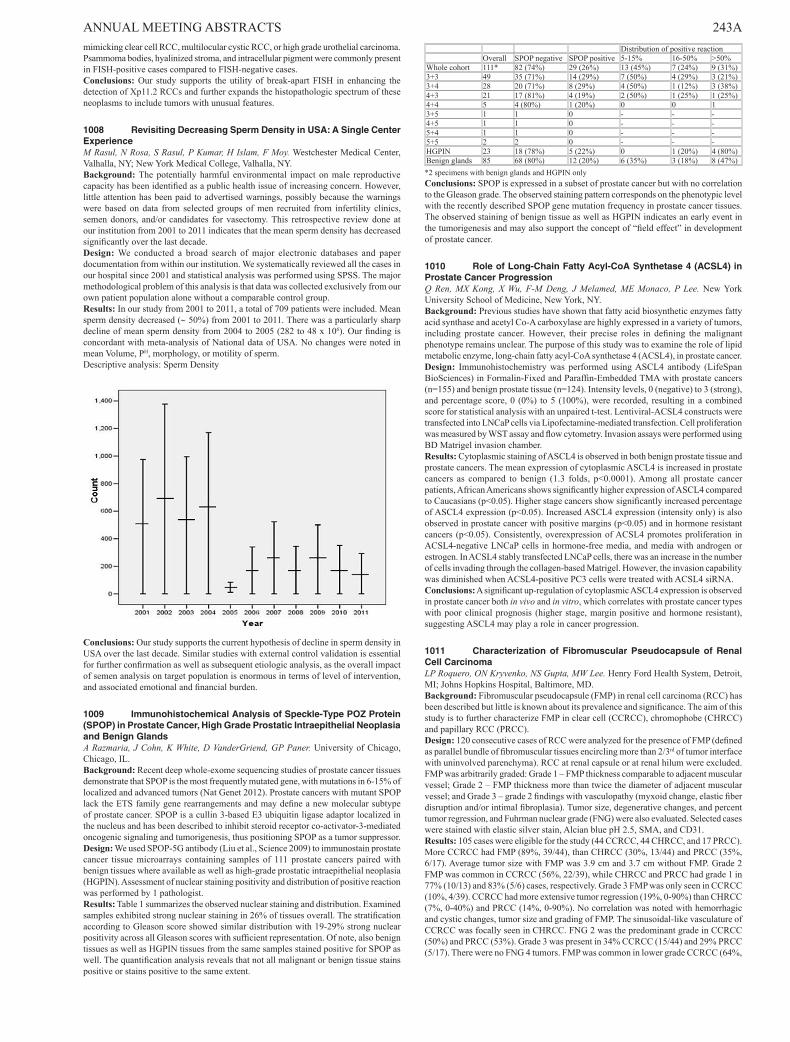
ANNUAL MEETING ABSTRACTS 243Amimicking clear cell RCC, multilocular cystic RCC, or high grade urothelial carcinoma. Psammoma bodies, hyalinized stroma, and intracellular pigment were commonly present in FISH-positive cases compared to FISH-negative cases.Conclusions: Our study supports the utility of break-apart FISH in enhancing the detection of Xp11.2 RCCs and further expands the histopathologic spectrum of these neoplasms to include tumors with unusual features.
1008 Revisiting Decreasing Sperm Density in USA: A Single Center ExperienceM Rasul, N Rosa, S Rasul, P Kumar, H Islam, F Moy. Westchester Medical Center, Valhalla, NY; New York Medical College, Valhalla, NY.Background: The potentially harmful environmental impact on male reproductive capacity has been identified as a public health issue of increasing concern. However, little attention has been paid to advertised warnings, possibly because the warnings were based on data from selected groups of men recruited from infertility clinics, semen donors, and/or candidates for vasectomy. This retrospective review done at our institution from 2001 to 2011 indicates that the mean sperm density has decreased significantly over the last decade.Design: We conducted a broad search of major electronic databases and paper documentation from within our institution. We systematically reviewed all the cases in our hospital since 2001 and statistical analysis was performed using SPSS. The major methodological problem of this analysis is that data was collected exclusively from our own patient population alone without a comparable control group.Results: In our study from 2001 to 2011, a total of 709 patients were included. Mean sperm density decreased (∼ 50%) from 2001 to 2011. There was a particularly sharp decline of mean sperm density from 2004 to 2005 (282 to 48 x 106). Our finding is concordant with meta-analysis of National data of USA. No changes were noted in mean Volume, PH, morphology, or motility of sperm.Descriptive analysis: Sperm Density
Conclusions: Our study supports the current hypothesis of decline in sperm density in USA over the last decade. Similar studies with external control validation is essential for further confirmation as well as subsequent etiologic analysis, as the overall impact of semen analysis on target population is enormous in terms of level of intervention, and associated emotional and financial burden.
1009 Immunohistochemical Analysis of Speckle-Type POZ Protein (SPOP) in Prostate Cancer, High Grade Prostatic Intraepithelial Neoplasia and Benign GlandsA Razmaria, J Cohn, K White, D VanderGriend, GP Paner. University of Chicago, Chicago, IL.Background: Recent deep whole-exome sequencing studies of prostate cancer tissues demonstrate that SPOP is the most frequently mutated gene, with mutations in 6-15% of localized and advanced tumors (Nat Genet 2012). Prostate cancers with mutant SPOP lack the ETS family gene rearrangements and may define a new molecular subtype of prostate cancer. SPOP is a cullin 3-based E3 ubiquitin ligase adaptor localized in the nucleus and has been described to inhibit steroid receptor co-activator-3-mediated oncogenic signaling and tumorigenesis, thus positioning SPOP as a tumor suppressor.Design: We used SPOP-5G antibody (Liu et al., Science 2009) to immunostain prostate cancer tissue microarrays containing samples of 111 prostate cancers paired with benign tissues where available as well as high-grade prostatic intraepithelial neoplasia (HGPIN). Assessment of nuclear staining positivity and distribution of positive reaction was performed by 1 pathologist.Results: Table 1 summarizes the observed nuclear staining and distribution. Examined samples exhibited strong nuclear staining in 26% of tissues overall. The stratification according to Gleason score showed similar distribution with 19-29% strong nuclear positivity across all Gleason scores with sufficient representation. Of note, also benign tissues as well as HGPIN tissues from the same samples stained positive for SPOP as well. The quantification analysis reveals that not all malignant or benign tissue stains positive or stains positive to the same extent.
Distribution of positive reaction Overall SPOP negative SPOP positive 5-15% 16-50% >50%Whole cohort 111* 82 (74%) 29 (26%) 13 (45%) 7 (24%) 9 (31%)3+3 49 35 (71%) 14 (29%) 7 (50%) 4 (29%) 3 (21%)3+4 28 20 (71%) 8 (29%) 4 (50%) 1 (12%) 3 (38%)4+3 21 17 (81%) 4 (19%) 2 (50%) 1 (25%) 1 (25%)4+4 5 4 (80%) 1 (20%) 0 0 13+5 1 1 0 - - -4+5 1 1 0 - - -5+4 1 1 0 - - -5+5 2 2 0 - - -HGPIN 23 18 (78%) 5 (22%) 0 1 (20%) 4 (80%)Benign glands 85 68 (80%) 12 (20%) 6 (35%) 3 (18%) 8 (47%)*2 specimens with benign glands and HGPIN onlyConclusions: SPOP is expressed in a subset of prostate cancer but with no correlation to the Gleason grade. The observed staining pattern corresponds on the phenotypic level with the recently described SPOP gene mutation frequency in prostate cancer tissues. The observed staining of benign tissue as well as HGPIN indicates an early event in the tumorigenesis and may also support the concept of “field effect” in development of prostate cancer.
1010 Role of Long-Chain Fatty Acyl-CoA Synthetase 4 (ACSL4) in Prostate Cancer ProgressionQ Ren, MX Kong, X Wu, F-M Deng, J Melamed, ME Monaco, P Lee. New York University School of Medicine, New York, NY.Background: Previous studies have shown that fatty acid biosynthetic enzymes fatty acid synthase and acetyl Co-A carboxylase are highly expressed in a variety of tumors, including prostate cancer. However, their precise roles in defining the malignant phenotype remains unclear. The purpose of this study was to examine the role of lipid metabolic enzyme, long-chain fatty acyl-CoA synthetase 4 (ACSL4), in prostate cancer.Design: Immunohistochemistry was performed using ASCL4 antibody (LifeSpan BioSciences) in Formalin-Fixed and Paraffin-Embedded TMA with prostate cancers (n=155) and benign prostate tissue (n=124). Intensity levels, 0 (negative) to 3 (strong), and percentage score, 0 (0%) to 5 (100%), were recorded, resulting in a combined score for statistical analysis with an unpaired t-test. Lentiviral-ACSL4 constructs were transfected into LNCaP cells via Lipofectamine-mediated transfection. Cell proliferation was measured by WST assay and flow cytometry. Invasion assays were performed using BD Matrigel invasion chamber.Results: Cytoplasmic staining of ASCL4 is observed in both benign prostate tissue and prostate cancers. The mean expression of cytoplasmic ASCL4 is increased in prostate cancers as compared to benign (1.3 folds, p<0.0001). Among all prostate cancer patients, African Americans shows significantly higher expression of ASCL4 compared to Caucasians (p<0.05). Higher stage cancers show significantly increased percentage of ASCL4 expression (p<0.05). Increased ASCL4 expression (intensity only) is also observed in prostate cancer with positive margins (p<0.05) and in hormone resistant cancers (p<0.05). Consistently, overexpression of ACSL4 promotes proliferation in ACSL4-negative LNCaP cells in hormone-free media, and media with androgen or estrogen. In ACSL4 stably transfected LNCaP cells, there was an increase in the number of cells invading through the collagen-based Matrigel. However, the invasion capability was diminished when ACSL4-positive PC3 cells were treated with ACSL4 siRNA.Conclusions: A significant up-regulation of cytoplasmic ASCL4 expression is observed in prostate cancer both in vivo and in vitro, which correlates with prostate cancer types with poor clinical prognosis (higher stage, margin positive and hormone resistant), suggesting ASCL4 may play a role in cancer progression.
1011 Characterization of Fibromuscular Pseudocapsule of Renal Cell CarcinomaLP Roquero, ON Kryvenko, NS Gupta, MW Lee. Henry Ford Health System, Detroit, MI; Johns Hopkins Hospital, Baltimore, MD.Background: Fibromuscular pseudocapsule (FMP) in renal cell carcinoma (RCC) has been described but little is known about its prevalence and significance. The aim of this study is to further characterize FMP in clear cell (CCRCC), chromophobe (CHRCC) and papillary RCC (PRCC).Design: 120 consecutive cases of RCC were analyzed for the presence of FMP (defined as parallel bundle of fibromuscular tissues encircling more than 2/3rd of tumor interface with uninvolved parenchyma). RCC at renal capsule or at renal hilum were excluded. FMP was arbitrarily graded: Grade 1 – FMP thickness comparable to adjacent muscular vessel; Grade 2 – FMP thickness more than twice the diameter of adjacent muscular vessel; and Grade 3 – grade 2 findings with vasculopathy (myxoid change, elastic fiber disruption and/or intimal fibroplasia). Tumor size, degenerative changes, and percent tumor regression, and Fuhrman nuclear grade (FNG) were also evaluated. Selected cases were stained with elastic silver stain, Alcian blue pH 2.5, SMA, and CD31.Results: 105 cases were eligible for the study (44 CCRCC, 44 CHRCC, and 17 PRCC). More CCRCC had FMP (89%, 39/44), than CHRCC (30%, 13/44) and PRCC (35%, 6/17). Average tumor size with FMP was 3.9 cm and 3.7 cm without FMP. Grade 2 FMP was common in CCRCC (56%, 22/39), while CHRCC and PRCC had grade 1 in 77% (10/13) and 83% (5/6) cases, respectively. Grade 3 FMP was only seen in CCRCC (10%, 4/39). CCRCC had more extensive tumor regression (19%, 0-90%) than CHRCC (7%, 0-40%) and PRCC (14%, 0-90%). No correlation was noted with hemorrhagic and cystic changes, tumor size and grading of FMP. The sinusoidal-like vasculature of CCRCC was focally seen in CHRCC. FNG 2 was the predominant grade in CCRCC (50%) and PRCC (53%). Grade 3 was present in 34% CCRCC (15/44) and 29% PRCC (5/17). There were no FNG 4 tumors. FMP was common in lower grade CCRCC (64%,

244A ANNUAL MEETING ABSTRACTS 25/39) and PRCC (83%, 5/6). SMA and CD31 showed linear slit-like endothelium-lined spaces within the FMP. Arterial elastic membrane disruption or fibrointimal mucin deposits were present in grade 3 FMP.Conclusions: FMP is rather characteristic of CCRCC, less frequent in CHRCC and rare in PRCC. The smooth muscle in FMP may be vascular in origin and may represent complex vasculopathy presumably from increased intratumoral vascular pressure of sinusoidal vascular network. Tumor regression and degenerative changes in relation to FMP need to be further elucidated.
1012 Incidental Secondary Pathological Findings in Prostate Needle Biopsies and Radical Prostatectomy SpecimensFL Rosario-Quinones, PD Unger, G-Q Xiao. Mount Sinai Medical Center, New York, NY.Background: Prostate needle biopsies and radical prostatectomies have become increasingly common specimens examined by pathologists. Existing literature has mainly focused on the main three pathologic processes that commonly affect the prostate gland; benign prostatic hyperplasia, inflammation, and carcinoma. No existing literature has systemically looked into additional pathologic processes in the prostate gland. We present a series of secondary pathologies identified in prostate biopsies and radical prostatectomies over the course of six years.Design: We retrospectively and prospectively reviewed all the prostate biopsies and radical prostatectomies collected at our institution between 2006-2012. A total of 8350 cases were reviewed, 135 of these were radical cystoprostatectomies for bladder cancer, with or without concurrent prostate cancer. Pelvic lymph node dissections pertaining to radical prostatectomies were also included in the study.Results: Secondary pathological findings were identified in 135 of the reviewed cases. (Table 1)Table 1. Incidental Secondary Pathologies Found in Prostate SpecimensFinding Prostate Biopsies Radical Prostatectomies Prostate Pelvic Lymph NodesGranulomas 11* 9* 12**Seminal Vesicle Amyloid 8 36 Lymphoma 2† 4††Malakoplakia 2 1 Periprostatic Atherosclerosis‡ Mild 27 Moderate 17 Severe 6 *Bacillus Calmette-Guerin (BCG) related. **3 cases were related to femoral prostheses, 9 cases had no known cause. † 1 small lymphocytic lymphoma/chronic lymphocytic leukemia (SLL/CLL), 1 diffuse large B-cell lymphoma. †† All 4 cases were SLL/CLL. ‡ 50 consecutive cases were reviewedConclusions: Our studies revealed periprostatic atherosclerosis as the most common incidental secondary pathology found in prostate specimens, reflective of the elderly population in this study. Seminal vesicle amyloidosis was the second most common finding. This is a localized form of amyloid disease and is usually asymptomatic. Granulomas were the third most common finding, seen in prostate biopsies, radical prostatectomies, and pelvic lymph nodes. The majority of these were attributable to BCG treatment for bladder carcinoma. Lymphomas involving the prostate and pelvic lymph nodes are rare, but could be the first indication of disease recurrence. Malakoplakia of the prostate gland is extremely rare, but one needs to recognize this lesion to avoid the pitfall of over diagnosing a carcinoma. In summary, although rare and mostly inconsequential, secondary pathologies found in prostate specimens may be of clinical importance and could prove useful in assisting medical treatment.
1013 Metastatic Chemotherapy Resistant Urothelial Carcinoma (UC): Next Generation Sequencing (NGS) Reveals a Diverse Spectrum of Genomic Alterations (GA) and Targets of TherapyJS Ross, T Nazeer, CE Sheehan, G Palmer, G Otto, S Downing, J Curran, R Yelensky, D Lipson, S Balasubramanian, L Garcia, K Mahoney, P Stephens, V Miller. Albany Medical Center, Albany, NY; Foundation Medicine Inc., Cambridge, MA.Background: Metastatic UC typically follows an aggressive clinical course and survival is modest despite use of systemic platin-based chemotherapy. The recent introduction of comprehensive NGS genomic profiling to the clinical setting has enabled the identification of novel and unanticipated drug targets of therapy for patients with metastatic chemoresistant UC.Design: NGS was performed on hybridization-captured, adaptor ligation based libraries using DNA extracted from 4 FFPE sections cut at 10 microns from 15 UC that had relapsed and progressed after primary surgery and conventional chemotherapy. The exons of 182 cancer-related genes were fully sequenced using the Illumina HiSeq 2000 to at an average sequencing depth of 1153X and evaluated for all classes of GA: point mutations (mut), insertions, deletions, copy number alterations (amp or hom del), and gene fusions/rearrangements. Actionable GA were defined as impacting the selection of targeted anti-cancer therapies on the market or in registered clinical trials (CT).Results: The 15 UC had a mean age of 63.5 with 4 female (27%) and 11 male (73%) patients. All 15 (100%) tumors were high grade and Stage IV at the time of NGS. NGS was performed on the primary UC in 8 (53%) including 6 TURBT (40%) and 2 cystectomies (13%) and on metastatic tumor in 7 UC (47%) including 2 lymph node (13%) and 1 (7%) each liver, lung, brain, psoas and abdominal wall biopsies, NGS discovered a total of 54 GA in the UC series with an average of 4.2 GA per tumor. Ten of fifteen (67%) patients’ tumors harbored at least one GA potentially associated with clinical benefit of targeted therapies including: PIK3CA (20%); FGFR1 (20%); CDKN2A/B (13%); CCND3 (13%); NF1 (7%); NF2 (7%); PTEN (7%); ATR (7%); BRCA2 (7%), MYCL1 (7%); ERBB2 (7%); FBXW7 (7%); RAF1 (7%); EGFR (7%); KRAS (7%); MDM4 (7%) and FANCA (7%). Other biologically relevant GA were
identified in: TP53 (60%); ARID1A (40%); RB1 (27%); KDM6A (13%); MCL1 (7%) and MYC (7%). The GA detected by NGS generated eligibility of the 15 UC for a total of 75 registered anti-UC CT (average 5 CT per patient).Conclusions: Deep NGS of metastatic UC uncovers an unexpectedly high frequency of actionable GA that has the potential to influence therapy selection and can direct patients to enter clinical trials to study the benefit of anti-UC targeted therapies. The diversity and spectrum of actionable UC GA identified in this study would be challenging to characterize using other diagnostic methods.
1014 Prostate Carcinomas Arising in Lynch Syndrome Patients Frequently Show Concordant Mismatch Repair DeficiencyC Rosty, MD Walsh, M Clendenning, JP Young, N Lindor, JL Hopper, MA Jenkins, DD Buchanan. Queensland Institute of Medical Research, Brisbane, Australia; University of Queensland, Brisbane, Australia; Envoi Pathology, Brisbane, Australia; Sullivan Nicolaides Pathology, Brisbane, Australia; Mayo Clinic, Scottsdale, AZ; University of Melbourne, Melbourne, Australia.Background: Lynch syndrome (LS) is an inherited predisposition to developing carcinomas of the colorectum, endometrium, small intestine, stomach, pancreatico-biliary tract, ovary and upper urinary tract. Mutations in mismatch repair (MMR) genes MLH1, MSH2, MSH6, and PMS2 underpin Lynch syndrome. While prostate adenocarcinoma is not considered one of the spectrum tumours for LS, case reports have documented loss of MMR expression in prostate carcinoma from LS patients.Design: We sought to investigate the incidence of MMR loss of expression in prostate carcinoma from LS carriers enrolled in the Australasian Colorectal Cancer Family Registry (ACCFR). MMR immunohistochemistry was performed on tissue samples from prostate carcinoma (biopsies and prostatectomy specimens).Results: There were 15 prostate carcinomas from 15 LS patients (mean age = 62.2 years). Eight individuals had been diagnosed with colorectal carcinoma, one individual with pancreatic carcinoma, and prostate carcinoma was the only malignancy diagnosed in five individuals. MMR-deficiency was detected in 12/15 (80%) cases: MLH1/PMS2 loss in 2, MSH2/MSH6 loss in 10. The pattern of loss of MMR expression was consistent with the individual’s germline mutation in all cases. All fifteen adenocarcinomas were of acinar type: 1 with Gleason score (GS) 6 (this tumour had normal MMR expression and arose in an MSH6 mutation carrier), 7 with GS 7, and 7 with GS ≥8. Tumour infiltrating lymphocytes were noted in low numbers in seven of the tumours.Conclusions: These findings indicate that MMR-deficiency is commonly observed in prostate adenocarcinomas arising in Lynch syndrome mutation carriers. The prostate tumours with MMR-deficiency from these carriers demonstrated frequent high histologic grade, a feature in common with MMR-deficient cancers from other sites. Prostate carcinoma was the only tumour in 5/15 MMR carriers.
1015 micro-RNA Expression in Lethal and Non-Lethal Urothelial Cancers of the BladderD Rotzer, A Perren, A Fleischmann. University of Bern, Bern, Switzerland.Background: MicroRNAs (miRNAs) are small non-coding RNA of 21–23 nucleotides which negatively regulate gene expression post-transcriptionally. miRNAs have been shown to play a role in tumorigenesis by regulating molecular pathways involved in e.g. epithelial–mesenchymal transition, metastasis and angiogenesis.Design: We studied 20 muscle-invasive UCCs treated by radical cystectomy and standardized pelvic lymphadenectomy, 10 were lethal and 10 could be cured by surgery alone. Total RNA was extracted from formalin-fixed and paraffin-embedded (FFPE) tissue after testing different protocols in comparison to fresh frozen tissue. Reverse transcription was done with Megaplex RT Primers (Pool A and B) and the TaqMan miRNA reverse transcription kit (both Applied Biosystems) on a ABI GeneAmp 9700 Thermocycler. The subsequent profiling of 754 miRNAs was performed using TaqMan Array Human microRNA cards (A v2.0 and B v3.0). For normalization the median expression of all miRNAs was used and fold changes of individual miRNAs were calculated applying the DDCt method. All reactions were performed in triplicate and a negative control lacking cDNA was included.Results: Mean expression level of 64 miRNAs were significantly different between lethal and non-lethal UCC (unadjusted p<0.05). However, the variance of the normalized CTs was large for all miRNAs in both groups and overlapped (roughly 50% of the top 20 miRNA). Therefore, expression signatures as classifier were tested and the scoring pair miRNA98 and miRNA204 performed rather well (p=0.089) given the small sample size. This miRNA pair allowed classifying 19 of the 20 patients correctly.Conclusions: miRNA expression profiles in muscle invasive bladder cancer may help to identify patients at risk for lethal disease. Larger series are needed to further investigate the prognostic role of miRNA expression and profiles.
1016 Clinicopathological Connotation of SOX2 Amplification and Expression in Urothelial CarcinomaS Roy, A Karunamurthy, AM Luvison, MB Acqafondata, KM Cieply, AV Parwani. University of Pittsburgh Medical Center, Pittsburgh, PA.Background: SOX2, an oncogene, is expressed and amplified in squamous cell carcinoma (SCC) of the esophagus, lung and head and neck. Very recently, ALDH1 and SOX2 expression by immunohistochemistry (IHC) was reported in urothelial carcinoma (UC) of the upper urinary tract demonstrating negative prognostic implication. We attempted to investigate the expression and amplification status of SOX2 in the light of clinicopathologic parameters.Design: A total of 19 consecutive radical cystectomy specimens with diagnosis of UC were retrieved from case archive after IRB approval. After review of H&E stained sections, targets were marked for fluorescence in-situ hybridization (FISH). Dual-color FISH for SOX2 was performed using clone RP11-43F17 (CHORI, Oakland, CA) labeled

ANNUAL MEETING ABSTRACTS 245Awith Spectrum Orange (Abbott Molecular) and D3Z1 α-satellite labeled Spectrum Green (Cytocell aquarius, Cambridge, UK). Approximately 60 well delineated and non-overlapping cells were analyzed in the targeted region. IHC was performed using rabbit monoclonal antibody against SOX2 (Epitomics, Inc). IHC scoring was based on intensity of staining (1+ to 4+). For SOX2 positive staining, a minimum of 10% cells showed nuclear staining. A 4+ staining intensity was a surrogate for protein overexpression.Results: The examined cohort had a M:F of 15:4 with mean age of 66.4 years. 89.5% (17/19) cases were diagnosed as high grade UC with four cases showing focal squamous and micropapillary features. The diagnosis of small cell carcinoma and micropapillary carcinoma comprised the remaining 2 cases. Overall, majority of UC showed very heterogeneous staining pattern of SOX2. 26% (5/19) cases demonstrated SOX2 overexpression (4+). In contrast SOX2 was amplified in only 21% (4/19) cases and 60% (3/5) cases with SOX2 overexpression. Interestingly, SOX2 was amplified in one case without SOX2 overexpression (2+). Amplification and expression did not correlate with T-stage, grade, tumor size and perineural invasion. Patients with SOX2 amplification demonstrated shorter median survival in contrast to SOX2 non-amplified cases (10.5 vs 15.5 months); difference of comparative magnitude was not seen in SOX2 expression patterns (13.1 vs 13.3 months).Conclusions: Although SOX2 amplification and expression was seen in UC, there was a lack of concordance. SOX2 expression by IHC in UC should be interpreted with caution due to highly heterogeneous staining. SOX2 amplified cases appear to show shorter median survival than SOX2 non-amplified cases; further analysis using a larger cohort is under progress in our institution to test this hypothesis.
1017 Somatic Copy Number Alterations by Whole Exome Sequencing Reveals YWHAZ and PTK2 as Potential Therapeutic Targets in Castration Resistant Prostate CancerK Ruenauver, R Menon, M Deng, F Kunze, D Bohm, W Vogel, V Scheble, F Fend, G Kristiansen, N Wernert, N Oberbeckmann, S Biskup, M Rubin, Z Shaikhibrahim, S Perner. Institute of Pathology, University Hospital of Bonn, Bonn, Germany; University Hospital of Tuebingen, Tuebingen, Germany; CeGaT GmbH, Tuebingen, Germany; Weill Cornell Medical College of Cornell University, New York, NY; Institute of Pathology, University Hospital of Tuebingen, Tuebingen, Germany.Background: Castration resistant prostate cancer (CRPC) is the most aggressive form of prostate cancer (PCa) with a poor prognosis and remains a therapeutic challenge. The key to the development of novel therapeutic targets for CRPC is to decipher the molecular alterations underlying this lethal disease. The aim of our study was to identify therapeutic targets for CRPC by assessing somatic copy number alterations (SCNA) by whole exome sequencing on five CRPC/normal paired formalin fixed paraffin embedded (FFPE) samples.Design: Genomic DNA was extracted from 5 CRPC/normal paired FFPE samples and sequenced using the SOLiD4 next generation sequencing (NGS) platform. Data were validated using fluorescence in-situ hybridization (FISH) on a PCa progression cohort containing 352 PCa samples. Gene copy number amplification status, mRNA and protein expression were determined in selected PCa cell lines. In vitro functional assays were performed with specific inhibition or siRNA knockdown in PCa cell lines.Results: Among a set of known amplified/deleted genes in PCa (PTEN, AR, cMYC, NKX3.1), whole exome sequencing identified the 8q12.2-24.22 region including PTK2 and YWHAZ to be amplified. FISH analysis of this region showed an increasing amplification frequency of PTK2 and YWHAZ with disease progression. PTK2 was amplified in 1% of the localized PCa and 35% of CRPC samples. YWHAZ was amplified in 4% of the localized PCa and 48% of the CRPC samples. High level amplification of PTK2/YWHAZ in PC3 cells was revealed by FISH analysis. PTK2 inhibition using the specific inhibitor TAE226 and YWHAZ siRNA knockdown significantly reduced proliferation and migration in PC3 cells.Conclusions: Our findings suggest that inhibition of PTK2 and YWHAZ could delay disease progression in CRPC patients harbouring amplification of the latter genes. The validated sequencing data show that FFPE tissue could be a promising alternative for SCNA screening using NGS technologies.
1018 Comparison of p40 (∆Np63) and p63 Expression in Prostate Tissues as Markers of Benign GlandsV Sailer, C Stephan, N Wernert, S Perner, G Kristiansen. University of Bonn, Bonn, Germany; Charite-Universitätsmedizin Berlin, Berlin, Germany.Background: The diagnosis of prostate carcinoma can be challenging, especially in small needle core biopsies. Immunohistochemistry provides added information regarding the loss of basal cells, which is a key feature of malignancy in the prostate gland. p63, a homolog of the p53 suppressor gene, is one of the standard markers for basal cells of the prostatic gland. Recently, it has been suggested that the p63 isoform p40 might be more specific as a basal cell marker. Here we compare the staining characteristics of p63 and p40 in normal and malignant prostate tissue.Design: A prostatectomy cohort (n=640) in tissue microarray format (with 5 cores per case) was evaluated for p63 (clone 4A4) and for p40 (rabbit polyclonal) immunoreactivity in malignant and benign tissues alike. Immunoreactivity of basal and secretory cells was evaluated in a semiquatitative manner and compared case-wise.Results: In benign tissues, p40 showed a highly similar immunoreactivity compared to p63. The staining patterns were totally equal 73% of cases, minor differences were detected in 27%. A mild to moderate cytoplasmatic p40 staining in tumour cells occurred in 59% of cancer cases. For the nuclear staining, differences were seen in carcinomas: 1.4 % of carcinomas were p63-positive, whereas p40 labelled only 0.6% of these cases.Conclusions: p40 can be used as a reliable marker of basal cells in prostate diagnostics and it appears to be more specific with a lower rate of false-positive cancer cases compared to p63. However, the additional cytoplasmic staining makes it a less attractive
partner to combine it with Alpha-methyl-Co-racemase (AMACR). This study also highlights that the number of p63-positive prostate carcinomas is probably higher than previously thought.
1019 Characteristics of Prostatic Adenocarcinoma in a Contemporary Australian Radical Prostatectomy Series from a Community without Population-Based PSA ScreeningH Samaratunga, J Yaxley, J Perry-Keene, M Adamson, B Delahunt. Aquesta Pathology, Brisbane, Queensland, Australia; Royal Brisbane Hospital, Brisbane, Queensland, Australia; Wellington School of Medicine and Health Sciences, University of Otago, Wellington, New Zealand.Background: With PSA testing, up to 30% of small potentially clinically insignificant cancers are detected. Possible overtreatment is an issue with some of these cases. The current study was performed to characterize prostate cancer in men undergoing radical prostatectomy in a community without population based PSA screening.Design: Clinical and pathological data of 2700 patients who had a radical prostatectomy (RP) between 2008 and 2012 were analysed. Specimens were entirely embedded and evaluated by routine haematoxylin and eosin staining. Tumours were graded using the ISUP modified Gleason grading system and staged according to the ISUP RP staging system. Tumors were considered potentially clinically insignificant if the tumor volume was <0.5 mL and Gleason score <7.Results: The mean age was 61.4 years (range 32-79) and the mean pre-operative PSA, 19.2ng/mL (range 0.4 – 69). Indications for PSA testing included an abnormal digital rectal examination, family history of prostate cancer, obstructive urinary symptoms, community awareness or general practitioner initiation. Potentially clinically insignificant tumors were found in 129 (4.7%) patients. Of 2571 significant cancers 2383 (88%) had a final Gleason score of 7 or greater, 43% had extraprostatic extension, 640 (23%) had a tumour volume of greater than 3 mL and 341 (12.6%) had surgical margin positivity. Seminal vesical involvement was found in 156 (5.8%) cases. Of 646 patients who had a lymphadenectomy, 29 (4.49%) had lymph node metastases.205 (7.5%) patients were aged ≤50 (mean age 47 years) of whom 188 (91%) had significant cancer.Conclusions: The vast majority of prostate cancers in populations without PSA screening are significant with a high proportion of cases showing high stage disease, seminal vesicle involvement and lymph node metastasis. Absence of population–based PSA screening appears to lead to a very high risk of prostate cancer being advanced at presentation. Selective PSA screening in men with minimum co-morbidities and a life expectancy of >15 years will lead to earlier stage disease and potentially improve long term local control and cancer free survival.
1020 Recurrence and Progression Rates in Non-Invasive Papillary Urothelial Neoplasms of the Bladder: A Study of Tumors with Mixed Low and High GradesRT Samdani, SG Sharma, RM Cox, HJ Spencer, N Gokden. University of Arkansas for Medical Sciences, Little Rock, AR.Background: The 2004 WHO/ISUP system categorizes non-invasive papillary urothelial neoplasms (PUN) into papilloma, papillary urothelial neoplasm of low malignant potential, papillary urothelial carcinoma low grade (LG) and high grade (HG). The tumors with mixed LG and HG areas do exist and WHO/ISUP system recommends that LG PUN cases that contain HG areas should be classified as HG. It is unclear from the existing literature whether the percentage of HG area affects the rates of recurrence and progression in mixed tumors.Design: Retrospectively (2006-12) cases with an initial biopsy/transurethral resection diagnosis of non-invasive HG and LG PUN, and follow-up biopsies were reviewed. Forty-two cases met the criteria with an average follow up of 10.7 months (range:3-28 months). Three groups were created: 1- Pure LG; 2- LG with HG foci (2A:<10%, 2B:11-50%, 2C: >51%); 3- Pure HG. The criteria for diagnosing HG foci were: prominent architectural and cytological distortion at low power, increased N/C ratio, pleomorphism, and clumped chromatin. Recurrence was defined as occurrence on follow up of >3 months. Progression in stage and/or grade was studied. Cystoscopic findings were reviewed. Fisher’s exact test utilized for statistical analysis.Results: The average age was 67 years (44-79 yrs), M:F ratio was 3:1. The cases are summarized in Table 1. Four mixed LG/HG, 2 pure HG, and 1 pure LG case revealed stage progression from non-invasive to invasive carcinoma. The HG foci in mixed cases with stage progression ranged from 5 to 80%. Cystoscopically the LG/HG group had multifocal lesions at initial presentation and recurrences similar to those of pure HG cases.Table 1: Summary of findings
Group No. of cases (n=42)
Recurrence n (%)
Grade Progression n (%)
Stage progression n (%)
Multifocality n (%) [1st bx]
Multifocality n (%) [Recurrence]
1 (p=0.87) 6 (60%) 0 1 (10%) 2 (20%) 1 (10%)2 20 4 (20%) 12 (60%) 14 (70%)2A 8 8 (100%) 0 1 3 52B 10 8 (80%) 2 (20%) 2 8 72C 2 2 (100%) 0 1 1 23 12 9 (75%) 0 2 (17%) 10 (83%) 12 (100%)
Conclusions: The LG/HG mixed tumors with recurrence, grade and stage progression faired similar to that of tumors composed of pure histologic grades. The extent of HG foci in mixed tumors appears not to have any effect on tumor recurrence (p=0.16), grade (p=0.48) and stage (p=0.87) progression. However, multifocality was statistically significant at the time of first biopsy (p=0.009) and recurrence (p<0.0001). Therefore, further studies with more patients and longer follow-up are needed to understand better the biology of mixed PUN.

246A ANNUAL MEETING ABSTRACTS 1021 The Utility of SF-1 and Cathepsin K as Differentiating Immunohistochemical Markers in Adrenal Cortical Carcinoma and Epithelioid AngiomyolipomaTD Samulski, S Bai, AL Nunez, S Wei, L Wang, Z Bing. Hospital of the University of Pennsylvania, Philadelphia, PA; New York University, New York, NY; University of Alabama, Birmingham, AL.Background: Distinguishing primary tumors of the kidney and adrenal gland can at times be challenging. This is especially true in cases that share morphologic and immunhistochemical characteristics, such as those cases of adrenal cortical carcinoma and epithelioid variant of angiomyolipoma (EAML); two rare, yet aggressive tumors. We explore the diagnostic utility of immunohistochemical staining for SF-1 and cathepsin K in combination for differentiating these two distinct neoplasms.Design: Fourteen cases of EAML were stained with cathepsin K and steroidogenic factor (SF-1) antibodies. 20 cases of adrenal cortical carcinoma (in tissue microarray) were also stained with cathepsin K antibody. Staining in all cases was evaluated and scored for intensity (0, negative; 1, weak intensity; 2, moderate intensity; 3, strong intensity) and extent of immunoreactivity (0, negative; 1, <30%; 2, 30-70%; 3, >70%). The final scores were achieved by the sum of intensity and extent of immunoreactivity divided by two.Results: Staining for cathepsin K in cases of epithelioid angiomyolipoma resulted in an average score of 2.8+/-0.3, comparable to an average score of 2.8+/-0.2 in cases of andrenal cortical carcinoma. Cases of EAML failed to demonstrate staining for SF-1, with an average score of 0. This is in contrast to SF-1 staining in adrenal cortical carcinoma. We have demonstrated previously that all adrenal cortical carcinomas showed strong nuclear staining for SF-1.Conclusions: In our experience, staining for cathepsin K and SF-1 was a concise panel in differentiating EAML and adrenal cortical carcinoma, in which both EAML and adrenal cortical carcinoma showed strong positivity for cathepsin K, yet only adrenal cortical carcinoma showed strong positivity for SF-1, while EAML was negative for this factor.
1022 Expression of EMT Markers in Sarcomatoid Urothelial Carcinoma of the BladderJM Sanfrancesco, S Gupta, A Shoskes, G Lieser, D Hansel. Cleveland Clinic Foundation, Cleveland, OH.Background: The sarcomatoid variant of urothelial carcinoma (UCa) of the urinary bladder is one of several rare, aggressive variants that have posed challenges for clinicians in determining prognosis and treatment. Epithelial-to-mesenchymal transition (EMT) has been described to occur in instances in which tumors develop increased motility and propensity to spread; this phenomenon has been associated with the development of a spindled morphology in multiple in vitro settings. We therefore explored the expression of EMT markers in sarcomatoid UCa – a highly aggressive variant of UCa that is characterized by a spindled, mesenchymal-like appearance.Design: We compiled 26 cases of sarcomatoid UCa diagnosed at our institution between the years 2000 to 2012. The cases consisted of biopsies (n=3), transurethral resections (n=3), and radical cystectomy /cystoprostatectomies (n=20). A tissue microarray (TMA) containing multiple representations of each tumor was constructed and stained for N-cadherin (Ventana, 1:200 dilution), B-catenin (BD-Biosciences, 1:250 dilution), pS6 (Ventana, 1:25 dilution), Vimentin (Ventana, 1:100 dilution), ZEB1 (Ventana, 1:100 dilution), FoxC2 (Ventana, 1:25 dilution), E-cadherin (Invitrogen, 1:250 dilution), and SNAIL/SL (Ventana, 1:50 dilution). Interpretation of immunohistochemical (IHC) staining was completed using a 0-3 scale, with most prominent expression, based on intensity, being classified at 3 and no expression being classified as 0.Results: Of the 26 patients utilized in this study, 15 were men and 11 were women (M:F ratio = 1.4) ranging from 42-92 years of age. IHC staining of ≥ 2 was considered positive and was diffusely and uniformly expressed in the neoplastic cells with sarcomatoid features. Vimentin and FoxC2 were positive in all cases, and positive staining for SNAIL/SL and B-catenin was 88.5% (23/ 26) and 80.8% (21/26) respectively. E-cadherin showed a positive staining pattern in only 4 of 26 cases (15.4%). ZEB1, pS6, and N-cadherin showed mixed patterns of positivity with 80.1% (18/26), 53.8% (14/26), and 46.2% (12/26), respectively.Conclusions: Sarcomatoid UCa of the bladder appears to demonstrate consistent properties of EMT, including loss of E-cadherin expression and increased Vimentin and SNAIL/SL expression. Variable staining for additional EMT markers, such as ZEB1, pS6 and N-cadherin, remains to be explored in more detail. Ultimately, in vitro and in vivo functional studies that demonstrate alteration in behavior associated with EMT marker expression will be required to demonstrate the relationship between phenotype and tumor cell behavior.
1023 Expression of PAX8 in Non-Invasive Urothelial Carcinoma of the BladderJM Sanfrancesco, C Magi-Galluzzi, D Hansel. Cleveland Clinic Foundation, Cleveland, OH.Background: Pax8, one of nine members of the paired box gene family, is a transcription factor expressed in embryonic and normal adult tissues. This gene family is responsible for development of multiple organ systems by maintaining a progenitor state while preventing terminal differentiation. More recently, the expression of Pax8 in selected neoplasms, particularly thyroid and kidney, has been explored, although information in other tumor types is somewhat limited. We examined the expression pattern of Pax8 as a diagnostic marker in non-invasive papillary urothelial carcinoma (UCa), which has not been previously evaluated in detail.Design: Using a laboratory database search system, 80 cases of non-invasive UCa from 2005 to 2012 were compiled, including low-grade (n=41) and high-grade (n=39) disease. Follow-up for each patient was obtained to ensure that no progression to invasive carcinoma had occurred at the time of this study. Immunohistochemical (IHC)
staining with Pax8 was performed on constructed tissue microarrays. Interpretation of staining was completed using a 0-3 scale, with most prominent expression, based on intensity, being classified at 3 and no expression being classified as 0. IHC staining ≥ 2 was considered to be a positive result.Results: High levels of Pax8 (≥ 2 staining) were identified in 68.2% (28/41) of low-grade and 61.5% (24/39) of high-grade non-invasive UCa cases. The positive staining pattern was similar in distribution for both high- and low-grade UCa cases. More intense staining was appreciated in the superficial portion of the epithelium, but positivity was diffuse in a majority of neoplastic cells.Conclusions: Pax8 has been shown to be a sensitive and specific IHC marker in renal cell carcinoma. Based on our study, it is evident that Pax8 appears to be expressed in the majority of non-invasive, low-grade and high-grade papillary UCa of the bladder, suggesting a conserved role in the development and/or maintenance of non-invasive papillary lesions at this site. Further studies that evaluate the functional role of this transcription factor in bladder cancer are warranted.
1024 Sarcomatoid Urothelial Carcinoma of the Urinary Bladder: A Clinicopathologic Study of 31 CasesJM Sanfrancesco, D Hansel. Cleveland Clinic Foundation, Cleveland, OH.Background: Urothelial carcinoma (UCa) represents the most common type of bladder cancer (>90%), with multiple histological variants of UCa reported. One of the more aggressive types is the sarcomatoid variant; however, studies on outcomes are limited by the number of published articles that address this rare variant. We reviewed one of the larger cohorts of sarcomatoid UCa and present clinicopathological findings associated with this entity.Design: Using a laboratory database search, 31 cases of sarcomatoid UCa were compiled from 2000 to present, including biopsies (n=3), transuretheral resection (TUR) specimens (n=3), and radical cystectomy and cystoprostatectomy specimens (n=25). All cases were re-reviewed for histopathological parameters, including diagnosis, margin status, tumor stage, presence/absence of lymph node metastases, and concurrent in situ disease. Patient follow-up was obtained, with included disease course, treatment modalities, and survival data.Results: Of the 31 cases reviewed, nineteen patients were men and twelve were women (M:F ratio = 1.6:1); ages ranged from 45 to 92 (mean 70.3; median 71.5). All patients (n=31) had biopsy/TUR proven invasive UCa, with 58.1% (18/31) with sarcomatoid features at initial diagnosis. All patients who received only biopsy/TUR (n=6) had invasive UCa with sarcomatoid features and 50% (3/6) had metastases at diagnosis. None of these patients had previously been treated with Bacillus Calmette–Guérin (BCG) and 67% (4/6) of these patients received post-procedural chemotherapy and/or palliative radiation. Of the patients who underwent cystectomy or cystoprostatectomy (n=25), 36% (9/25) had lymph node metastases at resection, 16% (4/25) had a positive soft tissue margin, and 8% (2/25) had a margin involved by in situ disease. 64% (16/25) of patients received post-operative/procedural chemotherapy and one patient (4%) received combination chemo-and radiotherapy. At the time of this study, twenty-one patients were deceased (87.5%). Overall mean survival for the deceased patients was 9.1 months with four patients expiring secondary to surgical complications and medical co-morbidities. Seven patients were lost to follow-up.Conclusions: Sarcomatoid UCa represents an aggressive form of UCa, with the majority of patients dying of disease within one year of diagnosis. Further evaluation of molecular targets in this disease entity may be of value to identify potential new therapeutic options to enhance survival.
1025 Hormonotherapy in Prostate Cancer Influences Tumor Microvessel Density: New Perspectives for Prostate Cancer Treatment?R Santi, E Comperat, L Masieri, S Serni, P Camparo, M Carini, G Nesi. University of Florence, Florence, Italy; Hopital La Pitié Salpetrière, Paris, France; Centre de Pathologie Amiens Picardie, Amiens, France.Background: Radical prostatectomy (RP) represents an effective option for prostate cancer (PCa) treatment. In selected cases, surgery may follow neoadjuvant hormonotherapy. It has been proven that angiogenesis plays a crucial role in PCa growth. The aim of this study was to investigate the effects of hormonotherapy on Microvessel Density (MVD) and pathologic stage in a series of PCa patients treated with RP.Design: Ninety-six RP cases were retrospectively collected. Fifty-one patients had surgery only (untreated) while 45 received androgen ablation therapy (treated) for 3 months before surgery. Tissue microarrays (TMAs) were constructed using 3 tissue cores for each tumor sample. Endothelial expression of CD31 (DAKO, Denmark) and LYVE-1 (Santa Cruz, CA) was assessed immunohistochemically. CD31 and LYVE-1 staining hot spot areas were used to evaluate MVD with a semi-quantitative scoring system.Results: Median age of patients was 65.3 years (range: 49-75 years). Pathologic stage was T2 in 24 untreated and 28 treated patients and T3 in 21 untreated and 23 treated patients. Hormonotherapy was significantly associated with increased MVD measured by CD31 (p=0.009) and LYVE-1 (p<0.001) expression. In addition, a statistically significant correlation was demonstrated between high pathologic stage and increased MVD obtained by CD31 (p=0.026) but not by LYVE-1 staining.Conclusions: Our study showed a correlation between hormonotherapy and increase in the number of intra-tumoral vessels, particularly the lymphatic ones. Although the mechanism by which hormonotherapy increases MVD in PCa remains to be elucidated, anti-angiogenic therapy should be considered in PCa patients undergoing total androgen ablation.

ANNUAL MEETING ABSTRACTS 247A1026 Upper Urinary Tract Urothelial Carcinoma with Intratubular SpreadJ Sarungbam, M Choudhury, X Yang, M Zhong. New York Medical College, Valhalla, NY; Northwestern University Feinberg School of Medicine, Chicago, IL.Background: Upper urinary tract urothelial cell carcinomas (UUT-UCs) are uncommon, accounting 7% to 8% of all renal malignancies and representing 5 to 10% of all urothelial tumors. One of well-known and peculiar histopathological finding in UUT-UC is urothelial carcinoma with (retrograde) intratubular spread. However, this special feature has not been systematically studied.Design: All cases of UUT-UC with nephrouretrectomy from 2006 to 2012 were retrieved from the archives of pathology department. All clinic-pathological information were evaluated with emphasis on the presence of intratubular spread. A cocktail stain which contains immunohistochemistry stains of PAX8 and P63 with PAS was developed and validated to facilitate the study of intratubular spread.Results: A total of 54consecutive cases of upper urinary tract urothelial carcinomas were identified. The clinical-pathological features of these cases were similar to those that had been published before. Seventeen cases (31.5%) showed intratubular spread confirmed by either H & E stain or the cocktail stains. All of 17 cases (100%) with intratubular spread had tumor involvement of the renal calyx; 14 of 17 (82.4%) cases were high grade urothelial carcinoma; 13 of 17 cases (76.5%) were invasive urothelial carcinoma. According to diverse pathological patterns, we also further classified intratubular spread into 4 different types: typical; pagetoid; florid and intratubular spread with focal invasion.
Type Location # of tubules involvement
Recognizable on H&E Architecture
Association of tumor grade
Association of invasion
Typical Close to calyx Few Yes Tubular,
pseuglandular High/LowInvasion and non-invasion
Pagetoid Close to calyx Few Maybe Pagetoid, few
cells High/LowInvasion and non-invasion
Florid
Extended to medulla/cortex junction
More than 10 Yes
Pseuglandular, solid nest with smooth contour
High Invasion
Invasion from tubular spread
May reach cortex
Mult-focal, more than 10 on each focus
YesSolid nest with irregular contour
High Invasion
Pathological features of different types of intratubular spread. Conclusions: 1. Intratubular spread of urothelial carcinoma is a frequent phenomenon in UUT-UCs.2. All intratubular spread cases had tumor involvement at renal calyx; most cases are high grade and associated with true invasion. However, it also can be seen in cases with low grade and non-invasive carcinoma.3. In conjunction with morphological features, the cocktail stain comprising of PAX 8, p63, and PAS is helpful test to evaluate intratubular spread of urothelial carcinoma.
1027 ERG Immunohistochemistry in Prostatic Small Cell Carcinoma: Comparison to Fluorescence In Situ HybridizationLA Schelling, S Zhang, S Williamson, J Yao, M Wang, J Huang, R Montironi, A Lopez, AO Osunkoya, G MacLennan, L Cheng. Indiana University School of Medicine, Indianapolis, IN; University of California at Los Angeles, Los Angeles, CA; Polytechnic University of the Marche Region, United Hospitals, Ancona, Italy; Cordoba University, Cordoba, Spain; Emory University, Atlanta, GA; Case Western Reserve University, Cleveland, OH.Background: Due to morphologic and immunohistochemical similarities, small cell carcinoma (SMCC) arising from the prostate can be difficult to distinguish from SMCC arising from other organs, especially the urinary bladder. Recent studies have shown the presence of the TMPRSS2-ERG fusion gene to be specific for prostate carcinoma, and antibody to the ERG protein product has been found to correlate well with the presence of the gene fusion.Design: We evaluated 30 cases of SMCC of the prostate and compared them to 25 cases of urinary bladder SMCC. Each case was evaluated for the presence of the TMPRSS2-ERG fusion gene by fluorescence in situ hybridization (FISH) and compared to immunohistochemical staining with the ERG antibody.Results: Of the 30 cases of prostatic SMCC, 14 (47%) were positive for the TMPRSS2-ERG gene fusion by FISH and 6 (20%) were positive for ERG by immunohistochemistry (IHC). Of the 14 cases positive by FISH, 5 (35%) were positive by IHC, while 1 (7%) was positive by IHC but negative by FISH. In cases with a concurrent component of prostatic acinar adenocarcinoma, IHC showed concordant staining in both components. All cases of urinary bladder SMCC were negative for ERG by IHC and FISH.Conclusions: Immunohistochemical staining for ERG is a useful adjunctive tool for detection of the TMPRSS2-ERG fusion in SMCC of the prostate, although sensitivity appears somewhat decreased compared to molecular methods, and results that have been reported for prostatic adenocarcinoma in general. SMCC of the urinary bladder is negative for the ERG protein product by IHC and therefore ERG IHC may be a useful tool in confirming the prostatic origin of SMCC with unknown primary, particularly when molecular testing is not accessible.
1028 Significance of Increased Frequency of Her2/Neu Expression in Intraductal Carcinoma of the Prostate in Comparison to Conventional Prostatic AdenocarcinomaTM Schneider, AO Osunkoya. Emory University School of Medicine, Atlanta, GA.Background: Intraductal carcinoma of the prostate (IDC-P) is an aggressive variant of prostatic adenocarcinoma. HER2/neu is a protein that is expressed by the ERBB2 gene. The over expression of the ERBB2 gene is common in a number of malignancies, especially breast cancer, and HER2/neu is the target of monoclonal antibody Trastuzumab (Herceptin). Low levels of HER2/neu expression have recently been described in 17-20% of cases of conventional prostatic adenocarcinoma, with a greater incidence in cases with higher Gleason scores. In this study, we examined the expression of HER2/neu in IDC-P.Design: Confirmed cases of IDC-P were identified at our institution from the surgical pathology files and consultation cases of the senior author. Immunohistochemical stains were performed for HER2/neu using a rabbit anti-HER2 monoclonal antibody. The HER2/neu expression in the IDC-P component was documented. Per protocol, staining intensity of HER2/neu was interpreted as 0, 1+, 2+ or 3+.Results: Thirty one cases of IDC-P were identified. HER2/neu expression was as follows: 0 staining was identified in 11/31 (35%) cases, 1+ staining was identified in 12/31 (39%) cases, 2+ staining was identified in 6/31 (19%) cases, and 3+ staining was identified in 2/31 (7%) cases. Therefore, 20/31 (65%) cases had variable degrees of expression of HER2/neu, which is higher than is documented in the literature for conventional prostatic adenocarcinoma.Conclusions: Although low levels of Her2/neu expression is known to occur in 17-20 % of conventional prostatic adenocarcinoma, this study is the first to date to evaluate HER2/neu expression in IDC-P, and demonstrates that Her2/neu expression (low levels or overexpression) occurs in over 60% of patients with IDC-P. The increase in frequency of HER2/neu expression in this aggressive subset of prostatic adenocarcinoma indicates that patients with IDC-P may benefit from clinical trials with Trastuzumab (Herceptin) or newer generations of more efficient anti-Her2 drugs, such as the anti-Her2 recombinant humanized monoclonal antibody, possibly with modified protocols.
1029 ERG Expression in Intraductal Carcinoma of the Prostate: Comparison with Adjacent Conventional Acinar Prostatic AdenocarcinomaTM Schneider, AO Osunkoya. Emory University School of Medicine, Atlanta, GA.Background: Intraductal carcinoma of the prostate (IDC-P) is a variant of prostatic adenocarcinoma that has not been well characterized from the molecular standpoint. It remains debatable whether IDC-P represents colonization of benign glands by pre-existing conventional acinar prostatic adenocarcinoma, or progression of high grade prostatic intraepithelial neoplasia (PIN). TMPRSS2-ERG is the most common gene fusion in conventional acinar prostatic adenocarcinoma, identified in about 40-70% of cases. In this study we compared the expression of ERG in IDC-P and adjacent conventional acinar prostatic adenocarcinoma.Design: Thirty one confirmed cases of IDC-P with adjacent conventional acinar prostatic adenocarcinoma with available tissue blocks were identified at our institution. Immunohistochemical stains were performed for ERG using a rabbit anti-ERG monoclonal antibody. The ERG expression in the IDC-P component was compared with that in the adjacent conventional acinar prostatic adenocarcinoma.Results: Mean patient age was 65 years (range: 48-79 years). The tissue specimens were as follows: 15/31 (48%) cases from needle core biopsies, 6/31 (19%) cases from transurethral resections, 8/31 (26%) cases from radical prostatectomy specimens, and 2/31 (7%) cases from cystoprostatectomy specimens. Positive ERG expression was identified in 11/31 (35%) cases of IDC-P. In all 11/11 (100%) cases with positive ERG expression in the IDC-P component, ERG expression was also positive in the adjacent conventional acinar prostatic adenocarcinoma. In the 20/31 cases with negative ERG expression in the IDC-P component, ERG was also negative in the adjacent conventional acinar prostatic adenocarcinoma.Conclusions: It is highly conceivable that based on the identical ERG expression (positive or negative) in IDC-P and the adjacent conventional acinar prostatic adenocarcinoma, that IDC-P most likely represents colonization of benign glands by adjacent pre-existing conventional acinar prostatic adenocarcinoma. The decreased frequency of ERG expression in this cohort, however raises the possibility that the adjacent conventional acinar prostatic adenocarcinoma in cases of IDC-P, may have a slightly different pathobiology than conventional prostatic acinar adenocarcinoma not associated with IDC-P.
1030 Intraductal Carcinoma of the Prostate: An Institutional Clinicopathologic Review with Follow UpTM Schneider, AO Osunkoya. Emory University School of Medicine, Atlanta, GA.Background: Intraductal carcinoma of the prostate (IDC-P) is an uncommon and controversial variant of prostatic adenocarcinoma. There have been only a few large studies on this entity. Herein, we reviewed cases of IDC-P at our institution, with emphasis on clinicopathologic parameters and follow-up.Design: A search was made through the surgical pathology and consultation files at our institution between January 2008 and August 2012 for cases that were diagnostic of IDC-P (needle core biopsies, transurethral resections, and radical prostatectomy specimens). p63 was performed on all cases to confirm the presence of basal cells in a uniform distribution in the foci of IDC-P.Results: Thirty one cases were identified (incidence at our institution: 1.5%). Mean patient age was 65 years (range: 48-79 years). 18/31 (54%) patients were Caucasian and 13/31 (46%) patients were African American. Mean serum PSA level was 19 ng/ml (range: 1.27-1500 ng/ml). Fifteen cases (48%) were from needle core biopsies, 6/31 (19%) cases from transurethral resections, 8/31 (26%) cases from radical prostatectomy

248A ANNUAL MEETING ABSTRACTS specimens, and 2/31 (7%) cases from cystoprostatectomy specimens. All cases had adjacent acinar prostatic adenocarcinoma, bilateral involvement and demonstrated a desmoplastic stromal response. Gleason scores were as follows: 4+3=7 in 6/31 (19%) cases, 4+4=8 (10%) in 3/31 cases, 4+5=9 in 18/31 (58%) cases, 5+4=9 in 3/31 (10%) cases and 5+5=10 in 1/31 (3%) cases. Seven patients (23%) had previously received androgen deprivation therapy for prior acinar prostatic adenocarcinoma, however the areas with IDC-P and adjacent acinar prostatic adenocarcinoma did not show therapy related changes (therefore a Gleason score was assigned). Sixteen patients (52%) either presented with or progressed to AJCC 2010 Prognostic Group IV. Mean duration of follow up for all patients was 24 months (range: 1-52 months). At diagnosis or during follow up, 13/31 (42%) patients had lymph node metastasis, 7/31 (23%) patients had bone metastasis, 5/31 (16%) patients had urinary bladder involvement, and 8/31 (26%) patients died of disease within a mean duration of 14 months (range: 4-34 months).Conclusions: IDC-P is a relatively uncommon, aggressive and distinct histological variant of prostatic adenocarcinoma characterized by an expansile proliferation of malignant prostatic acinar cells within preexisting prostatic ducts and acini. It is associated with a high tumor burden, adjacent conventional acinar prostatic adenocarcinoma, and desmoplastic stromal response. IDC-P may also develop in the post androgen deprivation therapy setting.
1031 Utility of Phosphotungstic Acid Haematoxylin (PTAH) in the Differentiation of Renal Epithelial TumorsJK Schoolmeester, WR Sukov, JC Cheville. Mayo Clinic, Rochester, MN.Background: Proper histologic classification of renal cell carcinoma is critical as its subtypes demonstrate significant variation in their prognosis. Oncocytoma, a benign lesion, may show morphologic overlap with chromophobe (CHRCC), papillary (PRCC) and clear cell (CCRCC) renal cell carcinoma. Immunohistochemistry (IHC) can be helpful in differentiation of these lesions but can also lead to equivocal results. We evaluated the use of phosphotungstic acid haematoxylin (PTAH), a histochemical stain, in the differentiation PRCC, CCRCC, CHRCC and oncocytoma and compared these results to established IHC stains.Design: Twenty-five cases of CHRCC, PRCC, CCRCC and oncocytoma were each used for tissue microarray construction consisting of 4 cores per tumor (0.6 mm diameter). All 100 cores were stained with PTAH, CD117, AMACAR, CK7 and eCadherin. Each core was semiqualitatively graded as strong (2+), weak (1+) or none (0) and semiquantitatively graded as diffuse (>50%), intermediate (26-50%) or focal (1-25%). Staining of at least 1+, intermediate and greater was considered positive.Results: Staining results are summarized in Table 1. PTAH created unique patterns in all 4 tumor types. Oncocytoma demonstrated diffuse, uniformly strong cytoplasmic staining (Figure A); CHRCC showed dusty, weak cytoplasmic stain in variable quantities of cells (Figure B); PRCC exhibited either a basilar cytoplasmic distribution with apical clearing (Figure C) or rarely diffuse cytoplasmic staining similar to oncocytoma; CCRCC featured mostly a lack of staining (Figure D).
Conclusions: Our study shows that the unique staining pattern of PTAH consistently distinguished oncocytoma from CHRCC and is of greater utility than IHC in this respect. In conjunction with tumor morphology, PTAH stratified all tested subtypes of
RCC more accurately and reliably than IHC. We believe PTAH is of exceptional use in differentiation of oncocytoma, CHRCC, PRCC and CCRCC.
1032 Quantification of Tumor in the Diagnostic Biopsy and Correlation with Prostatectomy ReportL Schultz, CE Maluf, BF Pizzani, RC Silva, RH Falashi, MV Costa, MIO Schultz. Instituto de Anatomia Patológica, Piracicaba, SP, Brazil; Hospital dos Fornecedores de Cana, Piracicaba, SP, Brazil.Background: The percentage of tumor reported in the biopsy (PBx) is usually millimetrically estimated while the percentage of tumor in the prostatectomy (PProst) is quantified with graphic precision. Two distinct methods to measure the millimeters of cancer in the biopsy were applied in the same population and compared with the corresponding prostatectomy reports with atention to percentage of tumor and margin status.Design: Slides from the diagnostic biopsy and corresponding prostatectomy reports (ISUP recommendations) were retrieved from 39 patients diagnosed and treated by the same medical team between 2006-2010. PBx was originaly calculated by Additive Quantification (AQ) which sums the neoplastic foci. Linear Quantification (LQ) was also applied to report the linear extent of cancer in each core. The methods are equivalent when infiltration is continuous but when discontinuous LQ retrieves more millimeters. Percentage index was obtained as PProst/PBx. PBx and PProst were classified arbitrarily in Limited (<10%) and non-Limited (≥10%).Results: The quantification methods diverged in 14/39 (36%) patients with a mean of 5.8mm more retrieved by LQ in this population, upgrading 13/14 cases as non-Limited. This subset of biopsies was more often associated with positive surgical margins in the corresponding prostatectomy (6/14; p=0.034) independent of tumor extension (PProst of 21% vs. 19%; mean). However, in the subset of biopsies where tumor infiltration was continuous (AQ=AL), positive margins were less frequent (3/24; p=NS) and associated with higher PProst (31% vs. 6%; mean). Percetage index was more accurate in non-Limited tumors (p=0.014) independent of quantification method. Both methods showed good correlation between PBx and PProst but LQ was superior to AQ (0.90 vs. 0.88, p=0.04 and kappa=0.63). By AQ 29/39 (74%) patients were classified as Limited in the biopsy in contrast to only 16/39 (41%) by LQ, matching the prostatectomy reports that also classified 16/39 (41%) patients as Limited.Conclusions: Linear quantification of cancer improved the performance of PBx in predicting PProst relative to the traditional millimetric sum. The superior performance of this method seems to be particularly interesting in patients with limited volume of cancer. Limitation: This study is based on routine reports and does not directly access tumor biology.
1033 p53, pRb, Cyclin D1 and HER2 Expression in a Heterogeneous Cystectomy CohortL Schultz, SJT Spagnul, SM Bezerra, GR Damm, TS Giuliangelis, WH da Costa, GC Guimaraes, FA Soares, IW da Cunha. Antonio Prudente Foundation, São Paulo, Brazil; AC Camargo Hospital, São Paulo, Brazil.Background: Invasive urothelial carcinoma still carries a high mortality rate. Hence, reproducible and clinically useful prognostic markers are needed for better patient stratification. p53, pRb, cyclin D1 and HER2 are implicated in progression and invasive behavior in urothelial carcinoma. We investigated immunohistochemical expression of these markers and correlated with pathological stage and outcome.Design: Slides and paraffin blocks were retrieved from 98 patients with urothelial carcinoma treated by cystectomy at our institution (2000-2010). Immunohistochemistry was performed for each marker on TMA and whole slides. Pathological stage (pT) was categorized as Superficial (pTis/pTa/pT1; n=18), Muscle-invasive (pT2; n=22) and non-Organ Confined (non-OC) (pT3/pT4; n=58). For each marker cut-offs for altered expression were defined based on comparison to corresponding benign urothelial controls. Outcome data was available in 88 patients as disease specific survival (DSS), overall survival (OS) and progression.Results: Mean patient age was 66y (34-90) and M:F ratio was 1.85. Pre-operative BCG treatment was rendered in 16/78 (20%) patients. Altered expression of p53, pRb, cyclin D1 and HER2 was seen in 39 (39%), 21 (21%), 39 (39%) and 18 (18%) tumor samples, respectively. Markers within the same pathway significantly correlated: pRb with cyclin D1 and p53 which in turn correlated with HER2 (rr 0.2-0.3; p<0.03). Pathological stage was predictive of all outcome parameters. Abnormal expression of pRb and cyclin D1 associated with non-OC disease (p=0.05 and p=0.0001, respectively). Co-alteration of p53 and pRb was present in 14 tumors, 12 of which were non-OC (p=0.02). OS, DSS and progression rate were 46%, 67% and 37%, respectively (mean FU of 52,2mo). pRb loss was a predictor of OS in superficial tumors (p=0.02) while p53 showed a trend towards progression (p=0.08) but no marker was predictive of DSS.Conclusions: All markers were altered in urothelial carcinoma compared to benign controls. Proteins within the same pathway were well correlated. Although high cyclin D1 expression and pRb loss were significantly more frequent in non-OC disease, prognostic value was limited in our cohort.
1034 Gross Morphology Correlates with Non-Organ Confined Disease (Non-OC) in Cystectomy SpecimensL Schultz, GR Damm, WH da Costa, GC Guimaraes, IW da Cunha, FA Soares. Fundação Antonio Prudente, Sao Paulo, Brazil; AC Camargo Hospital, São Paulo, Brazil.Background: Gross and microscopic features correlate with pathological staging in cystectomy specimens. Current recommendations suggest that size and location are relevant but do not contemplate presence of ulceration or gross morphology of the lesion.Design: We reviewed slides from 100 patients with urothelial carcinoma treated by cystectomy at our institution (2000-2010). Adequate gross info was available on 98
ANNUAL MEETING ABSTRACTS 249Areports. Gross morphology description was categorized in 4 groups: I. Papillary (n=2); II. Exophytic (n=18); III. Vegetant, Sessile or Nodular (n=32); IV: Infiltrative, Ulcerative or Flat ulcerated (n=46). Presence of ulceration was accessed independent of Gross category. Pathological stage (pT) was grouped in Superficial (pTis/pTa/pT1; n=20), Muscle-invasive (pT2; n=24) and non-OC (pT3/pT4; n=56). Dome, anterior wall and bladder neck were considered adverse locations. Tumor volume was obtained in 80 reports multiplying the 3 largest dimensions. Microscopic features such as arterial and perineural invasion, node density and positive margins were also accessed.Results: Mean patient age, tumor size and volume was 66 (34-90), 3.8 cm (0.8-8.5) and 35 cm3 (0.4 – 194), respectively and M:F ratio was 1.85. Gross morphology and presence of ulceration significantly correlated with pT (p=0.003 and p=0.001, respectively). All tumors from adverse locations were at least muscle invasive (p=0.09). Tumors from Gross category IV were smaller (p=0.003 for cm and 0.007 for cm3), despite association with non-OC disease. Gross category IV and presence of ulceration did not correlate with microscopic parameters but all microscopic parameters correlated with non-OC disease and among each other (p<0.03). On a multivariate analysis for non-OC disease, Gross category IV was an independent predictor of non-OC disease (p=0.009), along with arterial and perineural invasion. Node density and positive margins did not keep the significance.Conclusions: Ulceration and tumor gross morphology are strongly associated with non-OC bladder cancer. Therefore including this data in the cystectomy report may optimize prognostic information, along with other classic gross and microscopic parameters.
1035 Preoperative Immunohistochemical Biomarker Panel Predicts Upstaging at Radical Cystectomy: A Prospective StudySF Shariat, BD Robinson, CE Matrai, A Bagrodia, V Rachakonda, E Xylinas, P Kapur, AI Sagalowsky, Y Lotan. Weill Cornell Medical College, New York, NY; UT Southwestern Medical Center, Dallas, TX.Background: Accurate tumor staging is essential for patient counseling and clinical decision making. Current processes for estimating clinical stage are imperfect, and discrepancies between clinical and pathologic stages are common. Retrospective studies found cell-cycle and related proliferative biomarkers can add information to standard pathologic tumor features and help improve prognostic accuracy after radical cystectomy (RC). We prospectively tested whether a panel of biomarkers can identify patients likely to be upstaged at RC and thereby improve current clinical staging.Design: We performed p53, p27, Ki67, and cyclin E1 immunohistochemistry on transurethral resection of the bladder (TURB) specimens from 107 patients treated with RC and bilateral lymphadenectomy for urothelial carcinoma of the bladder (UCB). The number of altered biomarkers was categorized as favorable (<2 altered markers) or unfavorable (>2 altered markers). The primary endpoint of the study was tumor upstaging at RC.Results: Expression of p53, p27, cyclin E1, and Ki-67 were altered in 73 (68%), 36 (34%), 7 (7%), and 86 (80%) patients, respectively. Overall, 2% of patients exhibited unaltered expression, 17% had 1 biomarker altered, 46% had 2 biomarkers altered, 25% had 3 biomarkers altered, and 10% had all 4 biomarkers altered. Clinical stage distribution was as follows: 18% cTa, 6.5% cTis, 38% cT1, 30% cT2, and 7.5% cT3. Fifty-seven patients (53%) were upstaged when T stage was considered alone and 64 (60%) when T and N stage were both considered as upstaging. Patients with unfavorable biomarker score had a higher likelihood of upstaging at RC (71% vs 44% when T stage considered, p=0.006; 79% vs 49% when T and N stage considered, p=0.003), nodal metastases (47% vs 14%, p<.001) and LVI (50% vs 22%, p=0.01). In multivariate analyses that adjusted for the effects of age, gender, clinical stage, concomitant CIS, and time from TURB to RC, unfavorable biomarker score was independently associated with upstaging (HR 1.64, p=0.03 for T stage alone; HR 1.79, p=0.04 for T and N stage).Conclusions: Assessment of the number of cell-cycle biomarkers in the TURB specimen improves the prediction of upstaging at RC. The combination of biomarkers improves prediction compared to single biomarkers likely due to the more comprehensive biologic profile. Such a biomarker panel may help identify non-muscle invasive UCBs which are clinically understaged and/or muscle invasive UCBs which may benefit from neoadjuvant chemotherapy.
1036 Clear Cell Papillary Renal Cell Carcinoma (RCC) Is the 4th Most Common Histologic Type of RCC: A Study of 290 Consecutive Nephrectomies for RCCS Shen, S Zheng, H Zhou, L Truong, J Ro, A Ayala. Methodist Hospital, Houston, TX; Hainan Medical College, Haikou, Hainan, China.Background: Clear cell papillary renal cell carcinoma (CCP-RCC) has recently been recognized as a distinct subtype of RCC due to its unique morphologic, immunohistochemical and genetic features and indolent clinical behavior. There have been a number of case series of sporadic or end-stage renal disease (ESRD) associated-CCP-RCC, but the incidence of this tumor has not been reported.Design: A total of 290 consecutive partial (n=137) or radical nephrectomies (n=153) for RCC from 2010-2012 in our hospital were reviewed. The following clinicopathologic features were studied: age and sex, tumor size, histologic subtype, Fuhrman nuclear grade, pathologic stage, associated ESRD. The diagnosis of CCP-RCC was based on the typical morphologic features and confirmed by immunohistochemistry (CK7 strongly positive, AMACR and RCC marker negative).Results: Among the 290 RCCs, twelve (4.1%) cases of CCP-RCC were identified. Clear cell (conventional), papillary, chromophobe, and unclassified RCC accounted for 70%, 16.6%, 5.9%, and 1.7%, respectively. The remaining five (1.7%) tumors were tubulocytic, mucinous tubular and spindle cell, Xp11 translocation and ESRD-associated RCC. Of the 12 CCP-RCCs, nine presented as solitary tumors and 2 coexisted with a clear cell RCC and papillary RCC. Four cases (33.3%) were associated with ESRD. The average patients’ age was 58.2 years (range 17.8-80.6) with an equal gender
distribution. The average size of tumors was 2.5 cm (range 0.8 cm to 6.0 cm). All tumors were of pT1 tumors (10 pT1a and 2 pT1b). Two cases were initially classified as clear cell RCC. The youngest patient was a 17.8 year old boy with VHL disease and also had multiple clear cell RCCs.Conclusions: In this study, we have shown that CCP-RCC is the fourth most common histologic subtype of RCC in the current series. It can be easily misclassified as clear cell RCC. Although it is frequently associated with ESRD and may coexist with other types of RCC, it occurs more often as a solitary renal mass. Immunohistochemical stains with a small panel of markers are helpful in making the correct histologic classification.
1037 Sarcomatoid and Rhabdoid RCC Show a Similar, Poor Prognosis Molecular Signature That Is Separable from Non-Sarcomatoid RCCK Sircar, T Majewski, S-Y Yoo, L Patel, K Wani, P Tamboli, B Czerniak, K Baggerly, K Aldape. MD Anderson Cancer Center, Houston, TX.Background: Sarcomatoid and rhabdoid transformation are predictors of adverse prognosis in renal cell carcinoma (RCC) with the majority of patients presenting with metastatic disease. Molecular characterization of these aggressive variants has been hampered by inadequate pathologically annotated frozen tissues. We sought to study sarcomatoid and rhabdoid RCC compared with clinically advanced non-sarcomatoid RCC using gene expression profiling and next generation sequencing approaches.Design: Samples from 27 patients with sarcomatoid clear cell RCC (stage 4, n=20; stage 3, n=6; stage 2, n=1) and 4 patients with rhabdoid clear cell RCC (stage 4, n=3; stage 3, n=1) were selected. These tumors had a minimum 5 x 5 mm area of pure sarcomatoid (S), rhabdoid (R) and epithelioid (E) histology. As controls, we used the epithelial component (E*) from 41 patients with non-sarcomatoid clear cell RCC (stage 4, n=24; stage 3, n=17). Lesional foci (E*, E, S, R) were macrodissected from FFPE blocks and RNA was extracted using the DASL protocol. The transcriptome was assessed by whole genome expression microarray (Illumina HumanRef-8 v3/v4). Next generation RNA-Seq (Illumina HiSeq) was performed on 3 E/S pairs and 1 E/R pair.Results: Bioinformatics analysis showed no significant gene expression differences between sarcomatoid (S) and rhabdoid (R) RCC. Non-sarcomatoid RCC (E*) did, however, show many differentially expressed genes (n=683 at FDR of 0.001, P=5.2 10-5) compared to sarcomatoid RCC (E/S). Supervised analysis for good (ccA) and poor (ccB) prognosis probes in RCC showed sarcomatoid RCC to be associated with ccB and non-sarcomatoid RCC to be associated with ccA (P=0.002). RNA-Seq did not show any chromosomal rearrangements in E, S or R samples and it confirmed the similarity in gene expression between E/S and E/R.Conclusions: Sarcomatoid and rhabdoid RCC are not distinguishable at the gene expression level and may potentially be approached similarly from a clinical standpoint. The molecular signature of sarcomatoid RCC represents the extreme end of poor prognosis RCC and is separable from stage matched non-sarcomatoid RCC. Factors governing the transition from epithelioid to sarcomatoid/rhabdoid morphology remain elusive with no chromosomal translocations detected. The contrast between non-sarcomatoid RCC (E*) and the epithelioid/sarcomatoid (E/S) components of sarcomatoid RCC suggests the potential for developing a panel of biomarkers that can detect sarcomatoid change in RCC.
1038 Renal Cell Carcinoma: Do Diabetics Differ from Non-Diabetics?D Sirohi, MM Nicolas, TJ Prihoda, SL Werner. University of Health Science Center at San Antonio, San Antonio, TX.Background: Epidemiological evidence indicates that individuals with diabetes are at higher risk of cancer. Poor glycemic control in diabetics may play a role in increased cancer risk. We showed a strong association between diabetes and renal cell carcinoma (RCC) in a cohort of 473 kidney cancer patients. Whether renal cell cancer and the non-neoplastic tissue morphology in diabetic and non-diabetic patients differ has not been previously evaluated.Design: A retrospective analysis of 76 consecutive patients who underwent nephrectomy for RCC from 2009 to 2012 was performed. Diabetic and non-diabetic patients with RCC were screened for age, gender and serum glucose levels. RCC cases were analyzed for histologic subtype, Fuhrman nuclear grade, tumor size and tumor stage based on the standard TNM system. Non-neoplastic tissue was available in 14 diabetics and 28 non-diabetics for evaluation and was assessed for morphology on H&E stained sections.Results: Of the 76 cases with RCC, 23 (30%) had a history of diabetes. At the time of nephrectomy, the mean age of diabetics was higher (56±7) compared to nondiabetics (51±9, p=0.04). Tumors in nondiabetics showed lymphovascular (p=0.02) and renal sinus invasion (p=0.06), whereas these features were not identified in tumors from diabetics. There was no difference in gender, race, tumor size/stage, nuclear grade, renal vein invasion, infiltrative pattern or subtype of RCC in diabetics compared to non-diabetics. In both groups, clear cell type and stage I tumors predominated (>75%). In the non-neoplastic tissue, glomeruli showed increased matrix expansion (p=0.01) and vessels showed increased arteriolonephrosclerosis (p=0.005) in diabetics compared to nondiabetics. There was no difference in mesangial cellularity, glomerular basement membrane thickness, interstitial fibrosis and tubular atrophy and arterionephrosclerosis between the two groups. Only 1 diabetic case with RCC had end-stage renal disease.Conclusions: In our cohort, RCC occurred at a later age in diabetics compared to non-diabetics and was associated with reduced lymphovascular/renal sinus invasion. Results provide important evidence that advanced diabetic nephropathy is not a prerequisite for the development of RCC, aggressive tumor type or more advanced stage.

250A ANNUAL MEETING ABSTRACTS 1039 t(6;11) Renal Cell Carcinoma (RCC): Report of Six New Genetically-Confirmed Cases and Expanded Immunohistochemical (IHC) Profile Emphasizing Renal Tubular MarkersN Smith, P Illei, N Gonzalez, V Reuter, G Netto, P Argani. Johns Hopkins Medical Institutions, Baltimore, MD; Memorial Sloan-Kettering Cancer Center, New York, NY.Background: Renal cell carcinoma (RCC) harboring the t(6;11) (p21;q12) translocation and resulting Alpha-TFEB gene fusion were officially recognized by the 2012 International Society of Urological Pathology Consensus Conference and only approximately 30 genetically confirmed cases have been reported. While these RCC are known to underexpress cytokeratins and label for melanocytic markers HMB45 and Melan A and the cysteine protease Cathepsin K by immunohistochemistry (IHC), a comprehensive IHC profile has not been reported.Design: We report 6 new t(6;11) RCC, all confirmed by break-apart TFEB fluorescence in situ hybridization. A tissue microarray containing these cases and seven others (13 total genetically confirmed cases of t(6;11) RCC) was constructed and immunolabeled for 15 different antigens. For PAX8 and Ksp-cadherin, whole sections of additional cases were also labeled. Labeling in >10% of cells was considered a positive result whereas labeling in 1-10% of cells was considered focal positive.Results: The 6 new t(6;11) RCC affected patients aged 3-23 years of age. t(6;11) RCC labeled frequently for PAX8 (12 of 21 cases), RCC Marker antigen (9 of 13 cases), CD117 (9 of 13 cases), and vimentin (9 of 13 cases). A majority of cases labelled focally for CD10 (9 of 13 cases). A subset of cases labeled for racemase (3 of 13 cases). Only rare focal labeling was seen for EMA (1 of 13 cases), PAX2 (2 of 13 cases), and Ksp-cadherin (3 of 19 cases). There was no significant labeling for CAIX, inhibin, SOX10, ER, EP-CAM, or MiTF.Conclusions: IHC labeling for PAX8, CD10, and RCC Marker antigen supports classification of these neoplasms as RCC despite frequent negativity for cytokeratins and EMA. CD117 and vimentin labeling are more frequent in the t(6;11) RCC compared to the related Xp11 translocation RCC which only rarely express these antigens (Am J Surg Path 2010;34: 1295-1303). In contrast to a recent report (Am J Surg Path 2012;36: 1327-1338), we find that Ksp-cadherin is not a consistent marker of the t(6;11) RCC.
1040 HOXB13-Related Familial Prostate Cancers: A Clinical, Histologic, and Molecular SurveySC Smith, N Palanisamy, A Johnson, K Zuhlke, J Siddiqui, AM Chinnaiyan, LP Kunju, KA Cooney, SA Tomlins. University of Michigan, Ann Arbor, MI.Background: Recent genetic epidemiologic studies identified a germline mutation in the homeobox transcription factor, HOXB13 (G84E substitution), that is associated with markedly increased risk of prostate cancer (CaP). Concurrently, recent molecular studies have characterized several molecular subclasses of CaP, including both a class defined by oncogenic fusion of androgen responsive genes to one of several ETS transcription factors (most frequently ERG, prevalence ∼[/underline]50%) and a mutually exclusive class defined by outlier-expression of the protein SPINK1 (∼[/underline]10%). No study has examined conventional CaP histologic features or ERG/SPINK1 status in HOXB13-related cases.Design: Prostatectomy cases and related clinicopathologic and outcome data were reviewed and tabulated for confirmed HOXB13 G84E cases from 1994-2011. We selected sections sampling dominant nodules and all additional cancer foci, re-graded foci by 2005 ISUP Gleason scoring criteria (as appropriate), and assessed histology. Sections were immunostained for ERG (clone EPR 3864) and SPINK1 (clone H00006690-M01); the molecular status of each disease focus was assessed as ERG positive (uniform nuclear stain of cancer cells), and/or SPINK1 positive (strong cytoplasmic stain of at least 10% of a focus), or negative for both molecular class-defining biomarkers.Results: Of 26 confirmed cases, material for 22 cases was available for analysis; this subset showed mean age 56.2y, serum PSA 9.5 ng/ml, and prostatectomy weight of 45.2g. Dominant nodules were Gleason 3+3 in 18%, 3+4 in 32%, 4+3 in 32%, and >7 in 18%, with cases showing an average of 6.4 foci (range 1-15). A single biochemical recurrence and death of disease was seen over an average of 40.2 months follow-up. Histologic review found a high prevalence of the pseudohyperplastic variant of prostate cancer, with 77% of cases showing at least one such focus. At least one focus per case showed pink crystalloids (82%) and pink luminal secretions (86%); although blue mucin was focally present in 23%, no case showed abundant blue mucin. Dominant nodules were ERG positive in 18%, SPINK1 positive in 23%, and negative in 59%; across all foci prevalence of ERG and SPINK1 were each 19% (negative foci 63%).Conclusions: HOXB13-related CaPs show prominent multifocality and frequent pseudohyperplastic features. Prevalence of ERG and SPINK1-related molecular classes are decreased and increased, respectively, compared to results reported in unselected prostatectomy series. If confirmed in additional series, these findings suggest that HOXB13 mutations may predispose to distinct CaP morphology and molecular subtypes.
1041 Gata3 Expression in Paragangliomas: A Pitfall That Could Lead to Misdiagnosis of Urothelial CarcinomaJS So, JI Epstein. Johns Hopkins Medical Institutions, Baltimore, MD.Background: GATA3 is a zinc finger transcription factor, which is expressed in various normal and neoplastic tissues. Amongst tumors, it labels urothelial carcinoma, collecting duct carcinoma of the kidney, breast carcinoma, lymphoma, and uncommonly endometrial carcinoma. Few studies have investigated its positivity in various neoplasms that may mimic urothelial neoplasms. It is increasingly being used as a relatively specific marker for urothelial carcinoma, especially in the differential diagnosis with adenocarcinoma of the prostate where GATA3 is negative. In this study, we evaluated GATA3 expression in urinary bladder paragangliomas, which may closely mimic urothelial carcinomas, as paragangliomas can have a diffuse growth pattern, bizarre
nuclei, mitoses, deep muscle or vascular invasion, or focal hemorrhage and necrosis. Diagnostic difficulties can also be encountered in significantly cauterized and small biopsy specimens.Design: We retrieved 12 cases of paragangliomas involving the urinary bladder and 20 cases of paragangliomas from non-urologic sites using the Hopkins Pathology Data Base (PDS) system. Whole slides were stained for the bladder tumors and paragangliomas from other sites were constructed into tissue microarrays, wherein 3 cores from each tumor were taken.Results: GATA3 was positive in 10 of the 12 (83.3%) urinary bladder paragangliomas. Most (6/10) of the staining was diffusely strong (3+) staining, while the rest (4/10) which were positive showed mixed intensities (strong 3+ to moderate 2+). 15/20 (75%) paragangliomas outside of the bladder were positive for GATA3 staining. Moderate (2+) or Strong (3+) staining was seen in 13/20 (65%) of extra-vesical paragangliomas, ranging from 5%-100% of the cells labeling (mean 58.5%, median 60%). In the remaining 7/20 (35%) cases, only weak (2/7) or negative (5/7) immunoreactivity for GATA3 was seen. Overall for paragangliomas regardless of site, 79.2% were positive for GATA3.Conclusions: Recognition of GATA3 positivity in paragangliomas will aid pathologists in preventing a misdiagnosis of a urothelial tumor based on GATA3 expression, which is critical given the differences in treatment, follow-up, and prognosis between bladder paragangliomas and urothelial carcinoma. When presented with a metastasis from an unknown primary, knowledge of GATA3 immunoreactivity in paragangliomas can also prevent a misdiagnosis of metastatic urothelial carcinoma.
1042 Truncating Mutations of STAG2 Define a Molecular Subgroup of Aneuploid Bladder Cancers with Poor PrognosisDA Solomon, J Bondaruk, SF Shariat, Z-F Wang, AG Elkahloun, S Zhang, N Navai, J Gerard, BD Robinson, D Zhoung, M Rink, B Volkmer, R Hautmann, R Kufer, PCW Hogendoorn, G Netto, D Theodorescu, J-S Kim, B Czerniak, M Miettinen, T Waldman. Lombardi Cancer Center, Georgetown University, Washington, DC; University of California, San Francisco, CA; University of Texas MD Anderson Cancer Center, Houston, TX; Weill Cornell College of Medicine, New York, NY; National Cancer Institute, Bethesda, MD; National Human Genome Research Institute, Bethesda, MD; University Medical Center Hamburg, Hamburg, Germany; Hospital Kassel, Kassel, Germany; University Hospital Ulm, Ulm, Germany; Hospital Am Eichert, Goppingen, Germany; Leiden University Medical Center, Leiden, Netherlands; Johns Hopkins University School of Medicine, Baltimore, MD; University of Colorado Cancer Center, Aurora, CO.Background: Aneuploidy is among the most common hallmarks of cancer, yet the underlying genetic mechanisms are still poorly defined. We have recently identified STAG2 as a gene that is somatically mutated in human cancer and whose inactivation leads directly to chromosomal instability and aneuploidy (Solomon et al, Science 2011 Aug 19). However, the complete tumor spectrum harboring STAG2 mutations and the clinical significance of STAG2 inactivation in cancer remain undefined.Design: We used IHC to screen a panel of 2,214 tumors from each of the major tumor types for somatic loss of STAG2 expression. Sanger sequencing of the STAG2 gene was performed on 109 urothelial carcinomas, and Affymetrix CytoscanHD Arrays were performed on STAG2 mutant tumors. Clinical data from 354 patients with bladder cancer treated with radical cystectomy was correlated with tumor STAG2 status.Results: In addition to frequent loss in melanoma and Ewing’s sarcoma as previously described, complete loss of STAG2 expression was discovered in 52/295 urothelial carcinomas (18%). STAG2 loss was only rarely present in all other tumor types examined. DNA sequencing revealed somatic mutations of STAG2 in 23/109 urothelial carcinomas (21%), the majority of which were nonsense or frameshift mutations. STAG2 mutation was found to be an early event in bladder tumorigenesis and correlated with the presence of numerous chromosomal copy number aberrations per tumor. Additionally, STAG2 loss in urothelial carcinoma patients was significantly associated with increased lymph node metastasis (p=0.02), earlier disease recurrence (p=0.03), and reduced cancer-specific survival (p=0.03).Conclusions: These results identify STAG2 as the most commonly mutated gene in bladder cancer discovered to date, and demonstrate that STAG2 loss defines an aggressive subgroup of aneuploid bladder cancers with poor prognosis.
1043 Rb and PTEN Deletions and p53 Mutations Are Common in Prostatic Small Cell CarcinomaA Sood, H Rahimi, W Wang, N Gupta, JI Epstein, AM De Marzo, TL Lotan. Johns Hopkins Medical Institutions, Baltimore, MD.Background: Small cell carcinoma (SCC) of the prostate is a rare subtype with an aggressive clinical course. Previously, we examined expression of retinoblastoma (Rb), PTEN, and p53 proteins by immunohistochemistry (IHC) in prostatic SCC and found Rb protein loss in 90% of prostatic SCC cases, and loss of PTEN and expression of p53 (suggestive of underlying p53 mutation) in more than half of cases. Here, we use a combination of direct sequencing and copy number variant (CNV) analysis to assess Rb, PTEN and p53 gene status in a subset of these prostatic SCC cases.Design: Direct sequencing for exons 4, 5, 7 and 8 of p53 was performed on DNA purified from 7 prostatic SCC samples using established protocols. Sequence variants reported here were confirmed on both the forward and reverse sequencing reactions, as well as in an independent PCR. Predicted deleterious effect was assessed using the IARC database. DNA from these same 7 samples, plus one additional prostatic SCC sample was also subjected to CNV analysis using the nCounter Cancer Copy Number Assay (nanoString Technologies).Results: CNV analysis showed Rb loss in 75% (6/8) of prostatic SCC samples, of which 83% (5/6) showed hemizygous loss and 17% (1/6) of cases showed homozygous loss at the Rb locus. Of the cases showing Rb gene loss, 100% (6/6) showed Rb protein loss by IHC. Of the cases without Rb gene loss by CNV analysis, 50% (1/2) showed
ANNUAL MEETING ABSTRACTS 251ARb protein loss by IHC. For PTEN, 38% (3/8) of cases showed allelic loss, with 66% (2/3) showing hemizygous and 33% (1/3) showing homozygous loss. 100% (3/3) of cases with PTEN allelic loss showed PTEN protein loss by IHC. Similar to what we have reported previously for acinar prostatic carcinoma, 60% (3/5) of cases without PTEN allelic loss showed PTEN protein loss. Finally, 57% (4/7) of prostatic SCC cases showed p53 mutation by sequence analysis, including two cases with a well-described deleterious missense mutation in the DNA binding domain (ex 5: c.524G>A), another deleterious mutation in a splice site (in 8: c.919+1G>A) and one novel predicted deleterious mutation (ex 7 c.695,696 TC>AA). Of mutated cases, 75% (3/4) showed p53 expression by IHC compared to only 30% (1/3) of cases without detectable mutations.Conclusions: Loss of Rb and PTEN tumor suppressors by deletion, and mutation of p53 are common events in prostatic SCC and lesions at these loci correlate with the results of validated IHC assays. Similar to previous studies of lung SCC, we found Rb gene loss in the majority of prostatic SCC cases, suggesting that this event is critical to the development of SCC in multiple organ systems.
1044 Stromal Optic Anisotropy as a Prostate Prognosis MarkerS Sridharan, V Macias, K Tangella, A Kajdacsy-Balla, G Popescu. University of Illinois at Urbana-Champaign, Urbana, IL; University of Illinois at Chicago, Chicago, IL; Christie Clinic, Urbana, IL.Background: We obtained prostate cancer tissue microarray (TMA) set of patients who underwent prostatectomy from the Cooperative Prostate Cancer Tissue Resource (CPCTR) collection. Each patient with prostate cancer recurrence was matched by Gleason score, pTNM stage, and age at prostatectomy to another patient who did not show recurrence (nested case-control design). We measured the unstained TMA slide using spatial light interference microscopy (SLIM) which measures the refractive index of tissue.Design: 400 cores from 112 patients (approximately 4 cores per patient) obtained from CPCTR were imaged using SLIM with the 40X/0.75NA objective. 56 patients in this group had prostate cancer recurrence and 56 patients did not have recurrence. We measured optical anisotropy, which is the square of the ratio of phase gradient to phase variance, in a single layer of stroma adjoining 8-14 glands per patient. Most subjects (93%) are in the D’Amico intermediate risk category.Results: We observed that the value of anisotropy in stroma was higher for patients with prostate cancer recurrence than for patients who did not have recurrence of prostate cancer. Using a cut-off un-scaled anisotropy value of 17, we are able to predict recurrence with a sensitivity of 80% and specificity of 60%, the AUC is 0.7. We scaled the anisotropy value to a linear 0-4 scale, 0 corresponded to 25% chance of recurrence, 1 to 36% chance of recurrence, 2 or 3 to 60% chance of recurrence and 4 to 80% chance of recurrence. SLIM performed better than CAPRA-S, arguably one of the best prognostic tools used by urologists to predict risk of recurrence (sensitivity = 46%; specificity= 68%; AUC= 0.57).Conclusions: SLIM is a sensitive tool in the prediction of prostate cancer recurrence. Further studies need to be performed to develop risk-group specific cut-offs and to validate current results.
1045 p16 and HPV ISH Expression in Urothelial Carcinoma with Squamous Differentiation and Primary Squamous Cell Carcinoma of the Urinary Bladder: An Institutional ReviewKA Stevens, AO Osunkoya. Emory University School of Medicine, Atlanta, GA.Background: Human papilloma virus (HPV) has a well established role in the oncogenesis of numerous premalignant and malignant squamous lesions, especially those of the gynecologic tract and oropharynx. p16 protein is usually overexpressed in cells infected with HPV, and is often used as a surrogate marker for HPV infection at these sites. The role of HPV infection in squamous lesions of the urinary bladder remains questionable, and reported rates of HPV infection at this site have varied widely. In this study, we sought to further characterize p16 expression patterns in malignant bladder tumors with squamous morphology, and to evaluate the possible role of HPV in the oncogenesis of these tumors.Design: A search was made through our surgical pathology files, and expert consultation files of the senior author, for cases of urothelial carcinoma with squamous differentiation (UCASD) and primary squamous cell carcinoma (SCC) of the bladder from 2006-2012. Squamous cell carcinoma of the gynecologic tract was excluded in all female patients. Immunohistochemical analysis for p16 expression was performed on a representative section from each case. Cases were considered p16 positive if the tumor cells demonstrated diffuse 2+ or 3+ staining. In addition, each case was further evaluated for the presence of high risk HPV types 16 and 18 by in situ hybridization (ISH) probes.Results: A total of 42 cases with available tissue blocks were identified. The majority of cases 34/42 (81%) were UCASD and 8/42 (19%) cases were primary SCC of the bladder. Thirty eight cases were resection specimens, and 4 were from biopsies. Mean patient age was 66 years (range: 37-93 years). There was a slight male predominance 23/42 (55%) and 19/42 (45%) patients were female. p16 was positive in 14/42 (33%) cases; 11/42 (26%)were UCASD and 3/42 (7%) were primary SCC of the bladder. Only 1/42 (2%) cases was positive for high risk HPV ISH, which was a case of p16 positive primary SCC of the bladder trigone in a 37 year old female.Conclusions: Only a single case was positive for both p16 and high risk HPV ISH, suggesting that other cellular mechanisms must be responsible for p16 overexpression in the vast majority of cases in this setting. This study also highlights that p16 expression used in isolation, will be of little diagnostic utility in the distinction between squamous lesions of the bladder and squamous lesions arising from the cervix or vagina. We recommend the utilization of morphology, HPV ISH expression, and clinical/radiologic correlation in this setting.
1046 HER2 Status and Association with Clinical Features and Outcome in Micropapillary Urothelial CarcinomaWR Sukov, SA Schneider, RF Tarrell, JC Cheville. Mayo Clinic, Rochester, MN.Background: Given the success of HER2 targeted therapies therapy in breast carcinoma, recent efforts have been made to identify HER2 abnormalities in other malignancies. Recent studies have suggested that HER2 over expression and/or HER2 amplification may be frequent in micropapillary urothelial carcinoma.Design: Patients with micropapillary urothelial carcinoma treated by cystectomy were identified retrospectively. Clinical and pathologic features were reviewed. Formalin fixed, paraffin embedded tumor tissue was tested for HER2 expression by immunohistochemistry (Ventana) and HER2 status by fluorescence in situ hybridization (Hercept, Abbott Molecular). HER2 score and HER2 status were determined based on CAP/ASCO guidelines for breast carcinoma.Results: 61 patients with micropapillary urothelial carcinoma were identified. Median follow-up was 9.6 years (4.0-21.1 years) with 36 patients dying from disease. By immunohistochemistry 9 (15%) tumors were HER2=0, 21 (34%) were HER2=1+, 15 (25%) were HER2=2+, and 16 (26%) were HER2=3+. HER2 amplification was identified in 9 (15%) tumors, of which 7 were 3+, 1 was 2+ and 1 was 0 by immunohistochemistry. Corresponding lymph node metastases were tested in 14 cases. HER2 status was concordant in 13 tumors. The 4 HER2 amplified primary tumors showed amplification in metastases. In one case the primary tumor was not amplified and the metastasis showed HER2 amplification. HER2 status was not associated with gender, age or pathologic tumor stage at surgery. Patients with HER2 amplified tumors had shorter cancer-specific survival vs. patients with non-amplified tumors (51% and 13% vs. 86% and 55% 1 and 3 year survival, p<0.001). Cancer-specific survival relative to absolute HER2 number and HER2/CEP17 ratio showed that average HER2 copies >5.7 or a ratio of >1.12 were associated with worse cancer-specific survival (p<0.001, p=0.003 respectively).Conclusions: HER2 overexpression and HER2 amplification are frequent in micropapillary urothelial carcinoma and HER2 amplification appears to be associated with worse cancer-specific survival. Micropapillary urothelial carcinoma typically demonstrates an aggressive clinical behavior and identification of HER2 abnormalities may provide an important therapeutic option in these patients.
1047 Differences in Prostate Cancer Grade, Stage, and Location in Radical Prostatectomy Specimens from United States and JapanH Takahashi, JI Epstein, S Wakui, T Yamamoto, B Furusato, M Zhang. Jikei University School of Medicine, Tokyo, Japan; Johns Hopkins Hospital, Baltimore, MD.Background: Although it is recognized that prostate cancer is different in Japanese and North American men, there have been few histological studies comparing prostate cancer at radical prostatectomy between the two groups in the modern era.Design: 373 consecutive totally embedded RP cases (162 US; 211 Japan) from 2012 were reviewed. In addition to routine parameters, tumor location (peripheral zone vs. transition zone) and tumor volume was recorded. Tumor volume was calculated by 4/3Πx(L/2)x(W/2)x(H/2) which is the measurement of a scalene ellipsoid.Results: United States men were significantly younger (mean age 58.7y) than Japanese (mean age 64.6y) (p< 0.00001). Japanese tumors were characterized by: higher grade; larger volume; more advanced stage; increased lymphovascular invasion; and more likely to originate in the transition zone. In multivariate analysis, independent predictors of high tumor volume were Gleason grade and country of origin (Japan).Radical Prostatectomy Findings United States Japan p-valueGleason Score 3+3=6 41.4% 12.3% Gleason Score 3+4=7 36.4% 47.4% Gleason Score 4+3=7 12.4% 19.4%
Gleason Score 4+4=8 4.9% 4.3% Overall Grade Difference
Gleason Score 9 or 10 4.9% 16.6% <0.00001 100% 100% pT3a: Extra-prostatic Extension/SV &LN Negative 25.3% 50% <0.00001pT3b: Seminal Vesicle Invasion &LN Negative 3.3% 10% <0.00001Lymph Node (LN) Metastases 1.3% 7.4% <0.00001Lymphovascular Invasion 4.5% 31.2% <0.00001Tumor Volume 1.1cm3 4.7cm3 <0.00001Soley TZ Location 0.6% 41.8% <0.00001
Conclusions: A major factor accounting for the larger, higher stage and grade tumors in Japanese is the lower prevalence of screening for prostate cancer in Japan. Whereas early, small, asymptomatic lower grade cancers are detected in the United States with screening, some Japanese cancers are only diagnosed when they are detectable on rectal examination or when symptomatic. Another contributing factor for the larger more advanced stage tumors in Japan may be explained by their location in the transition zone, where they are not palpable until advanced. Furthermore, these more anterior tumors are difficult to sample on needle biopsy possibly leading to a delay in diagnosis. The finding of a difference in zonality of prostate cancer between United States and Japanese cases is novel and may reflect differences in biology rather than different health care practice between the groups. If this data is confirmed, consideration should be given to encorporate transition zone sampling as part of routine needle biopsies in Japanese men.

252A ANNUAL MEETING ABSTRACTS 1048 High Expression and Diagnostic Utility of Internexin alpha in Clear Cell Sarcoma of the Kidney: Comparative Proteomics Analysis and Immunohistochemical StudyY Tanaka, A Katayama, T Inoue, H Iwafuchi, H Kishimoto, M Nakayama, N Nagahara, H Orimo, M Hamazaki, M Tanaka, M Yoshida, R Ijiri, N Kitagawa, M Shinkai, H Kigasawa. Kanagawa Children’s Medical Center, Yokohama, Kanagawa, Japan; Nippon Medical School, Bunkyo-ku, Tokyo, Japan; Osaka City General Hospital, Miyakojima-ku, Osaka, Japan; Shizuoka Children’s Hospital, Shizuoka, Japan; Saitama Children’s Medical Center, Saitama, Japan; Osaka Medical Center and Research Institute for Maternal and Child Health, Izumi, Osaka, Japan.Background: Clear cell sarcoma of the kidney (CCSK) is the second most frequent pediatric renal tumor and is considered to be a highly malignant mesenchymal tumor. To date few specific biomarkers for CCSK have been identified. It is sometimes difficult to differentiate CCSK from other renal tumors on conventional histopathology and immunohistochemistry.Design: To find more specific positive biomarkers for CCSK we performed comparative proteomics analysis, western blotting, and immunohistochemical study. By comparative proteomics analysis between fresh frozen samples of CCSK and congenital mesoblastic nephroma cellular type (CMN-C) which is a mostly non-aggressive mesenchymal tumor that may be morphologically misdiagnosed as CCSK, we found some candidate proteins that are contained predominantly in CCSKs. Among these proteins, we focused on internexin-alpha (INA), a class IV intermediate filament and an example of neural biomarkers. The specificity of INA-expression in CCSK was examined by western blotting and immunohistochemical study on various types of pediatric renal tumors, including 21 CCSKs, 27 nephroblastomas (25 with no anaplasia, 2 with anaplasia), 15 CMNs (10 CMN-Cs and 5 classical type), 8 rhabdoid tumors of the kidney (RTKs), 3 Ewing sarcoma family tumors (ESFTs), 2 synovial sarcomas (SSs), and 1 anaplastic sarcoma of the kidney (ASK).Results: On western blotting, INA was detected not in CMN-Cs but in CCSKs. On immunohistochemical study, 21 CCSKs showed diffuse positivity for INA. Twenty-five nephroblastomas without anaplasia, 15 CMNs, 3 ESFTs, 2 SSs, and 1 ASK showed negative results. Two nephroblastomas with anaplasia and 6 of 8 RTKs showed scattered positivity for INA. In renal tissues adjacent to the tumors, positivity for INA was observed only in a few peripheral nerves.Conclusions: INA is a considerably specific positive marker for CCSK and is promising in the differential diagnosis of pediatric renal tumors. Significance of high expression of this neural biomarker in this enigmatic tumor and its relevance to the extent of malignancy remains to be clarified.
1049 Renal Cell Carcinoma: A Study of PMS2 and MSH6 Expression in Histologic Subtypes with Clinicopathologic CorrelationPS Tauchi-Nishi, D Chen, M Morisada, J Fife, H Shinoda, D Shimizu. Queens Medical Center, Honolulu, HI; University of Hawaii, Honolulu, HI.Background: Microsatellite instability is seen with varying frequency in renal cell carcinoma (RCC). Previous studies of RCC, ranging from 32 to 127 patients, have utilized immunohistochemical staining for mismatch repair (MMR) proteins MLH1 and MSH2 with varying results. In these articles, loss of expression for MLH1 varied between 7-92% for clear cell RCC, 20-64% for papillary RCC, and 0-58% for chromophobe RCC. In comparison, MSH2 loss ranged from 19-56% for clear cell, 0-62% for papillary, and 0-25% for chromophobe RCC. More recent investigations have demonstrated that MLH1 and MSH2 are the obligatory partners for their respective heterodimers, PMS2 and MSH6, and that the latter proteins may be preferable in the initial screen for MMR abnormalities in extracolonic malignancies. In our study, we examined the 3 main histologic subtypes of RCCA for expression of PMS2 and MSH6, and compared these findings with other clinicopathologic factors.Design: We identified 75 RCC patients, including 25 of each histologic subtype, within the Queens Medical Center database from January 2008 to July 2012. Immunohistochemical staining of the tumors was performed for MSH6 and PMS2, and the results were correlated with clinicopathologic features, including patient age and gender, tumor location, size, grade, and stage.Results: All 75 RCC cases expressed PMS2. All 25 clear cell RCCs revealed no loss of MSH6 expression. Six (24%) of the 25 chromophobe RCCs, and 3 (12%) papillary RCCs failed to stain for MSH6. No significant correlations were observed between loss of MSH6 expression and any of the clinicopathologic factors.Conclusions: Our study demonstrated a possible correlation between non-clear cell histology and loss of MSH6 proteins. There was no association with any other clinicopathologic features.
1050 Clinicopathologic Features of Hereditary Leiomyomatosis and Renal Cell Carcinoma (HLRCC) Encountered as Sporadic Kidney CancerA Toubaji, HA Al-Ahmadie, SW Fine, A Gopalan, SK Tickoo, VE Reuter, Y-B Chen. Memorial Sloan-Kettering Cancer Center, New York, NY.Background: HLRCC is an autosomal dominant disorder characterized by skin and uterine leiomyomas and/or renal cell carcinoma (RCC). Unlike other syndromic RCCs, renal cancer of HLRCC is often unilateral and solitary, clinically mimicking sporadic RCC.Design: We reviewed the clinicopathologic features of 8 patients initially presented as sporadic RCC cases, but were later proved to have Fumarate hydratase (FH) germline mutations, indicating their hereditary background.Results: There were 5 men and 3 women. Median age was 35.5 yrs (18-61). Presenting symptoms were flank pain (6), hematuria (3), incidentally found mass of kidney or other metastatic sites (3). Only one patient had uterine fibroids at age 32 and one patient had family history of RCC in first-degree relatives. HLRCC was not clinically suspected
at time of presentation in any case. 7 patients had unilateral, solitary tumor; one had two tumors in the same kidney. Median tumor size was 6.6 cm (3-15). All patients had advanced diseases (pT3 and lymph nodes or distant metastasis). Histologically, all 8 cases showed mixed architectural patterns. Papillary was the dominant pattern in only 2 cases. In others, the dominant patterns were tubulopapillary (4), tubular (1) and solid (1). Five of 8 tumors contained collecting duct carcinoma (CDC)-like areas (multinodular growth pattern, infiltrating tubules, solid nests or individual cells surrounded by desmoplastic stroma). Intratumoral inflammatory infiltrates were prominent in 3 of these 5 tumors. Prominent tubulocystic component was seen in 3 of the CDC-like tumors, focally resembling tubulocystic carcinoma. Sarcomatoid differentiation was identified in one case. While all tumors exhibited the proposed hallmark of HLRCC (large eosinophilic nucleolus surrounded by a clear halo), this feature was often not uniformly present throughout the tumor. The possibility of HLRCC was raised by the diagnosing pathologists in 7of 8 cases, resulting in genetic counseling and FH mutation test that confirmed the diagnosis. One case initially diagnosed as unclassified RCC in 2006 was retrospectively identified and confirmed.Conclusions: Renal cancer of HLRCC may clinically resemble a sporadic RCC. Histologically, besides type 2 papillary RCC, it may more often also mimic CDC and tubulocystic carcinoma. Relevant history and evidence of skin/uterine lesions are often difficult to elicit in the routine clinical encounter; it is therefore important for pathologists to be aware of and recognize the histologic features suggestive of these aggressive renal tumors.
1051 Stromal Epithelial Unit of the Prostate: A Potentially New Histological Finding in the Prostate with an Unknown FunctionKJ Trautmann, JD Kronz. St. Alphonsus Medical Center-Nampa, Nampa, ID.Background: Prostatic secretions flow through ducts until they reach the prostatic urethra. The stroma surrounding these ducts has a haphazard appearance. We have noted a unique histologic finding where the prostatic stroma is organized in a circular fashion around a central duct which appears to be draining several smaller ducts. We named this finding “stromal epithelial unit (SEU)” as its function is unknown. This is an initial descriptive study of SEU.Design: SEU cases were collected over a 5 month period. The cases were evaluated for: Age; Location; Epithelial state of the central duct; Number of connecting ducts; Cytoplasmic and nuclear detail of central duct; Basal cells; Inflammation; Sclerosis; Epithelial state of surround glands; Peripheral vessels; stromal nuclei; Stomal nuclei intranuclear inclusions; bulging from core; PIN4 and S100 staining.Results: 119 needle core biopsies and one RP specimen were reviewed with 22 SEUs found in 18 patients (15%). Age range was 54-76 (mean 65.5 yrs). SEU location was: 9 mid; 4 base; 3 apex; 4 Transitional and 2 Not stated. The majority of SEUs had small nuclei 17/22 (77%). 11/22 SEUs had atrophic cytoplasm and the remainder had normal cytoplasm. Basal cells were identified in 21 of 22 SEUs. Number of connecting ducts varied from 0 to 7 (Average 2). Epithelial state of surrounding ducts was normal in 17/22 (77%) cases. No SEUs had surrounding sclerosis. 68% of SEU had surrounding vessels. Stomal nuclei were round and spindled in 72%. Stromal cell intranuclear inclusions were identified in 5/22 cases (22.7%). The SEU bulged from the needle core in 36% of cases. S100 stains demonstrated small nerve staining in 79% of cases which was decreased in density as compared to surrounding non-SEU stroma. One RP SEU was identified, located just posterior to the verumontanum and was similar to biopsy SEU.Conclusions: To our knowledge SEUs have not been previously described. SEUs appear to be a point at which multiple small ducts empty into a central duct near the verumontanum. This central duct is surrounded by a circular shaped organized stroma that has several unique features including stromal intranuclear inclusions; surrounding vessels and decreased density and size of S100 identified nerve fibers. The function of SEUs is not known and may represent an active or passive microsphincter or sensory function. It is unlikely that this may represent the earliest manifestation of sclerotic atrophy or benign prostatic hyperplasia. Further investigation is needed including detailed study of RP material.
1052 Downgrading of the Biopsy Gleason Score in Prostatectomy SpecimensKM Treurniet, D Trudel, J Sykes, AJ Evans, T van der Kwast. Leiden University Medical Centre, Leiden, Netherlands; University Health Network, Toronto, Canada.Background: Downgrading a prostate cancer from a Gleason score 7 on a biopsy to a Gleason score 6 on the radical prostatectomy (RP) could mean that a patient has been exposed to excess treatment and morbidity. The introduction of the 2005 ISUP Gleason grading system may have changed the frequency and the causes of downgrading.Design: Our objective was to assess the contemporary frequency of downgrading from a biopsy Gleason score 7 to a Gleason score 6 or lower at RP in a Canadian referral centre and the possible causes of this phenomenon. We identified all cases of prostate cancers biopsies that were graded as Gleason score 7 between 2008 and 2011 and their corresponding RP specimens, provided that both the biopsy and the RP were performed at our institution. Data was extracted from the diagnostic reports and an experienced genito-urinary pathologist reviewed the biopsy and the prostatectomy slides from the cases that were downgraded from a Gleason score 7 at biopsy to a Gleason score 6 or lower at RP. Using the student t-test, differences between the downgraded and non-downgraded cases with regard to pathologic features were assessed in both the pre- and post-review data. Binary logistic regression was used to identify possible predictors for downgrading.Results: A total of 327 biopsies with Gleason score 7 and matched RP were identified. According to pathology reports, 8.8% of these were downgraded. After slide review, this frequency decreased to 4.5%. For originally reported and post-review cases, respectively 76% and 71.4% of the downgraded cases represented RP Gleason score 6 with a tertiary grade. Agreement for presence of tertiary grade 4 in RP was moderate
ANNUAL MEETING ABSTRACTS 253Abetween pre and post-review data (kappa 0.53). The predominant Gleason 4 patterns found in downgraded cases were ill-formed and small fused glands. In the pre-review dataset, both the number of cores with high-grade component (OR=0.56, 95% confidence interval (CI) 0.39-0.80, p=0.001) and the number of cores containing cancer were found to be predictive for downgrading (OR=1.23, 95% CI 1.01-1.50, p=0.042). In the post-review dataset, the only predictor of downgrading was the number of biopsy cores with a high-grade component (OR=0.20, 95% CI 0.07-0.60, p=0.004).Conclusions: The frequency of Gleason score 7 in biopsies subsequently downgraded in RP is low and could be attributed in most cases to the presence of a tertiary Gleason grade 4 pattern found in the RP specimen. Further, the number of biopsy cores with a high-grade component was found to be the best negative predictor of downgrading.
1053 Prostate Cancer within 0.01 cm of a Negative Surgical Margin of a pT2 Prostatectomy Specimen Predicts RecurrenceLD True, JP Izaard, V-L Funda, R Schmidt, WJ Ellis, PH Lange, DW Lin, B Dalkin, JL Wright. University of Washington, Seattle, WA.Background: A positive surgical margin of a radical prostatectomy specimen (RP) is an independent predictor of biochemical recurrence (BCR) of prostate-confined carcinoma (PcPCa). Earlier studies failed to find that cancer close to the margin increased the probability of BCR. However, these studies were limited by either small sample size or large cancer to surgical margin distances. We hypothesized that PcPCa within 0.01 cm of a negative surgical margin is associated with an increased BCR rate.Design: Since 1998, we recorded the margin status of all RP specimens in a prospective database. Between 1998 and 2005 we categorized the margin status of all pT2N0 prostate cancers as either “negative,” “close to” (between 2 to 6 cells from the margin), “virtually at” (≤ 2 cells from the margin) or “positive” (ink on the margin touching cancer). From 2005 to 2011 we used a template to subcategorize margin status as either “positive”, “abuts” (within 0.01 cm of the margin) or “negative” (> 0.01 cm from the margin). Use of the template led to more consistent margin categorization. We included all men undergoing RP without neoadjuvant therapy and without detectable post-op PSA. We employed multivariate Cox regression analysis to determine if margin status was predictive of BCR, adjusting for Gleason score, age, race, adjuvant radiotherapy, and diagnostic serum PSA.Results: Of 1588 patients who underwent RP, 12% had BCR (median follow-up 25 months). On multivariate analysis the frequency of BCR of patients with margins that “abuts” (HR 1.6, 95% CI 1.2-3.7) was close to that of patients with positive margins (HR 2.2, 95% CI 1.6-3.1). There was no difference in the frequency of BCR of patients with margins that were “virtually at” the cancer than of margins that were positive (HR 1.1, 95% CI 0.6-1.8).BCR based on Margin StatusMargin status Frequency (%) BCR (%) Hazard RatioPositive 19 19 2.9Virtually at (≤ 2 cells) 6 5 2.4Abuts (< 0.1 mm) 7 12 1.6Negative 68 8 NA
Conclusions: Prostate cancer that is within 0.01 cm of, but not at, the margin of a pT2 node-negative RP is associated with a BCR rate that is similar to that of patients with a positive surgical margin. We recommend that the margin status of cancers that are within 0.01 cm of a negative margin be reported as “cancer abuts the margin.” A readily available “ruler” to categorize margin status is a 40x objective; a linear distance of 0.1 mm occupies 1/5th of the field diameter of most 40x objectives.
1054 Concomitant CIS Is a Risk Factor for the Occurrence of High Risk Bladder Cancer after Radical NephroureterectomyT Tsuzuki, T Majima, N Sassa, S Ishida, R Hattori, M Gotoh. Nagoya Daini Red Cross Hospital, Nagoya, Japan; Nagoya University Hospital, Nagoya, Japan; Chukyo Hospital, Nagoya, Japan; Nagoya Firsti Red Cross Hospital, Nagoya, Japan.Background: Twenty to 50% of patients who have radical nephroureterectomy for upper urinary tract urothelial cancer subsequently develop bladder recurrence. Some of them recurred as high risk bladder cancers, which are advanced or Bacille de Calmette et Guérin (BCG) resistant, and require radical cystectomy. This study investigates the factors contributing to the occurrence of high risk bladder cancer after radical nephroureterectomy for upper urinary tract urothelial cancer.Design: A total of 214 patients who developed intravesical recurrence following radical nephroureterectomy for upper urinary tract urothelial cancer were retrospectively analysed. Univariate and multivariate Cox regression models addressed high risk bladder cancer recurrence-free survival.Results: Among 214 patients, 45 (21%) developed high risk bladder cancer at a median of 13 months (range, 3–93 months) following radical nephroureterectomy. The 5-year high risk bladder cancer recurrence-free survival was 78%. Thirty patients of those had lesions at high risk of progression (7 points or higher on the EORTC risk table); 12 patients had a pT2-stage or higher lesion; 3 patients had carcinoma in situ lesion resistance to 2 cycles of intravesical BCG therapy. In univariate analysis, age (>70 years) and concomitant carcinoma in situ were significantly associated with the occurrence of high risk bladder cancer (HR 2.14, 95% CI 1.18–3.88, p = 0.011; and HR 8.06, 95% CI 2.48–26.13, p < 0.001). In multivariate analysis, only concomitant CIS had a significant impact on recurrence (HR 8.78, 95% CI 2.08–37.9, p = 0.004).Conclusions: Concomitant carcinoma in situ is a risk factor for the occurrence of high risk bladder cancer after radical nephroureterectomy.
1055 Presence of Intraductal Carcinoma of the Prostate Is a Significant Prognostic Parameter for Prostate CancerT Tsuzuki, K Kimura, M Kato, R Ishida, N Sassa, R Hattori, M Gotoh. Nagoya Daini Red Cross Hospital, Nagoya, Japan; Nagoya Medical Center, Nagoya, Japan; Nagoya University Hospital, Nagoya, Japan; Nagoya Firsti Red Cross Hospital, Nagoya, Japan.Background: Presence of intraductal carcinoma of the prostate (IDCP) is an adverse prognostic factor for radical prostatectomy cases. The endpoint in most IDCP studies is prostate-specific antigen (PSA) failure. We aimed to evaluate whether IDCP is also an adverse prognostic factor for cancer-free survival (CFS) and cancer-specific survival (CSS).Design: We retrospectively evaluated 206 high-risk prostate cancer patients treated with radical prostatectomy between 1991 and 2005 in the hospitals that the authors were affiliated to and reviewed slides of prostatectomy specimens. Data on age, serum PSA level, biopsy Gleason score (bGS, >8 or not), surgical margin (SM), pT stage (pT2 or more), extraprostatic extension (EPE), seminal vesicle invasion (SVI), lymph node metastasis (LN), and neoadjuvant therapy were analyzed. Patient characteristics were analyzed using Fisher’s exact test. Multivariate Cox proportional hazard regression models were developed to predict CFS and CSS.Results: The median age of the patients was 69 years. The median follow-up period was 83 months. IDPC component was detected in 102 patients. Forty-four patients showed clinical failure, and 20 patients died of the disease. Patients with IDCP showed a higher increased PSA level, higher increased bGS, and more advanced stage (including pT, EPE, SVI, and LN) than those without IDCP (p < 0.0001). In univariate analysis, IDCP (p < 0.0001), PSA level (p < 0.0001), bGS (p = 0.0006), SM (p < 0.0001), pT (p < 0.0001), EPE (p = 0.0001), SVI (p < 0.0001), and LN (p < 0.0001) were significantly associated with PFS; IDCP (p = 0.004), PSA level (p < 0.0001), SM (p = 0.0013), pT (p = 0.025), SVI (p = 0.0012), and LN (p = 0.0002) were significantly associated with CSS. In multivariate analysis, IDCP (p = 0.0034; hazard ratio (HR), 3.086), PSA level (p = 0.0129; HR, 1.005), SM (p = 0.0293; HR, 2.155), and bGS (p = 0.0386; HR, 3.489) were significantly associated with PFS; PSA level (p = 0.0009; HR, 1.009) and IDCP (p = 0.0416; HR, 3.884) were significantly associated with CSS.Conclusions: The presence of IDPC is prognostic factor for both PFS and CSS.
1056 Can Pathologists Reliably Categorize Visually Interpreted Immunohistochemical Stains at a Level More Precise Than Present, Equivocal, Absent?F Vakar-Lopez, J Xia, R Etziioni, LD True. University of Washington, Seattle, WA; Fred Hutchinson Cancer Research Center, Seattle, WA.Background: Visually interpreted immunoperoxidase stains (IPOX) are the most widely used prognostic and predictive tissue-based biomarker assays (TBBA). Clinical decisions rely upon accurate interpretation of TBBAs, i.e. expression of ER or her2 predicts response of breast cancer to tamoxifen and trastuzumab, respectively. Though high rates of inter-lab variance have decreased when sources of pre-analytical variability, i.e. length of fixation, have been standardized, variance by pathologists in assessment of IPOXs remains a problem. As new androgen deprivation agents are developed to treat prostate cancer, the demand for TBBAs to qualify a patient for targeted therapy will increase.Design: Our goal was to determine the largest number of categories of staining intensity that pathologists could reproducibly use in assessing IPOXs. Two pathologists independently assessed anti-androgen receptor immunostains of tissue microarrays containing 398 cores of prostate cancer and paired benign prostate glands from prostatectomy specimens. Cancer and benign glands were visually assessed as % of cells (at 5% increments) at each of 3 levels of staining - absent, faint/equivocal, intense. Concordance of the pathologists in assessing intense staining was evaluated using 6 different scales. Percent of cancer and benign glands that stained intensely in each of the 398 cores was grouped into categories of percent. The categories of percent of each element in each core that stained intensely ranged from 4 to 12 (on a scale of 0 to 100%).Results: Of the 398 cores, we differed on whether cancer or benign glands, respectively, were present in 109 cores. We excluded these cores from the study. Concordance of assessment of intense staining ranged from 3% (36 to 43% of cancer cells in the 12 category scale) to 54% (51 to 74% of cancer cells in the 4 category scale).Conclusions: Since concordance was, at best, 54%, we question whether IPOX for biomarkers can be used at any level of precision greater than 3 levels of staining – Present, Equivocal, or Absent. However, our study has caveats: (1) It is retrospective. The pathologists did not prospectively categorize immunostained elements into a pre-designated range of staining intensity. (2) Since we didn’t have a threshold (at present there is no prostate cancer TBBA on which treatment decisions are based), we couldn’t base our evaluations on a threshold value. (3) Concordance may be higher for other TBBAs, which may have narrower ranges of staining intensity.
1057 Significance of Prostatic Atypical Small Acinar Proliferation in Surgical Pathology Quality AssuranceM Van Ness, O Lee, M Dall’Era, R Gandour-Edwards. UC Davis, Sacramento, CA.Background: Atypical small acinar proliferation (ASAP) in the prostate has emerged as a significant lesion. Multiple authors have demonstrated that ASAP confers an average 40% risk of eventual prostate cancer. Urologic practice recommends an immediate repeat biopsy. The frequency of ASAP in large prostate biopsy series varies from 1-23% with an average of 5%. The k value of expert pathologists regarding ASAP ranges from 0.29-0.49 and the k value for general pathologists is 0.14-0.27.Design: Utilizing appropriate IRB/HIPAA guidelines, we reviewed patients with ASAP for the five year period 2007-2011. These were ultrasound guided 12-14 core specimens. Our protocol performs three level sections with H&E staining of levels 1 and 3. Level 2 is utilized for triple stain immunohistochemistry as needed. Forty six biopsies were

254A ANNUAL MEETING ABSTRACTS reviewed by two pathologists who were unaware of the original diagnosis. The reviewer choices were ASAP, negative (benign) or carcinoma.Results: ASAP was diagnosed on 5.3% of biopsies between 2007-2011 with a range of 2.8-7.9%. The average total biopsies performed was 280 per year. Twenty three patients had follow up biopsy. The followup results were: 16 (70%) negative/benign; 6 (26%) ASAP and 1 (4%) carcinoma. Immunohistochemistry was utilized in 40% of these.
Cohen’s kappa (k) coefficient, a statistical measure of inter-rate agreement, was applied to quantify significant agreement or differences between the two reviewing pathologists and the original diagnosis. The k value between reviewer 1 and reviewer 2 was 0.57; between reviewer 1 and original diagnosis 0.42 and between reviewer 2 and original diagnosis 0.45.Conclusions: Our findings demonstrate acceptable agreement between both reviewers and original diagnoses and is comparable to previous studies. Our incidence of ASAP is also comparable. The low rate of subsequent cancer is likely due to our short period of followup. We intend to follow these patients for the next 3 years. We propose that the rate of ASAP with patient outcome be performed as part of quality assurance as a monitor of resource utilization. With the evolving health care environment, surgical pathologists have an important role to define markers of value for health outcomes.
1058 The Impact of Routine Frozen Section Analysis (FSA) during Partial Nephrectomy (PN) on Surgical Margin (SM) Status and Long-Term Outcomes: A Clinicopathological Study of 433 CasesS Venigalla, G Wu, H Miyamoto. University of Rochester, Rochester, NY.Background: PN is the preferred treatment for Stage T1 kidney tumors as studies show favorable long-term tumor control and benefits to nephron preservation. Intraoperative FSA is frequently performed during PN to ensure complete tumor resection. We investigate the utility of routine FSA during PN by evaluating its impact on final SM status and, more importantly, on long-term prognosis.Design: We retrospectively reviewed the data from a consecutive series of patients (n=433) who underwent PN (n=447) for a suspected renal tumor at our institution from 2004 to 2012. The lesions included 338 renal cell carcinomas (RCCs) [240 clear cell RCCs (CCRCCs), 77 papillary RCCs (PRCCs), 18 chromophobe RCCs, 3 other RCCs], 2 metastatic tumors, 73 other renal neoplasms, and 34 other benign conditions.Results: FSA was performed in 293 (68%) patients/300 (67%) PNs respectively, while no FSA was done in 140 (32%) patients/147 (33%) PNs. Positive SMs (+SMs) were found significantly more often (p<0.001) in the no FSA group (18%) than in the FSA group (4%). There were also significant correlations between FSA and SM status in subgroups of patients: all RCCs (p<0.001), CCRCCs (p<0.001), PRCCs (p=0.014), pT1 tumors (p<0.001), exophytic (p<0.001) and endophytic (p=0.011) tumors, Fuhrman grades 1-2 (p<0.001) and 3-4 (p=0.040) tumors, and upper (p<0.001) and mid (p=0.026) pole tumors. Final SMs were less likely to be positive if more than one FSA were sent compared to when only one FSA was sent (p=0.036). There were 11 tumor recurrences during follow-up (mean: 24.6; range: 1-92 months); 6 (2%) in the FSA group and 5 (4%) in the no FSA group (p=0.340). Neither FSA diagnosis (p=0.436) nor final SM status (p=0.295) was significantly associated with long-term outcomes. Kaplan-Meier analysis in RCC cases revealed that, overall, FSA did not considerably contribute to preventing recurrence (p=0.111). However, FSA significantly improved outcomes in patients with pT1 tumors (p=0.007), but not ≥pT2 tumors (p=0.475). Performing FSA strongly correlated with better outcomes in patients with exophytic tumors who underwent PN laparoscopically (p=0.011). No impact of FSA on recurrence was seen in patients with different locations, histologic types, and Fuhrman grades.Conclusions: Although FSA overall reduces the risk of +SM, our data argues against routine FSA during PN as long-term outcomes are not affected by FSA. Longer follow-up times may be needed to confirm our findings. FSA may, however, be useful in select patients who have pT1 and/or exophytic tumors.
1059 GATA3 Expression Alteration in High Grade and High Stage Urothelial CarcinomaC Villa, B Zhu, C Luan, XJ Yang. Northwestern University, Chicago, IL.Background: GATA3 has been found to be a sensitive marker for urothelial carcinoma, breast carcinoma and transitional proliferations of the female genital tract. The specific role of GATA3 expression in high grade urothelial carcinomas including the
micropapillary variant of urothelial carcinoma and with respect to pathologic stage has not been reported. We sought to investigate whether GATA3 expression is retained in high grade lesions of urothelial carcinoma and if any correlation exists with respect to pathologic stage.Design: Urothelial carcinomas queried from the Surgical Pathology archives at Northwestern University were examined by immunohistochemistry for GATA3 expression. Immunostaining was carried out with a GATA3 specific monoclonal antibody. Nuclear staining for GATA3 was graded as negative (absent or weak expression, <5% of carcinoma cells) and positive (moderate or strong expression, >5% of carcinoma cells) with focal (5-50% of carcinoma cells) or diffuse (>50% of carcinoma cells) pattern. GATA3 expression was analyzed with respect to grade of tumor (low versus high) and pathologic stage (<T2 versus T ≥2).Results: A total of 92 urothelial carcinomas were analyzed including 19 micropapillary variants, 57 high grade urothelial carcinoma and 16 low grade urothelial carcinomas. Diffuse, positive GATA3 expression was seen in 100% (19/19) of micropapillary variants, 88% (14/16) of low grade urothelial carcinomas and 77% (44/57) of high grade urothelial carcinoma. Loss of GATA3 expression was significantly associated with higher pathologic stage (T ≥2 in 13/58) compared with lower stage (T<2 in 1/34) (p<0.05).Conclusions: Our results indicate that GATA3 immunoreactivity does not change significantly in micropapillary and high grade variants of urothelial carcinoma. However, decrease or loss of GATA3 expression was seen more frequently in tumors with pathologic stage of T ≥2. The findings of retained GATA3 expression in micropapillary urothelial carcinoma, which often shows a loss of expression of other urothelial markers such as p63, may be helpful for determining the origin of micropapillary carcinoma of unknown primary.
1060 Impaired Spermatogenesis in Ipsilateral Testes with Germ Cell TumorsC Villa, B Zhu, RE Brannigan, JT Choy, SM Rohan, XJ Yang. Northwestern University, Chicago, IL.Background: Fertility preservation is a growing concern in patients with testicular germ cell tumors (GCT). A subset of patients presenting with azoospermia before chemotherapy may undergo testicular sperm extraction to preserve fertility. Little is known about the spermatogenesis status and histology of the background testicular tissue in patients with testicular GCT. We sought to investigate the histology of ipsilateral testicular tissue in patients with GCT to evaluate for features of infertility.Design: Infertility features [spermatogenesis (SG), intratubular germ cell neoplasia (ITGCN), germ cell sloughing (GCS), leydig cell hyperplasia, chronic inflammation] were investigated near (≤0.5 cm) and distant (>0.5 cm) from GCT in orchiectomy specimens. SG was analyzed and graded based on the modified Johnsen score (SG intact: score 10, decreased: score 8-9, severely decreased: score 1-7). Additional features were graded as follows: ITGCN(present, absent); GCS(present, absent); leydig cell hyperplasia(0: < 10 leydig cells/cluster, 1: 10-25 cells/cluster, 2: 25-50 cells/cluster, 3: >50 cells/cluster); chronic inflammation(0: absent, 1: mild, 2: moderate, 3: severe). These features of infertility were compared between groups with pure seminomas and non- seminomas (pure embryonal, mixed GCT with and without seminoma), as well as areas near versus areas distant from tumor.Results: 45 orchiectomy specimens were analyzed including 24 pure seminomas and 21 non-seminoma tumors. SG was impaired nearest the tumors compared with distant SG in all tumors (p<0.05) and pure seminomas (p<0.05). No significant difference in SG was observed when comparing seminomas and non-seminomas. ITGCN was present in 93% (24/25) of cases nearest and 47% (21/45) of cases distant to the tumor (p<0.05). GCS was more prevalent distant from the tumor than near the tumor [60% (27/45) and 47% (21/45), respectively (p=0.3)]. When comparing other features of infertility (leydig cell hyperplasia, chronic inflammation), there was no significant difference between tumor type and distance.Conclusions: Significant impairment of ipsilateral SG is associated with GCT nearest the tumors compared with distant SG. SG appears not to be influenced by tumor type. ITGCN is significantly more frequent nearest the tumor; however, nearly half of cases distant from the tumor contain ITGCN. GCS is more prevalent away from versus near the tumor. Although SG may be intact in tissue distant from GCT, other features of infertility may be prevalent. These results may be helpful when considering testicular sperm extraction.
1061 PSA Screening and Deaths from Prostate Cancer after Diagnosis; a Population Based AnalysisMS Wachtel, AL Haynes, Jr., S Dahlbeck, W de Riese. Texas Tech University Health Sciences Center, Lubbock, TX; Texas Tech University, Lubbock, TX.Background: The United States Preventative Health Task Force recently recommended prostate specific antigen (PSA) screening be abandoned. Prior analyses of Surveillance, Epidemiology, and End Results (SEER) data lacked 10 y follow-up, recommended due to the diseases’ slow progression, to determine the decline in incidence based mortality. We hypothesized 1983-2009 SEER data might better assess a 1991-1990 decline in prostate cancer death using a 10 y followup.Design: SEER*stat 7.0.9 obtained via SEER rate sessions annual age-adjusted incidence rates and standard errors (SE) for histologically proven prostatic adenocarcinoma among men 40-84 y lacking prior cancer diagnosis with ≥ 1 month follow-up. 1983-2009 estimates were obtained for prostate cancer diagnoses overall, with loco-regional spread (LR), and with distant (D2) spread. 1983-1999 estimates were obtained for prostate cancer diagnoses with associated prostate cancer deaths within 10 years of diagnosis (incidence based mortality). Joinpoint Regression Program 3.5.3, using Hudson’s method, fit log-linear models to estimate joinpoints and average annual percentage changes (APC), calculated SE and confidence intervals (CI), and performed tests of parallelism.
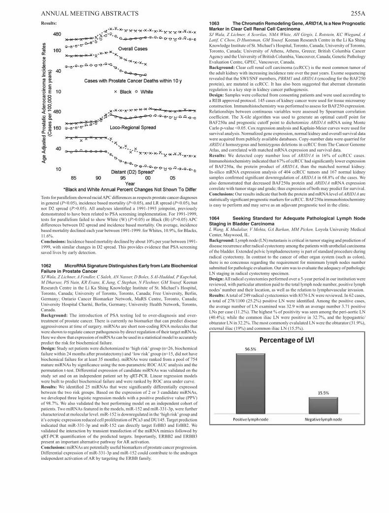
ANNUAL MEETING ABSTRACTS 255AResults:
Tests for parallelism showed racial APC differences as respects prostate cancer diagnoses in general (P<0.05), incidence based mortality (P<0.05), and LR spread (P<0.05), but not D2 spread (P>0.05). All analyses identified a 1991-1993 joinpoint, previously demonstrated to have been related to PSA screening implementation. For 1991-1999, tests for parallelism failed to show White (W) (P>0.05) or Black (B) (P>0.05) APC differences between D2 spread and incidence based mortality. On average, incidence based mortality declined each year between 1991-1999. for Whites, 10.9%, for Blacks, 11.6%.Conclusions: Incidence based mortality declined by about 10% per year between 1991-1999, with similar changes in D2 spread. This provides evidence that PSA screening saved lives by early detection.
1062 MicroRNA Signature Distinguishes Early from Late Biochemical Failure in Prostate CancerSJ Wala, Z Lichner, A Fendler, C Saleh, AN Nasser, D Boles, S Al-Haddad, P Kupchak, M Dharsee, PS Nuin, KR Evans, K Jung, C Stephan, N Fleshner, GM Yousef. Keenan Research Centre in the Li Ka Shing Knowledge Institute of St. Michael’s Hospital, Toronto, Canada; University of Toronto, Toronto, Canada; Free University, Berlin, Germany; Ontario Cancer Biomarker Network, MaRS Centre, Toronto, Canada; University Hospital Charité, Berlin, Germany; University Health Network, Toronto, Canada.Background: The introduction of PSA testing led to over-diagnosis and over-treatment of prostate cancer. There is currently no biomarker that can predict disease aggressiveness at time of surgery. miRNAs are short non-coding RNA molecules that were shown to regulate cancer pathogenesis by direct regulation of their target mRNAs. Here we show that expression of miRNAs can be used in a statistical model to accurately predict the risk for biochemical failure.Design: Study set patients were dichotomized to ‘high risk’ group (n=26, biochemical failure within 24 months after prostatectomy) and ‘low risk’ group (n=15, did not have biochemical failure for at least 35 months). miRNAs were ranked from a pool of 754 mature miRNAs by significance using the non-parametric ROC AUC analysis and the permutation t-test. Differential expression of candidate miRNAs was validated on the study set and on an independent patient set by qRT-PCR. Linear regression models were built to predict biochemical failure and were ranked by ROC area under curve.Results: We identified 25 miRNAs that were significantly differentially expressed between the two risk groups. Based on the expression of 2 or 3 candidate miRNAs, we developed three logistic regression models with a positive predictive value (PPV) of 98.7%. We also validated the best performing model on an independent cohort of patients. Two miRNAs featured in the models, miR-152 and miR-331-3p, were further characterized at molecular level. miR-152 is downregulated in the ‘high risk’ group and it’s ectopic expression reduced cell proliferation of PCa3 and DU145. Target prediction indicated that miR-331-3p and miR-152 can directly target ErBB3 and ErBB2. We validated the interaction by transient transfection of the miRNA mimics followed by qRT-PCR quantification of the predicted targets. Importantly, ERBB2 and ERBB3 present an important alternative pathway for AR activation.Conclusions: miRNAs are potentially useful biomarkers of prostate cancer progression. Differential expression of miR-331-3p and miR-152 could contribute to the androgen independent activation of AR by targeting the ERBB family.
1063 The Chromatin Remodeling Gene, ARID1A, Is a New Prognostic Marker in Clear Cell Renal Cell CarcinomaSJ Wala, Z Lichner, A Scorilas, NMA White, AH Girgis, L Rotstein, KC Wiegand, A Latif, C Chow, D Huntsman, GM Yousef. Keenan Research Centre in the Li Ka Shing Knowledge Institute of St. Michael’s Hospital, Toronto, Canada; University of Toronto, Toronto, Canada; University of Athens, Athens, Greece; British Columbia Cancer Agency and the University of British Columbia, Vancouver, Canada; Genetic Pathology Evaluation Centre, GPEC, Vancouver, Canada.Background: Clear cell renal cell carcinoma (ccRCC) is the most common tumor of the adult kidney with increasing incidence rate over the past years. Exome sequencing revealed that the SWI/SNF members, PBRM1 and ARID1A (encoding for the BAF250 protein), are mutated in ccRCC. It has also been suggested that aberrant chromatin regulation is a key step in kidney cancer pathogenesis.Design: Samples were collected from consenting patients and were used according to a REB approved protocol. 145 cases of kidney cancer were used for tissue microarray construction. Immunohistochemistry was performed to assess for BAF250 expression. Relationships between continuous variables were assessed by Spearman correlation coefficient. The X-tile algorithm was used to generate an optimal cutoff point for BAF250a and prognostic cutoff point to dichotomize ARID1A mRNA using Monte Carlo p-value <0.05. Cox regression analysis and Kaplain-Meier curves were used for survival analysis. Normalized gene expression, normal kidney and overall survival data were acquired from publicly available databases. Copy-number data were quarried for ARID1A homozygous and hemizygous deletions in ccRCC from The Cancer Genome Atlas, and correlated with matched mRNA expression and survival data.Results: We detected copy number loss of ARID1A in 16% of ccRCC cases. Immunohistochemistry indicated that 67% of ccRCC had significantly lower expression of BAF250a, the protein product of ARID1A, than the matched normal kidney. In-silico mRNA expression analysis of 404 ccRCC tumors and 167 normal kidney samples confirmed significant downregulation of ARID1A in 68.8% of the cases. We also demonstrated that decreased BAF250a protein and ARID1A mRNA expression correlate with tumor stage and grade; thus expression of both may predict for survival.Conclusions: Our results indicate that both the protein and mRNA level of ARID1A are statistically significant prognostic markers for ccRCC. BAF250a immunohistochemistry is easy to perform and may serve as an adjuvant prognostic tool in the clinic.
1064 Seeking Standard for Adequate Pathological Lymph Node Staging in Bladder CarcinomaL Wang, K Mudaliar, V Mehta, GA Barkan, MM Picken. Loyola University Medical Center, Maywood, IL.Background: Lymph node (LN) metastasis is critical in tumor staging and prediction of disease recurrence after radical cystectomy among the patients with urothelial carcinoma of the bladder. Extended pelvic lymphadenectomy is part of standard procedure during radical cystectomy. In contrast to the cancer of other organ system (such as colon), there is no concensus regarding the requirement for minimum lymph nodes number submitted for pathologic evaluation. Our aim was to evaluate the adequacy of pathologic LN staging in radical cystectomy specimen.Design: All radical cystectomies performed over a 5-year period in our institution were reviewed, with particular attention paid to the total lymph node number, positive lymph nodes’ number and their location, as well as the relation to lymphovascular invasion.Results: A total of 249 radical cystectomies with 8376 LN were reviewed. In 62 cases, a total of 278/1100 (25.2%) positive LN were identified. Among the positive cases, the average number of LN examined was 32.9 with an average number 3.71 positive LNs per case (11.2%). The highest % of positivity was seen among the peri-aortic LN (40.4%); while the common iliac LN were positive in 32.7%, and the hypogastric/obturator LN in 32.2%. The most commonly evalulated LN were the obturator (31.9%), external iliac (19%) and common iliac LN (15.5%).
256A ANNUAL MEETING ABSTRACTS
Among the cases with positive lymph nodes, 56.5% had evidence of lymphovascular invasion, in contrast to 15.5% in cases with negative lymph nodes.Conclusions: Since the postive LN rate is 11.2%, ten LNs must be submitted to yield one positive lymph node. According to the 7th edition of the AJCC Cancer Staging Manual, pN1 represents one positive regional LN and pN2 represents multiple positive lymph regional lymph nodes. Thus, at least 20 lymph nodes should evaluated for adequate lymph node staging. Since, for pN3 staging a common iliac LN is required, this LN should also be evaluated.
1065 Immunohistochemical Characterization of Gleason Pattern 4-Associated Pattern 3 Prostate CancerA Ward, A Sowalsky, L Quintana, S Balk, H Ye. Beth Israel Deaconess Medical Center, Boston, MA.Background: Prostate cancer (PCa) are frequently multifocal and indolent in contemporary series. Extended core biopsies increase overall cancer detection rate but miss small-volume high grade tumors. One potential solution is to identify molecular signatures of Gleason pattern 3 (G3) tumors that are associated with Gleason pattern 4 and 5 (G4 and G5) tumors. Previous studies from our group revealed that TMPRSS2-ERG breakpoints of adjacent ERG(+) G3 and G4 tumors were exactly the same but distinct among individuals, suggesting they are clonally related. Adjacent G3/G4 tumors are genetically similar but may show additional PTEN loss in G4. Affymetrix arrays revealed dysregulation of a small set of genes in G3-associated G4. Among those, PTEN, neuropilin-2 (NRP2), B lymphoma Mo-MLV insertion region 1 homolog (BMI1), and IGF-1 receptor (IGF-1R), which had been shown to cross-talk and promote progression of PCa, showed a reasonable correlation in our array data.Design: Slides of radical prostatectomy specimens of localized PCa without prior treatment from 2010 to 2012 were retrieved and mapped for all tumor foci. Study group consists of 22 cases with an index tumor of Gleason score 7 (3+4 or 4+3) and separate foci of Gleason score 6 tumor. Control group consists of 9 cases with a final Gleason score 6. PIN-4, ERG, PTEN, NRP2, BMI1, and IGF-1R immunostains are performed on consecutive sections.Results: Study cases contained 22 index tumors of Gleason 7 and 43 distant foci of Gleason 6 tumors (1 to 8 foci per case). 9 control cases contained 10 tumor foci of Gleason 6 tumors. In the study group, adjacent G3 and G4 tumors showed 100% concordance in ERG expression, indicating a common clonal origin. 15 of 43 (34.9%) distant G3 foci showed a discordant ERG expression compared to the index tumors. Among 75 tumors in all cases, 61 foci of tumor-associated high grade PIN were identified in 47 tumors. 53 of 61 (86.9%) PIN demonstrated a concordant ERG expression as their adjacent tumors. PTEN loss was detected in 6 of 22 (27.3%) Gleason 7 tumors and 0 of 43 co-existing Gleason 6 tumors in the study group, and 0 of 10 Gleason 6 tumors in the control group, respectively. PTEN loss was present in some Gleason 7-associated PIN. No PTEN loss was seen in any benign tissue or non-tumor associated PIN.Conclusions: PTEN loss may be one of the molecular signatures of G4-associated G3 tumors. PTEN stain results need to be validated with increased sample size and correlated with FISH findings. NRP2, BMI1, and IGF-1R immunostains are under progress, which may reveal more candidates to characterize aggressive G3 tumors.
1066 GATA3 Expression in Nested, Micropapillary, and Plasmacytoid Urothelial CarcinomasJ Warrick, N Palanisamy, J Siddiqui, R Mehra, A Wu, S Tomlins, LP Kunju. University of Michigan, Ann Arbor, MI.Background: GATA3 has been previously shown to demonstrate high levels of expression in breast carcinoma and urothelial carcinoma (UC). UCs with nested, micropapillary, and plasmacytoid features are associated with poor outcome. To our knowledge, GATA3 expression has not been evaluated in nested or micropapillary UC. The purpose of this project was to evaluate GATA3 expression in these histologic variants of UC by immunohistochemistry (IHC). A benign mimic of nested UC (proliferative cystitis) and benign urothelium were also evaluated for comparison.Design: GATA3 IHC (sc-268; Santa Cruz Biotech; Santa Cruz, CA) was performed on nested (n=9), micropapillary (n=10), and plasmacytoid (n=7) UC cases retrieved from our archives (22 cases) or tissue microarrays (TMA; 3 micropapillary, 1 plasmacytoid). Benign bladder urothelium (n=20, from TMA) and proliferative cystitis (n=3) were also
evaluated. Nuclear staining intensity (graded as negative, weak, moderate, or strong; moderate and strong considered positive) and percentage of cells demonstrating GATA3 expression were assessed.Results: GATA3 positivity was noted in 89% (8/9) of nested, 100% (10/10) of micropapillary, and 57% (4/7) of plasmacytoid UCs. Strong nuclear staining was seen in the majority of micropapillary (9/10) and nested (5/9) UCs. All positive plasmacytoid UCs showed strong nuclear staining. Micropapillary UCs showed diffuse GATA3 expression in the majority of cases (9/10 showed ≥50% cells staining), while plasmacytoid UCs (2/4 positive cases showed ≤50% cells staining) and nested UCs (5/9 cases showed ≤50% cell staining) tended to show focal expression. GATA3 was positive in most benign urothelium (16/20 cases; all moderate to strong) and all proliferative cystitis cases (3/3; all strong). Staining tended to be diffuse in benign urothelium (11/16 positive cases showed >50% cells staining) and proliferative cystitis (2/3 showed >50% of cells staining).Conclusions: GATA3 expression as detected by IHC is present in the majority of nested, micropapillary, and plasmacytoid UCs. These findings corroborate one previous report regarding the expression of GATA3 in the plasmacytoid variant of UC. Our current study suggests high level of GATA3 expression in nested and micropapillary UCs as well. GATA3 may be useful for accurately classifying the primary site of a micropapillary carcinoma as bladder if metastatic breast carcinoma can be excluded. Benign urothelium and proliferative cystitis both demonstrate strong expression of GATA3 in the majority of cases, making GATA3 unsuitable for differentiating these from the nested pattern of UC.
1067 Tissue PCA3 Evaluation in Prostate Cancer by In-Situ HybridizationJ Warrick, N Palanisamy, A Young, J Siddiqui, A Wu, R Mehra, S Carskadon, J Wei, A Chinnaiyan, S Tomlins, LP Kunju. University of Michigan, Ann Arbor, MI.Background: The PROGENSA PCA3 assay, an FDA approved urine based assay for assessing the risk of prostate cancer (PCa) on rebiopsy, quantifies the non-coding transcript PCA3, which is over-expressed in >95% of all prostate cancers. As tissue evaluation of PCA3 with immunohistochemistry (IHC) is not feasible, we evaluated tissue PCA3 expression by RNA-ISH (in-situ hybridization) in PCa, HGPIN, and benign mimics (including adenosis and atrophy) at radical prostatectomy (RP). We also correlated tissue PCA3 expression in PCa with ERG status by IHC.Design: Of 301 men presenting for biopsy with pre-biopsy urine PCA3 and T2:ERG levels, 41 men underwent RP. All prostatectomies were mapped. PCA3 ISH was scored as 0-4 per RNAscope scoring Criteria (Advanced Cell Diagnostics) for all evaluable PCa foci, HGPIN, atrophy, adenosis, and benign glands. PCA3 ISH scores >1 were considered positive. For each case, total number, greatest linear dimension, Gleason score, total PCA3 score (sum of scores of each PCa focus), and total PCA3-positive linear tumor dimension were documented. Correlations with urine PCA3 and ERG IHC (EPR 3864) status of PCa foci were performed.Results: The 41 radical prostatectomies had a median of 3 tumor foci (range 1-11) and median total linear tumor dimension 2.6 cm (SD 1.4 cm). The median urine PCA3 was 40 (SD 38, range 3-186). The sensitivity of PCA3 ISH for PCa was 54% alone and 70% when combined with ERG. 88% and 95% of patients had at least one PCa focus positive with PCA3 alone, and either PCA3 or ERG, respectively. The majority of HGPIN foci (70%) adjacent to PCa were positive with PCA3. PCA3 ISH specificity was >99% for PCa and HGPIN combined (only one benign gland showed focal staining; all adenosis and atrophy were negative). Index nodule size and total linear tumor dimension correlated with PCA3 ISH score (p<0.01). Urine PCA3 showed positive, but non-significant, correlation with total PCA3 score (rs=0.21, p=0.19,) and PCA3 positive linear tumor size (rs=0.09, p=0.57). Only 2/18 (11%) cases with PCA3 ISH score <6 had urine PCA3 >60, while cases with PCA3 score >6 showed a broad range of urine PCA3. PCA3 ISH in PCa foci positively correlated with ERG IHC status (p<0.01).Conclusions: The sensitivity of PCA3 ISH for PCa detection is similar to that of ERG. Combined PCA3 ISH and ERG improves sensitivity of PCa detection over either marker alone. PCA3 RNA ISH is highly specific for PCa and HGPIN. Although no statistically significant correlation between urine PCA3 and total PCA3 score was seen in this cohort, concurrent low total PCA3 score and high urine PCA3 was a rare finding.
1068 Foamy Gland Carcinoma of the Prostate in Needle Biopsy: Incidence, Gleason Grade, and Comparative AMACR vs ERG ExpressionJI Warrick, PA Humphrey. University of Michigan, Ann Arbor, MI; Washington University, Saint Louis, MO.Background: Foamy gland carcinoma (FGC) is a variant pattern of prostatic acinar adenocarcinoma that may be deceptively benign-appearing, and has been shown to be a cause of missed carcinoma on prostate needle core biopsy (NCB). Also, immunohistochemical staining for AMACR is limited by a comparatively lower sensitivity for foamy gland carcinoma compared to usual acinar adenocarcinoma. FGC has not been characterized in a consecutive series of prostate NCBs, and it is not known if ERG immunohistochemistry provides added value beyond AMACR in detection of FGC. The purpose of this study was to characterize FGC on NCB, with focus on incidence, Gleason score, and expression of AMACR and ERG.Design: 476 consecutive prostate cancer NCB cases were identified from our archives, diagnosed over a 2 year period. All cases were re-reviewed, and Gleason score was assigned using the modified ISUP/AFIP guidelines. Presence and extent of foamy gland carcinoma were quantified. Immunohistochemistry for AMACR and ERG was performed on a representative slide from each case. Slides stained with AMACR were evaluated for intensity (scored 0-3) and proportion of cells staining (scored 0-4). Intensity and proportion scores were added to give an overall score (maximum 7). ERG staining was scored as previously described.

ANNUAL MEETING ABSTRACTS 257AResults: Of 476 consecutive prostate cancer diagnosed on NCB, 18% (85 cases) contained a FGC component. 7% (32 of 476) of all cases were pure FGC on NCB, representing 38% (32 of 85) of cases with a FGC component. Of the 85 cases identified, the following Gleason scores were assigned to the FGC component: 79% (67 cases) 3+3=6, 14% (12 cases) 3+4=7, 1% (1 case) 4+3=7, 4% (3 cases) 4+4=8, 1% (1 case) 4+5=9, and 1% (1 case) 5+3=8. 73 carcinomas with a FGC component had sufficient tissue for immunohistochemical studies. Of these, 90% (66 cases) were AMACR positive. Of the AMACR positive cases, 36% (24 of 66 cases) showed AMACR-low staining (sum score <6). 41% (30 cases) were ERG positive. None of the AMACR negative cases were ERG positive.Conclusions: FCG is a common finding on prostate NCB, is usually found in association with usual acinar adenocarcinoma, and most commonly has Gleason score 6 on NCB. 90% of FGCs in our series were AMACR positive, indicating equivalent sensitivity to that seen in usual acinar adenocarcinoma, a finding different from that reported in the literature. ERG immunohistochemistry did not provide added diagnostic value beyond AMACR.
1069 Targeted Seminal Vesicles Biopsies: Incidence and Clinicopathological FindingsKE Watts, C Magi-Galluzzi. Cleveland Clinic, Cleveland, OH.Background: Invasion of prostatic adenocarcinoma (PCA) into seminal vesicles (SV) is generally accepted as an index of poor prognosis. SV are not routinely biopsied for evaluation of PCA, however some clinicians choose to include SV biopsies (SVB) along with prostate biopsies (PBx) in certain clinical scenarios. The aim of this study was to review our experience with targeted SVB in the diagnosis and management of PCA.Design: All patients who underwent targeted SVB at our institution between 1995 and July 2012 were retrieved from the pathology electronic archives. Histological findings, reason for SVB, previous treatment [radiation (XRT), androgen deprivation therapy (ADT), cryotherapy (CRYO), brachytherapy (BRACHI)], and clinical follow up were reviewed.Results: Of the 170 (0.8%) men with targeted SVB identified in our database, 164 (96%) had SV tissue present in the biopsy specimen. Eighty-three (51%) men with SVB had been previously diagnosed with PCA and 77 (47%) had been formerly treated for PCA: 40 had received XRT, 6 XRT+ADT, 19 BRACHY, 9 CRYO, and 3 ADT. SVB were involved by PCA (SVI) in 22 (13%) patients, 9 (5%) of whom were biopsied for the first time due to elevated PSA. Atypical glands, suspicious for cancer were detected in 1 case (0.6%). In 16 (10%) cases only SV were sampled, of which 3 (19%) showed SVI: 14 (87%) of the patients had been previously diagnosed with PCA; a SV mass was incidentally detected on CT in another case. In 11 (7%) men SVB were performed prior to XRT and in 42 (26%) prior to CRYO. The 9 (5%) men with PCA detected in SV biopsied for the first time due to elevated PSA were managed as follows: 4 received XRT+ADT, 1 orchiectomy, 1 ADT + radical prostatectomy (RP), 1 RP + salvage XRT, 1 received no treatment due to metastatic esophageal cancer, and one case was lost at follow-up.Conclusions: Targeted SVB are not a common procedure in the evaluation for PCA extension and staging. However, SV involvement was found in 13% of patients undergoing SVB. Most of these patients had been previously treated for PCA and were being considered for additional therapy, but 9 (5%) were found to have SVI on initial biopsy for elevated PSA, highlighting the potential prognostic implications of SVB at initial diagnosis. Positive SVB results can aid in the selection of treatment options and in the prediction of outcome for individual patients by providing confirmation of locally advanced disease.
1070 miRNA Expression in Lower and Upper Urothelial Carcinoma and the Potential Clinical ApplicationS Wei, Y Yao, PK Gupta, Z Bing. Hospital of the University of Pennsylvania, Philadelphia, PA.Background: Diagnosis and follow-up of patients with urinary tract tumors currently are based on urine cytology, radiographic imaging, and ureteroscopy. Although urine cytology has a high specificity, it is not sensitive. Ureteroscopy is a reliable approach for identification of macroscopic disease. But the disadvantage of ureteroscopy is also evident, such as, discomfort, urinary tract infection, as well as failing to detect microscopic disease. MicroRNAs (miRNA) are short noncoding RNA molecules that posttranscriptionally modulate protein expression. There are distinct miRNA alterations characterizing urinary bladder urothelial carcinoma. In this study, we investigate the possibility of using miRNAs as a non-invasive markers in the screening or follow-up of urothelial cell carcinoma (UCC) from upper urinary tract and bladder.Design: Total RNA was extracted from 87 bladder or renal washing fluid (22 high grade UCC, 13 low grade UCC, 25 normal control, 27 cases of positive history of UCC but negative at time of test (HIS)). All the diagnoses were confirmed by cytology and/or surgical pathology. Expression of miRNA markers miR-96, miR-182, miR-183, miR-200c, miR-21, miR-141, miR-30b were determined using quantitative real time PCR.Results: Quantitative real time PCR showed miRNA is stable in the urinary tract washing fluid. miRNA-96, 182, 183, 141, 30b, 21 and 200c are overexpressed in high grade UCC compared to low grade UCC, normal control and HIS. There is no significant difference between the low grade, normal and HIS. In addition, UCC from upper GU tract and bladder cancers demonstrate similar changes of these target miRNAs.Conclusions: Our study shows miRNAs are stable in the bladder or renal washing, and the target miRNAs (miRNA-96, 182, 183, 141, 30b, 21 and 200c) are overexpressed in high grade tumors. Real-time PCR of a panel of miRNA could be used as a screening test as well as a followup test for recurrence of UCC patient.
1071 The Immunohistochemical Distinction between Clear-Cell Renal-Cell Carcinoma and Other Forms of Clear-Cell CarcinomaSM Wendroth, MJ Mentrikoski, MR Wick. University of Virginia, Charlottesville, VA.Background: It is well-known that clear-cell renal-cell carcinoma (CC-RCC) has the ability to metastasize throughout the body, and to imitate the clear-cell variants of many other tumors morphologically. Thus, immunostains are often required to discriminate between such lesions. Despite the availability of several markers for CC-RCC, their individual sensitivities and specificities for that tumor are variable. This study was designed to assess the relative diagnostic values of commonly-used potential labels for CC-RCC (PAX2, PAX8, CD10, RCC-Ag, and adipophilin [ADP]) in its separation from other clear-cell carcinomas of non-renal origin. CA125 stains were also performed to aid in the distinction between renal and Mullerian neoplasms.Design: Fifty-one non-renal clear-cell carcinomas and 26 examples of CC-RCC were retrieved from the authors’ institutional archives. The non-renal tumors included carcinomas of breast, liver, lung, vagina, cervix, urinary bladder, salivary gland, ovary, skin, and lung. Immunostaining for the specified determinants was performed on each specimen using standard methods and an automated platform. Staining results were simply recorded as either positive (immunoreactivity in > 5% of tumor cells) or negative. Intensity of labeling was graded as 1 to 3+. The sensitivity and specificity of each stain, vis-à-vis the diagnosis of CC-RCC, was then calculated using Bayesian methods.Results: The overall sensitivity of CD10, ADP, RCC-Ag, PAX2, and PAX8 for the diagnosis of CC-RCC was 85%, 100%, 72%, 81%, and 100%, respectively. Respective individual specificities were 74%, 52%, 100%, 73%, and 60%. Analytic exclusion of mullerian tumors (from vagina, cervix, and ovary) enhanced the specificities of PAX (to 97%) and PAX8 (to 89%), while decreasing the specificities of CD10 (to 68%) and ADP (to 47%). CA125 was not expressed in any CC-RCC cases, but it was sensitive and specific for the identification of mullerian neoplasms, with respective results of 81% and 93%.Conclusions: No individual immunohistologic marker in this study was both optimally sensitive and specific for the diagnosis of CC-RCC, thus confirming the need for a moderately-sized panel of analytes in the differential diagnosis of that neoplasm. PAX2 and PAX8 show excellent specificity for CC-RCC, but they have lesser contextual value in women patients because of potential shared reactivity in mullerian tumors. In those cases, CA125 is an effective additional marker that discriminates between lesions of the kidneys and the female genital tract.
1072 Evaluation of ∆Np63 Expression in Urothelial Cell CarcinomaKA West, DJ Buonocore, D Lucido, L Kiss, B Wang, V Ghali. Beth Israel Medical Center, New York, NY.Background: Depth of invasion of urothelial cell carcinomas (UCCs) is an important prognostic factor. UCCs are known to express p63 by immunohistochemistry (IHC). Some biologic studies have implicated the ∆Np63 isoform (known for its use in non-small cell lung cancer) in conferring invasive potential in UCC; however, the data is limited and results are conflicting. There is a paucity of literature regarding immunolabeling of ∆Np63 (using the p40 antibody) in UCC. We sought to investigate IHC expression of ∆Np63 in UCC in order to further elucidate its effects on tumor behavior.Design: We searched our archives for UCCs diagnosed between January-September 2012. Fifty (50) high-grade (30 invasive and 20 non-invasive) and 10 low-grade UCCs were selected. Cases that were poorly preserved or indeterminate for invasion were excluded. IHC was performed on all cases using the p63 antibody (4A4+Y4A3), and the p40 antibody (∆Np63). IHC expression was semiquantitatively scored based on the percentage of tumor cells positive as follows: 90-100% (3+), 50-89% (2+), 10-49% (1+), <10% (0). A Χ2 test was used to determine the statistical significance of loss of p40 staining compared to p63 in high-grade non-invasive and invasive UCCs.Results: p40 staining was uniformly less intense than p63 in normal and lesional tissue. Normal urothelium displayed constitutional (3+) expression of both markers with sparing of the umbrella cells. All of the low-grade UCCs exhibited diffuse (3+) expression of both markers. The results of IHC staining of the high-grade UCCs are listed in Table 1. All cases showed some degree of staining for p63. p40 staining was completely absent in four cases. In 78% of high-grade UCCs, the expression of p40 mimicked that of p63, while in 22% p40 was expressed in fewer cells. The results of the X2 test showed no significant difference between the stains for non-invasive high-grade UCCs (p= 0.1448); however, in high-grade invasive tumors, loss of p40 as compared to p63 was highly statistically significant (p=0.0092).Table 1
IHC Ab Score High grade UCC, Invasive (n=30)
High grade UCC, Non-invasive (n=20)
Total High grade UCC (n=50)
p63 3 24 (80%) 16 (80%) 40 (80%) 2 4 (13.3%) 3 (15%) 7 (14%) 1 2 (6.7%) 1 (5%) 3 (6%) 0 0 (0%) 0 (0%) 0 (0%) p40 3 18 (60%) 14 (70%) 32 (64%) 2 6 (20%) 4 (20%) 10 (20%) 1 3 (10%) 1 (5%) 4 (8%) 0 3 (10%) 1 (5%) 4 (8%)
Conclusions: ∆Np63 is preserved in low-grade UCC, and is more likely to be lost in invasive than non-invasive high-grade UCC. However, loss of ∆Np63 does not appear to be necessary or sufficient for progression to invasion, as it is preserved in many invasive UCCs, and is lost in some non-invasive tumors.

258A ANNUAL MEETING ABSTRACTS 1073 SF-1 Immunohistochemical Expression in TumorsM Wilkerson, F Lin, J Shi. Geisinger Medical Center, Danville, PA.Background: Steroidogenic factor 1 (SF-1) is a nuclear receptor that functions with other transcription factors to regulate several genes in the hypothalamic-pituitary-gonadal axis, and in gonadal and adrenal steroidogenesis. SF-1 is required for regulation of sex determination and differentiation, reproduction, and steroidogenesis. Nuclear expression of SF-1 has been described in adrenal cortical neoplasms and normal adrenal cortex, endometriosis, Leydig cells, Sertoli cells, and theca and granulosa cells. Published data on SF-1 expression in tumors is limited. In this study, we investigated the expression of SF-1 in a large series of tumors from various organs.Design: Immunohistochemical evaluation of SF-1 expression (R&D systems, SF-1/NR5A1 antibody, clone N1665) was performed on 984 tumors from various organs using tissue microarray sections. The staining distribution was recorded as negative (<5% of tumor cells stained), 1+ (5-25%), 2+ (26-50%), 3+ (51-75%), or 4+ (>75%).Results: The positive staining results (%) and the total number of cases for each entity (N) are summarized in Table 1. Twenty-three of 984 cases demonstrated positive nuclear staining for SF-1 protein and these cases consisted of adrenal cortical adenomas and testicular Leydig cell tumors. In addition, benign interstitial Leydig cells and Sertoli cells in seminiferous tubules evaluated for ITGCN all demonstrated positive staining for SF-1.Table 1. Summary of SF-1 immunostaining in 998 casesTumor N # Positive % PositiveAdrenal cortical adenoma 24 20 83.3Testicular Leydig cell tumor 3 3 100Testicular ITGCN 15 Testicular classic seminoma 21 Testicular embryonal CA 18 Testicular yolk sac tumor 6 Urothelial CA primary to bladder 45 Pulmonary neuroendocrine CA 26 Pulmonary ADC 52 Pulmonary squamous cell CA 40 Pancreatic endocrine CA 16 Clear renal cell CA 68 Papillary renal cell CA 17 Chromophobe renal cell CA 23 Infiltrating duct CA of breast 94 Infiltrating lobular CA of breast 26 Esophgeal ADC 26 Gastric ADC 12 Colonic ADC 28 Hepatocellular CA 17 Papillary CA of thyroid 42 Follicular CA of thyroid 30 Follicular adenoma of thyroid 51 Prostatic ADC 87 Ovarian serous ADC 43 Endocervical ADC 22 Endometrial ADC 132 Total tumors 984 23 2.3ADC-adenocarcinoma; CA-carcinomaConclusions: Our data demonstrate that SF-1 is a highly sensitive and specific marker for tumors of steroid producing cells, including adrenal cortical adenomas and Leydig cell tumors. In addition, it can be used as a marker for normal Leydig and Sertoli cells. SF-1 does not appear to be expressed in testicular germ cell tumors, or in tumors from other organs.
1074 Androgen Receptor Expression in Genitourinary NeoplasmsE Williams, J Higgins, J McKenney, A Sangoi, M Troxell. Oregon Health & Science University, Portland, OR; Stanford, Stanford, CA; Cleveland Clinic Foundation, Cleveland, OH; El Camino Hospital, Mountain View, CA.Background: Androgen receptor (AR) is strongly expressed in the vast majority of prostatic adenocarcinomas. Historically, AR expression was considered to be relatively specific to prostate carcinoma, maintained in poorly differentiated carcinomas, and amenable to immunohistochemical analysis. However, there remains little published data regarding the specificity of AR for prostate cancer, even amongst genitourinary cancers. Few studies attempting to correlate AR status with outcome in bladder carcinoma report variable rates of AR positivity, while we are aware of only a rare study of AR in renal carcinomas.Design: Tissue microarrays containing prostate, kidney, and bladder tumors were stained with an antibody to AR. The microarrays were constructed as 0.6 mm tissue cores, most cases were represented at least in duplicate. Unstained tissue microarray sections were stained with antibody clone AR27 (Vector Laboratories, Burlingame CA) at a dilution of 1:10 after cc1 standard retrieval on a Ventana XT instrument, with Ultraview detection (Ventana, Tucson AZ). Cores were scored by 2 observers as negative, weak positive, strong positive, or absent/unscoreable.Results: As expected, 95% of prostate carcinomas were positive for AR with 73% of tumors strongly positive (N=230). Interestingly, 19% of invasive urothelial carcinomas of the bladder and 16% of renal pelvis were also positive, though many stained weakly (N=190 and 43, respectively); furthermore, 33% of non-invasive urothelial carcinomas were AR positive (N=107). A substantial percentage of primary renal cell carcinomas were AR positive, with 19% of 307 cases staining. From a metastatic renal cell carcinoma cohort, 28% of metastases were AR positive (N=126). Incidentally, 16% of angiomyolipomas and 6% of oncocytomas were also AR positive (N=12 and 15, respectively).Conclusions: Our data show that the sensitivity of AR immunohistochemistry for prostate cancer is 94.8%, among our cohort of invasive genitourinary tumors. However, the specificity of AR is only 79.5%, since 15-30% of invasive urothelial and primary or metastatic renal cell carcinomas are also AR positive. Thus, the specificity of AR is
not ideal for diagnostic confirmation of a poorly differentiated carcinoma of suspected genitourinary origin. AR may remain useful as a component of an immunostain panel.
1075 Cystic Partially Regressed Clear Cell Renal Cell Carcinoma: Clinical and Morphologic Distinction from Multilocular Cystic Renal Cell CarcinomaSR Williamson, GT MacLennan, A Lopez-Beltran, R Montironi, PH Tan, G Martignoni, DJ Grignon, JN Eble, L Cheng. Indiana University, Indianapolis, IN; Case Western Reserve University, Cleveland, OH; Cordoba University, Cordoba, Spain; Polytechnic University of the Marche Region, Ancona, Italy; Singapore General Hospital, Singapore, Singapore; University of Verona, Verona, Italy.Background: Extensively cystic and sclerotic clear cell renal cell carcinoma (RCC) with only a minor solid component is unusual. Diagnostic terminology for such cases can be ambiguous, leading to confusion with multilocular cystic RCC, which by definition lacks a solid component.Design: We studied clinicopathologic features in clear cell RCCs with a solid epithelial component that precluded diagnosis of multilocular cystic RCC, yet which had predominant cystic or sclerotic components. We elected to designate these cancers as “cystic partially regressed clear cell renal cell carcinomas”.Results: Twenty-one tumors were retrieved from patients aged 39 to 74 years (mean 58, male:female 1.6:1). Tumor size ranged from 1.0 to 17.0 cm (mean 4.7) and stage was pT1a (64%), pT1b (21%), pT2a (5%), pT2b (5%) and pT3a (5%). Fuhrman grade was 2 (52%), 1 (34%) or 3 (14%). The solid epithelial component comprised from 5 to 30% of the total tumor (median 10%), while the cystic component comprised 15 to 80% (median 60%) and a sclerotic, hyalinized component comprised 10 to 70% (median 25%). Thin fibrovascular septa lined by clear cells were almost always present (95%); aggregates of clear cells were present within the thin fibrovascular septa in 90% of cases. Broad zones of sclerosis often contained scattered clear cells (86%) and ranged in appearance from a cellular fibroxanthomatous reaction to collagenous and scar-like with a residual network of capillaries. Other features included hemosiderin deposition (76%), calcification (38%), Gamna-Gandy-like nodules (14%) and granulomatous reaction (10%). Two tumors were monitored with imaging prior to resection and showed no change in size over 1 year prior to resection. Long-term follow-up was available for 11 patients, of which none showed aggressive behavior.Conclusions: Cystic partially regressed clear cell RCC is a unique histologic variant of clear cell RCC, composed of cysts with solid epithelial and sclerotic components that differentiate it from multilocular cystic RCC.
1076 Clear Cell Papillary Renal Cell Carcinoma-Like Tumors in Patients with Von Hippel-Lindau Disease Are Unrelated to Sporadic Clear Cell Papillary Renal Cell CarcinomaSR Williamson, S Zhang, JN Eble, DJ Grignon, G Martignoni, M Brunelli, S Gobbo, LA Baldridge, L Cheng. Indiana University, Indianapolis, IN; University of Verona, Verona, Italy.Background: Clear cell papillary renal cell carcinoma (CCPRCC) shares morphologic overlap with clear cell renal cell carcinoma, though it lacks the typical chromosome 3p and VHL gene abnormalities. Rare cases have been reported in von-Hippel Lindau (VHL) patients (germline mutation of the VHL gene), the significance of which is uncertain.Design: Renal tumor slides from patients with VHL disease were examined for morphologic features of CCPRCC. CCPRCC-like tumors were studied with immunohistochemistry for CK7, carbonic anhydrase IX (CAIX), CD10, and alpha-methylacyl-coA-racemase (AMACR). Fluorescence in situ hybridization (FISH) was performed (5 tumors) with probes to chromosomes 3p14, 3p25, 7 and 17. Clear cell renal cell carcinomas from the same patients (n=2) and VHL patients without CCPRCC-like tumors (n=8) were also studied for comparison.Results: Fourteen tumors with CCPRCC-like features were identified in 4 VHL patients. Gross appearance in 2 tumors closely resembled clear cell renal cell carcinoma (yellow-orange), while other tumors were tan, red-brown, or extensively cystic. Histologic features included: prominent papillary structures (64%), small papillary tufts (79%), flattened peripheral cysts (64%), branched tubules (71%), and “secretory cells” with apically-aligned nuclei (43%). The typical immunoprofile of CCPRCC (CK7+, CAIX+, CD10-, AMACR-) was observed in only 2/11 stained tumors (18%), both from the same patient, one of which showed 3p deletion by FISH. The remainder showed negative or focal CK7 reactivity (55%), diffuse, strong expression of CD10 (64%), or both (18%). Three tumors (27%) also showed strong AMACR staining. Several possessed 3p deletion by FISH (60%), similar to those of VHL patients without CCPRCC-like tumors (75%).Conclusions: Tumors that histologically closely resemble CCPRCC occur in patients with VHL disease, but they usually lack the typical immunohistochemical and molecular profile of sporadic CCPRCC, suggesting that they do not share the same carcinogenesis.
1077 Urethral Caruncle: A Lesion Related to IgG4-Associated Sclerosing Disease?SR Williamson, M Scarpelli, A Lopez-Beltran, R Montironi, MR Conces, L Cheng. Indiana University, Indianapolis, IN; Polytechnic University of the Marche Region, Ancona, Italy; Cordoba University, Cordoba, Spain.Background: Urethral caruncle is a benign, polypoid urethral mass that occurs almost exclusively in postmenopausal women. Despite that these lesions have been long recognized and clinically managed with topical medications or excision, their pathogenesis is not well understood. In order to shed light on the etiology of urethral caruncle, we investigated the possibilities of autoimmune, viral, and inflammatory myofibroblastic proliferations as possible etiologies.Design: In 38 patients with urethral caruncle, we utilized immunohistochemistry for immunoglobulin G (IgG), immunoglobulin G4 (IgG4) to assess for a potential

ANNUAL MEETING ABSTRACTS 259Aautoimmune etiology. Immunohistochemistry was performed in nine patients for Epstein-Barr virus, BK virus, human herpesvirus 8, human papillomavirus, adenovirus, and anaplastic lymphoma kinase.Results: Four patients (11%) showed infiltrates of ≥50 IgG4-positive plasma cells per high power field; all showed an IgG4:IgG ratio greater than 40%. One patient with increased counts below these thresholds had rheumatoid arthritis; none had documented autoimmune pancreatitis or other known manifestations of systemic IgG4-related sclerosing disease. All lesions showed negative reactions with the remaining markers of viral or inflammatory myofibroblastic etiologies.Conclusions: Urethral caruncle is a benign inflammatory and fibrous polypoid urethral mass of unclear etiology. Using immunohistochemical markers, it appears unrelated to the viral infections that result in tumor formation and other mucosal abnormalities of various body sites. Abnormal expression of ALK protein, as seen in inflammatory myofibroblastic tumors, is absent. Increased numbers of IgG4-positive plasma cells in a subset of lesions raise the possibility that some cases may be related to the autoimmune phenomena of IgG4-associated disease.
1078 Renal Core Biopsies of Angiomyolipomas: Review of a Single Institution Eight Year Experience with an Emphasis on Angiomyolipomas with Epithelioid FeaturesAJ Wu, S Wolf, S Tomlins, R Mehra, LP Kunju. University of Michigan, Ann Arbor, MI.Background: With the increased use of image guided core biopsies (cbx) of renal masses, cbx of benign entities such as angiomyolipomas (AMLs) are being encountered. AMLs on cbx can present both diagnostic and therapeutic implications when epithelioid features are present; our goal was to review our experience with cbx of AMLs with an emphasis on AMLs with epithelioid features (eAMLs).Design: The clinical, radiologic, morphologic and immunohistochemical features of all cbx with a diagnosis of AML from 1/2004 to 10/2012 at our institution were reviewed.Results: Thirty five AMLs were diagnosed on cbx (an incidence of <1% of all biopsied renal masses). The most common indication for cbx was unusual radiographic features, usually lipid poor/solid appearing masses (mean size=2.9 cm; 1.3-6 cm). One patient had tuberous sclerosis. Seven (20%) were diagnosed as eAML; the remainder were diagnosed as classic AML. Only 4 (11%) AMLs, all with a cbx diagnosis of eAML, were excised; of these, 3 and 1 case had an excision diagnosis of eAML and classic AML respectively. The most helpful features on cbx in predicting an excision diagnosis of eAML were: a high percentage of atypical epithelioid cells (at least 70-80%), epithelioid cells arranged in sheets with minimal to absent intervening fat or spindled smooth muscle, and a paucity of large dysplastic appearing vessels; features not present on cbx were marked atypia, necrosis, or increased mitoses. Immunohistochemistry was performed in 40% (14/35) of all cbx, including all cbx with a diagnosis of eAML. None of the AMLs in our study metastasized and of the AMLs which were excised, only one (with an excision diagnosis of eAML) recurred (median followup 20.5 months).Conclusions: In our experience, ruling out RCC in cbx of eAML was a morphologic dilemma in a subset of cases but was usually easily ruled out with immunohistochemistry. The most common diagnostic dilemma on cbx was distinguishing classic AML from eAML; the most helpful features were the presence of a high proportion of atypical epithelioid cells arranged in sheets with a paucity of intermixed smooth muscle, fat, and dysplastic vessels.
1079 Metastases to the Kidney: A Clinicopathologic Study of Forty One Cases with an Emphasis on Deceptive FeaturesAJ Wu, LP Kunju, J Reynolds, S Tomlins, R Mehra, C Przybycin. University of Michigan, Ann Arbor, MI; Cleveland Clinic, Cleveland, OH.Background: Metastatic tumors (MTs) to the kidney are rare; our goal was to review our experience with MTs to the kidney with an emphasis on potentially deceptive features.Design: The clinical, radiologic, morphologic, and immunohistochemical features of all MTs to the kidney between 5/1987 and 8/2012 at our institution were reviewed. Autopsy cases and any cases in which the MT was incidentally found in a nephrectomy were excluded. Only patients who had a known primary diagnosis and a definitive final diagnosis of MT were included.Results: Forty one cases (13 nephrectomies (N), 26 core biopsies (Bx), 2 FNAs) were included, accounting for an incidence of <1% of renal masses. A primary malignancy was diagnosed, either prior to or concurrently with the MT, in all cases (mean interval between diagnoses = 5.5 y; 0- 21.6 y). The majority (88%) were carcinomas and the most common primary site was lung (20 (49%) including 11 adenocarcinomas, 8 squamous cell carcinomas, 1 small cell carcinoma); followed by breast (5), gynecologic tract (5), head and neck (3), colon (2), prostate (2), testis (1), skin (1), tibia (1) and adrenal (1). A primary renal tumor was suspected clinically and radiologically prior to the Bx/N in 37% of cases. Unusual features included: common unilateral (85%) and unifocal (88%) involvement, lack of other known distant organ metastasis (32%), a long time interval (>10 y) between diagnosis of primary tumor and MT (20%), and in a small subset of cases, lack of a discrete mass (5%), medullary location (15%), and extension into the renal vein (10%). Nearly all of the MTs had an infiltrative growth pattern (98%), associated lymphovascular invasion (78%), and extended beyond the kidney to involve perirenal/sinus fat and/or adjacent organs (71%). The most common diagnostic dilemma was excluding urothelial carcinoma or collecting duct/high grade renal cell carcinoma; however, in occasional cases, a MT from an unusual site mimicked a low grade renal cell carcinoma. Immunohistochemical stains were performed in only 40% of the cases and were confirmatory in all of these cases.Conclusions: In our experience, MTs to the kidney most commonly present as solitary unilateral masses and in a substantial subset of cases can both clinically and pathologically mimic a primary renal tumor. A high index of suspicion, vigilance in investigating clinical history, and certain morphologic clues such as an infiltrative growth pattern should help avoid misdiagnoses in these rare cases.
1080 ERG Protein Expression and PTEN Loss Are Uncommon in Prostate Cancer of Chinese PopulationT Xing, X Pei, W Fang, H He. Peking University, Health Science Center, Beijing, China.Background: In Western countries, gene fusions involving the ERG oncogene and deletions of the tumor suppressor gene PTEN are frequent alterations in prostate cancer (PCa). Recent reports demonstrated that TMPRSS2-ERG gene fusion correlated with the ethnicity. It was less common in Asian PCa of Japanese and Korean populations (16-28%) than Western PCa (40-50%), however the studies of TMPRSS2-ERG fusion status in Chinese PCa were very limited and contradictory. The ERG protein expression in Chinese PCa patients has not been evaluated to date. The objective of this study was to determine the frequency of ERG positive PCa by immunohistochemistry (IHC), PTEN deletion by IHC, and correlation with clinico-pathological parameters.Design: IHC was performed on a single standard tissue section from the index tumor of 48 radical prostatectomies, enriched for relatively high grade (46% Gleason Score (GS) 7, 27% GS 8-9) and pathological stage (29% T3) tumors, utilizing the ERG mAb (EPR 3864;Epitomics,CA) and PTEN mAb (D4.3;CST,MA). Nuclear ERG expression was scored as positive or negative in a given tumor focus. Cytoplasmic PTEN staining was scored as normal (increased or equal staining compared to adjacent benign acini) or markedly decreased/absent dichotomously.Results: Only 6% (3/48) of all the cases showed ERG positivity, with one case showing intratumoral ERG heterogeneity. The three ERG expressing cases were of GS ≤7. Two cases were pathological T2c and one was T3a. Seventeen percent (8/48) of cases showed PTEN loss, and of these, 25% (2/8) showed heterogeneity. In PTEN loss cases, 38% (3/8) had GS 8-9, however which was not significantly higher than 25% (10/40) in PTEN normal cases (P=0.08). PTEN loss did not correlate with GS or pathological stage. There was no association between ERG protein expression and PTEN loss (P>0.05 for all correlations).Conclusions: Interestingly, ERG protein expression and PTEN loss are infrequent in Chinese PCa. Six percent of ERG positivity is even lower than that in PCa of other asian populations. Furthermore there was no association between ERG protein expression and PTEN loss. Our preliminary study suggests that there are different pathways of prostate carcinogenesis in Chinese population. Further studies of large specimen are underway.
1081 Diagnostic and Prognostic Role of Immunohistochemical Expression of Napsin A in Renal Cell Carcinoma: A Study Including 233 Primary and Metastatic CasesB Xu, S Abourbih, K Sircar, W Kassouf, A Aprikian, S Tanguay, F Brimo. McGill University Health Center, Montreal, QC, Canada; University of Texas MD Anderson Cancer Center, Houston, TX.Background: Napsin-A is an aspartic protease that is predominantly expressed in the proximal renal tubules and type II pneumocytes. Although it has been reported to be present in a proportion of renal cell carcinomas (RCCs), utilization of napsin-A immunohistochemistry as a routine diagnostic tool for RCC, and the correlation of the level of expression with histological features and clinical outcomes have not yet been established.Design: Using tissue miscoarrays we assessed the immunohistochemical expression of napsin-A in a cohort of clear cell and papillary RCCs including 81 and 125 primary and metastatic cases, respectively, in addition to 24 primary and matched metastatic cases.Results: Napsin-A expression was demonstrated in 86 of 222 (39%) clear cell RCCs (CRCCs) and 16 of 21 (76%) papillary RCCs (PRCCs) with a strong and diffuse staining pattern observed in PRCC and a relatively weak and focal positivity in CRCCs. Compared with primary CRCCs, a comparable proportion of metastatic CRCCs retained napsin-A expression (45/132, 34%), suggesting the potential utility of napsin-A in the evaluation of metastatic tumors. The expression of napsin-A was also found to be inversely correlated with aggressive local tumor characteristics, such as advanced pathological stage (27/49 for T1/T2 vs 12/41 for T3/T4) and high Fuhrman nuclear grade (33/60 for grades 1/2 vs 8/30 for grades 3/4). Lastly, the expression of the marker correlated inversely with survival in a cohort of 22 patients with locally advanced CRCCs and an overall follow-time of 45 months; while none of patients who died of RCC (0/10) expressed napsin-A in their primary tumors, half (4/8) of those showing no evidence of disease post-nephrectomy expressed the marker. Patients alive with disease were intermediate between the two groups (25%) (p = 0.04).Conclusions: Our findings show the utility of napsin-A in the diagnosis of CRCC but especially PRCC. Napsin-A can therefore be added to the list of markers routinely used in the diagnosis of PRCC as it stains the majority of our cases in a strong and diffuse fashion. It can also be valuable in the diagnosis of metastatic PRCC and CRCC. In the current study, napsin-A expression was inversely related to adverse clinical outcome and aggressive local tumor characteristics such as advanced pathological stage and high nuclear grade.
1082 Enhancer of Zeste Homolog 2 (EZH2) Expression Is Associated with Metastasis and Adverse Clinical Outcome in Clear Cell Renal Cell CarcinomaB Xu, S Abourbih, K Sircar, W Kassouf, A Aprikian, S Tanguay, F Brimo. McGill University Health Center, Montreal, QC, Canada; University of Texas MD Anderson Cancer Center, Houston, TX.Background: Enhancer of Zester Homolog 2 (EZH2), a histone methyltransferase mediating chromatin condensation and epigenetic modulation has been shown to be over-expressed in various human carcinomas and to be associated with adverse clinico-pathological characteristics and biologic behavior. The expression of EZH2 in renal cell carcinoma (RCC) is poorly characterized with conflicting results from a limited number of studies.

260A ANNUAL MEETING ABSTRACTS Design: Using tissue microarrays, EZH2 immnoexpression was evaluated in 223 clear cell (CRCC) and 21 papillary (PRCC) carcinoma cases, both in the primary and metastatic settings (45% and 55% of cases, respectively).Results: EZH-2 expression was present in 75% and 76% of all CRCCs and PRCCs, respectively. In comparison with primary CRCCs in which 67% (61/91) of cases stained for EZH2, metastatic CRCCs expressed the marker more commonly (81% or 107/132) (c2 test, p < 0.05). Furthermore, in comparison to primary tumors, metastatic tumors significantly over-expressed the marker in terms of staining intensity, percentage of positivity per core, and H-score (one way ANOVA, p < 0.000001).
These observations were confirmed in a cohort of 22 matched primary and metastatic cases. In primary tumors, EZH2 expression was associated with higher nuclear grade. Among the 22 locally advanced primary tumors (pT3/4) and 43 metastatic RCCs in which clinical follow-up was available, patients who suffered RCC-related deaths significantly over-expressed the marker in comparison to those who did not.Figure 1. Overexpression of EZH2 in metastasis (C and D), while the primary clear cell RCC of the same patient shows no expression of EZH2 (A and B).Conclusions: EZH2 expression is associated with metastasis and worse clinical outcome in RCC. Our study, together with emerging evidence derived from other human carcinomas, indicate that EZH2 is a key molecule driving oncogenesis, tumor invasiveness, and metastasis.
1083 Is Papillary Renal Cell Carcinoma Derived from Proximal Convoluted Tubules?XJ Yang, K Dykema, L Jia, E Gersbach, F Lin, S Shen, BT Teh. Northwestern University Feinberg School of Medicine, Chicago, IL; Van Andel Research Institute, Grand Rapids, MI; Geisinger Health System, Danville, PA; Methodist Hospital, Houston, TX.Background: Papillary renal cell carcinoma (RCC) is hypothesized to be of proximal tubular origin. However, the supporting evidence is limited. In order to test this hypothesis, we examined the expression of a number of markers preferentially expressed in the proximal tubules and / or distal tubules of the normal kidney in papillary RCC.Design: A total of 18 markers that are preferentially expressed in proximal (5), distal (4), both proximal and distal tubules (5), and neither (4) were studied in two groups of histologically confirmed papillary RCC by measuring their mRNA levels or protein levels. The mRNA levels were obtained from expression microarray analysis of 35 cases of papillary RCC, and gene product levels measured by immunoreactivity were evaluated in a group of papillary RCC (at least 35 cases for each marker).Results: Three of the five proximal tubule markers and one of four distal tubule markers are expressed in papillary RCC, while three of five markers normally present in both proximal and distal tubules are positive in papillary RCC. Two of four markers normally not present in proximal or distal tubules are also positive in papillary RCC.Table renal markers in Papillary RCCProximal tubule markers PRCC Both proximal and distal tubules PRCCGST-a Neg (m/p) S100A Pos (p)CD10 Pos** (p) CA9 Neg (m/p)Villin Neg (m) CK19 Neg (p)RCCma Pos (p) Pax2 Pos (p)AMACR* Pos (m/p) Pax8 Pos (p)Distal tubule markers Neither proximal or distal tubules Ksp-cad* Neg (m/p) Kim1 Pos (p)CK7 Pos (m/p) Vimentin Pos (m/p)E-cadherin Neg (m) CK20 Neg (p)MUC1 Neg (m) Ckit Neg (m/p)**Pos: at least 70% cases are positive measured by mRNA (m) and / or protein (p). *AMACR strong in proximal tubules and weak or negative in distal tubules. *Ksp-cad strong in distal tubules and weak or negative in proximal tubules. Conclusions: Based on our results of the gene and protein expression, papillary renal cell carcinoma shows differentiation of both distal and proximal tubules. In addition, they express markers normally absent in both proximal and distal tubules of the kidney. Therefore, papillary renal cell carcinoma is most likely derived from multipotent stem cells in the kidney rather than renal proximal convoluted tubules as previously claimed.
1084 Immunohistochemical Expression of Maspin, p53 and Ki67 in Bladder Urothelial Tumors, and Their Relationship with Clinicopathological Parameters*
G Yavuz, S Bircan. Suleyman Demirel University School of Medicine, Isparta, Turkey.Background: Maspin is a member of the serpin (serin protease inhibitor) family and has been shown to be a suppressor of tumor growth. In this study, we aimed to investigate maspin, p53 and Ki67 expressions in bladder urothelial tumors immunohistochemically, and to examine their relationship with clinicopathologic parameters.Design: We examined 84 bladder urothelial tumors and 10 nontumoral bladder tissues. Urothelial tumors were classified according to the 2004 World Health Organisation. pT stages of all cases were categorized as noninvasive (pTa), lamina propria invasive (pT1) and muscularis propria invasive cases (pT2+). Immunohistochemistry was performed on tissue microarray (TMA) sections for maspin, p53 and Ki67 antibodies.Results: This study included 6 (7.1%) papillary urothelial neoplasm of low malignant potential (PUNLMP), 38 (45.2%) low grade (LG) and 40 (47.6%) high grade (HG) carcinomas. Thirty (35.7%), 17 (20.2%) and 37 (44%) cases had pTa, pT1 and pT2+ stages, respectively. Maspin expression was found in 3 (30%) of nontumoral cases and 53 (63,1%) of urothelial tumor, which was significantly higher in tumoral cases (p=0.048). It was significantly higher in pTa (73.3%) and pT1 (76.5%) groups than pT2+ muscle invasive cases (48.6%) (p=0.050). Eight (%9,5), 21 (%25) and 24 (%28,6) urothelial tumor showed +1, +2 and +3 maspin immunostaining level, respectively. The level of maspin expression inversely correlated with pT stages of the tumors (p=0.012, r=-0.274), and it was tend to be inversely correlated with histological grade (p=0.071, r=-0.198). P53 and Ki67 expressions were detected in 20 (23.8%) and 50 (59.5%) of tumoral cases, respectively. P53 expression was significantly higher in HG and pT2+ muscle invasive cases (p<0.0001, p<0.0001, respectively), and Ki67 expression was also tended to be higher in HG and pT2+ cases (p=0.051, p=0.057, respectively). Furthermore, maspin expression showed a significant inverse correlation with p53 expression (p=0.015).Conclusions: According to our results, maspin expression is decreased with increasing grade and pT stages of urothelial tumors. In contrast, p53 expression seems to be correlated with high grade and invasive stage of the tumors. Our results suggest that maspin expression may play an important role in the progression of urothelial tumors, and its downregulation together with upregulation of p53 probably facilitates invasion in bladder urothelial carcinomas.*This study was supported by Suleyman Demirel University Scientific Research Projects Coordination Unit.
1085 Prevalence of ERG Rearrangement, SPINK1 Overexpression and PTEN Deletion in Prostate Cancer of Korean MenGS Yoon, K Park, T MacDonald, JY Choi, Z Chen, MA Rubin, JM Mosquera. Kyungpook National University, Daegu, Korea; Weill Medical College of Cornell University, New York, NY.Background: It is well documented that the prevalence of prostate cancer (PCA) in Asian men is lower than that of Caucasian men; however, risk factors and molecular mechanisms behind this difference have not been fully elucidated. In this study, we investigated some molecular events occurring in PCA of Korean men and searched for any significance in their clinical presentation.Design: 122 robotic-assisted laparoscopic prostatectomy specimens from a single academic institution in South Korea represented in tissue microarrays were studied. ERG and SPINK1 expression was evaluated by immunohistochemistry, and ERG rearrangement and PTEN deletion was assessed by fluorescence in situ hybridization. Results were correlated with clinical parameters including age, pre-biopsy PSA, tumor grade and stage, surgical margin status, lymphovascular invasion, and biochemical recurrence.Results: ERG gene fusion was present in 35% (42/119 - 51% with translocation through deletion, 49% with translocation through insertion), SPINK1 overexpression in 7% (8/109), and PTEN deletion (all hemizygous) in 16% (15/93) of assessable cases. Age at diagnosis ranged from 50 to 76 (median 67 years) and PSA ranged from 3.3 to 286.8 ng/mL (median 10.6 ng/mL). Twelve (9.8%) subjects had PCA Gleason 6, 68 (55.7%) had Gleason 7, 12 (9.8%) had Gleason 8, 13 had Gleason 9 (10.7%) and 1 (0.8%) had Gleason 10 tumors. Pathological stage in 43 (35%) cases was pT2, 47 (39%) had pT3a disease, and 32 (26%) had pT3b. Surgical margins were positive in 63 (52%) specimens, and lymphovascular invasion was identified in 18 (15%). Biochemical recurrence occurred in 22 (18%) patients. In addition to surgery, 16 patients received hormone therapy. There was no significant association between these molecular alterations and clinical parameters. It was five times more likely for ERG-negative tumors to be PTEN wild type compared to ERG-positive PCA in this cohort (p=0.009). Among 83 patients with ERG, SPINK1, and PTEN data available, 38 (46%) had PCA that was negative for all three molecular biomarkers.Conclusions: The frequency of ERG rearrangement, SPINK1 overexpression, and PTEN deletion in Korean men with PCA is comparable to those previously reported in Caucasian cohorts. Many Korean men are not routinely PSA screened and present with symptoms at time of diagnosis. This late detection could explain high PSA levels at diagnosis and higher pathological tumor stage and grade. This study also confirms the significant association between ERG fusion and loss of PTEN.

ANNUAL MEETING ABSTRACTS 261A1086 Benign Testicular Teratomas in Postpubertal Patients: A Clinical, Histopathologic, and Molecular Genetic Analysis of 24 CasesC Zhang, DM Berney, MS Hirsch, L Cheng, TM Ulbright. Indiana University School of Medicine, Indianapolis, IN; Queen Mary University of London, London, United Kingdom; Harvard Medical School, Boston, MA.Background: Benign postpubertal testicular teratomas, apart from rare dermoid cysts, are not widely recognized. We aimed to expand the reported experience with testicular dermoid cyst and also to bring to the attention of both pathologists and treating physicians a small subset of postpubertal teratomas that do not meet the criteria for dermoid cyst but that nonetheless have benign features and clinical outcomes.Design: We collected 24 cases of benign postpubertal teratoma, including 10 dermoid cysts. H&E-stained sections of all were reviewed. Postpubertal status was based on active spermatogenesis in the testis. Inclusion required that the following features were absent: 1) intratubular germ cell neoplasia; 2) cytological atypia of the teratoma; 3) microlithiasis; and 4) more than focal tubular atrophy and sclerosis immediately adjacent to the teratoma. Patient demographic information and the gross findings were obtained from pathology reports. Follow-up was obtained from referring physicians. FISH for chromosome 12p abnormalities was performed on formalin-fixed, paraffin-embedded tissue.Results: Patients ranged from 12-59 years old. In most cases, the tumor was cystic or partially cystic. On microscopic examination, the 10 dermoid cysts were lined by keratinizing, stratified squamous epithelium with associated pilosebaceous units and sweat glands in an organoid pattern. Squamous cysts also occurred in 9 of 14 non-dermoid teratomas, which, by definition, lacked adnexal structures. Apart from 2 dermoid cysts additional elements occurred in all cases, most commonly ciliated or respiratory-type epithelium (67%) and smooth muscle (67%). Organoid structures were present in 5 of the non-dermoid teratomas, usually glandular arrangements of ciliated epithelium, with or without goblet cells, encircled by thick bundles of smooth muscle with, in some cases, embedded seromucinous acini. FISH for chromosome 12p was performed in 17 cases, and none displayed an abnormal 12p. All 18 patients with follow-up information were alive at post-operative intervals of 5-168 months.Conclusions: This study supports the recognition and separate classification of not only dermoid cysts but also a small subset of benign testicular teratomas in postpubertal patients that share many features with dermoid cysts but lack cutaneous-type adnexal structures.
1087 Testicular Fibrothecoma: A Report of 16 CasesM Zhang, C-S Kao, TM Ulbright, JI Epstein. Johns Hopkins Hospital, Baltimore, MD; Indiana Univerity School of Medicine, Indianapolis, IN.Background: Fibrothecomas of the testis are rare with only a single prior series of 3 cases.Design: A search of pathology reports from two institutions between 1997 and 2012 identified 16 patients with testicular fibrothecomas.Results: 15/16 patients had orchiectomy, 1 an excisional biopsy. The mean age was 44 years (16 to 69). Of the 15 patients with information, 14 presented with a palpable testicular mass and 1 with scrotal heaviness. Prior histories included bilateral orchidopexy as a child (n=1) and testicular atrophy receiving testosterone replacement (n=1). The average size was 1.8 cm (0.5 to 7.6 cm). Of 8 cases with a gross description,6 were white-tan (n=6) or red-tan (n=1) well-circumsribed masses, and 1 was an ill-defined yellow-tan lesion with hermorrhage. All cases were intratesticular although 13/16 were attached to the tunica albuginia with others centered around the rete testis (n=2), or indeterminate on biopsy (n=1). 11/16 were fairly well circumscribed although not encapsulated with 1 infiltrative and 4 unevaluable. 4/16 showed entrapment of seminiferous tubules. 50% showed mixed storiform and short fascicular patterns, 6 storiform only, and 2 short fascicles only. 50% of lesions were hypercellular. 9/16 exhibited small dilated blood vessels. Cases were equally split between plumper ovoid and spindled pointed nucle, both lacking prominent nucleoli. 11 had 0-2 mitoses/10 HPF, 3 had 4-5/10HPF, and 2 had 9-10/10HPF. Collagen deposition ranged from none to extensive with most moderate (5/16) either in bands or investing single cells. IHC positivity was as follows: inhibin (11/12, patchy to diffuse); calretinin (4/9); Melan-A (4/4); pan keratin (4/10); BCL2 (3/4); CD34 (3/7); S-100 (4/8); muscle specific actin (6/6); and desmin (5/6). Patients were followed for a mean of 29 months (6-108 mos.). All were well with no evidence of disease. Of the 2 men with 9-10 mitoses/10 HPF, 1 died of other causes 6 years later and the other had no evidence of disease at 5 years.Conclusions: Testicular fibrothecomas are rare benign neoplasms with somewhat variable histology that may show worrisome features including minimal invasion into surrounding testis, high cellularity, and elevated mitotic rates. Some may resemble monophasic synovial sarcoma, a neoplasm not reported in the testis, but show collagen deposition around individual cells, frequent inhibin and occasional CD34 staining, and lack a hermangiopericytomatous vasculature. Their immunoprofile is variable and typically not diagnostic.
1088 Chronic Inflammation on Initial Benign Prostate Biopsy Is a Negative Predictor of Subsequent Cancer DetectionC Zhao, S Venigalla, H Miyamoto. University of Rochester, Rochester, NY.Background: Experimental evidence suggests that inflammation promotes prostate carcinogenesis. However, the presence of prostatic inflammation may cause a persistent increase in prostate-specific antigen (PSA) levels in individuals with a negative prostate biopsy, prompting unnecessary subsequent biopsies.Design: In a retrospective, blinded manner, we analyzed the initial prostate biopsies of 114 consecutive patients who received a diagnosis of either benign (n=80) or focal high-grade prostatic intraepithelial neoplasia (n=34) between January 2008 and April 2011 at our institution, and who underwent a subsequent prostate biopsy after an
average of 18.5 months (range: 1-42 months). The total number of inflammatory cells, including lymphocytes and neutrophils, mainly identified in periglandular regions and/or the stroma, was counted from a single level of an entire biopsy.Results: On subsequent biopsy, 29 (25%) patients were found to have prostate cancer while no carcinoma was identified in the remainder (75%) of cases. The number of lymphocytes per core was significantly increased in non-cancer (NCA) group (mean±SEM: 114.3±15.6), compared to adenocarcinoma (PCA) group (mean±SEM: 62.1±8.2) (P=0.004). It was noteworthy that no cancer was subsequently detected in 16 cases which averaged more than 157.5 lymphocytes per core. When the severity of lymphocytic infiltration was divided into 3 groups: 1) none/mild (<100/core); 2) moderate (100-200 /core); and 3) severe (>200/core), its difference between the NCA and PCA groups was still statistically significant (P=0.013). The density of lymphocytes (per cm2) was also greater (P=0.006) in NCA (mean±SEM: 890.4±124.0) than in PCA (mean±SEM: 494.0±65.1). Additionally, prostate volume (cc) determined by ultrasound was significantly greater in NCA group (mean±SEM: 55.6±3.0 vs. 43.5±2.9; P=0.042). In contrast, no significant differences (NCA vs. PCA; mean±SEM) in age distribution [62.9±1.0 vs. 63.0±1.5 (years); P=0.959], PSA level [6.5±1.0 vs. 6.4±0.8 (ng/mL); P=0.903], and neutrophil number [8.6±2.2 vs. 7.9±5.0 (per core); P=0.888] were observed between the two groups.Conclusions: Increased chronic inflammation, but not increased acute inflammation, on initial benign prostate biopsies is likely to be a predictor of subsequent negative biopsies. Therefore, the inclusion of the presence, quality, and severity of chronic inflammation in pathology reports, along with other clinical parameters such as prostate volume, may strengthen subsequent non-cancer diagnoses, resulting in fewer unnecessary biopsies.
1089 GATA3 Expression in Matched Paired Primary and Metastatic Urothelial CarcinomasL Zhao, GP Paner, JB Taxy, T Antic, M Tretiakova. University of Chicago, Chicago, IL.Background: GATA3 has been recognized as a promising marker for primary urothelial carcinoma (UC), consistently showing higher expression levels than urothelial markers thrombomodulin and uroplakin III. However, the preservation of GATA3 expression in metastatic UC when compared to a primary site has not been investigated. The aim of this study was to evaluate the sensitivity of GATA3 as a diagnostic marker for metastatic UC by comparing expression of GATA3 in matched primary and metastatic conventional urothelial carcinomas, and to correlate it with well established UC-associated markers CK7 and p63.Design: Tissue microarrays including 68 matched primary and metastatic UCs of conventional morphology were constructed, with normal urothelium as a control. Each case was represented by five 1 mm cores: 2 duplicate cores from the primary tumor and 2-3 cores from lymph node metastases. Immunohistochemical staining for GATA3, CK7 and p63 was performed with appropriate controls.Results: Strong nuclear reactivity with GATA3 was observed in normal urothelium, 88.2% of primary UC and 91.2% of metastatic UC, showing similar sensitivity when compared to CK7 and significantly higher sensitivity when compared to p63 (p<0.05).Comparison of GATA3, CK7 and p63 expression in matched primary and metastatic UCMarker Primary UC Metastatic UCGATA3 88.2% (60/68) 91.2% (62/68)CK7 86.8% (59/68) 88.2% (60/68)p63 73.5% (50/68) 76.5% (52/68)
From 68 paired primary-metastases cases, GATA3 demonstrated persistent positive staining in 60 cases. No cases demonstrated loss of GATA3 expression in metastatic sites. Two cases were positive for GATA3 in metastatic UC only. CK7 was lost in two and gained in three metastatic UCs. P63 was lost in one and gained in three metastatic UCs.Conclusions: GATA3 demonstrates high sensitivity for urothelial carcinoma, which is comparable to CK7 and superior to p63. Matched primary-metastasis pairing study demonstrates preservation of GATA3 expression in all cases. To our knowledge this is the first study showing that GATA3 nuclear positivity is maintained at the same level in metastatic sites of conventional UC, thus making GATA3 a valuable marker for metastatic carcinomas of unknown primary origin.
1090 Clinical Outcomes of Patients with pT2b and Small Size pT3a Renal Cell Carcinomas Are SimilarH Zhong, K Attwood, Y Huang, B Xu. Cancer Institute of New Jersey, Robert Wood Johnson Medical School, UMDNJ, New Brunswick, NJ; Roswell Park Cancer Institute, Buffalo, NY; State University of New York at Buffalo, Buffalo, NY.Background: Tumor size is one of the critical parameters for pathological staging in organ confined tumors and it is less significant in locally advanced tumors. This study aimed to compare clinicopathologic features of large pT2 (pT2b, >10cm) and small pT3a (≤7cm, designated as pT3as in the current study) renal cell carcinomas (RCCs).Design: Sixty cases of pT2b and 87 cases of pT3as RCCs were identified between 1995 and 2011 by using 2010 UICC/AJCC TNM staging system. Their pathological features were reviewed. Patient clinical outcomes were followed-up by NCI-designated Comprehensive Cancer Center-based registry plus reviewing medical records.Results: Median follow-up was 36.8 months (IQR: 18.0 62.5). Compared to pT3as, pT2b was more frequent in younger patients (p<0.001), while lymphovascular invasion (LVI) (p=0.040) and positive surgical margins (p=0.003) were less common with pT2b. A total of 25 pT2b (41.7%) and 37 pT3as (42.5%) patients had died, mostly due to cancer: 21 pT2b (35.0%) and 30 pT3as (34.5%). Frequencies of patients with persistent disease, local recurrence and distant metastasis were similar between two groups. Kaplan-Meier analysis showed no significant differences between the two groups in overall survival (OS), disease specific survival (DSS), progression free survival (PFS) and disease free survival (DFS). The calculated 3-year OS, DSS, PFS and DFS rates (pT2b versus pT3as) were 71.1% vs 65.7%, 75.4% vs 72.3%, 41.2% vs 48.9% and 50.5% vs 58.7%, respectively. In addition, COX proportional hazards regression model demonstrated that

262A ANNUAL MEETING ABSTRACTS Fuhrman nuclear grade was significantly associated with OS (p=0.008), DSS (p=0.002), PFS (p=0.002) and DFS (p=0.003), whereas LVI and surgical margin statuses were associated with PFS. When adjusting for lymph node status, patients with negative lymph node showed more favorable PFS (p=0.002) while pT3as patients in fact had more favorable PFS (p=0.031) and DFS (p=0.045) as compared to pT2b patients.Conclusions: Although pT2b and pT3as RCCs have certain differences in their clinical and pathological features, the patients’ clinical outcomes are essentially similar. The PFS and DFS of patients with pT2b versus pT3as RCCs become statistically significantly different when adjusted for lymph node status.
1091 Genomic Profiles Suggest Renal Oncocytic Tumors Are Intermediate between Oncocytoma and Eosinophilic Chromophobe Renal Cell Carcinoma, but More Closely Related to the FormerM Zhou, K Smith, C Magi-Galluzzi. New York University, New York, NY; Cleveland Clinic, Cleveland, OH.Background: Renal oncocytoma (RO) and eosinophilic chromophobe renal cell carcinoma (ChRCC-eo) are considered to represent benign and malignant ends of the same morphological spectrum. Tumors with some, but not all, morphological features of RO or ChRCC-eo are termed “renal oncocytic tumor (ROT)” and are thought to represent an intermediate entity between RO and ChRCC-eo. However, the genetic evidence to support this assumption is scarce.Design: 13 RO, 11 ChRCC-eo and 12 ROT were subject to array-based comparative genomic hybridization. Genomic DNA was extracted from formalin-fixed, paraffin-embedded tumor and adjacent normal tissue and hybridized to Affymetrix SNP 6.0 Array chips, which contained 1.8 million genetic markers, including 906,600 single nucleotide polymorphisms (SNPs) and 946,000 probes for copy number variation (CNV) detection.Results: The total number of genes with alterations in at least 3 tumor samples of each group compared to matched adjacent normal tissue (with amplifications [+] and deletions [-] added together) was 27495, 35664 and 20535 in RO, ChRCC-eo and ROT, respectively. Table 1 listed the frequency of specific chromosomal alterations.Chromosomal alterations RO (n=13) ChRCC-eo (n=11) ROT (n=12)
-1 8 (61.5%) 10 (90.9%) 9 (75.0%)-2 Negative 8 (72.7%) Negative-6 Negative 8 (72.7%) Negative-10 Negative 10 (90.9%) Negative-13 Negative 7 (63.6%) Negative-17 Negative 8 (72.7%) Negative+7 7 (53.8%) 4 (36.4%) 3 (25.0%)+11 7 (53.8%) Negative 3 (25.0%)-3p21.3 (30.8%) 3 (27.3%) 7 (58.3%)-4q12 3 (23.1%) Negative 6 (50.0%)Negative indicates no alteration or inconsistent results. Deletion of chromosome 1 was seen in the majority of all 3 tumors types. -2, -6, -10, -13 and -17 were seen in the majority of ChRCC-eo, but not in RO or ROT. Amplification of chromosome 7 was seen in half of RO, a third of ChRCC-eo and a quarter of ROT. +11 were detected in half of RO, none of ChRCC-eo and a quarter of ROT. -3p21.3 and -4q12 were seen in approximately half of ROT, but in a minority or none of RO and ChRCC-eo.Conclusions: Genomic profiles suggest that ROTs are related to both RO and ChRCC-eo and have a makeup intermediated between the two. However, ROT is more closely related RO, a benign tumor, in keeping with our previous finding that all ROTs have benign clinical outcomes.
1092 The Expression Levels of Cytoplasmic and Membranous Jagged-1 Are Associated with Progression and Metastasis of Prostate CancerX Zhou, H Zhu, S Redfield, M Mollaee, Z He, J Lewin, L Miele. University of Misissippi Medical Center, Jackson, MS; University of Mississippi Medical Center, Jackson, MS.Background: A few previous studies have suggested that Jagged-1, one of ligands for Notch receptors and an important component of Notch signaling pathways, is associated with bone metastasis of prostate cancer. In this study, we investigated the subcellular location of Jagged-1 and the association of its expression levels with various prostatic pathologies.Design: Immunohistochemistry (IHC) for Jagged-1 was performed on tissue microarray (TMA) slides containing 238 formalin-fixed and paraffin-embedded tissues from various organs/tissues, including 42 benign changes, 24 prostatic intraepithelial neoplasia (PIN), 146 localized prostate cancers and 31 metastatic prostate cancers. The expression level of Jagged-1 was reported as a final IHC score calculated as staining extent score (0-3) multiplied by intensity score (0-3) with a maximal score of 9. ANOVA and t-test were employed to analyze the difference of IHC scores among and between different pathologic categories.Results: IHC signals for Jagged-1 were seen in both cytoplasm (in 243 specimens) and cellular membranes (in 108 specimens). The expression levels of both cytoplasmic and membranous Jagged-1 were the highest in metastatic prostate cancer. These were significantly higher than that in localized prostate cancer or in benign prostatic changes. Similarly, IHC scores for both membranous and cytoplasmic Jagged-1were significantly higher in localized prostate cancer as compared with benign changes. We also found that high grade localized prostate cancers had significantly higher membranous and cytoplasmic IHC scores than low grade localized prostate cancers. Although the overall expressed level of Jagged-1were higher in cytoplasm than that in plasma membrane, additional significant differences in Jagged-1 IHC scores were only observed in membrane: significantly higher in Caucasians (vs. African Americans) and in positive capsule invasion (vs. negative capsule invasion).
Conclusions: This study indicates that expression levels of both membranous and cytoplasmic Jagged-1correlate with progression and metastasis of prostate cancer. The expression level of membranous Jagged-1 may more precisely predict the progression and metastasis of prostate cancer as compared with cytoplasmic Jagged-1.
1093 Significantly Decreased p63 and p40 Expression in Urothelial Carcinoma (UC) with Glandular Differentiation (GD)B Zhu, X Lin, SM Rohan, C Villa, E Gersbach, X Yang. Northwestern University Feinberg School of Medicine, Chicago, IL.Background: UC has a great capacity for morphological plasticity. GD is the second most common form of divergent differentiation. UC with GD is associated with aggressive clinical behavior. MPUC is a variant of UC that also has aggressive behavior. Some authors have suggested that MPUC might represent a form of GD. We investigated p63 and p40 immunohistochemistry (IHC) patterns in MPUC and UC with/without GD.Design: IHC for p63 and p40 was performed on paraffin embedded tissue sections of 62 cases of high grade UCs (HGUC) of different stages (46 ≥T2, 16 < T2) with/without GD, 16 low grade UCs (LGUC) (14 Tis, 2 T1 with GD) and 20 MPUCs (16 ≥T2, 4 <T2).Results: See table1 and table2.Table1. p63 and p40 expression in MPUC and UC with/without GD.
Marker Positivity MPUC (n=20)
HGUC≥T2 (n=46)
GD in HGUC ≥T2 (n=16)
HGUC <T2 (n=16)
GD in HGUC <T2 (n=10)
LGUC (n=16)
p63Positive Cases (%)
0.0 73.9 37.5 93.8 40.0 100.0
Positive Cells (%) 0.0 ± 0.0 30.0 ± 30.8 6.5 ± 11.0 55.0 ± 24.5 13.0 ± 22.6 89.4 ± 3.1
P Value < 0.001* < 0.01 < 0.001
p40Positive Cases (%)
10.0 93.5 82.3 100.0 80.0 100.0
Positive Cells (%) 0.5 ± 1.6 60.0 ± 29.4 18.2 ± 18.6 60.6 ± 23.6 20.5 ± 23.9 92.8 .±
5.5 P Value < 0.001* < 0.01 < 0.001 Chi-Square test and Student T test. *: MPUC vs. other groups.
Table2. p63 and p40 expression in MPUC and UC with/without GD component of all the cases studied.Positivity MPUC
(n=20) HGUC (n=69) GD in HGUC (n=32) LGUC (n=42) P value
p63 positive tumor cells (%) 0.0 ± 0.0 37.1 ± 30.9 9.0 ± 16.7 86.1 ± 6.0 P1<0.001, P2=0.017,
P3<0.001p40 positive tumor cells (%) 0.5 ± 1.5 51.4 ± 28.2 16.9 ± 20.1 89.8±5.8 P1<0.001, P2<0.001,
P3<0.001Student t test. P1: MPUC vs GD in HGUC; P2: GD in HGUC vs. HGUC; P3: GD in HGUC vs. LGUC. Conclusions: Our study revealed a loss of p63 expression in MPUC and decreased p63 expression in high grade and high stage of conventional UC. p40 expression was only seen in a small subset of cells in rare MPUC cases and significantly decreased p40 expression was seen in HGUC. Expression of both p63 and p40 was significantly decreased in MPUC and UC with GD. The similar staining pattern seen in MPUC and UC with GD is consistent with the idea that these two UC variants may be related. Importantly, when using p63 or p40 to confirm the urothelial origin of a carcinoma, it is important to realize that MPUC and areas of GD in UC often do not express these markers.
1094 Comparison of p40 to p63 Expression in Micropapillary Urothelial Carcinoma (MPUC) and Conventional Urothelial Carcinoma (UC)B Zhu, X Lin, SM Rohan, C Villa, E Gersbach, Y Wang, X Yang. Northwestern University, Chicago, IL.Background: MPUC is an aggressive variant of UC. p63 , a tumor suppressor gene, encodes two major isoforms (TA and ∆N) that differ in the structure of N-terminal domains. The widely used anti-p63 antibody, 4A4, recognizes both isoforms. The anti-p40 antibody recognizes ∆Np63 isoform only. Recently, decrease in p63 expression (4A4 labeling) has been described in high grade and high stage UC. Expression of p40 in UC has not been well studied.Design: Immunohistochemistry (IHC) for p63 and p40 was performed on paraffin embedded tissue sections of 62 high grade UCs (HGUC), 16 low grade UCs (LGUC) (14 Tis and 2 T1) and 20 MPUCs (4<T2 and 16≥T2). Only nuclear labeling of p63 or p40 was considered positive.Results: p63 and p40 expression were identified in non-neoplastic surface urothelium.Table 1. IHC for p63 and p40 in UC.
Marker Positivity MPUC (n=20)
HGUC ≥T2 (n=46)
HGUC <T2 (n=16)
LGUC (n=16) P Value
P63 Positive cases (%) 0.0 73.9 93.7 100.0 P1 < 0.001
Positive Tumor Cells (%)
0.0 30 ± 30.8 55 ± 24.5 89.4 ± 3.1 P1 < 0.001,P2 < 0.001, P3 < 0.001
P40 Positive cases (%) 10.0 93.5 100 100.0 P1 < 0.001
Positive Tumor Cells (%)
0.5 ± 1.6 60.0 ± 29.4 60.6 ± 23.6 92.8 ± 5.5 P1 < 0.001,P2 < 0.001, P3 < 0.001
Student t test and Chi-Square test. P1: MPUC vs. HGUC or LGUC. P2: HGUC ≥ T2 vs. LGUC. P3: HGUC< T2 vs. LGUC.
ANNUAL MEETING ABSTRACTS 263ATable 2. IHC for p63 and p40 in MPUC, LGUC or HGUC component of all cases studied and in tumors of different stages.Marker Positivity MPUC
(n=20)HGUC (n=69)
LGUC (n=42)
<T2 (n=36)
≥T2 (n=62) P values
p63Positive Tumor Cells (%)
0.0 ± 0.0 37.1 ± 30.9 86.1 ± 6.0
64.2 ± 32.6 22.3 ± 29.6
P1 < 0.001, P2 < 0.001, P3 < 0.001, P4 < 0.001
p40Positive Tumor Cells (%)
0.5 ± 1.5 51.4 ± 28.2 89.8 ± 5.8 68.3 ± 32.6 38.5 ± 33.6
P1 < 0.001, P2 < 0.001, P3 < 0.001, P4 < 0.001
Student T test. P1: MPUC vs. HGUC; P2: MPUC vs. LGUC; P3: HGUC vs. LGUC; P4: <T2 vs.≥T2Conclusions: p63 expression is seen diffusely in all LGUCs, significantly decreased in HGUC and lost in MPUC. p40 expression is also decreased in HGUC and markedly decreased in MPUC. Based on these results, loss of p63 expression in a UC appears to be associated with adverse features—namely high grade, high stage, or MPUC. p40 is more frequently expressed in urothelial carcinoma irrespective of tumor grade, stage, or MPUC when compared to p63 and thus, may be a superior diagnostic marker than 4A4(p63). Further study including more cases and other UC variants are needed to substantiate these findings.
1611 Added Value of ERG to PIN Cocktail for Evaluation of Atypical Small Acinar Proliferations (ASAP) of ProstateEE Chang, DJ Luthringer, M de Peralta-Venturina, MB Amin. Cedars-Sinai Medical Center, Los Angeles, CA.Background: High correlation with prostate cancer (PCa) potentiates ERG as an additional marker to the PIN cocktail in determining ASAP versus PCa diagnosis. ERG is positive in 11-16% of atypical biopsies, but previous studies do not directly address the added diagnostic value of ERG to the PIN cocktail. This study determines the impact of ERG on the final diagnosis beyond assessment based on H&E and PIN cocktail (HE+PIN).Design: Biopsies from 7/2010-5/2012 diagnosed as ASAP after HE+PIN (PINATYP excluded) were stained retrospectively with ERG(EPR3864). All cores with ASAP by H&E received from 6/2012-9/2012 were evaluated with PIN cocktail and ERG results. Subsequently, all ERG-positive cases from both arms were independently reviewed by 3 urologic pathologists to render diagnostic impression based on H&E only, HE+PIN, and HE+PIN+ERG evaluation. A negative ERG result is non-contributory.Results: In the 107 retrospective cases, ERG was negative in 97. Of the 10(9%) ERG-positive cases, 3 cases by majority and 1 unanimously were upgraded from ASAP to PCa based on HE+PIN evaluation given absent basal cells and variably upregulated racemase. After HE+PIN+ERG, 1 case was upgraded to PCa based on ERG staining while 5 remained ASAP: 4 due to presence of rare basal cells despite increased racemase, 1 considered insufficient malignant histology by majority. In the prospective arm, ERG was positive in 3/24(13%). After HE+PIN, converted diagnoses were 13(54%) PCa and 5(21%) benign. One PCa was ERG-positive; benign cases were all ERG-negative. Of the remaining 7(29%) ASAP cases, 2 were positive for ERG: 1 was converted to PCa, the other remained ASAP due to weak inconsistent staining in suspicious glands and weak positivity in morphologically benign glands. In total, ERG was positive in 10%(13/131) of ASAPs; ERG impacted the diagnosis in 1.5%(2/131) of ASAPs.Conclusions: The difficulty in achieving unanimous consensus diagnosis highlights the subjective aspect of HE+PIN interpretation and ASAP designation. ERG positivity adds to the malignant quality of atypical glands, but whether this contribution to histology and PIN exceeds the threshold for diagnosis of PCa varies. Regardless, overall ERG positivity in ASAP is low, and even within ERG-positive cases, the same conclusion was often achieved by just HE+PIN. ASAPs with at least rare basal cells were never designated as PCa, and it is unlikely ERG would change this. The limited added utility should be considered with costs and resources for its upfront use in routine evaluation of ASAP within standard practice.
Gynecologic & Obstetrics1095 Mitotically Active Microglandular Hyperplasia of the Cervix: Absence of K-Ras Mutation and Implications for Differential DiagnosisR Abi-Raad, A Alomari, P Hui, N Buza. Yale University School of Medicine, New Haven, CT.Background: Microglandular hyperplasia (MGH) of the cervix is a benign lesion characterized by proliferation of tightly packed endocervical glands with relatively uniform nuclei and rare to absent mitoses. Endometrioid endometrial adenocarcinomas (EAC) with a microglandular pattern can closely mimic MGH of the cervix resulting in a significant diagnostic dilemma, especially when present in the endocervical curettage. Significant nuclear atypia and increased mitotic activity (>1 mitosis/10 high power field (HPF)) are generally considered to be in favor of EAC.Design: The aim of this study was to review the morphologic features of MGH with special focus on mitotic activity and nuclear atypia in correlation with K-ras mutation analysis. A total of 380 cases of MGH were identified in our departmental archives between January 2005 and June 2012. Of those, 68 were identified in endocervical polyps, which were subjected to an initial screening to further select the final study population of 45 cases with extensive MGH arising in endocervical polyps. All available slides were reviewed by two gynecologic pathologists and the mitotic activity and degree of nuclear atypia were evaluated. Ten cases with increased mitotic activity (5 or more mitoses/ 10 HPF) were subjected to K-ras mutation analysis by single-strand conformation polymorphism. Clinical history and follow-up data were retrieved from the patients’ electronic medical records. For a control group, we identified 10 cases of EAC
with microglandular pattern, 7 of which were also subjected to K-ras mutation analysis.Results: Fifteen of the 45 MGH cases (33.3%) had 5 or more mitoses/10 HPF. The highest mitotic count was 11/10 HPF, which was observed in 3 cases. Moderate cytologic atypia was seen in 13 MGH cases. Follow-up was available for 29 MGH patients (mean follow-up time 37 months), all of which were alive and well. K-ras mutation was absent in all 10 MGH cases tested. In the EAC group, 4 of the 7 cases tested positive for K-ras mutation.Conclusions: MGH of the cervix may show significant mitotic activity (up to 11/10 HPF) in a relatively high proportion of cases without negatively affecting the clinical prognosis. Therefore, mitotic activity cannot be reliably used to differentiate between MGH and EAC in small biopsies. K-ras mutation analysis may be a helpful adjunct in difficult cases.
1096 Genetic Reclassification of Undifferentiated Endometrial Sarcoma: Clinical RelevanceRH Ali, S Kurihara, M Endo, M Rouzbahman, LN Hoang, N Melnyk, A Marino-Enriquez, P Dal Cin, JA Fletcher, E Oliva, DG Huntsman, Y Oda, MR Nucci, C-H Lee. University of British Columbia, Vancouver, Canada; Kyushu University, Fukuoka, Japan; University of Toronto, Toronto, Canada; Brigham and Women’s Hospital, Boston, MA; Massachusetts General Hospital, Boston, MA; British Columbia Cancer Agency, Vancouver, Canada.Background: Undifferentiated endometrial sarcoma (UES) is the current designation for tumors formerly considered as high-grade endometrial stromal sarcoma (ESS). Some UES exhibit uniform nuclear features (UES-U) whereas others exhibit nuclear pleomorphism (UES-P). We recently identified YWHAE-FAM22 genetic rearrangement in tumors diagnosed as UES or high-grade ESS. YWHAE-FAM22 tumors are clinically more aggressive than low-grade ESS with JAZF1-SUZ12 genetic rearrangement, but it is yet unclear whether YWHAE-FAM22 tumors differ in clinical course from UES lacking YWHAE-FAM22.Design: We employed YWHAE-FAM22 split-apart fluorescence in situ hybridization and RT-PCR assays to genetically classify 50 UES/high-grade ESS as YWHAE-FAM22 ESS vs. YWHAE-FAM22-negative UES (UES-U and UES-P).Results: Of the 50 tumors, 12 showed appreciable nuclear pleomorphism at 40x magnification (UES-P) and none of these had YWHAE-FAM22 rearrangement. Among the 38 tumors with uniform nuclear features, 24 had YWHAE-FAM22 rearrangement (YWHAE-FAM22 ESS), including 12 with focal low-grade component that mimicked classic low-grade ESS. The remaining 14 cases with uniform nuclear features lacked apparent YWHAE-FAM22 rearrangement (UES-U). The clinical features are shown in Table 1 with the result of Kaplan-Meier analysis shown in Figure 1.Table 1 YWHAE-FAM22 UES-U UES-PAge (average +/- SE) 46 +/- 3 55 +/-5 60 +/-3FIGO (1988) stage 1 6 (27%) 5 (42%) 5 (45%)2-4 16 (73%) 7 (58%) 6 (55%)Follow-up (with >1 year) Died of disease 6 (32%) 7 (58%) 8 (80%)Alive with disease 8 (42%) 2 (17%) 0Alive with no disease 5 (26%) 3 (25%) 2 (20%)Adjuvant therapy Chemotherapy and/or radiation 12 (75%) 11 (84%) 10 (91%)None 4 (25%) 2 (16%) 1 (9%)
Conclusions: Due to the high-grade nuclear features present, YWHAE-FAM22 ESS is frequently classified as UES. Our findings suggest that YWHAE-FAM22 ESS is associated with a better prognosis than UES and it is therefore important to distinguish YWHAE-FAM22 ESS from UES.