MR imaging-guided interventions in the genitourinary tract: an evolving concept
18
MR Imaging–Guided Interventions in the Genitourinary Tract: An Evolving Concept Fiona M. Fennessy, MD, PhD*, Kemal Tuncali, MD, Paul R. Morrison, MSc, Clare M. Tempany, MD MR imaging–guided interventions are a well- established form of routine patient care in many centers around the world. There are many different approaches, depending on magnet design and clin- ical need. The rationale behind this is based initially on MR imaging providing excellent inherent tissue contrast, without ionizing radiation risk for pa- tients. MR imaging–guided minimally invasive therapeutic procedures have major advantages over conventional surgical procedures. In the geni- tourinary tract, MR imaging guidance can play a role in tumor detection, localization, and staging and can provide accurate image guidance for mini- mally invasive procedures for the confirmation of pathology, tumor treatment, and treatment moni- toring. Depending on the body part accessed, RADIOLOGIC CLINICS OF NORTH AMERICA Radiol Clin N Am 46 (2008) 149–166 This work was supported in part by National Institutes of Health grant U41RR019703. Department of Radiology, Harvard Medical School/Brigham and Women’s Hospital, 75 Francis Street, Boston, MA 02115, USA * Corresponding author. E-mail address: [email protected] (F.M. Fennessy). - Genital tract: female - Magnetic resonance–guided focused ultrasound surgery for treating uterine fibroids Fundamentals of magnetic resonance– guided focused ultrasound surgery Magnetic resonance–guided focused ultrasound surgery equipment for fibroid treatment Patient selection for magnetic resonance– guided focused ultrasound surgery of uterine fibroids Treatment planning Clinical trials in the treatment of uterine fibroids with magnetic resonance– guided focused ultrasound surgery - Cryotherapy for uterine fibroids - Genital tract: male - MR imaging–guided prostate biopsy MR imaging–guided prostate biopsy: technique MR imaging–guided prostate biopsy: the future - MR imaging–guided brachytherapy for prostate cancer Patient selection Procedure Outcomes Brachytherapy: the future - Focused ultrasound surgery in the prostate - MR imaging–guided catheter-based ultrasound thermal therapy of the prostate - Urinary tract - MR imaging–guided radiofrequency ablation in the kidney - MR imaging–guided percutaneous cryotherapy of renal tumors - The future - Summary - References 149 0033-8389/08/$ – see front matter ª 2008 Elsevier Inc. All rights reserved. doi:10.1016/j.rcl.2008.01.004 radiologic.theclinics.com
-
Upload
independent -
Category
Documents
-
view
1 -
download
0
Transcript of MR imaging-guided interventions in the genitourinary tract: an evolving concept

R A D I O L O G I CC L I N I C S
O F N O R T H A M E R I C A
Radiol Clin N Am 46 (2008) 149–166
149
MR Imaging–Guided Interventionsin the Genitourinary Tract:An Evolving ConceptFiona M. Fennessy, MD, PhD*, Kemal Tuncali, MD,Paul R. Morrison, MSc, Clare M. Tempany, MD
- Genital tract: female- Magnetic resonance–guided focused
ultrasound surgery for treating uterinefibroids
Fundamentals of magnetic resonance–guided focused ultrasound surgery
Magnetic resonance–guided focusedultrasound surgery equipment forfibroid treatment
Patient selection for magnetic resonance–guided focused ultrasound surgery ofuterine fibroids
Treatment planningClinical trials in the treatment of uterine
fibroids with magnetic resonance–guided focused ultrasound surgery
- Cryotherapy for uterine fibroids- Genital tract: male- MR imaging–guided prostate biopsy
MR imaging–guided prostate biopsy:
MR imaging–guided prostate biopsy: thefuture
- MR imaging–guided brachytherapy forprostate cancer
Patient selectionProcedureOutcomesBrachytherapy: the future
- Focused ultrasound surgery in the prostate- MR imaging–guided catheter-based
ultrasound thermal therapy of theprostate
- Urinary tract- MR imaging–guided radiofrequency
ablation in the kidney- MR imaging–guided percutaneous
cryotherapy of renal tumors- The future- Summary- References
MR imaging–guided interventions are a well- therapeutic procedures have major advantages
technique
established form of routine patient care in manycenters around the world. There are many differentapproaches, depending on magnet design and clin-ical need. The rationale behind this is based initiallyon MR imaging providing excellent inherent tissuecontrast, without ionizing radiation risk for pa-tients. MR imaging–guided minimally invasive
This work was supported in part by National InstitutesDepartment of Radiology, Harvard Medical School/BrighMA 02115, USA* Corresponding author.E-mail address: [email protected] (F.M. Fennessy).
0033-8389/08/$ – see front matter ª 2008 Elsevier Inc. All rightsradiologic.theclinics.com
over conventional surgical procedures. In the geni-tourinary tract, MR imaging guidance can playa role in tumor detection, localization, and stagingand can provide accurate image guidance for mini-mally invasive procedures for the confirmation ofpathology, tumor treatment, and treatment moni-toring. Depending on the body part accessed,
of Health grant U41RR019703.am and Women’s Hospital, 75 Francis Street, Boston,
reserved. doi:10.1016/j.rcl.2008.01.004

Fennessy et al150
a customizable magnet bore configuration and mag-netic resonance (MR)-compatible devices can bemade available. The advent of molecular and meta-bolic imaging and the use of higher strength mag-nets likely will improve diagnostic accuracy andallow patient-specific targeted therapy, designed tomaximize disease control and minimize side effects.
Genital tract: female
One of the most unique and exciting MR-guidedinterventional procedures in the female pelvis isMR-guided focused ultrasound surgery (MRgFUS).In addition, MR is used to guide other interventionsand therapies, such as biopsies and gynecologictumor treatments. The latter have been done in sev-eral centers, guiding the placement of radiationcatheters for delivery of high-dose radiation in cer-vical or endometrial cancer [1].
Magnetic resonance–guided focusedultrasound surgery for treating uterinefibroids
Uterine fibroids are the most common female pel-vic tumor, occurring in approximately 25% ofwomen [2]. Although many patients remain asymp-tomatic, others suffer from symptoms, such aspelvic pain, menorrhagia, dysmenorrhagia, dyspar-eunia, urinary frequency, and infertility. Ultrasound(US) usually is the first diagnostic imaging modal-ity of choice for fibroids, demonstrating a well-defined, usually hypoechoic mass. Providing goodinherent tissue contrast, MR imaging is the optimalmodality for fibroid detection, accurate localiza-tion, and volumetrics.
A wide spectrum of treatment options for uterinefibroids exists, ranging from expectant waiting tomedical management to myomectomy to hysterec-tomy. Women, however, increasingly are seekingless invasive treatment options, perhaps motivatedby fertility preservation and the possibility of re-duced postprocedure recovery time. A good exam-ple of a less invasive choice is uterine arteryembolization, a procedure that has demonstratedsignificant growth and interest since its introductionin 1995 [3]. Only MRgFUS, however, is completelynoninvasive. Approved by the United States Foodand Drug Administration (FDA) in October 2004,much of the worldwide experience with MRgFUShas been with treatment of uterine fibroids, withmore than 3500 patients treated to date.
Fundamentals of magnetic resonance–guidedfocused ultrasound surgery
The potential surgical application of focused ultra-sound surgery (FUS) was first demonstrated in1942 [4]. Since then, it has been evaluated
extensively in animal [5,6] and human [7] brainsand in the kidney, prostate, liver, bladder [8–11],and eye [12] within clinical trials. Clinical accep-tance, however, was hampered because of the diffi-culty in controlling the focal spot position, definingthe beam target precisely, and coping with the lackof feedback about thermal damage.
MR imaging can satisfy the requirements of FUS,having excellent anatomic resolution and high sen-sitivity for tumor visualization, thereby offeringaccurate planning of the tissue to be targeted. By ex-ploiting the temperature dependence of the waterproton resonant frequency [13], MR-based tempera-ture mapping is possible. This allows for targeting ofthe beam during subthreshold US exposures[14] and online estimation of the ablated volume[15,16]. Phase imaging is used to estimate thetemperature-dependent proton resonant-frequencyshift using a fast spoiled gradient-recalled-echo se-quence (SPGR) [17]. Therefore, obtaining tempera-ture-sensitive MR images before, during, and aftereach sonication can monitor tissue temperature eleva-tions, including any slight elevations in normal adja-cent surrounding tissue, thereby preventing damage.
Magnetic resonance–guided focusedultrasound surgery equipment for fibroidtreatment
Sonications are performed using an MR-compatiblefocused US system that is built into a table that dockswith a compatible MR scanner. The system consistsof a focused piezoelectric phased-array transducer(208 elements, frequency 0.96–1.14 MHz) that is lo-cated within the specially designed table surroundedby a water tank. A thin plastic membrane covers thewater tank and allows the US beam to propagate intothe tissue. Patients lie in a prone position in the mag-net, with the anterior abdominal wall positionedover the water tank. The location of the focal spotis controlled electronically by the transducer arraythat controls the volume of coagulation necrosis.
Patient selection for magneticresonance–guided focused ultrasoundsurgery of uterine fibroids
The FDA has approved this procedure for premeno-pausal women who have symptomatic uterinefibroids and who have no desire for future pregnancy.This treatment is not indicated for pregnant women,postmenopausal women, or those who have contra-indications to contrast-enhanced MR imaging. If mul-tiple fibroids are present, clinical symptomatologyand accessibility to the target fibroids are reviewedand a target fibroid is selected. The anterior abdomi-nal wall is evaluated for extensive scarring. Thosewomen who have such scarring are excluded fromtreatment because of the risk for skin burns [18].
MR Imaging–Guided Interventions 151
Treatment planning
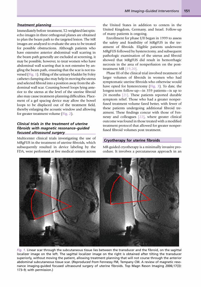
Immediately before treatment, T2-weighted fast spin-echo images in three orthogonal planes are obtainedto plan the beam path to the targeted lesion. The MRimages are analyzed to evaluate the area to be treatedfor possible obstructions. Although patients whohave extensive anterior abdominal wall scarring inthe beam path generally are excluded at screening, itmay be possible, however, to treat women who haveabdominal wall scarring that is not extensive by an-gling the beam path, ensuring that the scar is not tra-versed (Fig. 1). Filling of the urinary bladder by Foleycatheter clamping also may help in moving the uterusand selected fibroid into a position away from the ab-dominal wall scar. Coursing bowel loops lying ante-rior to the uterus at the level of the uterine fibroidalso may cause treatment-planning difficulties. Place-ment of a gel spacing device may allow the bowelloops to be displaced out of the treatment field,thereby enlarging the acoustic window and allowingfor greater treatment volume (Fig. 2).
Clinical trials in the treatment of uterinefibroids with magnetic resonance–guidedfocused ultrasound surgery
Multicenter clinical trials investigating the use ofMRgFUS in the treatment of uterine fibroids, whichsubsequently resulted in device labeling by theFDA, were performed at five medical centers across
Fig. 1. Linear scar through the subcutaneous tissue lies belocalizer image on the left. The sagittal localizer imagesuperiorly, without moving the patient, allowing treatmeabdominal subcutaneous tissue scar. (Reproduced from Fenance imaging-guided focused ultrasound surgery of ut173–9; with permission.)
the United States in addition to centers in theUnited Kingdom, Germany, and Israel. Follow-upof many patients is ongoing.
Enrollment for phase I/II began in 1999 to assessthe safety and feasibility of MRgFUS in the tre-atment of fibroids. Eligible patients underwentMRgFUS followed by hysterectomy, and subsequentpathologic examination of the uterus and fibroidshowed that MRgFUS did result in hemorrhagicnecrosis in the area of nonperfusion on the post-treatment MR [19,20].
Phase III of the clinical trial involved treatment oflarger volumes of fibroids in women who hadsymptomatic uterine fibroids who otherwise wouldhave opted for hysterectomy (Fig. 3). To date, thelongest-term follow-up—in 359 patients—is up to24 months [21]. These patients reported durablesymptom relief. Those who had a greater nonper-fused treatment volume fared better, with fewer ofthese patients undergoing additional fibroid tre-atment. These findings concur with those of Fen-nessy and colleagues [22], where greater clinicaloutcome was found in those treated with a modifiedtreatment protocol that allowed for greater nonper-fused fibroid volumes post treatment.
Cryotherapy for uterine fibroids
MR-guided cryotherapy is a minimally invasive pro-cedure. It involves a percutaneous approach in an
tween the transducer and the fibroid, on the sagittalon the right is obtained after tilting the transducernt planning that will not course through the anteriornnessy FM, Tempany CM. A review of magnetic reso-erine fibroids. Top Magn Reson Imaging 2006;17(3):

Fig. 2. The sagittal localizer image on the left demonstrates bowel loops coursing between the anterior abdom-inal wall and the uterine fibroid. After placement of a spacer device (sagittal localizer image on the right) underthe anterior abdominal wall, the bowel loops are displaced, allowing for treatment through a larger acousticwindow. (Reproduced from Fennessy FM, Tempany CM. A review of magnetic resonance imaging-guidedfocused ultrasound surgery of uterine fibroids. Top Magn Reson Imaging 2006;17(3):173–9; with permission.)
Fennessy et al152
interventional setting with multiple (1 to 5) needle-like 17-G cryotherapy probes. Each probe createsa tear-drop shaped volume of frozen tissue aboutits tip (approximately 2.5-mm diameter); the simul-taneous use of multiple probes gives a larger volumeof treated tissue in the same time frame as treatingwith a single probe. The freeze is provided by pres-surized argon gas that circulates within the probe.Typical treatments involve a cycling of the gas thatdelivers a freeze-thaw-freeze to destroy tissue, witheach stage of the cycle 10 to 15 minutes in duration.MR imaging–guided cryotherapy has been evolvingthrough experiment and clinical use during the past20 years to target a range of tumors in various organsystems [23]. Compared with US that has shadowartifacts, visibility of the ice ball for monitoring isnot as limited.
There are several promising reports of MR imag-ing–guided cryotherapy to treat symptomatic pa-tients who have uterine fibroids [24–27]. Duringcryotherapy, the ice appeared as a signal void inthe image as a result of the short MR relaxationtime of the solid ice, giving a clear demarcationbetween frozen and unfrozen tissue. Though all re-ported relief from deletorious symptoms, short-term clinical outcome, however, is reported inonly 8 of 9 treated patients, who demonstrated onaverage, 65% volume reduction in uterine size [25].
One of these studies [26] was performed transva-ginally, with the investigators proposing that suchan approach had the advantage of providing directaccess, especially for submucosal tumors. Proce-dures usually are performed with epidural anes-thesia in a horizontally open MR imaging scannerwith multiple 2- to 3-mm cryotherapy probes(Fig. 4). Gradient-echo and T2-weighted spin-
echo sequences were used to guide probeplacement and monitor the treatment cycle offreeze-thaw-freeze (Fig. 5).
Percutaneous ablation of fibroids is a nascentprocedure and not practiced widely. This methodof ablation has found a place in treating other partsof the body but not necessarily treating uterinefibroids, possibly because of the recent emergenceof other minimally invasive procedures, such asuterine artery embolization, or noninvasive proce-dures, such as MRgFUS.
Genital tract: male
The leading cause of cancer death in men over 50,prostate cancer, affects one man in six in his life-time. The American Cancer Society estimates thatin 2007 in the United States, 218,890 new casesof prostate cancer will be diagnosed, and approxi-mately 27,050 men will die of the disease [28].There is only a 33% 5-year survival rate in menwho have metastatic disease [29], making early tu-mor detection and localized treatment a necessity.
MR imaging–guided prostate biopsy
Early diagnosis and cancer localization within theprostate gland usually are found through digital ex-amination and serum prostate-specific antigen(PSA) measurement, followed by transrectal ultra-sound (TRUS)-guided biopsy. Image-guided pros-tate biopsy with ultrasound (US) has becomea universally accepted tool [30], but because ofa low sensitivity and specificity for tumor detection[31], interest continues in the development ofa more accurate technique. In addition, for men

Fig. 3. Imaging of a uterine fibroid pretreatment (A, B) and post-treatment (C) with MRgFUS. Sagittal T2-weighted image (A), obtained with the patient in the prone position overlying the US transducer, demonstratesa large solitary uterine fibroid of low-signal intensity. Sagittal SPGR post gadolinium (B) demonstrates homog-enous enhancement of the fibroid. After treatment, sagittal SPGR post gadolinium (C) demonstrates a new largenonperfused area within the fibroid, consistent with treatment-induced necrosis.
MR Imaging–Guided Interventions 153
who have increasing PSA levels and repeatedly neg-ative TRUS-guided prostate biopsies (the concernbeing that a sampling error may result in a false-negative biopsy), for those in whom a transrectalbiopsy is not possible, or for those who are reluc-tant to undergo transrectal biopsy because of its rec-ognized complications, such as infection,hematuria, hematospermia, and rectal bleeding[32–34], an alternative approach may be necessary.
MR imaging can outline prostate architecture andsubstructure. Although the specificity for diagnosismay be limited, MR imaging can demonstrate sus-picious nodules in the peripheral zone, the mostcommon site for prostate cancer. On T2-weightedimages, tumor is demonstrated most commonly
by focal or diffuse regions of decreased signal inten-sity relative to the high-signal-intensity normalperipheral zone. MR imaging is used most routinelyfor staging men who have known cancer. The re-ported accuracy of prostate cancer detection andstaging on MR images varies widely, with reportsof accuracy ranging from 54% to 93%, likelybecause of differences in techniques and interob-server variability [35–38].
Its role in detection and characterization, particu-larly in the initial diagnosis of high-risk patients orthose who have previous negative biopsy findingsbut persistently high PSA levels, is increasing astechniques such as MR spectroscopy imaging(MRSI) and dynamic contrast enhancement

Fig. 4. Photograph demonstrating the set-up for per-cutaneous MR imaging–guided cryotherapy for uter-ine fibroids in an open horizontal 0.3-T AIRIS II(Hitachi, Tokyo, Japan) scanner. (Courtesy of YusukeSakuhara, MD, Department of Radiology, HokkaidoUniversity Hospital, Sapporo, Japan.)
Fennessy et al154
become more widely available. The ultimate roleand application in clinical practice, however, re-main controversial [35]. MR imaging contributessignificant incremental value to TRUS-guided bi-opsy and digital rectal examination in cancer detec-tion and localization in the prostate [39]. It offers anexcellent second-line alternative to those who havefailed to obtain a diagnosis with conventionalmethods.
MR imaging–guided prostate biopsy:technique
Two basic strategies have been explored forMR imaging–guided prostate biopsy. The first is cor-egistration of previously acquired diagnostic MRimages to TRUS images, localizing suspected tumorlesions on MR and correlating these locations to the
Fig. 5. Axial T2-weighted spin-echo sequence demon-strating a probe in the left anterolateral aspect ofa uterine fibroid. The diffuse low-signal intensity inthe fibroid represents the ice-ball. (Courtesy ofYusuke Sakuhara, MD, Department of Radiology,Hokkaido University Hospital, Sapporo, Japan.)
US [40]. The second strategy is stereotactic needleinterventions within diagnostic MR scanners usingcareful patient positioning. By implementing surgi-cal navigation software originally developed forneurosurgery [41,42] and adapting the technicalcapabilities of MR imaging–guided prostate brachy-therapy in an open configuration magnet [43],biopsy of suspected tumor foci in the peripheralzone is made possible (Fig. 6) [44]. In addition,the feasibility of transrectal needle access to prostatetumors has been assessed in a closed-bore 1.5-Tmagnet [45–48] in a small number of patients,which potentially could provide for additionalfunctional and spectroscopic imaging in compari-son with a 0.5-T scanner. The procedure requiresthe use of a specialized device that consists of a nee-dle guide and support system. The same guidancealso has been used recently in a 3-T system [49].Larger studies of clinical usefulness, however, arenecessary. With progress in biologic imaging ofthe prostate gland, it is likely that MR imaging guid-ance will play an increasing role in the diagnosisand treatment of prostate cancer.
MR imaging–guided prostate biopsy:the future
The move toward targeted interventions, for diagno-sis and treatment, underscores the need for preciseimage-guided needle placement. Based on a pa-tient’s anatomy and lesion detection in pretreat-ment MR imaging, a graphic planning interfacethat allows desired needle trajectories to be speci-fied, through MR-compatible robotic assistance,recently has been described [50]. Avoiding the lim-itations of a fixed-needle template is a positive moveforward for tissue sampling and treatment. As thefield of prostate imaging moves to higher strengthmagnets, namely 3 T, the biopsy devices are recon-figured to allow sampling in a closed-bore environ-ment. A recent study of prostate biopsy using 3-TMR imaging guidance described it as a promisingtool for detecting and sampling cancerous regionsin patients who have known prostate cancer [49];however, the role (even at 3 T) of MR imaging–guided prostate biopsy as a screening tool inpatients who have elevated PSA levels and recentprevious negative biopsy remains to be determined.
MR imaging–guided brachytherapyfor prostate cancer
Established options for the management of local-ized prostate cancer include one or a combinationof the following: radical prostatectomy, externalbeam radiation therapy, brachytherapy, or watchfulwaiting. In radiation therapy, the goal is to achievethe prescribed dose throughout the prostate gland

Fig. 6. Imaging before and during MR imaging–guided prostate biopsy. Axial (A) and coronal (B) T2-weightedspin-echo sequence outline areas to be biopsied. In this example, an area in the left midgland is demonstrated(arrow), reformatted to the same spatial location as the corresponding real-time axial (C) and coronal images (D)taken during needle insertion. The biopsy needle is seen in cross section as a circle of low-signal intensity (arrow)on the axial gradient-echo real-time image (C) and as a longitudinal area of low-signal intensity (arrow) on thecoronal gradient-echo real-time image (D).
MR Imaging–Guided Interventions 155
while minimizing toxicity to adjacent structuresand minimizing morbidity from the procedure.Prostate brachytherapy is one of the more popularradiation methods in the prostate and involvesthe percutaneous placement of I-125 radiationseeds into the gland under image guidance. This isdone most commonly with TRUS. It also can bedone with MR guidance and the goal in both proce-dures is to optimize seed placement and allow max-imal dose to the prostate peripheral zone tumorand minimal dose to the urethra and rectum.
Imaging-guided radiation therapy, therefore, al-lows directed tumor treatment, decreasing the chan-ces of disease spreading outside the gland, whilehealthy prostate tissue and its neighboring struc-tures are not overdosed. This is extremely importantfor structures such as the urethra, in which over-ra-diation may cause stricture and fistualization that
can be avoided with good image guidance [51,52].Radiation dose fall-off is sharp at the rectal walland at the urethra. Unlike external beam radiother-apy, there is no entrance or exit dose. Brachytherapy,therefore, has the potential to achieve superior tu-mor control with decreased morbidity and side ef-fects. It is not, however, without its own set ofcomplications, such as rectal irritation and ulcera-tion, incontinence, and impotence resulting frominadvertent delivery of radiation dosing to the rec-tum, bladder, and urethra.
Patient selection
Low-risk prostate cancer patients who have a highprobability of organ-confined disease are screenedappropriately with an endorectal coil MR for po-tential treatment with brachytherapy monother-apy. Most centers include patients who have

Fennessy et al156
stage T1-T2a (according to the American JointCommittee on Cancer/International UnionAgainst Cancer 1997 staging), PSA level of 10ng/mL or less, and a Gleason score of 6 or lower.The few contraindications to the procedure in-clude prior transurethral resection of the prostateor morbid obesity (equipment cannot sustainthe weight).
Procedure
MR-guided prostate brachytherapy using open con-figuration 0.5-T and 1.5-T scanners are described[43,45]. Using the open 0.5-T magnet, patients areplaced supine between the two magnets in thelithotomy position under general anesthesia. AFoley catheter is inserted, the skin is prepared anddraped in a sterile fashion, and a template for nee-dle guidance is placed against the perineum. A rec-tal obturator then is placed and T2-weighted imagesare acquired in the axial, coronal, and sagittalplanes and used to outline the urethra, peripheralzone, and anterior rectal wall. Surgical simulationsoftware outlines these areas, and the targeted vol-ume is calculated using designated planning soft-ware [53]. Seed number and depth of catheterinsertion are calculated.
While gradient-echo MR images are obtained inreal time [44], seed-loaded catheters then are posi-tioned in the prostate gland (Fig. 7). The images arecompared to their intended locations, according tothe radiation therapist plan. Dose-volume histo-grams of the urethra, anterior rectal wall, and targetvolume are obtained before final deployment ofseeds. Six weeks after the procedure, CT imaging(to identify the seeds accurately) and MR imaging(for prostate anatomic correlation) are fused to cal-culate the final dose distribution to the gland andsurrounding tissues.
Although open-bore magnets offer good patientaccessibility and allow satisfactory prostate tumorand anatomic depiction, higher-quality MR inter-vention images in a closed 1.5-T system also havebeen investigated [45]. This system uses a custom-ized perineal template, an endorectal imagingcoil, and a lockable positioning arm. Patients areplaced in the left lateral decubitus position. Al-though patient accessibility with this techniquemay be limited because of the closed-bore configu-ration and the 60-cm diameter bore, the investiga-tors found that dependence on deformableregistration between image sets (high-field 1.5-T di-agnostic images and low-field–strength interven-tional images) was reduced.
Outcomes
Short-term toxicity after MR-guided brachytherapyis rare, with no gastrointestinal or sexual dysfunction
reported during the first month after treatment[54]. Within 24 hours of removal of the Foley cath-eter, acute urinary retention was reported in 12% ofmen, which was self-limited to within 1 to 3 weeksof treatment. Prostatic volume and transitionalzone volume, determined by MR imaging, andnumber of brachy therapy seeds placed were foundto be significant predictors of acute urinaryretention.
The long-term genitourinary and rectal toxicitywas compared between those who receivedMR-guided brachytherapy alone and those whoreceived combined MR-guided brachytherapy andexternal beam radiation therapy [55]. The 4-yearestimates of rectal bleeding requiring coagulationfor patients who underwent MR-guided brachyther-apy compared with patients who received com-bined-modality therapy were 8% versus 30%. The4-year estimate of freedom from radiation cystitiswas 100% versus 95% for patients who receivedMR-guided brachytherapy alone and patients whoreceived combined-modality therapy, respectively.In a separate study evaluating the long-term toxicityin patients who received MR-guided brachytherapyas a salvage procedure for radiation therapy failure[56], the 4-year estimate of grade 3 or 4 gastrointes-tinal or genitourinary toxicity was reported at 30% ofall patients, with 13% requiring an intervention,such as a colostomy or urostomy for fistula repair.
Supplemental external beam radiotherapy, inaddition to brachytherapy seed implantation, hasbeen given to patients who have intermediate- tohigh-risk prostate cancer (according to the D’Amicorisk stratification for prostate cancer) [57]. Thiscombination of radiation therapy has demon-strated good long-term results [58,59], resulting ina 15-year biochemical relapse-free survival equalto 80.3% for intermediate-risk disease and 67.2%for high-risk disease.
Brachytherapy: the future
The current manual method of needle placement,using a fixed-needle template guide, constrainsneedle orientation. The manual method alsomakes use of manual computation and transcrip-tion of needle coordinates that are prone to hu-man error. The future points toward a systemthat incorporates an interactive planning interfacewith MR-compatible robotic assistance. Such a de-vice, which serves as a dynamic guide for preciseneedle placement, has been developed [60]. Likelythe future direction for percutaneous MR imaging–guided prostatic interventions, this MR-compatiblerobotic device has been integrated with a softwareplanning interface, allowing physicians to specifydesired needle trajectories based on MR imaginganatomy.

Fig. 7. Pre-, intra-, and postoperative MR imaging–guided brachytherapy in prostate cancer. Preoperative 1.5-T(A) axial T2-weighted spin-echo image through the prostate base, demonstrating low signal intensity in theperipheral zone (arrows), previously demonstrated to be tumor. Intraoperative 0.5-T (B) axial T2-weightedspin-echo T2 weighted spin-echo image through the same area. Intraoperative axial gradient-echo MR images(C) obtained in real time during needle and seed placement in the prostate base. The larger round areas repre-sent the needles (arrows), before deployment, and the small round areas represent the deployed seeds (arrow-heads). A postoperative axial SPGR (D) through the prostate base demonstrates multiple round areas of lowsignal in the peripheral zone (arrowheads), consistent with deployed seeds.
MR Imaging–Guided Interventions 157
Focused ultrasound surgery in the prostate
As discussed previously regarding the female genitaltract, there is growing interest in MRgFUS because ofits many potential applications as a minimally inva-sive therapy. US-guided FUS (USgFUS) has beenused predominantly in Europe for the treatmentof prostate cancer. Limitations include difficulty intreating the anterior prostate or small-volume pros-tates, and lack of long-term follow-up.
Literature describing the results of USgFUS forprostate cancer suggests that USgFUS treatment isa valuable option for well-differentiated and
moderately differentiated tumors and for local re-currence after external-beam radiation therapy[61–63]. USgFUS treatment is whole-gland therapy,without selective tumor-directed targeted treatment,that should allow for minimal disruption of normalfunction. USgFUS arguably is limited by the lack ofdirect temperature and thermal dose measurementsduring thermocoagulation. Without the latter, theenergy delivery cannot be controlled or monitorednor can the thermal dose be measured accurately.
To address these challenges, MR imaging–com-patible prostate applications have been developedfor hyperthermia [64], and phased-array

Fennessy et al158
applicators for thermal ablation [65]. Insightec(Haifa, Israel) has developed a MRgFUS systemfor prostate treatment (Fig. 8). A major potentialadvantage of MR imaging guidance is its ability tomap functional changes in prostate tissue, withthe possibility of 3-D tumor mapping before andduring treatment. Overall, noninvasive thermal ab-lation using MR imaging guidance should improveprostate treatment significantly and its applicationshould increase in the near future.
MR imaging–guided catheter-basedultrasound thermal therapy of the prostate
In a similar mode, catheter-based US devices (ininterstitial and transurethral configurations) havebeen evaluated in canine prostate models in vivoand found to produce spatially selective regions ofthermal destruction in the prostate [66–68]. Tran-surethral US devices with tubular transducers havebeen developed, which can coagulate sectors ofthe prostate using preshaped angular patterns[69,70]. Devices with finer spatial control using pla-nar [71,72] or curvilinear transducers [73] can berotated slowly using a computer-controlled,MR-compatible stepper motor while under MR im-aging guidance and feedback (Fig. 9). The feasibil-ity of MR imaging–guided interstitial US thermaltherapy of the prostate has been evaluated in anin vivo canine prostate model [66]. MR
Fig. 8. (A) MR imaging–based temperature image duringmuscle during a test of an MR imaging–compatible transr(B) The thermal lesion (arrow) seen in T2-weighted imaginfascia layer. (From Sokka SD, Hynynen K. The feasibilityaperiodic intracavitary ultrasound phased array. Phys Me
imaging–compatible, multielement interstitial USapplicators were used. The applicators were insertedtransperineally into the prostate with the energy di-rected ventrally away from the rectum. This studydemonstrated a large volume of ablated tissuewithin the prostate and, importantly, demonstratedcontiguous zones of thermal coagulation. At least inan animal model and using MR guidance, transure-thral and interstitial treatment strategies have,therefore, demonstrated significant potential forthermal ablation of localized prostate cancer.
Urinary tract
Renal cell carcinoma (RCC) is the sixth leadingcause of cancer death [74], and its incidence inthe United States is rising [75]. Partial nephrec-tomy, a nephron-sparing surgical method, hasreplaced radical nephrectomy for the treatment ofsmall RCC. Less invasive methods also haveemerged that can be performed laparoscopically(partial nephrectomy and cryosurgery) or percuta-neously (radiofrequency ablation [RFA] and cryoa-blation). Image-guided percutaneous ablationshave the potential to replace others as the leastinvasive and least costly [76] of all nephron-sparingtreatments clinically available, particularly in pa-tients who are poor surgical candidates because ofcomorbid disease and patients who have renalinsufficiency, solitary kidney, or multiple RCC.
a sonication (130 W for 30 seconds) into rabbit thighectal phased array applicator for MRgFUS of prostate.g. The bright region to the right of the lesion is a tissueof MRI-guided whole prostate ablation with a lineard Biol 2000;45:3373–83; with permission.)

Fig. 9. MR imaging–guided catheter-based US thermal therapy of the prostate: real-time temperature image(left), maximum temperature image (middle), and thermal dose (right) of the prostate during catheter-basedUS thermal therapy. The transurethral catheter, with a rotating curvilinear transducer array, is depicted asthe round low-signal intensity structure within the prostate gland [69]. (Courtesy of Kim Butts Pauly, PhD,Viola Rieke, MD, and Graham Sommer, PhD, Stanford University School of Medicine, Stanford, CA; and ChrisDiederich, PhD, UCSF, San Francisco, CA.)
MR Imaging–Guided Interventions 159
MR imaging–guided radiofrequency ablationin the kidney
RFA is a focal thermal tumor therapy method in whichtissue is heated by an electric current. The current ispresent with a high density surrounding a percutane-ously placed electrode that is driven by an electricalgenerator. The circuit is completed by the placementof grounding pads on a patient. The electrode is placedinterstitially and intended to be activated to createa volume of coagulative necrosis in place of the tumor.
Many reports of successful treatment of renaltumors with percutaneous RFA have been pub-lished [77–85]. Real-time monitoring of RFA, how-ever, is not possible with CT or US because thethermal ablation zone is not visible with theseimaging modalities. RFA can be monitored withMR imaging [86,87] but with limitations. Radiofre-quency energy has to be interrupted during MRimaging because of the significant interference itcauses otherwise. Furthermore, the temperature-sensitive, very short repetition time/echo time gra-dient-echo sequences typically are not suitable fordetailed visualization of retroperitoneal anatomy.
Specific to MR imaging–guided RFA in the kidney,the first report was an in vivo study in porcine kid-ney [88]. The procedures were performed in a 0.2-Topen magnet and demonstrated the suitability ofMR imaging for guiding needle placement and thebenefit of its inherent soft-tissue sensitivity wherethe electrode could be placed and the thermal lesionobserved. Clinical studies of MR imaging–guidedRFA in the kidney subsequently reported the safetyand efficacy of the procedure [87,89] in tumors lessthan 4 cm in diameter. No recurrences at 25 months’post procedure were reported.
Overall, RFA is a feasible therapeutic modality forkidney lesions, under MR imaging, CT, or USguidance [90]. Although RFA is performed moreroutinely under CT guidance, many practices turn
to MR imaging for the assessment and long-termfollow-up of treated patients [91–93].
MR imaging–guided percutaneouscryotherapy of renal tumors
Cryoablation, a focal thermal tumor therapy methodwhich uses extreme cold to establish coagulativenecrosis, has several advantages over RFA. WhileRFA may require the need to perform multiple over-lapping ablations of larger tumors, with percutane-ous cryoablation, larger tumors can be treatedsimultaneously with the placement of multiple ap-plicators [94,95]. Evidence suggests that renal tu-mors more likely are treated in one session withcryoablation compared to RFA [96]. Lower doses ofmedications are required for intravenous conscioussedation suggesting that percutaneous cryoablationof renal tumors is associated with less intraprocedur-al pain than with percutaneous RFA [97].
US monitoring of cryoablation is limited by an in-ability to image the entire ice ball because of acousticshadowing from the edge closest to the US probe[98]. Cryoablation of renal tumors can be monitoredwith CT because the ice ball is readily apparent as a hy-poattenuating structure in the renal parenchyma[94,95,98–103]. There are two main limitationswith CT, however. One is that the portion of the iceball in the perinephric fat, a hyperattenuating region,provides only a modest contrast-to-noise ratio[104,105] compared to surrounding fat, and the abla-tion zone edge is not demarcated clearly in fatty tissue(Fig. 10A). This limits its use for real-time monitoringof the effect of ablation on adjacent critical structures,such as bowel, ureter, pancreas, and adrenal gland.Another is that the streak artifact created by the appli-cators with CT imaging can interfere with ice ball vis-ibility (Fig. 10B).
Since the initial clinical reports of MR imaging–guided percutaneous cryoablation of renal tumors

Fig. 10. CT–guided percutaneous cryotherapy of renal yumors. A 77-year-old woman who had RCC of the right kid-ney upper pole. Unenhanced transverse CT images obtained during percutaneous cryoablation performed in theright lateral decubitus position show that (A) low contrast-to-noise ratio and poor edge definition of ice ball (ar-rows) in the perinephric fat renders assessment for overlap of ablation zone with adjacent adrenal gland (arrow-head) difficult, and (B) streak artifact from applicator interferes with visualization of portion of the ice ball (arrow).
Fennessy et al160
[98,100] in 2001, several investigators have shown thefeasibility and safety of the procedure [106–112], alldemonstrating the advantages of MR imaging moni-toring during percutaneous cryoablation procedures.
MR imaging depicts the ice ball as a signal voidregion with high contrast-to-noise ratio comparedto surrounding tissues, with sharp edge definitionin multiple planes and with minimal applicator arti-fact (Fig. 11). Ice ball volume on intraprocedural MRimaging correlates well with volume of cryonecrosison postprocedural MR imaging [113]. Because theice ball is well depicted on all pulse sequences, theablation can be monitored using pulse sequencesthat display tumor or adjacent critical structuresbest [111]. If images demonstrate incomplete cover-age of tumor, additional applicators may be placedto improve coverage [111,112]. Alternatively, if theice ball edge approaches adjacent critical structures,the freezing can be stopped [111]. Applicators can becontrolled individually. Additional maneuvers toreduce risk for injury to surrounding bowel, suchas water instillation described for CT-guided abla-tions [114], also can be performed during MRimaging–guided cryoablations [111]. A noninvasivemethod of external manual displacement of bowelduring MR imaging–guided cryoablation of renaltumors also is described—a maneuver unique tocryoablation procedures performed in an open-configuration interventional MR imaging unit [115].
Limitations of MR imaging–guided cryoablationinclude the high cost of MR imaging units and itslimited availability, generally long procedure times,smaller gantry sizes compared to CT scanners, and
inability to detect ST-T segment changes of cardiacischemia on an EKG in the magnetic environmentduring procedures.
In summary, image-guided percutaneous ablativetherapies have the potential to replace conventionalsurgical treatment of small RCC. Compared withother image-guided ablative therapies, with itsvast advantages and minimal limitations, MRimaging–guided percutaneous cryoablation is wellpoised to play an important role in the manage-ment of renal tumors.
The future
Recent developments in MR imaging parallelingthose in computer-assisted surgery have set up anideal environment for MR-compatible robotic sys-tems and manipulators. Materials used in mecha-tronic devices inside the magnet ideally shouldhave a magnetic susceptibility similar to that ofhuman tissue and be electrical insulators to avoidimage distortion. Although image quality isreduced because of reduction in static field strength,interventional open-bore magnets have fewerspatial constraints. Alternatively, closed MR scan-ners can impose severe constraints on proceduralmanipulations, despite their imaging advantage ofhigher field strengths. New wide- and short-bore1.5-T magnets (Espree, Siemens, Erlangen, Ger-many) will expand the use of interventional MR im-aging. Emerging use of 3-T magnets forinterventions will bring about improved monitor-ing of thermal therapies. Much research is

Fig. 11. MR imaging–guided percutaneous cryotherapy of renal tumor. A 70-year-old man who had RCC of theright kidney lower pole treated with MR imaging–guided percutaneous cryoablation. (A) Transverse T2-weighted fast recovery fast spin–echo sequence image obtained before treatment in 1.5-T MR image showsa small exophytic renal mass in the lower pole of the right kidney anteriorly (arrow). (B) Intraprocedural trans-verse gradient-echo image obtained in 0.5-T open configuration interventional MR imaging shows that sharpedge definition of signal void ice ball (arrows) contributes to monitoring of tumor coverage and assessmentof proximity to adjacent ureter (arrowhead), renal collecting system (1), and colon (*), which is being displacedby an interventionalist’s hand (curved arrow). (C) An 18-month follow-up contrast-enhanced transverse CT im-age shows no enhancement in the involuted ablation zone (arrows).
MR Imaging–Guided Interventions 161
underway evaluating material selection, positiondetection sensors, different actuation models andtechniques, and design strategies [116]. Once theengineering hurdle is overcome, systems must un-dergo clinical validation before introduction intothe commercial realm.
Summary
MR imaging has become part of routine care in manyplaces around the world, for tumor detection,
localization, and staging. In the genitourinary tract,MR imaging guidance is playing an increasing role inminimally invasive procedures for confirmation oftumor pathology and for tumor treatment andtreatment monitoring. It offers inherent ability fortumor detection and biopsy guidance and, currently,MR-guided ablative therapies are an increasing andreal alternative to more invasive surgical options.As the capabilities of MR imaging expand and newerimaging modalities become more accessible (PETimaging, for example), the need for nonrigid

Fennessy et al162
registration of multiple modalities will be necessary.A combination of functional imaging and high-resolution tumor detail in the genitourinary tract,in a patient-specific treatment environment, shouldincrease demand and the use of semi-invasive ornoninvasive technology. Clearly, the pressure is onto provide MR-compatible devices and methodol-ogy that easily integrate with imaging and are sup-portive of patients’ clinical needs.
References
[1] Stewart AJ, Viswanathan AN. Current controver-sies in high-dose-rate versus low-dose-ratebrachytherapy for cervical cancer. Cancer2006;107(5):908–15.
[2] Stewart EA. Uterine fibroids. Lancet 2001;357:293–8.
[3] Ravina J, Herbreteau D, Ciraru-Vigneron N,et al. Arterial embolisation to treat uterinemyomata. Lancet 1995;346:671–2.
[4] Lynn JG, Zwemer RL, Chick AJ, et al. A newmethod for the generation and use of focusedultrasound in experimental biology. J Gen Phys-iol 1942;26:179–93.
[5] Fry WJ, Barnard JW, Fry FJ. Ultrasonicallyproduced localized selective lesions in thecentral nervous system. Am J Phys Med 1955;34:413–23.
[6] Lele PP. A simple method for production oftrackless focal lesions with focused ultrasound:physical factors. J Physiol 1962;160:494–512.
[7] Heimburger RF. Ultrasound augmentation ofcentral nervous system tumor therapy. IndianaMed 1995;78:469–76.
[8] Gelet A, Chapelon JY, Bouvier R, et al. Localcontrol of prostate cancer by transrectal highintensity focused ultrasound therapy: prelimi-nary results. J Urol 1999;161:156–62.
[9] Paterson RF, Barret E, Siqueira TM Jr, et al. Lap-aroscopic partial kidney ablation with highintensity focused ultrasound. J Urol 2003;169(1):347–51.
[10] Yang R, Sanghvi NT, Rescorla FJ, et al. Extracor-poreal liver ablation using sonography-guidedhigh-intensity focused ultrasound. Invest Radiol1992;27(10):796–803.
[11] Watkin NA, Morris SB, Rivens IH, et al. A feasi-bility study for the non-invasive treatment ofsuperficial bladder tumours with focusedultrasound. Br J Urol 1996;78(5):715–21.
[12] Lizzi FL, Deng CX, Lee P, et al. A comparison ofultrasonic beams for thermal treatment ofocular tumors. Eur J Ultrasound 1999;9(1):71–8.
[13] Ishihara Y, Calderon A, Watanabe H, et al. Aprecise and fast temperature mapping usingwater proton chemical shift. Magn Reson Med1995;34(6):814–23.
[14] Hynynen K, Vykhodtseva NI, Chung AH, et al.Thermal effects of focused ultrasound on the
brain: determination with MR imaging. Radiol-ogy 1997;204(1):247–53.
[15] Chung AH, Jolesz FA, Hynynen K. Thermaldosimetry of a focused ultrasound beam invivo by magnetic resonance imaging. MedPhys 1999;26(9):2017–26.
[16] McDannold N, Tempany CM, Fennessy FM,et al. Uterine leiomyomas: MR imaging-basedthermometry and thermal dosimetry duringfocused ultrasound thermal ablation. Radiology2006;240(1):263–72.
[17] Chung AH, Hynynen K, Colucci V, et al. Opti-mization of spoiled gradient-echo phaseimaging for in vivo localization of focusedultrasound beam. Magn Reson Med 1996;36(5):745–52.
[18] Leon-Villapalos J, Kaniorou-Larai M,Dziewulski P. Full thickness abdominal burnfollowing magnetic resonance guided focusedultrasound therapy. Burns 2005;31(8):1054–5.
[19] Tempany CM, Stewart EA, McDannold N, et al.MR imaging-guided focused ultrasound surgeryof uterine leiomyomas: a feasibility study. Radi-ology 2003;226(3):897–905.
[20] Stewart EA, Gedroyc WM, Tempany CM, et al.Focused ultrasound treatment of uterine fibroidtumors: safety and feasibility of a noninvasivethermoablative technique. Am J Obstet Gynecol2003;189(1):48–54.
[21] Stewart EA, Gostout B, Rabinovici J, et al. Sus-tained relief of leiomyoma symptoms by usingfocused ultrasound surgery. Obstet Gynecol2007;110(2 Pt 1):279–87.
[22] Fennessy FM, Tempany C, McDannold N, et al.Uterine leiomyomas: MR imaging-guidedfocused ultrasound surgery–results of differenttreatment protocols. Radiology 2007;243(3):885–93.
[23] Morrison PR, Silverman SG, Tuncali K, et al.MRI guided cryotherapy. J Magn Reson Imaging2008;27(2):410–20.
[24] Sewell PE, Arriola RM, Robinette L, et al. Real-time I-MR-imaging-guided cryoablation ofuterine fibroids. J Vasc Interv Radiol 2001;12(7):891–3.
[25] Cowan BD, Sewell PE, Howard JC, et al.Interventional magnetic resonance imagingcryotherapy of uterine fibroid tumors: prelimi-nary observation. Am J Obstet Gynecol 2002;186(6):1183–7.
[26] Dohi M, Harada J, Mogami T, et al. MR-guidedtransvaginal cryotherapy of uterine fibroidswith a horizontal open MRI system: initialexperience. Radiat Med 2004;22(6):391–7.
[27] Sakuhara Y, Shimizu T, Kodama Y, et al.Magnetic resonance-guided percutaneous cry-oablation of uterine fibroids: early clinical ex-periences. Cardiovasc Intervent Radiol 2006;29(4):552–8.
[28] American Cancer Society. Cancer facts andfigures 2007. Publication no. 500807. Atlanta(GA): American Cancer Society; 2006.

MR Imaging–Guided Interventions 163
[29] American Cancer Society. Cancer facts and fig-ures. Publication no. 500807. Atlanta (GA):American Cancer Society; 2008.
[30] Lee F, Gray JM, McLeary RD, et al. Prostatic eval-uation by transrectal sonography: criteria fordiagnosis of early carcinoma. Radiology 1986;158:91–5.
[31] Terris MK. Sensitivity and specificity of sextantbiopsies in the detection of prostate cancer;preliminary report. Urology 1999;54:486–9.
[32] Aus G, Hermansson CG, Hugosson J, et al.Transrectal ultrasound examination of theprostate: complications and acceptance bypatients. Br J Urol 1993;71:457–9.
[33] Collins GN, Lloyd SN, Hehir M, et al. Multipletransrectal ultrasound-guided prostatic biop-sies: true morbidity and patient acceptance. BrJ Urol 1993;71:460–3.
[34] Rodriguez LV, Terris MK. Risks and complica-tions of transrectal ultrasound guided prostateneedle biopsy: a prospective review of the liter-ature. J Urol 1998;160:2115–20.
[35] Rifkin MD, Zerhouni EA, Gatsonis CA, et al.Comparison of magnetic resonance imagingand ultrasonography in staging early prostatecancer: results of a multi-institutional coopera-tive trial. N Engl J Med 1990;323:621–6.
[36] Schnall MD, Pollack HM. Magnetic resonanceimaging of the prostate gland. Urol Radiol1990;12(2):109–14.
[37] Cornud F, Flam T, Chauveinc L, et al. Extrapro-static spread of clinically localized prostatecancer: factors predictive of pT3 tumor and ofpositive endorectal MR imaging examinationresults. Radiology 2002;224(1):203–10.
[38] Outwater EK, Petersen RO, Siegelman ES, et al.Prostate carcinoma: assessment of diagnosticcriteria for capsular penetration on endorectalcoil MR images. Radiology 1994;193(2):333–9.
[39] Mullerad M, Hricak H, Kuroiwa K, et al. Com-parison of endorectal magnetic resonanceimaging, guided prostate biopsy and digitalrectal examination in the preoperative anatom-ical localization of prostate cancer. J Urol 2005;174:2158–63.
[40] Perrotti M, Han KR, Epstein RE, et al. Prospec-tive evaluation of endorectal magneticresonance imaging to detect tumor foci inmen with prior negative prostatic biopsy: a pilotstudy. J Urol 1999;162:1314–7.
[41] Hata N, Morrison PR, Kettenbach J, et al. Com-puter-assisted intra-operative MRI monitoringof interstitial laser therapy in the brain:a case report. SPIE J Biomed Optics 1998;3:302–11.
[42] Gering D, Nabavi A, Kikinis R, et al. Anintegrated visualization system for surgicalplanning and guidance using image fusionand interventional imaging. Medical ImageComputing and Computer-Assisted
Intervention (MICCAI), Cambridge, EnglandSeptember 22, 1999.
[43] D’Amico AV, Cormack R, Tempany CM, et al.Real-time magnetic resonance image-guidedinterstitial brachytherapy in the treatment ofselect patients with clinically localized prostatecancer. Int J Radiat Oncol Biol Phys 1998;42:507–15.
[44] Hata N, Jinzaki M, Kacher D, et al. MR imaging-guided prostate biopsy with surgical navigationsoftware; device validation and feasibility.Radiology 2001;220:263–8.
[45] Susil RC, Camphausen K, Choyke P, et al.System for prostate brachytherapy and biopsyin a standard 1.5 T MRI scanner. Magn ResonMed 2004;52(3):683–7.
[46] Susil RC, Menard C, Kreiger A, et al. Transrectalprostate biopsy and fiducial marker placementin a standard 1.5T magnetic resonance imagingscanner. J Urol 2006;175(1):113–20.
[47] Beyersdorff D, Winkel A, Hamm B, et al. MRimaging-guided prostate biopsy with a closedMR unit at 1.5 T: initial results. Radiology2005;234(2):576–81.
[48] Kreiger A, Susil RC, Menard C, et al. Design ofa novel MRI compatible manipulator for imageguided prostate interventions. IEEE TransBiomed Eng 2005;52(2):306–13.
[49] Singh AK, Kreiger A, Lattouf JB. Patient selec-tion determines the prostate cancer yield ofdynamic contrast-enhanced magnetic reso-nance imaging-guided transrectal biopsies ina closed 3-Tesla scanner. BJU Int 2008;101(12):181–5.
[50] DiMaio SP, Pieper S, Chinzei K, et al. Robot-assisted needle placement in open MRI: systemarchitecture, integration and validation. Com-put Aided Surg 2007;12(1):15–24.
[51] Lee WR, Hall MC, McQuellon RP, et al. A pro-spective quality-of-life study in men withclinically localized prostate carcinoma treatedwith radical prostatectomy, external beamradiotherapy, or interstitial brachytherapy. Int JRadiat Oncol Biol Phys 2001;51(3):614–23.
[52] Zelefsky MJ, Yamada Y, Marion C, et al. Im-proved conformality and decreased toxicitywith intraoperative computer-optimized trans-perineal ultrasound-guided prostate brachy-therapy. Int J Radiat Oncol Biol Phys 2003;55(4):956–63.
[53] Kooy HM, Cormack RA, Mathiowitz RV, et al. Asoftware system for interventional magneticresonance image-guided prostate brachyther-apy. Comput Aided Surg 2000;5(6):401–13.
[54] D’Amico AV, Cormack R, Kumar S, et al. Real-time magnetic resonance imaging-guidedbrachytherapy in the treatment of selectedpatients with clinically localized prostate can-cer. J Endourol 2000;14:367–70.
[55] Albert M, Tempany CM, Schultz, et al. Lategenitourinary and gastrointestinal toxicity after

Fennessy et al164
magnetic resonance image-guided prostatebrachytherapy with or without neoadjuvantexternal beam radiation therapy. Cancer 2003;98(5):949–54.
[56] Nguyen PL, Chen MH, D’Amico AV, et al. Mag-netic resonance image-guided salvage brachy-therapy after radiation in select men whoinitially presented with favorable-risk prostatecancer: a prospective phase 2 study. Cancer2007;110(7):1485–92.
[57] D’Amico AV, Moul J, Carroll PR, et al. Cancer-specific mortality after surgery or radiation forpatients with clinically localized prostate cancermanaged during the prostate-specific antigenera. J Clin Oncol 2003;21(11):2163–72.
[58] Sylvester JE, Grimm PD, Blasko JC, et al. 5-Yearbiochemical relapse free survival in clinicalStage T1-T3 prostate cancer following com-bined external beam radiotherapy and brachy-therapy; Seattle experience. Int J Radiat OncolBiol Phys 2007;67(1):57–64.
[59] Lawton CA, DeSilvo M, Lee WR, et al. Results ofa phase II trial of transrectal ultrasound-guidedpermanent radioactive implantation of theprostate for definitive management of localizedadenocarcinoma of the prostate (radiationtherapy oncology group 98-05). Int J RadiatOncol Biol Phys 2007;67(1):39–47.
[60] Chinzei K, Miller K. Towards MRI guided surgi-cal manipulator. Med Sci Monit 2001;7:153–63.
[61] Poissonier L, Chapelon JY, Rouviere O, et al.Control of prostate cancer by transrectal HIFUin 227 patients. Eur Urol 2007;51(2):381–7.
[62] Uchida T, Ohkusa H, Nagata Y, et al. Treatmentof localized prostate cancer using high-intensityfocused ultrasound. BJU Int 2006;97(1):56–61.
[63] Ficarra V, Antoniolli SZ, Novara G, et al. Short-term outcome after high-intensity focusedultrasound in the treatment of patients withhigh-risk prostate cancer. BJU Int 2006;98(6):1193–8.
[64] Smith NB, Buchanan MT, Hynynen K. Transrec-tal ultrasound applicator for prostate heatingmonitored using MRI thermometry. Int J RadiatOncol Biol Phys 1999;43(1):217–25.
[65] Sokka SD, Hynynen KH. The feasibility ofMRI-guided whole prostate ablation with a lin-ear aperiodic intracavitary ultrasound andphased array. Phys Med Biol 2000;45(11):3378–83.
[66] Nau WH, Diederich CJ, Ross AB, et al. MRI-guided interstitial ultrasound thermal therapyof the prostate: a feasibility study in the caninemodel. Med Phys 2005;32(3):733–43.
[67] Pauly KB, Diedrich CJ, Rieke V, et al. Magneticresonance-guided high-intensity ultrasoundablation of the prostate. Top Magn Reson Imag-ing 2006;17(3):195–207.
[68] Diederich CJ, Nau WH, Ross AB, et al. Catheter-based ultrasound applicators for selectivethermal ablation: progress towards MRI-guided
applications in prostate. Int J Hyperthermia2004;20(7):739–56.
[69] Diederich CJ, Stafford RJ, Nau WH, et al. Tran-surethral ultrasound applicators with direc-tional heating patterns for prostate thermaltherapy: in vivo evaluation using magneticresonance thermometry. Med Phys 2004;31(2):405–13.
[70] Hazle JD, Diederich CJ, Kangasniemi M, et al.MRI-guided thermal therapy of transplantedtumors in the canine prostate using a directionaltransurethral ultrasound applicator. J MagnReson Imaging 2002;15(4):409–17.
[71] Chopra R, Burtnyk M, Haider MA, et al. Methodfor MRI-guided conformal thermal therapy ofprostate with planar transurethral ultrasoundheating applicators. Phys Med Biol 2005;50(21):4957–75.
[72] Ross AB, Diederich CJ, Nau WH, et al. Highlydirectional transurethral ultrasound applicatorswith rotational control for MRI guided prostaticthermal therapy. Phys Med Biol 2004;49(1):189–204.
[73] Ross AB, Diederich CJ, Nau WH, et al. Curvilin-ear transurethral ultrasound applicator forselective prostate thermal therapy. Med Phys2005;32(6):1555–65.
[74] Godley PA, Ataga KI. Renal cell carcinoma. CurrOpin Oncol 2000;12:260–4.
[75] Chow WH, Devesa SS, Warren JL, et al. Risingincidence of renal cell cancer in the UnitedStates. JAMA 1999;281:1628–31.
[76] Link RE, Permpongkosol S, Gupta A, et al. Costanalysis of open, laparoscopic, and percutane-ous treatment options for nephron-sparingsurgery. J Endourol 2006;20:782–9.
[77] Gervais DA, McGovern FJ, Wood BJ, et al. Ra-dio-frequency ablation of renal cell carcinoma:early clinical experience. Radiology 2000;217:665–72.
[78] Ogan K, Jacomides L, Dolmatch BL, et al. Percu-taneous radiofrequency ablation of renaltumors: technique, limitations, and morbidity.Urology 2002;60:954–8.
[79] Farrell MA, Charboneau WJ, DiMarco DS, et al.Imaging-guided radiofrequency ablation ofsolid renal tumors. AJR 2003;180:1509–13.
[80] Mayo-Smith WW, Dupuy DE, Parikh PM,et al. Imaging-guided percutaneous radiofre-quency ablation of solid renal masses:technique and outcomes of 38 treatment ses-sions in 32 consecutive patients. AJR 2003;180:1503–8.
[81] Roy-Choudhury SH, Cast JE, Cooksey G, et al.Early experience with percutaneous radiofre-quency ablation of small solid renal masses.AJR 2003;180:1055–61.
[82] Su LM, Jarrett TW, Chan DYS, et al. Percutane-ous computed tomography-guided radiofre-quency ablation of renal masses in highsurgical risk patients: preliminary results.Urology 2003;61:26–33.

MR Imaging–Guided Interventions 165
[83] Zagoria RJ, Hawkins AD, Clark PE, et al. Percu-taneous CT-guided radiofrequency ablation ofrenal neoplasms: factors influencing success.AJR 2004;183:201–7.
[84] Gervais DA, McGovern FJ, Arellano RS, et al.Radiofrequency ablation of renal cell carci-noma: part 1, indications, results, and rolein patient management over 6-year periodand ablation of 100 tumors. AJR 2005;185:64–71.
[85] Varkarakis IM, Allaf ME, Inagaki T, et al. Percu-taneous radiofrequency ablation of renalmasses: results at a 2-year mean followup.J Urol 2005;174:456–60.
[86] Lewin JS, Connell CF, Duerk JL, et al. InteractiveMRI-guided radiofrequency interstitial thermalablation of abdominal tumors: clinical trialfor evaluation of safety and feasibility. JMRI1998;8:40–7.
[87] Lewin JS, Nour SG, Connell CF, et al. Phase IIclinical trial of interactive MR imaging-guidedinterstitial radiofrequency thermal ablation ofprimary kidney tumors: initial experience. Radi-ology 2004;232(3):835–45.
[88] Merkle EM, Shonk JR, Duerk JL, et al. MR-guided RF thermal ablation of the kidney ina porcine model. AJR Am J Roentgenol 1999;173(3):645–51.
[89] Boss A, Clasen S, Kuczyk M, et al. Magneticresonance-guided percutaneous radiofre-quency ablation of renal cell carcinomas: a pi-lot clinical study. Invest Radiol 2005;40(9):583–90.
[90] Boss A, Clasen S, Kuczyk M, et al. Image-guidedradiofrequency ablation of renal cell carci-noma. Eur Radiol 2007;17(3):725–33.
[91] Merkle EM, Nour SG, Lewin JS. MR imagingfollow-up after percutaneous radiofrequencyablation of renal cell carcinoma: findings in18 patients during first 6 months. Radiology2005;235(3):1065–71.
[92] Memarsadeghi M, Schmook T, Remzi M, et al.Percutaneous radiofrequency ablation of renaltumors: midterm results in 16 patients. Eur JRadiol 2006;59(2):183–9.
[93] Kawamoto S, Permpongkosol S, Bluemke DA,et al. Sequential changes after radiofrequencyablation and cryoablation of renal neoplasms:role of CT and MR imaging. Radiographics2007;27(2):343–55.
[94] Atwell TD, Farrell MA, Callstrom MR, et al.Percutaneous cryoablation of 40 solid renaltumors with US guidance and CT monitoring:initial experience. Radiology 2007;243:276–83.
[95] Atwell TD, Farrell MA, Callstrom MR, et al.Percutaneous cryoablation of large renalmasses: technical feasibility and short-termoutcome. AJR 2007;188:1195–200.
[96] Matina SF, Ahrarb K, Cadedduc JA, et al. Resid-ual and recurrent disease following renal energy
ablative therapy: a multi-institutional study.J Urol 2006;176:1973–7.
[97] Allaf ME, Varkarakis IM, Bhayani SB, et al. Paincontrol requirements for percutaneous ablationof renal tumors: cryoablation versus radiofre-quency ablation—initial observations. Radiol-ogy 2005;237:366–70.
[98] Tacke J, Speetzen R, Heschel I, et al. Imaging ofinterstitial cryotherapy—an in vitro comparisonof ultrasound, computed tomography, andmagnetic resonance imaging. Cryobiology1999;38:250–9.
[99] Saliken J, McKinnon J, Gray R. CT for monitor-ing cryotherapy. AJR 1996;166:853–5.
[100] Sandison GA, Loye MP, Rewcastle JC, et al.X-ray CT monitoring of iceball growth and ther-mal distribution during cryosurgery. Phys MedBiol 1998;43:3309–24.
[101] Gupta A, Allaf ME, Kavoussi LR, et al. Comput-erized tomography guided percutaneous renalcryoablation with the patient under conscioussedation: initial clinical experience. J Urol2006;175:447–53.
[102] Permpongkosol S, Link RE, Kavoussi LR, et al.Percutaneous computerized tomographyguided cryoablation for localized renal cellcarcinoma: factors influencing success. J Urol2006;176:1963–8.
[103] Littrup PJ, Ahmed A, Aoun HD, et al. CT-guidedpercutaneous cryotherapy of renal masses.J Vasc Interv Radiol 2007;18:383–92.
[104] Harada J, Dohi M, Mogami T, et al. Initialexperience of percutaneous renal cryosurgeryunder the guidance of a horizontal open MRIsystem. Radiat Med 2001;19:291–6.
[105] Shingleton WB, Sewell J, Patrick E. Percutane-ous renal tumor cryoablation with magneticresonance imaging guidance. J Urol 2001;165:773–6.
[106] Shingleton WB, Sewell PE. Percutaneous renalcryoablation of renal tumors in patients withvon Hippel-Lindau disease. J Urol 2002;167:1268–70.
[107] Shingleton WB, Sewell PE. Percutaneous cry-oablation of renal cell carcinoma in a trans-planted kidney. BJU International 2002;90:137–8.
[108] Sewell PE, Howard JC, Shingleton WB, et al.Interventional magnetic resonance image-guided percutaneous cryoablation of renaltumors. South Med J 2003;96:708–10.
[109] Shingleton WB, Sewell PE. Cryoablation ofrenal tumours in patients with solitary kidneys.BJU International 2003;92:237–9.
[110] Kodama Y, Abo D, Sakuhara Y, et al. MR-guidedpercutaneous cryoablation for bilateral multiplerenal cell carcinoma. Radiat Med 2005;23:303–7.
[111] Silverman SG, Tuncali K, vanSonnenberg E,et al. Renal tumors: MR imaging guided percuta-neous cryotherapy–initial experience in 23 pa-tients. Radiology 2005;236:716–24.

Fennessy et al166
[112] Miki K, Shimomura T, Yamada H, et al. Percuta-neous cryoablation of renal cell carcinomaguided by horizontal open magnetic resonanceimaging. Int J Urol 2006;13:880–4.
[113] Silverman SG, Tuncali K, Adams DF, et al. MRimaging-guided percutaneous cryotherapy ofliver tumors: initial experience. Radiology2000;217:657–64.
[114] Farrell MA, Charboneau JW, Callstrom MR,et al. Paranephric water instillation: a techniqueto prevent bowel injury during percutaneous
renal radiofrequency ablation. AJR 2003;181:1315–7.
[115] Tuncali K, Morrison PR, Tatli S, et al. MRI-guided percutaneous cryoablation of renaltumors: use of external manual displacementof adjacent bowel loops. Eur J Radiol 2006;59:198–202.
[116] Elhawary H, Zivanovic A, Davies B, et al. Areview of magnetic resonance imaging compat-ible manipulators in surgery. Proc Inst MechEng [H] 2006;220(3):413–24.