Breast - Nature
51
24A ANNUAL MEETING ABSTRACTS (0%). The majority of cases (7/9 cases tested; 78%) had PDGFRA mutations in exon 12 (n=1) or exon 18 (n=6). One case (11%) had the mutation in KIT exon 11, and the remaining one had no mutation in both KIT and PDGFRA genes. Conclusions: The morphologic, phenotypic and genotypic features of KIT-negative EGIST are similar to those of KIT-negative gastric GIST. Although the origin of EGIST is debatable (really soft tissue primary vs. GI primary with secondary extension to soft tissue and eventual loss of connection to GI wall), KIT-negative EGIST should be considered as a potential abdominal soft tissue neoplasm. Immunohistochemical stain and molecular analysis are necessary not only to confirm the diagnosis but also to determine the therapeutic strategy. 87 Challenging Benign Fibro-Osseous Lesions of the Craniofacial Complex in Children. M Yanes, J Mota, S Dickson, P Devilliers, H Rivera. Central University of Venezuela, Caracas, Venezuela; University of Alabama, Birmingham. Background: Benign fibro-osseous lesions (BFL) are a group of developmental, reactive and neoplastic processes characterized by the replacement of normal bone by fibrous tissue. The WHO reclassified this group, in 2005, into ossifying fibroma, juvenile ossifying fibroma, fibrous dysplasia and osseous dysplasia. The purpose of this study was to analyze the histopathologic characteristics of BFL in children that may help avoid a misdiagnosis. Design: A total of 4.500 cases from the 2000-2010 archives of the Oral Pathology Laboratory, Faculty of Dentistry, and 902 cases from the 1998- 2010 archives of the Bone Pathology Section, Faculty of Medicine, Central University of Venezuela. All cases corresponding to BFL affecting children were selected. Data according to gender, anatomical location, histologic type and clinical diagnosis were analyzed. STATA (V.10.1) and SPSS (V.18) software and Fisher exact test were used for statistical analysis. Results: There were ten cases of fibrous dysplasia (FD), six cases of ossifying fibroma (OF), and four of juvenile ossifying fibroma (JOF). The most common misdiagnosis was ossifying fibroma as fibrous dysplasia, due to lack of radiologic and clinical correlation. FD was most commonly observed among females during the first decade. OF was observed mostly in males while JOF was equally distributed. According to anatomic site, craniofacial FD was predominant, OF was most frequently observed in the mandible while JOF was present in the maxilla only. Conclusions: The present study emphasizes the need to recognize these asymptomatic lesions during the first decade of life, which present with asymmetry, high recurrence rate and aggressive behavior. It is essential to correlate the radiologic, clinical and histologic features to avoid misdiagnosis. 88 Expression of Epithelial Markers in Nodular Fasciitis and Fibromatosis: An Immunohistochemical Study. S Yasir, M Nadji, P Kurzawa, GP Nielsen, A Rosenberg. University of Miami, Jackson Memorial Hospital, FL; Great Poland Cancer Centre, Poznan; Massachusetts General Hospital, Boston. Background: Myofibroblasts are distinct cell type and share morphological and functional characteristics with fibroblasts and smooth muscle cells. Immunohistochemically, they express myogenic antigens and have been documented to stain with epithelial markers. Myofibroblasts participate in a variety of disease processes and are the dominant cell type in specific tumor-like proliferations and neoplasms including reactive lesions, such as nodular fasciitis (NF), benign indolent and locally aggressive neoplasms such as dermatofibroma and fibromatosis, respectively, and low and high-grade myofibroblastic sarcomas. In viscera, subtypes of carcinomas have been identified that mimic benign myofiblastic lesions and because of the potential diagnostic pitfall that a keratin positive myofibroblastic tumor may introduce, we performed an immunohistochemical analysis to further explore the epithelial expression profile of well documented cases of nodular fasciitis and fibromatosis that arose in the soft tissues. Design: Cases were identified from the surgical pathology files of Massachusetts General Hospital and consisted of resection specimens of 18 cases of nodular fasciitis and 20 cases of fibromatosis received between 2007 and 2010. A panel of 5 epithelial markers comprising CK-7, CAM5.2, AE1/AE3, P63 and estrogen receptor (ER) was performed on all cases. Cytoplasmic staining was assessed for the keratin antibodies and only nuclear immunoreactivity was considered positive for P63 and ER. Results: Of eighteen cases of nodular fasciitis 9 were females (11-49 years) and 11 were males (5-69 years) with a mean age of 30.7 years (5-69 years). Of these eighteen cases 11 were located in upper extremity, 5 in head and neck, 1 in lower extremity and 1 in trunk. Of twenty cases of fibromatosis 14 were females (14-72 years) and 6 were males (15-28 years) with a mean age of 36.9 years (14-72 years). Of twenty cases 5 were located in lower extremity, 5 in trunk, 4 in shoulder, 3 in abdominal wall, 2 in buttocks and 1 in pelvis. All cases of nodular fasciitis (18/18) and fibromatosis (20/20) were negative for CK-7, CAM5.2, AE1/AE3, P63 and ER. Conclusions: Nodular fasciitis and fibromatosis do not express epithelial immunohistochemical markers. Accordingly, a keratin positive tumor in which the differential diagnosis is between nodular fasciitis or fibromatosis and a variant of a spindle cell carcinoma is very unlikely to be one of these myofibroblastic proliferations. 89 MDM2 and CDK4 Are Coexpressed in a Subset of Extraskeletal Osteosarcoma. A Yoshida, T Ushiku, T Motoi, M Fukayama, H Tsuda, T Shibata. National Cancer Center, Tokyo, Japan; University of Tokyo, Japan; Teikyo University, Tokyo, Japan. Background: MDM2 and CDK4 gene amplification and the resultant protein overexpression is a characteristic of the majority of well-differentiated/dedifferentiated liposarcomas (WDLSs/DDLSs). Heterologous osteosarcomatous differentiation rarely occurs in WDLS/DDLS, and such a variant was previously shown to retain MDM2 and CDK4 immunohistochemical overexpression. Our recent encounter of a case of DDLS with massive osteosarcomatous differentiation, for which the diagnosis of extraskeletal osteosarcoma (ESOS) was seriously considered on initial biopsy, posed a question regarding the relationship between DDLS and ESOS. To clarify this, we undertook the immunohistochemical analysis of archival extraskeletal osteosarcomas. Design: We retrieved 10 cases of extraskeletal osteosarcoma from the files of participating institutions (1960-2010). The tumor occurred in 7 men and 3 women, and the sites included retroperitoneum (n = 1), deep extremity (n = 4), superficial extremity (n = 3), pleura (n = 1), and ovary (n = 1). Nine cases were high-grade tumors; 1, low- grade. No cases showed histological/radiological evidence of coexistent liposarcomatous component. Visceral examples (n = 2) lacked coexisting carcinoma/mesothelioma or cytokeratin expression. A representative section of each case was immunostained with antibodies for MDM2 and CDK4. The results were expressed by staining intensity, which was graded from weak to strong, and by the extent of focal (1-10%) or diffuse (>10%) expression. Results: Four (40%) cases were found to coexpress MDM2 and CDK4 mostly in a diffuse manner with moderate to strong intensity. They represented 50% of the 8 soft tissue ESOSs and 80% of the 5 deep-seated cases. One low-grade tumor was immunoreactive. The remaining 6 cases were negative for both the markers, and they included all the 3 superficially sited tumors and 2 visceral cases. Conclusions: A subset of soft tissue ESOS, particularly when deep-seated, shows MDM2 and CDK4 coexpression. This subgroup may be related to DDLS despite the apparent lack of a coexisting liposarcomatous component. 90 MDM2 and CDK4 Coexpression and Coamplification Identifies among High-Grade Osteosarcomas a Distinct Subset Transformed from Low-Grade Osteosarcoma. A Yoshida, T Ushiku, T Motoi, M Fukayama, H Tsuda, T Shibata. National Cancer Center, Tokyo, Japan; University of Tokyo, Japan; Teikyo University, Tokyo, Japan. Background: Low-grade osteosarcomas (LGOSs), namely, parosteal osteosarcoma and low-grade central osteosarcoma, are characterized by amplification of MDM2 and CDK4, which results in overexpression of the encoded proteins. LGOSs may occasionally transform to higher grade sarcomas (“dedifferentiation”), which often take the form of high-grade osteosarcomas. Interestingly, previous studies have reported MDM2 and CDK4 amplification and/or overexpression in a minority (5-10%) of “conventional” high-grade osteosarcomas. We hypothesized that a high-grade osteosarcoma with MDM2/CDK4 amplification and/or overexpression may actually represent a transformed osteosarcoma whose precursor low-grade component is unrecognized. Design: Eighty-one consecutive untreated biopsy samples coded “high-grade osteosarcoma of the bone” were immunostained with antibodies for MDM2 and CDK4. Sixteen selected cases were also studied by quantitative real-time PCR for gene amplification status. Corresponding surgical resection materials of all the biopsy cases were subsequently reviewed to find out if low-grade osteosarcomatous component coexisted. Results: Five (6%) cases showed coexpression of MDM2 and CDK4, and 2 of the 3 successfully studied cases harbored gene coamplification. Histological review of the resectates revealed low-grade component in 4 cases (80%), and the only tumor lacking the low-grade element showed focal weak immunoreactivity and no gene amplification. Of the remaining 76 cases, 5 (6%) were immunoreactive to either MDM2 or CDK4 alone, and 71 (88%) were negative for both MDM2 and CDK4. No gene amplification was detected in the 5 successfully studied cases lacking MDM2 and CDK4 coexpression; informative resection materials available for 57 cases revealed no coexisting low-grade component. Conclusions: High-grade osteosarcomas rarely show MDM2 and CDK4 coexpression, and most positive cases, particularly when associated with gene amplification, correspond to those transformed from precursor low-grade osteosarcoma. Immunostaining may thus be useful in identifying a distinct subset of osteosarcoma, and contribute to the precise subclassification of this malignancy. Breast 91 A New Pathological Response Index (PRI) for Neoadjuvant Chemotherapy Accurately Predicts Clinical Outcomes of Locally Advanced Primary Breast Cancers (LAPBC). TMA Abdel-fatah, PM Mosley, A Lee, S Pinder, JS Reis-Filho, IO Ellis, SY Chan. School of Molecular Medical Science, Nottingham University City Hospitals, United Kingdom; Kings College London, Guy’s and St Thomas’Hospitals, London, United Kingdom; Institute of Cancer Research, London, United Kingdom. Background: Pathological complete response (pCR) after neoadjuvant therapy (NACT) predicts overall survival (OS). However; pCR is not a perfect surrogate for OS, given that a significant number of patients that do not achieve pCR benefit from chemotherapy. Furthermore, residual cancer cells after NACT includes a wide range of responses from near pCR to complete resistance. Design: In this study we performed a comprehensive pathological assessment for 195 surgical specimens of LAPBC, removed after receiving anthracycline based chemotherapy with or without Taxane with long clinical follow-up (median > 10 years). Results: A multivariate Cox regression model revealed that large size (>3cm) of the residual tumuor (p=0.001), presence of lympho-vascular invasion (LVI) after NACT (p=0.004), absence of fibrotic reaction at the site of the primary tumour or lymph nodes
-
Upload
khangminh22 -
Category
Documents
-
view
5 -
download
0
Transcript of Breast - Nature
24A ANNUAL MEETING ABSTRACTS (0%). The majority of cases (7/9 cases tested; 78%) had PDGFRA mutations in exon 12 (n=1) or exon 18 (n=6). One case (11%) had the mutation in KIT exon 11, and the remaining one had no mutation in both KIT and PDGFRA genes.Conclusions: The morphologic, phenotypic and genotypic features of KIT-negative EGIST are similar to those of KIT-negative gastric GIST. Although the origin of EGIST is debatable (really soft tissue primary vs. GI primary with secondary extension to soft tissue and eventual loss of connection to GI wall), KIT-negative EGIST should be considered as a potential abdominal soft tissue neoplasm. Immunohistochemical stain and molecular analysis are necessary not only to confirm the diagnosis but also to determine the therapeutic strategy.
87 Challenging Benign Fibro-Osseous Lesions of the Craniofacial Complex in Children.M Yanes, J Mota, S Dickson, P Devilliers, H Rivera. Central University of Venezuela, Caracas, Venezuela; University of Alabama, Birmingham.Background: Benign fibro-osseous lesions (BFL) are a group of developmental, reactive and neoplastic processes characterized by the replacement of normal bone by fibrous tissue. The WHO reclassified this group, in 2005, into ossifying fibroma, juvenile ossifying fibroma, fibrous dysplasia and osseous dysplasia. The purpose of this study was to analyze the histopathologic characteristics of BFL in children that may help avoid a misdiagnosis.Design: A total of 4.500 cases from the 2000-2010 archives of the Oral Pathology Laboratory, Faculty of Dentistry, and 902 cases from the 1998- 2010 archives of the Bone Pathology Section, Faculty of Medicine, Central University of Venezuela. All cases corresponding to BFL affecting children were selected. Data according to gender, anatomical location, histologic type and clinical diagnosis were analyzed. STATA (V.10.1) and SPSS (V.18) software and Fisher exact test were used for statistical analysis.Results: There were ten cases of fibrous dysplasia (FD), six cases of ossifying fibroma (OF), and four of juvenile ossifying fibroma (JOF). The most common misdiagnosis was ossifying fibroma as fibrous dysplasia, due to lack of radiologic and clinical correlation. FD was most commonly observed among females during the first decade. OF was observed mostly in males while JOF was equally distributed. According to anatomic site, craniofacial FD was predominant, OF was most frequently observed in the mandible while JOF was present in the maxilla only.Conclusions: The present study emphasizes the need to recognize these asymptomatic lesions during the first decade of life, which present with asymmetry, high recurrence rate and aggressive behavior. It is essential to correlate the radiologic, clinical and histologic features to avoid misdiagnosis.
88 Expression of Epithelial Markers in Nodular Fasciitis and Fibromatosis: An Immunohistochemical Study.S Yasir, M Nadji, P Kurzawa, GP Nielsen, A Rosenberg. University of Miami, Jackson Memorial Hospital, FL; Great Poland Cancer Centre, Poznan; Massachusetts General Hospital, Boston.Background: Myofibroblasts are distinct cell type and share morphological and functional characteristics with fibroblasts and smooth muscle cells. Immunohistochemically, they express myogenic antigens and have been documented to stain with epithelial markers. Myofibroblasts participate in a variety of disease processes and are the dominant cell type in specific tumor-like proliferations and neoplasms including reactive lesions, such as nodular fasciitis (NF), benign indolent and locally aggressive neoplasms such as dermatofibroma and fibromatosis, respectively, and low and high-grade myofibroblastic sarcomas. In viscera, subtypes of carcinomas have been identified that mimic benign myofiblastic lesions and because of the potential diagnostic pitfall that a keratin positive myofibroblastic tumor may introduce, we performed an immunohistochemical analysis to further explore the epithelial expression profile of well documented cases of nodular fasciitis and fibromatosis that arose in the soft tissues.Design: Cases were identified from the surgical pathology files of Massachusetts General Hospital and consisted of resection specimens of 18 cases of nodular fasciitis and 20 cases of fibromatosis received between 2007 and 2010. A panel of 5 epithelial markers comprising CK-7, CAM5.2, AE1/AE3, P63 and estrogen receptor (ER) was performed on all cases. Cytoplasmic staining was assessed for the keratin antibodies and only nuclear immunoreactivity was considered positive for P63 and ER.Results: Of eighteen cases of nodular fasciitis 9 were females (11-49 years) and 11 were males (5-69 years) with a mean age of 30.7 years (5-69 years). Of these eighteen cases 11 were located in upper extremity, 5 in head and neck, 1 in lower extremity and 1 in trunk. Of twenty cases of fibromatosis 14 were females (14-72 years) and 6 were males (15-28 years) with a mean age of 36.9 years (14-72 years). Of twenty cases 5 were located in lower extremity, 5 in trunk, 4 in shoulder, 3 in abdominal wall, 2 in buttocks and 1 in pelvis. All cases of nodular fasciitis (18/18) and fibromatosis (20/20) were negative for CK-7, CAM5.2, AE1/AE3, P63 and ER.Conclusions: Nodular fasciitis and fibromatosis do not express epithelial immunohistochemical markers. Accordingly, a keratin positive tumor in which the differential diagnosis is between nodular fasciitis or fibromatosis and a variant of a spindle cell carcinoma is very unlikely to be one of these myofibroblastic proliferations.
89 MDM2 and CDK4 Are Coexpressed in a Subset of Extraskeletal Osteosarcoma.A Yoshida, T Ushiku, T Motoi, M Fukayama, H Tsuda, T Shibata. National Cancer Center, Tokyo, Japan; University of Tokyo, Japan; Teikyo University, Tokyo, Japan.Background: MDM2 and CDK4 gene amplification and the resultant protein overexpression is a characteristic of the majority of well-differentiated/dedifferentiated
liposarcomas (WDLSs/DDLSs). Heterologous osteosarcomatous differentiation rarely occurs in WDLS/DDLS, and such a variant was previously shown to retain MDM2 and CDK4 immunohistochemical overexpression. Our recent encounter of a case of DDLS with massive osteosarcomatous differentiation, for which the diagnosis of extraskeletal osteosarcoma (ESOS) was seriously considered on initial biopsy, posed a question regarding the relationship between DDLS and ESOS. To clarify this, we undertook the immunohistochemical analysis of archival extraskeletal osteosarcomas.Design: We retrieved 10 cases of extraskeletal osteosarcoma from the files of participating institutions (1960-2010). The tumor occurred in 7 men and 3 women, and the sites included retroperitoneum (n = 1), deep extremity (n = 4), superficial extremity (n = 3), pleura (n = 1), and ovary (n = 1). Nine cases were high-grade tumors; 1, low-grade. No cases showed histological/radiological evidence of coexistent liposarcomatous component. Visceral examples (n = 2) lacked coexisting carcinoma/mesothelioma or cytokeratin expression. A representative section of each case was immunostained with antibodies for MDM2 and CDK4. The results were expressed by staining intensity, which was graded from weak to strong, and by the extent of focal (1-10%) or diffuse (>10%) expression.Results: Four (40%) cases were found to coexpress MDM2 and CDK4 mostly in a diffuse manner with moderate to strong intensity. They represented 50% of the 8 soft tissue ESOSs and 80% of the 5 deep-seated cases. One low-grade tumor was immunoreactive. The remaining 6 cases were negative for both the markers, and they included all the 3 superficially sited tumors and 2 visceral cases.Conclusions: A subset of soft tissue ESOS, particularly when deep-seated, shows MDM2 and CDK4 coexpression. This subgroup may be related to DDLS despite the apparent lack of a coexisting liposarcomatous component.
90 MDM2 and CDK4 Coexpression and Coamplification Identifies among High-Grade Osteosarcomas a Distinct Subset Transformed from Low-Grade Osteosarcoma.A Yoshida, T Ushiku, T Motoi, M Fukayama, H Tsuda, T Shibata. National Cancer Center, Tokyo, Japan; University of Tokyo, Japan; Teikyo University, Tokyo, Japan.Background: Low-grade osteosarcomas (LGOSs), namely, parosteal osteosarcoma and low-grade central osteosarcoma, are characterized by amplification of MDM2 and CDK4, which results in overexpression of the encoded proteins. LGOSs may occasionally transform to higher grade sarcomas (“dedifferentiation”), which often take the form of high-grade osteosarcomas. Interestingly, previous studies have reported MDM2 and CDK4 amplification and/or overexpression in a minority (5-10%) of “conventional” high-grade osteosarcomas. We hypothesized that a high-grade osteosarcoma with MDM2/CDK4 amplification and/or overexpression may actually represent a transformed osteosarcoma whose precursor low-grade component is unrecognized.Design: Eighty-one consecutive untreated biopsy samples coded “high-grade osteosarcoma of the bone” were immunostained with antibodies for MDM2 and CDK4. Sixteen selected cases were also studied by quantitative real-time PCR for gene amplification status. Corresponding surgical resection materials of all the biopsy cases were subsequently reviewed to find out if low-grade osteosarcomatous component coexisted.Results: Five (6%) cases showed coexpression of MDM2 and CDK4, and 2 of the 3 successfully studied cases harbored gene coamplification. Histological review of the resectates revealed low-grade component in 4 cases (80%), and the only tumor lacking the low-grade element showed focal weak immunoreactivity and no gene amplification. Of the remaining 76 cases, 5 (6%) were immunoreactive to either MDM2 or CDK4 alone, and 71 (88%) were negative for both MDM2 and CDK4. No gene amplification was detected in the 5 successfully studied cases lacking MDM2 and CDK4 coexpression; informative resection materials available for 57 cases revealed no coexisting low-grade component.Conclusions: High-grade osteosarcomas rarely show MDM2 and CDK4 coexpression, and most positive cases, particularly when associated with gene amplification, correspond to those transformed from precursor low-grade osteosarcoma. Immunostaining may thus be useful in identifying a distinct subset of osteosarcoma, and contribute to the precise subclassification of this malignancy.
Breast91 A New Pathological Response Index (PRI) for Neoadjuvant Chemotherapy Accurately Predicts Clinical Outcomes of Locally Advanced Primary Breast Cancers (LAPBC).TMA Abdel-fatah, PM Mosley, A Lee, S Pinder, JS Reis-Filho, IO Ellis, SY Chan. School of Molecular Medical Science, Nottingham University City Hospitals, United Kingdom; Kings College London, Guy’s and St Thomas’Hospitals, London, United Kingdom; Institute of Cancer Research, London, United Kingdom.Background: Pathological complete response (pCR) after neoadjuvant therapy (NACT) predicts overall survival (OS). However; pCR is not a perfect surrogate for OS, given that a significant number of patients that do not achieve pCR benefit from chemotherapy. Furthermore, residual cancer cells after NACT includes a wide range of responses from near pCR to complete resistance.Design: In this study we performed a comprehensive pathological assessment for 195 surgical specimens of LAPBC, removed after receiving anthracycline based chemotherapy with or without Taxane with long clinical follow-up (median > 10 years).Results: A multivariate Cox regression model revealed that large size (>3cm) of the residual tumuor (p=0.001), presence of lympho-vascular invasion (LVI) after NACT (p=0.004), absence of fibrotic reaction at the site of the primary tumour or lymph nodes

ANNUAL MEETING ABSTRACTS 25A(LN) after NACT (p<0.001), and presence of ≥4 positive axillary LN including at least one apical LN (p=0.003) at surgery were significantly associated with shorter progression free survival (PFS). These results were used to develop a PRI from which 4 subgroups with distinct clinical outcomes were identified. Patients with PRI-1 (n=92) had a good clinical outcomes in both ER+ (10-year PFS; 91%) and ER- tumours (10-year PFS; 84%). Patients with PRI-1 who did not show pCR (n=54) had equivalent OS and PFS as those with PRI-1 who achieved pCR (n=38); p=NS. Patients with PRI-2, PRI-3 and PRI-4 had a 3-14 fold increase in the risk of progression compared to those with PRI-1. ER+ patients with either PRI-3 or PRI-4 had 5-year PFS rates of 38%-45% despite ongoing treatment with adjuvant therapy.Conclusions: In conclusion, a PRI including size of residual tumour, LN stage, LVI and any evidence of fibrotic reaction following NACT may accurately predict the disease progression rate, identify a greater proportion of patients who could potentially benefit from the NACT and may be able to spared further adjuvant therapy (near pCR), help to improve the sensitivity of pathological response to predict tumours response/resistance to a given NACT regimen and enable biological markers to be studied in a good prognostic group other than pCR.
92 2007 ASCO/CAP Guidelines: Impact on HER 2 IHC Results.G Acosta Haab, I Frahm, S Sarancone, V Caceres. Hospital Marie Curie, Buenos Aires, Argentina; Sanatorio Mater Dei, Buenos Aires, Argentina; Laboratorio Quantum, Rosario, Santa Fe, Argentina; Productos Roche, Tigre, Buenos Aires, Argentina.Background: Precise, validated and reproducible HER 2 assessment is critical for breast cancer patient’s management. A joint ASCO/CAP Task Force updated guidelines and recommendations for HER2 testing in 2007, an important step forward in attempting to improve HER2 accuracy. Guidelines counsel how results should be interpreted and reported: HER2 3 + immunohistochemical (IHC) scoring has changed the threshold from 10 to 30% membrane staining. The aim of study was to determine the impact of the 2007 ASCO/CAP guidelines on HER 2 IHC results.Design: HER2 over-expression was prospectively analyzed by IHC test in 4318 invasive breast carcinomas cases from March 2009 to September 2010. HER-2 was performed using policlonal antibody anti Her 2 (DAKO), microwave antigenic recovery, detection system EnVision (Dako) and developed with diaminobenzidine. Results were recorded simultaneously with both scoring criterion.Results: Based on the updated guidelines, we found:2007 ASCO/CAP HER 2 Testing Score in 4318 casesScore Cases %Negative 3581 82.93Equivocal 131 3.03Positive 606 14.03
10 cases/606 (1.65 %) were down-scored from 3+ to 2+ (equivocal), 2/10 were core biopsies then reanalyzed in tissues samples and scored again as 3 +, and 4 /10 were also dubious by FISH (1.8-2.2).Conclusions: Our study demonstrated that 2007 ASCO/CAP Guidelines threshold staining change, in fact, has no effect upon reducing the number of 3+ cases (1.65%). Core biopsies should be analyzed with caution. The ASCO/CAP Task Force recognizes that there is an equivocal gray zone or borderline category that is occasionally encountered when interpreting the results (4 cases).
93 Mucinous Variant of Invasive Micropapillary Carcinoma (MIMPC) of the Breast – Analysis of Clinicopathologic Features in Comparison with Pure Mucinous and Invasive Micropapillary Carcinoma (IMPC).G Acs, NN Esposito, C Laronga. Ruffolo Hooper & Associates, Tampa, FL; Moffitt Cancer Center, Tampa, FL.Background: IMPC of the breast are aggressive tumors frequently showing lymphatic invasion and nodal metastasis. MIMPC, a rare subtype of IMPC, is frequently confused with invasive mucinous carcinoma (IMC). Limited data suggest that MIMPC is more aggressive compared to IMC, suggesting that their distinction has clinical importance. We compared the clinicopathologic features of MIMPC to IMPC and usual IMC in a prospective series of breast cancers.Design: We selected 209 invasive breast carcinomas (43 IMC, 20 MIMPC and 146 IMPC) for the study. All H&E slides were reviewed and histologic tumor features were determined according to established criteria. Follow-up of patients was performed on the basis of medical records. The clinicopathologic features and outcome of MIMPC were compared to IMC and IMPC.Results: The results are summarized in the Table. At a mean follow-up of 32.7 months, tumor recurrence was observed in 21 (10%) cases (0 IMC, 3 MIMPC and 18 IMPC). Recurrence free survival was significantly lower in MIMPC and IMPC compared to IMC (p=0.0228 and p=0.0385, respectively).Summary of results IMC (n=43) MIMPC (n=20) IMPC (n=146) pSize (cm) 1.3 (1.4±0.1) 1.8 (2.8±0.6) 2.0 (3.0±0.2) 0.0028Grade (%) Low 22 (51.1) 3 (15.0) 14 (9.6) <0.0001 Intermediate 19 (44.2) 15 (75.0) 79 (54.1) High 2 (4.7) 2 (10.0) 53 (36.3) Lymphatic invasion (%) Absent 42 (97.7) 7 (35.0) 45 (30.8) <0.0001 Present 1 (2.3) 13 (65.0) 101 (69.2) Nodal metastasis (%) Absent 40 (93.0) 8 (40.0) 38 (26.0) <0.0001 Present 3 (7.0) 12 (60.0) 108 (74.0) ER status (%) Positive 43 (100.0) 20 (100.0) 127 (87.0) 0.0143 Negative 0 (0.0) 0 (0.0) 19 (13.0) PR status (%) Positive 38 (88.4) 15 (75.0) 109 (74.6) 0.1601 Negative 5 (11.6) 5 (25.0) 37 (25.4) HER2 status (%) Positive 2 (4.7) 2 (10.0) 23 (15.8) 0.149 Negative 41 (95.3) 18 (90.0) 123 (84.2)
Conclusions: MIMPC are aggressive tumors showing clinicopathologic features and behavior similar to IMPC, significantly worse compared to IMC. Given the difference in biologic behavior, distinction of MIMPC from IMC is important for the appropriate clinical management of patients.
94 A Nomogram To Predict Oncotype DX Recurrence Scores for ER Positive Breast Cancer Based on Routine Histopathologic Characteristics.G Acs, K Turaga, NN Esposito, C Laronga. Ruffolo Hooper & Associates, Tampa, FL; Moffitt Cancer Center, Tampa, FL; University of South Florida College of Medicine, Tampa; Medical College of Wisconsin, Milwaukee.Background: Oncotype DX is an RT-PCR based 21-gene molecular assay validated to provide prognostic and predictive information in patients with ER positive, node negative breast cancers. The Oncotype DX Recurrence Score (RS) is divided into three risk categories as low (<18), intermediate (18-30) and high (>30) risk of distant tumor recurrence at 10 years. Although it has been recently shown that the RS risk category can also be estimated using traditional histopathologic variables in approximately 2/3rd of cases using the Magee study equation, RS has not been adequately compared to prediction based on traditional histologic features. Our objective was to generate a nomogram to predict the Oncotype DX RS for ER positive breast cancers based on routine histopathologic characteristics.Design: The study included 348 patients with ER positive primary breast cancer who underwent Oncotype DX testing between 2006-2010. The histopathologic features of the cases were prospectively determined by 2 pathologists (GA and NNE) without knowledge of the RS results. Multivariate linear regression modeling and transformation of data were used to develop the model which was then tested on an independent validation dataset. Receiver operating characteristics (ROC) curves and concordance statistics were calculated.Results: There were 153 patients in the training dataset and 195 patients in the validation dataset. Variables included in the final nomogram were histologic type, tubule formation, nuclear grade, number of mitosis per 10 high power fields, presence of lymphatic invasion, percentage of ER and PR positivity, HER2/neu status (all with p<0.05) and total number of lymph nodes examined (p=0.15). The area under the ROC curve was 0.76 (0.68-0.82) for prediction of RS≥18 (increased risk of recurrence) and 0.64 (0.54-0.74) for prediction of score >30 (benefit from adjuvant chemotherapy). In the validation dataset, the ROC curve area was 0.66 (0.60-0.72) for prediction of score ≥18 and 0.82 (0.67-0.95) for prediction of score >30.Conclusions: Our results suggest that a nomogram based on routine histopathologic parameters can be used to predict the Oncotype DX RS. Although the nomogram may be refined based on more cases and needs to be further validated, it may serve as a surrogate marker for RS and may be judiciously utilized in making treatment decisions.
95 Clinicopathologic Analysis of Low Grade Invasive Breast Carcinomas (BC) with intermediate and High Oncotype DX Recurrence Scores.G Acs, NN Esposito, C Laronga. Ruffolo Hooper & Associates, Tampa, FL; Moffitt Cancer Center, Tampa, FL.Background: Oncotype DX is an RT-PCR based 21-gene molecular assay validated to provide prognostic and predictive information in patients with ER positive, node negative BC. The Oncotype DX Recurrence Score (RS) is divided in to three risk categories as low (<18), intermediate (18-30) and high (>30) risk of distant tumor recurrence at 10 years. Although the RS was shown to correlate with several histopathologic tumor features, there is a significant proportion of cases showing an apparent “discrepancy” between RS and risk estimates based on traditional clinicopathologic tumor features. We analyzed the histopathologic features in low grade BC associated with an RS of >18.Design: The study included 117 patients with ER positive low grade BC who underwent Oncotype DX testing between 2006-2010. The histopathologic features of the cases were prospectively determined by 2 pathologists (GA and NNE) without knowledge of the RS results. The tumor stroma was evaluated for increased cellularity, presence of inflammatory cells, presence of prior biopsy site and dense fibrosis. Double immunostain for pancytokeratin and Ki67 was performed on representative tumor blocks to assess cell proliferation in tumor versus stromal/inflammatory cells. The clinicopathologic features of BC with RS <18 were compared to those with RS ≥18.Results: Thirty-seven (32%) and 80 (68%) cases showed RS <18 and ≥18, respectively. We found no significant difference between the two group of BC with regard to patient age, menopausal status, tumor size, tubule formation, nuclear grade, mitotic score, number of mitoses per 10 high power fields, lymphatic invasion, nodal metastasis, percent ER reactivity, HER2 status, presence of biopsy site and dense fibrosis. BC associated with RS ≥18 showed lower percent PR reactivity (p=0.0574), increased stromal cellularity (p=0.0007) and presence of inflammatory cells (p=0.0007). Double immunohistochemical stains showed increased cellular proliferation in stromal/inflammatory cells compared to carcinoma cells in cases associated with a cellular stroma and inflammatory infiltrate.Conclusions: The presence of increased stromal cellularity and associated inflammatory cells in low grade BC may contribute to an apparently increased risk of recurrence according to Oncotype DX testing. Careful assessment and correlation with histopathologic features in such “discordant” cases may help in determining appropriate patient management.

26A ANNUAL MEETING ABSTRACTS 96 Intra-Tumoral Heterogeneity for HER2 Gene Amplification Is Common Using the CAP Recommended Criteria and Does Not Correlate with High Protein Expression: Time for a New Look at How To Report Heterogeneity.KH Allison, SM Dintzis, RA Schmidt. University of Washington Medical Center, Seattle.Background: The College of American Pathologists (CAP) recently published recommendations for reporting intra-tumoral heterogeneity for HER2 gene amplification in breast cancers. These guidelines recommend reporting cases with between 5-50% of cells with HER2:CEP17 ratios > 2.2 as “heterogeneous for HER2 gene amplification.” We examined the implications of applying these recommendations to clinical practice and reviewed the HER2 protein expression by immunohistochemistry (IHC) in the cases with FISH heterogenous results to determine if these criteria are likely to identify additional cases with protein over-expression.Design: We collected HER2 and CEP17 counts in 32,116 tumor cells from 1329 consecutive breast cancer cases analyzed by FISH at UWMC. Data from Excel counting sheets were imported to a SQL Server database for analysis and statistics. Cases were categorized as CAP heterogenous if between 5-50% of cells counted had HER2:CEP17 ratios > 2.2. These results were then compared with the original FISH CAP/ASCO ratio criteria. Concurrent HER2 IHC results for CAP heterogeneous cases were also pulled from the database for review.Results: 313 of 1,329 cases (23%) met the proposed CAP criteria for heterogeneous HER2 gene amplification by HER2:CEP17 ratio. Of these cases, 80% were considered non-amplified by the traditional criteria. The below table shows the classification of all cases by both criteria. Of the 284 heterogeneous cases available for IHC review, only 1% were positive for protein over-expression by IHC and the majority were either IHC equivocal (51%) or IHC negative (48%) for HER2 protein over-expression.Original Classification vs Classification by CAP Heterogeneity Reporting RecommendationOriginal Classification by Ratio
Classification by CAP Heterogeneity Recommendation
Amplified (>50% cells > 2.2)
Heterogeneous (5-50% > 2.2) Not Amplified (<5% > 2.2)
Amplified 178 12 0Equivocal 2 49 2Not Amplified 0 252 834
Conclusions: A significant proportion of breast cancers contain HER2 heterogeneity based on CAP reporting recommendations. The majority of heterogeneous cases were non-amplified by the original classification system and do not over-express HER2 by IHC, bringing into question if the threshold for these reporting recommendations is too low (at 5% of cells) to be clinically relevant. Additional data-driven evidence is needed to determine the most clinically relevant schemes for reporting the common finding of HER2 heterogeneity.
97 Breast Cancer (BC) in Mexican Women Younger Than Age 45 Years. A Clinicopathologic (CP) Study of 1,320 Cases.I Alvarado-Cabrero, R Valencia-Cedillo, S Barroso-Bravo. Mexican Oncology Hospital, Mexico, DF, Mexico.Background: Race is an independent risk factor in young women. It is presumed that BC in African countries occurs in younger age, similar to that seen in the African-American women. Also, literature data suggest that there is a disproportionate number of Latinas among young BC patients.Design: From 1998 to 2009, 6,600 patients with BC were identified at our Institution, of those, only women ≤ 45 years old were analyzed. Clinical and pathological data such as: family history (FH), gynecologic concerns, stage, histologic type and grade were recorded. Tumor markers(ER, PR and HER2 Neu) were performed by immunohistochemistry. Here, we investigated, the frequency, the CP features and biomarker expression of women ≤ 45 years old with BC.Results: There were, 1320(20%) cases with an age range of 25-45 (mean 38 yrs). Risk factors included: positive FH in 52(4%), use of oral contraceptives in 316(24%), age at menarche 12, range (11-15 yrs), parity 2, range (0-5 children). The mean tumor size was 4.5cm (2-14cm). Patients were staged as follows: 24(2%) Stage I, 345(26%) stage II, 714(54%) stage III and 237(18%) stage IV. Positive lymph nodes were present in 950(72%) patients. 1135(86%) cases were Invasive Ductal Carcinoma, 106(8%) mixed ductal/lobular, 53(4%) metaplastic, 13(1%) lobular and 13(1%), In situ. 1029 (78%) were grade III, 238(18%) grade II, 53(4%), grade I. A greater proportion of patients (38%) had luminal B tumors (ER/PR+ and HER2+ or ER/PR+, HER2- and grade 3) followed by Triple Negative (TN) (26%), Luminal A(19%) and HER2+ (17%).Conclusions: At our Institution, 1320(20%) of 6,600 BC patients were ≤ 45 years old. These tumors have poor prognostic features such as: higher stage at presentation and a predominance of high-grade tumors. The Luminal B subtype was the most frequent followed by the Triple Negative.
98 A Follow-Up Study of 283 Patients Diagnosed with Papillary Lesions of the Breast.H Arabi, S Bandyopadhyay, B Albashiti, H Skeirek, S Yadam, Y Hussein, A Almradi, R Ali-Fehmi. Wayne State Univ, Detroi, MI.Background: There is abundant evidence that atypical papillary lesions of the breast are associated with a significant risk of carcinoma. The clinical significance of diagnosing a “benign” papilloma in breast biopsy specimens is controversial. The objective of this study was to evaluate the risk associated with benign and atypical papillomas in limited biopsy specimens.Design: Using our institutional database, we identified 415 consecutive breast biopsies indicating the presence of a papillary lesion between 1997 and 2000. Follow up data were obtained from our institutional record and the national SEER registry. The cases
were categorized as benign papillomas and atypical papillomas (papillomas with atypical architectural/cytological features or papilloma with coexistent atypical ductal hyperplasia). All papillomas with coexistent DCIS or invasive carcinoma were excluded. Statistical correlation of these categories with patient follow up was determined using Chi-square test.Results: 283 of 415 papillary lesions (68%) had subsequent histologic f/u. The mean age at diagnosis for all patients was 52 years (19-93 y). The median f/u was 106 months. 250 cases out of 283 were classified as benign papilloma with no atypia and 33 as atypical papilloma. In patients who were initially diagnosed with atypical papilloma, significant disease (in-situ, papillary and /or invasive carcinoma) was identified on the f/u excision in 3 (9%) of 33 (1 LCIS, 1 papillary ca, 1 poorly differentiated invasive Ca). In patients who were initially diagnosed with benign papilloma, significant disease was identified on the f/u excision in 4 (1.6 %) of 250 (1 Low grade DCIS, 1 Papillary Ca, 2 invasive poorly differentiated Ca), (p=0.04).The mean age at diagnosis of patients with significant disease was 50.4 years (33-80 y). The median interval between the initial biopsy and diagnosis of significant disease was 52 months (1- 119 m). The mean size of papilloma at the time of initial diagnosis and subsequent histologic sampling was 9 mm and 12mm, respectively. Persistent papillomas were found in all follow up excisions. Follow-up diagnosis P value
Initial diagnosis Benign In-situ, papillary or Invasive Carcinoma 0.04
Benign Papilloma (N=250) 246 (98.4%) 4 (1.6%) Atypical Papilloma (N=33) 30 (91%) 3 (9%)
Conclusions: Our data suggest that in addition to atypical papillary lesions, papillomas without atypia also are at an increased risk for developing in-situ, papillary or invasive cancer, justifying close follow up and possible surgical excision of all papillary lesions.
99 Prognost ic S igni f icance of P IK3CA Mutat ions and Immunophenotypes in Lymph Node-Positive Breast Carcinomas.FI Aranda, L Sanchez-Tejada, C Alenda, G Peiro, J Segui, M Niveiro, A Paya, JB Laforga, E de Alava, J Palacios, M Martin. Hospital General Universitario, Alicante, Spain; Centro Investigación del Cáncer, Salamanca, Spain; Hospital Virgen del Rocío, Sevilla, Spain; GEICAM, Madrid, Spain.Background: PIK3CA activating mutations have been identified in approximately one-fourth of breast carcinomas (BC), which in turn activate the PI3K/Akt pathway and contribute to tumor progression. The purpose of the study was to evaluate PIK3CA mutations in a series of lymph node-positive (LNP) infiltrating BC stratified by immunophenotypes and its prognostic significance.Design: A total of 501 LNPBC patients included in the GEICAM 9906 clinical trial were studied. Immunohistochemistry (IHC) was applied on tissue microarrays for ER and PgR (cut-off Allred score 3), Ki67 (cut-off 15%), p53 (cut-off 20%) and HER2 (all 2+ and <30% 3+ confirmed by dual-CISH).Tumors were classified according to immunophenotype as luminal A (ER and/or PgR positive, HER2-negative, Ki67 low and p53 negative), luminal B (ER and/or PgR positive, HER2-negative, Ki67 high and/or p53 positive), HER2-positive and triple-negative (ER/PgR/HER2-negative), DNA was extracted from formalin-fixed paraffin-embedded tissues using standard methods. PIK3CA mutation analysis was performed by allelic discrimination based on real-time chemistry TaqMan MGB probes in ABI Prism 7500 Sequence Detection System (Applied Biosystems) in 397 BC. Minimun clinical follow-up 98 months. Disease-free and overal survival (DFS and OS) were calculated by the Kaplan-Meier method (log rank test). A p-value <0.05 was considered significant.Results: PIK3CA mutations were observed in 24% tumors, ER positive in 83%, PgR in 67%, high Ki67 in 31% and p53 positive in 35%. Immunophenotypes were as follows: 42% luminal A. 30% luminal B, 9% HR+/HER2+, 5% RH-/HER2+, and 13% HR-/HER2-. DFS and OS was better for patients with HER2-negative, ER+, PR+, p53-negative tumors, and specifically for those with luminal A and B subtypes (log rank p<0.05). However, PI3KCA mutations and Ki67 showed only a trend (p=0.19 and p=0.15, respectively).Conclusions: Our data in a series of LNPBC support that tumor stratification according to immunophenotypes has prognostic relevance. However, PI3KCA mutation status does not proportion additional information.Supported by grant FIS 06/1488
100 Repeating Breast Cancer Prognostic and Predictive Markers.S Bakhtary, KC Jensen. Stanford University Medical Center, CA; Palo Alto Veterans Health Care System, Palo Alto, CA.Background: Estrogen receptor (ER), progesterone receptor (PR), and human epidermal growth factor receptor 2 (HER2) are important prognostic factors in breast cancer and impact clinical decisions regarding adjuvant systemic therapy. Prior to the American Society of Clinical Oncology (ASCO)/College of American Pathologists (CAP) Recommendations published in January 2007, it was estimated that approximately 20% of HER2 testing may be inaccurate. The publication outlined validation procedures, quality assurance, and proficiency testing requirements for CAP-accredited laboratories performing HER2 testing beginning in 2007 to improve accuracy. Similar guidelines have been proposed for ER and PR testing. Despite testing standardization, breast cancer predictive markers are sometimes performed on initial core biopsies and then repeated on the resection specimen.Design: Breast cancer cases with predictive marker testing from January 2007 to August 2010 were reviewed. Of these 1233 cases, 226 had ER, PR, and/or HER2 repeat testing. All predictive marker test values, dates of testing, and status of neoadjuvant therapy were recorded. Also noted was whether testing was performed at an outside institution, and whether those studies were reviewed at Stanford.

ANNUAL MEETING ABSTRACTS 27AResults: Of the 226 cases with testing on more than one specimen, overall repeat test agreement was 96% for ER (211/220), 90% for PR (197/220), and 94% for HER2 (205/217). Fifty-one cases (23%) were ER/PR negative on initial testing with 8% change in status at retest. Fifty-six cases (25%) were initially performed and then repeated at Stanford with 9% status change at retest. Seventy-six cases (34%) were initially performed at an outside institution and not reviewed at Stanford with 13% status change at retest. Eighty-six cases (38%) were initially performed elsewhere and reviewed at Stanford with 19% status change at retest.Conclusions: Since the establishment of guidelines by ASCO/CAP in January 2007, less than 10% of breast cancer cases have had a status change in predictive marker results, suggesting that widespread test and report standardization has been effective. At least some of these changes could be attributed to sampling at the time of core biopsy and interpretation of stains. Considerations for repeat testing include delayed treatment and increased health care cost.
101 Comparing the Intrinsic Tumor Subtype of Invasive and In-Situ Components of Breast Carcinoma: Analysis of 34 Tumors Using the PAM50 RT-PCR Assay.N Banet, CM Perou, KA Hoadley, PS Bernard, C Livasy. University of North Carolina, Chapel Hill; Carolinas Medical Center, Charlotte, NC; University of Utah, Salt Lake City.Background: The PAM50 is a multigene RT-PCR assay used to identify the intrinsic subtype of breast carcinomas. Intrinsic subtypes have been identified in both invasive and in-situ carcinomas. Comparing the gene expression profiles of the invasive and in-situ components of breast carcinomas may improve our understanding of breast cancer evolution and have implications for interpreting multigene assay results. The aim of this study is to compare the tumor subtype of matched invasive and in-situ components of breast carcinomas.Design: A heterogeneous group of 43 invasive breast carcinomas, each containing an in-situ component, was selected from our files. Tumor rich 1 mm cores taken from both the invasive and in-situ components of tumors underwent qRT-PCR and PAM50 subtyping. Intrinsic subtype and quantitative ER and HER2 expression levels for corresponding in-situ and invasive components were compared.Results: Nine cases were excluded due to identification of a normal-like subtype. Results of the remaining 34 cases are summarized below. INV LUM A INV LUM B INV BASAL-LIKE INV HER2-ENRICHEDDCIS LUM A 9 1 0 2DCIS LUM B 0 4 0 0DCIS BASAL-LIKE 0 1 7 2DCIS HER2-ENRICHED 1 2 0 5
25 of 34 (73%) of cases showed the same intrinsic subtype in both the invasive and in-situ components. All invasive basal-like carcinomas (7/7) were associated with basal-like DCIS. 9 cases (27%) demonstrated a different intrinsic tumor subtype between the invasive and in-situ components. These cases also demonstrated significant differences in quantitative ER and/or HER2 expression levels between the invasive and in-situ components.Conclusions: The intrinsic tumor subtype of the in-situ and invasive components of most breast carcinomas is the same. A significant minority of cases (27%) demonstrated differences in tumor subtype and quantitative ER and/or HER2 expression levels between the in-situ and invasive components indicative of intra-tumoral heterogeneity. These findings suggest that multigene assay results from whole sections of invasive carcinoma could be altered by contaminating DCIS with a significantly different gene expression profile. It may be important to microdissect invasive tumors containing a high percentage of DCIS before analysis.
102 Lymphovascular Breast Carcinoma Tumoral Emboli Form through Stem Cell Initiated Self-Budding Histogenesis.SH Barsky, Y Ye, JD Tellez, M Belcher, FM Robertson. University of Nevada School of Medicine and Nevada Cancer Institute, Reno; University of Texas MD Anderson Cancer Center, Houston.Background: Florid lymphovascular tumoral emboli is the diagnostic signature of inflammatory breast cancer (IBC) and other aggressive metastasizing breast cancers but the genesis of the florid numbers of emboli observed in these cases is not readily explanable. It has been assumed that lymphovascular tumoral emboli form as a result of either direct lymphovascular invasion or the induction of encircling lymphovasculogenesis but both these events are thought to be rare events in tumor progression and therefore would not readily explain the large number of emboli which are observed in the human cases.Design: We carried out both animal and in vitro imaging studies with our human xenograft model of inflammatory breast cancer, MARY-X, which exhibits florid lymphovascular tumoral emboli and in vitro spheroids. We also carried out morphometric studies on the density of the lymphovascular emboli in 10 IBC and 100 non-IBC cases. In addition we carried out laser capture microdissection of these emboli and compared the pooled emboli to non-embolic tumoral areas by RT-PCR and IHC studies.Results: Animal and in vitro multicolor imaging studies using anti-E-cadherin and anti-podoplanin antibodies showed evidence of self-budding histogenesis within the lymphovascular spaces with one parent embolus giving rise to daughter emboli. Correspondingly, budding spheroidgenesis was observed in vitro. Density studies of the lymphovascular tumoral emboli in the human cases showed their numbers distributed over an exponential rather than linear range. By both RT-PCR and IHC studies, lymphovascular tumoral emboli compared to their respective non-embolic invasive carcinoma areas exhibited five-ten fold higher stem cell marker transcripts and proteins including Stellar, H19, Rex-1, Nestin, CD133 and Aldehyde Dehydrogenase 1 (ALDH1)
as well as stem cell transcriptional determinants including OCT4, SOX2, and Nanog. In addition stem cell signaling pathways including Notch3, Bmi-1 and Hedgehog were activated selectively within the lymphovascular tumoral emboli.Conclusions: These studies, while not addressing the genesis of the initial embolus, show conclusively that emboli beget emboli. This process of self-budding histogenesis is probably stem cell initiated. This phenomenon occurs exclusively within the lymphovascular spaces and explains the exponential increases in embolic number seen in human cases with florid lymphovascular tumoral emboli.
103 Histologic Image-Based Classifier for Predicting Outcome of ER+ Breast Cancers.AN Basavanhally, S Ganesan, M Feldman, J Tomaszewski, A Madabhushi. Rutgers, The State University of New Jersey, Piscataway; The Cancer Institute of New Jersey, New Brunswick; Hospital of the University of Pennsylvania, Philadelphia.Background: The Oncotype DX molecular assay uses expression levels of 21 genes to produce a Recurrence Score (RS) and help determine prognosis for ER+ breast cancer (BCa) patients. However, it suffers from translational limitations (e.g. time and cost per test). It has been shown that prognostic information in ER+ BCa is reflected in histopathology, but manual analysis suffers from inter-pathologist variability. We address these shortcomings via the Image-based Risk Score (IbRiS), a computerized decision support system that predicts disease outcome using only digitized images of H & E stained biopsy samples.Design: IbRiS is based on the idea that differences in outcome are reflected by variations in tissue architecture. Hence we extract quantitative image features relating the spatial arrangement of BCa nuclei. Nuclei are first automatically detected using a color deconvolution scheme to isolate hematoxylin stain. Nuclear centroids are treated as vertices for the construction of 3 graphs, from which 50 features are extracted for each image. The data is subsequently projected down into a reduced space via a machine learning scheme called Graph Embedding (Fig. 1).Results: A total of 300 histopathology images were obtained from 50 patients with corresponding B-R grades and RS values. In Fig. 1, each data point is an image in the reduced space and B-R grade labels represent disease outcomes. The labels suggest that the data manifold models risk of disease progression on a smooth continuum. By “unwrapping” the manifold, prognostic thresholds could be constructed to guide therapy similar to RS. In addition, cross-validation shows that the image-based features correlate B-R grade and RS values (low vs. high) at 0.90 ± 0.02 accuracy.
Conclusions: IbRiS is a novel companion prognostic tool that automatically detects BCa nuclei, quantifies the spatial arrangement of those nuclei, and reveals the underlying manifold that can stratifies outcome. By using only digitized histopathology, IbRiS offers a fast, inexpensive, and highly accessible decision support system for ER+ BCa prognosis.
104 Co-Expression of p16 and p53 in the Spectrum of Ductal Intraepithelial Neoplasias.CJ Bechert, JY Kim, FA Tavassoli. Yale University, New Haven, CT.Background: Ductal intraepithelial neoplasias 1-3 (DCIS, grades 1-3) develop recurrences in 10-20% of the cases, about 50% of which are invasive. Currently, most women with DIN1-3 are treated by lumpectomy followed by radiation therapy with or without hormonal therapy. A better understanding of the true nature of each patient’s DIN lesion would result in a more personalized therapeutic approach and ultimately improved clinical outcomes. Prior studies have shown that invasive breast carcinomas positive for p16 or p53 have a higher frequency of recurrence and a more aggressive course. The co-expression of these markers in the entire spectrum of DIN and its potential correlation with the grade of the lesions has not been evaluated in the past.Design: Immunostains for p16 and p53 were evaluated on 297 DIN lesions from cases diagnosed between 1991 and 2008 at Yale New Haven Hospital. The lesions ranged from low-risk DIN (IDH) to DIN3 and included some with invasive carcinoma. The cases were reviewed separately by 2 pathologists, and blocks were selected for immunohistochemical staining along with positive and negative controls. The intensity of nuclear staining for p53 was scored from 0 to 4; cases were considered positive if there was at least (2+) intensity in at least 10% of the cells. Immunoreaction for p16 (nuclear and cytoplasmic) was considered positive if at least 25% of the cells were positive.Results: The lesions consisted of 37 LRDIN (IDH), 67 flat DIN1 (FEA), 25 DIN1 ≤ 2mm (AIDH), 46 DIN1 > 2mm (DCIS grade-1), 86 DIN2, and 36 DIN3. Co-expression of p16 and p53 was noted in 13%, 12%, 21%, 24%, 32% and 64% of each DIN category respectively. The frequency of positivity for both p16 and p53 increased with increasing grade of DIN. Furthermore, double positivity (p16+/p53+) was noted in 77% of DIN3 lesions associated with invasive carcinoma.

28A ANNUAL MEETING ABSTRACTS Conclusions: Co-expression of p16 and p53 increases with advancing grade of DIN and is maximally expressed among high grade DIN lesions associated with invasive carcinoma. This information could be used to identify or predict DIN lesions prone to invasion and potentially also to recurrence and would significantly impact management and therapies offered to patients with DIN.
105 Computational Image Analysis Identifies New Morphologic Features That Predict Breast Cancer Outcome.AH Beck, RB West, M van de Vijver, D Koller. Stanford University, CA; Netherlands Cancer Institute, Amsterdam, Netherlands.Background: Tumor morphology encodes abundant biological and clinical information; however, the molecular basis of clinically significant morphological features is poorly understood. The goal of this project is to develop an image analysis and machine learning pipeline to quantify morphologic features in breast cancer, to build quantitative image-based models predictive of patient outcome, and to identify genes driving the clinically significant morphologic phenotypes.Design: Microscopic images and expression profiling data were obtained from H&E stained breast cancer tissue microarrays (TMAs) from the Netherlands Cancer Institute. 670 images from TMA cores from 248 patients were used. We used image analysis techniques to identify and segment nuclei and to characterize their morphological features such as shape, texture, heterogeneity, and relationships to neighbors. We quantified 139 morphologic features from each nucleus and 20 global features from each image. For each patient, features were summarized by mean and standard deviation. Survival predictions were made using 5-fold cross-validation.Results: At each fold, Principal Component Analysis (PCA) was performed on a reduced data matrix, consisting of the training cases and the top image features associated with survival on the training cases. On the held-out cross-validation data, the second principal component (PC2) was highly associated with survival (p = 0.002). In a multivariate model with grade, lymph node status, ER, size, and the 70 gene prognosis signature, the significant predictors of survival were the 70 gene prognosis signature (p=0.006), PC2 (p=0.02), and ER (p=0.04). Grade, lymph node status, and size did not make a statistically significant contribution to survival prediction in this model (all p >0.05). PC2 contains features that characterize nuclear chromatin heterogeneity and nuclear pleomorphism. The set of annotated genes most predictive of this morphologic phenotype was enriched for proteins expressed at mutagenesis sites, proteins involved in regulation of metabolic processes, and proteins expressed in the nucleus and involved in DNA repair.Conclusions: We have developed an image analysis and machine learning system to extract quantitative morphologic data from breast cancer microscopic images, build prognostic models from image features, and predict genes that regulate the morphologic phenotypes. We have characterized a novel quantitative nuclear phenotype associated with patient outcome, and we have identified a set of genes predictive of this phenotype.
106 EGFR Over-Expression, Genetic Heterogeneity and Mutation in Triple Negative Breast Carcinomas.A Behdad, J Lopategui, S Bose. Cedar-Sinai Medical Center, Los Angeles, CA.Background: EGFR is a growth factor receptor that is activated in many cancers. Several agents targeting EGFR are in advanced clinical development for treatment of various human cancers notably lung, colon and head and neck. Triple negative breast carcinomas (TNBCA) are an aggressive but heterogeneous group of tumors characterized by a lack of well-defined targeted therapies. A subgroup of these tumors has been shown to overexpress EGFR thus raising the feasibility of targeted therapy. This study is designed to systematically evaluate the over-expression, amplification and mutation of EGFR in TNBCA, and determine its relation to conventional prognostic factors.Design: 25 cases of TNBCA diagnosed between 2009-2010 were randomly selected from our files. Cases were evaluated for EGFR expression by immunohistochemistary (IHC), amplification by FISH using the EGFR and CEP 7 probes and mutations on exon 19 and 21 by PCR. Results were correlated with various prognostic factors including tumor size, lymph node metastasis, TNM stage and proliferation index (Ki-67 expression).Results: 13 (52%) cases showed EGFR overexpression by IHC as demonstrated by partial/complete membrane staining in 5 to 80% (median 25.8%) of tumor cells. Significant heterogeneity was noted in expression in different areas of the tumor. No correlation was observed between IHC results and various prognostic factors. 17 (68%) cases demonstrated intra-tumoral genetic heterogeneity for EGFR amplification (ITGHEA), including 13 (52%) cases with more than 6 EGFR copies per cell in 2.5 to 37.5% of carcinoma cells and 4 (16%) cases with EGFR/CEP7 ratio of more than 2.2 in 2.5 to 12.5 % of tumor cells. Cases with ITGHEA did not correlate with any of the prognostic markers with the exception of Ki67. Tumors with ITGHEA tended to show higher rates of proliferation than those without it (p=0.02). FISH results did not correlate with IHC results. No EGFR mutations were identified.Conclusions: 1. Triple negative breast carcinomas demonstrated EGFR overexpression and intratumoral genetic heterogeneity for EGFR amplification in a significant proportion of cases. 2. Intratumoral genetic heterogeneity for EGFR amplification correlated significantly with proliferation index. 3. No correlation was observed between EGFR over expression and intratumoral genetic heterogeneity for EGFR amplification. 4. Additional studies are warranted to determine the role of EGFR overexpression and intratumoral genetic heterogeneity for EGFR amplification in the selection of patients for targeted therapy.
107 Calreticulin Expression in Breast Cancer: Correlation with Prognostic Factors.SL Bohman, C Cohen, G Oprea, HC Sullivan, B Zbytek, A Adams. Emory University, Atlanta, GA.Background: Calreticulin, an endoplasmic reticulum protein that aids in maintaining intracellular calcium and in protein folding, plays an important role in autoimmunity and has an association with certain types of cancer. Recently, its role in the pathogenesis of breast carcinoma and as a marker of aggressive behavior has been suggested. Our goal is to correlate calreticulin expression in breast carcinoma with tumor subclass (estrogen receptor [ER]-positive vs ER/progesterone receptor/HER2-neu “triple” negative) and other clinicopathologic features.Design: Invasive breast carcinomas from two patient groups, one ER-positive and the other triple negative (TN), were identified. Tissue microarrays (TMA) were created from representative tissue blocks, and an immunoperoxidase stain for calreticulin (Upstate) was performed on TMA sections. The cytoplasmic positivity was scored using an intensity score derived by multiplying the staining intensity (0-3) by the percentage of tumor cells immunopositive. Staining was designated as negative/weak (scores ≤ 100) and moderate/strong (scores >100). Calreticulin expression was compared to features including tumor subclass, race, tumor size, grade, lymph node status, angiolymphatic invasion, and Oncotype DX (Genomic Health) recurrence score.Results: We identified 248 patients, 27 (10.9%) with no or weak calreticulin expression, and 221 (89.1%) with moderate to strong staining. TN (n=123) and ER-positive patients (n=125) showed no difference in frequency of calreticulin expression (p=0.7184). There was a trend for association of calreticulin expression with higher tumor grade (Table 1). Calreticulin expression showed no correlation with race, tumor size, lymph node status, or angiolymphatic invasion. For the ER-positive subset, there was no correlation with Oncotype DX recurrence score.Table 1: Correlation of Calreticulin Expression with Grade Grade I Grade II Grade III P ValueCalreticulin No/Weak 4 (1.6%) 16 (6.5%) 7 (2.8%) Calreticulin Moderate/Strong 33 (13.4%) 82 (33.3%) 104 (42.3%) 0.0689
Conclusions: The majority of invasive breast carcinomas showed moderate to strong calreticulin expression, with no significant difference in expression between the ER-positive and TN subclasses. Although no significant correlations were found with regard to calreticulin expression and common prognostic features, there was a trend for association between calreticulin staining and higher tumor grade. Additional studies may further elucidate the role of calreticulin in breast carcinoma.
108 Survivin Expression in Estrogen Receptor-Positive and Triple Negative Invasive Breast Carcinoma: Correlation with Clinicopathologic Features.KD Bohman, C Cohen, HC Sullivan, B Zbytek, AL Adams. Emory University, Atlanta, GA.Background: Survivin, a member of the inhibitor-of-apoptosis (IAP) family of proteins, suppresses apoptosis and plays a role in the regulation of cell division. While not normally expressed in adult tissues, it is present in fetal development and is abnormally expressed in many types of cancer. The objective of our study was to compare the immunohistochemical expression of survivin in invasive breast carcinoma in two patient populations, one estrogen receptor (ER)-positive and the other ER/progesterone receptor/HER2 negative (triple negative (TN)). Additionally, we examined the correlation of survivin expression with other clinicopathologic factors including race, tumor size, grade, angiolymphatic invasion and lymph node metastasis.Design: Tissue microarrays were constructed from representative formalin-fixed, paraffin-embedded tumor samples from 140 ER-positive and 145 TN-patients. Five micrometer sections were stained with antibody to survivin. The sections were evaluated for intensity of reactivity (0-3) and the percentage of reactive cells. A score was derived from the product of the intensity of reactivity multiplied by the percentage of reactive cells. Cases were categorized as having negative/weak (scores ≤100) or moderate/strong (scores>100) survivin expression.Results: The expression of survivin overall and by subgroup is noted in Table 1. The ER-positive subgroup showed a stronger correlation with survivin expression than the TN group (p<0.0001). Tumors in Caucasian patients were significantly more likely to express survivin than those in African-American patients (p=0.0005), Table 2. There was no correlation of survivin expression with tumor size, grade, angiolymphatic invasion or lymph node metastasis.Table 1 Survivin Negative/Weak Survivin Moderate/ StrongOverall 124 (43.5%) 161 (56.5%)ER-Positive (n=140) 41 (29.3%) 99 (70.7%)Triple Negative (n=145) 83 (57.2%) 62 (42.8%)
Table 2 Survivin Negative/Weak Survivin Moderate/ StrongCaucasian (n=158) 56 (35.4%) 102 (64.6%)African-American (n=111) 64 (57.7%) 47 (42.3%)
Conclusions: Moderate to strong survivin expression was present in approximately 57% of invasive breast carcinomas overall. However, ER-positive tumors were significantly more likely to express survivin compared to those which are triple negative. Breast carcinomas in Caucasian patients were significantly more likely to express survivin than those in African-Americans. No correlation between survivin expression and tumor size, grade, angiolymphatic invasion or lymph node metastasis was noted.
ANNUAL MEETING ABSTRACTS 29A109 Expression of Transcription Factors [FOXA1-GATA3] in ER-Positive Node-Negative Breast Cancer.OL Bohn, G Carter, A Brusky, R Jankowitz, P Badve, M Chivukula. MetroHealth Medical Center, Cleveland, OH; Magee-Womens Hospital UPMC, Pittsburgh, PA.Background: In breast cancer, the determination of estrogen receptor (ER) status is essential in the decision on therapeutic strategies. Microarray analyses have shown that Forkhead box A1 (FOXA1) and GATA binding protein 3 (GATA3) are expressed in close association with ER alpha. The aim of this study is to investigate the relation between transcription factors FOXA1 and GATA3 expression and recurrence in a group of ER-positive, node negative breast cancer tumors.Design: One hundred and nine (109) ER-positive node-negative cases were retrieved from the pathology database and included in this study. We determined ER, FOXA1 and GATA3 expression by immunohistochemistry. Nuclear staining of more than 10% of the tumor cells defined positive FOXA1 and GATA-3 using a cumulative H-score based on proportionality and intensity scores [H = 0-10 negative, 11-150 low, 151-250 intermediate, 251-300 high]. ER was considered positive if at least 1% of tumor cells showed nuclear expression (H-score >1).Results: ER-positive tumors were more frequently Grade 2 (59.6%), followed by Grade 1 (20.1%) and Grade 3 (19.3%). The cases were classified according to the recurrence. No recurrence (NR) (Group-1) was seen in 57/109 cases (52.3%), locoregional recurrence (LRR)(Group-2) was observed in 13 cases (11.9%) and distant metastasis (DM)(Group-3) in 39 cases (35.8%). Overall, FOXA1 overexpression (moderate to strong nuclear expression) was detected in 99.08% of the ER-Positive cases (H score mean = 219.5) and GATA3 overexpression (moderate to strong nuclear expression) was detected in 98.15% cases (H score mean=213.1).Conclusions: FOXA1 and GATA3 expression is directly associated to ER status and does not predict LRR or DM at this time. Correlation of FOXA1 and GATA3 expression with other predictor markers such as HER 2/neu and Ki-67are in progress.
110 Can Conventional Histopathologic Prognostic Parameters of Invasive Breast Carcinoma Predict the Oncotype DX™ Recurrence Score.S Bose, AASR Mannan, T Rathgeb. Cedars-Sinai Medical Center, Los Angeles.Background: The Oncotype DXTM (ODX) is a 21-gene RT-PCR based commercial assay that is being increasingly used in the management of ER positive (+), lymph node negative (-) breast cancers. The assay provides prognostic and predictive information in the form of a recurrence risk score (RS) that separates patients into low, intermediate, or high risk. This study is designed to determine if histologic and conventional immunophenotypic features of breast carcinoma are able to predict the ODX RS.Design: Morphologic and immunophenotypic features of 224 invasive breast carcinomas excised between 2006 and 2010 were compared to the ODX RS. Features examined included components of the Modified Bloom-Richardson score i.e. nuclear and histologic grade, and mitosis; and expression of ER, PR, Her2/neu receptor and Ki67. Percent positivity was recorded for ER, PR and Ki67. Her2/neu was evaluated according to CAP guidelines. We modeled the continuous ODX score using linear regression, fitted with STATA 10 software.Results: 100 (45%) of the carcinomas had a low RS, 91 (41%) an intermediate RS, and 31 (14%) had a high RS. The linear combination of histopathologic variables, 1.82 nuclear grade + 0.64 histologic grade + 3 mitoses – 0.13 ER – 0.08 PR + 0.17 Ki67 + 4.1 Her2/neu result, was statistically significantly related to the ODX score, F(7, 210) = 39.29, p < .0001. The sample multiple correlation coefficient was 0.57, indicating that approximately 75% of the variance of the ODX RS in the sample can be accounted for by the linear combination of pathology measures. In terms of categories of risk, in 58% of cases in our sample, our classification agreed with the ODX classification of risk. Of the 91 cases with the intermediate ODX RS, 34 (15.2%) had a low risk assessment while 5 (2.2%) had a high score with our method. See table.Correlation of the ODX RS with our pathology-based scorePercentage (n) Oncotype DX Recurrence Score Total casesOur pathology-based score 1 (low) 2 (intermediate) 3 (high) 1 (low) 29 (65) 15.2 (34) 0.9 (2) 45.1 (101)2 (intermediate) 15.6 (35) 23.2 (52) 5.8 (13) 44.6 (100)3 (high) 0 (0) 2.2 (5) 8 (18) 10.3 (23)Total cases 44.6 (100) 40.6 (91) 14.7 (33) 100 (224)
Conclusions: 1. Linear combination of histopathologic variables shows good correlation with Oncotype DX recurrence score. 2. For cases assigned an intermediate Oncotype DX recurrence risk, which leaves physicians with indeterminate course of action, our risk assessment may augment the decision for treatment selection.
111 Synchronous/Mixed Ductal and Lobular Carcinomas of the Breast: Further Support of the Precursor Nature of Lobular Neoplasia and Its Marker Status for Low Grade Breast Carcinogenesis.FI Boulos, FA Fedda, S Ayanian, G Zaatari. American University of Beirut Medical Center, Lebanon.Background: Synchronous/Mixed ductal and lobular carcinomas of the breast are rare, accounting for less than 5% of all female breast cancers. Their histologic features and associations have seldom been described, and their general preneoplastic environment has not been adequately addressed. The purpose of this study is to describe those co-incidental tumors and their directly and indirectly associated precursor lesions, to better understand the neoplastic processes leading to their occurrence.Design: A search of the Pathology files of the American University of Beirut identified 18 out of 2476 cases that have unequivocal patterns of invasive ductal (IDC) and invasive lobular (ILC) carcinoma. These were assessed for grade, stage, presence of
flat epithelial atypia, atypical hyperplasia, in-situ ductal (DCIS) and lobular (LCIS) carcinoma, hormone receptor (ER and PR) status, Her2/neu overexpression, and E-cadherin positivity.Results: The patients’ average age was 49.6 years. Sixteen had their tumors in one breast and 2 in both breasts. The ductal and lobular components were separate in six tumors (6/18), contiguous in two (2/18), and mixed in seven (7/18). The distinct ductal and lobular phenotypes were confirmed histologically, and in 17/18 cases by respective E-cadherin positivity and negativity. The ductal component was of grade 1 in nine cases, grade 2 in seven, and grade 3 in two. DCIS was present in 14/18 cases, while LCIS was present in 15/18 cases. Both were simultaneously present in 12/18 cases. Thirteen IDC were intimately related to DCIS while sixteen ILC were intimately related to LCIS. Estrogen Receptor was positive in all tested tumors (17/18), while Her2/neu was overexpressed in one. Lymph nodes were available in 11/18 cases, and 4/11 were involved. Two cases had mixed ductal and lobular nodal disease with the predominant type mirroring the dominant type in the breast, one had pure lobular, and one pure ductal metastasis.Conclusions: Our findings reinforce the precursor nature of DCIS and LCIS in breast carcinogenesis, in view of the initimate localization of the in-situ and distinct corresponding invasive components in synchronous/mixed tumors. Also, the predominance of low grade IDC in our cases (50%) as opposed to what is reported in the literature, suggests that the presence of lobular neoplasia favors the development of lower grade invasive ductal carcinomas.
112 Papillary Lesions of the Male Breast.R Bozanovic, A Lytwyn, A Samkari, K Dhamanaskar, L Elavathil. McMaster University, Hamilton, ON, Canada.Background: The majority of breast cancers occur in women. However, the papillary variants of both in situ and invasive carcinomas are more frequent in men. The literature regarding the spectrum of papillary lesions in men is limited, consisting of case reports and only few case series. We report the papillary lesions in men encountered in our institution, and classify them according to recently published criteria (Collins et al.2008).Design: We searched the our institutional computerized pathology database for male papillary breast lesions for the period from January 1990 to May 2010. Clinical histories and radiological imaging were reviewed. All pathology reports and H&E slides as well as immunohistochemical stains to evaluate for presence of myoepithelial cells are reviewed. Diagnoses were reclassified according to recently published reviews.Results: Fifteen cases are identified. The age of patients ranged form 38 to 85 (median 62) years. They all presented with a retroareolar palpable mass. Four patients also had nipple discharge. Duration of symptoms varied from 2 weeks up to 12 years. Gynecomastia was present in 4 cases. Three patients had positive family history for breast cancer; of these, 2 had BRCA 1 / BRCA 2 genetic testing and both were normal. Pathology review showed 6 intracystic papillary carcinomas (IPC), 4 IPC with invasive ductal carcinoma arising from IPC and invading into adjacent breast tissue, 1 papillary ductal carcinoma in situ (DCIS), 2 papilloma with superimposed DCIS, 1 papilloma with superimposed ADH, and 1 benign papilloma with apocrine changes. The papillary lesions ranged in size from 0.7 cm to 3.8 cm (median 2.1 cm). The invasive carcinomas ranged in size from less than 0.1 cm to 1.9 cm (median 0.5 cm). Immunohistochemistry for estrogen and progesterone receptors were performed in 12 cases and all showed positivity for both receptors. Four patients underwent lumpectomy and 11 mastectomy. One of 7 patients with axillary node sampling showed axillary metastatic spread.Conclusions: In our study the papillary lesions in the male breast consisted predominantly of intracystic papillary carcinoma. As in the female breast, we encountered a similar spectrum of papillary lesions ranging from benign papillomas to in situ papillary carcinomas with invasion.
113 Lack of the Tailored Use of Anthracycline in the Lobular Subtype of Breast Carcinoma: Evidence on the Topoisomerase-IIa Amplicon.E Brunello, M Brunelli, E Manfrin, A Nottegar, S Bersani, M Vergine, M Chilosi, G Martignoni, F Bonetti. University of Verona, Italy.Background: In breast carcinoma, the topoisomerase-IIa gene amplification appears to be a good predictor bio-marker of response to anthracyclines. Interestingly, the lobular subtype usually do not respond to chemotherapies such as those including doxorubicin/anthracycline. Few data area available when matching topoisomerase-IIa gene and lobular breast carcinoma, thus we sought to assess the topoisomerase-IIa status in the lobular subtype.Design: 46 infiltrative lobular carcinomas, 13 with available matched loco-regional lymph-nodal metastases were recruited. Tissue microarrays have been built by punching three neoplastic cores per case. Whole tumorous tissue sections were simultaneously evaluated. Topoisomerase-IIa gene amplification was evaluated by both chromogenic (Zytolight) (CISH) and fluorescent (Olympus) (FISH) in situ analyses. We also assessed the Her-2/neu status by CISH, FISH and SISH (Ventana). Amplification was assessed as recommanded criteria. HER-2 immunoexpression was assessed by using Hercept test.Results: 44/46 (95%) of the cases did not reveal topoisomerase-IIa amplification whereas two of the 46 (5%) cases were amplified by using all three techniques. Eleven of the 13 metastatic sites did not reveal amplification neither in the primary neither in the matched metastases (85%); the two remaining were amplified (15%). All cases revealed an homogeneous status on all three neoplastic cores. The two cases showing Her-2/neu and topoisomerase-IIa amplification scored 3+ the remaining not-amplified cases scored 0 or 1+ in 40 and 2+ in 4 cases.
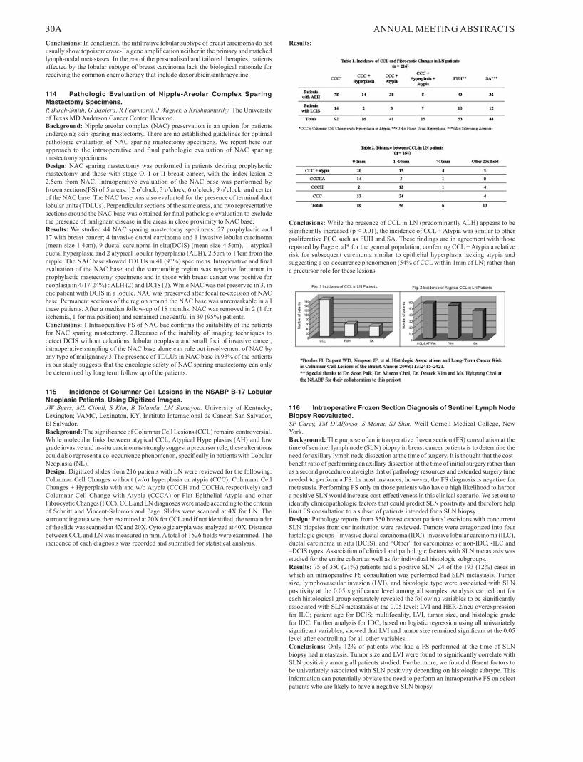
30A ANNUAL MEETING ABSTRACTS Conclusions: In conclusion, the infiltrative lobular subtype of breast carcinoma do not usually show topoisomerase-IIa gene amplification neither in the primary and matched lymph-nodal metastases. In the era of the personalised and tailored therapies, patients affected by the lobular subtype of breast carcinoma lack the biological rationale for receiving the common chemotherapy that include doxorubicin/anthracycline.
114 Pathologic Evaluation of Nipple-Areolar Complex Sparing Mastectomy Specimens.R Burch-Smith, G Babiera, R Fearmonti, J Wagner, S Krishnamurthy. The University of Texas MD Anderson Cancer Center, Houston.Background: Nipple areolar complex (NAC) preservation is an option for patients undergoing skin sparing mastectomy. There are no established guidelines for optimal pathologic evaluation of NAC sparing mastectomy specimens. We report here our approach to the intraoperative and final pathologic evaluation of NAC sparing mastectomy specimens.Design: NAC sparing mastectomy was performed in patients desiring prophylactic mastectomy and those with stage O, I or II breast cancer, with the index lesion ≥ 2.5cm from NAC. Intraoperative evaluation of the NAC base was performed by frozen sections(FS) of 5 areas: 12 o’clock, 3 o’clock, 6 o’clock, 9 o’clock, and center of the NAC base. The NAC base was also evaluated for the presence of terminal duct lobular units (TDLUs). Perpendicular sections of the same areas, and two representative sections around the NAC base was obtained for final pathologic evaluation to exclude the presence of malignant disease in the areas in close proximity to NAC base.Results: We studied 44 NAC sparing mastectomy specimens: 27 prophylactic and 17 with breast cancer; 4 invasive ductal carcinoma and 1 invasive lobular carcinoma (mean size-1.4cm), 9 ductal carcinoma in situ(DCIS) (mean size-4.5cm), 1 atypical ductal hyperplasia and 2 atypical lobular hyperplasia (ALH), 2.5cm to 14cm from the nipple. The NAC base showed TDLUs in 41 (93%) specimens. Introperative and final evaluation of the NAC base and the surrounding region was negative for tumor in prophylactic mastectomy specimens and in those with breast cancer was positive for neoplasia in 4/17(24%) : ALH (2) and DCIS (2). While NAC was not preserved in 3, in one patient with DCIS in a lobule, NAC was preserved after focal re-excision of NAC base. Permanent sections of the region around the NAC base was unremarkable in all these patients. After a median follow-up of 18 months, NAC was removed in 2 (1 for ischemia, 1 for malposition) and remained uneventful in 39 (95%) patients.Conclusions: 1.Intraoperative FS of NAC bae confirms the suitability of the patients for NAC sparing mastectomy. 2.Because of the inability of imaging techniques to detect DCIS without calcations, lobular neoplasia and small foci of invasive cancer, intraoperative sampling of the NAC base alone can rule out involvement of NAC by any type of malignancy.3.The presence of TDLUs in NAC base in 93% of the patients in our study suggests that the oncologic safety of NAC sparing mastectomy can only be determined by long term follow up of the patients.
115 Incidence of Columnar Cell Lesions in the NSABP B-17 Lobular Neoplasia Patients, Using Digitized Images.JW Byers, ML Cibull, S Kim, B Yolanda, LM Samayoa. University of Kentucky, Lexington; VAMC, Lexington, KY; Instituto Internacional de Cancer, San Salvador, El Salvador.Background: The significance of Columnar Cell Lesions (CCL) remains controversial. While molecular links between atypical CCL, Atypical Hyperplasias (AH) and low grade invasive and in-situ carcinomas strongly suggest a precursor role, these alterations could also represent a co-occurrence phenomenon, specifically in patients with Lobular Neoplasia (NL).Design: Digitized slides from 216 patients with LN were reviewed for the following: Columnar Cell Changes without (w/o) hyperplasia or atypia (CCC); Columnar Cell Changes + Hyperplasia with and w/o Atypia (CCCH and CCCHA respectively) and Columnar Cell Change with Atypia (CCCA) or Flat Epithelial Atypia and other Fibrocystic Changes (FCC). CCL and LN diagnoses were made according to the criteria of Schnitt and Vincent-Salomon and Page. Slides were scanned at 4X for LN. The surrounding area was then examined at 20X for CCL and if not identified, the remainder of the slide was scanned at 4X and 20X. Cytologic atypia was analyzed at 40X. Distance between CCL and LN was measured in mm. A total of 1526 fields were examined. The incidence of each diagnosis was recorded and submitted for statistical analysis.
Results:
Conclusions: While the presence of CCL in LN (predominantly ALH) appears to be significantly increased (p < 0.01), the incidence of CCL + Atypia was similar to other proliferative FCC such as FUH and SA. These findings are in agreement with those reported by Page et al* for the general population, conferring CCL + Atypia a relative risk for subsequent carcinoma similar to epithelial hyperplasia lacking atypia and suggesting a co-occurrence phenomenon (54% of CCL within 1mm of LN) rather than a precursor role for these lesions.
116 Intraoperative Frozen Section Diagnosis of Sentinel Lymph Node Biopsy Reevaluated.SP Carey, TM D’Alfonso, S Monni, SJ Shin. Weill Cornell Medical College, New York.Background: The purpose of an intraoperative frozen section (FS) consultation at the time of sentinel lymph node (SLN) biopsy in breast cancer patients is to determine the need for axillary lymph node dissection at the time of surgery. It is thought that the cost-benefit ratio of performing an axillary dissection at the time of initial surgery rather than as a second procedure outweighs that of pathology resources and extended surgery time needed to perform a FS. In most instances, however, the FS diagnosis is negative for metastasis. Performing FS only on those patients who have a high likelihood to harbor a positive SLN would increase cost-effectiveness in this clinical scenario. We set out to identify clinicopathologic factors that could predict SLN positivity and therefore help limit FS consultation to a subset of patients intended for a SLN biopsy.Design: Pathology reports from 350 breast cancer patients’ excisions with concurrent SLN biopsies from our institution were reviewed. Tumors were categorized into four histologic groups – invasive ductal carcinoma (IDC), invasive lobular carcinoma (ILC), ductal carcinoma in situ (DCIS), and “Other” for carcinomas of non-IDC, -ILC and –DCIS types. Association of clinical and pathologic factors with SLN metastasis was studied for the entire cohort as well as for individual histologic subgroups.Results: 75 of 350 (21%) patients had a positive SLN. 24 of the 193 (12%) cases in which an intraoperative FS consultation was performed had SLN metastasis. Tumor size, lymphovascular invasion (LVI), and histologic type were associated with SLN positivity at the 0.05 significance level among all samples. Analysis carried out for each histological group separately revealed the following variables to be significantly associated with SLN metastasis at the 0.05 level: LVI and HER-2/neu overexpression for ILC; patient age for DCIS; multifocality, LVI, tumor size, and histologic grade for IDC. Further analysis for IDC, based on logistic regression using all univariately significant variables, showed that LVI and tumor size remained significant at the 0.05 level after controlling for all other variables.Conclusions: Only 12% of patients who had a FS performed at the time of SLN biopsy had metastasis. Tumor size and LVI were found to significantly correlate with SLN positivity among all patients studied. Furthermore, we found different factors to be univariately associated with SLN positivity depending on histologic subtype. This information can potentially obviate the need to perform an intraoperative FS on select patients who are likely to have a negative SLN biopsy.

ANNUAL MEETING ABSTRACTS 31A117 ASPN, GJB2, ENPP2, ST6GAL2 and TMSB10 Are Related with Invasiveness in Ductal Breast Carcinomas.B Castellana, E Serrano, G Peiro, B Ojeda, C Alonso, T Vazquez, J Munoz, A Barnadas, E Lerma. Hospital de la Santa Creu i Sant Pau. Universitat Autònoma, Barcelona, Spain; Hospital de Alicante, Spain.Background: The mechanism of progression from in situ ductal carcinoma (DCIS) to invasive ductal carcinoma (IDC) remains largely unknown. We compared gene expression patterns in simultaneous in situ and invasive areas of ductal carcinomas to obtain insight into the molecular basis for the invasiveness.Design: We performed differential gene expression profiling using microarray analysis in 21 tumors. Careful dissection was done to obtain separately foci of DCIS and IDC. mRNA was extracted by standard procedure and purified. The mRNA concentration and quality was assessed by mRNA 6000 Nano LabChip Kit (Agilent Bioanalyzer). Samples were analyzed upon Affymetrix GeneChip®Human Gene 1.0 ST Array. We surveyed the expression of 10 differentially expressed genes asporin (ASPN),cytokeratin5 (KRT5),maspin (SERPINB5),connexin26 (GJB2), ST6 beta-galactosamide alpha-2,6-sialyltranferase 2 (ST6GAL2), Autotaxin (ENPP2),vimentin (VIM),E-cadherin (CDH1),peroxiredoxin4 (PRDX4),thymosin beta10 (TMSB10).Results: Invasiveness was associated with: 1) up-regulation of several genes related to epithelial-mesenchymal transition (ASPN, THBS2, FN1, SPARC, COL11A1), cell motility and progression (PLAUR, PLAU, BGN, ADAMTS16, ENPP2), extracellular matrix degradation (MMP11, MMP13, MMP14) and growth/proliferation (ST6GAL2, GJB2) and 2) down regulation of genes related to cell adhesion (TNXB, TNXA, FVIII) and intermediate filaments cytoskeleton (KRT5) (all P<0.05). Differential gene expression was confirmed by real time-PCR analysis in ASPN, GJB2, ENPP2, ST6GAL2, KRT5, VIM and TMBS10 (P<0.05).Conclusions: ASPN, GJB2, ENPP2, ST6GAL2 and TMSB10 were highly up-regulated in IDC compared with DCIS. Therefore, these genes may be potentially involved in the mechanisms of invasion from DCIS to IDC.
118 Down-Regulation of Proliferation and Cell Adhesion and Up-Regulation of Cell Migration Associated Genes in Lobular Breast Carcinomas.B Castellana, E Lerma, E Serrano, G Peiro, D Escuin, A Tibau, C Pons, M Perez, A Barnadas. Hospital de la Santa Creu i Sant Pau. Universitat Autònoma, Barcelona, Spain; Hospital de Alicante, Spain.Background: The aim of this study was to identify differential gene expression between Invasive lobular carcinoma (ILC)and hormone positive inavsive ductal carcinomas (IDC).Design: Twenty seven frozen tumor samples were selected from our tumor bank including 10 ILC and 17 IDC (10 luminal A, 7 luminal B). mRNA was isolated by standard procedure, purified and mRNA concentration and quality was checked by RNA 6000 Nano LabChip Kit (Agilent Bioanalyzer). Samples were analyzed upon Affymetrix GeneChip®Human Gene 1.0 ST Array. The expression of 8 differentially expressed genes pleckstrin homology domain containing, family A member 7 (PLEKHA7), thymosin beta 10 (TMSB10), claudin 7 (CLDN7), peroxiredoxine 4 (PRXD4),cytokeratine 5 (KRT5),metalloproteinase 2 (MMP-2),maspin (SERPINB5) and E-cadherin (CDH1) was validated by quantitative real time-PCR. CLDN7, KRT5, CDH1, MMP-2 were studied by immunohistochemistry.Results: Compared to IDCs, ILCs displayed down-regulation of genes related to cell cycle and proliferation (AREG, ANXA2P1), cell adhesion (CASK), genes involved in TGF-beta signaling (AREG, ANXA2P1), actin cytoskeleton remodeling (TMSB10, TMSB4X), and ubiquitin proteins (UBE2L3, UBE2E3). Furthermore, ILC presented up-regulation of cell migration associated genes (FUT8, CRIPAK), ions binding and transport (GRIA2, SHROOM1, CACNA2D2), lipid/prostaglandin biosynthesis genes (PNPLA7, JMJD7-PLA2G4B) and genes related to catabolic processes (ANUBL1, CSAD). Real time PCR validated the differential expression of PLEKHA7, TMSB10, CLDN7, PRXD4, KRT5, MMP-2 and SERPINB5 (p<0.05), but not the down-regulation of CDH1 (P=ns). Furthermore, up-regulation of PLEKHA7 and PRDX4 together with down-regulation of TMSB10, and SERPINB5 allowed the differentiation between ILCs and luminal B IDCs (P<0.05). Expression differences were confirmed by immunohistochemistry for claudin 7, cytokeratin 5/6, E-cadherin and matrix metalloproteinase 2.Conclusions: ILCs vs. IDC display down-regulation of genes related to proliferation, cell adhesion, actin cytoskeleton and ubiquitin proteins, and up-regulation of cell migration associated genes, ions binding and transport and to catabolic processes. PLEKHA7 and PRDX4, up-regulation and SERPINB5 and TMSB10 down-regulation allowed the differentiation between ILC and luminal B IDCs.
119 Histopathological Variables on a Scale to Best Refine Prognosis in Breast Cancer Patients.I Castellano, S Beatrice, C Marchio’, L Chiusa, E Allia, A Vandone, R Arisio, A Durando, M Donadio, A Sapino. University of Turin, Italy; Ospedale San Giovanni Battista, Molinette, Turin, Italy; Sant’Anna Hospital, Turin, Italy.Background: Breast cancer encompasses different lesions with heterogeneous clinical behaviour. Universally acknowledged prognostic factors are size, grade of differentiation, lymph-node status, estrogen and progesterone receptor (ER/PR) expression, proliferation index and HER2 expression. Recently molecular signatures have been proposed as better predictors of outcome, at least in ER+ patients. We here investigate whether a score based on routine histopathological/immunophenotypical parameters would discriminate between two categories of patients: those with good prognosis from those with poor prognosis.
Design: 1162 breast cancer cases diagnosed between 1994 and 2004 were retrieved and reviewed. Histological grade, vascular invasion, size, margins were assessed. Complete clinical follow-up data were collected. Immunohistochemistry for ER, PR, HER2, Ki-67, androgen receptors was performed. Univariate and multivariate analyses were sequentially used to identify among all clinico-pathological parameters those which were independent predictors of overall and disease-free survival (OS/DFS). Score values ranging between -1 and 2 were applied to each of the independent predictors, depending on their power of statistical correlation with OS and DFS at Cox analysis. A second-round univariate analysis followed.Results: First-round multivariate analysis identified size, vascular invasion, number of metastatic lymph-nodes and percentage of androgen receptor positivity as independent prognostic factors. Score values were assigned to each parameter as follows: ≤ 3 metastatic lymph-nodes=1; >3 metastatic lymph-nodes=2; size <15mm=1; size>15mm= 2; absence of vascular invasion=1; presence of vascular invasion=1; AR negative tumour=0; AR positive tumour= -1. The final “weighted-score” ranged between 1 and 5. At second-round univariate analysis values of weighted-score between 1 and 3 identified a group of patients with good prognosis, whereas values of 4 and 5 corresponded to subgroups of patients with poorer prognosis.Conclusions: A weighted-score based on routine histopathological/immunohistochemical parameters significantly discriminates between two categories (good versus poor prognosis/low-risk versus high-risk of recurrence) and may represent a valuable tool to best refine prognosis in breast cancer patients.
120 Tumor Sampling Using Tissue Microarray Can Alter the Ki-67 Proliferation Index and Cause Misclassification of Breast Tumors.C Chisholm, C Yau, C Ruud, VO Speights. Scott & White Memorial Hospital, Temple, TX.Background: Metastatic breast cancer is a devastating diagnosis that can be difficult to adequately treat. Breast cancers have been recently divided into several subgroups: Luminal A (ER+/Her2-, Ki-67 <14%), Luminal B (ER+/Her2-, Ki-67 >14%), ER+/Her2+, ER-/Her2+, and triple negative (ER-/PR-/Her2-). These subgroups have different clinical behavior and response to hormonal or chemotherapy. Lymphocyte infiltration of breast tumors has been shown very recently to potentially predict an increased response to chemotherapy. Many studies are performed using tissue microarray. We evaluated whether the leading edge of the tumor has a higher proliferation index than the center and whether tumor sampling for Ki-67 could lead to misclassification of tumors.Design: 110 patients were identified with breast cancer metastases diagnosed between 1999-2007. Pertinent demographic, clinical, and pathological data were collected after permission was granted from the institutional review board. The primary breast tumors were retrieved and sampled in the center and leading edge using a 3 mm microarray punch. The microarray tissue was arranged into the blocks in such a manner as to provide no reliable way to predict which tissue samples were taken from the same tumor. These were then stained with Ki-67 and the proliferation index was visually estimated.Results: Ki-67 proliferation indexes were significantly higher at the leading edge (31.53) than in the center (28.08) of the tumor (p=0.0061). When classified according to the Ki-67 from the center, 16 patients were identified as Luminal A and 36 were classified as Luminal B. Overall survival was 57 months and 56 months, respectively. Using the Ki-67 from the leading edge reclassified 2 (12.5%) Luminal A patients as Luminal B such that 14 were now Luminal A and 38 were Luminal B. Overall survival data for Luminal A and Luminal B changed dramatically: 67 months and 50 months, respectively, suggesting that these two tumors were indeed Luminal B based on clinical behavior.Conclusions: The leading edge of the tumor was conclusively shown to proliferate at a higher rate than the center of the tumor. Approximately 13% of tumors were classified as Luminal A when the center of the tumor was sampled. Sampling the periphery of the tumor reclassified these as Luminal B consistent with their clinical behavior. When performing breast cancer research using tissue microarray methods, the leading edge must be sampled to avoid inappropriate classification.
121 Long Non-Coding RNA Expression Levels Correlate with Proliferation Index in Breast Carcinoma.KM Chisholm, R Li, Y Wan, HY Chang, RB West. Stanford University School of Medicine, CA.Background: Long intervening non-coding RNAs (lincRNAs) are RNAs that do not code for proteins but can interact with proteins, and some are thought to influence chromatin remodeling. Experimental evidence suggests that in cancer, they can influence Polycomb repressive complexes to retarget to an occupancy pattern resembling that of the embryonic state. In a recent paper by Gupta et al., 2010, the expression level of lincRNA in the HOX locus, including HOTAIR, is a predictor of breast cancer metastasis. The current project is undertaken to determine if lincRNAs can be measured in formalin-fixed paraffin-embedded tissue, and to see if HOX locus lincRNAs correlate with clinicopathologic features.Design: The expression levels of HOTAIR and two other HOX locus lincRNAs (ncHoxA1 and ncHoxD4), which were identified in the Gupta et al., 2010 paper to cluster with metastatic breast carcinomas, are observed in breast carcinoma. Probes of 400 to 500 nucleotides were created based upon unique non-conserved sequences. These probes were hybridized to tissue microarrays containing 283 primary breast carcinomas and control specimens from formalin-fixed paraffin embedded tissues. The stains were then scored by eye on a two- or three-tiered scoring system, and were correlated with known clinicopathologic features of the breast carcinomas including metastasis, hormone status (ER, PR, and HER2/neu), and Ki67.Results: Increased HOTAIR expression correlated with an increased proliferation rate (p = 0.042), while increased ncHoxA1 expression demonstrated a trend with increased proliferation rate. In addition, ncHoxA1 and ncHoxD4 lincRNAs had correlating expression levels (p = <0.0001). When these latter two lincRNAs had correlating

32A ANNUAL MEETING ABSTRACTS expression levels, the proliferation rate was more often increased (p = 0.043), especially when they both had increased expression (p = 0.052).Conclusions: This is the first study to show that RNA in situ hybridization of formalin fixed paraffin-embedded clinical material can be used to quantify levels of long non-coding RNAs. This approach offers a method to make observations on lincRNAs that may influence the cancer epigenome in a tissue-based technique. The current study provides evidence that these HOX locus lincRNAs correlate with proliferation index in carcinoma; this finding could be due to their influence on chromatin remodeling.
122 Cytogenetic Alterations in Her2/Neu Gene (HER2) and Chromosome 17 (chr17) by Fluorescent In-Situ Hybridization (FISH) in Immunohistochemically (IHC) Equivocal (2+) Breast Cancers.DA Chitale, J Sanchez, A Adeyinka. Henry Ford Hospital, Detroit, MI; Henry Ford Hospital, Detroit.Background: HER2 protein overexpression & gene amplification are prognostic markers for aggressive breast cancers and predictive of response to the trastuzumab, making accurate HER2 status critical. Equivocal HER2IHC results have been recognized as the main source of discrepancy between IHC and FISH results. We sought to determine the frequency of HER2 amplification, intratumoral heterogeneity, HER2 gene duplication, aneuploidy of chr17 and other genetic alterations by FISH in HER2IHC equivocal breast cancers.Design: We retrieved all cases of breast cancer tested for HER2FISH using commercial 2 color probes [HER2, centromeric enumeration probe (CEP17)] over last 4 years. At least 60 invasive tumor nuclei were analyzed by 2 technologists. Ratio of HER2:CEP17 signals was calculated & scored per CAP/ASCO guidelines (Negative-less than 1.8, equivocal-1.8 to 2.2; positive-over 2.2). >30% nuclei with three or more CEP17 signals irrespective of HER2 copy number were considered to have aneuploidy chr17. Duplication of HER2 gene was defined as 1) ratio>1.3 but <2, 2) <30% of nuclei with 3 or more CEP17, 3) <60% of nuclei with 1 CEP17.Results: 545/730 cases tested for HER2FISH were equivocal by HER2IHC. 420/545(77.1%) were negative for HER2 amplification, 91/545(16.7%) positive [42(7.7%) high+, 49(9.0%) low+ (ratio 2.2 -3.9)], 34/545(6.2%) equivocal. Significant heterogeneous HER2 signal with duplication/amplification of HER2 gene with/without aneuploidy of chr17 was most frequent in low+ and equivocal HER2FISH [44/545 (8.1%)-28 low +, 13 equivocal, 3 negative]. Duplication of HER2 gene was noted in 29/34(85.3%) of HER2FISH equivocal cases in contrast to 21/49(42.9%) low+ cases. 35/545 tumors revealed abnormality in chr17 [30 polysomy,3 deletion of CEP17, 1 monosomy, 1 amplification of CEP17 with normal HER2].Conclusions: About 17% of HER2IHC equivocal cases showed HER2 amplification. Heterogeneity of HER2 signal was relatively a low occurrence but was prevalent in low+ cases thus supporting routine HER2FISH testing on biopsies where fixation is better than excisional specimens. Duplication of HER2 gene was more prevalent in HER2FISH equivocal cases. Duplication likely led to increased protein expression of 2+ intensity by IHC. 6.4% of our cases revealed abnormality of chr17 which is thought to be an independent adverse factor in breast cancers. Studies using array comparative genomic hybridization will give further insight into status of gains and loss on entire chr17.
123 Histologic Spectrum of Breast Imaging Reporting and Data System-4 (BIRADS- 4) Category Lesions – A Retrospective Study of 2015 Cases.DA Chitale, A Stark, RJ Zarbo, D Nathanson, M Burke, N Tang, F Fernandez-Madrid. Henry Ford Hospital, Detroit, MI; Henry Ford Hospital, Detroit; Wayne State Univ., Detroit; Karmanos Comprehensive Cancer Institute, Detroit.Background: Annually about 1,700,000 women undergo breast biopsies with only 20% pathologically malignant. The economic cost of biopsies is estimated at $ 3.5 billion per annum. In addition, the psychological impact on women and possible complication of radiographic evaluation of future mammograms due to prior biopsy are the other significant concerns. Optimal clinical work-up and the need for a more standardized guideline for radiographically suspicious lesions remain contentious. We report the histologic spectrum and findings on positive predictive value (PPV) of sub-classification of mammographically suspicious lesions (BIRADS-4).Design: We identified sequential cases of BIRADS-4 (n=2015) the radiology database that was then linked to pathology database. Sub-classification of BIRADS4: a/Low suspicion for malignancy, b/Intermediate suspicion for malignancy, c/ moderate concern but not classic for malignancy, were recorded. Cases were broadly stratified by their pathologic diagnoses into 4 groups: benign non-neoplastic, benign neoplastic, borderline [atypical duct/lobular hyperplasia] and malignant. Only the biopsy with final diagnosis was considered where more than one biopsy was performed. All statistical analyses were performed using SAS v. 9.1Results: 221 women did not undergo biopsy and were excluded from analysis. Of 1794 biopsies, 984 (54.8%) were benign non-neoplastic, 403/1794 (22.5%) benign neoplastic; 41/1794 (2.3%) borderline histology, 366/1794 (20.4%) malignant. Only 435 (24.2%) of BIRADS- 4 were further sub-classified: 4a (69.1%, n=301), 4b (13.9%, n=60), 4c (17%, n=74). 61/74 (82.4%) patients with 4c score were diagnosed with either borderline or malignant lesions compared to 21/301 (7%) of 4a. PPV for BIRADS 4b was 31.7% in contrast to 82.4% of 4c (p<0.0001). Both these PPV were statistically significant compared to BIRADS-4a (p<0.0001).Conclusions: Based on the present method of sub-classification of BIRADS4 lesions, BIRADS 4c has the highest PPV. BIRADS 4b provides added diagnostic value compared to BIRADS 4a, however with much lower PPV than 4c. Correlation of histologic features and clinical information with mammographic findings may help fine tune the radiologists’ interpretations to reduce false positive results in BIRADS 4a/4b scores and thereby reduce the rate of unnecessary biopsies.
124 Impact of Additional Magnetic Resonance Imaging (MRI)-Guided Biopsies in Management of Breast Cancer Patients Initially Diagnosed by Non-MRI (Ultrasound/Stereotactic) Modalities.M Chivukula, JM Lloyd, D Dabbs. UPMC, Pittsburgh, PA.Background: The American Cancer Society (ACS) estimates 54,010 and 207,090 new cases of in-situ and invasive breast carcinoma (IC) in the year 2010 among women, respectively with an estimated 40,230 breast cancer-associated deaths. MRI is a tool that is being utilized with increasing frequency in the evaluation and management of breast cancer. Although the ACS has recommendations regarding the role of MRI in initial breast cancer screening, its appropriate use as an additional modality has not been clearly defined. The aim of this study is to retrospectively determine the impact of additional MRI in surgical management of patients initially diagnosed by non-MRI modalities.Design: The pathology database was searched for women diagnosed with invasive and/or in-situ breast carcinoma between 2005-2009 by non-MRI modalities (ultrasound/stereotactic) who underwent an additional MRI-guided biopsy. Patients with contralateral MRI-guided biopsies only and patients without follow-up were excluded. 230 patients were retrieved. Patient age, initial diagnostic modality and diagnosis, pre-operative MRI-guided biopsy diagnosis, and definitive surgical procedure (breast conservation therapy (BCT) versus mastectomy) were recorded.Results: 122 patients were included (mean age=55; range 26-82). The pathology of the initial diagnoses consisted of invasive ductal carcinoma (IDC) (n=83), invasive lobular carcinoma (ILC) (n=15), invasive mixed carcinoma (n=3), and ductal carcinoma in-situ (DCIS) (n=21). Additional MRI biopsy results revealed malignant diagnoses (group 1) in 56/122 patients (46%). Diagnoses consisted of IDC-22, ILC-7, invasive mixed carcinoma-3, DCIS-19, LCIS-4, and carcinoma in-situ-1. 66/122 patients (54%) had a benign diagnosis (group 2). In group 1, 16 (29%) ultimately underwent BCT and 40 (71%) ultimately underwent mastectomy for definitive surgical management. In group 2, 55 (83%) ultimately underwent BCT and 11 (17%) ultimately underwent mastectomy for definitive surgical management.Conclusions: 1. Additional MRI detected a significant lesion (DCIS/IC) in more than half of the patient cohort. 2. The patients with malignant diagnoses on additional MRI-guided biopsy have a higher rate of mastectomy in comparison to BCT (71% vs. 29%). 3. The patients with a benign diagnosis on additional MRI-guided biopsy have a higher rate of BCT in comparison to mastectomy (83% vs. 17%). 4. Additional MRI-guided biopsies impact the surgical decision to manage patients with mastectomy versus BCT.
125 Metadherin (MTDH/AEG1): A Novel Marker for Metastasis and Chemoresistance Expression in Lymphnode Negative(LN -), Estrogen Receptor Positive (ER+) Breast Cancer Women.M Chivukula, A Brufsky, R Jankowitz, K Mcmanus, G Carter. Magee Women’s Hospital of UPMC, Pittsburgh, PA.Background: Gene expression profiling of breast cancer has identified two biologically distinct estrogen receptor positive (ER+) subtypes of breast cancer namely luminal A and luminal B. Luminal B tumors tend to have higher proliferation and poorer prognosis than luminal A tumors based on the molecular profiling. Studies to distinguish luminal B from luminal A tumors by immunohistochemistry are evolving. We undertook the study to perform immunohistochemistry in a large cohort of luminal A tumors and to further define this group in terms of non –recurrence and recurrence.Design: 262 patients with ER (+), lymph node negative (LN -) from 1990-2003 were selected. Hematoxylin and eosin (H&E) slides were reviewed and representative tumor blocks were retrieved from our pathology files. Immunohistochemistry staining were performed on formalin fixed, paraffin-embedded tissue blocks using ER, Ki-67 and HER 2.Results: Of the 262 patients, 212 were Non –recurrence group, 56 patients presented with recurrence {distant recurrence (DR) (44/56); loco regional recurrence (LRR) (12/56)} with a median follow-up of 10.1 years. Initial results in the non –recurrence group (145 cases) and all cases of recurrence to date are shown in Table 1.Metadherin (MTDH) expression in LN (-), ER+ patients in Non recurrence, locoregional and distant recurrence groups nuclear membrane staining cytoplasmic staining 3+ 2+ 1+ - 0 3+ 2+ 1+ - 0non-recurrence(NR) (n-28) 32% (9/28) 54% (15/28) 18% (5/28) 7% (2/28) 54% (15/28) 39% (11/28)
recurrence group locoregional recurrence (LRR) (n-12) 100% ( 12/12) 0 (0/12) 0 (0/12) 8% ( 1/12) 33% (4/12) 58% (7/12)Distant recurrence (DR) (n-43) 0% (0/43) 49% (21/43) 51% (22/43) 0 % (0/43) 14% (6/43) 88% ( 38 /43)
Conclusions: The non –recurrence (ER +) tumors show low-moderate nuclear grade in contrast to recurrence (ER+) group which showed moderate –high nuclear grade.When HER 2 and Ki-67 were used to distinguish these groups. We found that the non-recurrence group showed lower proliferation index in comparison to recurrence group ( p= 0.0168)HER 2 tends to be strongly expressed in the recurrence group (both DR, LRR) compared to the non recurrence group (p=1.000).Expression of ER, HER2 and the Ki67 index appears to distinguish the luminal A breast cancers which can present with recurrence versus the non-recurrence.
126 A Survey of 45 Biomarkers for Basal-Like Breast Cancer.JR Choo, D Gao, S Leung, C Chow, SYH Lau, J Cheng, TO Nielsen. University of British Columbia, Vancouver, Canada.Background: Basal-like breast cancer was originally identified by gene expression profiling, and it is associated with high-risk disease not responsive to available targeted therapies. Numerous biomarkers for basal-like breast cancer have been proposed. The

ANNUAL MEETING ABSTRACTS 33Afollowing study performs a comprehensive immunohistochemical investigation of proposed biomarkers against a gene expression profile gold standard.Design: Parallel analysis of 45 proposed basal-like biomarkers from literature and gene expression profile data was done on a breast cancer tissue microarray of cases (n = 122) where subtype was previously assigned by a PAM50 gene expression profile gold standard. Twenty-five of the 45 biomarkers were significantly associated with basal-like breast cancer (Table 1). Statistical analysis was performed using the Fisher’s exact test corrected for multiple comparisons.Results: Table 1 Biomarker* Sensitivity Specificity Accuracy** α-B-crystallin 42 92 73Caveolin 2 19 98 65Ck 14 27 100 67Ck 17 51 92 75Ck 5 72 90 82Ck 5/6 50 87 72c-Kit 43 96 75Claudin 4 66 73 72Cyclin E 72 66 77EGFR 41 94 72Fascin 59 93 77IMP3 25 100 70ki67 94 50 65Moesin 71 84 82Negative ER 92 67 72Negative PR 92 46 57Nestin 55 95 83NGFR 22 100 67p16 81 82 80p53 55 81 70P-cadherin 80 61 72PPH3 94 58 73S100A9 63 82 75Skp2 62 79 73TRIM29 72 86 80 *Alphabetical order, adjusted p <.05; **Based on cases with complete data for all 45 biomarkers. Biomarkers with a statistically non-significant association (adjusted p >.05) to basal-like breast cancer included: anilin, CAIX, caveolin 1, CD44, CD44v6, FABP7, FOXC1, negative Her2, Integrin β4, laminin5, loss of BRCA1, Met, negative p27, p63, p-glycoprotein, pS6rp, negative PTEN, SMAD4, VEGF and vimentin.Conclusions: Nestin is the most accurate single biomarker for basal-like breast cancer among the 45 tested against a gene expression profile gold standard. These results can be used to build an optimal multi-biomarker panel, for which validation on an independent sample cohort would be required.
127 Androgen Receptor Expression Is Usually Maintained in Initial Surgically-Resected Breast Cancer Metastases, but Often Lost in Terminal Metastases Found at Autopsy.A Cimino-Mathews, J Hicks, A DeMarzo, BH Park, P Argani. The Johns Hopkins Hospital, Baltimore, MD.Background: The androgen receptor (AR) is expressed in approximately 70% of primary breast carcinomas (PBCs), including those negative for ER and Her2. AR is a promising therapeutic target for breast carcinoma; however, no prior studies have evaluated AR expression in metastatic breast carcinomas (MBCs) in relation to their matched PBCs. Here, we examine AR expression in a cohort of initial surgically-resected metastases, as well as in a separate cohort of end-stage metastases harvested at autopsy, compared to their respective matched PBCs.Design: Tissue microarrays (TMAs) were constructed from archived paraffin tissue blocks of PBCs and surgically-resected matched MBCs from 16 patients. In addition, we evaluated previously-constructed single patient TMAs constructed from archived paraffin tissue blocks of PBCs and from multiple MBCs sampled at rapid autopsies on 16 patients who died of widely MBC. TMAs were labeled by immunohistochemistry for ER, PR, and Her2 to classify cases into the following categories: luminal (ER/PR+ Her2- in PBC and MBC), triple negative (TNC) (ER/PR/Her2- in PBC and MBC), Her2 (ER/PR- Her2+ in PBC and MBC), and luminal loss (loss of ER or PR from PBC to MBC). AR expression was scored as labeling intensity (none=0, weak=1, moderate=2, strong=3) multiplied by percentage nuclear labeling (0-100%), with any labeling considered a positive result.Results:
Conclusions: AR expression is almost always concordant between matched PBC and surgically-resected MBC, with a trend towards increased expression in MBC. This finding validates AR as a therapeutic target in MBC and suggests that AR expression may
need to be reevaluated in MBC even if the PBC is negative. However, AR expression is decreased with a trend towards complete loss in end-stage MBC, particularly in the setting of cases showing hormone receptor loss (luminal loss cases). This suggests a shift of AR expression between initial and end-stage MBC and points to an opportunity for targeted anti-androgen therapy at an earlier stage in disease progression.
128 Basal Cytokeratin and Epidermal Growth Factor Receptor Expression Are Not Predictive of Response to Platinum Based Therapy for Women with Triple Negative Breast Cancer.M Cioffi-Lavina, J Hurley, M Jorda, GR Walker, C Gomez-Fernandez. University of Miami Jackson Memorial Hospital, FL; University of Miami, FL; University of Miami Hospitals, FL; Sylvester Comprehensive Cancer Center, Miami, FL.Background: Experimental data suggests that triple negative breast cancers (TNBC) may have increased sensitivity to platinum-based chemotherapy, particularly in BRCA1 mutation carriers, whose cancers cluster among the basal-like subtype defined by gene expression profiling. The most consistent immunophenotype seen in basal-like TNBC includes expression of basal cytokeratin (CK5/6) and/or epidermal growth factor receptor (EGFR). We investigated whether the expression of CK5/6 and/or EGFR could predict for response to neoadjuvant platinum based therapy in a cohort of patients with triple negative breast cancers.Design: We reviewed 79 TNBC diagnosed between 1999 and 2009, treated with neoadjuvant docetaxel plus platinum salts, with a pathologic complete response rate (pCR) of 26.6 % (21of 79), as defined by the absence of invasive disease in the breast and axilla. Immunohistochemistry for CK5/6 (DAKO) and EGFR (DAKO) was performed in all cases using the LSAB method. Fisher’s exact test was used to compare pCR rates by expression of CK5/6 and/or EGFR.Results: The pCR rate of 26.6% did not vary significantly (p=0.418) by expression of at least CK5/6 or EGFR; i.e. pCR achieved in 5 (19.2%) of 26 TNBC positive for at least one of the markers was comparable to pCR achieved in 16 (30.2%) of 53 TNBC negative for both CK5/6 and EGFR.Conclusions: Although the expression of basal cytokeratins and/or EGFR can be used to identify TNBC that have a basal-like phenotype, the expression of these markers alone is not sufficient to predict for response to platinum based chemotherapy.
129 Measurement of Residual Cancer Burden in Breast Cancer Patients Treated with Neoadjuvant Chemotherapy and Correlation with Biomarker Expression and Molecular Subtypes.A Cockburn, Y Peng, D Euhus, B Haley, V Sarode. University of Texas Southwestern, Dallas.Background: The assessment of tumor response to neoadjuvant chemotherapy (NAC) is important for prognostication of patients with breast cancer. The residual cancer burden (RCB) can be quantified using the UT MD Anderson RCB calculator index. This study aims to correlate tumor biomarker expression and clinicopathologic parameters with RCB status in patients with locally advanced breast cancer treated with NAC.Design: Two hundred and three patients with locally advanced breast cancer were treated with NAC at our institution between the years 2000-2009. Pathology slides and reports from 156 patients who completed NAC and definitive surgery were reviewed retrospectively. Tumor cellularity percent, tumor bed size, DCIS component, number of positive lymph nodes and size of the largest metastatic focus were assessed. The RCB class was assigned using the RCB index (www.mdanderson.org/breastcancer_RCB). Pathologic complete response (pCR), RCB I, RCB II and RCB III correspond to pCR, minimal, moderate and extensive residual disease respectively. Results of biomarker expression (ER, PR, HER2, Ki67, and p53) and DNA ploidy performed on core biopsies were analyzed and correlated with RCB. Tumors were classified into the luminal A, luminal B, HER2 and triple negative molecular groups using immunohistochemistry.Results: The median age was 46 years. Thirty-eight cases exhibited pCR; 5 RCB I; 56 RCB II; and 58 RCB III. The mean RCB score for RCB I, RCB II and RCB III was 0.13, 2.28, and 4.16, respectively. Race, age, tumor grade, PR expression and DNA ploidy showed no significant correlation with RCB class. Clinical stage (p <0.05), high Ki67, p53 overexpression, ER and HER2 expression showed significant difference between the RCB classes.Tumor Biomarkers Correlated with RCB Classification pCR/RCB I RCB II-III P ValueKi67 </=30% 4 41 0.001>30% 38 73 p53 </=10% 16 67 0.01>10% 26 41 ER Negative 28 49 0.02Positive 15 65 PR Negative 32 69 0.14Positive 11 45 HER2 Nonamplified 20 83 0.003Amplified 23 31
HER2 and triple negative subtypes were more frequently associated with pCR/RCB I than luminal subtypes (p=0.002).Conclusions: RCB can be evaluated retrospectively from routine pathology material using the MD Anderson RCB calculator index to more accurately classify tumor response to neoadjuvant chemotherapy.

34A ANNUAL MEETING ABSTRACTS 130 Comparison of Tumor Biomarker Expression in Breast Cancer Patients before and after Neoadjuvant Chemotherapy.A Cockburn, Y Fang, Y Peng, B Haley, V Sarode. University of Texas Southwestern, Dallas.Background: Tumor biomarker expression performed on core biopsies prior to neoadjuvant chemotherapy (NAC) is critical for planning treatment in patients with locally advanced breast cancer. In patients with significant residual disease, changes in biomarker expression after NAC may have prognostic and predictive significance.The aim of this study is to compare biomarker (ER, PR, HER2, Ki67 and p53) expression in breast cancer patients before and after chemotherapy.Design: Patients with locally advanced breast cancer who received NAC from 2000-2009 were identified at our institution and retrospectively analyzed. Tumor biomarkers were performed on core biopsies prior to chemotherapy and repeated on the post-NAC definitive surgical specimen. Analysis of biomarkers was performed in a routine fashion using standard immunohistochemical techniques and analyzed by a computer assisted image analysis. All HER2 borderline and positive (3+) immunohistochemical cases were confirmed by FISH analysis. The change in biomarker expression was compared to a control group who did not receive chemotherapy and had biomarker results on both core and excision specimens. Differences in expression in the two groups were analyzed.Results: A total of 203 patients who received NAC were identified, of which 173 had complete data sets. Thirty-eight (22%) patients achieved pathologic complete response while 135 (78%) had residual tumor. The control group consisted of 51 patients. Results of biomarker expression in the NAC treated group showed significant change in ER, PR, HER2, and Ki67.Neoadjuvant Chemotherapy Group
No Change Change (pos-neg/neg-pos) P Value
ER + 69 11 0.027ER - 54 1 PR + 42 15 0.02PR - 69 8 HER2 + 25 13 <0.0001HER2 - 91 6 Ki67 >15% 81 26 0.009Ki67 <15% 13 14 p53 >10% 41 7 NSp53 <10% 74 5
Except for p53, the control group exhibited no significant change in biomarker expression.Control group
No Change Change (pos-neg/neg-pos) P Value
ER + 43 0 NSER - 8 1 PR + 33 5 NSPR - 10 3 HER2 + 1 0 NSHER2 - 49 1 Ki67 >15% 20 9 NSKi67 <15% 17 5 p53 >10% 7 8 0.001p53 <10% 33 3
Conclusions: Neoadjuvant chemotherapy significantly alters tumor biomarker expression; therefore they should be repeated on the definitive specimens for further management of these patients.
131 Are Prognostic Factors the Same for Non-Invasive and Invasive Local Recurrence (LR) in Patients with Ductal Carcinoma In Situ (DCIS) Treated with Breast-Conserving Therapy (BCT)?L Collins, N Achacoso, R Haque, L Nekhlyudov, S Fletcher, C Quesenberry, S Schnitt, L Habel. Beth Israel Deaconess Medical Center, Boston; Kaiser Permanente, California; Harvard Pilgrim Health Care Institute, Boston.Background: Several patient, pathologic and treatment factors have been reported to be associated with an increased risk of LR in patients with DCIS treated by BCT. Approximately half of such LR are non-invasive (DCIS) and half are invasive carcinomas. There is a perception that pathologic risk factors for non-invasive and invasive LR are the same, but few prior studies have addressed this issue.Design: We conducted a case-control study of patients with DCIS treated with BCT at 3 integrated health plans to assess risk factors for LR. Slides of the index DCIS were reviewed from 225 patients who developed a LR (cases) and 394 patients without a LR (controls; matched to cases on age, diagnosis year, health plan, time since diagnosis). Conditional logistic regression was used to estimate the relative risk (RR) of LR associated with pathologic factors, controlling for confounding variables.Results: Overall, the only pathologic features independently associated with an increased risk of LR were larger lesion extent and close/positive excision margins. However, when stratified by type of LR, these features were found to be significantly associated with non-invasive but not with invasive LR. The relationship between DCIS extent and type of LR is shown in the Table.Extent (#low power fields)
RR and 95% CI for Non-invasive LR
RR and 95% CI for Invasive LR
1 1.0 Ref 1.0 Ref2-5 2.4 0.9-6.5 1.0 0.3-3.16-9 3.5 1.1-11.0 1.1 0.3-3.810-14 5.3 1.8-16.2 0.7 0.2-2.615-19 6.7 1.9-23.4 1.2 0.2-5.9>20 3.9 1.2-12.2 1.6 0.4-5.4
Both close and positive margins were associated with a significant increase in the risk of non-invasive LR (RR=2.2 and RR=5.3; 95%CI 1.1-4.3 and 2.1-13.4 respectively),
but not with invasive LR (RR=1.9 and 1.7; 95%CI 0.9-4.2 and 0.6-4.9 respectively). No other pathologic features studied (e.g. nuclear grade, architectural pattern, comedo necrosis) were associated with either type of LR.Conclusions: We found prognostic factors for non-invasive and invasive LR following BCT for DCIS differed. Tumor burden (extent and margin status) was associated with non-invasive but not invasive LR. Features of DCIS specifically associated with invasive LR were not identified. These results raise the possibility that investigating other factors, such as the microenvironment, may be of value for identifying risk factors for invasive LR.
132 Radial Scars and Breast Cancer Risk: Update from the Nurses’ Health Study (NHS) and Meta-Analysis.LC Collins, S Aroner, JL Connolly, SJ Schnitt, GA Colditz, RM Tamimi. Beth Israel Deaconess Medical Center and Harvard Medical School, Boston; Channing Institute, Brigham and Women’s Hospital, Boston; Washington University School of Medicine, St. Louis.Background: Radial scars (RS) are benign sclerosing breast lesions characterized by a central fibroelastotic nidus from which altered ducts and lobules radiate. The relationship between RS and breast cancer is controversial. In particular, estimates of subsequent breast cancer risk among women with RS have varied in the few epidemiologic studies that have investigated this association.Design: We performed an updated analysis of the association between RS and breast cancer risk in a case-control study of benign breast disease and breast cancer risk nested within the NHS. There were 394 cases (women with a benign breast biopsy [BBB] who developed breast cancer) and 1607 controls (women with a BBB who did not develop breast cancer). BBB slides were reviewed for the presence of RS. Logistic regression was used to compute odds ratios (ORs) and 95% confidence intervals (CIs) for the association between RS and breast cancer risk. A meta-analysis was also conducted that included data from our study and 5 prior studies of breast cancer risk in relation to RS.Results: In the NHS update, women with RS (52 cases, 103 controls) had an increased breast cancer risk compared with those without RS (RR=2.2, 95%CI:1.5-3.1). Risk was attenuated but remained significant after adjustment for BBD category (RR = 1.74 95% CI: 1.19-2.56). The meta-analysis similarly indicated that RS are associated with an increased risk of breast cancer (RR=1.5, 95%CI:1.1-2.0) (Figure), but RS did not further increase the risk among women with either proliferative lesions without atypia or among those with atypical hyperplasia. The meta-analysis suggested that the association between RS and breast cancer risk is stronger in the first 10 yrs after BBB than ≥10 yrs (<10 yrs:RR=2.1, 95%CI:1.5-2.9; ≥10 yrs:RR=1.2, 95%CI:0.9-1.7; p for heterogeneity=0.02).
Conclusions: Our updated analysis of the NHS data and a meta-analysis indicate that RS are associated with a modest increase in the risk of subsequent breast cancer. Further, this risk is highest in the first 10 years after benign breast biopsy.
133 Classifications of Pathological Response and Long-Term Follow-Up in Breast Cancer Patients Treated with Neoadjuvant Chemotherapy.AD Corben, C Teo, R AbiRaad, A Bombonati, FC Koerner, A Taghian, EF Brachtel. Memorial Sloan-Kettering Cancer Center, New York, NY; Tan Tock Seng Hospital, Singapore, Singapore; Massachusetts General Hospital and Harvard Medical School, Boston.Background: Breast cancer, especially in locally advanced stages, is increasingly treated with neoadjuvant chemotherapy (NACT) to reduce local disease and to assess response to treatment. Chemotherapy can cause a spectrum of morphological alterations in tumors and lymph nodes. Various histopathological classification schemes are available to analyze the response. Our study compares practical application and predictive use of several of those schemes.Design: Sixty-two patients were enrolled in a randomized phase II clinical trial for sequential NACT with doxorubicin and paclitaxel. Study pathologists reviewed H&E sections from patients’ tumors sampled before (core biopsy) and after treatment (excision or mastectomy). Response was assessed following NSABP-B18 criteria, Miller-Payne grading system (MPG), Residual Disease in Breast and Nodes (RDBN, a derivative of the Modified Nottingham Prognostic Index), Sataloff tumor (T), Sataloff lymph nodes (N), and Residual Cancer Burden (RCB) as determined by web calculator. Results of the pathological classifications were correlated with disease-free survival (DFS) by Kaplan-Meier curves with mean (median) clinical follow-up of 80 (86) months.Results: No patient with no residual invasive carcinoma after NACT relapsed (n=5), whereas 27% of all patients developed metastatic disease during follow-up. Lymph
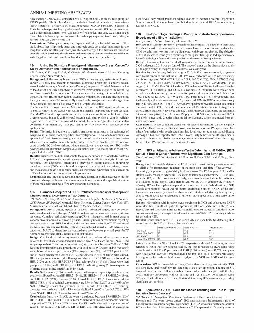
ANNUAL MEETING ABSTRACTS 35Anode status (N0,N1,N2,N3) correlated with DFS (p<0.0001), as did the four groups of RDBN (p=0.02). The Kaplan-Meier curves of other classifications indicated associations (RCB, Sataloff-N) or showed incongruent patterns (NSABP-B18, MPG, Sataloff-T). Post-chemotherapy histologic grade showed an association with DFS but the number of well-differentiated tumors (n=5) was too low for statistical analysis. We did not detect a correlation between age, menopause, chemotherapy sequence, tumor size, estrogen receptor or HER-2 status with DFS.Conclusions: Pathological complete response indicates a favorable survival. This study shows that lymph node status and histologic grade are critical parameters for the long-term outcome after post-neoadjuvant chemotherapy. Classification schemes that strongly weigh lymph node involvement and tumor grade demonstrate better correlation with long-term outcome than those based only on tumor size or cellularity.
134 Using the Signature Phenotype of Inflammatory Breast Cancer To Study Dormancy and Resistance to Therapeutics.AD Corben, C D’Arcy, S Modi, G Chiosis, ML Alpaugh. Memorial Sloan-Kettering Cancer Center, New York, NY.Background: Inflammatory breast cancer (IBC) is the most aggressive form of breast cancer. Clinically IBC presents as reddened, edematous breast that is tender to touch. Associated thickening of the skin (peau d’ orange) may occur. Clinical features are due to the distinct signature phenotype of extensive intravasation in situ of the lymphatic and blood vessels by tumor emboli. The importance of studying IBC is underlined by the fact that non-IBC primary lesions often recur with the IBC signature phenotype and locally advanced non-IBC successfully treated with neoadjuvant chemotherapy often shows residual carcinoma exclusively in the lymphovasculature. The human IBC xenograft model, MARY-X, captures the IBC signature phenotype (i.e.tumor emboli grow exclusively within the murine lymphatic and blood vessels). The MARY-X in vitro spheroids and in vivo tumor emboli form on the basis of an, overexpressed, intact E-cadherin/α,β-catenin axis and exhibit a gain in cellular organization. The overexpression of the intact, E-cadherin/α,β-catenin axis is also consistent with human IBC. This provides an in vitro model with tractable in vivo applications.Design: The major impediment to treating breast cancer patients is the resistance of lymphovascular emboli to therapeutics. To investigate we 1) developed a novel ex vivo approach of fresh tissue sectioning and treatment of breast cancer specimens (n=20) which were analyzed by immunohistochemistry (IHC), 2) IHC analysis of retrospective cases of both IBC (n=10) (with and without neoadjuvant therapy) and non-IBC (n=10) paying particular attention to lymphovascular emboli and 3) validated data in MARY-X, a pre-clinical model of IBC.Results: Tissue sections (200 µm) of both the primary tumor and associated metastases followed by exposure to therapeutic agents allows for an efficient analysis of treatment response. Tight aggregates (spheroids) of previously loosely-associated infiltrating ductal carcinoma (IDC) nests formed in response to treatment in the primary tumor and metastases and do not undergo apoptosis. Persistent expression or re-expression of E-cadherin was found in resistant side populations.Conclusions: The findings suggest that the mere formation of tight aggregates due to molecular changes of breast carcinoma can confer resistance to therapy. Identification of these molecular changes offers new therapeutic strategies.
135 Hormone Receptor and HER2 Profiles before and after Neoadjuvant Chemotherapy: Experience at Two Centers.AD Corben, C D’Arcy, R Abi-Raad, A Bombonati, A Taghian, M Akram, FC Koerner, JH Eichhorn, EF Brachtel. Memorial Sloan-Kettering Cancer Center, New York, NY; Massachusetts General Hospital and Harvard Medical School, Boston.Background: Breast cancer, especially in locally advanced stages, may be treated with neoadjuvant chemotherapy (NACT) to reduce local disease and assess treatment response. Complete pathologic response (pCR) is infrequent, and in most cases a variable amount of residual tumor is present. Current guidelines recommend repeating hormone receptor and HER2 studies on the residual tumor after NACT. We evaluated the hormone receptor and HER2 profiles in a combined cohort of 120 patients who underwent NACT to determine the concordance rate between pre- and post-NACT hormone receptor and HER2 results at our institutions.Design: One hundred and twenty women with locally advanced breast cancer were selected for this study who underwent diagnosis (pre-NACT core biopsy), NACT and surgery (post-NACT excision or mastestomy) at our centers between 2000 and 2010. Routine immunoperoxidase procedures with antibodies by Dako® and/or Ventana® were followed for estrogen receptor (ER), progesterone (PR), and HER2 proteins. ER and PR were considered positive if >1%, and negative if <1% of tumor cells stained. HER2 expression was scored following guidelines. HER2 FISH was performed on HER-2 (2+) cases with HER2/CEP 17 dual-color probe by Vysis®. Cases were then grouped as ER (+/-) and HER2 (+/-), with HER2+ indicating strong (3+) overexpression of HER2 and/or HER2 amplification by FISH.Results: Sixteen cases (13%) showed complete pathological response (pCR) at excision, evenly distributed between ER-HER2- (24%), ER-HER2+ (19%), ER+HER2+ (19%), and ER+HER2- (19%). 3 cases (19%) showed ER+ HER2 (2+) with no available HER2-FISH. 68% of residual carcinomas were ER+ before NACT, as were 68% after NACT, although 7 cases changed from ER+ to ER- and 3 from ER- to ER+, reducing the actual concordance to 89%. PR+ cases declined from 65% (pre-NACT) to 59% (post-NACT). HER2 (3+) cases declined from 24% to 17%.Conclusions: Tumors with pCR are evenly distributed between ER+/HER2+, ER+/HER2-, ER-/HER2+ and ER-/HER- subsets. Most residual invasive carcinomas maintain the pre-NACT ER, PR and HER2 status. The ER profile changed in a proportion of cases (11%) from ER+ to ER-, or ER- to ER+; a slightly decreased PR expression
post-NACT may reflect treatment-related changes in hormone receptor expression. Several cases of pCR may have contributed to the decline of HER2 overexpressing tumors post-NACT.
136 Histopathologic Findings in Prophylactic Mastectomy Specimens: Experience of a Single Institution.IA Czyszczon, S Sahoo. University of Louisville, KY.Background: Recently, the rate of prophylactic mastectomy (PM) has been increasing to reduce the risk of developing breast carcinoma. However, it is controversial whether CPM benefits most women who are diagnosed with breast carcinoma. The objective of this study was to identify the frequency of malignant findings in PM specimens and identify pathologic factors that can predict involvement of PM specimens.Design: A retrospective review of all prophylactic mastectomies between January 2004 and August 2010 was performed. The stage of tumor on the disease side and the pathologic findings in the prophylactic breast were analyzed.Results: Between January 2004 and August 2010, 1469 women were newly diagnosed with breast cancer at our institution. 200 PM were performed on 185 patients during the following years: 2004, 4/223 (1.8%); 2005, 18/220 (8.2%); 2006, 16/206 (7.8%); 2007, 34/181 (18.8%); 2008, 43/209 (20.6%); 2009, 51/269 (19.0%); 2010 (as of August), 34/161 (21.1%). Of 185 patients, 170 underwent CPM for ipsilateral invasive carcinoma (136 patients) and DCIS (33 patients). 27 patients were treated with neoadjuvant chemotherapy. Tumor stage for ipsilateral carcinoma is as follows: Tis, 10%; T1, 53%; T2, 30%; T3, 4.9%; T4, 1.4%. Forty-nine of 138 (36%) patients had ipsilateral lymph node involvement. 15 patients had BPM for BRCA mutation, strong family history, or LCIS. 15 of 170 (8.8%) CPM specimens revealed occult carcinoma: 7 invasive and 8 DCIS. The index carcinoma in all 15 patients was infiltrating ductal carcinoma; 4 had locally advanced disease, 1 had multifocal disease, and the remaining 10 had node-negative T1 or T2 tumors. Prophylactic SLNB was performed in 158/200 PM (79%) cases; only 2 patients had nodal involvement, which originated from the index carcinoma.Conclusions: The rate of PM has dramatically increased at our institution over the past 6 years. Occult carcinoma (DCIS and invasive) is not uncommon in CPM specimens. One-third of our patients with occult carcinoma had locally advanced or multifocal disease. Although it has been reported that CPM is more likely to harbor occult carcinoma in patients with invasive lobular carcinoma, none of our cases were of lobular histology. None of the BPM specimens had malignant lesions.
137 SP3, an Alternative to HercepTest in Determining HER-2/Neu (H2N) Status in Breast Cancer Patients with Significant Cost Savings.TM D’Alfonso, Y-F Liu, S Monni, SJ Shin. Weill Cornell Medical College, New York.Background: Accurately determining H2N status in breast cancer patients who may be eligible for trastuzumab treatment in the most cost- and time-effective ways is increasingly important in light of rising healthcare costs. The FDA-approved HercepTest (Dako) is widely used to determine H2N status by immunohistochemistry (IHC) in these patients. SP3, a rabbit monoclonal antibody, is an immunostain that can be performed at a fraction of the cost of using HercepTest. We set out to compare the reliability of using SP3 vs. HercepTest compared to fluorescence in situ hybridization (FISH). Needle core biopsies (NCB) and subsequent excisional biopsies (EXBX) of the same tumor were concurrently studied to also evaluate intratumoral staining heterogeneity as a potential cause for discordance in different sized specimens of the same tumor using these antibodies.Design: 100 patients with invasive breast carcinoma in NCB and subsequent EXBX were identified. On all 200 patients’ specimens, IHC was performed with SP3 and HercepTest and dual-color FISH for H2N amplification on sequential unstained tumor sections. A cost analysis was performed based on current ASCO/CAP practice guidelines for assessing H2N.Results: Concordance with FISH, and sensitivity and specificity for detecting H2N overexpression by SP3 and HercepTest are shown: FISH concordance (%) Sensitivity (%) Specificity (%)SP3 NCB 91 69.2 93.8HercepTest NCB 92 61.5 87.5SP3 EXBX 90 69.2 95HercepTest EXBX 90 69.2 96.2
Using HercepTest and SP3, 15 and 8 NCB, respectively, showed 2+ staining and were reflexed to FISH. For 100 patients studied, the cost for assessing H2N status using a combination of SP3 ($7 per test) and FISH ($290 per test) was $3,768, compared with a cost of $17,879 for HercepTest ($114 per test) and FISH. Intratumoral staining heterogeneity for both antibodies was negligible in NCB and EXBX of the same tumor.Conclusions: SP3 is comparable to HercepTest with respect to agreement with FISH, and sensitivity and specificity for detecting H2N overexpression. The use of SP3 obviated the need for FISH in a number of cases which when coupled with this less costly antibody produced a total cost savings of $14,111 in the 100 patients studied. SP3 is a reliable alternative to HercepTest for determining H2N status while providing significant cost savings.
138 Cytokeratin 7 & 20: Does the Classic Teaching Hold True in Triple Negative Breast Cancers?SM Davion, KP Siziopikou, M Sullivan. Northwestern University, Chicago, IL.Background: The term “breast cancer” (BC) encompasses a heterogenous group of tumors that includes triple negative carcinomas (TNC). As molecular differences within BC were described, it became evident that some TNC expressed a different cytokeratin

36A ANNUAL MEETING ABSTRACTS (CK) profile. However, much of the immunohistochemical (IHC) studies on BC that are still cited today were performed prior to these discoveries. The aims of this study were to: 1) determine if the classic differential CK profile for BC (CK7+/CK20-) holds true in TNC; 2) see if CK 5/6+ TNC is associated with aberrant CK expression.Design: After IRB approval, the pathology database was reviewed for TNC and 99 cases were selected. Relevant patient and pathologic staging data were recorded. Tissue miroarrays (TMA) with 0.2 cm cores were made with TNC in triplicate and control tissue. TMAs were stained with CK7 (DAKO OV-TL12/30), CK20 (DAKO Ks208) & CK5/6 (DAKO DS/16B4). The CK 7 & 20 IHC was interpreted as follows: positive (>90% of tumor cells stain), patchy (10-90%), focal (<10%) & negative (<1%). The CK 5/6 was called either positive (any staining) or negativeResults: The average age of our TNC cohort was 54.6 (range 32-93). The average tumor size was 2.8 cm and 50% had involved lymph nodes. Of the 99 patients included in the TMA, one was dropped from the final analysis due to tissue loss. 76 of the TNC showed strong diffuse positivity with CK7 (78%). The remaining 22 TNC showed patchy to negative results (see table 1). As expected, the vast majority of TNC were CK20 negative but 5 (5.1%) were positive with 3 of these showing a diffuse staining pattern. No TNC were CK7-/20+. 53% of the TNC were positive for CK 5/6. However, CK 5/6 positivity did not predict an aberrant CK 7 & 20 staining pattern (3/5 dual positive cases and 1/2 dual negative cases were CK 5/6 positive) Of the 7 patients whose TNC showed a “non-classic” 7/20 pattern, no difference in age, race, tumor characteristics or staging was identified.CK 7 in TNCCK 7 STAINING PATTERN NUMBER OF CASES % OF CASESPOSITIVE 76 77.6PATCHY 15 15.3FOCAL 5 5.1NEGATIVE 2 1
Conclusions: TNC can show a different expression pattern of CK expression, a finding that carries over to CK7 and 20. As the differential CK are often used in the setting of metastatic disease with an unknown primary, a negative CK7 or a positive CK20 may not definitively rule out a TNC. As the samples being evaluated grow smaller, the patchy to focal CK7 positivity present in 20% of our cases may also become clinically significant.
139 Mucocele-Like Lesions in Needle Core Biopsies: Is Excision Always Necessary?S Davion, K Siziopikou, M Feldman, EB Mendelson, M Sullivan. Northwestern University Feinberg School of Medicine, Chicago, IL.Background: When a mucocele-like lesion (MLL) is diagnosed on needle core biopsy (CB), surgical excision is the current standard of care as the spectrum of associated lesions range from benign mucin filled cysts to carcinoma. In this study we examined both the radiologic and pathologic findings in patients with MLL on CB, and correlated them with the excision findings. Our goal was to determine the upgrade rate of MLL at a high volume academic breast center as well as to determine if there is a specific patient population in which close clinical follow-up may be recommended as a treatment option rather than excision.Design: The pathology information system at Northwestern Memorial Hospital was searched for all MLL diagnosed on CB between 1/1/2003 and 7/30/2010. 44 cases were identified. Pertinent imaging findings (calcifications vs. mass, size of lesion), patient demographic information (age, personal history of breast cancer), and pathologic diagnosis in the excision specimen were recorded.Results: MLL was diagnosed in 44 of 22,792 CB performed (0.2%) in the time interval included in this study. The 41 patients (some had >1 CB) ranged in age from 27 – 82 years (average = 54). The majority of the biopsies were stereotactic targeting calcifications (42/44; 95%) with two ultrasound guided biopsies for mass lesions (4.5%). 26 CB were diagnosed as MLL (59%), 16 as MLL with atypia (including FEA & ADH; 36%), 1 each as MLL with LCIS and MLL with DCIS (2.3%). No excision pathology was available for 10 patients. The remaining 31 patients underwent surgical excision, 21 of which were benign (68%). Of the remainder, 2 had ADH (3.5%), 6 had DCIS (19%) and 2 had invasive cancer (IC).The risk of finding a more signifiant lesion at the time of exicision was highly associated with the presence of atypia in CB. No MLL (0/16) were upgraded while 7/14 (50%) of MLL with atypia had either invasive or in-situ carcinoma on excision. One of 2 MLLs associated with masses was upgraded to DCIS (50% upgrade-rate) while 6/29 MLLs that presented as calcifications were upgraded at excision (21% upgrade-rate).Conclusions: MLLs diagnosed on CB associated with either atypia or a mass lesion had a 50% upgrade rate in our patient population. None of the patients with MLL alone and only calcifications on imaging showed more significant findings on excision and it may be appropriate to offer these patients close clinical follow-up as a treament option.
140 p53 Expression in Triple Negative Breast Carcinomas: Evidence of Racial and Age-Related Differences.S Davion, M Sullivan, SM Rohan, KP Siziopikou. Northwestern University Feinberg School of Medicine, Chicago, IL.Background: The triple negative subtype of breast carcinomas (TNBC), defined in the molecular classification of breast cancer as estrogen receptor (ER), progesterone receptor (PR) and epidermal growth factor receptor 2 (HER2) negative, is a heterogeneous group of lesions with different pathological characteristics, molecular alterations, clinical features and biologic behavior. TNBC is thought to be associated with a higher recurrence rate and poor overall prognosis. In this study we evaluated the role of p53, a mediator of cellular response to DNA damage, in this high risk and therapeutically challenging group of breast carcinomas.
Design: Our patient population consisted of 197 patients diagnosed with TNBC at Northwestern Memorial Hospital (2005-2010, mean age: 54, range 29-93). Electronic medical records were reviewed to determine the demographics. Pathologic tumor characteristics were reviewed and included histologic type, tumor grade, tumor size, presence of lymphatic invasion (LVI), lymph node status and p53 expression (by immunohistochemistry (IHC), Ventana, Bp53-11). Tissue microarrays (in triplicate including controls) were made from 99 of these TNBC for IHC evaluation of basal cytokeratin expression (CK5/6, DAKO DS/16B4).Results: Overall, TNBC were almost exclusively infiltrating ductal carcinomas (182/197, 92.3%) of high histologic grade. 163/197 were grade 3 tumors, and the remaining were grade 2. p53 was expressed in 54.7% and was not associated with the tumor grade, tumor size, presence of LVI nor lymph node status. However, a higher percentage (60.4%) of the basal phenotype TNBC cases expressed p53 compared to just half (51.4%) of the non-basal phenotype TNBC. In addition, more than three quarters (77.7%) of African- American (AA) patients with TNBC expressed p53 compared to just over half (57.1%) of Caucasians (p<0.01). Of interest 50% of the TNBC patients younger than 50 expressed the basal/p53 positive phenotype compared with only 23.4% of the TN patients older than 50 (p<0.01).Conclusions: 1. p53 is expressed in over half of the TNBC studied. 2. AA patients are more likely to have a basal phenotype TNBC with high expression of p53. 3. TNBC patients younger than 50 years of age are twice as likely to have p53 expressing TNBC of the basal phenotype than patients older than 50 y.o. These findings suggest that p53 mutations may play a role in the aggressive behavior of TNBC in young and AA patients. In addition, the results suggest that p53 status may be a specific prognostic indicator in these subgroups of TNBC patients.
141 Expression of the Cancer Stem Cell Marker ALDH1 in Triple Negative Breast Cancer: Prognostic Impact and Tumor Microenvironment.M De Brot, RM Rocha, FA Soares, H Gobbi. Universidade Federal de Minas Gerais, Belo Horizonte, Minas Gerais, Brazil; Hospital A.C. Camargo, Sao Paulo, Brazil.Background: Triple negative breast cancer (TNC; ER, PR, and HER2-negative) is a high risk cancer that lacks the benefit of specific treatment. Emerging studies suggest that some malignancies may obey a cancer cell hierarchy similar to that observed in normal tissues. The theory of cancer stem cells (CSC) in breast cancer and its association with tumor initiation and drug resistance can have implications on targeted therapy. We characterized expression of ALDH1, a putative CSC marker, in a cohort of 140 patients with TNC to examine its association with clinicopathological features, prognosis, and outcome.Design: Clinical and pathological data were obtained. A TMA containing 2 cores of each tumor was constructed and IHC for ER, PR, HER2, CK5, CK14, EGFR, p63, caveolin, p53 and ALDH1 was performed. Both tumor cell and stromal expression of ALDH1 was evaluated.Results: The majority of tumors were high grade (83.7%) and of ductal/no special type (80.8%). Tumor cells exhibited cytoplasmic expression of ALDH1 in 26/140 (18.7%) cases, while stromal expression was detected in 117/140 (83.6%) cases. ALDH1 expression in tumor cells was not associated with overall survival or disease-free survival (OS; DFS) in our series. In contrast, stromal expression was significantly associated with best OS (p=0.044). Cox multivariate analysis was carried out and ALDH1 stromal expression was shown to be an independent prognostic factor (RR=0,382;p=0.003;IC=0,202-0,723). Tumor cell and stromal expression of ALDH1 did not correlate with any of the other parameters examined, including age, tumor size, type, histological grade, nodal status, metastases, and relapse. Moreover, triple-negative tumors also express CK5 (75.0%), CK14 (29.0%), EGFR (28.6%), p63 (28.6%), caveolin (14.3%), and p53 (67.1%). We considered basal-like cancers to be any tumor ER/PR/HER2-negative, CK5 and/or CK14-positive. A basal-like phenotype was detected in 105/140 (75.0%) triple negative tumors and ALDH1 expression was demonstrated in both tumor cells (21/105 cases; 20.0%) and stroma (87/105 cases; 82.9%).Conclusions: ALDH1 is more frequently seen in stromal cells than in epithelial tumor cells. Additionally, stromal expression is associated with best OS in our series of TNCs. Stromal cells expressing ALDH1within the tumor could represent one of the means through which tumor microenvironment play a role in determining prognosis in TNC.
142 Extramammary Metastases to the Breast and Axilla: A Study of 78 Cases.D DeLair, AD Corben, J Catalano, E Brogi, C Vallejo, LK Tan. Memorial Sloan-Kettering Cancer Center, New York, NY.Background: Breast and axillary metastases from extramammary malignancies (EM) are rare but their recognition is critical as treatment and prognosis differ from those of breast carcinoma (BC). We sought to assess clinical and pathologic features of EM to the breast and axilla.Design: A search of the 1990-2010 pathology database identified patients (pts) with EM involving the breast and/or axilla. The clinical and pathologic features were reviewed.Results: A total of 78 pts had EM involving the breast (78%), axilla (6%), or both (16%). Tumor types, primary sites, and clinical features are summarized in Tables 1 & 2. The most frequent EM was carcinoma (56%), with ovarian high grade serous (HGS) being the most common. Melanoma (23%) and a wide variety of sarcomas (21%) were the next most frequent. The majority of pts had advanced disease (78%) when the breast/axillary lesion developed. In 13% of cases, an initial diagnosis of primary BC was rendered, HGS being the most frequently misdiagnosed. Pathologic features common among metastases included formation of a well-circumscribed nodule surrounded by a fibrous capsule and absence of in situ carcinoma. In contrast to other organs in

ANNUAL MEETING ABSTRACTS 37Awhich multiple metastatic lesions are frequent, the majority of metastases to the breast formed a solitary lesion.Table 1: Tumor types and primary sites (n=78)Carcinoma n=44 (56%)Ovary 12/44 (27%)Lung 10/44 (23%)GI tract 6/44 (13%)GYN tract (excluding ovary) 4/44 (9%)GU tract 4/44 (9%)Thyroid 3/44 (7%)Merkel Cell 3/44 (7%)Others 2/44 (5%) Melanoma n=18 (23%) Sarcoma n=16 (21%)Uterine leiomyosarcoma 5/16 (31%)Rhabdomyosarcoma 3/16 (19%)Liposarcoma 2/16 (13%)Others 6/16 (37%)
Table 2: Clinical features (n=78)Median age 54 yr (range 15-83)Female/male 85%/15% (66/12)Median size 1.68 cm (range 0.05-18 cm)Unilateral/bilateral 88%/12% (69/9)Solitary mass 71% (59)Other metastases at diagnosis 78% (61)Initially diagnosed as primary breast carcinoma 13% (10) (high grade serous 6/10, 60%)Interval from primary to breast metastasis 4.5 yr (range synchronous-16 yr)
Conclusions: A wide range of primary sites can metastasize to breast and axilla and although a rare occurrence, a significant portion of these tumors may be misdiagnosed as primary BC. The importance of a complete clinical history is emphasized as the majority of patients had advanced disease upon development of the breast/axillary lesion. In addition, pathologists should be aware of certain histologic features commonly seen in EM to the breast/axilla in order to avoid unnecessary treatment and/or procedures.
143 HER2 Testing on Breast Carcinoma Metastases to Bone by Fluorescence In Situ Hybridization: A Study of 28 Cases with an Emphasis on Preanalytic Factors.BC Dickson, M Wood, J Mazurkiewicz, MC Chang. Mount Sinai Hosp, Toronto, ON, Canada; Univ of Toronto, ON, Canada.Background: Bone is a common site of metastasis for breast carcinoma. Treatment of metastatic disease with trastuzumab depends on HER2-amplification status, which can be assessed by fluorescence in situ hybridization (FISH). Recent studies have shown that bone marrow biopsies (BMBx) are an acceptable source of tumor tissue for FISH testing, when using highly standardized decalcification protocols (e.g. at least 85% success using EDTA, Zustin et al. 2009). Based on our experience as a reference center for HER2 testing, BMBx received from different centres vary in their handling. The purpose of this study is to examine differences in BMBx quality with respect to preanalytic factors.Design: A five-year review of BMBx tested for HER2 by FISH was conducted as part of a QA assessment, with 28 cases identified, including 8 internal cases and 20 from 8 different referring centers. Each center was polled regarding the routine handling of bone marrow biopsies. For each case, the HER2 amplification ratio, slide quality, and preanalytic factors (pepsin time, fixation time, fixative, decalcification method) were recorded, if available.Results: Of the 28 cases, 20 (71%) yielded readable FISH signals. The most common reasons for unreadable FISH were absent/faint signals (6/8), and hollow nuclei (2/8). Other cases were evaluable, with some being suboptimal due to autofluorescence (4/20), small sample (2), overlapping nuclei (3), and crush artifact (2). All of the unsatisfactory cases originated from 3 of the 9 labs and were decalcified in HCl (commercial solutions) for 2 hours. Of the readable cases, 18 were non-amplified, and 2 were HER2-equivocal. Of these, the slide quality did not correlate with pepsin time or fixation time. Decalcification solutions included neutral EDTA (1/20), formic acid (7/20), or HCl (12/20); however, 11/12 cases using HCl had a rapid process (30 min), or used dilute (0.1 N) solutions. Although readable, cases from 3 of 9 centers used a hematology protocol for BMBx including short fixation (60-90 min in formalin), or a variant fixative (B+).Conclusions: BMBx are potential specimens for HER2 FISH testing, but may be susceptible to variation in preanalytic handling. Prolonged decalcification (e.g. 2 hrs. in HCl) results in higher failure rates. Documentation of the decalcification method should be considered for all BMBx submitted for HER2 FISH. Labs with universal protocols for BMBx optimized for hematologic evaluation should consider separate protocols for metastatic tumor.
144 FISH Testing for HER2 Expression May Yield Discordant Results Depending on Reporting Criteria Used.SM Dintzis, KH Allison, RA Schmidt. University of Washington Medical Center, Seattle.Background: Amplification status of the HER2 gene may be assayed using FISH. Currently, the established CAP/ASCO guidelines allow two different systems for reporting the results of the FISH assay: average HER2 signals per cell or the average ratio of HER2 and chromosome 17 (CEP17) signals. Although both resulting systems classify cases as amplified, not amplified or equivocal, the two systems may yield different interpretations for the same case. We analyzed a large dataset to further examine concordance between the two methodologies.
Design: HER2 and CEP17 counts were collected from 32,116 tumor cells from 1329 consecutive breast cancer cases using data from Excel counting sheets imported to a SQL Server database for analysis and statistics. We specifically computed concordances between results generated using HER2 signals per cell versus HER2:CEP17 ratio. Discordant cases were reviewed to obtain concurrent HER2 immunohistochemical staining, tumor grade and stage.Results: Amplification status derived from HER2 signals per cell or HER2:CEP17 ratio was concordant in 89.8% of cases. Most discordances involved cases classified as equivocal by either method. Equivocal classification occurred in 7.8% of cases reported by HER2 signals per cell and 4.0% of cases reported by HER2:CEP17 ratio. 1% of cases were amplified by one method and not amplified by the other. Of these, 4 cases were classified as amplified using HER2 signals per cell only (> 6 signals per cell) and not amplified using HER2:CEP17 ratio (< ratio 1.8). On review of HER2 immunohistochemical stains, none of these cases scored 3+ positive; 3 cases were 2+ indeterminate and 1 case was 1+. An additional 9 cases were classified as amplified using the HER2/CEP17 ratio (>2.2) but not amplified by HER2 signals per cell. All “ratio only” amplified cases showed mean CEP17 of less than 1.5. None of the “ratio only” positive cases showed 3+ positive immunohistochemical staining; 3 were IHC negative and 6 were 2+ indeterminate. In addition, the “ratio only” positive cases were generally lower Nottingham grade tumors.Conclusions: This very large dataset shows that Her2 FISH reporting using the two established CAP/ASCO guidelines is frequently discordant for equivocal cases and rarely more seriously discordant. Because treatment decisions are made based on HER2 status, a two system approach which yields discordant results is not satisfactory. Further research is needed to determine which approach is likely to yield the most clinically relevant results.
145 Post Treatment High Ki67 Expression Level (>10%) Is Significantly Associated with Distant Metastasis in Triple-Negative Breast Carcinomas Treated with Neoadjuvant Chemotherapy Followed by Surgery.L Duan, J Yan, V Sarode, R Rao, AM Leitch, D Euhus, D Cao, X-J Xie, Y Peng. University of Texas Southwestern Medical Center, Dallas; Washington University School of Medicine, St. Louis, MO.Background: Triple-negative (TN) breast carcinoma characterized by negativity for estrogen receptor, progesterone receptor, and Her2, is a group of aggressive tumors. TN tumors usually have high Ki67 expression that is associated with poor prognosis. Neoadjuvant chemotherapy followed by surgery has become the standard of care for locally advanced breast cancer. Previous studies have demonstrated that chemotherapy can alter Ki67 expression level. The aims of this study were to investigate the association between post treatment Ki67 expression and disease progression in patients with TN breast carcinoma in the setting of post neoadjuvant chemotherapy.Design: Clinical characteristics and tumor profiles were analyzed in 33 TN tumors that were high-grade invasive carcinomas. All of the patients developed distant metastasis after the treatment of neoadjuvant chemotherapy followed by surgery. Ki67 immunostain was performed on the resected tumors. The tumors were divided into 2 groups based on the Ki67 expression (high Ki67 >10% vs. low Ki67 ≤10%). The associations between Ki67 expression and distant metastasis were compared between the 2 groups.Results: The Ki67 expression level (>10%) was significantly associated with distant metastasis in the TN tumors (p<0.05; Fisher’s exact test.
To determine if the Ki67 expression was an independent predictor for distant metastasis in the patients, univariate and multivariate analyses were performed. Although the multivariate analysis did not show statistical significance, the uivariate analysis revealed that the high Ki67 expression was borderline significantly associated with distant metastasis (p=0.056).Conclusions: Our results indicate that post treatment high Ki67 expression (>10%) is significantly associated with distant metastasis in TN tumors, suggesting that Ki67 may have potential predictive value for disease progression in TN breast cancer patients after the treatment of neoadjuvant chemotherapy followed by surgery. A larger case series to study post treatment Ki67 is needed to better delineate its role as potential prognostic marker.
146 Minimally Invasive Paget Disease of Breast: An Under-Recognized Entity in the Breast.X Duan, NE Sneige, AE Gullett, VG Prieto, E Resetkova, CT Albarracin. University of Texas Health Science Center, Houston; MD Anderson Cancer Center, Houston, TX.Background: Mammary Paget disease (MPD) is an intraepidermal adenocarcinoma. In contrast to extramammary Paget disease (EMPD), invasion of MPD cells into the dermis has not been reported except for a case illustrated in Rosen’s textbook on Breast Pathology. This retrospective study was designed to evaluate for invasion in MPD and to characterize its clinicopathologic features.Design: We identified 595 MPD between 1985 and 2010 with available slides from 146 patients. There were six cases of MPD with minimal invasion (invMPD); verified by pathologists from three subspecialties (breast, gynecologic, and dermatology). The

38A ANNUAL MEETING ABSTRACTS depth of invasion was measured from the dermal-epidermal junction to the focus of deepest invasion. Also evaluated were the extent of intraepidermal MPD and histologic features of the underlying breast carcinoma.Results: InvMPD was associated with invasive ductal carcinoma (IDC, 2 cases), ductal carcinoma in situ (DCIS) with microinvasion (1), and DCIS alone (2 extensive and 1 focal). The depth of invasion ranged from 0.06 to 0.58 mm and the horizontal spread from 0.06 to 0.65 mm. Immunohistochemical studies demonstrated that the underlying breast tumors were positive for ER, PR, and HER2 in one IDC and one DCIS. These markers were all negative in the other IDC. The microinvasion case was ER-PR-HER2+ and another DCIS was ER+PR-HER2+. Information of these markers in one DCIS was not available. Two of the six cases had lymph node metastasis and isolated tumor cells, respectively. One patient died of heart attack at two years after diagnosis and the remaining patients were alive without disease at their last follow-up visit (8 months to 8 years).Conclusions: We provide the first series of MPD with invasion into the dermis. Our results indicate that invMPD can present in association with either invasive or in situ breast cancer with varying expression of ER/PR and HER2. At least two of six cases had some lymph node involvement however, analysis of even larger series may be necessary to determine the possible prognostic significance of this variant.
147 Survival of Breast Intracystic/Solid Papillary Carcinoma Compared to Ductal Carcinoma In Situ.L Elavathil, J Sussman, N Akhtar-Danesh, A Manan, A Lytwyn. Juravinski Hospital, Hamilton, ON, Canada; Juravinski Cancer Centre, Hamilton, ON, Canada; McMaster University, Hamilton, ON, Canada.Background: It is suggested that breast intracystic and solid papillary carcinomas (I/SPC) that lack peripheral myoepithelial cells are low grade invasive carcinomas (IC), and rare axillary node involvement is cited as evidence of malignancy. But non-papillary DCIS may lack myoepithelial cells. Axillary metastases occur in 1-2% of patients with DCIS; these are attributed to occult IC. Therefore, myoepithelial cell absence and regional metastases do not distinguish between in situ and IC. Survival may be a more valid outcome measure.Design: We searched the pathology database from 1987-2008 for intracystic and solid papillary carcinomas and DCIS, and excluded those with previous ipsilateral breast carcinoma or without treatment information. Two pathologists independently reviewed all H&E slides, and p63 and SMMHC stains of selected blocks of I/SPC cases. Cases with peripheral myoepithelial cells, carcinoma beyond the cyst wall or with IC in adjacent breast were excluded. Survival was obtained from medical charts, family physicians, and death registry. Survival analysis with Cox proportional hazards model adjusted for all variables was performed.Results: 30 intracystic and 4 solid papillary carcinomas and 206 DCIS were the study group.Characteristic IPC (n=34) DCIS (n=172)Median age, yrs (range) 70 (36-88) 59 (37-85)Axillary dissection 6 (18%) 17 (10%)Positive axillary lymph nodes 0 (0%) 0 (0%)Median size, mm (range) 15 (5-80) 15 (2-60)Nuclear grade 1 2 (6%) 19 (10%)Nuclear grade 2 32 (94%) 96 (56%)Nuclear grade 3 0 (0%) 57 (33%)Comedo pattern 1 (3%) 107 (62%)Positive margin 4 (12%) 24 (14%)Optimum treatment* 18 (53%) 150 (87%)
*Mastectomy; or excision with radiation Median follow-up was 73 months (range 13-167) for I/SPC and 84 months (range 8-168) for DCIS. There were 9 (26%) deaths in the I/SPC group and 10 (6%) in the DCIS group (Hazard ratio (HR) 5.2, p<0001). Among age, size, nuclear grade, comedo, margin, and treatment status, only age showed significant effect on overall survival (HR 2.5, p=0.001). After adjusting for age, HR for overall survival was 2.5 (95% confidence interval 0.8-7.3, p=0.102).Conclusions: The difference in overall survival between patients with I/SPC and DCIS was mainly due to differences in age. After adjustment for age, I/SPC and DCIS did not differ significantly in overall survival. However, further studies are warranted to confirm these findings.
148 Ductal Carcinoma (DCIS) in Young Women: A Study of the Pathological and Immunohistochemical Features of Pure DCIS in Women <40 Years of Age.H Elwood, H Nassar-Warzecha. The Johns Hopkins Hospital, Baltimore, MD.Background: The features of invasive breast carcinoma in women younger than 40 y of age have been extensively described; the tumors in this age group are more likely to be higher stage and display more aggressive pathological and molecular features than tumors occurring in older women. The characteristics of DCIS lesions in this population (<40 y) have not been well studied.Design: DCIS cases from women age 18-40 y, diagnosed during the years 2000-2010 were evaluated. We excluded cases with invasion (or microinvasion) in current, previous or subsequent specimens. Pathological features of the DCIS and the adjacent (background) breast tissue, and node status were noted on re-review of the slides. We constructed TMA from paraffin blocks of 20 of these cases. Each case was represented by four 1mm spots (3 DCIS and 1 benign). In total 1 TMA was obtained and labeled with Ki67, CK5/6, HER2, p63 and SMM-HC antibodies. ER and PR status was retrieved from pathology reports; 31 results were available. The cases were subtyped as Luminal A (ER+ and/or PR+; HER2-), Luminal B (ER+ and/or PR+; HER2+), HER2+(ER-, PR-; HER2+), basal-like (BL) (ER-, PR-, HER2-; CK5/6+) or unclassified triple negative (UTN) (ER-, PR-, HER2-,CK5/6-).
Results: Within 10 y period we identified 43 women <40 y of age at the diagnosis of DCIS. DCIS grade was high in 19 (44%), intermediate in 23 (53.5%) and low in 1 (2.5%). Necrosis was present in 35 (81%) cases and calcifications in 29 (67%). Background breast tissue showed non-proliferative changes in 6 (16%), proliferative changes in 29 (74%) cases and ALH in 4 (10%) cases. 1/18 patients with node dissection had micrometastases; in this case, DCIS was high grade with a T size of 5.5 cm.Three cases were negative for ER and PR. Strong positivity in >90% of the DCIS was seen in 22 (71%) ER and 7(22%) PR stains. HER2 was 3+ in 6 (30%) and 0-1+ in 13 (65%) cases. Ki67 index ranged from 1 to 50% with an average of 10%; 42% of cases had a Ki67 >10%. SMM-HC and p63 labeled myoepithelial cells (MEC) around DCIS ducts in 100% of lesions, with a variable staining among cases (5-100% of the MEC).DCIS subtypes were: Luminal A in 58.5%, luminal B in 29.5%, HER2+ in 6% and UTN in 6% of the cases.Conclusions: DCIS in women younger than 40 y appears to have aggressive features including a predominance of intermediate to high grade lesions, necrosis, and high Ki67. Luminal A subtype constitues the majority of the cases however, there is a high proportion of luminal B subtype, which appears to be higher than that described in DCIS in women of all ages (13%).
149 Hydromark: A Breast Biopsy Site Marker That Elicits a Deceiving Tissue Response, Difficult To Identify as Biopsy Site on Subsequent Excision.M Em, P Kane, C Bernstein, R Palermo, C Tornos. Stony Brook University Medical Center, NY.Background: Marking devices are commonly deployed after core biopsies of breast to identify the area and facilitate the localization of future surgical excision. There are many different types of marking devices, and most are associated with florid foreign body giant cell reaction and other stromal changes that allow the pathologist to easily recognize the site of a previous biopsy under the microscope. We have encountered a series of cases in which the biopsy site was more difficult to visualize, and all had prior core biopsies in which the marking device was a HydroMARK. This is a report of these 9 cases.Design: Nine cases identified within the last 7 months were retrieved from the radiology files. They were all patients who had an US guided core biopsy using Hydromark as a marking device and who underwent subsequent surgical excision. Radiological and clinical information was obtained from the radiologist performing the core biopsies. All H&E slides from the excisional biopsies were reviewedResults: All core biopsies were performed for a radiological density and US abnormality. Time elapsed between core biopsy and excision ranged from 11 to 148 days (mean 26 days). All 9 cases underwent needle localized surgical excision, with the clip and / or the biopsy site found by the radiologist on the specimen radiograph. Histologic sections from the biopsy site showed cystically dilated empty spaces that on low magnification were similar to dilated large ducts seen with fibrocystic changes. On high magnification, the spaces were lined by orderly arranged histiocytes with pseudostratification, also mimicking the lining of a cystic duct. Five of the cases had associated isolated multinucleated giant cells within the lining, only seen on higher magnification. None of the cases had stromal reaction outside these cystic spaces. Foreign material was not identified in any of the cases.Conclusions: The HydroMARK radiographic marking device creates a tissue response that on low magnification mimics fibrocystic changes and can easily be overlooked, making the identification of a biopsy site more difficult. This type of reaction is probably due to the type of bioabsorbable material (polyethylene glycol-based hydrogel) deployed in association with the device, which hydrates after placement. Pathologists should be aware of this histomorphology when trying to confirm the presence of a biopsy site in a surgical excision specimen.
150 Molecular Classification of Screen Detected Breast Cancers.G Farshid, A Walker, G Battersby. BreastScreen SA, Wayville, SA, Australia; SA Pathology, Adelaide, SA, Australia.Background: Detection by mammographic screening confers a survival advantage of approximately 20% compared to symptomatic breast cancers. The improved prognosis is only partly explained by stage migration. The distribution of the molecular subtypes of screen detected breast cancers has been studied little, with varying findings regarding HER2 amplification. We wished to assess a large series of cases with whole section immunohistochemistry and in situ hybridization to ascertain the possible role of molecular profile in the improved prognosis associated with screening.Design: For invasive breast cancers diagnosed during 2007-08 in our statewide screening program we analyzed patient and tumour related variables including ER, PR and HER2. Molecular subtypes were defined as luminal A (ER+ and/or PR+, HER2-), luminal B (ER+ and/or PR+, HER2+), HER2 (ER-, PR-, and HER2+) and basal-like (triple negative). Results were compared with the findings in previous studies in our population.Results: During this time 698 invasive cancers were diagnosed (mean age 61.7 +/- 8.1 yrs), mean diameter 15.3mm (+/- 10.8). 114 cases (16.3%) were node positive. The carcinoma was stage I in 67.6%, IIA in 21.7%, IIB in 8.8%, III in 1.3% and IV in 0.4%. For the 682 cases with established biomarker status, 578 (84.8%) were luminal type A, 41 (6.0%) luminal type B, 23 (3.4%) HER2 subtype and 40 (5.9%) were triple negative. The proportion of ER positive cancers is substantially higher than that of symptomatic cancers in our population (89.8% vs. 68.8%), while the 9.4% rate of HER2 positivity is significantly lower than the national average of 15.4% for all breast cancers.Conclusions: The molecular profile of screen-detected breast cancers is different from that of symptomatic cancers. Since HER2 amplification is a prognostic factor even for small breast cancers, these differences, particularly the relative paucity of HER2 amplification, may account for some of the survival advantages attributable to

ANNUAL MEETING ABSTRACTS 39Amammographic detection. The observation of very low rates of HER2 positivity in this cohort suggests that HER2 amplification may not be an early genetic event, but be acquired later in the progression of the disease. It is also possible that HER2 negative cancers, prevalent in screening, are less likely to present as symptomatic tumors. The finding of significant differences in the biomarker profiles of screen detected versus symptomatic cancers has implications for laboratory quality assurance programs in setting target ranges for positive results.
151 The Pre-Operative Diagnosis Rate as a Quality Measure in Breast Cancer Diagnosis – Current Indications for Surgical Biopsy.G Farshid, S Waksmundzki, A Walker, P Downey, S Pieterse, G Gill. BreastScreen SA, Wayville, SA, Australia.Background: The 2009 Consensus Conference on Image Detected Breast Cancer considered image guided breast biopsy best practice, recommending it as the gold standard for initial diagnosis. This panel called for audits of diagnostic surgical biopsy, aiming for 5-10% of cancers. A high pre-operative diagnosis rate is a key accreditation standard for population based breast cancer screening programs. We wished to track the pre-operative cancer diagnosis rate in our statewide screening program and assess the reasons for the persisting need for diagnostic surgical biopsy.Design: For the 20 yr period 1989-2009 we tracked the proportion of DCIS and invasive cancers diagnosed in our program without requiring open biopsy, noting key events, such as the introduction of vacuum assisted core biopsies. For the period 2006-2009 we audited cases referred for surgical biopsy, categorizing the reasons for this recommendation.Results: Starting at 29.6% in 1989, the pre-operative diagnosis rate was 64.1% in 1994 before automated core biopsies were introduced. It reached 82.3% in 1999 before vacuum assisted core biopsy was used and has since increased further to 92.8%. Annually we provide screening mammograms for >70,000 women. In the last 4 yrs, 280 women were referred for open biopsy (mean 70/yr, R 65-79), after core biopsy in 88.2% of them. The core biopsy findings occasioning referral for open biopsy were: radiologic-pathologic discordance 16.1%, papillary lesion 11.1%, ADH 9.6%, FEA 9.6%, non-representative sample (no calcium) 7.5%, technical difficulties 7.1%, suspicious for invasive cancer 5.7%, radial scar 5.0%, non-diagnostic core biopsy (no lesion diagnosed) 3.6%, lobular neoplasia 3.2%, suspicious for DCIS 2.9%, atypical apocrine process 1.1%, suspicious for phyllodes 1.1%, mucinous lesion 1.1%, inflammatory lesion 0.4%, cytology-core discrepancy 0.7% and miscellaneous reasons 2.5%. Core biopsy was not done in the remaining 11.8% of patients. Client factors (fainting, pain, refusal) in 2.9%, a bleeding tendency in 4.6% and prior FNAB use in 4.3% led to withholding of core biopsies. Of the lesions assessed by surgical biopsy 38.9% were malignant.Conclusions: A high pre-operative diagnosis rate of malignancy indicates the effectiveness of the assessment process. Pre-operative diagnosis rates exceeding 90% are achievable in modern screening programs. Despite the use of image guided biopsy techniques, various indications for surgical biopsy remain. Currently, radiologic pathologic discordance is the chief reason for surgical biopsy.
152 Is Breast Excision Necessary When Flat Epithelial Atypia Is Diagnosed on Breast Core Biopsy?A Faustin, C Annaiah, S Bose, F Dadmanesh. Cedars Sinai Medical Center, Los Angeles, CA.Background: Flat epithelial atypia (FEA) is a preneoplastic condition that is associated with low-grade ductal carcinoma in situ (DCIS), invasive tubular carcinoma, and lobular neoplasia. FEA is characterized by the replacement of normal luminal epithelial cells with atypical cuboidal to tall columnar cells arranged in a single or stratified layer. Although genetic studies have shown similarities to low grade DCIS, follow-up studies have shown low rates of progression. An increased incidence of FEA is being encountered on needle core biopsies due to its frequent association with calcifications on mammography.This causes a dilemma in management of patients, raising questions of whether these lesions need complete excision. This study was therefore conducted to determine the necessity of complete excision following a needle core biopsy.Design: Thirty-three cases of pure FEA on needle core biopsy were retrieved from departmental files from 2005 to 2010. Of the thirty-three cases, 11 (33%) underwent subsequent excision. These women ranged in age from 45-69 years of age. H&E slides were reviewed to confirm diagnosis.Results: Details of initial mammographic findings and diagnoses on follow-up surgeries are provided in Table 1. Subsequent excision detected infiltrating carcinoma in 2 (18%) cases of pure FEA.Table 1. Mammography and Subsequent Findings on ExcisionMammographic Findings No. of cases Diagnosis on Follow Up SurgeryCalcifications Only 5 IDC (1) ADH (2) UDH (2)Calcifications and mass 2 Benign (1) + fibroadenoma FEA (1) + cystAsymmetric Enhancement 1 FEA (1)N/A 3 ITC (1) FEA (2)FEA Flat epithelial atypia; ADH atypical ductal hyperplasia; UDH usual ductal hyperplasia; IDC infiltrating ductal carcinoma in situ; ITC infiltrating tubular carcinoma; G Grade; N/A mammography performed at outside facilityConclusions: Needle core biopsies with pure FEA showed malignancy in a significant number of cases on subsequent excision. Additional studies are needed to validate excisional biopsies as standard management for FEA. In the event that an excision is not performed active surveillance of these lesions is necessary for early detection of any cancers.
153 Absence of Histologic Differences between Benign Breast Tissue from BRCA Carriers and Controls Despite Differential Gene Expression.L Feeley, L Bordeleau, S Richter, F O’Malley, D Pinnaduwage, L Collins, B Youngson, G Glendon, M Musgrave, J Mahoney, W Leong, J Lipa, D McCready, I Andrulis, AM Mulligan. St Michael’s Hospital, Toronto, Canada; University of Toronto, ON, Canada; Oncology, McMaster University, Hamilton, Canada; Cancer Care Ontario, Toronto, Canada; University of California, Los Angeles, Canada; Samuel Lunenfeld Research Institute, Toronto, Canada.Background: Prophylactic mastectomy (PM) is an effective risk reducing strategy in BRCA1/2 mutation carriers. The objective of this study was to compare histologic findings in PM specimens prior to the onset of breast cancer with those from healthy controls and to correlate these findings with gene expression profiles using microarray technology.Design: We prospectively collected PM specimens from BRCA1/2 mutation carriers (n=21), and reduction mammoplasty (RM) specimens from healthy controls at population risk of breast cancer (n=13). Tissue samples were bisected with one half fresh frozen for molecular analyses to 1) enable global assessment of gene expression and 2) identify differences based on cellular function and biologic themes. The remaining half was fixed in formalin for histologic assessment.Results: Invasive carcinoma was not identified. There were no significant differences in histologic findings between the 2 groups using Fischer exact test: PM: normal (n=8), non-proliferative breast disease (n=7), proliferative breast disease (n=6); RM: normal (n=5), non-proliferative breast disease (n=4), proliferative breast disease (n=2), lobular neoplasia (n=2). Class comparison identified differential gene expression between RM and PM tissues with overexpression of genes relating to proliferation and transcription specifically in the PM tissues. Immunohistochemistry to examine differences in protein expression is in progress.Conclusions: We found differential gene expression between benign breast tissue from BRCA1/2 mutation carriers and healthy controls without identifiable morphologic correlates on routine histologic examination. These findings suggest that early molecular changes in patients with hereditary breast cancer precede the development of detectable histological precursor lesions.
154 Progesterone Receptor (PR) Status and Tumor Grade – Key Factors in Predicting Oncotype DX Results.S Frame, M Burge, P McGrath, H Wright, E Romond, M-L Fjallskog, L Samayoa. University of Kentucky, Lexington; VAMC, Lexington, KY; Uppsala University, Sweden; Instituto Internacional de Cancer, San Salvador, El Salvador.Background: Oncotype DX is recommended for all node negative (-), Her-2neu (-), Estrogen Receptor (ER) positive (+) breast cancer patients. Using the same parameters as established by this test, our study identifies patients in which equivalent information can be obtained without performing Oncotype DX, thereby optimizing the cost effectiveness of the test.Design: Recurrence Scores (RS) from 93 patients were correlated with tumor size, tumor grade (G) and PR status. Patients were then grouped in to low, intermediate and high risk categories. We calculated the mean RS and percentage of patients in each risk category for GI, GII and GIII tumors. This data was submitted for statistical analyses using the Fisher exact test. Tumor grade was calculated using the modified Bloom Richardson score system. ER and PR status was determined by Immunohistochemistry (IHC) using appropriate controls. All patients sent for Oncotype DX were pre-screened for strong ER positivity (> 70% of tumor cells) and PR positivity (> 1% of tumor cells).Results:
Conclusions: This analysis validates previous studies stressing the value of PR status in ER (+), node and Her-2neu (-) patients and shows that G and PR status are reliable predictors of RS Oncotype DX results (p<0.01 for all risk categories). The data suggests that Oncotype DX results may not be as useful in patients with G I tumors regardless of their PR status (mean RS of 16) and in PR (-) patients with G III tumors (mean RS of 45). These results could translate into a 20% reduction ($73,935) in the number of tests needed.

40A ANNUAL MEETING ABSTRACTS 155 Atypical Apocrine Adenosis of the Breast: Long Term Follow-Up in 37 Patients.NE Fuehrer, LC Hartmann, TM Allers, RA Vierkant, MH Frost, DW Visscher. Mayo Clinic, Rochester, MN.Background: Atypical apocrine adenosis (AAA) is an uncommon breast lesion in which the cellular population demonstrates cytological alterations that may be confused with apocrine DCIS. The clinical significance and management of AAA are unclear owing to lack of long term follow-up studies.Design: We identified 37 AAA cases in the benign breast disease cohort at our institution which included 9340 women who had benign breast biopsies between 1967 and 1990. AAA was diagnosed during blinded pathology re-review performed by a single pathologist. Breast cancer diagnoses in the AAA patients subsequent to initial benign biopsy were then identified through questionnaires and multiple queries of computerized records (average 14 years of follow-up).Results: Breast carcinoma subsequently developed in three women (8%) with AAA, diagnosed after follow up intervals of 4, 10 and 12 years. One of the three was contralateral to the original biopsy. The age at the time of diagnosis of AAA was 55, 47, and 63 years for those that developed subsequent carcinoma. The average age at diagnosis of AAA for all patients in the group was 59.3 years.Conclusions: 1) AAA was a very uncommon lesion during the pre-needle core biopsy era, accounting for less than 1% of cases in our cohort. 2) AAA is diagnosed at an older age than other patients with benign breast disease. There does not appear to be an association with age and risk for developing carcinoma for patients diagnosed with AAA. 3) We found no evidence that AAA should be regarded as an aggressive lesion or unstable direct histological precursor to breast carcinoma.
156 Classification by Molecular Subtypes of 70 Male Breast Carcinomas According to Immunohistochemical Profile.NE Fuehrer, AM Locante, JS Scow, JC Boughey, SS Shah, CA Reynolds, RE Jimenez. Mayo Clinic, Rochester, MN.Background: Breast cancer is rare in men. For 2010, the American Cancer Society reports an estimated 1,970 new cases, representing 1% of all breast cancers. In addition, approximately 390 men will die from breast cancer. As a result of its relative rarity, prognostic factors and the clinical outcome of male breast carcinomas (MBC) are not as well understood as they are in women. The classification based on molecular subtypes is still being defined for MBC.Design: 165 MBC patients in our records had pathologic material reviewed at our institution from 1952 to 2010. Cases were reviewed to confirm the original diagnosis and evaluate amount of tumoral tissue. Paraffin blocks with sufficient residual tumor for tissue microarray (TMA) construction were available for 77 patients. Resulting TMA was evaluated for the following panel of immunohistochemical (IHC) stains: estrogen receptor (ER), progesterone receptor (PR), HER-2/neu (HER2), cytokeratin 5/6 (CK5/6), and epidermal growth factor receptor (EGFR). Tumors were classified according to their IHC profile in groups analogous to molecularly-defined sub-types. These included luminal A (ER+/PR+/HER2-), luminal B (ER+/PR+/HER2+), HER2 overexpressing (ER-/PR-/HER2+), basal-like triple negative (ER-/PR-/HER2-, and CK5/6+ and/or EGFR+), and other triple-negative (ER-/PR-/HER2-/EGFR-/CK5/6-).Results: Age at diagnosis ranged from 34 to 86 years (mean and median 64). 70 cases had sufficient tissue on the TMA for scoring of all 5 markers. All tumors were invasive ductal carcinomas (IDC), except for 2 intracystic papillary carcinomas and 1 Paget’s disease. Histologic grade distribution for IDC was 3% (2/67) for grade 1, 51% (34/67) for grade 2, and 46% (31/67) for grade 3. All non-invasive cases were high nuclear grade. Luminal A tumors accounted for 85.7% (60/70), luminal B 1.4% (1/70), HER2 overexpressing 2.9% (2/70, including the Paget’s disease case), basal-type 1.4% (1/70), and other triple negative 8.6% (6/70).Conclusions: MBC occurs in a wide age range; most are invasive and of ductal histologic type, with intermediate to high histologic grade. MBC are phenotypically more homogeneous than female counterparts; however, while luminal A subtype accounts for the vast majority of MBC, occasional luminal B, HER2, basal subtypes and other triple negatives may be identified.
157 Aurora A Kinase (AURKA) Expression and Clinical Outcome in Invasive Mammary Carcinoma.J Garbaini, KA Kim, CE Sheehan, AB Boguniewicz, JS Ross. Albany Medical College, NY.Background: AURKA, a serine/threonine kinase associated with cell proliferation and mitotic spindle formation, has been studied in a variety of solid tumors including mammary carcinoma. In the following study, AURKA expression was evaluated based on cell localization and clinic-pathologic parameters including patient survival.Design: Formalin-fixed, paraffin-embedded tissue sections from 177 cases of invasive mammary carcinoma (128 ductal carcinomas (IDC) and 49 lobular carcinomas (ILC) were immunostained by automated methods (Ventana Medical Systems Inc., Tucson, AZ) using a mouse monoclonal antibody to Aurora A (Abcam, Cambridge, MA). Cytoplasmic and nuclear immunoreactivity was semiquantitatively scored based on staining intensity and distribution and the results were correlated with morphologic and prognostic variables.Results: Both cytoplasmic and nuclear AURKA immunoreactivity was observed. Cytoplasmic AURKA overexpression was noted in 102/177 (57%) tumors; 77/128 (60%) IDC and 25/49 (51%) ILC. AURKA cytoplasmic overexpression correlated overall with high grade (p=0.002) and advanced stage (p=0.039); and within the IDC subgroup correlated with high grade (p=0.005), advanced stage (p=0.007) and showed a trend toward association with positive lymph node status (p=0.066). Nuclear AURKA overexpression was noted in 111/177 (63%) tumors; 72/128 (60%) IDC and 39/49 (80%)
ILC and correlated with ILC tumor type (p=0.004), and overall with low grade (p=0.05), early stage (p=0.039), ER positive status (p=0.001), PR positive status (p=0.03), and HER2 negative status (p=0.014). There was no correlation between cytoplasmic and nuclear expression. On multivariate analysis, early age at diagnosis, advanced stage and disease recurrence were independent predictors of shortened survival.Conclusions: AURKA cytoplasmic overexpression was associated with high grade and advanced stage in both invasive ductal and lobular carcinoma. Nuclear AURKA overexpression correlated with low grade and early stage carcinomas, ER and PR positive status, and HER2 negative status. Loss of localization of AURKA from the nuclear mitotic apparatus is thus linked to high grade carcinoma. Further study of AURKA expression in mammary carcinoma and the continued exploration of AURKA inhibitors in breast cancer clinical trials appear warranted.
158 The Association between Vascular Endothelial Growth Factor (VEGF) Expression and Outcome in Invasive Breast Cancer Is Modified by Intrinsic Subtype: Results from the Nurses’ Health Study.H Gilmore, Y Liu, R Tamimi, S Schnitt, G Colditz, L Collins. Beth Israel Deaconess Medical Center, Boston; Washington University School of Medicine, St. Louis; Channing Laboratory, Brigham and Women’s Hospital, Boston.Background: VEGF is important in breast carcinogenesis. In particular, VEGF up-regulation is associated with angiogenesis, tumor cell migration, invasion and resistance to apoptotic stimuli. While VEGF expression is correlated with several adverse prognostic factors including larger tumor size, higher grade, hormone receptor negativity, HER2 positivity, and lymph node metastasis, its role as an independent prognostic factor is controversial. Moreover, the relationship between VEGF expression and outcome in the breast cancer intrinsic subtypes is unclear.Design: Tissue microarray (TMA) sections of invasive breast cancers from women enrolled in the Nurses’ Health Study were immunostained for ER, PR, HER2, CK5/6, EGFR and VEGF. Cancers were categorized as luminal A (ER+/PR+, HER2- and grade 1 or 2); luminal B (ER+/PR+, HER2+ or ER+/PR+, HER2- and grade 3); HER2-type (ER-/PR-, HER2+); and basal-like (ER-/PR-/HER2- and EGFR or CK5/6+). Any VEGF staining of tumor cell cytoplasm was considered positive. Cox proportional models were used to estimate hazard ratios (HR) of overall and breast cancer-specific mortality (BSCM) and distant recurrence, adjusted for epidemiological, clinicopathological and related molecular factors, and diagnosis year.Results: Overall, 72.5% of 1,788 breast cancers were VEGF+. VEGF expression was significantly correlated with tumor subtype (p<.0001), with higher frequency in luminal B, HER2-type and basal-like when compared with luminal A type. VEGF expression was not related to outcome when the whole population was analyzed. However, VEGF expression was significantly associated with increased risks for both distant recurrence (HR=1.5, 95%CI=1.1-2.1) and BCSM (HR=1.4, 95%CI=1.01-2.0) among luminal A cancers. There was no association between VEGF expression and outcome in women with luminal B, HER2-type or basal-like cancers. In the luminal A subset, VEGF expression was not associated with outcome in 902 patients who received hormonal therapy. However, among 262 patients treated without chemotherapy, VEGF expression was significantly associated with BCSM (HR=5.6, 95%CI=1.2-26.7).Conclusions: An association between VEGF expression and outcome among women with breast cancer was seen only in luminal A tumors. Among this group, VEGF expression appears to be a prognostic marker but not a predictive factor for response to adjuvant hormonal therapy.
159 Outcomes of Breast Cancer Patients with Micrometastasis and Isolated Tumor Cells in Sentinel Lymph Nodes.NC Gloyeske, W Goreal, M O’Neil, C Connor, OW Tawfik, F Fan. University of Kansas Medical Center, Kansas City.Background: The prognostic value and clinical implication of micrometastasis and isolated tumor cells (ITCs) in sentinel lymph nodes are still not clearly defined. This study was designed to collect follow up data on breast cancer patients with micrometastases and ITCs in their sentinel lymph nodes.Design: Approximately 1000 sentinel lymph node biopsies were performed at our medical center between 2000 and 2010. Among them, 25 cases of micrometastasis (2.5%) and 9 cases of ITCs (0.9%) were identified. Patients treated with neoadjuvant chemotherapy were excluded from this study. Primary tumor type, grade, size, lymphovascular invasion (LVI), additional axillary lymph node information, tumor biomarkers, local and distant metastases as well as survival were recorded.Results: Micrometastases Isolated Tumor CellsCase number 25 9Tumor type* IDC-24; ILC-1 IDC-4; ILC-3; DCIS-2Tumor grade (I-III) I-2; II-18; III-5 I-2; II-3; III-2; grade II DCIS-2Tumor size (cm) 0.7-5 0.2-1.8LVI Present-8; Absent-17 All absentFollow-upLength 8-96 months 11-84 months
Additional axillary nodes
12 cases had additional axillary node dissection, only 1 case had one additional node with micrometastasis.
None performed
Local recurrence2/25 cases. Both had additional axillary node dissection with no additional positive node.
None
Distant metastasis 2/25 cases (1 had local recurrence before metastasis) None
Death 2/25 cases None*IDC-invasive ductal carcinoma, ILC-invasive lobular carcinoma, DCIS-ductal carcinoma in-situ

ANNUAL MEETING ABSTRACTS 41ATwo patients developed distant metastases (lung, spine, liver, neck) 12 and 6 months after the primary breast cancer surgery and later died. Both cases had grade III invasive ductal carcinoma with LVI and negative estrogen receptor/progesterone receptor/Her-2/neu expressions (triple negative).Conclusions: Completion axillary node dissection is not necessary in patients with micrometastases and ITCs in sentinel lymph nodes as it does not impact local recurrence. Finding of ITCs did not have prognostic significance. Micrometastasis, however, may be associated with distant metastasis in certain patients, especially when the primary tumor was high grade and triple negative.
160 Histopathology of MRI-Guided Breast Core Biopsies.L Goicochea, C Campassi, M Watts, O Ioffe. University of Maryland Medical Center, Baltimore.Background: Breast MRI has been increasingly used for screening women at high risk for breast cancer and as part of the work-up of newly diagnosed breast carcinoma. Before the advent of MRI-guided core biopsies, histologic correlates of abnormal MRI findings have been difficult to identify in surgical resection specimens. Examination of the MRI-directed core biopsies allows for a more precise radiologic-histologic correlation; however, this has not yet been addressed by pathology literature.Design: 56 consecutive MRI-guided 9 and 10 gauge vacuum-assisted breast core biopsies in 48 women (31-76 years) were retrospectively reviewed. Lesions which had mammographic or sonographic correlates were excluded from review. The indications for MRI study were high-risk screening in 27, and work-up of newly diagnosed cancer in 21 patients.Results: Malignancy was found in 3 of 31 (9.7%) biopsies performed for high-risk screening, and 3 of 25 (12%) additional tumors were found during work-up of new breast cancers (NS); all of the latter carcinomas were ipsilateral while two thirds were contralateral in women with previous diagnosis of breast cancer. The only significant MRI features predictive of malignancy were plateau kinetics (40% of cancers vs 7% benign lesions) and MRI size of the lesion –19.8 vs 12.3 mm, p<.05. Plateau kinetics were also seen in 2 sclerosing/papillary lesions and one fat necrosis. Benign histologic findings which were considered histologic correlates of MRI abnormalities were: proliferative breast disease in 18 including intraductal papilloma/sclerosing lesion in 9; inflammation (lymphocytic mastopathy and duct ectasia) in 10; trauma/iatrogenic cause in 7; fibroadenomatoid nodules and pseudoangiomatous stromal hyperplasia (PASH) in 7; and 2 mucocele-like lesions. The target was thought to have been missed in 7 cases, in one of which excision revealed invasive carcinoma. Overall, surgical excision was carried out in 17 cases and resulted in upgrade to malignancy in all atypical lesions and all mucocele-like lesions but none of the benign processes listed above.Conclusions: MRI-guided breast core biopsies offer an opportunity to find precise morphologic correlates for MRI-detected abnormalities. We have found that proliferative and inflammatory lesions are the most common causes of benign MRI enhancement. Excision should be recommended for all non-concordant histologic results, as well as for atypical and mucocele-like lesions.
161 The 8p11.2 Amplicon Is Associated with Hormonal Treatment Resistance and a Worse Clinical Outcome: Validation by FISH, aCGH and Gene Expression Profiling.AA Gru, AL Salavaggione, J Snyder, Y Tao, J Hoog, MJ Ellis, C Allred. Washington University School of Medicine, St. Louis, MO.Background: The 8p11.2 region has been show to be amplified in a significant proportion of breast cancers (10-20%). Specific genes contained within the amplicon, including FGFR1, are important in breast development, cell cycle proliferation, and modulation of hormone receptors. The purpose of this study was to evaluate the relationship between the amplicon and its clinical significance expanding our previous results in a large patient population using novel molecular techinques.Design: We analyze the status of the 8p11.2 region using Fluorescent in-situ hybridization (FISH) in three separate patient groups of the SPECS (n=139), P024 (n=173) and POL (n=84) clinical trials. The P024 and POL included patients treated with neoadjuvant letrozole and tamoxifen in clinical stage II or III hormone receptor positive breast cancers that were ineligible for surgery. Amplification was validated with the use of gene expression microarray (SPECS), and array (aCGH) comparative genome hybridization (POL). Standard clinical-pathological features (age, race, tumor type, grade, size, nodal status, ER, PgR, HER2, Ki-67, PIK3CA mutation) were obtained. The PEPI (preoperative endocrine prognostic index), breast cancer specific survival (BCSS) and relapse free survival (RFS) were obtained from previously published data. SPSS V13.0 software was used to evaluate the significance of relationships between gene copy number and the other variables (student t-test, Fisher and Chi-square tests, Kaplan-Meir curves, and Cox-Wilson regression).Results: The 8p11.2 region was amplified in 13.8% (55/396) of patients in the SPECS, P024 and POL trials. Polysomy for the chromosome 8 was seen in up to 20% of cases. Increased gene expression correlated with amplification and polysomy by FISH and aCGH (2.18 vs 1.5, p≤0.05). The overall sensitivity and specificity of the FISH assay was 83 and 97%. Tumors with amplification had a higher Ki-67 proliferation index (10.3 vs 5.6, p=0.016), a higher prevalence of ERBB2 amplification (23% vs 5%, p=0.001) and PEPI score (3 vs 3.5, p=0.005), implicating resistance to hormonal treatment. The amplification was also associated with a lower RFS (14 vs 68 months, p=0.028) and BCSS (19 vs 82 months, p=0.052).Conclusions: 8p11.2 region is frequenctly amplified in breast cancers. Amplification is strongly associated with a specific gene signature with a worse clinical outcome and hormonal refractoriness.
162 Different Patterns of Stem Cell-Related Markers Expression between Luminal B and Invasive Micropapillary Carcinoma of the Breast.N Gruel, A Cedenot, M Richardson, P Freneaux, F Reyal, A Fourquet, P Cottu, X Sastre-Garau, T Dubois, O Delattre, A Vincent-Salomon. Institut Curie, Paris, France.Background: Stem cell markers expression in molecular subtypes of breast cancer have been studied but their expression in histological subtypes of breast cancers remains to be assessed. Invasive micropapillary carcinomas (IMPC) are recognized as being part of luminal B tumors. Our aims were to assess the expression of stem cells markers in this special subtype of breast carcinomas and to compare it to that of luminal A and B carcinomas.Design: We analysed by immunohistochemistry the expression of stem cell-like markers CD44 and ALDH1, of CD24 marker of luminal differentiation, ER, PR and ERBB2 in 28 cases of IMPC. Invasive ductal carcinoma (IDC) (30 cases luminal A (ER+ve and 29 grade I and 1 grade II with low mitotic index; 36 cases luminal B, ER+ve and grade III, ERBB2 3+) were chosen as controls. CD44 and CD24 were evaluated on serial tissue sections. The cut-off was > 10% of positive cells for CD44, CD24, ALDH1, ER and PR, >30% for ERBB2. CD24 positivity was also evaluated at the apical (A) and the latero-basal membranes and the cytoplasm (C). As stem cells are frequently defined as being CD44+/CD24-, we evaluated the number of cases that demonstrated this combined phenotype.Results: The majority of the IMPC were grade II (64%), ER+ve (92%), PR+ve (80%) and 25 % of the cases were ERBB2 +ve. Therefore, a majority of IMPC were luminal B carcinomas (grade II with a mitotic score of 2 or 3 and / or ERBB2 3+). IMPC showed 27/28 (96%), 22/28 (79%) and 1/28 (4%) positive cases for CD24, CD44 and ALDH1 respectively. Luminal A showed 22/30 (73%), 27/30 (90%) and 0/30 (0%) positive cases for CD24, CD44 and ALDH1 respectively. Luminal B showed 31/36 (86%), 26/36 (72%), 1/36 (3%) positive cases for CD24, CD44 and ALDH1 respectively. Strikingly, 23 out of the 27 (85%) CD24+ve IMPC presented an inverted apical staining contrasting with only 11 out of the 22 (50%) CD24+ve luminal A (p= 8.10-3), 4 out of the 31 (13%) luminal B (p= 4.10-8) CD24+ve cases. No statistical difference was observed for CD44 labeling between luminal A, B and IMPC. No IMPC case was CD44+/CD24-. In contrast, 12 luminal cases (8 and 4 cases of luminal A and B) presented this phenotype (p=1.6.10-2).Conclusions: IMPC belong to the molecular luminal B group of tumors but harbor a different pattern of stem cell-like markers expression with a significantly higher rate of CD24 posivity at the inside-out apical membrane than the luminal A and B carcinomas with well oriented apical membrane and no CD44+/CD24- positive cells. This phenotype could participate to the specific pattern of IMPC.
163 The Breast Cancer-Body Fat Enigma: Paradoxical Changes in Fat and Serum Biomarkers in Response to Fatty Acid Intake.RW Hardy, M-C Shen, LM Evans, EC Toline, RA Desmond, GP Siegal. University of Alabama at Birmingham.Background: Stearate is a long-chain saturated fatty acid (C18:0) that has been shown to decrease breast cancer cell proliferation, migration and invasion in vitro, and reduce tumor growth and metastasis in vivo. Obesity is a risk factor for breast cancer. Our hypothesis was that dietary stearate inhibited breast cancer in part by decreasing body fat. Our goal was to determine whether there was an association between dietary stearate and adipose tissue.Design: Four groups of 10 athymic mice each were fed 1 of 4 diets for 18 weeks (low fat diet, 5% corn oil; stearate diet, 17% stearate and 3% safflower oil; corn oil diet, 17% corn oil and 3% safflower oil; safflower oil diet, 20% safflower oil). Body composition was assessed by quantitative magnetic resonance (QMR) spectroscopy and dual energy X-ray absorptionmetry (DXA). Serum concentrations of glucose, leptin, adiponectin, insulin, IL-6 and monocyte chemoattractant protein-1 (MCP-1) were also determined.Results: In a repeated measures model for total body fat (TBF) and total body lean mass (TBLM) the stearate group had significantly reduced TBF compared to the low fat group (p=0.003); mice on the high fat diets (stearate, corn oil and safflower oil) had significantly increased TBLM compared to the low fat group (p<0.001, 0.002 and <0.001, respectively). Mice on the stearate diet had significantly reduced kidney weight compared to the other diet groups (low fat, p=0.025; corn oil, p=0.003; safflower oil, p=0.013); mice on the stearate diet had significantly less abdominal fat compared to the low fat and corn oil groups (p<0.001 and 0.004, respectively). Mice on the stearate diet had significantly reduced level of glucose compared to the low fat and safflower oil groups (p=0.018 and <0.001, respectively). Mice on the high fat diets (stearate, corn oil and safflower oil) had significantly reduced level of leptin compared to the low fat group (p=0.002, 0.031 and 0.002, respectively), and mice on the stearate and safflower oil diets had significantly reduced level of leptin compared to the corn oil group (p=0.023 and 0.003, respectively). Mice on the stearate diet had significantly increased level of MCP-1 compared to the low fat and safflower oil groups (p=0.016 and 0.021, respectively).Conclusions: Studies have suggested that glucose and leptin have cancer promoting effects, while MCP-1 may promote or inhibit breast cancer. Overall these data have supported our hypothesis that dietary stearate decreases total body fat, and that this decrease in body fat may be related to stearate inhibition of breast cancer.

42A ANNUAL MEETING ABSTRACTS 164 The Change in Breast Cancer Survival Rates after the Histological Grade and Estrogen Receptor Status Are Fully Integrated into an Expanded TNM.DE Henson, AM Schwartz, D Chen, LE Kostun, S Cheng. The George Washington University, Washington, DC; USUHS, Bethesda, MD.Background: The TNM has only 3 prognostic variables and is not able to integrate new ones. We have developed a novel non-supervised learning algorithm than is able to integrate any number of additional prognostic factors. We demonstrate the changes in survival and hazard rates after fully integrating ER and histologic grade (G) into the TNM system for breast cancer.Design: The new algorithm integrates G and ER into the TNM by calculating disease specific survival and hazard rates for all combinations of prognostic factors. Breast cancer cases, 67,254 after exclusions, were obtained from NCI’s SEER Program for years 1990-1997. Only invasive cancers were included. TNM definitions were similar to the AJCC and were not changed by the integration of G and ER. Patients lost to follow up were censored at time contact was lost. All racial/ethnic groups were included. G4 was merged with G3 cases and considered high grade. Survival rates were determined to 10 years. The logrank test was used to assess statistical significance (p<05).Results: There were 72 combinations (3G, 3T, 4N, and 2ER) of prognostic factors and thus 72 survival rates. For G1,T1, N0; G2,T1, N0; and G3,T1, N0, the10-year survival rates were 97%, 94%, and 89% respectively. The overall survival rate was 30% for all cases of T3, N3. However, by stratification, the 10-year rates for G1,T3, N3; G2,T3, N3; and G3,T3, N3 cases were, respectively, 87%, 42%, and 24%. For G2, T3, N3, ER+ and G2, T3, N3, ER- the 10 year survival rates were, respectively, 47% and 27%. For G2, T1, N2, ER+ and G2, T1, N2, ER- the 10-year survival rates were, respectively, 74% and 58%. Similar survival rates were observed with different combinations of prognostic factors. In all combinations, the survival rates decreased with decreasing grade and with loss of ER expression.Conclusions: Adding G and ER to the TNM significantly changes survival. G progressively decreased all survival rates even N3 cases which was not expected. Incorporating additional prognostic factors in the TNM should provide a more accurate assessment of outcome, since survival depends on the prognostic factors. The histologic grade and estrogen receptor status should be integrated into the TNM, which is now possible.
165 Genomic Analysis of Matched In Situ to Invasive Ductal Carcinomas Provides Direct Evidence for Selection of Invasive Cancer Cells with Specific Genetic Aberrations.L Hernandez, MB Lambros, C Cabral, R Vatcheva, A Mackay, P Wilkerson, R Natrajan, JS Reis-Filho. Institute of Cancer Research, London, United Kingdom.Background: The progression of ductal carcinoma in situ (DCIS) to invasive ductal carcinoma (IDC) is reported to be a complex biological phenomenon, whose underlying mechanisms are yet to be fully elucidated. Recent massively parallel sequencing studies have demonstrated that breast cancers are composed of a mosaic of non-modal populations that in addition to the founder oncogenic genetic hits harbour additional genetic aberrations. Here we sought to determine whether the pattern of copy number aberrations between matched DCIS and IDC samples differs and if progression from DCIS to IDC would be mediated by the selection of a subpopulation of cancer cells with specific genetic aberrations or by the acquisition of specific copy number aberrations.Design: Frozen samples of breast cancer containing in the same specimen bona fide areas of DCIS and invasive carcinomas from 13 patients were retrieved and microdissected under a stereomicroscope. DNA was extracted from microdissected lesions and subjected to microarray-based comparative genomic hybridisation (aCGH) using a 32K bacterial artificial chromosome array platform. Fluorescence and chromogenic in situ hybridisation was performed for selected genomic regions to validate the aCGH findings.Results: The DCIS components were of grade I, II and III in 1, 4 and 8 cases respectively. 12 were oestrogen receptor (ER) positive and 2 harboured HER2 gene amplification. No differences in histological grade, receptor status or HER2 were observed between matched DCIS and IDC samples. aCGH analysis revealed identical patterns of copy number aberrations in the majority of the matched samples analysed. However, in 4 cases, although the matched DCIS and IDC components harboured similar patterns of genetic aberrations, the IDC component harboured additional copy number aberrations, including amplifications of 7q11, providing direct evidence that the modal population of matched DCIS and IDC samples is not necessarily identical in terms of their repertoire of copy number aberrations. These results were confirmed by in situ hybridisation analysis.Conclusions: Our results provide direct evidence that, in some cases, the progression of DCIS to IDC is driven by either i) the selection of non-modal clones that harbour a specific repertoire of genetic aberrations or ii) the acquisition of specific genetic aberrations by cancer cells in the progression from the in situ to invasive stage.
166 Preoperative Staging of the Axilla in Clinically Node Negative Breast Cancer Patients – The Memorial Sloan Kettering Sentinel and Non-Sentinel Nomograms and the University of Kentucky Approach Using Axillary Ultrasound and Fine Needle Aspiration Biopsy.RA Hillard, Y Musgrave, H Wright, P Macgrath, E Romond, A Moore, LM Samayoa. University of Kentucky, Lexington; VAMC, Lexington; Instituto Internacional de Cancer, San Salvador, El Salvador.Background: Widespread mammographic screening has resulted in an increased number of patients with node negative (-) disease. Despite Sentinel Node (SN) mapping, an accurate approach for determining the extent of axillary surgery for these patients
remains open to debate. This study compares extent of axillary disease using the SN and Non-Sentinel (NSN) Memorial Sloan Kettering Nomograms (MSKN) to previous data obtained using Axillary Ultrasound (AU) and Ultrasound Guided Fine Needle Aspiration Biopsy (US-FNA).Design: Retrospective data from 204 clinically node (-) cancer patients was entered into the SN and the NSN MSKN (http://www.mskcc.org/mskcc/html/15938.cfm). This data was then compared to our previous results from a different patient population following the algorithm below (1,2,3,4).
Results:
Conclusions: While the SN MSKN accurately predicted SN positivity, the NSN MSKN overestimated the risk for additional disease in the axilla; potentially leading to unnecessary extensive surgery in 20% of the patients. Seemingly, the results using the combined US-FNA approach accurately predicted final axillary status (N1a disease) in 97% of the LR patients and in 100% of the HR patients with micrometastatic disease – providing additional data for a more accurate surgical approach.
167 WWOX Gene Methylation, WWOX and FHIT Protein Expression in Primary Breast Tumors and Breast Parenchymal Tissues and Their Relationship to Prognostic and Epidemiological Data.C Himmetoglu Ussakli, K Huebner, G Guler. Hacettepe University Faculty of Medicine, Ankara, Turkey; The Ohio State University Medical Center, Columbus.Background: Tumor suppressor genes,FHIT and WWOX,are encoded by fragile loci FRA3B and FRA16D at chromosomes 3p14.2 and 16q23.3,and lost concordantly in breast cancer.These genes are frequently inactivated due to loss of heterozygosity and hypermethylation at their control sites.The expression of their protein products has been shown to decrease in the neighbouring breast parenchyma of tumor tissues as well.This concordant loss of expression suggest that these common fragile regions of the genome,where certain environmental/chemical factors result in damage, have important functions in carcinogenesis.The aim of this study was to determine the methylation status of invasive tumor tissues as well as benign breast parenchyma and correlate these results with the epidemiological and prognostic parameters.Design: Fresh frozen tumor tissues from 26 patients and formaldehyde fixed paraffin embedded tumor tissues from 50 patients with a diagnosis of primary breast cancer and follow-up of at least 5 years were selected from the archives of Hacettepe University.In addition to benign parenchymas within 4 cm of invasive tumor and more than 4 cm away,24 controls from reduction mammoplasties were selected for analysis.Expression of Wwox, Fhit, ER, PR and HER2 proteins were determined by immunohistochemistry.Methylation status of 34 CpG islands in promoter and exon 1 region of WWOX gene was analysed quantitatively by pyrosequencing.Results: Wwox hypermethylated tumors were significantly more in patients younger than 40 years old (p=0.024) as well as in patients with a family history for breast cancer

ANNUAL MEETING ABSTRACTS 43A(p=0.05).Hypermethylation in tumor tissues was significantly more in patients with systemic metastasis (p=0.05) on follow-up.Methylation in breast parenchyma was correlated with lymphovascular invasion (p=0.002) and family history (p=0.015).Conclusions: These results support the hypothesis that WWOX gene hypermethylation is associated with hereditary and early breast cancer development via abnormal DNA damage response. Epigenetic alterations such as hypermethylation are thought to promote mutations in genome by silencing tumor suppressor genes.These epigenetic alterations are proving to be consistent and early events in neoplastic progression and the reversible nature of methylation offers the potential to revert aspects of the cancer phenotype with the appropriate therapy.
168 Problem: Equivocal Color of Ink on Lumpectomy Margins upon Microscopy. Solution: Naked Eye Assessment of the Margin on the Corresponding Tissue Block.SA Hoda, L Cervino, JG Jones. Weill Cornell Medical College, New York, NY.Background: Tumor removal and adequate margin clearance are major goals of breast lumpectomies. However, histological evaluation of margins can be problematic due to 2 main reasons: “trickling” of ink into tissue crevices and uncertainty regarding “true” color of ink. The latter problem has received minimal attention.Design: All glass slides and selected corresponding tissue blocks from 50 randomly-selected archived breast lumpectomy specimens (all from 2010) were reviewed with particular attention to uncertainty regarding the true color of applied ink. In all cases, 6 colors (black, blue, green, yellow, orange and red) had been applied to fresh specimens. Davidson Marking System’s ink (Bradley Products, Bloomington, MN) was used. Acetone was applied after application of ink. Lumpectomies were then formalin-fixed (> 6hours) and processed routinely. Tissue blocks were paraffin-embedded. Histological sections were cut at 5u, and stained with H&E. Results: BLACK ink was not a problem per se in any case. BLUE ink was a problem when applied too thick, wherein it simulated black (in 9/50: 18% cases). Blue ink was also a problem when it abutted yellow-inked surface, turning green (in 9/50: 18% cases). GREEN ink was a problem when applied too thick, simulating blue or black (in 5/50:10% of cases). Blue ink was also a problem when it abutted yellow-inked surface, turning green (in 8/50: 16% cases). YELLOW ink was a problem when applied too thick, simulating orange (in 6/50: 12% cases). Yellow ink was also a problem when it abutted red-inked surface, turning orange (in 5/50: 10% cases). ORANGE ink was a problem when applied too thick, simulating red (in 5/50: 10% cases). RED ink was a problem when applied too thick, simulating black (in 3/50: 6% cases). Naked eye assessment of problematic margins in corresponding tissue block, with gross examination correlation, resolved the problem in all cases. Ink “trickling” into tissue crevices along the margin was not the focus of this work; however, “trickling” was present in 9/50 (18%) cases.Conclusions: Assessment of true color at a margin in breast lumpectomies can be difficult when the ink is applied too thick. True color can also be difficult to determine at interfaces of yellow- and blue-inked surfaces, and yellow- and red-inked surfaces. Naked eye examination of the equivocal color of ink at margin on the corresponding tissue block provides a solution, as it appears that tissue blocks retain the color of originally applied ink significantly better than H&E-stained sections.
169 Morphologic and Immunohistochemistry (IHC) Profile of Luminal A Subtype Breast Cancer Patients with Recurrence and Non-Recurrence: A Single Institution Retrospective Review Study with 10 Year Follow-Up.S Hooda, G Carter, A Brufsky, R Jankowitz, P Badve, M Chivukula. University of Pittsburgh Medical Center, PA.Background: Breast cancer was classified into five subtypes by Perou et al using cDNA microarray into luminal A and B, HER2, basal and normal subtypes. Gene expression profiling of breast cancers has also further identified two biologically distinct estrogen receptor positive (ER+) subtypes of breast cancer namely luminal A and luminal B. Studies to distinguish luminal B from luminal A tumors by immunohistochemistry are evolving. We undertook the study to perform immunohistochemistry in a large cohort of luminal A tumors and to further define this group in terms of non-recurrence and recurrence.Design: 268 patients with ER (+), lymph node negative (LN -) from 1990-2003 were selected. Hematoxylin and eosin (H&E) slides were reviewed and representative tumor blocks were retrieved from our pathology files. Immunohistochemistry staining were performed on formalin fixed, paraffin-embedded tissue blocks using ER, Ki-67 and HER 2.Results: Of the 268 patients, 212 were Non-recurrence group, 56 patients presented with recurrence {distant recurrence (DR) (44/56); loco regional recurrence (LRR) (12/56)} with a median follow-up of 10.1 years. Majority of non-recurrence cases (88%) had a nottingham grade of 1 or 2 and only 12% of the cases were grade 3. All of the LRR cases were nottingham grade 2. Majority of DR cases(94%) had a grade of 2 or 3. Other results in the non-recurrence group and all cases of recurrence to date are shown in Table 1 and Table 2.
HER 2 expression in all three groups HER 2 { % (n)} Negative(0-1) Equivocal(2) Strong(3)NR (80) 86% (69) 3% (2) 11%(9)DR (27) 74% (20) 7% (2) 19% (5)LRR (14) 64% (9) 14% (2) 21% (3)
Ki-67 index expression in all three groups Ki-67 Index { % (n) Low(0-10) Moderate(11-25) High (26-50) Very High(>51)NR (145) 58% (83) 15% (22) 14% (21) 13% (19)DR (40) 40% (16) 20% (8) 20% (8) 20% (8)LRR (14) 57% (8) 29% (4) 14% (2) 0
Conclusions: The non-recurrence (ER +) tumors show low-moderate nuclear grade in contrast to recurrence (ER+) group which showed moderate-high nuclear grade.We found that the non-recurrence group showed lower proliferation index in comparison to recurrence group ( p= 0.0168)HER 2 tends to be strongly expressed in the recurrence group (both DR, LRR) compared to the non recurrence group (p=1.000).Expression of HER2 and Ki67 and Nottingham grade appears to distinguish the luminal A breast cancers which can present with recurrence versus the non-recurrence.
170 Discordance of Estrogen Receptor (ER), Progesterone Receptor (PR) and Her-2/Neu Expression between Primary Breast Carcinoma and Brain Metastasis.S Hooda, G Carter, M Chivukula. UPMC, Pittsburgh, PA.Background: About a third of invasive breast carcinomas metastasize to brain. Breast cancer patients who developed brain metastasis had higher proportions of triple-negative and HER2+/ER- tumor expression. There is paucity of information on the discordance of Estrogen Receptor(ER), Progesterone Receptor (PR) and Her-2/neu status between primary and metastatic breast carcinoma although this might affect the systemic therapy of the patient’s metastatic disease. The aim of our study is to analyze the ER, PR and Her2/neu expression of primary breast carcinoma that metastasize to brain.Design: We performed a retrospective analysis of 20 patients with breast carcinoma with documented brain metastasis. We searched the laboratory information system (LIS) (Copath plus) for the ER, PR and Her2/neu data on these patients.Results: Only the cases with paired data available on atleast one of the markers were included in the study. We found no discordance in ER staining between the primary and metastases. The discordance rate for PR was 22% and for HER2/neu was 7%. Out of the 2 cases that showed discordance in PR staining, 1 case lost PR and the other gained PR in metastases. We found Her2/neu discordance in only 1 case which showed positivity in the metastases.Discordance of primary breast carcinoma and brain metastasis Concordant cases Discordant casesER (n=11) 11 (100%) 0PR (n=9) 7 (78%) 2 (22%)Her2/neu (n=14) 13 (93%) 1 (7%)
Conclusions: Absence of discordance for ER is in contrast with some of the previous studies. This might be related to the fact that only brain metastases cases were included in our study while the previous studies have included cases from all metastatic sites. Discordance in PR and HER 2 might reflect the heterogeneity that exists for these receptors. Due to the minor discordances in PR and Her2/neu, we suggest that all the receptors be repeated on the metastatic breast cancer tissue specimens.
171 Number of Surgical Excisions Needed To Achieve Pathologically Negative Margins and Ultimate Total Mastectomy (TM) Rates for Invasive Mammary Carcinoma (IMC) and DCIS in a Nationally Accredited Breast Center.S Hu, R Jimenez, A Ricci, Jr. Hartford Hospital, CT; Hartford Hospital.Background: Conservative surgery (CS) is a cornerstone of initial breast cancer treatment. Whether this can be achieved however is largely a function of disease extent and in particular, pathologic margin status. We conducted this study to determine the number of excisions required to achieve negative margins and to determine the ultimate TM rate when repeated attempts at conservative surgery failed.Design: Our Copath® database was queried for breast core biopsies (CB) diagnosed as primary IMC or DCIS in 2008. CB and all subsequent breast accessions were abstracted with particular attention to margin status. Margins reported as positive or < 1 mm were considered “positive” for this study; margins > 1 mm were considered “negative”. A pathologic review was performed in 25% of cases. Surgical revisions under the same anaesthesia were recorded as one excision.Results: Sixty cases of DCIS & 246 cases of IMC (w/w/o DCIS) were collected. Two cases of DCIS & 24 of IMC were treated elsewhere leaving, respectively 58 & 222 cases for this study. For DCIS 11/58 (19%) of patients opted for immediate TM. Of the remaining 47, 27 had positive margins (PM) after the first excision and ultimately (after 1-2 re-excisions) an additional 7 patients underwent TM (overall TMs = 18/58; 31%). For IMC 68/222 (31%) patients opted for immediate TM. Of the remaining 154, 89 had PM (DCIS and/or invasive carcinoma) after the first excision and ultimately (after 1-2 re-excisions) an additional 22 patients underwent TM (overall TMs = 90/222; 40%). For DCIS 20/47 had initial negative margins (NM), 11/21 had NM after re-excision and 1/4 had NM after re-re-excision (total CS rate = 40/58 (69%) including patients radiated with focally PM). For IMC 65/154 had initially NM, 48/64 had NM after re-excision & 5/7 had NM after re-re-excision (total CS rate = 132/222 (60%) including patients radiated with focally PM).

44A ANNUAL MEETING ABSTRACTS Conclusions: In 2008 successful CS was achieved for 40/58 (69%) of DCIS and 132/222 (60%) of IMC. Whereas initial TM rates may reflect patient preference and/or surgical counselling, ultimate TM rates are a function of disease extent and pathologic marginal status. Although generally preferred over TM(1), to press conservation much beyond this point could put patients at risk for ipsilateral breast tumor recurrences (IBTR).(1)NCI Recommendation http://www.cancer.gov/cancertopics/pdq/treatment/breast/Patient/page5
172 Syndecan-1/CD138 Expression in Triple Negative Breast Carcinoma.AL Husman, AL Adams, C Cohen, AJ Page, HC Sullivan, GM Oprea. Emory University, Atlanta, GA.Background: Syndecan-1/CD138 (Sdc1), a transmembrane heparan sulfate proteoglycan, modulates many biologic processes relevant to tumor progression, including cell proliferation, adhesion, migration, and angiogenesis. Altered Sdc1 expression has been described in several different tumor types; in breast cancer, increased expression confers an unfavorable prognosis with poor response to traditional chemotherapy regimens. We examined the expression of Sdc1 in triple negative breast cancer (TNBC) from African American (AA) and Caucasian (CS) patients to determine if variable expression correlates with the clinicopathologic and survival discrepancies in these patient populations.Design: Invasive TNBC negative for ER, PR and Her-2 (scored as 0, 1, or 2+ with no amplification by FISH) were included. Tissue microarrays (TMAs), constructed with two 1 mm cores from each carcinoma, were stained with monoclonal antibody to Sdc1. Immunostain was scored for intensity (0-3) and tumor cells staining (%). Tumors that scored ≥1 for intensity with ≥5% tumor cells staining were considered positive. Sdc1 expression was compared in AA and CS patients in reference to clinicopathologic parameters (age, tumor size, tumor grade, angiolymphatic invasion [ALI], lymph node status, distant metastasis) and follow-up data (recurrence, died of disease [DOD]).Results: Of a total 122 patients, 70 were AA and 52 were CS. Sdc1 was positive in 81.1% of TNBC, with no statistical difference between AA and CS cohorts (AA=77.1%, CS=86.5%, p=0.189). Syndecan (+) Syndecan (-)Clinicopathologic Parameters AA CS p-value AA CS p-valueAge (mean, yrs) 73.5 58.2 0.630 48.7 59.0 0.04Tumor Size (mean, cm) 3.46 2.38 0.622 3.8 1.0 <0.01Grade III (%) 77.8 71.1 0.492 87.5 71.4 0.557ALI (%) 25.0 39.5 0.183 37.5 16.7 0.616>1 LN Involvement (%) 44.2 31.0 0.207 31.3 16.7 0.634Distant Metastasis (%) 34.0 26.3 0.486 25.0 16.7 1.000Follow-Up Data Recurrence (%) 87.0 88.9 0.890 87.5 85.7 0.891DOD (%) 63.0 97.8 0.002 75.0 100 0.234
In TNBC with Sdc1 expression, the death rate was higher in the CS cohort. With no Sdc1 expression, the AA cohort was significantly younger with greater tumor burden.Conclusions: Sdc1 expression was positive in 81% of TNBC. Its presence in CS patients was associated with a higher rate of death from disease. In contrast, its absence in AA patients correlated with poor clinicopathologic parameters.
173 EZH2 Expression in Triple Negative Breast Carcinoma.Y Hussein, S Bandyopadhyay, B Albashiti, A Almradi, Z Nahleh, T Jazaerly, H Jaratli, R Ali-Fehmi. Wayne State University, Detroit, MI; Karmanos Cancer Institute, Detroit, MI; Detoit Medical Center, Detroit, MI.Background: The Polycomb Group Protein EZH2 is a transcriptional repressor involved in cell cycle regulation and has been linked to aggressive breast cancer and is being evaluated as a possible target therapy. Triple-negative (ER, PR, and Her-2 negative) breast carcinomas (TN) have aggressive clinical behavior and lack the benefit of targeted therapy. In this study, we investigated the expression of EZH2 in TN compared to non-TN breast carcinomasDesign: Tissue microarrays were constructed with 261 consecutive invasive breast carcinomas diagnosed at our institution over three years period with a median follow-up of 42 months. Immunohistochemistry for EZH2, CK5/6, EGFR and P53 was performed using standard procedures. EZH2 nuclear staining was scored based on intensity (0-3+) and it was categorized into low and high expression. The results were correlated with clinico-pathological variables and patient’s outcome. Data was statistically analyzed using Chi-square or Fisher’s exact tests, and survival was calculated by the Kaplan-Meier methodResults: Among the 261 cases, 57 (21%) cases were categorized as TN, and 204 (79%) as non-TN. High expression of EZH2 was detected in 87 (33%) cases, and it was strongly associated with a TN phenotype (p<0.0001) compared to all other non-TN tumors. High EZH2 was noted in 41 out of 57 (72%) of TN versus 46 out of 204 (22%) of non-TN tumors. Increased EZH2 expression also significantly correlated with younger age (mean age 54 years, p=0.008), higher grade (p=0.04), and high P53 expression (p=0.006). However, no correlation was observed with race, tumor size, lymph node status, and EGFR and CK5/6 expression. Survival analyses demonstrated that high EZH2 patients compared to those with low EZH2, had a trend towards poorer overall survival (68% vs 80%; p=0.07).Conclusions: Our results show that high EZH2 expression is significantly associated with TN breast carcinoma and might be a potential therapeutic target for this aggressive subgroup of breast cancer, which warrants further investigations.
174 Differences in the Prevalence of Triple-Negative Breast Cancer and the Expression of Selected Tumor Markers in African American and Caucasian Breast Cancer Patients.Y Hussein, S Bandyopadhyay, M Cote, B Albashiti, D Shi, A Almradi, Q Ahmed, R Ali-Fehmi. Wayne State University, Detroit, MI; Karmanos Cancer Institute, Detroit, MI.Background: African-American (AA) breast cancer patients experience higher mortality rates than Caucasian (C) patients. To better understand the reasons for this disparity we compared various clinico-pathologic factors including the prevalence of triple-negative breast cancer (ER-negative, PR-negative, and Her-2-negative) between AA and C breast cancer patients. Differences in the expression of selected markers (Glut-1, EGFR, and COX-2), associated with poor breast cancer outcome were also evaluated, between the two groups.Design: We identified 513 consecutive patients with invasive breast cancer (317 AA, 196 C), diagnosed at our institution between 2004 and 2006, with a median follow-up period of 42 months. The clinicopathologic findings, and the ER, PR and Her-2 status were reviewed. Immunohistochemical stains for Glut-1, EGFR, and COX-2 were performed on tissue micro-arrays using standardized procedures. Clinical follow up was obtained from our medical records and SEER database. Data was statistically analyzed using Chi-square or Fisher’s exact test, and survival was calculated by Kaplan Meier method.Results: Compared to C, AA patients had significantly larger tumors (p=0.006), higher frequency of grade III tumors and lymph node-positive disease. Triple-negative tumors were significantly more prevalent among the AA patients (34.4%) versus C patients (25%) (p=0.03) and AA patients had significantly poorer overall survival (69% versus 82%; p=0.002). These findings are consistent with previous reports. Interestingly, in this study we also found that the expression of Glut-1, EGFR, and Cox-2 was significantly higher in AA patients (p<0.04) compared to C patients.Conclusions: In addition to triple-negative tumors, AA patients were more likely than C patients to express Glut-1, EGFR, and COX-2. This data might suggest pathobiological differences between these two groups, which warrants further investigations.
175 Pleomorphic Lobular Carcinoma In Situ on Breast Core Biopsies: An Under-Diagnosed Entity, Clinical Significance, and Immunoprofile.Y Hussein, S Bandyopadhyay, B Albashiti, Q Ahmed, S Yadam, L Fathallah, A Almradi, H Arabi, R Ali-Fehmi. Wayne State University, Detroit, MI; St John Hospital and Medical Center, Detroit, MI.Background: Pleomorphic lobular carcinoma in situ (PLCIS) is a recently characterized entity that shares morphologic features with solid ductal carcinoma in situ (DCIS) which may lead to misclassification. The objective of this study is to determine the frequency of misinterpreted PLCIS cases as solid DCIS from our pathology archives and to evaluate the clinico-pathologic and immunohistochemical (IHC) features of the re-classified PLCIS cases.Design: 100 consecutive breast core biopsies over a period of 10 years, originally diagnosed as solid DCIS (intermediate/high grade) without invasion, were identified from our database for E-cadherin staining. Re-classification of solid DCIS into PLCIS was based on E-Cadherin negativity and morphology. The re-classified cases were matched with 12 consecutive cases of classic lobular carcinoma in situ (C-LCIS) that were also retrieved from our database. A panel of IHC stains included ER, PR, P53, and Ki-67 was done to compare the immunoprofile characteristics of the PLCIS with C-LCIS. The subsequent resection specimens were reviewed.Results: Among 100 cases of solid DCIS, 6 (6%) cases were revised as PLCIS. E-cadherin was negative in all the 6 cases. Resection specimen revealed invasive lobular carcinoma as well as LCIS and DCIS in 2 cases, DCIS in 2 cases, and LCIS in 1 case. Compared to C-LCIS, PLCIS showed higher expression of P53 (66% vs 8%, p=0.02). No significant difference was seen in ER expression (100% vs 92%) and PR expression (50% vs 67%) between the 2 groups. Moderate to high proliferation index with Ki-67 staining was observed in 4 out of 6 PLCIS cases (66.7%) compared to 1 out of 12 of C-LCIS (8%)(p=0.02).Conclusions: Our study shows that PLCIS is an under-recognized entity in breast core biopsies and can show aggressive features such as high proliferation rate and p53 mutation. Although the current recommendation is to treat them similar to DCIS, further investigation with larger number of cases and long term outcome is warranted.
176 Should Incidental Microscopic Radiologically Occult Atypical Duct Hyperplasia of the Breast Be Excised?S Jaffer, I Scordi-Bello, C Nagi, IJ Bleiweiss. The Mount SInai Medical Center, New York.Background: With improved radiologic studies and greater sampling of the breast, the incidence of incipient and incidental lesions has increased. For example, we have occasionally noticed incidental, radiologically occult, microscopic foci of atypical duct hyperplasia (ADH) seen in isolation, unrelated and away from the targeted area of calcifications or the mass being investigated. Given the standard of recommending excision of ADH diagnosed on core needle biopsy of the breast due to upstaging to ductal carcinoma in situ (DCIS) and associated sampling error, we currently also extend this practice for incidental microscopic foci of ADH, but question its necessity.Design: To answer this question, we retrospectively identified 65 cases of incidental ADH from 1/1/2000 to 12/31/2008 using the pathology computerized data base. We defined incidental ADH as being microscopic (<0.2cm) and not associated with the targeted radiologic lesion, be it calcifications or a mass. We reviewed all core biopsies to confirm the diagnosis of incidental ADH and follow up excision specimens when available. Clinical, radiologic and pathologic information was gathered and correlated.

ANNUAL MEETING ABSTRACTS 45AResults: Of the 65 patients, follow up information was available in 45. Forty three patients underwent excision whereas 2 patients refused surgery and have had >2 years stable radiologic follow up. The age of the patients ranged from 37 to 85 years (mean=57). The cases were detected mostly by stereotactic core biopsy (38) done for calcifications or ultrasound guided biopsies done for a mass (5), specifically a fibroadenoma (FA). The character of the calcifications in the sterotactic cases were as follows: clustered=20, new=4, faint=3, pleomorphic=4, indeterminant=5 nodular=2. The calcifications were identified in fibrocystic changes in 33 cases and in FA in 5. By pattern, the ADH were subdivided as follows: cribriform = 27, flat=9, micropapillary = 5. In 2 cases the ADH presented as focal pagetoid spread. The findings on the excision specimens were as follows: no residual atypia = 24, residual ADH = 15 and DCIS= 4. All the DCIS cases were continuous with ADH and low grade with the exception of 1 case which was high grade. The cases with residual ADH and DCIS remained unassociated with calcifications.Conclusions: The pattern of ADH was not predictive of residual ADH or DCIS. ADH persisted on excision specimens in about 35% of cases or was upstaged to DCIS in 9.3% of cases. Based on our data, we would recommend excision of incidental radiologic occult ADH.
177 Idiopathic Granulomatous Mastitis a Rare Entity Often Masquerading as Carcinoma: Report of 6 Cases.D Jaggessarsingh, PH Kane, C Bernstein, M Singh, P Farrelly, B O’Hea, C Tornos. Stony Brook University Medical Center, NY.Background: Idiopathic granulomatous mastitis (IGM) is a rare benign entity of unknown etiology that occurs in women of childbearing age and can mimic infection or malignancy clinically and radiologically. Some authors have suggested an immunologic pathogenesis. The diagnosis is based on exclusion of malignancy, infectious and other granulomatous processes, and histopathologic confirmation is necessary Because of its low prevalence (2.4 per 100,000 women aged 20-40 years) many physicians are not familiar with this entity. We report our experience with 6 cases seen at our institution.Design: Six cases identified between 1997 and 2010 were retrieved from our pathology files. Clinical details and follow up were obtained from the patients’ charts and from the contributing surgeons and radiologists. H&E stained sections and special stains for micro-organisms were re-examined in all 6 cases.Results: The mean age of the patients was 35 years (range 21 – 50 years). Clinical presentation included palpable breast masses in 5 cases, and swelling and discomfort in 1 case. The lesion was unilateral in all cases. One patient was lactating at the time of presentation, and 4 patients were 4 to 48 months post partum. The lesion was clinically suspicious for malignancy in 3 cases. Imaging studies were suspicious for malignancy in 3 patients, and one had a suspicious axillary node on MRI. Two cases underwent core biopsy and 4 cases surgical excision. All cases had similar histologic findings: abundant non-necrotizing granulomata, centered around lobules with associated chronic inflammation. In 1 case the granulomata were confluent and involved the entire lobule. Microabscesses were seen in 3 cases. A thin layer of neutrophils circumferentially surrounding a centrally located empty lumen was seen in 3 cases. All cases had negative stains for acid fast bacilli and fungi, and one case had negative PCR for AFB. Other causes of granulomatous inflammation were excluded in all cases including other infections, sarcoidosis, foreign body material, previous surgery/instrumentation, and trauma.Conclusions: IGM is a rare entity that causes non-necrotizing granulomatous inflammation of the breast predominantly in young women, and can mimic malignancy clinically and radiologically. Correct diagnosis requires the exclusion of other granulomatous causing processes. Greater awareness of this entity will help making timely and accurate diagnosis.
178 Interaction of Upstream Stimulatory Factor 2 and FOXA1 in Breast Cancer.RK Jain, R Mehta, PM Sojitra, H Nakshatri, S Badve. Indiana University School of Medicine, Indianapolis.Background: Forkhead box protein A1 (FOXA1), a transcription factor plays an important role in controlling nearly 50% of estrogen receptor target genes and has been deemed as a ‘pioneer factor’. In earlier studies it has been demonstrated that FOXA1 is a good prognostic marker that correlates with Luminal subtype A tumors. The Upstream stimulatory factor 2 (USF2), an E-box binding transcription factor has a DNA binding domain similar to FOXA1. Prior studies conducted in prostate cancer have shown direct physical interaction between FOXA1 and USF2. The aim of this study is to understand the relationship of USF2 and FOXA1 in breast cancer.Design: Expression of USF2 was analyzed on a breast cancer tissue microarray (TMA) using immunohistochemistry (IHC). The TMA consisted of 1 mm tissue cores from 114 breast cancer patients. Data regarding age, tumor type, grade, tumor size, nodal status, ER, PR and HER2 status were available for this cohort. FOXA1 and GATA3 staining was done using previously described methods while for USF2 mouse antihuman monoclonal antibody (Abcam, USA) was used. Both nuclear and cytoloplasmic staining of USF2 was scored using Histoscore methodResults: Mean age of patients and tumor size was 57 years and 3.2 cm respectively. Continuous cytoloplasmic score was positively associated with ER (p= 0.011), PR (p=0.013), GATA3 (p=0.016) and FOXA1 (p=0.007). Continuous nuclear score also positively correlated with ER (p= 0.003), PR (p<0.0001), GATA3 (p=0.004) and FOXA1 (p=0.031).Conclusions: USF2 appears to be one of the components of FOXA1 transcriptional protein complex. It promises to be a good prognostic marker in breast cancer. Further studies to identify such subsets of patients are warranted.
179 Core Biopsy Diagnosis vs. Excision Diagnosis of Papillary Lesions of the Breast: The Impact of Breast Pathology Subspecialization.K Jakate, M Debrot, FP O’Malley, AM Mulligan. University of Toronto, ON, Canada; St. Michael’s Hospital, Toronto, Canada.Background: Classifying papillary lesions of the breast on core biopsy (CB) can be challenging. The extent to which subspecialty training in breast pathology might mitigate such difficulties has not yet been reported. We investigated change in diagnoses upon surgical excision according to subspecialist training in breast pathology as well as interobserver agreement between specialized breast (BP) and non-breast pathologists (NBP) in classifying these lesions.Design: CB of 259 papillary lesions from 245 patients diagnosed between 2000-2010 were classified by both a BP and NBP into benign, atypical, DCIS/encapsulated papillary carcinoma (EPC) or invasive. Rates of change in diagnostic category in the surgical excision specimen were calculated for BP and NBP. Comparisons were performed using Χ2 test. Kappa values were calculated for interobserver agreement.Results: Of 156 lesions with subsequent excision, 81 were originally diagnosed as benign, 39 atypical, 26 DCIS/EPC and 10 invasive on CB. There was no significant difference between BP and NBP in the rate of upgrade from benign to atypical/malignant diagnoses; although, downgrades from atypical/malignant to benign were more commonly seen for NBP (P=0.002)(Table 1). Overall, the BP CB diagnosis was less likely to differ from the excision diagnosis (P=0.0001).Table 1Change in Diagnosis NBP N(%) BP N(%) P-valueBenign to atypical 10(6.4) 11(7.1) 1.0Benign to malignant 14(9) 6(3.8) .11Atypical to malignant 23(15) 17(11) .40Atypical to benign 10(6.4) 2(1.3) .04Malignant to benign 5(3.2) 0 .07Other 19(12) 13(8.3) .35No change 75(48) 107(69) .0001NBP=non-breast pathologist, BP=breast pathologist; Total N=156There was no difference in radiologic size of the lesion or number of cores between benign papillomas which were upgraded and those that were not. No benign lesions were upgraded to malignant if radiologic-pathologic concordance was seen. Interobserver agreement between BP and NBP diagnoses was in the ‘fair agreement’ range (Κ=0.39) with perfect agreement in 66.4% of cases.Conclusions: Correlation between CB and excision diagnoses for breast papillary lesions is significantly greater for BP than NBP. This is largely due to a tendency to overcall atypia or malignancy on CB by NBP. Upgrades from benign to atypical or malignant did not significantly differ according to subspecialization and were not seen in a setting of radiology-pathology concordance, suggesting conservative management in these cases may be appropriate.
180 High Mitotic Counts after Neoadjuvant Chemotherapy (NAC) Provide Complementary Prognostic Information to Residual Cancer Burden.D Janice, S Lesley, S Nella, L Olivier, S Joseph, F Susan. Montefiore Medical Center (MMC) and the Albert Einstein College of Medicine (AECOM), Bronx, NY; MMC & AECOM, Bronx, NY.Background: Patients (pts) with localized invasive breast cancer (IBC) who have a good pathologic response after neoadjuvant chemotherapy (NAC) prior to resection have improved survival. Systems for evaluating pathologic response after NAC include AJCC and Residual Cancer Burden (RCB), which rely on measurements of residual tumor volume in the breast and lymph nodes. We hypothesized that mitotic activity of the residual tumor in the breast post-chemotherapy (PC) may provide additional prognostic information.Design: We evaluated 38 IBC cases from 37 pts with Her2-negative/Her2-borderline disease who received NAC, which included doxirubicin, cyclophosphamide and paclitaxel. The study population included 16 triple negative, 21 ER + and 1 Her-2 borderline tumor. the authors (SF and/orJD) reviewed H&E stained slides of the PC resected tumor for mitotic count (MC- number of mitoitic figures per 10 high power fields (HPF- field diameter 0.55 mm) and also determined factors required to calculate RCB (www.mdanderson.org/.../calculators-rcb-pathology-protocol2.pdf). We reviewed imaging and laboratory reports.Results: Ten pts ( 26%, 95% confidence intervals (CI) 16%,47%) had a pathologic complete response in the breast and 10 patients (26%, 95% CI 16%,47%) developed a distant recurrence after a median of 16 months ( range 1-39 months) after starting NAC. For the 28 tumors with residual disease in the breast after NAC, distant recurrence occured in 9 of 14 tumors ( 64%) with a MC> 18 compared with 0 of 14 tumors with a MC <18 (p=0.0006). Distant recurrence occured in 9 of 22 tumors (42%) with an RCB score of III compared with 1 of 16 tumors (6%) with an RCB score of 0-II (p=0.01), When considering both RCB score and MC in the 22 tumors with RCBIII, distant recurrence occurred in 9 of 12 tumors with MC>18 per 10 HPF compared with none of the 10 tumors with MC<18 per 10 HPF (p<0.0001).Conclusions: Our data suggest that High MC ( >18 per 10 HPF) in residual invasive breast cancer after NAC is significantly associated with an increased risk of distant recurrence and can identify tumors with a high RCB score at highest risk for distant relapse

46A ANNUAL MEETING ABSTRACTS 181 Molecular Subtype of Breast Carcinoma: Comparison of the Incidence, Locoregional Recurrence and Distant Metastasis between African American and Caucasian Women.DL Johnson, HT Richard, CN Powers, MO Idowu. Virginia Commonwealth University, Richmond.Background: While there is improved overall 5 year survival in breast cancer patients, African American (AA) women seems to lag behind Caucasians (C). There are several reports of an aggressive molecular subtype of breast cancer disproportionately affecting AA women. Our aim is to determine the incidence of the different molecular subtypes of breast cancer in AA and C; and to determine locoregional recurrence and distant metastasis in AA and C in the different molecular subtypes.Design: Cases of breast cancer in women with follow-up information from 1999 to 2010 were reviewed. The cases were divided into Luminal A (either ER and/or PR+, Her2-), Luminal B (either ER and/or PR+, Her2 +/-), HER2(ER-, PR-, Her2+) and Triple negative (TN)(ER-,PR-,Her2-. We did not further subclassify the TN phenotype into basal-like or non basal like. Each molecular subtype was then subdivided into AA and C. Other minority racial categories were excluded. Presnce of locoregional recurrence (in the breast or axilla) or distant metastasis was determined from the clinical/pathologic follow-up information. Only patients with follow-up of at least 48 months are included in the study.Results: Of the 6,646 consecutive breast cancer patients during the study period, 810 patients had adequate follow-up information and were included in the study. The median ages at presentation are shown in Table 1. The predominant molecular phenotype in all patients is luminal A with 530 patients (66%C, 34%AA), however, there were disproportionately more locoregional recurrence and distant metastasis in AA. There were 75 with Luminal B (80%C, 15%AA), 42 with Her2 (50%C, 50%AA), and 163 with TN (37%C, 63%AA) respectively. There were almost twice as many metastases in AA compared to C in the Her2 and TN phenotype. With the exception of luminal B, AA showed disproportionately higher rates of locoregional recurrence and distant metastasis.
Luminal A(n=530) Luminal B(n=75) Her2(n=42) Triple Negative( n=163)
C(N=348) AA (N=182) C(N= 60) AA(N=15) C(N=21) /
AA(N=21) C(N=61) AA (N=102)
Locoregional recurrence 4(1%) 5(2.7%) 1(1.7%) 0(0%) 1(4.8%) /
1(4.8%) 1 (1.6%) 5 (4.9%)
Locoregional recurrence 8(2.3%) 12(6.6%) 8(13.3%) 2(13.3%) 2(9.5%) /
4(19%) 5(8.2%) 20 (19.6%)
Median Age (yrs) at presentation 56 55 49 47 54 / 49 54 52
Conclusions: With the exception of Luminal B molecular phenotype, there are disproportionately more AA patients with distant metastasis and locoregional recurrence, which appears more dramatic in the Her2 and TN phenotype.
182 Tubular Breast Carcinoma: Critical Evaluation of ER/PR Expression and Gene Expression Profile.J Jorns, D Visscher, D Thomas, P Healy, S Daignault-Newton. University of Michigan, Ann Arbor.Background: Tubular carcinoma (TC) is a rare breast cancer subtype with low lymph node (LN) involvement and favorable overall prognosis. Like other well-differentiated carcinomas, TC is typically estrogen and progesterone receptor (ER/PR) positive. However, there are few studies examining staining intensity, which is often heterogeneous, resembling that of normal breast. Early gene expression profiling has shown that TCs fit in the luminal A (LumA) subgroup, which are low grade, good prognosis tumors with high ER expression. However, a gene expression profile distinguishing TC from other LumA tumors is yet to be described.Design: Tubular (T) (N=25), Mixed ductal/tubular (M) (N=15), and Ductal (D) (N=27) groups were compared. All were mBR grade 1 and stratified by % of tubular features defined as: open, angulated tubules; cytoplasmic tufting; and low grade cytology with ≥90%, 25-89%, and <25% tubular features for T, M, and D groups, respectively. Tumors were examined by 2 pathologists with expertise in breast pathology. Slides with representative tumor were immunostained for ER/PR and both % staining in tumor and normal glands (within the same slide) were assessed using a quantitative and validated method (VIAS). 12 benign macromastia cases served as positive controls. 10 tumors from each of the T and D groups were evaluated by gene expression profiling using the RT-PCR-based PAM-50 assay. Other clinicopathologic features assessed were age at diagnosis, tumor size and LN status.Results: Mean age at diagnosis was 58, 57, and 59 yrs and tumor size 0.7, 0.8, and 0.8 cm for T, M, and D groups, respectively. All were LN stage pN0. There was no difference in ER/PR staining in normal glands among groups nor was there a significant relationship between tumor subtype and PR%. TCs had a statistically-significant decreases in tumor ER% (Tukey Kramer adjusted pairwise comparison p=0.003) and difference between tumor and normal ER% (wilcoxin signed-rank test p=0.025) when compared to IDC, with intermediate values in the mixed group; mean ER% were 79%, 87%, and 94% and mean ER differences between tumor and normal were 14.1%, 25.9%, and 32.6% in T, M, and D groups, respectively. 20 tumors sent for PAM-50 revealed 9/10 TC and 8/10 IDC to be LumA, 1 TC and 1 IDC to be normal-breast like, and 1 IDC insufficient for analysis. PAM-50 raw data is currently under analysis.Conclusions: TCs are LumA breast cancers with exceptional prognosis even amongst this subgroup. Differential ER expression between TC and grade 1 IDC supports pathogenetic distinction.
183 Intraoperative Frozen Section Analysis of Margins in Breast Conserving Surgeries Significantly Decreases Re-Operative Rates: One Year Experience at an Outpatient Surgery Center.J Jorns, D Visscher, M Sabel, T Breslin, A Chang, S Daignaut-Newton, J Myers, A Wu. University of Michigan, Ann Arbor.Background: Intraoperative frozen section (FS) analysis to achieve negative margins is not routinely performed in patients undergoing breast conserving therapy (BCT), resulting in re-operation rates ranging from 32-63%. This abstract outlines the development of a FS practice in this population at a single outpatient surgical center.Design: Study patients (S) receiving BCT ± sentinel lymph node biopsy (SLNB) with FS (N=181) over one year and Controls (C) who underwent the same procedures at the same center in the year prior to FS capability (N=188) were compared. For FS analysis of margins (FSM) in BCT, grossly suspicious margins were sampled, frozen in liquid nitrogen, and cut on a standard cryostat. FS of SLNB (FS SLNB) included sectioning and submission of the entire SLN. Re-excision of positive/close margins was performed and sometimes submitted for FS. Completion axillary lymph node dissections (cALND) were performed following positive SLNB. Factors reviewed were age, imaging, tumor type, grade, size, co-existent DCIS, multifocality, #blocks frozen, #close/positive margins, #re-excisions, and reasons for re-operation.Results: Average turn-around-time was 23.6 min/case. An average of 6 blocks/lumpectomy was frozen. Only 35 (19.4%) patients who had FSM ± FS SLNB required re-operation compared to 104 (55.3%) without FS, with a statistically significant decrease in patients requiring multiple surgeries to complete therapy.#Surgeries Required to Complete Therapy Study Control1 145 (80.6%) 84 (44.7%)2 33 (18.3%) 92 (48.9%)3 2 (1.1%) 12 (6.4%)
Indications for additional surgeries included re-excision lumpectomy/mastectomy and cALND for 33 and 5 patients in the S group and 102 and 21 patients in the C group. SLNB (11) was also an indication for re-operation in the C group. Half of intraoperative false negative (FN) margin results were influenced by close/positive margins in re-excised specimens (based on initial FS results) sent only for permanent section. Statistically significant factors associated with FN margins included lobular subtype, multifocality and extensive DCIS. FS SLNB (N=182) showed 147 (80.8%) TN, 32 (17.6%) TP, and 3 (1.6%) FN results. SLNB FNs were due to sampling (2) and interpretation (1) error.Conclusions: FSM can be performed rapidly and effectively in an outpatient setting for patients undergoing BCT, providing significant reduction in re-operation rates.
184 Poly (ADP-ribose) Polymerase (PARP)-1 Expression in BRCA1-Associated Breast Cancers: Relationship to Estrogen Receptor (ER) Status.JS Kaplan, S Schnitt, L Collins, K Fetten, V Villegas-Bergazzi, KM Sprott, N Tung. Beth Israel Deaconess Medical Center, Boston, MA; Harvard Medical School, Boston, MA; On-Q-ity, Waltham, MA.Background: BRCA1 is integral to the repair of double strand DNA breaks through homologous recombination. Cells deficient in BRCA1 are sensitized to the inhibition of the enzyme poly (ADP-ribose) polymerase (PARP) which is critical in single strand DNA repair through base excision repair. PARP inhibitors have demonstrated significant clinical activity in BRCA1-associated invasive breast cancers (IBC). However, whether or not PARP inhibitors are as effective in treating the less common ER-positive (ER+) BRCA1-associated IBC as the more frequent ER-negative (ER-) BRCA1-associated IBC has not previously been evaluated.Design: To address this issue, we compared the levels of PARP-1 expression by immunohistochemistry in 60 ER+ and 85 ER- IBC arising in women with BRCA1 germline mutations. In addition, ER+ BRCA1-associated IBC were matched on age and year of diagnosis with 174 ER+ sporadic breast cancers. Tissue microarrays (TMAs) containing these tumors were constructed and TMA sections were immunostained with an antibody to PARP-1 (Serotec, 1:80,000). PARP-1 nuclear expression was scored for intensity and distribution using the Allred scoring system blinded to carcinoma category. Differences between groups were compared using the Kruskal-Wallis test.Results: ER+ BRCA1-associated IBC were not significantly different from ER- BRCA1-associated IBC with respect to their PARP-1 staining intensity and distribution (p=0.45; see Table). In contrast, ER+ BRCA1-associated IBC had a significantly higher percentage of tumors showing strong and diffuse PARP-1 staining and a significantly lower percentage of tumors that were negative for PARP-1 expression when compared to the matched ER+ sporadic cancers (p=0.002; see Table).Distrbution of Allred Scores for Poly(ADP-ribose) Polymerase (PARP)-1 ExpressionAllred Score ER- BRCA1 ER+ BRCA1 ER+ Sporadic0 11.4% 6.3% 23.4%2 0 0 03 0 0 0.7%4 0 0 2.1%5 2.9% 4.2% 06 15.7% 14.6% 24.1%7 28.6% 27.1% 22.0%8 41.4% 47.9% 27.7%
Conclusions: The results of this study show for the first time that 1) similarly high levels of PARP-1 expression are seen in ER+ and ER- BRCA1-associated breast cancers and 2) PARP-1 expression levels are higher in ER+ BRCA1-associated breast cancers than in ER+ sporadic breast cancers. These results suggest that ER status should not be taken into consideration when evaluating the efficacy of PARP inhibitor therapy in women with BRCA1-associated breast cancer.

ANNUAL MEETING ABSTRACTS 47A185 Is the CD44+/CD24-Phenotype a Biologically Significant Subgroup among Triple Negative Breast Cancers?R Kaplan, L Lee, J Yan, Y-F Liu, P Chadwick, S Monni, SJ Shin. Weill Cornell Medical College, New York.Background: Recent studies have reported that CD44+/CD24- epithelial tumor cells are most common in basal-like carcinomas and that patients with tumors of this phenotype experience an unfavorable prognosis. The question of whether this phenotype within the basal-like molecular subtype has common biologic behavior or significance remains unanswered. Triple negative breast cancers (TNBC) which lack protein overexpression for ER, PR and HER-2/neu are almost entirely of the basal-like molecular subtype. The goal of this study was to identify TNBC exhibiting the CD44+/CD241 phenotype and identify any common clinicopathoogic variables which may further define this subgroup.Design: After confirmation of negative ER (<1%), PR (<1%) and HER-2/neu (0 or 1+) status by immunohistochemistry (IHC), TNBC specimens of 162 patients were studied. Using a tissue microarray platform, IHC staining for CD44, CD24, EGFR, CK5 and CK14 were performed. For CD44, CD24 and EGFR, staining in >10% of tumor cells of any intensity was considered a positive result. For CK5 and CK24, staining in >5% of tumor cells of any intensity was considered a positive result. Results were recorded and data were statistically analyzed.Results: 146 TNBC cases were interpretable for all immunostains studied. 132 of 146 (90%) were positive for at least one basal marker (EGFR, CK5, CK14). TNBC subclassified into the following CD44/CD24 subgroups: 42 were CD44+/CD24+; 76 were CD44+/CD24-, 12 were CD44-/CD24+; 16 were CD44-/CD24-. No significant association at the 0.05 level was identified when comparing subgroups to clinicopathologic variables of age, tumor size, nodal status, histologic type (ductal, lobular, other), histologic grade, presence of in-situ carcinoma or lymphovascular invasion.Conclusions: CD44+/CD24- phenotype was the most common subgroup in our cohort of TNBC. However, patients in this subgroup did not share common clinicopathologic factors at any statistically significant level. Our findings suggest that although the CD44+/CD24- phenotype is prevalent in TNBC, it does not convey particular biologic significance of such tumors in this basal-iike molecular subgroup.
186 Intraoperative Frozen Section Analysis of Margins in Breast Cancer.M Kasami, T Uematsu, C Sugiyama. Shizuoka Cancer Center Hospital and Research Institute, Nagaizumi, Shizuoka, Japan.Background: Accurate intraoperative examination of surgical margins and lymph nodes is important in order to reduce the need for additional surgery. The objective of the current study is to determine the accuracy of the intraoperative margins on frozen sections (MFS) and lymph nodes (LFS).Design: The authors retrospectively reviewed the records of 765 patients with MFS analysis and 893 patients with LFS analysis during breast cancer surgery, and 695 patients without analysis of MFS. Records were taken from a single institution from September 2002 through August 2007. We compared the final margin status between cases with MFS (at least one block) and cases without MFS by permanent serial sections of surgical specimen (SUR). We were also compared residual malignancy in additional resection because of positive SUR (51 cases), local recurrence with or without radiation, distant metastasis and prognosis between cases with positive margins (<2mm or 2-5mm) and those with negative margins. The mean follow up periods was 5.6 years.Results: The accuracy of LFS was 97%, however, the accuracy of MFS was much lower. Negative margins by MFS turned out positive by SUR in 101 out of 609 (17%) and positive margins by MFS were also positive in 39 out of 89 (44%) after additional intraoperative resections. Overall positive rate was 20%.For cases without MFS, 49 out of 149 (33%) cases showed positive margins. Margin status was statistically better in cases with MFS than in those without MFS (p<0.0001). Residual malignancy was found in 23 out of 41 cases (45%) with positive margins of less than 2mm, and in 4 out of 10 cases (40%) with positive margins of 2-5mm on SUR. Only 3 out of 14 cases of local recurrence had positive margins on SUR.Comparison of the Final Margins between Cases With MFS and Those Without MFSsurgery(n) MFS SUR SUR SUR negtive(n) ≤5mm(n) atypia(n)conserving(621) negative 508 101 12conserving(82), mastectomy(11)⊗ positive 50 39 4conserving(51) atypia 26 16 9concerving(149) not examined 96 49 4mastectomy(546) not examined 536 4 6n: number of cases, ⊗ In 11 cases the decision to do conserving surgery was changed to mastectomy on site because of positive MFS. Conclusions: The accuracy of MFS is not as good as the accuracy of LFS. It is worthwhile to perform MFS for negative margins but margin status is not related to prognosis because of a few local recurrence.
187 Immunohistochemical Staining Patterns of Low-Grade Adenosquamous Carcinoma (LGASC) Different from Other Small Glandular Proliferations of the Breast (SGPB).K Kawaguchi, SJ Shin. Weill Cornell Medical College, New York, NY.Background: Background: LGASC is a diagnosis made exclusively on morphologic features. As an uncommon entity, LGASC is readily mistaken for other SGPB including invasive, well-differentiated duct carcinoma, syringomatous adenoma, and radial sclerosing lesion. While immunohistochemical (IHC) staining characteristics have been elucidated in other SGPB, the same have not been established for LGASC, largely due to seemingly inconsistent staining patterns observed not only within individual but also
among consecutive cases. We set out to characterize IHC staining patterns of LGASC using commonly employed IHC stains used in the work-up of SGPB.Design: 29 LGASC cases were retrieved from our files. The diagnosis was confirmed in each case. IHC stains for myoepithelial (M) markers (p63, Smooth Muscle Myosin [SMM], CD10, Calponin and SMA) and cytokeratins (CK) (CKAE1/3, CK7, Cam 5.2, CK 5/6, K903) were performed. Staining patterns within lesional epithelium and adjacent stroma for each stain were recorded.Results: The glandular epithelium and adjacent stroma variably stained with M and CK markers. For M markers, lesional glands demonstrated circumferential staining in most (>80%) cases with either complete (∼75%) or weak, discontinuous (∼35%) staining using any one stain. In the adjacent stroma, “lamellar” staining was seen in ∼45% of cases using any one stain (Fig a-SMM). Diffuse stromal positivity was seen in >50% of cases with CD10, Calponin and SMA whereas no such staining was seen with p63 and in ∼ 12.5% of cases with SMM. For CKs, lesional glands stained for one or more stains in all cases. Staining was diffuse with either uniform or variably intensity in >75% of cases. In addition, all stains demonstrated “core” staining in 25% to 67% using any one stain (Fig b-K903). The stroma was negative in almost all cases for CKs.
Conclusions: LGASC stain “inconsistently in a consistent fashion” for M and CK IHC stains commonly employed in the work-up of SGPB. These are staining patterns unique to LGASC and help distinguish it from other SGPB.
188 Differentiating between Metastatic Carcinoma of Breast Origin (MCBO) and Primary Lung Carcinoma: A Search for the Ideal Immunopanel.K Kawaguchi, Y-F Liu, P Chadwick, P Wagner, S Monni, E Brogi, SJ Shin. Weill Cornell Medical College, New York; Memorial Sloan Kettering Cancer Center, NY.Background: Differentiating between MCBO and primary lung carcinomas is currently a diagnostic quandary as there are no immunostains that are highly sensitive and specific for breast. A commonly employed immunopanel including CK7, CK20, ER, PR and GCDFP-15 in addition to pertinent negative stains specific for other sites is often utilized but commonly proves insufficient. Furthermore, the significance of “focal positivity” using any one stain is questionable. The purpose of our study was to identify immunostains either alone or as a group that would better distinguish between these two entities.Design: Tissue microarrays containing duplicate 0.6 mm diameter cores of 109 metastatic breast ca and 102 primary lung ca cases were constructed. 4 µ-thick sections were used to stain with antibodies against CK 7 (Dako), CK20 (Dako), ER (Novocastra), PR (Novocastra), Androgen Receptor (AR) (Biogenex), Mammoglobin (Dako), Napsin (IBL), GATA-3 (Santa Cruz Biotech), and TTF-1 (NeoMarkers). For each case, percentage of tumor cells stained as well as intensity of staining was recorded. An H-score was calculated (range 0-300) and the data statistically analyzed.

48A ANNUAL MEETING ABSTRACTS Results: Recursion partition analysis was employed to identify which immunostain or combination of immunostains could be effective in differentiating between MCBO and primary lung carcinomas. TTF-1 was determined to be the best classifier. Carcinomas with TTF-1 H-scores <30 were identified as MCBO samples and those with H-scores >=30 as primary lung carcinomas. This classifier was trained on a data set with 43 MCBO samples and 39 primary carcinomas, and its misclassification error was 14.5%. It was selected among more complex models as having the lowest cross-validation misclassification error (16.8%). When applied to an independent cohort of similar size, its misclassification error was 15.9%. Further investigation showed that GATA-3 and Napsin could also be employed effectively, with H-scores of above or below 52.2 and 55 in MCBO, respectively.Conclusions: TTF-1, GATA-3, and Napsin proved to be most effective in distinguishing between MCBO and primary lung carcinomas among the immunostains studied. A recursion partition analysis of H-scores showed that these three immunostains were nearly equally proficient in separating between the two entities.
189 The Clinicopathological Predictive Factors of a Therapeutic Effect with Preoperative Neoadjuvant Chemotherapy for Locally Advanced Breast Cancers.T Kawasaki, T Kondo, H Yamauchi, T Nakazawa, T Yamane, K Mochizuki, O Takahashi, H Yagata, H Tsunoda, S Nakamura, T Maruyama, A Inoue, M Inoue, S Inoue, K Kono, H Fujii, R Katoh. University of Yamanashi, Japan; St. Luke’s International Hospital, Tokyo, Japan.Background: Neoadjuvant chemotherapy (NAC) is the standard of care for patients with locally advanced breast cancer, and it is being used increasingly with operable breast cancer. In this study, we analyzed how the clinicopathological parameters obtained from pre-NAC needle biopsy can predict the chemotherapeutic effect on invasive breast carcinomas.Design: Primary breast carcinomas (165 cases) having a 3 cm or greater invasive tumor size and/or with axillary lymph node metastasis received NAC (5-fluorouracil-epirubicin-cyclophosphamide followed by docetaxel) and subsequent surgical resection. We investigated the association between the histological NAC effects using Japanese Breast Cancer Society assessment criteria and the clinicopathological factors of the pre-NAC needle biopsy. Factors included nuclear atypia (NA), mitotic index (MI), nuclear grade (NG), histological grade (HG), proliferative pattern (papillotubular, solid or trabecular) and size of cancer cell nests, degree of lymphocyte infiltration, cancer cells’ metaplastic, lobular or neuroendocrine features, estrogen receptor (ER) and progesterone receptor (PgR) immunoreactivities and HER2.Results: There was no histological effect in 13 cases (8%), mild in 57 cases (35%), moderate in 46 cases (28%), marked in 37 cases (22%) and complete in 12 cases (7%). The clinicopathological features seen in the tumor groups having a greater response to NAC were high MI, high NG, high HG, solid architectural pattern and large size of cancer cell nests, marked lymphocyte infiltrate, negativity for ER/PgR and over expression of HER2 (P<0.05). Paradoxically, we saw similar histopathological results in 9 (5%) of the 13 cases that had tumor enlargement and no effect from NAC, i.e. high MI, high NG, high HG, solid growth pattern and large size of cancer cell nests, and nonreactivity for ER/PgR. However, these 9 cases were negative for HER2, with metaplastic changes including spindle cell features and squamous differentiation in 4 cases and/or a high NA in 3 cases.Conclusions: Pre-NAC needle biopsy specimens for locally advanced breast cancers include many predictive pathological factors for NAC efficiency which may be helpful for NAC case selection and follow-up care. Although NAC was generally more effective for high-grade tumor groups, tumor enlargement during NAC also occurred in some of these groups, particularly in the triple negative (ER-/PgR-/HER2-) cancers, including metaplastic and/or undifferentiated carcinomas.
190 Neuroendocrine Tumors (NETs) Account for 44% of Breast Cancers Detected by the Clinical Symptom of Bloody Nipple Discharge.T Kawasaki, T Kondo, H Yamauchi, T Yamane, T Nakazawa, K Mochizuki, Y Ishii, H Yagata, H Tsunoda, S Nakamura, T Maruyama, A Inoue, M Inoue, S Inoue, K Kono, H Fujii, R Katoh. University of Yamanashi, Japan; St. Luke’s International Hospital, Tokyo, Japan.Background: Neuroendocrine ductal carcinoma in situ (NE-DCIS) of the breast has characteristic clinicopathological features such as bloody nipple discharge (BND) (72%), a low frequency of preoperative diagnosis (67%) and low-grade pathological parameters, and can, therefore, be regarded as a distinct variant of DCIS. In our study, we investigated the association between the clinical symptom of BND and neuroendocrine tumors (NETs) of the breast.Design: We studied 144 patients with BND out of 315 patients who came to the hospital for a thorough examination of symptomatic nipple discharge. Of these 144 patients with BND, 89 (62%) were histopathologically examined and 55 (38%) were followed with periodic diagnostic imaging such as ultrasonography and mammography.Results: Of the 89 cases examined histologically, the pathological diagnosis was carcinoma in 55 cases (62%), intraductal papilloma in 18 cases (20%) and benign lesions including fibro-cystic disease in 16 cases (18%). Among the 55 carcinomas, 24 cases (44%) were diagnosed as a NET in which >50% of cells immunohistochemically expressed specific NE markers (chromogranin A and/or synaptophysin). In addition, NETs made up 17% (24/144) of all the cases having BND. These 24 NETs were subclassified into NE-DCIS (9 cases, 38%), microinvasive NET (7 cases, 29%) and solid NE carcinoma (8 cases, 33%). All the solid NE carcinomas had an extensive intraductal (NE-DCIS) component and were classified into pT1a (5 cases), pT1b (1 case), pT1c (1 case) or pT2 (1 case) according to the 2002 TNM Staging Classification of Breast Carcinomas. All 24 NETs were of low to occasionally intermediate nuclear and/or histological grades, had diffuse and strong immuno-reactivity for estrogen
and/or progesterone receptors and negativity for HER2 (i.e. so-called luminal A subtype). Axillary lymph node metastasis was identified in only one case (4%) with micrometastasis in a sentinel lymph node.Conclusions: Breast NETs (NE-DCISs and invasive NETs with an extensive NE-DCIS component) accounted for a large share of cancers detected in patients having BND. NE-DCIS tends to be under-diagnosed preoperatively and, thus, a better understanding of its clinicopathological features may lead to more early detections of NETs of the breast.
191 Invasive Mucinous Breast Carcinomas Lack PIK3CA/AKT Pathway Mutations.E Kehr, A Warrick, T Neff, R Lewis, M Degnin, C Beadling, C Corless, M Troxell. OHSU, Portland, OR.Background: Activating mutations in the PIK3CA/AKT signal transduction pathway are found in 25-30% of invasive breast cancers, making PIK3CA one of the most commonly mutated gene in breast cancer. Lobular carcinomas have an even higher mutation frequency, but other special types of breast cancer have not been systematically investigated. Invasive mucinous breast carcinoma is characterized by tumor cells secreting and floating in pools of mucin, and when pure, tend to have a favorable prognosis. We sought to evaluate invasive mucinous carcinomas for known activating point mutations.Design: Fifteen cases of invasive mammary mucinous carcinoma/carcinoma with mucinous features were identified from pathology archives (2002-2010). Lesional tissue was macrodissected from unstained paraffin sections; areas of more solid invasive carcinoma, or precursor lesions were separately isolated in 4 of the cases. Genomic DNA was extracted and screened for a panel of known hotspot mutations using PCR and mass-spectroscopy analysis (Sequenom MassARRAY). The mutation panel covers 321 mutations in 30 genes, including ABL, AKT1/2/3, BRAF, CDK4, CTNNB1, EGFR1, ERBB2, FBX4, FBXW7, FGFR1/2/3, FLT3, GNAQ, HRAS, JAK2, KIT, KRAS, MAPK2K1/2, MET, NRAS, PDGFRA, PIK3CA, PTPN11, RET, SOS1, and TP53.Results: Of the 15 invasive carcinomas, none had activating point mutations in PIK3CA, AKT, or genes encoding other targeted signaling proteins. The S8R substitution in the FBX4 gene, a ubiquitin ligase, was identified in one invasive mucinous carcinoma; however, this is thought to be a single nucleotide polymorphism, with unknown significance. Interestingly, an activating PIK3CA exon 20 mutation (H1047R) was identified in a focus of columnar cell lesion/flat epithelial atypia, but was not found in the same patient’s invasive mucinous carcinoma.Conclusions: The lack of PIK3CA/AKT mutations in this series of mammary mucinous carcinomas (0%) is markedly lower than the 25-30% mutation frequency reported in invasive ductal carcinomas, implying that the pathogenetics of mammary mucinous carcinomas may be unique. Further larger studies are indicated to confirm and extend these observations.
192 HER2 Testing: The Equivocal Category by Immunohistochemistry Should Be Expanded To Include Cases with Any 2+ Staining and Cases with 3+ Staining in <70% Instead of <30% of Tumor Cells To Have High Concordance Rate with FISH.T Khoury, D Tan, W Bshara. Roswell Park Cancer Center, Buffalo; MD anderson Medical Center, Houston.Background: HER2 testing in breast carcinoma identifies patients who are eligible for target therapy with trastuzumab. The ASCO-CAP recommended testing cases with fluorescence in situ hybridization (FISH) if <30% of tumor cells had complete strong membranous staining (3+) by immunohistochemistry (IHC). However, the discordance rate between IHC and FISH is still relatively high and poses a major clinical issue. We re-defined the equivocal (2+) category of HER2 by IHC to increase the concordance rate with FISH.Design: Cases were scored by Aperio automotaed scoring system that gives percentage of each staining category (from 0 to 3). Cases that have HER2 IHC staining between 2+ (weak to moderate complete) in ≥ 1% of tumor cells and 3+ (complete strong) in < 90% of the tumor cells were tested by FISH. As part of quality assurance, we tested 41 cases with 3+ in ≥90% of tumor cells and 100 cases with 0+ and 1+ staining.Results: We retrospectively identified 106 cases that met these criteria. In all cases, HER2 was amplified in 18 (17%), borderline in 5 (4.7%) and negative in 83 (78.3%) cases. There were two categories that are not required to be tested by FISH based on ASCO-CAP criteria. The first category, cases with 2+ staining between ≥1% and <10. In this category, there were 15 cases, 2 (13.3%) had HER2 amplification by FISH. The second category, cases with 3+ staining between >30 and <90. In this category, there were 16 cases, 10 (62.5%) were amplified, 5 (31.3%) not amplified and one (6.3%) borderline (table). All cases with 3+ staining in ≥70% had HER2 amplification. IHC and FISH concordance rate in all QA cases was 100%.Conclusions: In order to increase the concordance rate for HER2 testing between IHC and FISH, the equivocal category should include any 2+ staining and cases with 3+ staining in <70% of tumor cells.HER2 FISH results based on IHC score in the ASCO-CAP negative categories total amplified-FISH borderline-FISH not amplified-FISHall cases 106 18 (17)* 5 (4.7) 83 (78.3)IHC 2+ between 1% and 10% 15 2 (13.3) 0 (0) 13 (87.7)IHC 3+ between 30% and 90% 16 10 (62.5) 1 (6.3) 5 (31.3)
*N (%)

ANNUAL MEETING ABSTRACTS 49A193 Association of Breast Cancer Stem Cells Identified by Aldehyde Dehydrogenase-1A1 Expression with Poor Prognosis in the Conventional Cytotoxic Neoadjuvant Chemotherapy.T Khoury, A Foluso, R Chandraseekhar, W Gregory, AB DeLeo, S Ferrone, Y Wang, L Monhollen, D Tan, X Wang. Roswell Park Cancer Institute, Buffalo; University of Pittsburgh Medical Center, PA; MD-Anderson Cancer Center, Houston.Background: In vitro studies have shown that breast cancer stem cells are associated with resistance to chemotherapy. However, their clinical significance remains to be clarified. The purposes of this study were to investigate the clinicopathologic characteristics of breast cancers with ALDH1A1+ cancer stem cells and whether the presence of cancer stem cells in these tumors is clinically significant for resistance to conventional chemotherapy in breast cancer.Design: Primary breast cancer patients (n =513) of various types were included in this study. Tissue microarrays of the studied cases were stained with ALDH-1A1. The clinicopathologic information were recorded including patients age, menopausal status, race, tumor size, SBR grade, histologic type, node status, stage, biomarkers status (Hormonal receptors and HER2), therapy modality (adjuvant and neoadjuvant), hormonal, systemic, and radiation, and survival data including disease free (DFS) and overall survival (OS). Fisher’s exact test and Kaplan-Meier test were used for statistical analysis.Results: ALDH-1A1 expression was detected in 53 of 513 (10.3%) cases. It was significantly correlated with triple negative tumors (basal type), larger tumor size, and advanced tumor stage in the univariate and multivariate analyses. Moreover, it was significantly correlated with worse DFS (p=0.01) and OS (p=0.005) in patients who were treated with cytotoxic neoadjuvant chemotherapy (Adriamycin and cyclophosphamide with or without Taxane) (n=38).Conclusions: ALDH-1A1 is more frequently expressed in basal type breast cancer. It could be a useful marker to predict worse clinical outcome after system chemotherapy in the neoadjuvant setting.
194 Is Estrogen Receptor Assessment Necessary in Well Differentiated Invasive Ductal Carcinomas (IDCs) Graded by the Nottingham Combined Histologic Scoring System?AL Klein, JA Hanson, A Goodwin, DL Weaver, AB Ambaye. Fletcher Allen Health Care, Burlington, VT; University of Vermont, Burlington.Background: Hormone therapy is the standard of care for invasive carcinomas positive for estrogen and progesterone receptors (ER/PR). ASCO/CAP guidelines recommend reporting a positive result if ≥1% of tumor cells stain for ER. In addition, the guidelines warn that up to 20% of ER/PR assays are reported incorrectly, usually due to false negative results. Studies have shown that IDCs with low nuclear grade are ER/PR positive. However, the correlation of ER/PR positivity with IDCs graded according to the Nottingham scoring system is not well established. Our data indicates that well differentiated IDCs are positive for ER in nearly 100% of cases, thereby questioning the application of ER/PR assays in these tumors.Design: 2,065 cases of IDC from 2000 to 2010 were retrospectively assessed. ER and PR quantification was performed on formalin fixed, paraffin embedded tissue and was interpreted by one of three breast pathologists. The association between Nottingham tumor grade, nuclear grade, and ER/PR expression was examined.Results: 595 cases of well differentiated IDC were included, of which eight (1.3%) were negative for ER (<1% nuclear staining) (table). Two tumors were mucinous, one had apocrine features, one was papillary, one was intracystic, and one tumor had DCIS with microinvasion. The eight ER negative tumors were PR negative in all but one case. Moderately and poorly differentiated IDCs were negative for ER in 8.2% and 51.1% respectively. ER negativity with respect to nuclear grade was observed in 0.9%, 3.4%, and 43.1% of nuclear grade I, II, and III IDCs, respectively.Differentiation Total # ER neg (p<0.0001) PR neg (p<0.0001)well 595 8 (1.3%, 95% CI 0.6%-2.6%) 45 (7.6%, 95% CI 5.6%-10.0%)mod 834 68 (8.2%) 139 (16.7%)poor 636 325 (51.1%) 344 (54.0%)Total 2065 Nuclear Grade Total # ER neg (p<0.0001) PR neg (p<0.0001)I 112 1 (0.9%, 95% CI 0.02%-4.9%) 11 (9.8%, 95% CI 5.0%-16.9%)II 1122 38 (3.4%) 43 (3.8%)III 831 358 (43.1%) 405 (48.7%)Total 2065
Conclusions: ER negativity in well differentiated IDCs graded according to the Nottingham scoring system is exceedingly rare and should be interpreted with caution. Given the high rate of false negative reporting, we conclude that a negative result in these cases is of limited clinical utility and the patient should be offered hormone therapy.
195 Basal Marker Reactivity in Triple Negative Breast Carcinomas Does Not Predict Pathologic Complete Response Following Neoadjuvant Chemotherapy.JA Kraus, S Beriwal, DJ Dabbs, R Bhargava. Magee-Womens Hospital of UPMC, Pittsburgh, PA.Background: Neoadjuvant chemotherapy (NACT) has assumed an increasingly prominent role in the management of breast cancer. Pathologic complete response (pCR) following NACT is most commonly achieved in hormone receptor negative breast carcinomas, including “triple negative” and “ERBB2” tumors. The objective of this study is to assess the value of “basal-like” immunohistochemical (IHC) markers in predicting pCR following NACT in triple negative breast carcinomas.Design: From 2008 to 2010, consecutive cases of triple negative breast carcinoma treated with NACT were identified in the pathology database at our institution. Sixty-three of these cases had at least one basal marker IHC result. Percent tumor volume reduction
following NACT and IHC results for CK5 (or CK5/6), CK14, CK17 and EGFR were obtained from pathology reports. Any reactivity was considered a positive result as per the British Columbia group criteria. pCR was defined as absence of invasive carcinoma in the breast and regional lymph nodes following NACT.Results: The overall rate of pCR was 28% (28 of 101). Eighty-nine percent (17 of 19) of tumors with pCR were positive for CK5 (compared to ninety percent [36 of 40] of those without pCR; p = 1.00).CK5 Status with respect to pCR CK5 Positive CK5 Negative TotalTumors with pCR 4 39 43Tumors without pCR 2 18 20Total 6 57 63p = 1.000Similarly, eighty-five percent (17 of 20) of tumors with pCR were positive for EGFR (compared to ninety-two percent [35 of 38] of those without pCR; p = 0.4055).EGFR Status with respect to pCR EGFR Positive EGFR Negative TotalTumors with pCR 3 35 38Tumors without pCR 3 17 20Total 6 52 58p = 0.405The reactivity for CK14 and CK17 was also not predictive of pCR (p values of 1.0 and 0.485 respectively). Percent tumor volume reduction (TVR) was significantly higher in CK5 negative cases (93.8% compared to 65.9% TVR in CK5 positive tumors; p = 0.006), but showed no association with the other basal markers. All triple negative tumors in this study were positive for at least one of the basal IHC markers tested.Conclusions: Basal marker reactivity in triple negative breast carcinomas does not predict pathologic complete response following neoadjuvant chemotherapy. Although the CK5 negative tumors were associated with higher percent tumor volume reduction in this study, a larger data set should be examined for confirmation. Other basal markers showed no significant association with TVR. The role of “basal phenotype” IHC markers in diagnostic pathology should be carefully re-assessed.
196 Minimal Residual Disease in Blood and Bone Marrow in Lymph Node Negative Early Stage Breast Cancer.S Krishnamurthy, A Lodhi, C Hall, S Jackson, A Anderson, A Bhattacharya, B Singh, A Lucci. MD Anderson Cancer Center, Houston, TX.Background: There is currently tremendous interest in standardizing the detection of circulating tumor cells (CTCs) in peripheral blood and disseminated tumor cells (DTCs) in bone marrow (BM) and in understanding the implications of their detection in breast cancer. We undertook this study to evaluate the occurrence of CTCs and DTCs in node negative early stage breast cancer and to correlate their presence with currently utilized standard prognostic and predictive indicators.Design: Peripheral blood and BM aspirations were collected from patients with early stage breast cancer and tested for CTCs by the Cell search test (Veridex) and for DTCs using pancytokeratin immunostains of 10 cytospins of BM aspirates enriched by density gradient method. The presence of any CTCs and DTCs was correlated with tumor type, tumor grade, estrogen receptor (ER), progesterone receptor (PR), human epidermal growth factor receptor 2(HER2) and with the Oncotype Dx Recurrence score (RS).Results: A total of 132 patients (112 invasive ductal (IDC), 13 invasive lobular(ILC) and 7 other types) were studied. We detected CTCs in 35/132(27%) and DTCs in 34/107(32%) of the patients. CTCs occurred independent of DTCs in 88% of the patients and together in only 10% (p=0.8). CTCs occurred significantly more in high grade tumors; 35% vs 23% in intermediate and in 15% low grade tumors (p=0.04) and in ILCs compared to IDCs; 54% vs 33% (p=0.05). CTCs were noted in 26% ER+ vs 28% ER-, 23% PR+ vs 26% PR- and in 31% HER2+ vs 26% HER2- patients. DTCs were equally prevalent in low (42%), intermediate(35%) and high (23%) grade tumors and in IDCs (33%) and ILCs (30%). DTCs were noted in 34% ER+ vs 24% ER -, 32%PR+ vs 31% PR- and in 25% HER2+ vs 33% HER2- tumors. Oncotype DX testing was performed in 32 ER+ patients in whom CTCs and DTCs were detected in 17% and 40% with low, 50% and 50% with intermediate and in 25% and 33% with RS. There was no correlation between CTCs and DTCs with ER,PR,HER2 and with RS of primary tumor.Conclusions: 1. Patients with node negative early stage breast cancer had CTCs significantly more often in high histologic grade invasive tumors and in those of lobular phenotype. 2. CTCs and DTCs did not correlate with ER, PR and HER2 status and with RS of the primary tumor. 3. The lack of correlation of the occurrence of CTCs and DTCs suggests independent modes of dissemination to blood and bone marrow. 4. The implications of our findings in this population needs to be validated in larger prospective multi-institutional clinical trials.
197 Absence of Microsatellite Instability in Mucinous Carcinomas of the Breast.M Lacroix-Triki, MB Lambros, FC Geyer, PH Suarez, JS Reis-Filho, B Weigelt. Institute of Cancer Research, London, United Kingdom; Institut Claudius Regaud, Toulouse, France; Cancer Research UK, London Research Institute, United Kingdom.Background: Mucinous carcinomas of the breast have been shown to be distinct from invasive ductal carcinomas of no special type (IDC-NSTs) at the genomic and transcriptomic levels. Previous studies have demonstrated a relative paucity of chromosomal copy number aberrations in mucinous carcinomas. Microsatellite instability (MSI) is a form of genetic instability that often results from defects in mismatch repair. Whilst reported as uncommon in breast cancer, it is a prominent feature of subsets of colorectal, ovarian and endometrial tumours, particularly in the context of Lynch syndrome, where it has been associated in particular with tumours of mucinous

50A ANNUAL MEETING ABSTRACTS histology. In addition, MSI-high tumours have been shown to have few chromosomal copy number aberrations. The aims of this study were to determine whether a subset of mucinous carcinomas of the breast would display MSI, and whether mucinous carcinomas would more frequently display an MSI-high phenotype than IDC-NSTs.Design: Thirty-five mucinous breast carcinomas and a cohort of 245 invasive breast cancers, of which 180 IDC-NSTs, were assessed by immunohistochemistry on tissue microarray using the MSI markers MSH2, MSH6, MLH1 and PMS2. In addition, nine cases of mucinous carcinomas were microdissected and subjected to MSI analysis by PCR for the MSI-high markers BAT26 and BAT40.Results: None of the mucinous carcinomas studied here showed any MSI-high phenotype by immunohistochemistry, as defined by complete absence of expression of at least two markers. Among the mucinous carcinomas, 100%, 94.3%, 100% and 94.1% were positive for MLH1, MSH2, MSH6 and PMS2 respectively. Consistent with these results, none of the mucinous cancers displayed MSI using two validated markers of MSI-high (i.e. BAT26 and BAT40). Out of the 245 invasive breast cancers analysed, four cases (1.6%) were negative for two markers, two of which were successfully retested on full sections and were finally recorded as microsatellite stable. Subgroup analysis of the 180 IDC-NSTs revealed that three cases (1.7%) were negative for two markers, one of which was shown to be microsatellite stable. The remaining two cases were recorded as inconclusive as full sections were not available for retesting.Conclusions: Our results demonstrate that MSI-high phenotype is remarkably rare in invasive breast cancer, and that, in contrast to mucinous cancers of other anatomical sites, MSI is not a common event in mucinous carcinomas of the breast.
198 Inter-Observer Variability by Breast Pathologists in the Distinction between Fibroadenomas with Increased Stromal Cellularity and Phyllodes Tumors.TJ Lawton, G Acs, P Argani, G Farshid, M Gilcrease, N Goldstein, FC Koerner, J Rowe, M Sanders, C Reynolds. Seattle Breast Pathology Consultants, WA.Background: Fibroepithelial lesions with cellular stroma are frequently termed “cellular fibroadenoma” and the criteria for distinguishing them from a phyllodes tumor are vague and subjective. However, the clinical implications and surgical management for these two lesions are usually different.Design: We selected 21 cases of fibroepithelial lesions that were sent in consultation with the differential diagnosis of cellular fibroadenoma vs. phyllodes tumor. One to two representative slides of each case along with patient age were sent to 10 pathologists who specialize in breast pathology. Included with the cases were the WHO criteria for phyllodes tumors and a diagnosis form with an option of “other.” For the purposes of data reporting, fibroadenoma and cellular fibroadenoma are considered similar.Results: In only two cases was there uniform agreement as to whether the tumor represented a fibroadenoma or phyllodes tumor (one case each). However, in the phyllodes tumor case subclassification varied from benign to malignant. In nine cases, the diagnoses ranged from fibroadenoma to borderline phyllodes tumor. In four of these cases, more than one pathologist made the diagnosis of borderline phyllodes tumor. In four cases the diagnoses were split nearly equally (5/5 or 6/4) between fibroadenoma and benign phyllodes tumor. If the diagnoses of fibroadenoma/cellular fibroadenoma and benign phyllodes tumor were combined and separated from the borderline and malignant phyllodes tumors, there was 100% agreement in 52% of cases (11/21) and 90% agreement in 81% of cases (17/21).Conclusions: The distinction between a cellular fibroadenoma and a phyllodes tumor can be subjective. Even pathologists who specialize in breast pathology disagree on this distinction in a significant proportion of cases, but the decision for further surgical treatment can rest on these subjective criteria. However, there is considerable agreement when cellular fibroadenomas and benign phyllodes tumors are distinguished from borderline and malignant phyllodes tumors. Further studies are needed to determine if there is a clinically significant difference between cellular fibroadenomas and benign phyllodes tumors for the subset of cases in which the distinction is subjective and apparently not reproducible.
199 Comparison of Protein Expression and Molecular Subtype in Breast Cancer before and after Neoadjuvant Chemotherapy.H-C Lee, H Seol, H Ko, IA Park. Seoul National University College of Medicine, Republic of Korea; Chungbuk National University College of Medicine, Cheongju, Republic of Korea.Background: Molecular expression as well as stage and grade are associated with the outcome of patients with breast cancer. The changes of certain molecules after chemotherapy (CT) will affect the response to CT and can determine the prognoses of the patients.Design: Using immunohistochemistry, we evaluated the changes of expression of the estrogen receptor (ER), progesterone receptor (PR), c-erbB2, p53, bcl-2, and EGFR and Ki-67 proliferation index in the specimens obtained from 222 cases with breast cancer before and after neoadjuvant chemotherapy (NCT). Determination of HER2 status for molecular subtyping was done by fluorescence in situ hybridization (FISH) in equivocal cases.Results: After NCT, 23 of 217 cases (10.1%) showed different ER expression. However, 54 of 217 (24.8%) revealed altered PR expression after NCT, 50 of which were decreased. The expression of HER2/neu changed from negative to positive in five cases(2.3%) and changed from positive to negative in 13 cases(6.0%) of 218 cases. The expression changes after NCT were shown in 18 of 170 (10.6%) for p53, 29 of 169 (17.2%) for bcl-2, 19 of 162 (11.7%) for EGFR. Using paired Student t-test, the Ki-67 proliferation index before and after NCT was significantly decreased (15.09% vs. 8.05%, p<0.001). The change of molecular subtype occurred in 29 cases(20.3%)
of 143 cases. The change of molecular subtype was resulted from HER2 loss in 16 cases (11.2%), HER2 gain in three cases(2.1%), hormone receptor (HR) loss in nine cases(6.3%) and HR gain in two cases(1.4%).Conclusions: PR expression and Ki-67 proliferation index were significantly decreased after NCT. The change of molecular subtype after NCT was resulted from chiefly HER2 loss and partly HR loss.
200 The Comparison of Different Methods of Axillary Lymph Node Analysis in Metastatic Breast Carcinomas.Y Li, P Rajan, S Alia, C Ersahin. Loyola University Medical Center, Maywood, IL.Background: Status of the axillary lymph nodes (LNs) is one of the main prognostic factors in breast cancer staging. Counting the number of positive axillary LNs is the only node-related factor for evaluation of breast cancer recognized by American Joint Committee on Cancer (AJCC). However, the number of positive LNs may not completely reflect the degree of tumor involvement due to inaccurate counting in cases of matted LNs, and different tumor load in positive LNs. Therefore, to provide a more quantitative and objective estimate of LN involvement, we compared different methods of axillary LN assessment.Design: The surgical reports and medical records of 292 breast cancer patients between 1998 to 2000 in our institution were retrospectively analyzed. We evaluated the axillary LN status by 1) counting positive LNs for N stage; 2) measuring the cross-sectional metastatic cancer area in positive LNs using computer software (Olympus MicroSuite 5); 3) calculating the percent positive LNs (# of positive LNs/# of total LNs, +LN%). Patient survival and prognosis were compared among these 3 groups by using Kaplan-Meier analysis.Results: N stages are divided into N0, N1, N2 and N3 groups according to AJCC, cross-sectional LN cancer areas divided into <50, 50-500, >500 mm2 groups, and +LN% into <10%, 10-50%, and >50% groups. Our analysis have shown that all 3 methods can predict patient survival, in that patients with less LN involvement including N1 tumors, LN cancer area <50 mm2, and +LN% <10% have better prognosis than the ones with more LN tumor involvement (p<0.001, Fig. 1-3). However, in the groups with more LN cancer involvement, survival difference is demonstrated more apparently by the +LN% and LN cancer area methods.
Conclusions: In our study, calculating the cross-sectional LN cancer area by computer aided analysis and percentage of positive lymph nodes have shown better predictive values compared to conventional N staging system. The advance of computer technology makes the cancer area measurement quick and easy for tumor load assessment in positive LNs. Future studies with more patients are needed to clarify the role of these methods in breast cancer staging.
201 Bcl-2 Expression in Different Mammary Carcinoma Subtypes. Correlation with Nottingham Histologic Grade.M Lioni, R Bhat, S Cavone, F Garcia. Drexel University College of Medicine, Philadelphia, PA.Background: Bcl-2 expression in mammary carcinoma (BCa) is emerging as a favorable prognostic marker and tool to further prognosticate BCa. Aim: To compare the Bcl-2 expression in relation to Nottingham histologic grade (NHG) and its components in different BCa subtypes (Luminal A, Luminal B, Triple negative and HER2+).Design: A total of 410 consecutive de-identified primary BCa without adjuvant therapy in the University Hospital pathology database from 1997-2010, were included in the study. These were further classified into 235 Luminal A (ER+, and/or PR+, HER2–), 45 Luminal B (ER+, and/or PR+, HER2+), 89 Triple negative (ER–, PR–, HER2–), and 41 HER2+/ER– (ER–, PR–, and HER2+). Expression of Bcl-2 (quantified with Immuno score (IS)= Intensity + 1 x % positive cells) in relation to NHG was evaluated for each group. Further correlation was done with all three components of the NHG and Ki67 (quantified with image analysis). Statistical analysis was done with SPSS software using Chi-square test and Spearman’s correlation (P≤0.05).

ANNUAL MEETING ABSTRACTS 51AResults:
When analyzing the individual components of the NHG by subtype, only Luminal A and triple negative BCa showed significant correlation (p=0.01) with mitotic count and/or nuclear grade in two of the grading categories (Grade I and II). Grade II Luminal A BCa when divided into high Ki-67 (>20%) and low Ki-67 <20%) had significant inverse correlation with Bcl-2 IS (P≤0.01).Conclusions: 1) Bcl-2 correlates with all NHG in Luminal A and Triple negative BCa and does not with Luminal B and HER2 sub-types. 2) Of all three components of NHG mitotic count and nuclear grade have significant correlation in these two BCa sub-types, and tubule formation does not. 3) NHG Grade II Luminal A BCa can be further categorized by Bcl-2 and Ki-67 expression.
202 Reevaluation of Expression of Estrogen Receptor (ER), Progesterone Receptor (PR), GCDFP-15 and Mammaglobin in Carcinomas from Various Organs.H Liu, J Shi, JW Prichard, SC Meschter, F Lin. Geisinger Medical Center, Danville, PA.Background: When working on a tumor of unknown origin, a breast primary is nearly always included in the diagnostic consideration, and a positive immunostaining profile for CK7, ER, PR, GCDFP-15 and mammaglobin supports the breast primary. However, the data in the literature are inconsistent and may not be entirely reproducible. In this study, we re-evaluated the expression of ER, PR, GCDFP-15 and mammaglobin in a large series of carcinomas from various organs using a single immunostaining system (Ventana XT).Design: Immunohistochemical evaluation of the expression of ER, PR, GCDFP-15 and mammaglobin on 873 cases of carcinomas from various organs using tissue mircroarray sections was performed. The antibody information was summarized in Table 1.Results: The positive staining results (%) and the total number of cases for each entity (N) are summarized in Table 2. No immunoreactivity for ER, PR, GCDFP-15, and mammaglobin was seen in lung ADC (N=55), lung SCC (N=49), papillary RCC (N=25), clear cell RCC (N=36), colonic ADC (N=38), papillary thyroid CA (N=45), follicular thyroid CA (N=36), esophageal ADC (N=30), gastric ADC (N=18), pancreatic ADC (N=70), cholangiocarcinoma (N=11), urothelial CA (N=40), prostatic ADC (N=100), adrenal cortical neoplasm (N=29) and hepatocellular C (N=18).Table 1. Summary of the antibodiesAntibody Vendor Catalog No. Clone Dilution AR ITER Ventana 790-4324 SP1 Predilute CC1 Mild 40 min @ 37°CPR Ventana 790-2223 1E2 Predilute CC1 Mild 24 min no heatGCDFP-15 Cell Marque 257M-18 23A3 Predilute CC1 Standard 32 min @ 37°CMammaglobin Ventana 760-4263 31-A5 Predilute CC1 Standard 40 min @ 42°CAR-antigen retrieval; IT-incubation time; min-minutes; no heat-without heat treatment
Table 2. Summary of immunostaining resultsTumor ER (%) PR (%) GCDFP-15 (%) Mammaglobin (%)Breast ductal CA 60 (102/169) 29 (40/137) 30 (53/176) 41 (72/176)Breast lobular CA (N=48) 83 71 40 75Endocervical ADC (N=17) 12 24 0 0Ovarian serous CA (N=15) 80 60 0 0Pancreatic endocrine neoplasm (PEN) (N=17) 0 53 0 0
ADC-adenocarcinoma; CA-carcinoma; RCC-renal cell carcinomaConclusions: Our data indicate that 1) mammaglobin and GCDFP-15 are highly specific markers for identifying a tumor of breast primary; 2) mammaglobin has a higher sensitivity than GCDFP-15 in the diagnosis of both lobular and ductal carcinomas; 3) if a gynecological primary and PEN are excluded, carcinomas from various organs are usually negative for both ER and PR.
203 Lymphocytic Lobulitis and Type2 and 3 Lobules Are Seen More Frenquently in Prophylactic Mastectomy from Women with BRCA Mutation.AL Lo, SE Mendrinos. NYU School of Medicine, New York, NY.Background: A woman’s risk of developing breast cancer is greatly increased if she inherits a deleterious BRCA1 or BRCA2 mutation, and past studies have demonstrated a higher number of premalignant lesions in breast specimens of BRCA patients who undergo prophylactic mastectomy. There is not, however, a great deal of literature examining benign findings or histological changes in these patients.Design: 30 consecutive cases of BRCA positive patients who underwent bilateral prophylactic mastectomies without atypical findings and 37 age-matched controls that were BRCA negative who underwent breast reduction surgery with no atypical findings, between 2005 and 2010, were examined. Predominant and secondary lobule types were
recorded, as was the presence or absence of ductal hyperplasia, papillomas, lymphocytic lobulitis, radial scar, fibroadenomatous change, apocrine change and pseudoangiomatous stromal hyperplasia in accordance with established criteria. Lymphocytic lobulitis was defined as a diffuse increase in the number of lymphocytes in the intralobular specialized and perilobular stroma, present in >50% of the lobules.Results: Lymphocytic lobulitis was identified in a larger number of BRCA positive prophylactic mastectomies (22/30 versus 2/37, p=<0.001). Additionally, BRCA patients showed a greater propensity to exhibit type 3 lobules as either the predominant or secondary lobule type, in contrast to patients in the breast reduction group (33% vs 5%). We also noted that in lobulitis positive cases with both a predominant and secondary lobule type, the lobulitis was more prominent in lobules of higher designation (types 2 and 3). No other significant differences identified.Conclusions: Our study demonstrates that there is a significant difference in presence of lobulitis between patients with and without the BRCA mutation. Additionally, BRCA positive patients may demonstrate a different distribution of lobule types, in comparison to controls, and lobulitis may preferentially focus on these differences. Further studies are required to define possible relationships between BRCA status, lymphocytic lobulitis, lobule types, and possible cancer development.
204 Trends and Histopathological Findings in Prophylactic Mastectomies: An 18 Year Retrospective Study.AL Lo, B Singh. New York University Langone Medical Center, NY.Background: The incidence in bilateral prophylactic mastectomies (BPM) and contralateral prophylactic mastectomies (CPM) for BRCA positive and high risk patients has increased in recent years. We examined the trend for these types of surgeries at our institution and compared histopathological findings with those of a reduction mammoplasty (RM) group.Design: Pathology database at Tisch Hospital, NYULMC was searched for CPM and BPM between 1992 to 2009. Patients with no history of breast cancer who underwent RM were also identified. Reports were evaluated for indications for procedure, age and histopathological characteristics.Results: We identified 446 patients who underwent CPM for breast cancer and 79 patients who underwent BPM for BRCA positivity. 1150 RM patients were identified over the same time period. The rate of all prophylactic mastectomies (CPM & BPM) increased from 4 cases in 1992 to 22 in 2004, and from 30 in 2005 to 114 in 2009.
By contrast, the number of RM showed variability depending on the year but remained approximately the same over time (average 60.5, median 61). The incidence of premalignant lesions was statistically higher in CPM & BPM (PM) groups than RM:

52A ANNUAL MEETING ABSTRACTS
: atypical ductal hyperplasia (51/525(PM) vs 13/1150 (RM), p=<0.001), and atypical lobular hyperplasia (56/525(PM) vs 24/1150(RM), p=<0.001), lobular carcinoma in situ (62/525(PM) vs 13/1150(RM), p=<0.001). Similarly, the incidence of malignant lesions was also statistically higher in the PM group: invasive carcinoma (5/525 (PM) vs 2/1150 (RM), p=<0.05), ductal carcinoma in situ (35/525 (PM) vs 5/1150 (RM), p=<0.001).Conclusions: In this cohort we observed a marked increase in PM for high risk and BRCA-positive patients since 2005. This is the largest series of histopathological findings in PM specimens since this change in practice. A statistically significant incidence of premalignant and malignant lesions continues to be seen in PM as compared to RM.
205 Genomic Profiling of Adenoid Cystic Carcinomas of the Breast.M-A Lopez-Garcia, D Wetterskog, MB Lambros, FC Geyer, R Vatcheva, Z Gatalica, B Weigelt, JS Reis-Filho. Institute of Cancer Research, London, United Kingdom; Hospital Universitario Virgen del Rocio, Seville, Spain; Creighton University Medical Center, Omaha; CRUK London Research Institute, United Kingdom.Background: Adenoid cystic carcinoma (AdCC) of the breast is a rare type of breast cancer, of histological low grade, and of triple negative and basal-like phenotype. Genome-wide genetic aberrations in AdCCs have so far only been investigated in AdCCs of the salivary gland but not AdCCs of the breast. The only genomic aberration reported for breast AdCCs is the chromosomal translocation t(6;9) leading, primarily, to the fusion of the oncogene MYB with the transcription factor NFIB. The aims of this study were to determine whether breast AdCCs are entities distinct from histological grade- matched invasive ductal carcinomas (IDC-NSTs) and of basal-like IDC-NSTs.Design: Seventeen AdCCs of the breast were microdissected and subjected to genetic analysis with high-resolution microarray-based comparative genomic hybridisation (aCGH). aCGH profiles for breast AdCCs were compared to those of histological grade- matched IDC-NSTs and basal-like IDC-NSTs. Unsupervised and supervised aCGH analysis methods were employed.Results: aCGH analysis revealed that breast AdCCs display ‘simplex’ genomic profiles, lacking amplifications and having common regions (>40%) of gains on 16p13.3, 17q, 1p36, 4p16, 11p15, 12p13.3, and loss on 6q23.3-q27. AdCCs significantly less frequently harboured the copy number aberrations found in the grade-matched IDC-NSTs, including gains of 1q and 16p or losses of 8p, 11q, 16q and 22q. Moreover, AdCCs of the breast significantly differed from basal like IDC-NSTs at the genomic level, given that they less frequently harboured gains of 6p, 8q and 10p and losses of 5q, 12q, and 15q. In fact, significant differences between AdCCs and grade-matched and basal-like IDC-NSTs affected 22% and 54% of the genome, respectively.Conclusions: AdCCs of the breast showed significantly lower levels of genetic instability and copy number aberrations than grade-matched and basal-like IDC-NSTs. Based on the patterns of copy number changes, breast AdCCs represent a distinct entity from both grade-matched and basal-like IDC-NSTs. The low level of genetic instability indicate that other genetic aberrations such as the MYB-NFIB fusion transcript may be responsible for the pathogenesis in the majority of the AdCCs of the breast.
206 Effect of Ischemic Time, Fixation Time and Fixatives on Her-2/Neu IHC and FISH Results in Breast Cancer.AC Lowe, G Nanjangud, IP Shintaku, S Shapourifar-Tehrani, N Rao, N Moatamed, D Lu, S Apple. UCLA, Los Angeles, CA.Background: Accurate determination of Her-2/neu status in breast carcinoma is essential. Variation in pre-analytic factors may be a source of discordant results. Recently, ASCO/CAP recommended a <1 hour (hr) ischemic time and fixation for 6-72 hrs in 10% formalin to normalize pre-analytic variables. We studied the effects of an
ischemic time of 4 days at 4o C and the pre-analytical variables of fixative type and fixation time, ranging from 0 to 168 hrs, on Her-2/neu IHC and FISH.Design: We studied a 10 cm invasive ductal carcinoma with known Her-2/neu overexpression by IHC and FISH on the previous biopsy. The clinical sample was fixed in 10% formalin for 12 hours within 1 hr of receipt. The remaining tumor was stored fresh at 4o C for 4 days and subsequently cut into 97 core needle biopsy-sized pieces. Each piece was fixed in 20 mL of 10% formalin for 1, 2, 3, 4, 5, 6, 7, 8, 9, 10, 11, 12, 24, 48, 72 or 168 hrs or 15% formalin, Pen-Fix, Bouin’s solution, Sakura molecular fixative or zinc formalin at similar intervals. One sample was not fixed at all. All were processed on the Sakura Tissue-Tek VIP tissue processor. IHC for Her-2/neu (DAKO) were done. Two pathologists independently reviewed the H&E’s and interpreted the IHC blindly. All blocks had FISH Her-2/neu analysis using FDA approved PathVysion.Results: 89 of 97 blocks had invasive tumor, which showed no histologic degradation after 4 days of ischemic time. The clinical sample showed 3+ HER-2/neu staining by IHC and FISH amplification. IHC: The 10% formalin samples fixed for 1 and 3 hrs show weaker, variable IHC staining (1+ score). The 1 hr Bouin and 1 hr molecular-fixed samples also showed 1+ IHC staining. Almost all the remaining samples of all fixative types and durations, including the unfixed sample, showed IHC scores of 3+. FISH: Of the 89 blocks with invasive tumor, 57 (64%) showed amplification and 32 (36%) resulted in no hybridization. All 10% formalin-fixed samples showed amplification. The Bouin-fixed samples show amplification at 1 hr but failed to hybridize at all other times. The unfixed sample did not hybridize. No samples hybridized without amplification.Conclusions: For IHC, the 1 and 3 hr 10% formalin, 1 hr Bouin and 1 hr molecular-fixed samples showed weaker IHC Her-2/neu staining; all other samples retained IHC staining quality. For FISH, 10% formalin fixation worked best, followed by 15% formalin, zinc formalin, Pen-Fix and molecular fixatives. Bouin fixation resulted in no hybridization. IHC and FISH for Her-2/neu were accurate beyond the ASCO/CAP guidelines for fixation even with 4 days of ischemic time.
207 Cyclin D1 and CYCLIN E Expression Analysis in Pakistani Breast Tumors Linked with BRCA1 Mutations.A Loya, MU Rashid, D Torres, M Tehseen, S Bajwa, A Amin, U Hamann. Shaukat Khanum Memorial Cancer Hospital and Research Center, Lahore, Pakistan; Deutsches Krebsforschungszentrum (DKFZ), Heidelberg, Germany; Instituto de Genetica Humana, Pontificia Universidad Javeriana, Bogota, Colombia; Blumenthal Cancer Center, Carolinas Medical Center, Charlotte, NC.Background: BRCA1 and, to some extent, BRCA2-associated breast cancers have been described to have a distinct morphologic and immunohistochemical profile compared to non-BRCA1/2-associated tumors. Several biomarkers have been investigated to better characterize differences in these groups. Given the paucity of such data in the Asians, this study was conducted to investigate the expression of the cell cycle regulators cyclin E and cyclin D1 in breast tumors obtained from BRCA1/2 mutation carriers in the Pakistani population.Design: One hundred and fifty-one families deemed to be at high risk for BRCA1/2 mutations (initial breast cancer diagnosis ≤ 30 years of age (n=84), family history of breast/ovarian cancer (n=56), male breast cancer (n=11)) were identified at the SKMCH & RC from June 2001 to November 2004. Clinical and histopathological data and blood samples for DNA isolation were obtained from all patients. Comprehensive BRCA1/2 mutation screening was performed using protein-truncation test, single-strand conformational polymorphism analysis, and denaturing high-pressure liquid chromatography analysis followed by DNA sequencing of variant signals detected by these assays (reference). Tumor tissue was available for 124 (70.4%) participants; 20 harbored a BRCA1 mutation, five had a BRCA2 mutation and 99 were negative for a BRCA1/2 mutation. Immunohistochemical expression of cyclin E and cyclin D1 was interpreted by a single pathologist, blinded to BRCA status.Results: Breast cancer in BRCA1/2 carriers (n=25) and non-carriers (n=99) was diagnosed at a similar median age of 30 (range 22-48) and 29 years (range 22-73), respectively (p=0.37). BRCA1/2-associated breast tumors expressed cyclin E (n=20, 80%) more frequently than non-BRCA1/2 tumors (n=59, 59%; p=0.06). BRCA1/2-associated tumors had a lower frequency of cyclin D1 expression (n=13, 52%) than non-BRCA1/2 tumors (n=39, 39%; p=0.26). BRCA1-associated tumors had a significantly higher cyclin E expression (n=17, 85%; p=0.03) but lower cyclin D1 (n=12, 60%; p=0.08) expression compared to non-BRCA1/2 tumors.Conclusions: These data suggest that differential expression of cyclin E in BRCA1-associated breast tumors may have potential to serve as a biomarker in the Pakistani population that needs to be validated in a larger study.
208 Claudin-Low Breast Cancer: A Novel Subtype Associated with Basal-Like Phenotype and Metaplastic Breast Cancer.S Lu, K Singh, S Mangray, R Tavares, L Noble, RA DeLellis, M Resnick, E Yakirevich. Alpert Medical School of Brown University, Providence, RI.Background: Tight junction proteins are key molecular components governing cellular adhesion, polarity, and glandular differentiation. Loss of tight junction integrity is an important step in tumor progression and metastasis. Gene expression profiling has enabled the stratification of breast cancers into distinct molecular subtypes, such as luminal, HER2+ and basal-like. Recently, a novel claudin-low molecular subtype of breast cancer has been described. Our goal was to investigate the protein expression patterns of claudins in the different molecular subtypes of breast cancer and to better characterize the claudin-low subtype.Design: On the basis of IHC expression of ER, HER2, CK5/6 and EGFR a total of 253 consecutive grade 3 invasive ductal carcinoma cases were stratified into 73 luminal (ER+), 70 HER2 positive (HER2+), and 97 basal-like, including 18 metaplastic breast
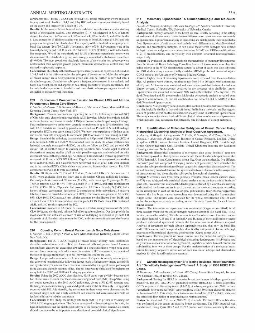
ANNUAL MEETING ABSTRACTS 53Acarcinomas (ER-, HER2-, CK5/6 and /or EGFR+). Tissue microarrays were analyzed for expression of claudins 1,3,4,7 and 8 by IHC and scored semiquantitatively based on the extent and intensity on a scale of 0-3+.Results: In the normal breast luminal cells exhibited membranous claudin staining for all of the claudins studied. Low expression (0-1+) was detected in 83% of tumors stained for claudin 1, 68% claudin 3, 59% claudin 4, 56% claudin 7, and 60% claudin 8. Low expression of all five claudins was detected in 36 of 253 cases (14.2%) and this group was designated the claudin-low. The majority of the claudin-low subgroup were basal-like cancers (26 of 36, 72.2%). In contrast, only 4 of 36 (11.1%) tumors were of the luminal phenotype and 6 of 36 cases (16.7%) were HER2+ (P<0.001). Within the basal-like subgroup, 78% of the metaplastic and 15.2% of the non-metaplastic tumors were claudin-low. The claudin-low group was strongly associated with disease recurrence (P=0.006). The most prominent histologic features of the claudin-low subgroup were nested rather than syncytial growth pattern, prominent desmoplasia, central scar, and marked lymphocytic response.Conclusions: This study is the first to comprehensively examine expression of claudins 1,3,4,7 and 8 in the different molecular subtypes of breast cancer. Molecular subtypes of breast cancer are a heterogeneous group and can be further subdivided into a claudin low group. Claudin-low subtype is a frequent phenomenon in metaplastic and basal-like breast cancer and appears to be a strong predictor of disease recurrence. The loss of claudin expression in basal-like and metaplastic subgroups suggests its role in epithelial-to-mesenchymal transition.
209 Outcomes of Prospective Excision for Classic LCIS and ALH on Percutaneous Breast Core Biopsy.C Luedtke, M Murray, T Nehhozina, M Akram, L Liberman, E Brogi. Memorial Sloan-Kettering Cancer Center, New York, NY.Background: There is no consensus regarding the need for follow-up excision (EXC) of CBs with only classic lobular neoplasia (cLN)[atypical lobular hyperplasia (ALH) or classic lobular carcinoma in situ (cLCIS)] and concordant radio-pathologic findings. Few small retrospective series report upgrade to carcinoma in up to 25% of patients (pts) with EXC, but data are limited by possible selection bias. Pts with cLN on CB undergo prospective EXC at our center since 6/2004. We report our experience with these cases and assess their rate of upgrade to carcinoma [DCIS or invasive carcinoma] on EXC.Design: Search of the pathology database identified 957pts with CB diagnosis (DX) of cLN between 6/2004 and 5/2009. We excluded from the study pts whose CB had other lesion(s) routinely managed with EXC, pts with no follow up EXC, and pts with CB and/or EXC at another center, to exclude any selection bias. A radiologist examined the pertinent imaging studies of all pts with possible upgrade, and excluded pts with discordant radio-pathologic findings. All slides of CBs and EXCs with an upgrade were reviewed. ALH and cLCIS DX followed Page’s criteria. Immunoperoxidase studies for E-cadherin, p120, and b-catenin were performed on cLN of all CBs with upgrade and in the matched EXCs. Clinical data were extracted from medical records. The 95% confidence intervals (CI) were calculated.Results: Of 80 pts with CB DX of cLN alone, 2 pts had 2 CBs of cLN alone and 11 (13%) were excluded from the study due to discordant CB and radiologic findings. Our study cohort consists of 69 women (71 CBs), with mean age 53 y (range 31-73). The CB targeted Ca2+ in 47/71 (66%), a mass in 7/71 (10%), and MRI enhancement in 17/71 (24%). Of the 69 pts who had prospective EXC for cLN only, 24 (34%) had a history of breast carcinoma (1 ipsilateral, 23 contralateral; 16 invasive ductal, 3 invasive lobular, 1 invasive mixed ductal/lobular, 4 DCIS). Carcinoma was present in 2/71 (3%; 95% CI, 0-10%) EXCs as a 2.3 mm tubular carcinoma with adjacent 2 mm DCIS, and a 2 mm focus of low to intermediate nuclear grade DCIS. Both index CBs contained ALH, and IHC results supported the DX.Conclusions: Prospective EXC of cLN alone in a CB had an upgrade rate of 3% (95% CI, 0-10%), and yielded only minute foci of low grade carcinoma. Our data provide the most accurate and unbiased estimate of risk of underlying carcinoma in pts with CB diagnosis of cLN and no other reason for EXC, and constitute a fundamental reference for their management.
210 Counting Cells in Breast Cancer Lymph Node Metastases.C Luedtke, L Tan, E Brogi, S Patil, D Giri. Memorial Sloan-Kettering Cancer Center, New York, NY.Background: The 2010 AJCC staging of breast cancer axillary nodal metastases classifies isolated tumor cells (ITCs) as clusters of cells not greater than 0.2 mm or nonconfluent clusters not exceeding 200 cells in a single histologic lymph node cross section. Since counting cells adds a new dimension to ITC assessment, we examined the rate of upstage from pN0(i+) to pN1mi when cell counts are used.Design: Lymph nodes were selected from a cohort of 83 patients initially node negative that converted to node positive following deeper levels with hematoxylin and eosin (HE) and cytokeratin (CK) stains. Each case was reassessed by a surgical breast pathologist using glass and digitally scanned slides. The pN stage was re-calculated for each patient using both the 2002 and 2010 AJCC staging guidelines.Results: Using the 2002 AJCC classification, 43/83 cases were pN0(i+) because they had cluster sizes <0.2mm. 2 of these cases were upgraded from pN0(i+) to pN1mi using cell count according to the 2010 AJCC guidelines, giving a 5% (2/43) upstage rate. Both upgrades occurred using glass and digital slides with CK stain only. No upgrades occurred with HE. Additionally, the metastases in these cases were characterized by dispersed single cells and nearly confluent cell clusters and were associated with an ipsilateral invasive lobular carcinoma.Conclusions: In this study, the upstage rate from pN0(i+) to pN1mi is 5% using the 2010 AJCC staging guidelines. The factors associated with upstaging are the stain, the type of metastases, and the histological subtype of the primary breast cancer. Cell counts should continue to be an important consideration of potential clinical significance.
211 Mammary Liposarcoma: A Clinicopathologic and Molecular Analysis.PL Lyle, JF Simpson, JA Bridge, JM Cates, DL Page, ME Sanders. Vanderbilt University Medical Center, Nashville, TN; University of Nebraska, Omaha.Background: Primary sarcomas of the breast are rare, usually occurring in the setting of malignant phyllodes tumor. Heterologous differentiation can occur, most commonly as liposarcoma. Liposarcomas arising in this setting are histologically indistinguishable from liposarcomas of soft tissue, and include well differentiated, dedifferentiated, myxoid, and pleomorphic subtypes. In soft tissue, the different subtypes have distinct biologic behavior and genetic alterations including MDM2 and CDK4 amplifications, t(12;16) translocations, and polyploidy with complex structural rearrangements, respectively.Design: We evaluated the clinicopathologic characteristics of mammary liposarcomas from the Vanderbilt Breast Pathology Consultive Practice. Liposarcomas were classified using criteria in the WHO classification system. A subset of cases was also subjected to FISH studies using a commercially available MDM2 probe and custom-designed CDK4 probe at the University of Nebraska Medical Center.Results: Eighty cases of mammary liposarcoma were retrieved from the consultation files. All patients were women, ranging in age from 14 to 96 years, with a mean age of 52 years. All tumors were unilateral and showed an equal distribution of sidedness. Eighty percent of liposarcomas occurred in the presence of a phyllodes tumor. Liposarcoma was classified as follows: 50% well-differentiated, 30% myxoid, 15% de-differentiated and 5% pleomorphic. Molecular cytogenetic analysis revealed extra copies of chromosome 12, but not amplification for either CDK4 or MDM2 in two dedifferentiated liposarcomas.Conclusions: Malignant phyllodes tumors often contain liposarcomatous elements that are histologically similar to those of soft tissue. Preliminary molecular studies suggest a lack of genetic alterations that are characteristically found in soft tissue liposarcomas. This may account for the markedly different clinical behavior of mammary liposarcoma which includes local recurrence but extremely rare incidence of distant metastasis.
212 Identification of Molecular Subtypes of Breast Cancer Using Hierarchical Clustering: Analysis of Inter-Observer Agreement.A Mackay, B Weigelt, A Grigoriadis, B Kreike, R Natrajan, R A’Hern, DS Tan, M Dowsett, A Ashworth, JS Reis-Filho. Institute of Cancer Research, London, United Kingdom; CRUK London Research Institute, United Kingdom; KCL Breakthrough Breast Cancer Research Unit, London, United Kingdom; Institute for Radiation Oncology, Arnhem, Netherlands.Background: Hierarchical clustering of microarray data using ‘intrinsic’ gene sets has been employed to classify breast cancers into the molecular subtypes: basal-like, HER2, luminal A, B and C, and normal breast-like. Over the past decade, five different ‘intrinsic’ gene sets composed of varying numbers of genes have been described for molecular subtype identification of breast cancers by hierarchical cluster analysis. The aim of this study was to determine the agreement between observers in the assignment of breast cancers into the molecular subtypes by hierarchical clustering.Design: Microarray data from three publicly available breast cancer datasets (total n=779) were subjected to hierarchical cluster analysis using the five distinct ‘intrinsic’ gene lists. Five observers analysed the dendrograms obtained by hierarchical clustering and classified the breast cancers in each dataset into the molecular subtypes according to the description in each of the five original publications. Inter-observer agreement between the five breast cancer researchers was determined using the free-marginal Kappa score. The results were analysed for the whole classification and for each molecular subtype separately according to each ‘intrinsic’ gene list for each breast cancer dataset.Results: The inter-observer agreement was substantial (Kappa scores ≥0.61) in all three datasets when four molecular subtypes had to be identified (i.e. basal-like, HER2, luminal, normal breast-like). With the introduction of the subdivision of luminal cancers into either luminal A, B and C or luminal A and B, none of the classification systems produced substantial agreement between the five observers in all datasets analysed. Analysis of agreement for each subtype separately revealed that only the basal-like and HER2 cancers could be reproducibly identified by independent observers through inspection of hierarchical clustering dendrograms (Kappa scores ≥0.81).Conclusions: The assignment of breast cancers into the molecular subtype classes based on the interpretation of hierarchical clustering dendrograms is subjective and only shows a modest inter-observer agreement, in particular when luminal cancers are subclassified into two or three groups. For the implementation of a molecular breast cancer taxonomy, objective definitions of each molecular subtype and standardised methods for their identification are essential.
213 Genetic Heterogeneity in HER2 Testing Revisited: How Nonuniform Are “Genetically Heterogeneous” Tumors? A Study of 1550 HER2 FISH Cases.JI Malowany, J Mazurkiewicz, M Wood, MC Chang. Mount Sinai Hospital, Toronto, ON, Canada; Univ. of Toronto, ON, Canada.Background: Testing for HER2 in invasive breast carcinomas is both prognostic and predictive. The 2007 ASCO/CAP guidelines interpret HER2/CEP17 ratios as positive (>2.2), negative (<1.8) and equivocal (1.8-2.2). A subsequent guideline (2009) defined “genetically heterogeneous” (GH) tumors as those with 5-50% (non-clustered) of nuclei with a ratio of >2.2. This study characterizes cases tested for HER2 with GH data, and the statistical distribution of amplified nuclei within a tumor.Design: We identified 1550 cases (2009-2010) in which FISH for HER2 amplification was performed at our center on invasive breast carcinomas. The FISH protocol was standardized, using Vysis HER2 and CEP17 probes, with manual counts by the same

54A ANNUAL MEETING ABSTRACTS technologists (either JM, MW; or both) according to ASCO/CAP Guidelines (min. 20 nuclei/case, up to 120). Clusters of amplified cells were scored separately. Data on every cell nucleus counted were retrieved and analyzed by linear regression of the Q-Q plot against a normal distribution. This provides an estimate of normality of the tumor population.Results: Of 1550 FISH tests for HER2, 1097 (71%) were nonamplified, 265 (17%) were amplified, and 188 (12%) were equivocal. A total of 450 (29%) had GH. Among these GH cases, the ratio was nonamplified in 283 (63%), amplified in 17 (4%), and equivocal in 150 (33%). The amplified subpopulation in GH tumors was larger if the overall ratio was close to 2.2 (Figure 1). However, the number of nuclei >2.2 in a GH tumor did not correlate with deviation from a normal distribution (Figure 2).
Conclusions: “Genetic heterogeneity”, as defined by ASCO/CAP (2009), does not distinguish between true intratumoral nonuniformity and HER2 amplification within a normal distribution. Studies using statistical approaches are needed to determine the prevalence of true intratumoral subpopulations, and their clinical significance.
214 Presence of Atypia in Breast Papillary Lesions in Core Biopsy: Does It Matter to the Clinician?M Mashhour, A Shehata, L Alriyees, RS Saad. Sunnybrook Health Sciences Centre, Toronto, ON, Canada.Background: The presence of atypical or usual epithelial proliferations within papillary breast lesions complicates their interpretation. The distinction between benign and malignant papillary proliferations on core biopsy can be very challenging without formal excision. There is few available data on clinical significance and outcome of papillary
lesions, with superimposed atypia (atypical ductal hyperplasia partially replacing the benign elements). The objective of this study is to evaluate the diagnostic implication of atypia in breast papillary lesions diagnosed on core biopsies.Design: We retrieved core biopsies diagnosed as papillary lesion from the hospital archives during last 8-years period. One hundred and forty two biopsy specimens of papillary breast lesions with their subsequent resections were reviewed. Cases were divided into two groups: Papillary lesions without atypia (73 cases with their subsequent lumpectomies) and papillary lesions with atypia (69 cases with their subsequent lumpectomies). Corresponding excision biopsies were examined in all cases and findings were correlated with core biopsy.Results: Of the 73 of papillary lesions without atypia, their subsequent surgical resections showed intraductal papilloma (IDP) in 34/73 (47%), fibrocystic changes (FCC) in 30/73 (41%), atypical papillary proliferation in 3/73 (4%), lobular carcinoma in situ (LCIS) in 2/73 (3%), and invasive ductal carcinoma (IDC), ductal carcinoma in situ (DCIS) and atypical ductal hyperplasia (ADH), 1/73 (1%) each case. For the papillary lesions with atypia, subsequent resections showed IDC in 6/69 (9%), DCIS in 21/69 (30%), intracystic carcinoma in 2/69 (3%), atypical papillary lesion in 4/69 (6%), ADH in 2/69 (3%), and LCIS in 2/69 (3%). The remaining 32/69 (46%) cases showed FCC. There is a significant correlation between the presence of atypia in papillary lesion and detection of a significant lesion on excisional biopsy (P< 0.001).Conclusions: Our study showed a significant correlation between presence of atypia in papillary lesions diagnosed on core biopsy and the presence of malignancy in lumpectomy. Therefore, evaluation and reporting of atypia is crucial in core biopsy report of papillary lesions and may be helpful to the clinician for patient management.
215 Study of Concordance between RT-PCR and Immunohistochemical Testing of Hormone Receptor and HER2 Status in Breast Cancer Specimens.R Mehta, RK Jain, PM Sojitra, Y Gokmen-Polar, S Badve. Indiana University School of Medicine, Indianapolis.Background: The need for good prognostic and predictive markers is vital to breast cancer management. The 21- gene RT-PCR assay (Oncotype Dx) has enabled the estimation of recurrence risk in estrogen receptor (ER) positive patients. With widespread use of this assay in routine clinical practice, the question remains if receptor status detection is perfect by any of immunohistochemistry (IHC) or RT-PCR. This study is a retrospective analysis of all Oncotype Dx reports and their corresponding pathology reports at our institution to determine the concordance of the receptor status between IHC and RT-PCR and correlate them with important clinico-pathological variables.Design: Our institution houses over 300 reports of Oncotype Dx assay performed on pathological tumor blocks from patients operated or treated here. These patients were seen at this instutuion between 2004 and 2010 and were referred for the Oncotype Dx assay after being verified as ER and/or PR positive. The HER2 RT-PCR scores were categorized as negative (<10.7) or positive (≥11.5) using assay guidelines. Any case with positive result on IHC and/or FISH for HER2 was considered as positive for this receptor.Results: For 228 patients, the individual RT-PCR scores for ER and PR as well as their immunohistochemical percentage staining were available. HER2 information by qRT-PCR as well as by IHC/FISH was available for 164 patients only. Unequivocal cases by IHC/FISH or RT-PCR HER2 were excluded for the purpose of clarity. A strong correlation between ER status by IHC and RT-PCR score from the Oncotype DX assay reports (r=0.30, 95% CI: 0.174-0.419; p<0.0001) was noted. Similar observations were noted for concordance of PR IHC percentage staining with RT-PCR scores (r=0.62; 95% CI: 0.532-0.698; p<0.0001). A strong concordance of HER2 results (98%) was observed when RT-PCR results were compared with IHC/FISH data (p=0.002).There was one equivocal case by RT-PCR which was also equivocal by IHC as well as FISH.Conclusions: A very strong concordance is noted between IHC tests used to detect receptor status during routine pathological practice and centralized quantitative RT-PCR results obtained by the Oncotype-Dx assay. The single gene expression data can be safely to used to assess quality/ performance of IHC in clincal laboratories.
216 MDGI, MIG-6 and EIG-121 Are Negative Regulators in EGFR Trafficking and Potential Biomarkers to EGFR Inhibitor Therapy in Triple-Negative Breast Carcinomas.D Meseure, S Vacher, K Drak Alsibai, M Trassard, F Lerebours, JM Guinebretiere, R Lidereau, I Bieche. Institut Curie, Hopital Rene Huguenin, St Cloud, France.Background: Understanding complexity of EGFR regulation mechanisms and molecular basis of resistance to EGFR inhibitors is crucial. EGFR targeted therapies are actually performed in triple negative breast carcinomas (TNBC), without identified activating EGFR gene alteration and with a still controversial EGFR expression status. In order to identify new targeted modalities, we explored three post traductional EGFR trafficking molecules: MIG-6 (Mitogen-Induced Gene-6), EIG-121 (Estrogen-Induced Gene-121) and MDGI (Mammary-Derived Growth Inhibitor). MDGI induces intra-cellular accumulation of EGFR. MIG-6 and EIG-121 specifically link EGFR with (i) the SNARE protein STX8 (MIG-6) or (ii) the autophagosome marker LC3 (EIG-121) and are implicated in a tumor suppression mechanism through recruitment of internalized receptors to late endosomes, lysosomal degradation and autophagy.Design: EGFR, MDGI, EIG-121 and MIG-6 mRNA level expressions were investigated by real-time quantitative RT-PCR in a series of 471 breast carcinomas (BC). BC were subdivised into 4 sub-groups according to RH (ERa and PR) and HER2 status: PPP (ER+, PR+, HER2+; n=56), PPN (ER+, PR+, HER2-; n=296), NNP (ER-, PR-, HER2+; n=48) and NNN (ER-, PR-, HER2-; n=71). Immunohistochal assays (IHC) were performed in a series of 88 BC using a panel of anti-EGFR, EIG-121, MIG-6 and MDGI antibodies (Abs). Immunostaining was assessed using intensity score (0 to 4+).
ANNUAL MEETING ABSTRACTS 55AResults: In the TNBC subgroup, RT-PCR identified low expression levels of EGFR (global: 84.6%, NNN: 63.5%, NNP: 69.6%, PPN: 91.5%, PPP: 88%), MDGI (global: 25.7%, NNN: 33.3%, NNP: 23.9%, PPN: 26%, PPP: 16%), EIG121 global: 16.1%, NNN: 60.3%, NNP: 13%, PPN: 7.5%, PPP: 12%) and MIG-6 (global: 25.7%, NNN: 28.6%, NNP: 32.6%, PPN: 51.2%, PPP: 42%). Protein underexpression was also confirmed in TNBC by IHC for EGFR (59%), MDGI (36%), EIG-121 (65%) and MIG-6 (31%). A statistically positive correlation was observed between EGFR, MDGI, EIG-121 and MIG-6 RNA levels and protein expression status (Kruskall Wallis’s H test; P<10-5).Conclusions: Underexpression of the three EGFR post-traductional negative regulators MDGI, EIG-121 and MIG-6 can potentially create an aberrant EGFR-mediated oncogenic signaling pathway in BC. In the TNBC subgroup, MDGI, MIG-6 and EIG-121 might be particularly useful biomarkers in targeted EGFR therapy when EGFR expression level is normal to high and/or MDGI, EIG121 and MIG-6 expression levels are low.
217 Invasive Pleomorphic Lobular Carcinoma: A Clinicopathologic and Immunohistochemistry Study of 40 Cases.L Monhollen, C Morrison, F Ademuyiwa, R Chanddrasekhar, G Wilding, T Khoury. Roswell Park Cancer Institute, Buffalo, NY.Background: Invasive pleomorphic lobular carcinoma (PLC) is a rare aggressive variant of invasive lobular carcinoma. Given its rarity, the clinicopathologic features of this entity are not well studied. It has been reported that this type is a good responder to trastuzumab. The purpose of this study was to review its clinicopathologic findings.Design: Cases were collected from the clinical database and pathology department search engine in Roswell Park Cancer Institute from 1995 to 2010. Cases were either in house or referred from another hospital. Histologic features were reviewed including SBR grade, percentage of grade 3 nuclei (>10% was required to consider tumor pleomorphic), in situ component (ductal or lobular), histologic pattern, margin status, and lymphovascular invasion. Cases with available tissue blocks were constructed in tissue microarray (TMA) blocks (n= 34). E-cadherin, estrogen receptor, progesterone receptor, and HER2 by fluorescence in situ hybridization were performed on the TMA slides. Other clinicopathologic data were also collected including tumor size and stage, nodal status, therapy modality, disease free and overall survival. Fisher’s exact test and Logrank test were used for statistical analysis.Results: There were 43 cases that met our criteria. There 29 SBR grade 2 and 14 SBR grade 3. There were 5 histologic patterns (18 classic, 13 mixed, 1 nested, 8 solid, and 3 trabecular). Hormone receptors were negative in 7 (16.3%) cases and positive in 31 (72.1%) cases (5 cases had no hormone receptors data). HER2 was amplified in 10 (23.3%) cases and negative in 31 (72.1%) cases (2 cases had no HER2 data). Clinically, 9 patients had local recurrence and 10 died of disease with median follow-up of 47.7 (1.84-156.57) months. Two patients were treated with trastuzumab and 24 patients received adjuvant chemotherapy. One patient that was treated with trastuzumab developed tumor recurrence in 2 years. There was no correlation between any of the clinicopathologic parameters and clinical outcome.Conclusions: Although PLC is a morphologic form of lobular carcinoma, it has features of ductal carcinoma NOS. It might not differ in terms of trastuzumab response from other type.
218 Pleomorphic Lobular Carcinoma: Immunohistochemistry Study of 27 Cases.L Monhollen, C Morrison, F Ademuyiwa, R Chandrasekhar, G Wilding, T Khoury. Roswell Park Cancer Institute, Buffalo, NY.Background: Analysis of gene expression profiling data on breast cancers has revealed “molecular subclasses” that may have prognostic significance. “Basal type” has been proposed to be present in pleomorphic lobular carcinoma (PLC). The purpose of this study was to evaluate the molecular subtypes of PLC by IHC and to explore the incidence of GCPD-15 and mammaglobin expression.Design: Cases were collected from the clinical database and pathology department search engine in Roswell Park Cancer Institute from 1995 to 2010. Cases were either in house or referred from another hospital. Histologic features were reviewed including SBR grade, percentage of grade 3 nuclei (>10% was required to consider tumor pleomorphic). Cases with available tissue blocks were constructed in tissue microarray (TMA) blocks (n= 27). E-cadherin, estrogen receptor (ER), progesterone receptor (PR), cytokeratin 5/6, CAM, GCPD-15, mammaglobin, CD117, vimentin, EGFR, p53 and HER2 by fluorescence in situ hybridization were performed on the TMA slides. ER and PR were scored using Allred scoring system. All other markers were scored from 0 to 3; >1 was considered positive. Statistical analyses to assess the significance of the difference between two correlated proportions based on the same sample were performed using the exact McNemar’s test.Results: There were 3 (11.1%) cases of HER2 type, 5 (18.5%) of triple negative type and 10 (37%) of luminal type. Overall, basal type markers were as follows: CD117 in 4 of 24 (16.7%), CK5/6 in 4 of 26 (15.4%), EGFR 1 of 27 (3.7%) and vimentin 1 of 26 (3.8%). Among the cases that had all the available markers (n=22), only two (9.1%) cases were positive for at least two of the basal type markers, one for vimentin, p53 and CD117 and one for CK5/6, EGFR and p53. The latter was triple negative. Moreover, PLC appears to express mammaglobin (15 of 25 cases) more frequently than GCPD-15 (8 of 25 cases) with borderline significance (p=0.07, McNemar’s test).Conclusions: From this small series, basal type PLC by IHC is rare and HER2 subtype is more common than conventional lobular carcinoma. Although GCPD-15 is traditionally recognized as a sensitive marker for PLC, mammaglobin appears to be more sensitive. Additional studies with larger numbers of cases are needed to validate our data.
219 The Histopathologic Findings of Magnetic Resonance Imaging Enhancement of Breast Lesions with Histologically Mass-Negative Lesion: A Single Institution Experience.L Monhollen, P Kumar, R Chandrasekhar, G Wilding, T Khoury. Roswell Park Cancer Institute, Buffalo, NY.Background: Magnetic resonance imaging (MRI) is increasingly being used to detect breast lesions for patients with high risk or as part of staging with varying results. The histologically non-mass lesions that are enhanced by MRI have had little attention. The objective of this study is to review the histologic findings on these biopsies.Design: One hundred-sixty five cases were selected from the archives at Roswell Park Cancer Institute between 2001 and 2010. We defined histologically mass lesion as carcinoma (in situ or invasive), fibroadenoma, papilloma or radial scar. All other lesions were considered mass-negative lesions. These lesions included stromal fibrosis, pseudoangiomatous stromal hyperplasia (PASH), inflammation, sclerosing adenosis, apocrine metaplasia, duct ectasia and ductal hyperplasia. One or more histologic entity could be present in one biopsy. These lesions were graded from 0 to 3, based on the degree of involvement; ≥1 was considered present. Statistical analyses to assess the significance of the difference between two correlated proportions based on the same sample were performed using the exact McNemar’s test.Results: A total of 80 (48.5%) cases had mass lesions that would account for enhancement on MRI, 35 (21.2%) carcinoma and 45 (27.3%) were other mass lesions including radial scar, papilloma or adenoma. The remaining 85 (51.5%) cases, classified as mass-negative, had stromal fibrosis in 84.7%, PASH in 50.6%, ductal hyperplasia in 47.1%, sclerosing adenosis in 37.7%, apocrine metaplasia in 34.1%, duct ectasia in 32.9% and inflammation in 32.9%. Stroma fibrosis was significantly more common than all other findings (p-value <0.0001), while PASH was significantly more common than duct ectasia and inflammation only (p value 0.03 each).Conclusions: Our results indicate that the pathologist should expect that fibrosis and PASH to be the most common findings in histologically mass-negative enhanced-lesions by MRI. It is unclear why these changes produce MRI enhancement. More studies to explore the reason are needed.
220 High Expression of DCN and HSP90B1 in Breast Cancer Is Associated with LN and Distant Metastases and Decreased Overall Survival.JC Moreno, D Tran-Thanh, T Cawthorn, L Jacks, S Ackloo, P Hong Zhu, M Dharsee, G Sardana, J Chen, KR Evans, D McCready, WL Leong, SJ Done. Campbell Family Institute for Breast Cancer Research, Toronto, ON, Canada; University Health Network, Toronto, ON, Canada; University of Toronto, ON, Canada; Ontario Cancer Biomarker Network, Toronto, Canada.Background: Patients with local breast cancer have a better 5 year overall survival (OS) (98%) than patients with lymph node (LN) (83.6%) or distant metastasis (23.4%). Biomarkers that improve patient classification before metastases are evident have clinical utility. During a biomarker discovery study our laboratory found a correlation between high expression of Decorin (DCN) and HSP90B1 and LN metastasis.Decorin is a key modulator of the tumour microenvironment, mainly in an antioncogenic role, but also increases migration of osteosarcoma cells and is highly expressed in endothelial cells undergoing angiogenesis. HSP90B1 excerpts a pro-oncogenic role that facilitates cell survival decreasing apoptosis and stimulating cell proliferation, transcription and migration. To determine the clinical significance of high expression of DCN and HSP90B1 in tumour tissues, we examined the expression of both proteins in 990 different breast cancers and correlated that expression with clinical parameters.Design: Stage 1 and 2 prognostic human breast cancer tissue microarrays annotated with clinicopathological data, including disease-free survival (DFS) and OS, were purchased from the National Cancer Institute. Immunohistochemical scores were determined using a semi-quantitative scoring system under light microscopy and image analysis software. DCN staining was evaluated separately in the stroma (DS) and the malignant epithelium (DE). HSP90B1 was scored in the malignant epithelium only (HE).Results: High expression of DE was associated with LN metastasis p<0.0001, higher number of positive LNs p<0.0001 and worse OS p: 0.01. High expression of DS was not associated with LN status. High HSP90B1 expression was associated with presence of distant metastasis p<0.0002, worse OS p<0.0001 and worse DFS p<0.0001.Conclusions: These results suggest that high expression of DCN in invasive breast cancer cells could be used as a prognostic marker for OS and LN metastasis. In addition, high expression of HSP90B1 appear to be a prognostic marker for OS and distant metastasis and a predictive marker for hormone therapy benefit even in patients with negative hormone receptor status.
221 Src Expression in Infiltrating Breast Carcinoma with Hormone Receptor Positive: Prognostic Significance.TA Muci, L Sanchez-Tejada, C Alenda, M Niveiro, J Segui, G Peiro, FI Aranda. Hospital General Universitario, Alicante, Spain.Background: The family SRC are nonreceptor tyrosine kinases and one of the best-studied targerts for cancer therapy. Elevated Src protein has been shown in human cancer, including infiltrating breast carcinoma (IBC). The aims of this study was to investigate the expression and the prognostic significance of Src in a series of breast carcinoma with hormonal receptors (HR) positive and Her-2 negative.Design: A total of 186 cases of IBC with HR positive/Her-2 negative, with lymphadenectomy and without neoadjuvant treatment were retrieved from the Surgical Pathology files. Median clinical follow-up was 54 months (range 11 to 247). Age ranged from 24 to 88 yrs-old (median 61, SD 13)). Histologic grade (HG) was assessed according to Nottingham criteria. Immunohistochemical (IHC) staining was performed in whole sections for ER (cut-of 10%), PgR (cut-off 10%), Ki67 (cut-off 15%), p53

56A ANNUAL MEETING ABSTRACTS (cut-off 20%) and Her-2 (2+ and <30% 3+ confirmed by FISH). Src (Src phospho Y418 antibody ab47411, Abcam) staining was performed on tissue microarrays, and nuclear (N-Src) and citoplasmatic (C-Src) were independently scored (cut-off 5%). For PIK3CA mutation study, DNA was extracted from formalin-fixed, paraffin-embedded tissues using standard methods. Analysis was performed by allelic discrimination based on real-time chemistry TaqMan MGB probes in ABI Prism 7500 Sequence Detection System (Applied Biosystems). Significant associations were identified using Chi-square and Fisher’s exact test. Actuarial survival was calculated by the Kaplan-Meier method (log rank test). A p-value <0.05 was considered significant.Results: Increased N-Src expression was observed in 21% of tumors, C-Src in 30%, high Ki67 in 31% and p53 in 3%. PIK3CA mutations were detected in 24.6% tumors. Tumors with N-Src overexpression showed PIK3CA mutation (p=0.011) and a trend towards low HG (p=0.17). In contrast, C-Src was associated only with lymph node negative status (p=0.013). Patients whose tumors showed N-Src overexpression had longer survival (OS, p=0.038) but no differences were found for c-Src expression levels (p=ns)Conclusions: Our findings suggest that N-src overexpression in IBC with HR positive is a favourable prognostic factor.Supported by grant PC04 FIHGUA
222 Breast Cancer Risk in Women with Diagnosis of Flat Epithelial Atypia: Follow-Up Study in a Benign Breast Disease Cohort.A Nassar, S Narendra, CA Reynolds, RA Vierkant, M Frost, LC Hartmann, DW Visscher. Mayo Clinic, Rochester, MN.Background: Atypical columnar lesions or flat epithelial atypia (FEA) have been noted to accompany ADH, DCIS or invasive cancers, implying that they represent high risk index lesions. However, the future risk association for breast cancer has yet to be studied in multiple cohorts. This study was undertaken to assess the risk for subsequent breast cancer in a group of women with FEA identified in a retrospective cohort of women with benign breast disease (BBD)Design: Columnar cell lesions (CCLs) were assessed in 9087 women in the BBD Cohort who underwent excisional breast biopsy between 1967-1991. CCLs were further classified into columnar cell change, columnar cell hyperplasia and FEA. Development of breast cancer, laterality, age and time to follow-up was extracted from the database.Results: CCLs were identified in 2025 (22%) of BBD subjects. FEA was detected in 99 cases (4.9% of the CCLs group) with an overall cohort frequency of 1.1% (99/9087). Median age at time of benign biopsy was 46.4 years (range 21.8 to 73.3 years). Out of 99 patients, 17 had no follow-up (last follow-up was at time of benign biopsy). Breast cancer was detected in follow up of 15.1% of FEA cases. Of the 15 women who did develop breast cancer, this occurred from 2.7 years to 31 years from time of benign biopsy (median time to cancer was 12.8 years). 11 out of 15 cancers occurred on the same side as the benign biopsy.Conclusions: The data suggest that women with FEA are at increased risk for breast cancer although we cannot be certain based on these data only due to lack of appropriate control cases for comparison. Based on these limited data, follow-up excisional biopsy is recommended for women with a diagnosis of FEA.
223 Functional Characterisation of the 19q12 Amplicon in Grade 3 Breast Cancers.R Natrajan, D Wetterskog, MB Lambros, FC Geyer, A Mackay, R Vatcheva, JS Reis-Filho. Institute of Cancer Research, London, United Kingdom.Background: Grade III (GIII) invasive ductal carcinomas of no special type (IDC-NST) comprise up to 60% of all invasive breast cancers and have an aggressive clinical behavior. Amplification of 19q12 is found in a subgroup of estrogen receptor (ER)-negative HER2 amplified (4%) and basal-like (16%) subtypes of breast cancer. This amplicon comprises 9 genes, including cyclin E1 (CCNE1), which has been proposed as its driver due to a strong association with mRNA over-expression. The aims of this study were to identify functionally the genes within the 19q12 amplicon whose expression is required for the survival of cancer cells harboring this amplification.Design: We analysed a series of 297 frozen breast cancers with high-resolution microarray CGH to assess the frequency of 19q12 amplification. A subset of 48 GIII micro-dissected IDC-NST were subjected to mRNA expression arrays, and data integrated with aCGH to find those genes that were significantly over-expressed when amplified. Breast cancer cell lines with (MDAMB157 and HCC1569) and without (Hs578T, MCF7, MDAMB231 and ZR75.1) 19q12 amplification were screened with an RNAi library targeting the genes mapping to the smallest region of amplification on 19q12. Individual short interfering RNA (siRNA) SMARTpools targeting these genes were plated in 96-well plates and cell viability was assessed after 9 days using CellTiter-Glo Luminescent Cell viability assay.Results: We identified 19q12 amplification in 5% of breast cancers, which was significantly associated with high grade and ER negative tumors. Of the 9 genes mapping to the smallest region of amplification, UQCRFS1, POP4, C19ORF12, CCNE1 and C19ORF2 were significantly over-expressed when amplified (P<0.05). Silencing of POP4, PLEKHF1, CCNE1 and ZNF537 selectively inhibited cell viability in 19q12-amplified cells compared to control cells. Over-expression of CCNE1 in amplified cells was found to confer sensitivity to doxorubicin, cisplatin and paclitaxel.Conclusions: Expression of POP4, PLEKHF1, CCNE1 and ZNF537 are required for the survival of cancer cells displaying their amplification and suggest that the 19q12 amplicon may harbor more than one ‘driver’. Furthermore identification of tumors with CCNE1 amplification may identify a subgroup of patients that are responsive to common chemotherapy agents.
224 Papillomas on Core Needle Biopsy: Analysis of 88 Cases.A Nayak, MZ Gilcrease, LP Middleton, S Carkaci, J Zhang, N Sneige, L Huo. The University of Texas M.D. Anderson Cancer Center, Houston.Background: Although excision of breast intraductal papillomas with atypia is generally recommended, the clinical management of papillomas without atypia diagnosed on core needle biopsy (CNB) is controversial. This study aims to evaluate such lesions and correlate their clinical, radiological and histologic features with findings at excision or clinico-radiological follow-up.Design: The computer database of our Department of Pathology was searched for papillary lesions on CNB. Exclusion criteria included non- availability of histologic material for review, concurrent diagnosis of ductal carcinoma in situ (DCIS)/invasive carcinoma in the ipsilateral breast, presence of atypical ductal hyperplasia or lobular neoplasia within or outside of the papilloma in the same CNB, or incidental papillomas. The resulting 88 cases were the subjects of the study, including 36 cases from 2003 to 2010 with subsequent surgical excision and 52 cases from 1998 to 2008 with ≥2 years of clinico-radiological follow-up. Slides of core biopsies were reviewed to confirm the diagnosis, and multiple clinical, radiological and histologic features were evaluated.Results: The presence of nipple discharge, hypoechoeic mass on ultrasonography, and incomplete removal of the targeted lesion by CNB were significantly associated with those lesions that underwent excision versus clinicoradiological follow-up (p=0.0449; p=0.0025; p=0.0001, Fisher’s exact test). Of the 36 cases with excision, 8 (22.2%) had an upgrade on excision (invasive papillary carcinoma, grade 2, 3.0 cm, 1 case; invasive ductal carcinoma, grade 1, 0.9 cm, 1 case; DCIS, grade 1, 2 cases, grade 2, 3 cases and grade 3, 1 case). Fifty-two cases with clinico-radiological follow-up remained stable (24 to 148 months; median, 48 months). The overall upgrade rate was 9%.Within the excision group, the only clinical, radiologic or histologic feature significantly associated with upgrade was nipple discharge (p=0.0075), but only 4 of 8 patients with upgrade had a nipple discharge. However, 33 patients with a mass ≥1cm in size included 6 of the 8 upgraded cases (75%).Conclusions: The results of our study suggest that surgical excision of intraductal papilloma without atypia diagnosed on CNB should be considered for at least those patients who present with nipple discharge and/or a mass ≥1 cm.
225 Squamous Cell Carcinoma of the Breast: A Clinicopathologic Study of 21 Cases.A Nayak, S Krishnamurthy, Y Wu, MZ Gilcrease. The University of Texas M.D. Anderson Cancer Center, Houston.Background: Primary squamous cell carcinoma (SCC) of the breast is a rare form of invasive breast carcinoma defined as having greater than 90% squamous differentiation. Studies evaluating the clinical behavior of this tumor have yielded inconsistent results. Further, the biological behavior of this tumor has not been adequately studied in the context of its varying histomorphology. Our aim was to evaluate the clinical and histological features of this tumor and to correlate these with disease outcome.Design: A total of 21cases of primary SCC of the breast were selected for analysis from the computerized database of the Department of Pathology between the years 1985 and 2010. Exclusion criteria included non-availability of histology slides for review and tumors with less than 90% squamous differentiation. Clinical and histological features were evaluated and correlated with disease outcome.Results: The 5-yr overall survival (OS) of patients with primary SCC of the breast was 51% ± 13%. The only statistically significant features associated with OS were patient age and tumor keratinization. Patients greater than 60 yrs of age had decreased OS (log rank, P=0.035). Five-yr survival rate for patients greater than 60 yrs of age and 60 yrs or less was 36% ± 20% and 60 ± 16%, respectively. Patients with tumors having at least focal keratinization had improved OS as compared to patients with nonkeratinizing tumors (87% ± 12% vs. 26% ± 15%; log rank, P=0.027). There was a trend for patients with cystic tumors and those with associated DCIS to have improved OS. In contrast, patients with tumors having spindle cell metaplasia and/or acantholytic change showed a trend towards decreased survival, but the differences were not statistically significant. Most tumors (17/21) were moderately or poorly differentiated, most had a high nuclear grade (15/21), and most were Nottingham histologic grade 3 (17/21). Among the few patients with tumors that were well differentiated or with intermediate nuclear or histologic grade, there were no deaths during the follow-up period, but the number of patients in these categories was insufficient to show statistical significance. Lymph node status, mitotic rate, tumor necrosis, clear cell change, and the presence of a pleomorphic component were not associated with OS.Conclusions: Primary SCC of the breast tends to be aggressive, particularly in patients over 60 yrs of age, but the presence of at least focal keratinization is associated with significantly improved overall survival.
226 Expression of Progesterone Receptor Is a Robust Marker for Long Term Survival in Breast Cancer.S Nofech-Mozes, A Plotkin, SA Narod, P Sun, E Rawlinson, WM Hanna. University of Toronto, ON, Canada.Background: Previous studies demonstrated that progesterone receptor (PR) expression is a poor predictive marker for response to endocrine therapy however indicate its role in predicting disease free survival at 5 years when incorporated together with estrogen receptor (ER), and HER2 in prognostic models. This study investigated the effect of hormone receptors on overall survival (OS).Design: We studied 1017 primary breast cancer cases from the Henrietta Banting database of well characterized cases with a mean follow up of 8.3±5.5 years, treated in a single institution. Immunohistochemistry for ER, PR, Her2 and stem cell marker ALDH1 (BD Bioscience, 1:1000) was applied to tissue microarray arrayed in triplicates. Pertinent clinical and pathological data were retrieved from the medical charts. Relative

ANNUAL MEETING ABSTRACTS 57Arisk for death was calculated for age, tumor size, nodal status and immunohistochemical markers (ER, PR, Her2 and ALDH1) in univariate and multivariate analyses. Estimates were considered statistically significant for two-tailed values of P < 0.05.Results: On Univariate analysis, poor OS was associated with size (RR 1.27 CI 1.21-1.33, p<0.0001), positive lymph nodes (RR 2.78 CI 2.12-3.63, p<0.0001), Her2+ (RR 1.61 CI 1.27-2.05, p<0.0001), ALDH1 in more than 50% of the tumor (RR 1.76 CI 1.10-2.79, p=0.02). Improved OS was associated with age (RR 0.98 CI 0.97-0.99, p<0.0001), ER+ (RR 0.57 CI 0.44-0.74, p<0.0001) and PR+ (RR 0.58 CI 0.46-0.74, p<0.0001).On multivariate analysis, only size (RR 1.17 CI 1.11-1.24, p<0.0001) and positive lymph nodes (RR 2.54 CI 1.90-3.40, p<0.0001) were associated with poor outcome while age (RR 0.98 CI 0.97-0.99, p=0.0004), and PR+ (RR 0.66 CI 0.49-0.90, p=0.008) were independent markers of improved survival. Her2+ was marginal (RR 1.28 CI 1.00-1.46, p=0.05). Interestingly, ER expression lost its predictive ability when adjusted to the other variables (RR 0.90 CI 0.64-1.27, p=0.55).Conclusions: PR expression emerged as a significant good independent prognostic factor for long term overall survival. The prognostic value of PR is more significant than other biomarkers such as ER, Her2 and ALDH1.
227 Difference in Melatonin Receptor (MT1) Expression in Triple Negative Breast Carcinoma (TNBC) between African American (AA) and Caucasian (CS) American Women.GM Oprea-Ilies, E Haus, LL Sackett-Lundeen, A Adams, C Cohen. Emory University, Atlanta, GA; University of Minnesota, Minneapolis; Regions Hospital, St. Paul, MN.Background: The pineal hormone melatonin exerts a regulatory function on cell proliferation in many tissues and an antiproliferative effect on human tumor cell lines in vitro and on the growth of human breast cancer cell xenografts in vivo. The membrane bound G-protein coupled M1 melatonin receptor (MT1) is present in human breast tumor cell lines and when activated mediates the growth suppressive action of melatonin on breast tumor cells. The MT1 receptor was identified in small studies of largely ER and PR positive primary breast cancers. We studied the presence of the receptor in triple negative breast cancers (TNBC) by immunohistochemical (IHC) methods. Active melatonin receptor may be of interest in TNBC in which no specific adjuvant treatment is available.Design: Invasive TNBCs diagnosed during a7-year period were reviewed. Only tumor that did not show staining for ER, PR and Her-2 (scored as 0, 1, or 2+, with no amplification by FISH) were included. Tissue microarrays (TMAs), constructed with two 1 mm representative cores from each carcinoma, were stained with the monoclonal antibody MT1. The scoring of the IHC results was semiquantitative, using 0-3 for intensity and percentage for number of tumor cells staining. Tumors that scored 2-3 for intensity with ≥10% tumor cells staining were considered positive. MT1 expression was studied in AA and CS patients.Results: MT1 was positive in 80 (78%) AA and 53 (98%) CS patients.The incidence of TNBC expressing melatonin MT1 receptor is significantly lower in AA as compared with CS ( 78 vs 98 % ; Chi square 4.019, p = 0.045). Table 1 details MT1 IHC staining in the AA and CS population.MT1 IHC by age and raceGroup Number Mean St Dev SEAA younger than 50 yo 35 69.06 72.54 12.26AA older than 50 yo 45 75.20 82.64 12.32CS younger than 50 yo 10 138.50 89.01 28.15CS older then 50 yo 43 170.60 106.71 16.27df: 3; F-test: 11.665, p Value: 0.0001Conclusions: 1. The incidence of TNBC expressing melatonin MT1 receptor is significantly lower in AA as compared with CS.2. The degree of immunohistochemical staining of MT1 receptor in TNBC expressing the receptor as measured by a score of staining intensity and percentage of cells staining is significantly (p=0.001) lower in the AA.3. This may indicate a biologic difference of TNBC in AA and CS, which will have to be further explored.
228 Melatonin MT1 Receptor in Triple Negative Breast Carcinoma (TNBC) and Estrogen Receptor Positive Invasive Ductal Carcinoma (ER+DC).GM Oprea-Ilies, E Haus, LL Sackett-Lundeen, H Sullivan, R Bush, A Adams, C Cohen. Emory University, Atlanta, GA; University of Minnesota, Minneapolis; Regions Hospital, St. Paul, MN.Background: The pineal hormone melatonin exerts a regulatory function on cell proliferation in many tissues and an antiproliferative effect on human tumor cell lines in vitro and on the growth of human breast cancer cell xenografts in vivo. The membrane bound G-protein coupled M1 melatonin receptor is present in human breast tumor cell lines and when activated mediates the growth suppressive action of melatonin on breast tumor cells. The M1 receptor was identified in small studies of largely ER and PR positive primary breast cancers. Breast carcinomas with TNBC and ER+DC phenotypes show differences in biologic behavior, drug sensitivity, course of disease and survival. In view of the experimental work showing varying melatonin effects on different human breast cancer cell lines and xenografts it appeared of interest to explore the presence of melatonin receptors in TNBC and ER+DC.Design: Invasive TNBCs and ER positive IDC diagnosed in our institution were reviewed. Tissue microarrays (TMAs), constructed with two 1 mm representative cores from each carcinoma, were stained with monoclonal antibody to MT1. The scoring of the immunohistochemistry results was semiquantitative, using 0-3 for intensity and % for
number of tumor cells staining. Tumors that scored 2-3 for intensity with ≥10% tumor cells staining were considered positive. MT1 expression was compared in TNBC vs. ER+ IDCs. Statistical analysis was performed using Anova and chi square.Results: 115 TNTB and 111 ER+ cases were evaluated. MT1 was positive in 86% TNTB and in 90% ER+ IDC. The frequency and intensity of MT1 staining are presented in table 1.MT1 IHC by age and racedf = 3 F-test: 11.665 p Value: 0.0001 Number Mean St Dev SEAA younger then 50 year of age 35 69.06 72.54 12.26AA older then 50 year of age 45 75.20 82.64 12.32CS younger then50 year of age 10 138.50 89.01 28.15CS older then 50 year of age 43 170.60 106.71 16.27
Conclusions: 1. The MT1 melatonin receptor is expressed in a high percentage in both TNBC and ER+BC with no difference in incidence between the two phenotypes.2. However, the immunohistochemical expression of MT1 is significantly stronger in the ER+BC tumors than in the TNBC3.This difference may contribute to the different behavior of the two phenotypes and may represent a possible pathway for novel therapeutic approach.
229 Factors Influencing Accuracy of Sentinel Node Frozen Section for Breast Cancer: A Review of 654 Cases Including 1678 Sentinel Nodes.D Pandya, M Singh, B O’Hea, P Farrelly, M Burk, C Rizk, C Tornos. Stony Brook University Medical Center, NY; Stony Brook University Medicfal Center, NY.Background: Sentinel lymph node biopsy (SLN) is the standard of care for surgical assessment of axillary node status in breast cancer. However the accuracy and methodology of frozen section (FS) is still controversial. The aim of this study was to assess accuracy of FS diagnosis compared to permanent section and identify factors influencing accuracy in a large series of cases treated at our institution.Design: We identified 654 consecutive cases of SLN biopsies for breast cancer performed at our institution from 1/2006 to 9/2010 that underwent frozen section evaluation, including a total of 1678 SLN. Among these cases there were 65 that had discrepant frozen section diagnosis. The frozen sections and permanent sections of these 65 cases were reviewed. Parameters evaluated included size and location of the metastases in the node, slides containing the metastases (FS, permanent routine, or deeper levels), type of carcinoma (ductal vs lobular), and impact on final stage.Results: The 65 discrepant cases included a total of 77SLN nodes that were negative on FS and positive on permanent sections, with an overall false negative rate of 4.5%. The discrepant diagnosis were due to sampling error of tumor deposits not present on the FS in 53 nodes (68%), positive cells detected on immumostains only in 19 nodes (24%), and pathologist error overlooking metastases present on the FS in 5 cases (6%). The sampling error was further subclassifed into cases in which the deposits were subcapsularly located and not present on the FS for technical difficulties in cutting tissue close to fat (37 nodes), and cases in which the deposits were clearly deeply located in the node and only seen in deeper sections (15 nodes). Most metastases missed on the frozen section were small ranging from <0.2 to 8.0 mm (mean 1.3mm). The false negative FS had an impact on nodal stage in 45 cases (6.8% of all cases). The FS accuracy rate for ductal carcinoma was 91.2% compared to lobular 79.4%.Conclusions: False negative frozen sections are predominantly due to technical difficulties in cutting entire nodes including subcapsular areas close to perinodal fat. The accuracy is different for ductal versus lobular carcinomas, and impact on nodal staging is seen in 6.8% of all cases.
230 Osteopontin and Osteopontin-c Protein and mRNA Expression Correlate with Breast Cancer Aggressiveness.G Peiro, F Ortiz-Martinez, E Lerma, E Adrover, M Planelles, L Sanchez-Tejada, FI Aranda, FJ Gutierrez-Avino, D Giner. Hospital General Universitari d’Alacant, Spain; Hospital de la Santa Creu i Sant Pau, Barcelona, Spain.Background: Osteopontin (OPN) is a secreted non-collagenous phosphoprotein that functions as both an extracellular matrix (ECM) component and a cytokine. Experimental and clinical studies suggest its association with tumor development, invasion and metastasis. The aim of this study was to investigate OPN (total and splice variant -c) protein and mRNA expression in a series of breast carcinoma (BC).Design: We studied OPN-t and OPN-c expression immunohistochemically (IHC) on paraffin-embedded tissue microarrays containing 440 BC, stratified by immunophenotypes: 25% Luminal A and B (ER/PR+), 43% HER2+ (≥30% cells 3+ by IHC and/or FISH/CISH amplification), and 32% basal/TN (ER/PR/HER2-negative +/-CK5/6+/-EGFR). Cytoplasm staining was semiquantitatively scored based on intensity (0-3+) and distribution (0-100%) (score 0-300). We further analyzed OPN-t and OPN-c mRNA expression by real-time quantitative PCR in 120 samples. The correlation between OPN results and clinicopathological factors was evaluated.Results: Median patients’ age was 55 years (range 28-89) with a median follow-up of 90 months. OPN-t overexpression (34%) was found more frequent among tumors of older patients (73%; p<0.000), grade 3 (71%; p=0.03), basal/TN subtype, (61%; <0.000) and vascular invasion (54%; p=0.10). OPN-c was detected in larger tumors (57%; p=0.03), grade 3 (66%; p=0.03), HER2 subtype (48%; p=0.001), with positive lymph-node status (58%; p=0.017) and vascular invasion (52%; p=0.07). Similarly, increased OPN-c mRNA was associated with larger tumors (64%; p=0.09), positive lymph nodes (83%; p=0.003) and advanced stage (III-IV) (54%; p=0.005).Conclusions: Our results support that increased levels of OPN-t and especially the OPN-c splice variant are related with breast cancer aggressiveness. Therefore, OPN has a potential value as a tumor progression marker and a target for its functional suppression. Supported by Grants FCVI-HGUA (PI-C/2008/02), ACOMP/2009/195 and GE-018/09

58A ANNUAL MEETING ABSTRACTS 231 The Evaluation of the Breast Biomarkers in Multifocal/Multicentric Breast Carcinomas.M Pekmezci, A Salhadar, P Rajan, C Ersahin. Loyola University Medical Center, Maywood, IL.Background: There are controversies regarding the diagnostic and therapeutic management of multifocal/multicentric (MF/MC) breast carcinomas. Estrogen receptor (ER), progesterone receptor (PR), human epidermal growth factor receptor 2 (HER2) and Ki67 are well accepted biomarkers in the management of breast carcinomas. However, it is not clear whether repeating these biomarkers in each tumor focus is necessary or has diagnostic value. The purpose of this study is to evaluate whether breast carcinoma biomarkers vary among separate tumor foci of MF/MC breast carcinomas, and whether this variation correlates with the morphological features.Design: We have reviewed MF/MC invasive breast carcinomas diagnosed between January 2001 and June 2010 at our institution. Cases with biomarker analyses performed on more than one focus were included in the study. Tumor morphology, Nottingham grade, Ki67, ER, PR and HER2 status were evaluated. ER, PR and HER2 results were classified as positive or negative. ER/PR positivity is immunohistochemical (IHC) staining of minimum 1% of the tumor cells, and HER2 positivity is IHC staining of 3+ (uniform, intense membrane staining of >30% of invasive tumor cells) or a positive flourescent in situ hybridization test. Ki67 results were classified as favorable (<10%), intermediate (10-20%) and unfavorable (>20%).Results: Of 20 MF/MC breast carcinomas included in the study, only 2 cases had tumors with variable morphology in different foci. The tumor marker characteristics of these 2 cases were presented in table 1.Tumor marker characteristics of cases with variable morphology Morphology ER PR HER2 Ki67CASE 1 Tumor 1 Ductal Grade 1 + + + 10-20% Tumor 2 Lobular Grade 1 + + + 10-20%CASE 2 Tumor 1 Ductal Grade 2 + + - 10-20% Tumor 2 Ductal Grade 3 - - - >20%
The remainder of 18 MF/MC breast carcinomas with uniform morphology showed identical ER, PR, HER2 and Ki67 results in both foci.Conclusions: Morphologically similar tumors from different foci of MF/MC breast carcinomas show identical ER, PR, HER2 and Ki67 results. Testing of these biomarkers at more than one focus is not necessary during the histopathological evaluation of the MF/MC breast carcinomas. Presence of variations in morphology including tumor architecture and Nottingham grade warrants the analysis of biomarkers on multiple foci.
232 Delay to Formalin Fixation Effect on Breast Carcinoma Biomarkers.M Pekmezci, A Salhadar, P Rajan, C Ersahin. Loyola University Medical Center, Maywood, IL.Background: ASCO/CAP guidelines recommend that estrogen receptor (ER), progesterone receptor (PR) and human epidermal growth factor receptor 2 (HER2) status be determined on all invasive breast cancers, and the time to fixation should be kept ≤1 hour to comply with these recommendations. In this study we investigated the effect of delay to fixation on breast biomarkers in our institution.Design: Breast tumors with preceding core biopsy that were surgically resected (lumpectomy or mastectomy) at our institution between 2004 and 2009, and had biomarker tests performed on both specimens were included in the study. According to our institutional protocols, core biopsy specimens were immediately placed in 10% neutral buffered formalin (≤1hour). However, the excision specimens from the same patients placed in formalin within 1 to 5 hours. Fixation times for both specimens were between 6 to 48 hours. Tumor architecture, nuclear or Nottingham grade and ER, PR and HER2 status were retrospectively collected. ER, PR and HER2 results were classified as positive and negative.Results: Of 35 cases included in the study 30 were invasive tumors (ductal-19; lobular-8; papillary-3) and 5 were ductal carcinoma in-situ. Tumor morphologies (architecture and nuclear or Nottingham grade) were identical in biopsies and excisions in all cases. ER, PR and HER2 results on biopsy and excision specimens were presented in table 1.Breast Biomarkers in Breast Biopsy and Excisions Discrepancy
Biopsy* (positive/tested)
Excision** (positive/tested) Biopsy(+); Excision(-) Biopsy(-); Excision(+)
ER 24/35 22/35 2 0PR 22/35 20/35 4 2HER2 2/29 2/29 0 0*: Time to fixation ≤1hour; **: Time to fixation: 1-5 hoursOf 24 cases with ER-positive biopsies, 2 were ER-negative in the subsequent excision specimen. Similarly, 4 PR-negative excisions had previous PR-positive biopsies. Majority of these biopsies were weak positive for hormone receptors (Allred score 3). There were 2 biopsies with false-negative PR results where subsequent excision specimens were PR-positive. HER2 results were consistent between biopsies and excisions in all cases tested.Conclusions: Our results show that delay to fixation has a negative effect on ER and PR testing. These results are consistent with the studies leading to ASCO/CAP guideline recommendations. HER2 results, however, were not affected by the delay to fixation in our limited number of patients. Previous studies have shown that PR may give false negative results because of wide heterogeneity of staining in tissue. This might explain false negative PR results in two biopsies.
233 Missed Malignancy in Benign Papillary Lesions of the Breast Diagnosed on Biopsy: Cases with Surgical Follow-Up.S Petkiewicz, S Islam. The Ottawa Hospital, ON, Canada.Background: Papillary lesions are a frequent breast biopsy finding and include intraductal papillomas, papillary lesions with or without atypia and papillary carcinomas. It is controversial whether or not to excise benign papillary lesions diagnosed on biopsy because only a fraction of lesions are later found to contain malignancy. The purpose of this study was to determine the frequency of missed malignancy in papillary breast lesions at our institution and to determine the factors that may have contributed to the missed diagnosis.Design: A search of hospital pathology records of the past 5 years was performed and benign papillary lesions of the breast with their paired resection specimens were selected for further study. The frequency of malignancy found at final resection was calculated and characteristics of the biopsy that may have contributed to the missed diagnosis of malignancy were sought. The histologic sections from the biopsy and resection specimen were reviewed and examined for possible sources of error.Results: Of the 39 pairs of biopsy-diagnosed papillary lesions with subsequent resections, 44% of the final resections were found to harbor a malignancy. The most common finding was DCIS, but papillary carcinoma and invasive carcinoma were also diagnosed at resection. The size of the tumor, as determined by radiology, and the method of biopsy did not affect the frequency of occult malignancy. The presence of calcifications seen on imaging was associated with a higher risk of subsequent diagnosis of malignancy. The most common source of error appears to be insufficient sampling of the breast lesion on biopsy with the finding of DCIS on resection that was not present in the biopsy tissue.Conclusions: Whether or not to excise papillary lesions of the breast continues to be a matter of debate. We have demonstrated that at our institution nearly half the women diagnosed with benign papillary lesions of the breast who go on to surgical resection, will be found to harbor a malignancy. Radiological findings did not predict the discovery of malignancy at resection. Our data indicate that the most common cause for this discrepancy is sampling error with the malignancy not being present within the biopsy material. Due to the high rate of malignancy at resection, our data suggest that women diagnosed with papillary lesions on biopsy should undergo local resection in order to avoid missing a malignancy that may not have been sampled in the biopsy.
234 ER IHC Testing in Accordance with Guidelines Established the American Society of Clinical Oncology (ASCO) and by the College of American Pathologists (CAP).T Phillips, A Strickland, S Webster, R Welcher. Dako North America, Inc., Carpinteria, CA.Background: A recent and important event in the field of breast cancer diagnostics is the publication of consensus guidelines for hormone receptor testing jointly formulated by ASCO/CAP1. As noted by the ASCO/CAP panel, the ER/PR pharmDx™ kit (Dako) is one example of an assay format that meets the clinical and analytical criteria against which laboratories can validate their assays. In this study IHC protocols based on the anti-ER monoclonal antibodies 1D5 (mouse) and SP1 (rabbit) were compared to ER pharmDx staining of breast cancer tissue.Design: Protocols developed for concentrated versions of 1D5 and SP1 were validated against ER pharmDx according to the recommended procedures. Formalin fixed, paraffin embedded breast cancer specimens were chosen for this purpose based on their ER status (determined from prior screening with ER pharmDx):ER Status # Specimens (1D5 Study) # Specimens (SP1 Study)Negative 21 231% - 10% Positive 6 8> 10% Positive 55 30
Stained slides were scored by estimation of Percent Positive tumor cells and designation of positive or negative status according to ASCO/CAP guidelines, i.e. a positive specimen must exhibit 1% or more of tumor cells positive for ER.Results: Both clones showed performance comparable to ER pharmDx staining (overall agreement was 98.8% for 1D5, 100% for SP1). Additionally, both clones exhibited positive agreement greater than 90% and negative agreement greater than 95%, consistent with published performance benchmarks2.Metric 1D5 vs. pharmDx SP1 vs.pharmDxOverall Agreement 98.8% 100%Positive Agreement 98.4% 100%Negative Agreement 100% 100%
Conclusions: These results indicate that anti-ER clones 1D5 and SP1, when used with appropriately optimized and validated protocols, can provide performance consistent with current ASCO/CAP guidelines for hormone receptor testing.1Hammond et al., 2010, J Clin Oncol 28:2784-2795.2Fitzgibbons et al., 2010, Arch Pathol Lab Med 134:930-935.
235 Stem Cell Marker (ALDH1) Is a Strong Prognostic Factor in Breast Cancer.A Plotkin, S Nofech-Mozes, SA Narod, P Sun, E Rawlinson, WM Hanna. University of Toronto, ON, Canada.Background: Data from preclinical models have recently identified ALDH1 (Aldehyde dehydrogenase 1) as a marker of cancer stem cells. We have previously demonstrated that the proportion of tumors that express stem cell marker ALDH1 is significantly higher in aggressive types of breast cancers. The aim of this study is to examine ALDH1 as a prognostic marker in breast cancer.Design: ALDH1 expression was examined in 1017 breast cancer cases from the Henrietta Banting database of well characterized cases with a mean follow up of 8.3 years, treated in a single institution. Immunohistochemistry (BD Bioscience, 1:1000)

ANNUAL MEETING ABSTRACTS 59Awas applied to tissue microarray arrayed in triplicates. For the purpose of this study only strong cytoplasmic stain was considered positive while nuclear or weak stain was considered nonspecific. Results were scored in 3-tiered system based on the percentage of positive cells (negative= no stain, low = 1-50%, high high>50%). The association between ALDH1, and the standard prognostic factors, including age, tumor size, grade, nodal status, ER, PR, HER2 and Ki67 status was examined. Survival analyses to evaluate the outcome according to ALDH1 expression using univariate and multivariate models were done.Results: ALDH1 immunohistochemistry revealed 275 negative, 680 ALDH1-low and 64 ALDH1-high cases. OS was significantly poorer in ALDH1-low and high cases when these groups were compared to ALDH1 negative cases (RR 1.34 (CI 1.01-1.78) and RR1.76 (CI 1.1-2.79) respectively). The difference in survival by ALDH1 expression persisted beyond 5 years (Figure 1). ALDH1 expression was significantly associated with size (p<0.0001), positive lymph nodes (p<0.0001), high grade (p=0.001), positive HER2 (p=0.001), ki67 (p=0.04), positive PR (p=0.04) but not with ER (p=0.21).
Conclusions: This is the largest single institution cohort that examined the prognostic significance of ALDH1. Our study demonstrates that ALDH1 is a robust prognostic factor in breast cancer and supports the biological rationale and hypothesis that expression of this stem cell marker is associated with poor overall survival in all subsets of breast cancer in this large data base.
236 Cold Ischemia Time: Effect on HER2 Detection by In-Situ Hybridization and Immunohistochemistry.BP Portier, E Downs-Kelly, JJ Rowe, D Patil, GT Budd, C Lanigan, Z Wang, D Hicks, DL Rimm, RR Tubbs. Cleveland Clinic, OH; Taussig Cancer Institute, Cleveland, OH; University of Rochester, NY; Yale University, New Haven, CT.Background: The American Society of Clinical Oncology/College of American Pathologists (ASCO/CAP) HER2 testing guidelines address pre-analytical variables (type and length of fixation) known to effect HER2 assay results. The guidelines suggest time to tissue fixation (cold ischemia time; CIT) be as short as possible, however little has been published on CIT. To that end, we evaluated HER2 status by two different ISH methods and via IHC in a cohort of breast carcinomas with varying CIT.Design: CIT was tracked in minutes for 84 invasive mammary carcinomas; all tissues were fixed in 10% neutral buffered formalin for ≥6 hours and ≤48 hours. A tissue microarray was constructed and HER2 status was assessed via fluorescence in situ hybridization (FISH; PathVysion, Abbott-Vysis, Chicago, IL) and dual in situ hybridization (Dual ISH) method under development (Ventana, Tucson, AZ). HER2/CEP 17 ratios were recorded per ASCO/CAP guidelines and signal strength in stromal and neoplastic nuclei was recorded (0 absent to 3 strong). HER2 IHC (PATHWAY anti-HER2 (4B5) Ventana, Tucson, AZ) was also performed, scored per ASCO/CAP.Results: CIT was stratified into <1hr (n=45), 1:00-1:59 (n=27), 2:00-2:59 (n=6), and >3 hrs (n=6). Detectable endogenous stromal signals, an excellent internal control, were seen with both ISH methodologies at all CIT points. Greater than 75% of cases had signal strength of 2 or 3 in neoplastic and stromal cells over all CIT points. FISH and dual ISH were equivalent within the same CIT with a 100% concordance in HER2 status determination. Complete agreement between ISH methodologies and IHC was seen in 77% (65/84) of cases; % per CIT categories were 76, 89, 67, and 50%, respectively. Minor discordances (ie FISH non-amplified and IHC equivocal) were identified in 18% (15/84) of cases with major discordances (ie FISH amplified and IHC equivocal or negative) identified in 5% (4/84) of cases.Conclusions: In this series which is the largest to date evaluating CIT while controlling for fixation type and length, no discernible effect on HER2 signal was detected in CIT up to and greater than 3 hours. In addition, 77% of cases were concordant between IHC and ISH with only 5% (4 cases) having a major discordance; well within the expected “biologic” discordance seen when employing ISH versus IHC based testing.
237 Testing the Limits: Reevaluation of the Upper Limit of Formalin Fixation Time Recommended in the ASCO/CAP HER2 Testing Guidelines.S Redfield, D Bell, L Rezeanu, A Shaye Brown. The University of Mississippi Medical Center, Jackson.Background: The guideline recommendations for HER2 testing published by the College of American Pathologists (CAP) state that breast tumors submitted for HER2 testing should be fixed in 10% neutral phosphate buffered formalin (NBF) for no more than 48 hours prior to processing. One of the concerns for this upper limit cut-off time is the fact the excess cross-linking of proteins caused by prolonged formalin fixation can lead to false negative immunohistochemistry (IHC) results. This cut-off presents significant performance issues for laboratories that do not routinely embed tissues on weekends, since specimens received on Fridays will not be embedded until Monday mornings. The purpose of this study is to evaluate this recommended upper limit of formalin fixation time to determine if it could be adjusted from 48 hours to 72 hours, as is currently recommended by CAP for estrogen / progesterone assays.Design: In this study, all invasive breast carcinoma specimens received in our laboratory on Fridays were processed routinely, with our histotechnologists embedding tissue early on Monday mornings. Both immunohistochemistry (IHC) and fluorescence in situ hybridization (FISH) were performed on all specimens using FDA-approved assays (HercepTest, Dako; PathVysion kit, Abbott). The results from the 23 cases that were negative for HER2 by IHC were compared to results of HER2 FISH, as defined by the CAP guideline recommendations.Results: The IHC results of the 23 cases of invasive breast carcinoma that were HER2 negative (either 0 or 1+, as defined by the CAP HER2 guideline reccomendations) after fixation in 10% NBF for greater than 48 hours, but less than 72 hours showed a 100% correlation with the HER2 FISH results.Conclusions: The CAP recommendation for the upper limit of formalin fixation for HER2 testing could potentially be increased from 48 hours to 72 hours. We did not find any disagreement between IHC and FISH results for specimens that were fixed in 10% NBF for greater than 48 hours but less than 72 hours. By adjusting the suggested upper limit of formalin fixation, the recommendations for fixation time would be the same for both HER2 and estrogen / progesterone assays, which would make standardization of fixation times more efficient. For laboratories that do not employ histotechnologists to embed tissue on weekends, changing the upper limit of fixaton time is more cost-effective than hiring additional staff of paying existing staff overtime. It also decreases costs to the patient since IHC is less expensive than FISH.
238 Clinical Utility of a p63-CK7/18-CK5/14 Antibody Cocktail in Diagnostic Breast Pathology.ES Reisenbichler, JR Ross, O Hameed. University of Alabama at Birmingham.Background: An antibody cocktail directed against p63, cytokeratin (CK)5/14 and CK7/18 (Biocare) is reported to be useful in distinguishing non-invasive from invasive breast lesions as well as for the characterization of intraductal epithelial proliferations. There are, however, limited studies that evaluate its use in clinical practice.Design: A retrospective review of all breast material at a university medical center was performed to identify cases that were immunostained with the above antibody cocktail. The clinical, radiological and pathological findings were reviewed to determine the indication(s) for immunohistochemistry, quality of staining, and interpretation of findings. Additional p63 immunostaining alone was performed to further determine the utility of the antibody cocktail in the evaluation of invasion.Results: The antibody cocktail was performed on 2.3% of breast cases over a 6 month period.There were 44 cases that were immunostained to confirm or exclude invasion. Twenty two (50%) of these had easily-identifiable p63/CK5/14-positive myoepithelial cells, while the remainder lacked any such staining confirming the diagnosis of invasive carcinoma. In 27 cases with available diagnostic material for additional p63 immunostaining, the cocktail better highlighted myoepithelial cells by staining both nuclei and cytoplasm. Easier identification of invasion was also facilitated by the moderate to strong CK7/18 expression seen in invasive foci, especially those composed of single cells.There were 10 cases that were immunostained to help determine the nature of an intraductal proliferation. The cocktail demonstrated a mosaic staining pattern of both CK7/18 and CK5/14 positive epithelial cells in 3 (30%) cases consistent with usual hyperplasia; homogenous CK7/18 expression in the remaining cases supported the diagnosis of atypical ductal hyperplasia or carcinoma in-situ.Conclusions: The most frequent use of a p63-CK7/18-CK5/14 antibody cocktail was to evaluate for the presence/absence of invasive carcinoma. While p63 alone is of proven diagnostic utility (as are other myoepithelial markers), the antibody cocktail appears to be more useful because of both better labeling of myoepithelial cells and labeling of invasive tumor cells on the same section. This might be crucial in cases with minute foci of invasion, where the inclusion of a “positive stain” can lend more support to the diagnosis of carcinoma than just relying on the absence of myoepithelial cell staining. The antibody cocktail also aided in the distinction between hyperplastic and atypical/carcinomatous intraductal proliferations.
239 Neoadjuvant Treatment of Breast Carcinoma Does Not Affect the Accuracy of Intraoperative Sentinel Lymph Node Evaluation.HT Richard, DL Johnson, MO Idowu, CN Powers. Virginia Commonwealth University, Richmond.Background: The use of neoadjuvant therapy in breast carcinoma patients with tumors ≥ 2cm has increased significantly. The consequence of this treatment on the

60A ANNUAL MEETING ABSTRACTS accuracy of intraoperative sentinel lymph node (SLN) evaluation is currently a subject of debate. This study evaluates the effects of neoadjuvant therapy on the intraoperative evaluation of SLN.Design: We reviewed SLN from consecutive breast cancer patients between 1999 and 2010 with and without prior neoadjuvant therapy. The intraoperative diagnoses were then correlated with permanent section to determine the true positive (TP), true negative (TN), false negative (FN) and false positive (FP) rates. The proportion cases with positive lymph nodes on axillary dissection in the non-neoadjuvant (NNA) and neoadjuvant (NA) treated groups was then compared. Isolated tumor cells are included in micrometastasis. Statistical analysis was performed using the Chi square test.Results: In this study 2169 SLN from 982 consecutive patients were reviewed. No significant difference was noted between the TP and FN rates of the NNA and NA groups (p=0.471). However, a significantly greater percentage of FN lymph nodes in the NA patients (3.5% vs. 1.8%) had metastases >2 mm noted on permanent section (p=0.00653). Additionally, a greater proportion of cases with SLN metastases >2mm had positive lymph nodes on completion axillary dissection as compared to those <2mm in both groups. True Positive False Negative
Macromets (>2mm)
Micromets (<2mm)
Macromets (>2mm)
Micromets (<2mm)
No prior neoadjuvant (NNA) (n=1969 SLN)
142(7%) 25(1.3%) 35(1.8%) 73(3.7%)
Neoadjuvant (NA) (n=200 SLN) 14(7%) 6(3%) 7(3.5%) 7(3.5%)
p value 0.47109 0.00653
No prior neoadjuvant (n=224 cases) Neoadjuvant (n=23 cases)
Macromets on SLN (>2mm)
Micromets on SLN (<2mm)
Macromets on SLN (>2mm)
Micromets on SLN (<2mm)
Positive LN on axillary dissection
104(46%) 10(5%) 13(57%) 1(4%)
Negative LN on axillary dissection
67(30%) 43(19%) 5(22%) 4(17%)
p value 0.34 (SLN >2mm in NA vs NNA and subsequent +axillary LN) & 0.95 (SLN <2mm in NA vs NNA and subsequent + axillary LN)
Conclusions: Overall, neoadjuvant therapy does not significantly affect the FN rate of intraoperative SLN evaluation. Furthermore, there is no significant difference in rate of positive LN on axillary dissection between non-neoadjuvant and neoadjuvant treated patients.
240 Cost-Benefit Analysis of Intraoperative Diagnosis of Axillary Sentinel Lymph Nodes in Breast Cancer Patients.HT Richard, DL Johnson, CN Powers, MO Idowu. Virginia Commonwealth University, Richmond.Background: Cost containment is critical in the current healthcare era. False negative(FN) intraoperative interpretation of sentinel lymph nodes(SLN) often leads to a delayed axillary dissection with additional costs. While it has been reported that there is no increase in morbidity between immediate and delayed axillary dissection, information regarding cost-benefit analysis for intraoperative SLN assessment is limited. This study evaluates the estimated potential savings associated with true positive(TP) SLN versus the additional costs of subsequent intervention for FN diagnoses.Design: We reviewed all SLN received for intraoperative evaluation on patients with invasive breast carcinoma undergoing initial partial or total mastectomy between 1999 and 2010. Sentinel lymph nodes in DCIS cases were excluded. FN and TP rates were calculated based on correlation with permanent section. Surgical costs were estimated based on total billing per procedure for a representative patient population. Collateral cost associated with chemotherapy, radiotherapy and lost wages due to hospitalization were not included.Results: 982 cases, with a median number of 2 LN/case, were reviewed. 11% (n=108) of the cases had FN SLN; 53% (n=57) underwent delayed axillary dissection 3-4 weeks following the initial surgery, while 47% (n=51) of FN cases had micrometastasis (2mm-0.2mm) or isolated tumor cells (<0.2mm) and did not undergo completion axillary dissection. Patients with TP SLN underwent immediate axillary dissection with no need for subsequent surgical intervention. The average cost per patient of initial resection with or without axillary dissection was $32,239. On average, delayed axillary dissection led to an additional charge of $19,120 per patient, which introduces an overall additional cost of $1,089,840. However, the estimated cost savings associated with TP SLN was found to be $3,193,040, leading to an overall savings of $2,103,200 associated with correct intraoperative SLN interpretation. # of cases Procedure Cost savings Additional costsTrue Positive 167(17%) Immediate axillary dissection $3,193,040 $0False Negative 108(11%) Delayed axillary dissection (n=57) $0 $1,089,840False Positive 4(0.4%) Immediate axillary dissection $0 $0True Negative 703(72%) No additional surgery $0 $0
Conclusions: While there is a modest additional cost associated with FN diagnoses, the estimated savings related to TP diagnoses nets an appreciable overall savings, justifying intraoperative evaluation of sentinel lymph nodes.
241 The Method of Intraoperative Evaluation Does Not Affect the False Negative Rate of Sentinel Lymph Nodes in Breast Cancer Patients.HT Richard, DL Johnson, CT Garrett, MO Idowu, CN Powers. Virginia Commonwealth University, Richmond.Background: Axillary lymph node metastasis is an important prognostic indicator in breast cancer patients, and correct intraoperative interpretation is an important step in patient management. The best method to evaluate intraoperative sentinel lymph nodes (SLN) has been debated. In this study, we compare the interpretation of intraoperative sentinel lymph nodes in non-neoadjuvant (NNA) and neoadjuvant (NA) treated patients by touch imprint cytology (TIC), frozen section (FS), and touch imprint cytology + frozen section (TIC + FS).Design: Intraoperative evaluation of sentinel lymph nodes in consecutive breast cancer patients performed between 1999 and 2010 were reviewed. The patients were separated into non-neoadjuvant or neoadjuvant treated groups and then further subdivided by method of evaluation. True negative (TN), true positive (TP), false negative (FN) and false positive (FP) rates were calculated for each category based on permanent section, and statistical analysis was performed using the Chi square test.Results: The intraoperative diagnoses of 2169 SLN from 982 consecutive breast cancer patients were reviewed. No significant difference was noted between TIC, FS and TIC + FS in either the NNA (p=0.8939) or NA (p=0.3806) patient populations. The percent of positive SLN within the NNA TIC (17.2%) and TIC+FS groups (20.0%) did not differ significantly, nor did the percent of FP (0.71% TIC vs. 0.51% TIC+FS) and FN (7.9% TIC vs. 8.5% TIC+FS). In the NA patients, the percent of FP (0.0% TIC vs. 2.2% TIC+FS) and FN (10.1% TIC vs. 13.0% TIC+FS) were not significantly different. Overall, positive SLN were slightly less likely to be undercalled (FN) if a frozen section was performed. but the results were not statistically significant (TIC+FS – 33% of 18 positive cases called negative vs TIC – 48% of 31 positive cases called negative; p = 0.30). Non-Neoadjuvant Treated(n=1969) Neoadjuvant Treated(n=200) TIC(n=1542) FS(n=37) TIC + FS(n=390) TIC(n=148) FS(n=7) TIC + FS(n=45)TP 142(9.2%) 5(13.51%) 45(1.54%) 16(10.81%) 2(28.51%) 12(26.67%)TN 1266(82.2%) 28(75.68%) 310(79.49%) 117(79.05%) 5(71.43%) 26(57.78%)FP 11(0.71%) 0(0%) 2(0.51%) 0(0%) 0(0%) 1(2.22%)FN 123(7.9%) 4(10.8%) 33(8.46%) 15(10.14%) 0(0%) 6(13.33%)
Conclusions: We found no advantage to performing frozen section in addition to touch imprint cytology for the intraoperative diagnosis of metastatic breast cancer in SLN, as either method is adequate for evaluation of sentinel lymph nodes.
242 Carcinoma In Situ Involving Sclerosing Adenosis: Diagnostic Pearls To Aid the Practicing Pathologist.D Richards, AA Ayala, LP Middleton. UT MD Anderson Cancer Center, Houston, TX; Methodist Hospital, Houston, TX.Background: Involvement of pre-existing benign lesions by ductal carcinoma in situ (DCIS) or lobular neoplasia (LN) can present difficult diagnostic challenges, and can easily cause misdiagnosis and mismanagement when presented on core biopsy specimens. Our objective was to gather the largest case series of DCIS and LN involving sclerosing adenosis (SA), and to report the characteristic features of these lesions, in order to provide histologic criteria for the diagnostic pathologist.Design: Our database was searched (1999 to 2010) for core biopsy material diagnosed as carcinoma in situ involving adenosis. Glass slides and pathology reports were reviewed. Cases were studied for salient features, and clinical follow-up was obtained.Results: 31 cases of DCIS or LN involving SA were obtained (12 cases of DCIS, 19 cases of LN including LCIS and ALH). Histomorphologic features commonly seen with DCIS or LN involving SA included lobulocentric architecture (31/31, 100%), myoepithelial cells visible by H&E at least focally (31/31, 100%), and separate areas of SA uninvolved by neoplasia (29/31, 93.5%). Features that were sometimes seen included hyaline basement membranes surrounding the lesion (14/31, 45.2%), DCIS/LN apart from the area of involvement by SA (16/31, 51.6%), and calcifications associated with DCIS/LN/SA (12/31, 38.7%). Uncommonly seen features included desmoplasia (6/31, 19.4%), dense inflammation (4/31, 12.9%), and single epithelial cells enveloped by flattened myoepithelial cells (6/31, 19.4%). Of the ten cases of DCIS with known follow-up, four showed DCIS involving either SA or a complex SA on excision (4/10, 40%), four had only DCIS (4/10, 40%), one had DCIS with a small 1.8 mm focus of predominantly tubular carcinoma (1/10, 10%), and one showed invasive ductal carcinoma (IDC) on excision (1/10, 10%). The latter case of IDC occurred in a patient who had a delay of three years from diagnosis to surgical resection. Of the eight cases of LN with surgical follow-up, seven had LCIS (7/8, 87.5%), and one showed fibroadenoma and SA with no residual LN in the excised specimen (1/8, 12.5%). No invasive carcinoma was identified in any of the resections for LN involving SA.Conclusions: Lobular lesions involving SA were more common than ductal lesions. DCIS involving adenosis were best diagnosed by the low-power appearance. Immunohistochemical stains for myoepithelial cells were utilized only in particularly difficult cases. The presence of desmoplasia does not preclude the diagnosis of carcinoma in situ involving adenosis.
243 Etanercept as a New Agent for the Treatment of ErbB-2 Overexpressing Breast Tumors.MA Rivas, M Tkach, E Maronna, R Sanchez Marull, W Beguelin, CJ Proietti, MC Diaz Flaque, EH Charreau, PV Elizalde, R Schillaci, I Frahm. Instituto de Biología y Medicina Experimental-CONICET, Buenos Aires, Argentina; Sanatorio Mater Dei, Buenos Aires, Argentina.Background: ErbB2 overexpressing breast cancer is associated with high aggressiveness and elevated metastatic potential. Patients with this diagnosis usually undergo treatment

ANNUAL MEETING ABSTRACTS 61Awith the monoclonal antibody Herceptin. However, about 70% of patients develop primary or secondary resistance to Herceptin. We previously demonstrated that tumor necrosis factor alpha (TNF) induces proliferation of the BT-474 and SKBR-3 human breast cancer cell lines, through the transactivation of ErbB2. Moreover, we observed that TNF induces proliferation of these cell lines even in the presence of Herceptin.Design: In the present study we evaluated the expression of TNF in the Herceptin-resistant JIMT-1 human breast cancer cell line and the effectiveness of in vivo blockage of TNF with Etanercept, a TNF receptor 2-FcIgG fusion protein, on JIMT-1 tumor growth.Results: JIMT-1 cell line was grown in RPMI medium in the presence of 10%, 1% and 0.1% fetal calf serum and TNF expression was detected by Western blot. We observed that in all culture conditions JIMT-1 synthesize the pro-TNF protein of 26 kDa. As JIMT-1 is a cell line that overexpress ErbB-2 and show in vivo and in vitro resistance to Herceptin inhibitory effect, and taking into account that TNF is able to transactivate ErbB-2 and to promote growth of breast cancer cells, we asked whether TNF played a role in JIMT-1 growth in vivo. With that purpose we injected 3x10⁶ JIMT-1 cells in NIH nude mice and when the tumors reached approximately 20mm3 (at day 9), we separated animals at random and started the treatment with Etanercept 5 mg/kg though ip injection twice a week or with human IgG as control (8 animals/group). Tumor growth was monitored along 24 days. We observed at day 35th that Etanarcept treatment inhibited 46% JIMT-1 tumor growth respect to IgG-treated animals (P<0.001). Histopathological analysis showed solid papilar, file tumor and G3, GN3 in both experimental groups. However IgG-injected animals showed tumors with no necrosis or fibrosis, while Etanercept-treated tumors exhibited 30-20% necrosis and 10% fibrosis. Moreover mitotic count per HPF was 7 and between 4-6 in IgG and Etanercept group respectively.Conclusions: As TNF has been shown to be present in the tumor microenvironment of a significant proportion of human infiltrating breast cancers, our findings would have clinical implication and propose Etanercept as a new agent to overcome clinically observed Herceptin resistance.
244 Histological Characterization of Carcinoma In Situ and Benign Lesions Associated with Metaplastic Carcinoma of the Breast.A Rodrigues, C Leal. Instituto Português de Oncologia do Porto Francisco Gentil, EPE, Porto, Portugal.Background: Metaplastic carcinomas (MC) of the breast comprise a rare but heterogeneous group of tumours, representing less than 1% of all breast cancers. In MC a variable portion of the tumor display a “metaplastic” phenotype ( either squamous, spindle/sarcomatous or matrix-producing). There are different classification schemes of MC and the prognostic correlation is not uniform. While the main focus of the literature concerning MC has been the categorization of the invasive components, there are some reports regarding the association with in situ carcinomas (CIS), ductal (DCIS) or lobular, cysts, papillomas and sclerosing lesions, which have not been fully accounted for. The aim of this study is to characterize the CIS and the benign mammary lesions associated with MC.Design: 51 cases of MC, diagnosed between 1980 and September 2010, were retrieved and broadly categorized into one of the three following groups (according to the predominant component): squamous, spindle cell and matrix-producing. The cases were then screened for the presence of CIS and all the benign lesions present were also documented.Results: All the patients were female, with a mean age of 63 years (25 – 99 years). The average tumour size was 4,5cm (0,3 – 23,5cm). CIS was found in 24 cases (47%), 23 (95,8%) of them were DCIS, 20 (83,3%) had high nuclear grade, and 12 (50%) cases presented with apocrine features. The distribution of CIS on the aforementioned groups is as follows: 17 cases (70,8%) in the spindle cell group; 5 (20,8%) in the squamous group and 2 (8,3%) in the matrix-producing group. In 14 (8 from the spindle cell group and 6 from the squamous group) cases, there was evidence of a cyst partially or completely involved by carcinoma. Concerning the benign lesions, columnar cell change was found in 26 cases (50,9%), ductal hyperplasia in 22 (43,1%, the majority of which were florid), micropapillomas in 9 (17,6%) and sclerosing lesions in 9 cases (17,6%).Conclusions: DCIS is associated with MC of all types but more frequently in the cases with predominant spindle/sarcomatous component. A special association with the equally rare apocrine DCIS deserves further attention.
245 The Diagnostic Utility of the “Minimal Carcinoma (MC) Triple Stain” in Breast Carcinomas.DS Ross, Y-F Liu, J Pipa, SJ Shin. Weill Cornell Medical College, New York, NY.Background: Minimal breast carcinoma (≤ 3 mm in size) is an increasingly common and diagnostically challenging lesion encountered in daily practice. Accurate classification (i.e. lobular vs. ductal; in-situ vs. invasive) directly impacts subsequent clinical management, especially when the focus is near a surgical margin or present in a needle core biopsy (NCB). Immunohistochemistry enables pathologists to fully characterize such lesions however in some instances the number of immunostains necessary exceeds the amount of lesional tissue available for study. We assessed the diagnostic utility of a combined immunostain of 3 commonly employed antibodies (CK7, p63 and E-cadherin) to evaluate these problematic lesions.Design: 148 specimens containing minimal (≤ 3 mm in size) breast carcinoma were identified. In each case, the MC Triple Stain which consisted of Vector Blue (Vector laboratories, Inc.), DAB and Refine Red (Leica Microsystems) chromogens utilized for p63 (clone 4A4, Biogenex), E-cadherin (clone HECD-1, Invitrogen), and CK7 (clone OV-TL 12/30, Dako), respectively, was prepared with a parallel H&E stained slide. Observations of staining characteristics in MC were recorded.Results: The MC Triple Stain demonstrated diagnostic staining in all but one case studied, specifically, 79 invasive ductal carcinomas (43 excisional biopsy [EXBX], 36
NCB), 68 invasive lobular carcinomas (ILC) (38 EXBX, 30 NCB), 49 ductal carcinoma in-situ (36 EXBX, 13 NCB), and 37 lobular carcinoma in-situ (27 EXBX, 10 NCB). See Figure 1: ILC surrounding a benign duct.
In one ILC, the MC Triple Stain stained only the surrounding breast tissue (appropriately) and not the MC focus. MC in 32 of 81 EXBX (40%) was located ≤ 2 mm from the inked surgical margin; all were fully diagnostic by the MC Triple Stain despite morphologic distortion due to concomitant cautery artifact and tissue disruption.Conclusions: The MC Triple Stain is diagnostically useful in fully characterizing breast carcinoma of minimal size. This combination immunostain offers an accurate and tissue-conserving method to fully characterize and diagnose small, morphologically problematic foci of breast carcinoma while ideally leaving more tissue for adjunctive studies necessary for further clinical management and treatment in these patients.
246 Immunostains Laminin-5 2, Kalinin B1 and -Crystallin in the Evaluation of Malignant Spindle Cell Tumors of the Breast.DS Ross, TM D’Alfonso, Y-F Liu, S Monni, SJ Shin. Weill Cornell Medical College, New York.Background: Morphologic features of malignant mammary spindle cell tumors often overlap and immunohistochemical (IHC) stains are utilized for further characterization and diagnosis. The distinction among these entities is critical since the clinical management of these patients widely differ. In addition to cytokeratins and p63, laminin-5 γ2 (LG), kalinin B1 (i.e. laminin-5 β3) (KB) and αβ-crystallin (AB) are relatively new antibodies which are reportedly positive in the vast majority of metaplastic carcinomas. Laminin-5, consisting of the α3, β3 and γ2 chains, is a component of the basement membrane of epithelial tissues. AB is a small heat shock protein that exerts cytoprotective effects and inhibits apoptosis. We evaluated the utility of these 3 antibodies in distinguishing spindle cell metaplastic carcinomas (SMC) from other malignant mammary spindle cell tumors (MMST).Design: 74 MMST were identified in our files (44 metaplastic carcinomas of pure or predominant spindle cell type, 14 sarcomas, 16 malignant phyllodes tumors [MPT]). IHC stains of LG (clone D4B5, Millipore), KB (clone 17, BD) and AB (clone 1B6.1-3G4, Stressgen) were performed using the Bond Max Autostainer (Leica Microsystems). For each case, LG, KB and AB was evaluated for staining intensity (negative-0, weak-1+, moderate-2+, strong-3+) and distribution (focal-25%, regional-50%, diffuse-75%) in lesional cells. Staining results were analyzed.Results: In a given tumor, LG positivity of any intensity or distribution best separated SMC from other MMST. In our cohort, LG was positive in 31 of 44 (70%) SMC in contrast to 2 of 30 (7%) other MMST. In the majority of SMC studied, LG staining in lesional cells was diffuse (41%) or regional (23%). In the 30% of LG-negative SMC, moderate positivity for KB (i.e. at least 1+ staining in a diffuse pattern or 2+ staining in a regional pattern) identified an additional 16% of SMC cases. The LG-KB combination identified 38 of 44 (86%) of SMC studied with an overall misclassification error of 18%. Internal model validation suggests LG positivity is more accurate a classifier than the LG-KB combination. AB was the least discriminating IHC stain.Conclusions: LG is a valuable and discriminatory IHC stain in identifying SMC from other MMST such as sarcomas and MPT. In our cohort, KB positivity in LG-negative tumors helped identify additional cases of SMC. Although validation of our findings is necessary, LG and KB are useful and should be included in the routine IHC panel employed in this diagnostic scenario.
247 Epithelial and Myoepithelial Biomarkers Expression and Risk of Subsequent Loco-Regional Relapses after Conservative Treatment of Ductal Carcinomas In Situ.A Rouquette, V Gobeaux, A Cedenot, D Hajage, N Gruel, S Alran, B Sigal-Zafrani, Y Kirova, A Fourquet, A Vincent-Salomon. Institut Curie, Paris, France.Background: The identification of biomarkers associated with risk of subsequent recurrence remains challenging for ductal carcinomas (DCIS) in situ treated with conservative surgery and radiation therapy. Our aims were to assess by immunohistochemistry the molecular subtypes, the stem cell, the proliferation epithelial cell and myoepithelial cell markers expression in a series of DCIS in order to evaluate the risk of relapse associated with these different biomarkers.

62A ANNUAL MEETING ABSTRACTS Design: The expression of ER, PR, ERBB2, CD44, CD24, ALDH1, KI67 (MIB1), p16, Aurora A and B, CD10, p63 were determined on DCIS tissue microarrays. For ER, PR and Ki67, 10%, for ERBB2 30% of positive cells were used as cut-off. The molecular subtypes were defined as luminal A: ER+ ERBB2- and Ki67<10%; luminal B: ER+, Ki67 >10% or ERBB2+; ERBB2 ER- and ERBB2 3+; triple negative: ER-PR- ERBB2-. The stem cell phenotype was defined as the co-expression of CD44+/CD24-/ALDH1+. Modifications of myoepithelial expression pattern were defined as the semi-quantitative evaluation of the presence and the positivity of myoepithelial cells with the analyzed marker compared to adjacent normal lobules (> or < 50%). All patients’ charters were reviewed and clinical data recorded. All of the patients underwent tumorectomy followed by radiation therapy.Results: The majority of the cases were ER (75%), PR (61%) positive. ERBB2 was +ve in 21% of the cases. KI67, p16, Aurora A and B were positive in 34%, 89%, 7% and 5.4% of the cases, respectively. A total of 55 cases were defined as luminal A (50%), 36 cases as luminal B (33%), 13 cases as ERBB2 and 3 cases were triple negative. The majority of the cases was CD44 +ve (96%), CD24 +ve (53%) and ALDH1 –ve (93%). Less than 50% of myoepithelial cells were present and positive with CD10 and p63 in 19 and 43% of the cases, respectively. At 10 years, 9 patients presented a relapse. No statistically significant link was observed between molecular subtypes, proliferation markers, the stem cell phenotype defined as CD44+/CD24-/ALDH1+, the myoepithelial cell modifications and a risk of relapse.Conclusions: DCIS were in majority of luminal types (A: 50% or B: 33%) and few cases were basal-like. Myoepithelial cell density around DCIS lesions frequently decreased. In contrast to what has been proposed for DCIS treated by surgery alone, in this retrospective small series, DCIS treated by surgery and radiotherapy, no epithelial nor myoepithelial cell marker could help to identify cases associated with the higher risk of relapse.
248 Comparison of Genomic Instability in Mucinous A and B Carcinomas of the Breast.A Sanchez-Salazar, SR Gunn, I-T Yeh. UTHSCSA, San Antonio, TX.Background: Mucinous carcinomas of the breast are tumors in which at least one third of the volume of tumor is comprised of extracellular mucinous secretions. Type A is defined as a paucicellular tumor with no neuroendocrine differentiation, whereas type B is defined as hypercellular with neuroendocrine differentiation. Previous molecular analyses have found mucinous tumors to have a low level of genomic instability. The purpose of this is study is to compare the level of genomic instability of type A to type B mucinous carcinomas.Design: 25 mucinous breast carcinomas with fresh frozen tissue were retrieved from our files. The cases were reviewed for H&E histological features of type A or B and estrogen and progesterone receptor expression by immunohistochemistry. Representative sections of the tumors were dissected from the normal tissue present in the blocks and subjected to microarray comparative genomic hybridization using Agilent (Santa Clara, CA, USA) human genome CGH microarray kit 105A. Analyses were performed using Agilent genomic workbench 5.0 software.Results: All mucinous carcinomas were estrogen and progesterone receptor positive. 24 of the cases were pure mucinous carcinomas (19 type A and 5 type B) and one mixed mucinous and ductal carcinoma (type A). 11/20 type A and 4/5 type B mucinous carcinomas showed a complex level of genomic instability with three or more (up to 12) of the chromosomes showing instability. There were no abnormalities found in 4 type A and 1 type B tumors. The remainder showed a low level of genomic instability (5 type A). Of the most complex cases (showing 7 or more chromosomes with genomic instability), 4 were type A and 3 were type B mucinous carcinomas. Chromosome 1 was most frequently abnormal: 1p partial loss (2 type A, 2 type B) and 1q full gains (6 type A and 2 type B). Chromosome 16 showed q loss in 8 cases, including 7 type A and 1 type B. 4 cases showed concurrent 1q gain and 16q loss, including 3 type A and 1 type B.Conclusions: There does not appear to be a difference in genomic instability between type A and B mucinous carcinoma with both types showing a range of low to high genomic instability. 4/25 mucinous carcinomas, including 3/20 type A and 1/5 type B, showed the classical pattern of low-grade invasive ductal carcinomas, with concurrent 1q gain and 16q loss.
249 Her2 Status Following Neoadjuvant Trastuzumab Therapy for Primary Breast Cancer.MAG Sanders, AL Richardson, A Ly. Brigham and Women’s Hospital, Boston, MA.Background: HER2 amplified breast cancer is associated with poor prognosis, but neoadjuvant anti-HER2 targeted therapy with trastuzumab in combination with traditional chemotherapy regimens improves outcome. HER2 status in residual and metastatic/recurrent disease (MRD) following neoadjuvant trastuzumab has not been independently reported in a large series.Design: The surgical pathology archives at Brigham and Women’s Hospital were searched for consecutive cases of HER2+ primary invasive breast cancer (BC) treated with neoadjuvant therapy that included trastuzumab from 2005 to 2010. Patient age, treatment modalities, and clinical follow-up data were recorded. HER2 status of BC was recorded for the initial biopsy, residual tumor at the time of definitive surgery, and subsequent MRD.Results: A total of 121 cases were identified. Pre-treatment HER2 status was determined on core biopsies by 3+ immunohistochemistry (IHC) in 101 cases or by FISH amplification in 20 cases. After therapy, 86 received mastectomy and 35 received breast-conserving excision. Sentinel node was sampled in all cases, with 85 undergoing axillary dissection. Forty-one cases (41/121; 34%) showed complete pathologic response (pCR). Of the 80 cases with residual tumor, 47 cases (47/80, 59%) retained HER2 positivity, 3 were IHC 2+ but no FISH analysis was performed, and 12 cases (12/80,
15%) were HER2 negative. HER2 status of residual tumor was not reported in 18 cases; 6 of these were near-pCR (Miller-Payne grade 4).Post-operative MRD was documented in 19 cases (19/121; 15%). The time from surgery to MRD ranged from 129 to 1494 days. The HER2 status of both residual tumor and MRD was known in 14 cases: in 13 cases both remained HER2+, and in 1 case both lost HER2 amplification. In 2 cases where residual tumor HER2 status was unknown and in 1 case showing pCR, subsequent MRD was HER2+.Conclusions: In the setting of neoadjuvant therapy with trastuzumab, a significant proportion of cases with incomplete pathologic response showed loss of HER2 amplification. MRD that developed in a small percent of cases showed the same HER2 status as residual tumor. Although targeted neoadjuvant therapy with trastuzumab has improved outcome for HER2+ BC, our review has identified a subset that does not respond optimally. In addition, the loss of HER2 amplification in residual tumor persists in subsequent disease, arguing in favor of evaluating HER2 status in new disease sites as these may require a change in treatment regimen. Novel agents, such as those that target factors downstream of the HER2 receptor, may hold promise for this group of patients.
250 Microcalcifications of the Breast: A Mammographic-Histologic Correlation Study Utilizing a Newly Designed Path/Rad Tissue Tray.G Schafer, M Redick, M Inciradi, W Smith, F Fan, O Tawfik. Kansas Univ Med Ctr, KS.Background: The introduction of screening mammography have brought about a greater knowledge of early breast cancer characteristics. This improvement led to in a new era of breast cancer diagnosis resulting in reduction in size of suspicious lesions and shift from surgical to image guided core needle biopsies (CNB). The reduction in lesional size is best exemplified by the detection of clusters of calcifications. Establishing correlation between histologic and imaging findings is required for accurate diagnosis. Failure to perform proper correlation may result in misdiagnosis and delay of treatment. Currently, there are no standardized multidisciplinary protocols for evaluating such lesions. Pathologists and radiologists generally make their interpretations independently and mostly communicate through written reports, creating uncertainty in diagnosis.Design: This study was undertaken to standardize the procedure of CNB program in correlating histologic mammographically detectable calcification utilizing a specially designed Path/Rad Tissue Trays. Following mammographic identification of the lesion, CNBs are harvested, placed in Path/Rad Tissue Trays and x-rayed to confirm sampling of the lesion. Images of CNBs with calcifications are marked by the radiologists and sent to the pathologist along with the biopsies. Trays with CNBs are then placed into cassettes and sent to the lab where they are embedded without disturbing orientation.Results: 19 CNBs with mammographic evidence of calcification were analyzed using the tissue trays and results were correlated with radiologic findings. Identification and localization of targeted microcalcifications was easily accomplished by radiologists and pathologists in 16 of 19 cases. Confirmation of microcalcifations was accomplished following deeper sectioning into tissue blocks from the rest of the discrepant biopsies.
Conclusions: A systematic approach is recommended to standardize reporting of calcifications. The use of Path/Rad Tissue Trays has created a level of concordancy between pathologists and radiologists that previously didn’t exist. It improved diagnostic reliability, encouraged communication between pathologists and radiologists and minimized false and/or delay in cancer diagnosis.
251 “High-ICR”: A Unifying Data-Driven Reporting System for Her2 FISH Analysis Based on Intra-Tumor Heterogeneity.RA Schmidt, SM Dintzis, KH Allison. University of Washington, Seattle.Background: The College of American Pathologists Expert Panel (CAP-EP) proposal for reporting intra-tumoral heterogeneity for Her2 by FISH is based on the number of individually amplified cells and a presumed definition of individual cell amplification. We explored a large set of breast cancer cases to determine objectively the degree of individual cell amplification that is most likely appropriate and assessed a novel reporting system based on that definition.Design: We collected data from 1329 consecutive breast cancer cases analyzed for Her2 amplification by FISH using the Abbott PathVysion Her-2 DNA probe kit, including

ANNUAL MEETING ABSTRACTS 63AHer2 and CEP17 counts from 32,116 individual cells. Both individual cell ratios (ICR) and overall case ratios were calculated. The lowest ICR characteristic of amplified cases was determined by comparing the ICRs found in cases classified as amplified by both CAP-EP and CAP/ASCO ratios (Consensus Amplified (CA); n=136) with those of cases considered non-amplified by both criteria (Consensus Non-amplified (CNA); n=763) and with those of the remaining cases (n=430).Results: CA, CNA and other cases are all composed of multiple ICR populations. There is essentially no overlap between the ICRs of CA and CNA cases; cells of the remaining cases span both groups but are predominantly those of CNA cases. An ICR of 4.0 is the lowest that identifies amplified cases. Using ICR>=4 to define “High-ICR” cells, cases are divided into Amplified (>90% High-ICR cells), Non-amplified (<=10% High-ICR cells) and Heterogeneous (>10 to 90% High-ICR cells). High-ICR classification correlates closely with CAP/ASCO ratio reporting (below) but differs markedly from CAP-EP criteria due to different treatment of cells with ICRs of 2.5 and 3.Interpretation Comparison CAP/ASCO RatioHigh-ICR Not ampl. Equivocal AmplifiedNot ampl. 1080 31 7Heterogeneous 6 22 92Amplified 0 0 91
Conclusions: We find that intra-tumoral heterogeneity is ubiquitous in breast cancer, although often below clinically significant levels. ICR >= 4.0 is an appropriate cutoff for recognizing significantly abnormal cells. The proposed High-ICR reporting system appears to classify more patients as definitively not amplified, identifies a few cancers with significant cells from ratio-nonamplified cases, and potentially unifies reporting by heterogeneity and case ratio methods. Further, it uses reporting terminology that reflects the underlying biology of the cancer (heterogeneous) rather than ambiguity about a cutoff (equivocal).
252 HER2/Neu Genetic Heterogeneity (GH) in Breast Cancer as Defined by CAP Guidelines Correlates with Negative Prognostic Factors.H Shafi, M Schmidt, S Bose. Cedars-Sinai Medical Center, Los Angeles.Background: HER2/neu (HER2) is overexpressed in about 20-30% of invasive breast carcinomas and is a significant determinant of prognosis and therapy. Intratumoral heterogeneity of HER2 amplification has been reported in 5% to 30%, raising concern that this may affect response to therapy owing to the selection of resistant sub-clones lacking HER2 amplification. Additionally, GH may potentially contribute to inaccurate assessment of HER2 status. Recently, guidelines established by CAP define HER2 GH as present when 5-50% of infiltrating carcinoma cells has a HER2/CEP17 ratio of greater than 2.2. In this prospective study, we sought to investigate the prevalence of HER2 GH as defined by the CAP guidelines and determine its effect on prognosis.Design: Two hundred two consecutive cases of infiltrating ductal carcinoma diagnosed in our laboratory between 4/10-8/10 were reviewed. Fixation of breast tissue and evaluation of prognostic markers were performed according to CAP guidelines. IHC (by image analysis) and FISH were used for evaluation of ER, PR, Ki-67 and HER2 expression and HER2 amplification. FISH studies were performed using probes for both HER2 and CEP17. In each case, 20-40 cells were counted and ratio of HER2/CEP17 signal was calculated for each cell. Cases with GH were recorded to determine incidence and relationship to various prognostic markers including tumor size, tumor grade, lymph node metastasis, ER, PR, Ki-67, and Her-2/Neu expression. Statistical analyses were performed using the Fisher exact test. A two sided p value of 0.05 was considered significant.Results: 53 (26%) of 202 cases showed GH. GH of 5% was noted in the majority (22/52, 42%) of cases. In the remainder GH ranged from 10-50%. All cases were negative or equivocal on IHC with the exception of one case that was positive (3+). Cases with GH showed greater tumor size (p=0.01), lymph node metastasis (p=0.005), a lower expression of PR (p=0.02), and higher Ki-67 positivity (p=0.01), as compared to those without GH. Higher levels of GH were associated with equivocal/positive IHC (p=0.03).Conclusions: 1) Intratumoral heterogeneity for HER2/neu gene amplification was demonstrated in 26% of breast cancers. 2) Cancers with GH showed greater tumor size, lymph node metastasis, lower expression of PR, and higher Ki-67 positivity. 3) Higher levels of GH were associated with equivocal/positive IHC. 4) Additional studies are warranted to determine the effect of therapy on cancers with GH.
253 Molecular Subtypes of DCIS in African-American (AA) and Caucasian (CA) Women.B Sharaf Aldeen, J Feng, H Nassar-Warzecha. Wayne State University/Detroit Medical Center, MI; Johns Hopkins Medical Institutions, Baltimore, MD.Background: Molecular subtypes of breast cancer have been extensively studied in invasive carcinoma. Few studies have shown that the same classification could be applied to DCIS. We report the distribution of the molecular breast cancer subtypes in DCIS among AA and CA women, their association with prognostic factors and their impact on follow-up.Design: TMAs were constructed from paraffin blocks of 94 DCIS cases selected from a cohort of 217 AA and 141 CA patients with DCIS diagnosed between 1996 and 2000 (data previously reported). We included cases with >1 focus of DCIS and available blocks. Each case was represented by two to five 1mm diameter spots (average=2.5 ±1.1) depending on the amount of DCIS; 3 TMA blocks were obtained and labeled by antibodies for ER, PR, HER2, KI67, CK5/6. The cases were subtyped as Luminal A (ER+ and/or PR+; HER2-), Luminal B (ER+ and/or PR+; HER2+), HER2+ (ER-, PR-; HER2+), basal-like (BL) (ER-, PR-, HER2-; CK5/6+) or unclassified triple negative (UTN) (ER-, PR-, HER2-,CK5/6-). Information on DCIS grade, size and follow-up were obtained.
Results: In this study, 67 (71%) patients were AA, and 27 (29%) were CA with mean age at diagnosis of 61 ± 12 y for AA and 58 ± 11 y for CA. The table summarizes the distribution of DCIS subtypes by ethnic group and the DCIS grade (G), mean expression of KI67 (% KI67) and average DCIS size (T) in each group and subtype.AA CAMol. types, n (%) G1 / G3 % KI67 T (cm) Mol. types, n (%) G1 / G3 % KI67 T (cm)Luminal A, 52 (80) 16 / 12 3 1.98 Luminal A, 25 (92.6) 5 / 6 3.1 1.35Luminal B, 7 (10.8) 1 / 4 2 2.59 Luminal B, 2 (7.4) 0 / 0 2.5 0.7HER2+, 1 (1.5) 0 / 1 8 1.5 HER2+, 0 n/a n/a n/aBL, 2 (3.1) 0 / 2 3.5 4.5 BL, 0 n/a n/a n/aUTN, 3 (4.6) 0 / 3 6 2.1 UTN, 0 n/a n/a n/a
Mean follow-up was 116±36 months. Recurrence was seen in 8 AA patients (2 DCIS, 6 inv. ca.) and 1 CA (DCIS). Margins in these cases were uninvolved. DCIS was luminal A in 6 AA and 1 CA and luminal B in 2 AA patients (they recurred as inv. ca.).Conclusions: 1) In AA patients, the distribution of molecular subtypes is different in DCIS than that previously reported in invasive carcinoma; in the latter 55% were luminal A vs. 80% of DCIS lesions in our study. 2) HER2+, BL, and UNT DCIS were seen only in AA patients; in these subtypes, KI67 was higher than in luminal types and none had G1 DCIS. 3) The risk of recurrence of DCIS might not be higher in non-luminal subtypes, larger studies are needed to confirm this finding.
254 Higher Levels of GATA3 Predict Better Survival in Women with Breast Cancer.D Shen, NK Yoon, EL Maresh, Y Elshimali, S Apple, S Horvath, V Mah, S Bose, D Chia, HR Chang, L Goodglick. Beth Israel Deaconess Medical Center, Harvard Medical School, Boston, MA; UCLA David Geffen School of Medicine, Los Angeles, CA; Cedars Sinai Medical Center, Los Angeles, CA.Background: The GATA family members are zinc finger transcription factors involved in cell differentiation and proliferation. GATA3 in particular is necessary for mammary gland maturation, and its loss has been implicated in breast cancer development.Design: Our goal was to validate the ability of GATA3 expression to predict survival in breast cancer patients. Protein expression of GATA3 was analyzed on a high density tissue microarray consisting of 242 cases of breast cancer. We associated GATA3 expression with patient outcomes and clinicopathological variables.Results: Expression of GATA3 was significantly increased in breast cancer, in situ lesions, and hyperplastic tissue compared to normal breast tissue. GATA3 expression decreased with increasing tumor grade. Low GATA3 expression was a significant predictor of disease-related death in all patients, as well as in subgroups of estrogen receptor positive or low grade patients. Additionally, low GATA3 expression correlated with increased tumor size and estrogen and progesterone receptor negativity.Conclusions: GATA3 is an important predictor of disease outcome in breast cancer patients. This finding has been validated in a diverse set of populations. Thus, GATA3 expression has utility as a prognostic indicator in breast cancer.
255 CUL4A, a Promising New Therapeutic Target for Triple Negative (Basal-Like) Breast Cancer Patients.SJ Shin, L Lee, P Chadwick, YF Liu, S Monni, P Zhou. Weill Cornell Medical College, New York.Background: Chromosomal amplification sites tend to differ among molecular breast cancer subtypes suggesting that specific genes are critical in neoplastic development/progression (DP) in these tumors. Recently, amplification of 13q34 was reported in 20% of basal-like (BL) breast cancers. CUL4A, an ubiquitin ligase that mediates degradation of cellular proteins involved in cellular processes including cell cycle, DNA replication/repair, DNA damage checkpoint, transcription and translation is thought to be a driver in tumor DP at this locus. Triple negative breast cancers (TNBC) almost all of which are of the BL subtype have not been well-characterized for CUL4A overexpression.Design: After confirmation of negative ER (<1%), PR (<1%) and HER-2/neu (0 or 1+) status by immunohistochemistry (IHC), TNBC specimens of 162 patients (pts) were studied. Using a tissue microarray platform, IHC staining for CUL4A (Bethyl Labortatories) and its family member CUL4B (Proteintech) was performed. IHC was assessed using the Histoscore (H score) method (range 0-300) and nuclear [N] vs cytoplasmic [C] localization was recorded. Data were statistically analysed.Results: In 153 of 162 (94%) cases, invasive tumor cells showed N and/or C staining for either/both proteins. For CUL4A, the average H score was 84 and 10 for N and C staining, respectively while for CUL4B, it was 42 and 76. A statistically significant difference was found between N and C staining of both CUL4A and CUL4B (p<0.0001, Wilcoxon signed-rank test) N staining for CUL4A differed between invasive ductal and non-ductal histologic types, with the former having higher N CUL4A H scores (p=0.04, Wilcoxon rank-sum test [WRST]). N staining for CUL4A was higher in pts with positive nodal disease (p=0.03, WRST). No statistically significant association at the 0.05 level was found when comparing N staining for CUL4A to age, tumor size, histologic grade, lymphovascular invasion, or in-situ carcinoma. For CUL4B, C staining was associated with age (p=0.05) only and N staining did not correlate with any clinicopathologic variables studied.Conclusions: Dysregulated CUL4A expression occurs in 94% of TNBC and although some are likely due to upregulation via the Wnt/ beta-catenin pathway, these data support the high likelihood that CUL4A is a driver in tumor DP and maintenance in many more BL breast cancers than previously thought. N staining of CUL4A significantly correlates with a subgroup of pts with positive nodal disease and/or ductal type tumors. CUL4A has the potential to be a new therapeutic target/biomarker in TNBC pts for whom treatment options are severely limited.

64A ANNUAL MEETING ABSTRACTS 256 Diagnostic Accuracy of Ductal Carcinoma In Situ: Results of Eastern Cooperative Oncology Trial 5194.JF Simpson, ME Sanders, DL Page. Vanderbilt University Medical Center, Nashville, TN.Background: The lay press has recently highlighted concerns regarding the over-diagnosis of ductal carcinoma in situ (DCIS). Given the difference in natural history and therapeutic implications, reliably separating DCIS from atypical ductal hyperplasia (ADH) is critical. Misdiagnosis rates of 20% have been quoted, but are anecdotal; no systematic study of this issue has been undertaken. The Eastern Cooperative Oncology (ECOG) Trial 5194 was conducted to investigate whether women with small examples of DICS could forego radiation therapy. Particpating institutions were required to submit all H&E slides of DCIS for central review before a woman was deemed eligible for the trial. The current study was undertaken to examine the frquency of over-diagnosis of DCIS.Design: Women were eligible for ECOG 5194 if (1) non-palpable DCIS lesions were sequentially embedded so that the extent of involvement could be accurately determined, (2) low or intermediate grade DCIS was 2.5 cm or smaller, (3) high grade DCIS was 1.0 cm or smaller, and (4) margins of excision were negative by at least 3 mm. Slides from the originating pathologist (OP) were reviewed centrally by us, with immediate feedback to the OP. Patients were eligible if the margin was <3 mm if a subsequent re-excision had negative (>3mm) margins. Data collected included patient age, laterality of DCIS, size, type, DCIS grade and margin distance. For the current study, diagnoses from the CP and OP were compared. All submitted cases had been diagnosed as DCIS by the OP.Results: Slides from 711 women were received for central review. Six hundred sixty-one women were eligble and enrolled in this prospective trial. Median age was 60 years (range 28-88); median DCIS size was 6 mm. For intermediate or high grade DCIS, the CP diagnosis agreed with the OP diagnosis of DCIS in 99% of cases. The CP review disagreed with the OP diagnosis of low grade DCIS in 19% of cases; most were classified as atypical ductal hyperplasia on CP review. Other reclassified cases included lobular carcinoma in situ, usual hyperplasia without atypia, mucocele-like lesion, and papillary apocrine change.Conclusions: The distinction between ADH and low grade DCIS is based on cytology, architecture, and extent of involvement. The cases that were downgraded to ADH on central review did not fulfill the extent criterion. Given the differences in natural history and therapeutic implication, careful evaluation of extent of involvement will assure an accurate diagnosis of low grade DCIS.
257 Differential S100 Protein Family Expression in the Basal-Like Breast Carcinoma Subtype.K Singh, S Lu, H Sidhu, L Noble, R Tavares, R DeLellis, M Resnick, E Yakirevich. Rhode Island Hospital/Alpert Medical School of Brown University, Providence.Background: The S100 proteins are a calcium-binding family of proteins comprising 20 known members. Altered expression of S100 proteins has been described in breast, lung, bladder, kidney, thyroid, gastric, prostate and oral cancers. S100 proteins are commonly up-regulated in tumors and this is often associated with tumor progression. S100A2 and S100A9 have been documented as tumor suppressors in some cancers and as tumor promoters in others. Our aim was to investigate the expression pattern of S100, S100A2, S100A4, S100A6, and S100A9 in the different molecular subtypes of breast carcinoma.Design: Tissue microarrays were constructed from 254 cases of grade 3 breast carcinoma. The cases were stratified into 3 subtypes based on expression of ER, HER2, CK5/6 and EGFR: 1) luminal (ER+), 2) HER2 (HER2+/ER-), and 3) basal-like (ER-/HER2-/CK5/6+ and /or EGFR+). Immunohistochemical expression of S100, S100A2, S100A4, S100A6 and S100A9 proteins was assessed in tumor cells and peritumoral stroma on a scale of 0-3+.Results: The cases were stratified into 72 luminal, 70 HER2+ and 112 basal-like. Normal breast myoepithelial cells exhibited nuclear and cytoplasmic S100 staining. Normal luminal epithelium showed nuclear S100A2 and nuclear and cytoplasmic S100A6 expression. S100A4 and S100A9 were expressed in tumor cells, immune cells and the stromal cells but were absent in normal breast. Tumor cells showed both nuclear and cytoplasmic localization of all S100 proteins. The S100, S100A2, and S100A4 expression in tumor cells was significantly higher in basal-like subtype as opposed to ER+ and HER2+ tumors (P<0.0001). Expression of S100A6 and S100A9 proteins was significantly lower in the HER2 subtype as compared to the basal-like and luminal subtypes (P<0.0001). Expression of all S100 proteins in the tumor cells showed strong association with CK5/6 and EGFR expression (p<0.001).Expression of S100 Proteins in Molecular Subtypes of Breast Cancer S100+ (%) S100A2+ (%) S100A4+ (%) S100A6+ (%) S100A9+ (%)ER+ 26 (38) 34 (50) 44 (69) 55 (83) 43 (69)HER2 25 (40) 33 (52) 46 (73) 45 (73) ** 19 (32) **Basal 77 (77) * 73 (72) * 89 (89) * 84 (81) 71 (74)* P<0.0001vs ER+ and HER2, ** P<0.0001 vs Basal and ER+Conclusions: This study is the first to comprehensively examine expression of the S100 proteins in the different molecular subtypes of breast cancer. We demonstrated that S100 proteins are differentially expressed and that expression of the S100, S100A2 and S100A4 proteins is strongly associated with basal-like subtype.
258 Glucocorticoid Receptor Expression in Breast Carcinoma Molecular Subtypes: An Immunohistochemical Study.K Singh, S Lu, L Noble, R Tavares, R DeLellis, M Resnick, E Yakirevich. Rhode Island Hospital/Alpert Medical School of Brown University, Providence.Background: Glucocorticoids (GCs) are steroid hormones involved in a variety of physiologic and pathologic processes, such as cellular differentiation, growth,
inflammation, and the immune response. GCs mediate their effect by binding to the glucocorticoid receptors (GRs), which are members of the steroid hormone receptor superfamily expressed in a variety of target tissues, including the breast. Our goal was to systematically evaluate GR expression by immunohistochemistry in the various breast cancer molecular subtypes.Design: On the basis of IHC expression of ER, HER2, CK5/6 and EGFR a total of 246 consecutive grade 3 invasive ductal carcinoms were stratified into 65 luminal (ER+), 70 HER2 positive (HER2+), 100 basal-like, including 18 metaplastic carcinomas (ER-, HER2-, CK5/6 and /or EGFR+), and 11 all 4 markers negative. Tissue microarrays were analyzed for GR expression by IHC with a rabbit polyclonal Ab PA1-511A and scored semiquantitatively based on the extent and intensity on a scale of 0-3+.Results: Strong nuclear GR expression was present in normal ductal and lobular myoepithelial cells, whereas luminal cells were negative. In the breast carcinoma cases the staining pattern was similar to that in normal breast with predominant nuclear GR localization. GR expression was found in the majority of the metaplastic breast carcinomas (77.8%), in 45.12% of basal-like subgroup, 29.2% of luminal, 24.3% of HER2+, and 18.2% of all marker negative subtypes (P<0.01). There was a strong positive association of GR expression with basal markers cytokeratin 5/6 and EGFR (P<0.001and P<0.05, respectively). Expression of GR was inversely associated with HER2; however, there was no significant correlation of GR with ER or PR. Significantly more frequent GR expression was associated with tumors of low stage (stage 1 and 2), P=0.04.GR Expression in Breast Carcinoma Molecular Classes
Luminal (ER+) HER2 Basal-Like Non-metaplastic Basal-like metaplastic All Negative
GR+ 19 (29.2%) 17 (24.3%) 37 (45.1%) 14 (77.8%) 2 (18.2%)GR- 46 (70.8%) 53 (75.7%) 45 (54.9%) 4 (22.2%) 9 (81.8%)Total 65 70 82 18 11
Conclusions: This study is the first to comprehensively examine the GR expression in the different molecular subtypes of breast cancer. Expression of GR is strongly associated with metaplastic breast cancer and the basal-like molecular subtype. Loss of GR expression in tumors of advanced stage may be involved in cancer progression. GR targeted therapy in metaplastic breast cancer and basal-like group may represent a novel anti-cancer hormonal therapy.
259 Regulatory T-Cells in Breast Cancer Sentinel Nodes: FOXP3 Expression Analysis.PM Sojitra, M Thorat, RK Jain, R Mehta, S Badve. Indiana University School of Medicine, Indianapolis.Background: Host immune response play an important role in breast cancer. Primary breast tumors with predominant Th-1 response have better survival. Tumors with higher proportions of T-regs including Th-1 to Th-2 shift have poor outcome. FOXP3 (forkhead box P3) is a member of forkhead / winged helix family of transcriptional regulators and functions as the master regulator in the development and function of T-regs. FOXP3 is also a good immunohistochemistry (IHC) marker for identifying T-regs. It is generally accepted that recruitment of T-regs in tumor may help cancer cells to evade host immune responses. However host immune responses after cancer cells have escaped primary tumor is not well established. This study analyzes FOXP3 expression in sentinel nodes (SN) with and without metastatic breast cancer.Design: SNs with metastasis (n= 68) or without metastasis (N=70) were randomly selected and analyzed for expression of FOXP3 by IHC using FOXP3 antibody 236/E7 (Abcam). Cortical cells expressing FOXP3 (nuclear expression) were counted in 10 high power fields. Results of estrogen (ER),progesterone (PR) and HER2 receptor status of the primary tumors were available for these patients. Data were analyzed using SPSS 18.0 software.Results: SN positive and SN negative groups were well balanced for all clinico-pathological parameters. FOXP3 expression was evaluable in 66 SN positive and 69 SN negative;3 cases were lost during IHC staining. The mean number of FOXP3 positive cells in SN positive cases and SN negative cases did not differ significantly (mean 139 and 132 respectively; p=0.540). Younger patients (age <35) had higher FOXP3 expression (mean 162) as compared to older patients (mean =133; p=0.250) but did not reach statistical significance. FOXP3 expression did not differ significantly between 2 groups even after adjusting for age. FOXP3 expression also did not correlate with molecular subtypes of breast cancer based on IHC (Neilsen et al) definition.Conclusions: T-regs recruitment is not altered after regional metastasis indicating evasion of local immune mechanism(s). This may be one reason why secondary immunological approaches (e.g. antibody therapies) rather than primary approaches (e.g. vaccines) have been more successful in breast cancer with evidence of nodal or systemic metastasis.
260 Lobular Neoplasia in Core Biopsy of Breast: Clinical Implications.JJ Speiser, E Drinka, C Godellas, C Perez, A Salhadar, C Ersahin, J Sinacore, P Rajan. Loyola University, Maywood, IL.Background: Lobular neoplasia (LN), defined as lobular carcinoma in situ (LCIS) and/or atypical lobular hyperplasia (ALH), has been traditionally recognized as a marker of increased risk for breast cancer development. However, the management of patients with LN identified on core biopsy is controversial. Although the recommendation is surgical excision, there have been studies suggesting that management in the form of “watchful waiting” along with radiological surveillance may be appropriate for this indolent lesion.Design: A retrospective review of pathology database was performed to identify all cases of pure LN diagnosed by core biopsy (January 2000 – August 2010). Patient’s age, family history, radiological findings (BIRADS), histopathological diagnosis at excision and follow-up status were noted. The rates of upgrade to a clinically significant lesion

ANNUAL MEETING ABSTRACTS 65Aat surgical excision (ductal carcinoma in situ (DCIS) and infiltrating carcinoma) were recorded. Sensitivity, specificity and positive predictive value, as it relates to pathological and radiological findings, were calculated. Optimal data analysis was used to correlate imaging and excision biopsy findings.Results: Thirty-one cases of lobular neoplasia detected on core biopsy were found, of which subsequent excision biopsy findings were available for 24 cases. The age ranged from 39 to 80 years (mean 44 years). Family history was present in 11/24 cases. All radiologically benign (BIRADS 2) findings were benign on excision biopsy. Histopathological diagnoses at excision were as follows: DCIS and infiltrating carcinoma (6/24, 25%), LN (12/24, 50%) and benign histology (6/24, 25%). Twenty-three out of 24 patients were alive and well until 2010; one patient died of cause unrelated to breast disease. Sensitivity, specificity and positive predictive value of radiological findings in detecting a significant lesion on excision were 100%, 75% and 100%, respectively. Optimal data analysis revealed a BIRADS 4 on imaging predicts the possibility of finding DCIS and/or infiltrating carcinoma on excision after core biopsy diagnosis of LN.Conclusions: In this study, 25% of our patients with a core biopsy diagnosis of LN had co-existent DCIS or infiltrating carcinoma. All patients with a biologically significant lesion on excision had suspicious (BIRADS 4) imaging findings. Therefore, we recommend that clinical management for patients with a core biopsy diagnosis of LN should be assessed in a multidisciplinary setting involving the surgeon, radiologist and pathologist.
261 Organotropism and Prognostic Marker Discordance in Distant Metastases of Breast Carcinoma: Fact or Fiction? A Clinicopathologic Analysis.PE St. Romain, R Madan, OW Tawfik, I Damjanov, F Fan. University of Kansas Medical Center, Kansas City.Background: Prior studies have suggested that the type of breast cancer influences the location of distant metastases (“organotropism”), and that there may be discordance of ER and Her2 expression between primaries and metastases. The aim of this study was to review breast cancers with distant metastases to further elucidate the relationship between tumor type and metastatic site, and to compare biomarker expression between primary and metastatic tumors.Design: We retrospectively identified 102 biopsy-proven cases of breast cancer metastatic to distant sites from 2000 to 2010. These cases were paired with corresponding primaries and reviewed for histologic subtype, grade, lymphvascular invasion (LVI), lymph node metastasis (LNM), and expression of ER and Her2.Results: Ages ranged from 29-90 years (mean 55) at time of biopsy-proven metastasis. Time from diagnosis of primary to metastasis ranged from 0 years (metastases at presentation) to 22 years. Histologic subtypes included ductal (88), lobular (11), metaplastic (1), mucinous (1), and mixed ductal/lobular (1). Metastatic sites for all types excluding lobular included lung, bone/bone marrow, skin, cervix, liver, omentum, brain, pleural fluid, esophagus, colon, and stomach. Additional unique metastatic sites for lobular included peritoneum, gallbladder, pancreas, small intestine, appendix, ovary and fallopian tube. Over half the primaries with available data were grade III (III = 25, II = 13, I = 2; n = 40). The majority of cases with available data on LNM and LVI were positive at time of primary diagnosis (37/45 and 18/30 respectively). Biomarkers were available on 73 metastases: 37 were ER+/Her2-, 6 were ER+/Her2+, 8 were ER-/Her2+, and 22 were ER-/Her2-. We found no association between ER/Her2 profile and metastatic site. Out of 34 cases with paired prognostic markers for primary and metastatic sites, 7 (20%) demonstrated discordance in ER/Her2 profile between the primary and the metastasis.Conclusions: (1) We did not find evidence of organotropism of metastases when primary breast cancers were grouped by ER/Her2 profile. Also, the tendency to biopsy the most accessible organ in cases of multi-organ involvement introduces a bias into this type of analysis. (2) As reported previously, lobular carcinoma had a striking predilection to metastasize to abdominal organs when compared to non-lobular carcinoma. (3) The ER/Her2 profile of metastatic breast cancer did not always match that of the primary tumor. Hence, it is important to repeat the prognostic markers of metastasis.
262 A Complex Genetic Basis for Breast Cancer Subtypes.TP Stricker, CD Brown, V Montoya, M McNerney, R Kittler, S Karmakar, RL Grossman, KP White. University of Chicago, IL; University of. Illinois at Chicago.Background: Breast cancer is a heterogenous disease, with subtypes that have different morphologies, gene expression profiles, biological behaviors and treatments. Cancer is also a genetic disease, and both inherited and acquired variants have been implicated in carcinogenesis. However, the genetic contribution to disease heterogeneity is largely unknown. We hypothesize that tumors with similar behavior and morphology will have a shared set of genetic variants, distinct from genetic variants that define other subtypes.Design: We used next generation sequencing of RNA to identify expressed single nucleotide variants (SNVs) within 11 ER+ and 15 TN breast cancers. Participants in the study span a range of ages, tumor stages, and race. We have designed a bioinformatic pipeline to accurately genotype these samples, as well as quantify gene expression level and isoform usage. Genotypes were annotated based on type (synonymous, non-synonymous or in untranslated regions) and, using previously established databases, whether alleles had previously been seen in the human population (termed common) or are novel alleles (termed rare). Finally, the predicted deleteriousness of each variants was assessed using a comparative evolutionary genomic methods.Results: To determine whether shared single nucleotide variants (SNVs) classify distinct genetic subtypes of breast cancer, we examined genetic variation in these samples by principal component analysis. Common neutral polymorphisms segregate tumors by genetic ancestry, mixing the phenotypically defined ER+ and TN tumor subtypes. However, rare deleterious variants, which potentially contribute directly to
cancer phenotypes, generate two genetically distinct groups, corresponding to ER+ and TN tumors. Interestingly, genotyping of normal tissue for these patients showed that only 2.3% of these rare deleterious alleles that distinguish breast cancer subtypes are somatic mutations. Many of these rare deleterious alleles are in genes in cancer related pathways and processes, such as apoptosis and mitosis.Conclusions: Thus, ER+ and TN tumors are genetically distinct diseases and these genetic distinctions are driven by rare inherited deleterious alleles. These results suggest the possibility of using genotypes to guide prevention and screening prior to the development of disease.
263 Triple Negative Breast Carcinoma in African American vs. Caucasian Populations: Clinicopathologic Parameters, Marker Expression, and Outcome.HC Sullivan, A Adams, GM Oprea, AJ Page, C Cohen. Emory University, Atlanta, GA.Background: Breast cancers are classified based on the presence or absence of hormone receptors and growth factor oncogenes including estrogen receptor, progesterone receptor, and HER2. Those cancers that do not stain positively for any of these markers are Triple negative breast carcinomas (TNBrCa) and do not benefit from specific targeted therapy. 15% of breast carcinomas are of the TNBrCa type. Epidemiologically, African American (AA) women are more likely to have TNBrCa with younger age of onset and poorer survival than Caucasians (CS). We compared clinicopathologic parameters and immunohistochemical markers of prognostic and/or predictive significance, and outcome (overall and recurrence free survival, recurrence, dead of disease) for both groups.Design: Invasive TNBrCa from AA (n =94) and CS (n =68) patients were studied. Clinicopathologic criteria (age, tumor size, grade, lymph node status, angiolymphatic invasion) and survival were compared. Marker expression (CK5/6, CK7, CK8, CK14, CK18, CK19, p53, p63, topoisomerase, androgen receptor, Ki-67, c-kit, EGFR, p-cadherin, vimentin, CD44), assessed in tissue microarrays with 2 1mm cores from each carcinoma, was scored based on intensity (0-3) and percent of tumor cells stained.Results: Clinicopathologic characteristics, marker expression, and outcome of TNBrCa in AA and CS womenCharacteristic/marker African American no. (%) Caucasian no. (%) P-valueMean age (y) 52.28 59.06 0.002Tumor size (cm) 3.2 2.0 <0.01> 1 + lymph node 37/90 (41) 16/61 (26) 0.06CK5/6 8/88 (9) 50/63 (79) <0.001CK8 63/82 (77) 57/63 (91) 0.031CK19 65/89 (73) 51/58 (88) 0.030c-Kit (CD117) 49/85 (58) 19/62 (31) 0.001Androgen receptor 6/86 (7) 11/59 (19) 0.032Ki-67 73/83 (88) 33/61 (54) <0.001CD44 47/85 (55.3) 44/59 (74.6) 0.018Dead of disease 33/94 (35) 3/68 (4) <0.001Overall survival (y) 5.8 6.3 <0.01
All other clinicopathologic parameters, markers, and outcomes were present at similar frequencies in both populations.Conclusions: AA TNBrCa were more aggressive occurring at a younger age, being larger, with higher proliferation, patients more frequently dying of disease, with a shorter overall survival, and with a trend towards positive lymph node status. The heterogeneity of marker expression suggests variation in the genetics of breast carcinomas in different races.
264 Six1 Expression in Triple Negative Breast Carcinoma: Correlation with Prognostic Parameters and Outcome.HC Sullivan, A Adams, GM Oprea, AJ Page, C Cynthia. Emory University School of Medicine, Atlanta, GA.Background: Homeobox genes give rise to transcription factors that play an essential role in embryologic development. Misexpression of these genes is implicated in the development of cancers. One homeobox gene, homeobox 1 homolog homeoprotein, known as Six1, is overexpressed in hepatocellular, ovarian, and breast cancer. Six1 induces epithelial-mesenchymal transition in breast carcinoma cell lines and may be involved in metastasis. Patients with triple negative breast carcinoma (TNBrCa) have poorer prognostic factors and poorer survival compared to those with breast cancers that express hormones and/or HER2. TNBrCa demonstrate a propensity to metastasize to visceral organs. We studied TNBrCa for Six1 expression in relation to the aggressive nature and metastatic tendency of TNBrCa.Design: 130 invasive TNBrCa were immunostained for Six1 in tissue microarrays (TMA) with 2 1mm cores of each carcinoma. Clinicopathologic criteria (age, tumor size, grade, lymph node status, angiolymphatic invasion) and survival data were obtained. Carcinoma TMAs were stained for CK5/6, CK7, CK8, CK14, CK18, CK19, p53, p63, topoisomerase, androgen receptor, Ki-67, c-kit, EGFR, p-cadherin, vimentin, and CD44. Clinicopathologic criteria, marker expression, and outcome (overall and recurrence free survival, dead of disease, recurrence) were compared in Six1-positive and -negative tumors.Results: Six1 was present in 89 (69%) TNBrCa, 55 (74%) being African American and 34 (61%) Caucasian (p=0.098).Clinicopathologic characteristics, marker expression, and outcome of TNBrCa in Six1-positive and -negative patientsCharacteristic/marker Six1-positive no. (%) Six1-negative no. (%) P-valueTumor size (cm) 3.2 2.16 0.05Grade III 72/97 (74) 25/97 (26) 0.047Lymphovascular invasion 32/88 (36) 8/39 (21) 0.076Metastases 32/79 (41) 8/36 (22) 0.056Topoisomerase 68/86 (79) 22/35 (63) 0.064Ki-67 73/85 (86) 19/37 (51) <0.001

66A ANNUAL MEETING ABSTRACTS Other clinicopathologic criteria, markers, and outcomes were not significantly different in both populations.Conclusions: Six1 is present in almost 70% of TNBrCa. Six1-positive TNBrCa show poor prognostic parameters (large tumor size, high grade, high Ki-67 proliferation) compared to Six1-negative carcinomas. Additionally, a strong tendency to lymphovascular invasion and metastases was noted in Six1-positive tumors.
265 PTEN, Phospho-Akt and IGF-1 Receptor Expression in Triple Negative Breast Cancers: An Immunohistochemical Study with Outcome Correlation.PHH Tan, AA Thike, PY Cheok. Singapore General Hospital, Singapore.Background: Triple negative (TN) breast cancers are defined by the absence of estrogen receptor (ER), progesterone receptor (PR) and c-erbB2 expression. Oncologic management options for this group of aggressive tumors are limited. There is a need to understand the mechanisms that propel tumor cell growth. In this study, we investigate protein expression of PTEN, a tumor suppressor gene, as well as phospho-Akt and IGF-1 receptor in a series of TN breast cancers.Design: The cohort comprised 144 TN breast cancers diagnosed between 2005 to 2007, to which antibodies to basal markers (CK14, 34βE12, EGFR), PTEN, phospho-Akt and IGF-1 receptor were applied to sections cut from tissue microarray blocks, using the streptavidin-biotin method. Intensity and proportion of tumor cells stained were assessed. Follow-up was obtained from casenotes. DFS and OS were defined as time from diagnosis to recurrence or death respectively, and correlated with protein immunohistochemical expression using H-scores. A p value <0.05 defined statistical significance.Results: Median age was 53 years. Majority (84%) were Chinese, 6% Malay, 6% Indian, and 4% of other ethnic origins. Tumor size ranged from 0.6 to 18 cm (mean 3.2 cm, median 2.5 cm). Infiltrative ductal carcinoma was the commonest subtype (94%). Histologic grade 3 tumors predominated (86%). Node positivity occurred in 33%. CK14, 34βE12 and EGFR confirmed 87% to be basal-like. PTEN, phospho-Akt, IGF-1 receptor were expressed in 37%, 92% and 99% of cases. There was a statistically significant association of phospho-Akt with basal-like expression (p=0.018), IGF-1 receptor staining percentage and immunoreactive score (p=0.022, p=0.007). Follow-up ranged from 5 to 67 months (mean, median 36 months). Recurrences occurred in 17% and deaths in 10% of women. DFS was significantly reduced in PTEN negative TN breast cancer using a minimum of 10% stained cells as the cutoff (p=0.011). OS was statistically diminished with IGF1-receptor expression using a H-score of 100 as the threshold (p=0.041).Conclusions: PTEN, phospho-Akt and IGF1-receptor appear to have biological roles in TN breast cancer. Loss of PTEN expression augurs a worse DFS, which may be partly related to therapy resistance. The prognostic utility of IGF1-receptor can be harnessed as a potential treatment option using small molecule inhibitors of this receptor. Phospho-Akt correlation with basal-like expression implicates its involvement in this group of tumors and can be further interrogated to achieve effective treatment alternatives.
266 HER3 Expression in Human Breast Carcinomas Is Associated with Tumor Size, Lymph Node Metastasis and Estrogen Receptor Status.C Teixido, V Peg, T Moline, M Sanchez-Martin, A Pandiella, S Ramon y Cajal, J Hernandez-Losa. Vall Hebron University Hospital, Barcelona, Spain; Center for Cancer investigation, Salamanca, Spain.Background: Estrogen receptor (ER) and progesterone receptor (PR) are crucial pronostic factors in breast cancer. Resistance to anti-estrogens, such as tamoxifen, limits the efficacy of these compounds in ER positive breast cancer and several mechanisms through increased expression of EGFR and/or HER2 have been proposed. Additionally, cross-talk between HER receptors and ER has been described. Here we evaluate HER3 expression in a large series of breast carcinoma and its correlation with both clinicopathological features (histological grade, tumor size, lymph node status) and molecular biomarkers (ER, PR, HER2 and Ki67).Design: 196 primary breast carcinomas were collected from the archives of the Pathology Department of Vall d’Hebron University Hospital from 2001 to 2008. HER3 immunohistochemistry (IHC) was performed in whole sections from formalin fixed paraffin embedded blocks and cases were evaluated by two independent pathologists as follows: 0 (no expression), 1 (weak expression or moderate staining in <10% of neoplastic cells), 2 (moderate staining in >10% neoplastic cells) and 3 (strong staining). Cases were then considered as positive (if scored 2 or 3) or negative (if scored 0 or 1). Correlation between HER3 membrane staining expression, with usual pathological factors and other conventional biomarkers (ER, PR, HER2 and Ki67) was analyzed using Chi-Square and Kruskal-Wallis statistical tests.Results: In this series, we showed that HER3 was significantly more expressed in HER2 positive tumors (p=0.037). Moreover, HER3 expression positively correlated with ER staining (p=0.001), tumor size (p=0.001) and lymph node metastasis (p=0.012). Only a trend was observed with histological grade (p=0.053) and no statistical differences were seen with PR or Ki67.Conclusions: In this study, we show that HER3 expression was higher in HER2 positive tumors. Furthermore, HER3 expression positively correlated with tumor size, lymph node metastasis and ER staining. These results support the hyphotesis that HER3 upregulation may play a role in tumor aggressiveness in HER2 positive, ER positive breast tumors. HER3, dimerizing with HER2 and hyperactivating downstream pathways, may be related to anti-estrogen resistance in this subset of patients. Ongoing studies will elucidate whether HER3 expression positively correlates with Tamoxifen resistance and, if so, whether the addition of anti-HER3 agents has clinical benefit in patients refractory to anti-estrogen therapy.
267 Axillary Lymph Node Status in Breast Cancer by Frozen Section Diagnosis.JA Thome, CF Mendiburu, FMC Palma, AR Moriel, AR Fett-Conte. Rio Preto Medical School,, São José do Rio Preto, Sao Paulo, Brazil; Institute of Pathology and Cytopathology, São José do Rio Preto, Sao Paulo, Brazil; Rio Preto Medical School, São José do Rio Preto, Sao Paulo, Brazil.Background: Intra-operative frozen section analysis (FS) has become a routine procedure to evaluate the status of axillary lymph nodes in breast cancer. A retrospective study was carried out in our institution to evaluate the accuracy and sensitivity of FS.Design: We compared FS results with the final pathological diagnosis. Three sections cut at 150-µm intervals from fresh sentinel lymph node specimens (LNS) (2 mm thick) were analyzed by FS. Postoperative histology (hematoxylin-eosin and immunohistochemistry) was performed on other sections. The results of 177 LNS from women with breast carcinoma were reviewed.Results: Metastases were detected in 22 (12%) of the SLN. FS detected 13 macrometastases and 1 micrometastase. Another 8 micrometastases were found (3 by immunohistochemistry) among cases of negative frozen sections in the postoperative analysis. FS diagnosis data proved to have an overall accuracy of 95%, sensitivity of 64% and specificity of 100%. The accuracy and sensitivity of FS were suboptimal for the detection of micrometastases.Conclusions: Our results show that FS is highly accurate and sensitive to detect macrometastases, but fails to detect micrometastases. More sections need to be analyzed to increase the sensitivity for metastases smaller than 2 mm and negative FS results should be confirmed in postoperative analysis. The use of immunohistochemistry improves micrometastase detection and can be used in the analysis of permanent sections.
268 Expression Level of Estrogen Receptor Determined by PCR Quantification of mRNA from Whole Slide Formalin-Fixed, Paraffin-Embedded Breast Cancer Tissue Sections Correlates with Semiquantitative Immunohistochemical Level and Biochemical Measurements.T Tramm, G Hennig, J Alsner, FB Soerensen, J Overgaard. Aarhus University Hospital, Denmark; Siemens Healthcare Diagnostics Holding GmbH, Eschborn, Germany; Vejle Hospital, Denmark.Background: Formalin fixed, paraffin embedded tissue (FFPE) often contains an admixture of normal tissue, premalignant changes and invasive cancer. This has called into question the specificity of the results from gene expression analysis of the total amount of mRNA extracted from a whole slide tissue section. Often a threshold for tumor cell content in the FFPE blocks is being used or macrodissection is performed to avoid contamination from non-neoplastic tissue. The use of FFPE for gene expression analysis has been considered impractical, time consuming and characterized by a low grade of automation. The need for macrodissection further ads to these issues.Design: Two FFPE blocks from each of 21 breast carcinomas were chosen. From each block a whole section and a manually trimmed, tumor enriched section (discarding surrounding non-invasive tissue) were prepared. mRNA was isolated with a fully automated technique currently under development at Siemens (Siemens Healthcare Diagnostics, Deerfield, IL). Tumor content defined as invasive carcinoma with interposed stroma was estimated stereologically. Eluates were analyzed with qRT-PCR for housekeeping gene RPL37A and 3 target genes (ESR1, PGR and ERBB2). Raw data (CT values) for target genes were normalized to RPL37A, and relative expression levels calculated and compared to immunohistochemical and biochemical data.Results: RNA was successfully extracted from all sections, and gene expression reliably quantified. Agreement between whole slide and trimmed sections were optimal, indicating that expression levels for ESR1, PGR and HER2 are not strongly influenced by contamination from surrounding tissue. Agreement between RNA- and protein expression determined by immunohistochemistry as well as by Enzyme Immuno Assay was excellent for ESR1, making it possible to define a mRNA treshold, distinguishing between ESR1-positive and negative samples.Conclusions: Isolation and quantification of ESR1, PGR and HER2 mRNA from FFPE with qRT-PCR are feasible without prior trimming of tissue. High level of agreement between quantitative mRNA expression level, semiquantitative IHC level and biochemically measured quantitative protein level for ESR1. Quantitative expression analysis using qRT-PCR in routinely processed FFPE is feasible and could be adapted in diagnostic testing.
269 Unnecessary Subsequent Surgery Might Be Avoided for a Diagnosis of Atypical Ductal Hyperplasia on a Core Needle Biopsy by Evaluating Histologic Features: A Study of 71 Cases.BM Turner, P Kumar, R Chandraseekhar, G Wilding, T Khoury. Roswell Park Cancer Institute, Buffalo, NY.Background: Significant discordance has been reported between the image guided core needle biopsy (CNB) diagnosis of atypical ductal hyperplasia (ADH) and the diagnosis of a malignant lesion (ductal carcinoma in situ [DCIS] or invasive carcinoma) on subsequent excisional biopsy (EB). Subsequent EB which only identifies ADH or a more benign lesion results in greater risk for the patient and increased healthcare costs. The purpose of our study is to further examine the histologic features of ADH that might predict a malignant lesion so that unnecessary subsequent surgery might possibly be avoided.Design: The database at Roswell Park Cancer Institute, Buffalo, NY, was examined between 1996 and 2010 for all diagnoses of ADH on CNB. Seventy-one cases of ADH with subsequent EB were identified. Histologic features of ADH were evaluated according to pattern-type (micropapillary, cribriform or both), presence of Ca++ in ducts, number of cores sampled, number of cores and ducts involved with ADH, size of ADH

ANNUAL MEETING ABSTRACTS 67Afocus, and multifocality. These histologic features were evaluated between benign vs. malignant outcomes on subsequent EB, using Fisher’s exact test.Results: There were 43 (61%) cases with benign results and 28 (39%) with malignant results on subsequent EB. The presence of calcium in benign ducts was a statistically significant marker for benign outcome on EB. The presence of ADH in more than one core, size greater than 2 mm, and multifocality were all statistically significant predictors of a malignant outcome on EB (Table I).Conclusions: Our results indicate that there are histologic features that might help in predicting malignant findings on a subsequent EB. These histologic features, along with imaging studies can help clinicians to subcategorize patients into high and low risk groups for malignancy, thus possibly avoiding unnecessary subsequent surgery. Table I : Histologic features predicting benign vs. malignant findings on subsequent EB Histologic feature Results Benign Malignant*** p-value Ca++ IN BENIGN DUCTS* Positive 14(32.6)** 17(65.4) 0.01 Negative 29(67.4) 9(34.6) NUMBER OF INVOLVED CORES 1 31(72.1) 11(39.3) 0.03 2-3 8(18.6) 11(39.3) ≥4 4(9.3) 6(21.4) LARGEST SIZE-MM <2 29(67.4) 6(21.4) 0.0002 ≥2 14(32.6) 22(78.6) MULTIFOCALITY Positive 29(67.4) 11(39.3) 0.03 Negative 14(32.6) 17(60.7)*Two CNB’s with malignancy on EB had no benign ducts; **N (%); ***Invasive carcinoma or DCIS.
270 The Role of Type XI and Type XVII Collagen in Breast Cancer Progression.AC Vargas, P Simpson, L Annette, S Chanel, A Reed, DS Leonard, L Sunil. UQ Centre for Clinical Research, Brisbane, Australia.Background: The COL11A1 is an extracellular matrix protein with a role in cell adhesion. COL11A1 has been shown to be significantly regulated in colorectal cancer compared to adenomas (Fisher et al, 2001) and recently, it was shown to be highly over-expressed (>100 fold) in primary breast cancer compared to their matched lymph node metastasis (Ellsworth et al, 2009). However, there is no current data about the role of COL11A1 in breast cancer compared to its precursor lesion, Ductal Carcinoma In Situ (DCIS). On the other hand, COL17A1 role in breast carcinogenesis has not been investigated yet.Design: Sixteen FFPE samples of patients with matched IDC, DCIS and normal breast were retrieved from the archives of the RBWH. Tumour microdissection, RNA extraction and Gene Expression profiling (DASL, Illumina) were performed. Real Time-PCR (RT-PCR) and immunofluorescence (IF) were used for validation in an additional cohort of 20 IDC-DCIS fresh frozen samples.Results: Gene Expression Profiling showed that two collagen molecules were differentially expressed in IDC compared to DCIS in an inverse reciprocal pattern.
COL17A was expressed in the normal breast with progressive down-regulation in DCIS and IDC. On the other hand, COL11A1 was expressed at low levels in both normal breast and DCIS with significant up-regulation in the invasive compartment. RT-PCR efficiently confirmed the microarray results. IF also showed COL11A1 expression in the cytoplasm of the tumour cells.
Focal positive expression was observed only in the stroma surrounding the tumour (< 3 mm), indicating that close tumour stromal-epithelial interactions are required for its expression. COL17A1 showed negative expression in IDC with weak scatter staining in the normal breast.
Conclusions: We hypothesize that COL11A1 play a role in the IDC-DCIS transition and therefore, local invasion and metastasis. Down-regulation of COL17A1 in breast cancer has not yet been reported but further work is required to confirm this observation.
271 Membranous Expression of Activated Ezrin/Radixin/Moesin (ERM) Protein Is Positively Linked to Triple Negative Breast Cancer.S Varma, A Day, J Cass, V Hoskin, B Sartova, H Mak, L Raptis, Y Madarnas, S Sengupta, B Elliott. Queen’s University and Kingston General Hospital, Kingston, ON, Canada; Cancer Research Institute, Kingston, ON, Canada.Background: The cytoskeletal crosslinker protein, ezrin, a member of the ezrin/radixin/moesin (ERM) family, is expressed at the apical membrane in non-neoplastic breast lobules, but shows strong cytoplasmic expression in invasive breast cancers. Recently, another ERM family member, moesin, has been shown to be a distinguishing marker of triple negative (ER/PR/Her2 -ve) breast cancers (Int J Oncol. 34:983-93, 2009). However, we expect that the activated forms of ezrin and/or moesin may hold the potential for better predictive power. The focus of this report is an examination of expression and localization of activated/ phosphorylated ERM proteins in a cohort of human breast cancer.Design: Using a triplicate core TMA of formalin-fixed tissue, we investigated 63 primary invasive breast cancers including 16% triple negative cases from women under 50 years of age. Immunohistochemistry was performed with antibody specific for pTERM, which shares epitope specificity with ezrin pT567 and moesin pT558, and for total ezrin and moesin proteins. Other clinico-pathological biomarkers (ER, PR, Her2, and p53) were also assessed. The stains were analyzed by two independent evaluators with resolution of discordant cases by a senior Pathologist. Cases were dichotomized according to triple negative versus all other cancer subtypes. A two-sided exact Fisher test was used to assess association of biomarkers with each category.Results: pTERM showed continuous membranous staining in 19% (12/63) of breast cancer cases, compared to tight apical expression in breast lobules from 20 normal reduction mammoplasty specimens. A statistically significant association of pTERM with the triple negative breast cancer cases compared to all other cancer subtypes was observed. In particular, the prevalence of triple negative breast cancer was 42% and 10% in the pTERM positive and negative cases, respectively (p<0.017). Expression of total ezrin and moesin proteins and their co-localization with pTERM are currently being assessed.Conclusions: We found unique association of membranous pTERM staining in a sub-population of triple negative human breast cancers. Since pTERM represents phosphorylated forms of both ezrin and moesin, our findings suggest that presence of activated states of these two ERM proteins may be a distinguishing feature of triple negative breast cancers.
272 Chromosomal 7 and 17 Polysomy in Triple Negative Invasive Breast Carcinoma.C Velosa, P Storto, JF Silverman, U Krishnamurti. West Penn Hospital, Pittsburgh, PA; Allegheny General Hospital, Pittsburgh, PA.Background: Breast cancer is a heterogeneous malignancy encompassing several entities that have distinct morphologic features as well as clinical behavior. We have previously evaluated the adverse significance of unamplified polysomy 17 in invasive breast carcinomas (IBC). Triple negative IBC are those that lack ER, PR, and HER2 expression, a subset of which are basal like. The aim of this study was to evaluate triple negative IBC for HER1 and HER2 amplification and chromosomal 7 and 17 status.Design: 45 cases of triple negative IBC (lacking ER, PR, and HER2 expression by IHC) from 2005 to 2010 were selected. IHC stains for cytokeratin 5/6 (CK5/6) and EGFR were performed in all cases. Triple negative IBC that were CK 5/6 positive and/or EGFR positive were considered to be of basal type. FISH for HER1 and HER2 amplification was performed on 11 cases of basal type and 6 cases of non-basal type triple negative IBC. Polysomy was defined as the presence of >3.0 centromere copies/cell and HER1 or HER2 amplification was defined by HER1/CEP7 or HER2/CEP17 copy ratio >2.2.Results: Of the 45 triple negative IBC, 13 cases (29.0%) were of basal type. 5 cases were positive for EGFR and CK 5/6 by IHC. Table 1 shows the mean of HER1, CEP7, HER2, and CEP17 copy numbers. Amplification of HER1 or HER2 was not detected in any of the cases. Higher mean HER1 and CEP7 copy numbers were seen in basal type triple negative IBC compared to the non-basal type, although the differences were not statistically significant. Polysomy 7 was present in 59.0% of all triple negative IBC, in 8 of 11 (72.7%) of basal type and in 2 of 6 (33.0%) of non-basal type triple negative IBC. Polysomy 17 was seen in 3 of 11 (27.7%) of basal type IBC. Polysomy 17 was not detected in any non -basal type triple negative IBC.Table 1
HER1 Copy/Cell Mean+- SEM
CEP7 Copy/Cell Mean+- SEM
HER2 Copy/Cell Mean+- SEM
CEP17 Copy/Cell Mean+- SEM
Basal Type (CK5/6+) 4.4+- 3.0 4.2+- 6.6 2.6 +- 0. 9 2.6 +- 0.8Non-basal Type CK5/6-) 2.5 +- 0.7 2.4 +- 0.7 2.1 +- 0.6 1.9 +- 0.5
Conclusions: No amplification of HER1 or HER2 genes is seen in triple negative IBC. Polysomy 7 is seen in 59.0 % of all triple negative IBC with a greater percentage in the basal type (72.7%) compared to non-basal type IBC (33.0%). Polysomy 17 is detected only in basal type of triple negative IBC (27.7%). The presence of polysomy 17 may represent another contributory adverse prognostic finding in triple negative IBC, similar to findings we reported in non-triple negative IBC.

68A ANNUAL MEETING ABSTRACTS 273 Prospective Study of HER2 Status in 1386 Invasive Breast Carcinomas. The Impact of the ASCO/CAP 2007 Guidelines.M Verdu, M Gonzalez, R Roman, N Rodon, C Pubill, B Martinez, M Gorriz, B Garcia-Pelaez, M Calvo, X Puig. BIOPAT, Biopatologia Molecular, SL, Grup Assistencia, Barcelona, Spain; Hospital de Barcelona, SCIAS, Grup Assistencia, Spain; Histopat Laboratoris, Barcelona, Spain; Universitat Barcelona, Spain.Background: The introduction of the ASCO/CAP guidelines in 2007 followed the need to unify methodologies used in the HER2 evaluation and to guarantee the predictive test value in the different laboratories. The aim of this study is to review our experience in the assessment of the HER2 status and evaluate the impact of the application of the guidelines in our results.Design: A cohort of 1386 primary invasive breast cancer formalin-fixed, paraffin-embedded samples, from 2000 to 2010, were prospectively analyzed by immunohistochemistry (IHC) performed by ABC immunoperoxidase staining, using a mouse monoclonal antibody (CB11) and fluorescent “in situ” hybridization (FISH) with chromosome 17 centromeric probe. The IHC results were evaluated following the HercepTest scoring and the FISH results following the manufacturer’s recommendations till the implantation of the ASCO/CAP guidelines in 2007.Results: We observed a decrease in the percentage of positive (14,2 vs 6,7%) and negative (62,4 vs 36,5%) cases by IHC. Nevertheless, using FISH as a gold standard there was an evident improvement in the accuracy of this technique (90,9 vs 97,8%) with an increase in its specificity (91,6 vs 97,8%), sensitivity (87,9 vs 97,7%), positive (71,3 vs 89,6%) and negative (97,0 vs 99,6%) predictive values. The number of equivocal cases also increased considerably (17,2 vs 56,1%). The newly defined FISH equivocal category represents a 5,6% of cases and causes a decrease in the number of positive cases (21,4 vs 16%). Both IHC and FISH results were shown to be significantly dependent on the evaluation method by a loglineal model.Conclusions: In our experience, the recommendations of ASCO/CAP guidelines significantly increase the number of equivocal results; nevertheless improve the exactitude of the IHC method. Their application decreases the number of positive cases by FISH, and therefore, the indications of specific treatment. The dependency found between IHC and FISH results and the evaluation method should always be taken into consideration when assessing the HER2status.
274 Correlation of Oncotype Dx Recurrence Score with Pathological Characteristics and Semiquantitative ER, PR and HER-2 Expression Score in Breast Cancer.RK Virk, R Jaffar, D Kandil, RM Quinlan, A Khan. UMass Memorial Medical Center, Worcester, Ma; Children’s Memorial Hospital, Northwestern University, Chicago, IL; UMass MemorialMedical Center, Worcester, Ma.Background: Stage I & II, estrogen receptor (ER) positive, HER-2 negative, breast cancer is often submitted for a 21-gene set expression profile by reverse transcriptase polymerase chain reaction (RT-PCR) commercially available as Oncotype Dx (Genomic Health, Redwood City, CA). This test helps classifying breast tumors into high risk, intermediate risk and low risk of recurrence, which guides in planning adjuvant therapy. In this study, we aimed to correlate pathologic characteristics of the tumor and semi-quantitative hormone receptor and HER-2 status with the Oncotype Dx recurrence score.Design: We retrieved all cases from our archives sent for Oncotype Dx over the past 4 years. The tumor characteristics, including size, grade, lymphovascular invasion (LVI), necrosis, ER, progesterone receptor (PR) and HER-2 status were tabulated. A total of 238 cases were identified and stratified according to the Oncotype Dx recurrence score. ER immunoreactivity was graded into 4 groups: 1 with 2-10%, 2 with 11-25%, 3 with 26-75% and 4 with >75% tumor cell nuclei positive. PR immunoreactivity was graded into 5 groups: 0 with <2%, 1 with 2-10%, 2 with 11-25%, 3 with 26-75% and 4 with >75% tumor cell nuclei positive. HER-2 evaluation was done by image analysis using Advanced Chromavision Imaging System (ACIS) and expression was divided into 3 groups, group 1 with a score of 0, 2 with scores of 0.1 to 1.0 and 3 with scores of 1.1 and above. Statistical analysis was performed using chi square test.Results: As shown in table 1, there was statistically significant correlation between Oncotype Dx recurrence score and tumor grade, necrosis, ER, PR and HER-2 immunoreactivity scores.Table 1 Oncotype Dx Recurrence Score p value# Cases High Low Tumor grade I 1 41 II 19 77 III 9 12 p=0.0015Necrosis Present 9 9 Absent 20 127 p=0.0026ER grade 1 4 3 2 0 1 3 5 6 4 19 121 p=0.0015PR grade 0 1 4 1 11 9 2 4 11 3 5 28 4 7 79 p=0.0001HER-2 score 1 5 31 2 8 49 3 16 54
Conclusions: In breast cancer low ER, PR and high HER-2 immunoexpression scores together with high grade, and presence of necrosis are associated with a high Oncotype recurrence score and may be useful in planning adjuvant chemotherapy if Oncotype Dx test is not available.
275 Characterization of Androgen Receptor in Breast Cancer Tissue.A Vora, E Siegel, L Hennings, L Talley, S Korourian. University of Arkansas for Medical Sciences, Little Rock.Background: The role of androgens and androgen receptors in breast cancer remains unclear. The purpose of this study was to investigate the correlation of androgen-receptor expression with breast cancer survival.Design: Tissue array blocks from 150 women diagnosed with invasive breast carcinoma were used. These patients were divided into three groups: The D5 group (N=50) were patients who died of cancer within 5 years of initial diagnosis, most of whom had stage III and IV at the time of initial diagnosis. The remaining 100 patients had stage II cancer at the time of initial presentation, and were divided into the T2N1 group (1-3 positive lymph nodes, N=52) or the T2N0 group (negative lymph nodes, N=48). Immunohistochemical (IHC) staining for androgen receptor (AR). The result of estrogen receptor (ER) and progesterone receptor (PR) was also available.Results: 36 out of 48 patients with T2N0 were AR positive, as were 43/52 T2N1 tumors, compared to 27/50 D5 patients. D5 patients showed a significantly lower level of expression compared to T2N0 and T2N1 patients (p<.0001). There was no significant difference in average AR expression between T2N0, T2N1 populations (p=.0746). There was a significant association between average AR expression and ER expression (p<.0001). As the ER intensity increased, the average AR score increased as well. AR expression also correlated with PR intensity (p<.0001).Conclusions: D5 tumors expressed significantly lower level of androgen receptor. This suggests that androgens and androgen receptors may play a role in cancer progression.
276 Histopathological Analysis of Nipple Areola Complex Involvement by Breast Carcinoma in 787 Consecutive Therapeutic Mastectomy Specimens from a Single Institution.J Wang, X Xiao, J Wang, H Chen, L Baxter, K Skinner, D Hicks, P Tang. Univeristy of Rochester Medical Center, NY; RTI Health Solution, Research Triangle Park, NC.Background: Breast conserving therapy (BCT) has become the standard of care for majority of the breast cancer patients. However, mastectomy is often required when patients are not candidates for BCT or not wish to undergo BCT. One of the arguments for mastectomy is that it can eliminate possible occult disease involving nipple areola complex (NAC).Design: Here we analyzed the rates and types of NAC involvement and factors associated with it in 787 consecutive therapeutic mastectomies in our institution between 1997 and 2009. Clinical and pathological factors including patients’ age, tumor location, tumor type, tumor multifocality, tumor size, histological grade, nuclear grade, expression of ER, PR and HER2, margin status, and lymph node status were reviewed and recorded.Results: Among the 787 breast carcinomas with mastectomies (488 for IDC, 197 for DCIS, 63 for ILC, 22 for IDC plus ILC, 13 LCIS and 4 for phyllodes tumor), 82 (10.42%) cases demonstrated NAC involvement, which included DCIS (17 cases), IDC (15 cases), Paget’s disease (26 cases), LCIS (11 cases), ILC (3 cases), intraductal papilloma (3 cases), dermal lymphatic invasion (4 case), and other lesions (3 cases). All 6 clinically diagnosed cases were Paget’s, and 21 grossly diagnosed cases were 12 Paget’s disease, 5 IDC, 2 LCIS, 1 DCIS, and 1 LVI. NAC involvement was most significantly associated with tumors involving all 4 quadrants (p<0.0001), tumors greater than 5cm (p=0. 0012 and p=0.0061 for invasive and in situ carcinoma), and tumors with high histologic grade (p=0.0315) and HER2 over-expression (p=0.0403). The NAC involvement was not associated with the age of patients, tumor type, tumor multifocality, nuclear grade, status of surgical margin, status of lymph node, and ER and PR expression.Conclusions: In conclusion, only 8.5% and 25.6% of nipple lesions can be identified clinically and grossly, respectively; NAC involvement is strongly associated with tumors present in all 4 quadrants, large tumor size, high histologic grade, and HER2 over-expression. Appropriate surgical procedure should be used based on each patient’s relative risk for NAC involvement.
277 The Expression of Tocopherol Associated Protein (TAP) Is Associated with Recurrence and Survival Rates in Node Positive Breast Cancer Patients.X Wang, B Ring, D Hicks, S Yeh, K Woolf, R Beck. University of Rochester Medical Center, NY; Clarient Institute, Huntsville.Background: TAP is a vitamin E binding protein. It has been shown to exert a tumor suppressor-like function in a vitamin E dependent or independent fashion. Previous studies have demonstrated that while TAP is expressed in normal/benign breast luminal cells, it is down regulated in 57% of invasive breast carcinomas.Design: Immunohistochemical stain for TAP was performed on (1) a tissue micro-array with a breast cancer cohort comprising 288 patient samples from the Comprehensive Cancer Institute of Huntsville (CCIH), with a median follow up time 1892 days; (2) 71 breast carcinomas identified in University of Rochester for which the Oncotype DX recurrene scores (RS) have been determined.Results: In the CCIH breast cohort, TAP is positive in 90 and negative in 198 of total 288 breast carcinomas. In 271 patients with follow up data, patients with TAP positive tumors had a lower recurrence rate (N=271, p=0.023) and better survival rate (N=265, p=0.002). This association is stronger in node positive patients (recurrence: N=118 and

ANNUAL MEETING ABSTRACTS 69Ap=0.0004; survival: N=114 and p=0.0002), while not significant in node negative patients (recurrence: N=151 and p=0.31; survival: N=151 and p=0.87). In node positive patients, the association between TAP positivity and better prognosis is significant independent of chemo status. In patients with ER+/PR+/Her2- tumors (N=51), TAP positivity is associated with better survival (p=0.007), but the association with lower recurrence did not reach significance (p=0.078). In patients with Her2+ tumors, TAP positivity is associated with lower recurrence (N=61, p=0.001) and better survival (N=60, p=0.009) in node positive patients, but not in node negative patients. No association of TAP with better prognosis was identified in patients with triple negative tumors. In the 71 tumors with RS, TAP is positive in 47 cases (66.2%), with 29 of 43 (67.4%) in low risk group, 16 of 24 (66.7%) in intermediate risk group, and 2 of 4 (50%) in high risk group.Conclusions: TAP as a tumor suppressor-like factor is down regulated in breast carcinomas. This down regulation is associated with higher recurrence rate and lower survival rate, especially in node positive patients, regardless of chemotherapy status. TAP expression is not associated with the RS of the Oncotype DX 21 genes assay which is targeting the node negative patients. This is consistent with the CCIH cohort results, in which TAP expression is not associated with the prognosis in node negative patients.
278 Analysis of Class III Beta Tubulin by Molecular Subtype in Breast Cancer – A Pilot Study.Y Wang, J Sparano, J Sunkara, S Horwitz, H McDaid. Albert Einstein College of Medicine, Bronx, NY.Background: Class III beta tubulin (βΙΙΙ-tubulin) is expressed normally in tissues of neuronal lineage and in human malignancies, including non-small cell lung cancer, ovarian, gastric, pancreatic and breast cancers. Aberrant overexpression of βIII-tubulin in these tumors is associated with unfavorable outcome and resistance to chemotherapy regimens. Prior studies that examined tubulin isotypes in advanced breast cancer indicated that increased βΙΙΙ-tubulin expression was associated with resistance to docetaxel, but did not evaluate the relationship between βΙΙΙ-tubulin expression and breast cancer subtypes. In this analysis, we evaluated patterns of βΙΙΙ-tubulin expression by breast cancer subtypes.Design: βΙΙΙ-tubulin antibody (Clone TUJ1, dilution 1:500, Covance, Emeryville, CA) was used to evaluate 50 randomly selected cases including 17 luminal A (ER/PR+, Her2 0-1+ by IHC), 7 luminal B (ER/PR+, Her2 3+ by IHC), 6 Her2 enriched (ER/PR-, Her2 3+ by IHC), and 20 basal-like breast cancers (ER/PR-, Her2 0-1+ by IHC). Immunohistochemistry for βΙΙΙ-tubulin was scored by one of the co-authors (YW) by assigning a percentage which represented the estimated proportion of positive-staining tumor cells and an intensity score which represented the average intensity of positive tumor cells (0, none; 1, weak; 2 intermediate; 3, strong). Overexpression was defined as an intensity score of 3 occurring in more than 30% of tumor cells.Results: βΙΙΙ-tubulin overexpression was present in 22/50 (44%) cases, including 0/7 (0%) grade I, 4/15 (27%) grade II, and 18/28 (64%) grade III tumors. βΙΙI-tubulin overexpression was more common in grade II-III tumors compared with grade I tumors (51% vs. 0%, p=0.01, 2-sided Fisher’s exact test). When evaluated by molecular subtypes, βΙΙΙ-tubulin overexpression occurred in 4/17 (24%) luminal A, 2/7 (29%) luminal B, 5/6 (83%) Her2 and 11/20 (55%) basal-like tumors. βΙΙΙ-tubulin overexpression was less common in luminal A and B subtypes compared with Her2 and basal-like subtypes (25% vs. 62%, p=0.01, 2-sided Fisher’s exact test).Conclusions: Aberrant overexpression of βΙΙΙ-tubulin is associated with high grade tumors and Her2 and basal-like breast cancer subtypes. Further evaluation is needed in order to determine whether βΙΙΙ-tubulin overexpression is independently associated with resistance to chemotherapy, including antitubulins and other agents.
279 Expression of Two Stem Cell Markers (ALDH1 and Notch1) in Inflammatory Breast Carcinoma.JF Wang, P Liu, Y Gong. The University of Texas MD Anderson Cancer Center, Houston.Background: Inflammatory breast cancer (IBC) is a rare but the most lethal form of breast carcinoma. Despite the current use of multimodality treatment, the clinical outcome of IBC patients is poor. Studies have shown that tumors arising from cancer stem cells (CSCs) are associated with drug resistance, tumor recurrence and aggressiveness. Whether dysregulation of CSCs has been implicated in IBC biology remains unclear. The aim of this study was to examine the expression of two stem cell markers, ALDH1 and Notch1, in IBC.Design: Tissue microarray samples obtained from 75 surgically removed IBCs between September 1994 and August 2004 were immunohistochemically stained with ALDH1 and Notch1. Patients’ information, tumor characteristics, and routine biomarkers were retrospectively reviewed. Positive ALDH1 status was defined as >1% of cancer cells showing cytoplasmic staining with at least moderate intensity. High expression of Notch1 was defined as >20% of cancer cells showing cytoplasmic and/or nuclear staining with at least moderate intensity. The expression status was correlated with overall survival (OS), and other pathologic variables.Results: Patient age ranged from 23 to 75 years (median, 49 years). All patients received chemotherapy and 38% also received hormonal therapy. Median follow-up time was 3.51 years, and by the time of analysis, 44 patients had died. Median OS was 3.87 years (95% CI, 2.22, 8.79). The 5-year OS rate was 43.4%. Positive ALDH1 expression was found in 32.4% (24/74) tumors, and high expression of Notch1 was found in 42.5% (31/73) tumors. Univariate analysis of OS revealed no statistically significant association between positive ALDH1 expression and OS (P = 0.21) or between high expression of Notch1 and OS (P = 0.61). Furthermore, no significant association was found between the status of either marker and other clinicopathologic variables (age, race, nodal status, histologic type, lymphovascular invasion, nuclear grade, ER, PR,
and HER2 status). ER and triple-negative status were significantly associated with OS in univariate analysis, and ER status remained an independent predictor of poor OS in multivariate analysis.Conclusions: In this cohort of IBC patients, expression of ALDH1 and Notch1 did not significantly predict OS and did not correlate with other clinicopathologic variables, failing to confirm the significant role of stem cells in IBC biology. Further study with other stem cell markers is required to elucidate this issue.
280 Survival Impact of Occult Metastases in NSABP B-32: Sentinel Lymph Node Biopsy Versus Axillary Dissection in Node-Negative Breast Cancer.DL Weaver, T Ashikaga, DN Krag, JM Skelly, SJ Anderson, SP Harlow, TB Julian, EP Mamounas, N Wolmark. University of Vermont College of Medicine, Burlington; National Surgical Adjuvant Breast and Bowel Project, Pittsburg, PA; Allegheny General Hospital, Pittsburgh, PA; Aultman Caner Center, Canton, OH.Background: Retrospective and observational analyses suggest occult lymph node metastases are an important prognostic factor for recurrence or survival. Prospective data with clinical outcome in randomized sentinel node trials has not been available.Design: Women with breast cancer were randomized to sentinel lymph node biopsy (SLNB) plus axillary dissection or SLNB alone. SLN paraffin tissue blocks from cases with pathologically negative SLNs were centrally evaluated for occult metastases deeper in the blocks. Routine and cytokeratin immunohistochemical (IHC) stains were used at two widely spaced additional levels 0.5 and 1.0 mm deeper; findings were blinded and not used for clinical treatment decisions. Initial evaluation at participating sites was designed to identify all macrometastases >2 mm.Results: Occult metastases were detected in 15.9% (95% confidence interval [CI]: 14.7% – 17.1%) of the 3887 cases. Log-rank tests indicated a significant difference between occult metastasis-positive and -negative patients for overall survival (OS; p=0.03), disease-free survival (DFS; p=0.02), and distant disease-free interval (DDFI; p=0.04), respectively. Corresponding adjusted hazard ratios (HR) for OS, DFS, and DDFI are 1.40 (CI: 1.05 – 1.86), 1.31 (CI: 1.07 – 1.60), and 1.30 (CI: 1.02 – 1.66), respectively. Five year Kaplan-Meier estimates for OS for patients with and without occult metastases detected were 94.6% and 95.8%, respectively. In a subset analysis by occult metastasis categorical size, HRs for isolated tumor cell clusters (ITC) and micrometastases are 1.29 and 1.66 (OS), 1.19 and 1.41 (DFS), 1.19 and 1.42 (DDFI), and, for survival without breast cancer death, 1.38 (CI: 1.02 – 1.87) and 1.91 (CI: 1.41 – 2.59), compared to no metastases having been detected. Five year Kaplan-Meier estimates of survival without breast cancer death are 98.4%, 97.8%, and 96.0% when no metastases, ITCs, or micro/macrometastases are detected.Conclusions: Occult metastases are an independent prognostic variable in sentinel nodes that are negative on initial examination; however, the outcome difference magnitude at five years is small (1.2%). ITCs exert less impact than micrometastases. A clinical benefit from additional evaluation, including IHC, of initially negative sentinel nodes in breast cancer is not supported by this data.
281 Prognostic Factors in Breast Carcinoma (BC) with Distant Metastasis.S Wei, Z Ren, Y Li, O Hameed. University of Alabama at Birmingham.Background: Most fatalities from BC are due to metastases that are resistant to adjuvant therapies. Thus, factors associated with clinical outcomes in patients with metastatic BC are of significant importance. To date, a number of prognostic factors have been constituted in early BC, including tumor size, nodal status, estrogen/progesterone receptor (ER/PR) and HER2 status. However, markers with prognostic power in advanced BC have not been well established. The aim of this study was to identify the clinicopathological factors significant for survival in patients with metastatic BC.Design: We analyzed all BC patients with distal organ (bone, visceral organ, brain) metastasis in our institution between 1997 and 2003. The clinicopathologic factors were examined, including age, race, tumor size, tumor type, histologic grade, number of positive nodes, ER, PR and HER2 status, to identify factors significant for survival postmetastasis.Results: Of 2,738 BC patients diagnosed between 1997 and 2003, 198 had distant metastasis either at the time of diagnosis (n=64) or subsequently (n=134). By univariate analysis, age, race, tumor size and number of positive nodes were not associated with clinical outcomes after metastasis, whereas histologic grade [p=0.005, hazard ratio (HR)=1.3 (1.1-1.6)], ER [p=0.0001, HR=0.5 (0.4-0.7)], PR [p=0.0001, HR=0.5 (0.4-0.7)], and HER2 status [p=0.04, HR=0.7 (0.5-0.9)] of the primary tumor were significantly associated with survival. Multivariate analysis revealed that only PR [p=0.004, HR=0.6 (0.4-0.9)] and HER2 status [p=0.004, HR=0.5 (0.3-0.7)] were significant while ER status was borderline [p=0.06; HR=0.7 (0.5-1.0)].Conclusions: While the prognostic variables in early BC have been established, such knowledge is not entirely applicable to advanced BC. Compared to other clinical and pathological findings, our findings indicate that biomarker expression is better associated with outcomes after the development of metastatic disease. Specifically, we found that PR expression was independently associated with outcome and may add significant prognostic value beyond ER expression alone, especially since the latter was a relatively weaker prognostic biomarker. Another significant finding was the fact that HER2 overexpression/amplification was associated with a more favorable outcome in this patient cohort. It is unclear why this is in contrast to the worse prognosis associated with HER2 in early BC but could potentially be related to better response to specific targeted therapies. Further evaluation of this may provide new insights into BC biology and clinical decision making.

70A ANNUAL MEETING ABSTRACTS 282 Factors Associated with Presence/Absence of Residual Disease after Initial Breast-Conserving Surgery for Ductal Carcinoma In Situ (DCIS).S Wei, C Kragel, O Hameed. University of Alabama at Birmingham.Background: Breast-conserving surgery (BCS) is a standard treatment option in women with localized DCIS. Margin status is considered a major risk factor for residual disease and local recurrence. Reexcision is common in BCS, partly due to lack of consensus on what might constitute a “positive” or “close” margin. In this study, we aimed to identify potential predictive factors for the presence/absence of residual disease after initial BCS.Design: A search of our surgical pathology files was performed to identify cases with a diagnosis of DCIS at initial needle core biopsies (NCB) between January 2005 and December 2009. Those with DCIS only and with subsequent BCS were reviewed. The histologic features, the extent of tumor and margin status were recorded to identify factors significant for residual tumor.Results: There were 759 cases with a diagnosis of DCIS present at initial NCB. Of 232 with DCIS only, 94 underwent mastectomy, and 108 underwent BCS. Among the BCS cases, 20 (43%) out of 47 cases with close margins (<2mm) had residual disease on reexcision. None of the histologic features analyzed (nuclear grade, necrosis, type, and calcification) was associated with residual tumor, whereas % of blocks with DCIS (55±17% vs. 29±15%; p<0.0001), the total number of close margins (2.6±1.3 vs. 1.5±0.8; p=0.002) and the number of involved ducts/TDLUs at the margin (30±19 vs. 13±11; p=0.0003) were significant predictors. The parameters significantly associated with the absence of DCIS were <33% of blocks with DCIS (p<0.0001; odds ratio (OR)=0.01, 95% CI 0-0.25), only one close margin (p=0.002; OR=0.1, 95% CI 0.02-0.44), and <10 ducts/TDLUs with DCIS at the margin (p=0.004; OR=0.1, 95% CI 0.02-0.52). Separate “cavity” margins were obtained from 68 (63%) patients. Although lack of DCIS in 18 (26%) of these additional margins prevented unnecessary subsequent reexcision, the presence of DCIS in 12 (18%) cases led to additional surgery.Conclusions: In this study, 57% (27/47) of BCS for DCIS with “close” margins were not associated with residual disease on reexcision. While none of the histologic features was associated with residual tumor, the extent of DCIS in the whole specimen, the total number of close margins and the extent of DCIS at the margin were significant predictors. In addition, additional margin sampling does not necessarily reduce the reexcision rate, probably related to the multifocal and patchy nature of DCIS.
283 Comparison of the ASCO/CAP and the Allred Scoring/Interpretation Methods in Determination of Estrogen Receptor (ER) and Progesterone Receptor (PgR) Immunoreactivity Using an Immunohistochemical (IHC) Assay.R Welcher, T Phillips, D Chenoweth. Dako NA, Carpinteria, CA.Background: The Dako ER/PR pharmDx™ Kit is an FDA-cleared product identifying ER and PgR expression in normal and neoplastic FFPE tissues. The kit was clinically validated by comparison of positive and negative specimens defined by the Allred scoring/interpretation method (Allred System). Subsequently, an Expert Panel representing ASCO and CAP has published guidelines for IHC testing and results reporting for ER and PgR in breast CA. As pointed out by the ASCO/CAP panel, the ER/PR pharmDx Kit is one example of an assay format that meets the analytical and clinical validation criteria against which labs can validate their assays. The ASCO/CAP guideline for interpretation defines a specimen as ER or PR positive if ≥1% of tumor cell nuclei are immunoreactive. The pathologist may also provide a composite score such as an H, Allred or Quick score. The objective of the current study was to compare the ER and PR results obtained with the ER/PRpharmDx Kit using the Allred System to the results obtained using the ≥1% cut-off for positivity recommended by ASCO/CAP.Design: The original concordance testing of the ER/PRpharmDx Kit to clinically validated IHC protocols (Harvey ER and Mohsin PR) was conducted on IBC tissues. Results were determined using the Allred Sytem for all protocols. Concordance of at least 90% between the Harvey ER and Mohsin PR protocols and the ER/PR Kit was required. For this investigation, the raw data for these specimens was re-analyzed using the ASCO/CAP guidelines for positivity.Results: The ER results using the Allred System for 212 tissues were 99.5% concordant with the ASCO/CAP guideline results. The PgR scores using the Allred System for 203 tissues were 98% concordant with the ASCO/CAP guideline results. Of 212 cases, one discordant ER case was positive with the Allred System and negative using the ≥1% cut-off for positivity. Of 203 cases, four discordant PgR cases were positive with the Allred System and negative using the ≥1% cut-off for positivity. These discordant cases demonstrated <1% of positively stained cells but at moderate intensity.Conclusions: The results generated by the ER/PR pharmDx Kit and initially interpreted using the Allred System exhibit a high degree of concordance with results generated by re-analysis of the raw data using the definition of positivity as ≥1% tumor nuclei staining. The frequency of discordant results was low. These data support that use of either the ASCO/CAP interpretation guidelines or the Allred System would be effective for patient diagnosis.
284 Discordance for Estrogen Receptor (ER) Status between Labs Is Still Very High, Despite ASCO/CAP Guidelines.AW Welsh, M Harigopal, D Rimm. Yale University School of Medicine, New Haven, CT.Background: In efforts to reduce the Estrogen Receptor (ER) false-negative rate (measured by immunohistochemistry (IHC)), new ASCO/CAP guidelines decreased the threshold for ER positivity from 10% of nuclei “positive”, to 1%. However, these guidelines failed to define the threshold of staining intensity, using the term “any immunoreactivity”. Here, we assess the variability in staining and interpretation
between labs, and examine misclassification as a result of this variability, compared to misclassification as a result of percent-positive threshold (10% versus 1% cutoff).Design: A retrospective breast cancer tissue microarray (TMA) cohort from Yale consisting of 672 patients was stained for ER in three different CLIA-certified labs in New England all using automated staining machines and FDA cleared antibodies. Each TMA was scored by three observers (two certified pathologists and one student), according to the new ASCO/CAP guidelines, including both a percentage score (%-positive) and intensity score (0-3). Scores were binarized to determine ER status (positive/negative), using both 10% and 1%-positive cutoffs.Results: Comparing the 10% to 1% cut-off in nine comparisons (3 TMAs X 3 observers), the maximum difference was 3.3% difference and the minimum was 0%. The average difference was 1.1% and none of the differences were statistically significant. We then compared the difference between labs using the current 1%-cutoff which showed the misclassification of ER status between Lab2 and Lab3 to average 15.7% (± 2.8). Between Lab2 versus Lab4 we found an average misclassification rate of 29.1% (± 1.2). For Lab3 versus Lab4 we found a misclassification rate of 17.2% (± 0.7). When examining the misclassified cases, we found the scores for percentage-positive to show roughly even distribution from 5% to 100%-positive cells.Conclusions: Using current standard IHC methods and the ASCO/CAP guidelines, we have found a highly significant level of misclassification between labs ranging from 15.7% to 29.1%. While a limitation of this study is that it was done on TMAs, the level of misclassification is observed independent of the scoring (10% vs 1% cutoff) guidelines suggesting the observation would be similar on whole tissue sections. If these results are generalizable, between 15 and 30% of patients may be under or over-treated as a function of which lab processes their specimen. We believe these results suggest a need for guidelines for standardization of the “any reactivity” ER-positivity threshold.
285 Lymph Node Status of Triple Negative Breast Carcinoma: Results of a Large Cohort Study.YH Wen, G Gupta, Z Bowser, A Ho, E Brogi. Memorial Sloan-Kettering Cancer Center, New York, NY.Background: Few studies have reported on the rate of axillary lymph node (LN) involvement at presentation of triple negative breast carcinoma (TNBC) compared to other subtypes.Design: We mined the 2001-2010 hospital database for age, size and LN status of pts with BC, to assess rate and extent of LN metastasis of TN compared to other BC. Pts with stage IV disease, neoadjuvant therapy, or unknown LN status were excluded.Results: Our search yielded 13,626 BC, including 1957 TN, 9698 ER/PR(+)/HER2(-) (Lum A), 1177 ER/PR(+)/HER2(+) (Lum B) and 794 ER/PR(-)/HER2(+) (HER2). Out of 11801 pts with sentinel (SLN) biopsy, 4113 (34.9%) were SLN(+). TN had fewer SLN(+) (28.5%) than other BC (35.3% Lum A, 39.0% Lum B, 37.6% HER2; p=0.0001). 5812 pts (42.7%) were LN(+). The overall rate of LN(+) in TN was similar to that in Lum A (41.5% vs 41.3%), and lower than that in Lum B (50.0%) or HER2 (51.1%) BC (p=0.0001). Pts with T1 TNBC had ≥4 LNs(+) more often than pts with T1 Lum A BC (7.3% vs 4.6%, p=0.0001), but T2 and T3 TN pts had lower rate of both LN(+) and >=4 LN)(+) than all other pts with BC. All BC TN Lum A Lum B HER2N (%) 13626 1957 (14.4%) 9698 (71.2%) 1177 (8.6%) 794 (5.8%)Mean age (range), y 56 (19-96) 54 (19-89) 57 (19-96) 52 (22-89) 53 (25-89)Mean size, mm 18 22 17 37 32LN (+) 42.7% 41.5% 41.3% 50.0%* 51.1%*SLN (+), n=11801 34.9% 28.5% 35.3%* 39.0%* 37.6%**p=0.0001 (compared to TN)
# LN (+) T1 T2 T3 TotalTN 0 816/1164 (70%) 301/692 (44%) 27/91 (30%) 59% 1-3 263/1164 (23%) 251/692 (36%) 24/91 (26%) 28% >=4 85/1164 (7%) 140/692 (20%) 40/91 (44%) 14%Lum A 0 5040/7378 (68%) 621/2056 (30%)* 25/236 (11%)* 59% 1-3 2001/7378 (27%) 920/2056 (45%) 77/236 (33%) 31% >=4 337/7378 (5%)* 515/2056 (25%)** 134/236 (57%)** 10%*Lum B 0 489/780 (63%)* 98/352 (28%)* 1/40 (2.5%)* 50%* 1-3 234/780 (30%) 148/352 (42%) 8/40 (20%) 33% >=4 57/780 (7%) 106/352 (30%)* 31/40 (77.5%)* 17%**HER2 0 312/521 (60%)* 72/245 (29%)* 3/23 (13%) 49%* 1-3 148/521 (28%) 89/245 (36%) 2/23 (9%) 31% >=4 61/521 (12%)** 84/245 (34%)* 18/23 (78%)** 21%**p<0.0001, ** p<0.05 (compared to TN)
Conclusions: TNBC show overall LN(+) similar to Lum A BC, and lower than Lum B and HER2 subtypes. TNBC are less often SLN(+) than other BC. T1 TNBC have higher rate of extensive LN involvement (>4 LN) compared to Lum A BC, but T2 and T3 TNBC have fewer LN mets than all other BC.
286 MYB-NFIB Gene Fusion: A Characteristic Feature of Adenoid Cystic Carcinoma of the Breast.D Wetterskog, M-A Lopez-Garcia, MB Lambros, FC Geyer, C Cabral, R Vatcheva, K Savage, B Weigelt, JS Reis-Filho. Institute of Cancer Research, London, United Kingdom; Hospital Universitario Virgen del Rocio, Seville, Spain; Cancer Research UK, London Research Institute, United Kingdom.Background: Adenoid cystic carcinoma (AdCC) of the breast is a rare form of triple negative (i.e. oestrogen receptor, progesterone receptor and HER2 negative) breast cancer which is reported to have a favourable clinical outcome. A recent study has demonstrated in a small number of breast AdCCs that these tumours harbour a chromosomal translocation t(6;9) (q22–23;p23–24) leading to the formation of a fusion transcript involving the MYB and NFIB genes. The MYB-NFIB fusion gene is reported

ANNUAL MEETING ABSTRACTS 71Ato lead to overexpression of MYB and to have oncogenic properties. In this study, we sought to investigate the prevalence of the t(6;9) (q22–23;p23–24) and the MYB-NFIB fusion gene in a large series of breast AdCCs.Design: Sixteen cases diagnosed as AdCCs of the breast were reviewed by 3 pathologist with an interest in breast cancer. The presence of the t(6;9) (q22–23;p23–24) translocation was investigated by fluorescence in situ hybridisation (FISH) with in-house produced split-apart and fusion probes. Expression of the fusion gene transcript was analysed by reverse transcription polymerase chain reaction (RT-PCR) and direct-sequencing of RNA obtained after tumour microdissection.Results: The t(6;9) (q22–23;p23–24) translocation was detected in 13 of 16 of the diagnosed AdCCs of the breast using FISH. RT-PCR confirmed the presence of the fusion gene transcript and the breakpoint was separately confirmed by direct sequencing of the products. Histological review of two of the cases lacking the fusion gene revealed them to be adenomyoepitheliomas with AdCC-like features. One bona fide AdCC of the breast lacked the t(6;9) (q22–23;p23–24) translocation and the MYB-NFIB fusion transcript.Conclusions: The vast majority of breast AdCCs do harbour the MYB-NFIB fusion gene. Our study provides direct evidence, however, that a subset of AdCCs of the breast does not harbour the t(6;9) (q22–23;p23–24) translocation and is potentially driven by other oncogenic events.
287 Non-Atypical Papillary Lesions of the Breast Diagnosed on Core Biopsy: Follow-Up Surgical Excision of 110 Cases.DT Wheeler, BL Strauss. QUEST Diagnostics, Las Vegas, NV.Background: The management of non-atypical papillary lesions of the breast diagnosed on core biopsy remains controversial. Although there is consensus on the recommendation for excision of any papillary lesion showing atypia, there is varied opinion on whether or not papillary lesions can be accurately diagnosed on core biopsy. Our objective was to review the follow-up surgical excision of non-atypical papillary lesions diagnosed by core biopsy and determine how often the lesions were upgraded, and if there were any clinical or histologic features that may be predictive of a false negative core sample.Design: Core biopsy cases with a papillary lesion were identified from our local pathology database over the 5 year period 2005-2010. Fine needle aspiration cytology specimens and any core biopsy with ADH, DCIS, or invasive carcinoma were excluded. Any case that did not have a surgical excision available was also excluded. Both the core biopsies and surgical excisions were independently reviewed by the two authors.Results: There were 110 cases which met the above criteria. The papillary lesions ranged in size from 0.2 cm to 2.2 cm (median = 0.7 cm). The papillary lesions were associated with calcifications in 33 (30%) cases. The follow-up surgical excisions revealed non-atypical papillomas in 91 (83%) cases, no residual lesion in 14 (13%) cases, ADH in 3 (3%) cases, and DCIS in 2 (2%) cases, for a negative predictive value (NPV) of 95%. No invasive carcinomas were identified. The cases showing ADH and DCIS ranged in size from 0.5 cm to 2.2 cm (median = 1.7 cm). None of the cases showing ADH or DCIS were associated with calcifications.Conclusions: Our study shows a false negative rate of 5% for non-atypical papillary lesions diagnosed on core biopsy. In our series, if a size threshold of >1.5 cm was used for recommending excision, the NPV of the core sample could be increased from 95 to 99% and only 3 (3%) non-atypical papillary lesions would have been excised unnecessarily. This study supports the accuracy of the core biopsy in assessing atypia in papillary lesions and suggests the use of a size cutoff in clinical management to decrease the number of unnecessary surgical excisions.
288 Identification of Drivers of the 11q13 Amplicon.P Wilkerson, A Mackay, MB Lambros, R Natrajan, H Horlings, MJ van de Vijver, JS Reis-Filho. Institute of Cancer Research, London, United Kingdom; Academic Medical Center, Amsterdam, Netherlands.Background: Amplification of the 11q13-q14 locus occurs in approximately 15% of breast cancers, and has a complex amplicon structure. Potential drivers of this amplicon (i.e. genes whose expression is essential for the survival of cancer cells harbouring their amplification) include GAB2, PAK1, CTTN and CCND1. Previous studies have demonstrated that amplicons may contain more than one driver. Importantly, it has become apparent that amplicon drivers are consistently overexpressed when amplified. We sought to identify the potential drivers of this amplicon by integrating microarray-based comparative genomic hybridisation (aCGH) and gene expression data and to identify models to study the 11q13-q14 amplicon.Design: 331 invasive ductal carcinomas of no special type (IDC-NSTs) and 46 breast cancer cell lines were profiled using an in-house 32K bacterial artificial chromosome (BAC) aCGH platform. 54 cases with amplification of 11q13-q14 were then examined to define the smallest regions of amplifications (SRAs) in the 11q13-q14 amplicon. Using 3 independent data sets, we integrated aCGH and expression data to define the genes that map to the SRA and are consistently overexpressed when amplified.Results: Fine mapping of the 11q13-14 amplicon revealed its complexity and demonstrated that 88 genes are recurrently amplified in the SRAs. Out of these genes, 25 were overexpressed when amplified in each independent dataset of primary breast cancers. This list included the known drivers of the 11q13-q14 amplicon (e.g. GAB2, PAK1, CCND1, and CTTN). Sixteen of these genes were common to both tumour data sets and including genes likely to be drivers of specific cores of the 11q13-q14 amplicon, such as CTTN, PAK1, GAB2, ORAOV1 and RSF1. Sixteen breast cancer cell lines harbouring 11q13-q14 amplification were identified in which 39 genes were significantly over-expressed when amplified. Fifteen of these genes were common between the 3 data sets.
Conclusions: We have identified a list of genes consistently overexpressed when amplified that map to the SRAs of the 11q13-q14 amplicon in primary breast cancers, and may constitute potential drivers of this amplicon. We have also characterised the genomic profiles of 16 breast cancer cell lines that can be employed to model this amplicon. These genes will be systematically tested in cell line models using RNA interference screens to determine whether their expression is essential for the survival of cancer cells harbouring their amplification.
289 Distribution of Estrogen Receptor (ER) Staining by Image Analysis in a Large Cohort of Breast Cancer Cases: Results from the Nurses’ Health Study.G Williams, R Hu, R Tamimi, L Collins. Beth Israel Deaconess Medical Center, Boston; Channing Laboratory, Brigham and Women’s Hospital, Boston.Background: The distribution of ER staining in breast cancers by immunohistochemistry (IHC) has been shown in some studies to be largely bi-modal with tumors being either negative or strongly and diffusely positive, which is likely a function of the methodology used. When evaluated using continuous measures such as rtPCR, ER expression shows a normal distribution. Therefore, we sought to evaluate the proportion of tumor cells demonstrating ER positivity in a large cohort of breast cancers using image analysis as a means of accurately quantifying tumor cell positivity.Design: Tissue microarray (TMA) sections of breast cancers from women enrolled in the Nurses’ Health Study were immunostained for ER. Slides were then scanned using ScanScope slide scanning system (Aperio Technologies Inc., Vista, CA) and ER staining was quantified using a specific algorithm, Nuclear v9, for nuclear size, intensity, roundness, curvature, compactness, and elongation using Aperio Image scope system. For this portion of the analysis, tumor cell positivity from the TMA core with the greatest ER positivity was related to tumor grade and menopausal status.Results: Image analysis data of ER was available for 708 breast cancers. The mean percent of tumor cell ER positivity was 46%, (median=42%, max=99.6%) with an even distribution of positivity seen from <1 to 100% (see figure).
Results of ER positivity according to tumor grade and menopausal status are shown in the table.Percent ER positivity by tumor grade and menopausal statusTumor Feature # Mean % ER positive nucleiGrade 1 163 58Grade 2 322 54Grade 3 90 28Premenopausal 64 39Postmenopausal 654 53
Conclusions: Image analysis systems facilitate quantification of ER in breast cancer. ER positive high grade tumors have less tumor cell positivity compared with lower grade tumors. ER+ tumors arising in premenopausal women also have substantially lower tumor cell positivity compared with postmenopausal breast cancers. Utilization of image analysis systems to relate quantitative ER to epidemiologic risk factors may yield additional information about breast cancer risk factors and outcome.
290 Impact of EZH2 and ALDH1 Expression on Ductal Carcinoma In Situ (DCIS) Recurrence.AK Witkiewicz, J Kline, G Schwartz, K Toy, ME Gonzalez, CG Kleer. Thomas Jefferson University, Philadelphia, PA; University of Michigan, Ann Arbor.Background: There are no biomarkers able to predict which DCIS cases will recur after breast conserving therapy. Polycomb protein enhancer of zeste homologue 2 (EZH2) and stem cell marker ALDH1 have been implicated in breast carcinogenesis. In invasive breast carcinoma EZH2 is an independent predictor of breast cancer recurrence and death. Data on ALDH1 in invasive breast cancer is contradictory but ALDH1 expression was shown to be associated with poor outcome in some studies.The aim of this study was to evaluate the expression EZH2 and ALDH1 in a large cohort of DCIS patients treated at one institution with wide-excision and close follow-up.Design: 151 DCIS patients were included in the study. Expression of EZH2 and ALDH1 was assessed by employing a standard immunoperoxidase method with anti-EZH2 (BD Biosciences, Mouse Monoclonal, Clone 11, 1:250) and anti-ALDH1 (BD Biosciences, Mouse Monoclonal, Clone 44, 1:5000, DAB) antibodies. The EZH2 expression was scored as high (>15%) versus low (0-15%). Stromal ALDH1 staining was scored as

72A ANNUAL MEETING ABSTRACTS negative, weak, moderate or strong and epithelial staining was scored as negative or positive (any cell staining). ALDH1 positivity was defined as strong stromal and/or epithelial staining. Association between markers expression and recurrence was assessed using Fisher’s exact test.Results: Of the 151 DCIS cases, 41 recurred (26 as DCIS and 15 as invasive carcinoma). There was a statistically significant association between high EZH2 expression and DCIS recurrence, either as DCIS or invasive carcinoma. Of the cases 41 that recurred, 32 (78%) had high EZH2 and 9 (22%) had low EZH2 (Fisher’s exact test, p=0.0036)Positive ALDH1 showed marginally significant association with recurrence (p=0.07). However, its significance increased when considered in combination with EZH2. High EZH2/positive ALDH1 were seen in 73% of recurrent vs. 35% of not recurrent DCIS. Low EZH2/negative ALDH1 were detected in 26% of recurrent vs. 64% of non recurrent DCIS (Fisher’s exact p=0.0075). No association was found between these markers and the type of recurrent disease (DCIS vs. invasive).Conclusions: High expression of EZH2 and positive ALDH1 in DCIS is associated with increased risk of recurrence either as invasive or in situ carcinoma after wide excision. Our study opens the field to further investigate their utility, alone or in combination, as biomarkers of recurrence in patients with DCIS.
291 NYBR1 Expression in Breast and Gynecologic Tract Carcinomas.AH Woodard, J Yu, DJ Dabbs, AV Florea, S Beriwal, R Bhargava. Magee-Womens Hospital of UPMC, Pittsburgh, PA.Background: The distinction of breast carcinomas from CK7+/ER+/CK20 negative gynecologic tumors is often difficult and other immunohistochemical markers, such as mammaglobin, may be positive in both. GCDFP-15 has low sensitivity to be an effective breast marker. Vimentin stains endometrioid endometrial carcinomas, but in one third of cases may be negative or focally expressed. NYBR1 is a mammary differentiation antigen that may be helpful in these difficult cases.Design: Tissue microarrays containing 185 primary breast carcinomas and 142 gynecologic carcinomas were stained with NYBR1 mouse monoclonal antibody (NY-BR-1#2). All tumors were semi-quantitatively scored using the H-score method where the score ranges from 0 (negative) to 300 (diffuse strong reactivity).Results: Among primary breast carcinomas, the overall sensitivity of NYBR1 was 60% (H-score >10). Sensitivity was slightly higher for ER+ tumors: 70% (104/148).NYBR1 expression with respect to hormone receptors and HER2 expression
Breast tumor group H-score 0 H-score 1-10 H-score 11-50 H-score 51-150 H-score >150 Total
ER+/HER2— 26 11 22 45 27 131ER+/HER2+ 5 2 7 2 1 17ER—/PR—/HER2+ 4 1 1 1 1 8ER—/PR—/HER2— 25 0 1 3 0 29Total 60 4 31 51 29 185
NYBR1 expression was associated with ER expression (p <0.0001) and lower Nottingham grade (p<0.0001). Most (67%) of the Nottingham grade 3 tumors were negative for NYBR1 expression while most (63%) of the Nottingham grade 1 tumors showed moderate to strong expression (H-score >50). Endometrial tumors were rarely positive for NYBR1 and had weak expression (mean H-score 26). Endocervical tumors were almost completely negative with only negligible expression in some (H-score 1-10). Ovarian tumors were mostly negative with only one endometrioid type showing NYBR1 expression (H-score 60)NYBR1 Expression in gynecologic tumorsSite and tumor type NYBR1 negative NYBR1 positive TotalEndometrial endometrioid 35 4 39Endometrial non-endometrioid 13 3 16Endocervical carcinoma 34 0 34Ovarian clear cell 34 0 34Ovarian endometrioid 3 1 4Ovarian serous 15 0 15H score >10 considered as positiveConclusions: Moderate to strong NYBR1 expression in a CK7+/ER+/CK20 negative tumor supports the diagnosis of a primary breast carcinoma rather than a gynecologic tumor. For breast carcinoma, NYBR1 is a more specific marker than mammaglobin as endometrioid tumors are only rarely (and weakly) positive with NYBR1, but mammaglobin is moderately positive in 40% of endometrioid tumors. NYBR1 expression is not useful in distinguishing between endometrial and endocervical tumors.
292 Yes-Associated Protein (YAP) Is Expressed in a Subset of Triple-Negative Breast Cancers.S Yasir, R Tufail, C Gomez-Fernandez, Z Nawaz, P Pal, M Jorda. University of Miami, Jackson Memorial Hospital, FL; University of Miami, FL.Background: Breast cancer is a heterogenous disease comprising several molecular and morphological entities with distinct prognostic and therapeutic differences. It is critical to identify novel markers for distinguishing these subgroups. Here we report the immunohistochemical utility of YAP, a gene involved in several aspects of breast cancer signaling. YAP is a co-activator of estrogen (ER) and progesterone (PR) receptors.Design: We randomly selected 54 cases of breast carcinoma obtained between 2007 and 2009. Cases were divided into 3 groups based on estrogen and progesterone receptor status and HER2 immunoreactivity and into 2 groups based on morphologic characteristics: glandular/tubular and solid/syncytial. The glandular/tubular group was characterized by glandular/tubular structures with scores 1 to 3 of Nottingham scoring system. The solid/syncytial group was characterized by sheets of malignant cells without glandular/tubular structures. All cases including 10 breast control samples were stained
using YAP antibody. Nuclear immunoreactivity was considered positive. Chi-squared and Fisher tests were used for statistical analysis.Results: Twenty-four (44%) cases were negative for ER, PR and HER2 (triple negative tumors). Of those, 14 (63%) were negative and 9 (37%) were positive for YAP; twenty-three (43%) cases were positive for ER and PR and negative for HER2. Of these, 14 (61%) were positive and 9 (39%) were negative for YAP; seven cases (13%) were HER2 positive (5 triple positive and 2 were negative for ER and PR). Of these, 4 (57%) were positive and 3 (43%) were negative for YAP. When the cases were evaluated by morphologic characteristics, 39 (73%) were classified as glandular/tubular of which 26 (67%) were positive and 13 (33%) were negative for YAP. Fifteen (27%) cases were classified as solid/syncytial, of which 14 (93%) were negative and only 1 case (7%) expressed YAP. A positive correlation was observed between glandular/tubular morphology and immunoreactivity for YAP independent of hormone status and HER2 expression (p<0.0001).Conclusions: 1- YAP is more frequently expressed in ER/PR positive breast tumors; 2- Approximately two-thirds of ER/PR positive tumors expressed YAP; 3- Approximately one-third of triple negative tumors expressed YAP; 3- YAP positive triple negative tumors demonstrate glandular/tubular morphology; 4- Triple negative tumors with a glandular/tubular morphology may share molecular characteristics with ER/PR positive tumors.
293 p27 and Ki-67 in Invasive Breast Carcinoma with Unamplified Chromosome 17 Polysomy.O Yergiyev, JF Silverman, U Krishnamurti. Allegheny General Hospital, Pittsburgh, PA; Western Pennsylvania Hospital, Pittsburgh.Background: p27 is a cyclin dependent kinase inhibitor and Ki-67 is a cell proliferation marker. While decreased p27 and increased Ki-67 expression have been demonstrated to be adverse prognostic factors in invasive breast carcinoma(IBC), their expression has not been correlated with HER2 amplification or with chromosome 17 polysomy. We have earlier demonstrated that IBC with unamplified 17 polysomy is associated with several adverse prognostic indicators, including increased p53 positivity, with p53 positive cases being similar to cases with HER2 amplification (Appl Immunohistochem Mol Morphol, e-pub Sept 2010). The aim of this study was to correlate p27 and Ki-67 staining with chromosome 17 status and p53 positivity.Design: Polysomy 17 was defined by the presence of greater than three chromosome 17 centromere copies/cell. Three groups: N (no polysomy and no amplification, 30 cases), P (17 polysomy without Her2 amplification, 43 cases) and A (Her2 amplification without 17 polysomy, 27 cases)were stained for p27 and Ki-67. Percentage of Ki-67 positive cells was noted. The product of the percentage of positive cells and intensity of staining on a 0-3 scale was noted as p27 score. Staining results were correlated with amplification/polysomy and p53 status.Results: The mean ±SEM p27 score in N, P and A was 136±20, 116±16 and 104±20 respectively. Although the results show a decreasing trend, the difference was not statistically significant. Mean p27 score did not show significant correlation with p53 status.The mean Ki-67 index in groups N, P and A was 8, 12 and 13 respectively (not statistically significant). The table shows the distribution of Ki-67 index in the three groups. While both groups P and A show a greater percentage of cases with Ki-67 > 10%, the differences were not significant.Ki-67 Index n 1-10% 11-25% 26-50% >51%N 30 25/30 (83%) 2/30 (7%) 2/30 (7%) 1/30 (3%)P 43 28/43 (65%) 8/43 (19%) 6/43 (14%) 1/43 (2%)A 27 15/27 (55%) 8/27 (30%) 4/27 (15%) 0/27 (0%)
Within group P, p53+ cases (23%) had a higher mean Ki-67 index of 24 versus 9 in the p53- group (P=0.0027).Conclusions: In IBC with unamplified chromosome 17 polysomy, Ki-67 index correlates with p53 positivity. Along with p53, Ki-67 may be of value in identifying a subset of unamplified chromosome 17 polysomy cases that are have more adverse prognostic factors and may benefit from more aggressive therapy. p27 failed to show significant correlation with polysomy 17, HER2 amplification, or p53 status.
294 Metaplastic Breast Carcinomas Are Enriched for ALDH-1 Positive Stem Cells in Non-Glandular Elements.Y Zhang, K Toy, CG Kleer. University of Michigan, Ann Arbor.Background: Metaplastic breast carcinomas are distinct aggressive form of breast cancers characterized by areas of non glandular differentiation, which may be squamous/spindle and/or sarcomatous. Understanding their pathogenesis would result in more effective treatments. Recent studies have shown that cancer cells undergoing epithelial to mesenchymal transition (EMT) exhibit stem cell-like characteristics and more aggressive properties. ZEB1, a potent inducer of EMT through E-cadherin transcriptional control, confers a stem cell phenotype in immortalized human mammary epithelial cells. However, the relevance of these studies to human metaplastic breast cancer has not been demonstrated. We hypothesized that metaplastic breast carcinomas may be enriched for EMT markers and express the cancer stem cell marker ALDH1.Design: Double staining immunohistochemistry was performed to determine the expression of EMT markers E-cadherin and ZEB1; and single staining for breast cancer stem cell marker ALDH1 in 27 primary metaplastic breast carcinomas. E-cadherin expression was classified as normal (membranous staining) or aberrant (negative or reduced expression). ZEB1 staining was scored as positive (any nuclear expression) or negative, and ALDH1 staining as positive (any tumor cell with cytoplasmic staining) or negative, as previously reported.Results: Of the 27 metaplastic breast carcinomas 20 had squamous and/or spindle areas, and 7 had heterologous elements (6 chondroid and 1 osseous). An associated glandular

ANNUAL MEETING ABSTRACTS 73Acomponent was seen in 11 tumors. Detailed analysis of each individual component revealed ALDH1 positive cells in 62% of the spindle and squamous component, and in 75% of the heterologous elements. In contrast, ALDH1 positive cells were rare in the glandular component (9%), Fisher exact, p=0.0017. E-cadherin was aberrant in all metaplastic components while it was normal in the glandular areas. A significant correlation among aberrant E-cadherin, positive ZEB1 and the presence of ALDH1 positive stem cells was observed in over 90% of the spindle areas and heterologous elements (Chi Square test, p<0.05).Conclusions: Our data show for the first time that metaplastic carcinomas are enriched for ALDH1 positive cancer stem cells, specifically in the metaplastic components, not in the glandular areas. ZEB1, an E-cadherin transcriptional repressor and inducer of EMT, may play a critical role in E-cadherin downregulation and in the increase of ALDH1 positive cancer stem cells detected in metaplastic breast carcinomas.
295 Evaluation of Raf-1 Kinase Inhibitor Protein in Breast Carcinoma and Its Potential Significance.PJ Zhang, J Culin, P Lal, G Acs. Hospital of the University of Pennsylvania, Philadelphia; Moffitt Cancer Center, Tampa, FL.Background: Raf-1 kinase inhibitor protein (RKIP) is an inhibitor of the Raf/mitogen-activated protein kinase kinase/extracellular signal-regulated kinase (ERK) pathway, which regulates fundamental cellular functions such as proliferation, transformation and survival. Recently, RKIP has been recognized as a suppressor of metastasis in experimental models. Decreased RKIP expression was observed in metastatic compared to primary tumors in prostate and breast cancer. However, little is known about its prognostic role in breast cancer.Design: Samples of well characterized primary invasive ductal carcinoma from 65 patients with follow-up data from 10 to 177 months (mean 93) were assembled to TMA with three 2mm cores representing each case. The TMA section was stained with RKIP antibody (FL-187, 1:100, Santa Cruze) on BondMax autostainer (Leica). RKIP immunoreactivity was semiquantitated for % of positive tumor cells and staining intensity in scale of 0 to 2 (0 as negative, 1 as week and 2 as strong). A score was generated in each case by the product of the % and intensity scale.Results: Overall, RKIP immunoreactivity was scored from 0 to 200 with median of 75 and mean of 77. Decreased RKIP expression was seen in tumors with high nuclear grade (p=0.006), high mitotic count (p=0.001), ER (p=0.005) and PR negative (p=0.045) status (t-test). The statistics remained significant when cases were divided by score of 80 (one SD above the mean) to high and low RKIP groups on Chi-square test (p values=0.0169, 0.0002, 0.0013 and 0.0164 respectively). No significant difference was detected between tumors with or without nodal or distant metastasis or lymphovascular invasion. There was a strong trend for decreased recurrence free survival in the low RKIP group (p=0.0661, log rank test) but the trend became less obvious for overall survival (p=0.095).Conclusions: Decreased RKIP expression is associated with adverse pathologic features of the primary tumors such as high mitotic count, high histologic grade and negative ER and PR status. Despite its potential role in metastasis, level of RKIP expression in primary tumors does not correlate with metastatic status of the tumors. Patients with tumors showing low RKIP expression showed a strong trend for decreased recurrence free survival. Although low RKIP expression in tumors showed a weaker trend for decreased overall survival, larger series might be needed to better demonstrate its impact on patient outcome.
296 Does PTEN Loss and Activation of the mTOR Pathway Underlie Breast Cancer Metastases?R Zheng, A Cimino-Mathews, B Gurel, AM De Marzo, J Hicks, P Argani. Johns Hopkins, Baltimore, MD.Background: Loss of tumor suppressor gene PTEN results in hyperactivation of PI3K/AKT/mTOR pathway, leading to uncontrolled proliferation and survival. In experimental models of breast cancer, PTEN loss has been associated with resistance to targeted therapy (trastuzumab in Her2-positive tumors, tamoxifen in ER-positive tumors). However, the status of PTEN and downstream targets pmTOR and pS6 in primary breast carcinomas (PBC) and matched metastatic breast carcinomas (MBC) has not been assessed.Design: Tissue microarrays (TMAs) were constructed from archived paraffin tissue blocks of PBCs and resected matched MBCs from 16 individual patients. TMAs were labeled by immunohistochemistry (IHC) for ER, PR, and Her2 to classify cases into luminal A (ER/PR+, Her2-), triple negative (TNC) (ER/PR/Her2-), or Her2 (ER/PR-, Her2+). These 3 TMAs, comprising 270 tumor spots from 32 specimens, were labeled by IHC for PTEN, pmTOR and pS6. PTEN labeling was scored as absent (which correlates well with PTEN gene deletion), reduced, or intact. Intensity of pmTOR and pS6 labeling were graded as 0 – 3 (0 = no labeling, 1 = weak, 2 = moderate, 3 = strong). A H-score for pmTOR and pS6 labeling was calculated by multiplying the intensity by percentage of immunoreactive neoplastic cells.Results: The cohort (n = 16) included 8 luminal A, 6 TNC, and 2 Her2 cases. Two of 16 cases (12.5%) demonstrated PTEN loss in both PBC and the corresponding MBC. One out of 16 cases (6.25%) demonstrated detectable PTEN expression in the PBC and loss of PTEN expression in the corresponding MBC; in this case, there was an almost 4 fold increase of pS6 labeling and 2 fold increase of pmTOR labeling in the MBC compared with the PBC. All 3 of these cases were TNC. The remaining 13 cases (13 of 16, 81.25%) retained PTEN expression in both PBC and MBC. PTEN labeling did not correlate with pmTOR or pS6 labeling; however, there was a significant increase in labeling of pS6 (p = 0.005) in MBC (mean H-score = 105.9) versus PBC (mean H-score = 40.4), and a trend towards increased labeling of pmTOR (p = 0.074) in MBC (mean H-score = 123.7) versus PBC (mean H-score = 79.1).
Conclusions: PTEN loss from PBC to MBC is uncommon, and in this study PTEN loss was limited to TNC and not PBC which proved to be resistant to targeted therapy (luminal A and Her2 cases). The increased labeling for pmTOR and pS6 in MBC likely reflects increased mTOR pathway activation, which may reflect increased metastatic potential of clones with pathway activation or selection for increased pathway signaling by systemic therapy. Regardless, these results support targeting of the mTOR pathway in MBC.
297 Pathways Up-Regulated in Basal-Like Breast Cancer.M Zhong, L Gao, X Zhao, M Geller, F Ye, D Zhang, D Song. The Mount Sinai Medical Center, New York, NY; The First Hospital, Jilin University, Changchun, China.Background: Breast cancer is the most common malignancy in women and is second only to lung carcinoma in cancer mortality. Molecular classification of breast cancer in the last decade has generated a new era of personalized medicine for this heterogeneous disease. Basal-like breast cancer (BLBC) is a particularly aggressive subtype defined by a robust cluster of genes expressed in the basal (outer) layer of the adult mammary gland. BLBC is a major clinical challenge because these tumors are prevalent in young women and often have a rapidly relapsing course. Additionally, most BLBC lack expression of steroid hormone receptors and human epidermal growth factor receptor 2 (HER2), limiting targeted therapeutic options for these predominantly triple-negative breast cancers. The aim of this study is to identify activated protein pathways in BLBCDesign: Protein Pathway Array was used to assess the level of protein expression and phosphorylation in several breast cancer cell lines (MDA-MB-231 [Basal like]; MCF-7 [Luminal A]; T47D [Luminal B]) and a normal breast cell line (MCF10A). A total of 159 antibodies representing the most important signal transduction pathways involved in proliferation, apoptosis, angiogenesis, invasion and metastasis were evaluated. Several potential therapeutic kinase proteins were also assessed.Results: A total 42 proteins (including phospho-PDK1, p-CDC2, cyclin E1, cPKC alpha, HSP90, PCNA, AKT, CDC2, PTEN, NFkB, CDC42 and FAS) were differentially expressed between normal (MCF10A) and cancer cell lines. Compared MDA-MB-231 with other cancer cells, 10 proteins were differentially expressed in MDA-MB-231. Among these proteins, 7 were up-regulated (p-P53, p-RB, p38, ERK, CDK6, NOTCH and CDC42) and 3 was down-regulated (p-PKC delta, E-cadherin and TNF-alpha). The expression levels of these proteins were further confirmed in 37 breast cancer tissues using Protein Pathway Array.Conclusions: Our study shows that distinct sets of signaling pathways are activated in BLBC. The growth factor pathway (p38/ERK), Notch pathway, p53 pathway and RB pathway were predominantly up-regulated or activated in BLBC. In contrast, the PKC, E-cadherin and TNF pathways were down-regulated. Furthermore, different set of cell cycle progression proteins (CDK46 and CDC42) were upregulated in BLBC. Our data suggest that several pathways other than HER2 and ER pathways were activated in BLBC which may explain its distinct clinical behaviors. These findings may be used to design future clinical trials of BLBC based on the activation of various signaling pathways.
298 Pathologic Response and Tumor Thickness at Tumor-Normal Interface (TNI): Potential Prognostic Factors of Disease-Free Survival (DFS) in Liver Metastases of Breast Cancer (LMBC).J Zhou, D Rosen, A Brouquet, D Abbott, EM Loyer, F Meric-Bernstam, HM Kuerer, SA Curley, EK Abdalla, KK Hunt, J-N Vauthey, A Sahin, D Maru. The University of Texas M.D.Anderson Cancer Center, Houston.Background: Pathologic response to preoperative chemotherapy defined as percentage of residual tumor cells and tumor thickness at TNI are independent predictors of survival in hepatic colorectal metastases. Pathologic predictors of survival after resection of LMBC are unknown. This study evaluated these two parameters by blinded independent review and correlated them with DFS.Design: Thirty eight patients (average age 43 yrs. range 25-63 yrs., 2000-2010) who underwent resection of LMBC after preoperative chemotherapy were analyzed. H&E sections from LMBC were independently reviewed by two pathologists (JZ, DR) who were blinded to the clinical data and patient outcomes. The pathologic response was defined as complete (no tumor cells)/major (<50% residual tumor cells), or minor (≥50% residual tumor cells). The maximum thickness of uninterrupted tumor cells was measured perpendicular to the TNI. Other parameters including tumor size, margin status and clinical parameters including radiologic response using RECIST criteria, survival outcome were derived from the surgical oncology database. ROC analysis was used to determine the ability of TNI to predict pathologic response. Kappa statistics were used to determine interobserver agreement of pathologic response and TNI criteria between pathologists. Univariate analysis was used to determine predictors of DFS.Results: The mean ± SD for number of LMBC was 1.6 ± 1.8, and mean ± SD for tumor size was 2.4 ± 2 cm. Twenty-seven patients (71%) had complete or partial radiologic response. Pathologic response to chemotherapy by residual tumor cell category was major in 16 (including complete in 11) and minor in 22 patients. Mean ± SD tumor thickness at TNI was 3.9 ± 4.1 mm and 17 patients had tumor thickness at TNI > 3 mm. Interobserver agreement was good among two pathologists for both pathologic response (k = 0.87) and TNI assessments (k = 0.49). TNI predicted pathologic response with excellent accuracy (AUROC = 0.974). There was a trend toward association of complete/partial radiologic response with major pathologic response (p = 0.07) and with tumor thickness at TNI (p = 0.07). By univariate analyis, positive surgical margins (p < 0.001), tumor size > 5cm (p = 0.001), TNI > 3 mm (p = 0.04), and minor pathologic response (p = 0.04) were associated with worse DFS.Conclusions: Tumor size, pathologic response, tumor thickness at TNI and positive margin are potential predictors of DFS after resection of LMBC and should be studied in a larger patient population.

74A ANNUAL MEETING ABSTRACTS 299 Luminal A Breast Adenocarcinoma Has Similar Rate of Metastatic Progression as Luminal B but Greater Overall and Disease Free Survival.R Zreik, C Chisholm, C Yao, C Ruud, VO Speights. Scott & White Memorial Hospital, Temple, TX.Background: Breast cancer has been classified into subtypes based on type of differentiation (ductal, lobular, etc) and/or immunohistochemical and fluorescence in-situ hybridization characteristics (Luminal A, Luminal B, etc). We studied patients who were found to have metastases at the time of their initial breast cancer diagnosis to evaluate overall/disease free survival and sites of metastases based on their biochemical phenotype.Design: 108 patients with metastatic breast carcinoma at presentation were identified from 1999-2007. These were sub-typed into Luminal A (ER+/Her2-, Ki-67 <14%), Luminal B (ER+/Her2-, Ki-67 >14%), ER+/Her2+, ER-/Her2+, and triple negative (ER-/PR-/Her2-). Data was compiled regarding the histological criteria of the primary tumor, the sites of metastasis, and overall/disease free survival.Results: Breast Cancer Results
Variable Luminal A (N=18)
Luminal B (N=34)
ER+/Her2+ (N=21)
ER-/Her2+ (N=10)
Triple Negative (N=25)
Histologic Type Ductal 13 26 18 9 24Lobular 5 6 3 1 0Other 0 2 0 0 1Histologic Grade 1 4 1 0 0 02 8 23 9 1 33 6 10 12 9 22Survival Alive 4 8 6 2 2Dead 14 26 15 8 23Overall (months) 67 50 44 52 32Disease Free (months) 55 37 36 35 37Initial Site of Metastasis Visceral 6 22 12 7 14Bone 15 17 13 4 12Brain 0 0 3 1 3Other 3 5 5 2 6Site of Metastasis Progression Visceral 9 20 7 5 9Bone 12 12 6 2 4Brain 0 1 1 0 4Other 3 4 41 2 8
Luminal A tumors was the second most infrequent sources of metastases. Luminal A had the longest overall and disease free survival. Luminal A most frequently metastasized to the bone at presentation (83%). ER-/Her+ tumors were the most frequent source of initial visceral metastasis (70%). Brain metastases were most commonly found at diagnosis of ER+/Her2+ (14%) and triple negative tumors (12%). Triple negative tumors most frequently spread to brain (16%).Conclusions: Luminal A tumors, commonly thought to have a less aggressive clinical course, had more frequent bone metastases than their more aggressive counterparts. Metastatic progression involving the brain was most common among patients with triple negative tumors. Overall/disease free survival and histologic grade were the only major differences between Luminal A and Luminal B, with Luminal A having much improved survival and lower histologic grade. Rates of metastatic progression between Luminal A and Luminal B tumors were similar.
Cardiovascular300 Progenitor Cells for Abdominal Aortic Aneurysm.I Aboshady, D Vela, KG Khalil, LM Buja. The Texas Heart Institute, Houston; The University of Texas HSC, Houston.Background: Current forms of treatment of abdominal aortic aneurysm (AAA) utilize open surgical repair or endovascular exclusion with a stent graft; both of which have major side effects with potentially life-threatening consequences. The aim of this study is to assess the potential role of progenitor cells in attenuating the progression, preventing rupture and providing treatment of aortic aneurysmal dilatation.Design: AAA was induced in the infrarenal abdominal aorta of forty two 20-week old C57BL/6 Apo E -/- mice; maintained on a western diet beginning at week 4; by periarterial application of calcium chloride (0.5M). Angiotensin II was administered to dedicated groups of animals (500-1000ng/min) for 28 days via subcutaneous osmotic minipumps to enhance aneurysm growth. Stem cell antigen-1 positive, c-kit positive, Lin-negative progenitor cells; separated using immunomagnetic beads, were isolated from primary cultures of bone marrow of green-fluorescent-protein (GFP) C57BL/6 mice to facilitate tracking. Sorting of cells was done through fluorescent antibody cell sorting flow cytometry using FITC-conjugated rat anti-mouse Sca-1 antibody. Cells were injected intramurally at the site of maximum dilatation using sharpened capillary tubes to accommodate the diameter of the vessel wall.Results: Measurements of the maximum cross-sectional diameter of the aneurysmal and normal segments of the aorta were done before and after each step; in situ (using a specialized calibrated digital camera), in vivo using state-of-the-art high-resolution micro-ultrasound imaging system (Vevo 770) designed especially for small animal imaging research and from histology (Visualsonics Inc., Toronto, Canada). Echocardiographic and digital measurements showed ∼ 11.5% reduction of the mean maximum cross-sectional diameter after application of progenitor cells to the aneurysmal segments of the suprarenal aorta (mean=0.93 +/- 0.28 vs. 1.04+/-0.24 mm) and ∼ 10.1% reduction (mean=0.69 +/- 0.12 vs. 0.77 +/- 0.09 mm) in the aneurysmal
segments of the infrarenal aorta compared to non-aneurysmal segments and controls. Histopathologic examinations of the ex vivo aortic sections showed non-significant differences in measurements among the aneurysmal and non-aneurysmal segments from test groups and controls.Conclusions: This is a less invasive, fast and potentially effective stem cell therapy to help in delaying eventual rupture of AAA. Further studies are needed to further assess and maximize the capabilities and limitations of this novel technique.
301 Integrated Microscopy Techniques for Analyzing Postmortem Intravascular Stents.FJ Clubb, SD Darrouzet, AW Roberts, BR Weeks, M Buja. Texas A&M University, College Station; Texas Heart Institute, Houston.Background: The use of intravascular stents in treatment of coronary artery disease represents a major means for treating coronary artery disease. Coronary stents are implanted in thousands of patients every year in the United States (650,000 in 2006). While there are many reasons for a physician to implant a stent, they, like all implanted medical devices, carry a potential for failure. Current technologies for direct evaluation of vessels with stents include radiography and light microscopy (paraffin or plastic embedding). Though these techniques are fraught with limitations. We have developed a method of integrating micro-computed tomography (microCT) with microground tissue sectioning. By integrating these two techniques, we developed an efficient and a cost effective means of examining postmortem intravascular stent implants.Design: Human autopsy samples were collected from patients with stents implanted in the coronary arteries (n = 6). The samples were processed for high-resolution radiographs and then microCT scans performed. The vessels were then processed for microgrinding using the data from the microCT scan acquire sections in the area of interest. These microground sections allow processing to occur without decalcification of plaques or removal of the stent struts prior to sectioning, providing a clear and complete view of the state of the tissue in the area of interest. The resulting histology was then compared to the three dimensional volume.Results: The high resolution scans of the metallic stents allow for detection of fractures in the stent struts as well as calcified plaques within the vascular wall. Such areas of interest can then be localized, allowing the pathologist exclude the vast majority of tissue from processing for histology. In all six cases, the microCT scans demonstrated complications with the stent deployment or problems that arose post-implant, which were then verified by the corresponding histology.Conclusions: High-resolution microCT (voxel size of ∼13µm) allows efficient identification of areas of interest in coronary vessels with implanted stents in a non-destructive manner. Employing MicroCT for precision-guided microground sections eliminates the need for serial sections on coronary tissue and allows for rapid localization of complications followed by microground light microscopy histology sections for examination.
302 Cardiac Pathology in Nephrogenic Systemic Fibrosis: Correlative Histopathology and Gadolinium Analysis Using Scanning Electron Microscopy/Energy Dispersive X-Ray Spectroscopy.RL Davis, D Xia, JL Abraham. SUNY Upstate Medical University, Syracuse, NY.Background: Nephrogenic systemic fibrosis (NSF) is a skin and frequently systemic fibrosing disorder seen in patients with impaired renal function, related to Gadolinium (Gd) release from its chelated form in MRI contrast agents. Little is known about the cardiac pathology of NSF. We review cardiac findings in 11 NSF autopsy cases sent to our lab for review and multi-organ Gd analysis.Design: Autopsy slides containing myocardial tissue were reviewed for consensus grading of pattern and extent of calcification, fibrosis, and other findings. Analyses of tissues in situ in paraffin blocks using variable pressure scanning electron microscopy and energy-dispersive x-ray spectroscopy (SEM/EDS) were done by one pathologist (RLD).Results: Patients ages range from 41-80 (median 64) yr., with 5 women, 6 men. The only consistent histopathologic findings in the hearts in these NSF autopsies are vascular calcification and interstitial myocardial fibrosis. Some cases also show calcification in cardiomyocytes. Gd deposition was detected in hearts of 11 of 11 cases examined to date using SEM/EDS. Gd-containing deposits are confirmed in vascular calcification as well as in interstitial fibrotic areas, including in fibrocytes. In some cases, the Gd-containing deposits are visible by light microscopy (confirmed by correlative LM-SEM/EDS analysis; see Figure 1). As in all tissues in NSF cases analyzed to date, the Gd deposits in cardiac tissues are in the form of insoluble Gd-phosphate-calcium compounds.