Liver cirrhosis & Complications - KSUMSC
24
Liver cirrhosis & Complications Content Explanation Notes Important ھذه اﻟﻣﺣﺎﺿرة ھﻲ ﺗﻛرﯾم ﻟﻛل ﻣن ﯾﻌﻣل وﻻ م،ﻟﻛل ﻣن ﯾﻌﻣل ﺑﺎﻟﺧﻔﺎء،ﻟﻛل اﯾﺎدي ﺗدﻓﻌﻧﺎّ ﯾﻛر ﻣن ظﮭورﻧﺎ ﻻ ﻧرى وﺟوه اﺻﺣﺎﺑﮭﺎ
-
Upload
khangminh22 -
Category
Documents
-
view
1 -
download
0
Transcript of Liver cirrhosis & Complications - KSUMSC

Liver cirrhosis &Complications Content Explanation Notes Important
ھذه المحاضرة ھي تكریم لكل من یعمل وال یكّرم،لكل من یعمل بالخفاء،لكل ایادي تدفعنا
من ظھورنا ال نرى وجوه اصحابھا

Define Cirrhosis
� Recognize the types of cirrhosis
� Recognize the causes and the pathogenicMechanisms leading to cirrhosis.� Describe the pathological findings in cirrhotic livers.
Recognize the major complications of cirrhosis
Understand the pathogenetic mechanismsunderlying the occurrence of the complications
Recognize the clinical features inherent to theabove mentioned complications
Describe the pathological findings of the different complications
Objectives from Dr slides
Liver anatomy and physiology by osmosis

Cirrhosis
● Cirrhosis refers to the diffuse transformation of the liver into regenerative parenchymal nodules surrounded by fibrous bands no triad and no portal vein.
● it is among the top 10 causes of death in the Western world.● It is the end-stage of chronic liver disease only if the patient survive to
this stage
Main Worldwide
Causes
These two are the most common
I. alcohol abuse most
common cause in western countries.
II. viral hepatitis most
common cause in ksa
Other causes include:
III. biliary disease
IV. iron overload hemochromatosis and wilson’s disease
1)Fibrosis bridges of fibres tissue
in the form of delicate bands or broad scars/septae
2)Nodules
containing regenerating hepatocytes encircled by fibrosis, with diameters varying from very small (<3 mm, micronodules) to large (several centimeters, macronodules)
3)Disruption of the architecture of the entire liver
Vascular architecture is reorganized
by the parenchymal damage and scarring, with the formation of abnormal interconnections between vascular inflow and hepatic vein outflow channels Bridges
Fibrosis is the key feature of progressive damage to the liver.
Once cirrhosis has developed, reversal is thought to be rare
اللي بالوردي ھو الھیباتوسایتس. او باألصح ماتبقىمنھاRegenerative nodules
With bridges of fibres tissue
Define liver Cirrhosis and types
liver Cirrhosis
Cirrhosis is defined by 3
characteristics
Features of cirrhosis
Cirrhosis by Osmosis
Robbins: majority of cases of cryptogenic cirrhosis, without clear etiology, are now recognized as “burned-out” NASH
collaterals بیصیر عندنا تفرعات
liver Cirrhosis
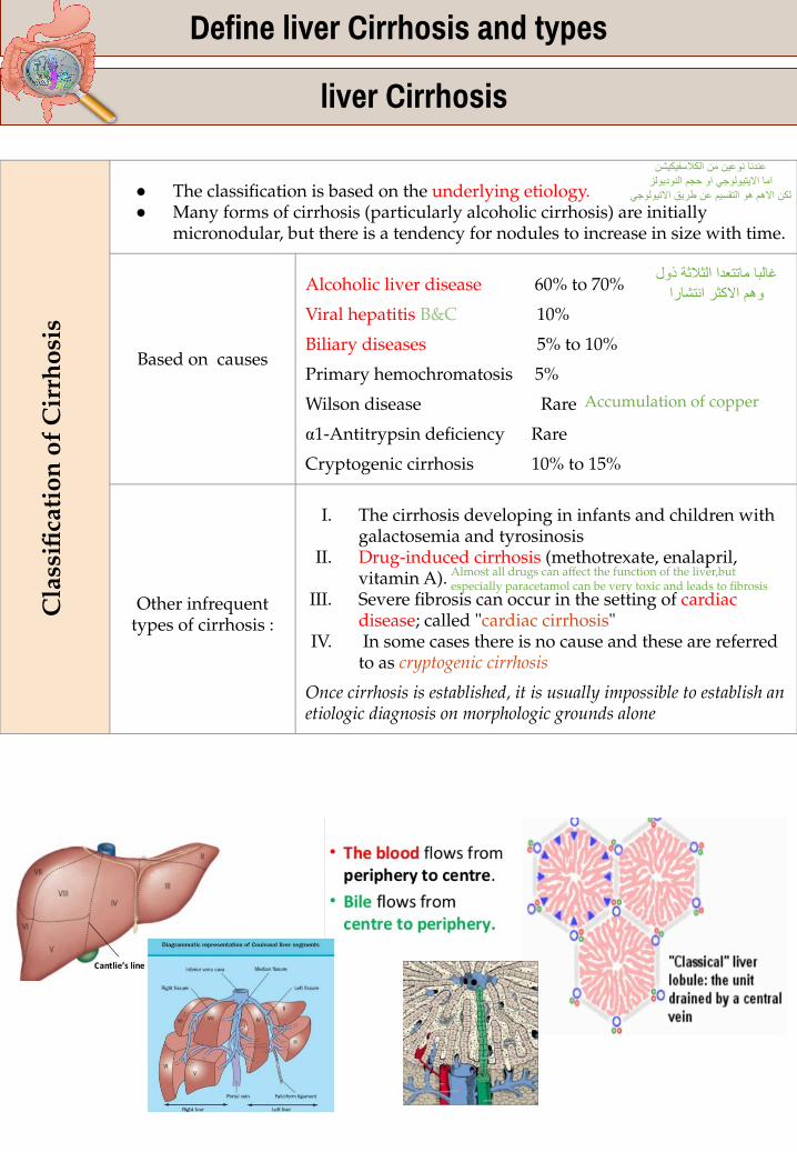
Define liver Cirrhosis and types
● The classification is based on the underlying etiology.● Many forms of cirrhosis (particularly alcoholic cirrhosis) are initially
micronodular, but there is a tendency for nodules to increase in size with time.
Based on causes
Alcoholic liver disease 60% to 70%
Viral hepatitis B&C 10%
Biliary diseases 5% to 10%
Primary hemochromatosis 5%
Wilson disease Rare
α1-Antitrypsin deficiency Rare
Cryptogenic cirrhosis 10% to 15%
Other infrequent types of cirrhosis :
I. The cirrhosis developing in infants and children with galactosemia and tyrosinosis
II. Drug-induced cirrhosis (methotrexate, enalapril, vitamin A).
III. Severe fibrosis can occur in the setting of cardiac disease; called "cardiac cirrhosis"
IV. In some cases there is no cause and these are referred to as cryptogenic cirrhosis
Once cirrhosis is established, it is usually impossible to establish an etiologic diagnosis on morphologic grounds alone
Cla
ssifi
cati
on o
f C
irrh
osis
عندنا نوعین من الكالسفیكیشناما االیتیولوجي او حجم النودیولز
لكن االھم ھو التقسیم عن طریق االتیولوجي
غالبا ماتتعدا الثالثة ذولوھم االكثر انتشارا
Accumulation of copper
Almost all drugs can affect the function of the liver,but especially paracetamol can be very toxic and leads to fibrosis

Define liver Cirrhosis and types
● The pathogenic processes in cirrhosis are progressive fibrosis and reorganization of the vascular microarchitecture of the liver
● In the normal liver, interstitial collagens (types I and III) are concentrated in portal tracts and around central veins. The type IV collagen(reticulin) is in the space of Disse.
● In cirrhosis, types I and III collagen are deposited in the lobule, creating delicate or broad septal tracts
● There is loss of fenestrations in the sinusoidal endothelial cells (capillarization of sinusoids, that is the sinusoidal space comes toresemble a capillary rather than a channel for exchange of solutes between hepatocytes and plasma)
● The major source of excess collagen in cirrhosis is the perisinusoidal stellate cells ( Ito cells), which lie in the space of Disse.
● Although Ito cells normally function is a vitamin A fat- storing cells, during the development of cirrhosis they become activated and transform into myofibroblast-like cells. Lay down collagen due to injury of hepatocytes
Collagen synthesis is stimulated by:1) Chronic inflammation, with production of inflammatory cytokines.2) Cytokine production by activated endogenous cells (Kupffer cells, endothelial cells, hepatocytes, and bile duct epithelial cells).3) Disruption of the normal extracellular matrix.4) Direct stimulation of stellate cells by toxin
Path
ogen
esis
of C
irrh
osis
Robbins: Three processes are central to the pathogenesis of cirrhosis: death of hepatocytes, extracellular matrix deposition, and vascular reorganization.
Due to the fibrosis

Gross features
Macronodular Micronodular
The nodules seen here are larger than 3 mm maybe reach to 2-3 cm and, hence, this is an example of "macronodular" cirrhosis. In radiology more than 2 cm can be cancer so biopsy is required.
Micronodular cirrhosis : The regenerative nodules are quite small, averaging less than 3 mm in size. The most common cause for this is chronic alcoholism.
Microscopically
Regenerative nodules of hepatocytes are surrounded by fibrous connective tissue contains large amount of collagen that bridges between portal tracts. Within this collagenous tissue are scattered lymphocytes as well as a proliferation of bile ducts:
Describe the pathological findings in cirrhotic livers.
Clinical Features :All forms of cirrhosis may be clinically silent. When symptomatic they lead to nonspecific clinical manifestations: anorexia, weight loss, weakness, osteoporosis and in advanced disease,frank debilitation,jaundice*, Incipient or overt hepatic failure may develop. At first the liver function is compensated (capacity of liver is huge, ممكن أشیل نص الكبد وما یصیر عندي تأثیر) so there’s no symptoms until it reach uncompensated stage. (*) it will appear because the liver can’t deal with bilirubin which produced from broken RBC’s.
The ultimate mechanism of most cirrhotic deaths is:1. Progressive liver failure so become uncompensated liver disease 2. A complication related to portal hypertension maybe the patient will have
bleeding from esophageal varices 3. The development of hepatocellular carcinoma
Morphology :
Cirrhosis is a chronic progressive mechanism
He will have only a risk factor

Chronic Hepatitis Morphology
•Chronic Viral Hepatitis (hepatitis B and C virus)
•Autoimmune hepatitis
•Biliary Cirrhosis
•Alcoholic liver disease
Causes of Liver
Cirrhosis
Some changes are shared with acute hepatitis. ● Hepatocyte injury, necrosis, and regeneration .● Sinusoidal cell reactive changes.● Portal tract Inflammation: (by lymphocytes)
○ Confined to portal tracts or ○ Spillover into adjacent parenchyma, with necrosis of
hepatocytes ("interface hepatitis") or ○ Bridging inflammation and necrosis
● Fibrosis: Continued loss of hepatocytes results in fibrous septa formation which ultimately leads to cirrhosis
HBV: "ground-glass" hepatocytes, "sanded" nuclei Not very important because we can diagnose the patient basically by blood test for HB BHCV: bile duct damage, lymphoid aggregate formation
● Cirrhosis: The end-stage outcome
Ground Glass
HBsAg +vePiecemeal necrosis in
chronic hepatitisSpillover into adjacent
parenchyma, with necrosis of hepatocytes
"interface hepatitis"Lymphoid aggregate
Viral hepatitis C which is at a high stage with extensive fibrosis and progression to macronodular cirrhosis, as evidenced by the
large regenerative nodule at the center right.
Because of lymphocytes it’s chronic
inflammation
Dilated vein
Ground glass appearance ممسوح كأنھ قزاز(Inclusion of viral particles in the cytoplasm)
Recognize the causes and the pathogenic . Mechanisms leading to cirrhosis
In our country this arrangement of causes is more common.
← Is a feature of active chronic hepatitis.
Secondary biliary cirrhosisPrimary biliary cirrhosisPrimary sclerosing cholangitis
Stage 1:Increase fibrous tissue in portal tract.stage 2:Bridging fibrosis and later on these bridging fibrosis will complete from area to other area until the liver will be cirrhotic.
Notes the bile ducts and lymphocytes passes through the other side parenchymaThis fibrosis and lymphocytes will reach the bile duct.

● is a chronic hepatitis with histologic features like that of chronic viral hepatitis. There’re portal inflammation, interface hepatitis and fibrosis.
● This disease may run an indolent or severe course.From the name itself the patient has autoantibodies against the liver.
Two primary types of autoimmune hepatitis:
Type 1 autoimmune hepatitis is most often seen in middle-age women and is characteristically associated with anti-nuclear and anti–smooth muscle antibodies.
Type 2 autoimmune hepatitis is most often seen in children or teenagers and is associated with anti–liver kidney microsomal autoantibodies. very rare.
● Absence of viral serologic markers● Elevated serum IgG and γ-globulin levels (>1.5 times
normal)● High serum titres of autoantibodies in 80% of cases, including
antinuclear (ANA), antismooth muscle (SMA) and anti-mitochondrial antibodies.
● � anti–liver kidney microsome-1 antibodies and anti– liver cytosol-1 antibodies.
● In untreated severe disease, as many as 40% of patients die within 6 months of diagnosis, and cirrhosis develops in at least 40% of survivors.
● Treatment include immunosuppressive therapy, and liver transplantation.
● Associated with other autoimmune diseases eg. Rheumatoid arthritis, Sjogren’s syndrome etc. Sjogren’s syndrome: antibodies directed against salivary and lacrimal glands, so there will be dryness of the mouth and eyes.
Recognize the causes and the pathogenic . Mechanisms leading to cirrhosis
Aut
oim
mun
e H
epat
itis
Robbins: In viral hepatitis, fibrosis typically follows years or decades of slowly accumulating parenchymal injury, whereas in autoimmune hepatitis, there appears to be an early phase of severe cell injury and inflammation followed by rapid scarring

Recognize the causes and the pathogenic . Mechanisms leading to cirrhosis
Intrahepatic Biliary Tract Disease
Secondary biliary cirrhosis
Primary biliary cirrhosis
Primary sclerosing cholangitis
Secondary biliary cirrhosis common
-Prolonged obstruction of the extrahepatic biliary tree results in profound alteration of the liver itself. Biliary cirrhosis = obstruction of bile duct and as a result there will be secondary cirrhosis in the liver.
-The most common cause of obstruction in adults is extrahepatic cholelithiasis (gallstones), followed by malignancies of the biliary tree or head of the pancreas and strictures resulting from previous surgical procedures
-Obstructive conditions in children include biliary atresia (congenital), cystic fibrosis, choledochal cysts (a cystic anomaly of the extrahepatic biliary tree)
Morphology -Secondary inflammation resulting from biliary obstruction initiates periportal fibrosis, which eventually leads to hepatic scarring and nodule formation, generating secondary biliary cirrhosis.
-Subtotal obstruction may promote secondary bacterial infection of the biliary tree (ascending cholangitis), which aggravates the inflammatory injury. Enteric organisms such as coliforms and enterococci are common cause.
Etiology Extrahepatic bile duct obstruction: biliary atresia, gallstones, stricture, carcinoma of pancreatic head
Sex predilection None.
Symptoms and signs Pruritus because of accumulation of bile salts and bilirubin metabolites, jaundice, malaise, dark urine, light stools, hepatosplenomegaly
Laboratory findingsImportant
Conjugated hyperbilirubinemia, increased serum alkaline phosphatase, bile acids, cholesterolNo increase in serum AMA or IgM
Prominent bile stasis in bile ducts, bile ductular proliferation with surrounding neutrophils, portal tract edema
Important pathologic findings before cirrhosis develops

Recognize the causes and the pathogenic . Mechanisms leading to cirrhosis
Primary Biliary Cholangitis
-Primary biliary cirrhosis is a chronic, progressive, and often fatal cholestatic liver disease, characterized by the destruction of intrahepatic bile ducts, portal inflammation and scarring, and the eventual development of cirrhosis and liver failure. There will be lymphocytes and granuloma which are destroying the bile duct epithelium why? because there are autoantibodies against the mitochondria (antimitochondrial autoantibodies)
-The primary feature of this disease is a nonsuppurative, granulomatous inflammatory destruction of medium-sized intrahepatic bile ducts.
-Cirrhosis develops only after many years and not in all cases. Previously called Primary biliary cirrhosis
-middle-aged women, female: male predominance (6:1).
autoimmune etiology, 90% of patients have circulating "antimitochrondrial antibodies" In serology of the patient we looking for it for diagnosis of primary biliary cholangitis.
Clinical features
-Signs of obstruction: pruritus, jaundice, hepatomegaly. Xanthomas and xanthelasmas rise owing to cholesterol retention(accumulation of cholesterol leading to small yellow dots or nodules in the skin).
-Over a period of time patients develop portal hypertension and hepatic encephalopathy.
-Serum alkaline phosphatase and cholesterol are elevated; hyperbilirubinemia is a late development.
-Association with other autoimmune diseases (e.g., Sjögren syndrome and Hashimoto's thyroiditis )
-During the precirrhotic stage, portal tracts and bile ducts are infiltrated by lymphocytes and may exhibit noncaseating granulomatous inflammation. There is bile duct destruction.
-With time, there is bile ductular proliferation, inflammation, and necrosis of the adjacent periportal hepatic parenchyma.
-Over years to decades, relentless portal tract scarring and bridging fibrosis lead to cirrhosis.
-In most cases, the end-stage picture is indistinguishable from secondary biliary cirrhosis or the cirrhosis that follows chronic hepatitis from other causes
Pathogenesis
Morphology

Recognize the causes and the pathogenic . Mechanisms leading to cirrhosis
Primary sclerosing cholangitis
Characters ● characterized by inflammation and obliterative fibrosis of intrahepatic and extrahepatic bile ducts,with dilation of preserved segments. There’s sclerosing around bile duct.
● Characteristic "beading" of a barium column in radiographs of theintrahepatic and extrahepatic biliary tree is attributable to the irregular strictures and dilations of affected bile ducts.
● It is commonly seen in association with inflammatory bowel disease , particularly chronic ulcerative colitis less in crohn’s
● Males predominate 2:1
Pathogenesis unknown
Morphology ● � Primary sclerosing cholangitis is a fibrosing cholangitis of bile ducts, with a lymphocytic infiltrate, progressive atrophy of the bile duct epithelium, and obliteration of the lumen.
● The concentric periductal fibrosis around affected ducts("onion-skin fibrosis") is followed by their disappearance, leaving behind a solid, cordlike fibrous scar.
● As the disease progresses, the liver becomes cirrhotic like that seen with primary and secondary biliary cirrhosis
Alcoholic liver disease
features 1. Steatosis (fatty change) 2. Hepatitis (steatohepatitis) 3. Fibrosis
causes of death ● � Hepatic failure● � Massive gastrointestinal hemorrhage● � Intercurrent infection (to which affected individuals
arepredisposed)● � Hepatorenal syndrome● � Hepatocellular carcinoma (3%–6% of cases)
یصیر التأثیر بالتدریج وعلى مدة
Robbins: alcoholic cirrhosis has the same morphologic and clinical features as cirrhosis caused by viral hepatitis.
Not specific Fibrosis
FAT
Fibrosis
You can see fibrosis around the bile duct and thats why we call it sclerosing
when he stops drinking liver will return normal (reversible)
Irreversible and happens after a continuous exposure
We stain with trichrome stain to see collagenThe blue color resemble fibrosis

Complications of liver Cirrhosis
Portal Hypertension
● Resistance to blood flowprehepatic, intrahepatic, and posthepatic.
● The dominant intrahepatic cause is cirrhosis(This is accounting for most cases of portal hypertension)
● Portosystemic shunts develop when blood flow is reversed from the portal to systemic circulation.
● due to intrasinusoidal hypertension from regenerative nodule compression
1. Hepatic failurea. Coagulopathyb. Hypoalbuminemiac. Hepatic encephalopathy
2. Portal hypertension:a. Variceal bleedingb. Splenomegalyc. Hemorrhoidsd. Periumbilical venous collaterals (caput
medusa)
3. Ascites4. Hepatorenal syndrome5. Hyperestrinism in males6. Hepatocellular carcinoma7. Spontaneous bacterial
peritonitis8. Jaundice and cholestasis
Esophageal Varices
● Instead of returning directly to the heart, venous blood from the GI tract is delivered to the liver via the portal vein before reaching the inferior vena cava.
● This circulatory pattern is responsible for the first-pass effect in which drugs and other materials absorbed in the intestines are processed by the liver before entering the systemic circulation..
● Diseases that impede this flow cause portal hypertension and can lead to the development of esophageal varices, an important cause of esophageal bleeding.
Portal hypertension
Portosystemic Shunt
● Cardioesophageal junction (esophagogastric varices)
● Abdominal wall collaterals (caput medusae)around the umbilicus
● Rectum (hemorrhoids)
بالعربي الضغط على البورتال ڤین یزید فالدم مایقدر یمر فیبدا یدور طرق اسھل علیھ ویسوي كولالترال في اماكن كثیرة فيالجسم ھذي الكوالترالز تسوي توسعة للڤینز وتسوي نزیف

Complications of liver Cirrhosis
•Portal hypertension results in the development of collateral channels at sites where the portal and caval systems communicate. Although these collateral veins allow some drainage to occur, they lead to development of a congested subepithelial and submucosal venous plexus within the distal esophagus. (varices)
•90% of cirrhotic patients develop varices most commonly in association with alcoholic liver disease
•Hepatic schistosomiasis it can cause chronic liver disease
•Varices can be detected by venogram: tortuous dilated veins lying primarily within the submucosa of the distal esophagus and proximal stomach. Venous channels directly beneath the esophageal epithelium may also become massively dilated.
•Varices may not be grossly obvious in surgical or postmortem specimens, because they collapse in the absence of blood flow .
•Variceal rupture results in hemorrhage into the lumen or esophageal wall, in which case the overlying mucosa appears ulcerated and necrotic. If rupture has occurred in the past, venous thrombosis, inflammation, and evidence of prior therapy may also be present.
•Asymptomatic if no rupture or rupture leads to massive hematemesis and it is a surgical emergency
•Inflammatory erosion of thinned overlying mucosa
•Increased tension in progressively dilated veins
•Increased vascular hydrostatic pressure associated with vomiting are likely to contribute to medical emergency that is treated by any of several methods:1.Sclerotherapy inject a substance that
will cause coagulation in the blood vessel
2.Endoscopic balloon tamponade the
balloon causes pressure in dilated vessels
Preventing bleeding
3.Endoscopic rubber band ligation
Mor
phol
ogy:
Path
ogen
esis
Cli
nica
l fea
ture
s:
Very dilated vien
ESOPHAGEAL VARICES: دوالي المريء

ESOPHAGEAL VARICES: دوالي المريء
Complications of liver Cirrhosis
•Long-standing congestion may cause congestive splenomegaly (spleen weight may reach up to 1000 gm) due to increase portal hypertension there will be accumulated blood in the spleen
•The massive splenomegaly may induce hematologic abnormalities attributable to hypersplenism, such as thrombocytopenia or pancytopenia A condition in which a person’s body has too few red blood cells, white blood cells, and platelets.
•is the accumulation of excess fluid in the peritoneal cavity:
•85% of cases are caused by cirrhosis.
•Serous: less than 3 gm/dL of protein (largely albumin)
•Pathogenesis:
Increase in portal vein hydrostatic pressure
Decreases oncotic pressure due to decrease number of protein.
Liver is unable to metabolize aldosterone aldosterone causes water retention As a result of ascitis there will be Spontaneous bacterial peritonitis
Sple
nom
egal
yA
scit
es
•Half of patients die from the first bleeding episode either as a direct consequence of hemorrhage or following hepatic coma triggered by hypovolemic shock.
•Additional 50% within 1 year. 100% the second time
•Each episode has a similar rate of mortality.
•Over half of deaths among individuals with advanced cirrhosis result from variceal rupture.
Epid
emio
logy
Robbins: It usually becomes clinically detectable when at least 500 mL have accumulated, but many liters may collect, causing massive abdominal distention
Other Complications of liver Cirrhosis

Complications of liver Cirrhosis
Jaundice and Icterus
A yellowish or greenish pigmentation of the skin and sclera of the eyes respectively due to high bilirubin levels.
CholestasisCharacterized by systemic retention of not only bilirubin but also other solutes eliminated in bile.(particularly bile salts and cholesterol).
Causes of Jaundice
Prehepatic causes of jaundice:
Bilirubin overproduction
Due to hemolysis and hematoma resorption, lead to elevated levels of unconjugated (indirect) bilirubin.
Intrahepatic disorders
Can lead to unconjugated or conjugated hyperbilirubinemia. The conjugated (direct) bilirubin level is often elevated by alcohol, infectious hepatitis, drug reactions, and autoimmune disorders.
Posthepatic disorders
(Obstruction of the flow of bile)
Can cause conjugated hyperbilirubinemia. Gallstone formation is the most common posthepatic process that causes jaundice; however, the differential diagnosis also includes serious conditions such as biliary tract infection, pancreatitis, and malignancies.
Bilirubin metabolism and elimination
Coagulopathy
The liver is the source of a number of coagulation factors that decline in the face of liver failure, leading to easy bruising and bleeding. Due to failure to production of the factor proteins
Hepatitis and alcoholism= Increase in unconjugated bilirubin Obstruction = increase in the conjugated bilirubin
Biopsy shows more than 2cm mass
Complications of liver Cirrhosis
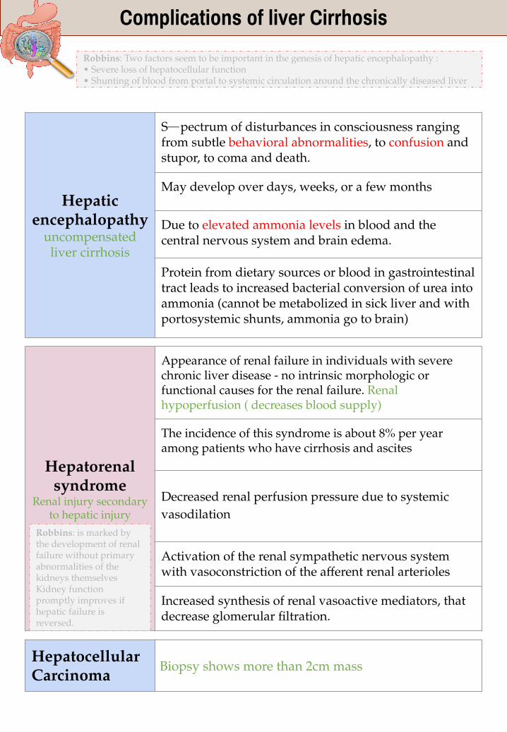
Hepatic encephalopathy
uncompensated liver cirrhosis
S�pectrum of disturbances in consciousness ranging from subtle behavioral abnormalities, to confusion and stupor, to coma and death.
May develop over days, weeks, or a few months
Due to elevated ammonia levels in blood and the central nervous system and brain edema.
Protein from dietary sources or blood in gastrointestinal tract leads to increased bacterial conversion of urea into ammonia (cannot be metabolized in sick liver and with portosystemic shunts, ammonia go to brain)
Hepatorenal syndrome
Renal injury secondary to hepatic injury
Appearance of renal failure in individuals with severe chronic liver disease - no intrinsic morphologic or functional causes for the renal failure. Renal hypoperfusion ( decreases blood supply)
The incidence of this syndrome is about 8% per year among patients who have cirrhosis and ascites
Decreased renal perfusion pressure due to systemic vasodilation
Activation of the renal sympathetic nervous system with vasoconstriction of the afferent renal arterioles
Increased synthesis of renal vasoactive mediators, that decrease glomerular filtration.
Hepatocellular Carcinoma
Robbins: Two factors seem to be important in the genesis of hepatic encephalopathy : • Severe loss of hepatocellular function• Shunting of blood from portal to systemic circulation around the chronically diseased liver
Robbins: is marked by the development of renal failure without primary abnormalities of the kidneys themselves Kidney function promptly improves if hepatic failure is reversed.

Summary
Autoimmune hepatitis Secondary Biliary Cirrhosis
Etiology Unclear. Triggers for the immune reaction may include viral infections or drug or toxin exposures
Extrahepatic bile duct obstruction: biliary atresia, gallstones, stricture, carcinoma of pancreatic head.
Predilection Female predominance, particularly in young and perimenopausal women
None.
Symptoms and Signs
An acute clinical illness is a common presentation (40%). Sometimes the disease is fulminant, progressing to hepatic encephalopathy within 8 weeks of onset.
Pruritus, jaundice, malaise, dark urine, light stools, hepatosplenomegaly.
Laboratory Findings
-Elevated serum IgG and γ-globulin level -High serum titers of autoantibodies in 80% of cases, including antinuclear (ANA), antismooth muscle (SMA), anti-mitochondrial antibodies.
Conjugated hyperbilirubinemia, increased serum alkaline phosphatase, bile acids, cholesterol No increase in serum AMA or IgM
Pathologic findings before cirrhosis develops
-Necrosis and inflammation, indicated by extensive interface hepatitis.-Plasma cell predominance
Prominent bile stasis in bile ducts, bile ductular proliferation with surrounding neutrophils, portal tract edema.
Primary Biliary Cholangitis Primary sclerosing cholangitis
Etiology Possibly autoimmune Unknown, possibly autoimmune; 50-70% associated with inflammatory bowel disease
sex Pred. Female to male: 6:1 Female to male: 1:2
Symptoms and Signs
Same as secondary biliary cirrhosis (Pruritus, jaundice, malaise, dark urine, light stools, hepatosplenomegaly)
Same as secondary biliary cirrhosis (Pruritus, jaundice, malaise, dark urine, light stools, hepatosplenomegaly) insidious onset
Laboratory Findings
Same as secondary biliary cirrhosis (Conjugated hyperbilirubinemia, increased serum alkaline phosphatase, bile acids, cholesterol) plus elevated serum autoantibodies (especially antimitochondrial antibody-AMA)
Same as secondary biliary cirrhosis (Conjugated hyperbilirubinemia, increased serum alkaline phosphatase, bile acids, cholesterol) plus elevated serum IgM, hypergammaglobulinemia
Pathologic findings before cirrhosis develops
Dense lymphocytic infiltrate in portal tracts with granulomatous destruction of bile ducts
Periductal portal tract fibrosis, segmental stenosis of extrahepatic and intrahepatic bile ducts

Summary
Complications of Liver Cirrhosis
Portal Hypertension
- �Resistance to blood flowprehepatic, intrahepatic (Cirrhosis), and posthepatic.
- Portosystemic shunt develop (when blood flow is reversed from portal to systemic circulation).
- May result in: esophageal varices or hemorrhoids or caput medusae.
Esophageal Varices
- Portal hypertension results in the development of collateral channels. Although these collateral veins allow some drainage to occur, they lead to development of varices.
- If ruptured → Massive hematemesis → can be fatal.- Develop mostly in patients with cirrhosis associated with alcoholic
liver disease.- �Can be seen in hepatic schistosomiasis.
Splenomegaly- Long-standing congestion may cause congestive splenomegaly.- May induce hematologic abnormalities attributable to
hypersplenism, such as thrombocytopenia or pancytopenia.
Ascites The accumulation of excess fluid in the peritoneal cavity
�Spontaneous bacterial peritonitis
Jaundice and Icterus
- Yellowish or greenish pigmentation of the skin and sclera of the eyes respectively due to high bilirubin levels.
- Causes: Prehepatic (Bilirubin overproduction), Intrahepatic, Posthepatic (Obstruction of the flow of bile).
Cholestasis �Systemic retention of not only bilirubin but also other solutes eliminated in bile.
Coagulopathy �The liver is the source of a number of coagulation factors that decline in the face of liver failure, leading to easy bruising and bleeding.
Hepatic Encephalopathy
Disturbances in consciousness from subtle behavioral abnormalities, to confusion and stupor, to coma and death, due to elevated ammonia levels.
Hepatorenal Syndrome
- Renal failure in individuals with severe chronic liver disease.- Decreased renal perfusion pressure and glomerular filtration.
Hepatocellular Carcinoma

Pathoma
CIRRHOSISA. End-stage liver damage characterized by disruption of the normal hepaticparenchyma by bands of fibrosis and regenerative nodules of hepatocytesB. Fibrosis is mediated by TGF-~ from stellate cells which lie beneath the endothelialcells that line the sinusoids.C. Clinical featuresl. Portal hypertension leads toi. Ascites (fluid in the peritoneal cavity ) ii. Congestive splenomegaly/hypersplenism iii. Portosystemic shunts (esophageal varices, hemorrhoids, and caput medusae)iv. Hepatorenal syndrome (rapidly developing renal failure secondary to cirrhosis)2. Decreased detoxification results ini. Mental status changes, asterixis, and eventual coma (due to increase serum ammonia); metabolic, hence reversibleii. Gynecomastia, spider angiomata, and palmar erythema due to hyperestrinismii. jaundice3. Decreased protein synthesis leads toi. Hypoalbuminemia with edemaii. Coagulopathy due to decreased synthesis of clotting factors; degree of deficiency) is followed by PT.
IV. ALCOHOL-RELATED LIVER DISEASEA. Damage to hepatic parenchyma due to consumption of alcohol1. Most common cause of liver disease in the West B. Fatty liver is the accumulation of fat in hepatocytesI. Results in a heavy, greasy liver; resolves with abstinence C. Alcoholic hepatitis results from chemical injury to hepatocytes; generally seen with binge drinkingl. Acetaldehyde (metabolite of alcohol) mediates damage.2. Characterized by swelling of hepatocytes with formation of Mallory bodies(damaged cytokeratin filaments,), necrosis, and acute inflammation 3. Presents with painful hepatomegaly and elevated liver enzymes (AST > ALT); may result in deathD. Cirrhosis is a complication of long-term, chronic alcohol-induced liver damage;noccurs in 10-20% of alcoholics
V. NONALCOHOLIC FATTY LIVER DISEASEA. Fatty change, hepatitis, and/or cirrhosis that develop without exposure to alcohol (or other known insult) B. Associated with obesity C. Diagnosis of exclusion; ALT > AST

Pathoma
.IX. PRIMARY SCLEROSING CHOLANGITISA. Inflammation and fibrosis of intrahepatic and extrahepatic bile ducts Hemochromatosis. A, Iron deposition in hepatocytes. B, Prussian blue stain. c, Lipofuscin in hepatocytes for comparison.l. Periductal fibrosis with an 'onion-skin' appearance 2. Uninvolved regions are dilated resulting in a "beaded" appearance on contrast imaging.B. Etiology is unknown, but associated with ulcerative colitis; p-ANCA is often positive. C. Presents with obstructive jaundice; cirrhosis is a late complication. D. Increased risk for cholangiocarcinoma
VII. WILSON DISEASEA. Autosomal recessive defect (ATP7B gene) in ATP-mediated hepatocyte copper transport I. Results in lack of copper transport into bile and lack of copper incorporation into ceruloplasminB. Copper builds up in hepatocytes, leaks into serum, and deposits in tissues.l. Copper-mediated production of hydroxyl free radicals leads to tissue damage. C. Presents in childhood with1. Cirrhosis2. neurologic manifestations (behavioral changes, dementia, chorea, and Parkinsonian symptoms due to deposition of copper in basal ganglia) 3. Kayser-Fleischer rings in the cornea D. Labs show t urinary copper,-!. serum ceruloplasmin, and t copper on liver biopsy.E. Increased risk of hepatocellular carcinoma F. Treatment is D-penicillamine (chelates copper).
VIII. PRIMARY BILIARY CIRRHOSISA. Autoimmune granulomatous destruction of intrahepatic bile ducts1. Classically arises in women (average age is 40 years) 2. Associated with other autoimmune diseasesB. Etiology is unknown; antimitochondrial antibody is present.C. Presents with features of obstructive jaundiceD. Cirrhosis is a late complication.

ESOPHAGEAL VARICESA. Dilated submucosal veins in the lower esophagusB. Arise secondary to portal hypertension1. Distal esophageal vein normally drains into the portal vein via the left gastric vein. 2. In portal hypertension, the left gastric vein backs up into the esophageal vein, resulting in dilation (varices).C. Asymptomatic, but risk of rupture exists1. Presents with painless hematemesis2. Most common cause of death in cirrhosis
Pathoma
JAUNDICE:A. Yellow discoloration of the skin earliest sign is scleral icterus (yellow discoloration of the sclera).B. Due to i serum bilirubin, usually > 2.5 mg/dL C. Arises with disturbances in bilirubin metabolism D. Normal bilirubin metabolism1. RBCs are consumed by macrophages of the reticuloendothelial system.2. Protoporphyrin (from heme) is converted to unconjugated bilirubin (UCB).3. Albumin carries UCB to the liver.4. Uridine glucuronyl transferase (UGT) in hepatocytes conjugates bilirubin.5. Conjugated bilirubin (CB) is transferred to bile canaliculi to form bile, which is stored in the gallbladder. 6. Bile is released into the small bowel to aid in digestion 7. Intestinal flora convert CB to urobilinogen, which is oxidized to stercobilin(makes stool brown) and urobilin (partially reabsorbed into blood and filtered by kidney, making urine yellow).
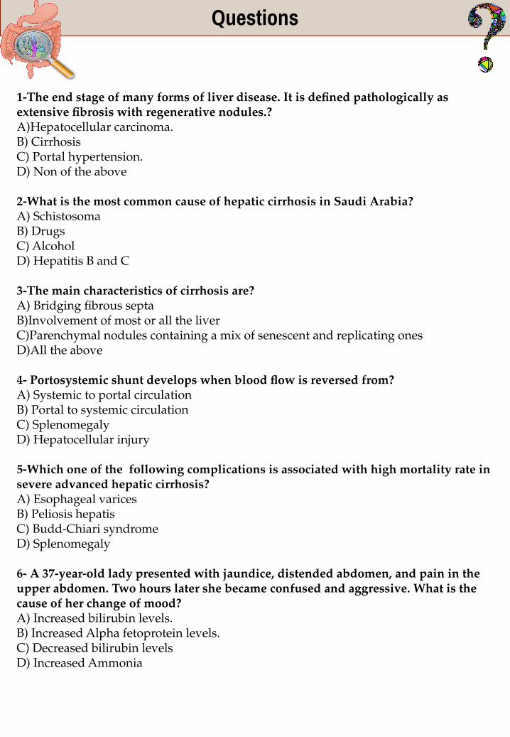
1-The end stage of many forms of liver disease. It is defined pathologically as extensive fibrosis with regenerative nodules.?A)Hepatocellular carcinoma.B) CirrhosisC) Portal hypertension.D) Non of the above
2-What is the most common cause of hepatic cirrhosis in Saudi Arabia?A) SchistosomaB) DrugsC) AlcoholD) Hepatitis B and C
3-The main characteristics of cirrhosis are?A) Bridging fibrous septaB)Involvement of most or all the liverC)Parenchymal nodules containing a mix of senescent and replicating onesD)All the above
4- Portosystemic shunt develops when blood flow is reversed from? A) Systemic to portal circulationB) Portal to systemic circulation C) SplenomegalyD) Hepatocellular injury
5-Which one of the following complications is associated with high mortality rate in severe advanced hepatic cirrhosis?A) Esophageal varicesB) Peliosis hepatisC) Budd-Chiari syndromeD) Splenomegaly
6- A 37-year-old lady presented with jaundice, distended abdomen, and pain in the upper abdomen. Two hours later she became confused and aggressive. What is the cause of her change of mood?A) Increased bilirubin levels.B) Increased Alpha fetoprotein levels.C) Decreased bilirubin levelsD) Increased Ammonia
Questions

Case1: A 62-year-old man is brought to the emergency room in a disoriented state. Physical examination reveals jaundice, splenomegaly, and ascites. The patient has a coarse flapping tremor of the hands, palmar erythema, and diffuse spider angiomata. The abdomen displays dilated paraumbilical veins. Serum levels of ALT, AST, alkaline phosphatase, and bilirubin are all mildly elevated. Soon after admission, the patient vomits a large amount of blood.
● what is the diagnosis ?Liver cirrhosis.
● what are the most common etiologies of this disorder?Alcohol abuse and viral hepatitis. Other causes include biliary disease, and iron overload.
● Other appropriate tests?Liver biopsy
● what are the possible complications of liver cirrhosis?1. Portal Hypertension :Ascites,Portosystemic venous shunts and Splenomegaly.2. Liver failure.3. Hepatocellular Carcinoma.
7- A 62-year-old man is brought to the emergency room in a disoriented state. Physical examination reveals signs of poor hygiene and an odor of alcohol, as well as jaundice, splenomegaly, and ascites. The patient has a coarse flapping tremor of the hands, palmar erythema, and diffuse spider angiomata. The abdomen displays dilated paraumbilical veins. Serum levels of ALT, AST, alkaline phosphatase, and bilirubin are all mildly elevated. Soon after admission, the patient vomits a large amount of blood. Which of the following is the most likely underlying cause of hematemesis in this patient?
A)Hepatic steatosis.B) Acute alcoholic hepatitis.C) CirrhosisD) Acute gastritis.
Answers: 1-B2-D3-D4-B5-A6-D7-C
Questions

كل الشكر والتقدیر للجھود العظیمة.من قبل أعضاء فریق علم األمراض الكرام
قادة فریق علم األمراض
اعضاء فریق علم األمراض
شیرین العكیليفایز غیاث الدرسوني
مھا الَعمريمجد البراك
بتول الرحیميمنیرة المسعد
مشاعل القحطانيِرناد الفرم
غادة الحیدري دانة القاضيمھا بركھ
رزان الزھراني لین الحكیم عھد القرین
وجدان الشامريغرام جلیدانلیلى الصباغِریناد الغریبينورة القاضي
راكان محمد الغنیمسلطان ناصر الناصر
عادل عبدالعزیز السحیبانيحسن محمد العرینيتركي عید الشمريعبدالجبار الیماني
عبدهللا المعیذرمنصور العبرة
خالد العقیليتركي آل بنھار
عبدالعزیز الضرغامسعد الفوزانعادل ابراھیم
عبدااللھ الدوسريعبدهللا السرجاني
محمد المحیمیدعبدالرحمن آل الشیخ
عبدالرحمن الداوود