LECTURE XII: Vision Accommodation - KSUMSC
14
LECTURE XII: Vision Accommodation MALE SLIDES IMPORTANT FEMALE SLIDES LECTURER’S NOTES EXTRA EDITING FILE GSLIDES
-
Upload
khangminh22 -
Category
Documents
-
view
0 -
download
0
Transcript of LECTURE XII: Vision Accommodation - KSUMSC
LECTURE XII: Vision Accommodation
MALE SLIDESIMPORTANT FEMALE SLIDES LECTURER’S NOTESEXTRAEDITING FILE GSLIDES

Cones are served by Type X retinal ganglionic cells, which have limited dendritic distribution across the retina, therefore each fiber supplies a limited amount of cones. Note that cones tend to be more slender in the fovea and thicker in the peripheral parts of the retina, this helps the fovea in visual acuity, retinal X ganglionic cells allow accurate point-to-point spatial information to be carried from the cones into the primary visual cortex, allowing details of objects to be perceived more clearly.
Vision Accommodation1 Lecture Twelve
OBJECTIVES- At the end of this lecture ,the student should be able to Describe visual acuity &
depth perception- Contrast photopic and scotopic vision- To know visual pathway and field of vision- Describe the process of accommodation reflex and its pathway, contrasting the
refraction of light by the lens in near vision and in far vision- Identify and describe pupillary light reflex , its pathway and -relate these to
clinical situations as argyl Robertson pupil- Identify the lateral geniculate body and visual cortex functions
VISUAL ACUITYThe degree to which the details and contours of objects are perceived , it is usually defined in terms of the shortest distance by which two lines can be separated and still be seen as 2 lines
VISUAL THRESHOLD● It Is the minimal amount of light that elicit sensation of light● How to measure visual acuity? By Snellen chart ● Normal acuity = d/D = 6/6- Distance of Patient / D distance of normal person ● A person of 6/12 has less vision than normal vision
DUPLICITY THEORY OF VISIONTwo kinds of vision under different conditions
1. PHOTOPIC VISION (bright light vision)a. Served by conesb. High visual acuity = colors & details (BOX 12-1)c. Low sensitivity to light = needs high visual threshold to be stimulated
2. SCOTOPIC VISION (night vision, dimlight vision)a. Served by rodesb. Low visual acuity = no colors or details c. Great sensitivity to light = low visual threshold
Figure 12-1
BOX 12-1: GUYTON AND HALL
Vision Accommodation2
Footnotes
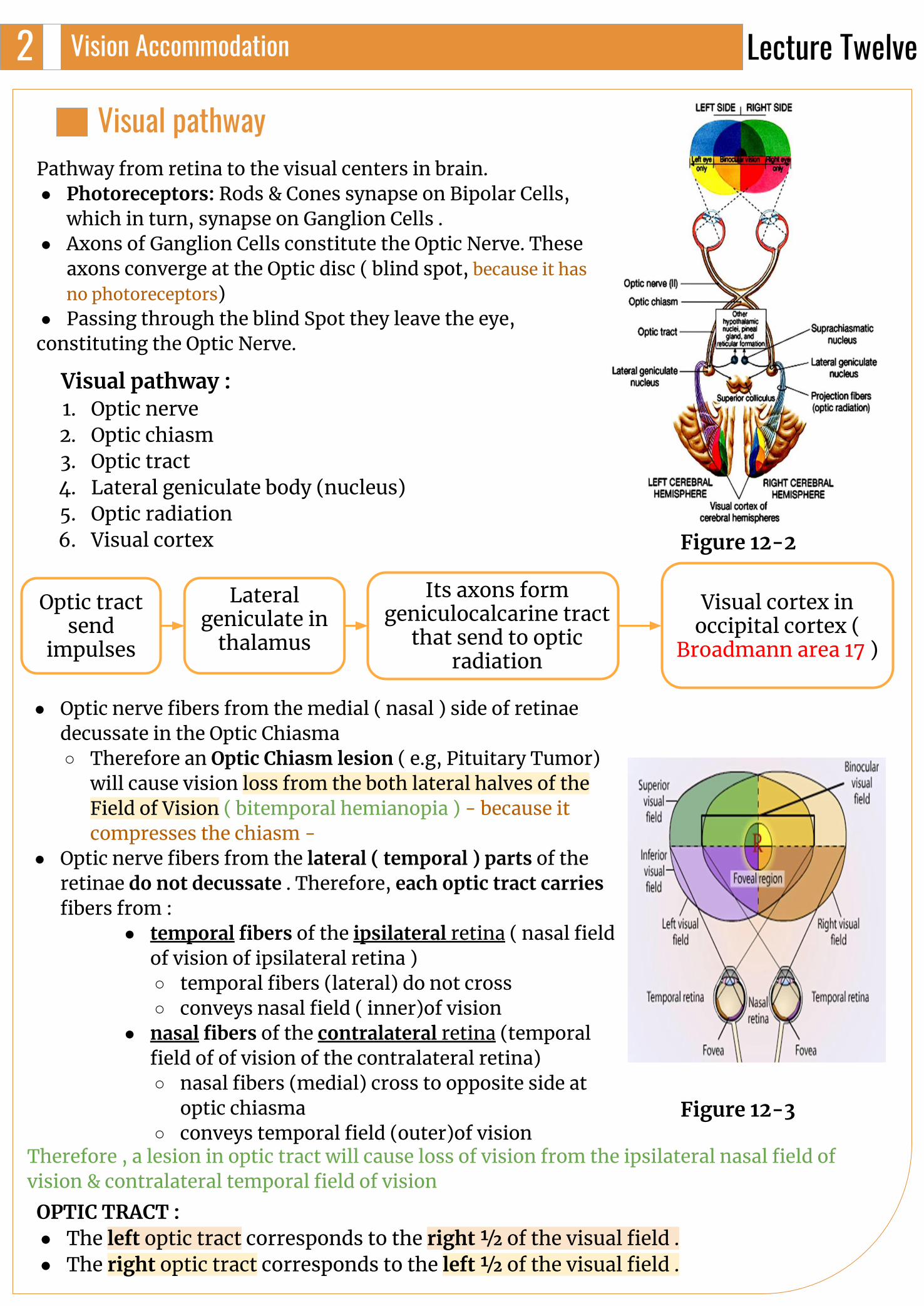
Visual pathwayPathway from retina to the visual centers in brain.● Photoreceptors: Rods & Cones synapse on Bipolar Cells,
which in turn, synapse on Ganglion Cells .● Axons of Ganglion Cells constitute the Optic Nerve. These
axons converge at the Optic disc ( blind spot, because it has no photoreceptors)
● Passing through the blind Spot they leave the eye,constituting the Optic Nerve.
Visual pathway : 1. Optic nerve2. Optic chiasm3. Optic tract4. Lateral geniculate body (nucleus)5. Optic radiation6. Visual cortex
Its axons form geniculocalcarine tract
that send to optic radiation
Visual cortex in occipital cortex (
Broadmann area 17 )
Optic tract send
impulses
Lateral geniculate in
thalamus
● Optic nerve fibers from the medial ( nasal ) side of retinae decussate in the Optic Chiasma ○ Therefore an Optic Chiasm lesion ( e.g, Pituitary Tumor)
will cause vision loss from the both lateral halves of the Field of Vision ( bitemporal hemianopia ) - because it compresses the chiasm -
● Optic nerve fibers from the lateral ( temporal ) parts of the retinae do not decussate . Therefore, each optic tract carries fibers from :
● temporal fibers of the ipsilateral retina ( nasal field of vision of ipsilateral retina ) ○ temporal fibers (lateral) do not cross○ conveys nasal field ( inner)of vision
● nasal fibers of the contralateral retina (temporal field of of vision of the contralateral retina)○ nasal fibers (medial) cross to opposite side at
optic chiasma○ conveys temporal field (outer)of vision
OPTIC TRACT :● The left optic tract corresponds to the right 1⁄2 of the visual field .● The right optic tract corresponds to the left 1⁄2 of the visual field .
Therefore , a lesion in optic tract will cause loss of vision from the ipsilateral nasal field of vision & contralateral temporal field of vision
Lecture Twelve
Figure 12-2
Figure 12-3
Vision Accommodation3
Footnotes
VISUAL PATHWAY
Accomodation (focusing)It Is an active process for modification of the refractive power of the eye to view a nearby object by increasing the curvature of lens
Accomodation (focusing)
Accomodation mechanismCiliary muscle
contraction Relaxation of the
suspensory ligamentLens more
convexIncrease diopteric power of the lens
Near object focused on the retina
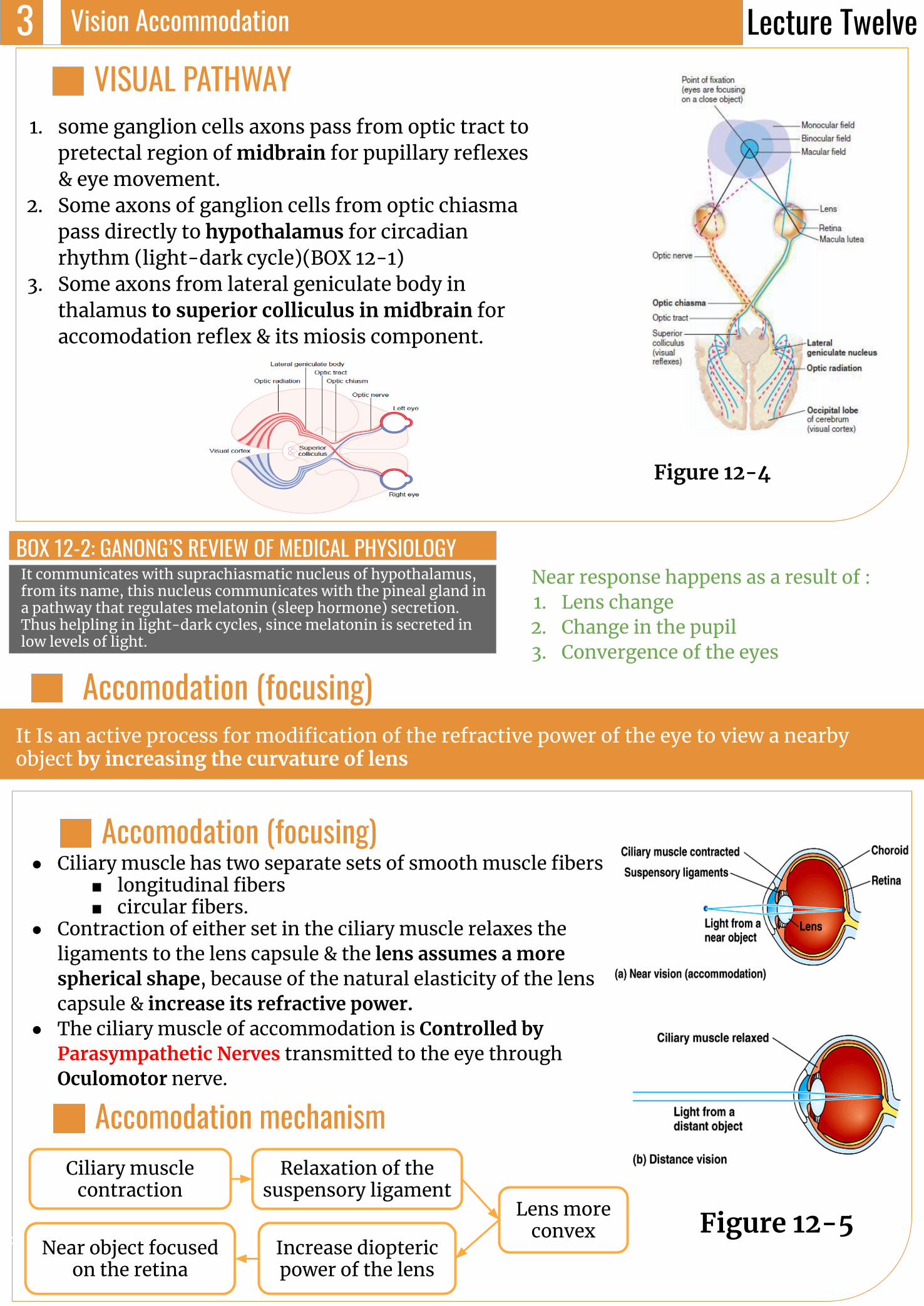
1. some ganglion cells axons pass from optic tract to pretectal region of midbrain for pupillary reflexes & eye movement.
2. Some axons of ganglion cells from optic chiasma pass directly to hypothalamus for circadian rhythm (light-dark cycle)(BOX 12-1)
3. Some axons from lateral geniculate body in thalamus to superior colliculus in midbrain for accomodation reflex & its miosis component.
● Ciliary muscle has two separate sets of smooth muscle fibers ■ longitudinal fibers ■ circular fibers.
● Contraction of either set in the ciliary muscle relaxes the ligaments to the lens capsule & the lens assumes a more spherical shape, because of the natural elasticity of the lens capsule & increase its refractive power.
● The ciliary muscle of accommodation is Controlled by Parasympathetic Nerves transmitted to the eye through Oculomotor nerve.
Near response happens as a result of : 1. Lens change2. Change in the pupil3. Convergence of the eyes
Lecture Twelve
Figure 12-4
Figure 12-5
It communicates with suprachiasmatic nucleus of hypothalamus, from its name, this nucleus communicates with the pineal gland in a pathway that regulates melatonin (sleep hormone) secretion. Thus helpling in light-dark cycles, since melatonin is secreted in low levels of light.
BOX 12-2: GANONG’S REVIEW OF MEDICAL PHYSIOLOGY
Vision Accommodation4
Footnotes
Continued
Accommodation Reflex
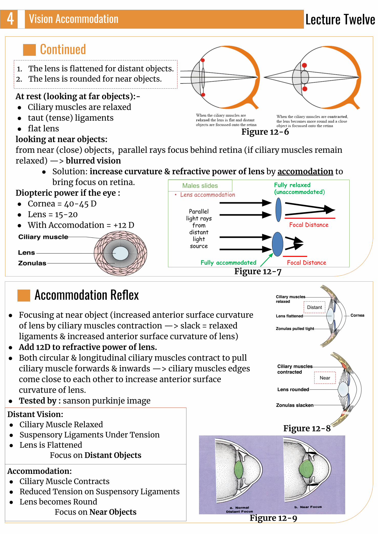
1. The lens is flattened for distant objects.2. The lens is rounded for near objects.
At rest (looking at far objects):-● Ciliary muscles are relaxed ● taut (tense) ligaments ● flat lenslooking at near objects:from near (close) objects, parallel rays focus behind retina (if ciliary muscles remain relaxed) —> blurred vision
● Solution: increase curvature & refractive power of lens by accomodation to bring focus on retina.
Diopteric power if the eye : ● Cornea = 40-45 D● Lens = 15-20 ● With Accomodation = +12 D
● Focusing at near object (increased anterior surface curvature of lens by ciliary muscles contraction —> slack = relaxed ligaments & increased anterior surface curvature of lens)
● Add 12D to refractive power of lens.● Both circular & longitudinal ciliary muscles contract to pull
ciliary muscle forwards & inwards —> ciliary muscles edges come close to each other to increase anterior surface curvature of lens.
● Tested by : sanson purkinje image
Distant Vision:● Ciliary Muscle Relaxed● Suspensory Ligaments Under Tension ● Lens is Flattened
Focus on Distant Objects
Accommodation:● Ciliary Muscle Contracts ● Reduced Tension on Suspensory Ligaments● Lens becomes Round
Focus on Near Objects
• Lens accommodation
Parallel light rays
from distant
light source
Focal Distance
Focal Distance
Fully relaxed (unaccommodated)
Fully accommodated
Males slides
Near
Distant
Lecture Twelve
Figure 12-6
Figure 12-7
Figure 12-8
Figure 12-9
Vision Accommodation5
Footnotes
Nearest point to eye at which object can brought into focus on retina by accommodation.- 10 years = 9 cm- At 60 years = 80-100 cm, due to hardness of lens & loss of accommodation.
LOOKING AT A CLOSE OBJECT (NEAR RESPONSE)A. Convergence of both visual axis.B. Pupil constriction.C. Accommodation.
NEAR POINT
Presbyopia (Triade):-1. Loss of accommodation & focus behind retina.2. Loss of lens elasticity.3. Near point recede.
- Correction by biconvex lens
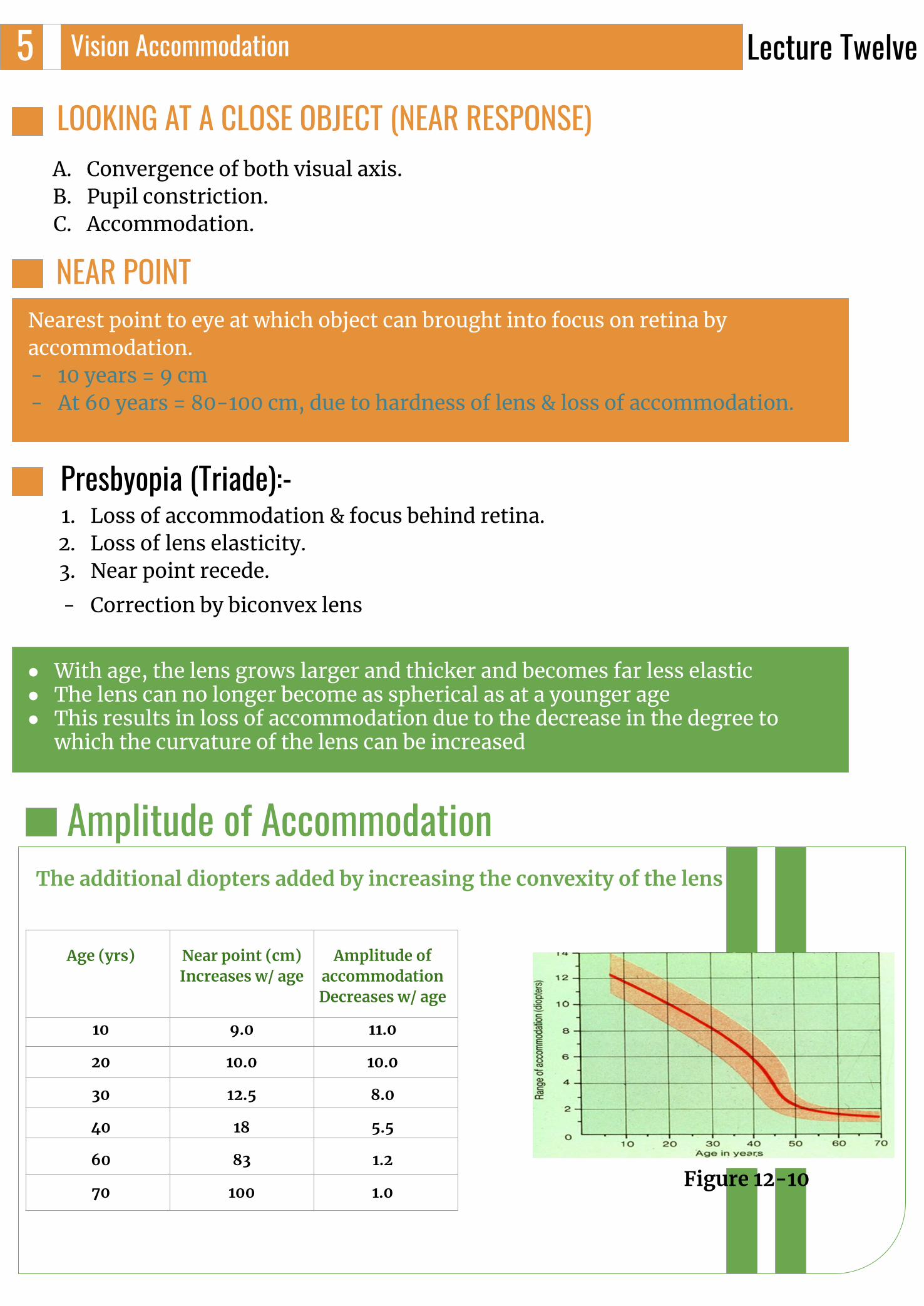
Amplitude of Accommodation
Age (yrs) Near point (cm)Increases w/ age
Amplitude of accommodationDecreases w/ age
10 9.0 11.0
20 10.0 10.0
30 12.5 8.0
40 18 5.5
60 83 1.2
70 100 1.0
The additional diopters added by increasing the convexity of the lens
● With age, the lens grows larger and thicker and becomes far less elastic ● The lens can no longer become as spherical as at a younger age ● This results in loss of accommodation due to the decrease in the degree to
which the curvature of the lens can be increased
Lecture Twelve
Figure 12-10
Vision Accommodation6
Footnotes
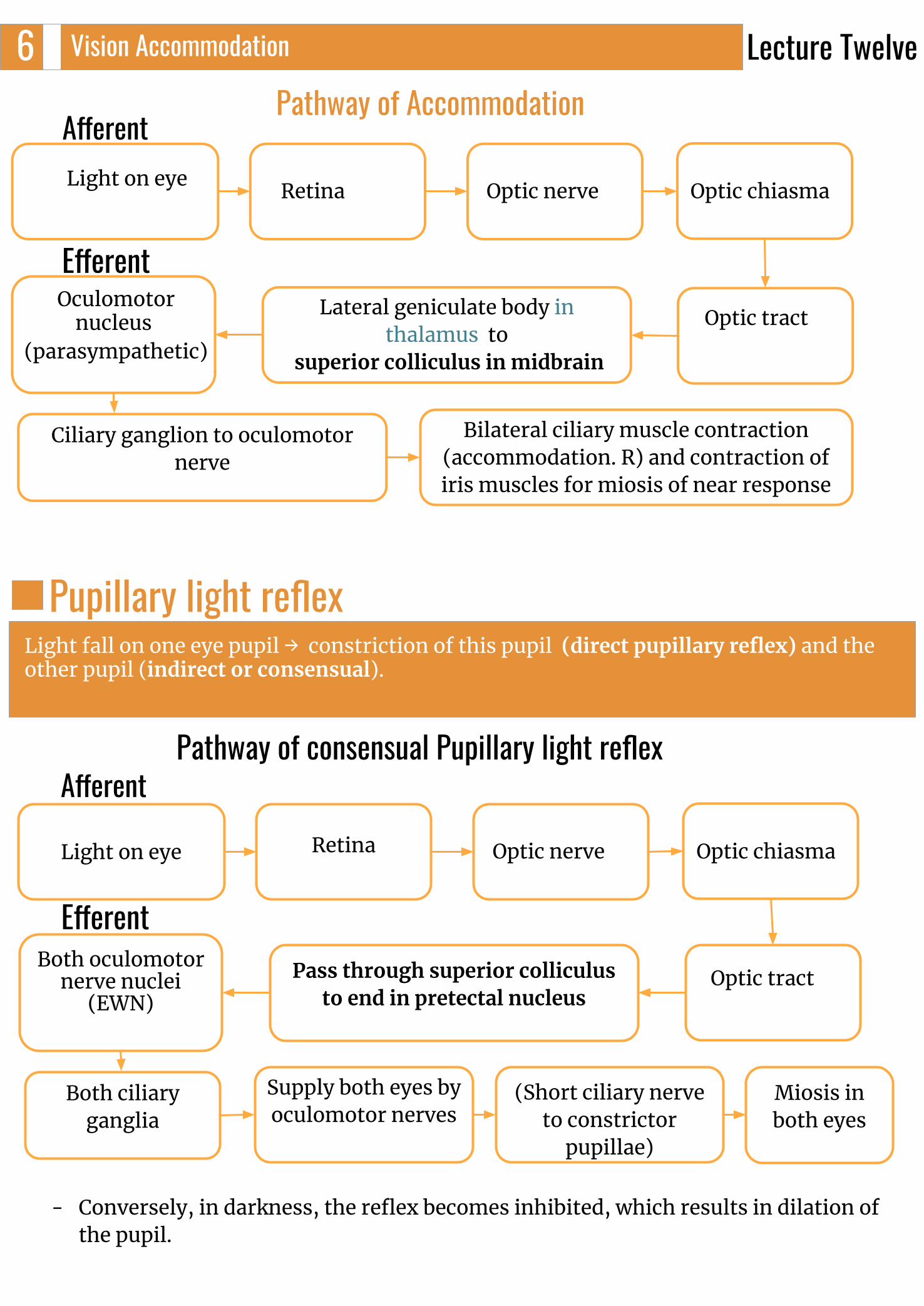
Pathway of AccommodationAfferent
Efferent
Light on eye Retina Optic nerve Optic chiasma
Optic tractLateral geniculate body in thalamus to
superior colliculus in midbrain
Oculomotor nucleus
(parasympathetic)
Ciliary ganglion to oculomotor nerve
Bilateral ciliary muscle contraction (accommodation. R) and contraction of iris muscles for miosis of near response
Pupillary light reflex
Pathway of consensual Pupillary light reflex
Optic nerve Optic chiasma
Optic tract
Light on eye Retina
Pass through superior colliculus to end in pretectal nucleus
Both oculomotor nerve nuclei
(EWN)
Both ciliary ganglia
Supply both eyes by oculomotor nerves
(Short ciliary nerve to constrictor
pupillae)
Miosis in both eyes
- Conversely, in darkness, the reflex becomes inhibited, which results in dilation of the pupil.
Light fall on one eye pupil → constriction of this pupil (direct pupillary reflex) and the other pupil (indirect or consensual).
Afferent
Efferent
Lecture Twelve
- left LGB (similar to left optic tract) has all layers receive from RIGHT ½ of visual field.- Right LGB (similar to right optic tract) has all layers receive from LEFT ½ of visual field. - LGB has 6 layers.
Function of LGB:-1. acts as a relay station for visual information from optic tract to cortex. 2. It has point to point transmission with high
degree of fidelity spatial 3. Acts as a gate which controls signal transmission to visual cortex
i.e control how much signals reach visual cortex 4. color vision & detect shapes & texture5. N.B: - It receives gating control signals from two major sources:
➧Corticofugal fibers returning in a backward direction from the primary visual cortex to the lateral geniculate nucleus ➧Reticular areas of the mesencephalon. Both of these are inhibitory and, when stimulated, can turn off transmission through selected portions of the dorsal lateral geniculate nucleus
Vision Accommodation7
Footnotes
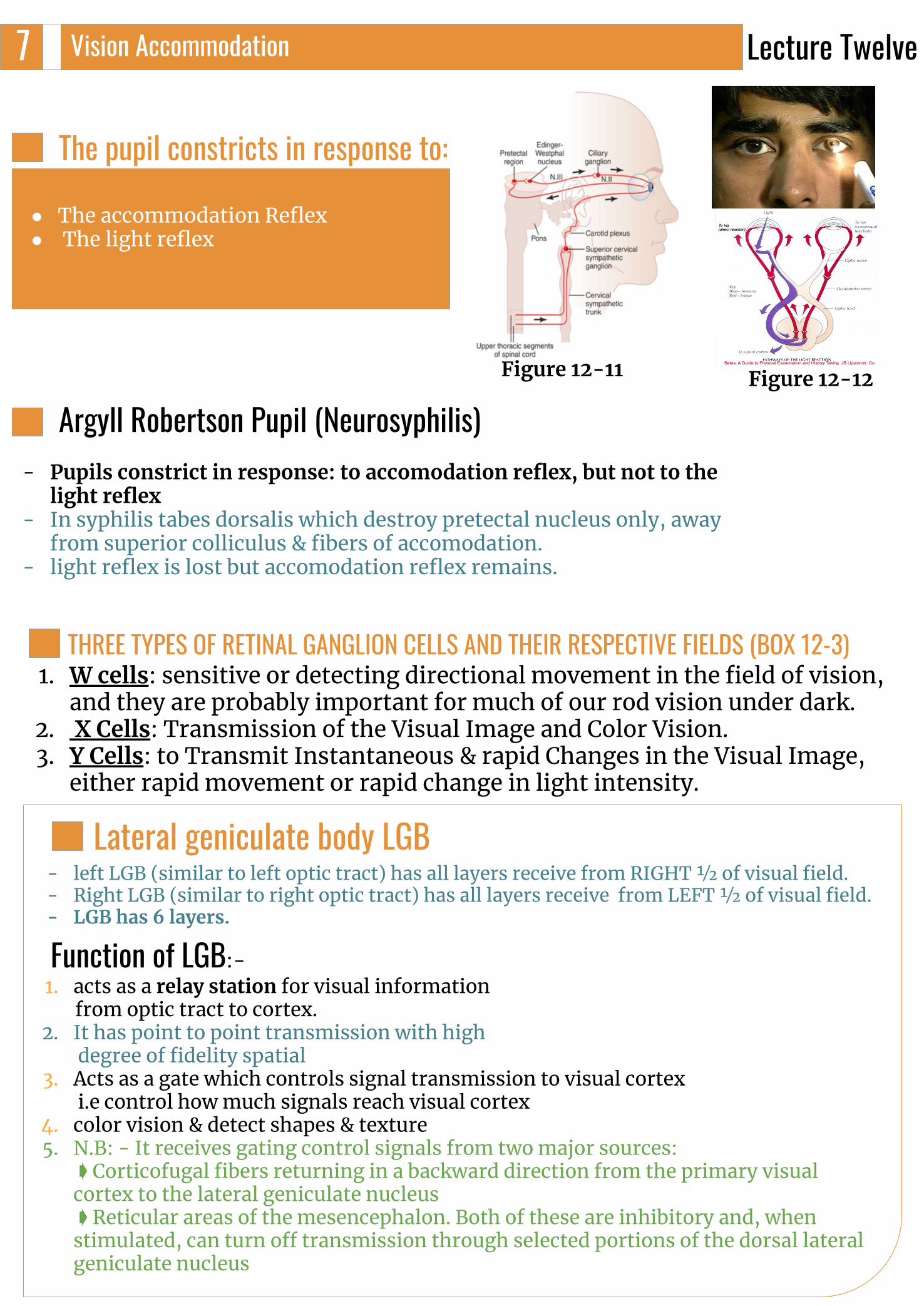
The pupil constricts in response to:
● The accommodation Reflex● The light reflex
Lateral geniculate body LGB
THREE TYPES OF RETINAL GANGLION CELLS AND THEIR RESPECTIVE FIELDS (BOX 12-3)1. W cells: sensitive or detecting directional movement in the field of vision,
and they are probably important for much of our rod vision under dark.2. X Cells: Transmission of the Visual Image and Color Vision. 3. Y Cells: to Transmit Instantaneous & rapid Changes in the Visual Image,
either rapid movement or rapid change in light intensity.
Lecture Twelve
Figure 12-11 Figure 12-12
Argyll Robertson Pupil (Neurosyphilis)- Pupils constrict in response: to accomodation reflex, but not to the
light reflex - In syphilis tabes dorsalis which destroy pretectal nucleus only, away
from superior colliculus & fibers of accomodation. - light reflex is lost but accomodation reflex remains.
- W Cells: These cells have wide dendritic distribution along the retina, thus they are efficient in detecting the shift of focus in the visual field.
- X Cells: Their dendritic fields do not distribute widely within the retina, therefore they transmit signals from discrete, small areas of the retina. Consequently, it is mainly through X Cells that details regarding visual acuity and color are transmitted. Each X Cell receive input from at least one cone, therefore it is most probably responsible for transmitting all color vision.
- Y Cells: Much like W cells these cells have wide dendritic distribution along the retina, Y and W cells make synaptic connections with the superior colliculus and mediate their functions through transmitting information about rapid changes in the visual field.
-
Vision Accommodation8
Footnotes
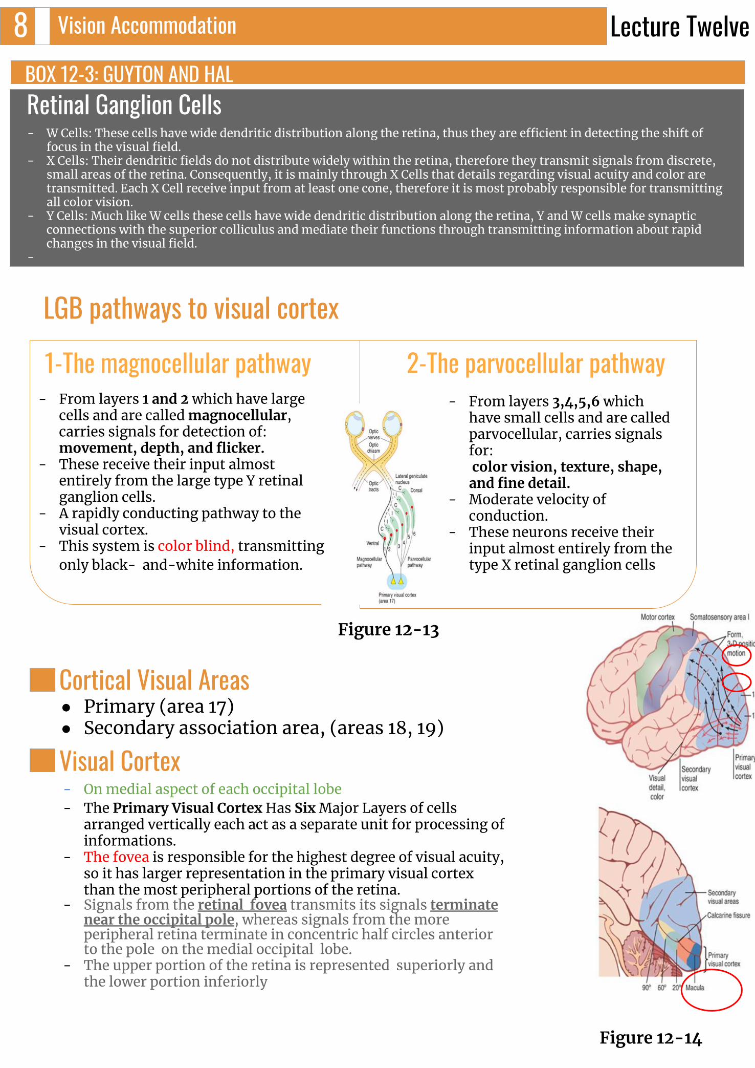
LGB pathways to visual cortex
1-The magnocellular pathway 2-The parvocellular pathway- From layers 1 and 2 which have large
cells and are called magnocellular, carries signals for detection of:movement, depth, and flicker.
- These receive their input almost entirely from the large type Y retinal ganglion cells.
- A rapidly conducting pathway to the visual cortex.
- This system is color blind, transmitting only black- and-white information.
- From layers 3,4,5,6 which have small cells and are called parvocellular, carries signals for: color vision, texture, shape, and fine detail.
- Moderate velocity of conduction.
- These neurons receive their input almost entirely from the type X retinal ganglion cells
Cortical Visual Areas● Primary (area 17) ● Secondary association area, (areas 18, 19)
Visual Cortex- On medial aspect of each occipital lobe - The Primary Visual Cortex Has Six Major Layers of cells
arranged vertically each act as a separate unit for processing of informations.
- The fovea is responsible for the highest degree of visual acuity, so it has larger representation in the primary visual cortex than the most peripheral portions of the retina.
- Signals from the retinal fovea transmits its signals terminate near the occipital pole, whereas signals from the more peripheral retina terminate in concentric half circles anterior to the pole on the medial occipital lobe.
- The upper portion of the retina is represented superiorly and the lower portion inferiorly
Lecture Twelve
Figure 12-13
Figure 12-14
BOX 12-3: GUYTON AND HALRetinal Ganglion Cells
Vision Accommodation9
Footnotes
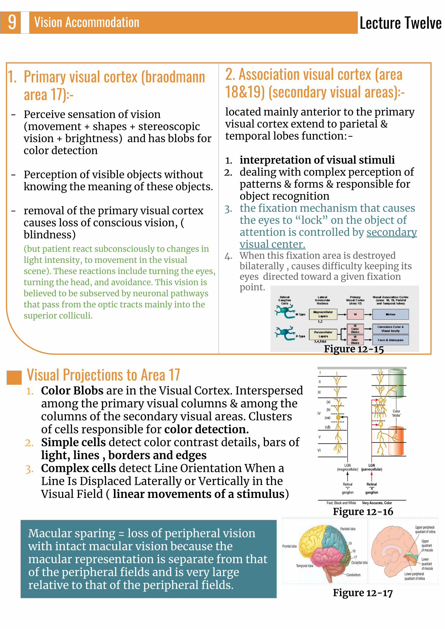
1. Primary visual cortex (braodmann area 17):-
- Perceive sensation of vision (movement + shapes + stereoscopic vision + brightness) and has blobs for color detection
- Perception of visible objects without knowing the meaning of these objects.
- removal of the primary visual cortex causes loss of conscious vision, ( blindness)(but patient react subconsciously to changes in light intensity, to movement in the visual scene). These reactions include turning the eyes, turning the head, and avoidance. This vision is believed to be subserved by neuronal pathways that pass from the optic tracts mainly into the superior colliculi.
2. Association visual cortex (area 18&19) (secondary visual areas):-located mainly anterior to the primary visual cortex extend to parietal & temporal lobes function:-
1. interpretation of visual stimuli 2. dealing with complex perception of
patterns & forms & responsible for object recognition
3. the fixation mechanism that causes the eyes to “lock” on the object of attention is controlled by secondary visual center.
4. When this fixation area is destroyed bilaterally , causes difficulty keeping its eyes directed toward a given fixation point.
Visual Projections to Area 171. Color Blobs are in the Visual Cortex. Interspersed
among the primary visual columns & among the columns of the secondary visual areas. Clusters of cells responsible for color detection.
2. Simple cells detect color contrast details, bars of light, lines , borders and edges
3. Complex cells detect Line Orientation When a Line Is Displaced Laterally or Vertically in the Visual Field ( linear movements of a stimulus)
Macular sparing = loss of peripheral vision with intact macular vision because the macular representation is separate from that of the peripheral fields and is very large relative to that of the peripheral fields.
Lecture Twelve
Figure 12-15
Figure 12-16
Figure 12-17

Vision Accommodation100 Determination of Distance of an Object from the
Eye—“Depth Perception” (BOX 12-4)
A person normally perceives distance by threemajor means:
1. The sizes of the images of known objects on the retina2. the phenomenon of moving parallax :when the person
moves his head to one side or the other, the images of close-by objects move rapidly across the retinas, while the images of distant objects remain almost completely stationary
3. Binocular vision through the phenomenon of stereopsisThe perception of depth and 3-dimensional structure obtained on thebasis of visual information deriving from two eyes by individuals withnormally developed binocular vision
Lecture Twelve
Figure 12-18
- The retina can distinguish the distance of objects if the object’s size is known by the brain, since the size of the object is known, and the image size on the retina is known, the brain computes this set of information to calculate the relative distance of the object.
- Binocular vision can also help detect depth and relative distance of objects, that is, each eye sees objects at a slightly different angle, this causes each eye to see different aspects of a certain object. This gives the perception of three dimensions and depth. This can be demonstrated by closing one eye or the other alternatively and observing how the object will appear slightly different.
BOX 12-4: GUYTON AND HAL
Vision Accommodation110
Footnotes
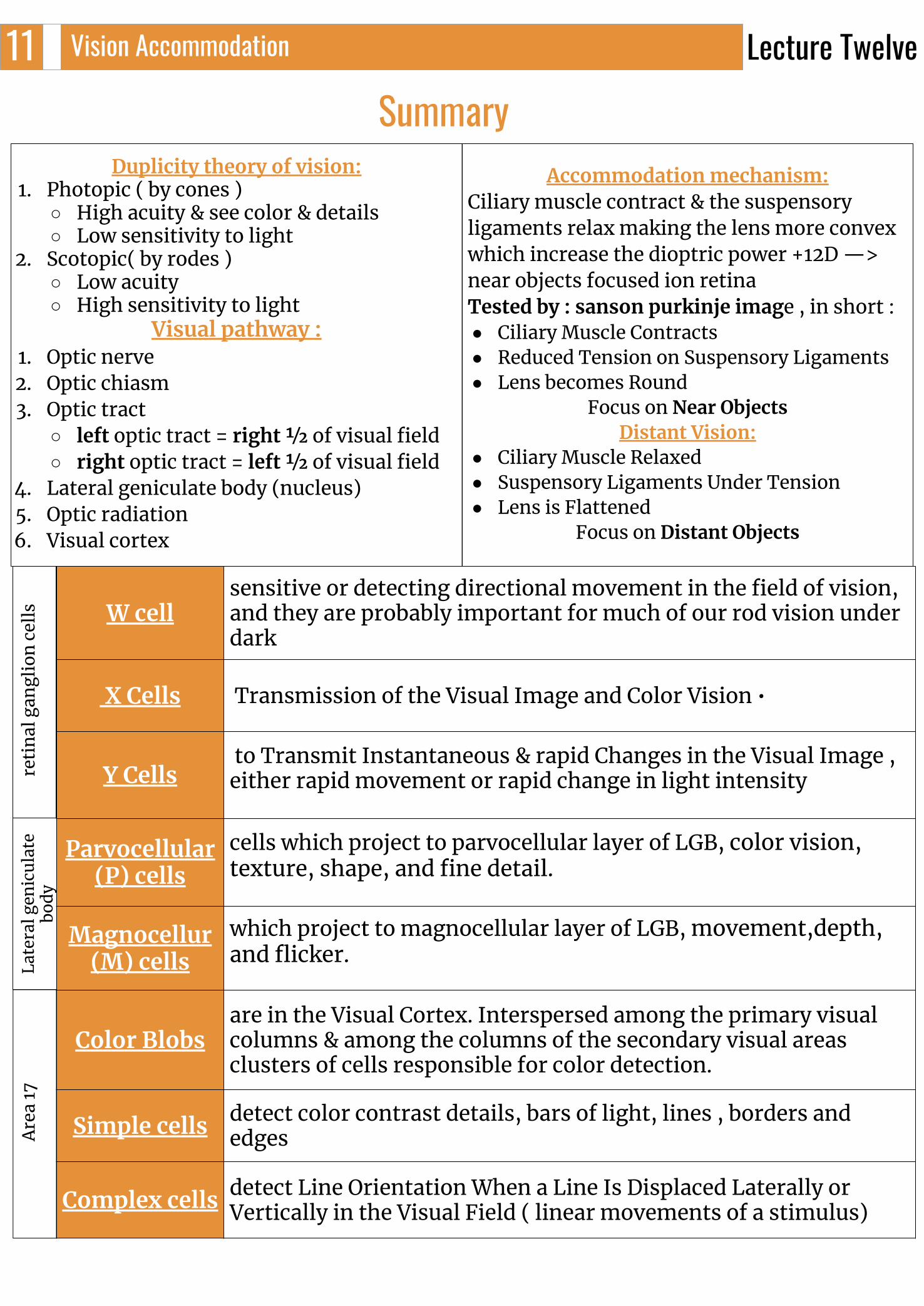
W cellsensitive or detecting directional movement in the field of vision, and they are probably important for much of our rod vision under dark
X Cells Transmission of the Visual Image and Color Vision •
Y Cells to Transmit Instantaneous & rapid Changes in the Visual Image , either rapid movement or rapid change in light intensity
Parvocellular (P) cells
cells which project to parvocellular layer of LGB, color vision, texture, shape, and fine detail.
Magnocellur(M) cells
which project to magnocellular layer of LGB, movement,depth, and flicker.
Color Blobsare in the Visual Cortex. Interspersed among the primary visual columns & among the columns of the secondary visual areas clusters of cells responsible for color detection.
Simple cells detect color contrast details, bars of light, lines , borders and edges
Complex cells detect Line Orientation When a Line Is Displaced Laterally or Vertically in the Visual Field ( linear movements of a stimulus)
ret
inal
gan
glio
n c
ells
Late
ral g
enic
ulat
e bo
dy
Are
a 17
Summary Duplicity theory of vision:
1. Photopic ( by cones ) ○ High acuity & see color & details ○ Low sensitivity to light
2. Scotopic( by rodes ) ○ Low acuity ○ High sensitivity to light
Visual pathway : 1. Optic nerve2. Optic chiasm3. Optic tract
○ left optic tract = right 1⁄2 of visual field ○ right optic tract = left 1⁄2 of visual field
4. Lateral geniculate body (nucleus)5. Optic radiation6. Visual cortex
Accommodation mechanism: Ciliary muscle contract & the suspensory ligaments relax making the lens more convex which increase the dioptric power +12D —> near objects focused ion retinaTested by : sanson purkinje image , in short : ● Ciliary Muscle Contracts ● Reduced Tension on Suspensory Ligaments● Lens becomes Round
Focus on Near ObjectsDistant Vision:
● Ciliary Muscle Relaxed● Suspensory Ligaments Under Tension ● Lens is Flattened
Focus on Distant Objects
Lecture Twelve

MCQ:1.when light falls on one eye the response will be:
A. Relaxation of ciliary musclesB. Contraction of ciliary muscles in one eyeC. Relaxation of iris musclesD. Bilateral ciliary muscle contraction
2.In Argyll robertson pupil the pupils constrict in response to:A. Light reflex but not accommodationB. Accommodation but not lightC. Doesn't respond to accommodation and lightD. Both accommodation and light
3.Large cells that detects movement, depth, and flicker:A. Parvocellular cellsB. Magnocellular cellsC. Y cellsD. X cells
4. The cones is … A. High visual acuity & low sensitivity to lightB. High visual acuity & great sensitivity to lightC. Low visual acuity & great sensitivity to lightD. Low visual acuity & low sensitivity to light
5. From which part of the brainstem there are fibers that inhibit lateral geniculate nucleus ? A. MidbrainB. Medulla C. Red nucleusD. Pins
Sac1.What do color blobs, simple, and complex cells detect in the visual cortex?2. What is the mechanism of accommodation?
SAC1. Color blobs: color detection Simple cells: detect color contrast details, bars of light, lines , borders and edgesComplex cells:detect Line Orientation
SAC2. Ciliary muscle contract then the suspensory ligament relax making the lens more convex which increase the dioptric of the lens causing the near objects to be focused on the retina ANSWER KEY: D, B, B, A, A
REFERENCES- Guyton and Hall Textbook of Medical Physiology
- Ganong’s Review of Medical Physiology
FEMALE PHYSIOLOGY CO-LEADERS Maha Alnahdi, Ghaliah Alnufaei
MALE PHYSIOLOGY CO-LEADERS Nayef Alsaber, Hameed M. Humaid
Shahd Alsalamh, Sarah Alhelal