PLASMA PROTEINS - KSUMSC
14
1 PLASMA PROTEINS Overview: • Functions and characteristics of plasma proteins • Measurement of plasma proteins and diagnosis of diseases • Electrophoretic patterns of plasma proteins • Acute phase proteins *IMPORTANT *EXTRA CORRECTIONS LINK
-
Upload
khangminh22 -
Category
Documents
-
view
1 -
download
0
Transcript of PLASMA PROTEINS - KSUMSC
1
PLASMA PROTEINS
Overview:
• Functions and characteristics of
plasma proteins
• Measurement of plasma proteins
and diagnosis of diseases
• Electrophoretic patterns of plasma proteins
• Acute phase proteins
*IMPORTANT *EXTRA CORRECTIONS LINK

2
Plasma contains >300 different proteins
Many pathological conditions affect level of pps
Mostly synthesized in the liver
Some are produced in other sites
A normal adult contains ~70g/L of pps
PLASMA PROTEINS (PPS)
Transport (Albumin, prealbumin, globulins)
Maintain plasma oncotic pressure* (Albumin)
Defense (Immunoglobulins and complement)
Clotting and fibrinolysis (Thrombin and plasmin)
FUNCTIONS OF PPSMEASUREMENT OF PPS
Quantitative measurement of a specific protein:
Chemical or immunological reactions
Semiquantitative measurement by electrophoresis:
Proteins are separated by their electrical charge in electrophoresis
Five separate bands of proteins are observed
These bands change in diseases.
EXTRA
The major types of proteins
present in plasma are:
Albumin>>formed in the liver.
Fibrinnogen>>formed in the
liver.
Globulins>>
-50-80% in liver
- the remaining are almost
entirely by the lymphoid tissue.
*this function is done by all plasma proteins, but since albumin has the
highest level among the plasma proteins it's the most imp.

3
Types of Plasma Proteins
prealbumin albuminα1-
Globulins
a1-Antitrypsin
α-fetoprotein
α2-Globulins
Ceruloplasmin haptoglobin
β-Globulins
C-reactive protein(CRP)
transferrinβ2-
microglobulin
γ-Globulins
PREALBUMIN (TRANSTHYRETIN) A transport protein for:
Thyroid hormones
Retinol (vitamin A)
Migrates faster than albumin in electrophoresis*
Separated by immunoelectrophoresis
Lower levels found in:
liver disease
nephrotic syndrome,
acute phase inflammatory response
malnutrition
Short half-life (2 days)%
*This is why it is called pre-albumin NOT because it is the albumin precursor, albumin
precursor is PreProAlbumin which is converted to ProAlbumin then secreted as albumin.
4
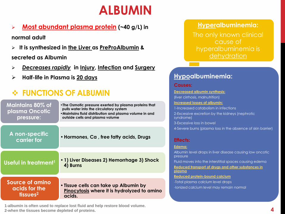
ALBUMIN Most abundant plasma protein (~40 g/L) in
normal adult
It is synthesized in the Liver as PreProAlbumin &
secreted as Albumin
Decreases rapidly in Injury, Infection and Surgery
Half-life in Plasma is 20 days
•The Osmotic pressure exerted by plasma proteins that pulls water into the circulatory system
•Maintains fluid distribution and plasma volume in and outside cells and plasma volume
Maintains 80% of plasma Oncotic
pressure:
• Hormones, Ca , free fatty acids, DrugsA non-specific
carrier for
• 1) Liver Diseases 2) Hemorrhage 3) Shock 4) Burns
Useful in treatment1
• Tissue cells can take up Albumin by Pinocytosis where it is hydrolyzed to amino acids.
Source of amino acids for the
tissues2
Hyperalbuminemia:
The only known clinical cause of
hyperalbuminemia is dehydration
Hypoalbuminemia:
Causes:
Decreased albumin synthesis:
(liver cirrhosis, malnutrition)
Increased losses of albumin:
1-Increased catabolism in infections
2-Excessive excretion by the kidneys (nephroticsyndrome)
3-Excessive loss in bowel
4-Severe burns (plasma loss in the absence of skin barrier)
Effects:
Edema:
Albumin level drops in liver disease causing low oncotic pressure
Fluid moves into the interstitial spaces causing edema
Reduced transport of drugs and other substances in plasma
Reduced protein-bound calcium
-Total plasma calcium level drops
-Ionized calcium level may remain normal
FUNCTIONS OF ALBUMIN
1-albumin is often used to replace lost fluid and help restore blood volume.
2-when the tissues become depleted of proteins.

5
-Synthesized by the liver and macrophages
-An acute-phase protein that inhibits proteases
(Proteases are produced endogenously and from leukocytes andbacteria)
-E.g. of protease:-
Digestive enzymes (trypsin, chymotrypsin)
Other proteases (elastase, thrombin)
-Infection leads to protease release from bacteria and
leukocytes
Types of a1-Antitrypsin ( over 30 types are known)
The most common is M type.
Genetic deficiency of α1-Antitrypsin:-
Synthesis of the defective a1-Antitrypsin (in liver)
Which cannot be secreted.
So, a1-Antitrypsin accumulates in hepatocytes +deficient in
plasma
Clinical Consequences of a1-Antitrypsin Deficiency:
Neonatal jaundice with evidence of cholestasis*
Childhood liver cirrhosis
Pulmonary emphysema in young adults
Laboratory Diagnosis:-Electrophoresis: Lack of a1-globulin band in protein
electrophoresis-Quantitative measurement of a1-Antitrypsin by:
Radial immunodiffusion,
isoelectric focusing or nephelometry
a1-antitrypsin
α1-Globulins a-Fetoprotein (AFP)
Synthesized in the developing embryo and
fetus by the parenchymal cells of the liver
AFP levels decrease gradually during intra-
uterine life and reach adult levels at birth
(AFP level of less than 10 ng/mL is normal for adults)
Function is unknown but it may protect fetus
from immunologic attack by thethe mother (the doctor said; this protein may play a role in Gas exchanging between mother and fetus)
No known physiological function in adults
AFP is a tumor marker for:-
Hepatoma ,testicular cancerAn extremely high level of AFP in blood could be a sign of liver tumors.
Imbalance of maternal α-Fetoprotein:
Elevated maternal AFP levels are
associated with:
Neural tube defect,Anencephaly
Decreased maternal AFP levels are
associated with:
Increased risk of Down’s syndrome
*is a decrease in bile flow due to impaired secretion by hepatocytes or to obstruction of bile flow through intra-or extrahepatic bile ducts

6
α2-GlobulinsSynthesized by the liver
Contains >90% of serum copper
An oxidoreductase that inactivates ROS causing tissue damage in
acute phase response
Wilson’s disease:
Due to low plasma levels of ceruloplasmin
Copper is accumulated in the liver and brain
Important for iron absorption from the
intestine
CERULOPLASMIN
Synthesized by the liver
Binds to free hemoglobin to form complexes that are metabolized in the
RES
Plasma level decreases during
hemolysis
Limits iron losses by preventing Hb loss
from kidneys
Haptoglobin
EXTRA-When red blood cells are destroyed,
the hemoglobin is released. Haptoglobin is
always present in the blood waiting to bind to
released hemoglobin.
macrophages bring
the haptoglobin-hemoglobin complex to
the liver, where haptoglobin and hemoglobinare separated and the iron is recycled.
-In hemolytic anemia, so many red cells are
destroyed that most of the available
haptoglobin is needed to bind the released
hemoglobin. The more severe the hemolysis,
the less haptoglobin remains in the blood.
7
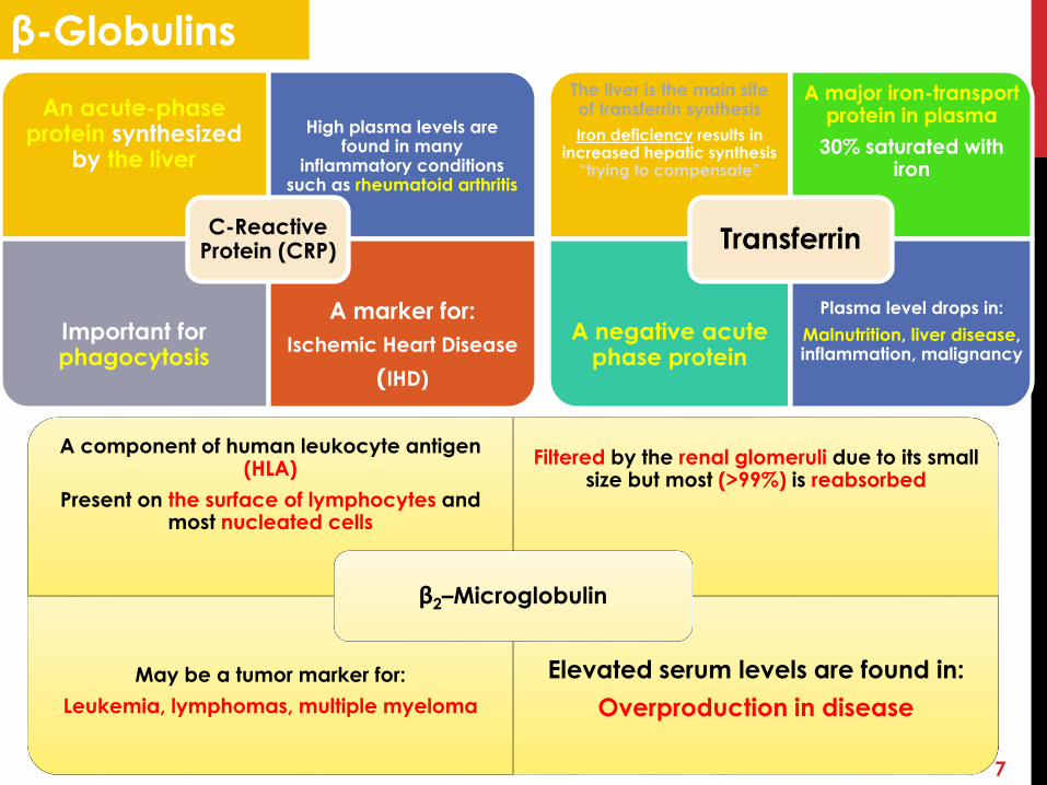
β-Globulins
The liver is the main site of transferrin synthesis
Iron deficiency results in increased hepatic synthesis
“trying to compensate”
A major iron-transport protein in plasma
30% saturated with iron
A negative acute phase protein
Plasma level drops in:
Malnutrition, liver disease, inflammation, malignancy
Transferrin
A component of human leukocyte antigen (HLA)
Present on the surface of lymphocytes and most nucleated cells
Filtered by the renal glomeruli due to its small size but most (>99%) is reabsorbed
May be a tumor marker for:
Leukemia, lymphomas, multiple myeloma
Elevated serum levels are found in:
Overproduction in disease
β2–Microglobulin
An acute-phase protein synthesized
by the liver
High plasma levels are found in many
inflammatory conditions such as rheumatoid arthritis
Important for phagocytosis
A marker for:
Ischemic Heart Disease
IHD))
C-Reactive Protein (CRP)
8
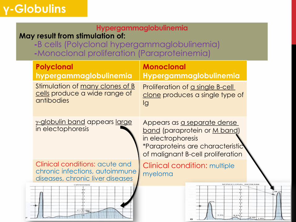
γ-Globulins
HypergammaglobulinemiaMay result from stimulation of:
-B cells (Polyclonal hypergammaglobulinemia) -Monoclonal proliferation (Paraproteinemia)
Monoclonal
Hypergammaglobulinemia
Polyclonal
hypergammaglobulinemia
Proliferation of a single B-cell
clone produces a single type of
Ig
Stimulation of many clones of B cells produce a wide range of antibodies
Appears as a separate dense
band (paraprotein or M band)
in electrophoresis
*Paraproteins are characteristic
of malignant B-cell proliferation
-globulin band appears largein electophoresis
Clinical condition: multiple
myeloma
Clinical conditions: acute and chronic infections, autoimmune diseases, chronic liver diseases

9
Acute Phase Proteins
Positive Acute Phase Proteins:Plasma protein levels increase in:
Infection, inflammation , malignancy, trauma, surgeryThese proteins are called acute phase reactants
Synthesized due to body’s response to injury
Examples: a1-Antitypsin, haptoglobin, ceruloplasmin, fibrinogen, c-reactive protein
Mediators cause these proteins to increase after injury
Mediators: Cytokines (IL-1, IL-6), tumor necrosis factors a and , interferons, platelet activating factor Functions:
1. Bind to polysaccharides in bacterial walls2. Activate complement system3. Stimulate phagocytosis
Negative Acute Phase Proteins:These proteins decrease in inflammation:
Albumin, prealbumin, transferrinMediated by inflammatory response via cytokines and hormonesSynthesis of these proteins decrease to save amino acids for positive acute phase proteins
albumin is low because it's a negative acute
phase proteins.
Other bands are higher because they contain
positive acute phase proteins. e.g. β-
Globulin band contains CRP

10
emzyme Function Condition
Prealbumin
Transport :-
1) Thyroid hormones.
2) Retinol.
Lower level in :-
1) liver disease.
2) Nephrotic syndrome.
3) Malnutrition.
4) Acute phase inflammatory response.
Albumin
1) Maintain oncotic pressure.2) Non specific carrier.3) Useful in treatment of liver
disease & shock & hemorrhage.
Hypoalbuminemia :- Decrease albumin synthesis. Loss of albumin:-
• Excessive loss in bowel• Nephrotic syndrome.• Burns.
a1 -antitrypsin
Genetic deficiency of a1–antitrypsin:- Neonatal jaundice. Childhood liver cirrhosis. Pulmonary emphysema.
a-fetoprotein
Unknown function . Elevated maternal neural tube defect. Decreased maternal down syndrome. Tumor marker hepatoma &testicular cancer.
Ceruloplasmin Important in iron absorption from intestine .
Wilson’s disease.
Haptoglbin
Bind to free hemoglobin to form complexes to limit iron loss and to prevent Hb loss in kidney .
Decrease during hemolysis.
B2 -Microglobulin
Elevated level found in overproduction in
disease. Tumor marker leukemia & lymphomas &
multiple myeloma.
C-Reactive protein
Important in phagocytosis. Elevated in:- Inflammation (rheumatoid arthritis). Marker for ischemic heart disease.
SUMMARY

MCQS1)Prealbumin found in lower level in :
A. Liver disease.
B. Nephrotic syndrome.C. Malnutrition.D. All.
2)80 % of plasma oncotic pressure is maintained by
albumin.
A. True.B. False.
3)Hypoalbuminemia could be carried by:-
A. Decrease in synthesis in liver.B. Loss of albumin.C. Sever burns.D. All of them.
4)Clinical consequences of alpha (a)-antitrypsin
deficiency :-
A. Jaundice.B. Pulmonary emphysema.C. Liver cirrhosis.D. All above.E. A & B.
5) Ceruloplasmin important in :-
A. Iron absorption from intestine.
B. Transplant.
C. Non of them.
D. Both of them.
6)Which protein is important in prevent
Hb loss from kidney :
A. Haptoglobin.
B. Ceruloplasmin.
C. Transferrin.
D. Non of them.
7)High plasma level found in rheumatoid arthritis.
A. Haptoglobin.
B. C-reactive protein.
C. B2 –Microglobin.
D. Ceruloplasmin.
8)Monoclonal proliferation marker for multiple sclerosis.
A. True.
B. False.
9)Which one is true regarding transferrin :-
A. Negative acute phase protein.
B. Major iron transport .
C. Level drop in malignance.
D. Limit iron loss by prevent Hb loss from
kidney.
E. Positive acute phase protein.
F. A & B &C .
G. A &B & C & D.
Answers:1)D 2)A 3)D 4)D 5)A 6)A 7)B 8)B 9)F
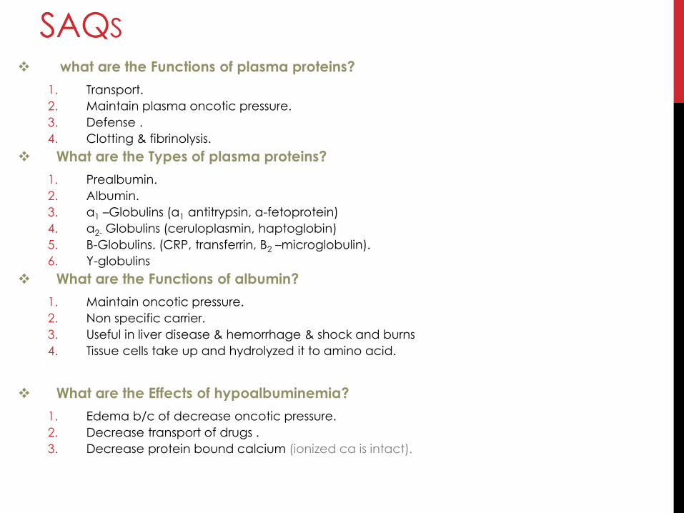
what are the Functions of plasma proteins?
1. Transport.
2. Maintain plasma oncotic pressure.
3. Defense .
4. Clotting & fibrinolysis.
What are the Types of plasma proteins?
1. Prealbumin.
2. Albumin.
3. a1 –Globulins (a1 antitrypsin, a-fetoprotein)
4. a2- Globulins (ceruloplasmin, haptoglobin)
5. B-Globulins. (CRP, transferrin, B2 –microglobulin).
6. Y-globulins
What are the Functions of albumin?
1. Maintain oncotic pressure.
2. Non specific carrier.
3. Useful in liver disease & hemorrhage & shock and burns
4. Tissue cells take up and hydrolyzed it to amino acid.
What are the Effects of hypoalbuminemia?
1. Edema b/c of decrease oncotic pressure.
2. Decrease transport of drugs .
3. Decrease protein bound calcium (ionized ca is intact).
SAQS
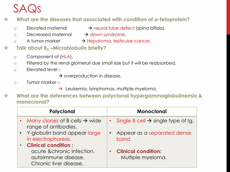
What are the diseases that associated with condition of a-fetoprotein?
o Elevated maternal neural tube defect (spina bifida).
o Decreased maternal down syndrome.
o A tumor marker Hepatoma, testicular cancer.
Talk about B2 –Microblobulin briefly?
o Component of (HLA).
o Filtered by the renal glomeruli due small size but it will be reabsorbed.
o Elevated level :-
overproduction in disease.
o Tumor marker :-
Leukemia, lymphomas, multiple myeloma.
What are the deferences between polyclonal hypergammaglobulinemia &
monoclonal?
Polyclonal Monoclonal
• Many clones of B cells wide
range of antibodies.
• Y-globulin band appear large
in electrophoresis.
• Clinical condition :acute &chronic infection.
autoimmune disease.
Chronic liver disease.
• Single B cell single type of Ig.
• Appear as a separated dense
band.
• Clinical condition:Multiple myeloma.
SAQS

Omar faisal
Abdulaziz Alsaud
Mohammad Alotaibi
Najla Al-draiweesh
14
DONE BY:
If You Have Any Questions Or Comments Please Inform Us: [email protected]