Selected expression profiling of full-length proteins and their variants in human plasma
10
Serum/Plasma Proteome Selected Expression Profiling of Full-Length Proteins and Their Variants in Human Plasma Urban A. Kiernan, Dobrin Nedelkov, Kemmons A.Tubbs, Eric E. Niederkofler and Randall W. Nelson* *Intrinsic Bioprobes, Inc, 625 S. Smith Road Suite 22,Tempe, AZ 85281 7 Clinical Proteomics Journal Copyright ©Humana Press Inc. All rights of any nature whatsoever are reserved. ISSN 1542-6416/04/01:7–16/$25.00 *Author to whom all correspondence and reprint requests should be addressed: E-mail: [email protected] Abstract With increased interest in clinical pro- teomics—the comparative investigation of dif- ferential protein expression patterns for use in the diagnostic and prognostic assessment of disease states—the demand for techniques that can readily identify changes in select proteome components is greater than ever before. This article describes a targeted proteomics approach to recover and quantify C-reactive protein (CRP) directly from human plasma. CRP, a putative biomarker for cardiac health, was isolated from microliter volumes of human plasma by using novel proteomics tools that combine micro-scale affinity capture with matrix assisted laser desorption/ionization time-of- flight mass spectrometry (MALDI-TOF MS) detection. Native CRP was analyzed along with serum amyloid P component (SAP) and retinol binding protein (RBP), that were intentionally targeted to generate a selected protein expres- sion profile. A number of qualitative changes were readily observed within these profiles, including micro heterogeneity in the SAP glycan, C-terminally truncated versions of RBP, and detection of a novel truncated variant of CRP. After quantitative validation of increasing plasma CRP concentrations, the approach was applied to the analysis of eight plasma samples obtained from individuals with known medical histories. The result of the analyses are eight protein profiles, revealing increasing CRP levels that can be associated with individuals ailing from post-surgery inflammation, chronic rheu- matoid arthritis, and recent acute myocardial infarction. The technique described in this arti- cle lays the foundation for selected protein pro- filing for use in biomarker discovery, as well as in clinical and diagnostic applications.
-
Upload
independent -
Category
Documents
-
view
0 -
download
0
Transcript of Selected expression profiling of full-length proteins and their variants in human plasma
Serum/Plasma Proteome
Selected Expression Profiling of Full-Length Proteinsand Their Variants in Human PlasmaUrban A. Kiernan, Dobrin Nedelkov, Kemmons A.Tubbs,Eric E. Niederkofler and Randall W. Nelson*
*Intrinsic Bioprobes, Inc, 625 S. Smith Road Suite 22,Tempe,AZ 85281
7
Clinical Proteomics JournalCopyright ©Humana Press Inc.All rights of any nature whatsoever are reserved.ISSN 1542-6416/04/01:7–16/$25.00
*Author to whom all correspondence and reprint requests should be addressed: E-mail: [email protected]
AbstractWith increased interest in clinical pro-
teomics—the comparative investigation of dif-ferential protein expression patterns for use inthe diagnostic and prognostic assessment ofdisease states—the demand for techniques thatcan readily identify changes in select proteomecomponents is greater than ever before. Thisarticle describes a targeted proteomics approachto recover and quantify C-reactive protein(CRP) directly from human plasma. CRP, aputative biomarker for cardiac health, wasisolated from microliter volumes of humanplasma by using novel proteomics tools thatcombine micro-scale affinity capture with matrixassisted laser desorption/ionization time-of-flight mass spectrometry (MALDI-TOF MS)detection. Native CRP was analyzed along withserum amyloid P component (SAP) and retinol
binding protein (RBP), that were intentionallytargeted to generate a selected protein expres-sion profile. A number of qualitative changeswere readily observed within these profiles,including micro heterogeneity in the SAPglycan, C-terminally truncated versions of RBP,and detection of a novel truncated variant ofCRP. After quantitative validation of increasingplasma CRP concentrations, the approach wasapplied to the analysis of eight plasma samplesobtained from individuals with known medicalhistories. The result of the analyses are eightprotein profiles, revealing increasing CRP levelsthat can be associated with individuals ailingfrom post-surgery inflammation, chronic rheu-matoid arthritis, and recent acute myocardialinfarction. The technique described in this arti-cle lays the foundation for selected protein pro-filing for use in biomarker discovery, as well asin clinical and diagnostic applications.
Clinical Proteomics _______________________________________________________________ Volume 1, 2004
Key Words: Relative quantification; MALDI-TOFMS; C-reactive protein; human plasma; proteinexpression profiles; immunoassay.
Introduction
Clinical proteomics focuses on the use ofrapid fractionation approaches in combinationwith mass spectrometry (MS) to directly profilehuman biofluids, plasma in particular. Bydesign, these studies are meant to establishprotein signatures, in the form of mass spec-tra, that are able to distinguish betweenhealthy and diseased states in individuals.Indeed, numerous pioneering investigationshave shown clinical proteomics to be one of themost promising new approaches for definingdiagnostic signatures related to ovarian, breast,prostate and lung cancers (1–3). However, aparticular concern of these studies is that manyof the profiling approaches distinguish healthyfrom diseased states based on computer learn-ing algorithms that may not identify a land-mark mass-to-charge (m/z) value as actuallybeing a protein species. Such an approach maybe misleading if the m/z values used have nocorresponding peptide signals. Moreover, todate no clinical proteomics study has indepen-dently “rediscovered” known biomarkers ofvarious cancers (e.g., prostate specific antigen[PSA] is not detected in individuals sufferingfrom prostate cancer). In missing, or not specif-ically targeting these proteins as part of the pro-filing, these studies do not take full advantageof past clinical/diagnostic studies, the resultsof which are the basis of numerous currentdiagnostic assays (4). Accordingly, there exists agrowing demand to discover and identify pro-teins for use as putative biomarkers, and at thesame time detect proteins that are presentlyused (and validated) as diagnostic species (5).
Given that the identity of the biomarker isknown (either old or newly found), it is
clearly more efficient to construct assays thattarget them directly, rather than to continue toassay samples using the global discoverymethodologies. A foremost advantage to usinga targeted approach in combination with massspectrometry is the ability to significantlyimprove the limit of detection of the assay.Specifically, affinity approaches are able toselectively concentrate target proteins priorto mass spectrometry, the result of which isa highly purified form of the target proteinentering the mass spectrometer without anyaccompanying interferences stemming from thepresence of other proteins (e.g., ion suppressionor overlapping signals). A second advantage isthe use of targeted high-performance massspectrometric assays to discern and character-ize genomic and epigenomic differences thatare found in proteins. Because of genetic andposttranslational variations, any protein underinvestigation has the potential of existing withinor between individuals as multiple species.Mass spectrometry of highly purified pro-teins has been used for many years to bothdetect and characterize and/or identify thesevariations (6). This long-standing ability is ide-ally complimented by a simple fractionationapproach that can be used to selectively con-centrate chosen proteins directly from biologi-cal milieu.
Thus, findings made through efforts inthe fields of clinical biology and medicine,including proteomics, stand to be improvedupon by: (1) knowing the identity of thesignature/biomarker proteins, (2) reducing thesignature profile to its essential components,and (3) detecting structural and quantitativevariations—present in natural populations—encountered in the proteins. Moreover, pro-teomics technologies responsive to these needsmust be readily applied in the analysis ofhundreds-to-thousands of samples.
8 ________________________________________________________________________________ Kiernan et al.

Volume 1, 2004 _______________________________________________________________ Clinical Proteomics
Overview of the Mass SpectrometricImmunoassay Process
Previous works have described a hybridprotein analysis technology—mass spectromet-ric immunoassay (MSIA)—that is used to selec-tively retrieve target proteins from complexbiological milieu for mass spectrometriccharacterization and quantification (7–10).Figure 1 gives a brief overview of the process.As applied here, a human plasma sample isrepeatedly drawn and expelled through apipettor tip containing a stationary phase towhich an affinity ligand is covalently bound(termed MSIA-Tip). After rinsing, MALDImatrix is drawn into the MSIA-Tip, breakingthe affinity interaction. The resulting matrix/analyte mixture is deposited directly onto amass spectrometer target for subsequent matrix-assisted laser desorption/ionization time-of-flight mass spectrometry (MALDI-TOF MS).Analyte detection/characterization is essen-tially a combination of affinity isolation viaimmobilized antibodies and characterizationof retained species by MALDI-TOF MS. Inthis sense, target proteins and their variantsare selectively concentrated from plasma, andMALDI-TOF MS is used to (1) unambiguouslyidentify the wild-type target protein by detec-tion at a known molecular weight and (2) rec-ognize the presence of variants retained by theantibody at masses shifted away from the wild-type protein. This ability forms the basis of anassay capable of identifying protein variantsresulting from posttranslational modifications(9), pointmutations (10), or truncations (11–13).The simultaneous analysis of mass-resolvedspecies lays the foundation for the rigorousquantification of a target protein (14) or thedevelopment of multiple-analyte assays (7).
Presented here is the development of amulti-analyte MSIA for use in the expres-
sion profiling of a select group of proteins—C-reactive proteins (CRP), retinol binding pro-tein (RBP), and serum amyloid P component(SAP) (and their variants)—present in humanplasma. The central target in these analyses isCRP, a known clinical protein biomarker ofinflammation that has been determined tohave short- and long-term predictive value ofpatient outcomes with cardiovascular diseaseand the development of coronary events thatcan be used to identify the onset and recur-rence of myocardial infarction (MI) (15–18),stroke (19–20), and angina (21–22). The twoother protein species, RBP and SAP werechosen as complement proteins (for use innormalizing the MSIA profiles between indi-viduals) based on a few simple criteria. First,for optimization of MALDI-TOF MS condi-tions (e.g., matrix, delayed-extraction), theproteins must have similar, but fully resolvedmolecular weights. Second, the complementproteins are found in healthy populations atroughly equivalent and reasonably stable con-centrations. Lastly, MSIA conditions for thecomplement proteins (e.g., extraction andrinses) are the same as for the target protein.
Materials and Methods
Samples
Plasma samples from eight individuals (fivemales and three females betweent the ages of26 and 48) were acquired following a procedureapproved by the IBI’s Institutional ReviewBoard (IRB), and only after each subjecthad signed an Informed Consent form. Sam-ples were acquired, under sterile conditions,through a lancet-punctured finger using twononheparinized (75 µL volume) microcolumns(Drummond Scientific Co., Broomall, PA). Each150 µL whole blood sample collected wasimmediately combined with 200 µL HEPES
Protein Profiling in Human Plasma _____________________________________________________________ 9

Clinical Proteomics _______________________________________________________________ Volume 1, 2004
buffered saline (10 mM HEPES, 150 mM NaCl,pH 7.4 [HBS]) containing 50 mM ethylenedi-amine tetraacetic acid (EDTA) and 2 µL of aprotease inhibitor cocktail set II (Calbiochem,San Diego, CA), and centrifuged for 2 min (at7000 rpm/2500g) to pellet red blood cells. The
supernatant (diluted plasma, 250 µL) wasdecanted from each sample and stored at–70°C until ready for use.
For the standard addition quantificationexperiments, a single plasma sample was dis-persed into eight 25 µL aliquots. Increasing
10 _______________________________________________________________________________ Kiernan et al.
Fig. 1. Schematic of the MSIA process. Biological fluid is passed through the MSIA-Tip and targeted analytes areaffinity bound within the solid support. Nonaffinity reactive components of the biological fluid are removedfrom the MSIA-Tip with repetitive rinses. Purified target analyte is then eluted from the tip onto a MALDItarget for subsequent TOF MS analysis.
Volume 1, 2004 _______________________________________________________________ Clinical Proteomics
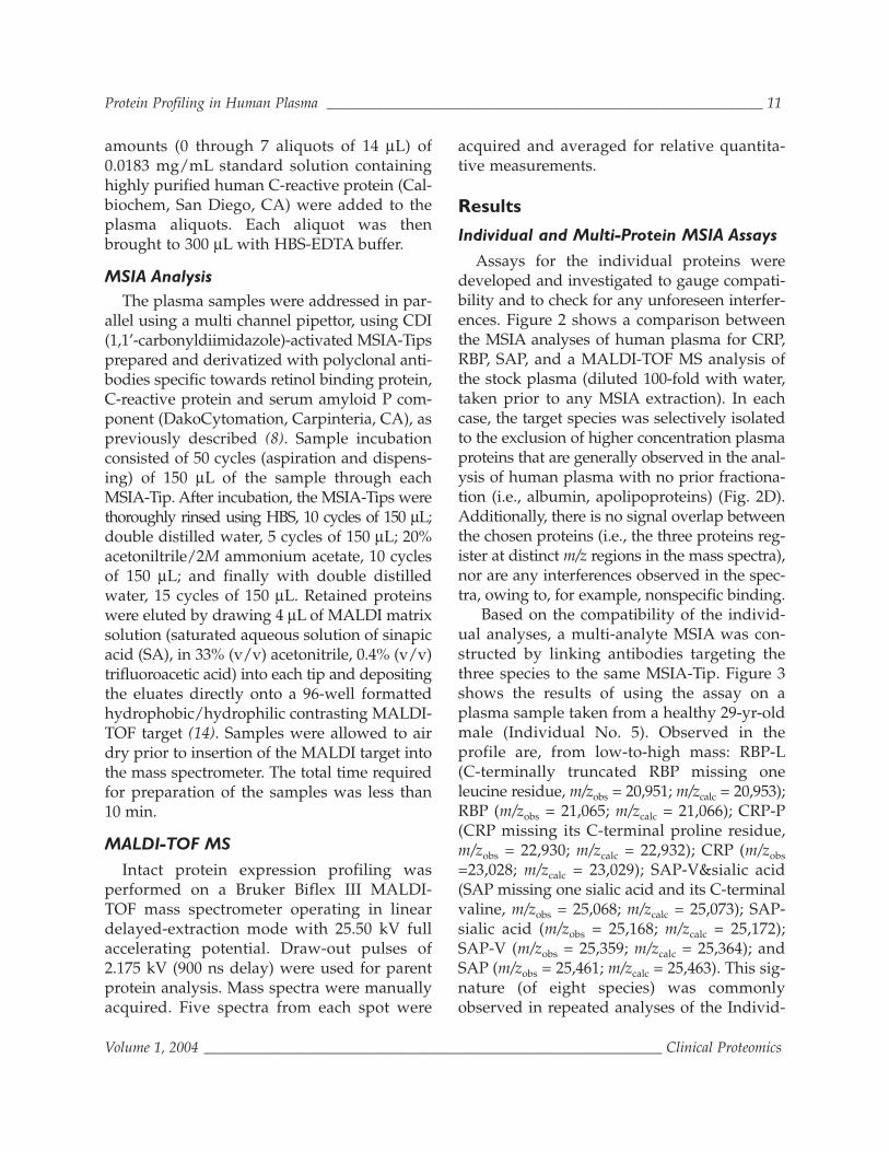
amounts (0 through 7 aliquots of 14 µL) of0.0183 mg/mL standard solution containinghighly purified human C-reactive protein (Cal-biochem, San Diego, CA) were added to theplasma aliquots. Each aliquot was thenbrought to 300 µL with HBS-EDTA buffer.
MSIA AnalysisThe plasma samples were addressed in par-
allel using a multi channel pipettor, using CDI(1,1’-carbonyldiimidazole)-activated MSIA-Tipsprepared and derivatized with polyclonal anti-bodies specific towards retinol binding protein,C-reactive protein and serum amyloid P com-ponent (DakoCytomation, Carpinteria, CA), aspreviously described (8). Sample incubationconsisted of 50 cycles (aspiration and dispens-ing) of 150 µL of the sample through eachMSIA-Tip. After incubation, the MSIA-Tips werethoroughly rinsed using HBS, 10 cycles of 150 µL;double distilled water, 5 cycles of 150 µL; 20%acetoniltrile/2M ammonium acetate, 10 cyclesof 150 µL; and finally with double distilledwater, 15 cycles of 150 µL. Retained proteinswere eluted by drawing 4 µL of MALDI matrixsolution (saturated aqueous solution of sinapicacid (SA), in 33% (v/v) acetonitrile, 0.4% (v/v)trifluoroacetic acid) into each tip and depositingthe eluates directly onto a 96-well formattedhydrophobic/hydrophilic contrasting MALDI-TOF target (14). Samples were allowed to airdry prior to insertion of the MALDI target intothe mass spectrometer. The total time requiredfor preparation of the samples was less than10 min.
MALDI-TOF MS
Intact protein expression profiling wasperformed on a Bruker Biflex III MALDI-TOF mass spectrometer operating in lineardelayed-extraction mode with 25.50 kV fullaccelerating potential. Draw-out pulses of2.175 kV (900 ns delay) were used for parentprotein analysis. Mass spectra were manuallyacquired. Five spectra from each spot were
acquired and averaged for relative quantita-tive measurements.
Results
Individual and Multi-Protein MSIA Assays
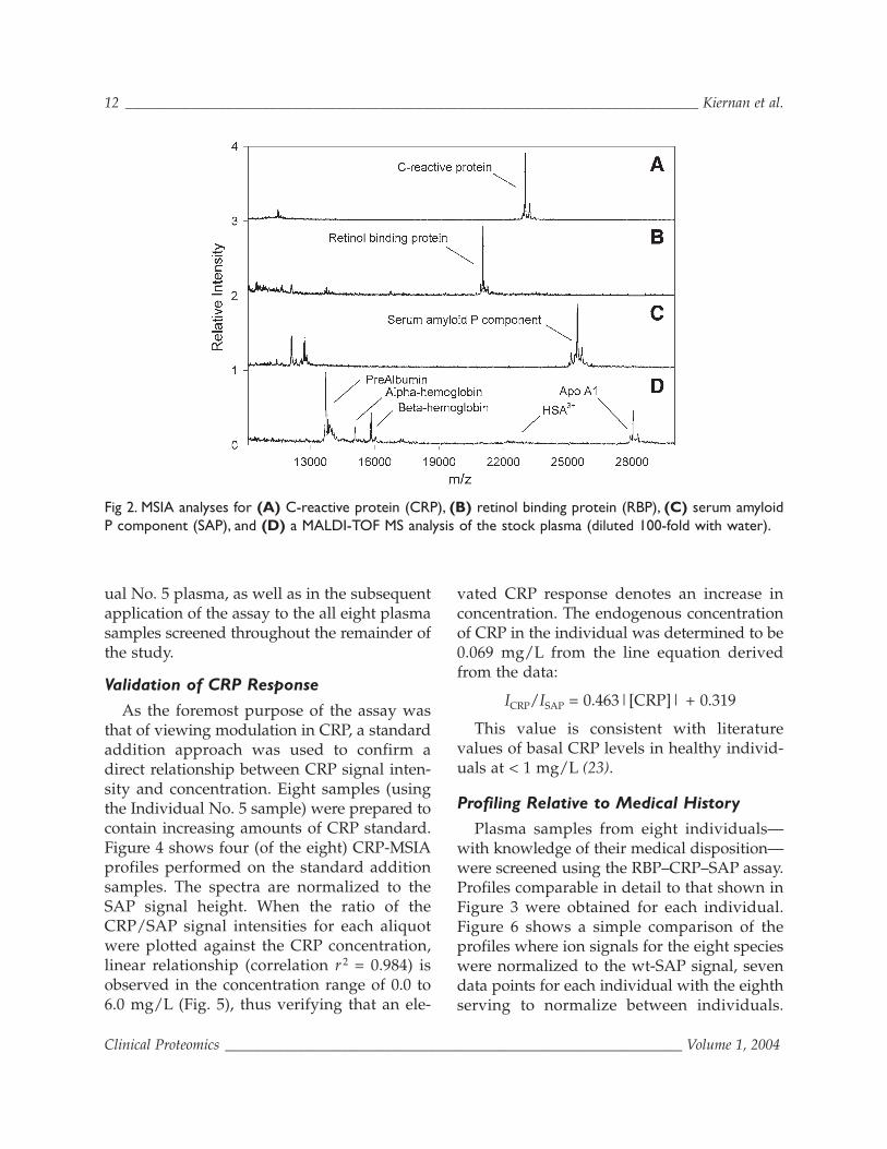
Assays for the individual proteins weredeveloped and investigated to gauge compati-bility and to check for any unforeseen interfer-ences. Figure 2 shows a comparison betweenthe MSIA analyses of human plasma for CRP,RBP, SAP, and a MALDI-TOF MS analysis ofthe stock plasma (diluted 100-fold with water,taken prior to any MSIA extraction). In eachcase, the target species was selectively isolatedto the exclusion of higher concentration plasmaproteins that are generally observed in the anal-ysis of human plasma with no prior fractiona-tion (i.e., albumin, apolipoproteins) (Fig. 2D).Additionally, there is no signal overlap betweenthe chosen proteins (i.e., the three proteins reg-ister at distinct m/z regions in the mass spectra),nor are any interferences observed in the spec-tra, owing to, for example, nonspecific binding.
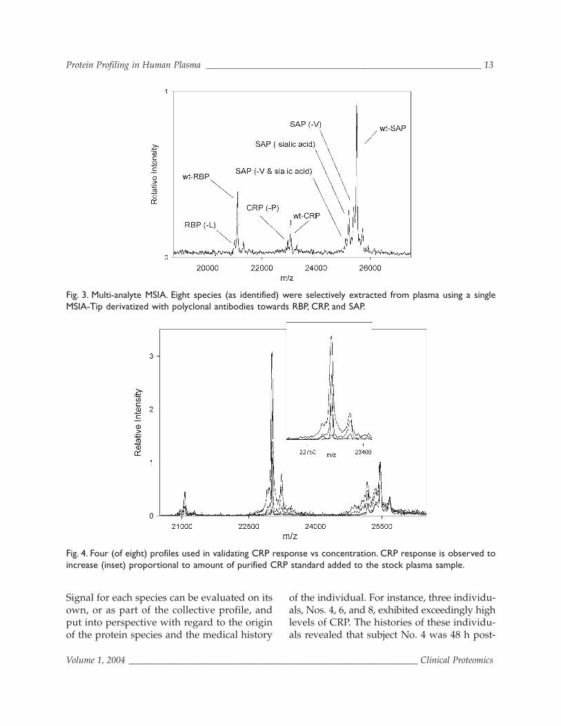
Based on the compatibility of the individ-ual analyses, a multi-analyte MSIA was con-structed by linking antibodies targeting thethree species to the same MSIA-Tip. Figure 3shows the results of using the assay on aplasma sample taken from a healthy 29-yr-oldmale (Individual No. 5). Observed in theprofile are, from low-to-high mass: RBP-L(C-terminally truncated RBP missing oneleucine residue, m/zobs = 20,951; m/zcalc = 20,953);RBP (m/zobs = 21,065; m/zcalc = 21,066); CRP-P(CRP missing its C-terminal proline residue,m/zobs = 22,930; m/zcalc = 22,932); CRP (m/zobs
=23,028; m/zcalc = 23,029); SAP-V&sialic acid(SAP missing one sialic acid and its C-terminalvaline, m/zobs = 25,068; m/zcalc = 25,073); SAP-sialic acid (m/zobs = 25,168; m/zcalc = 25,172);SAP-V (m/zobs = 25,359; m/zcalc = 25,364); andSAP (m/zobs = 25,461; m/zcalc = 25,463). This sig-nature (of eight species) was commonlyobserved in repeated analyses of the Individ-
Protein Profiling in Human Plasma ____________________________________________________________ 11
Clinical Proteomics _______________________________________________________________ Volume 1, 2004
ual No. 5 plasma, as well as in the subsequentapplication of the assay to the all eight plasmasamples screened throughout the remainder ofthe study.
Validation of CRP Response
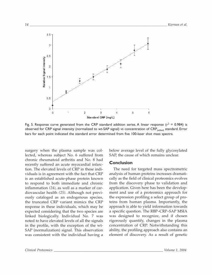
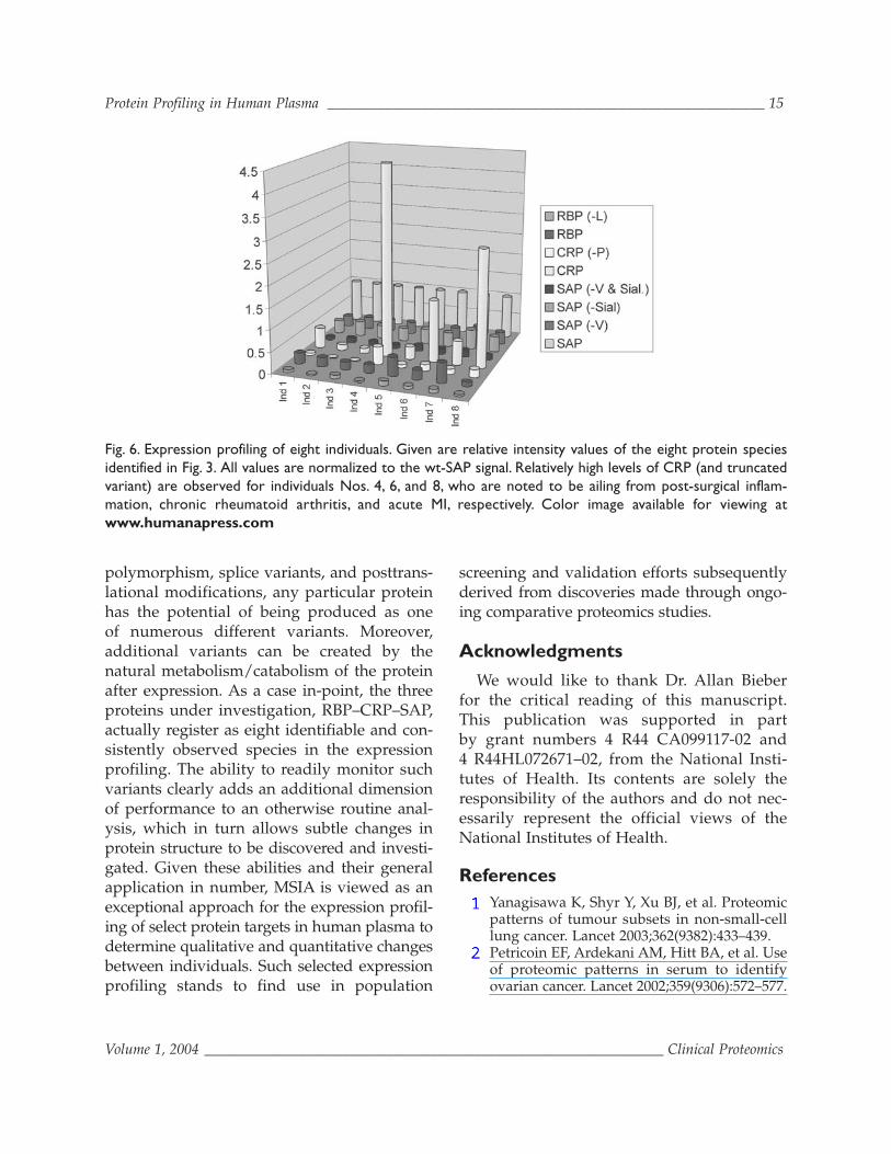
As the foremost purpose of the assay wasthat of viewing modulation in CRP, a standardaddition approach was used to confirm adirect relationship between CRP signal inten-sity and concentration. Eight samples (usingthe Individual No. 5 sample) were prepared tocontain increasing amounts of CRP standard.Figure 4 shows four (of the eight) CRP-MSIAprofiles performed on the standard additionsamples. The spectra are normalized to theSAP signal height. When the ratio of theCRP/SAP signal intensities for each aliquotwere plotted against the CRP concentration,linear relationship (correlation r 2 = 0.984) isobserved in the concentration range of 0.0 to6.0 mg/L (Fig. 5), thus verifying that an ele-
vated CRP response denotes an increase inconcentration. The endogenous concentrationof CRP in the individual was determined to be0.069 mg/L from the line equation derivedfrom the data:
ICRP/ISAP = 0.463|[CRP]| + 0.319
This value is consistent with literaturevalues of basal CRP levels in healthy individ-uals at < 1 mg/L (23).
Profiling Relative to Medical History
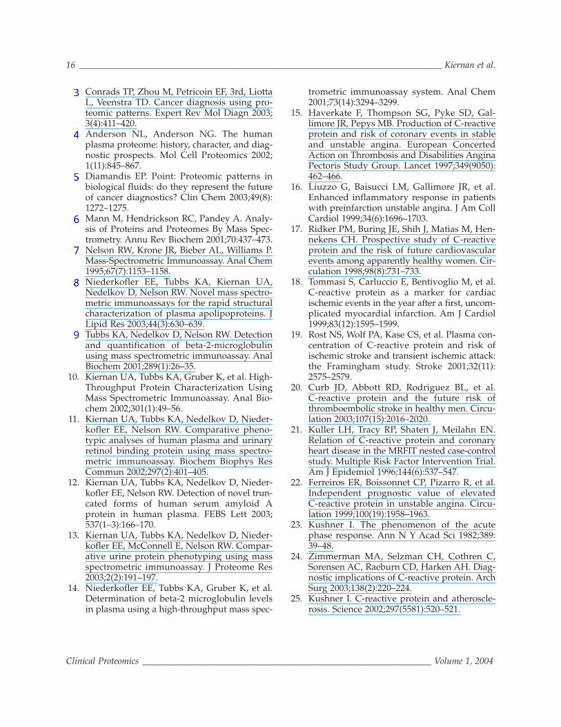
Plasma samples from eight individuals—with knowledge of their medical disposition—were screened using the RBP–CRP–SAP assay.Profiles comparable in detail to that shown inFigure 3 were obtained for each individual.Figure 6 shows a simple comparison of theprofiles where ion signals for the eight specieswere normalized to the wt-SAP signal, sevendata points for each individual with the eighthserving to normalize between individuals.
12 _______________________________________________________________________________ Kiernan et al.
Fig 2. MSIA analyses for (A) C-reactive protein (CRP), (B) retinol binding protein (RBP), (C) serum amyloidP component (SAP), and (D) a MALDI-TOF MS analysis of the stock plasma (diluted 100-fold with water).
Volume 1, 2004 _______________________________________________________________ Clinical Proteomics
Signal for each species can be evaluated on itsown, or as part of the collective profile, andput into perspective with regard to the originof the protein species and the medical history
of the individual. For instance, three individu-als, Nos. 4, 6, and 8, exhibited exceedingly highlevels of CRP. The histories of these individu-als revealed that subject No. 4 was 48 h post-
Protein Profiling in Human Plasma ____________________________________________________________ 13
Fig. 3. Multi-analyte MSIA. Eight species (as identified) were selectively extracted from plasma using a singleMSIA-Tip derivatized with polyclonal antibodies towards RBP, CRP, and SAP.
Fig. 4. Four (of eight) profiles used in validating CRP response vs concentration. CRP response is observed toincrease (inset) proportional to amount of purified CRP standard added to the stock plasma sample.
Clinical Proteomics _______________________________________________________________ Volume 1, 2004
surgery when the plasma sample was col-lected, whereas subject No. 6 suffered fromchronic rheumatoid arthritis and No. 8 hadrecently suffered an acute myocardial infarc-tion. The elevated levels of CRP in these indi-viduals is in agreement with the fact that CRPis an established acute-phase protein knownto respond to both immediate and chronicinflammation (24), as well as a marker of car-diovascular health (25). Although not previ-ously cataloged as an endogenous species,the truncated CRP variant mimics the CRPresponse in these individuals, which may beexpected considering that the two species arelinked biologically. Individual No. 7 wasnoted to have elevated levels of all the signalsin the profile, with the exception of the wt-SAP (normalization) signal. This observationwas consistent with the individual having a
below average level of the fully glycosylatedSAP, the cause of which remains unclear.
ConclusionThe need for targeted mass spectrometric
analysis of human proteins increases dramati-cally as the field of clinical proteomics evolvesfrom the discovery phase to validation andapplication. Given here has been the develop-ment and use of a proteomics approach forthe expression profiling a select group of pro-teins from human plasma. Importantly, theapproach is able to yield information towardsa specific question. The RBP–CRP–SAP MSIAwas designed to recognize, and if chosenrigorously quantify, changes in the plasmaconcentration of CRP. Notwithstanding thisability, the profiling approach also contains anelement of discovery. As a result of genetic
14 _______________________________________________________________________________ Kiernan et al.
Fig. 5. Response curve generated from the CRP standard addition series. A linear response (r 2 = 0.984) isobserved for CRP signal intensity (normalized to wt-SAP signal) vs concentration of CRP(added) standard. Errorbars for each point indicated the standard error determined from five 100-laser shot mass spectra.
Volume 1, 2004 _______________________________________________________________ Clinical Proteomics
polymorphism, splice variants, and posttrans-lational modifications, any particular proteinhas the potential of being produced as oneof numerous different variants. Moreover,additional variants can be created by thenatural metabolism/catabolism of the proteinafter expression. As a case in-point, the threeproteins under investigation, RBP–CRP–SAP,actually register as eight identifiable and con-sistently observed species in the expressionprofiling. The ability to readily monitor suchvariants clearly adds an additional dimensionof performance to an otherwise routine anal-ysis, which in turn allows subtle changes inprotein structure to be discovered and investi-gated. Given these abilities and their generalapplication in number, MSIA is viewed as anexceptional approach for the expression profil-ing of select protein targets in human plasma todetermine qualitative and quantitative changesbetween individuals. Such selected expressionprofiling stands to find use in population
screening and validation efforts subsequentlyderived from discoveries made through ongo-ing comparative proteomics studies.
Acknowledgments
We would like to thank Dr. Allan Bieberfor the critical reading of this manuscript.This publication was supported in partby grant numbers 4 R44 CA099117-02 and4 R44HL072671–02, from the National Insti-tutes of Health. Its contents are solely theresponsibility of the authors and do not nec-essarily represent the official views of theNational Institutes of Health.
References1. Yanagisawa K, Shyr Y, Xu BJ, et al. Proteomic
patterns of tumour subsets in non-small-celllung cancer. Lancet 2003;362(9382):433–439.
2. Petricoin EF, Ardekani AM, Hitt BA, et al. Useof proteomic patterns in serum to identifyovarian cancer. Lancet 2002;359(9306):572–577.
Protein Profiling in Human Plasma ____________________________________________________________ 15
Fig. 6. Expression profiling of eight individuals. Given are relative intensity values of the eight protein speciesidentified in Fig. 3. All values are normalized to the wt-SAP signal. Relatively high levels of CRP (and truncatedvariant) are observed for individuals Nos. 4, 6, and 8, who are noted to be ailing from post-surgical inflam-mation, chronic rheumatoid arthritis, and acute MI, respectively. Color image available for viewing atwww.humanapress.com
Clinical Proteomics _______________________________________________________________ Volume 1, 2004
3. Conrads TP, Zhou M, Petricoin EF, 3rd, LiottaL, Veenstra TD. Cancer diagnosis using pro-teomic patterns. Expert Rev Mol Diagn 2003;3(4):411–420.
4. Anderson NL, Anderson NG. The humanplasma proteome: history, character, and diag-nostic prospects. Mol Cell Proteomics 2002;1(11):845–867.
5. Diamandis EP. Point: Proteomic patterns inbiological fluids: do they represent the futureof cancer diagnostics? Clin Chem 2003;49(8):1272–1275.
6. Mann M, Hendrickson RC, Pandey A. Analy-sis of Proteins and Proteomes By Mass Spec-trometry. Annu Rev Biochem 2001;70:437–473.
7. Nelson RW, Krone JR, Bieber AL, Williams P.Mass-Spectrometric Immunoassay. Anal Chem1995;67(7):1153–1158.
8. Niederkofler EE, Tubbs KA, Kiernan UA,Nedelkov D, Nelson RW. Novel mass spectro-metric immunoassays for the rapid structuralcharacterization of plasma apolipoproteins. JLipid Res 2003;44(3):630–639.
9. Tubbs KA, Nedelkov D, Nelson RW. Detectionand quantification of beta-2-microglobulinusing mass spectrometric immunoassay. AnalBiochem 2001;289(1):26–35.
10. Kiernan UA, Tubbs KA, Gruber K, et al. High-Throughput Protein Characterization UsingMass Spectrometric Immunoassay. Anal Bio-chem 2002;301(1):49–56.
11. Kiernan UA, Tubbs KA, Nedelkov D, Nieder-kofler EE, Nelson RW. Comparative pheno-typic analyses of human plasma and urinaryretinol binding protein using mass spectro-metric immunoassay. Biochem Biophys ResCommun 2002;297(2):401–405.
12. Kiernan UA, Tubbs KA, Nedelkov D, Nieder-kofler EE, Nelson RW. Detection of novel trun-cated forms of human serum amyloid Aprotein in human plasma. FEBS Lett 2003;537(1–3):166–170.
13. Kiernan UA, Tubbs KA, Nedelkov D, Nieder-kofler EE, McConnell E, Nelson RW. Compar-ative urine protein phenotyping using massspectrometric immunoassay. J Proteome Res2003;2(2):191–197.
14. Niederkofler EE, Tubbs KA, Gruber K, et al.Determination of beta-2 microglobulin levelsin plasma using a high-throughput mass spec-
trometric immunoassay system. Anal Chem2001;73(14):3294–3299.
15. Haverkate F, Thompson SG, Pyke SD, Gal-limore JR, Pepys MB. Production of C-reactiveprotein and risk of coronary events in stableand unstable angina. European ConcertedAction on Thrombosis and Disabilities AnginaPectoris Study Group. Lancet 1997;349(9050):462–466.
16. Liuzzo G, Baisucci LM, Gallimore JR, et al.Enhanced inflammatory response in patientswith preinfarction unstable angina. J Am CollCardiol 1999;34(6):1696–1703.
17. Ridker PM, Buring JE, Shih J, Matias M, Hen-nekens CH. Prospective study of C-reactiveprotein and the risk of future cardiovascularevents among apparently healthy women. Cir-culation 1998;98(8):731–733.
18. Tommasi S, Carluccio E, Bentivoglio M, et al.C-reactive protein as a marker for cardiacischemic events in the year after a first, uncom-plicated myocardial infarction. Am J Cardiol1999;83(12):1595–1599.
19. Rost NS, Wolf PA, Kase CS, et al. Plasma con-centration of C-reactive protein and risk ofischemic stroke and transient ischemic attack:the Framingham study. Stroke 2001;32(11):2575–2579.
20. Curb JD, Abbott RD, Rodriguez BL, et al.C-reactive protein and the future risk ofthromboembolic stroke in healthy men. Circu-lation 2003;107(15):2016–2020.
21. Kuller LH, Tracy RP, Shaten J, Meilahn EN.Relation of C-reactive protein and coronaryheart disease in the MRFIT nested case-controlstudy. Multiple Risk Factor Intervention Trial.Am J Epidemiol 1996;144(6):537–547.
22. Ferreiros ER, Boissonnet CP, Pizarro R, et al.Independent prognostic value of elevatedC-reactive protein in unstable angina. Circu-lation 1999;100(19):1958–1963.
23. Kushner I. The phenomenon of the acutephase response. Ann N Y Acad Sci 1982;389:39–48.
24. Zimmerman MA, Selzman CH, Cothren C,Sorensen AC, Raeburn CD, Harken AH. Diag-nostic implications of C-reactive protein. ArchSurg 2003;138(2):220–224.
25. Kushner I. C-reactive protein and atheroscle-rosis. Science 2002;297(5581):520–521.
16 _______________________________________________________________________________ Kiernan et al.