Structural variants: changing the landscape of chromosomes ...
10
Structural variants: changing the landscape of chromosomes and design of disease studies Lars Feuk, Christian R. Marshall, Richard F. Wintle and Stephen W. Scherer * The Centre for Applied Genomics and Program in Genetics and Genomic Biology, The Hospital for Sick Children, Department of Molecular and Medical Genetics, University of Toronto, Ontario, Canada Received February 23, 2006; Revised and Accepted March 9, 2006 The near completeness of human chromosome sequences is facilitating accurate characterization and assessment of all classes of genomic variation. Particularly, using the DNA reference sequence as a guide, genome scanning technologies, such as microarray-based comparative genomic hybridization (array CGH) and genome-wide single nucleotide polymorphism (SNP) platforms, have now enabled the detec- tion of a previously unrecognized degree of larger-sized (non-SNP) variability in all genomes. This heterogen- eity can include copy number variations (CNVs), inversions, insertions, deletions and other complex rearrangements, most of which are not detected by standard cytogenetics or DNA sequencing. Although these genomic alterations (collectively termed structural variants or polymorphisms) have been described previously, mainly through locus-specific studies, they are now known to be more global in occurrence. Moreover, as just one example, CNVs can contain entire genes and their number can correlate with the level of gene expression. It is also plausible that structural variants may commonly influence nearby genes through chromosomal positional or domain effects. Here, we discuss what is known of the prevalence of structural variants in the human genome and how they might influence phenotype, including the conti- nuum of etiologic events underlying monogenic to complex diseases. Particularly, we highlight the newest studies and some classic examples of how structural variants might have adverse genetic consequences. We also discuss why analysis of structural variants should become a vital step in any genetic study going forward. All these progresses have set the stage for a golden era of combined microscopic and sub- microscopic (cytogenomic)-based research of chromosomes leading to a more complete understanding of the human genome. INTRODUCTION In the past few years, several studies have identified a pre- viously uncharacterized prevalence of structural variants of DNA along chromosomes in the size range of 1 kb or greater, adding to the catalog of variants in the human genome (Table 1). Namely, sub-microscopic (usually less than 3 Mb) copy number variations (CNVs) and inversions have been found to occur in every genome studied at high fre- quencies when compared with the equivalent classes of cyto- genetically detectable rearrangements (1 – 8). Similar findings are also now being made more readily in disease gene studies. These discoveries have come somewhat later than the description (and generation of comprehensive maps) of single nucleotide polymorphisms (SNPs) (9,10), microsatel- lites (11,12) and minisatellites (13), as well as catalogs of cytogenetically detectable heteromorphisms and rearrange- ments, because of limits of resolution in the technology at that time. However, new developments in genome-wide scan- ning methodologies using genomic clone and oligonucleotide- based arrays occurring in parallel with the availability of a reference human genome sequence now provide opportunity to generate advanced maps of structural variation in world- wide populations. Moreover, next generation sequencing tech- nologies and computational comparisons of sequences from different sources will yield a vast number of variants primarily in the ,1 kb size range that have not been described pre- viously. Comprehensive reviews describing the discovery # The Author 2006. Published by Oxford University Press. All rights reserved. For Permissions, please email: [email protected] *To whom correspondence should be addressed at: The Centre for Applied Genomics, The Hospital for Sick Children, MaRS Centre—East Tower, 101 College Street, Room 14-701, Toronto, Ontario, Canada M5G 1L7. Tel: þ1 4168137613; Fax: þ1 4168138319; Email: [email protected] Human Molecular Genetics, 2006, Vol. 15, Review Issue 1 R57–R66 doi:10.1093/hmg/ddl057 Downloaded from https://academic.oup.com/hmg/article/15/suppl_1/R57/632718 by guest on 07 February 2022
-
Upload
khangminh22 -
Category
Documents
-
view
0 -
download
0
Transcript of Structural variants: changing the landscape of chromosomes ...
Structural variants: changing the landscape ofchromosomes and design of disease studies
Lars Feuk, Christian R. Marshall, Richard F. Wintle and Stephen W. Scherer*
The Centre for Applied Genomics and Program in Genetics and Genomic Biology, The Hospital for Sick Children,
Department of Molecular and Medical Genetics, University of Toronto, Ontario, Canada
Received February 23, 2006; Revised and Accepted March 9, 2006
The near completeness of human chromosome sequences is facilitating accurate characterization andassessment of all classes of genomic variation. Particularly, using the DNA reference sequence as aguide, genome scanning technologies, such as microarray-based comparative genomic hybridization(array CGH) and genome-wide single nucleotide polymorphism (SNP) platforms, have now enabled the detec-tion of a previously unrecognized degree of larger-sized (non-SNP) variability in all genomes. This heterogen-eity can include copy number variations (CNVs), inversions, insertions, deletions and other complexrearrangements, most of which are not detected by standard cytogenetics or DNA sequencing. Althoughthese genomic alterations (collectively termed structural variants or polymorphisms) have been describedpreviously, mainly through locus-specific studies, they are now known to be more global in occurrence.Moreover, as just one example, CNVs can contain entire genes and their number can correlate with thelevel of gene expression. It is also plausible that structural variants may commonly influence nearbygenes through chromosomal positional or domain effects. Here, we discuss what is known of the prevalenceof structural variants in the human genome and how they might influence phenotype, including the conti-nuum of etiologic events underlying monogenic to complex diseases. Particularly, we highlight the neweststudies and some classic examples of how structural variants might have adverse genetic consequences.We also discuss why analysis of structural variants should become a vital step in any genetic study goingforward. All these progresses have set the stage for a golden era of combined microscopic and sub-microscopic (cytogenomic)-based research of chromosomes leading to a more complete understanding ofthe human genome.
INTRODUCTION
In the past few years, several studies have identified a pre-viously uncharacterized prevalence of structural variants ofDNA along chromosomes in the size range of 1 kb orgreater, adding to the catalog of variants in the humangenome (Table 1). Namely, sub-microscopic (usually lessthan �3 Mb) copy number variations (CNVs) and inversionshave been found to occur in every genome studied at high fre-quencies when compared with the equivalent classes of cyto-genetically detectable rearrangements (1–8). Similar findingsare also now being made more readily in disease gene studies.
These discoveries have come somewhat later than thedescription (and generation of comprehensive maps) of
single nucleotide polymorphisms (SNPs) (9,10), microsatel-lites (11,12) and minisatellites (13), as well as catalogs ofcytogenetically detectable heteromorphisms and rearrange-ments, because of limits of resolution in the technology atthat time. However, new developments in genome-wide scan-ning methodologies using genomic clone and oligonucleotide-based arrays occurring in parallel with the availability of areference human genome sequence now provide opportunityto generate advanced maps of structural variation in world-wide populations. Moreover, next generation sequencing tech-nologies and computational comparisons of sequences fromdifferent sources will yield a vast number of variants primarilyin the ,1 kb size range that have not been described pre-viously. Comprehensive reviews describing the discovery
# The Author 2006. Published by Oxford University Press. All rights reserved.For Permissions, please email: [email protected]
*To whom correspondence should be addressed at: The Centre for Applied Genomics, The Hospital for Sick Children, MaRS Centre—East Tower, 101College Street, Room 14-701, Toronto, Ontario, Canada M5G 1L7. Tel: þ1 4168137613; Fax: þ1 4168138319; Email: [email protected]
Human Molecular Genetics, 2006, Vol. 15, Review Issue 1 R57–R66doi:10.1093/hmg/ddl057
Dow
nloaded from https://academ
ic.oup.com/hm
g/article/15/suppl_1/R57/632718 by guest on 07 February 2022
and properties of, in particular, CNVs, but also other structuralvariants, have been published recently (14–19). Here, wehighlight the latest findings, with a particular emphasis onthe new sub-microscopic variants being increasingly describedin the �1 kb to �3 Mb size range and how they may influencephenotype or be involved in disease.
STRUCTURAL VARIATION INFLUENCING
PHENOTYPE
Changes in DNA that affect gene function (often throughaffecting dosage) can have a deleterious effect on the repro-ductive fitness of an organism, and in some cases representlethal mutations. In these circumstances, the variants wouldeventually be destined to disappear, but they can often existin a heterozygous form for many generations. In betweenthese extremes of selectively neutral variants and lethalmutations lie variants that can influence physiological, bio-chemical, morphological and pathological variation in thehuman population. Recent descriptions of numerous ‘gene-sized’ (the average size of a gene being �70 kb) sub-microscopic structural variants in all genomes have generatedsignificant excitement in the field (20–25), because (i) it waspresumed they probably should exist for the same reasons asSNPs and microscopic variants; (ii) their sheer size (oftenaffecting hundreds to thousands of nucleotides of DNA)increases the likelihood that the alteration is, in fact, agenomic lesion explaining disease outcomes; (iii) as such,some will also be shown to predispose to disease eitherdirectly or in combination with other variants and factors
and (iv) some will provide substrate for evolutionary change.The description of all variants will be important for manywide-ranging reasons, better resolving a more completelyannotated reference genome sequence to understanding impli-cations in pharmacogenomics and clinical diagnostic testing.
It has been well established in many classic (26–33) as wellas in more recent studies of monogenic disease (34), oligoge-netic disease (35–41), and most recently in complex diseasethat the study of such chromosome rearrangements can bethe most rapid approach to identify candidate susceptibilityloci and genes (that then need to be confirmed in othersamples). For complex diseases (note that in some cases,these were the Mendelian sub-forms of complex diseasedemonstrating the same phenotypic endpoint), examplesinclude: in autism, X-chromosome deletions led to the identi-fication of the neuroligin NLGN3 and NLGN4 genes (42); inschizophrenia, a familial chromosome 1 translocation led tothe discovery of the DISC1 and DISC2 genes (43); in dyslexia,distinct chromosomes 3 and 15 translocations led to the dis-covery of ROBO1 and DYXC1, respectively (44,45); insevere speech and language disorder, a chromosome 7q31translocation pinpointed the FOXP2 gene (46); in Tourettesyndrome, a de novo inversion led to SLITRK1 involvement(47); in severe expressive language delay, microduplicationof the Williams–Beuren syndrome locus on chromosome7q11.23 (48) and in early onset Parkinson and Alzheimer’sdisease, duplications of SNCA and APP on chromosomes 4and 21, respectively, have been shown to be causative (49,50).
Indeed, the primary message of this review is to increase theawareness of the necessity for including steps for screeningfor structural variants in genetic experiments. This was
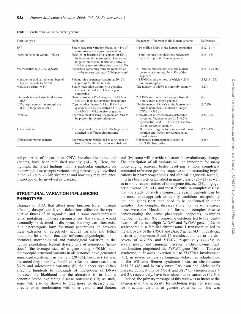
Table 1. Genetic variation in the human genome
Variation type Definition Frequency (if known) in the human genome References
SNP Single base pair variation found in .1% ofchromosomes in a given population
�10 million SNPs in the human population (112–114)
Insertion/deletion variant (InDel) Deletion or insertion of a segment of DNA.Includes small polymorphic changes andlarge chromosomal aberrations. InDels.1 kb in size are often also called CNVs
�1 million insertion/deletions polymorph-isms .1 bp in the human genome
(115,116)
Microsatellite (e.g. CAn repeats) Sequences containing variable numbers of1–6 bp repeats totaling,200 bp in length
.1 million microsatellites in the humangenome, accounting for �3% of thesequence
(114,117,118)
Minisatellite and variable numbers oftandem repeats (VNTRs)
Polymorphic sequence containing 20–50copies of 6–100 bp repeats
�150 000 minisatellites, of which �20%are polymorphic
(13,119,120)
Multisite variant (MSV) Single nucleotide variant with complexcharacteristics due to CNV or geneconversion
The number of MSVs is currently unknown (121)
Intermediate-sized structural variant(ISV)
Gain or loss of a DNA sequence .8 kb insize also includes inversion breakpoints
297 ISVs were identified using a fosmidlibrary from a single genome
(4)
CNV; copy number polymorphism(CNP); large-scale CNV
Copy number change .1 kb. If the fre-quency is .1%, it is called a CNP. LCVsare CNVs �50 kb in size or greater
The frequency of CNVs in the human gen-ome is unknown. Estimates of largerCNVs (.50 kb)
(1,2,19)
Inversion Rearrangement causing a segment of DNA tobe present in reverse orientation
Estimates of microscopically detectableinversion frequencies are 0.12–0.7%(pericentric) and 0.1–0.5% (paracentric);sub-microscopic unknown
(122,123)
Translocation Rearrangement in which a DNA fragment isattached to different chromosome
1/500 is heterozygous for a reciprocal trans-location and 1/1000 for Robertsoniantranslocations
(122–124)
Unbalanced rearrangements Rearrangements which lead to a net gain orloss of DNA are referred to as unbalanced
Unbalanced rearrangements occur in�1/1500 live births
(125)
R58 Human Molecular Genetics, 2006, Vol. 15, Review Issue 1
Dow
nloaded from https://academ
ic.oup.com/hm
g/article/15/suppl_1/R57/632718 by guest on 07 February 2022
exquisitely demonstrated in a recent study showing that copynumber polymorphism in the FCGR3 gene predisposes to glo-merulonephritis in humans and rats (51). Preliminary datasuggest that dozens of CNVs alone will be found in a givengenome when assessed using comprehensive scanning method-ologies. The sub-microscopic variants will be intermediate insize and frequency in comparison to occurrence of cytogenetically
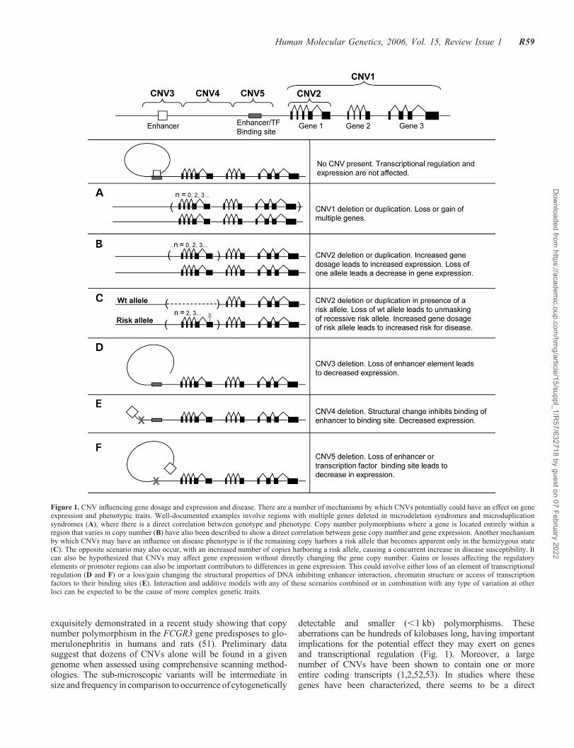
detectable and smaller (,1 kb) polymorphisms. Theseaberrations can be hundreds of kilobases long, having importantimplications for the potential effect they may exert on genesand transcriptional regulation (Fig. 1). Moreover, a largenumber of CNVs have been shown to contain one or moreentire coding transcripts (1,2,52,53). In studies where thesegenes have been characterized, there seems to be a direct
Figure 1. CNV influencing gene dosage and expression and disease. There are a number of mechanisms by which CNVs potentially could have an effect on geneexpression and phenotypic traits. Well-documented examples involve regions with multiple genes deleted in microdeletion syndromes and microduplicationsyndromes (A), where there is a direct correlation between genotype and phenotype. Copy number polymorphisms where a gene is located entirely within aregion that varies in copy number (B) have also been described to show a direct correlation between gene copy number and gene expression. Another mechanismby which CNVs may have an influence on disease phenotype is if the remaining copy harbors a risk allele that becomes apparent only in the hemizygous state(C). The opposite scenario may also occur, with an increased number of copies harboring a risk allele, causing a concurrent increase in disease susceptibility. Itcan also be hypothesized that CNVs may affect gene expression without directly changing the gene copy number. Gains or losses affecting the regulatoryelements or promoter regions can also be important contributors to differences in gene expression. This could involve either loss of an element of transcriptionalregulation (D and F) or a loss/gain changing the structural properties of DNA inhibiting enhancer interaction, chromatin structure or access of transcriptionfactors to their binding sites (E). Interaction and additive models with any of these scenarios combined or in combination with any type of variation at otherloci can be expected to be the cause of more complex genetic traits.
Human Molecular Genetics, 2006, Vol. 15, Review Issue 1 R59
Dow
nloaded from https://academ
ic.oup.com/hm
g/article/15/suppl_1/R57/632718 by guest on 07 February 2022

correlation between increases in gene copy number and increasedlevels of mRNA (53–56). Polymorphic deletions containingentire genes have also been described, where a fraction of thepopulation are homozygous for the deletion allele and, therefore,do not have the gene present in their genome (6,8,57).Most of thegenes in this category belong to gene families or are recentlyduplicated in evolutionary history, and this may increase the tol-erance for null alleles.
Specific categories of genes seem over-represented in CNVsincluding those important for interaction with the surroundingenvironment, such as olfaction and response to externalstimuli (19,58). Examples of such polymorphic genes includeglutathione S-transferase genes (59,60), cytochrome P450genes (61–65) and the complement component C4 (66). Ineach case, changes to gene copy number have been shown togive rise to concomitant changes in the level of enzymeactivity, with phenotypic consequences. Another example isthe CCL3L1 gene, where the increased copy number has beenshown to be protective against HIV infection (56).
Inversions represent another class of structural variation(Table 1), but knowledge of their prevalence in the humangenome is more limited. This is partly due to a lack of technol-ogies for robust and inexpensive discovery of such balancedrearrangements. In addition, preliminary data indicate thatinversion variants are less abundant than CNVs in thehuman genome (4). However, there are a number of well-documented cases where inversion variants can be associatedwith disease predisposition, primarily in microdeletion syn-dromes. In these instances, the inversion variant need not bea direct cause of the disease, but instead it can act as a riskfactor for microdeletion to occur in the offspring, as appears
to be the case in Williams–Beuren (67), Angelman (68) andSotos syndromes (69).
POTENTIAL LONG-RANGE (POSITION) EFFECTS
OF STRUCTURAL VARIANTS ON GENES
As discussed, structural variants can affect dosage by directlyinterrupting genes, but it is important to appreciate that theycan have an equivalent effect at a distance (in an indirectmanner) (Fig. 1). Although genes only represent a smallportion (,3%) of the human genome and there are hundredsof putative ‘gene deserts’, sometimes millions of base pairsin size (70–72), there is now substantial evidence that regulat-ory elements of genes can reside up to a million base pairs ormore away (Fig. 1; Table 2) (Supplementary Material,Table S1). Thus, structural variants cannot be presumed tobe selectively neutral because they encompass only non-coding segments, but instead a careful assessment of nearbygenes that may be affected via a ‘position effect’ mechanismalso needs to be considered.
Position effect refers to the alteration of a gene’s expressionpattern as a result of a change in its genomic location or chro-matin environment. This phenomenon has been most exten-sively studied in Drosophila (73) and yeast (74), but anincreasing number of examples in humans have been reported,including a variety of developmental disorders such as aniridia(75–78), holoprosencephaly (79–81), campomelic dysplasia(82–90), thalassemias (91–94), X–Y sex reversal (95,96)and others (Supplementary Material, Table S1). Positioneffects can be caused by a variety of mechanisms. These
Table 2. Selected recently published examples of potential position effects caused by structural variants
Indication OMIM Locus Gene(s)involved
Distance fromgene
Type of rearrangement Effect on gene(s) Reference Comments
Severe speechand languagedisorder
608636 7q31.1 FOXP2 At least 680 kb 50 Balanced translocation Postulated down-regulation
(70)
Blepharophimosissyndrome (BPES)
110100 3q22.3 FOXL2 101–231 kb 50;28.7 kb 30
Four different microdeletions(126 kb to 1.9 Mb in size)50; 188 kb microdeletion 30
Postulated down-regulation
(101)
Campomelicdysplasia
114290 17q24.3 SOX9 400 and 900 kb50; 1.3 Mbdown-stream(complex case)
Two balanced translocationsand one complex balancedtranslocation
Postulated down-regulation
(82,83) Mild acampomelicphenotype in twopatients
Peter’s anomaly 604229 1q41 TGFB2 500 kb 30 Balanced translocation Postulated down-regulation
(98)
Potocki–Shaffersyndrome
601224 11p11.2 ALX4 .15 kb 30 �1.37 Mb deletion Postulated down-regulation
(100) Atypical phenotype(parietal foramina)
Short stature 312865 Xp22.33 SHOX 250–350 kb 50 Ring (X) with deletion of700–900 kb of Xp and Xqpseudoautosomal regions
Postulated down-regulation
(104) Multiple other genesdeleted
Spastic paraplegiatype II with axonalneuropathy
312920 Xq22.2 PLP1 135–185 kb 30 100–150 kb duplication Postulated down-regulation
(102,126) Unusual phenotype;PLP1 duplicationsand deletions leadto Pelizaeus–Merzbachersyndrome
Townes–Brockssyndrome
107480 16q12.1 SALL1 .180 kb 50 Balanced translocation Postulated down-regulation
(127)
X-linked recessivehypoparathyroidism
307700 Xq27.1 SOX3 67 kb 30 Deletion of �25 kb withinsertion of �340 kb
Postulated down-regulation
(99)
R60 Human Molecular Genetics, 2006, Vol. 15, Review Issue 1
Dow
nloaded from https://academ
ic.oup.com/hm
g/article/15/suppl_1/R57/632718 by guest on 07 February 2022
include translocation of a gene into a heterochromatic regionresulting in the methylation of promoter regions and conse-quent down-regulation of expression, chromosome breakage(translocations, inversions, deletions and duplications) thatseparates a gene from some or all of its transcriptionalcontrol elements or otherwise alters gene expression (94), orgenomic rearrangements that bring a gene into close proximityto a positive regulatory element (97). In recent reviews(80,97), several additional examples of congenital abnormal-ities resulting in either obvious or postulated position effectsin humans have been reported. Here, we call attention tosome studies from the past few years, highlighting how thestructural variants can be involved in disease through differentmechanisms of action (Table 2) (additional historical studiesare summarized in Supplementary Material, Table S1).
In most cases, the effect of genomic rearrangement on geneexpression has been inferred, rather than observed directly.This is often due to the unavailability of appropriate tissueor developmental timing of expression that would rendergene expression analysis impossible. As an example, a trans-location that disrupts the HDAC9 gene at 7p21.1 has its reci-procal breakpoint on chromosome 1, �500 kb from theTGFB2 gene. The patient carrying this translocation hasPeter’s anomaly, a defect of the anterior chamber of the eye,and as Tgfb2 null mice have very similar developmental eyedefects, and therefore, the authors consider a position effectat TGFB2, rather than HDAC9 disruption, to be the mostlikely underlying pathology (98). A more complex exampleis a 23–25 kb deletion and 340 kb insertion at the deletionpoint, 67 kb 30 to the SOX3 gene, found in a patient withX-linked recessive hypoparathyroidism (99). It is presumedthat down-regulation of SOX3 results in the phenotype, asSOX3 has been observed to be expressed in the developingparathyroid of mouse embryos.
In other recent examples, Wakui et al. (100) report a patientwith a large deletion located just 30 of the ALX4 gene havingatypical manifestation of Potocki–Shaffer syndrome. Beysenet al. (101) describe a number of patients with Blepharophi-mosis syndrome (one of the syndromes most frequentlyreported to be associated with position effects in humans),each having a microdeletion near the FOXL2 gene. Leeet al. (102) describe a patient with an atypical phenotype(spastic paraplegia type II with axonal neuropathy) becauseof a duplication near the PLP1 gene, deletions and dupli-cations of which usually result in Pelizaeus–Merzbacher syn-drome (103). Mild forms of campomelic dysplasia, a skeletalmalformation syndrome, have also been reported, as a result ofbalanced translocations near the SOX9 gene in three differentpatients [two with simple reciprocal translocations and onewith a complex translocation (82,83)]. Ellison et al. (104)report a patient with a ring (X) chromosome that is presumedto cause a down-regulation of the SHOX gene, resulting inshort stature in that patient, although a significant amount ofXp and Xq material is also deleted, including several othergenes. Finally, in an extreme case carrying a de novo t(6;7)(p21.1;q36) reciprocal translocation exhibiting both holo-prosencephaly and cleidocranial dysplasia, there are twoapparent position effect mutations in the same individual:the 7q36 breakpoint mapping 15 kb telomeric to the 50 endof Sonic Hedgehog causes holoprosencephaly and the 6p21.1
breakpoint mapping 800 kb upstream of CBFA1 (RUNX2)causes cleidocranial dysplasia (80).
It is striking that a majority of genes reported to be affectedby apparent position effects in humans are involved in devel-opmental syndromes. This could be due to ascertainment bias,as phenotypes in these patients tend to be either atypical orunusually mild. Alternatively, it could be that other classesof genes, e.g. those encoding enzymes, are much more tolerantto positional silencing or down-regulation and that individualswith such rearrangements thus escape clinical notice. It hasalso been suggested that large ‘gene deserts’ often foundaround the developmental genes (71) may serve as enhancedtargets for chromosomal rearrangements (105). Notwithstand-ing, the take home message from these studies and others isthat the structural variant need not only affect what weusually define as the classical gene unit to have an effect;proximal and distal genes also need to be considered.
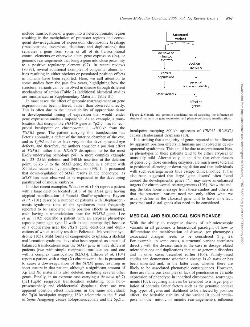
MEDICAL AND BIOLOGICAL SIGNIFICANCE
With the ability to recognize dozens of sub-microscopicvariants in all genomes, a hierarchical paradigm of how todifferentiate the manifestation of disease- (or phenotype-)associated changes needs to be considered (Fig. 2).For example, in some cases, a structural variant correlatesdirectly with the disease, such as the case in dosage-relatedmicrodeletions and duplications that cause genomic disordersand in other cases described earlier (106). Family-basedstudies can demonstrate whether a change is de novo or hasbeen inherited and, in the latter case, whether there arelikely to be associated phenotypic consequences. However,there are numerous examples of lack of penetrance or variableexpression of phenotype in inherited chromosomal rearrange-ments (107), requiring analysis be extended to a larger popu-lation of controls. Other factors such as the genomic context(e.g. types of genes and likelihood to be affected by positioneffect), the heritable stability of the variant (it could predis-pose to other mitotic or meiotic rearrangements), influence
Figure 2. Genetic and genomic considerations of assessing the influence ofstructural variants on gene expression and phenotype/disease manifestation.
Human Molecular Genetics, 2006, Vol. 15, Review Issue 1 R61
Dow
nloaded from https://academ
ic.oup.com/hm
g/article/15/suppl_1/R57/632718 by guest on 07 February 2022
of other variants and possible parent-of-origin effects (e.g.imprinted regions) all need to be considered when evaluatingthe effect at the genic level.
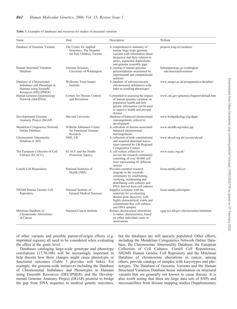
Databases cataloging large-scale genotype and phenotypecorrelations (17,70,108) will be increasingly important tohelp discern how these changes might cause phenotypic orfunctional outcomes (Table 3 provides web links). Forexample, the genome-wide initiatives including the Databaseof Chromosomal Imbalance and Phenotypes in Humansusing Ensembl Resources (DECIPHER) and the Develop-mental Genome Anatomy Project (DGAP) promise to bridgethe gap from DNA sequence to medical genetic outcomes,
but the databases are still sparsely populated. Other efforts,including the Mendelian Cytogenetics Network Online Data-base, the Chromosome Abnormality Database, the EuropeanCollection of Cell Cultures, Coriell Cell Repositories,NIGMS Human Genetic Cell Repository and the MitelmanDatabase of chromosome aberrations in cancer, amongothers, provide catalogs of samples with karyotypes and phe-notypes. The Database of Genomic Variants and the HumanStructural Variation Database house information on structuralvariants that are generally not known to cause disease. It isalso worth noting that there are large data sets of SNPs andmicrosatellites from disease mapping studies (Supplementary
Table 3. Examples of databases and resources for studies of structural variation
Name Host Description Website
Database of Genomic Variants The Centre for AppliedGenomics, The Hospitalfor Sick Children, Toronto
A comprehensive summary ofhuman large-scale genomicvariants with information aboutfrequency and their relation togenes, segmental duplicationsand genome assembly gaps
projects.tcag.ca/variation/
Human Structural VariationDatabase
Genome Sciences,University of Washington
A catalog of human genomicpolymorphisms ascertained byexperimental and computationalanalyses
humanparalogy.gs.washingtonedu/structuralvariation/
Database of ChromosomalImbalance and Phenotype inHumans using EnsemblResources (DECIPHER)
Wellcome Trust SangerInstitute
A database of sub-microscopicchromosomal imbalances withlinks to resulting phenotypes
www.sanger.ac.uk/postgenomics/decipher/
Human Genome EpidemiologyNetwork (HuGENet)
Centers for Disease Controland Prevention
Committed to assessing the impactof human genome variation onpopulation health and howgenetic information can be usedto improve health and preventdisease
www.cdc.gov/genomics/hugenet/default.htm
Developmental GenomeAnatomy Project (DGAP)
Harvard University Database of balanced chromosomalrearrangements critical todevelopment
www.bwhpathology.org/dgap/
Mendelian Cytogenetics NetworkOnline Database
Wilhelm Johannsen Centrefor Functional GenomeResearch
A collection of disease-associatedbalanced chromosomalrearrangements
www.mcndb.org/index.jsp
Chromosome AbnormalityDatabase (CAD)
NHS, UK A collection of both constitutionaland acquired abnormal karyo-types reported by UK RegionalCytogenetics Centers
www.ukcad.org.uk/cocoon/ukcad/
The European Collection of CellCultures (ECACC)
ECACC and the HealthProtection Agency
A cell-culture collection toservice the research communityconsisting of over 40 000 celllines representing 45 differentspecies
www.ecacc.org.uk/
Coriell Cell Respository National Institutes ofHealth (NIH)
Provides essential researchreagents to the scientificcommunity by establishing,verifying, maintaining anddistributing cells cultures andDNA derived from cell cultures
locus.umdnj.edu/ccr
NIGMS Human Genetic CellRepository
National Institute ofGeneral Medical Sciences
Supplies scientists with thematerials for acceleratingdisease gene discovery withhighly characterized, viable andcontaminant-free cell culturesand DNA samples
locus.umdnj.edu/nigms/
Mitelman Database ofChromosome Aberrationsin Cancer
National Cancer Institute Relates chromosomal aberrationsto tumor characteristics, basedon either individual cases orassociations
cgap.nci.nih.gov/chromosomes/mitelman
R62 Human Molecular Genetics, 2006, Vol. 15, Review Issue 1
Dow
nloaded from https://academ
ic.oup.com/hm
g/article/15/suppl_1/R57/632718 by guest on 07 February 2022

Material, Tables S2 and S3) that could be analyzed for theirputative CNV content in a manner similar to what has beendone in autism (109) and HapMap samples (6,8).
CONCLUSIONS AND FUTURE STUDIES
The complexity of variation in the human genome continuesto be unraveled, providing opportunity to explain geneticcontributions to disease in a more comprehensive manner.Going forward into the next few years, studies examiningthe role of sub-microscopic structural variation will becomea predominant theme because of significant advances in tech-nology allowing for the scanning of genomes at relativelyhigh resolution. In fact, on the basis of the numbers of discov-eries and impact alone in the past 2 years, it could be arguedthat we have entered a ‘cytogenomic’ era for discovery inhuman genetics. In large-scale population-based wholegenome association studies (Supplementary Material,Table S3), and in any disease gene study, a component ofassessing structural variation content should be incorporated.However, comprehending the contribution of these variantswill require the understanding of wide-ranging data fromsimple presence or absence (in cases and controls) to the pos-ition and context in the genome (Fig. 2). It will be importantto determine the new mutation rate (25,110) of these variantsacross the genome, including the heterochromatic regions.The next frontier will be to fully catalog all the structuralvariants in the �1 kb to 3 Mb size range discussed here, butalso all other variations in the 1 bp to 1 kb size range(Table 1), which will probably be best discerned throughpersonalized genome (re-)sequencing (111). Coupling allof this information to large cohorts of meticulously pheno-typed sample collections and corresponding databases wouldprovide insight toward understanding the etiology of manyunresolved diseases.
SUPPLEMENTARY MATERIAL
Supplementary Material is available at HMG Online.
ACKNOWLEDGEMENTS
The work was supported by funds from The Centre forApplied Genomics, Hospital for Sick Children, GenomeCanada/Ontario Genomics Institute, the Canadian Institutesof Health Research (CIHR) and the McLaughlin Centre forMolecular Medicine. L.F. is supported by the SwedishMedical Research Council, C.R.M. by The Hospital for SickChildren Research Training Centre and S.W.S. is an Investi-gator of CIHR and International Scholar of the HowardHughes Medical Institute.
Conflict of Interest statement. None declared.
REFERENCES
1. Iafrate, A.J., Feuk, L., Rivera, M.N., Listewnik, M.L., Donahoe, P.K.,Qi, Y., Scherer, S.W. and Lee, C. (2004) Detection of large-scalevariation in the human genome. Nat. Genet., 36, 949–951.
2. Sebat, J., Lakshmi, B., Troge, J., Alexander, J., Young, J., Lundin, P.,Maner, S., Massa, H., Walker, M., Chi, M. et al. (2004) Large-scale copynumber polymorphism in the human genome. Science, 305, 525–528.
3. Sharp, A.J., Locke, D.P., McGrath, S.D., Cheng, Z., Bailey, J.A.,Vallente, R.U., Pertz, L.M., Clark, R.A., Schwartz, S., Segraves, R. et al.(2005) Segmental duplications and copy-number variation in the humangenome. Am. J. Hum. Genet., 77, 78–88.
4. Tuzun, E., Sharp, A.J., Bailey, J.A., Kaul, R., Morrison, V.A.,Pertz, L.M., Haugen, E., Hayden, H., Albertson, D., Pinkel, D. et al.(2005) Fine-scale structural variation of the human genome. Nat. Genet.,37, 727–732.
5. Feuk, L., Macdonald, J.R., Tang, T., Carson, A.R., Li, M., Rao, G.,Khaja, R. and Scherer, S.W. (2005) Discovery of human inversionpolymorphisms by comparative analysis of human and chimpanzeeDNA sequence assemblies. PLoS Genet., 1, e56.
6. Conrad, D.F., Andrews, T.D., Carter, N.P., Hurles, M.E. andPritchard, J.K. (2006) A high-resolution survey of deletionpolymorphism in the human genome. Nat. Genet., 38, 75–81.
7. Hinds, D.A., Kloek, A.P., Jen, M., Chen, X. and Frazer, K.A. (2006)Common deletions and SNPs are in linkage disequilibrium in the humangenome. Nat. Genet., 38, 82–85.
8. McCarroll, S.A., Hadnott, T.N., Perry, G.H., Sabeti, P.C., Zody, M.C.,Barrett, J.C., Dallaire, S., Gabriel, S.B., Lee, C., Daly, M.J. et al. (2006)Common deletion polymorphisms in the human genome. Nat. Genet., 38,86–92.
9. Altshuler, D., Brooks, L.D., Chakravarti, A., Collins, F.S., Daly, M.J.and Donnelly, P. (2005) A haplotype map of the human genome. Nature,437, 1299–1320.
10. The International HapMap Consortium (2003) The InternationalHapMap Project. Nature, 426, 789–796.
11. Murray, J.C., Buetow, K.H., Weber, J.L., Ludwigsen, S.,Scherpbier-Heddema, T., Manion, F., Quillen, J., Sheffield, V.C.,Sunden, S., Duyk, G.M. et al. (1994) A comprehensive human linkagemap with centimorgan density. Cooperative Human Linkage Center(CHLC). Science, 265, 2049–2054.
12. Kong, A., Gudbjartsson, D.F., Sainz, J., Jonsdottir, G.M.,Gudjonsson, S.A., Richardsson, B., Sigurdardottir, S., Barnard, J.,Hallbeck, B., Masson, G. et al. (2002) A high-resolution recombinationmap of the human genome. Nat. Genet., 31, 241–247.
13. Nakamura, Y., Leppert, M., O’Connell, P., Wolff, R., Holm, T.,Culver, M., Martin, C., Fujimoto, E., Hoff, M., Kumlin, E. et al. (1987)Variable number of tandem repeat (VNTR) markers for human genemapping. Science, 235, 1616–1622.
14. Buckley, P.G., Mantripragada, K.K., Piotrowski, A., Diaz de Stahl, T.and Dumanski, J.P. (2005) Copy-number polymorphisms: mining the tipof an iceberg. Trends Genet., 21, 315–317.
15. Freeman, J.L., Perry, G.H., Feuk, L., Redon, R., McCarroll, S.A.,Althshuler, D.M., Aburatani, H., Jones, K., Tyler-Smith, C.,Hurles, M.E. et al.(2006) Copy number variation: new insights ingenome diversity. Genome Res., submitted.
16. Stankiewicz, P. and Lupski, J.R. (2002) Genome architecture,rearrangements and genomic disorders. Trends Genet., 18, 74–82.
17. Bugge, M., Bruun-Petersen, G., Brondum-Nielsen, K., Friedrich, U.,Hansen, J., Jensen, G., Jensen, P.K., Kristoffersson, U., Lundsteen, C.,Niebuhr, E. et al. (2000) Disease associated balanced chromosomerearrangements: a resource for large scale genotype–phenotypedelineation in man. J. Med. Genet., 37, 858–865.
18. Lupski, J.R. and Stankiewicz, P. (2005) Genomic disorders: molecularmechanisms for rearrangements and conveyed phenotypes. PLoS Genet.,1, e49.
19. Feuk, L., Carson, A.R. and Scherer, S.W. (2006) Structural variation inthe human genome. Nat. Rev. Genet., 7, 85–97.
20. Carter, N.P. (2004) As normal as normal can be? Nat. Genet., 36,931–932.
21. Eichler, E.E. (2006) Widening the spectrum of human genetic variation.Nat. Genet., 38, 9–11.
22. Lee, C. (2005) Vive la difference! Nat. Genet., 37, 660–661.23. Nadeau, J.H. and Lee, C. (2006) Genetics: copies count. Nature, 439,
798–799.24. Check, E. (2005) Human genome: patchwork people. Nature, 437,
1084–1086.25. van Ommen, G.J. (2005) Frequency of new copy number variation in
humans. Nat. Genet., 37, 333–334.
Human Molecular Genetics, 2006, Vol. 15, Review Issue 1 R63
Dow
nloaded from https://academ
ic.oup.com/hm
g/article/15/suppl_1/R57/632718 by guest on 07 February 2022

26. Cawthon, R.M., Weiss, R., Xu, G.F., Viskochil, D., Culver, M.,Stevens, J., Robertson, M., Dunn, D., Gesteland, R., O’Connell, P. et al.(1990) A major segment of the neurofibromatosis type 1 gene: cDNAsequence, genomic structure, and point mutations. Cell, 62, 193–201.
27. Call, K.M., Glaser, T., Ito, C.Y., Buckler, A.J., Pelletier, J., Haber, D.A.,Rose, E.A., Kral, A., Yeger, H., Lewis, W.H. et al. (1990) Isolationand characterization of a zinc finger polypeptide gene at the humanchromosome 11 Wilms’ tumor locus. Cell, 60, 509–520.
28. Royer-Pokora, B., Kunkel, L.M., Monaco, A.P., Goff, S.C.,Newburger, P.E., Baehner, R.L., Cole, F.S., Curnutte, J.T. andOrkin, S.H. (1986) Cloning the gene for an inherited humandisorder—chronic granulomatous disease—on the basis of itschromosomal location. Nature, 322, 32–38.
29. Monaco, A.P., Neve, R.L., Colletti-Feener, C., Bertelson, C.J.,Kurnit, D.M. and Kunkel, L.M. (1986) Isolation of candidate cDNAsfor portions of the Duchenne muscular dystrophy gene. Nature, 323,646–650.
30. Burghes, A.H., Logan, C., Hu, X., Belfall, B., Worton, R.G. andRay, P.N. (1987) A cDNA clone from the Duchenne/Becker musculardystrophy gene. Nature, 328, 434–437.
31. Friend, S.H., Bernards, R., Rogelj, S., Weinberg, R.A., Rapaport, J.M.,Albert, D.M. and Dryja, T.P. (1986) A human DNA segment withproperties of the gene that predisposes to retinoblastoma andosteosarcoma. Nature, 323, 643–646.
32. Gessler, M., Poustka, A., Cavenee, W., Neve, R.L., Orkin, S.H. andBruns, G.A. (1990) Homozygous deletion in Wilms tumours of azinc-finger gene identified by chromosome jumping. Nature, 343,774–778.
33. Wallace, M.R., Marchuk, D.A., Andersen, L.B., Letcher, R.,Odeh, H.M., Saulino, A.M., Fountain, J.W., Brereton, A., Nicholson, J.,Mitchell, A.L. et al. (1990) Type 1 neurofibromatosis gene: identificationof a large transcript disrupted in three NF1 patients. Science, 249,181–186.
34. Vissers, L.E., Veltman, J.A., van Kessel, A.G. and Brunner, H.G. (2005)Identification of disease genes by whole genome CGH arrays. Hum. Mol.Genet., 14 (Spec no. 2), R215–R223.
35. Rosenberg, C., Knijnenburg, J., Bakker, E., Vianna-Morgante, A.M.,Sloos, W., Otto, P.A., Kriek, M., Hansson, K., Krepischi-Santos, A.C.,Fiegler, H. et al. (2006) Array-CGH detection of micro rearrangementsin mentally retarded individuals: clinical significance of imbalancespresent both in affected children and normal parents. J. Med. Genet., 43,180–186.
36. Schoumans, J., Ruivenkamp, C., Holmberg, E., Kyllerman, M.,Anderlid, B.M. and Nordenskjold, M. (2005) Detection of chromosomalimbalances in children with idiopathic mental retardation by array basedcomparative genomic hybridisation (array-CGH). J. Med. Genet., 42,699–705.
37. de Vries, B.B., Pfundt, R., Leisink, M., Koolen, D.A., Vissers, L.E.,Janssen, I.M., Reijmersdal, S., Nillesen, W.M., Huys, E.H., Leeuw, N.et al. (2005) Diagnostic genome profiling in mental retardation.Am. J. Hum. Genet., 77, 606–616.
38. Shaw-Smith, C., Redon, R., Rickman, L., Rio, M., Willatt, L.,Fiegler, H., Firth, H., Sanlaville, D., Winter, R., Colleaux, L. et al.(2004) Microarray based comparative genomic hybridisation(array-CGH) detects submicroscopic chromosomal deletions andduplications in patients with learning disability/mental retardation anddysmorphic features. J. Med. Genet., 41, 241–248.
39. Miyake, N., Shimokawa, O., Harada, N., Sosonkina, N., Okubo, A.,Kawara, H., Okamoto, N., Kurosawa, K., Kawame, H., Iwakoshi, M.et al. (2006) BAC array CGH reveals genomic aberrations in idiopathicmental retardation. Am. J. Med. Genet. A, 140, 205–211.
40. Bauters, M., Van Esch, H., Marynen, P. and Froyen, G. (2005) Xchromosome array-CGH for the identification of novel X-linked mentalretardation genes. Eur. J. Med. Genet., 48, 263–275.
41. Lugtenberg, D., de Brouwer, A.P., Kleefstra, T., Oudakker, A.R.,Frints, S.G., Schrander-Stumpel, C.T., Fryns, J.P., Jensen, L.R.,Chelly, J., Moraine, C. et al. (2005) Chromosomal copy numberchanges in patients with non-syndromic X-linked mental retardationdetected by array CGH. J. Med. Genet., doi:10.1136/jmg.2005.036178.
42. Jamain, S., Quach, H., Betancur, C., Rastam, M., Colineaux, C.,Gillberg, I.C., Soderstrom, H., Giros, B., Leboyer, M., Gillberg, C. et al.(2003) Mutations of the X-linked genes encoding neuroligins NLGN3and NLGN4 are associated with autism. Nat. Genet., 34, 27–29.
43. Millar, J.K., Wilson-Annan, J.C., Anderson, S., Christie, S.,Taylor, M.S., Semple, C.A., Devon, R.S., Clair, D.M., Muir, W.J.,Blackwood, D.H. et al. (2000) Disruption of two novel genes by atranslocation co-segregating with schizophrenia. Hum. Mol. Genet., 9,1415–1423.
44. Taipale, M., Kaminen, N., Nopola-Hemmi, J., Haltia, T., Myllyluoma, B.,Lyytinen, H., Muller, K., Kaaranen,M., Lindsberg, P.J., Hannula-Jouppi, K.et al. (2003) A candidate gene for developmental dyslexia encodes anuclear tetratricopeptide repeat domain protein dynamically regulated inbrain. Proc. Natl Acad. Sci. USA, 100, 11553–11558.
45. Hannula-Jouppi, K., Kaminen-Ahola, N., Taipale, M., Eklund, R.,Nopola-Hemmi, J., Kaariainen, H. and Kere, J. (2005) The axonguidance receptor gene ROBO1 is a candidate gene for developmentaldyslexia. PLoS Genet., 1, e50.
46. Lai, C.S., Fisher, S.E., Hurst, J.A., Vargha-Khadem, F. andMonaco, A.P. (2001) A forkhead-domain gene is mutated in a severespeech and language disorder. Nature, 413, 519–523.
47. Abelson, J.F., Kwan, K.Y., O’Roak, B.J., Baek, D.Y., Stillman, A.A.,Morgan, T.M., Mathews, C.A., Pauls, D.L., Rasin, M.R., Gunel, M. et al.(2005) Sequence variants in SLITRK1 are associated with Tourette’ssyndrome. Science, 310, 317–320.
48. Somerville, M.J., Mervis, C.B., Young, E.J., Seo, E.J., del Campo, M.,Bamforth, S., Peregrine, E., Loo, W., Lilley, M., Perez-Jurado, L.A.et al. (2005) Severe expressive-language delay related toduplication of the Williams–Beuren locus. N. Engl. J. Med., 353,1694–1701.
49. Singleton, A.B., Farrer, M., Johnson, J., Singleton, A., Hague, S.,Kachergus, J., Hulihan, M., Peuralinna, T., Dutra, A., Nussbaum, R.et al. (2003) alpha-Synuclein locus triplication causes Parkinson’sdisease. Science, 302, 841.
50. Rovelet-Lecrux, A., Hannequin, D., Raux, G., Le Meur, N.,Laquerriere, A., Vital, A., Dumanchin, C., Feuillette, S., Brice, A.,Vercelletto, M. et al. (2006) APP locus duplication causes autosomaldominant early-onset Alzheimer disease with cerebral amyloidangiopathy. Nat. Genet., 38, 24–26.
51. Aitman, T.J., Dong, R., Vyse, T.J., Norsworthy, P.J., Johnson, M.D.,Smith, J., Mangion, J., Roberton-Lowe, C., Marshall, A.J., Petretto, E.et al. (2006) Copy number polymorphism in Fcgr3 predisposes toglomerulonephritis in rats and humans. Nature, 439, 851–855.
52. Groot, P.C., Mager, W.H. and Frants, R.R. (1991) Interpretation ofpolymorphic DNA patterns in the human alpha-amylase multigenefamily. Genomics, 10, 779–785.
53. Hollox, E.J., Armour, J.A. and Barber, J.C. (2003) Extensive normalcopy number variation of a beta-defensin antimicrobial-gene cluster.Am. J. Hum. Genet., 73, 591–600.
54. Aldred, P.M., Hollox, E.J. and Armour, J.A. (2005) Copy numberpolymorphism and expression level variation of the humanfalphag-defensin genes DEFA1 and DEFA3. Hum. Mol. Genet., 14,2045–2052.
55. Linzmeier, R.M. and Ganz, T. (2005) Human defensin gene copynumber polymorphisms: comprehensive analysis of independentvariation in alpha- and beta-defensin regions at 8p22–p23. Genomics,86, 423–430.
56. Gonzalez, E., Kulkarni, H., Bolivar, H., Mangano, A., Sanchez, R.,Catano, G., Nibbs, R.J., Freedman, B.I., Quinones, M.P., Bamshad, M.J.et al. (2005) The influence of CCL3L1 gene-containing segmentalduplications on HIV-1/AIDS susceptibility. Science, 307, 1434–1440.
57. Sprenger, R., Schlagenhaufer, R., Kerb, R., Bruhn, C., Brockmoller, J.,Roots, I. and Brinkmann, U. (2000) Characterization of the glutathioneS-transferase GSTT1 deletion: discrimination of all genotypes bypolymerase chain reaction indicates a trimodular genotype–phenotypecorrelation. Pharmacogenetics, 10, 557–565.
58. Nguyen, D.Q., Webber, C. and Ponting, C.P. (2006) Bias of selection onhuman copy-number variants. PLoS Genet., 2, e20.
59. Hayes, J.D. and Strange, R.C. (2000) Glutathione S-transferasepolymorphisms and their biological consequences. Pharmacology, 61,154–166.
60. McLellan, R.A., Oscarson, M., Alexandrie, A.K., Seidegard, J.,Evans, D.A., Rannug, A. and Ingelman-Sundberg, M. (1997)Characterization of a human glutathione S-transferase mu clustercontaining a duplicated GSTM1 gene that causes ultrarapid enzymeactivity. Mol. Pharmacol., 52, 958–965.
R64 Human Molecular Genetics, 2006, Vol. 15, Review Issue 1
Dow
nloaded from https://academ
ic.oup.com/hm
g/article/15/suppl_1/R57/632718 by guest on 07 February 2022
61. Rao, Y., Hoffmann, E., Zia, M., Bodin, L., Zeman, M., Sellers, E.M. andTyndale, R.F. (2000) Duplications and defects in the CYP2A6 gene:identification, genotyping, and in vivo effects on smoking. Mol.
Pharmacol., 58, 747–755.62. Koppens, P.F., Hoogenboezem, T. and Degenhart, H.J. (2002)
Duplication of the CYP21A2 gene complicates mutation analysis ofsteroid 21-hydroxylase deficiency: characteristics of three unusualhaplotypes. Hum. Genet., 111, 405–410.
63. Dalen, P., Dahl, M.L., Ruiz, M.L., Nordin, J. and Bertilsson, L. (1998)10-Hydroxylation of nortriptyline in white persons with 0, 1, 2, 3, and 13functional CYP2D6 genes. Clin. Pharmacol. Ther., 63, 444–452.
64. Mitsunaga, Y., Kubota, T., Ishiguro, A., Yamada, Y., Sasaki, H.,Chiba, K. and Iga, T. (2002) Frequent occurrence of CYP2D6�10duplication allele in a Japanese population. Mutat. Res., 505, 83–85.
65. Ingelman-Sundberg, M. (2002) Polymorphism of cytochrome P450 andxenobiotic toxicity. Toxicology, 181–182, 447–452.
66. Chung, E.K., Yang, Y., Rennebohm, R.M., Lokki, M.L., Higgins, G.C.,Jones, K.N., Zhou, B., Blanchong, C.A. and Yu, C.Y. (2002) Geneticsophistication of human complement components C4A and C4B andRP-C4-CYP21-TNX (RCCX) modules in the major histocompatibilitycomplex. Am. J. Hum. Genet., 71, 823–837.
67. Osborne, L.R., Li, M., Pober, B., Chitayat, D., Bodurtha, J., Mandel, A.,Costa, T., Grebe, T., Cox, S., Tsui, L.C. et al. (2001) A 1.5 million-basepair inversion polymorphism in families with Williams–Beurensyndrome. Nat. Genet., 29, 321–325.
68. Gimelli, G., Pujana, M.A., Patricelli, M.G., Russo, S., Giardino, D.,Larizza, L., Cheung, J., Armengol, L., Schinzel, A., Estivill, X. et al.(2003) Genomic inversions of human chromosome 15q11–q13 inmothers of Angelman syndrome patients with class II (BP2/3) deletions.Hum. Mol. Genet., 12, 849–858.
69. Visser, R., Shimokawa, O., Harada, N., Kinoshita, A., Ohta, T.,Niikawa, N. and Matsumoto, N. (2005) Identification of a 3.0-kb majorrecombination hotspot in patients with Sotos syndrome who carry acommon 1.9-Mb microdeletion. Am. J. Hum. Genet., 76, 52–67.
70. Scherer, S.W., Cheung, J., MacDonald, J.R., Osborne, L.R.,Nakabayashi, K., Herbrick, J.A., Carson, A.R., Parker-Katiraee, L.,Skaug, J., Khaja, R. et al. (2003) Human chromosome 7: DNA sequenceand biology. Science, 300, 767–772.
71. Ovcharenko, I., Loots, G.G., Nobrega, M.A., Hardison, R.C., Miller, W.and Stubbs, L. (2005) Evolution and functional classification ofvertebrate gene deserts. Genome Res., 15, 137–145.
72. Hillier, L.W., Graves, T.A., Fulton, R.S., Fulton, L.A., Pepin, K.H.,Minx, P., Wagner-McPherson, C., Layman, D., Wylie, K., Sekhon, M.et al. (2005) Generation and annotation of the DNA sequences of humanchromosomes 2 and 4. Nature, 434, 724–731.
73. Schotta, G., Ebert, A., Dorn, R. and Reuter, G. (2003) Position-effectvariegation and the genetic dissection of chromatin regulation inDrosophila. Semin. Cell Dev. Biol., 14, 67–75.
74. Tham, W.H. and Zakian, V.A. (2002) Transcriptional silencing atSaccharomyces telomeres: implications for other organisms. Oncogene,21, 512–521.
75. Crolla, J.A. and van Heyningen, V. (2002) Frequent chromosomeaberrations revealed by molecular cytogenetic studies in patients withaniridia. Am. J. Hum. Genet., 71, 1138–1149.
76. Lauderdale, J.D., Wilensky, J.S., Oliver, E.R., Walton, D.S. andGlaser, T. (2000) 30 deletions cause aniridia by preventing PAX6 geneexpression. Proc. Natl Acad. Sci. USA, 97, 13755–13759.
77. Fantes, J., Redeker, B., Breen, M., Boyle, S., Brown, J., Fletcher, J.,Jones, S., Bickmore, W., Fukushima, Y., Mannens, M. et al. (1995)Aniridia-associated cytogenetic rearrangements suggest that a positioneffect may cause the mutant phenotype. Hum. Mol. Genet., 4, 415–422.
78. Crolla, J.A., Cross, I., Atkey, N., Wright, M. and Oley, C.A. (1996)FISH studies in a patient with sporadic aniridia and t(7;11) (q31.2;p13).J. Med. Genet., 33, 66–68.
79. Belloni, E., Muenke, M., Roessler, E., Traverso, G., Siegel-Bartelt, J.,Frumkin, A., Mitchell, H.F., Donis-Keller, H., Helms, C., Hing, A.V.et al. (1996) Identification of sonic hedgehog as a candidate generesponsible for holoprosencephaly. Nat. Genet., 14, 353–356.
80. Fernandez, B.A., Siegel-Bartelt, J., Herbrick, J.A., Teshima, I. andScherer, S.W. (2005) Holoprosencephaly and cleidocranial dysplasia in apatient due to two position-effect mutations: case report and review ofthe literature. Clin. Genet., 68, 349–359.
81. Wallis, D.E., Roessler, E., Hehr, U., Nanni, L., Wiltshire, T.,Richieri-Costa, A., Gillessen-Kaesbach, G., Zackai, E.H., Rommens, J.and Muenke, M. (1999) Mutations in the homeodomain of the humanSIX3 gene cause holoprosencephaly. Nat. Genet., 22, 196–198.
82. Velagaleti, G.V., Bien-Willner, G.A., Northup, J.K., Lockhart, L.H.,Hawkins, J.C., Jalal, S.M., Withers, M., Lupski, J.R. and Stankiewicz, P.(2005) Position effects due to chromosome breakpoints that mapapproximately 900 kb upstream and approximately 1.3 Mb downstreamof SOX9 in two patients with campomelic dysplasia. Am. J. Hum.Genet., 76, 652–662.
83. Erdel, M., Lane, A.H., Fresser, F., Probst, P., Utermann, G. and Scherer, G.(2004) A new campomelic dysplasia translocation breakpoint maps400 kb from SOX9. Eur. J. Hum. Genet., Suppl. 12, 136.
84. Wagner, T., Wirth, J., Meyer, J., Zabel, B., Held, M., Zimmer, J.,Pasantes, J., Bricarelli, F.D., Keutel, J., Hustert, E. et al. (1994)Autosomal sex reversal and campomelic dysplasia are caused bymutations in and around the SRY-related gene SOX9. Cell, 79,1111–1120.
85. Ninomiya, S., Isomura, M., Narahara, K., Seino, Y. and Nakamura, Y.(1996) Isolation of a testis-specific cDNA on chromosome 17q from aregion adjacent to the breakpoint of t(12;17) observed in a patient withacampomelic campomelic dysplasia and sex reversal. Hum. Mol. Genet.,5, 69–72.
86. Foster, J.W., Dominguez-Steglich, M.A., Guioli, S., Kowk, G.,Weller, P.A., Stevanovic, M., Weissenbach, J., Mansour, S., Young, I.D.,Goodfellow, P.N. et al. (1994) Campomelic dysplasia and autosomal sexreversal caused by mutations in an SRY-related gene. Nature, 372,525–530.
87. Pop, R., Conz, C., Lindenberg, K.S., Blesson, S., Schmalenberger, B.,Briault, S., Pfeifer, D. and Scherer, G. (2004) Screening of the 1 MbSOX9 50 control region by array CGH identifies a large deletion in a caseof campomelic dysplasia with XY sex reversal. J. Med. Genet., 41, e47.
88. Wirth, J., Wagner, T., Meyer, J., Pfeiffer, R.A., Tietze, H.U.,Schempp, W. and Scherer, G. (1996) Translocation breakpoints inthree patients with campomelic dysplasia and autosomal sex reversalmap more than 130 kb from SOX9. Hum. Genet., 97, 186–193.
89. Wunderle, V.M., Critcher, R., Hastie, N., Goodfellow, P.N. andSchedl, A. (1998) Deletion of long-range regulatory elements upstreamof SOX9 causes campomelic dysplasia. Proc. Natl Acad. Sci. USA, 95,10649–10654.
90. Pfeifer, D., Kist, R., Dewar, K., Devon, K., Lander, E.S., Birren, B.,Korniszewski, L., Back, E. and Scherer, G. (1999) Campomelicdysplasia translocation breakpoints are scattered over 1 Mb proximal toSOX9: evidence for an extended control region. Am. J. Hum. Genet., 65,111–124.
91. Romao, L., Osorio-Almeida, L., Higgs, D.R., Lavinha, J. andLiebhaber, S.A. (1991) Alpha-thalassemia resulting from deletion ofregulatory sequences far upstream of the alpha-globin structural genes.Blood, 78, 1589–1595.
92. Higgs, D.R., Wood, W.G., Jarman, A.P., Sharpe, J., Lida, J.,Pretorius, I.M. and Ayyub, H. (1990) A major positive regulatoryregion located far upstream of the human alpha-globin gene locus. GenesDev., 4, 1588–1601.
93. Driscoll, M.C., Dobkin, C.S. and Alter, B.P. (1989) Gamma deltabeta-thalassemia due to a de novo mutation deleting the 50 beta-globingene activation-region hypersensitive sites. Proc. Natl Acad. Sci. USA,86, 7470–7474.
94. Barbour, V.M., Tufarelli, C., Sharpe, J.A., Smith, Z.E., Ayyub, H.,Heinlein, C.A., Sloane-Stanley, J., Indrak, K., Wood, W.G. andHiggs, D.R. (2000) Alpha-thalassemia resulting from a negativechromosomal position effect. Blood, 96, 800–807.
95. McElreavey, K., Vilain, E., Barbaux, S., Fuqua, J.S., Fechner, P.Y.,Souleyreau, N., Doco-Fenzy, M., Gabriel, R., Quereux, C., Fellous, M.et al. (1996) Loss of sequences 30 to the testis-determining gene, SRY,including the Y pseudoautosomal boundary associated with partialtesticular determination. Proc. Natl Acad. Sci. USA, 93, 8590–8594.
96. McElreavy, K., Vilain, E., Abbas, N., Costa, J.M., Souleyreau, N.,Kucheria, K., Boucekkine, C., Thibaud, E., Brauner, R., Flamant, F.et al. (1992) XY sex reversal associated with a deletion 50 to the SRY‘HMG box’ in the testis-determining region. Proc. Natl Acad. Sci. USA,89, 11016–11020.
97. Kleinjan, D.J. and van Heyningen, V. (1998) Position effect in humangenetic disease. Hum. Mol. Genet., 7, 1611–1618.
Human Molecular Genetics, 2006, Vol. 15, Review Issue 1 R65
Dow
nloaded from https://academ
ic.oup.com/hm
g/article/15/suppl_1/R57/632718 by guest on 07 February 2022

98. David, D., Cardoso, J., Marques, B., Marques, R., Silva, E.D., Santos, H.and Boavida, M.G. (2003) Molecular characterization of a familialtranslocation implicates disruption of HDAC9 and possible positioneffect on TGFbeta2 in the pathogenesis of Peters’ anomaly. Genomics,81, 489–503.
99. Bowl, M.R., Nesbit, M.A., Harding, B., Levy, E., Jefferson, A.,Volpi, E., Rizzoti, K., Lovell-Badge, R., Schlessinger, D., Whyte, M.P.et al. (2005) An interstitial deletion–insertion involving chromosomes2p25.3 and Xq27.1, near SOX3, causes X-linked recessivehypoparathyroidism. J. Clin. Invest., 115, 2822–2831.
100. Wakui, K., Gregato, G., Ballif, B.C., Glotzbach, C.D., Bailey, K.A.,Kuo, P.L., Sue, W.C., Sheffield, L.J., Irons, M., Gomez, E.G. et al.(2005) Construction of a natural panel of 11p11.2 deletions and furtherdelineation of the critical region involved in Potocki–Shaffer syndrome.Eur. J. Hum. Genet., 13, 528–540.
101. Beysen, D., Raes, J., Leroy, B.P., Lucassen, A., Yates, J.R.,Clayton-Smith, J., Ilyina, H., Brooks, S.S., Christin-Maitre, S.,Fellous, M. et al. (2005) Deletions involving long-range conservednongenic sequences upstream and downstream of FOXL2 as a noveldisease-causing mechanism in blepharophimosis syndrome. Am. J. Hum.Genet., 77, 205–218.
102. Lee, J.A., Madrid, R.E., Sperle, K., Ritterson, C.M., Hobson, G.M.,Garbern, J., Lupski, J.R. and Inoue, K. (2006) Spastic paraplegia type 2associated with axonal neuropathy and apparent PLP1 position effect.Ann. Neurol., 59, 398–403.
103. Muncke, N., Wogatzky, B.S., Breuning, M., Sistermans, E.A., Endris, V.,Ross, M., Vetrie, D., Catsman-Berrevoets, C.E. and Rappold, G. (2004)Position effect on PLP1 may cause a subset of Pelizaeus–Merzbacherdisease symptoms. J. Med. Genet., 41, e121.
104. Ellison, J.W., Tekin, M., Sikes, K.S., Yankowitz, J., Shapiro, L.,Rappold, G.A. and Neely, K.E. (2002) Molecular characterization of aring X chromosome in a male with short stature. Hum. Genet., 110,322–326.
105. Lettice, L.A. and Hill, R.E. (2005) Preaxial polydactyly: a model fordefective long-range regulation in congenital abnormalities. Curr. Opin.Genet. Dev., 15, 294–300.
106. Inoue, K. and Lupski, J.R. (2002) Molecular mechanisms for genomicdisorders. Annu. Rev. Genomics Hum. Genet., 3, 199–242.
107. Ravnan, J.B., Tepperberg, J.H., Papenhausen, P., Lamb, A.N.,Hedrick, J., Eash, D., Ledbetter, D.H. and Martin, C.L. (2005)Subtelomere FISH analysis of 11 688 cases: an evaluation of thefrequency and pattern of subtelomere rearrangements in individuals withdevelopmental disabilities. J. Med. Genet., doi:10.1136/jmg.2005.036350.
108. Ioannidis, J.P., Gwinn, M., Little, J., Higgins, J.P., Bernstein, J.L.,Boffetta, P., Bondy, M., Bray, M.S., Brenchley, P.E., Buffler, P.A. et al.(2006) A road map for efficient and reliable human genomeepidemiology. Nat. Genet., 38, 3–5.
109. Yu, C.E., Dawson, G., Munson, J., D’Souza, I., Osterling, J., Estes, A.,Leutenegger, A.L., Flodman, P., Smith, M., Raskind, W.H. et al. (2002)Presence of large deletions in kindreds with autism. Am. J. Hum. Genet.,71, 100–115.
110. Repping, S., van Daalen, S.K., Brown, L.G., Korver, C.M., Lange, J.,Marszalek, J.D., Pyntikova, T., van der Veen, F., Skaletsky, H.,
Page, D.C. et al. (2006) High mutation rates have driven extensivestructural polymorphism among human Y chromosomes. Nat. Genet.,doi:10.1038/ng1754.
111. Shendure, J., Mitra, R.D., Varma, C. and Church, G.M. (2004) Advancedsequencing technologies: methods and goals. Nat. Rev. Genet., 5, 335–344.
112. International Human Genome Sequencing Consortium (2004) Finishingthe euchromatic sequence of the human genome. Nature, 431, 931–945.
113. Kruglyak, L. and Nickerson, D.A. (2001) Variation is the spice of life.Nat. Genet., 27, 234–236.
114. Lander, E.S., Linton, L.M., Birren, B., Nusbaum, C., Zody, M.C.,Baldwin, J., Devon, K., Dewar, K., Doyle, M., FitzHugh, W. et al.(2001) Initial sequencing and analysis of the human genome. Nature,409, 860–921.
115. Weber, J.L., David, D., Heil, J., Fan, Y., Zhao, C. and Marth, G. (2002)Human diallelic insertion/deletion polymorphisms. Am. J. Hum. Genet.,71, 854–862.
116. Dawson, E., Chen, Y., Hunt, S., Smink, L.J., Hunt, A., Rice, K.,Livingston, S., Bumpstead, S., Bruskiewich, R., Sham, P. et al. (2001) ASNP resource for human chromosome 22: extracting dense clusters ofSNPs from the genomic sequence. Genome Res., 11, 170–178.
117. Litt, M. and Luty, J.A. (1989) A hypervariable microsatellite revealed byin vitro amplification of a dinucleotide repeat within the cardiac muscleactin gene. Am. J. Hum. Genet., 44, 397–401.
118. Ellegren, H. (2004) Microsatellites: simple sequences with complexevolution. Nat. Rev. Genet., 5, 435–445.
119. Jeffreys, A.J., Wilson, V. and Thein, S.L. (1985) Hypervariable‘minisatellite’ regions in human DNA. Nature, 314, 67–73.
120. Naslund, K., Saetre, P., von Salome, J., Bergstrom, T.F., Jareborg, N.and Jazin, E. (2005) Genome-wide prediction of human VNTRs.Genomics, 85, 24–35.
121. Fredman, D., White, S.J., Potter, S., Eichler, E.E., Dunnen, J.T. andBrookes, A.J. (2004) Complex SNP-related sequence variation insegmental genome duplications. Nat. Genet., 36, 861–866.
122. Van Dyke, D.L., Weiss, L., Roberson, J.R. and Babu, V.R. (1983) Thefrequency and mutation rate of balanced autosomal rearrangements inman estimated from prenatal genetic studies for advanced maternal age.Am. J. Hum. Genet., 35, 301–308.
123. Gardner, R.J.M. and Sunderland, G.R. (2004) Chromosome Abnormalititesand Genetic Counseling, 3rd edn. Oxford University Press, New York.
124. Warburton, D. (1991) De novo balanced chromosome rearrangementsand extra marker chromosomes identified at prenatal diagnosis: clinicalsignificance and distribution of breakpoints. Am. J. Hum. Genet., 49,995–1013.
125. Nussbaum, R.L., McInnes, R.R. and Willard, H.F. (2004) Thompson &Thompson Genetics in Medicine, 6th edn. W.B. Saunders, Philadelphia,PA.
126. Lee, J.A., Cheung, S.W., Ward, P.A., Inoue, K. and Lupski, J.R. (2005)Prenatal diagnosis of PLP1 copy number by array comparative genomichybridization. Prenat. Diagn., 25, 1188–1191.
127. Marlin, S., Blanchard, S., Slim, R., Lacombe, D., Denoyelle, F.,Alessandri, J.L., Calzolari, E., Drouin-Garraud, V., Ferraz, F.G.,Fourmaintraux, A. et al. (1999) Townes–Brocks syndrome: detection ofa SALL1 mutation hot spot and evidence for a position effect in onepatient. Hum. Mutat., 14, 377–386.
R66 Human Molecular Genetics, 2006, Vol. 15, Review Issue 1
Dow
nloaded from https://academ
ic.oup.com/hm
g/article/15/suppl_1/R57/632718 by guest on 07 February 2022