Single-Molecule Imaging of Transcriptionally Coupled and Uncoupled Splicing
Upload
khangminh22Category
view
4download
0
�����������������
Citation: Kim, B.-H.; Woo, T.-G.;
Kang, S.-M.; Park, S.; Park, B.-J.
Splicing Variants, Protein-Protein
Interactions, and Drug Targeting in
Hutchinson-Gilford Progeria
Syndrome and Small Cell Lung
Cancer. Genes 2022, 13, 165.
https://doi.org/10.3390/
genes13020165
Academic Editors: Christos
K. Kontos and Pinelopi I. Artemaki
Received: 11 December 2021
Accepted: 17 January 2022
Published: 18 January 2022
Publisher’s Note: MDPI stays neutral
with regard to jurisdictional claims in
published maps and institutional affil-
iations.
Copyright: © 2022 by the authors.
Licensee MDPI, Basel, Switzerland.
This article is an open access article
distributed under the terms and
conditions of the Creative Commons
Attribution (CC BY) license (https://
creativecommons.org/licenses/by/
4.0/).
genesG C A T
T A C G
G C A T
Review
Splicing Variants, Protein-Protein Interactions, and DrugTargeting in Hutchinson-Gilford Progeria Syndrome and SmallCell Lung CancerBae-Hoon Kim 1, Tae-Gyun Woo 1, So-Mi Kang 2, Soyoung Park 2 and Bum-Joon Park 1,2,*
1 Rare Disease R&D Center, PRG S&T Co., Ltd., Busan 46241, Korea; [email protected] (B.-H.K.);[email protected] (T.-G.W.)
2 Department of Molecular Biology, College of Natural Science, Pusan National University, Busan 46274, Korea;[email protected] (S.-M.K.); [email protected] (S.P.)
* Correspondence: [email protected]
Abstract: Alternative splicing (AS) is a biological operation that enables a messenger RNA to encodeprotein variants (isoforms) that give one gene several functions or properties. This process providesone of the major sources of use for understanding the proteomic diversity of multicellular organisms.In combination with post-translational modifications, it contributes to generating a variety of protein–protein interactions (PPIs) that are essential to cellular homeostasis or proteostasis. However, cellsexposed to many kinds of stresses (aging, genetic changes, carcinogens, etc.) sometimes derive canceror disease onset from aberrant PPIs caused by DNA mutations. In this review, we summarize howsplicing variants may form a neomorphic protein complex and cause diseases such as Hutchinson-Gilford progeria syndrome (HGPS) and small cell lung cancer (SCLC), and we discuss how protein–protein interfaces obtained from the variants may represent efficient therapeutic target sites to treatHGPS and SCLC.
Keywords: Hutchinson-Gilford progeria syndrome (HGPS); small cell lung cancer (SCLC); alternativesplicing (AS); Progerin; DX2; protein-protein interactions (PPIs)
1. Introduction
Around 44 years ago, Joseph Sambrook discovered the exon and intron in a genefrom an adenovirus, and Walter Gilbert suggested that different exon fusions in a genemight be spliced together to make different mRNA isoforms [1,2]. As a result of scientists’efforts (the Human Genome Project) to create a global view of genomes, 20 years ago,scientists have calculated that an average human protein-coding gene has 8.8 exons, withan average of 145 nt. The intron length and the lengths of the 5′ and 3′ UTR are, on average3365 nt, 770 nt, and 300 nt, respectively [3]. Most protein-coding genes have introns thatare degraded in the nucleus by the RNA splicing machinery. Exon usage is very oftenalternative. The cell decides to get rid of introns from the pre-mRNA or include exon partsin the mature mRNA [4]. There are around 20,000 human gene-encoding proteins andaround 150,000 isoforms from RNA transcripts. Consequently, in general, each humangene has about seven RNA isoforms. Recently, it was reported that over 30% of tissue-dependent transcriptional variations are covered by local splicing variations, which aredescribed as a split in a splice graph into or from a single exon [5]. The resulting differencesamong the mature mRNA isoforms produced by alternative splicing (AS) mean they canencode different protein products, or affect the localization, stability, and translation ofmRNA. When we consider that the high level of complexity in the human genome maybe illustrated by the variability of each gene with AS, it is natural to think of the fine-tuning control of these as necessary and flexibility as risky. Considering the importanceof RNA splicing for protein diversity and keeping the function of the organism, it is clear
Genes 2022, 13, 165. https://doi.org/10.3390/genes13020165 https://www.mdpi.com/journal/genes
Genes 2022, 13, 165 2 of 11
that disrupting normal splicing patterns can cause gene dysregulation and disease. Forexample, autosomal dominant forms with mutations in the splicing core factors can causeretinitis pigmentosa and Cerebro-Costo-Mandibular Syndrome [6–10], although there arefew reports of mutations in core splicing elements that result in human diseases. This islikely because disruptions of basic factors in the splicing apparatus are normally morefatal compared to mutations deriving from aberrant splicing of each gene. [11]. Besidessplicing core factors, many human diseases linked to defective splicing have been reported.Aberrant splicing could be caused by mutations disturbing either trans-acting regulatorygenes (proteins) or cis-acting regulatory sequences, with the latter more frequently recorded.
It was recently reported that AS affects more than 85% of human protein-coding genesfor cells and increases their use of genetic information [12]. Protein parts are encoded byapproximately 75% alternative exons [13], and the alternate use of exons permits multipleproteins to be made from one gene, which intensifies the coding potential of the genome.It is suggested that most alternative exons encode coiled or loop regions on the proteinsurface, meaning AS can affect the functional interface formation of many proteins [14].Alternative splicing generates protein isoforms with various biological properties, whichcan significantly affect protein–protein interaction (PPI), subcellular localization, or enzy-matic ability [15]. This means that each cell might have evolved to increase the numberof protein pools to respond more efficiently to harmful stress conditions brought by AS.PPIs are the main biochemical mechanisms of cellular life and are frequently disturbed indisease condition. We predict that the numbers of protein interactions (an interactome)in humans may add up to between 130,000 and 600,000 [16,17] (the number has beenincreasing overtime). Protein interactions in functional multi-protein complexes that haveroles in basic processes such as protein transport, making an RNA copy of a gene sequence(transcription), producing proteins from mRNA (translation), interaction/communicationbetween cells, protein modification, signaling cascades, and functional holoenzyme canbe explained by the PPIs. It is not surprising that when the homeostatic condition ofan organism or a single cell is interrupted (as a result of several stresses or in a diseasecondition), the normal patterns of PPIs can be disturbed. Furthermore, aberrant PPIs fromsplice variants, which are produced from mutations of a single gene in cis, could be thecauses of cancerous or disease-onset cells such as those of Hutchinson-Gilford progeriasyndrome (HGPS) and Small Cell Lung Cancer (SCLC) [18,19] (Figures 1 and 2).
Genes 2022, 13, x FOR PEER REVIEW 3 of 11
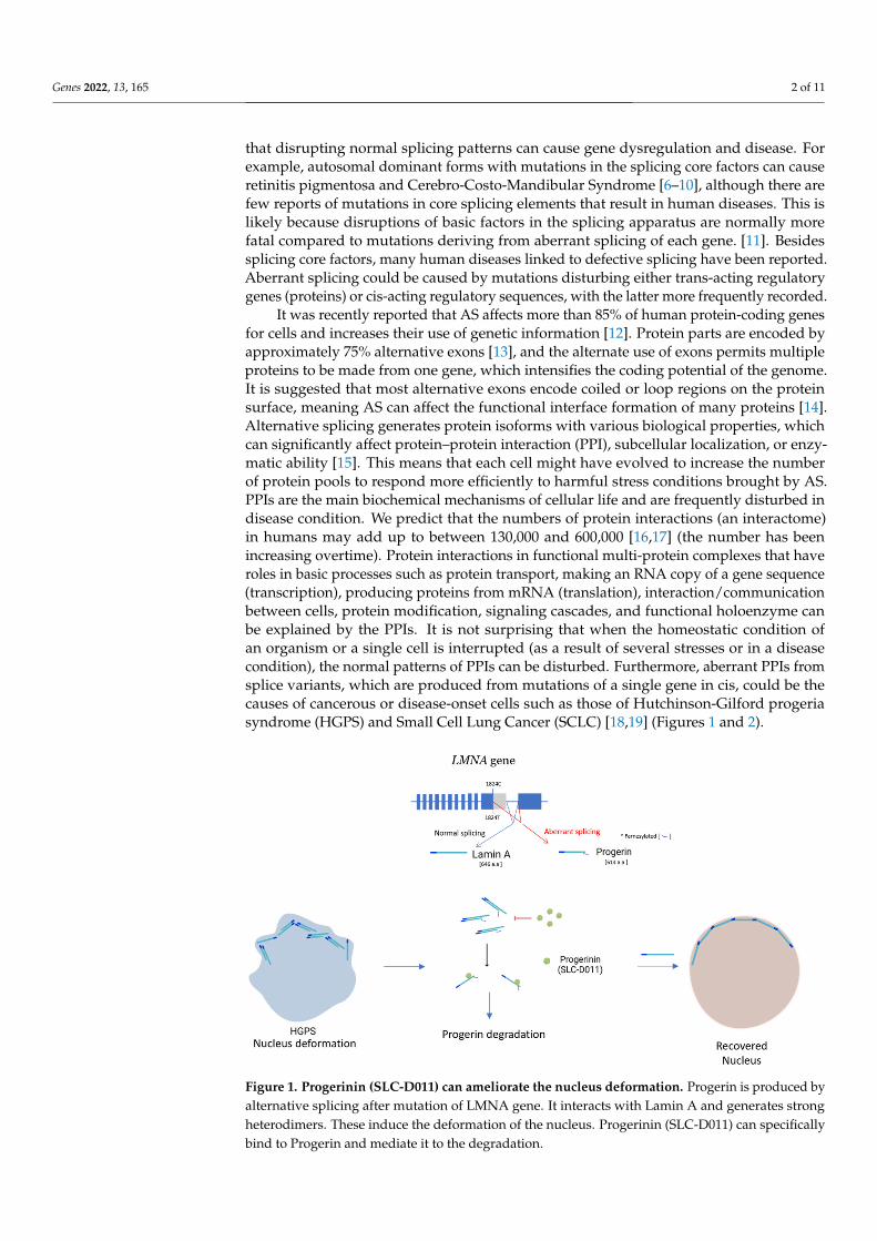
Figure 1. Progerinin (SLC-D011) can ameliorate the nucleus deformation. Progerin is produced by alternative splicing after mutation of LMNA gene. It interacts with Lamin A and generates strong heterodimers. These induce the deformation of the nucleus. Progerinin (SLC-D011) can specifically bind to Progerin and mediate it to the degradation.
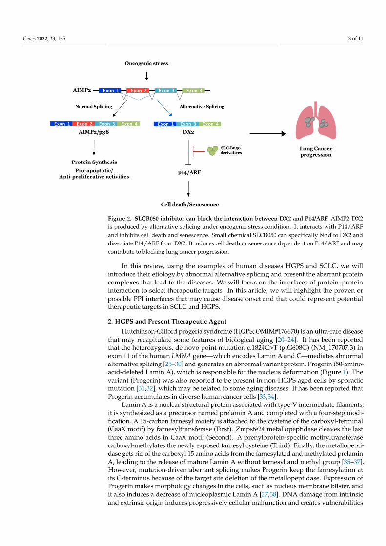
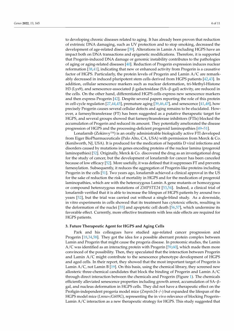
Figure 2. SLCB050 inhibitor can block the interaction between DX2 and P14/ARF. AIMP2-DX2 is produced by alternative splicing under oncogenic stress condition. It interacts with P14/ARF and inhibits cell death and senescence. Small chemical SLCB050 can specifically bind to DX2 and disso-ciate P14/ARF from DX2. It induces cell death or senescence dependent on P14/ARF and may con-tribute to blocking lung cancer progression.
2. HGPS and Present Therapeutic Agent Hutchinson-Gilford progeria syndrome (HGPS; OMIM#176670) is an ultra-rare dis-
ease that may recapitulate some features of biological aging [20–24]. It has been reported that the heterozygous, de novo point mutation c.1824C>T (p.G608G) (NM_170707.3) in exon 11 of the human LMNA gene—which encodes Lamin A and C—mediates abnormal alternative splicing [25–30] and generates an abnormal variant protein, Progerin (50-amino-acid-deleted Lamin A), which is responsible for the nucleus deformation (Figure
Figure 1. Progerinin (SLC-D011) can ameliorate the nucleus deformation. Progerin is produced byalternative splicing after mutation of LMNA gene. It interacts with Lamin A and generates strongheterodimers. These induce the deformation of the nucleus. Progerinin (SLC-D011) can specificallybind to Progerin and mediate it to the degradation.
Genes 2022, 13, 165 3 of 11
Genes 2022, 13, x FOR PEER REVIEW 3 of 11
Figure 1. Progerinin (SLC-D011) can ameliorate the nucleus deformation. Progerin is produced by alternative splicing after mutation of LMNA gene. It interacts with Lamin A and generates strong heterodimers. These induce the deformation of the nucleus. Progerinin (SLC-D011) can specifically bind to Progerin and mediate it to the degradation.
Figure 2. SLCB050 inhibitor can block the interaction between DX2 and P14/ARF. AIMP2-DX2 is produced by alternative splicing under oncogenic stress condition. It interacts with P14/ARF and inhibits cell death and senescence. Small chemical SLCB050 can specifically bind to DX2 and disso-ciate P14/ARF from DX2. It induces cell death or senescence dependent on P14/ARF and may con-tribute to blocking lung cancer progression.
2. HGPS and Present Therapeutic Agent Hutchinson-Gilford progeria syndrome (HGPS; OMIM#176670) is an ultra-rare dis-
ease that may recapitulate some features of biological aging [20–24]. It has been reported that the heterozygous, de novo point mutation c.1824C>T (p.G608G) (NM_170707.3) in exon 11 of the human LMNA gene—which encodes Lamin A and C—mediates abnormal alternative splicing [25–30] and generates an abnormal variant protein, Progerin (50-amino-acid-deleted Lamin A), which is responsible for the nucleus deformation (Figure
Figure 2. SLCB050 inhibitor can block the interaction between DX2 and P14/ARF. AIMP2-DX2is produced by alternative splicing under oncogenic stress condition. It interacts with P14/ARFand inhibits cell death and senescence. Small chemical SLCB050 can specifically bind to DX2 anddissociate P14/ARF from DX2. It induces cell death or senescence dependent on P14/ARF and maycontribute to blocking lung cancer progression.
In this review, using the examples of human diseases HGPS and SCLC, we willintroduce their etiology by abnormal alternative splicing and present the aberrant proteincomplexes that lead to the diseases. We will focus on the interfaces of protein–proteininteraction to select therapeutic targets. In this article, we will highlight the proven orpossible PPI interfaces that may cause disease onset and that could represent potentialtherapeutic targets in SCLC and HGPS.
2. HGPS and Present Therapeutic Agent
Hutchinson-Gilford progeria syndrome (HGPS; OMIM#176670) is an ultra-rare diseasethat may recapitulate some features of biological aging [20–24]. It has been reportedthat the heterozygous, de novo point mutation c.1824C>T (p.G608G) (NM_170707.3) inexon 11 of the human LMNA gene—which encodes Lamin A and C—mediates abnormalalternative splicing [25–30] and generates an abnormal variant protein, Progerin (50-amino-acid-deleted Lamin A), which is responsible for the nucleus deformation (Figure 1). Thevariant (Progerin) was also reported to be present in non-HGPS aged cells by sporadicmutation [31,32], which may be related to some aging diseases. It has been reported thatProgerin accumulates in diverse human cancer cells [33,34].
Lamin A is a nuclear structural protein associated with type-V intermediate filaments;it is synthesized as a precursor named prelamin A and completed with a four-step modi-fication. A 15-carbon farnesyl moiety is attached to the cysteine of the carboxyl-terminal(CaaX motif) by farnesyltransferase (First). Zmpste24 metallopeptidase cleaves the lastthree amino acids in CaaX motif (Second). A prenylprotein-specific methyltransferasecarboxyl-methylates the newly exposed farnesyl cysteine (Third). Finally, the metallopepti-dase gets rid of the carboxyl 15 amino acids from the farnesylated and methylated prelaminA, leading to the release of mature Lamin A without farnesyl and methyl group [35–37].However, mutation-driven aberrant splicing makes Progerin keep the farnesylation atits C-terminus because of the target site deletion of the metallopeptidase. Expression ofProgerin makes morphology changes in the cells, such as nucleus membrane blister, andit also induces a decrease of nucleoplasmic Lamin A [27,38]. DNA damage from intrinsicand extrinsic origin induces progressively cellular malfunction and creates vulnerabilities
Genes 2022, 13, 165 4 of 11
to developing chronic diseases related to aging. It has already been proven that reductionof extrinsic DNA damaging, such as UV protection and to stop smoking, decreased thedevelopment of age-related disease [39]. Alterations in Lamin A including HGPS have animpact both on DNA transactions and epigenetic modifications. Therefore, it is supportedthat Progerin-induced DNA damage or genomic instability contributes to the pathologiesof aging or aging-related diseases [40]. Reduction of Progerin expression induces nuclearreformation [38,41], indicating that new or enhanced activity from Progerin is a causativefactor of HGPS. Particularly, the protein levels of Progerin and Lamin A/C are remark-ably decreased in induced pluripotent stem cells derived from HGPS patients [42,43]. Inaddition, cellular senescence markers such as nuclear deformation, tri-Methyl-HistoneH3 (Lys9), and senescence-associated β galactosidase (SA–β-gal) activity, are reduced inthe cells. On the other hand, differentiated HGPS cells express new senescence markersand then express Progerin [42]. Despite several papers reporting the role of this proteinin cell-cycle regulation [27,44,45], premature aging [39,46,47], and senescence [41,48], howprecisely Progerin causes several cellular defects and aging remains to be elucidated. How-ever, a farnesyltransferase (FT) has been suggested as a putative therapeutic target forHGPS, and several groups showed that farnesyltransferase inhibitors (FTIs) blocked theaccumulation of Progerin and reduced its amount. They potentially ameliorated the diseaseprogression of HGPS and the processing-deficient progeroid laminopathies [49–51].
Lonafarnib (Zokinvy™) is an orally administrable biologically active FTI developedfrom Eiger BioPharmaceuticals (Palo Alto, CA, USA) with permission from Merck & Co.(Kenilworth, NJ, USA). It is produced for the medication of hepatitis D viral infections anddisorders caused by mutations in genes encoding proteins of the nuclear lamina (progeroidlaminopathies) [52]. Originally, Merck & Co. discovered the drug as an investigational drugfor the study of cancer, but the development of lonafarnib for cancer has been canceledbecause of low efficacy [52]. More usefully, it was defined that it suppresses FT and preventsfarnesylation. Subsequently, it reduces the aggregation of Progerin-like proteins includingProgerin in the cells [51]. Two years ago, lonafarnib achieved a clinical approval in the USfor the sake of reduction the risk of mortality in HGPS and for the medication of progeroidlaminopathies, which are with the heterozygous Lamin A gene mutants or homozygousor compound heterozygous mutations of ZMPSTE24 [53,54]. Indeed, a clinical trial oflonafarnib verified that it is able to increase the lifespan of HGPS patients by around twoyears [52], but the trial was carried out without a single-blind study. As a downside,in vitro experiments in cells showed that its treatment has cytotoxic effects, resulting inthe deformation of the nuclei [55] and apoptotic cell death [56,57], which undermine itsfavorable effect. Currently, more effective treatments with less side effects are required forHGPS patients.
3. Future Therapeutic Agent for HGPS and Aging Cells
Park and his colleagues have studied age-related cancer progression andProgerin [18,34,58]. They got the idea for a possible aberrant protein complex betweenLamin and Progerin that might cause the progeria disease. In proteomic studies, the LaminA/C was identified as an interacting protein with Progerin [59,60], which made them moreconvinced of the possibility. Then, they speculated that the interaction between Progerinand Lamin A/C might contribute to the senescence phenotype development of HGPSand aged cells. In their report, they showed that the most important target of Progerin isLamin A/C, not Lamin B [19]. On this basis, using the chemical library, they screened newallosteric three-chemical candidates that block the binding of Progerin and Lamin A/Cthrough direct interaction between the chemicals and Progerin (Figure 1). The chemicalsefficiently alleviated senescence properties including growth arrest, accumulation of SA–β-gal, and nucleus deformation in HGPS cells. They did not have a therapeutic effect on theProfigin-independent progeria model mice (Zmpste24–/–) but expanded the lifespan of theHGPS model mice (Lmna+/G609G), representing the in vivo relevance of blocking Progerin–Lamin A/C interaction as a new therapeutic strategy for HGPS. This study suggested that
Genes 2022, 13, 165 5 of 11
an optimization of chemicals that target the interface of Progerin-Lamin binding mighthelp to develop a promising medication for HGPS [19]. Thereafter, Park and his colleaguestried medical chemistry using the candidate chemical and secured an advanced drug can-didate, Progerinin (SLC-D011), in which the in vivo pharmacokinetics were improved. Thechemical could extend the lifespan of homozygous Lmna G609G mice by about 10 weeks.Furthermore, heterozygous Lmna+/G609G mice treated by oral administration lived 14weeks longer than the mice treated with lonafarnib (farnesyl-transferase inhibitor), whichcould live for only around 2 weeks longer than the control [61]. Additional comparativetoxicological studies in mice, rats, and dogs demonstrated that SLC-D011 is safe to test in ahuman clinical trial (NCT04512963).
Recently, several articles showed that the antisense oligonucleotide or base editing thattargets the point mutation c.1824C>T could rescue the aberrant splicing [62,63]. Diverseengineered Cas9 variants recognizing the sequences of modified PAM and an enhancedcleaving specificity have been evolved, and they may make it possible for us to expandthe selective scope of CRISPR/Base-editors [64–67], allowing single vector delivery by anadeno-associated vector and demonstrating to be notably practical for medicinal appli-cations. These give us an idea about the possibility of in vivo base-editing as potentialtherapeutics for many genetic diseases including HGPS by directly fixing their main reason.However, for human in vivo treatment, how much off-target effects on chromosomal levelcan be reduced in base editing steps and how efficiently viral delivery system is able toreduce side effects as like gene integration, immune activation, etc., remains to be properlyaddressed.
Furthermore, they extended the effect of Progerinin to Werner syndrome, a rareprogressive genetic disorder that results from a functional defect of the human WRNprotein, a member of the RecQ DNA helicase family [68]. The Progerin-inhibitor (SLC-D011) could ameliorate senescence in the fibroblasts and cardiomyocytes derived fromWerner syndrome-iPSCs, suggesting that Progerin is able to accumulate and cause agingphenotype under WRN-deficient conditions and the phenomenon may be prevented bySLC-D011 [69]. Interestingly, under the natural aging process, Progerin can be produced andincreased with age [47,70]. Natural aging and HGPS share common characteristics, suchas alopecia, osteolysis, atherosclerosis, and cardiovascular complications. Cardiovascularproblems are one of the most common causes of death in the elderly [71]. Progerinin(SLC-D011) could reduce fibrosis in the hearts of Lmna G609G progeroid mice and alsoimprove their heart-beat rate [61]. These results suggest that SLC-D011 may reduce theburden of cardiovascular disease in normal aging.
4. SCLC, Splicing Variant DX2, and Target Molecules
Human lung cancers consist of two groups—small cell lung cancer (SCLC) and non-small cell lung cancer (NSCLC). NSCLC has three subsets: large cell lung cancer, squamouscell lung carcinoma, and adenocarcinoma. Among human lung cancers, SCLC is involvedin around 20% of the cancers and has very aggressive properties. Smoking tobacco isthe typical threat for SCLC, and more than 90% of patients suffering from SCLC havesmoking experience [72–74]. SCLC has diverse genetic alterations, meaning it is geneticallyunstable [75,76]. Most SCLC cells have variations in chromosome 3p and frequentlyexpress mutations in oncogenes such as RB1, TP53, etc. [77,78]. The tendency toward earlydissemination, rapid tumor cell growth, and neuroendocrine features are the characteristicsof SCLC disease. Most patients (around 70%) show an extensive stage of disease (ES-SCLC)at the time of diagnosis, and the remaining show a limited stage of disease (LS-SCLC).SCLC has a poor prognosis, with an average survival of 10 months for ES-SCLC patientsand survival of around 4 years for LS-SCLC patients [79].
Immunotherapy using checkpoint inhibitors such as Pembrolizumab, which is a hu-manized monoclonal antibody that binds the PD-1 receptor, and Nivolumab, which is afully human PD-1 immune checkpoint inhibitor antibody, has shown promising medici-nal effects by modulating the immune microenvironment in SCLC [80,81]. Furthermore,
Genes 2022, 13, 165 6 of 11
combinations of chemotherapy and immunotherapy and other treatment, such as SRA737plus low dose gemcitabine with anti-PD1 antibody, have currently been in clinical trials forSCLC and other malignancies [82]. It has not been clearly understood whether the antigensallowing the immune system can discriminate cancer cells from non-cancer cells. However,recent studies have suggested that frameshifted peptides from RNA containing neo-openreading frames can be an origin of neoantigens that give rise to a cancer-specific immuneresponse, expediting the development of novel therapeutics and selectively enhancing Tcell activation against neoantigens. [83–86]. However, there is no proper anticancer drugwith lower side effect against SCLC as of yet.
Aminoacyl-tRNA synthetase-interacting multifunctional protein 2 (AIMP2), alsoknown as p38 and JTV-1, plays an important part in controlling cell fate and has ananti-proliferative role through enhancing the TGF-β-driven growth-arrest signaling [87],and it also elevates the cell death signaling via p53 or through TNF-α [88,89]. Because ofthis, AIMP2-deficient mice experienced neonatal mortality by lung failure resulting fromthe over-proliferation of pulmonary epithelial cells. Additionally, heterozygous mice, whichhave a reduced expression of AIMP2, were highly susceptible to tumorigenesis [90]. Theseresults suggested that AIMP2 might be a haploinsufficient tumor suppressor with a uniqueworking mechanism. Different from the expected roles, housekeeping or a scaffolding onthe crucial multienzyme complex, it had diverse cellular functions such as acting as a p53activator and substrate of Parkin [91,92]. The base substitution of A152 to G at its exon2 region was found in cells transformed by carcinogens. The single mutation within splicedonor or the splice acceptor sites generated exon 2 skipping and led to the accumulationof AIMP2-DX2 (also called DX2) in the cells (Figure 2). DX2, an aberrant splicing variantof AIMP2, has been shown to be at higher abundance in human lung cancer [92,93] andincreased by benzopyrene [93], which exhibits genotoxic and carcinogenic effects associatedwith smoking [94]. From this point, Park and his colleagues speculated that splicing variantDX2 might be the critical factor of lung cancer initiation or progression, inspiring themto study the DX2 function in human lung cancer, particularly, SCLC due to the strongco-relation with smoking. They studied the expression patterns of DX2 in various humanlung cancer cell lines and reported that DX2 became stable by various oncogenic formsof signaling including Her2/Neu-AKT or K-Ras activation. Then, they proved that DX2could block the activation of oncogene-induced p14/ARF by direct binding, but not p53,supporting the report about p14/ARF inactivation without genetic mutation in SCLC [95].Thus, they speculated that one of the plausible target points at which to treat SCLC was toprevent DX2 from binding to p14/ARF. Their next step was screening chemicals inhibitingthe interaction between DX2 and p14/ARF. Through ELISA-based chemical screening,they found a small chemical inhibitor (SLCB050) blocking the interaction and additionallyincreasing p14/ARF and suppressing DX2 (Figure 2). It also suppressed the cell viabil-ity of SCLC cell lines with the p14/ARF-dependent manner [18]. They further showedits significantly enhanced anti-cancer effect in DX2/K-RasLA2 double-transgenic (DK)mice when treated together with GN25, inhibiting the binding of p53-Snail [96] or withAdriamycin, having antimitotic and cytotoxic activity in various cancer cells [18,97]. Thechemical SLCB050 is being developed by making derivatives to improve the drug potency,efficacy, and bioavailability (BA). It is believed that the DX2 inhibitor is another examplereflecting how aberrant protein–protein interfaces formed by a splicing variant in diseasecells can make for an efficient drug targeting site.
5. Conclusions
The complex network of direct interactions between proteins, known as the inter-actome, and the balanced homeostatic status in cellular protein systems, known as pro-teostasis, have been widely recognized as important concepts for therapeutic targets forvarious diseases. Alternative splicing is involved in gene regulation and diversificationby increasing its coding potential [12–15]. The disruption of the balance among splicingvariants or neomorphic protein complexes by unusual alternative splicing variants may
Genes 2022, 13, 165 7 of 11
lead to pathological disorders. However, it is not evident whether disease induction isgenerated by a change in alternative splicing or whether the change just indicates a basicdefect. Therefore, further studies into PPI networks, and research that defines the globalproteostasis of cells, are necessary to provide a better interpretation of the fundamentalcellular biochemistry and physiology of diseases. In this regard, new findings of unusualsplicing variants linked to human diseases and of ways to address their pathogenic mecha-nisms might give us better insights into the diagnosis and therapy of diseases. Therefore,it is important to examine culprit genes where aberrant alternative splicing events areinduced when exposed to stresses such as aging, genetic changes, carcinogens, etc., tosupport the development of pharmacological interventions against many diseases that donot have treatments to date. In this review, using the examples of SCLC and HGPS, wefocused specifically on small molecules that inhibit protein–protein interactions (PPIs) byinteracting directly with one protein partner from a splicing variant, rather than bindingat functional sites of the other partners. We hope that this review may have meaningfulramifications to biologists developing chemical PPI inhibitors.
Author Contributions: Conceptualization, B.-H.K., and B.-J.P.; investigation, B.-H.K., T.-G.W., S.-M.K., S.P.; writing—original draft preparation, B.-H.K.; writing—review and editing, T.-G.W., S.-M.K.,S.P.; visualizing, S.-M.K., S.P.; supervision, B.-J.P. All authors have read and agreed to the publishedversion of the manuscript.
Funding: This research received no external funding.
Institutional Review Board Statement: Not applicable.
Informed Consent Statement: Not applicable.
Acknowledgments: This work was supported by a two year research grant from Pusan NationalUniversity (2021–2023).
Conflicts of Interest: The authors declare no conflict of interest.
References1. Sambrook, J. Adenovirus Amazes at Cold Spring Harbor. Nature 1977, 268, 102–104. [CrossRef] [PubMed]2. Gilbert, W. Why Genes in Pieces? Nature 1978, 271, 501. [CrossRef] [PubMed]3. Lander, E.S.; Linton, L.M.; Birren, B.; Nusbaum, C.; Zody, M.C.; Baldwin, J.; Devon, K.; Dewar, K.; Doyle, M.; FitzHugh, W.; et al.
Initial Sequencing and Analysis of the Human Genome. Nature 2001, 409, 860–921. [PubMed]4. Ben-Dov, C.; Hartmann, B.; Lundgren, J.; Valcarcel, J. Genome-Wide Analysis of Alternative Pre-mRNA Splicing. J. Biol. Chem.
2008, 283, 1229–1233. [CrossRef]5. Vaquero-Garcia, J.; Barrera, A.; Gazzara, M.R.; Gonzalez-Vallinas, J.; Lahens, N.F.; Hogenesch, J.B.; Lynch, K.W.; Barash, Y. A New
View of Transcriptome Complexity and Regulation through the Lens of Local Splicing Variations. eLife 2016, 5, e11752. [CrossRef]6. Wilkie, S.E.; Vaclavik, V.; Wu, H.; Bujakowska, K.; Chakarova, C.F.; Bhattacharya, S.S.; Warren, M.J.; Hunt, D.M. Disease
Mechanism for Retinitis Pigmentosa (RP11) Caused by Missense Mutations in the Splicing Factor Gene PRPF31. Mol. Vis. 2008,14, 683–690.
7. Vithana, E.N.; Abu-Safieh, L.; Allen, M.J.; Carey, A.; Papaioannou, M.; Chakarova, C.; Al-Maghtheh, M.; Ebenezer, N.D.; Willis, C.;Moore, A.T. A Human Homolog of Yeast Pre-mRNA Splicing Gene, PRP31, Underlies Autosomal Dominant Retinitis Pigmentosaon Chromosome 19q13. 4 (RP11). Mol. Cell 2001, 8, 375–381. [CrossRef]
8. Boon, K.; Grainger, R.J.; Ehsani, P.; Barrass, J.D.; Auchynnikava, T.; Inglehearn, C.F.; Beggs, J.D. Prp8 Mutations that Cause HumanRetinitis Pigmentosa Lead to a U5 snRNP Maturation Defect in Yeast. Nat. Struct. Mol. Biol. 2007, 14, 1077–1083. [CrossRef]
9. Krausová, M.; Stanek, D. snRNP Proteins in Health and Disease. In Seminars in Cell & Developmental Biology; Academic Press:Cambridge, MA, USA, 2018; pp. 92–102.
10. Lehalle, D.; Wieczorek, D.; Zechi-Ceide, R.; Passos-Bueno, M.R.; Lyonnet, S.; Amiel, J.; Gordon, C. A Review of CraniofacialDisorders Caused by Spliceosomal Defects. Clin. Genet. 2015, 88, 405–415. [CrossRef]
11. Tazi, J.; Bakkour, N.; Stamm, S. Alternative Splicing and Disease. Biochim. Biophys. Acta (BBA) Mol. Basis Dis. 2009, 1792, 14–26.[CrossRef] [PubMed]
12. Kampa, D.; Cheng, J.; Kapranov, P.; Yamanaka, M.; Brubaker, S.; Cawley, S.; Drenkow, J.; Piccolboni, A.; Bekiranov, S.; Helt, G.;et al. Novel RNAs Identified from an in-Depth Analysis of the Transcriptome of Human Chromosomes 21 and 22. Genome Res.2004, 14, 331–342. [CrossRef] [PubMed]
13. Blencowe, B.J. Alternative Splicing: New Insights from Global Analyses. Cell 2006, 126, 37–47. [CrossRef]
Genes 2022, 13, 165 8 of 11
14. Romero, P.R.; Zaidi, S.; Fang, Y.Y.; Uversky, V.N.; Radivojac, P.; Oldfield, C.J.; Cortese, M.S.; Sickmeier, M.; LeGall, T.; Obradovic,Z.; et al. Alternative Splicing in Concert with Protein Intrinsic Disorder Enables Increased Functional Diversity in MulticellularOrganisms. Proc. Natl. Acad. Sci. USA 2006, 103, 8390–8395. [CrossRef] [PubMed]
15. Stamm, S.; Ben-Ari, S.; Rafalska, I.; Tang, Y.; Zhang, Z.; Toiber, D.; Thanaraj, T.; Soreq, H. Function of Alternative Splicing. Gene2005, 344, 1–20. [CrossRef] [PubMed]
16. Bonetta, L. Interactome under Construction. Nature 2010, 468, 851–852. [CrossRef] [PubMed]17. Venkatesan, K.; Rual, J.; Vazquez, A.; Stelzl, U.; Lemmens, I.; Hirozane-Kishikawa, T.; Hao, T.; Zenkner, M.; Xin, X.; Goh, K. An
Empirical Framework for Binary Interactome Mapping. Nat. Methods 2009, 6, 83–90. [CrossRef] [PubMed]18. Oh, A.Y.; Jung, Y.S.; Kim, J.; Lee, J.H.; Cho, J.H.; Chun, H.Y.; Park, S.; Park, H.; Lim, S.; Ha, N.C.; et al. Inhibiting DX2-p14/ARF
Interaction Exerts Antitumor Effects in Lung Cancer and Delays Tumor Progression. Cancer Res. 2016, 76, 4791–4804. [CrossRef]19. Lee, S.; Jung, Y.; Yoon, M.; Kang, S.; Oh, A.; Lee, J.; Jun, S.; Woo, T.; Chun, H.; Kim, S.K. Interruption of Progerin–lamin A/C
Binding Ameliorates Hutchinson-Gilford Progeria Syndrome Phenotype. J. Clin. Investig. 2016, 126, 3879–3893. [CrossRef]20. Gordon, L.B.; Rothman, F.G.; López-Otín, C.; Misteli, T. Progeria: A Paradigm for Translational Medicine. Cell 2014, 156, 400–407.
[CrossRef]21. Burke, B.; Stewart, C.L. Life at the Edge: The Nuclear Envelope and Human Disease. Nat. Rev. Mol. Cell Biol. 2002, 3, 575–585.
[CrossRef]22. Kipling, D.; Davis, T.; Ostler, E.L.; Faragher, R.G. What Can Progeroid Syndromes Tell Us about Human Aging? Science 2004, 305,
1426–1431. [CrossRef]23. Miller, R.A. ‘Accelerated Aging’: A Primrose Path to Insight? Aging Cell 2004, 3, 47–51. [CrossRef] [PubMed]24. López-Otín, C.; Blasco, M.A.; Partridge, L.; Serrano, M.; Kroemer, G. The Hallmarks of Aging. Cell 2013, 153, 1194–1217. [CrossRef]
[PubMed]25. De Sandre-Giovannoli, A.; Bernard, R.; Cau, P.; Nav, C.; Amiel, J.; Boccaccio, I.; Lyonnet, S.; Stewart, C.L.; Munnich, A.; Le Merrer,
M.; et al. Lamin a Truncation in Hutchinson-Gilford Progeria. Science 2003, 300, 2055. [CrossRef] [PubMed]26. McClintock, D.; Gordon, L.B.; Djabali, K. Hutchinson–Gilford Progeria Mutant Lamin A Primarily Targets Human Vascular Cells
as Detected by an Anti-Lamin A G608G Antibody. Proc. Natl. Acad. Sci. USA 2006, 103, 2154–2159. [CrossRef] [PubMed]27. Goldman, R.D.; Shumaker, D.K.; Erdos, M.R.; Eriksson, M.; Goldman, A.E.; Gordon, L.B.; Gruenbaum, Y.; Khuon, S.; Mendez, M.;
Varga, R. Accumulation of Mutant Lamin A Causes Progressive Changes in Nuclear Architecture in Hutchinson–Gilford ProgeriaSyndrome. Proc. Natl. Acad. Sci. USA 2004, 101, 8963–8968. [CrossRef] [PubMed]
28. Eriksson, M.; Brown, W.T.; Gordon, L.B.; Glynn, M.W.; Singer, J.; Scott, L.; Erdos, M.R.; Robbins, C.M.; Moses, T.Y.; Berglund,P. Recurrent De Novo Point Mutations in Lamin A Cause Hutchinson–Gilford Progeria Syndrome. Nature 2003, 423, 293–298.[CrossRef]
29. Ahmed, M.S.; Ikram, S.; Bibi, N.; Mir, A. Hutchinson–Gilford Progeria Syndrome: A Premature Aging Disease. Mol. Neurobiol.2018, 55, 4417–4427. [CrossRef]
30. Kashyap, S.; Shanker, V.; Sharma, N. Hutchinson-Gilford Progeria Syndrome: A Rare Case Report. Indian Dermatol. Online J. 2014,5, 478–481. [CrossRef]
31. McClintock, D.; Ratner, D.; Lokuge, M.; Owens, D.M.; Gordon, L.B.; Collins, F.S.; Djabali, K. The Mutant Form of Lamin A thatCauses Hutchinson-Gilford Progeria is a Biomarker of Cellular Aging in Human Skin. PLoS ONE 2007, 2, e1269. [CrossRef]
32. Scaffidi, P.; Misteli, T. Lamin A-Dependent Nuclear Defects in Human Aging. Science 2006, 312, 1059–1063. [CrossRef] [PubMed]33. Tang, Y.; Chen, Y.; Jiang, H.; Nie, D. Promotion of Tumor Development in Prostate Cancer by Progerin. Cancer Cell Int. 2010, 10,
1–10. [CrossRef]34. Jung, Y.; Lee, S.; Lee, S.; Chung, J.; Jung, Y.J.; Hwang, S.H.; Ha, N.; Park, B. Loss of VHL Promotes Progerin Expression, Leading
to Impaired p14/ARF Function and Suppression of p53 Activity. Cell Cycle 2013, 12, 2277–2290. [CrossRef]35. Coutinho, H.D.M.; Falcão-Silva, V.S.; Gonçalves, G.F.; da Nóbrega, R.B. Molecular Ageing in Progeroid Syndromes: Hutchinson-
Gilford Progeria Syndrome as a Model. Immun. Ageing 2009, 6, 1–7. [CrossRef]36. Gordon, L.B.; Cao, K.; Collins, F.S. Progeria: Translational Insights from Cell Biology. J. Cell Biol. 2012, 199, 9–13. [CrossRef]37. Young, S.G.; Meta, M.; Yang, S.H.; Fong, L.G. Prelamin A Farnesylation and Progeroid Syndromes. J. Biol. Chem. 2006, 281,
39741–39745. [CrossRef] [PubMed]38. Scaffidi, P.; Misteli, T. Reversal of the Cellular Phenotype in the Premature Aging Disease Hutchinson-Gilford Progeria Syndrome.
Nat. Med. 2005, 11, 440–445. [CrossRef] [PubMed]39. Vermeij, W.P.; Hoeijmakers, J.H.J. Base editor repairs mutation found in the premature-ageing syndrome progeria. Nature 2021,
589, 522–524. [CrossRef] [PubMed]40. Gonzalo, S. DNA damage and lamins. Adv. Exp. Med. Biol. 2014, 773, 377–399.41. Huang, S.; Chen, L.; Libina, N.; Janes, J.; Martin, G.M.; Campisi, J.; Oshima, J. Correction of Cellular Phenotypes of Hutchinson-
Gilford Progeria Cells by RNA Interference. Hum. Genet. 2005, 118, 444–450. [CrossRef]42. Liu, G.; Barkho, B.Z.; Ruiz, S.; Diep, D.; Qu, J.; Yang, S.; Panopoulos, A.D.; Suzuki, K.; Kurian, L.; Walsh, C. Recapitulation of
Premature Ageing with iPSCs from Hutchinson–Gilford Progeria Syndrome. Nature 2011, 472, 221–225. [CrossRef] [PubMed]43. Zhang, J.; Lian, Q.; Zhu, G.; Zhou, F.; Sui, L.; Tan, C.; Mutalif, R.A.; Navasankari, R.; Zhang, Y.; Tse, H. A Human iPSC Model of
Hutchinson Gilford Progeria Reveals Vascular Smooth Muscle and Mesenchymal Stem Cell Defects. Cell Stem Cell 2011, 8, 31–45.[CrossRef]
Genes 2022, 13, 165 9 of 11
44. Cao, K.; Capell, B.C.; Erdos, M.R.; Djabali, K.; Collins, F.S. A Lamin A Protein Isoform Overexpressed in Hutchinson–GilfordProgeria Syndrome Interferes with Mitosis in Progeria and Normal Cells. Proc. Natl. Acad. Sci. USA 2007, 104, 4949–4954.[CrossRef]
45. Dechat, T.; Shimi, T.; Adam, S.A.; Rusinol, A.E.; Andres, D.A.; Spielmann, H.P.; Sinensky, M.S.; Goldman, R.D. Alterations inMitosis and Cell Cycle Progression Caused by a Mutant Lamin A Known to Accelerate Human Aging. Proc. Natl. Acad. Sci. USA2007, 104, 4955–4960. [CrossRef] [PubMed]
46. Burtner, C.R.; Kennedy, B.K. Progeria syndromes and ageing: What is the connection? Nat. Rev. Mol. Cell. Biol. 2010, 11, 567–578.[CrossRef]
47. Ghosh, S.; Zhou, Z. Genetics of aging, progeria and lamin disorders. Curr. Opin. Genet. Dev. 2014, 26, 41–46. [CrossRef]48. Huang, S.; Risques, R.A.; Martin, G.M.; Rabinovitch, P.S.; Oshima, J. Accelerated Telomere Shortening and Replicative Senescence
in Human Fibroblasts Overexpressing Mutant and Wild-Type Lamin A. Exp. Cell Res. 2008, 314, 82–91. [CrossRef] [PubMed]49. Rusiñol, A.E.; Sinensky, M.S. Farnesylated Lamins, Progeroid Syndromes and Farnesyl Transferase Inhibitors. J. Cell. Sci. 2006,
119, 3265–3272. [CrossRef]50. Toth, J.I.; Yang, S.H.; Qiao, X.; Beigneux, A.P.; Gelb, M.H.; Moulson, C.L.; Miner, J.H.; Young, S.G.; Fong, L.G. Blocking Protein
Farnesyltransferase Improves Nuclear Shape in Fibroblasts from Humans with Progeroid Syndromes. Proc. Natl. Acad. Sci. USA2005, 102, 12873–12878. [CrossRef]
51. Capell, B.C.; Erdos, M.R.; Madigan, J.P.; Fiordalisi, J.J.; Varga, R.; Conneely, K.N.; Gordon, L.B.; Der, C.J.; Cox, A.D.; Collins, F.S.Inhibiting Farnesylation of Progerin Prevents the Characteristic Nuclear Blebbing of Hutchinson-Gilford Progeria Syndrome.Proc. Natl. Acad. Sci. USA 2005, 102, 12879–12884. [CrossRef]
52. Dhillon, S. Lonafarnib: First Approval. Drugs 2021, 81, 283–289. [CrossRef] [PubMed]53. Eiger BioPharmaceuticals Inc. ZOKINVYTM (Lonafarnib) Capsules, for Oral Use [US Prescribing Information]. Available online:
https://www.Zokinvy.Com/Pdf/ZOKINVY_US_prescribing_information.Pdf (accessed on 23 November 2020).54. Food, U. Drug Administration. FDA Approves First Treatment for Hutchinson-Gilford Progeria Syndrome and some Progeroid
Laminopathies [Media Release]. Available online: https://www.fda.gov/news-events/press-announcements/fda-approves-first-treatment-hutchinson-gilford-progeria-syndrome-and-some-progeroid-laminopathies (accessed on 23 November 2020).
55. Verstraeten, V.L.; Peckham, L.A.; Olive, M.; Capell, B.C.; Collins, F.S.; Nabel, E.G.; Young, S.G.; Fong, L.G.; Lammerding, J. ProteinFarnesylation Inhibitors Cause Donut-Shaped Cell Nuclei Attributable to a Centrosome Separation Defect. Proc. Natl. Acad. Sci.USA 2011, 108, 4997–5002. [CrossRef] [PubMed]
56. Blondel, S.; Egesipe, A.; Picardi, P.; Jaskowiak, A.; Notarnicola, M.; Ragot, J.; Tournois, J.; Le Corf, A.; Brinon, B.; Poydenot,P. Drug Screening on Hutchinson Gilford Progeria Pluripotent Stem Cells Reveals Aminopyrimidines as New Modulators ofFarnesylation. Cell Death Dis. 2016, 7, e2105. [CrossRef]
57. Basso, A.D.; Kirschmeier, P.; Bishop, W.R. Thematic Review Series: Lipid Posttranslational Modifications. Farnesyl TransferaseInhibitors. J. Lipid Res. 2006, 47, 15–31. [CrossRef] [PubMed]
58. Lee, S.J.; Lee, S.H.; Ha, N.C.; Park, B.J. Estrogen Prevents Senescence through Induction of WRN, Werner Syndrome Protein.Horm. Res. Paediatr. 2010, 74, 33–40. [CrossRef] [PubMed]
59. Kubben, N.; Voncken, J.W.; Demmers, J.; Calis, C.; van Almen, G.; Pinto, Y.M.; Misteli, T. Identification of Differential ProteinInteractors of Lamin A and Progerin. Nucleus 2010, 1, 513–525. [CrossRef]
60. Zhong, N.; Radu, G.; Ju, W.; Brown, W.T. Novel Progerin-Interactive Partner Proteins hnRNP E1, EGF, Mel 18, and UBC9 Interactwith Lamin A/C. Biochem. Biophys. Res. Commun. 2005, 338, 855–861. [CrossRef] [PubMed]
61. Kang, S.M.; Yoon, M.H.; Ahn, J.; Kim, J.; Kim, S.Y.; Kang, S.Y.; Joo, J.; Park, S.; Cho, J.; Woo, T.G. Progerinin, an OptimizedProgerin-Lamin A Binding Inhibitor, Ameliorates Premature Senescence Phenotypes of Hutchinson-Gilford Progeria Syndrome.Commun. Biol. 2021, 4, 1–11.
62. Osorio, F.G.; Navarro, C.L.; Cadinanos, J.; Lopez-Mejia, I.C.; Quiros, P.M.; Bartoli, C.; Rivera, J.; Tazi, J.; Guzman, G.; Varela, I.;et al. Splicing-directed therapy in a new mouse model of human accelerated aging. Sci. Transl. Med. 2011, 3, 106–107. [CrossRef][PubMed]
63. Koblan, L.W.; Erdos, M.R.; Wilson, C.; Cabral, W.A.; Levy, J.M.; Xiong, Z.-M.; Tavarez, U.L.; Davison, L.M.; Gete, Y.G.; Mao, X.;et al. In vivo base editing rescues Hutchinson–Gilford progeria syndrome in mice. Nature 2021, 589, 608–614. [CrossRef]
64. Chatterjee, P.; Jakimo, N.; Lee, J.; Amrani, N.; Rodriguez, T.; Koseki, S.R.T.; Tysinger, E.; Qing, R.; Hao, S.; Sontheimer, E.J.; et al.An engineered ScCas9 with broad PAM range and high specificity and activity. Nat. Biotechnol. 2020, 38, 1154–1158. [CrossRef]
65. Kim, H.K.; Lee, S.; Kim, Y.; Park, J.; Min, S.; Choi, J.W.; Huang, T.P.; Yoon, S.; Liu, D.R.; Kim, H.H. High-throughput analysis ofthe activities of xCas9, SpCas9-NG and SpCas9 at matched and mismatched target sequences in human cells. Nat. Biomed. Eng.2020, 4, 111–124. [CrossRef]
66. Miller, S.M.; Wang, T.; Randolph, P.B.; Arbab, M.; Shen, M.W.; Huang, T.P.; Matuszek, Z.; Newby, G.A.; Rees, H.A.; Liu, D.R.Continuous evolution of SpCas9 variants compatible with non-G PAMs. Nat. Biotechnol. 2020, 38, 471–481. [CrossRef]
67. Pausch, P.; Al-Shayeb, B.; Bisom-Rapp, E.; Tsuchida, C.A.; Li, Z.; Cress, B.F.; Knott, G.J.; Jacobsen, S.E.; Banfield, J.F.; Doudna, J.A.CRISPR-CasΦ from huge phages is a hypercompact genome editor. Science 2020, 369, 333–337. [CrossRef]
68. Kudlow, B.A.; Kennedy, B.K.; Monnat, R.J. Werner and Hutchinson–Gilford Progeria Syndromes: Mechanistic Basis of HumanProgeroid Diseases. Nat. Rev. Mol. Cell Biol. 2007, 8, 394–404. [CrossRef] [PubMed]
Genes 2022, 13, 165 10 of 11
69. Kang, S.; Yoon, M.; Lee, S.; Ahn, J.; Yi, S.A.; Nam, K.H.; Park, S.; Woo, T.; Cho, J.; Lee, J. Human WRN is an Intrinsic Inhibitor ofProgerin, Abnormal Splicing Product of Lamin A. Sci. Rep. 2021, 11, 1–14. [CrossRef]
70. Cao, K.; Blair, C.D.; Faddah, D.A.; Kieckhaefer, J.E.; Olive, M.; Erdos, M.R.; Nabel, E.G.; Collins, F.S. Progerin and TelomereDysfunction Collaborate to Trigger Cellular Senescence in Normal Human Fibroblasts. J. Clin. Investig. 2011, 121, 2833–2844.[CrossRef] [PubMed]
71. Yazdanyar, A.; Newman, A.B. The Burden of Cardiovascular Disease in the Elderly: Morbidity, Mortality, and Costs. Clin. Geriatr.Med. 2009, 25, 563–577. [CrossRef]
72. Jackman, D.M.; Johnson, B.E. Small-Cell Lung Cancer. Lancet 2005, 366, 1385–1396. [CrossRef]73. Pleasance, E.D.; Stephens, P.J.; O’Meara, S.; McBride, D.J.; Meynert, A.; Jones, D.; Lin, M.; Beare, D.; Lau, K.W.; Greenman, C. A
Small-Cell Lung Cancer Genome with Complex Signatures of Tobacco Exposure. Nature 2010, 463, 184–190. [CrossRef]74. Travis, W.D. Update on Small Cell Carcinoma and its Differentiation from Squamous Cell Carcinoma and Other Non-Small Cell
Carcinomas. Mod. Pathol. 2012, 25, S18–S30. [CrossRef] [PubMed]75. Sabari, J.K.; Lok, B.H.; Laird, J.H.; Poirier, J.T.; Rudin, C.M. Unravelling the Biology of SCLC: Implications for Therapy. Nat. Rev.
Clin. Oncol. 2017, 14, 549–561. [CrossRef] [PubMed]76. Subbiah, S.; Nam, A.; Garg, N.; Behal, A.; Kulkarni, P.; Salgia, R. Small Cell Lung Cancer from Traditional to Innovative
Therapeutics: Building a Comprehensive Network to Optimize Clinical and Translational Research. J. Clin. Med. 2020, 9, 2433.[CrossRef]
77. George, J.; Lim, J.S.; Jang, S.J.; Cun, Y.; Ozretic, L.; Kong, G.; Leenders, F.; Lu, X.; Fernández-Cuesta, L.; Bosco, G. ComprehensiveGenomic Profiles of Small Cell Lung Cancer. Nature 2015, 524, 47–53. [CrossRef]
78. Peifer, M.; Fernández-Cuesta, L.; Sos, M.L.; George, J.; Seidel, D.; Kasper, L.H.; Plenker, D.; Leenders, F.; Sun, R.; Zander, T.Integrative Genome Analyses Identify Key Somatic Driver Mutations of Small-Cell Lung Cancer. Nat. Genet. 2012, 44, 1104–1110.[CrossRef]
79. Van Meerbeeck, J.P.; Fennell, D.A.; De Ruysscher, D.K. Small-Cell Lung Cancer. Lancet 2011, 378, 1741–1755. [CrossRef]80. Navarro, A.; Felip, E. Pembrolizumab in Advanced Pretreated Small Cell Lung Cancer Patients with PD-L1 Expression: Data
from the KEYNOTE-028 Trial: A Reason for Hope? Transl. Lung Cancer. Res. 2017, 6, S78–S83. [CrossRef]81. US Food and Drug Administration. FDA Grants Nivolumab Accelerated Approval for Third-Line Treatment of Metastatic Small
Cell Lung Cancer. FDA. Available online: https://www.fda.gov/drugs/resources-information-approved-drugs/fda-grants-nivolumab-accelerated-approval-third-line-treatment-metastatic-small-cell-lung-cancer (accessed on 20 August 2018).
82. Sen, T.; Della Corte, C.M.; Milutinovic, S.; Cardnell, R.J.; Diao, L.; Ramkumar, K.; Gay, C.M.; Stewart, C.A.; Fan, Y.; Shen, L.; et al.Combination treatment of the oral CHK1 inhibitor, SRA737, and low-dose gemcitabine enhances the effect of programmed deathligand 1 blockade by modulating the immune microenvironment in SCLC. J. Thorac. Oncol. 2019, 14, 2152–2163. [CrossRef]
83. El Sayed, R.; Blais, N. Immunotherapy in Extensive-Stage Small Cell Lung Cancer. Curr. Oncol. 2021, 28, 4093–4108. [CrossRef][PubMed]
84. Schumacher, T.N.; Schreiber, R.D. Neoantigens in cancer immunotherapy. Science 2015, 348, 69–74. [CrossRef]85. Koster, J.; Plasterk, R.H.A. A library of Neo Open Reading Frame peptides (NOPs) as a sustainable resource of common
neoantigens in up to 50% of cancer patients. Sci. Rep. 2019, 9, 6577. [CrossRef]86. Spaanderman, I.T.; Peters, F.S.; Jongejan, A.; Redeker, E.J.W.; Punt, C.J.A.; Bins, A.D. Framing the potential of public frameshift
peptides as immunotherapy targets in colon cancer. PLoS ONE 2021, 16, e0251630. [CrossRef]87. Kim, M.J.; Park, B.; Kang, Y.; Kim, H.J.; Park, J.; Kang, J.W.; Lee, S.W.; Han, J.M.; Lee, H.; Kim, S. Downregulation of FUSE-Binding
Protein and C-Myc by tRNA Synthetase Cofactor p38 is Required for Lung Cell Differentiation. Nat. Genet. 2003, 34, 330–336.[CrossRef]
88. Han, J.M.; Park, B.J.; Park, S.G.; Oh, Y.S.; Choi, S.J.; Lee, S.W.; Hwang, S.K.; Chang, S.H.; Cho, M.H.; Kim, S. AIMP2/p38, theScaffold for the Multi-tRNA Synthetase Complex, Responds to Genotoxic Stresses Via p53. Proc. Natl. Acad. Sci. USA 2008, 105,11206–11211. [CrossRef]
89. Choi, J.W.; Kim, D.G.; Park, M.C.; Um, J.Y.; Han, J.M.; Park, S.G.; Choi, E.; Kim, S. AIMP2 Promotes TNFα-Dependent ApoptosisVia Ubiquitin-Mediated Degradation of TRAF2. J. Cell. Sci. 2009, 122, 2710–2715. [CrossRef]
90. Choi, J.W.; Um, J.Y.; Kundu, J.K.; Surh, Y.; Kim, S. Multidirectional Tumor-Suppressive Activity of AIMP2/p38 and the EnhancedSusceptibility of AIMP2 Heterozygous Mice to Carcinogenesis. Carcinogenesis 2009, 30, 1638–1644. [CrossRef]
91. Kim, S.; You, S.; Hwang, D. Aminoacyl-tRNA Synthetases and Tumorigenesis: More than Housekeeping. Nat. Rev. Cancer 2011,11, 708–718. [CrossRef]
92. Lee, Y.; Karuppagounder, S.S.; Shin, J.; Lee, Y.; Ko, H.S.; Swing, D.; Jiang, H.; Kang, S.; Lee, B.D.; Kang, H.C. Parthanatos MediatesAIMP2-Activated Age-Dependent Dopaminergic Neuronal Loss. Nat. Neurosci. 2013, 16, 1392–1400. [CrossRef]
93. Choi, J.W.; Kim, D.G.; Lee, A.; Kim, H.R.; Lee, J.Y.; Kwon, N.H.; Shin, Y.K.; Hwang, S.; Chang, S.; Cho, M. Cancer-AssociatedSplicing Variant of Tumor Suppressor AIMP2/p38: Pathological Implication in Tumorigenesis. PLoS Genet. 2011, 7, e1001351.[CrossRef]
94. Denissenko, M.F.; Pao, A.; Tang, M.; Pfeifer, G.P. Preferential Formation of Benzo[a]Pyrene Adducts at Lung Cancer MutationalHotspots in P53. Science 1996, 274, 430–432. [CrossRef]
95. Gazzeri, S.; Della Valle, V.; Chaussade, L.; Brambilla, C.; Larsen, C.J.; Brambilla, E. The Human p19ARF Protein Encoded by the β
Transcript of the p16INK4a Gene is Frequently Lost in Small Cell Lung Cancer. Cancer Res. 1998, 58, 3926–3931. [PubMed]

Genes 2022, 13, 165 11 of 11
96. Lee, S.H.; Shen, G.; Jung, Y.; Lee, S.; Chung, J.; Kim, H.; Xu, Y.; Choi, Y.; Lee, J.; Ha, N. C Antitumor Effect of Novel Small ChemicalInhibitors of Snail-p53 Binding in K-Ras-Mutated Cancer Cells. Oncogene 2010, 29, 4576–4587. [CrossRef]
97. Chang, B.; Xuan, Y.; Broude, E.V.; Zhu, H.; Schott, B.; Fang, J.; Roninson, I.B. Role of p53 and p21 Waf1/Cip1 in Senescence-LikeTerminal Proliferation Arrest Induced in Human Tumor Cells by Chemotherapeutic Drugs. Oncogene 1999, 18, 4808–4818.[CrossRef] [PubMed]
Copyright © 2022 FDOKUMEN