Expression of the Gene for Mitoribosomal Protein S12 Is Controlled in Human Cells at the Levels of...
10
Expression of the Gene for Mitoribosomal Protein S12 Is Controlled in Human Cells at the Levels of Transcription, RNA Splicing, and Translation* (Received for publication, March 29, 1999, and in revised form, July 7, 1999) Paolo Mariottini‡, Zahid H. Shah§, Janne M. Toivonen§, Claudia Bagni¶, Johannes N. Spelbrink§, Francesco Amaldi¶, and Howard T. Jacobs§i** From the ‡Department of Biology, Universita ´ di “Roma Tre,” Rome, I-00146, Italy, the §Institute of Medical Technology and Tampere University Hospital, University of Tampere, Tampere, Fin-33101, Finland, the ¶Department of Biology, Universita ´ di Roma “Tor Vergata,” Rome, I-00133, Italy, and the iInstitute of Biomedical and Life Sciences, University of Glasgow, Glasgow, G12 8QQ, Scotland, United Kingdom The human gene RPMS12 encodes a protein similar to bacterial ribosomal protein S12 and is proposed to rep- resent the human mitochondrial orthologue. RPMS12 reporter gene expression in cultured human cells sup- ports the idea that the gene product is mitochondrial and is localized to the inner membrane. Human cells contain at least four structurally distinct RPMS12 mRNAs that differ in their 5*-untranslated region (5*- UTR) as a result of alternate splicing and of 5* end het- erogeneity. All of them encode the same polypeptide. The full 5*-UTR contains two types of sequence element implicated elsewhere in translational regulation as fol- lows: a short upstream open reading frame and an oli- gopyrimidine tract similar to that found at the 5* end of mRNAs encoding other growth-regulated proteins, in- cluding those of cytosolic ribosomes. The fully spliced (short) mRNA is the predominant form in all cell types studied and is translationally down-regulated in cul- tured cells in response to serum starvation, even though it lacks both of the putative translational regulatory elements. By contrast, other splice variants containing one or both of these elements are not translationally regulated by growth status but are translated poorly in both growing and non-growing cells. Reporter analysis identified a 26-nucleotide tract of the 5*-UTR of the short mRNA that is essential for translational down-regula- tion in growth-inhibited cells. Such experiments also confirmed that the 5*-UTR of the longer mRNA variants contains negative regulatory elements for translation. Tissue representation of RPMS12 mRNA is highly vari- able, following a typical mitochondrial pattern, but the relative levels of the different splice variants are similar in different tissues. These findings indicate a complex, multilevel regulation of RPMS12 gene expression in re- sponse to signals mediating growth, tissue specializa- tion, and probably metabolic needs. In mammals, mitochondrial DNA encodes 13 polypeptide subunits of the mitochondrial respiratory chain and oxidative phosphorylation system (1). Expression of these genes depends upon a dedicated apparatus of transcription, RNA processing, and translation, mainly encoded by nuclear genes. Typically these exhibit clear eubacterial affinities compared with their counterparts that specify cytosolic or nuclear isologues. Genes encoding components of the apparatus of mitochon- drial gene expression are regulated in a variety of ways. For example, the mRNA for the mitochondrial (mt) 1 transcription factor-A is present at different levels in different tissues (2), and its expression is in turn regulated by transcription factors of the nuclear respiratory factor group (3). Despite examples such as this, plus some knowledge obtained from studies in yeast (4), rather little is known of how genes for the apparatus of mitochondrial gene expression are regulated in mammalian cells. In principle this may have both developmental and phys- iological aspects and would be expected to involve regulation at different levels in the pathway of gene expression. We have set out to study a gene encoding a key mitochondrial ribosomal protein, the homologue of Escherichia coli ribosomal protein S12 (rps12). Tentative identification of the single copy gene designated RPMS12 (5, 6) as encoding the mitochondrial isologue of rps12 in humans was based on phylogenetic analy- sis of the encoded polypeptide, which groups it with homo- logues found in eubacteria and plant/protistan organelles, but correspondingly distant from the identified cytosolic homologue in yeast (Rps28p). Moreover, compared with the Rps12 proteins of eubacteria, or those encoded by organelle DNAs, the human gene product shows an N-terminal extension exhibiting fea- tures similar to those of mitochondrial targeting peptides. However, formal proof that the RPMS12 gene product is mito- chondrially localized is lacking. We therefore set out initially, using a reporter gene approach, to verify that RPMS12 encodes a mitochondrial protein. Ribosomal protein genes are regulated in unusual ways, perhaps not surprisingly, given their central importance in biosynthesis. In eubacteria and archaea, operons containing ribosomal protein genes are typically autoregulated by one of the proteins they encode (7). The 11 gene S10 operon of E. coli, for example, is regulated by ribosomal protein L4 binding to the S10 leader and simultaneously repressing translation and also promoting transcriptional attenuation (8). In yeast, ribosomal * This work was supported by CNR Grant 98.59.PF31, Ministero Universita ´ e Ricerca Scientifica e Tecnologia (Print Project), the Finn- ish Academy, Juselius Foundation, Tampere University Hospital Med- ical Research Fund, and the European Union. The costs of publication of this article were defrayed in part by the payment of page charges. This article must therefore be hereby marked “advertisement” in ac- cordance with 18 U.S.C. Section 1734 solely to indicate this fact. ** To whom correspondence should be addressed: Institute of Medical Technology, University of Tampere, P. O. Box 607, 33101 Tampere, Finland. Tel.: 1358-50-341-2894; Fax: 1358-3-215-7710; E-mail: [email protected]. 1 The abbreviations used are: mt, mitochondrial; uORF, upstream open reading frame; UTR, untranslated region; nt, nucleotide(s); mRNP, messenger ribonucleoprotein; TOP, terminal oligopyrimidine; RT-PCR, reverse transcriptase-polymerase chain reaction; PBS, phos- phate-buffered saline; PAGE, polyacrylamide gel electrophoresis; bp, base pair; PBL, peripheral blood lymphocyte; kb, kilobase pair; np, nucleotide pair. THE JOURNAL OF BIOLOGICAL CHEMISTRY Vol. 274, No. 45, Issue of November 5, pp. 31853–31862, 1999 © 1999 by The American Society for Biochemistry and Molecular Biology, Inc. Printed in U.S.A. This paper is available on line at http://www.jbc.org 31853 at GREENDATA-see 375274, on July 3, 2012 www.jbc.org Downloaded from
-
Upload
independent -
Category
Documents
-
view
3 -
download
0
Transcript of Expression of the Gene for Mitoribosomal Protein S12 Is Controlled in Human Cells at the Levels of...
Expression of the Gene for Mitoribosomal Protein S12 IsControlled in Human Cells at the Levels of Transcription,RNA Splicing, and Translation*
(Received for publication, March 29, 1999, and in revised form, July 7, 1999)
Paolo Mariottini‡, Zahid H. Shah§, Janne M. Toivonen§, Claudia Bagni¶,Johannes N. Spelbrink§, Francesco Amaldi¶, and Howard T. Jacobs§i**
From the ‡Department of Biology, Universita di “Roma Tre,” Rome, I-00146, Italy, the §Institute of Medical Technologyand Tampere University Hospital, University of Tampere, Tampere, Fin-33101, Finland, the ¶Department of Biology,Universita di Roma “Tor Vergata,” Rome, I-00133, Italy, and the iInstitute of Biomedical and Life Sciences,University of Glasgow, Glasgow, G12 8QQ, Scotland, United Kingdom
The human gene RPMS12 encodes a protein similar tobacterial ribosomal protein S12 and is proposed to rep-resent the human mitochondrial orthologue. RPMS12reporter gene expression in cultured human cells sup-ports the idea that the gene product is mitochondrialand is localized to the inner membrane. Human cellscontain at least four structurally distinct RPMS12mRNAs that differ in their 5*-untranslated region (5*-UTR) as a result of alternate splicing and of 5* end het-erogeneity. All of them encode the same polypeptide.The full 5*-UTR contains two types of sequence elementimplicated elsewhere in translational regulation as fol-lows: a short upstream open reading frame and an oli-gopyrimidine tract similar to that found at the 5* end ofmRNAs encoding other growth-regulated proteins, in-cluding those of cytosolic ribosomes. The fully spliced(short) mRNA is the predominant form in all cell typesstudied and is translationally down-regulated in cul-tured cells in response to serum starvation, even thoughit lacks both of the putative translational regulatoryelements. By contrast, other splice variants containingone or both of these elements are not translationallyregulated by growth status but are translated poorly inboth growing and non-growing cells. Reporter analysisidentified a 26-nucleotide tract of the 5*-UTR of the shortmRNA that is essential for translational down-regula-tion in growth-inhibited cells. Such experiments alsoconfirmed that the 5*-UTR of the longer mRNA variantscontains negative regulatory elements for translation.Tissue representation of RPMS12 mRNA is highly vari-able, following a typical mitochondrial pattern, but therelative levels of the different splice variants are similarin different tissues. These findings indicate a complex,multilevel regulation of RPMS12 gene expression in re-sponse to signals mediating growth, tissue specializa-tion, and probably metabolic needs.
In mammals, mitochondrial DNA encodes 13 polypeptidesubunits of the mitochondrial respiratory chain and oxidative
phosphorylation system (1). Expression of these genes dependsupon a dedicated apparatus of transcription, RNA processing,and translation, mainly encoded by nuclear genes. Typicallythese exhibit clear eubacterial affinities compared with theircounterparts that specify cytosolic or nuclear isologues.
Genes encoding components of the apparatus of mitochon-drial gene expression are regulated in a variety of ways. Forexample, the mRNA for the mitochondrial (mt)1 transcriptionfactor-A is present at different levels in different tissues (2),and its expression is in turn regulated by transcription factorsof the nuclear respiratory factor group (3). Despite examplessuch as this, plus some knowledge obtained from studies inyeast (4), rather little is known of how genes for the apparatusof mitochondrial gene expression are regulated in mammaliancells. In principle this may have both developmental and phys-iological aspects and would be expected to involve regulation atdifferent levels in the pathway of gene expression.
We have set out to study a gene encoding a key mitochondrialribosomal protein, the homologue of Escherichia coli ribosomalprotein S12 (rps12). Tentative identification of the single copygene designated RPMS12 (5, 6) as encoding the mitochondrialisologue of rps12 in humans was based on phylogenetic analy-sis of the encoded polypeptide, which groups it with homo-logues found in eubacteria and plant/protistan organelles, butcorrespondingly distant from the identified cytosolic homologuein yeast (Rps28p). Moreover, compared with the Rps12 proteinsof eubacteria, or those encoded by organelle DNAs, the humangene product shows an N-terminal extension exhibiting fea-tures similar to those of mitochondrial targeting peptides.However, formal proof that the RPMS12 gene product is mito-chondrially localized is lacking. We therefore set out initially,using a reporter gene approach, to verify that RPMS12 encodesa mitochondrial protein.
Ribosomal protein genes are regulated in unusual ways,perhaps not surprisingly, given their central importance inbiosynthesis. In eubacteria and archaea, operons containingribosomal protein genes are typically autoregulated by one ofthe proteins they encode (7). The 11 gene S10 operon of E. coli,for example, is regulated by ribosomal protein L4 binding to theS10 leader and simultaneously repressing translation and alsopromoting transcriptional attenuation (8). In yeast, ribosomal
* This work was supported by CNR Grant 98.59.PF31, MinisteroUniversita e Ricerca Scientifica e Tecnologia (Print Project), the Finn-ish Academy, Juselius Foundation, Tampere University Hospital Med-ical Research Fund, and the European Union. The costs of publicationof this article were defrayed in part by the payment of page charges.This article must therefore be hereby marked “advertisement” in ac-cordance with 18 U.S.C. Section 1734 solely to indicate this fact.
** To whom correspondence should be addressed: Institute of MedicalTechnology, University of Tampere, P. O. Box 607, 33101 Tampere,Finland. Tel.: 1358-50-341-2894; Fax: 1358-3-215-7710; E-mail:[email protected].
1 The abbreviations used are: mt, mitochondrial; uORF, upstreamopen reading frame; UTR, untranslated region; nt, nucleotide(s);mRNP, messenger ribonucleoprotein; TOP, terminal oligopyrimidine;RT-PCR, reverse transcriptase-polymerase chain reaction; PBS, phos-phate-buffered saline; PAGE, polyacrylamide gel electrophoresis; bp,base pair; PBL, peripheral blood lymphocyte; kb, kilobase pair; np,nucleotide pair.
THE JOURNAL OF BIOLOGICAL CHEMISTRY Vol. 274, No. 45, Issue of November 5, pp. 31853–31862, 1999© 1999 by The American Society for Biochemistry and Molecular Biology, Inc. Printed in U.S.A.
This paper is available on line at http://www.jbc.org 31853
at GR
EE
ND
AT
A-see 375274, on July 3, 2012
ww
w.jbc.org
Dow
nloaded from

protein expression does not appear to be controlled by such atranslational feedback mechanism (9), although a distinct typeof post-transcriptional regulation in which Rps14p binds to itsown pre-mRNA has been documented (10).
In vertebrate cells, cytosolic ribosomal protein mRNAs aretranslationally regulated in response to growth status (Ref. 4and references therein). Such regulation has been demon-strated in response to a number of physiological stimuli, suchas serum starvation (11–13), growth factor or mitogen stimu-lation (14–16), dexamethasone treatment (17), and contact in-hibition (18). In all these cases the fraction of ribosomal proteinmRNA associated with polysomes varies according to cellularneed, a higher proportion being loaded on polysomes (transla-tionally active state) in rapidly growing cells as opposed toresting cells, in which most of it is stored in the subpolysomalor mRNP fraction (translationally inactive state, Ref. 19). In allcases so far analyzed, a 59-UTR containing a terminal oligopy-rimidine (TOP) tract plays a critical role in the translationalcontrol of such genes (20, 21). This sequence element is believedto act via specific interactions with one or more regulatoryproteins (13, 22–24).
Little is known of how or even in what context the synthesisof mitochondrial ribosomal proteins is regulated. It is probablethat, like their cytosolic counterparts, mitochondrial ribosomalprotein genes are regulated in respect to cellular growth, al-though other types of regulation are also to be expected, forexample in response to cellular bioenergetic state, the avail-ability of different kinds of substrate and cell differentiation.Tissues such as heart, skeletal muscle, or pancreas, which arehighly dependent on mitochondrial ATP synthesis or in whichrapid response of the bioenergy-generating system is needed,may be predicted to exhibit specific modes of regulation of themitochondrial translational apparatus. The mRNA for one mi-tochondrial ribosomal protein (MRPL12) is known to be regu-lated in mouse cells by growth induction but at the transcrip-tional rather than the translational level (25, 26).
Sequence analysis of a full-length or nearly full-length cDNAderived from the human RPMS12 gene (5) revealed a long(.300 nt) 59-UTR that contains two features strongly sugges-tive of translational regulation. A short, upstream open read-ing frame (uORF), potentially encoding the pentapeptideMRACG, is located near the middle of the 59-UTR. Such uORFshave been demonstrated to play a role in translational regula-tion of many genes in both fungi and vertebrates (27–33). Theyare believed to act by facilitating futile ribosome initiation incompetition with the real AUG start site and also appear toregulate mRNA stability (30, 34). In some cases the short,encoded peptide also interferes directly with ribosome function(35). The 59-UTR of the human RPMS12 mRNA furthermorecontains a 16-nt oligopyrimidine (oligo(Y)) tract located about40 nt upstream of the AUG start codon. Although its placementdiffers from that seen in “classical” TOP mRNAs, the presenceof this sequence element and of the uORF prompted us toinvestigate the translational behavior of the mRNA.
In this paper we provide evidence, via a reporter gene ap-proach, supporting the idea that the RPMS12 gene does indeedencode a mitochondrially targeted protein. Transcript analysisvia a combination of cyberscreening, Northern blots, and RT-PCR assays reveals a complex set of transcripts generated byalternate splicing within the 59-UTR as well as 59 end hetero-geneity. Surprisingly, the fully spliced mRNA, which com-pletely lacks the putative elements of translational control(oligo(Y) tract and uORF), is translationally regulated in cul-tured cells according to growth status, whereas the othermRNA variants are not. The tissue representation of all splicevariants follows a typical “mitochondrial” pattern, but one
transcript found in cultured cells is absent from solid tissues.Our findings indicate a complex multilevel regulation of theexpression of a mitochondrial ribosomal protein gene in re-sponse to various physiological and developmental stimuli.
EXPERIMENTAL PROCEDURES
Cell Culture—HEK293-EBNA cells were maintained in Dulbecco’smodified Eagle’s medium, HyClone), 10% fetal calf serum, supple-mented with 50 mg/ml uridine, 1 mM glutamine, and 1 mM sodiumpyruvate plus 100 units/ml penicillin and 100 mg/ml streptomycin.HeLa, HEK293, and Xenopus kidney B3.2 cultured cells were grown inDulbecco’s modified Eagle’s medium (Sigma), 10% fetal calf serum,containing 50 mg/ml gentamicin and 2 mM glutamine. To induce a“downshift” to serum starvation conditions, cells were rinsed twice withPBS and detached with a limited amount of trypsin. After resuspensionin PBS to dilute the trypsin, cells were centrifuged at 2000 3 gmax for 5min at 4 °C, resuspended in serum-free medium, and incubated at 37 °Cfor a further 4 h.
DNA Transfection of Cultured Cells—Cells were grown to 80% con-fluence on 100-mm plates and transfected in 6 ml of Opti-MEM serum-free medium (HyClone) with 10 mg of plasmid DNA plus 40 ml ofLipofectAMINE reagent (Life Technologies, Inc.), following the manu-facturer’s recommendations. After 5 h of incubation 6 ml of Dulbecco’smodified Eagle’s medium (HyClone) containing 20% fetal calf serumwas added. For serum starvation conditions no serum was present inthis added medium. Cells were harvested 24 h after the start of trans-fection. Smaller scale transfections on 35-mm plates used 1 mg ofplasmid DNA plus 10 ml of LipofectAMINE.
Subcellular Fractionation—Cytoplasmic extracts for Western analy-sis were prepared from transfected HEK293-EBNA cells grown on35-mm plates, collected by pipetting up and down in 500 ml of PBS, andcentrifugation at 12,000 3 gmax for 5 min. The cell pellet was vortexedand lysed in 50 ml of PBS containing 1.5% (w/v) lauryl maltoside and 2.5mM phenylmethylsulfonyl fluoride at 4 °C for 30 min, and then centri-fuged at 16,000 3 gmax for 5 min at 4 °C. Ten ml of the supernatant wasused immediately for SDS-PAGE. Finer subcellular fractionation oftransfected cells was carried out using a standard procedure for mito-chondrial isolation (Ref. 36, adapted from Ref. 37). Essentially, trans-fected cells from each 100-mm plate were washed once with PBS,dislodged from the plate by pipetting up and down in 1.3 ml of ice-coldPBS, and centrifuged at 300 3 gmax for 10 min at 4 °C. The cell pelletwas resuspended by gentle pipetting in 10 volumes of ice-cold 0.133 M
NaCl, 5 mM KCl, 0.7 mM Na2HPO4, 25 mM Tris-HCl, pH 7.5, andcentrifuged again at 300 3 gmax for 10 min at 4 °C. The pellet wasresuspended by pipetting up and down in 500 ml of ice-cold 10 mM NaCl,1.5 mM CaCl2, 10 mM Tris-HCl, pH 7.5, kept on ice for 15 min, and thenhomogenized in a glass homogenizer with 18–25 strokes of a tightfitting pestle. Disruption of the cells was monitored by microscopy. Anequal volume of ice-cold 0.68 M sucrose, 2 mM EDTA, 20 mM Tris-HCl,pH 7.5, was added, and nuclei and cell debris were pelleted by twosequential centrifugations at 1,200 3 gmax for 10 min at 4 °C. Thecombined pellets (“nuclear fraction”) were washed once with 1 ml ofice-cold PBS, resuspended in a final total volume of 300 ml of ice-coldPBS, and repeatedly passed through a syringe needle to shear chroma-tin. Mitochondria from the post-nuclear supernatants of four 100-mmplates of transfected cells were recovered by centrifugation at 16,000 3gmax for 30 min at 4 °C. The supernatant from this step was saved as thecytosol fraction. Mitochondria were washed once with 1 ml of ice-coldPBS and finally resuspended by gentle pipetting in 160 ml of ice-coldPBS. From this suspension 20 ml was saved as the mitochondrialfraction, the remainder being further processed to yield various submi-tochondrial fractions, essentially as described in Ref. 38. Briefly, themitochondrial suspension was adjusted to 350 ml with ice-cold PBS, andan equal volume of ice-cold digitonin solution (4 mg/ml in PBS) wasadded. The suspension was kept on ice for 5 min, after which 700 ml ofice-cold PBS was added, and mitoplasts were recovered by centrifuga-tion at 16,000 3 gmax for 10 min at 4 °C. The supernatant was ultra-centrifuged at 144,000 3 gmax for 50 min at 5 °C to separate inter-membrane space (supernatant) and outer membrane (pellet) fractions.The outer membrane fraction was resuspended in 20 ml of ice-cold PBS.Mitoplasts were resuspended in 140 ml of ice-cold PBS, and 20 ml wassaved as the mitoplast fraction. The remainder was sonicated in a totalvolume of 300 ml on ice, 30 times for 2 s with 30-s intervals, using aVibracell High Intensity Ultrasonic Processor (Sonics & Materials,Inc.), fitted with the manufacturer’s micro-tip. The suspension was thenultracentrifuged at 144,000 3 gmax for 50 min at 5 °C to separate matrix(supernatant) and inner membrane (pellet) fractions. The matrix frac-
Regulation of Mitoribosomal Protein S12 Expression31854
at GR
EE
ND
AT
A-see 375274, on July 3, 2012
ww
w.jbc.org
Dow
nloaded from

tion was concentrated by trichloroacetic acid precipitation and resus-pended in 10 ml of 23 SDS-PAGE sample buffer (39). 2 M Tris was addeduntil the yellow color changed to blue, and the volume was adjusted to20 ml with PBS. The inner membrane fraction was gently resuspendedin 100 ml of ice-cold PBS. Fractions were stored on ice and generallyused for SDS-PAGE the same day (10 ml from each of the above frac-tions). Samples were stored as pellets (nuclei, mitochondria, mitoplasts,outer and inner membrane) or in suspension (cytosol, matrix) at280 °C. Protease resistance of imported mitochondrial proteins wasverified by incubating mitochondrial suspensions in minimal volumesof PBS for 10 min at room temperature either without any furtheradditions, with 50 mg/ml trypsin (Fluka), or with 0.05% lauryl maltosideplus 50 mg/ml trypsin. For preparing polysomes, the procedure for celllysis, sucrose gradient sedimentation, and analysis of the polysome/subpolysome distribution of mRNAs was essentially that described byMeyuhas et al. (19). Cells were directly lysed on the plate with 300 ml oflysis buffer (10 mM NaCl, 10 mM MgCl2, 10 mM Tris-HCl, pH 7.5, 1%Triton X-1000, 1% sodium deoxycholate, 36 units/ml RNase inhibitor(Amersham Pharmacia Biotech), 1 mM dithiothreitol) and transferredto an Eppendorf tube. After 5 min incubation on ice with occasionalvortexing, the lysate was centrifuged at 6,000 3 gmax for 8 min at 4 °C.The supernatant was frozen in liquid nitrogen and stored at 270 °C forlater analysis or immediately sedimented in a 5–70% (w/v) sucrosegradient in a buffer containing 100 mM NaCl, 10 mM MgCl2, 30 mM
Tris-HCl, pH 7.5. Fractions, collected while monitoring the opticaldensity at 254 nm, were ethanol-precipitated overnight at 220 °C.
Polyacrylamide Gel Electrophoresis and Western Blotting—SDS-PAGE used 12% polyacrylamide (Laemmli) gels run under standardconditions (39). Samples were heated at 95 °C for 5 min in SDS-PAGEsample buffer prior to loading. Wet blotting to HybondTM-C extra ni-trocellulose membrane (Amersham Pharmacia Biotech) was carried outat 100 V for 1 h at 4 °C (40). Blots were blocked 1 h at room temperaturein TBS-T (0.1% Tween) containing 0.5% freeze-dried fat-free milk pow-der, washed several times with TBS-T, and reacted with primary anti-body in TBS-T overnight at room temperature. Primary antibodies anddilutions used were mouse anti-Myc monoclonal 9E10 (Roche MolecularBiochemicals), 1:15,000 dilution of a 5 mg/ml stock, rabbit anti-gluta-mate dehydrogenase (kind gift of Dr. R. N. Lightowlers), 1:10,000,anti-complex IV (41), 1:10,000, mouse anti-cytochrome oxidase subunitII monoclonal antibody (kind gift of Dr. R. A. Capaldi), 1:10,000. Blotswere washed 2 times for 10 s, 1 time for 10 min, and 2 times for 5 minin TBS-T and then incubated for 1 h at room temperature with perox-idase-conjugated goat secondary antibody (anti-mouse IgG (Bio-Rad),1:10,000, or anti-rabbit IgG (Vector Laboratories, Inc.)) in TBS-T con-taining 0.5% normal goat serum (Vector Laboratories, Inc.). Blots werere-washed as above and finally with PBS. 5 ml of luminol solution (0.25mg/ml sodium luminol (Sigma), 0.009% H2O2, 0.1 M Tris-Cl, pH 6.8) and50 ml of enhancer solution (1.1 mg/ml para-hydroxycoumaric acid (Sig-ma) in Me2SO) were mixed and incubated on each blot for 1 min. Film(Kodak BiomaxTM ML) was exposed from 15 s to 45 min, as necessary.
Plasmid DNA Constructs—The RPMS12 coding region was amplifiedfrom a previously characterized full-length cDNA clone (5) using theExpand High Fidelity PCR System (Roche Molecular Biochemicals)plus chimeric primers S12–59 (CGGGATCCCGCACAGGGACGGCCC-AGGTGGC) and S12–36 (CCAAGCTTGGCTTCTTCTGCACGTGGCC-ACA). The PCR products were digested with restriction enzymesBamHI and HindIII and ligated into the pcDNA3.1(2)/Myc-His B vec-tor DNA (Invitrogen) cut with the same enzymes, to generate theRPMS12-Myc/S plasmid, encoding the RPMS12-Myc fusion protein. Asimilar strategy was employed for cloning of the full mRNA sequence,i.e. including the entire 59-UTR, to generate plasmid RPMS12-Myc/B,except that the 59 PCR primer used was S12–58 (CGGGATCCCGCCG-CGACCTCACCTTTAGGTC). All constructions were verified by com-plete DNA sequencing on both strands, using dye-terminator chemistryon the Perkin-Elmer ABI 310 Genetic Analyzer, with kit reagentssupplied by the manufacturer.
In Vitro Translation—1 mg of circular plasmid DNA of constructRPMS12-S/pcDNA3.1(2)/Myc-His B was used as template in the TNTT7 Quick Coupled Transcription/Translation System (Promega), in thepresence of [35S]methionine (Amersham Pharmacia Biotech, 1000 Ci/mmol), according to the manufacturer’s instructions. Five ml of the invitro translation reaction was adjusted to a final volume of 15 ml by theaddition of PBS, plus lauryl maltoside to 1.5% (w/v) and phenylmeth-ylsulfonyl fluoride to 2.5 mM, and then centrifuged at 16,000 3 gmax for1.5 min at 4 °C. The mitochondrial pellet from one plate of RPMS12-Myc-transfected cells was lysed in 25 ml of PBS containing 1.5% (w/v)lauryl maltoside and 2.5 mM phenylmethylsulfonyl fluoride at 4 °C for30 min and then centrifuged at 16,000 3 gmax for 1.5 min at 4 °C. 150
ml of each supernatant (in vitro translate and mitochondrial lysate)were used immediately for SDS-PAGE, in adjacent tracks of a 12%polyacrylamide gel. The gel was blotted to HybondTM-C extra nitrocel-lulose membrane (Amersham Pharmacia Biotech) as above, autoradio-graphed to detect the 35S-labeled translation product, and then pro-cessed for immunodetection of the Myc-tagged polypeptide.
Oligonucleotides and PCR—Sequences of oligonucleotides used tocreate PCR probes for Northern blots and for RT-PCR analysis ofRPMS12 mRNAs were as shown in the figures and legends. Those usedfor rpL4 transcript analysis were rpL4-A (CATCGTATTGAGGAGTTC-C), representing nucleotide pair (np) 439–458 of the rpL4 cDNA (42),and rpL4-B (TGGTGCTCGAAGGGCTCT), corresponding with np927–909, for b-actin transcript analysis b-ACT-A (CGCTCGTCGTCG-ACAACG) and b-ACT-B (AGGTCTCAAACATGATCT), which amplify a360-bp PCR product (43). For the control rpL22 transcript used inRT-PCR assays, the primers were rpL22 (CGTGGGCACGTCAGTCAC)(np 57–75, Ref. 44) and the KS vector primer (Promega), which amplifya 600-bp PCR product encompassing the cloned cDNA and a smallportion of the pBSK plasmid (Promega), thus avoiding the amplificationof the endogenous rpL22 mRNA. Probes for Northern analysis ofRPMS12 mRNAs were derived by PCR amplification of a full-lengthRPMS12 cDNA clone and were gel-purified using the QIAquick gelextraction kit (Qiagen) before radiolabeling. PCR was carried out onHeLa cell genomic DNA (200 ng) to check the specificity of the variousprimer pairs designed for transcript analysis. Products were purifiedusing either the QIAquick PCR purification or QIAquick gel extractionkit (Qiagen) and were sequenced by means of the Amplicycle sequenc-ing kit (Perkin-Elmer). For RT-PCR total RNA samples were reverse-transcribed into cDNA by the random hexanucleotides technique (39)using 200 units of M-MLV reverse transcriptase (RNase H2, recombi-nant). Reactions were carried out at 37 °C for 90 min. Four of the 20 mlof the reverse transcriptase reaction were PCR-amplified in a finalvolume of 50 ml, using 20 pmol of each specific primer, 200 mM of eachdNTP, and 0.5 units of Taq DNA polymerase (Amersham PharmaciaBiotech). To perform quantitative RT-PCR analysis on RNA extractedfrom polysome gradient fractions, each sample was reverse-transcribedinto cDNA together with 15 pg of an in vitro transcribed RNA for rpL22,included as an internal control to confirm that the amount of productwas not influenced by experimental variations in the reactions. Foreach PCR, different cycles and template amounts were tested, in orderto avoid conditions of saturation.
Extraction and Analysis of RNA—Total RNA was extracted fromgradient fraction pellets by the proteinase K method (19). For Northernblot analysis RNA was fractionated on formaldehyde-agarose gels andtransferred to GeneScreen Plus membrane (NEN Life Science Prod-ucts). Blotting and Northern hybridization were carried out essentiallyaccording to the manufacturer’s instructions. Northern hybridization tohuman cell line and tissue RNAs used filters purchased from CLON-TECH. Radioactive probes were prepared by the random priming tech-nique (39) using as templates the inserts of plasmids containing cDNAsfor human RPMS12 (5) and rpL4 (42), plus chicken b-actin (45) or PCRproducts for specific regions of the human RPMS12 mRNA 59-UTR (seelegends to Figs. 4–6). Probes were synthesized in the presence of[a-32P]dCTP (Amersham Pharmacia Biotech, 3000 Ci/mmol). Standardhybridization conditions were used (35), with final washes generally at55 °C in 0.13 SSC, 0.1% SDS. Re-washing at higher temperatures up to65 °C gave indistinguishable results.
Sequence Analysis—Analysis of data base sequences used the GCGpackage (46) (UK HGMP Resource Center, Cambridge) and on-linefacilities of the NCBI.
RESULTS
RPMS12 Encodes a Mitochondrially Targeted Polypeptide—The mitochondrial localization of the RPMS12 gene productwas investigated by means of a reporter fusion to an epitopefrom the human c-MYC protein, for which a monoclonal anti-body is available. Transient expression of the RPMS12-Mycfusion peptide was monitored by Western blotting, 24 h follow-ing lipofection into cultured HEK293-EBNA cells (Fig. 1). Inunfractionated cytoplasmic extracts solubilized by lauryl mal-toside treatment, a prominent, Myc antibody-reactive polypep-tide of approximately 21 kDa was detected in cells transformedwith the RPMS12-Myc construct (Fig. 1a) but not in cell ex-tracts from cells transfected with Myc fusion constructs forTUFM or LacZ or from mock-transfected cells. Subcellular
Regulation of Mitoribosomal Protein S12 Expression 31855
at GR
EE
ND
AT
A-see 375274, on July 3, 2012
ww
w.jbc.org
Dow
nloaded from

fractionation revealed that whereas the LacZ-Myc controlpolypeptide was mainly in the cytosol, the RPMS12-Mycpolypeptide was localized to mitochondria (Fig. 1b). RPMS12-Myc was resistant to trypsin digestion in intact mitochondria(Fig. 1f), but lauryl maltoside solubilization rendered it tryp-sin-digestible. Further subfractionation confirmed that it waspresent in mitoplasts and exclusively in the inner membranefraction after sonication (Fig. 1, c–e), colocalizing with a sub-unit of the respiratory chain (cytochrome oxidase subunit II),whereas glutamate dehydrogenase partitioned mainly to thematrix fraction. Careful alignment revealed that the electro-phoretic mobility of the mitochondrially localized product onSDS-PAGE gels was less than that of the in vitro translatedRPMS12-Myc polypeptide (Fig. 1g), indicating post-transla-tional proteolytic processing upon mitochondrial import. Afaint band corresponding to the unprocessed precursor is justvisible in mitochondrial extracts.
RPMS12 mRNA Is Alternately Spliced within the 59-UTR—Cyberscreening of dbEST followed by alignment into cDNAcontigs revealed the existence of three distinct classes of tran-script of the RPMS12 gene, with alternate splicing in the59-UTR (Table I and Fig. 2). The isoforms differ in respect to thetwo previously noted sequence elements suggestive of transla-tional regulation, namely the uORF and oligopyrimidine tract,as illustrated in Fig. 3. The long isoform a remains unsplicedwithin the 59-UTR, whereas isoform b is spliced to remove 101nt including the oligopyrimidine tract, which could function aspart of the splice-acceptor recognition sequence (Fig. 2). Thesplice sites respect the conventional GT . . . AG rule. The uORFremains intact in isoform b mRNA, with the splice donor sitelocated 11 nt beyond the uORF stop codon. All 15 cDNAsrepresenting isoforms a and b that extended over the codingregion splice site were correctly spliced at that position, indi-cating that these isoforms do not represent unprocessed nu-
clear RNA. Isoform c is spliced in a different way, removing a274-nt segment of 59-UTR containing both the uORF and oli-gopyrimidine tracts. The splice acceptor is the same as forisoform b, and the coding region intron was again spliced out inevery case.
Ten cDNAs (one representing isoform a, two representingisoform b, and seven representing isoform c) commence atapproximately the same position, indicating a probable com-mon transcriptional initiation region for all three isoforms.This is also consistent with the results of Northern analysis
TABLE I59-UTR variants of RPMS12 mRNA revealed by
cyberscreening of dbEST
mRNAisoform Number of cDNAs GenBankTM entriesa
a 7 AA257081, AA770435, AA587905,AA336097, AA256974, AI206816,N77629b
b 10 AI215059, AI004808, AA77323,AA911107, AA612835,AA568553, AA716415,AA868859, AA513737, AA483273
c 9 H72224, AA300997, AA148039,AA379276, AA379723,AA158941, AA074428,AI168197,c AA725879c
a GenBankTM entries H63370 and AA086145 do not fit this schemeand appear to represent aberrant splice products lacking substantialportions of the RPMS12 coding sequence.
b Shows numerous discrepancies from the other sequences, especiallyafter np 250. We have independently resequenced this cDNA (ImageClone 247801, GenBankTM Y11681, Ref. 5) finding all these discrepan-cies to be sequencing errors. Another GenBankTM entry, AA806508,also shows many discrepancies that are probably errors.
c These cDNAs extend to a putative upstream transcriptional startsite, as shown by the dashed line in Fig. 3a.
FIG. 1. Subcellular localization of RPMS12-Myc fusion protein. Western blots from SDS-12% PAGE gels were probed using the anti-Mycmonoclonal antibody. a, lauryl maltoside (cytoplasmic) extracts from mock-transfected cells and cells transfected with Myc-epitope taggedconstructs for RPMS12, TUFM (mitochondrial elongation factor EF-Tu, Ref. 6), and lacZ. Prestained marker sizes in kDa as indicated. b,subcellular fractionation of extracts from mock-transfected cells and from cells transfected with the RPMS12-Myc and LacZ-Myc constructs. TheRPMS12-Myc protein was located almost exclusively in the mitochondrial fraction, whereas LacZ-Myc was mainly found in the cytosol fraction. c,submitochondrial fractionation of extracts from mock-transfected and RPMS12-Myc-transfected cells. The RPMS12-Myc protein was detected inthe mitochondrial (mt) and inner membrane (IM) fractions but not the cytosol, outer membrane (OM), or mitochondrial matrix. d, part of the sameblot, stripped and reprobed for cytochrome oxidase subunit II (COXII), an integral protein of the inner membrane. e, Western blots of submito-chondrial fractions probed for Myc and glutamate dehydrogenase (GDH). f, Western blots of mitochondria from RPMS12-Myc-transfected cells,treated with trypsin or lauryl maltoside (LM). g, size comparison of RPMS12-Myc polypeptide translated in vitro (ivt, 35S autoradiography over51 h) and RPMS12-Myc imported to mitochondria in vivo (mt, probed with anti-Myc monoclonal, chemiluminescent signal over 5 s).
Regulation of Mitoribosomal Protein S12 Expression31856
at GR
EE
ND
AT
A-see 375274, on July 3, 2012
ww
w.jbc.org
Dow
nloaded from

(see below). Three of these ESTs with the isoform c splicecommence at np 17 or 18 of the sequence shown in Fig. 2, whichmay represent a predominant 59 end of this isoform. Two othercDNAs spliced as isoform c, (one from testis and the other frompooled organs) were found to extend at least a further 200 ntupstream and may represent an additional mRNA isoformgenerated by transcription from an upstream start site. Thisputative variant of isoform c is denoted by a dashed line in Fig.3a. Isoform d, as proposed in Fig. 3a on the basis of Northernblot data (see below), was not convincingly detected in dbEST.A small number of additional, RPMS12-related sequences indbEST probably represent aberrantly spliced RNAs or cloningartifacts.
Cyberscreening of dbEST for the 39 region of RPMS12 mRNArevealed no significant heterogeneity. Allowing for a low fre-quency of sequencing errors, all 36 cDNA sequences that ex-tended beyond the stop codon, plus 7 others located exclusivelyin the 39-UTR, formed a single contig, terminating at or veryclose to the previously inferred 39 end of the RPMS12 mRNAand just downstream of a conventional poly(A) addition signal.No consensus 39 ends were found upstream of this position.
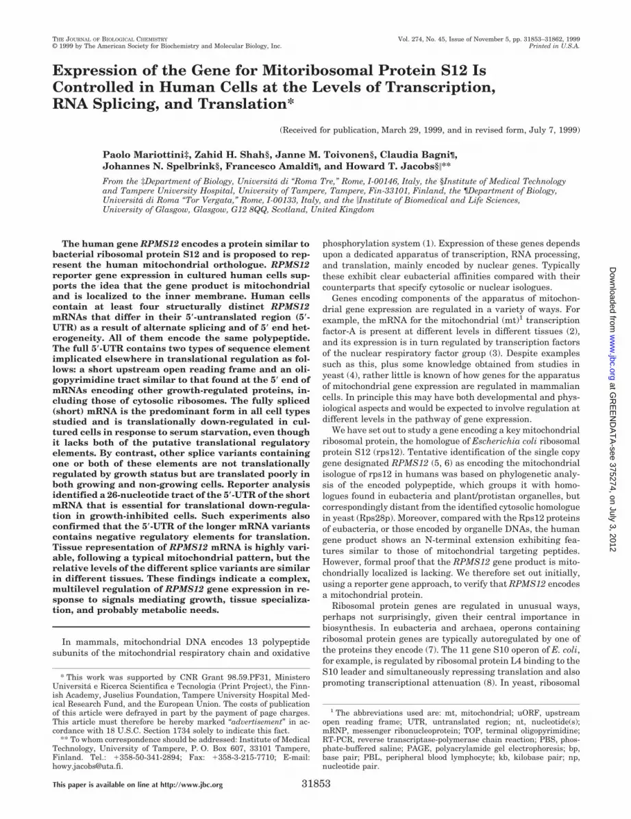
Northern analysis of RNA from various cell lines was carriedout using probes for successively more inclusive portions of theRPMS12 59-UTR. These were designed to detect the threesplice variants revealed by cyberscreening, in the order of in-creasing abundance (based on preliminary experiments). Blotswere initially probed at high stringency for a region of theisoform a splice variant (Fig. 3b) that is absent from isoforms band c. This revealed transcripts of two distinct size classes,estimated at 1 and 1.25 kb, whose relative abundance varied
between the cell types tested. The shorter transcripts are pro-posed to represent the 59-truncated isoform d, cyberscreeninghaving revealed no evidence for 39 heterogeneity, nor any rea-sonable match to the consensus poly(A) addition signal locatedelsewhere in the 39-UTR. Even if the length of the poly(A) tailin these shorter transcripts is much reduced, their overall sizeprecludes that they contain the full 59-UTR of isoform a. 59-Truncated transcripts containing the oligopyrimidine tractunique to isoform a are evident in dbEST (e.g. GenBankTM
entry AA257081), although they do not constitute a coherentclass indicating a specific 59 end. Our best guess is that they areheterogeneous, as indicated by the dashed line in Fig. 3a. It isunclear whether these transcripts indicate a downstream tran-scriptional start region or else 59 truncation in vivo. Theshorter transcripts were prominent in A549 lung carcinomacells but almost undetectable in the K-562 leukemia cell lineand were also absent from solid tissues (see below).
After stripping and reprobing for a region contained withinboth isoforms a and b, but not c, an additional, prominenttranscript of intermediate size was detected (Fig. 3, b and c),estimated at 1.15 kb. Both transcript classes detected by theearlier probe were recognized only weakly by the second probe,indicating that the intermediate sized transcripts, which mustrepresent isoform b, are considerably more abundant in all celllines tested than those of isoforms a or d. Blots were strippedand reprobed again for the full-length RPMS12 mRNA, detect-ing all classes of transcript. In this case, a prominent additionalspecies was detected in the 1.0-kb size range, i.e. migratingfaster than the isoform b transcripts, which must represent thefully spliced isoform c not detected by other probes. This tran-script appears to be the major isoform in all cells tested. Largertranscripts, which would be derived from a far upstream startsuch as tentatively inferred from cyberscreening, were presentonly at very low abundance. The ratio of isoforms (a 1 d), b andc appears to be similar in all cell lines studied.
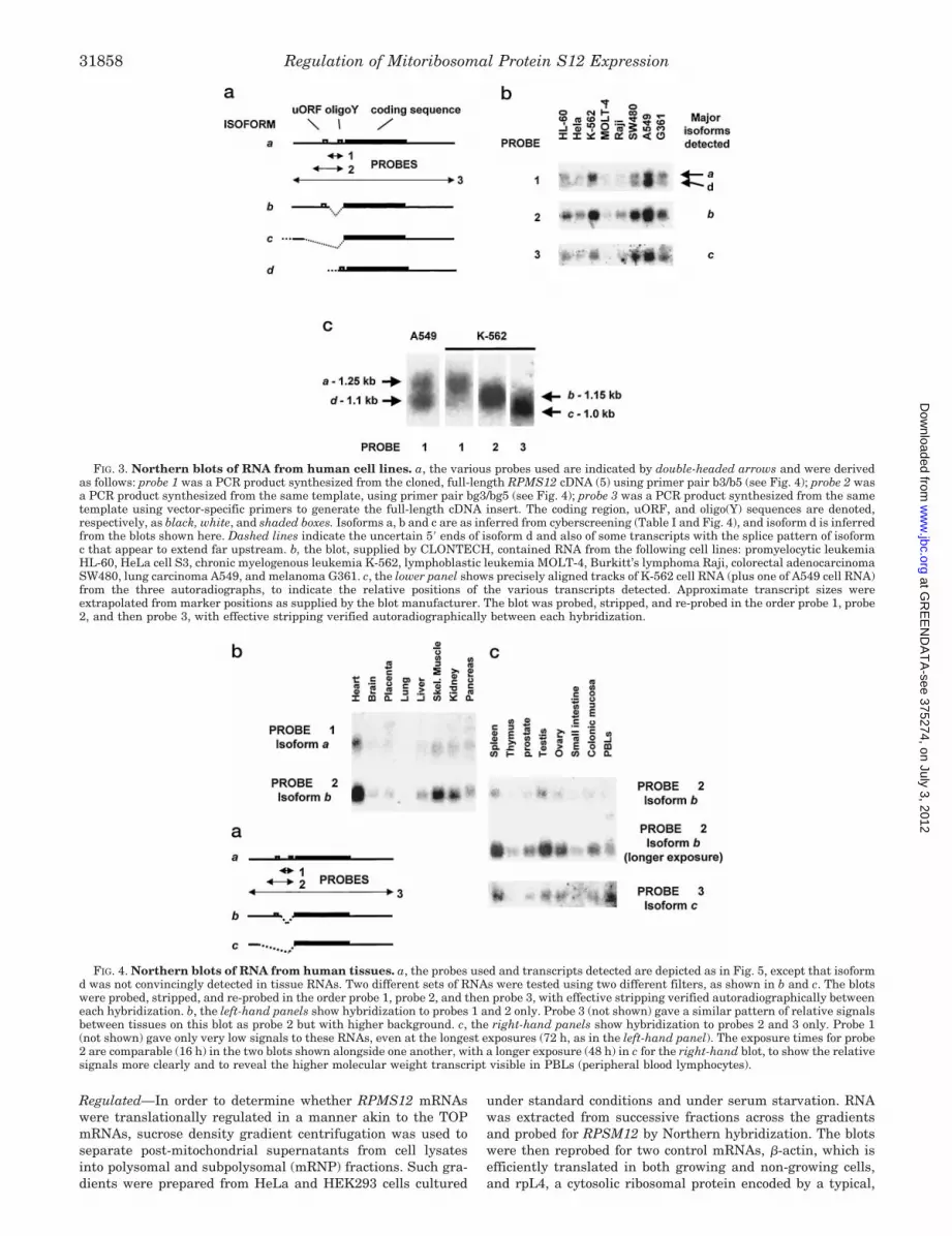
RPMS12 mRNAs Are Tissue Differentially Expressed—Theisoforms of RPMS12 mRNA were represented in the differentcell lines tested in very variable relative amounts. In order toinvestigate their expression in vivo, the same probes werehybridized sequentially to Northern blots of RNA from humantissues, as documented in Fig. 4. The pattern of relative abun-dance between tissues was similar for all three of the 59-UTRsplice variants, although the unspliced, 59-truncated isoform d,seen prominently in some cultured cells, was not detected. Theunspliced isoform a was detected in heart (Fig. 4b), but onlyweakly in most other tissues, and in many cases a largertranscript of approximately 2 kb was also detected by theisoform a-specific probe, possibly representing unspliced nu-clear RNA from which the coding region intron had also notbeen removed. The isoform b splice variant, which retains theuORF, was more highly represented in all tissues but wasespecially prominent in heart, skeletal muscle, and kidney (Fig.4, b and c). A similar pattern of hybridization was seen usingthe full-length probe (Fig. 4c), which detects also (and mainly)the shorter, fully spliced isoform c. Some minor differences canbe discerned, for example isoform c was detected more stronglyin peripheral blood lymphocytes (PBLs) than in colonic mucosaor thymus, and at about the same level as in testis, whereasisoform b was more prominent in colonic mucosa, thymus, andtestis than in PBLs (Fig. 4c). In general, isoform b appeared toshow more pronounced differences between tissues than eitherof isoforms a or c. Higher molecular weight transcripts thatcould correspond with the use of a far upstream transcriptionalstart site were not detected, except in PBLs, where a (;2.5 kb)species of unknown origin was detected by the isoform b probe.
The Major RPMS12 mRNA Splice Variant Is Translationally
FIG. 2. Sequences of RPMS12 transcripts alternately pro-cessed within the 5*-UTR, as inferred by cyberscreening ofdbEST (summarized in Table I). Transcripts are denoted a, b, and c,as in the text, with dashes indicating nucleotides absent in each givenisoform. The positions of various primers or their complements, used togenerate probes for Northern blots (b3/b5 and bg3/bg5) and for RT-PCR analysis (R1–R5), are indicated by arrows. The uORF and oligo(Y)tract are shown in italics. The start of the coding region of RPMS12 isshown alongside the corresponding amino acid sequence (one-lettercode). The position of the single coding region intron is denoted by thedouble arrowhead (..,,). A possible consensus start site for isoform cis found at nt 17/18, and the lowercase type upstream of this pointdenotes sequence found only in a minor fraction of ESTs with theisoform c splice pattern.
Regulation of Mitoribosomal Protein S12 Expression 31857
at GR
EE
ND
AT
A-see 375274, on July 3, 2012
ww
w.jbc.org
Dow
nloaded from
Regulated—In order to determine whether RPMS12 mRNAswere translationally regulated in a manner akin to the TOPmRNAs, sucrose density gradient centrifugation was used toseparate post-mitochondrial supernatants from cell lysatesinto polysomal and subpolysomal (mRNP) fractions. Such gra-dients were prepared from HeLa and HEK293 cells cultured
under standard conditions and under serum starvation. RNAwas extracted from successive fractions across the gradientsand probed for RPSM12 by Northern hybridization. The blotswere then reprobed for two control mRNAs, b-actin, which isefficiently translated in both growing and non-growing cells,and rpL4, a cytosolic ribosomal protein encoded by a typical,
FIG. 3. Northern blots of RNA from human cell lines. a, the various probes used are indicated by double-headed arrows and were derivedas follows: probe 1 was a PCR product synthesized from the cloned, full-length RPMS12 cDNA (5) using primer pair b3/b5 (see Fig. 4); probe 2 wasa PCR product synthesized from the same template, using primer pair bg3/bg5 (see Fig. 4); probe 3 was a PCR product synthesized from the sametemplate using vector-specific primers to generate the full-length cDNA insert. The coding region, uORF, and oligo(Y) sequences are denoted,respectively, as black, white, and shaded boxes. Isoforms a, b and c are as inferred from cyberscreening (Table I and Fig. 4), and isoform d is inferredfrom the blots shown here. Dashed lines indicate the uncertain 59 ends of isoform d and also of some transcripts with the splice pattern of isoformc that appear to extend far upstream. b, the blot, supplied by CLONTECH, contained RNA from the following cell lines: promyelocytic leukemiaHL-60, HeLa cell S3, chronic myelogenous leukemia K-562, lymphoblastic leukemia MOLT-4, Burkitt’s lymphoma Raji, colorectal adenocarcinomaSW480, lung carcinoma A549, and melanoma G361. c, the lower panel shows precisely aligned tracks of K-562 cell RNA (plus one of A549 cell RNA)from the three autoradiographs, to indicate the relative positions of the various transcripts detected. Approximate transcript sizes wereextrapolated from marker positions as supplied by the blot manufacturer. The blot was probed, stripped, and re-probed in the order probe 1, probe2, and then probe 3, with effective stripping verified autoradiographically between each hybridization.
FIG. 4. Northern blots of RNA from human tissues. a, the probes used and transcripts detected are depicted as in Fig. 5, except that isoformd was not convincingly detected in tissue RNAs. Two different sets of RNAs were tested using two different filters, as shown in b and c. The blotswere probed, stripped, and re-probed in the order probe 1, probe 2, and then probe 3, with effective stripping verified autoradiographically betweeneach hybridization. b, the left-hand panels show hybridization to probes 1 and 2 only. Probe 3 (not shown) gave a similar pattern of relative signalsbetween tissues on this blot as probe 2 but with higher background. c, the right-hand panels show hybridization to probes 2 and 3 only. Probe 1(not shown) gave only very low signals to these RNAs, even at the longest exposures (72 h, as in the left-hand panel). The exposure times for probe2 are comparable (16 h) in the two blots shown alongside one another, with a longer exposure (48 h) in c for the right-hand blot, to show the relativesignals more clearly and to reveal the higher molecular weight transcript visible in PBLs (peripheral blood lymphocytes).
Regulation of Mitoribosomal Protein S12 Expression31858
at GR
EE
ND
AT
A-see 375274, on July 3, 2012
ww
w.jbc.org
Dow
nloaded from
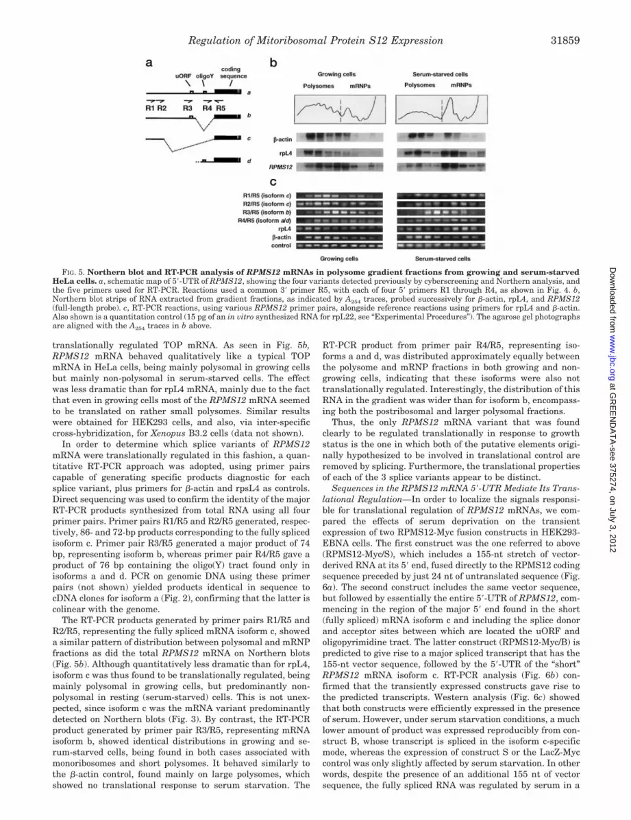
translationally regulated TOP mRNA. As seen in Fig. 5b,RPMS12 mRNA behaved qualitatively like a typical TOPmRNA in HeLa cells, being mainly polysomal in growing cellsbut mainly non-polysomal in serum-starved cells. The effectwas less dramatic than for rpL4 mRNA, mainly due to the factthat even in growing cells most of the RPMS12 mRNA seemedto be translated on rather small polysomes. Similar resultswere obtained for HEK293 cells, and also, via inter-specificcross-hybridization, for Xenopus B3.2 cells (data not shown).
In order to determine which splice variants of RPMS12mRNA were translationally regulated in this fashion, a quan-titative RT-PCR approach was adopted, using primer pairscapable of generating specific products diagnostic for eachsplice variant, plus primers for b-actin and rpsL4 as controls.Direct sequencing was used to confirm the identity of the majorRT-PCR products synthesized from total RNA using all fourprimer pairs. Primer pairs R1/R5 and R2/R5 generated, respec-tively, 86- and 72-bp products corresponding to the fully splicedisoform c. Primer pair R3/R5 generated a major product of 74bp, representing isoform b, whereas primer pair R4/R5 gave aproduct of 76 bp containing the oligo(Y) tract found only inisoforms a and d. PCR on genomic DNA using these primerpairs (not shown) yielded products identical in sequence tocDNA clones for isoform a (Fig. 2), confirming that the latter iscolinear with the genome.
The RT-PCR products generated by primer pairs R1/R5 andR2/R5, representing the fully spliced mRNA isoform c, showeda similar pattern of distribution between polysomal and mRNPfractions as did the total RPMS12 mRNA on Northern blots(Fig. 5b). Although quantitatively less dramatic than for rpL4,isoform c was thus found to be translationally regulated, beingmainly polysomal in growing cells, but predominantly non-polysomal in resting (serum-starved) cells. This is not unex-pected, since isoform c was the mRNA variant predominantlydetected on Northern blots (Fig. 3). By contrast, the RT-PCRproduct generated by primer pair R3/R5, representing mRNAisoform b, showed identical distributions in growing and se-rum-starved cells, being found in both cases associated withmonoribosomes and short polysomes. It behaved similarly tothe b-actin control, found mainly on large polysomes, whichshowed no translational response to serum starvation. The
RT-PCR product from primer pair R4/R5, representing iso-forms a and d, was distributed approximately equally betweenthe polysome and mRNP fractions in both growing and non-growing cells, indicating that these isoforms were also nottranslationally regulated. Interestingly, the distribution of thisRNA in the gradient was wider than for isoform b, encompass-ing both the postribosomal and larger polysomal fractions.
Thus, the only RPMS12 mRNA variant that was foundclearly to be regulated translationally in response to growthstatus is the one in which both of the putative elements origi-nally hypothesized to be involved in translational control areremoved by splicing. Furthermore, the translational propertiesof each of the 3 splice variants appear to be distinct.
Sequences in the RPMS12 mRNA 59-UTR Mediate Its Trans-lational Regulation—In order to localize the signals responsi-ble for translational regulation of RPMS12 mRNAs, we com-pared the effects of serum deprivation on the transientexpression of two RPMS12-Myc fusion constructs in HEK293-EBNA cells. The first construct was the one referred to above(RPMS12-Myc/S), which includes a 155-nt stretch of vector-derived RNA at its 59 end, fused directly to the RPMS12 codingsequence preceded by just 24 nt of untranslated sequence (Fig.6a). The second construct includes the same vector sequence,but followed by essentially the entire 59-UTR of RPMS12, com-mencing in the region of the major 59 end found in the short(fully spliced) mRNA isoform c and including the splice donorand acceptor sites between which are located the uORF andoligopyrimidine tract. The latter construct (RPMS12-Myc/B) ispredicted to give rise to a major spliced transcript that has the155-nt vector sequence, followed by the 59-UTR of the “short”RPMS12 mRNA isoform c. RT-PCR analysis (Fig. 6b) con-firmed that the transiently expressed constructs gave rise tothe predicted transcripts. Western analysis (Fig. 6c) showedthat both constructs were efficiently expressed in the presenceof serum. However, under serum starvation conditions, a muchlower amount of product was expressed reproducibly from con-struct B, whose transcript is spliced in the isoform c-specificmode, whereas the expression of construct S or the LacZ-Myccontrol was only slightly affected by serum starvation. In otherwords, despite the presence of an additional 155 nt of vectorsequence, the fully spliced RNA was regulated by serum in a
FIG. 5. Northern blot and RT-PCR analysis of RPMS12 mRNAs in polysome gradient fractions from growing and serum-starvedHeLa cells. a, schematic map of 59-UTR of RPMS12, showing the four variants detected previously by cyberscreening and Northern analysis, andthe five primers used for RT-PCR. Reactions used a common 39 primer R5, with each of four 59 primers R1 through R4, as shown in Fig. 4. b,Northern blot strips of RNA extracted from gradient fractions, as indicated by A254 traces, probed successively for b-actin, rpL4, and RPMS12(full-length probe). c, RT-PCR reactions, using various RPMS12 primer pairs, alongside reference reactions using primers for rpL4 and b-actin.Also shown is a quantitation control (15 pg of an in vitro synthesized RNA for rpL22, see “Experimental Procedures”). The agarose gel photographsare aligned with the A254 traces in b above.
Regulation of Mitoribosomal Protein S12 Expression 31859
at GR
EE
ND
AT
A-see 375274, on July 3, 2012
ww
w.jbc.org
Dow
nloaded from

similar manner to the endogenous transcript, whereas the iso-form completely lacking the natural 59-UTR signals was effi-ciently expressed but not regulated. This identifies a 26-ntregion, present in the spliced construct B mRNA but absentfrom the construct S mRNA (see Fig. 6a), that is required forgrowth regulation of translation. The region is located imme-diately upstream of the splice site. Furthermore, the fact thatconstruct S was efficiently expressed under both conditionstested is consistent with the earlier inference that the 59-UTRof the longer isoforms, containing the uORF and oligopyrimi-dine tract, is a negative element for translation.
DISCUSSION
These findings support the identification of the RPMS12gene product as a mitochondrial ribosomal protein, reveal un-expected complexity in the regulation of its expression, andidentify specific regions of the 59-UTR involved in translationalcontrol. We now address the implications of these findings.
Mitochondrial and Submitochondrial Localization of theRPMS12 Gene Product—The targeting to mitochondria of theRPMS12-Myc reporter protein strongly supports the previousassignment of RPMS12 as encoding the mitochondrial isologueof E. coli ribosomal protein S12. The protein is a member of awell characterized and conserved family of ribosomal proteins(5) and in most plants is even mitochondrially encoded (47–49).Comparable experiments with an RPMS12-GFP reporter fu-sion2 also showed colocalization to mitochondria. The assign-ment is further supported by the fact that the mitochondriallylocalized fusion protein was inaccessible to external protease,i.e. had been imported into the mitochondria, appears to havebeen proteolytically processed, and was localized to the inner
mitochondrial membrane. In yeast, the inner membrane is thesite of productive synthesis of the hydrophobic, mtDNA-en-coded mitochondrial proteins that contribute to the respiratorychain (50). It is therefore logical that an epitope-tagged mito-ribosomal protein will be localized there.
Multilevel Control of RPMS12 Gene Expression—The abovefindings lead to the conclusion that RPMS12 is regulated at thelevels of transcription, RNA processing, and translation. Thegeneration of mRNA isoforms with different patterns of trans-lational behavior would seem to be the major outcome of thealternate synthetic pathways.
Each of the isoforms of RPMS12 mRNA is clearly repre-sented in highly tissue-variable amounts. We were unable toperform a meaningful loading control hybridization with thecommercially supplied Northern blot membranes, since thefinal probe could not be completely stripped. However, theclaim of the manufacturer that the lanes are evenly loaded with2 mg of poly(A) RNA and quality controlled to check RNAintegrity means that minor variations in loading cannot ac-count for the order of magnitude differences in signal seenbetween the highest and lowest expressing tissues. The patternof relative abundance between tissues represents a typicalpattern for a gene involved in mitochondrial respiratory func-tion, with prominent expression in tissues highly dependent onoxidative metabolism such as heart, skeletal muscle, kidney,and to a lesser extent brain, liver, testis, and pancreas. Thegene encodes a conserved ribosomal protein indispensable fortranslation; hence, its expression is expected to follow a similarprofile to that of the mtDNA-encoded mRNAs that are trans-lated by mitoribosomes. The fact that the various mRNA iso-forms show similar patterns of tissue distribution as one an-other suggests that differential RNA processing is not of majorimportance in generating these different tissue levels of expres-2 Z. H. Shah, unpublished data.
FIG. 6. Reporter analysis of 5*-UTR signals. a, sequences of the predicted 59-UTRs of the mRNAs encoded by the two reporter constructsRPMS12-Myc/B and RPMS12-Myc/S. The 155 nt of vector sequence (not shown) are common to both. Most RNA made from construct B is splicedto remove the intron that contains the uORF and oligo(Y) as shown and as confirmed by the experiment shown in b. The two constructs share 21nt of sequence located immediately upstream of the start codon. Construct B contains an additional 26 nt of RPMS12 sequence from the other sideof the splice junction unique to isoform c, in the place of which construct S has 3 nt of intron-derived sequence but not the full splice acceptor. b,RT-PCR analysis of transgene-specific transcripts cells transiently transfected with RPMS12-Myc constructs B and S. PCR primers were the T7promoter primer (vector-specific) and primer R5 (see Fig. 2). The only construct B-derived transcript detected is the fully spliced isoform (194-bpproduct, whose structure was confirmed by direct sequencing). Construct S gives a 170-bp product that is colinear with the DNA. c, Westernanalysis of RPMS12-Myc fusion protein expression in mitochondria from cells mock-transfected (vector only) or transfected with constructs B orS and then grown in the presence or absence of serum. Equal amounts of protein are loaded on each lane. Shown alongside is the cytosolicexpression of a LacZ-Myc control in transfected cells grown with or without serum.
Regulation of Mitoribosomal Protein S12 Expression31860
at GR
EE
ND
AT
A-see 375274, on July 3, 2012
ww
w.jbc.org
Dow
nloaded from

sion. Instead the gene is most likely transcribed at differentrates in different tissues, although a contribution from RNAstability cannot be ruled out.
Transcription may also be regulated in another way, via theselection of alternate start sites. Tentative evidence for a farupstream start active in at least some tissues was obtained bycyberscreening of dbEST and is also suggested by the detectionin PBLs of a larger transcript carrying the b isoform splicepattern. More convincingly, the 59-truncated isoform d detectedin cultured cells suggests strongly the use of a downstreaminitiation site. Although 59 truncation by an exonucleolyticactivity in vivo cannot be ruled out, the fact that the isoform dtranscripts detected on Northern blots constitute a discrete sizeclass argues strongly that they derive instead from the use of aseparate initiation site, which would be located just upstreamof the oligopyrimidine tract, based on the transcript size. Thephysiological significance of alternate 59 termini in RPMS12mRNAs remains unknown. It may relate to translational reg-ulation, as discussed further below. The expression of a minorbut constitutively translated form of RPMS12 mRNA in thestem cell compartment which cultured cells represent makesobvious sense.
Alternate splicing in a 59-UTR is relatively uncommon. It hasbeen reported, for example, in the bovine gene encoding con-nexin-32 (51) and the human genes encoding reduced folatecarrier (52) and thrombopoietin (53). In principle, 59 spliceheterogeneity inferred from cyberscreening could be due to thepresence in the data base of sequences derived from partiallyprocessed nuclear RNA. In the case of RPMS12 this is highlyunlikely. Essentially all of the RPMS12 cDNA sequences de-posited in dbEST represent transcripts from which the codingregion intron has been correctly spliced out, yet a clear majorityof them are unspliced or partially spliced within the 59-UTR.Moreover, if the oligo(Y) tract is essential for recognition of thesplice acceptor site just upstream of the RPMS12 start codon,then isoform b could not be efficiently processed further toisoform c. In addition, unspliced or partially spliced 59-UTRvariants were found at least partly in the polysomal fraction incultured cells. Alternate splicing must therefore give rise toseveral different forms of translatable mRNA. Only in thosehuman tissues showing low expression (e.g. placenta, prostate,and ovary) was a significant fraction of the 59-UTR-unsplicedRPMS12 RNA of a size indicating that it might also be un-spliced in the coding region. These very low abundance tran-scripts may be nuclear but are a very minor fraction in tissuessuch as heart or skeletal muscle, showing prominent overallexpression.
The physiological significance of alternate splicing withinthe RPMS12 59-UTR remains unknown. In this study we havedemonstrated that the major RPMS12 mRNA (isoform c) issubject to a translational control mechanism that results in ahigher fraction of mRNA being loaded onto polysomes in pro-liferating cells compared with resting cells. However, the factthat only one of the four RPMS12 isoforms expressed in cul-tured cells appeared to be thus regulated suggests that theother splice variants may be subject to different kinds of controlat the translational level, such as described for other genes(54–56). The four isoforms differ by virtue of the presence ofputative elements involved in such regulation. Isoforms b andd contain, respectively, the uORF and the oligo(Y) tract alone,whereas isoform c contains neither, and isoform a containsboth. These putative elements could regulate translation inanother context than growth control.
Translational Regulation of Mitoribosome Biogenesis—Likecytosolic ribosomes, mitochondrial ribosomes are an essentialcomponent of the cellular machinery whose biosynthesis needs
to be stimulated in growing cells. The observed translationalregulation of RPMS12 therefore makes physiological sense. Animportant difference is the fact that the growth-regulatedmRNA lacks a 59-terminal oligopyrimidine tract. The regula-tion must therefore employ a mechanism somewhat distinctfrom that of TOP mRNAs, although it could share features withit. Our reporter assay identified a 26-nt region located at theextreme 59 end of isoform c mRNA that is essential for trans-lational down-regulation under conditions of growth inhibition.It is not especially pyrimidine-rich (10 nt are purines). In thereporter construct we used, it was located 155 nt away from the59 end, implying that its extreme 59 location in the naturalmRNA is also not critical for function. Importantly, the longerisoforms a and b, which are differently spliced and not growth-regulated, also contain this 26-nt tract. Therefore, the signalmediating growth-related translational down-regulation mustlogically include more than just this 26 nt, i.e. must extend overthe unique splice site present in the isoform c mRNA. Alterna-tively, its function may be over-ridden by other 59-UTR signalspresent in the longer isoforms.
TOP mRNAs are believed to be controlled via specific inter-actions between the TOP tract and regulatory proteins medi-ating mRNA recruitment to ribosomes (23, 24). The exact prop-erties of these proteins and the machinery with which theyinteract have not yet been characterized. It seems logical topostulate that the regulatory sequence unique to isoform cinteracts specifically with a negative regulatory protein that isonly present in an active state in growth-arrested cells. Thesame protein may independently interact with TOP mRNAs.Alternatively, different proteins may bind to RPMS12 and TOPmRNAs, independently influencing their ability to interactwith a common component of the translational recruitmentmachinery. The fact that the translational regulation ofRPMS12 mRNA is less dramatic than for some TOP mRNAswith an “optimal” oligo(Y) tract at the 59 end recalls the behav-ior of some mRNAs bearing poor pyrimidine tracts, whosetranslational regulation is less evident and/or dependent on thecellular context (57, 58).
The synthetic reporter construct RPMS12-Myc/S, whichlacks both the uORF and oligo(Y) motifs, was expressed asefficiently as the LacZ-Myc control. This supports the inferencethat the uORF and/or oligo(Y) tracts are negative regulatoryelements for translation, at least in cultured cells. One possi-bility is that the negative effect of the uORF is disabled wheremitochondrial function must be rapidly enhanced, for examplein response to bioenergetic needs or developmental signals.Our data suggest that isoform b is likely to be the mRNAvariant responsive to such signals. It is significantly moreabundant than isoform a in all tissues but especially in oxida-tive tissues showing high overall expression, such as heart andskeletal muscle. Moreover, the profile of its polysome distribu-tion in cultured cells, where it is found mainly associated withmonoribosomes and small polysomes, suggests that it is indeedsusceptible to futile initiation at the uORF, with rather fewribosomes traversing the genuine RPMS12 coding sequence.By contrast, the mRNAs containing the oligo(Y) tract (i.e. iso-forms a and d) are distributed quite differently, being moreprominent in both the overtly post-ribosomal fraction and inlarger polysomes than isoform b. This suggests that the isoforma and d mRNAs are regulated in a quite different fashion, beingonly inefficiently recruited into polysomes, but once there areless subject to futile initiation at the uORF. Our findingsprompt a more thorough investigation of the effects on trans-lation in different cellular contexts of the various 59-UTRelements.
The presence of multiple isoforms of RPMS12 mRNA in all
Regulation of Mitoribosomal Protein S12 Expression 31861
at GR
EE
ND
AT
A-see 375274, on July 3, 2012
ww
w.jbc.org
Dow
nloaded from

tissues potentially allows each cell type to respond to a varietyof intra- and intercellular signals, to enhance the biosynthesisof mitoribosomes according to cellular needs. The complex pat-tern of RPMS12 mRNAs seen in humans is not shared with themouse, however, where all cDNA sequences from the ortholo-gous gene represented in dbEST form a single contig, with arelatively short 59-UTR (approximately 90 nt), no uORF, andonly weak evidence for 59 heterogeneity. Regulation of mitori-bosome biosynthesis in the mouse might therefore employ dif-ferent mechanisms than in humans. By contrast, the presenceof a uORF may be a common feature in human mitoribosomalprotein mRNAs, as it has been found in the one other examplestudied in detail, MRPL12 (25).
Further analysis of the regulatory roles of the different 59-UTR elements of RPMS12 mRNA will enable the componentsof the regulatory machinery to be identified. Further clues willalso doubtless emerge from studies of other mRNAs encodingmitochondrial ribosomal proteins and other components of themitochondrial translational apparatus, both in humans and inother vertebrates.
Acknowledgments—We thank Richard Jackson for useful discus-sions. We are grateful to Liliana Mannucci and Pietro Pilo-Boyl forassistance in the cell culture experiments and to Claudia Crosio forsharing some unpublished results.
REFERENCES
1. Schatz, G., and Attardi, G. (1986) Annu. Rev. Cell Biol. 4, 289–3332. Larsson, N. G., Garman, J. D., Oldfors, A., Barsh, G. S., and Clayton, D. A.
(1996) Nat. Genet. 13, 296–3023. Scarpulla, R. C. (1997) J. Bioenerget. Biomembr. 29, 109–1194. Graack, H. R., and Wittmann-Liebold, B. (1998) Biochem. J. 329, 433–4485. Shah, Z. H., O’Dell, K., Miller, S. C. M., An, X., and Jacobs, H. T. (1997) Gene
(Amst.) 204, 55–626. Shah, Z. H., Migliosi, V., Miller, S. C. M., Wang, A., Friedman, T. B., and
Jacobs, H. T. (1998) Genomics 48, 384–3887. Zengel, J. M., and Lindahl, L. (1994) Prog. Nucleic Acids Res. Mol. Biol. 47,
331–3708. Freedman, L. P., Zengel, J. M., Archer, R. H., and Lindahl, L. (1987) Proc. Natl.
Acad. Sci. U. S. A. 84, 6516–65209. Tsay, Y. F., Thompson, J. R., Rotenberg, M. O., Larkin, J. C., and Woolford,
J. L. (1988) Genes Dev. 2, 664–67610. Fewell, S. W., and Woolford, J. L., Jr. (1999) Mol. Cell. Biol. 19, 826–83411. Geyer, P. K., Meyuhas, O., Perry, R. P., and Johnson, L. F. (1982) Mol. Cell.
Biol. 2, 685–69312. Kaspar, R. L., Rychlik, W., White, M. W., Rhoads, R. E., and Morris, D. R.
(1990) J. Biol. Chem. 265, 3619–362213. Loreni, F., and Amaldi, F. (1992) Eur. J. Biochem. 205, 1027–103214. Agrawal, M. G., and Bowman, L. H. (1987) J. Biol. Chem. 262, 4868–487515. Hammond, M. L., and Bowman, L. H. (1988) J. Biol. Chem. 263, 17785–1779116. Kaspar, R. L., Kakegawa, T., Cranston, H., Morris, D. R., and White, M. W.
(1992) J. Biol. Chem. 267, 508–51417. Meyuhas, O., Thompson, A. E., Jr., and Perry, R. P. (1987) Mol. Cell. Biol. 7,
2691–269918. Avni, D., Shama, S., Loreni, F., and Meyuhas, O. (1994) Mol. Cell. Biol. 14,
3822–383319. Meyuhas, O., Biberman, Y., Pierandrei-Amaldi, P., and Amaldi, F. (1996) in A
Laboratory Guide to RNA: Isolation, Analysis and Synthesis (Krieg, P. K.,ed) pp. 65–81, Wiley-Liss, Inc., New York,
20. Amaldi, F., and Pierandrei-Amaldi, P. (1997) Prog. Mol. Subcell. Biol. 18, 1–17
21. Meyuhas, O., Avni, D., and Shama, S. (1996) Translational Control, pp.363–388, Cold Spring Harbor Laboratory, Cold Spring Harbor, NY
22. Cardinali, B., di Cristina, M., and Pierandrei-Amaldi, P. (1993) Nucleic AcidsRes. 21, 2301–2308
23. Loreni, F., and Amaldi, F. (1997) FEBS Lett. 416, 239–24224. Amaldi, F., Camacho-Venegas, O., Cardinali, B., Cecconi, F., Crosio, C.,
Loreni, F., Mariottini, P., Pellizzoni, L., Pierandrei-Amaldi, P. (1995) Bio-chem. Cell Biol. 73, 969–977
25. Marty, L., and Fort, P. (1996) J. Biol. Chem. 271, 11468–1147626. Marty, L., Taviaux, S., and Fort, P. (1997) Genomics 41, 453–45727. Cao, J. H., and Geballe, A. P. (1995) J. Virol. 69, 1030–103628. Imataka, H., Nakayama, K., Yasumoto, K., Mizuno, A., Fujii-kuriyama, Y.,
and Hayami, M. (1994) J. Biol. Chem. 269, 20668–2067329. Yiu, G. K., Gu, W., and Hecht, N. B. (1994) Nucleic Acids Res. 22, 4599–460630. Oliveira, C. C., and McCarthy, J. E. G. (1995) J. Biol. Chem. 270, 8936–894331. Byrne, P. C., Sanders, P. G., and Snell, K. (1995) Biochem. Biophys. Res.
Commun. 214, 496–50232. Choi, H. S., Park, K. H., Kim, D. W., Lin, C. J., and Kim, K. H. (1996) Mol. Cell
6, 615–62133. Harigai, M., Miyashita, T., Hanada, M., and Reed, J. C. (1996) Oncogene 12,
1369–137434. Linz, B., Koloteva, N., Vasilescu, S., and McCarthy, J. E. G. (1997) J. Biol.
Chem. 272, 9131–914035. Lovett, P. S., and Rogers, E. J. (1996) Microbiol. Rev. 60, 366–38636. Hauswirth, W. W., Lim, L. O., and Turner, G. (1987) in Mitochondria: A
Practical Approach (Darley-Usmar, V. M., Rickwood, D., and Wilson, M. T.,eds) pp. 176–177, IRL Press at Oxford University Press, Oxford
37. Tapper, D. P., Van Etten, R. A., and Clayton, D. A. (1983) Methods Enzymol.97, 426–434
38. Ragan, C. I., Wilson, M. T., Darley-Usmar, V. M., and Lowe, P. N. (1987) inMitochondria, A Practical Approach (Darley-Usmar, V. M., Rickwood, D.,and Wilson, M. T., eds) pp. 80–81, IRL Press at Oxford University Press,Oxford
39. Sambrook, J., Fritsch, E. F., and Maniatis, T. (1989) Molecular Cloning: ALaboratory Manual, 2nd Ed., 18.47–18.75, Cold Spring Harbor Laboratory,Cold Spring Harbor, NY
40. Towbin, H., Staehelin, T., Gordon, J. (1979) Proc. Natl. Acad. Sci. U. S. A. 76,4350–4353
41. Nijtmans, L. G. J., Spelbrink, J. N., van Galen, M. J. M., Zwaart, M., Klement,P., and van den Bogert, C. (1995) Biochim. Biophys. Acta 1265, 117–126
42. Bagni, C., Mariottini, P., Annesi, F., and Amaldi, F. (1993) Biochim. Biophys.Acta 1216, 475–478
43. Rapanotti, M. C., Pucci, B., Amaldi, F., and Loreni, F. (1995) Gene (Amst.) 154,199–203
44. Ponte, P., Ng, S. Y., Engel, J., Gunning, P., and Kedes, L. (1984) Nucleic AcidsRes. 12, 1687–1696
45. Cleveland, D. W., Lopata, M. A., MacDonald, R. J., Cowan, N. J., Rutter, W. J.,and Kirschner, M. W. (1980) Cell 20, 95–105
46. Genetics Computer Group (1994) Program Manual for the Wisconsin Package,Version 8, Genetics Computer Group, Inc., Madison, WI
47. Perrotta, G., Regina, T. M R., Ceci, L. R., and Quagliariello, C. (1996) Mol. Gen.Genet. 251, 327–337
48. Grohmann, L., Brennicke, A., and Schuster, W. (1992) Nucleic Acids Res. 20,5641–5646
49. Maffey, L., Degand, H., and Boutry, M. (1997) Mol. Gen. Genet. 254, 365–37150. Fox, T. D. (1996) Experientia (Basel) 52, 1130–113551. Duga, S., Asselta, R., del Giacco, L., Malcovati, M., Ronchi, S., Tenchini, M. L.,
and Simonic, T. (1999) Eur. J. Biochem. 259, 188–19652. Zhang, L., Wong, S. C., and Matherly, L. H. (1998) Biochem. J. 332, 773–78053. Ghilardi, N., Wiestner, A., and Skoda, R. C. (1998) Blood 92, 4023–403054. Pelletier, J., Kaplan, G., Racaniello, V. R., and Sonenberg, N. (1988) Mol. Cell.
Biol. 8, 1103–111255. Lindquist, S. (1986) Annu. Rev. Biochem. 55, 1151–119156. Dandekar, T., Stripecke, R., Gray, N. K., Goossen, B., Constable, A.,
Johansson, H. E., and Hentze, M. W. (1991) EMBO J. 10, 1903–190957. Bagni, C., Mariottini, P., Terrenato, L., and Amaldi, F. (1992) Mol. Gen. Genet.
234, 60–6458. Caizergues-Ferrer, M., Mariottini, P., Curie, C., Lapeyre, B., Gas, N., Amalric,
F., and Amaldi, F. (1989) Genes Dev. 3, 324–333
Regulation of Mitoribosomal Protein S12 Expression31862
at GR
EE
ND
AT
A-see 375274, on July 3, 2012
ww
w.jbc.org
Dow
nloaded from