Glutaredoxin S12: Unique Properties for Redox Signaling
16
ORIGINAL RESEARCH COMMUNICATION Glutaredoxin S12: Unique Properties for Redox Signaling Mirko Zaffagnini, 1,2 Mariette Bedhomme, 1 Christophe H. Marchand, 1 Je ´ re ´ my Couturier, 3 Xing-Huang Gao, 1 Nicolas Rouhier, 3 Paolo Trost, 2 and Ste ´ phane D. Lemaire 1 Abstract Aims: Cysteines (Cys) made acidic by the protein environment are generally sensitive to pro-oxidant molecules. Glutathionylation is a post-translational modification that can occur by spontaneous reaction of reduced gluta- thione (GSH) with oxidized Cys as sulfenic acids (-SOH). The reverse reaction (deglutathionylation) is strongly stimulated by glutaredoxins (Grx) and requires a reductant, often GSH. Results: Here, we show that chloroplast GrxS12 from poplar efficiently reacts with glutathionylated substrates in a GSH-dependent ping pong mechanism. The pK a of GrxS12 catalytic Cys is very low (3.9) and makes GrxS12 itself sensitive to oxidation by H 2 O 2 and to direct glutathionylation by nitrosoglutathione. Glutathionylated-GrxS12 (GrxS12-SSG) is temporarily inactive until it is deglutathionylated by GSH. The equilibrium between GrxS12 and glutathione (E m(GrxS12-SSG) =- 315 mV, pH 7.0) is characterized by K ox values of 310 at pH 7.0, as in darkened chloroplasts, and 69 at pH 7.9, as in illuminated chloroplasts. Innovation: Based on thermodynamic data, GrxS12-SSG is predicted to accumulate in vivo under conditions of mild oxidation of the GSH pool that may occur under stress. Moreover, GrxS12-SSG is predicted to be more stable in chloroplasts in the dark than in the light. Conclusion: These peculiar catalytic and thermodynamic properties could allow GrxS12 to act as a stress-related redox sensor, thus allowing glutathione to play a signaling role through glutathionylation of GrxS12 target proteins. Antioxid. Redox Signal. 16, 17–32. Introduction I n plant cells, glutathione (c-GluCysGly) is the most abundant nonprotein thiol (15). It is present in many sub- cellular compartments, including chloroplasts where it can reach the concentration of 1–4.5 mM, and serves as a major antioxidant to keep the intracellular environment reduced (44). Moreover, glutathione provides electrons to the ascorbate cycle and to some thiol-peroxidases and is, therefore, important for the detoxification of several compounds including reactive oxygen species (16). In chloroplasts as in other compartments, glutathione is maintained in the reduced state by NADPH- dependent glutathione reductase (GR). Although in most physiological conditions intracellular glutathione is essentially in the reduced form (GSH), it can be converted to the oxidized form (GSSG) under oxidative stress conditions (16, 44). Recently, glutathione has been found to be involved in a reversible post-translational modification termed glutathio- nylation, which is mainly promoted by oxidative and ni- trosative stresses (22). This modification, consisting in the formation of a mixed disulfide between glutathione and a protein cysteine (Cys) residue, is typically considered a mechanism of protection of specific Cys residues from irre- versible oxidation to sulfinic (-SO 2 H) or sulfonic (-SO 3 H) acids (22). Besides protection, glutathionylation can also constitute a mode of regulation, as it can modulate the activity of target enzymes and thereby play a role in many cellular processes (11). Glutathionylation has been extensively studied in mam- malian cells, and emerging evidence suggests that it could constitute an important mechanism of regulation and signaling in plants as well (20, 34, 44, 53). Whether glutathionylation is important for protection or regulation in vivo, the modification needs to be removed when the stress conditions or the signaling Innovation Glutathionylation is an emerging post-translational modification that could play an important role in cell regulation and signaling. Deglutathionylation reactions are mainly controlled by glutaredoxins (Grx). This study shows that higher plant chloroplastic GrxS12 exhibits peculiar catalytic and thermodynamic properties that could allow this enzyme to act as a stress-related redox sensor allowing glutathione to play a signaling role through glutathionylation of GrxS12 target proteins. 1 Laboratoire de Biologie Mole ´culaire et Cellulaire des Eucaryotes, FRE3354 Centre National de la Recherche Scientifique, Institut de Biologie Physico-Chimique, Universite ´ Pierre et Marie Curies, Paris, France. 2 Department of Experimental Evolutionary Biology, University of Bologna, Bologna, Italy. 3 Unite ´ Mixte de Recherches 1136 Nancy Universite ´—INRA Interaction Arbres—Microorganismes, IFR 110 EFABA, Faculte ´ des Sciences, Vandoeuvre Cedex, France. ANTIOXIDANTS & REDOX SIGNALING Volume 16, Number 1, 2012 ª Mary Ann Liebert, Inc. DOI: 10.1089/ars.2011.3933 17
Transcript of Glutaredoxin S12: Unique Properties for Redox Signaling

ORIGINAL RESEARCH COMMUNICATION
Glutaredoxin S12: Unique Properties for Redox Signaling
Mirko Zaffagnini,1,2 Mariette Bedhomme,1 Christophe H. Marchand,1 Jeremy Couturier,3 Xing-Huang Gao,1
Nicolas Rouhier,3 Paolo Trost,2 and Stephane D. Lemaire1
Abstract
Aims: Cysteines (Cys) made acidic by the protein environment are generally sensitive to pro-oxidant molecules.Glutathionylation is a post-translational modification that can occur by spontaneous reaction of reduced gluta-thione (GSH) with oxidized Cys as sulfenic acids (-SOH). The reverse reaction (deglutathionylation) is stronglystimulated by glutaredoxins (Grx) and requires a reductant, often GSH. Results: Here, we show that chloroplastGrxS12 from poplar efficiently reacts with glutathionylated substrates in a GSH-dependent ping pong mechanism.The pKa of GrxS12 catalytic Cys is very low (3.9) and makes GrxS12 itself sensitive to oxidation by H2O2 and todirect glutathionylation by nitrosoglutathione. Glutathionylated-GrxS12 (GrxS12-SSG) is temporarily inactiveuntil it is deglutathionylated by GSH. The equilibrium between GrxS12 and glutathione (Em(GrxS12-SSG) = - 315 mV,pH 7.0) is characterized by Kox values of 310 at pH 7.0, as in darkened chloroplasts, and 69 at pH 7.9, as inilluminated chloroplasts. Innovation: Based on thermodynamic data, GrxS12-SSG is predicted to accumulatein vivo under conditions of mild oxidation of the GSH pool that may occur under stress. Moreover, GrxS12-SSG ispredicted to be more stable in chloroplasts in the dark than in the light. Conclusion: These peculiar catalytic andthermodynamic properties could allow GrxS12 to act as a stress-related redox sensor, thus allowing glutathione toplay a signaling role through glutathionylation of GrxS12 target proteins. Antioxid. Redox Signal. 16, 17–32.
Introduction
In plant cells, glutathione (c-GluCysGly) is the mostabundant nonprotein thiol (15). It is present in many sub-
cellular compartments, including chloroplasts where it canreach the concentration of 1–4.5 mM, and serves as a majorantioxidant to keep the intracellular environment reduced (44).Moreover, glutathione provides electrons to the ascorbate cycleand to some thiol-peroxidases and is, therefore, important forthe detoxification of several compounds including reactiveoxygen species (16). In chloroplasts as in other compartments,glutathione is maintained in the reduced state by NADPH-dependent glutathione reductase (GR). Although in mostphysiological conditions intracellular glutathione is essentiallyin the reduced form (GSH), it can be converted to the oxidizedform (GSSG) under oxidative stress conditions (16, 44).
Recently, glutathione has been found to be involved in areversible post-translational modification termed glutathio-nylation, which is mainly promoted by oxidative and ni-trosative stresses (22). This modification, consisting in theformation of a mixed disulfide between glutathione and aprotein cysteine (Cys) residue, is typically considered amechanism of protection of specific Cys residues from irre-
versible oxidation to sulfinic (-SO2H) or sulfonic (-SO3H) acids(22). Besides protection, glutathionylation can also constitute amode of regulation, as it can modulate the activity of targetenzymes and thereby play a role in many cellular processes(11). Glutathionylation has been extensively studied in mam-malian cells, and emerging evidence suggests that it couldconstitute an important mechanism of regulation and signalingin plants as well (20, 34, 44, 53). Whether glutathionylation isimportant for protection or regulation in vivo, the modificationneeds to be removed when the stress conditions or the signaling
Innovation
Glutathionylation is an emerging post-translationalmodification that could play an important role in cellregulation and signaling. Deglutathionylation reactionsare mainly controlled by glutaredoxins (Grx). This studyshows that higher plant chloroplastic GrxS12 exhibitspeculiar catalytic and thermodynamic properties thatcould allow this enzyme to act as a stress-related redoxsensor allowing glutathione to play a signaling rolethrough glutathionylation of GrxS12 target proteins.
1Laboratoire de Biologie Moleculaire et Cellulaire des Eucaryotes, FRE3354 Centre National de la Recherche Scientifique, Institut deBiologie Physico-Chimique, Universite Pierre et Marie Curies, Paris, France.
2Department of Experimental Evolutionary Biology, University of Bologna, Bologna, Italy.3Unite Mixte de Recherches 1136 Nancy Universite—INRA Interaction Arbres—Microorganismes, IFR 110 EFABA, Faculte des Sciences,
Vandoeuvre Cedex, France.
ANTIOXIDANTS & REDOX SIGNALINGVolume 16, Number 1, 2012ª Mary Ann Liebert, Inc.DOI: 10.1089/ars.2011.3933
17

event is over. This process, named deglutathionylation, ismainly catalyzed by glutaredoxins (Grx) (23).
Grx are small ubiquitous oxidoreductases belonging tothe thioredoxin family and bearing in their active site acharacteristic four-amino acids motif with one or two reactivecysteines (CXXC/S). Although thioredoxins mainly serve asprotein disulfide reductases, Grxs interact specifically withthe GSH moiety of proteins containing glutathione-mixeddisulfides (23, 54). In mammalian Grxs, the deglutathionyla-tion reaction was shown to start with the nucleophilic attack ofthe glutathionyl sulfur of the substrate by the N-terminal Cys ofGrx active site (18). The reaction releases the target protein in itsreduced form but generates a glutathionylated Grx interme-diate that is reduced back by GSH with concomitant formationof GSSG (Fig. 1) (14, 46). This latter reaction may constitute therate-limiting step of the overall process (46).
In higher plants, three classes of Grxs have been initiallydefined based on their active site sequences (30, 42, 43), andthe classification has been recently refined after identifying afourth class (8). Class I includes proteins with CXX(C/S) ac-tive site other than CGFS and are also named CPYC-typebecause of the most common active site motif. Class II con-tains exclusively Grxs with a CGFS motif (CGFS-type), class
III, which is specific to land plants, corresponds to Grxs with apeculiar CCX(C/S) active site (CC-type), and class IV regroupspeculiar proteins composed of a Grx domain with CXXC/Sactive site and two additional domains of unknown functionsin the C-terminal part. Here, we report the biochemical char-acterization of a chloroplastic Grx isoform from poplar(GrxS12) which belongs to class I and possesses an unusual28WCSYS32 active site sequence and a second C-terminal Cys(Cys87) that are both conserved in orthologs from other plantspecies. Class I contains 6 members in poplar and Arabidopsis(41, 44), GrxS12 being the only one lacking the second Cys inthe active site sequence motif. The x-ray crystal structure ofglutathionylated GrxS12 from poplar was recently solved (9)and found to contain a single GSH molecule covalently boundto active site Cys29. Interestingly, C-terminal Cys87 was foundto be located at proximity of the active site, though not inter-acting with bound GSH. Since Cys87 formed no disulfide witheither Cys29 or GSH, and the GrxS12-C87S (C87S) mutatedvariant displayed a similar deglutathionylating activity as thewild-type protein, a monothiol mechanism based on active siteCys29 was proposed for GrxS12 (Fig. 1) (9). By contrast, someGrxs display a dithiol mechanism, as they use a second Cys inthe deglutathionylation reaction. This was recently demon-strated for Chlamydomonas reinhardtii Grx3 (CGFS-type), inwhich removal of the glutathionyl moiety from the N-terminalCys of Grx active site is performed by a second Cys of the samemonomer, equivalent to Cys87 of GrxS12 (54). The resultingintramolecular disulfide was found to possess a low redoxpotential ( - 323 mV at pH 7.9) and to be insensitive to gluta-thione, whereas it could be reduced by ferredoxin:thioredoxinreductase (FTR) under photosynthetic conditions.
The present article shows that protein deglutathionylationcatalyzed by poplar GrxS12 follows a ping-pong mechanism.The reaction, based on Cys29, is highly sensitive to [GSH]/[GSSG] ratios and proceeds at maximal rates only when glu-tathione is extensively reduced. Typical of Grxs, catalyticCys29 was found to be extremely acidic (pKa = 3.9) and, thus,sensitive to alkylation and to oxidation by H2O2, besidesundergoing glutathionylation in the presence of gluta-thionylated substrates or nitrosoglutathione (GSNO). TheGrxS12-catalyzed deglutathionylation reaction is stronglystimulated by alkaline pH, thus possibly reflecting the im-portant role of deprotonated glutathione (GS - ) in the attack ofglutathionylated GrxS12 (step 2, Fig. 1). The standard redoxpotential of the mixed disulfide of glutathionylated GrxS12, toour knowledge the first measured among Grxs, is - 350 mV atpH 7.9, a typical pH value for photosynthesizing chloroplasts(47). Owing to its monothiol mechanism, to the low pKa ofcatalytic Cys29, and to the low redox potential of the mixeddisulfide, glutathionylated GrxS12 may accumulate in vivounder stress conditions, when the GR potential is much lessreducing than - 350 mV. These unique biochemical propertiescould allow GrxS12 to act as a stress-related redox sensor, thusallowing glutathione to play a signaling role through glu-tathionylation of GrxS12 target proteins.
Results
Poplar GrxS12 follows a ping-pongreaction mechanism
Artificial substrates such as glutathionylated b-mercap-toethanol (b-ME-SSG) produced by the spontaneous reaction
FIG. 1. Schematic representation of the deglutathionyla-tion activity of Grxs. The reaction mechanism here depictedis a monothiol mechanism, as a single cysteine (Cys) of theglutaredoxin (Grx) is involved in catalysis. The overall re-action catalyzed by Grx consists of two steps: nucleophilicattack of the glutathionyl sulphur of the glutathionylatedsubstrate (protein-SSG) by Grx thiolate (step 1) and nucleo-philic attack of the glutathionyl sulphur of the glutathiony-lated Grx (Grx-SSG) by reduced glutathione (also in thiolateform, step 2). Regeneration of glutathione (GSH) by gluta-thione reductase (GR) allows determination of Grx activity asNADPH oxidation rate. For in vitro assays of Grx activity, theglutathionylated protein (protein-SSG) can be substituted byglutathionylated b-mercaptoethanol (b-ME-SSG) in the clas-sical hydroxyethyldisulfide (HED) assay.
18 ZAFFAGNINI ET AL.

of GSH with hydroxyethyldisulfide (HED) are routinely usedin Grx activity assays (9, 21, 54). Based on this standard assay,purified GrxS12 displayed a specific activity of 70 lmol min - 1
mg - 1 and site-specific mutant C87S performed similarly(66 lmol min - 1 mg - 1), whereas the specific activity of mutantC29S was very low (3.2 lmol min - 1 mg - 1), thus confirmingthe essential role of Cys29 and the negligible role of Cys87 inGrxS12 catalysis. In the presence of 1 mM GSH, GrxS12 dis-played an apparent Km for HED of 0.3 mM and a turnovernumber of 23 s - 1 (Table 1). Under similar conditions (1 mMGSH), the Km of GrxS12 for glutathionylated bovine serumalbumin (BSA-SSG) was ten-fold lower (Km 27lM), whereasthe apparent turnover number was only slightly higher (31 s - 1)(Table 1). Apparent kinetic parameters of site-specific mutantC87S were very similar to those measured for the wild-typeprotein (Table 1).
A two-substrate kinetic analysis of GrxS12 was performedwith BSA-SSG and GSH. Plots of initial velocities versus sub-strate concentrations were hyperbolic, although not saturated atthe highest concentrations of either fixed substrate (Fig. 2A, B).Double reciprocal plots generated parallel line patterns, char-acteristic of a ping-pong mechanism (Fig. 2C, D). This impliesthat after the interaction of GrxS12 with BSA-SSG, reducedBSA is released before GSH can reduce the mixed disulfideof GrxS12-SSG (see Fig. 1). When apparent kcat values obtainedfrom nonlinear regression of original data were plotted as afunction of substrate concentration, responses were approxi-mately linear rather than hyperbolic (Fig. 2A, B). This effectprevented any reliable determination of true kinetic parameterskcat and Km via nonlinear regression. Indeed, secondary plots 1/kcat versus 1/[BSA-SSG] or 1/[GSH] projected to the origin, in-dicating that true Km and kcat values are too high to be experi-mentally determined (insets in Fig. 2A, B). On the other hand,since kcat/Km ratios in ping-pong mechanisms are not influencedby the concentration of the second substrate (a property that isvisually represented by the parallelism of double reciprocalplots), the catalytic efficiency parameter kcat/Km could be pre-cisely derived from kinetic data of Figure 2. These true kineticparameters were (2.5 – 0.1) · 104 M - 1 s - 1 for GSH and(8.0 – 0.6) · 105 M- 1 s- 1 for BSA-SSG, thereby implying that thetrue Km for BSA-SSG was 30-times lower than for GSH. In spiteof some variability in experimental conditions, GrxS12 catalyticefficiency (with both HED and BSA-SSG) appeared roughlycomparable with previously characterized Grx from human (18,27), yeast (12), and C. reinhardtii (21, 54).
Glutathionylated isocitrate lyase is efficientlyreactivated by GrxS12
Assays of Grx activity with HED or BSA-SSG as substratesare based on the capacity of Grx to transfer a GSH moietyfrom a glutathionylated substrate to free GSH, thereby gen-erating GSSG that is reduced by GR (see Fig. 1). Alternatively,GRX deglutathionylating activity can be directly assayed bymeasuring the reactivation of an enzyme whose activity isaffected by glutathionylation. Isocitrate lyase (ICL) from C.reinhardtii provides such an opportunity, as this enzyme,which undergoes glutathionylation in vivo (35), was recentlyshown to be reversibly inhibited by glutathionylation of itsactive site Cys (3). Glutathionylated ICL (ICL-SSG;& 100%inactivated) was, thus, used as a model substrate; and GrxS12was tested for its ability to restore ICL activity in the presenceof a suitable electron donor (Fig. 3). Again, mutant C87S be-haved identical to wild-type GrxS12 in ICL-SSG reactivation,whereas the activity of mutant C29S was 5% of the wild typeand not further investigated (not shown).
In control experiments, 20 mM dithiothreitol (DTT) aloneallowed full chemical reactivation of ICL-SSG (10 lM) in5 min, but lower concentrations of DTT (e.g., 0.5 mM) weremuch less efficient (Fig. 3A). In the absence of external re-ductants, reduced GrxS12 (5 lM) caused very rapid but par-tial reactivation of ICL-SSG (Fig. 3A). In the presence of bothDTT (0.5 mM) and GrxS12 (5 lM), reactivation of ICL-SSG(10 lM) proceeded at a slightly faster rate than with GrxS12alone and continued until ICL was fully activated (Fig. 3A).
Similar experiments were performed with GSH as a physi-ological reductant instead of DTT. In the absence of GrxS12,very slow reactivation was promoted by 2 mM GSH alone,which contained about 1% oxidized glutathione as a contami-nant (i.e., 20 lM GSSG). Under these conditions, addition of5 lM GrxS12 increased the initial velocity of ICL-SSG re-activation (Fig. 3B), but still reactivation kinetics with GSH wasmuch slower than with DTT (Fig. 3A). Reactivation of ICL-SSGby GrxS12 and GSH (contaminated by 1% GSSG) depended onGrxS12 concentration with an S0.5 of 4.5lM (Fig. 3C).
To check the effect of GSSG contamination on GrxS12 ac-tivity, a GSH-regenerating system (GRS) based on GR andNADPH was applied to guarantee full reduction of GSH. TheGRS had no detectable effects on the nonenzymatic reactivationof ICL-SSG by GSH (Fig. 3B), but strongly stimulated ICL-SSGreactivation by 5 lM GrxS12 (Fig. 3B). Moreover, the S0.5 for
Table 1. Apparent Kinetic Parameters of the Deglutathionylating Activity of GrxS12 and C87S Mutant
with Different Substrates
HEDa BSA-SSGb GSHc
Km(HED) kcat Km(BSA-SSG) kcat Km(GSH) kcat
Protein lM s - 1 lM s - 1 lM s - 1
GrxS12 310 – 40 23.1 – 0.9 26.6 – 1.8 31.3 – 2.5 (4.0 – 0.5) · 103d 92.8 – 5.3d
C87S 300 – 30 26.5 – 1.1 28.4 – 2.4 33.9 – 1.2 (3.1 – 0.1) · 103 102.8 – 6.4
Deglutathionylation activities of GrxS12 and C87S were determined with 30 nM glutaredoxin as described under ‘‘Materials and Methods’’section. Apparent Km and kcat values for each substrate were calculated by nonlinear regression using the Michaelis-Menten equation. Dataare represented as means – SD (n = 3).
aHED concentration was varied (from 0.1 to 1.5 mM), and the GSH concentration was maintained at 1 mM.bBSA-SSG concentration was varied (from 2.5 to 50 lM), and the GSH concentration was maintained at 1 mM.cGSH concentration was varied (from 0.5 to 3.5 mM), and the HED concentration was maintained at 0.7 mM.dValues taken from Ref. (8).BSA-SSG, glutathionylated bovine serum albumin; GSH, glutathione; HED, hydroxyethyldisulfide; SD, standard deviation.
GLUTAREDOXIN S12 AND REDOX SIGNALING 19
GrxS12 determined in the presence of 2 mM fully reduced GSH(i.e., GRS) was as low as 0.1lM (Fig. 3D); and time-coursereactivation of ICL-SSG (10 lM) by 0.1 lM GrxS12 and fullyreduced GSH followed a similar kinetics as with 5 lM GrxS12and partially reduced GSH (Fig. 3B).
To better understand the role of GSSG, different [GSH]/[GSSG] ratios were then tested. In Figure 3D, [GSH]/[GSSG]ratios were corrected for the unavoidable contamination ofGSSG in fresh GSH solutions. Such a contamination fixed apractical limit to the highest [GSH]/[GSSG] ratio that could betested. As shown in Figure 3D, after 15 min incubation, any[GSH]/[GSSG] ratio below 100 had the effect of decreasingthe deglutathionylating activity of GrxS12; and no significantactivity was detected at [GSH]/[GSSG] ratios below 5. On theother hand, complete and fast reactivation of ICL-SSG re-quired [GSH]/[GSSG] ratios higher than 100 that could onlybe obtained by the GRS (Fig. 3B). The effect of GSSG wasmainly on GrxS12, as reduced ICL was only marginally in-hibited by any of the [GSH]/[GSSG] ratios tested in Figure 3D(not shown).
Catalytic Cys29 of GrxS12 is very acidic (pKa 3.9)
Catalytic efficiency of Grxs, similar to other thiol-dependentoxidoreductases, may largely depend on the reactivity of thecatalytic Cys, which, in turn, depends on its acidity. The pKa ofthe thiol group of catalytic Cys29 was previously estimatedas *2.8 by following the pH-dependent interaction of theC87S mutant with the thiol-cleavable fluorophore (2-pyridyl)di-thiobimane (PDT-bimane) (9). Provided that themonothiol catalytic mechanism of GrxS12 is based on Cys29,we have re-determined the pKa of this Cys by following theactivity of wild-type GrxS12 after treatment with the thiolate-alkylating agent iodoacetamide (IAM) (18, 36).
By plotting the residual activity versus pH (corrected for thepH-dependency of GrxS12 activity in the absence of IAM), acomplete sigmoid titration curve was obtained, which gave anaccurate pKa value of 3.93 – 0.09 for the inactivation reaction(Fig. 4). Analysis of C87S mutant (pKa 3.82 – 0.11) fully con-firmed this value (Fig. 4). Though confirming that Cys29 isextremely acidic, the difference with the previous estimation
FIG. 2. Two-substrate ki-netic analysis of GrxS12-dependent deglutathionylationreactions reveals a ping-pong kinetic mechanism. (A)Deglutathionylation of glu-tathionylated bovine serumalbumin (BSA-SSG) by GrxS12(30 nM) in the presence offixed BSA-SSG concentrationswith varied GSH concentra-tions (open symbols). Datapoints represent the averageof three experiments, andstandard deviations (SDs) areomitted for clarity. From topto bottom: 40, 20, 10, 5 lMBSA-SSG. Closed diamondsrepresent apparent kcat valuesextrapolated from panel (B)(mean – SD of individual ex-periments). Inset show sec-ondary plots of the reciprocalapparent kcat versus the recip-rocal of GSH concentration.(B) Deglutathionylation ofBSA-SSG by GrxS12 in thepresence of fixed concentra-tions of GSH with variedBSA-SSG concentrations (opensymbols). Data points representthe average of three experi-ments, and SDs are omittedfor clarity. From top to bot-tom: 1, 0.5, 0.2, 0.1 mM GSH.Closed diamonds represent ap-parent kcat values extrapolatedfrom panel (A) (mean – SD ofindividual experiments). Inset
show secondary plots of the reciprocal apparent kcat versus the reciprocal of BSA-SSG concentration. (C) Double reciprocal plotsof data shown in panel (A). Interpolating lines are defined by Lineweaver-Burk linear equations including the apparent kineticparameters obtained by nonlinear regression of the original data in panel (A). (D) Double reciprocal plots of data shown in panel(B). Interpolating lines are defined by Lineweaver-Burk linear equations including the apparent kinetic parameters obtained bynonlinear regression of the original data in panel (B).
20 ZAFFAGNINI ET AL.

of Cys29 pKa (9) is most probably related to the use of differentmeasurement methods. In particular, the reduction of PDT-bimane disulfide by protein thiolates is slow under extremelyacidic conditions and requires very long incubations [up to2 h, see Ref. (9)] that may affect protein stability. Under theseconditions, the use of IAM may be advantageous, as alkyl-ation of protein thiolates by IAM is not pH-sensitive and it isfast enough to require a few minutes of incubation (3 min, Fig.4). We, thus, suggest that pKa determination of very acidicCys performed by IAM may be more reliable compared withthe method based on the cleavable fluorophore PDT-bimane.
GrxS12-catalyzed deglutathionylation is stronglystimulated by alkaline pH
The rate of GrxS12-catalyzed deglutathionylation of b-ME-SSG (HED assay) was found to be strongly pH-dependent.The activity was negligible at pH 6.5 and reached its maxi-mum above pH 9 (Fig. 5). The inflection point of the inter-polating curve was around 8.2. Such a response was onlyobserved in the presence of a large excess of GR (90 lg/ml) inthe coupled assay. In the presence of lower amounts of GR(6 lg/ml), as in the standard HED assay, the pH-rate profilewas bell shaped (Fig. 5). Since the pH optimum of yeast GR is
around 6.5 (7), the apparent inhibition of GrxS12 activity atalkaline pH was thought to derive from the inhibition of GR,which limited, in turn, the overall reaction. In fact, by increas-ing the concentration of GR, such inhibition could be overcome,and the pH-rate dependence became sigmoidal (Fig. 5).
Since thiol dissociation is a general prerequisite for thenucleophilic attack of disulfides (26), the inflection point of thesigmoidal pH-rate response should reflect the pKa of the thiolinvolved in the limiting step of the overall reaction. The threethiols involved have very distinct pKa values: GrxS12-thiol isvery acidic (pKa 3.9; Fig. 4), whereas the pKa of b-ME-SH isvery alkaline (9.8) (46) and exceeds by more than one unit thatof GSH (8.7) (46). Since the pKa of GSH (8.7) is the closest to8.2, we tentatively conclude that the nucleophilic attack madeby GS- on GrxS12-SSG (step 2 in Fig. 1) should limit the overallreaction catalyzed by GrxS12 under these conditions (Fig. 5).
GrxS12 is sensitive to alkylation and oxidation,but not nitrosylation
The reactivity of GrxS12 versus IAM was further charac-terized at the pH value normally used for plant Grx activityassays (pH 7.9), conditions leading to full dissociation ofCys29 (pKa 3.9). Incubation of GrxS12 with variable amounts
FIG. 3. Reactivation of glu-tathionylated isocitrate lyase(ICL-SSG) by GrxS12. (A)ICL-SSG was incubated withdithiothreitol (DTT) alone(0.5 mM, open circles; or20 mM, open triangles) or with5 lM reduced GrxS12 alone(black squares) or with 0.5 mMDTT plus 5 lM GrxS12 (blackcircles). During incubation,aliquots were withdrawnfrom the medium and imme-diately assayed for ICL activ-ity. (B) Reactivation in thepresence of GSH. ICL-SSGwas incubated with 2 mMGSH alone or with a GSH-regenerating system (GRS:2 mM GSH, 0.2 mM NADPH,6 lg/ml GR), which providedfull and constant reduction ofGSH (white circles). Alter-natively, ICL-SSG was incu-bated with 5 lM GrxS12 plus2 mM GSH alone (black trian-gles) or 5 lM GrxS12 plus GRS(black circles). Reactivation ofICL-SSG was also carried outin the presence of 0.1 lMGrxS12 and GRS (black dia-monds). (C) Reactivation ofICL-SSG as a function ofGrxS12 concentration and re-ducing system (either 2 mMGSH, black triangles; or GRS,black circles). ICL activity wasmeasured immediately after a constant incubation of 10 min for each sample. (D) Reactivation of ICL-SSG after 15 minincubation with 5 lM GrxS12 in the presence of different [GSH]/[GSSG] ratios. In each panel, ICL activities are expressed as apercentage of the maximal ICL activity measured after incubation with 20 mM DTT for 10 min. Data points represent theaverage of at least three experiments. Except for panel (D), SDs were omitted for clarity.
GLUTAREDOXIN S12 AND REDOX SIGNALING 21

of IAM caused inactivation of GrxS12 after a pseudo-first-order kinetics (Fig. 6A), which enabled apparent first-orderconstants (kapp) to be determined. Kitz-Wilson analysis ofthe data was used to generate the limiting constants for in-activation (kinact) and KI (dissociation constant) (Fig. 6C andTable 2) (29). The same conditions were used to compare theeffect of alkylation by IAM with the effect of oxidation byH2O2, a reaction that is also known to be favored by thethiolate state of Cys (38). GrxS12 was inactivated by H2O2 ina time- and concentration-dependent manner (Fig. 6B).Kitz-Wilson analysis (Fig. 6C) revealed that, based on second-order rate constants (kinact/KI; Table 2), H2O2 was 12-fold lessefficient than IAM as an inhibitor of GrxS12.
H2O2-dependent inhibition of GrxS12 is likely related to theoxidation of Cys residues into sulfenic acids (primary oxida-tion) that can be further irreversibly oxidized to sulfinic andsulfonic acids. In GrxS12, glutathionylation of Cys29 wasdetected by matrix-assisted laser desorption/ionization-timeof flight (MALDI-TOF) mass spectrometry after spontaneousreaction with H2O2 and GSH. The resulting mixed disulfideon GrxS12 was very stable and could only be reduced back byfully reduced GSH or DTT (9). Here, we show that afterGrxS12 inactivation by H2O2 (0.1 mM, 15 min) to 60% of itsoriginal activity, reactivation by a GRS was complete (Fig. 7).This result indicated that under these conditions, H2O2 causedonly primary oxidation to sulfenic acid such that GSH couldquantitatively regenerate active glutathionylated GrxS12.Consistently, no inhibition was observed when GrxS12 wastreated with H2O2 (0.1 mM) in the presence of GSH (0.5 mM)
(Fig. 7). By contrast, treatments with higher concentrations ofH2O2 (1 mM) for the same time led to 80% inhibition of GrxS12and only partial reactivation by the GRS (Fig. 7), suggestingthat, under these harsher conditions, part of the catalytic Cyswas irreversibly oxidized most likely to the sulfinate and/orsulfonate forms.
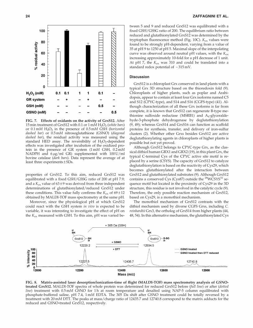
To investigate whether GrxS12 can be a target of nitrosyla-tion, we treated the protein with GSNO, a typical NO-donor.The reaction of GrxS12 with GSNO under aerobic conditionshad no effect on its activity (Fig. 7). Further, GSNO treatmentdid not induce any absorption band at 335 nm, characteristic forS-nitrosothiol groups (data not shown). Altogether, these re-sults suggest that neither Cys29 nor Cys87 may be nitrosylatedunder these conditions. However, GSNO can also induce theformation of glutathione-mixed disulfides. MALDI-TOF massspectrometry analysis revealed a mass increase of *305 Da inthe totality of GrxS12 treated with GSNO, consistent with theformation of one glutathione adduct. The 305 Da mass increasewas reversed by treatment with DTT (Fig. 8).
The midpoint redox potential of glutathionylatedGrxS12 is - 349 mV at the pH of 7.9 (as in illuminatedchloroplasts) and - 315 mV at pH 7.0 (as indarkened chloroplasts)
Due to the large shift of 305 Da in molecular mass, MALDI-TOF mass spectrometry allows to unambiguously quantifythe ratio between reduced and glutathionylated GrxS12 underdifferent conditions. We used this method to characterize theequilibrium between GrxS12 and GSH redox buffer. Experi-ments were performed at pH 7.9, a value that might occurin chloroplasts under photosynthetic conditions (47). Ratios be-tween reduced and glutathionylated GrxS12 were determined
FIG. 4. Determination of the pKa of the active site Cys ofGrxS12. Reduced GrxS12 (3 lM, black circles) and C87S mu-tant (3 lM, white circles) were incubated with iodoacetamide(IAM) (0.3 mM) at different pH values for 3 min at roomtemperature and then assayed using the standard HED assayafter 100-fold dilution. After incubation, the percentages of re-maining activity at each pH were determined by comparing theactivity of the enzyme incubated with and without IAM. Symbolsrepresent the average of at least three experiments – SDs. For thesake of clarity, SDs for C87S mutant data were omitted. The pKa
values were obtained by nonlinear regression using an adapta-tion of the Henderson-Hasselbalch equation (16). Interpolationcurves for GrxS12 and C87S mutant are represented as full anddashed sigmoids, respectively.
FIG. 5. pH-rate profile of GrxS12 activity. The activity ofGrxS12 was assayed with HED. The concentration of GR in theassay was either the standard concentration for the HED assay(6 lg/ml, open circles), or 15-fold increased (90 lg/ml, blackcircles). Buffers were potassium phosphate (pH 6.5–7.5), Tris-HCl (pH 7.5–9), and glycine (pH 9–10). GrxS12-dependentdeglutathionylating activity is represented as a percentage ofmaximal activity. Interpolation of experimental data with GRin excess (black circles) was made by nonlinear regression withthe Henderson-Hasselbalch equation.
22 ZAFFAGNINI ET AL.

after equilibration with different GSH/GSSG ratios (Fig. 9A)according to the following reaction:
GrxS12-SHþGSSG4GrxS12-SSGþGSH
The equilibrium constant of GrxS12 oxidation by GSSG(Kox) could be derived from the equilibrium concentration ofthe different species (Fig. 9B):
Kox¼ [GrxS12-SSG]=[GrxS12-SH] · [GSH]=[GSSG]
Based on three independent experiments, the average Kox
resulted to be 69 – 12 standard deviation (SD). The Em ofGrxS12 mixed disulfide could then be derived from the Kox bymeans of the Nernst equation:
Em(GrxS12 - SSG)¼Em(GSSG)�RT=nF · ln Kox
Considering an Em(GSSG) of - 294 mV at pH 7.9 (1) andsetting the value of n at 2 (as expected for a disulfide/dithioltwo-electron transfer process), the Kox value could then betranslated into a midpoint redox potential of GrxS12 mixeddisulfide (Em[GrxS12-SSG]) of - 349 – 2 mV.
Then, intrinsic tryptophan fluorescence was used as an al-ternative method to determine reduced and glutathionylatedGrxS12. Fluorescence emission of GrxS12 after excitation at290 nm was previously reported to be quenched by glu-tathionylation (9). The minimum distance between the aro-matic ring of the only tryptophan of GrxS12 (Trp28) and themixed disulfide is 3.74 A, as calculated from the crystalstructure of glutathionylated GrxS12 (9) (Fig. 10A). Short-distance interactions between tryptophan aromatic rings andsulfur atoms of disulfides result in intrinsic fluorescencequenching; reduced sulphydryl groups being much less ef-fective (10). Therefore, we compared the fluorescence signal ofglutathionylated and reduced GrxS12 after tryptophan-specific excitation at 295 nm (Fig. 10B) and took advantage ofthis important quenching effect to investigate the redox
FIG. 6. Kinetics of inactivation of GrxS12. The reactionwas initiated by the addition of varying concentrations ofinactivator (IAM or H2O2) to solutions containing reducedGrxS12 in Tris-HCl, pH 7.9. At the indicated time, aliquotswere withdrawn from the incubation mixtures and assayedfor GrxS12 activity as described in ‘‘Material and Methods’’Section. (A) Time- and concentration-dependent inactivationof GrxS12 by IAM. From top to bottom: 25, 50, 100, 250 lMIAM. (B) Time- and concentration-dependent inactivation ofGrxS12 by H2O2. From top to bottom: 25, 100, 250, 1000 lMH2O2. (C) Double-reciprocal plots of kapp versus IAM (blacktriangles) and H2O2 (black circles) concentrations obtainedaccording to Kitz and Wilson (26) to yield KI and kinact. Eachdata point represents the mean of three independently ob-tained data sets. The straight lines represent the best-fit linearregression through the raw data.
‰
Table 2. Kinetic Parameters for Inhibition of GrxS12by Iodoacetamide and H2O2
KI kinact kinact/KI
Compound lM min - 1 M - 1 min - 1
IAM 114 – 13 1.00 – 0.06 8.77 · 103
H2O2 112 – 20 0.082 – 0.006 7.32 · 102
The kinetic parameters of GrxS12 inhibition by IAM and H2O2
were determined by incubating the protein with variable amounts ofinactivator. At different times, aliquots were withdrawn to assayglutaredoxin activity using HED assay with 30 nM GrxS12. KI andkinact were determined according to (26). Data are represented asmeans – SD (n = 3).
IAM, iodoacetamide.
GLUTAREDOXIN S12 AND REDOX SIGNALING 23
properties of GrxS12. To this aim, reduced GrxS12 wasequilibrated with a fixed GSH/GSSG ratio of 200 at pH 7.9;and a Kox value of 63 – 9 was derived from three independentdeterminations of glutathionylated/reduced GrxS12 underthese conditions. This value fully confirms the Kox of 69 – 12obtained by MALDI-TOF mass spectrometry at the same pH.
Moreover, since the physiological pH at which GrxS12could react with the GSH system in vivo is expected to bevariable, it was interesting to investigate the effect of pH onthe Kox measured with GSH. To this aim, pH was varied be-
tween 5 and 9 and reduced GrxS12 was equilibrated with afixed GSH/GSSG ratio of 200. The equilibrium ratio betweenreduced and glutathionylated GrxS12 was determined by thetryptophan fluorescence method (Fig. 10C). Kox values werefound to be strongly pH-dependent, varying from a value of35 at pH 9 to 1250 at pH 5. Maximal slope of the interpolatingcurve was observed around neutral pH values, with the Kox
increasing approximately 10-fold for a pH decrease of 1 unit.At pH 7, the Kox was 310 and could be translated into astandard redox potential of - 315 mV.
Discussion
GrxS12 is a chloroplast Grx conserved in land plants with atypical Grx 3D structure based on the thioredoxin fold (9).Chloroplasts of higher plants, such as poplar and Arabi-dopsis, appear to contain at least four Grx isoforms named C5and S12 (CPYC-type), and S14 and S16 (CGFS-type) (41). Al-though characterization of all these Grx isoforms is far fromcomplete, it is known that GrxS12 can regenerate B-type me-thionine sulfoxide reductase (MSRB1) and A4-glyceralde-hyde-3-phosphate dehydrogenase by deglutathionylation(9, 49); whereas GrxS14 and GrxS16 can function as scaffoldproteins for synthesis, transfer, and delivery of iron-sulfurclusters (2). Whether other Grxs besides GrxS12 are activedeglutathionylating agents in chloroplasts of higher plants ispossible but not yet proved.
Although GrxS12 belongs to CPYC-type Grx, as the clas-sical dithiol human GRX1 and GRX2 (19), in this plant Grx, thetypical C-terminal Cys of the CPYC active site motif is re-placed by a serine (CSYS). The capacity of GrxS12 to catalyzedeglutathionylation is based on the reactivity of Cys29, whichbecomes glutathionylated after the interaction betweenGrxS12 and glutathionylated substrates (9). Although GrxS12contains a conserved Cys (Cys87) outside the 28WCSYS32 se-quence motif but located in the proximity of Cys29 in the 3Dstructure, this residue is not involved in the catalytic cycle (9).Therefore, the only possible reaction mechanism of GrxS12,based on Cys29, is a monothiol mechanism.
The monothiol mechanism of GrxS12 contrasts with thedithiol mechanism used by diverse CGFS Grxs, including C.reinhardtii Grx3, the ortholog of GrxS14 from higher plants (44,48, 54). In this alternative mechanism, the glutathionylated Cys
FIG. 7. Effects of oxidants on the activity of GrxS12. After15 min treatment of GrxS12 with 0.1 or 1 mM H2O2 (white bars)or 0.1 mM H2O2 in the presence of 0.5 mM GSH (horizontaldashed bar) or 0.5 mM nitrosoglutathione (GSNO) (diagonaldashed bar), the residual activity was measured using thestandard HED assay. The reversibility of H2O2-dependenteffects was investigated after incubation of the oxidized pro-tein in the presence of GR system (1 mM GSH, 0.2 mMNADPH and 6 lg/ml GR) supplemented with 100 U/mlbovine catalase (dark bars). Data represent the average of atleast three experiments – SDs.
FIG. 8. Matrix-assisted laser desorption/ionization-time of flight (MALDI-TOF) mass spectrometry analysis of GSNO-treated GrxS12. MALDI-TOF spectra of whole protein was determined for reduced GrxS12 before (full line) or after (dottedline) treatment with 0.5 mM GSNO for 1 h at room temperature and desalted using NAP-5 column equilibrated withphosphate-buffered saline, pH 7.4, 1 mM EDTA. The 305 Da shift after GSNO treatment could be totally reversed by atreatment with 20 mM DTT. The peaks at mass/charge ratio of 12435.7 and 12740.8 correspond to the matrix adducts for thereduced and GSNO-treated GrxS12, respectively.
24 ZAFFAGNINI ET AL.

of the Grx is attacked by a second Cys, rather than GSH, withformation of an internal disulfide bridge. In the case of Grx3,the catalytic cycle is closed by the reduction of this disulfideby FTR in a reaction linked to photosynthetic electron trans-port (54). It is possible that FTR-dependent CGFS-typeGrxs and GSH-dependent CPYC-type Grxs co-exist in chlo-roplasts of higher plants and perform similar reactions, butsince they are based on different reducing systems, this might
be useful to perform deglutathionylation in both light anddark conditions.
Kinetic properties of GrxS12
The mechanism of deglutathionylation performed byGrxS12 on a glutathionylated protein as BSA is here shown tobe ping pong. This mechanism implies that the interaction
FIG. 9. Redox titration of GrxS12 mixed disulfide with GR buffer. (A) Reduced GrxS12 (50 lM) was equilibrated withdifferent [GSH]/[GSSG] ratios in 30 mM Tris-HCl (pH 7.9) for 10 min before the reaction was quenched by 1.3% tri-fluoroacetic acid. GrxS12-SH/GrxS12-SSG ratios at equilibrium were determined by digital integration of MALDI-TOF massspectrometry peaks indicated in the figure by stars (GrxS12-SH) and diamonds (GrxS12-SSG). Spectra are displayed as afunction of GSH/GSSG ratios at equilibrium (see Materials and Methods section). A correction was introduced in thecalculations to account for the estimated breakage of 5% mixed disulfides by laser shooting. (B) The fraction of reducedGrxS12 that was measured by mass spectrometry after equilibration with different glutathione buffers was plotted againstthe [GSH]/[GSSG] ratio at equilibrium. Kox was obtained from nonlinear least-squares regression. Experimental data aredisplayed as means – SD obtained from three separate experiments.
GLUTAREDOXIN S12 AND REDOX SIGNALING 25

between reduced GrxS12 and BSA-SSG is followed by theseparation between reduced BSA and glutathionylatedGrxS12. In the second part of the reaction cycle, GrxS12-SSGreacts with GSH and releases a GSSG molecule while recov-ering its original reduced state. In all these reactions, thiolates(GrxS12-S - , GS - ) rather than thiols make the nucleophilicattack on mixed disulfides. Consistently, the reaction cata-lyzed by GrxS12 was strongly pH dependent and apparentlyinfluenced by the limited acidity of GSH (pKa 8.7) (46). As aconsequence, the overall reaction catalyzed by GrxS12 couldbe limited, at physiological pH values, by the GSH attack onGrxS12-SSG (step 2 of Fig. 1). A similar ping-pong reactionmechanism limited by the GSH attack was demonstrated forboth human Grx1 and Grx2 (19). The reaction mechanism ofGrxS12 is analogous to human Grxs also for the true kineticparameters kcat and Km (vs. both BSA-SSG and GSH) being toohigh to be experimentally determined. However, kcat/Km ra-tios of GrxS12 could be determined and found to be in theorder of 106 M - 1 s - 1 versus BSA-SSG and 104 M - 1 s - 1 versusGSH. Similarly, kcat/Km ratios of human Grx (Grx1, Grx2),though determined under slightly different conditions, werein the range of 105 M - 1 s - 1 versus BSA-SSG and 104–105 M - 1
s - 1 versus GSH (18, 27). The kinetic features of GrxS12 areconsistent with both binary complexes (GrxS12/BSA-SSGand GrxS12-SSG/GSH) being very short living or even non-existent. Lack of accumulation of binary complexes wouldexplain why GrxS12 could not be experimentally saturated
by either substrate. Although the reason for GrxS12-binarycomplexes being undetectable cannot be derived from pres-ent data, it can be safely concluded that GrxS12 practi-cally oscillates between a free reduced form and a freeglutathionylated form, with no intermediate complexes,during its catalytic cycle. The fact that free glutathionylatedGrxS12 is relatively stable is consistent with its successfulcrystallization (9).
Thermodynamic properties of GrxS12 and possiblephysiological roles in vivo
The midpoint redox potential of the glutathionylatedGrxS12 mixed disulfide was determined as - 349 mV at pH7.9, a value representative of stromal (chloroplast) pH underphotosynthetic conditions (47). This value was obtained byequilibration of GrxS12 with GSH/GSSG as redox buffer anddetermination of glutathionylated and reduced GrxS12 byMALDI-TOF mass spectrometry (Fig. 9), and confirmed byintrinsic tryptophan fluorescence (Fig. 10).
At pH 7.0, a pH value that represents the chloroplast stromain the dark, the Em of GrxS12 mixed disulfide was -315 mV(Fig. 10). The difference of 35 mV between Em at pH 7.0 and 7.9is in good agreement with the theory if we consider that at bothpH values, catalytic Cys29 of reduced GrxS12 would be de-protonated (pKa 3.9, Fig. 4), whereas GSH would be mostlyprotonated (pKa 8.7) (46). Reduction of GrxS12-SSG mixed
FIG. 10. pH-dependence ofGrxS12 Kox with GR buffer. (A)Detail of the crystallographic struc-ture of glutathionylated GrxS12 (8)showing the shortest distances be-tween the aromatic ring of Trp28and the sulfur atoms of the mixeddisulfide. The figure was made withPyMol based on file pdb 3FZ9. (B)Emission spectra of reduced (blackcircles) and glutathionylated (whitecircles) GrxS12 (excitation at295 nm) were recorded with 0.4 lMprotein at 25�C in 30 mM Tris-HCl,pH 7.9, 1 mM EDTA. Buffer blankwas run and subtracted from bothspectra. The fluorescence maximumof reduced GrxS12 was arbitrarilyset to 100 fluorescence units. (C)Reduced GrxS12 was equilibratedwith an initial [GSH]/[GSSG] ratioof 200:1 (2 mM total GSH concen-tration) at different pH in the fol-lowing buffers: sodium citrate (pH5.0), MES (pH 6.0), HEPES (pH 7.0),Tris-HCl (pH 7.9), glycine (pH 9.0).Equilibrium ratios between re-duced and glutathionylated GrxS12were determined by the tryptophanintrinsic fluorescence method. Kox
were calculated from equilibriumratios GSH/GSSG and GrxS12-SSG/GrxS12-SH as described in thetext. Data are means – SD of tripli-cate determinations.
26 ZAFFAGNINI ET AL.

disulfide would, thus, involve two electrons but only oneproton, at least at physiological pH:
GrxS12 - SSGþ 2e� þ 1 Hþ/GrxS12 - S� þGSH
implying that the standard redox potential of GrxS12 shoulddecrease by*30 mV for one pH unit increase, very similar to ourdeterminations. To our knowledge, this is the first redox char-acterization of a mixed disulfide of a wild-type Grx of anysource. A variant of yeast Grx1 (CPYC active site), in which theC-terminal Cys of the active site was exchanged to Ser to avoidformation of an internal disulfide and to stabilize the glutathio-nylated form, was recently reported to have an Em of - 295 mVat pH 7 for the mixed disulfide (25). Other redox analyses, per-formed on dithiol Grxs, generally showed less negative redoxpotentials usually comprised between - 170 and - 232 mV at pH7 (44). On the contrary, the plastidial Chlamydomonas Grx3 dis-plays a midpoint redox potential of - 323 mV at pH 7.9 (54), butthis disulfide is physiologically reduced by FTR ( - 374 mV at pH7.9) (24), a stronger reductant than GSH.
At neutral pH, the standard redox potential of glutathi-one ( - 240 mV) (1) is much less reducing than that of GrxS12( - 315 mV). However, the glutathione pool is largely reducedin vivo, and its actual redox potential might be much lower. Intransgenic Arabidopsis plants expressing roGFP2 in the cytosol,the GSH/GSSG ratio in vivo was estimated to be 105 and theredox potential around - 320 mV (28, 32), although it is cur-rently unknown whether roGFP2 fluorescence in vivo does onlyreflect the GSH redox state, or may be influenced by otherfactors (17). Much lower GSH/GSSG ratios of 10–20 were de-rived from direct determinations of GSH and GSSG in wholeplant tissues (17), but these exceedingly low ratios are hardlyrepresentative of the cytosol or chloroplasts, as most of GSSG islikely sequestered in compartments with low reducing capacity(e.g., vacuoles; (39). Whatever the actual GSH/GSSG ratio isunder optimal conditions, plenty of evidence demonstrate thatthe glutathione system in vivo is subject to oxidation on stress orwhen components of the antioxidant system of plants are ge-netically repressed [(28, 33); for a review see Ref. (17)]. In spiteof this, the amplitude of the GSH/GSSG variation in givencompartments such as chloroplasts is still uncertain.
Based on our Kox determinations for the reaction of GrxS12with GSSG (309 at pH 7.0 and 69 at pH 7.9), it can be easilycalculated that GrxS12 would be almost completely reducedat any physiological pH if it was in equilibrium with a GSH/GSSG ratio of 105. Under these conditions, GrxS12 could ef-ficiently work as a deglutathionylating agent, if necessary. Onthe other hand, a very limited oxidation of the GSH poolwould dramatically change this scenario. At a physiologicalconcentration of 2 mM GSH, oxidation of only 2% GSH wouldproduce 20 lM GSSG and establish a GSH/GSSG of 100. Inequilibrium with this GSH/GSSG ratio, glutathionylatedGrxS12 would exceed reduced GrxS12, and this effect wouldbe more pronounced at neutral than alkaline pH. As a result, avery limited oxidation of the GSH pool could lead to a stronginhibition of GrxS12 activity in vivo. Such inhibition would bestronger in the dark, when a neutral pH is established inchloroplasts, than in the light, when the photosynthetic ac-tivity tends to increase the stromal pH by about one unit (47).
A major difference between GrxS12 and other chloroplastGrxs of higher plants may reside in the stability of the glu-tathionylated form. Only monothiolic Grx such as GrxS12 can
exist in a stable glutathionylated form, because, even in theabsence of GSH, glutathionylation of dithiol Grxs could beslowly removed by formation of an intramolecular disulfidebetween the two active Cys. Glutathionylation of GrxS12 canresult from either reaction with H2O2 and GSH (9), or GSSG,or GSNO or by interaction with a glutathionylated substrate(A4-glyceraldehyde-3-phosphate dehydrogenase, (9); ICL,BSA, this work). Stability of GrxS12-SSG depends on pH and[GSH]/[GSSG] ratios and is, thus, predicted to increase in thedark and under stress conditions that could lower the [GSH]/[GSSG] ratio. Notably, the only Grx identified to date as atarget of glutathionylation under stress by proteomic ap-proaches in higher plants is indeed GrxS12 (13) and wespeculate that GrxS12-SSG may be involved in signaling.
In this scenario, the unique biochemical properties ofGrxS12 would allow glutathione to play a signaling rolethrough glutathionylation of GrxS12 targets. The rapid inac-tivation of GrxS12 by glutathionylation under mild oxidativestress would stabilize a number of glutathionylated proteinsthat could propagate the oxidative stress signal and possiblytrigger-specific adaptative responses. Once these responseshave been initiated, the decrease of the intracellular redoxpotential to a more reduced state should lead to reactivationof GrxS12 and deglutathionylation of GrxS12 targets. Thisproposed mechanism involving GrxS12 inactivation in sig-naling shares some similarities with the inactivation of 2-Cysperoxiredoxin (Prx) by overoxidation or phosphorylation(40). Indeed, it has been proposed that overoxidation of theperoxidatic Cys of 2-Cys Prx to the sulfinate form and itssubsequent slow reduction by sulfiredoxin (5, 50) could leadto transient accumulation of H2O2 and thereby allow H2O2 toplay a signaling role. This concept has been termed the‘‘floodgate’’ hypothesis (52). However, in the case of receptorsignaling, another mechanism appears to be involved, as amembrane-associated 2-Cys Prx was recently shown to bereversibly inactivated by phosphorylation in cells stimulatedvia growth factors or immune receptors (51). This localizedinactivation of 2-Cys Prx likely allows generating favorableH2O2 gradients around membranes, where signaling proteinsare concentrated, while minimizing the general accumulationof H2O2 to toxic levels.
Similarly, the unique biochemical properties of GrxS12 mayallow this enzyme to facilitate redox signaling by allowingaccumulation of glutathionylated proteins, as just mentioned.Other chloroplastic Grxs may either exhibit distinct specifi-cities or be regulated by other mechanisms such as binding toFe-S clusters that has been suggested to constitute a mecha-nism of regulation of Grx activity (4). Confirming the potentialcontribution of GrxS12 in redox signaling will require deter-mining GrxS12-specific targets and analyzing the regulationof Grx activities in vivo. Finally, as GrxS12 orthologs are ab-sent in green algae (30), its potential role as a facilitator ofredox sensing and signaling in chloroplasts may be linkedto the requirement for a tighter redox control in terrestrialplants, as previously suggested by the analysis of some redox-controlled Calvin cycle enzymes (31).
Materials and Methods
Materials and enzymes
NAP-5 columns and 5,5¢-dithiobis-2-nitrobenzoic acid(DTNB) were purchased from GE Healthcare (Piscataway, NJ
GLUTAREDOXIN S12 AND REDOX SIGNALING 27

USA) and Pierce (Rockford, IL), respectively. GSH and GSSGwere purchased from either Roche Diagnostics Corporation(Indianapolis, IN) or Sigma-Aldrich (St. Louis, MO). All otherchemicals were obtained from Sigma-Aldrich.
GR from baker’s yeast and bovine catalase were purchasedfrom Sigma (St. Louis, MO). Recombinant ICL from C. re-inhardtii (ICL) was purified as described in (3). Poplar GrxS12wild type (GrxS12) and mutants GrxS12-C29S (C29S) andGrxS12-C87S (C87S) were expressed and purified as previ-ously described (9). Purity of all Grx forms used in this workwas assessed by SDS-PAGE and Coomassie blue staining(Supplementary Fig. S1; Supplementary Data are availableonline at www.liebertonline.com/ars). Concentration of pu-rified proteins was determined from UV absorption at280 nm, and extinction coefficients were calculated from thesequence (e= 64,540 and 9970 M - 1 cm - 1 for ICL and GrxS12,respectively).
Preparation of BSA-SSG
BSA-SSG was prepared by treating the protein with 20 mMreduced DTT for 30 min at room temperature followed bydesalting on a NAP-5 column pre-equilibrated with 30 mMTris-HCl, pH 7.9. The reduced protein was then allowed toreact with a 50-fold molar excess of GSSG for 16 h at roomtemperature and desalted as just described. The degree of BSAglutathionylation was spectrophotometrically determined byfollowing NADPH oxidation at 340 nm in a cuvette contain-ing 0.2 mM NADPH, 0.5 mM GSH, 6 lg/ml GR, 25 nMGrxS12 wt, and a varying concentration of BSA-SSG. The BSAwas quantified at 280 nm (e= 43,600 M - 1 cm - 1), and theconcentration of protein-bound GSH groups was calculatedfrom the total NADPH consumption for each BSA concen-tration as described in (6). This procedure resulted in1.03 – 0.08 GSH per molecule of BSA (Supplementary Fig. S2),consistent with monoglutathionylation of the protein, al-though some heterogeneity in BSA-SSG preparations cannotbe excluded.
Activity measurements and determination of apparentkinetic parameters
Before activity measurements, GrxS12 and its variants wereincubated with 10 mM DTT for 30 min at room temperatureand desalted on a NAP-5 column pre-equilibrated in 30 mMTris-HCl, pH 7.9. Grx activity in the HED assay was deter-mined as previously described (54). Briefly, 0.7 mM HED wasadded to a mixture containing 100 mM Tris-HCl, pH 7.9,2 mM EDTA, 0.1 mg/ml BSA, 0.2 mM NADPH, 1 mM GSH,and 6 lg/ml GR. After 3 min of preincubation, 30 nM GrxS12was added to the sample cuvette, and buffer was added to thereference cuvette. This GrxS12 concentration was previouslychecked for being within the range of linear responses of ac-tivity versus enzyme concentration. The decrease in absor-bance was followed at 340 nm. GRX activity was determinedafter subtracting the spontaneous reduction rate observed inthe absence of Grx, and the number of lmoles of NADPHoxidized per second per lmole of enzyme (i.e., turnovernumber, s - 1) was calculated using the extinction coefficient ofNADPH (e340 = 6230 M - 1 cm - 1). The apparent Km for gluta-thione of C87S mutant was determined as previously de-scribed (9). Apparent kinetic parameters for glutathionylatedsubstrates were determined using either HED (0.1– 1.5 mM)
or BSA-SSG (2.5–50 lM) in a mixture of 100 mM Tris-HCl, pH7.9, 2 mM EDTA, 0.1 mg/ml BSA, 0.2 mM NADPH, 6 lg/mlGR plus 1 mM GSH, and 30 nM GrxS12. Three independentexperiments were performed at each substrate concentration,and the apparent Km and kcat values were calculated by non-linear regression using a Michaelis Menten equation with theprogram CoStat (CoHort software, Monterey, CA).
Two-substrate kinetic analysis
The catalytic mechanism of GrxS12 was determined byindependently varying the concentration of BSA-SSG andGSH to produce a 4 · 4 matrix of concentration sets. Deglu-tathionylation activity was determined as just described with30 nM GrxS12. Each individual data set for a varied substrateat a fixed concentration of the other substrate was analyzed bynonlinear regression analysis as just described. A pair of ap-parent kcat and apparent Km values was, thus, obtained at eachsingle concentration of the fixed substrate. Apparent kcat
values obtained in different experiments under identicalconditions were averaged ( – SD) and represented in second-ary plots (1/kcat vs. 1/[substrate]), from which the kinetic ef-ficiency parameters kcat/Km were derived.
Reactivation of ICL-SSG by GrxS12 and its variants
ICL-SSG was prepared as described in (3). All reactivationtreatments were performed in 50 mM HEPES-NaOH, pH 7.2at room temperature. Reactivation of ICL-SSG (10 lM) wasmeasured with GrxS12 (5 or 0.1 lM) or C87S (5 lM) or C29S(5 lM) in the presence of 0.5 mM DTT or 2 mM GSH alone or2 mM GSH in the presence of a GRS made of 0.2 mM NADPHand 6 lg/ml GR. Control reaction mixtures were performedin the same conditions by omitting Grxs. At indicated times,aliquots were withdrawn to assay ICL activity monitoredas described in (3). Half-saturating concentration for GrxS12and C87S mutant were determined by incubating ICL-SSG(10 lM) in the presence of varying concentrations of GrxS12and C87S mutant (0.5 to 20 lM) and 2 mM GSH alone, orwith GRS.
Influence of the [GSH]/[GSSG] ratio on GrxS12-dependent deglutathionylation of ICL-SSG
ICL-SSG (10 lM) was incubated in the presence of 5 lMGrxS12 and different [GSH]/[GSSG] ratios at a total concen-tration of 2 mM. After 15 min incubation, aliquots werewithdrawn to assay ICL activity monitored as described inRef. (3). Control experiments were carried out by incubatingreduced ICL (10 lM) with different [GSH]/[GSSG] ratios (asjust described), and protein activity was measured after15 min incubation. Freshly prepared solutions of GSH in-variably contain small amounts of GSSG, which was deter-mined by either directly assaying GSH with DTNB (54) or byenzymatic assay of GSSG by GR (1 lg/ml) and NADPH(0.2 mM). The degree of GSSG contamination was found be-tween 0.5% and 1% on a molar basis, and this value wasconsidered to calculate the effective [GSH]/[GSSG] ratio foreach reaction mixture.
pH-dependence of GrxS12 activity
To measure the pH-dependency of GrxS12-catalyzed de-glutathionylation, HED assays were performed as just
28 ZAFFAGNINI ET AL.

described except that b-ME-SSG was generated in a mixturecontaining 100 mM Tris-HCl, pH 7.9, 2 mM EDTA, 7 mMHED, and 10 mM GSH. After preincubation (3 min), Grx ac-tivity was determined by adding 50 ll of the preincubationmixture to 450 ll of the spectrophotometer assay systemcontaining 0.1 mg/ml BSA, 0.2 mM NADPH, and GR (6 or90 lg/ml). The following buffers were used: potassiumphosphate at pH 6.5–7.5; Tris-HCl at pH 7.5–9; and glycine atpH 9–10. Rates of GrxS12-dependent deglutathionylationwere represented as a percentage of maximal activity. Plots ofactivity versus pH were fit to a derivation of the Henderson-Hasselbalch equation as in (18).
Determination of the pKa of the catalytic Cys of GrxS12by IAM inactivation
The pH-dependence of inactivation of GrxS12 and C87Smutant by IAM was carried out by incubating reduced pro-teins (3 lM) with or without IAM (0.3 mM) for 3 min in so-dium citrate or phosphate buffer ranging from pH 2 to 6.5(18). After preincubation, Grx activity was determined byadding an aliquot of the preincubation mixture to the HEDassay system just described. The residual activity expressed asa percentage of maximal activity was plotted versus pH, andan adaptation of the Henderson-Hasselbalch equation wasused to calculate the pKa of the active site Cys as in (18).
IAM-dependent inactivation kinetics
Reduced GrxS12 (3 lM) was incubated in 30 mM Tris-HCl,pH 7.9, in the presence of IAM ranging from 25 to 250 lM atroom temperature. All reactions were initiated by the additionof IAM. At different times, aliquots were withdrawn from theincubation mixture and diluted 100-fold into the standardHED assay system, and enzyme activity was determined. Allinactivation experiments were performed in triplicate andmonitored relative to a control sample without IAM, which isset to 100% activity at each time point. Estimates of the con-centration of IAM that gives half-maximal rate of inactivation(KI) and first-order inactivation rate constants (kinact) weredetermined according to the Kitz-Wilson plot (29).
Effect of hydrogen peroxide and GSNO on GrxS12
Reduced GrxS12 (3 lM) was treated with H2O2 rangingfrom 25 lM to 1 mM or with 0.1 mM H2O2 in the presence of0.5 mM GSH in 30 mM Tris-HCl (pH 7.9) at room tempera-ture. At different times, enzyme activity was determined asjust described, and H2O2-dependent inactivation kineticswere analyzed as described in the previous section (29). In aseparate set of control experiments, it was demonstrated thatGR activity is not sensitive to the highest concentration ofH2O2 used (1 mM) after 100-fold dilution in an assay cuvettesystem containing 0.2 mM NADPH and 50 lM GSSG in100 mM Tris-HCl, pH 7.9 (data not shown). For reduction ofoxidized proteins, H2O2-treated samples were incubated for15 min at room temperature with the GRS (1 mM GSH,0.2 mM NADPH, and 6 lg/ml GR) in the presence of 100 U/ml bovine catalase.
To study S-nitrosylation, reduced GrxS12 was incubatedwith 0.5 mM GSNO in PBS (137 mM NaCl, 2.7 mM KCl,8.1 mM Na2HPO4, and 1.9 mM KH2PO4), pH 7.4, 1 mM EDTAat room temperature and enzyme activity was assayed at
different incubation times. S-nitrosylation of GrxS12 was an-alyzed by recording the UV-visible spectra of the proteintreated with 0.5 mM GSNO for 1 h and desalted on a NAP-5column equilibrated with PBS buffer. The desalted proteinsample was also used for MALDI-TOF mass spectrometryanalysis as described in (41).
Determination of Kox and midpoint redox potentialof GrxS12 by MALDI-TOF mass spectrometryin the presence of GSH
The redox potential of the mixed disulfide of GrxS12-SSGwas calculated after equilibration with different [GSH]/[GSSG] ratios. A MALDI-TOF mass spectrometry approachwas developed to quantify in each sample the ratio of reduced(GrxS12-SH) and glutathionylated GrxS12 (GrxS12-SSG) fromthe area of the corresponding peaks, which were separated byapprox 305 Da in mass. As observed by Iversen et al. (25), evenin completely glutathionylated samples, small amounts ofreduced protein were always detected, suggesting that ap-proximately 5% of mixed disulfide bonds were broken bylazer shooting (37). The relative amounts of GrxS12-SH andGrxS12-SSG were, thus, determined by numeric integration ofthe peak areas after correction for the fraction of GrxS12-SH,which was calculated to derive from GrxS12-SSG disulfidebreakage by shooting. The equilibrium constant of the reac-tion of GrxS12 with GSSG is defined as follows:
Kox¼ [GrxS12 - SSG]=[GrxS12 - SH] · [GSH]=[GSSG]
For determination of the Kox, reduced GrxS12 (50 lM) wasincubated in 30 mM Tris-HCl (pH 7.9) with different [GSH]/[GSSG] ratios, and the relative amounts of GrxS12-SH andGrxS12-SSG were measured as just described. GrxS12 wasadded to each mix of glutathione and allowed to equilibratefor 10 min before the reaction was quenched by 1.3% tri-fluoroacetic acid. Samples from each acid-quenched reactionwere analyzed as described in (45). The equilibrium concen-trations of GSH were calculated by considering the amount ofGSH released by the reaction of GSSG with GrxS12-SH.Conversely, the equilibrium concentration of GSSG was cal-culated after subtracting from the initial concentration ofGSSG, an amount of GSSG equal to the amount of GrxS12-SSG at equilibrium.
The standard redox potential of GrxS12 mixed disulfide wasderived from the Kox according to the Nernst equation. Thestandard redox potential of the GSH/GSSG couple at pH 7.9was considered to be - 295 mV, that is, 55 mV more negativethan at pH 7.0 ( - 240 mV) (1) based on the assumption that GSHis fully protonated at pH 7.9 (pKa(GSH)& 8.7) (46); and, there-fore, the pH-dependence of Em(GSH/GSSG) between 7.0 and 7.9should be linear with a slope of - 60 mV per pH unit.
Determination of the standard redox potentialof GrxS12 by tryptophan intrinsic fluorescenceand pH-dependence of the Kox
Fluorescence measurements were performed at 25�C inquartz cuvettes with 1 cm light-path length. The excitationwavelength was set at 295 nm (for selective observation of thetryptophan residue), and the emission was recorded between300 and 450 nm with a bandwidth of 10 nm at a scan speed of50 nm/min. To calibrate the measurements, the fluorescence
GLUTAREDOXIN S12 AND REDOX SIGNALING 29

signals of fully reduced and fully glutathionylated GrxS12(0.4 lM) were recorded under native conditions in 30 mMTris-HCl, pH 7.9, 1 mM EDTA. Buffer blanks were run foreach sample and were subtracted from the spectra.
The pH dependence of the Kox of GrxS12 with GSH/GSSG asredox buffer was determined at different pH values by thetryptophan intrinsic fluorescence method. Concentration of allbuffers was 100 mM: sodium citrate, pH 5.0; MES, pH 6.0;HEPES, pH 7.0; Tris-HCl, pH 7.9; glycine, pH 9.0. The reducedprotein (20 lM) was allowed to equilibrate for 1 h in the pres-ence of an initial [GSH]/[GSSG] ratio of 200:1. Afterward,fluorescence spectra were recorded in 10-fold diluted samples inthe same buffer as the incubation. For each sample, the fluo-rescence signal was measured at 345 nm (excitation at 295 nm),which corresponds to GrxS12 maximum fluorescence peak.
Acknowledgments
This work was supported in part by PRIN 2008 grant (toM.Z. and P.T.) from the Ministero dell’Istruzione, dell’Uni-versita e della Ricerca of Italy, by ANR Grant 08-BLAN-0153GLUTAPHOTO (to M.B. and S.D.L.) and by ANR JC07 204825(to J.C. and N.R.).
Author Disclosure Statement
The authors have no conflict of interest to disclose.
References
1. Aslund F, Berndt KD, and Holmgren A. Redox potentials ofglutaredoxins and other thiol-disulfide oxidoreductases ofthe thioredoxin superfamily determined by direct protein-protein redox equilibria. J Biol Chem 272: 30780–30786, 1997.
2. Bandyopadhyay S, Gama F, Molina-Navarro MM, Gual-berto JM, Claxton R, Naik SG, Huynh BH, Herrero E, Jac-quot JP, Johnson MK, and Rouhier N. Chloroplast monothiolglutaredoxins as scaffold proteins for the assembly and de-livery of [2Fe-2S] clusters. EMBO J 27: 1122–1133, 2008.
3. Bedhomme M, Zaffagnini M, Marchand CH, Gao XH,Moslonka-Lefebvre M, Michelet L, Decottignies P, and Le-maire SD. Regulation by glutathionylation of isocitrate lyasefrom Chlamydomonas reinhardtii. J Biol Chem 284: 36282–36291, 2009.
4. Berndt C, Hudemann C, Hanschmann EM, Axelsson R,Holmgren A, and Lillig CH. How does iron-sulfur clustercoordination regulate the activity of human glutaredoxin 2?Antioxid Redox Signal 9: 151–157, 2007.
5. Biteau B, Labarre J, and Toledano MB. ATP-dependent re-duction of cysteine-sulphinic acid by S. cerevisiae sulphir-edoxin. Nature 425: 980–984, 2003.
6. Ceylan S, Seidel V, Ziebart N, Berndt C, Dirdjaja N, andKrauth-Siegel RL. The dithiol glutaredoxins of African try-panosomes have distinct roles and are closely linked to theunique trypanothione metabolism. J Biol Chem 285: 35224–35237, 2010.
7. Colman R. Glutathione reductase (Yeast). Methods Enzymol17: 500–503, 1971.
8. Couturier J, Jacquot JP, and Rouhier N. Evolution and di-versity of glutaredoxins in photosynthetic organisms. CellMol Life Sci 66: 2539–2557, 2009.
9. Couturier J, Koh CS, Zaffagnini M, Winger AM, GualbertoJM, Corbier C, Decottignies P, Jacquot JP, Lemaire SD, Di-dierjean C, and Rouhier N. Structure-function relationship of
the chloroplastic glutaredoxin S12 with an atypical WCSYSactive site. J Biol Chem 284: 9299–9310, 2009.
10. Cowgill RW. Fluorescence and the structure of proteins. XII.Pancreatic trypsin inhibitor. Biochim Biophys Acta 140: 552–554, 1967.
11. Dalle-Donne I, Rossi R, Colombo G, Giustarini D, and Mil-zani A. Protein S-glutathionylation: a regulatory device frombacteria to humans. Trends Biochem Sci 34: 85–96, 2009.
12. Discola KF, de Oliveira MA, Rosa Cussiol JR, Monteiro G,Barcena JA, Porras P, Padilla CA, Guimaraes BG, and NettoLE. Structural aspects of the distinct biochemical propertiesof glutaredoxin 1 and glutaredoxin 2 from Saccharomycescerevisiae. J Mol Biol 385: 889–901, 2009.
13. Dixon DP, Skipsey M, Grundy NM, and Edwards R. Stress-induced protein S-glutathionylation in Arabidopsis. PlantPhysiol 138: 2233–2244, 2005.
14. Fernandes AP and Holmgren A. Glutaredoxins: glutathione-dependent redox enzymes with functions far beyond asimple thioredoxin backup system. Antioxid Redox Signal 6:63–74, 2004.
15. Foyer CH and Noctor G. Redox homeostasis and antioxidantsignaling: a metabolic interface between stress perceptionand physiological responses. Plant Cell 17: 1866–1875, 2005.
16. Foyer CH and Noctor G. Redox regulation in photosyntheticorganisms: signaling, acclimation, and practical implica-tions. Antioxid Redox Signal 11: 861–905, 2009.
17. Foyer CH and Noctor G. Ascorbate and glutathione: theheart of the redox hub. Plant Physiol 155: 2–18, 2011.
18. Gallogly MM, Starke DW, Leonberg AK, Ospina SM, andMieyal JJ. Kinetic and mechanistic characterization and versatilecatalytic properties of mammalian glutaredoxin 2: implicationsfor intracellular roles. Biochemistry 47: 11144–11157, 2008.
19. Gallogly MM, Starke DW, and Mieyal JJ. Mechanistic andkinetic details of catalysis of thiol-disulfide exchange byglutaredoxins and potential mechanisms of regulation. An-tioxid Redox Signal 11: 1059–1081, 2009.
20. Gao X, Bedhomme, M, Michelet, L, Zaffagnini, M, and Le-maire, SD. Glutathionylation in photosynthetic organisms.Adv Bot Res 52: 363–403, 2009.
21. Gao XH, Zaffagnini M, Bedhomme M, Michelet L, Cassier-Chauvat C, Decottignies P, and Lemaire SD. Biochemicalcharacterization of glutaredoxins from Chlamydomonas re-inhardtii: kinetics and specificity in deglutathionylation re-actions. FEBS Lett 584: 2242–2248, 2010.
22. Ghezzi P. Regulation of protein function by glutathionyla-tion. Free Radic Res 39: 573–580, 2005.
23. Gravina SA and Mieyal JJ. Thioltransferase is a specificglutathionyl mixed disulfide oxidoreductase. Biochemistry32: 3368–3376, 1993.
24. Hirasawa M, Schurmann P, Jacquot JP, Manieri W, Jacquot P,Keryer E, Hartman FC, and Knaff DB. Oxidation-reductionproperties of chloroplast thioredoxins, ferredoxin:thioredoxinreductase, and thioredoxin f-regulated enzymes. Biochemistry38: 5200–5205, 1999.
25. Iversen R, Andersen PA, Jensen KS, Winther JR, and Si-gurskjold BW. Thiol-disulfide exchange between glutar-edoxin and glutathione. Biochemistry 49: 810–820, 2010.
26. Jensen KS, Hansen RE, and Winther JR. Kinetic and ther-modynamic aspects of cellular thiol-disulfide redox regula-tion. Antioxid Redox Signal 11: 1047–1058, 2009.
27. Johansson C, Lillig CH, and Holmgren A. Human mito-chondrial glutaredoxin reduces S-glutathionylated proteinswith high affinity accepting electrons from either glutathioneor thioredoxin reductase. J Biol Chem 279: 7537–7543, 2004.
30 ZAFFAGNINI ET AL.

28. Jubany-Mari T, Alegre-Batlle L, Jiang K, and Feldman LJ.Use of a redox-sensing GFP (c-roGFP1) for real-time moni-toring of cytosol redox status in Arabidopsis thaliana water-stressed plants. FEBS Lett 584: 889–897, 2010.
29. Kitz R and Wilson IB. Esters of methanesulfonic acid as ir-reversible inhibitors of acetylcholinesterase. J Biol Chem 237:3245–3249, 1962.
30. Lemaire SD. The glutaredoxin family in oxygenic photo-synthetic organisms. Photosynth Res 79: 305–318, 2004.
31. Marri L, Zaffagnini M, Collin V, Issakidis-Bourguet E, Le-maire SD, Pupillo P, Sparla F, Miginiac-Maslow M, andTrost P. Prompt and easy activation by specific thioredoxinsof calvin cycle enzymes of Arabidopsis thaliana associated inthe GAPDH/CP12/PRK supramolecular complex. Mol Plant2: 259–269, 2009.
32. Meyer AJ, Brach T, Marty L, Kreye S, Rouhier N, Jacquot JP,and Hell R. Redox-sensitive GFP in Arabidopsis thaliana is aquantitative biosensor for the redox potential of the cellularglutathione redox buffer. Plant J 52: 973–986, 2007.
33. Mhamdi A, Hager J, Chaouch S, Queval G, Han Y, TaconnatL, Saindrenan P, Gouia H, Issakidis-Bourguet E, Renou JP,and Noctor G. Arabidopsis GLUTATHIONE REDUCTASE1plays a crucial role in leaf responses to intracellular hydro-gen peroxide and in ensuring appropriate gene expressionthrough both salicylic acid and jasmonic acid signalingpathways. Plant Physiol 153: 1144–1160, 2010.
34. Michelet L, Zaffagnini M, Marchand C, Collin V, Decottig-nies P, Tsan P, Lancelin JM, Trost P, Miginiac-Maslow M,Noctor G, and Lemaire SD. Glutathionylation of chloroplastthioredoxin f is a redox signaling mechanism in plants. ProcNatl Acad Sci U S A 102: 16478–16483, 2005.
35. Michelet L, Zaffagnini M, Vanacker H, Le Marechal P,Marchand C, Schroda M, Lemaire SD, and Decottignies P. Invivo targets of S-thiolation in Chlamydomonas reinhardtii. J BiolChem 283: 21571–2158, 2008.
36. Mieyal JJ, Starke DW, Gravina SA, Dothey C, and Chung JS.Thioltransferase in human red blood cells: purification andproperties. Biochemistry 30: 6088–6097, 1991.
37. Patterson SD and Katta V. Prompt fragmentation of disulfide-linked peptides during matrix-assisted laser desorption ioni-zation mass spectrometry. Anal Chem 66: 3727–3732, 1994.
38. Poole LB and Nelson KJ. Discovering mechanisms of sig-naling-mediated cysteine oxidation. Curr Opin Chem Biol 12:18–24, 2008.
39. Queval G, Jaillard D, Zechmann B, and Noctor G. Increasedintracellular HO availability preferentially drives glutathi-one accumulation in vacuoles and chloroplasts. Plant CellEnviron 34: 21–32, 2011.
40. Rhee SG and Woo HA. Multiple functions of peroxiredoxins:peroxidases, sensors and regulators of the intracellularmessenger H2O2, and protein chaperones. Antioxid RedoxSignal 15: 781–794, 2011.
41. Rouhier N. Plant glutaredoxins: pivotal players in redoxbiology and iron-sulphur centre assembly. New Phytol 186:365–372, 2010.
42. Rouhier N, Couturier J, and Jacquot JP. Genome-wide analysisof plant glutaredoxin systems. J Exp Bot 57: 1685–1696, 2006.
43. Rouhier N, Gelhaye E, and Jacquot JP. Plant glutaredoxins:still mysterious reducing systems. Cell Mol Life Sci 61: 1266–1277, 2004.
44. Rouhier N, Lemaire SD, and Jacquot JP. The role of gluta-thione in photosynthetic organisms: emerging functions forglutaredoxins and glutathionylation. Annu Rev Plant Biol 59:143–166, 2008.
45. Sicard-Roselli C, Lemaire S, Jacquot JP, Favaudon V, Marc-hand C, and Houee-Levin C. Thioredoxin Ch1 of Chlamy-domonas reinhardtii displays an unusual resistance towardone-electron oxidation. Eur J Biochem 271: 3481–3487, 2004.
46. Srinivasan U, Mieyal PA, and Mieyal JJ. pH profiles indic-ative of rate-limiting nucleophilic displacement in thiol-transferase catalysis. Biochemistry 36: 3199–3206, 1997.
47. Takizawa K, Cruz JA, Kanazawa A, and Kramer DM. Thethylakoid proton motive force in vivo. Quantitative, non-invasive probes, energetics, and regulatory consequences oflight-induced pmf. Biochim Biophys Acta 1767: 1233–1244, 2007.
48. Tamarit J, Belli G, Cabiscol E, Herrero E, and Ros J. Bio-chemical characterization of yeast mitochondrial Grx5monothiol glutaredoxin. J Biol Chem 278: 25745–25751, 2003.
49. Tarrago L, Laugier E, Zaffagnini M, Marchand C, Le Mare-chal P, Rouhier N, Lemaire SD, and Rey P. Regenerationmechanisms of Arabidopsis thaliana methionine sulfoxide re-ductases B by glutaredoxins and thioredoxins. J Biol Chem284: 18963–18971, 2009.
50. Woo HA, Chae HZ, Hwang SC, Yang KS, Kang SW, Kim K, andRhee SG. Reversing the inactivation of peroxiredoxins causedby cysteine sulfinic acid formation. Science 300: 653–656, 2003.
51. Woo HA, Yim SH, Shin DH, Kang D, Yu DY, and Rhee SG.Inactivation of peroxiredoxin I by phosphorylation allowslocalized H(2)O(2) accumulation for cell signaling. Cell 140:517–528, 2010.
52. Wood ZA, Poole LB, and Karplus PA. Peroxiredoxin evo-lution and the regulation of hydrogen peroxide signaling.Science 300: 650–653, 2003.
53. Zaffagnini M, Michelet L, Marchand C, Sparla F, Decottig-nies P, Le Marechal P, Miginiac-Maslow M, Noctor G, TrostP, and Lemaire SD. The thioredoxin-independent isoform ofchloroplastic glyceraldehyde-3-phosphate dehydrogenase isselectively regulated by glutathionylation. FEBS J 274: 212–226, 2007.
54. Zaffagnini M, Michelet L, Massot V, Trost P, and LemaireSD. Biochemical characterization of glutaredoxins fromChlamydomonas reinhardtii reveals the unique properties of achloroplastic CGFS-type glutaredoxin. J Biol Chem 283: 8868–8876, 2008.
Address correspondence to:Prof. Paolo Trost
Department of Experimental Evolutionary BiologyUniversity of Bologna
Via Irnerio 42I-40126 Bologna
Italy
E-mail: [email protected]
Dr. Stephane D. LemaireLaboratoire de Biologie Moleculaire et Cellulaire des Eucaryotes
FRE3354 CNRS/UPMCInstitut de Biologie Physico-Chimique
13 rue Pierre et Marie Curies75005 Paris
France
E-mail: [email protected]
Date of first submission to ARS Central, February 2, 2011; dateof final revised submission, June 24, 2011; date of acceptance,June 27, 2011.
GLUTAREDOXIN S12 AND REDOX SIGNALING 31

Abbreviations Used
b-ME-SSG¼ glutathionylated b-mercaptoethanol.BSA¼ bovine serum albumin
BSA-SSG¼ glutathionylated bovine serum albuminCys¼ cysteine
DTNB¼ 5,5¢-dithiobis-2-nitrobenzoic acidDTT¼dithiothreitolFTR¼ ferredoxin:thioredoxin reductaseGR¼ glutathione reductase
GRS¼GSH-regenerating systemGrx¼ glutaredoxin
GSH¼ glutathione (reduced form)
GSNO¼nitrosoglutathioneGSSG¼ glutathione (oxidized form)
HED¼hydroxyethyl disulfide
IAM¼ iodoacetamide
ICL¼ isocitrate lyase
ICL-SSG¼ glutathionylated isocitrate lyase
MALDI-TOF¼matrix-assisted laser desorption/ionization-time of flight
PDT-bimane¼ (2-pyridyl)di-thiobimane
Prx¼peroxiredoxin
SD¼ standard deviation
32 ZAFFAGNINI ET AL.