Redox Proteins of Mycobacterium tuberculosis
12
Journal of the Indian Institute of Science A Multidisciplinary Reviews Journal ISSN: 0970-4140 Coden-JIISAD © Indian Institute of Science Journal of the Indian Institute of Science VOL 94:1 Jan.–Mar. 2014 journal.iisc.ernet.in REVIEWS Redox Proteins of Mycobacterium tuberculosis Swastik Phulera 1 , Mohd. Akif 2 , Abhijit A. Sardesai 3 and Shekhar C. Mande 1* Abstract | Evolution of life took place in a reducing atmosphere. As a remi- niscence of this, cytoplasm of all living organisms is reducing in nature. Furthermore most biochemical reactions like energy production etc. are redox in nature requiring either an electron supplier or acceptor. The maintenance of redox state thus becomes extremely crucial for any cell. Cells typically devote considerable resources for the maintenance of their redox state. M. tuberculosis successfully evades an array of host generated redox stresses. For this M. tuberculosis employs a large number of redox sensors and effectors. In our laboratory we have been studying some of these central players with a hope to be able to gain insights into the pathogen. We shall try to discuss the relevance of redox and our efforts to understand the same in this review. 1 National Centre for Cell Science, NCCS Complex, Ganeshkhind, Pune 411 007, India. *[email protected] 2 Department of Biochemistry, University of Hyderabad, Gachibowli, Hyderabad, India. 3 Laboratory of Bacterial Genetics, Centre for DNA Fingerprinting and Diagnostics, 5-4-399/B, Nampally, Hyderabad 500 001, India. 1 Redox Balance in Mycobacterium tuberculosis and Its Importance Tuberculosis (TB), caused by Mycobacterium tuber- culosis (M.tb), is amongst the leading causes of infection related mortality and morbidity world- wide. 1 Infection typically occurs via the respiratory tract upon inhalation of droplet nuclei containing M.tb. Upon reaching the alveoli the bacteria are phagocytosed by alveolar macrophages and in the initial stages of intracellular existence, M.tb resides within the phagosome. Herein M.tb is exposed to a hostile host environment comprising reactive oxygen intermediates (ROI), reactive nitrogen intermediates (RNIs), lysosomal enzymes, and toxic peptides. Moreover, M.tb can persist inside immune-competent individuals for decades in a latent form and may reactivate to cause an active disease when the host is immune-compromised. M.tb elaborates multiple mechanisms to survive in the host environment and has a unique ability to persist for long durations in the host. The suste- nance of viability in the presence of severe oxida- tive stress appears to be of primary importance for successful pathogenesis of M.tb. The major process for detoxification of oxida- tive damage to proteins and DNA occurs via the activities of redox enzymes. In our laboratory we have been trying to understand the mechanisms used by M.tb to maintain its cytoplasmic redox status and elucidate the structure-function fea- tures of redox enzymes of M.tb (Figure 1). We believe that understanding these mechanisms can lead to newer avenues for the development of novel therapeutics for both the active and the latent form of infection. Typically redox balance is both sensed and maintained by a set of proteins with a CXXC and TXXC motifs. In this review we discuss the functional and structural features of redox enzymes of M.tb but restrict the discussion to the work performed by our laboratory. 2 CXXC and TXXC Derived Motif Proteins Encoded by the M.tb Genome Disulphide bonds can alternate with free thiols to play a major role in both the maintenance of proper structure and supply of reducing/oxidizing equivalents. Bacteria thus possess mechanisms that lead to the maintenance of thiol/disulphide home- ostasis. Thus, thiol-disulphide-oxidoreductases are found ubiquitously across life with functions ranging from oxidation/reduction/isomeriza- tion of disulphide bonds present in proteins to cytoplasmic protein reduction. They have been shown to be critical for protein folding reactions in bacterial periplasm and for electron supply in essential biochemical reactions. 2 The CXXC motif is amongst the most prevalent motif in proteins and is essential for the activity of thioredoxins (Trx) and thioredoxin like proteins. Moreover, CXXC derived motifs such as TXXC and SXXC motifs containing proteins also have possible redox functions. One such example is the perox- iredoxin family which is a representative member of proteins with the TXXC motif. 3 The two amino
-
Upload
independent -
Category
Documents
-
view
6 -
download
0
Transcript of Redox Proteins of Mycobacterium tuberculosis

Journal of the Indian Institute of Science
A Multidisciplinary Reviews Journal
ISSN: 0970-4140 Coden-JIISAD
© Indian Institute of Science
Journal of the Indian Institute of Science VOL 94:1 Jan.–Mar. 2014 journal.iisc.ernet.in
Rev
iew
s
Redox Proteins of Mycobacterium tuberculosis
Swastik Phulera1, Mohd. Akif 2, Abhijit A. Sardesai 3 and Shekhar C. Mande1*
Abstract | Evolution of life took place in a reducing atmosphere. As a remi-niscence of this, cytoplasm of all living organisms is reducing in nature. Furthermore most biochemical reactions like energy production etc. are redox in nature requiring either an electron supplier or acceptor. The maintenance of redox state thus becomes extremely crucial for any cell. Cells typically devote considerable resources for the maintenance of their redox state. M. tuberculosis successfully evades an array of host generated redox stresses. For this M. tuberculosis employs a large number of redox sensors and effectors. In our laboratory we have been studying some of these central players with a hope to be able to gain insights into the pathogen. We shall try to discuss the relevance of redox and our efforts to understand the same in this review.
1National Centre for Cell Science, NCCS Complex, Ganeshkhind, Pune 411 007, India.
2Department of Biochemistry, University of Hyderabad, Gachibowli, Hyderabad, India.3Laboratory of Bacterial Genetics, Centre for DNA Fingerprinting and Diagnostics, 5-4-399/B, Nampally, Hyderabad 500 001, India.
1 Redox Balance in Mycobacterium tuberculosis and Its Importance
Tuberculosis (TB), caused by Mycobacterium tuber-culosis (M.tb), is amongst the leading causes of infection related mortality and morbidity world-wide.1 Infection typically occurs via the respiratory tract upon inhalation of droplet nuclei containing M.tb. Upon reaching the alveoli the bacteria are phagocytosed by alveolar macrophages and in the initial stages of intracellular existence, M.tb resides within the phagosome. Herein M.tb is exposed to a hostile host environment comprising reactive oxygen intermediates (ROI), reactive nitrogen intermediates (RNIs), lysosomal enzymes, and toxic peptides. Moreover, M.tb can persist inside immune-competent individuals for decades in a latent form and may reactivate to cause an active disease when the host is immune-compromised. M.tb elaborates multiple mechanisms to survive in the host environment and has a unique ability to persist for long durations in the host. The suste-nance of viability in the presence of severe oxida-tive stress appears to be of primary importance for successful pathogenesis of M.tb.
The major process for detoxification of oxida-tive damage to proteins and DNA occurs via the activities of redox enzymes. In our laboratory we have been trying to understand the mechanisms used by M.tb to maintain its cytoplasmic redox status and elucidate the structure-function fea-tures of redox enzymes of M.tb (Figure 1). We believe that understanding these mechanisms
can lead to newer avenues for the development of novel therapeutics for both the active and the latent form of infection. Typically redox balance is both sensed and maintained by a set of proteins with a CXXC and TXXC motifs. In this review we discuss the functional and structural features of redox enzymes of M.tb but restrict the discussion to the work performed by our laboratory.
2 CXXC and TXXC Derived Motif Proteins Encoded by the M.tb Genome
Disulphide bonds can alternate with free thiols to play a major role in both the maintenance of proper structure and supply of reducing/oxidizing equivalents. Bacteria thus possess mechanisms that lead to the maintenance of thiol/disulphide home-ostasis. Thus, thiol-disulphide-oxidoreductases are found ubiquitously across life with functions ranging from oxidation/reduction/isomeriza-tion of disulphide bonds present in proteins to cytoplasmic protein reduction. They have been shown to be critical for protein folding reactions in bacterial periplasm and for electron supply in essential biochemical reactions.2 The CXXC motif is amongst the most prevalent motif in proteins and is essential for the activity of thioredoxins (Trx) and thioredoxin like proteins. Moreover, CXXC derived motifs such as TXXC and SXXC motifs containing proteins also have possible redox functions. One such example is the perox-iredoxin family which is a representative member of proteins with the TXXC motif.3 The two amino

Swastik Phulera et al.
Journal of the Indian Institute of Science VOL 94:1 Jan.–Mar. 2014 journal.iisc.ernet.in128
acids present in between the two cysteines in the CXXC motif have been shown to affect the redox potential of the protein and the pKa of the two cysteines, leading to a modulation of its activi-ty.4 The M.tb genome potentially codes for more than 200 proteins possessing this motif, and these
proteins are distributed across various functional categories (Figure 2). Three-dimensional struc-tures of some of these have been determined, thereby illuminating the structural diversity of their biochemical functions (Figures 3 and 4). In the structures of all these proteins characterized
Figure 1: Flow chart representation of the redox pathways in M.tb. Arrows indicate the direction of electron flow.
Figure 2: (Continued).

Redox Proteins of Mycobacterium tuberculosis
Journal of the Indian Institute of Science VOL 94:1 Jan.–Mar. 2014 journal.iisc.ernet.in 129
Figure 2: Distribution of CXXC proteins in M.tb. The entire proteome of M.tb was downloaded from the Tuberculist server. A perl script was run locally to find proteins with CXXC motif. These proteins were then also categorized according to functional categories as described in Tuberculist. Shown is the distribution of CXXC proteins across functional categories (A) Pie chart (B) Bar graph with X axis representing the number of such proteins.
Figure 3: The diversity of CXXC motif is depicted by representative images of M.tb CXXC containing pro-teins. The cartoon is coloured by secondary structure with red showing helix, yellow showing loops and blue showing α-helix. The CXXC motif is highlighted, coloured by atom and represented as sticks. The individual structures shown are (A) NrdH, pdb id: 4HS1, (B) FurB, pdb id: 2O03, (C) DipZ, pdb id: 2HYX, (D) FcoT, pdb id: 2PFC, (E) AhpD, pdb id: 1KNC, (F) Mycoredoxin, pdb id: 2LQQ, (G) Rubedoxin C with Zinc atom shown as sphere, pdb id: 2KN9, (H) TrxC, pdb id: 1LU4.
Swastik Phulera et al.
Journal of the Indian Institute of Science VOL 94:1 Jan.–Mar. 2014 journal.iisc.ernet.in130
so far, first Cys and the second Cys in the motif are always N-cap and N3 position of an α-helix (Figure 5). This structural arrangement brings other residues in close proximity, and plays an important role in the maintenance of the pKa of the active site Cys-residues.
3 The Peroxiredoxin Family of ProteinsAlkylhydroperoxidase (AhpC) and Thiol per-oxidase (Tpx) are important M.tb redox proteins belonging to the peroxiredoxin family of proteins. M.tb Tpx can be reduced by two of the three potential thioredoxins, TrxB and TrxC, encoded
Figure 4: CXXC motif in proximity to cofactors. (A) NADPH and FAD in TrxR, pdb id: 2A87 (B) heme ring in NirA, pdb id: IZJ9.

Redox Proteins of Mycobacterium tuberculosis
Journal of the Indian Institute of Science VOL 94:1 Jan.–Mar. 2014 journal.iisc.ernet.in 131
by M.tb, while AhpC can accept electrons only from TrxC.5
In M.tb, the thioderoxins (Trx) and thioredoxin reductase (TrxR) are thought to be involved in mediating peroxide, hydroperoxide and dini-trobenzene detoxification,5 though M.tb does not appear to possess the ortholog of the classical oxi-dative stress response regulator, OxyR present in several gram negative bacteria.6 Majority of the
oxidative radical scavenging is performed by the catalase peroxidase encoded by the katG gene. Our interest in AhpC started with the prior observation that it is overexpressed in isoniazid resistant strains of M.tb.7 In these strains the katG gene (encoding a catalase peroxidase) is inactive, rendering M.tb resistant to the activity of the prodrug isoniazid. We speculate that the overexpression of ahpC in the aforementioned isoniazid resistant strains may
Figure 5: The CXXC motif is typically located at the beginning of an α-helix, with the first cysteine surface exposed. Since the pKa of the first Cysteine is less than 7 in the reduced form under physiological condi-tions, this cysteine exists as thiolate ion. Representative image of M.tb NrdH in cartoon representation with surface, shown in sticks is the disulphide bond of the CXXC motif.
Figure 6: Cartoon representation of M.tb AhpC with cysteine residues shown as sticks and coloured by atom. The two molecules shown here form an intra-molecular dimer as indicated, pdb id: 2BMX.

Swastik Phulera et al.
Journal of the Indian Institute of Science VOL 94:1 Jan.–Mar. 2014 journal.iisc.ernet.in132
constitute an adaptive response to compensate for the lack of KatG though convincing evidence for this notion is lacking.
The alkyl hydroperoxidase (AhpC) of M.tb is unique in many ways compared with its well characterized ortholog from E. coli. AhpC has also been shown to be vital for mycobacterial survival within macrophages,20 and thus seemed an attractive drug target. We characterized AhpC biochemically for its functional and oligomeriza-tion properties8 to find that AhpC is a decameric protein,9 a pentamer of dimers held together by ionic interactions. Dimerization of individual subunits takes place through an inter-subunit disulphide linkage. The ionic interactions play a significant role in its activity. Furthermore, using UV absorption spectroscopy, we proposed that significant conformational changes take place dur-ing oxidation and reduction of the inter-subunit disulphide linkage.8 Such biochemical evidence were later corroborated in the crystal structure of AhpC (Figure 6).10
4 Thioredoxin System4.1 The thioredoxin pathwayThioredoxins represent a ubiquitously present class of proteins characterised by the Thiore-doxin fold.11 Most subclasses of thioredoxins function as general disulfide reductases, i.e. they have a broad spectrum of reducing targets. In some other cases they have a very specific tar-get. Prominent examples of electron supply by thioredoxin class of proteins are during deox-yribonucleotide synthesis, sulphur assimilation, detoxification of ROI and RNI, repair of pro-teins, and redox regulation.12 While maintaining its substrates in the reduced form, thioredoxin gets oxidized, which in turn is reduced by the FAD-containing enzyme TrxR, which extracts electrons from NADPH.13
The mechanism for the CXXC proteins in thioredoxins was first described for E. coli TrxA.14 Within the CXXC motif the two cysteines possess different pKas. In the case of E. coli TrxA pKa of the first cysteine is 6.7 (meaning that at physi-ological pH the -SH exists as S−) and that of the second cysteine is >9.14 The maintenance of pKa of the two Cys-residues is crucial for the electron transport between thioredoxin and its substrates, and between Trx and TrxR (Figure 5).
4.2 Structural and biochemical studies of thioredoxins and thioredoxin reductase reveal novel features
The genome of M.tb has the potential to code for three thioredoxins (TrxA, TrxB, and TrxC) and one
thioredoxin reductase. However, M.tb and several other bacteria do not code for any proteins of the glutathione reductase system6 making thioredox-ins of great interest, since inhibition of their activ-ity would potentially be lethal to the bacteria.
The three thioredoxins were analysed for their functional activity to reduce a model substrate, insulin, in the presence of DTT and were found to act as efficient reductants of insulin.15 The redox potential for TrxA, TrxB and TrxC was found to be −248, −262 and −269 mV respectively. When DTT was replaced by TrxR and NADPH, only TrxB and TrxC were found to be electron acceptors, clearly indicating that M.tb TrxA cannot accept electrons from M.tb TrxR. Furthermore, it was observed that in the H37Rv strain of M.tb, robust expres-sion of both trxB and trxC could be detected both in the presence and absence of oxidative stress whereas that for trxA was not detected, suggesting that expression of trxA may be rendered cryptic.15 Functional genetic studies in E. coli also demon-strate that TrxB and TrxC can and TrxA cannot compensate for the lack of thioredoxins in E. coli.
4.3 Structural studies on M.tb TrxRThe crystal structure of M.tb TrxR (Figure 4a) con-sists of a FAD-binding domain (residues 10–125 and 251–318) and a NADPH-binding domain (residues 126–250).16 The structure resembles E. coli TrxR closely with both the domains hav-ing a similar fold (RMS deviation of approxi-mately 1.7 Å). The structure also showed evidence for bound NADPH with Arg186, Arg187 and Arg191 interacting with 2’-phosphate of NADPH. Furthermore, the NADPH molecule had a high temperature factor indicating disorder in its bind-ing. The FAD-binding domain formed most of the inter-subunit interactions, which may be the reason for high flexibility of the NADPH domain allowing for large conformational changes during the redox cycle. Comparison of this structure with E. coli TrxR shows that the relative positions of the domains in M.tb TrxR are rotated by approxi-mately 11° compared to E. coli TrxR structure.
TrxR can have two possible conformations, i.e. oxidised (F
O) or reduced (F
R) and the TrxR mol-
ecule is speculated to alternate between the two conformations. The crystal structure solved by us was of the Fo conformation. Large-scale con-formational changes required for F
R/F
O transition
were probed using normal mode analyses (NMA). NMA revealed that in this case three dominating modes could explain most of the conformational changes needed to switch in between the two forms. The largest contributor was the 8th mode contributing about 35% of the required motions.
Redox Proteins of Mycobacterium tuberculosis
Journal of the Indian Institute of Science VOL 94:1 Jan.–Mar. 2014 journal.iisc.ernet.in 133
Examination of motions of this mode showed that rotation of the NADPH domain leads to surface exposure of the active site.
Furthermore, it was seen that the FO to F
R tran-
sition involved anti-correlated motions between the NADPH and FAD domain, which leads to the formation of a salt bridge between the side chains of Arg130 and of Glu48 concomitant with hydrogen-bond breakage between main-chain car-bonyl of Ile243 with guanidine group of Arg117. Moreover, the hydrogen bond between Ser138 and Asn51 was broken while that between Ser138 and Asn294 was seen. Concurrently the hinge region, which is a small β-sheet, remains stable to small perturbations enabling motion between the two domains. Overall it appeared that complex inter-domain conformational changes are needed for a change in conformation from the F
R to the F
O
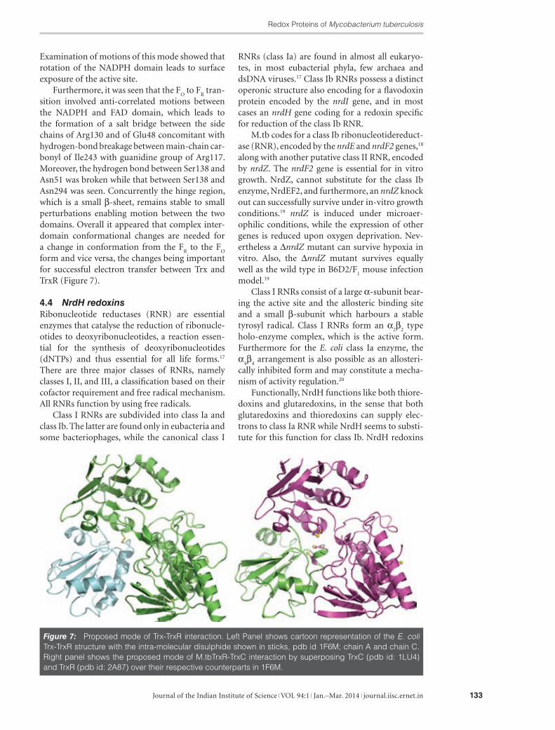
form and vice versa, the changes being important for successful electron transfer between Trx and TrxR (Figure 7).
4.4 NrdH redoxinsRibonucleotide reductases (RNR) are essential enzymes that catalyse the reduction of ribonucle-otides to deoxyribonucleotides, a reaction essen-tial for the synthesis of deoxyribonucleotides (dNTPs) and thus essential for all life forms.17 There are three major classes of RNRs, namely classes I, II, and III, a classification based on their cofactor requirement and free radical mechanism. All RNRs function by using free radicals.
Class I RNRs are subdivided into class Ia and class Ib. The latter are found only in eubacteria and some bacteriophages, while the canonical class I
RNRs (class Ia) are found in almost all eukaryo-tes, in most eubacterial phyla, few archaea and dsDNA viruses.17 Class Ib RNRs possess a distinct operonic structure also encoding for a flavodoxin protein encoded by the nrdI gene, and in most cases an nrdH gene coding for a redoxin specific for reduction of the class Ib RNR.
M.tb codes for a class Ib ribonucleotidereduct-ase (RNR), encoded by the nrdE and nrdF2 genes,18 along with another putative class II RNR, encoded by nrdZ. The nrdF2 gene is essential for in vitro growth. NrdZ, cannot substitute for the class Ib enzyme, NrdEF2, and furthermore, an nrdZ knock out can successfully survive under in-vitro growth conditions.19 nrdZ is induced under microaer-ophilic conditions, while the expression of other genes is reduced upon oxygen deprivation. Nev-ertheless a ∆nrdZ mutant can survive hypoxia in vitro. Also, the ∆nrdZ mutant survives equally well as the wild type in B6D2/F
1 mouse infection
model.19
Class I RNRs consist of a large α-subunit bear-ing the active site and the allosteric binding site and a small β-subunit which harbours a stable tyrosyl radical. Class I RNRs form an α
2β
2 type
holo-enzyme complex, which is the active form. Furthermore for the E. coli class Ia enzyme, the α
4β
4 arrangement is also possible as an allosteri-
cally inhibited form and may constitute a mecha-nism of activity regulation.20
Functionally, NrdH functions like both thiore-doxins and glutaredoxins, in the sense that both glutaredoxins and thioredoxins can supply elec-trons to class Ia RNR while NrdH seems to substi-tute for this function for class Ib. NrdH redoxins
Figure 7: Proposed mode of Trx-TrxR interaction. Left Panel shows cartoon representation of the E. coli Trx-TrxR structure with the intra-molecular disulphide shown in sticks, pdb id 1F6M; chain A and chain C. Right panel shows the proposed mode of M.tbTrxR-TrxC interaction by superposing TrxC (pdb id: 1LU4) and TrxR (pdb id: 2A87) over their respective counterparts in 1F6M.
Swastik Phulera et al.
Journal of the Indian Institute of Science VOL 94:1 Jan.–Mar. 2014 journal.iisc.ernet.in134
share high sequence similarity to glutaredoxins (Figure 8) but are paradoxically thioredoxin-like in their function. They behave like thioredox-ins in terms of capacity to accept electrons from thioredoxin reductase. NrdH redoxins possess a thioredoxin fold and contain an active site motif CXXC, features that are common to thioredoxins and glutaredoxins.
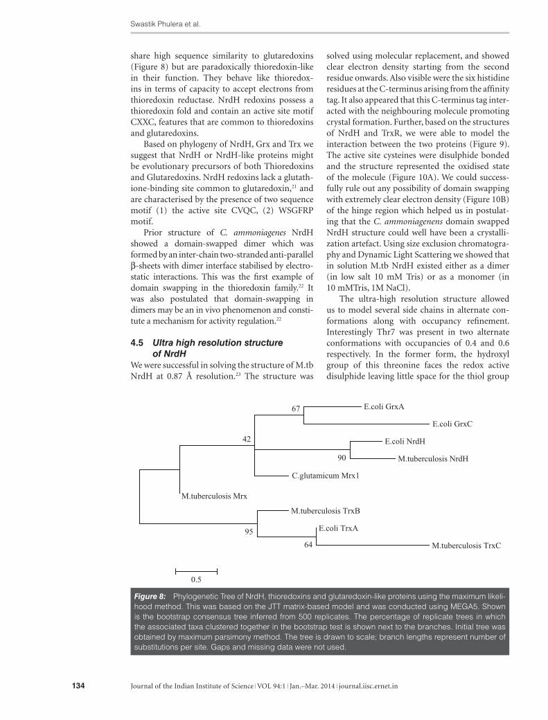
Based on phylogeny of NrdH, Grx and Trx we suggest that NrdH or NrdH-like proteins might be evolutionary precursors of both Thioredoxins and Glutaredoxins. NrdH redoxins lack a glutath-ione-binding site common to glutaredoxin,21 and are characterised by the presence of two sequence motif (1) the active site CVQC, (2) WSGFRP motif.
Prior structure of C. ammoniagenes NrdH showed a domain-swapped dimer which was formed by an inter-chain two-stranded anti-parallel β-sheets with dimer interface stabilised by electro-static interactions. This was the first example of domain swapping in the thioredoxin family.22 It was also postulated that domain-swapping in dimers may be an in vivo phenomenon and consti-tute a mechanism for activity regulation.22
4.5 Ultra high resolution structure of NrdH
We were successful in solving the structure of M.tb NrdH at 0.87 Å resolution.23 The structure was
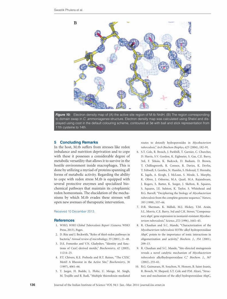
solved using molecular replacement, and showed clear electron density starting from the second residue onwards. Also visible were the six histidine residues at the C-terminus arising from the affinity tag. It also appeared that this C-terminus tag inter-acted with the neighbouring molecule promoting crystal formation. Further, based on the structures of NrdH and TrxR, we were able to model the interaction between the two proteins (Figure 9). The active site cysteines were disulphide bonded and the structure represented the oxidised state of the molecule (Figure 10A). We could success-fully rule out any possibility of domain swapping with extremely clear electron density (Figure 10B) of the hinge region which helped us in postulat-ing that the C. ammoniagenens domain swapped NrdH structure could well have been a crystalli-zation artefact. Using size exclusion chromatogra-phy and Dynamic Light Scattering we showed that in solution M.tb NrdH existed either as a dimer (in low salt 10 mM Tris) or as a monomer (in 10 mMTris, 1M NaCl).
The ultra-high resolution structure allowed us to model several side chains in alternate con-formations along with occupancy refinement. Interestingly Thr7 was present in two alternate conformations with occupancies of 0.4 and 0.6 respectively. In the former form, the hydroxyl group of this threonine faces the redox active disulphide leaving little space for the thiol group
Figure 8: Phylogenetic Tree of NrdH, thioredoxins and glutaredoxin-like proteins using the maximum likeli-hood method. This was based on the JTT matrix-based model and was conducted using MEGA5. Shown is the bootstrap consensus tree inferred from 500 replicates. The percentage of replicate trees in which the associated taxa clustered together in the bootstrap test is shown next to the branches. Initial tree was obtained by maximum parsimony method. The tree is drawn to scale; branch lengths represent number of substitutions per site. Gaps and missing data were not used.

Redox Proteins of Mycobacterium tuberculosis
Journal of the Indian Institute of Science VOL 94:1 Jan.–Mar. 2014 journal.iisc.ernet.in 135
this observation we proposed that in the oxidized state of NrdH, as captured by this crystal structure, Thr7 can exist in either of the two forms although in the reduced state only one of the forms would be allowed.
of Cys14 to exist in the reduced form. In the lat-ter form a small cavity is seen to exist next to Cys14, which might be required to accommodate the sulfhydryl group of the reduced Cys when it moves away from its oxidized position. Based on
Figure 9: Proposed mode of NrdH-TrxR interaction. Left Panel shows cartoon representation of the M.tbNrdH (yellow) (pdb id: 4HS1) superposed over the thioredoxin counterpart in E. coli Trx-TrxR struc-ture and M.tb TrxR (magenta) modelled as for figure 6. Right panel shows the Cα trace of NrdH and Trx in the proposed interaction modes of M.tb TrxR-TrxC/NrdH.
Figure 10: (Continued).
Swastik Phulera et al.
Journal of the Indian Institute of Science VOL 94:1 Jan.–Mar. 2014 journal.iisc.ernet.in136
5 Concluding RemarksIn the host, M.tb suffers from stresses like redox imbalance and nutrition deprivation and to cope with these it possesses a considerable degree of metabolic versatility that allows it to survive in the hostile environment inside macrophages. This is done by utilizing a myriad of proteins spanning all forms of metabolic activity. Regarding the ability to cope with redox stress M.tb is equipped with several protective enzymes and specialized bio-chemical pathways that maintain its cytoplasmic redox homeostasis. The elucidation of the mecha-nisms by which M.tb evades these stresses will open new avenues of therapeutic intervention.
Received 10 December 2013.
References 1. WHO, WHO Global Tuberculosis Report (Geneva: WHO
Press, 2013), Pages.
2. D. Ritz and J. Beckwith, “Roles of thiol-redox pathways in
bacteria,” Annual review of microbiology, 55 (2001), 21–48.
3. D.E. Fomenko and V.N. Gladyshev, “Identity and func-
tions of CxxC-derived motifs,” Biochemistry, 42 (2003),
11214–25.
4. P.T. Chivers, K.E. Prehoda and R.T. Raines, “The CXXC
Motif: A Rheostat in the Active Site,” Biochemistry, 36
(1997), 4061–66.
5. T. Jaeger, H. Budde, L. Flohe, U. Menge, M. Singh,
M. Trujillo and R. Radi, “Multiple thioredoxin-mediated
routes to detoxify hydroperoxides in Mycobacterium
tuberculosis,” Arch Biochem Biophys, 423 (2004), 182–91.
6. S.T. Cole, R. Brosch, J. Parkhill, T. Garnier, C. Churcher,
D. Harris, S.V. Gordon, K. Eiglmeier, S. Gas, C.E. Barry,
3rd, F. Tekaia, K. Badcock, D. Basham, D. Brown,
T. Chillingworth, R. Connor, R. Davies, K. Devlin,
T. Feltwell, S. Gentles, N. Hamlin, S. Holroyd, T. Hornsby,
K. Jagels, A. Krogh, J. McLean, S. Moule, L. Murphy,
K. Oliver, J. Osborne, M.A. Quail, M.A. Rajandream,
J. Rogers, S. Rutter, K. Seeger, J. Skelton, R. Squares,
S. Squares, J.E. Sulston, K. Taylor, S. Whitehead and
B.G. Barrell, “Deciphering the biology of Mycobacterium
tuberculosis from the complete genome sequence,” Nature,
393 (1998), 537–44.
7. D.R. Sherman, K. Mdluli, M.J. Hickey, T.M. Arain,
S.L. Morris, C.E. Barry, 3rd and C.K. Stover, “Compensa-
tory ahpC gene expression in isoniazid-resistant Mycobac-
terium tuberculosis,” Science, 272 (1996), 1641–43.
8. R. Chauhan and S.C. Mande, “Characterization of the
Mycobacterium tuberculosis H37Rv alkyl hydroperoxidase
AhpC points to the importance of ionic interactions in
oligomerization and activity,” Biochem. J., 354 (2001),
209–15.
9. R. Chauhan and S.C. Mande, “Site-directed mutagenesis
reveals a novel catalytic mechanism of Mycobacterium
tuberculosis alkylhydroperoxidase C,” Biochem. J., 367
(2002), 255–61.
10. B.G. Guimaraes, H. Souchon, N. Honore, B. Saint-Joanis,
R. Brosch, W. Shepard, S.T. Cole and P.M. Alzari, “Struc-
ture and mechanism of the alkyl hydroperoxidase AhpC,
Figure 10: Electron density map of (A) the active site region of M.tb NrdH, (B) The region corresponding to domain swap in C. ammoniagenes structure. Electron density map was calculated using Shelxl and dis-played using coot in the default colouring scheme, contoured at 3σ with ball and stick representation from 11th cysteine to 14th.

Redox Proteins of Mycobacterium tuberculosis
Journal of the Indian Institute of Science VOL 94:1 Jan.–Mar. 2014 journal.iisc.ernet.in 137
a key element of the Mycobacterium tuberculosis defense
system against oxidative stress,” J Biol Chem, 280 (2005),
25735–42.
11. J.L. Martin, “Thioredoxin—a fold for all reasons,” Struc-
ture, 3 (1995), 245–50.
12. H. Kadokura, F. Katzen and J. Beckwith, “Protein disulfide
bond formation in prokaryotes,” Annu Rev Biochem, 72
(2003), 111–35.
13. C.H. Williams, Jr., “Mechanism and structure of thiore-
doxin reductase from Escherichia coli,” FASEB J, 9 (1995),
1267–76.
14. G.B. Kallis and A. Holmgren, “Differential reactivity
of the functional sulfhydryl groups of cysteine-32 and
cysteine-35 present in the reduced form of thioredoxin
from Escherichia coli,” Journal of Biological Chemistry, 255
(1980), 10261–65.
15. M. Akif, G. Khare, A.K. Tyagi, S.C. Mande and
A.A. Sardesai, “Functional studies of multiple thioredox-
ins from Mycobacterium tuberculosis,” J. Bacteriol., 190
(2008), 7087–95.
16. M. Akif, K. Suhre, C. Verma and S.C. Mande, “Confor-
mational flexibility of Mycobacterium tuberculosis thiore-
doxin reductase: crystal structure and normal-mode
analysis,” Acta Crystallogr. D Biol. Crystallogr., 61 (2005),
1603–11.
17. P. Nordlund and P. Reichard, “Ribonucleotide reductases,”
Annu Rev Biochem, 75 (2006), 681–706.
18. F. Yang, S.C. Curran, L.S. Li, D. Avarbock, J.D. Graf,
M.M. Chua, G. Lu, J. Salem and H. Rubin, “Characteriza-
tion of two genes encoding the Mycobacterium tubercu-
losis ribonucleotide reductase small subunit,” J Bacteriol,
179 (1997), 6408–15.
19. S.S. Dawes, D.F. Warner, L. Tsenova, J. Timm, J.D.
McKinney, G. Kaplan, H. Rubin and V. Mizrahi, “Ribo-
nucleotide reduction in Mycobacterium tuberculosis:
function and expression of genes encoding class Ib and
class II ribonucleotide reductases,” Infect Immun, 71
(2003), 6124–31.
20. R. Rofougaran, M. Crona, M. Vodnala, B.M. Sjoberg and
A. Hofer, “Oligomerization status directs overall activity
regulation of the Escherichia coli class Ia ribonucleotide
reductase,” J Biol Chem, 283 (2008), 35310–18.
21. A. Jordan and P. Reichard, “Ribonucleotide reductases,”
Annu Rev Biochem, 67 (1998), 71–98.
22. M. Stehr and Y. Lindqvist, “NrdH-redoxin of Corynebac-
terium ammoniagenes forms a domain-swapped dimer,”
Proteins, 55 (2004), 613–19.
23. S. Phulera and S.C. Mande, “The crystal structure of Myco-
bacterium tuberculosis NrdH at 0.87 A suggests a possible
mode of its activity,” Biochemistry, 52, 4056–65.
Swastik Phulera is currently a doctoral stu-dent at the Laboratory of Structural Biology at the National Centre for Cell Science, Pune. He obtained B.Sc. (Hons) in Biomedical Sciences from University of Delhi in 2008 and M.Sc. in
Biotechnology from JNU, New Delhi in 2010. He is sup-ported by the Senior Research Fellowship from the CSIR, New Delhi. His research interests include understanding the structure function relationships of Redox proteins from Mycobacteria protein evolutionary studies using bioinfor-matics approaches.
Abhijit A. Sardesai is a staff scientist at the Laboratory of Bacterial Genetics at the Cen-tre for DNA fingerprinting and Diagnostics (CDFD) Hyderabad. He obtained his graduate (PhD) degree from the Centre for Cellular and
Molecular Biology, Hyderabad. Abhijit’s research involves the study of K+ (potassium) ion homeostasis in the bacte-rial cell, genetic and biochemical studies on ORFs from the bacterium M. tuberculosis, encoding components of cyto-plasmic protein folding and redox homeostasis and studies on solute export in E. coli.
Shekhar C. Mande obtained his PhD in Molec-ular Biophysics from the Indian Institute of Science in 1991. He has been working in the field of M.tb structural biology since 1996. His current research interests include obtaining
insights into biochemical functions of proteins using 3D structural knowledge and large scale network analysis of protein: protein interaction networks.
Mohd. Akif is currently an Assistant Professor at the Department of Biochemistry, School of Life Sciences, University of Hyderabad. He obtained his PhD from CDFD in 2008. His current research interest lies in structural characteriza-
tion of macromolecules and macromolecular complexes.