Molecular epidemiology and evolutionary genetics of Mycobacterium tuberculosis in Taipei

Das et al. BMC Genomics 2013, 14:404http://www.biomedcentral.com/1471-2164/14/404
RESEARCH ARTICLE Open Access
Genetic heterogeneity revealed by sequenceanalysis of Mycobacterium tuberculosis isolatesfrom extra-pulmonary tuberculosis patientsSarbashis Das1†, Tanmoy Roychowdhury1†, Parameet Kumar2, Anil Kumar2, Priya Kalra2, Jitendra Singh3,Sarman Singh3, HK Prasad2* and Alok Bhattacharya1,4*
Abstract
Background: Tuberculosis remains a major public health problem. Clinical tuberculosis manifests often aspulmonary and occasionally as extra-pulmonary tuberculosis. The emergence of drug resistant tubercle bacilli and itsassociation with HIV is a formidable challenge to curb the spread of tuberculosis. There have been concerted effortsby whole genome sequencing and bioinformatics analysis to identify genomic patterns and to establish arelationship between the genotype of the organism and clinical manifestation of tuberculosis. Extra-pulmonary TBconstitutes 15–20 percent of the total clinical cases of tuberculosis reported among immunocompetent patients,whereas among HIV patients the incidence is more than 50 percent. Genomic analysis of M. tuberculosis isolatesfrom extra pulmonary patients has not been explored.
Results: The genomic DNA of 5 extra-pulmonary clinical isolates of M. tuberculosis derived from cerebrospinal fluid,lymph node fine needle aspirates (FNAC) / biopsies, were sequenced. Next generation sequencing approach (NGS)was employed to identify Single Nucleotide Variations (SNVs) and computational methods used to predict theirconsequence on functional genes. Analysis of distribution of SNVs led to the finding that there are mixedgenotypes in patient isolates and that many SNVs are likely to influence either gene function or their expression.Phylogenetic relationship between the isolates correlated with the origin of the isolates. In addition, insertion sitesof IS elements were identified and their distribution revealed a variation in number and position of the element inthe 5 extra-pulmonary isolates compared to the reference M. tuberculosis H37Rv strain.
Conclusions: The results suggest that NGS sequencing is able to identify small variations in genomes of M. tuberculosisisolates including changes in IS element insertion sites. Moreover, variations in isolates of M. tuberculosis fromnon-pulmonary sites were documented. The analysis of our results indicates genomic heterogeneity in theclinical isolates.
Keywords: Extra-pulmonary Tuberculosis, Next-generation Sequencing, Genetic Heterogeneity, Single NucleotideVariations, Insertion Elements, Phylogeny, Spoligotyping
* Correspondence: [email protected]; [email protected]†Equal contributors2Department of Biotechnology, All India Institute of Medical Sciences, NewDelhi, India1School of Computational and Integrative Sciences, Jawaharlal NehruUniversity, New Delhi, IndiaFull list of author information is available at the end of the article
© 2013 Das et al.; licensee BioMed Central Ltd. This is an Open Access article distributed under the terms of the CreativeCommons Attribution License (http://creativecommons.org/licenses/by/2.0), which permits unrestricted use, distribution, andreproduction in any medium, provided the original work is properly cited.

Das et al. BMC Genomics 2013, 14:404 Page 2 of 14http://www.biomedcentral.com/1471-2164/14/404
BackgroundTuberculosis is a public health challenge. It is estimated thatone third of the human population harbor M. tuberculosis,however approximately only 20% of these infected indi-viduals go on to develop clinical tuberculosis [1]. Theinfection with M. tuberculosis usually results in pulmonarytuberculosis but it can also manifest in extra-pulmonarysites, such as tuberculous meningitis, endometritis, lymph-adenitis, pleuritis, etc. [2,3]. In India, around 15–20 percentcases of tuberculosis among immuno-competent adultshave been reported to occur at extra-pulmonary sites,whereas among HIV co-infected patients the incidenceincreases to more than 50% [4]. The classical route ofinfection is by inhalation of infectious droplets (respiratoryroute), however, infection can occasionally occur via alter-nate routes such as skin abrasions and open wounds [5].Extra-pulmonary tuberculosis has been always recognizedas a sequel to primary pulmonary infection [6-8]. Howexactly it occurs and its facilitation remains an enigma.The present study has been designed to sequence andanalyze extra-pulmonary isolates to identify genomicpatterns and features of M. tuberculosis isolated frompatients with extra-pulmonary tuberculosis.Genomic variations in M. tuberculosis have been studied
using a number of different methods, such as spoligotypingand variable-number tandem repeats (VNTRs) [9]. Thesestudies have shown variations among different clinical iso-lates of M. tuberculosis. None of these approaches gives acomplete picture of variations at the whole genome level,as each of the methods have their limitations. For example,the spoligotyping pattern would be determined by thestrain(s) present in the sample under investigation, andit would be difficult to determine if the observed patternis due to a dominant strain or a collective pattern of allthe strains present in the sample Therefore, it is not reli-able to use spoligotyping to establish mixed infection.On the other hand, MIRU-VNTR has been considereduseful for detecting mixed infection since it’s based onallelic variation. However, it is possible to have differentgenotypes with the same VNTR pattern, particularly incase of closely related isolates. Efficacy of DNA extrac-tion used in these assays can be potentially hampered bythe presence of clumps / aggregation of mycobacteria inclinical samples / cultures.The genome of M. tuberculosis strain H37Rv was
sequenced 15 years ago using the standard approachpioneered by Cole et al [10]. The analysis of the assembledsequences suggested that the genome size is 4.4 Mb.encoding about 4000 genes [10]. The genome analysisalso showed that there are a number of repeat families,particularly PPE and PGRS family of genes. These repeatfamilies of proteins may have a role in pathogenesis.Subsequently, a number of different species and isolatesof the M. tuberculosis complex have been sequenced.
On comparative analysis with other mycobacterial speciesthe MTB complex clustered separately showing a highdegree of sequence identity among this group of myco-bacterial species [11]. Different isolates showed poly-morphisms at the level of single nucleotides, number ofrepeats at a given loci, indels and synteny [12]. Attemptshave been made to map polymorphisms that are corre-lated with some of the phenotypes, such as drug resist-ance [13]. Though some correlations have been found aclear cut casual relationship has not been established sofar. With the introduction of next generation sequencing,genome sequences of several isolates have become avail-able and now it is possible to identify genetic markers forspecific phenotype. It has also become possible to identifyevolving patterns in genomes. For example, “hotspot” and“coldspot” regions have been identified using statisticalmethods and sequence information from large number ofisolates [14]. However, most of the genome data availableare from M. tuberculosis isolates derived from pulmonarytuberculosis patients. Therefore it is relevant that isolatesfrom extra pulmonary isolates derived from tuberculosispatients should also be analyzed at the genome level.
Results and discussionSequencing of M. tuberculosis isolates from extra-pulmonarytuberculosis patientsThe details of the patient derived samples and theM. tuberculosis clinical isolates used in the study aregiven in Table 1 and the techniques used for obtainingthese isolates have been described in “Methods”. Thesefive isolates were subjected to spoligotyping and theresults were compared to the shared-types (ST) andlineages/sub-lineages described in SpolDB4 (http://www.pasteur-guadeloupe.fr/tb/bd_myco.html [15]). Spoligotypingpattern revealed that of the five isolates, F85 and AC544could be assigned to the Beijing clade and T1 family re-spectively. However, the remaining three isolates (AC74,LN8 & F99) did not match with any of the previously de-scribed lineage of M. tuberculosis based on spoligotyping,hence have been categorized as undefined, (Table 2.).DNA was extracted from the isolates as described before
[16] and subjected to nucleotide sequencing using NGStechnology (IlluminaGA-IIx). Gross statistics derived fromsequence data are also shown in Table 1. In general thenumber of short reads was more than 3 million with anaverage length of 72 nucleotides for each isolate. We haveused the complete genome sequence of M. tuberculosisH37Rv strain (NC_000962.2) as the reference sequence.The short reads were aligned to the reference genomeas described in “Methods”. While genome coverage variedfrom 82% to about 92% for different isolates, unalignedreads were between 3 to 16% suggesting that the quality ofDNA preparations from these isolates of good quality.

Table 1 Description of clinical isolates used in the study and basic statistics of the sequencing data
Properties AC74 AC544 F85 F99 LN8
Isolation sites CSFa CSFa FNACb FNACb Lymph node from biopsy
Total reads 3611330*2 3045593*2 13609758*2 7100266*2 3072728*2
Average read length 72 72 72 72 72
Total reads aligned after filtering 5572900 4656246 21378146 11116118 4672838
Total reference length 4411532 4411532 4411532 4411532 4411532
% Total reference covered 88.42 91.51 90.17 81.87 86.36
% Reads aligned with reference 96.32 83.78 95.81 94.82 96.72
Optimized average read depth 72.97 53.03 287.71 148.08 60.41a Cerebro-spinal fluid.bLymph node using Fine-Needle Aspiration Cytology.
Das et al. BMC Genomics 2013, 14:404 Page 3 of 14http://www.biomedcentral.com/1471-2164/14/404
Sequence annotation and isolate comparisonM. tuberculosis H37Rv genome has been reported toencode 3988 protein encoding genes [10]. Nearly 84%of these genes displayed more than 90% coverage basedon our alignments in all strains except F99 (54% cover-age). Genes with less than 10% coverage and/or readdepth below 10 were considered as “missing genes” (seeAdditional file 1: Table S1). The total number of pre-dicted missing genes in all the five isolates varied from17 (AC544) to 74 (F99). Some of these genes (total 5)were missing in all the five isolates. Since genomic dele-tion can result in genes missing in an isolate, furtheranalysis was carried out to identify large deletions inthe isolates using Pindel [17], which detects breakpointsfor large deletion using paired-end data. Among themissing genes, 62 were predicted by Pindel as thosedue to genomic deletions. Out of these 62 genomic de-letions, 23 and 3 are from F99 and LN8 respectively(see Additional file 1: Table S1). In general a missinggene in one isolate was found in another. LysR familyactivates divergent transcription of linked target genesor unlinked regulons with diverse functions [18] and itis one such family that was missing in isolate AC74. Inyet another example of missing gene is the absence ofmalonyl CoA-acyl carrier protein transacylase, in all iso-lates except isolate AC544. This is an essential gene forthe transfer of malonyl group from coenzyme-A to acylcarrier protein AcpM, therefore it is required for biosyn-thesis of cell wall in M. tuberculosis [19]. In case of F99
Table 2 Spoligotype patterns of the five extra-pulmonary clin
S.no. Lab. no. Binary code
1 LN 8 □□□■■■□■■■■■■■■■■■■■■■■■■■■■□□□□■□■
2 AC 74 ■□□□□□□■■■■■■■■■■■■■□□□■■■■■□■■■□□□
3 AC 544 ■■■■■■■■■■■■■■■■■■■■■■■■■■■■■■■■□□□
4 F 85 □□□□□□□□□□□□□□□□□□□□□□□□□□□□□□□□□□■
5 F99 ■■■■■■■■■■■■■■■■■■■■■■■■■□■□□□□■□□□
several trans-membrane protein coding genes, such asRv2272 and Rv2273 were missing. Although we haveconsidered repeat regions by realigning short reads withmultiple matches, a number of prophage proteins (phiRV2)were not mapped. Prophage proteins are quite often presentin and around repeat regions and show polymorphismswith respect to their positions among different isolates.For example, two types of prophage proteins are presentin M. tuberculosis H37Rv and CDC1551, but absent inMycobacterium bovis [20]. A total of 68 (38%) missinggenes are from PE-PGRS gene family.
Analysis of single nucleotide variationsSNVs were classified as major or minor SNVs (seeMethods). All major SNVs were grouped either as (a)coding or (b) intergenic depending upon mapping loca-tion. To understand the effect of SNVs on the functionof respective protein products we further analyzed thosethat map to coding regions (see Additional file 2: Table S2).A total of 185 SNVs were identified that are commonamong all isolates in comparison to H37Rv strain (Figure 1).LN8 showed highest number of unique SNVs (not presentin other isolates) suggesting that LN8 has diverged muchmore compared to other isolates. Many SNVs that map tocoding regions are likely to cause major changes in theform of gene truncation (by nonsense mutation) and geneelongation (by missense mutation) in different isolates.Some of the SNVs that lead to gene truncation, such as atposition 234477 in Rv0197, and 1037911 in Rv0930 (pstA)
ical isolates
Octal code ST Lineage
■□□□■■■■ 73777777413071 Unidentified Unidentified
□■■■■■■■ 403777617560771 Unidentified Unidentified
□■■■■■■■ 777777777760771 53 T1
■■■■■■■■ 3771 1 BEIJING
□■■■■■■■ 777777775020771 Unidentified Unidentified

Figure 1 Common and unique SNVs present in the codingregions of the five isolates. LN8 has highest number of unique SNVs.
Das et al. BMC Genomics 2013, 14:404 Page 4 of 14http://www.biomedcentral.com/1471-2164/14/404
respectively were also observed in a few other isolates ofM. tuberculosis [21]. Rv0197 protein is a potential oxidore-ductase, containing the molybdopterin binding motif. Anon-synonymous mutation (arginine to stop) at position305 was observed in Rv0930 (phosphate transport integral
Figure 2 Plot showing RSCU (Relative Synonymous Codon Usages) ofvalues in an isolate and background are the same then it will lie above the
membrane ABC Transporter, PstA1 (ABC transportertrans-membrane protein) of isolate LN8, which displaysmaximum number of non-synonymous mutations that re-sult in stop codons (22) among all five isolates. Besidemodification in the gene products, SNVs are likely toaffect the level of expression of genes depending upon theusage of altered codons [22]. Relative Synonymous CodonUsage (RSCU) was calculated for all synonymous changesin each strain and the results showed 5–10 fold changesin codon usage for many altered codons as a consequenceof SNVs. Some of the examples of genes containing theseSNVs are enoyl-CoA hydratase (Rv0022), MCE-family pro-teins (Rv0591) and oxidoreductase (Rv3777) [23]. RSCUvalues of original codons and those for isolate AC74 isshown in Figure 2. Details are given in Additional file 3:Table S3. SNVs in the inter-genic regions (specially pro-moters regions) were also studied (see Additional file 4:Table S10) using the database of promoters from MycoRRdbdatabase as reference [24]. We were able to identify SNVin the promoter of the gene Rv2779c, a transcriptionalregulator in all isolates except AC544. Though, most ofthe regulatory regions in MycoRRdb database are com-putationally predicted motifs, some of these are alsomentioned in literature [25-27]. kstR2 is one such regu-latory motif, shown to be associated with cholesterolutilization in M.tuberculosis during infection [26]. One
isolate AC544 versus background (M. tuberculosis H37Rv). If RSCUdiagonal (red line).

Figure 3 Examples of major and rare variations are shown from isolates F85. (A) Major variation changes C to T (B) rare variation changes C to T.
Das et al. BMC Genomics 2013, 14:404 Page 5 of 14http://www.biomedcentral.com/1471-2164/14/404
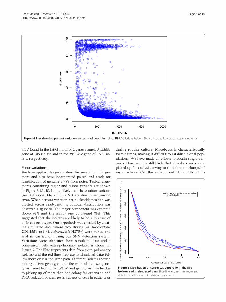
Figure 4 Plot showing percent variation versus read depth in isolate F85. Variations below 15% are likely to be due to sequencing error.
0.9 0.8
0.0
0.2
0.4
0.6
0.8
1.0
Consensus base ratio (CBR)
Num
ber
of p
ositi
ons
havi
ng C
BR
< x
/ N
umbe
r of
pos
ition
s ha
ving
CB
R <
0.9
extrapulmonary tuberculosis isolatessimulated data
0.7 0.6 0.5
Figure 5 Distribution of consensus base ratio in the fiveisolates and in simulated data. Blue line and red line representdata from isolates and simulation respectively.
Das et al. BMC Genomics 2013, 14:404 Page 6 of 14http://www.biomedcentral.com/1471-2164/14/404
SNV found in the kstR2 motif of 2 genes namely Rv3560cgene of F85 isolate and in the Rv3549c gene of LN8 iso-late, respectively.
Minor variationsWe have applied stringent criteria for generation of align-ment and also have incorporated paired end reads foridentification of genuine SNVs from noise. Typical align-ments containing major and minor variants are shownin Figure 3 (A, B). It is unlikely that these minor variants(see Additional file 2: Table S2) are due to sequencingerror. When percent variation per nucleotide position wasplotted across read-depth, a bimodal distribution wasobserved (Figure 4). The major component was centeredabove 95% and the minor one at around 85%. Thissuggested that the isolates are likely to be a mixture ofdifferent genotypes. Our hypothesis was checked by creat-ing simulated data where two strains (M. tuberculosisCDC1551 and M. tuberculosis H37Rv) were mixed andanalysis carried out using our SNV detection system.Variations were identified from simulated data and acomparison with extra-pulmonary isolates is shown inFigure 5. The Blue (represents data from extra-pulmonaryisolates) and the red lines (represents simulated data) fol-low more or less the same path. Different isolates showedmixing of two genotypes and the ratio of the two geno-types varied from 5 to 15%. Mixed genotypes may be dueto picking up of more than one colony for expansion andDNA isolation or changes in subsets of cells in patients or
during routine culture. Mycobacteria characteristicallyform clumps, making it difficult to establish clonal pop-ulations. We have made all efforts to obtain single col-onies. However it is still likely that mixed colonies werepicked up for analysis, owing to the inherent ‘clumps’ ofmycobacteria. On the other hand it is difficult to

Das et al. BMC Genomics 2013, 14:404 Page 7 of 14http://www.biomedcentral.com/1471-2164/14/404
reconcile the extent of variations observed, with ourcurrent understanding on changes that take place dur-ing mycobacterial proliferation.The likely reason for the origin of mixed genotype could
also be due to the fact that patients can be potentiallyre-infected / super infected by different strains or rapidgenomic changes take place in a subset of cells in pa-tients. Recent investigations have suggested that severalpulmonary isolates of M. tuberculosis display clonal het-erogeneity [28,29]. Heterogeneous population does offerselective advantage for survival of the tubercle bacilliwithin the hostile microenvironment of the host. Thesealterations may facilitate in vivo dissemination / migrationof the tubercle bacilli from the pulmonary infectious focito other organs, as has been speculated to occur in theinfected host [30,31].
Functional classification and enrichment study of geneshaving major SNVsSynonymous and non-synonymous SNVs that map in thecoding regions of genes were functionally classified onthe basis of COG (Cluster of Orthologous Group) [32].Functional annotation of uncharacterized proteins wasperformed according to Doerks et al. [33]. Maximum num-ber of SNVs among the isolates was found in genes thatbelong to COG category N (“Cell motility and secretion”)(Figure 6). M. tuberculosis genes in this category are
SN
V fr
actio
n
RNA pro
cess
ing a
nd m
odific
ation
Energ
y pro
ducti
on a
nd co
nver
sion
Cell cy
cle co
ntro
l, cell
divi
sion,
chro
mos
ome
parti
tionin
g
Amino
acid
tran
spor
t and
met
aboli
sm
Nucleo
tide
trans
port
and
met
aboli
sm
Carbo
hydr
ate
trans
port
and
met
aboli
sm
Coenz
yme
trans
port
and
met
aboli
sm
Lipid
trans
port
and
met
aboli
sm
Trans
lation
, ribo
som
al str
uctu
re a
nd b
iogen
esis
Trans
cript
ion
Replic
ation
, rec
ombin
ation
an
Cell w
all/m
embr
an
Post
0.0
0.1
0.2
0.3
0.4
0.5
Figure 6 COG classification of the genes carrying SNVs in the five isorepresents fraction of SNVs in each category after normalization.
mainly “ppe” genes. Polymorphisms in these genes aremostly non-synonymous types. In pathogenic mycobac-teria PE/PPE proteins are involved in direct interactionwith host immune system [34,35]. Yongjun Li et al., haveshown that in M. avium ppe genes are associated withgrowth in macrophages and virulence in mice [36]. There-fore, it is not surprising that these genes undergo high rateof variations compared to housekeeping genes [37]. Bio-logical processes, such as DNA synthesis, transcriptionand host-pathogen interaction, depend on coordinatedfunctional expression of multiple genes. Therefore geneenrichment analyses were used to identify functionally im-portant genes from SNV data [38]. For these studies,the Databases for Annotation, Visualization and Inte-grated Discovery tool (DAVID) was used [39,40]. Theoutput from DAVID revealed several gene clusters thatare significantly enriched (using Fisher exact p-value). GOterms of these enriched clusters showed that these areessentially membrane bound / trans membrane / two-component system and nucleotide binding proteins.Mutations in these genes would potentially alter bio-logical processes, as these categories of genes areneeded by the bacteria for interaction with the environ-ment. Genes that form part of the family of polyketidesynthase were also found among the enriched gene sets.The details of enriched clusters including enrichmentscores, p-values in different isolates are tabulated in the
AC544AC74F99F85LN8
d re
pair
e/en
velop
e bio
gene
sis
Cell m
otilit
y
trans
lation
al m
odific
ation
, pro
tein
turn
over
, cha
pero
nes
Inor
ganic
ion
trans
port
and
met
aboli
sm
Secon
dary
met
aboli
tes b
iosyn
thes
is, tr
ansp
ort a
nd ca
tabo
lism
Gener
al fu
nctio
n pr
edict
ion o
nly
Functi
on u
nkno
wn
Signal
trans
ducti
on m
echa
nism
s
Intra
cellu
lar tr
affic
king,
secr
etion
, and
vesic
ular t
rans
port
Defens
e m
echa
nism
s
lates. Different COG categories are represented by X-axis. Y-axis

Das et al. BMC Genomics 2013, 14:404 Page 8 of 14http://www.biomedcentral.com/1471-2164/14/404
Additional file 5: Table S4, Additional file 6: Table S5,Additional file 7: Table S6, Additional file 8: Table S7,Additional file 9: Table S8.
Study of insertion sequence elements (IS elements)IS elements have been extensively used as a marker forstrain identification in M. tuberculosis due to high nu-merical and positional polymorphisms (for a review see[41-44]). IS elements can influence gene expression de-pending upon the sites of insertion, for example, IS6110increases the expression of neighboring genes which areinvolved in virulence [45]. In this study an attempt wasmade to identify IS elements, particularly IS6110 fromshort read sequence data of the isolates and to derive adistribution across the genomes. The strategy used hasbeen described in “Methods”. The results are shown inthe Additional file 10: Table S9.Total number of IS elements was found to be 56 includ-
ing 16 copies of IS6110 in H37Rv strain[10]. Our studiesrevealed that the numbers of copies of IS6110 varied from5 to 19 in different isolates and their positions differedfrom that seen in M. tuberculosis H37Rv.To validate the predicted translocation or deletion of
the IS elements in the 5 extra-pulmonary isolates, PCRamplifications were carried out using primers targetingthe regions flanking the predicted coordinates of the ISelement. Hence the inclusion of the IS element wouldresult in the generation of an amplicon larger in sizecompared to the deletion of the IS element in the PCRtargeted site. In this regard various primer panels (A-H)were designed (Table 3) to amplify coordinate specified
Table 3 Primer sequences and standardized PCR parameters
PrimerPanels
Coordinate of IS element(w.r.t. H37Rv) present in strain
Primers flanking tco-ordinate of IS elem
A 3480373, + strand, in AC74 5’-CAGCAGGGATGGATTC5’-GCGAGTGGGATTCAGA
B 1527976, + strand, in AC544 5’-GTGGTGCCGTCGTTGTC5’-ATGCCCGTAATGTCTGC
C 1356494, + strand, in AC741356495, + strand, in F85
5’-TTCGTCTCAATGGGCAA5’-CACCAGGCACTTCGTTA
D 2550014-2551368, + strand, in H37Rv 5’-CTGCGACTGCGTTGGTA5’-ACGGTGGGGAAAGCCT
E 1541952-1543306, - strand, in H37Rv 5’-AGCAGGAGGAGCGGG5’-GGATAACAGGCGCGAA
F 1657017, + strand, in F85 5’-TCCCTACACTCGGTTCA5’-ACAGCAGCAGCGCCAC
G 2229658, - strand, in F99 5’-TGGTGGTCAGGGAAAA5’-CCCTCCCGTAGCAGCC
H(IS6110ref)
Multiple copies present in all 5’-CGTGAGGGCATCGAGG5’-GCGTAGGCGTCGGTGA
[Ref: Hermans P.W.M., van Soolingen D., Dale J.W. et al., Insertion element IS986 frotuberculosis. 1990 J. Clin. Microbiol. 28(9):2051-2058].
regions of H37Rv and the 5 extra-pulmonary isolates.The presence of IS6110 in the genome of all the isolateswas confirmed by PCR (Figure 7, Panel H). Of the 7 pairsof Primer panels used, each targeting a specific positionalco-ordinate, PCR products obtained for Panels A, B, D,and E completely matched with the predictions (Figure 7).For example, Panels A and B confirm the presence of theIS element at the predicted co-ordinate 3480373 for theclinical isolate AC74 (Figure 7 Panel A, Lane 4) and atposition 1527976 for the clinical isolate AC544 (Figure 7Panel B, Lane 5), respectively. Similarly, primers describedin Panels D and E yielded the expected amplicon of size205 and 264 bp respectively, as the IS element was pre-dicted to be absent in the 5 clinical isolates, (Figure 7Panels D, E, Lanes 4–8). Whereas, when the IS element ispresent, as seen in the reference M. tuberculosis H37Rvstrain, the expected amplicon size was 1563 bp (co-ordinate2550014–2551368) and 1622 bp (co-ordinate 1541952–1543306) respectively, (Figure 7 Panels D,E, Lane 3).Panels D and E validate some of the variations in the in-tegration sites of IS element in the 5 extra-pulmonaryisolates with respect to M. tuberculosis H37Rv. WhereasPanels A, B and C (Figure 7) confirm the insertion of ISelement at new locations, in 2 out of 5 isolates with re-spect to H37Rv strain. Further, it may be noted that theIS element was incorporated into the Rv3113 gene (pu-tative phosphatase, Figure 7, Panel A) in the clinical iso-late AC74. Similarly in the same clinical isolate, anothercopy of the IS element was found in the Rv1213 gene(probable glucose-1-phosphate adenylyl-transferase, Figure 7,Panel C) considered important for glycogen synthesis. It is
heent
Standardized annealing temp./ MgCl2concentration
Amplicon size (bp)
With IS W/o IS
ACC-3’GAG-3’
62°C/1mM 1836 481
TC-3’TGG-3’
60.2°C/1.5mM 1632 277
CTAC-3’TCG-3’
60.2°C/2.5mM 1552 197
ATC-3’GAAG-3’
61.3°C/1mM 1563 205
ACG-3’CCG-3’
72°C/1mM 1622 264
TCC-3’GG-3’
61.3°C/1mM 1552 197
GCC-3’GC-3’
61.3°C/1.5mM 1617 262
TGGC-3’CAAA-3’
65°C/1mM 245 0
m Mycobacterium tuberculosis: a Useful tool in Diagnosis and Epidemiology of

Figure 7 Analysis of PCR products derived from Primer pairs (Panels A-H) detailed in Table 3 using agarose gel electrophoresis. PanelsA, B and C represent specific coordinates of IS element insertion in the isolates, whereas panels D and E represent those from where IS elementhas been translocated in the isolates in comparison to the reference strain H37Rv. Panel H (IS6110 internal primers) confirms the presence ofIS6110 element in all the strains. Co-ordinates for IS element insertion/deletion and expected amplicon size in the respective strains are detailedin Table 3. Lane 1: Negative control; Lane 2: 100-3000 bp DNA Ladder; Lane 3–8: H37Rv DNA (Lane 3) and Triton extracts of isolates (Lane 4:AC74;Lane 5:AC544; Lane 6:LN8; Lane 7: F85; Lane 8: F99). [Lane 2: DNA Ladder; with IS element; : without IS element].
Das et al. BMC Genomics 2013, 14:404 Page 9 of 14http://www.biomedcentral.com/1471-2164/14/404
also clear from the data that different isolates may have adifferent history of movement of these elements. For ex-ample, in the clinical isolate AC544, the IS element wasobserved in Rv1358 gene (probable transcriptional regula-tory protein, Figure 7, Panel B) not observed in other iso-lates. We could not validate our predictions in a few cases(Figure 7, Panels C, F and G).
SNV based phylogenetic analysisPhylogenetic relationships among isolates were derivedfrom SNVs as described in “Methods”. Distance-basedmethod (“Neighbor Joining”) was used to derive distanceestimate between the isolates and branch assignmentswere validated using bootstrap. The results (Figure 8)showed that isolates AC74 and AC544 were present in thesame branch suggesting a common lineage. This is not
surprising as both the isolates were obtained from cerebro-spinal fluid. F85 and F99 were isolated using FNAC fromlymph nodes and these were placed close to CSF branchand separated from LN8, a lymph node derived isolate.
ConclusionsOur major finding from analysis of NGS data of extra-pulmonary isolates of M. tuberculosis is detection of gen-omic heterogeneity in isolates. The computational approachused by us can identify mixed genotypes even when one ofthe genotypes is represented at a low level. We have furtheranalyzed the functional significance of SNVs identified byusing different approaches, such as COG. NGS data wasalso utilized to identify IS elements and its insertion intovarious sites, among the different isolates. Some of thesepredictions were validated by experiments. Phylogenetic

Figure 8 Phylogenetic relationship. SNVs were used to derive thetree using “Neighbour Joining” method.
Das et al. BMC Genomics 2013, 14:404 Page 10 of 14http://www.biomedcentral.com/1471-2164/14/404
relationship among isolates is consistent with the origin ofthe isolates.
MethodsMycobacterium strains and genomic DNA isolationThe clinical isolates of M. tuberculosis used in this studyhave been maintained in the TB immunology laboratory,Department of Biotechnology, All India Institute of MedicalSciences (AIIMS), New Delhi, India. These isolates wereobtained from clinical samples derived from patients diag-nosed with extra-pulmonary tuberculosis. The isolates wereobtained from: (1) cerebrospinal fluid (CSF) of patientsclinically diagnosed as cases of Tubercular meningitis; (2)from fine needle aspirates / biopsies from patients withlymphadenopathy with discharging sinus / abscess forma-tion, (Table 1). All samples, after due processing, were inoc-ulated on Lowenstein-Jensen (LJ) slants for primaryisolation. Separate single colonies were propagated on LJmedia, whereas in case of confluent growth, the sampleswere processed and re-inoculated on fresh LJ slants forobtaining single colonies. Genomic DNA was extractedfrom each of the single colony isolates using standardprotocol. DNA was purified as per manufacturer’s instruc-tions by the QIAGEN column and the DNA preparationkit from illumina. After library preparation, the genomicDNA was fragmented in the range of 100 to 800 bases. Theresulting fragmented DNA was cleaned up using QIAquickcolumns (QIAGEN). The size distribution was checked byrunning aliquots of the samples on AgilentBioanalyzer7500 Nano chips. Illumina adapters were ligated to eachfragment. Fragments of ~ 300 bases were separatedusing Gel electrophoresis and sequenced at both endsusing illumina GAII sequencer. Sequencing depth inthese strains varied from 50 - 200x with average readlength of 72 bases.
Pre-processing and mapping of Short-reads withreference genomeSequencing error increases at the end of each cycle, sotrimming of short reads is therefore a vital process thatcould improve the quality of mapping of short reads tothe reference genome and identification of single nucleo-tide variations [46]. Trimming of the short reads wasdone depending on the average Phred quality score perbase in each strain. The average size of reads after trim-ming was 60 nucleotides. Non-ATGC character containingreads were also filtered before alignment.M. tuberculosis H37Rv (NC_000962.2) was used as
reference genome for the mapping of the short reads.All the short reads in each of the strains were separatelymapped with the reference genome using Bowtie ver-sion 0.12.7 [47]. To make the alignment more stringent,we used two criteria: (a) maximum of two mismatcheswere in the seed region of the reads and maximum sumof mismatch quality across alignment is less than 70 (b)disallowing all the reads that map at multiple sites inthe reference genome. The reference genome coverageof all the strains ranged from 86 - 94% with minimumread depth of 10. Short reads are also aligned followingless stringent criteria allowing reads to map multiplepositions, which helped in identification of repeat genes.
Identification of major and minor SNVsSNVs were identified from the alignment by using ourin-house Perl scripts and were classified into two classes,major SNVs and minor SNVs. Characteristic of majorSNVs are: (a) consensus base ratio (i.e. number of nucle-otides other than the reference divided by total numberof nucleotides at a position) more than 0.9, (b) minimumand maximum read depth are 10 and 3 x (average readdepth) respectively, (c) consensus base should be supportedby reads aligning in both forward and reverse directions,and (d) absence of any other variations present in a 3 bpwindow. Similarly minor SNVs have: (a) consensus baseratio less than 0.9, (b) more than 10% of the reads in apositions that show the same nucleotide change whilethe rest of the short reads have the same nucleotide asthe reference and (c) variable base should be supportedby at least one overlapping paired end reads. Detailedresults are listed in Additional file 2: Table S2.
Genetic heterogeneity model using simulated short readsNormally bacterial genome sequencing is carried outusing cells derived from a single colony. If the culture ispure, it is expected that all the members of the populationwill display identical variations as compared to the refer-ence genome with consensus base ratio near 1. On theother hand if the population contains more than one typeof bacteria, then some members will show variations whilerest will be identical to the reference sequence. These are

Das et al. BMC Genomics 2013, 14:404 Page 11 of 14http://www.biomedcentral.com/1471-2164/14/404
rare variations. Relative level of a rare variant depends onthe nature of the population that is the level of mixing ofdifferent genotypes. Here we tried to model the major andminor variations in a population using short read datagenerated by simulation.At first, M. tuberculosis CDC1551 genome was randomly
mutated to produce 1200 SNVs and then 10 million paired-end reads were generated using MAQ [48]. M. tuberculosisH37Rv was selected as the other genome and partitionedinto 5 non-overlapping parts in the proportion 80:15:3:2:1.Paired-end short reads were generated from these five partswith different compositions (See Figure 9).Let the number of reads generated from CDC1551 be
“m”. So, 80% of the genome will have approximately 0.8x m reads. Now if (0.8 x m)/9 reads from 80% of H37Rvare mixed with these, 80% of tuberculosis genome willbe represented by reads from CDC1551 and H37Rv in9:1 ratio. Similarly 15, 3, 2 and 1% of the genome wererepresented by 8:2,7:3,6:4 and 5:5 reads from CDC1551and H37Rv respectively. And then all the reads werealigned with the reference M. tuberculosis CDC1551 bybowtie 0.12.7 [47]. After the alignment, randomly gener-ated SNVs would show up as major variations while SNVsunique to H37Rv as rare variants.The distribution of consensus base ratio in extra-
pulmonary isolates and that in simulated data are plottedtogether (See Figure 5).
Identification of missing genes and insertion elementsMissing genes were identified from the alignment of readswith multiple maps from each isolate by identifying re-gions with minimum read depth below 10 and genecoverage less than 10. These regions are either missedwhile sequencing or are deleted in the genome of one ofthe isolates. Large deletions from the isolates were iden-tified using Pindel [17]. As sequencing depth normally
Figure 9 Cartoon representation showing partitioning of theM. tuberculosis H37Rv and CDC1551 genomes used ingenerating simulated reads.
achieved is around 100 for the whole genome, there isless chance of missing these regions during sequencing.Alignment of short reads with multiple matches helped
to identify IS elements. The distribution of IS elementsin particular IS6110 was done using the following steps.Initially, all reads were aligned against a single 1355 bpcopy of IS6110. Alignment was performed using BLAT[49]. Partially aligned or fragmented reads at the 5’ and3’ ends of the IS elements were then identified (Figure 10)and both 5’ and 3’ fragments were clustered separatelyusing BLASTCLUST (BLAST score-based single-linkageclustering) as reads coming from one genomic regionshould fall into one cluster. One representative sequencewas chosen from each cluster. Let, m is the number ofclusters generated from 5’ fragments and n from 3’ frag-ments. As we do not know specific pairing of 5’ and3’clusters, m x n sequences were generated by concatenat-ing each 5’ cluster representative with all of the 3’ clustersseparately. These m x n sequences were again alignedusing BLAT [35] with M. tuberculosis H37Rv genome. Anear perfect alignment suggested insertion of IS6110 inthe region of alignment whereas alignment with around1355 bp deletion suggested presence of IS6110 in thesame position as H37Rv.
Phylogenetic analysisTotal number of major SNVs identified in all five isolateswas 4550 and out of these, 2989 are present in coding re-gions and only in one of the isolates (unique SNV). Weused all unique SNV positions for calculating distance be-tween the isolates using pair wise alignment. Distancebased approach (“Neighbor-Joining”) was used to generatephylogenetic relationship (see Figure 8). Branch lengthindicates divergence distance.
Figure 10 Cartoon representation of reads aligned to the 5’and 3’ ends of IS6110. (A) Read aligned to 5’ and 3’ of IS6110 asexpected. (B) BLAT alignment of fragmented reads in the ends.

Das et al. BMC Genomics 2013, 14:404 Page 12 of 14http://www.biomedcentral.com/1471-2164/14/404
Validation of IS element predictionDNA extraction from the isolates was carried out as de-scribed before. Briefly, a single colony of M. tuberculosiswas picked and suspended in 100 μl of 0.1% TritonX-100. The suspension was boiled in a dry bath at 90°C for45 min and centrifuged at 10,000 rpm for 10 min. Thesupernate was used as template DNA in PCRs.Primers were designed targeting the regions flanking
the predicted co-ordinates of the region wherein IS6110has been predicted to be present. The insertion of ISelement would yield a larger PCR amplicon, comparedwith its absence at the targeted site. The primers used atvarious co-ordinates with the standardized PCR parame-ters have been described in Table 3. Briefly amplifica-tions using primer panels were carried out for 35 cycleswith 5 min initial and 1 min cyclic denaturation at 95°C;45 sec annealing at standardized temperature and 2 mincyclic and 7 min final extension at 72°C. For PCR amplifica-tion using IS6110 internal primers (Panel H), 30 cycles ofdenaturation at 95°C for 30 sec, annealing at 65°C for 30 secand extension at 72°C for 45 sec was carried out. PCRs wereset up with reagents obtained from Fermentas AB, Vilnius,Lithuania, using a thermocycler (Applied Biosystems, USA).The amplicons were analyzed on 1.5% agarose gel againsta 100 bp DNA Ladder (Thermo Scientific).
Spoligotyping of M. tuberculosisSpoligotyping kit was purchased from Isogen Life Science(De Meern, Netherlands). The kit contained the hybrid-ized membrane for spoligotyping, mini-blotter and biotin-labeled primers DRa (5’-GGT TTT GGG TCT GACGAC-3’) and DRb (5’-CCG AGA GGG GAC GGA AAC-3’) and controls. Primer DRa was biotin labeled hencethe amplified PCR product was biotinylated. The detec-tion was done with the streptavidin-POD (peroxidase)-conjugate and chemiluminescence (ECL) detection system.This system was purchased from Roche Applied Science(Mannheim, Germany). Whole procedure was performedaccording to the manufacturer’s instructions. Detailed pro-cedures were described previously by Kamerbeek et al.1997. The DNA of different strains was amplified byprimers DRa and DRb and the amplified products werehybridized with membranes containing the oligonucleo-tide probes.
PCR reactionForward (DRa) and reverse (DRb) primers, dNTP, 10Xbuffer, Taq and DNA template were mixed together andadded to 50 μl double-distilled water. PCR reaction wasperformed using Taq polymerase under the recommendedconditions namely 96°C for 3 min, then at 96°C for 1 min,55°C for 1 min and 72°C for 1 min. This procedure wasfollowed for 30 cycles and final extension was for 10 minat 72°C.
Membrane hybridizationThe biotin labeled PCR product was loaded to a mini-blotter for hybridization with the membrane containingthe oligo-nucleotide probes. Mini-blotter setup was in-cubated at 60°C for 60 min. The membrane was thenwashed at 60°C with 2X SSPE/0.5% SDS for 10 min,followed by incubation with 2X SSPE/0.5% SDS containing2.5 μl streptavidin-biotin at 42°C for 60 min. Finally, themembrane was washed twice with 2X SSPE/0.5%SDS at42°C for 60 min and twice with 2X SSPE for 5 min.
Detection of hybrid DNA with ECLThe membrane was incubated with the ECL detection sys-tem for 1 min and then covered with a transparent plasticfilm. The membrane was then placed a cassette and ex-posed to X-ray film.
Additional files
Additional file 1: List of missing genes in each of the isolates. Row colorrepresent common genes between the isolates.
Additional file 2: List of SNVs identified in all the five isolates.
Additional file 3: List of synonymous mutation and RelativeSynonymous Codon Usage(RSCU).
Additional file 4: The list of SNVs present in all five isolates withinthe Regulatory regions of genes. (Regulatory motifs werecomputationally predicted and stored in MycoRRdb database).
Additional file 5: David analysis of isolates ac544.
Additional file 6: David analysis of isolates ac74.
Additional file 7: David analysis of isolates F85.
Additional file 8: David analysis of isolates F99.
Additional file 9: David analysis of isolates F85.
Additional file 10: Study of IS6110 in different isolates.
Competing interestsThe authors declare no competing financial interests.
Authors’ contributionsAB, HKP and SD conceptualized the study and designed the experiments.AKu and PKu have isolated the genomic DNAs. SD and TR have done thecomputational analysis. PKa has done wet lab validations. JS and SSperformed spoligotyping of the isolates. AB, HKP, SD and TR have writtenthe manuscript. All authors read the manuscript.
AcknowledgementsThe Department of Biotechnology, Government of India for financialsupport, the Council of Scientific & Industrial Research, India for researchfellowship to S. Das.
Author details1School of Computational and Integrative Sciences, Jawaharlal NehruUniversity, New Delhi, India. 2Department of Biotechnology, All India Instituteof Medical Sciences, New Delhi, India. 3Division of Clinical Microbiology andMolecular Medicine, Department of Laboratory Medicine, All India Institute ofMedical Sciences, New Delhi, India. 4School of Life Sciences, JawaharlalNehru University, New Delhi, India.
Received: 4 September 2012 Accepted: 3 June 2013Published: 17 June 2013

Das et al. BMC Genomics 2013, 14:404 Page 13 of 14http://www.biomedcentral.com/1471-2164/14/404
References1. WHO: Tuberculosis. Reviewed February 2013: Fact sheet No. 104; 2013. http://
www.who.int/mediacentre/factsheets/fs104/en/index.html; WHO Media Centre.2. Fanning A: Tuberculosis: 6. Extrapulmonary disease. CMAJ: Canadian
Medical Association journal = journal de l’Association medicale canadienne1999, 160:1597–1603.
3. Golden MP, Vikram HR: Extrapulmonary tuberculosis: an overview. AmFam Physician 2005, 72:1761–8.
4. Sharma SK, Mohan A: Extrapulmonary tuberculosis. Indian J Med Res 2004,120:316–53.
5. Krishnan N, Robertson BD, Thwaites G: The mechanisms andconsequences of the extra-pulmonary dissemination of Mycobacteriumtuberculosis. Tuberculosis (Edinb) 2010, 90:361–366.
6. Varma TR: Genital tuberculosis and subsequent fertility. Internationaljournal of gynaecology and obstetrics: the official organ of the InternationalFederation of Gynaecology and Obstetrics 1991, 35:1–11.
7. Gupta N, Sharma JB, Mittal S, Singh N, Misra R, Kukreja M: Genitaltuberculosis in Indian infertility patients. International journal ofgynaecology and obstetrics: the official organ of the International Federationof Gynaecology and Obstetrics 2007, 97:135–8.
8. Abebe M, Lakew M, Kidane D, Lakew Z, Kiros K, Harboe M: Female genitaltuberculosis in Ethiopia. International journal of gynaecology and obstetrics:the official organ of the International Federation of Gynaecology andObstetrics 2004, 84:241–6.
9. Brown T, Nikolayevskyy V, Velji P, Drobniewski F: Associations betweenMycobacterium tuberculosis strains and phenotypes. Emerg Infect Dis2010, 16:272–80.
10. Cole ST, Brosch R, Parkhill J, Garnier T, Churcher C, Harris D, Gordon SV,Eiglmeier K, Gas S, Barry CE, Tekaia F, Badcock K, Basham D, Brown D,Chillingworth T, Connor R, Davies R, Devlin K, Feltwell T, Gentles S, Hamlin N,Holroyd S, Hornsby T, Jagels K, Krogh A, McLean J, Moule S, Murphy L, Oliver K,Osborne J, et al: Deciphering the biology of Mycobacterium tuberculosisfrom the complete genome sequence. Nature 1998, 393:537–44.
11. Vishnoi A, Roy R, Prasad HK, Bhattacharya A: Anchor-based whole genomephylogeny (ABWGP): a tool for inferring evolutionary relationship amongclosely related microorganisms [corrected]. PLoS One 2010, 5:e14159.
12. Bharti R, Das R, Sharma P, Katoch K, Bhattacharya A: MTCID: a database ofgenetic polymorphisms in clinical isolates of Mycobacteriumtuberculosis. Tuberculosis (Edinb) 2012, 92:166–172.
13. Das S, Yennamalli RM, Vishnoi A, Gupta P, Bhattacharya A: Single-nucleotidevariations associated with Mycobacterium tuberculosis KwaZulu-Natalstrains. J Biosci 2009, 34:397–404.
14. Das S, Duggal P, Roy R, Myneedu VP, Behera D, Prasad HK, Bhattacharya A:Identification of Hot and Cold spots in genome of Mycobacteriumtuberculosis using Shewhart Control Charts. Sci Rep 2012, 2:297.
15. Brudey K, Driscoll JR, Rigouts L, Prodinger WM, Gori A, Al-Hajoj SA, Allix C,Aristimuño L, Arora J, Baumanis V, Binder L, Cafrune P, Cataldi A, Cheong S,Diel R, Ellermeier C, Evans JT, Fauville-Dufaux M, Ferdinand S, Garcia de Viedma D,Garzelli C, Gazzola L, Gomes HM, Guttierez MC, Hawkey PM, Van Helden PD,Kadival GV, Kreiswirth BN, Kremer K, Kubin M, et al:Mycobacterium tuberculosiscomplex genetic diversity: mining the fourth international spoligotypingdatabase (SpolDB4) for classification, population genetics and epidemiology.BMC Microbiol 2006, 6:23.
16. Kumar P, SenMK, Chauhan DS, Katoch VM, Singh S, Prasad HK:Assessment of theN-PCR assay in diagnosis of pleural tuberculosis: detection ofM. tuberculosis inpleural fluid and sputum collected in tandem. PLoS One 2010, 5:e10220.
17. Ye K, Schulz MH, Long Q, Apweiler R, Ning Z: Pindel: a pattern growthapproach to detect break points of large deletions and medium sizedinsertions from paired-end short reads. Bioinformatics (Oxford, England)2009, 25:2865–2871.
18. Schell MA: Molecular biology of the LysR family of transcriptionalregulators. Annu Rev Microbiol 1993, 47:597–626.
19. Ghadbane H, Brown AK, Kremer L, Besra GS, Fütterer K: Structure ofMycobacterium tuberculosis mtFabD, a malonyl-CoA:acyl carrier proteintransacylase (MCAT). Acta Crystallogr Sect F Struct Biol Cryst Commun 2007,63(Pt 10):831–5.
20. Bibb L a, Hatfull GF: Integration and excision of the Mycobacteriumtuberculosis prophage-like element, phiRv1. Mol Microbiol 2002,45:1515–1526.
21. Ioerger TR, Feng Y, Ganesula K, Chen X, Dobos KM, Fortune S, Jacobs WR,Mizrahi V, Parish T, Rubin E, Sassetti C, Sacchettini JC: Variation among
genome sequences of H37Rv strains of Mycobacterium tuberculosisfrom multiple laboratories. J Bacteriol 2010, 192:3645–53.
22. Henry I, Sharp PM: Predicting gene expression level from codon usagebias. Mol Biol Evol 2007, 24:10–2.
23. Andersson GE, Sharp PM: Codon usage in the Mycobacteriumtuberculosis complex. Microbiology 1996, 142:915–925.
24. Midha M, Prasad NK, Vindal V: MycoRRdb: a database of computationallyidentified regulatory regions within intergenic sequences inmycobacterial genomes. PLoS One 2012, 7:e36094.
25. Vindal V, Ashwantha Kumar E, Ranjan A: Identification of operator siteswithin the upstream region of the putative mce2R gene frommycobacteria. FEBS Lett 2008, 582:1117–1122.
26. Kendall SL, Burgess P, Balhana R, Withers M, Ten Bokum A, Lott JS, Gao C,Uhia-Castro I, Stoker NG: Cholesterol utilization in mycobacteria iscontrolled by two TetR-type transcriptional regulators: kstR and kstR2.Microbiology 2010, 156:1362–1371.
27. Festa RA, Jones MB, Butler-Wu S, Sinsimer D, Gerads R, Bishai WR, Peterson SN,Darwin KH: A novel copper-responsive regulon in Mycobacteriumtuberculosis. Mol Microbiol 2011, 79:133–48.
28. Warren RM, Victor TC, Streicher EM, Richardson M, Beyers N, van Pittius NC G,Van Helden PD: Patients with active tuberculosis often have different strainsin the same sputum specimen. Am J Respir Crit Care Med 2004, 169:610–614.
29. Shamputa IC, Rigouts L, Eyongeta LA, El Aila NA, Van Deun A, Salim AH,Willery E, Locht C, Supply P, Portaels F: Genotypic and phenotypicheterogeneity among Mycobacterium tuberculosis isolates frompulmonary tuberculosis patients. J Clin Microbiol 2004, 42:5528–36.
30. Shaheen R, Subhan F, Tahir F: Epidemiology of genital tuberculosis ininfertile population. JPMA The Journal of the Pakistan Medical Association2006, 56:306–9.
31. MacLean A: Dewhurst’s textbook of Obstetrics and Gynaecology ForPostgraduates, Dewhurst’s textbook of Obstetrics and Gynaecology ForPostgraduates. 5th edition. London: Blackwell Science Ltd; 1995:562–567.
32. Tatusov RL, Natale DA, Garkavtsev IV, Tatusova TA, Shankavaram UT, Rao BS,Kiryutin B, Galperin MY, Fedorova ND, Koonin EV: The COG database: newdevelopments in phylogenetic classification of proteins from completegenomes. Nucleic Acids Res 2001, 29:22–8.
33. Doerks T, Van Noort V, Minguez P, Bork P: Annotation of the M. tuberculosishypothetical orfeome: adding functional information to more than half ofthe uncharacterized proteins. PLoS One 2012, 7:e34302.
34. Mukhopadhyay S, Balaji KN: The PE and PPE proteins of Mycobacteriumtuberculosis. Tuberculosis (Edinb) 2011, 91:441–447.
35. Akhter Y, Ehebauer MT, Mukhopadhyay S, Hasnain SE: The PE/PPE multigenefamily codes for virulence factors and is a possible source of mycobacterialantigenic variation: perhaps more? Biochimie 2012, 94:110–6.
36. Li Y, Miltner E, Wu M, Petrofsky M, Bermudez LE: A Mycobacterium aviumPPE gene is associated with the ability of the bacterium to grow inmacrophages and virulence in mice. Cell Microbiol 2005, 7:539–48.
37. Brunham RC, Plummer FA, Stephens RS: Bacterial antigenic variation, hostimmune response, and pathogen-host coevolution. Infect Immun 1993,61:2273–6.
38. Subramanian A, Tamayo P, Mootha VK, Mukherjee S, Ebert BL, Gillette MA,Paulovich A, Pomeroy SL, Golub TR, Lander ES, Mesirov JP: Gene set enrichmentanalysis: a knowledge-based approach for interpreting genome-wideexpression profiles. Proc Natl Acad Sci USA 2005, 102:15545–50.
39. Huang DW, Sherman BT, Lempicki RA: Bioinformatics enrichment tools:paths toward the comprehensive functional analysis of large gene lists.Nucleic Acids Res 2009, 37:1–13.
40. Huang DW, Sherman BT, Lempicki RA: Systematic and integrative analysisof large gene lists using DAVID bioinformatics resources. Nat Protoc 2009,4:44–57.
41. Eisenach KD: Use of an insertion sequence for laboratory diagnosis andepidemiologic studies of tuberculosis. Ann Emerg Med 1994, 24:450–3.
42. Gunisha P, Madhavan HN, Jayanthi U, Therese KL: Polymerase chainreaction using IS6110 primer to detect Mycobacterium tuberculosis inclinical samples. Indian J Pathol Microbiol 2001, 44:97–102.
43. Sankar S, Ramamurthy M, Nandagopal B, Sridharan G: An appraisal ofPCR-based technology in the detection of Mycobacterium tuberculosis.Mol Diagn Ther 2011, 15:1–11.
44. Warren RM, Van Helden PD, van Pittius NC G: Insertion element IS6110-basedrestriction fragment length polymorphism genotyping of Mycobacteriumtuberculosis. Methods in molecular biology (Clifton, NJ) 2009, 465:353–370.

Das et al. BMC Genomics 2013, 14:404 Page 14 of 14http://www.biomedcentral.com/1471-2164/14/404
45. Safi H, Barnes PF, Lakey DL, Shams H, Samten B, Vankayalapati R, Howard ST:IS6110 functions as a mobile, monocyte-activated promoter inMycobacterium tuberculosis. Mol Microbiol 2004, 52:999–1012.
46. Qu W, Hashimoto S-I, Morishita S: Efficient frequency-based de novoshort-read clustering for error trimming in next-generation sequencing.Genome Res 2009, 19:1309–15.
47. Langmead B: Aligning short sequencing reads with Bowtie. Currentprotocols in bioinformatics 2010, 32:11.7.1–11.7.14.
48. Li H, Ruan J, Durbin R: Mapping short DNA sequencing reads and callingvariants using mapping quality scores. Genome Res 2008, 18:1851–8.
49. Kent WJ: BLAT–-The BLAST-Like Alignment Tool. Genome Res 2002,12:656–664.
doi:10.1186/1471-2164-14-404Cite this article as: Das et al.: Genetic heterogeneity revealed bysequence analysis of Mycobacterium tuberculosis isolates from extra-pulmonary tuberculosis patients. BMC Genomics 2013 14:404.
Submit your next manuscript to BioMed Centraland take full advantage of:
• Convenient online submission
• Thorough peer review
• No space constraints or color figure charges
• Immediate publication on acceptance
• Inclusion in PubMed, CAS, Scopus and Google Scholar
• Research which is freely available for redistribution
Submit your manuscript at www.biomedcentral.com/submit
Copyright © 2022 FDOKUMEN