Variable host-pathogen compatibility in Mycobacterium tuberculosis
171
Molecular epidemiology of mycobacteria: Development and refinement of innovative molecular typing tools to study mycobacterial infections INAUGURALDISSERTATION zur Erlangung der Würde eines Doktors der Philosophie vorgelegt der Philosophisch-Naturwissenschaftlichen Fakultät der Universität Basel von Markus Hilty aus Vaduz (Liechtenstein) Basel, 2006
-
Upload
independent -
Category
Documents
-
view
0 -
download
0
Transcript of Variable host-pathogen compatibility in Mycobacterium tuberculosis
Molecular epidemiology of mycobacteria: Development
and refinement of innovative molecular typing tools to
study mycobacterial infections
INAUGURALDISSERTATION
zur
Erlangung der Würde eines Doktors der Philosophie
vorgelegt der
Philosophisch-Naturwissenschaftlichen Fakultät
der Universität Basel
von
Markus Hilty
aus
Vaduz (Liechtenstein)
Basel, 2006

Genehmigt von der Philosophisch-Naturwissenschaftlichen Fakultät der Universität Basel auf
Antrag der
Herren Prof. Dr. Marcel Tanner, Prof. Dr. Glyn Hewinson und PD. Dr. Jakob Zinsstag
Basel, den 14 Februar 2006
Prof. Dr. Hans-Jakob Wirz
Dekan

Table of contents __________________________________________________________________________________________
i
Table of contents
Acknowledgments iii
Summary v
Zusammenfassung vii
Résumé ix
Abbreviations xi
Chapter I: Introduction 1
1.1. Burden and epidemiology of human tuberculosis .......................................................... 2
1.2 Diagnosis of Mycobacterium tuberculosis complex........................................................ 3
1.3. Molecular epidemiology of Mycobacterium tuberculosis .............................................. 4
1.3.1 Spoligotyping ....................................................................................................................................... 5
1.3.2 Variable Number of Tandem Repeats Typing...................................................................................... 6
1.3.3. IS6110-RFLP and ligation-mediated PCR .......................................................................................... 7
1.4 Burden and epidemiology of M. bovis with particular reference to Africa ..................... 7
1.5 Molecular epidemiology of M. bovis ............................................................................... 9
1.6 Evolution and ecotypes of the Mycobacterium tuberculosis complex ............................ 9
1.7 Disease burden caused by Mycobacterium ulcerans ..................................................... 11
1.8 Using molecular typing tools to study M. ulcerans transmission.................................. 11
1.9. Rationale and research frame work............................................................................... 12
1.10 References of Introduction........................................................................................... 13
Chapter II: Goals and objectives 17
2.1. Goal............................................................................................................................... 18
2.2. Objectives ..................................................................................................................... 18
Chapter III: Molecular characterization and drug resistance testing of Mycobacterium
tuberculosis isolates from Chad 19
Chapter IV: Mycobacterium bovis Isolates from Tuberculous Lesions in Chadian Zebu
Carcasses 35
Chapter V: Evaluation of the discriminatory power of Variable Number Tandem
Repeats typing of Mycobacterium bovis strains 49
Chapter VI: Population structure of Mycobacterium bovis from a high incidence
country: Implications for molecular epidemiology and design of diagnostic candidates 61
Table of contents __________________________________________________________________________________________
ii
Chapter VII: Genetic diversity in Mycobacterium ulcerans isolates from Ghana revealed
by a newly identified locus containing a variable number of tandem repeats 69
Chapter VIII: Comparative Nucleotide Sequence Analysis of Polymorphic Variable-
Number Tandem-Repeat Loci in Mycobacterium ulcerans 83
Chapter IX: General discussion and conclusions 95
9.1 Abstract .......................................................................................................................... 96
9.2 Features of molecular epidemiological typing tools...................................................... 97
9.2.1 Discriminatory power of IS6110 RFLP, spoligotyping and MIRU-VNTR ....................................... 97
9.2.2 Molecular clock.................................................................................................................................. 97
9.2.3 Low heterogeneity and Convergence: the need for higher discriminatory power.............................. 99
9.3 Practical usage of molecular epidemiological results with special consideration of
Africa ................................................................................................................................. 100
9.3.1 Reinfection versus relapse or mixed infection versus micro evolution: the ‘correct’ diagnosis ...... 100
9.3.2 Degree of ongoing transmission, global mycobacterial population structure and outbreak
investigations ............................................................................................................................................ 101
9.3.3 Linking epidemiological and social science studies......................................................................... 102
9.3.4 Inter animal species transmission..................................................................................................... 103
9.3.5 Zoonotic transmission ...................................................................................................................... 104
9.4 Genotyping in M. ulcerans .......................................................................................... 105
9.5 Ten key messages and recommendations of this thesis ............................................... 105
9.6 References of conclusion ............................................................................................. 107
Appendix 1: Variable host-pathogen compatibility in Mycobacterium tuberculosis 111
Appendix 2: Species identification of non-tuberculous mycobacteria from humans and
cattle of Chad 127
Appendix 3: Methods 139
1. Ligation mediated PCR.................................................................................................. 139
2. Spoligotyping................................................................................................................. 141
3. MIRU- and ETR-VNTR typing ..................................................................................... 144
4. IS6110-Restriction Fragment Length Polymorphism typing......................................... 146
Curriculum vitae 153

Acknowledgments __________________________________________________________________________________________
iii
Acknowledgments
The present PhD project was kindly funded by the NCCR North-South and was undertaken
within a network of collaborations in Switzerland, Chad, Mauritania, United Kingdom and
Ghana. Numerous people were involved in many different ways and without whom this work
would never have been possible.
First and foremost my thanks go to my supervisor at the Swiss Tropical Institute, PD Dr.
Jakob Zinsstag. He was very approachable and easy to work with. I also wish to acknowledge
the members of Jakob Zinsstag’s group (Borna Mueller, Daniel Weibel, Salome Duerr,
Moustapha Ould-Taleb and Rea Tschopp) with whom I had great exchanges. Special thanks
go also to Esther Schelling, who gave great support in the initial planning stages and in
getting things started.
Many thanks must go to the people who work in Chad. Thank you to Colette Diguimbaye-
Djaibe for helping me with the isolation of Mycobacteria in N’Djaména and for the friendly
exchanges we had. I could not have done the typing work without the isolates from Chad.
Richard Ngandalo is acknowledged for accompanying me on a field trip for collecting
samples from clinically suspected tuberculosis cases and Sambo Guemgo for his work on
infant tuberculosis in N’Djaména. I also received great support from the employees of the
CSSI/ITS under the supervision of Dr. Daugla.
I would also like to mention and acknowledge the great support I received from Dr. Franca
Baggi and her team at the National Centre for Mycobacteria, Zurich during the first 18
months of my thesis.
I especially thank Prof. Gerd Pluschke for enabling and supervising my work on
Mycobacterium ulcerans. This topic counts as one of the most exciting ones during my thesis.
Concerning the work on M. ulcerans, I also want to acknowledge Dorothy Yeboah-Manu
who contributed a lot to making the M. ulcerans project a success and also the other students
and members of Prof. Pluschke’s group for a constructive working atmosphere.
From the Swiss Tropical Institute (STI) I further acknowledge Prof. Marcel Tanner, director
of the STI and head of the Individual Project 4 within the NCCR North South, who made this
thesis possible. He was also a great motivator during my time spent at the institute. My thanks
go also to Prof. Mitchell Weiss, head of ‘Gesundheitswesen und Epidemiologie’, Bianca
Plüss for her internship within our group, Stefan Dürr, who did his civil service in Chad and
all the other students from the GWE.

Acknowledgments __________________________________________________________________________________________
iv
I want to address another big thank you to Dr. Steven V. Gordon, Prof. Glyn Hewinson and
Dr. Noel Smith from the Veterinary Laboratory Agencies (VLA), Weybridge, for allowing
me to come and work at the VLA. I had very stimulating scientific exchanges and greatly
enjoyed the working atmosphere. From this group I thank also Dr. Carmen Garcia-Pelayo, Dr.
Javier Nunez-Gracia, Melissa Okker and Si Palmer for helping me with the micro array and
spoligotyping work.
However, this thesis would not have been possible without the private support I received too.
Thanks go to my sister, my grand mother, other family members and friends. Thank you to
Dr. Susanne Pfenninger and Dr. Heinz Lüscher for stimulating discussions.
Finally, and above all, I want to thank my mother Judith Hilty for her never ending support
and my girlfriend Andrea Drury. Andrea, you really helped me a lot.
Summary __________________________________________________________________________________________
v
Summary
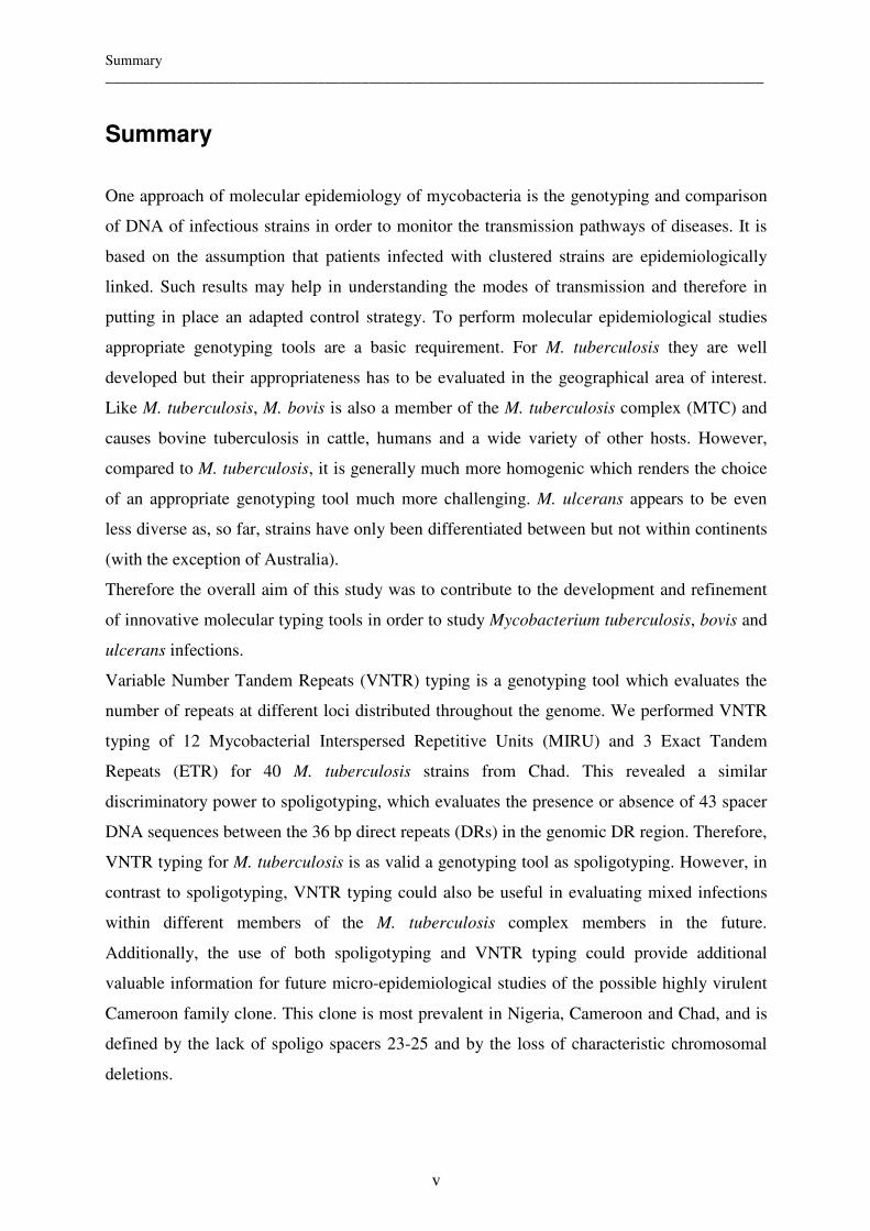
One approach of molecular epidemiology of mycobacteria is the genotyping and comparison
of DNA of infectious strains in order to monitor the transmission pathways of diseases. It is
based on the assumption that patients infected with clustered strains are epidemiologically
linked. Such results may help in understanding the modes of transmission and therefore in
putting in place an adapted control strategy. To perform molecular epidemiological studies
appropriate genotyping tools are a basic requirement. For M. tuberculosis they are well
developed but their appropriateness has to be evaluated in the geographical area of interest.
Like M. tuberculosis, M. bovis is also a member of the M. tuberculosis complex (MTC) and
causes bovine tuberculosis in cattle, humans and a wide variety of other hosts. However,
compared to M. tuberculosis, it is generally much more homogenic which renders the choice
of an appropriate genotyping tool much more challenging. M. ulcerans appears to be even
less diverse as, so far, strains have only been differentiated between but not within continents
(with the exception of Australia).
Therefore the overall aim of this study was to contribute to the development and refinement
of innovative molecular typing tools in order to study Mycobacterium tuberculosis, bovis and
ulcerans infections.
Variable Number Tandem Repeats (VNTR) typing is a genotyping tool which evaluates the
number of repeats at different loci distributed throughout the genome. We performed VNTR
typing of 12 Mycobacterial Interspersed Repetitive Units (MIRU) and 3 Exact Tandem
Repeats (ETR) for 40 M. tuberculosis strains from Chad. This revealed a similar
discriminatory power to spoligotyping, which evaluates the presence or absence of 43 spacer
DNA sequences between the 36 bp direct repeats (DRs) in the genomic DR region. Therefore,
VNTR typing for M. tuberculosis is as valid a genotyping tool as spoligotyping. However, in
contrast to spoligotyping, VNTR typing could also be useful in evaluating mixed infections
within different members of the M. tuberculosis complex members in the future.
Additionally, the use of both spoligotyping and VNTR typing could provide additional
valuable information for future micro-epidemiological studies of the possible highly virulent
Cameroon family clone. This clone is most prevalent in Nigeria, Cameroon and Chad, and is
defined by the lack of spoligo spacers 23-25 and by the loss of characteristic chromosomal
deletions.
Summary __________________________________________________________________________________________
vi
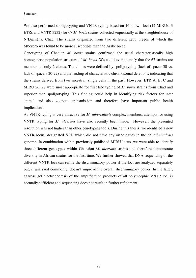
We also performed spoligotyping and VNTR typing based on 16 known loci (12 MIRUs, 3
ETRs and VNTR 3232) for 67 M. bovis strains collected sequentially at the slaughterhouse of
N’Djaména, Chad. The strains originated from two different zebu breeds of which the
Mbororo was found to be more susceptible than the Arabe breed.
Genotyping of Chadian M. bovis strains confirmed the usual characteristically high
homogenetic population structure of M. bovis. We could even identify that the 67 strains are
members of only 2 clones. The clones were defined by spoligotyping (lack of spacer 30 vs.
lack of spacers 20-22) and the finding of characteristic chromosomal deletions, indicating that
the strains derived from two ancestral, single cells in the past. However, ETR A, B, C and
MIRU 26, 27 were most appropriate for first line typing of M. bovis strains from Chad and
superior than spoligotyping. This finding could help in identifying risk factors for inter
animal and also zoonotic transmission and therefore have important public health
implications.
As VNTR-typing is very attractive for M. tuberculosis complex members, attempts for using
VNTR typing for M. ulcerans have also recently been made. However, the presented
resolution was not higher than other genotyping tools. During this thesis, we identified a new
VNTR locus, designated ST1, which did not have any orthologues in the M. tuberculosis
genome. In combination with a previously published MIRU locus, we were able to identify
three different genotypes within Ghanaian M. ulcerans strains and therefore demonstrate
diversity in African strains for the first time. We further showed that DNA sequencing of the
different VNTR loci can refine the discriminatory power if the loci are analyzed separately
but, if analyzed commonly, doesn’t improve the overall discriminatory power. In the latter,
agarose gel electrophoresis of the amplification products of all polymorphic VNTR loci is
normally sufficient and sequencing does not result in further refinement.

Zusammenfassung __________________________________________________________________________________________
vii
Zusammenfassung
In der Molekularen Epidemiologie der Mycobakterien können mit Hilfe der DNA
Genotypisierung Übertragunswege infektiöse Bakterienstämme verfolgt werden. Sie basiert
auf der Annahme, dass Patienten, welche mit gleichen (clustered) Stämmen infiziert sind,
eine epidemiologische Verbindung haben. Die Analyse genotypischer Ähnlichkeit kann
helfen zu besseren Bekämpfungsstrategien beizutragen. Um molekular epidemiologische
Studien überhaupt durchführen zu können sind angepasste Genotypisierungsmethoden eine
Grundvoraussetzung. Im Falle von M. tuberculosis sind sie gut entwickelt aber ihre
Eignungen müssen in den jeweiligen geographischen Gebieten evaluiert werden. Wie M.
tuberculosis ist auch M. bovis ein Mitglied des Mycobacterium tuberculosis Komplexes
(MTC) und verursacht die bovine Tuberkulose in Rindern, Menschen und in einer grossen
Bandbreite von anderen Wirten. Verglichen mit M. tuberculosis ist M. bovis jedoch im
allgemeinen viel homogener und deshalb ist die Wahl der geeigneten
Genotypisierungsmethode viel herausfordernder. Mycobacterium ulcerans scheint gar noch
homogener zu sein, da Stämme bis jetzt nur zwischen aber nicht innerhalb der Kontinente
unterschieden wurden (mit der Ausnahme von Australien).
Deshalb war es das übergeordnete Ziel dieser Doktorarbeit zu Entwicklung und Verbesserung
von innovativen, molekularen Typisierungsmethoden beizutragen um die Infektion von M.
tuberculosis, M.bovis und M.ulcerans zu studieren.
Das Typisieren von VNTR (Variable Number Tandem Repeats) ist eine
Genotypisierungsmethode welche die Anzahl von Repetitionen an verschiedenen, über das
ganze Genom verteilten Orten, evaluiert. Wir führten die Typisierung von VNTR an 12
MIRUs (Mycobacterial Interspersed Repetitve Units) und 3 ETRs (Exact Tandem Repeats)
für 40 M. tuberculosis Stämme vom Tschad durch. Es resultierte ein ähnlicher
Unterscheidungsgrad wie für das Spoligotyping, welches das Vorkommen von 43 ‚Spacer’
Sequenzen untersucht welche sich zwischen 36 Basenpaaren langen und direkten
Repetitionen in der DR (Direct repeat) befinden. Aus diesem Grund ist das Typisieren von
VNTRs als Genotypisierungsmethode genauso wertvoll wie das Spoligotyping. Das
Typisieren von VNTRs könnte jedoch, im Gegensatz zum Spoligotyping, für die Zukunft
nützlich werden um Mischinfektionen zwischen verschiedenen Mitgliedern des MTC zu
evaluieren. Zusätzlich könnte der Gebrauch von beiden Methoden, spoligotyping und
VNTRs, zusätzliche und wertvolle Informationen für zukünftige mikro-epidemiologische
Studien des möglicherweise sehr virulenten Klon der Kamerun Familie liefern. Dieser Klon
Zusammenfassung __________________________________________________________________________________________
viii
weist eine sehr hohe Prävalenz in Nigeria, Kamerun und Tschad auf und ist definiert durch
den Verlust der Spacer Sequenzen 23-25 und charakteristischen chromosomalen Löschungen.
Ebenfalls führten wir das Spoligotyping und das Typisieren der VNTRs anhand von 16
bekannten Orten (12 MIRUs, 3 ETRs und dem VNTR 3232) für 67 M. bovis Stämme durch,
welche sukzessive von Proben des Schlachthofs von N’Djaména, Tschad, erhalten wurden.
Die Stämme stammten von 2 verschiedenen Rinderrassen von welchen die Mbororo
gegenüber M. bovis empfänglicher war als die Arabe Rasse.
Das Genotypisieren von tschadischen M. bovis Stämmen bestätigte die üblicherweise hohe
homogene Populationsstruktur von M. bovis. Wir konnten sogar zeigen dass alle diese 67
Stämme Mitglieder von nur 2 Klonen sind. Diese Klone wurden definiert durch das
Spoligotyping (Verlust von Spacer Sequenzen 30 für den einen und Verlust von 20-22 für den
anderen Klon) und den Verlust von charakteristischen, chromosomalen Löschungen, was
darauf hinweisen könnte, dass alle Stämme von nur zwei einzelnen Zellen aus der
Vergangenheit abstammen. Abgesehen davon zeigte das Typisieren, dass ETR A, B, C und
MIRU 26 27 am geeignetsten für ein erstes, grobes Typisieren von M. bovis Stämmen von
Tschad ist und dem Spoligotyping überlegen ist. Dieser Befund könnte in der Zukunft helfen
Risikofaktoren für die zoonotische aber auch zwischen verschiedenen Tieren stattfindende
Übertragungswege zu identifizieren und könnte deshalb wichtige Konsequenzen für das
öffentlich Gesundheitswesen haben.
Da das Typisieren der VNTR für MTC Mitglieder sehr attraktiv ist, wurde erst kürzlich
Versuche gemacht dieses auch für M. ulcerans zu etablieren. Die präsentierte Auflösung war
jedoch nicht besser als diejenige von anderen Genotypisierungsmethoden. Während dieser
Doktorarbeit, haben wir einen VNTR identifiziert, welcher ST1 genannt wurde und keine
Analogien im M. tuberculosis Genom hatte. Im gemeinsamen Gebrauch mit einem bereits
beschriebenen und publizierten MIRU gelang es uns drei verschiedene Genotypen innerhalb
von ghanaischen Stämmen zu unterscheiden. So konnten wir zum ersten Mal Heterogenität
innerhalb von Afrikanischen Stämmen nachweisen. Des weiteren zeigten wir, dass das
Sequenzieren verschiedener VNTR die Auflösung verfeinern kann, wenn die polymorphen
VNTR separat aber nicht gemeinsam analysiert werden. Im letztgenannten Fall ist die
Agarose-Gel-Elektrophorese der amplifizierten, polymorphen VNTR Produkte normalerweise
ausreichend und das Sequenzieren ermöglicht keine weitere Verfeinerung.
Résumé __________________________________________________________________________________________
ix
Résumé
L’approche d'épidémiologie moléculaire des mycobactéries permet d’analyser et de comparer
l'ADN de souches infectieuses afin de suivre les voies de transmission des maladies. Elle est
basée sur la supposition que les patients infectés de mycobactéries génotypiquement
identiques sont liés épidémiologiquement. Ces résultats peuvent aider à comprendre les voies
de transmission et contribuer à adapter les stratégies de lutte.
Pour effectuer des études d’épidémiologie, des outils appropriés pour le typage d’ADN sont
une condition de base. Pour M. tuberculosis, ils sont bien développés mais doivent être
évaluées pour chaque zone géographique d'intérêt. Comme M. tuberculosis, M. bovis est aussi
un membre du complexe de M. tuberculosis (MTC) et cause la tuberculose chez le bétail,
l’homme et une large variété d'autres hôtes. Cependant, comparé à M. tuberculosis, l’ADN de
M. bovis est généralement beaucoup plus homogène et donc le choix de l'outil approprié pour
le typage est beaucoup plus complexe. M. ulcerans semble d’être encore moins variable car
jusque là, les souches ont pu être différenciées entre les continents, mais pas à l’intérieur des
continents (à l'exception de l'Australie).
Donc le but final de cette thèse était de contribuer au développement et au perfectionnement
d'outils moléculaires innovateurs pour le typage des mycobactéries pathogènes (M.
tuberculosis, M. bovis et M. ulcerans) et pour étudier l'infection causée par ces derniers.
Le typage par VNTR (variable number tandem repeats) est un outil moléculaire qui évalue le
nombre de répétitions à des sites différents répartis dans le génome. Nous avons effectué le
typage par VNTR de 12 MIRU (Mycobacterial Interspersed Repetitve Units) et 3 ETR (Exact
Tandem Repeats) pour 40 souches de M. tuberculosis du Tchad. La pouvoir de discrimination
était semblable au spoligotyping, qui évalue la présence ou l'absence de 43 séquences de la
région génomique DR (direct repeat) de l’ADN. Le typage de VNTR pour M. tuberculosis est
aussi valable comme outil d’épidémiologie moléculaire que le spoligotyping. Dans l’avenir le
typage par VNTR serait utile dans l'évaluation d'infections mixtes par les différents membres
de MTC. L’utilisation des deux méthodes : spoligotyping et typage par VNTR, pourrait
fournir des informations complémentaires de valeur pour des études futures sur la micro
épidémiologie du clone de la famille camerounaise. Ce clone, qui pourrait être fortement
virulent, est très répandu au Nigeria, au Cameroun et au Tchad et est défini par l’absence des
séquences DR 23-25 et par des délétions chromosomiques caractéristiques.
Résumé __________________________________________________________________________________________
x
Nous avons aussi effectué le spoligotyping et le typage par VNTR basés sur 16 locus connus
(12 MIRUS, 3 ETRS et VNTR 3232) pour 67 isolats de M. bovis, collectés à l'abattoir de
N'Djaména, Tchad. Les souches proviennent de deux races différentes de zébus dont le zébu
de race Mbororo est plus susceptible que le zébu de race Arabe.
Le typage moléculaire de souches tchadiennes de M. bovis a confirmé encore une fois la
structure fortement homogène de la population de M. bovis. Les 67 souches analysées
semblent être membres de seulement deux clones. Les clones ont été définis par le
spoligotyping (le manque de la séquence 30 pour l’un et des séquences 20-22 pour l’autre) et
par la découverte de délétions chromosomiques caractéristiques, indiquant que les souches
descendent de deux seules cellules ancestrales. De plus, ETR A, B, C et MIRU 26, 27 étaient
les plus appropriés pour un premier typage approximatif des souches de M. bovis du Tchad et
supérieurs au spoligotyping. Cela permettrait d’identifier les facteurs de risques pour la
transmission entre différents animaux, mais aussi la transmission zoonotique et pourrait donc
avoir des implications importantes pour la santé publique.
Comme l’utilisation de typage par VNTR est très attractive pour les membres MTC, on a
essayé récemment de l‘utiliser aussi pour M. ulcerans mais, la résolution obtenue n'était pas
meilleure aux 'autres outils moléculaires.
Dans le cadre de cette thèse, nous avons identifié un nouveau locus VNTR, désigné ST1, qui
n'avait pas de séquences similaires dans le génome de M. tuberculosis. Par la combinaison
avec un locus MIRU précédemment publié nous étions capables d'identifier trois génotypes
différents dans les souches ghanéennes de M. ulcerans et donc de trouver pour la première
fois une diversité parmi les souches africaines. Entre autre, nous avons pu montré que le
séquençage d'ADN des différents VNTRs peut raffiner le pouvoir discriminatoire si les
VNTR polymorphes sont analysés séparément, mais pas s’ils sont inclus ensemble pour
l'analyse. Et enfin,, l'électrophorèse par gel d’ agarose des produits amplifiés de tous les
VNTR polymorphes est suffisante et le séquençage ne contribue pas à une meilleure
résolution.
Abbreviations __________________________________________________________________________________________
xi
Abbreviations
AIDS Acquired Immune Deficiency Syndrome
AFB Acid Fast Bacilli
BCG Bacillus Calmette-Guèrin
BU Buruli Ulcer
bp base pairs
BTB Bovine Tuberculosis
CSSI Centre de Support en Santé International
dNTP Deoxyribonucleosidetriphosphate
DOTS Direct Observed Treatment Strategy
DNA Deoxyribonucleic Acid
DR Direct Repeat
ETR Exact Tandem Repeats
HGRTN Hôpital Général de Référence Nationale du Tchad
HIV Human Immunodeficiency Virus
IS Insertion Sequence
LJ Löwenstein Jensen
LRVZ/V Laboratoire de recherches vétérinaires et zootechniques de Farcha
LSP Large Sequence Polymorphism
MIRU Mycobacterial interspersed repetitive units
MLST Multilocus Sequence typing
MTC Mycobacterium tuberculosis complex
NALC N-Acetyl-L-Cystéine
NTM Non tuberculosis mycobacteria
PCR Polymerase Chain Reaction
PFGE Pulsed-field Gel Electrophoresis
PRPA PCR-restriction Profile Analysis
RD Region of Diversity
RFLP Restriction Fragment Length Polymorphism
SNP Single Nucleotide Polymorphism
STI Swiss Tropical Institute
TB Tuberculosis

Abbreviations __________________________________________________________________________________________
xii
VNTR Variable Number Tandem Repeats
WHO World Health Organization
ZN Ziehl Neelsen

Chapter I: Introduction __________________________________________________________________________________________
1
Chapter I: Introduction

Chapter I: Introduction __________________________________________________________________________________________
2
1.1. Burden and epidemiology of human tuberculosis
Despite the availability of anti-tuberculosis antibiotics, the disease burden of human
tuberculosis remains a very serious and wide-spread public health problem. At present,
approximately a third of the world population is infected with Mycobacterium tuberculosis,
which is a member of the Mycobacterium tuberculosis complex (MTC) and the main
causative organism for human tuberculosis. Today we consider that 2 million deaths and 8
million new human infections occur every year (11). Many of the 22 most affected countries
identified by the WHO are developing countries (Fig. 1) (13). There are various reasons why
tuberculosis control strategies have not yet succeeded:
- Resistance to antibiotics used in the treatment of tuberculosis. In different countries,
between 0 and 54 % of tuberculosis cases are multi drug resistant (15).
- Poverty connected to the problems of unemployment, access to good quality sanitary
services and urbanization
- Exponential increase of journeys and migration
- Co-existence of the Human Immunodeficiency Virus (HIV) fuels the epidemic of
tuberculosis on a large scale (11). Worldwide, 70.1 % (25.3 millions) of HIV positive
people live in sub-Saharan Africa (WHO / CDS / TB / 2002.296). In 1997, new cases
of TB totalled an estimated 7.96 million, including 3.52 million cases (44%) of
infectious pulmonary disease (smear-positive), with 16.2 million existing cases of
disease. An estimated 1.87 million people died of TB and the global case fatality rate
was 23% but exceeded 50% in some African countries with high HIV rates (11).
Fig. 1: Estimated
global incidence rates
of tuberculosis (2001).
(Source: World Health
Organization (WHO)
2003)
Chapter I: Introduction __________________________________________________________________________________________
3
Infection with HIV favours a new infection with mycobateria; however, it can also
reactivate a latent infection.
- DOTS (Directly Observed Treatment short course), a strategy promoted by the World
Health Organization (WHO) is either not implemented, ineffective or not feasible in
various countries.
- Although M. tuberculosis most often causes pulmonary tuberculosis, it is also the
causative agent for extra pulmonary tuberculosis. This form of tuberculosis is often
underdiagnosed.
- Mycobacterium bovis, another member of the MTC is also known to cause clinically
undistinguishable tuberculosis in humans. Its zoonotic importance for the burden of
human tuberculosis is unknown and currently under research (see also chapter on M.
bovis).
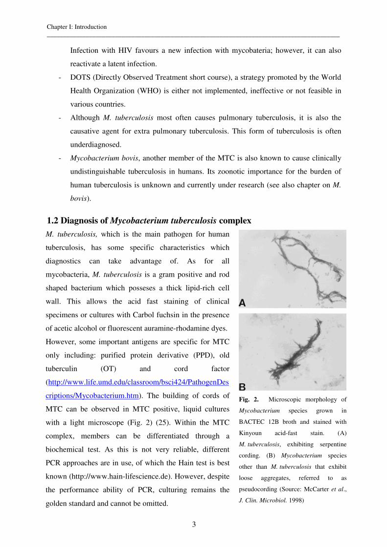
1.2 Diagnosis of Mycobacterium tuberculosis complex
.
M. tuberculosis, which is the main pathogen for human
tuberculosis, has some specific characteristics which
diagnostics can take advantage of. As for all
mycobacteria, M. tuberculosis is a gram positive and rod
shaped bacterium which posseses a thick lipid-rich cell
wall. This allows the acid fast staining of clinical
specimens or cultures with Carbol fuchsin in the presence
of acetic alcohol or fluorescent auramine-rhodamine dyes.
However, some important antigens are specific for MTC
only including: purified protein derivative (PPD), old
tuberculin (OT) and cord factor
(http://www.life.umd.edu/classroom/bsci424/PathogenDes
criptions/Mycobacterium.htm). The building of cords of
MTC can be observed in MTC positive, liquid cultures
with a light microscope (Fig. 2) (25). Within the MTC
complex, members can be differentiated through a
biochemical test. As this is not very reliable, different
PCR approaches are in use, of which the Hain test is best
known (http://www.hain-lifescience.de). However, despite
the performance ability of PCR, culturing remains the
golden standard and cannot be omitted.
Fig. 2. Microscopic morphology of
Mycobacterium species grown in
BACTEC 12B broth and stained with
Kinyoun acid-fast stain. (A)
M. tuberculosis, exhibiting serpentine
cording. (B) Mycobacterium species
other than M. tuberculosis that exhibit
loose aggregates, referred to as
pseudocording (Source: McCarter et al.,
J. Clin. Microbiol. 1998)
Chapter I: Introduction __________________________________________________________________________________________
4
1.3. Molecular epidemiology of Mycobacterium tuberculosis
Molecular epidemiology is a powerful approach for monitoring infectious diseases (32). It is
particularly important in the study of chronic diseases such as tuberculosis, where patients
with recurrent tuberculosis can be chronically infected with a given strain and relapse due to
reactivation of that strain or, in contrast, can be reinfected by a different strain after cure (42).
A correct distinction between these alternatives is essential for accurate estimation of the
success rates of tuberculosis programs (5). Moreover, it can give unique insights into the
international dissemination dynamics of M. tuberculosis by the comparison of isolates from
widespread geographic areas and allows one to analyze evolutionary changes of pathogen
populations (38). Molecular studies of M. tuberculosis are made extensively in industrialized
but only few developing countries. Molecular epidemiological results from developed
countries often show high polymorphism in the genetic patterns of M. tuberculosis complex
strains (4,19,44). This is explained by two factors (43): The relatively high percentage of
cases in low-incidence areas due to endogenous reactivation and the large proportion of cases
in these areas found amongst non-native populations originating from different geographical
origins, which introduce exotic strains not known in these areas.
However, in interpreting the proportion of clustered strains found in a study, knowledge of
the proportion of tuberculosis cases in the community included in the study is important. A
high number of tuberculosis cases analyzed in a community can overestimate the proportion
of recent transmission. On the other hand, a low number of samples can underestimate the
proportion of recent transmission because the percentage of clustered strains is known to
increase sharply at the beginning of a study till a certain number of cases is reached.
Furthermore, molecular epidemiological studies should give information on the study setting,
duration of study, the recruitment period and the definition of clustering used. The data on
clustering should be disaggregated at the very least by age, sex and immigration status (16).
If we consider Africa, apart from studies carried out in Tunisia and Egypt, where most of the
M. tuberculosis strains only belonged to a few genotype families (20), results have also been
obtained from the countries of Botswana (24) and South Africa. Wilkinson et al. (47) found a
high clustering rate of patterns (45%) in a rural area of KwaZulu Natal, South Africa. In
contrast, quite a low clustering rate was found in Botswana (24) and in the communities of
Ravensmead and Uitsig, Cape Town, South Africa (46). These contradicting results from high
incidence countries show how little is known when it comes to molecular epidemiology of
TB in Africa. In addition, many countries, like Chad, completely lack data from similar
studies suggesting that further research in these countries is urgently needed. In order to
Chapter I: Introduction __________________________________________________________________________________________
5
perform molecular epidemiological studies, one or more appropriate genotyping tools are
necessary. Nowadays, there exists a number of different ‘working’ tools, which are used
routinely or for special occasions:
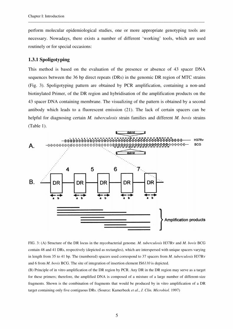
1.3.1 Spoligotyping
This method is based on the evaluation of the presence or absence of 43 spacer DNA
sequences between the 36 bp direct repeats (DRs) in the genomic DR region of MTC strains
(Fig. 3). Spoligotyping pattern are obtained by PCR amplification, containing a non-and
biotinylated Primer, of the DR region and hybridisation of the amplification products on the
43 spacer DNA containing membrane. The visualizing of the pattern is obtained by a second
antibody which leads to a fluorescent emission (21). The lack of certain spacers can be
helpful for diagnosing certain M. tuberculosis strain families and different M. bovis strains
(Table 1).
FIG. 3: (A) Structure of the DR locus in the mycobacterial genome. M. tuberculosis H37Rv and M. bovis BCG
contain 48 and 41 DRs, respectively (depicted as rectangles), which are interspersed with unique spacers varying
in length from 35 to 41 bp. The (numbered) spacers used correspond to 37 spacers from M. tuberculosis H37Rv
and 6 from M. bovis BCG. The site of integration of insertion element IS6110 is depicted.
(B) Principle of in vitro amplification of the DR region by PCR. Any DR in the DR region may serve as a target
for these primers; therefore, the amplified DNA is composed of a mixture of a large number of different-size
fragments. Shown is the combination of fragments that would be produced by in vitro amplification of a DR
target containing only five contiguous DRs. (Source: Kamerbeek et al., J. Clin. Microbiol. 1997)
Chapter I: Introduction __________________________________________________________________________________________
6
Table 1: Diagnostic spoligo spacer missing for M. tuberculosis family members (12), M. africanum and host
adapted M. bovis strains (35).
M. tb family members Spacer lacking M. tb (Beijing) 1-34 M. tb (Haarlem) 31, 33-36 M. tb (Latin America) 21-24, 33-36 M. tb (East African India) 29-32, 34 M. tb (Central Asia) 4-7, 23-34 M. tb (Cameroon) 23-25, 33-36 M. africanum (Type I) 9, 39 M. bovis (antelope) 9, 16, 39 M. bovis (seal/vole) 3, 9, 16, 39-43 M. bovis (caprine) 3, 9, 16, 39-43 M. bovis (cattle) 3, 9, 16, 39-43 M. bovis BCG 3, 9, 16, 39-43
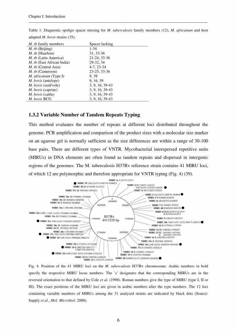
1.3.2 Variable Number of Tandem Repeats Typing
This method evaluates the number of repeats at different loci distributed throughout the
genome. PCR amplification and comparison of the product sizes with a molecular size marker
on an agarose gel is normally sufficient as the size differences are within a range of 30-100
base pairs. There are different types of VNTR. Mycobacterial interspersed repetitive units
(MIRUs) in DNA elements are often found as tandem repeats and dispersed in intergenic
regions of the genomes. The M. tuberculosis H37Rv reference strain contains 41 MIRU loci,
of which 12 are polymorphic and therefore appropriate for VNTR typing (Fig. 4) (39).
Fig. 4. Position of the 41 MIRU loci on the M. tuberculosis H37Rv chromosome. Arabic numbers in bold
specify the respective MIRU locus numbers. The `c' designates that the corresponding MIRUs are in the
reversed orientation to that defined by Cole et al. (1998). Roman numbers give the type of MIRU (type I, II or
III). The exact positions of the MIRU loci are given in arabic numbers after the type numbers. The 12 loci
containing variable numbers of MIRUs among the 31 analysed strains are indicated by black dots (Source:
Supply et al., Mol. Microbiol. 2000).
Chapter I: Introduction __________________________________________________________________________________________
7
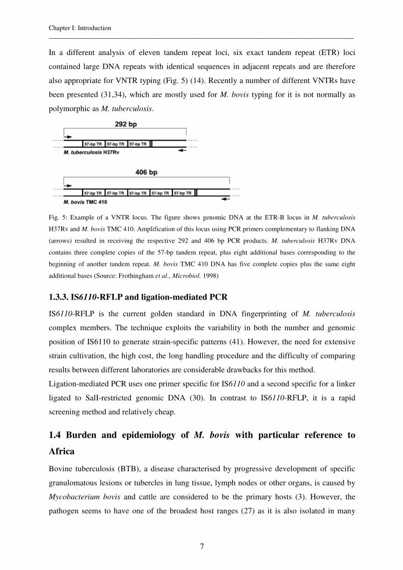
In a different analysis of eleven tandem repeat loci, six exact tandem repeat (ETR) loci
contained large DNA repeats with identical sequences in adjacent repeats and are therefore
also appropriate for VNTR typing (Fig. 5) (14). Recently a number of different VNTRs have
been presented (31,34), which are mostly used for M. bovis typing for it is not normally as
polymorphic as M. tuberculosis.
Fig. 5: Example of a VNTR locus. The figure shows genomic DNA at the ETR-B locus in M. tuberculosis
H37Rv and M. bovis TMC 410. Amplification of this locus using PCR primers complementary to flanking DNA
(arrows) resulted in receiving the respective 292 and 406 bp PCR products. M. tuberculosis H37Rv DNA
contains three complete copies of the 57-bp tandem repeat, plus eight additional bases corresponding to the
beginning of another tandem repeat. M. bovis TMC 410 DNA has five complete copies plus the same eight
additional bases (Source: Frothingham et al., Microbiol. 1998)
1.3.3. IS6110-RFLP and ligation-mediated PCR
IS6110-RFLP is the current golden standard in DNA fingerprinting of M. tuberculosis
complex members. The technique exploits the variability in both the number and genomic
position of IS6110 to generate strain-specific patterns (41). However, the need for extensive
strain cultivation, the high cost, the long handling procedure and the difficulty of comparing
results between different laboratories are considerable drawbacks for this method.
Ligation-mediated PCR uses one primer specific for IS6110 and a second specific for a linker
ligated to SalI-restricted genomic DNA (30). In contrast to IS6110-RFLP, it is a rapid
screening method and relatively cheap.
1.4 Burden and epidemiology of M. bovis with particular reference to
Africa
Bovine tuberculosis (BTB), a disease characterised by progressive development of specific
granulomatous lesions or tubercles in lung tissue, lymph nodes or other organs, is caused by
Mycobacterium bovis and cattle are considered to be the primary hosts (3). However, the
pathogen seems to have one of the broadest host ranges (27) as it is also isolated in many
Chapter I: Introduction __________________________________________________________________________________________
8
different host species such as wild boar, deer (33) badgers, goats, sheep, rabbits and pigs (8).
But while some species (wild boar, deer and badgers) are considered to be maintenance hosts
and therefore are dangerous natural reservoirs, others (goats, sheep, rabbits and pigs) act only
as a spillover for M. bovis. These hosts are infected with host adapted M. bovis substrains
rather than with classical M. bovis strains e.g. caprae (see 1.6 Evolution and ecotypes of
MTC).
In many industrialized countries, like Switzerland, bovine tuberculosis has been eradicated
due to elimination programs and milk pasteurization (29). However, the burden of disease is
still considerable in other industrialized countries such as the U.S. and U.K as the presence of
natural reservoirs (badgers, deer) makes eradication difficult. Although a vaccination exists,
the currently available BCG is not effective enough to completely prevent infection and
interferes with the PPD test and there are assumptions that the vaccination of cattle is not
deemed financially profitable (J. Zinsstag, personal communication). Indeed, during the 4th
M. bovis conference in 2005, policy makers agreed to vaccinate wild life reservoirs rather
than cattle.
In developing countries, the incidence of animal TB is especially high as control measures are
not at all or only partially applied (3). Additionally, some of them, such as the test and
slaughter policy are not feasible due to the lack of financial compensation (23). In Africa,
bovine TB represents a potential health hazard to both animals and humans, as nearly 85 % of
cattle and 83 % of the human population live in areas where the disease is prevalent (3).
M. bovis is additionally of particular interest from a public health perspective as man is also
susceptible to infection. The burden of tuberculosis in humans caused by M. bovis is largely
unknown or underdiagnosed due to the lack of adequate laboratory equipment but its presence
has been proven and infections due to M. bovis are described in various African countries (9).
Clinically, tuberculosis caused by M. bovis is not different from that caused by M.
tuberculosis, but M. bovis is resistant to the antibiotic pyrazinamide, which is a first line drug
in the treatment against human tuberculosis within the program of DOTS. In developing
countries consumption of unpasteurised milk, poorly heat-treated meat and close contact with
infected animals represent the main sources of infection for humans (3).
In conclusion there are three main reasons why eradication of bovine TB is recommended (3):
- loss in productivity due to infected animals
- animal market restrictions
- the risk of infection to the human population
Chapter I: Introduction __________________________________________________________________________________________
9
1.5 Molecular epidemiology of M. bovis
Different studies on M. bovis are carried out in order to improve the traceability of the M.
bovis infections and identification of the origin of the outbreak Haddad N. et al. (18)
genotyped 1266 M. bovis isolates in France and observed an apparently high level of
heterogeneity of 161 different clusters and a low frequency of the two main spoligotypes
clusters. In contrast, similar molecular studies in island countries like Great Britain (7) or
Australia (10) showed a low level of heterogeneity and a high frequency of the main
spolygotype clusters.
Again, very few such studies have been carried out in developing countries in Africa. Some
studies in Cameroon (26) and Tanzania (22) have shown similar results to those made in
Great Britain or Australia with a high homogeneity and thereby indicate a high recent
transmission rate. There have been no molecular epidemiological studies of M. bovis in Chad
before this PhD thesis.
There are also studies which look at transmission pathways from M. bovis between different
animal species, from animal to human (zoonotic) and from human to human. Serraino et al.
(33) report spoligotype clusters which include 9 strains isolated from wild boar and 11 strains
isolated from cattle, thus confirming the possibility of transmission between the two animal
species. V. Soolingen et al. (45) show clusters containing M. bovis isolated from humans and
cattle using the combination of the RFLP methods IS6110 and PGRS. One of the first results
indicating but not proving M. bovis zoonotic transmission between cattle and humans in
Africa is shown in a study from Tanzania, where the same M. bovis spoligotype was isolated
from man and cattle (23). Moreover, molecular epidemiological studies by Guerrero et al.
(17) showed the transmission of M. bovis MDR tuberculosis between HIV-1-positive patients.
It is suggested that transmission of M. bovis took place within hospitals and that advanced
HIV-1 immunosuppression was associated with the development of MDR tuberculosis.
As with M. tuberculosis, molecular epidemiology can also develop a better understanding of
the sources and modes of M. bovis transmission thereby enabling more effective control
measures to be implemented in bovine eradication programs.
1.6 Evolution and ecotypes of the Mycobacterium tuberculosis complex
Human and animal tuberculosis are caused by different members of the Mycobacterium
tuberculosis complex (MTC), of which M. tuberculosis and M. bovis are best known and
share 99.9 % of the same genome.
Chapter I: Introduction __________________________________________________________________________________________
10
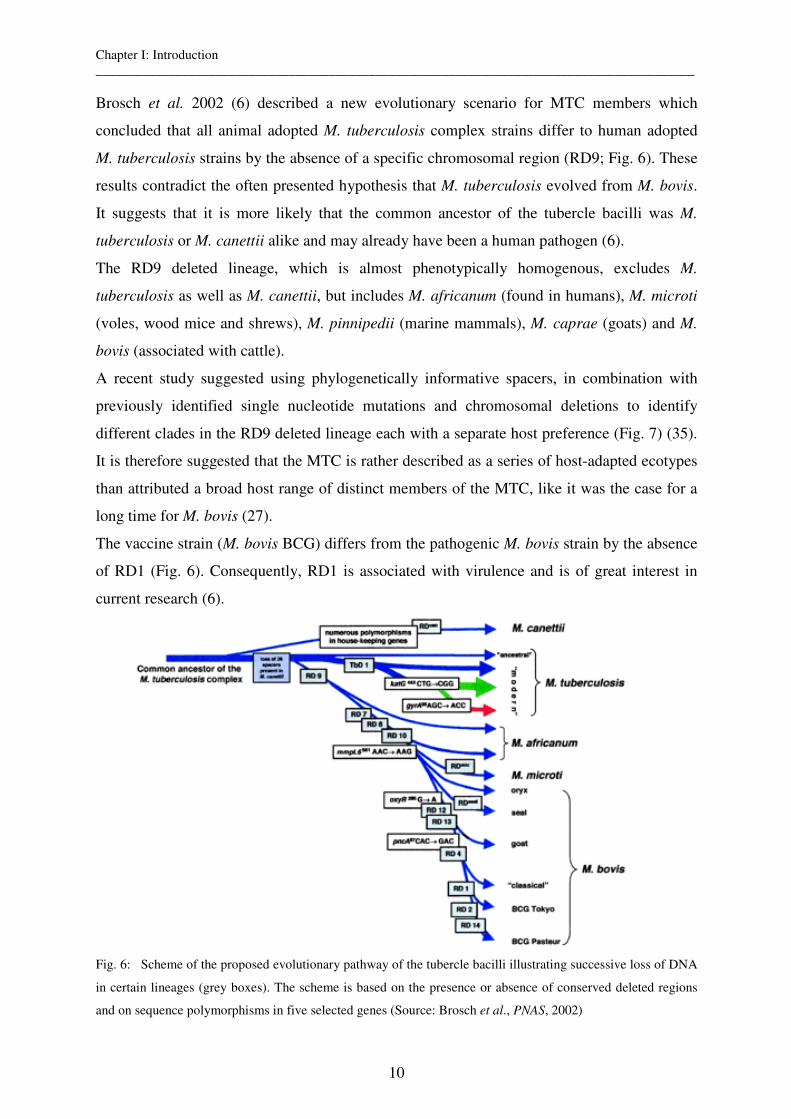
Brosch et al. 2002 (6) described a new evolutionary scenario for MTC members which
concluded that all animal adopted M. tuberculosis complex strains differ to human adopted
M. tuberculosis strains by the absence of a specific chromosomal region (RD9; Fig. 6). These
results contradict the often presented hypothesis that M. tuberculosis evolved from M. bovis.
It suggests that it is more likely that the common ancestor of the tubercle bacilli was M.
tuberculosis or M. canettii alike and may already have been a human pathogen (6).
The RD9 deleted lineage, which is almost phenotypically homogenous, excludes M.
tuberculosis as well as M. canettii, but includes M. africanum (found in humans), M. microti
(voles, wood mice and shrews), M. pinnipedii (marine mammals), M. caprae (goats) and M.
bovis (associated with cattle).
A recent study suggested using phylogenetically informative spacers, in combination with
previously identified single nucleotide mutations and chromosomal deletions to identify
different clades in the RD9 deleted lineage each with a separate host preference (Fig. 7) (35).
It is therefore suggested that the MTC is rather described as a series of host-adapted ecotypes
than attributed a broad host range of distinct members of the MTC, like it was the case for a
long time for M. bovis (27).
The vaccine strain (M. bovis BCG) differs from the pathogenic M. bovis strain by the absence
of RD1 (Fig. 6). Consequently, RD1 is associated with virulence and is of great interest in
current research (6).
Fig. 6: Scheme of the proposed evolutionary pathway of the tubercle bacilli illustrating successive loss of DNA
in certain lineages (grey boxes). The scheme is based on the presence or absence of conserved deleted regions
and on sequence polymorphisms in five selected genes (Source: Brosch et al., PNAS, 2002)
Chapter I: Introduction __________________________________________________________________________________________
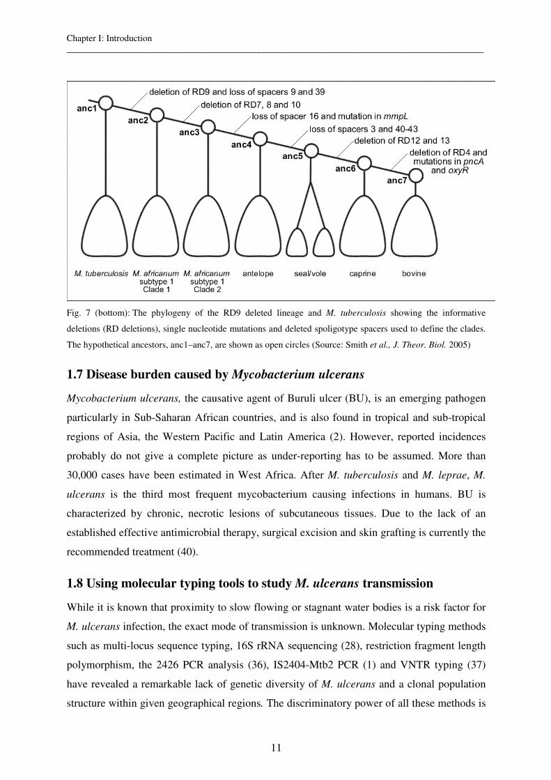
11
Fig. 7 (bottom): The phylogeny of the RD9 deleted lineage and M. tuberculosis showing the informative
deletions (RD deletions), single nucleotide mutations and deleted spoligotype spacers used to define the clades.
The hypothetical ancestors, anc1–anc7, are shown as open circles (Source: Smith et al., J. Theor. Biol. 2005)
1.7 Disease burden caused by Mycobacterium ulcerans
Mycobacterium ulcerans, the causative agent of Buruli ulcer (BU), is an emerging pathogen
particularly in Sub-Saharan African countries, and is also found in tropical and sub-tropical
regions of Asia, the Western Pacific and Latin America (2). However, reported incidences
probably do not give a complete picture as under-reporting has to be assumed. More than
30,000 cases have been estimated in West Africa. After M. tuberculosis and M. leprae, M.
ulcerans is the third most frequent mycobacterium causing infections in humans. BU is
characterized by chronic, necrotic lesions of subcutaneous tissues. Due to the lack of an
established effective antimicrobial therapy, surgical excision and skin grafting is currently the
recommended treatment (40).
1.8 Using molecular typing tools to study M. ulcerans transmission
While it is known that proximity to slow flowing or stagnant water bodies is a risk factor for
M. ulcerans infection, the exact mode of transmission is unknown. Molecular typing methods
such as multi-locus sequence typing, 16S rRNA sequencing (28), restriction fragment length
polymorphism, the 2426 PCR analysis (36), IS2404-Mtb2 PCR (1) and VNTR typing (37)
have revealed a remarkable lack of genetic diversity of M. ulcerans and a clonal population
structure within given geographical regions. The discriminatory power of all these methods is
Chapter I: Introduction __________________________________________________________________________________________
12
particularly insufficient to differentiate between African isolates. Innovative molecular
genetic fingerprinting methods are therefore required for local epidemiological studies aiming
to reveal transmission pathways and environmental reservoirs of M. ulcerans.
1.9. Rationale and research frame work
While a number of molecular epidemiological studies of M. tuberculosis are performed in
industrialized countries, data from similar works from the African continent, where incidence
rates are high, are rare. However, this data is needed as it could prove useful in the
tuberculosis control strategy of the different countries. In Chad, the results of molecular
epidemiological studies could help in proposing new and innovative control strategies,
showing for example risk factors for recent transmission of drug sensitive and resistance
strains and researching the degree of mixed infections. Furthermore, it could help in
evaluating the percentage of human tuberculosis infections due to M. bovis and in finding the
sources of infections.
Appropriate genotyping tools are a prerequisite for performing molecular epidemiological
studies of Mycobacterium tuberculosis complex and M. ulcerans strains. For M. tuberculosis,
these tools are well established and their degrees of appropriateness may only vary slightly
depending on geographical area. M. bovis, despite also being a member of the M. tuberculosis
complex, is generally much more homogenetic and the discriminatory power of the different
tools has to be evaluated with great care. Even less genomic diversity seems to be attributed
to M. ulcerans for which no typing tool was able to discriminate strains within the African
continent.
In an attempt to develop and evaluate innovative genotyping tools for the M. tuberculosis
complex in Chad and M. ulcerans strains in Ghana, a scientific partnership was established
between the Noguchi Memorial Institute for Medical Research of Legon and the Tema
Municipal Health Directorate of Tema in Ghana and the Laboratoire de recherches
vétérinaires et zootechniques de Farcha (LRVZ) and Centre de Support en Santé International
(CSSI) in Chad. An evaluation of VNTR for genotyping the M. tuberculosis complex and M.
ulcerans and its potential to enable micro epidemiological studies in the near future is
presented.
This thesis is co-funded by the NCCR North-South

Chapter I: Introduction __________________________________________________________________________________________
13
1.10 References of Introduction
1. Ablordey, A., R. Kotlowski, J. Swings, and F. Portaels. 2005. PCR amplification with primers based on IS2404 and GC-rich repeated sequence reveals polymorphism in Mycobacterium ulcerans. J.Clin.Microbiol. 43:448-451.
2. Asiedu, K., R. Scherpbier, and M. Raviglione. 2000. Buruli ulcer - Mycobacterium ulcerans
infection. WHO document WHO/CDS/CPE/GBUI/2000.1.
3. Ayele, W. Y., S. D. Neill, J. Zinsstag, M. G. Weiss, and I. Pavlik. 2004. Bovine tuberculosis: an old disease but a new threat to Africa. Int.J.Tuberc.Lung Dis. 8:924-937.
4. Bauer, J., Z. Yang, S. Poulsen, and A. B. Andersen. 1998. Results from 5 years of nationwide DNA fingerprinting of Mycobacterium tuberculosis complex isolates in a country with a low incidence of M. tuberculosis infection. J.Clin.Microbiol. 36:305-308.
5. Bloom, B. R. and C. J. L. Murray. 1992. Tuberculosis - Commentary on A Reemergent Killer. Science 257:1055-1064.
6. Brosch, R., S. V. Gordon, M. Marmiesse, P. Brodin, C. Buchrieser, K. Eiglmeier, T. Garnier, C.
Gutierrez, G. Hewinson, K. Kremer, L. M. Parsons, A. S. Pym, S. Samper, D. van Soolingen, and S. T. Cole. 2002. A new evolutionary scenario for the Mycobacterium tuberculosis complex. Proc.Natl.Acad.Sci.U.S.A 99:3684-3689.
7. Clifton-Hadley, R. S., J. Inwald, S. Hughes, N. Palmer, A. R. Sayers, K. Sweeney, J. D. A. van
Embden, and R. G. Hewinson. 1998. Recent advances in DNA fingerprinting using spoligotyping - Epidemiological applications in bovine TB. Journal of the British Cattle Veterinary Association 6:79-82.
8. Corner, L. A. 2005. The role of wild animal populations in the epidemiology of tuberculosis in domestic animals: How to assess the risk. Vet.Microbiol.
9. Cosivi, O., J. M. Grange, C. J. Daborn, M. C. Raviglione, T. Fujikura, D. Cousins, R. A.
Robinson, H. F. Huchzermeyer, K. de, I, and F. X. Meslin. 1998. Zoonotic tuberculosis due to Mycobacterium bovis in developing countries. Emerg.Infect.Dis. 4:59-70.
10. Cousins, D., S. Williams, E. Liebana, A. Aranaz, A. Bunschoten, J. Van Embden, and T. Ellis. 1998. Evaluation of four DNA typing techniques in epidemiological investigations of bovine tuberculosis. J.Clin.Microbiol. 36:168-178.
11. Dye, C., S. Scheele, P. Dolin, V. Pathania, and R. C. Raviglione. 1999. Global burden of tuberculosis - Estimated incidence, prevalence, and mortality by country. Jama-Journal of the American Medical Association 282:677-686.
12. Ferdinand, S., G. Valetudie, C. Sola, and N. Rastogi. 2004. Data mining of Mycobacterium tuberculosis complex genotyping results using mycobacterial interspersed repetitive units validates the clonal structure of spoligotyping-defined families. Res.Microbiol. 155:647-654.
13. Floyd, K., L. Blanc, M. Raviglione, and J. W. Lee. 2002. Resources required for global tuberculosis control. Science 295:2040-2041.
14. Frothingham, R. and W. A. Meeker-O'Connell. 1998. Genetic diversity in the Mycobacterium tuberculosis complex based on variable numbers of tandem DNA repeats. Microbiology-Uk 144:1189-1196.
15. Gleissberg, V. 1999. The threat of multidrug resistance: is tuberculosis ever untreatable or uncontrollable? Lancet 353:998-999.
Chapter I: Introduction __________________________________________________________________________________________
14
16. Glynn, J. R., J. Bauer, A. S. de Boer, M. W. Borgdorff, P. E. Fine, P. Godfrey-Faussett, and E.
Vynnycky. 1999. Interpreting DNA fingerprint clusters of Mycobacterium tuberculosis. European Concerted Action on Molecular Epidemiology and Control of Tuberculosis. Int J.Tuberc.Lung Dis. 3:1055-1060.
17. Guerrero, A., J. Cobo, J. Fortun, E. Navas, C. Quereda, A. Asensio, J. Canon, J. Blazquez, and E.
GomezMampaso. 1997. Nosocomial transmission of Mycobacterium bovis resistant to 11 drugs in people with advanced HIV-1 infection. Lancet 350:1738-1742.
18. Haddad, N., A. Ostyn, C. Karoui, M. Masselot, M. F. Thorel, S. L. Hughes, J. Inwald, R. G. Hewinson, and B. Durand. 2001. Spoligotype diversity of Mycobacterium bovis strains isolated in France from 1979 to 2000. Journal of Clinical Microbiology 39:3623-3632.
19. Heldal, E., H. Docker, D. A. Caugant, and A. Tverdal. 2000. Pulmonary tuberculosis in Norwegian patients. The role of reactivation, re-infection and primary infection assessed by previous mass screening data and restriction fragment length polymorphism analysis. Int.J.Tuberc.Lung Dis. 4:300-307.
20. Hermans, P. W. M., F. Messadi, H. Guebrexabher, D. Vansoolingen, P. E. W. Dehaas, H.
Heersma, H. Deneeling, A. Ayoub, F. Portaels, D. Frommel, M. Zribi, and J. D. A. Vanembden. 1995. Analysis of the Population-Structure of Mycobacterium-Tuberculosis in Ethiopia, Tunisia, and the Netherlands - Usefulness of Dna Typing for Global Tuberculosis Epidemiology. Journal of Infectious Diseases 171:1504-1513.
21. Kamerbeek, J., L. Schouls, A. Kolk, M. van Agterveld, D. van Soolingen, S. Kuijper, A. Bunschoten, H. Molhuizen, R. Shaw, M. Goyal, and J. Van Embden. 1997. Simultaneous detection and strain differentiation of Mycobacterium tuberculosis for diagnosis and epidemiology. J.Clin.Microbiol. 35:907-914.
22. Kazwala, K. D., Sinclaire, J. Challans, D. M. Kambarage, J. M. Sharp, J. Van Embden, C. J.
Daborn, and J. Nyange. 1997. Zoonotic importance of Mycobacterium tuberculosis complex organisms in Tanzania: a molecular biology approach. Actes Editions, Rabat, Morocco 199-204.
23. Kazwala, R. R., L. J. Kusiluka, K. Sinclair, J. M. Sharp, and C. J. Daborn. 2005. The molecular epidemiology of Mycobacterium bovis infections in Tanzania. Vet.Microbiol.
24. Lockman, S., J. D. Sheppard, C. R. Braden, M. J. Mwasekaga, C. L. Woodley, T. A. Kenyon, N.
J. Binkin, M. Steinman, F. Montsho, M. Kesupile-Reed, C. Hirschfeldt, M. Notha, T. Moeti, and J. W. Tappero. 2001. Molecular and conventional epidemiology of Mycobacterium tuberculosis in Botswana: A population-based prospective study of 301 pulmonary tuberculosis patients. Journal of Clinical Microbiology 39:1042-1047.
25. McCarter, Y. S., I. N. Ratkiewicz, and A. Robinson. 1998. Cord formation in BACTEC medium is a reliable, rapid method for presumptive identification of Mycobacterium tuberculosis complex. J.Clin.Microbiol. 36:2769-2771.
26. Njanpop-Lafourcade, B. M., J. Inwald, A. Ostyn, B. Durand, S. Hughes, M. F. Thorel, G. Hewinson, and N. Haddad. 2001. Molecular typing of Mycobacterium bovis isolates from Cameroon. J.Clin.Microbiol. 39:222-227.
27. O'Reilly, L. M. and C. J. Daborn. 1995. The epidemiology of Mycobacterium bovis infections in animals and man: a review. Tuber.Lung Dis. 76 Suppl 1:1-46.
28. Portaels, F., P. A. Fonteyne, H. DeBeenhouwer, P. DeRijk, A. Guedenon, J. Hayman, and W. M. Meyers. 1996. Variability in 3' end of 16S rRNA sequence of Mycobacterium ulcerans is related to geographic origin of isolates. Journal of Clinical Microbiology 34:962-965.
29. Pritchard, D. G. 1988. A century of bovine tuberculosis 1888-1988: conquest and controversy. J.Comp Pathol. 99:357-399.
Chapter I: Introduction __________________________________________________________________________________________
15
30. Prod'hom, G., C. Guilhot, M. C. Gutierrez, A. Varnerot, B. Gicquel, and V. Vincent. 1997. Rapid discrimination of Mycobacterium tuberculosis complex strains by ligation-mediated PCR fingerprint analysis. J.Clin.Microbiol. 35:3331-3334.
31. Roring, S., A. Scott, D. Brittain, I. Walker, G. Hewinson, S. Neill, and R. Skuce. 2002. Development of variable-number tandem repeat typing of Mycobacterium bovis: comparison of results with those obtained by using existing exact tandem repeats and spoligotyping. J.Clin.Microbiol. 40:2126-2133.
32. Savine, E., R. M. Warren, G. D. van der Spuy, N. Beyers, P. D. van Helden, C. Locht, and P. Supply. 2002. Stability of variable-number tandem repeats of mycobacterial interspersed repetitive units from 12 loci in serial isolates of Mycobacterium tuberculosis. Journal of Clinical Microbiology 40:4561-4566.
33. Serraino, A., G. Marchetti, V. Sanguinetti, M. C. Rossi, R. G. Zanoni, L. Catozzi, A. Bandera, W.
Dini, W. Mignone, F. Franzetti, and A. Gori. 1999. Monitoring of transmission of tuberculosis between wild boars and cattle: genotypical analysis of strains by molecular epidemiology techniques. J.Clin.Microbiol. 37:2766-2771.
34. Skuce, R. A., T. P. McCorry, J. F. McCarroll, S. M. M. Roring, A. N. Scott, D. Brittain, S. L.
Hughes, R. G. Hewinson, and S. D. Neill. 2002. Discrimination of Mycobacterium tuberculosis complex bacteria using novel VNTR-PCR targets. Microbiology-Sgm 148:519-528.
35. Smith, N. H., K. Kremer, J. Inwald, J. Dale, J. R. Driscoll, S. V. Gordon, D. van Soolingen, H. R.
Glyn, and S. J. Maynard. 2005. Ecotypes of the Mycobacterium tuberculosis complex. J.Theor.Biol.
36. Stinear, T., J. K. Davies, G. A. Jenkin, F. Portaels, B. C. Ross, F. Oppedisano, M. Purcell, J. A.
Hayman, and P. D. R. Johnson. 2000. A simple PCR method for rapid genotype analysis of Mycobacterium ulcerans. Journal of Clinical Microbiology 38:1482-1487.
37. Stragier, P., A. Ablordey, W. M. Meyers, and F. Portaels. 2005. Genotyping Mycobacterium ulcerans and Mycobacterium marinum by using mycobacterial interspersed repetitive units. J.Bacteriol. 187:1639-1647.
38. Supply, P., S. Lesjean, E. Savine, K. Kremer, D. van Soolingen, and C. Locht. 2001. Automated high-throughput genotyping for study of global epidemiology of Mycobacterium tuberculosis based on mycobacterial interspersed repetitive units. Journal of Clinical Microbiology 39:3563-3571.
39. Supply, P., E. Mazars, S. Lesjean, V. Vincent, B. Gicquel, and C. Locht. 2000. Variable human minisatellite-like regions in the Mycobacterium tuberculosis genome. Molecular Microbiology 36:762-771.
40. van der Werf, T. S., T. Stinear, Y. Stienstra, W. T. A. van der Graaf, and P. L. Small. 2003. Mycolactones and Mycobacterium ulcerans disease. Lancet 362:1062-1064.
41. van Embden, J. D., M. D. Cave, J. T. Crawford, J. W. Dale, K. D. Eisenach, B. Gicquel, P.
Hermans, C. Martin, R. McAdam, T. M. Shinnick, and . 1993. Strain identification of Mycobacterium tuberculosis by DNA fingerprinting: recommendations for a standardized methodology. J.Clin.Microbiol. 31:406-409.
42. Van Rie, A., R. Warren, M. Richardson, T. C. Victor, R. P. Gie, D. A. Enarson, N. Beyers, and P.
D. van Helden. 1999. Exogenous reinfection as a cause of recurrent tuberculosis after curative treatment. New England Journal of Medicine 341:1174-1179.
43. van Soolingen, D. 2001. Molecular epidemiology of tuberculosis and other mycobacterial infections: main methodologies and achievements. Journal of Internal Medicine 249:1-26.
44. van Soolingen, D., M. W. Borgdorff, P. E. de Haas, M. M. Sebek, J. Veen, M. Dessens, K.
Kremer, and J. D. van Embden. 1999. Molecular epidemiology of tuberculosis in the Netherlands: a nationwide study from 1993 through 1997. J.Infect.Dis. 180:726-736.

Chapter I: Introduction __________________________________________________________________________________________
16
45. van Soolingen, D., P. E. W. Dehaas, J. Haagsma, T. Eger, P. W. M. Hermans, V. Ritacco, A. Alito,
and J. D. A. Vanembden. 1994. Use of Various Genetic-Markers in Differentiation of Mycobacterium-Bovis Strains from Animals and Humans and for Studying Epidemiology of Bovine Tuberculosis. Journal of Clinical Microbiology 32:2425-2433.
46. Warren, R., J. Hauman, N. Beyers, M. Richardson, H. S. Schaaf, P. Donald, and P. van Helden. 1996. Unexpectedly high strain diversity of Mycobacterium tuberculosis in a high-incidence community. S.Afr.Med.J. 86:45-49.
47. Wilkinson, D., M. Pillay, J. Crump, C. Lombard, G. R. Davies, and A. W. Sturm. 1997. Molecular epidemiology and transmission dynamics of Mycobacterium tuberculosis in rural Africa.
Trop.Med.Int.Health 2:747-753.

Chapter II: Goals and objectives __________________________________________________________________________________________
17
Chapter II: Goals and objectives
Chapter II: Goals and objectives __________________________________________________________________________________________
18
2.1. Goal
To contribute to the development and refinement of innovative molecular typing tools for the
study of Mycobacterium tuberculosis, bovis and ulcerans infections.
2.2. Objectives
- Evaluation and analysis of the population structure of drug sensitive and resistant
Mycobacterium tuberculosis isolates from Chad
- Finding of possible human to animal transmission of MTC strains in Chad
- Evaluation and analysis of the population structure of Mycobacterium bovis in the varyingly
susceptible mbororo and arabe cattle breeds from Chad
- Evaluation of the most discriminative and appropriate typing tool to study Mycobacterium
bovis transmission in Chad
- Development of Variable Number of Tandem Repeats typing to study Mycobacterium
ulcerans infection
- Evaluation of the sequencing of different VNTR loci to enhance the discriminatory power
within M. ulcerans.
Chapter III: Mycobacterium tuberculosis isolates from Chad __________________________________________________________________________________________
19
Chapter III: Molecular characterization and drug resistance
testing of Mycobacterium tuberculosis isolates from Chad
Colette Diguimbaye,1 Markus Hilty,2 Richard Ngandolo,1 Hassane H. Mahamat,1 Gaby E.
Pfyffer,3 Franca Baggi,4 Marcel Tanner,2 Esther Schelling,2 and Jakob Zinsstag 2
1 Laboratoire de Recherches Vétérinaires et Zootechniques de Farcha, N’Djaména, Chad 2 Swiss Tropical Institute, Basel, Switzerland
3Department of Medical Microbiology, Kantonsspital Luzern, Switzerland 4 National Centre for Mycobacteria, University of Zurich, Switzerland
Modified and published in Journal of Clinical Microbiology 2006 Apr;44(4):1575-7

Chapter III: Mycobacterium tuberculosis isolates from Chad __________________________________________________________________________________________
20
Abstract
The establishment of a new mycobacteriology unit at the National Veterinary Laboratory of
Farcha, Chad, allowed us to identify the first cultures of Mycobacterium tuberculosis from
human patients in Chad. Of the 40 isolates obtained, thirty-three were tested for their
susceptibility to five drugs: streptomycin, isoniazid, rifampicin, ethambutol and
pyrazinamide. Thirteen (39%) were resistant to at least one of the drugs tested with resistance
to isoniazid, a first line drug in Chad, as most frequent (27%).
The use of spoligo- and MIRU/ETR typing for the strains’ molecular characterization
identified 13 isolates (32.5%) that all lacked Direct Repeat spacers 23-25 and therefore were
members of the “Cameroon family”. Members of this family are therefore endemic in Chad
as in Cameroon and Nigeria. Using microarray-based comparative genomics, two unique
deletions were identified and can be used for easy diagnostic strain identification by PCR and
to epidemiologically trace back this clone. Furthermore, spoligo-and MIRU/ETR typing
identified members of the Haarlem family, which may be inherently isoniazid resistant. The
added value and feasibility of performing modern, molecular typing techniques in resource-
poor settings is discussed.
Keywords: Mycobacterium tuberculosis, drug resistance, Cameroon family, spoligotyping,
VNTR-typing, microarray- based comparative genomics, Chad

Chapter III: Mycobacterium tuberculosis isolates from Chad __________________________________________________________________________________________
21
Introduction
In Chad, the annual incidence rate of pulmonary tuberculosis was estimated at 60-
120/100,000 in 1990 (24), but increased to 370/100,000 in 2000 (39) making Chad a high
incidence country. Together with the HIV/AIDS epidemic, tuberculosis became a major
public health problem (34). The current gold standard for diagnosis, recommended by the
WHO, is culture confirmation of Mycobacterium tuberculosis, the causative agent. However,
in Chad, the routine detection of M. tuberculosis by cultures has not been done due to the lack
of an adequate laboratory. Direct smear microscopy of sputum was the only method used and
false-positive, and false-negative classifications of tuberculosis cases must be assumed. The
WHO recommended treatment strategy for patients with open and extra-pulmonary
tuberculosis is directly observed chemotherapy (DOTS) and is adopted in most African
countries and specifically in Chad. An increase of drug resistances is feared due to non-
compliance during treatment, however, the lack of baseline data on drug resistance from these
countries makes monitoring difficult.
Next to drug resistance testing, it became routine practice to characterize and fingerprint M.
tuberculosis complex members with molecular typing tools and various reasons justify this.
Molecular typing is particularly recommended in the study of chronic diseases such as
tuberculosis, where patients with recurrent tuberculosis can be chronically infected with a
given strain and relapse due to reactivation of that strain or, patients could be reinfected by a
different strain after cure (37). A correct distinction between these two options is essential for
accurate estimation of the success rates of tuberculosis treatment programs (1). Furthermore,
typing data assists in identification of the source of infection and can serve as a laboratory
quality control for cross-contamination. Finally, fingerprinting data provides unique insights
in the national and international dissemination dynamics of M. tuberculosis by comparison of
isolates from different geographic areas and also allows to analyze evolutionary changes of
pathogen populations (32).
Recently, a variety of different molecular genetic typing tools for M. tuberculosis complex
isolates have been developed (38) with the most widely-used, IS6110 typing, as the gold
standard. However, spoligo-(16) and MIRU/ETR-typing (33) have shown advantages as they
are more cost-effective and easier to perform and to compare results between laboratories.
Most recently, microarray-based comparative genomic analysis of the M. tuberculosis
complex has defined a set of chromosomal deletions that are unique polymorphisms marking
all descendants of an ancestral strain (3,15,17,25,27,35). While these comparative studies
Chapter III: Mycobacterium tuberculosis isolates from Chad __________________________________________________________________________________________
22
initially made use of genome sequence information, the microarrays allow the screening of a
high number of M. tuberculosis strains for genomic deletions. This screening identified
‘diagnostic’ deletions which nowadays facilitate the unequivocal placing of an isolate in a
strain family, e.g. using genome level informed PCR (GLIP) (27,31).
In 2000, a mycobacteriology unit at the National Veterinary Laboratory of Farcha
(Laboratoire de Recherches Vétérinaires et Zootechniques) in Chad was setup. This unit
cultures, characterizes and tests for drug resistance of mycobacteria and is at the moment the
only one to do so in Chad. The outcome of the drug resistance tests of the first M.
tuberculosis isolates and the implications for public health and treatment control are shown
and discussed in this study. Furthermore, we show how fingerprinting and genome level
informed PCR (GLIP) can quickly provide information about drug resistance and other
epidemiologically important strains.
Materials and Methods
1. Clinical Specimens
Between March and July 2001, and February and October 2002, a total of 357 sputum and
282 urine samples were collected from tuberculosis patients at the National Reference
Hospital (Hôpital Général de Référence Nationale- HGRNT) in the Chadian capital
N’Djaména and at four rural health centres that were 50 to 300 kilometres away from
N’Djaména (Figure 1).
In the laboratory of the Reference Hospital, patient’s specimens (sputum and urine) were
collected with the patient’s consent and smears were processed twice per week. At the rural
health centres, a questionnaire was filled in with patients that were suspected to be
tuberculosis positive by the head of the health centre and specimens were collected with the
patient’s consent. Specimens were transported to the LRVZ on ice. The collection of
specimens in Nigeria and the isolation of strains used in this study was previously described
(5).
2. Specimen processing and cultivation of acid fast bacilli AFB
All specimens (sputum and urine) were decontaminated with N-acetyl-L-cysteine sodium
hydroxide (0.5% NALC in 2% NaOH) (18) and inoculated onto two Löwenstein–Jensen (LJ)
slants, one containing 0.75% glycerol and the other containing 0.6% sodium pyruvate. In
addition, liquid Middlebrook 7H9 medium containing OADC and PANTA (polymyxin,
amphotericin B, nadilixic acid, trimethoprim, azlocillin) was used in the latter parts of the
Chapter III: Mycobacterium tuberculosis isolates from Chad __________________________________________________________________________________________
23
study. The inoculated media were incubated at 37°C without CO2 for 8 weeks. Smears were
made from the sediment and were stained by the Ziehl-Neelsen method (18).
3. Identification, spoligotyping, and MIRU/ETR analysis of mycobacterial isolates
Growth of mycobacteria was confirmed by smear. AFB-positive colonies were subcultured
on 3 LJ slants and a Middlebrook 7H10 agar plate. Three biochemical tests (nitrate, niacin,
and 68°C catalase) (18) were used to identify M. tuberculosis complex from non-tuberculous
mycobacteria (NTM). The Lebeek test was used as an additional phenotypical test to
distinguish between the complex members (14).
The standard method for molecular identification of Mycobacterium tuberculosis complex
members was performed by real time PCR as described previously (19). Genotyping and
identification of M. tuberculosis isolates was done by spoligotyping (16) and obtained
spoligotypes were compared to the international database (SpolDB3.0) (10).
The reaction mixture for MIRU and ETR typing contained 1x Taq PCR buffer,
deoxynucleoside triphosphates (0.2 mM each), 1 U of AmpliTaq Gold DNA polymerase
(Perkin-Elmer Applied Biosystems), a 0.5 µM concentration of the primer pairs and
mycobacterial DNA in a final volume of 20 µl. 12 MIRU and 3 ETR primer pairs were used
(6,12). The reactions were carried out as previously described (14).
4. Microarray analysis and sequencing
DNA of two Cameroon family isolates (29) from Chad with spoligotypes 852 and 838
(SpolDB3.0) were applied to an M. tuberculosis amplicon-array and fluorescence scanned
with an Affymetrix 428 scanner as described (13). Data were analysed using GeneSpring 5.0
(Silicon Genetics, Redwood City,CA) and Mathematica (Wolfram Research). A cut-off for
the normalised test/control ratio of <2.0 was used and results were entered into gene deletion
lists.
PCR-Products of the deletions B and E were received as described (14) with the primers:
BF 5’ AACTAGTTGGGGCAGAAAGAAC / BR 5’ CTGAGTGCCCTTACCTCCAAG
EF 5’ AGCAAAAACATTGCTAGGTTCG / ER 5’ GGGTGGTGCTCTATTTGCAC
PCR products were sequenced directly with an ABI Prism 310 Genetic Analysis System.
The flanking sequences for Deletion B and E for two M. tuberculosis strains has been entered
in the EMBL database under accession numbers AM063041/AM063042 and
AM063039/AM063040, respectively.
5. Drug susceptibility test
Drug susceptibility testing was performed in the BACTEC MGIT 960 instrument (BD
Biosciences, Sparks, Md., USA): isoniazid (INH) 0.1µg/ml, rifampicin (RMP) 1µg/ml,

Chapter III: Mycobacterium tuberculosis isolates from Chad __________________________________________________________________________________________
24
ethambutol (EMB) 5µg/ml and pyrazinamide (PZA) 100µg/ml. Streptomycin (SM) 2µg/ml
and 10µg/ml was tested by the agar proportion method (18).
6. Data analysis
Cluster analysis was done with SAS (Statistical Analysis Systems Inc., Cary, USA, Version
8.02 Proc cluster) using the UPGMA algorithm.
Results and Discussion
Culturing of mycobacteria
In 2001 and 2002, a total of 357 sputum specimens and 282 urine samples were transported to
the mycobacteriology unit of the LRVZ (Table 1). 169 cultures from 123 sputum and 46 urine
samples showed growth of acid-fast bacilli on at least one medium. Additionally, one culture
was obtained from an extra-pulmonary sample. Subsequent performing of real time PCR
identified 34/123 (27.6 %) isolates from sputum, 5/46 (10.6 %) from urine and the extra
pulmonary sample as M. tuberculosis complex (MTC). Few of the remaining non-
tuberculosis mycobacteria (NTM) could be further characterized (8) while others were
considered to be environmental contaminants. A very high contamination rate with NTM was
observed for cultures obtained in 2001 when only solid media (Löwenstein) was used.
Consequently, a liquid media with antibiotic supplements (PANTA) was introduced which
led to higher proportions of MTC cultures. Still, of the samples collected in rural health
centres only few MTC in comparison to NTM isolates were obtained (Table 1; Fig. 1).
Considering the high number of clinically positive tuberculosis cases in these areas, this
relative paucity of MTC positive-cultures may be explained by inadequate sample collection,
long transport times and insufficient decontamination, rather than lower occurrence of the
disease.
Diagnosis of M. tuberculosis
In this study, real time PCR and spoligotyping were used as a reference method for
diagnosing MTC and M. tuberculosis isolates, respectively. As these methods are not yet
established in the Chadian mycobacteria laboratory we compared the PCR-based results to
those obtained with the biochemical tests (catalase, nitrate, niacin) and Lebeek media for
agreement assessment. With real time PCR, 40 MTC-positives were identified and
subsequent performing of spoligotyping resulted in 40 M. tuberculosis isolates. In
comparison, 35/40 strains showed a biochemical pattern of M. tuberculosis complex (heat-
resistant catalase negative), with 22/40 characterized as M. tuberculosis (heat-resistant
catalase negative, nitrate positive, and niacin positive). All of these isolates were aerophilic
Chapter III: Mycobacterium tuberculosis isolates from Chad __________________________________________________________________________________________
25
on Lebeek media (indicating M. tuberculosis rather than M. bovis or M. africanum). Thus
18/40 M. tuberculosis isolates were not detected with biochemical testing. Additionally, a
number of isolates were false classified as MTC by biochemical testing compared to real time
PCR, too (data not shown). Possible interference of NTM might explain the low predictive
value of the biochemical tests in our study and caution in interpreting biochemical tests is
suggested if comparison to other reference methods are lacking.
Considering the geographic location of Chad (Central Africa), we were surprised that
spoligotyping failed to reveal any M. africanum type I and II in our strain collection. M.
africanum I (lack of spoligo spacer 37-39) is very frequent in West Africa and found in
neighbouring Cameroon. However, it was suggested that there has been a decreasing trend of
M. africanum type I transmission in the last three decades (29). M. africanum II (lack of
spacer 40) is predominantly isolated in Uganda (28) and although we obtained 6 spoligotypes
lacking spacer 40 all of ours were aerophilic on Lebeek media which is not coherent with M.
africanum. Indeed, a recent study (26) on genomic analysis has not been able to differentiate
M. africanum sub-type II from modern M. tuberculosis, casting doubt on whether sub-type II
should be considered as separate to M. tuberculosis.
Molecular characterization of M. tuberculosis and identification of the Cameroon family
clone
In total, spoligotyping identified 26 different spoligotypes (Fig. 2). When compared with the
international database (SpolDB3.0) 8/26 spoligotypes are described for the first time (T1-T8)
(Figure 2). Twenty-one (52.5 %) isolates were clustered within 7 spoligotypes (DB 50, 52,
848, 244, 61, 838 and T1) while 19 spoligotypes were unique. With a total of 25 different
genotypes, of which 7 and 18 were clustered and unique, respectively, MIRU/ETR-VNTR
typing showed a similar degree of discrimination of M. tuberculosis isolates. Thirteen strains
(DB 61, 838, T1 and T6) had the Cameroon family spoligotype with lacking DR 23 – 25 (29).
This family was first described in Cameroon (29) to be an endemic strain family and its
presence in Chad is not surprising since everyday transborder movements of people are
frequent. Within this clone, clustering of the VNTR types differs the clusters received by
spoligotyping (Fig. 2). Therefore combined spoligo- and MIRU/ETR typing data could
provide valuable additional information for future micro-epidemiological studies of this
clone. Additionally, one VNTR cluster (V3) and three spoligotypes (T1, T6, T7) have not yet
been previously described in Cameroon (30) and possibly indicate that sub-clones are
spreading in Chad and in Cameroon (Fig. 2).
Chapter III: Mycobacterium tuberculosis isolates from Chad __________________________________________________________________________________________
26
Microarray-based comparative genomics showed 5 deletions (Table 2, A-E) for two M.
tuberculosis isolates from the Cameroon family. While deletions A, C and D are already
published in a comparable form in previous studies but with a different nomenclature
(2,3,11,15,17,27,35) the Deletions B and E are described only partly (15,35) or for the first
time here, respectively. Deletion B was found to be split in two sub-deletions of 1941 bp and
1381 bp with a conserved, but inversed, 240 bp region within Rv1674 (Table 2); sequence
characterization of Deletion E revealed to be 1749 bp, removing Rv3486 and parts of Rv3485
and Rv3487. All 13/13 from Chad and 14/14 test strains from Nigeria were positive while
27/27 control strains from Chad were negative for the deletions. These findings facilitate a
diagnostic PCR which can be used for improved back-tracing of this epidemiologically
important clone in Central Africa. From a public health perspective to trace back this clone
and try to interrupt its transmission pathways could prove effective in the tuberculosis control
strategy if proven that the high frequency of these strains arose because of the biological
differences in the organism. However, as for the Beijing strains (22), this remains to be
identified but could be investigated with a population-based assessment of transmission of
this clone related to the underlying levels of drug resistance, clinical and socio-economic
characteristics of human tuberculosis.
Demographic and resistance data of the M. tuberculosis isolates
Most M. tuberculosis isolates (23/40; 57 %) originated from male patients of the HGRNT in
N’Djaména (Figure 1 and 2). Drug susceptibility testing of 33 M. tuberculosis isolates
showed that 20 (60.6%) were susceptible to all drugs, while 13 (39.4%) were resistant to at
least one drug (Figure 2). Resistance to isoniazid was the most frequent (9 patients [27.3 %];
associated with resistance to ethambutol in 3 patients [9.1 %]). Resistance to ethambutol was
observed in four isolates (12.1%) and to pyrazinamide in 3/30 isolates (10 %). We did not
find any isolate that was resistant to rifampicin or streptomycin.
Looking at the resistance data, the level of resistance to INH is alarming (9/ 33; 27.3%) when
one considers that INH is used as a first line drug in Chad. Primary resistance to INH in other
African countries is usually lower: 8.3% in Ethiopia (4), 12.5% in Equatorial Guinea (36),
12.1% in Western province of Cameroon (20), and 6.6% in Northern Nigeria (9). However,
we did not find any strains resistant to both streptomycin and rifampicin. Usually, resistance
to these drugs is very common in Africa, except in Guinea Bissau (resistance to INH only; (7)
and in Congo (no RMP-resistant isolates; (21). The rate of resistance of the other drugs was
low compared to INH.

Chapter III: Mycobacterium tuberculosis isolates from Chad __________________________________________________________________________________________
27
Interestingly only 1/9 tested Cameroon family isolates showed resistance to commonly used
anti-mycobacterials. This indicates that resistance to drugs does not necessarily contribute to
the high frequency of this family. In contrast, the Haarlem family members (lack of spacer
31) showed resistance to INH. This could confirm data on Haarlem strains from Tunisia
which were also associated with resistance (23). However, our sample size is too small to
draw any general conclusion at this point.
Concluding remarks
This study makes several recommendations on how to culture and diagnose Mycobacterium
complex strains in a setting with infrastructural constraints and a high prevalence of not only
M. tuberculosis but also non-tuberculosis mycobacteria (NTM). First drug resistance results
of Chadian patients infected with M. tuberculosis show a high level of resistance to isoniazid,
a front-line drug for treatment. Molecular characterization revealed the presence of an
endemic clone (Cameroon family) which is based on the absence of spacer 23-25 and two
specific large genomic deletions and it seems that this clone has high prevalences in Chad,
Cameroon and Nigeria. Whether the dominance of these strains in this area is because of
biological differences in the mycobacterium remains to be identified but could become
important in the tuberculosis control strategy in the future. However, Microarray analysis led
to the proposition of a single deletion PCR that can be used in resource-poor countries for
easy detection of the Cameroon family strains
The new mycobacterial unit of the laboratory in Farcha will allow generating crucial
information to improve clinical care for TB patients and a basis for planning a National
Tuberculosis Program in Chad in the future.
Acknowledgements
We thank the technicians of the Swiss National Centre for Mycobacteria, the Swiss Tropical
Institute, and the “Laboratoire de Recherches Vétérinaires et Zootechniques de Farcha” who
have contributed to the project. We thank Stephen Gordon for advice and discussion and
Javier Nunez-Garcia (VLA Weybridge) for help with the microarray work. The NCCR North-
South (IP4 Health and Well-being) of the Swiss National Science Foundation and the Swiss
Agency for Development and Cooperation is acknowledged for financial support.

Chapter III: Mycobacterium tuberculosis isolates from Chad __________________________________________________________________________________________
28
Table 1: Specimens collected in 2001 and 2002, proportion of AFB positive smears and
positive cultures at least one medium
Site and year of collection
Nature of clinical specimen
Number of specimens
N° of AFB-positive smear (%)
N° of AFB-positive culture (%)
2001
Sputum 87 17 (19.5) 26 (29.8) N’Djaména
Urine 51 3 (5.8) 5 (09.8)
Sputum 73 7 (9.5) 26 (35.6) Am Doback
Urine 78 0 22 (28.2)
Sputum 23 2 (8.7) 9 (39.1) Dourbali
Urine 23 0 1 (4.3)
2002
Sputum 91 20 (21.9) 49 (53.8) N’Djaména
Urine 45 4 (08.8) 9 (20.0)
Sputum 44 0 4 (9.1) Massaguet
Urine 47 0 5 (10.6)
Sputum 39 2 (5.1) 9 (23.1) Mandalia
Urine 38 2 (5.2) 4 (10.5)
Sputum 357 48 (13.4) 123 (34.4) Total
Urine 282 9 (3.2) 46 (16.3)
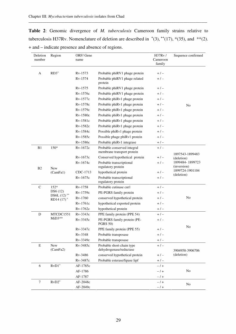
Chapter III: Mycobacterium tuberculosis isolates from Chad __________________________________________________________________________________________
29
Table 2: Genomic divergence of M. tuberculosis Cameroon family strains relative to
tuberculosis H37Rv. Nomenclature of deletion are described in +(3), ++(17), *(35), and **(2).
+ and – indicate presence and absence of regions.
Deletion number
Region ORF/ Gene name
H37Rv / Cameroon
family
Sequence confirmed
Rv-1573 Probable phiRV1 phage protein + / –
Rv-1574 Probable phiRV1 phage related protein
+ / –
Rv-1575 Probable phiRV1 phage protein + / –
Rv-1576c Probable phiRV1 phage protein + / –
Rv-1577c Probable phiRv1 phage protein + / –
Rv-1578c Probable phiRv1 phage protein + / –
Rv-1579c Probable phiRv1 phage protein + / –
Rv-1580c Probable phiRv1 phage protein + / –
Rv-1581c Probable phiRv1 phage protein + / –
Rv-1582c Probable phiRv1 phage protein + / –
Rv-1584c Possible phiRv1 phage protein + / –
Rv-1585c Possible phage phiRv1 protein + / –
A
RD3+
Rv-1586c Probable phiRv1 integrase + / –
No
Rv-1672c Probable conserved integral membrane transport protein
+ / –
Rv-1673c Conserved hypothetical protein + / –
Rv-1674c Probable transcriptional regulatory protein
+ / –
CDC-1713 hypothetical protein + / –
B1
B2
150* New (CamFa1)
Rv-1675c Probable transcriptional regulatory protein
+ / –
1897543-1899483 (deletion) 1899484- 1899723 (inversion) 1899724-1901104 (deletion)
Rv-1758 Probable cutinase cut1 + / –
Rv-1759c PE-PGRS family protein + / –
Rv-1760 conserved hypothetical protein + / –
Rv-1761c hypothetical exported protein + / –
C 152* DS6 (12) DS6L (12) ++ RD14 (17) +
Rv-1762c hypothetical protein + / –
No
Rv-3343c PPE family protein (PPE 54) + / –
Rv-3345c PE-PGRS family protein (PE-PGRS 50)
+ / –
Rv-3347c PPE family protein (PPE 55) + / –
Rv-3348 Probable transposase + / –
D MTCDC1551 MiD3**
Rv-3349c Probable transposase + / –
No
Rv-3485c Probable short-chain type dehydrogenase/reductase
+ / –
Rv-3486 conserved hypothetical protein + / –
E New (CamFa2)
Rv-3487c Probable esterase/lipase lipf + / –
3904958-3906706 (deletion)
AF-1785c – / +
AF-1786 – / +
6 RvD1+
AF-1787 – / +
No
RvD2+ AF-2048c – / + 7
AF-2049c – / + No
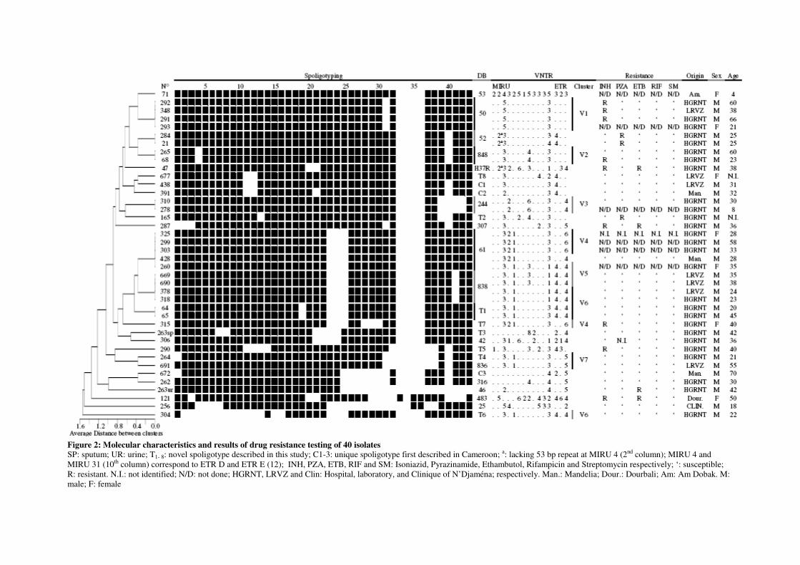
Figure 2: Molecular characteristics and results of drug resistance testing of 40 isolates SP: sputum; UR: urine; T1- 8: novel spoligotype described in this study; C1-3: unique spoligotype first described in Cameroon; a: lacking 53 bp repeat at MIRU 4 (2nd column); MIRU 4 and MIRU 31 (10th column) correspond to ETR D and ETR E (12); INH, PZA, ETB, RIF and SM: Isoniazid, Pyrazinamide, Ethambutol, Rifampicin and Streptomycin respectively; ‘: susceptible; R: resistant. N.I.: not identified; N/D: not done; HGRNT, LRVZ and Clin: Hospital, laboratory, and Clinique of N’Djaména; respectively. Man.: Mandelia; Dour.: Dourbali; Am: Am Dobak. M: male; F: female
Chapter III: Mycobacterium tuberculosis isolates from Chad __________________________________________________________________________________________
31
Reference List
1. Bloom, B. R. and C. J. L. Murray. 1992. Tuberculosis - Commentary on A Reemergent Killer. Science 257:1055-1064.
2. Brodin, P., K. Eiglmeier, M. Marmiesse, A. Billault, T. Garnier, S. Niemann, S. T. Cole, and R.
Brosch. 2002. Bacterial artificial chromosome-based comparative genomic analysis identifies Mycobacterium microti as a natural ESAT-6 deletion mutant. Infect.Immun. 70:5568-5578.
3. Brosch, R., S. V. Gordon, M. Marmiesse, P. Brodin, C. Buchrieser, K. Eiglmeier, T. Garnier, C.
Gutierrez, G. Hewinson, K. Kremer, L. M. Parsons, A. S. Pym, S. Samper, D. van Soolingen, and S. T. Cole. 2002. A new evolutionary scenario for the Mycobacterium tuberculosis complex. Proc.Natl.Acad.Sci.U.S.A 99:3684-3689.
4. Bruchfeld, J., G. Aderaye, I. B. Palme, B. Bjorvatn, S. Ghebremichael, S. Hoffner, and L.
Lindquist. 2002. Molecular epidemiology and drug resistance of Mycobacterium tuberculosis isolates from Ethiopian pulmonary tuberculosis patients with and without human immunodeficiency virus infection. J.Clin.Microbiol. 40:1636-1643.
5. Cadmus, S., S. Palmer, M. Okker, J. Dale, K. Gover, N. Smith, K. Jahans, R. G. Hewinson, and S.
V. Gordon. 2006. Molecular Analysis of Human and Bovine Tubercle Bacilli from a Local Setting in Nigeria. Journal of Clinical Microbiology 44:29-34.
6. Cowan, L. S., L. Mosher, L. Diem, J. P. Massey, and J. T. Crawford. 2002. Variable-number-tandem repeat typing of mycobacterium tuberculosis isolates with low copy numbers of IS6110 by using mycobacterial interspersed repetitive units. Journal of Clinical Microbiology 40:1592-1602.
7. Dias, F., S. G. Michael, S. E. Hoffner, L. Martins, R. Norberg, and G. Kallenius. 1993. Drug susceptibility in Mycobacterium tuberculosis of a sample of patients in Guinea Bissau. Tuber.Lung Dis. 74:129-130.
8. Diguimbaye, C., V. Vincent, E. Schelling, M. Hilty, R. Ngandolo, HH. Mahamat, G. Pfyffer, F.
Baggi, M. Tanner, and J. Zinsstag. 2006. Species identification of non-tuberculous mycobacteria from humans and cattle of Chad. Schw.Arch.Tierheilkunde 148(5):251-6.
9. Fawcett, I. W. and B. J. Watkins. 1976. Initial resistance of Mycobacterium tuberculosis in Northern Nigeria. Tubercle. 57:71-73.
10. Filliol, I., J. R. Driscoll, D. van Soolingen, B. N. Kreiswirth, K. Kremer, G. Valetudie, D. A. Dang,
R. Barlow, D. Banerjee, P. J. Bifani, K. Brudey, A. Cataldi, R. C. Cooksey, D. V. Cousins, J. W.
Dale, O. A. Dellagostin, F. Drobniewski, G. Engelmann, S. Ferdinand, D. Gascoyne-Binzi, M. Gordon, M. C. Gutierrez, W. H. Haas, H. Heersma, E. Kassa-Kelembho, M. L. Ho, A.
Makristathis, C. Mammina, G. Martin, P. Mostrom, I. Mokrousov, V. Narbonne, O. Narvskaya,
A. Nastasi, S. N. Niobe-Eyangoh, J. W. Pape, V. Rasolofo-Razanamparany, M. Ridell, M. L.
Rossetti, F. Stauffer, P. N. Suffys, H. Takiff, J. Texier-Maugein, V. Vincent, J. H. de Waard, C. Sola, and N. Rastogi. 2003. Snapshot of moving and expanding clones of Mycobacterium tuberculosis and their global distribution assessed by spoligotyping in an international study. J.Clin.Microbiol. 41:1963-1970.
11. Fleischmann, R. D., D. Alland, J. A. Eisen, L. Carpenter, O. White, J. Peterson, R. DeBoy, R.
Dodson, M. Gwinn, D. Haft, E. Hickey, J. F. Kolonay, W. C. Nelson, L. A. Umayam, M.
Ermolaeva, S. L. Salzberg, A. Delcher, T. Utterback, J. Weidman, H. Khouri, J. Gill, A. Mikula, W. Bishai, J. W. Jacobs, Jr., J. C. Venter, and C. M. Fraser. 2002. Whole-genome comparison of Mycobacterium tuberculosis clinical and laboratory strains. J.Bacteriol. 184:5479-5490.
12. Frothingham, R. and W. A. Meeker-O'Connell. 1998. Genetic diversity in the Mycobacterium tuberculosis complex based on variable numbers of tandem DNA repeats. Microbiology-Uk 144:1189-1196.
Chapter III: Mycobacterium tuberculosis isolates from Chad __________________________________________________________________________________________
32
13. Garcia-Pelayo, M. C., K. C. Caimi, J. K. Inwald, J. Hinds, F. Bigi, M. I. Romano, D. van
Soolingen, R. G. Hewinson, A. Cataldi, and S. V. Gordon. 2004. Microarray analysis of Mycobacterium microti reveals deletion of genes encoding PE-PPE proteins and ESAT-6 family antigens. Tuberculosis.(Edinb.) 84:159-166.
14. Hilty, M., C. Diguimbaye, E. Schelling, F. Baggi, M. Tanner, and J. Zinsstag. 2005. Evaluation of the discriminatory power of variable number tandem repeat (VNTR) typing of Mycobacterium bovis strains. Vet.Microbiol. 109:217-222.
15. Hirsh, A. E., A. G. Tsolaki, K. DeRiemer, M. W. Feldman, and P. M. Small. 2004. Stable association between strains of Mycobacterium tuberculosis and their human host populations. Proc.Natl.Acad.Sci.U.S.A 101:4871-4876.
16. Kamerbeek, J., L. Schouls, A. Kolk, M. van Agterveld, D. van Soolingen, S. Kuijper, A.
Bunschoten, H. Molhuizen, R. Shaw, M. Goyal, and J. Van Embden. 1997. Simultaneous detection and strain differentiation of Mycobacterium tuberculosis for diagnosis and epidemiology. J.Clin.Microbiol. 35:907-914.
17. Kato-Maeda, M., J. T. Rhee, T. R. Gingeras, H. Salamon, J. Drenkow, N. Smittipat, and P. M.
Small. 2001. Comparing genomes within the species Mycobacterium tuberculosis. Genome Res. 11:547-554.
18. Kent, P. T. and G. P. Kubica. 1985. Public health mycobacteriology- a guide for the level III laboratory. U.S.Departement of health and human Services publication, Atlanta, Ga.
19. Kraus, G., T. Cleary, N. Miller, R. Seivright, A. K. Young, G. Spruill, and H. J. Hnatyszyn. 2001. Rapid and specific detection of the Mycobacterium tuberculosis complex using fluorogenic probes andreal-time PCR. Mol.Cell Probes 15:375-383.
20. Kuaban, C., R. Bercion, J. Noeske, P. Cunin, P. Nkamsse, and N. S. Ngo. 2000. Anti-tuberculosis drug resistance in the West Province of Cameroon. Int.J.Tuberc.Lung Dis. 4:356-360.
21. M'Boussa, J., M. Bendo, F. Yala, R. Kounkou, E. Kaoudi, and F. Portaels. 1998. Etude préliminaire sur la sensibilité des mycobactéries au Centre Hospitalier Universitaire de Brazzaville. Bulletin OCEAC 31.
22. Malik, A. N. and P. Godfrey-Faussett. 2005. Effects of genetic variability of Mycobacterium tuberculosis strains on the presentation of disease. Lancet Infect.Dis. 5:174-183.
23. Mardassi, H., A. Namouchi, R. Haltiti, M. Zarrouk, B. Mhenni, A. Karboul, N. Khabouchi, N. C.
Gey van Pittious, E. M. Streicher, J. Rauzier, B. Gicquel, and K. Dellagi. 2005. Tuberculosis due to resistant Haarlem strain, Tunisia. Emerg.Infect.Dis. 11:957-961.
24. Massenet, D. and O. N. Djemadji. 1994. [Chad: bibliographic review of reported cases]. Med.Trop.(Mars.) 54:179-188.
25. Mostowy, S., D. Cousins, J. Brinkman, A. Aranaz, and M. A. Behr. 2002. Genomic deletions suggest a phylogeny for the Mycobacterium tuberculosis complex. J.Infect.Dis. 186:74-80.
26. Mostowy, S., A. Onipede, S. Gagneux, S. Niemann, K. Kremer, E. P. Desmond, M. Kato-Maeda, and M. Behr. 2004. Genomic analysis distinguishes Mycobacterium africanum. J.Clin.Microbiol. 42:3594-3599.
27. Nguyen, D., P. Brassard, D. Menzies, L. Thibert, R. Warren, S. Mostowy, and M. Behr. 2004. Genomic characterization of an endemic Mycobacterium tuberculosis strain: evolutionary and epidemiologic implications. J.Clin.Microbiol. 42:2573-2580.
28. Niemann, S., S. Rusch-Gerdes, M. L. Joloba, C. C. Whalen, D. Guwatudde, J. J. Ellner, K.
Eisenach, N. Fumokong, J. L. Johnson, T. Aisu, R. D. Mugerwa, A. Okwera, and S. K.
Chapter III: Mycobacterium tuberculosis isolates from Chad __________________________________________________________________________________________
33
Schwander. 2002. Mycobacterium africanum subtype II is associated with two distinct genotypes and is a major cause of human tuberculosis in Kampala, Uganda. J.Clin.Microbiol. 40:3398-3405.
29. Niobe-Eyangoh, S. N., C. Kuaban, P. Sorlin, P. Cunin, J. Thonnon, C. Sola, N. Rastogi, V.
Vincent, and M. C. Gutierrez. 2003. Genetic biodiversity of Mycobacterium tuberculosis complex strains from patients with pulmonary tuberculosis in Cameroon. J.Clin.Microbiol. 41:2547-2553.
30. Niobe-Eyangoh, S. N., C. Kuaban, P. Sorlin, J. Thonnon, V. Vincent, and M. C. Gutierrez. 2004. Molecular characteristics of strains of the cameroon family, the major group of Mycobacterium tuberculosis in a country with a high prevalence of tuberculosis. J.Clin.Microbiol. 42:5029-5035.
31. Rajakumar, K., J. Shafi, R. J. Smith, R. A. Stabler, P. W. Andrew, D. Modha, G. Bryant, P.
Monk, J. Hinds, P. D. Butcher, and M. R. Barer. 2004. Use of genome level-informed PCR as a new investigational approach for analysis of outbreak-associated Mycobacterium tuberculosis isolates. J.Clin.Microbiol. 42:1890-1896.
32. Supply, P., S. Lesjean, E. Savine, K. Kremer, D. van Soolingen, and C. Locht. 2001. Automated high-throughput genotyping for study of global epidemiology of Mycobacterium tuberculosis based on mycobacterial interspersed repetitive units. Journal of Clinical Microbiology 39:3563-3571.
33. Supply, P., E. Mazars, S. Lesjean, V. Vincent, B. Gicquel, and C. Locht. 2000. Variable human minisatellite-like regions in the Mycobacterium tuberculosis genome. Molecular Microbiology 36:762-771.
34. Tosi, C. H., M. N. Ngangro, N. Djimadoum, and V. Richard. 2002. [Study of HIV seroprevalence in patients with pulmonary tuberculosis in 1999 in Chad]. Med.Trop.(Mars.) 62:627-633.
35. Tsolaki, A. G., A. E. Hirsh, K. DeRiemer, J. A. Enciso, M. Z. Wong, M. Hannan, Goguet de la
Salmoniere YO, K. Aman, M. Kato-Maeda, and P. M. Small. 2004. Functional and evolutionary genomics of Mycobacterium tuberculosis: insights from genomic deletions in 100 strains. Proc.Natl.Acad.Sci.U.S.A 101:4865-4870.
36. Tudo, G., J. Gonzalez, R. Obama, J. M. Rodriguez, J. R. Franco, M. Espasa, P. R. Simarro, G. Escaramis, C. Ascaso, A. Garcia, and M. T. Jimenez De Anta. 2004. Study of resistance to anti-tuberculosis drugs in five districts of Equatorial Guinea: rates, risk factors, genotyping of gene mutations and molecular epidemiology. Int.J.Tuberc.Lung Dis. 8:15-22.
37. Van Rie, A., R. Warren, M. Richardson, T. C. Victor, R. P. Gie, D. A. Enarson, N. Beyers, and P.
D. van Helden. 1999. Exogenous reinfection as a cause of recurrent tuberculosis after curative treatment. New England Journal of Medicine 341:1174-1179.
38. van Soolingen, D. 2001. Molecular epidemiology of tuberculosis and other mycobacterial infections: main methodologies and achievements. Journal of Internal Medicine 249:1-26.
39. World Health Organisation (WHO). 2004. Tuberculosis.Fact Sheet N° 104.

Chapter IV: Mycobacterium bovis isolates from Chad
35
Chapter IV: Mycobacterium bovis Isolates from Tuberculous
Lesions in Chadian Zebu Carcasses
Diguimbaye-Djaibé C.1*, Hilty M.2*, Ngandolo R.1, Mahamat HH.1, Pfyffer G. E.3 , Baggi F.4,
Hewinson G.5, Tanner M.2, Zinsstag J.2 and Schelling E.2
1 Laboratoire de Recherches Vétérinaires et Zootechniques de Farcha, N’Djaména, Chad 2 Swiss Tropical Institute, Basel, Switzerland 3 Department of Medical Microbiology, Kantonsspital Luzern, Luzern, Switzerland 4 National Centre for Mycobacteria, University of Zurich, Switzerland 5 Veterinary Laboratories Agency, Weybridge, United Kingdom
Modified and published in Emerging Infectious Diseases 2006;12(5):769-771
*Contributed equally
Chapter IV: Mycobacterium bovis isolates from Chad
36
Abstract
During a prospective study (July to August 2002) at the slaughterhouse in N’Djaména, Chad,
meat inspectors have condemned 727/10’000 cattle carcasses due to tuberculosis-like lesions.
A significantly higher proportion of Mbororo than Arab cattle carcasses were entirely
declared unfit in comparison to partial condemnation of carcasses (33% versus 9%, p =
0.002). Microbiological examination of 201 lesions from 75 Mbororo zebu and 124 Arab
zebu carcasses confirmed bovine tuberculosis for 55 animals by isolation of Mycobacterium
bovis. M. bovis was more often cultured from specimens of Mbororo than of Arab cattle (p =
0.004). Spoligotypes of 53 out of 55 (96.4%) isolates showed lack of the spacer 30 as has
been described for isolates from Cameroon. Our strains were isolated from a slaughterhouse
with a bovine tuberculosis prevalence of 7% and 92.7% of strains were clustered. This
indicates a high recent transmission rate.
Keywords: Mycobacterium bovis, zebu, Arab breed, Mbororo breed, slaughterhouse,
spoligotyping, Chad

Chapter IV: Mycobacterium bovis isolates from Chad
37
Introduction
M. bovis is the causative agent of bovine tuberculosis in livestock such as cattle and farmed
deer. The disease is among these, which may affect trade between countries and is under OIE
(Office International des Epizooties) List B diseases i.e. transmissible diseases that are
considered to be of socio-economic and/or public health importance within countries and that
are significant in the international trade of animals and animal products. According to OIE
records for bovine tuberculosis of the past five years (1998-2003), 31 out of 50 African
countries have reported the occurrence of the disease in their respective countries (1).
Developing countries do not have the same means available for control and elimination as
industrialized countries had few decades ago. However, control measures have been put in
place in 35 of the 50 African countries reporting to OIE.
Furthermore, M. bovis is also recognized as a zoonotic pathogen that infects many people,
particularly in the developing world with highest prevalences of bovine tuberculosis and
HIV/AIDS co-infection (2).
In order to improve the traceability of the M. bovis infections and identify the origin of the
outbreak, different genotyping studies were made. An apparently high level of heterogeneity
of individual strains by the use of spoligotyping was observed in France (3). In contrast,
similar molecular studies in island countries as Great Britain (4) and Australia (5) showed a
low level of heterogeneity suggesting a high recent transmission rate between livestock. For
developing countries, particularly for Africa, few molecular genotyping studies - as
prevalence and incidence surveys in general - have been undertaken. Studies in Cameroon (6)
and Tanzania (7) show low heterogeneity of isolated strains as in Great Britain and Australia.
The slaughterhouse of Farcha (Société Moderne des Abattoirs) in N’Djaména is the largest
one in Chad. Among the 50’000 animals that are slaughtered annually, the main species are
cattle followed by small ruminants (8). The cattle population of Chad was estimated to be
5’595’000 in 2000 and is mainly composed of the zebu breeds (Bos indicus) Arab, Peul,
Mbororo, with the Toupouri and Kouri breeds (Bos taurus) less common. It is estimated that
90% of all slaughtered cattle are of the Arab breed, with 7% Mbororo and 3% Kouri (9).
Several studies in slaughterhouses have demonstrated that tuberculosis is an important cause
of condemnation, causing approximately 9% of all inspected carcasses to be condemned (10).
A retrospective study on causes of condemnation after meat inspection at the slaughterhouse
of Farcha showed that (i) most carcasses with tuberculous lesions were detected between the

Chapter IV: Mycobacterium bovis isolates from Chad
38
months of July and November, and (ii) more Mbororo cattle than animals of other breeds had
tuberculosis-like lesions (42/60 versus 132/1539) (11).
Tuberculin-positive cattle have been detected in Chadian cattle herds (12;13). At the
slaughterhouse, the diagnosis of tuberculosis is mainly based on the typical macroscopic
lesions of the organs rather than on Ziehl-Neelsen stained smears. Confirmation of a
suspected diagnosis of bovine tuberculosis after meat inspection has so far not been
confirmed by the isolation of M. bovis in Chad.
This study was aimed at isolating M. bovis from specimens of Mbororo and Arab cattle in
Chad, at characterizing the strains with molecular methods, and at comparing the isolates with
M. bovis strains from Cameroon (14).
Materials and Methods
1. Carcasses and specimens
At the Farcha slaughterhouse 727/10'000 cattle carcasses (7397 zebu Arab, 2596 zebu
Mbororo and 7 Kouri cattle) were condemned due to tuberculous lesions upon meat
inspection between July 1st and August 31st 2002. A sample from approx. every fourth
condemned carcass was collected between July 11th and August 29th 2002. Specimens from
201 affected organs (lymph nodes, lungs, and liver) from 199 carcasses were transported on
ice to the Chadian National Veterinary and Animal Husbandry Laboratory (Laboratoire de
Recherches Vétérinaires et Zootechniques de Farcha) and stored there at -20 °C prior to
processing. For each specimen, the following information was collected by two trainees:
breed, sex, partial or total condemnation of the carcass, date of collection, and nature of
specimen (15).
2. Specimen processing and cultivation of acid-fast bacilli (AFB)
Specimens were washed three times with sterile, distilled water. Tissue samples were cut into
5 or 6 pieces and put in a sterile plastic bag containing 10 ml of sterile saline for
homogenization. Samples were homogenized in a blender (type STOMACHER 80; Seward
Laboratory Systems, Bristol, U.K.) for 1 min, and repeated three time. Ten millilitres of the
suspension were transferred into a 50 ml conic FALCON® tube for decontamination.
Homogenized suspensions were decontaminated with N-acetyl-L-cysteine sodium hydroxide
(0.5% NALC- 2% NaOH) (16) and inoculated on two Löwenstein-Jensen (LJ) slants
containing a) glycerol (0.75%) and b) pyruvate (0.6%), but no glycerol. In addition,

Chapter IV: Mycobacterium bovis isolates from Chad
39
Middlebrook 7H9 medium containing OADC and PANTA (polymyxin, amphotericin B,
nadilixic acid, trimethoprim, azlocillin) was prepared. Inoculated media were incubated at
37°C (without CO2) for 8 weeks. Smears were prepared with one drop of the sediment after
centrifugation of the homogenized suspensions for detection of AFB by microscopy.
3. Identification of mycobacterial isolates
Growth of mycobacteria was confirmed by smear (stained by the Ziehl-Neelsen method).
AFB-positive colonies were subcultured on 3 LJ slants and a Middlebrook 7H10 agar plate.
Three biochemical tests (nitrate, niacin, and 68°C catalase) (17) were used to identify
mycobacteria and to distinguish between M. tuberculosis complex (MTC) and non-
tuberculous mycobacteria (NTM).
Species identification was performed by real time PCR (16) to confirm MTC isolates.
4. Genotyping of MTC strains
Genotyping of M. tuberculosis complex (MTC) strains was done at the National Centre for
Mycobacteria (NCM) by spoligotyping (18) and IS6110-based analysis of restriction
fragment length polymorphism (RFLP) (19). The latter was carried out with 50 % of
spoligotypes. Spoligotyping of all strains was repeated at the Veterinary Laboratories Agency,
Weybridge, to confirm the results of the first spoligotyping round.
Additionally, the presence of spacers 14 and 15 for 15 strains with weak or absent signals in
spoligotyping was confirmed by a PCR reaction with a primer-pair bridging the DNA region
from spacer 14 (3’ gtgtgatgcggatggtcggctc 5’) to 22 (5’ tgtctcaatcgtgccgtctgcgg 3’) (20). The
PCR reaction mixture contained 1x Taq PCR buffer, deoxynucleoside triphosphates (0.2 mM
each), 1 U of AmpliTaq Gold DNA polymerase (Perkin-Elmer Applied Biosystems), a 0.5
µM concentration of the primer pair and mycobacterial DNA to a final volume of 20 µl. After
10 min at 95°C, the PCR was performed for 40 cycles of 0.5 min at 94°C, 0.5 min at 65°C
and 1 min at 72°C. The reactions were terminated after an incubation of 10 min at 72°C. PCR
fragments were analyzed by agarose gel electrophoresis using 2 % NuSieve agarose. The size
of the amplicons was compared with a positive control of spacers 14 and 15.
5. Statistical analysis
A Chi-square test was used to analyze the co-variables (condemnation, culture growth)
between breeds. A multivariate regression model with M. bovis isolation as the outcome was
adjusted for co-variables. Cluster analysis was done with SAS (Version 8.02 Proc cluster,
USA Statistical Analysis Systems Inc., Cary, NC/ USA). The relationship of clusters to
geographical origin of animals, breed and type of condemnation was done by the Fisher test
(SAS, proc freq).
Chapter IV: Mycobacterium bovis isolates from Chad
40
Results
The overall prevalence of suspect lesions was 7.3%. A significantly higher (p = 0.04)
prevalence was found among Mbororo (8.2%; 212/2596) than Arab cattle (7%; 515/7397)
(15). Lesions were mainly found in the lymph nodes and lungs (Table 1). At the
slaughterhouse and in the sub-sample of 199 animals, entire condemnation of the carcass in
comparison to partial condemnation was observed more often among Mbororo than Arab
cattle (p ≤ 0.001 and p = 0.002) (Table 2). This difference between the two breeds was even
more accentuated in female cattle.
The proportion of positive specimens smears was relatively low (21.6%), with was no
difference evident between the two breeds (Table 3). Most AFB-positive smears originated
from lymph nodes (18%) and lungs (26%), while liver specimens (n=5) were always AFB-
negative. Of 201 specimens which were inoculated onto three types of media, 132 (65.7%)
showed growth of mycobacteria on at least one medium, whereas 55 (27.3%) remained
culture negative. Fourteen (7%) cultures were contaminated (Table 4). Ninety-eight of 161
(61%) AFB smear-negative specimens became culture positive.
Culture morphology and biochemical tests identified 58 MTC and 26 Non-Tuberculosis
Mycobacteria (NTM) strains. Real-time PCR confirmed that 56 strains belonged to MTC and
28 strains were NTM. 55 MTC strains were of the M. bovis spoligotype, while 1 failed to give
a spoligotype pattern. Overall, M. bovis was isolated from more than a fourth of tissues in
which tuberculosis had been suspected and in 42% of all positive cultures. There were
significantly more M. bovis strains isolated from Mbororo zebu (30/75) than from Arab zebu
(26/124) (p = 0.004) (Table 4). The difference remained statistically significant when
including the type of condemnation and type of organ in a multivariate logistic regression
model.
In total, twelve different spoligotypes were found among the 55 M. bovis isolates, with only
four spoligotypes were unique. Eight clusters of spoligotypes were identified. The number of
strains per cluster varied between 22 and 2 (Figure 1). We found that 5/8 clusters were
composed of strains which have been isolated from Mbororo and Arab zebus. The
distribution of the two breeds (Mbororo and Arab) within cluster differ significantly (p <
0.01).
All strains lacked spacers 3, 9, 16, 39-43 which is a characteristic of M. bovis. In addition,
53/55 strains did not have spacer 30. Upon RFLP analysis, the cluster 5 identified by RFLP
Chapter IV: Mycobacterium bovis isolates from Chad
41
was distinct in its spoligotypes (SP5 & 6); however, other RFLP clusters could not be further
distinguished by spoligotypes (Figure 2). For clusters RFLP1, 3b, 5 and 6a, a second band
was visible, while a second band was missing for RFLP2, 3a, 4 and 6b.
All Chadian strains showed a different RFLP pattern when compared with the BCG reference
strain of the NCM Zurich (clinical M. bovis BCG isolate after BCG vaccination at the
paediatric hospital of Zurich in 1999).
Discussion
For the first time, M. bovis has been isolated in specimens from Chad. The prevalence of
tuberculin-positive cattle was 0.8% (95% confidence interval 0.2- 1.4%) in the East (Ouaddaï
region) (12) and 16.9% (95% CI 10.4 – 23.5%) in the West of Chad (Chari- Baguirmi and
Kanem regions) (13). The latter study has been continued with 476 additional cattle in 34
herds and a prevalence of 11.5% (95% CI 6.9 – 18.5%) was found when considering the
herds as a random effect in the model. More tuberculin reactors were found among Mbororo
than Arab zebus (p = 0.02).
Usually, cattle breeds are not restricted to specific geographical zones in Chad; however, a
high proportion of Mbororo cattle was found in the West of the country (21). At Farcha, the
most frequently slaughtered breeds were Mbororo and Arab zebus. Mbororo zebu showed
generalized tuberculosis more often than Arab zebus which was reflected in the data through
a higher proportion of these animals having their entire carcass condemned. Similarly, a
higher susceptibility of Mbororo cattle to tuberculosis infection was also observed in
Cameroon (14). It would be interesting to understand the immunological basis of this
susceptibility in greater depth since it may have a bearing on the development of an improved
livestock vaccine.
Sixty-five percent of specimens with tuberculosis lesions were culture-positive; however,
only one fifth (21%) were smear positive. In this study, the proportion of smear-positive
specimens was low in comparison to Sudan (53%; (22). Spoligotyping was used as a
diagnostic tool, but also yielded important insight into the epidemiology of M. bovis. The
spectrum of spoligotype clusters differed between Mbororo and Arab zebus, but not between
the type of condemnation. Spoligotype clusters could not be related to geographical origin.
However, most of the cattle were bought at local markets and their geographical origin was
not known. The finding that 51/55 isolates (92.7%) were in 8 clusters indicates a substantial

Chapter IV: Mycobacterium bovis isolates from Chad
42
degree of recent transmission, an observation that is underlined when the prevalence of
tuberculosis lesions at the slaughterhouse is considered (7%).
Similar to Cameroonian M. bovis strains, our isolates most often lacked spacer 30, the only
exception being SP11 (Figure 2). A possible explanation for this observation is the cross
border movement of Chadian cattle to Cameroon. As in the study conducted by Njanpop-
Lafourcade et al. in Cameroon (14), the pre-dominant spoligotype in our study is SP1 (Figure
1) with a cluster of 22 strains (40%). SP1 corresponds to the pattern of cluster C1 in the
Cameroonian study and two other clusters described in Cameroon were found in Chad (C1 an
C5, similar respectively to SP2 and SP4 in this work). Certain Cameroonian clusters (C7, C8,
C9 and C10; (14) were only detected in the Adamaoua region but not in northern Cameroon
or in Chad (our study). Apparently, the established measures of the Cameroonian government
to prevent movement of cattle between the Adamaoua and the two regions of the North are
effective. As to the other neighbouring countries, we have not found any publications related
to molecular typing of M. bovis strains.
The comparison of the patterns found in Chad with the M. bovis spoligotype database
(www.mbovis.org) showed that SP1, 2, 4 and 10 have already been described. All other
patterns have never been described (www.bovis.org). All strains lacking spacers 27 and 28
(Figure 1 and Table 5) were isolated from Arab cattle. No other characteristics were observed
within the spoligotypes for strains isolated from Arab and Mbororo cattle. Fifteen strains (8
from Arab and 7 from Mbororo zebu) were typed with the IS6110 method, of which 11 and 4
isolates contained 2 or 1 band, respectively. Therefore, Chadian M. bovis strains belong to
low IS6110 copy number strains. Strains lacking spacer 30 had a band at 1.9 kB, in
accordance to the findings in Cameroon (14). Cousins et al. (5) found that 85 % of M. bovis
strains showed one band which was mainly at 1.9 kB. No association was found between the
number of bands and the cattle breed. IS6110 typing revealed 6 clusters and, thus, is of lower
discriminatory power than spoligotyping. However, spoligopatterns were differentiated by
RFLP due to the number of visible bands. Earlier, Zumarraga et al. (23) concluded that
spoligotyping alone is not sensitive enough for the discrimination of M. bovis strains for in-
depth epidemiological study. It would be interesting to see whether VNTR-typing (24) can
improve strain discrimination.
The recent establishment of the first mycobacterial laboratory in Chad allowed a confirmation
of the presence of bovine tuberculosis in Chadian herds. Future molecular epidemiology
studies are needed to shed light on the transmission of M. bovis between Chadian and
Chapter IV: Mycobacterium bovis isolates from Chad
43
Cameroonian cattle. Furthermore the observed higher susceptibility of Mbororo than Arab
zebus to M. bovis disease should be followed-up by immunological investigations.
Acknowledgements
We thank the technicians of the National Centre for Mycobacteria, the Swiss Tropical
Institute, and the “Laboratoire de Recherches Vétérinaires et Zootechniques de Farcha” who
have contributed to the project. The Swiss National Science Foundation is acknowledged for
financial support. This work received support from the NCCR North-South IP-4. We thank
Véronique Vincent, Institute Pasteur, Paris, for complimentary analyses and Steve Gordon
(VLA Weybridge) for advice and discussion
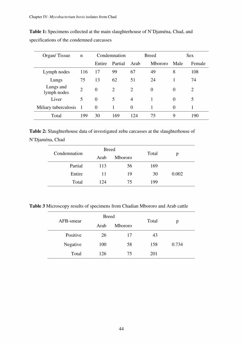
Chapter IV: Mycobacterium bovis isolates from Chad
44
Table 1: Specimens collected at the main slaughterhouse of N’Djaména, Chad, and
specifications of the condemned carcasses
Organ/ Tissue n Condemnation Breed Sex
Entire Partial Arab Mbororo Male Female
Lymph nodes 116 17 99 67 49 8 108
Lungs 75 13 62 51 24 1 74
Lungs and lymph nodes
2 0 2 2 0 0 2
Liver 5 0 5 4 1 0 5
Miliary tuberculosis 1 0 1 0 1 0 1
Total 199 30 169 124 75 9 190
Table 2: Slaughterhouse data of investigated zebu carcasses at the slaughterhouse of
N’Djaména, Chad
Breed Condemnation
Arab Mbororo Total p
Partial 113 56 169
Entire 11 19 30 0.002
Total 124 75 199
Table 3 Microscopy results of specimens from Chadian Mbororo and Arab cattle
Breed AFB-smear
Arab Mbororo Total p
Positive 26 17 43
Negative 100 58 158 0.734
Total 126 75 201
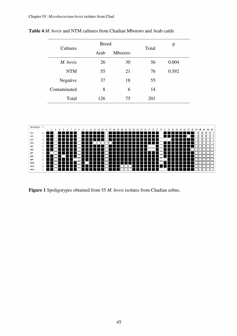
Chapter IV: Mycobacterium bovis isolates from Chad
45
Table 4 M. bovis and NTM cultures from Chadian Mbororo and Arab cattle
Breed p Cultures
Arab Mbororo Total
M. bovis 26 30 56 0.004
NTM 55 21 76 0.592
Negative 37 18 55
Contaminated 8 6 14
Total 126 75 201
Figure 1 Spoligotypes obtained from 55 M. bovis isolates from Chadian zebus.
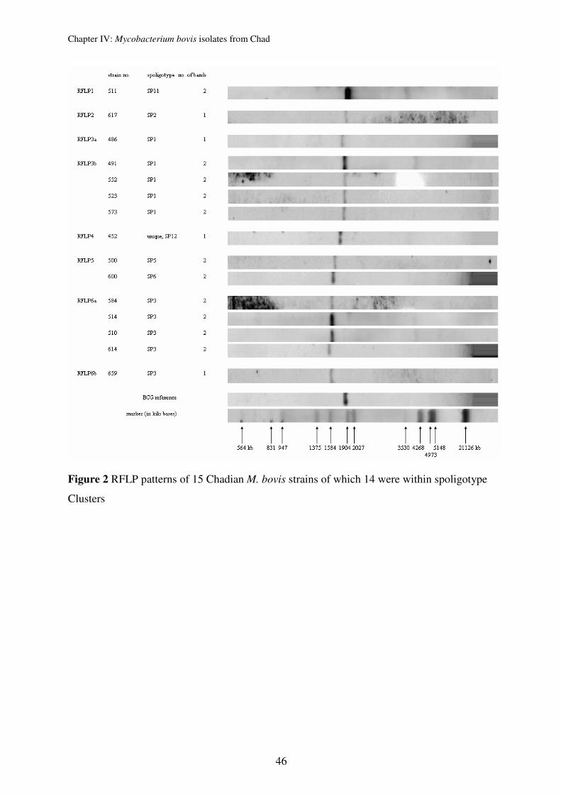
Chapter IV: Mycobacterium bovis isolates from Chad
46
Figure 2 RFLP patterns of 15 Chadian M. bovis strains of which 14 were within spoligotype
Clusters
Chapter IV: Mycobacterium bovis isolates from Chad
47
Reference List
1. Office International des Epizooties. http://www oie int/hs2/report asp. 2005.
2. Cosivi O, Grange JM, Daborn CJ, Raviglione MC, Fujikura T, Cousins D et al. Zoonotic tuberculosis due to Mycobacterium bovis in developing countries. Emerg Infect Dis. 1998;4:59-70.
3. Haddad N, Ostyn A, Karoui C, Masselot M, Thorel MF, Hughes SL et al. Spoligotype diversity of Mycobacterium bovis strains isolated in France from 1979 to 2000. Journal of Clinical Microbiology. 2001;39:3623-32.
4. Clifton-Hadley RS, Inwald J, Hughes S, Palmer N, Sayers AR, Sweeney K et al. Recent advances in DNA fingerprinting using spoligotyping - Epidemiological applications in bovine TB. Journal of the British Cattle Veterinary Association. 1998;6:79-82.
5. Cousins D, Williams S, Liebana E, Aranaz A, Bunschoten A, Van Embden J et al. Evaluation of four DNA typing techniques in epidemiological investigations of bovine tuberculosis. J Clin Microbiol. 1998;36:168-78.
6. Njanpop-Lafourcade BM, Inwald J, Ostyn A, Durand B, Hughes S, Thorel MF et al. Molecular typing of Mycobacterium bovis isolates from Cameroon. J Clin Microbiol. 2001;39:222-27.
7. Kazwala KD, Sinclaire, Challans J, Kambarage DM, Sharp JM, Van Embden J et al. Zoonotic importance of Mycobacterium tuberculosis complex organisms in Tanzania: a molecular biology approach. Actes Editions, Rabat, Morocco. 1997;199-204.
8. Direction de l'Elevage et des Ressources Animales (DERA). Rapport annuel d'Activités 2000. N'Djaména. 2001;1-36.
9. Ministère de l'Elevage. Rapport national sur les ressources zoo génétiques du Tchad. N'Djaména, Tchad. 2003;1-196.
10. Maho A, Mbakasse RN, Boulbaye N. Causes de saisies aux abattoirs du Tchad oriental. LRVZ/F In: Actes des IIIèmes Journées Agro-Sylvo-Pastorales, 29/11 au 03/12/1997, N'Djaména, Tchad. 1999.
11. Maho A, Bornarel P, Hendrix P. Rapport technique: Abattage et motifs de saisie (dominantes pathologiques) aux abattoirs du Tchad: cas de N'Djaména, Ati, Bol, Mongo et Oum Hadjer. LRVZ/F, N'Djaména, Tchad. 1994;1-17.
12. Delafosse A, Goutard F, Thebaud E. Epidémiologie de la tuberculose et de la brucellose des bovins en zone péri-urbaine d'Abéché, Tchad. Rev Elev Med Vet Pays Trop. 2002;55:5-13.
13. Schelling E, Diguimbaye C, Daoud S, Daugla DM, Bidjeh K, Tanner M et al. La tuberculose causée par Mycobacterium bovis : résultats préliminaires obtenus chez les pasteurs nomades Foulbés et Arabes dans le Chari-Baguirmi au Tchad. Sempervira. 2000;44-55.
14. Njanpop-Lafourcade BM, Inwald J, Ostyn A, Durand B, Hughes S, Thorel MF et al. Molecular typing of Mycobacterium bovis isolates from Cameroon. J Clin Microbiol. 2001;39:222-27.
15. Doutoum AM, Toko MA. Mycobactérioses bovines et saisies à l'abattoir de Farcha. Institut Universitaire des Sciences et Techniques d'Abéché (IUSTA). 2002.
16. Kraus G, Cleary T, Miller N, Seivright R, Young AK, Spruill G et al. Rapid and specific detection of the Mycobacterium tuberculosis complex using fluorogenic probes andreal-time PCR. Mol Cell Probes. 2001;15:375-83.
17. Kent PT, Kubica GP. Public health mycobacteriology- a guide for the level III laboratory. U S Departement of health and human Services publication, Atlanta, Ga. 1985.
Chapter IV: Mycobacterium bovis isolates from Chad
48
18. Kamerbeek J, Schouls L, Kolk A, van Agterveld M, van Soolingen D, Kuijper S et al. Simultaneous detection and strain differentiation of Mycobacterium tuberculosis for diagnosis and epidemiology. J Clin Microbiol. 1997;35:907-14.
19. van Embden JD, Cave MD, Crawford JT, Dale JW, Eisenach KD, Gicquel B et al. Strain identification of Mycobacterium tuberculosis by DNA fingerprinting: recommendations for a standardized methodology. J Clin Microbiol. 1993;31:406-9.
20. van Embden JD, van Gorkom T, Kremer K, Jansen R, Der Zeijst BA, Schouls LM. Genetic variation and evolutionary origin of the direct repeat locus of Mycobacterium tuberculosis complex bacteria. J Bacteriol. 2000;182:2393-401.
21. Nfi AN, Ndi C. Bovine tuberculosis at the Animal Research Antenna (ARZ) Bangangte, Western province, Cameroon. Bull Anim Hlth Prod Afri. 1997;45:1-3.
22. Sulieman MS, Hamid ME. Identification of acid fast bacteria from caseous lesions in cattle in Sudan. J Vet Med B Infect Dis Vet Public Health. 2002;49:415-18.
23. Zumarraga MJ, Martin C, Samper S, Alito A, Latini O, Bigi F et al. Usefulness of spoligotyping in molecular epidemiology of Mycobacterium bovis-related infections in South America. J Clin Microbiol. 1999;37:296-303.
24. Sola C, Filliol I, Legrand E, Lesjean S, Locht C, Supply P et al. Genotyping of the Mycobacterium tuberculosis complex using MIRUs: association with VNTR and spoligotyping for molecular epidemiology and evolutionary genetics. Infect Genet Evol. 2003;3:125-33.
Chapter V: Evaluation of VNTR typing of Mycobacterium bovis strains __________________________________________________________________________________________
49
Chapter V: Evaluation of the discriminatory power of Variable
Number Tandem Repeats typing of Mycobacterium bovis strains
Markus Hilty,1 Colette Diguimbaye, 1,2 Esther Schelling,1 Franca Baggi,3 Marcel Tanner1, and
Jakob Zinsstag1
Swiss Tropical Institute1, 4002 Basel, Switzerland,
Laboratoire de Recherches Vétérinaires et Zootechniques de Farcha2, N’Djaména, Chad and
Swiss National Centre for mycobacteria3, Zürich, Switzerland
Published in Veterinary Microbiology 2005 Aug 30;109(3-4):217-22

Chapter V: Evaluation of VNTR typing of Mycobacterium bovis strains __________________________________________________________________________________________
50
Abstract
The discriminatory power of Variable Number of Tandem Repeats (VNTR) typing based on
16 known loci (12 MIRUs, 3 ETRs and VNTR 3232) was assessed for Mycobacterium bovis
strains collected sequentially at the slaughterhouse of N’Djaména, Chad. Of 67 M. bovis
strains analysed, 67 % were clustered. In this study, VNTR typing was highly discriminative
with an overall allelic diversity (ho.a) of 0.922. We defined five loci (ETR A, B, C and MIRU
26, 27) as highly (h > 0.25), two loci (MIRU 4, and VNTR 3232) as moderately (0.11 < h <
0.25) and three loci (MIRU 16, 20, 31) as poorly (0.01 < h < 0.11) discriminative. Six loci
(MIRU 2, 10, 23, 24, 39, and 40) showed no polymorphism at all. VNTR typing of the five
highly discriminative loci (h = 0.917) proved to be most appropriate for first line typing of M.
bovis strains of Chad and superior than spoligotyping (hsp. = 0.789). In contrast to M.
tuberculosis strains, a consensus on VNTR loci needs to be found for M. bovis strains. The
selection of a generally agreed set of VNTR loci for molecular discrimination of M. bovis in
different geographical settings is discussed.
Keywords: Mycobacterium bovis; VNTR typing; spoligotyping
Chapter V: Evaluation of VNTR typing of Mycobacterium bovis strains __________________________________________________________________________________________
51
Introduction
Bovine tuberculosis is caused by a member of the Mycobacterium tuberculosis complex,
Mycobacterium bovis. The disease has one of the broadest host ranges (O’Reilly and Daborn,
1995) and leads to economic losses in livestock production and agriculture in many countries.
In addition, M. bovis is a classical zoonosis and was the main reason for the heavy promotion
of pasteurization in the early 20th century (Pritchard, 1988). Today, bovine tuberculosis
affects mainly people in developing countries but its role in the human TB epidemic fostered
by HIV/AIDS is not known (Cosivi et al., 1998). This is mainly due to the lack or inabilities
of laboratories in developing countries to isolate and diagnose M. bovis. In addition, only few
epidemiological studies aiming at identifying the proportion of M. bovis infection among
tuberculosis patients have been conducted in developing countries (Cosivi et al., 1995). The promotion of prevention of transmission of M. bovis is often hampered by the lack of
epidemiological data. With the recent description of molecular epidemiological tools, the
possibilities for finding answers to key questions like the importance and risk factors of inter-
bovine transmission and the role of wild animals as reservoirs are improved.
Insertion sequence 6110 restriction fragment length polymorphism (IS6110 RFLP) analysis is
the current “gold standard” and the most widely applied typing method for molecular
epidemiology of the M. tuberculosis complex (van Soolingen, 2001). However, this typing
method has important drawbacks as the method requires previous extensive strain cultivation
is technically demanding and expensive and results are difficult to compare between
laboratories because comparison of profiles requires sophisticated software for image analysis
and well trained users. Furthermore, the discrimination of strains with a low number of
IS6110 copies is insufficient, and this is especially true for M. bovis strains (Serraino et al.,
1999 and Kremer et al., 1999).
The spoligotyping (“spacer oligonucleotide typing”) method describes the presence or
absence of 43 spacer DNA sequences between direct repeats (DR) in the DR region of
tuberculosis complex (TBC) strains. This well established PCR-based method does not
require extensive culturing is more discriminative than RFLP for strains with no or few
copies of IS6110 and classifies the members of M. tuberculosis complex with a high level of
confidence (Kamerbeek et al., 1997).
Chapter V: Evaluation of VNTR typing of Mycobacterium bovis strains __________________________________________________________________________________________
52
The variable number tandem repeat (VNTR) typing is a recently developed PCR-based
method without requiring large quantities of DNA. Sequences including exact tandem repeats
(ETR) (Frothingham and Meeker-O’Connell, 1998), mycobacterial interspersed repetitive
units (MIRUs) (Supply et al., 2000 and Mazars et al., 2001) and two sets of Queen's
University Belfast (QUB) VNTRs (Roring et al., 2002 and Skuce et al., 2002) were presented.
The two main advantages of VNTR typing are: (i) the good stability of markers, and (ii)
results are easily comparable between laboratories (digital expression of results). VNTR
typing is highly discriminative for M. tuberculosis and has also the potential to be the method
of choice for M. bovis typing. However, it is not yet standardized and allelic diversity of loci
can vary from country to country and between M. tuberculosis complex species.
In this study, different VNTRs were used on a panel of 67 M. bovis strains isolated from 2000
to 2002 in Chad (Diguimbaye, unpublished data). The objective was to asses the
discriminatory power of each polymorphic locus and in combination with others to evaluate
the most appropriate combination of VNTRs for molecular-epidemiological studies of M.
bovis in Chad.
Materials and methods
Bacterial strains and spoligotyping
Sixty-seven M. bovis strains were isolated from the continuously collected samples from the
slaughterhouse in N’Djaména, Chad. Tuberculous lesions of carcasses and organs were from
two different zebu (Bos indicus) cattle breeds: Arab and Mbororo. Strains (8, 6 and 53) were
isolated in 2000, 2001 and 2002, respectively. All 67 strains were characterized by
spoligotyping (Kamerbeek et al., 1997).
VNTR–PCR analysis
The reaction mixture for all loci contained 1× Taq PCR buffer, deoxynucleoside triphosphates
(0.2 mM each), 1 U of AmpliTaq Gold DNA polymerase (Perkin-Elmer Applied
Biosystems), a 0.5 µM concentration of the primer pairs and mycobacterial DNA in a final
volume of 20 µl. 12 MIRU, 3 ETR and VNTR 3232 primer pairs were used (Cowan et al.,
2002 and FMO’Connell, 1998’Connell, 1998; Roring et al., 2002).
The reaction was carried out with a Perkin-Elmer 9600 cycler starting at a denaturing step of
10 min at 95 °C. After denaturation, the PCR was performed for 40 cycles of 0.5 min at
94 °C, 0.5 min at 65 °C and 1 min at 72 °C. The reactions were terminated by an incubation
of 10 min at 72 °C. PCR fragments were analyzed by agarose (Sigma) gel electrophoresis
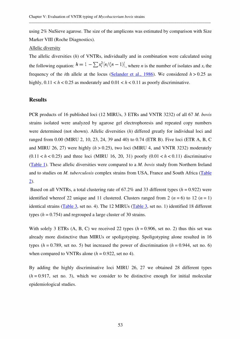
Chapter V: Evaluation of VNTR typing of Mycobacterium bovis strains __________________________________________________________________________________________
53
using 2% NuSieve agarose. The size of the amplicons was estimated by comparison with Size
Marker VIII (Roche Diagnostics).
Allelic diversity
The allelic diversities (h) of VNTRs, individually and in combination were calculated using
the following equation: , where n is the number of isolates and xi the
frequency of the ith allele at the locus (Selander et al., 1986). We considered h > 0.25 as
highly, 0.11 < h < 0.25 as moderately and 0.01 < h < 0.11 as poorly discriminative.
Results
PCR products of 16 published loci (12 MIRUs, 3 ETRs and VNTR 3232) of all 67 M. bovis
strains isolated were analyzed by agarose gel electrophoresis and repeated copy numbers
were determined (not shown). Allelic diversities (h) differed greatly for individual loci and
ranged from 0.00 (MIRU 2, 10, 23, 24, 39 and 40) to 0.74 (ETR B). Five loci (ETR A, B, C
and MIRU 26, 27) were highly (h > 0.25), two loci (MIRU 4, and VNTR 3232) moderately
(0.11 < h < 0.25) and three loci (MIRU 16, 20, 31) poorly (0.01 < h < 0.11) discriminative
(Table 1). These allelic diversities were compared to a M. bovis study from Northern Ireland
and to studies on M. tuberculosis complex strains from USA, France and South Africa (Table
2).
Based on all VNTRs, a total clustering rate of 67.2% and 33 different types (h = 0.922) were
identified whereof 22 unique and 11 clustered. Clusters ranged from 2 (n = 6) to 12 (n = 1)
identical strains (Table 3, set no. 4). The 12 MIRUs (Table 3, set no. 1) identified 18 different
types (h = 0.754) and regrouped a large cluster of 30 strains.
With solely 3 ETRs (A, B, C) we received 22 types (h = 0.906, set no. 2) thus this set was
already more distinctive than MIRUs or spoligotyping. Spoligotyping alone resulted in 16
types (h = 0.789, set no. 5) but increased the power of discrimination (h = 0.944, set no. 6)
when compared to VNTRs alone (h = 0.922, set no 4).
By adding the highly discriminative loci MIRU 26, 27 we obtained 28 different types
(h = 0.917, set no. 3), which we consider to be distinctive enough for initial molecular
epidemiological studies.
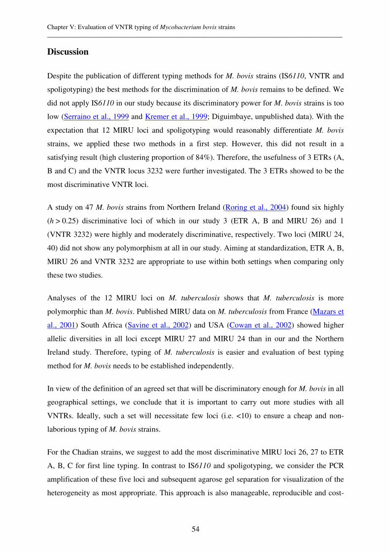
Chapter V: Evaluation of VNTR typing of Mycobacterium bovis strains __________________________________________________________________________________________
54
Discussion
Despite the publication of different typing methods for M. bovis strains (IS6110, VNTR and
spoligotyping) the best methods for the discrimination of M. bovis remains to be defined. We
did not apply IS6110 in our study because its discriminatory power for M. bovis strains is too
low (Serraino et al., 1999 and Kremer et al., 1999; Diguimbaye, unpublished data). With the
expectation that 12 MIRU loci and spoligotyping would reasonably differentiate M. bovis
strains, we applied these two methods in a first step. However, this did not result in a
satisfying result (high clustering proportion of 84%). Therefore, the usefulness of 3 ETRs (A,
B and C) and the VNTR locus 3232 were further investigated. The 3 ETRs showed to be the
most discriminative VNTR loci.
A study on 47 M. bovis strains from Northern Ireland (Roring et al., 2004) found six highly
(h > 0.25) discriminative loci of which in our study 3 (ETR A, B and MIRU 26) and 1
(VNTR 3232) were highly and moderately discriminative, respectively. Two loci (MIRU 24,
40) did not show any polymorphism at all in our study. Aiming at standardization, ETR A, B,
MIRU 26 and VNTR 3232 are appropriate to use within both settings when comparing only
these two studies.
Analyses of the 12 MIRU loci on M. tuberculosis shows that M. tuberculosis is more
polymorphic than M. bovis. Published MIRU data on M. tuberculosis from France (Mazars et
al., 2001) South Africa (Savine et al., 2002) and USA (Cowan et al., 2002) showed higher
allelic diversities in all loci except MIRU 27 and MIRU 24 than in our and the Northern
Ireland study. Therefore, typing of M. tuberculosis is easier and evaluation of best typing
method for M. bovis needs to be established independently.
In view of the definition of an agreed set that will be discriminatory enough for M. bovis in all
geographical settings, we conclude that it is important to carry out more studies with all
VNTRs. Ideally, such a set will necessitate few loci (i.e. <10) to ensure a cheap and non-
laborious typing of M. bovis strains.
For the Chadian strains, we suggest to add the most discriminative MIRU loci 26, 27 to ETR
A, B, C for first line typing. In contrast to IS6110 and spoligotyping, we consider the PCR
amplification of these five loci and subsequent agarose gel separation for visualization of the
heterogeneity as most appropriate. This approach is also manageable, reproducible and cost-

Chapter V: Evaluation of VNTR typing of Mycobacterium bovis strains __________________________________________________________________________________________
55
effective for a laboratory in the South. However, for in-depth studies we recommend not only
to use all polymorphic loci (ETR A, B, C; MIRU 4, 16, 20, 26, 27, 31; and VNTR 3232) but
also spoligotyping because this ensures the highest power of discrimination.
The results of this study are used within a molecular epidemiology study aiming at
identifying risk factors for recent transmission of M. bovis. Results will also clarify the trade
and trans-border translocations of cattle between Cameroon and Chad. In both countries, M.
bovis strains lacking DR spacer 30 have been found (Diguimbaye, unpublished data;
Njanpop-Lafourcade et al., 2001). Improved discrimination of strains is needed for better
tracing back of animals. Furthermore, VNTR typing ensures a good quality control for
laboratory cross-contaminations of M. bovis cultures especially useful for laboratories with
infrastructural restraints.
In conclusion, this work illustrates the necessity of defining the appropriate combination of
heterogenic loci for each country and M. tuberculosis complex panel studied and therefore
advises caution in comparing results of different studies. Furthermore, a first line and in-depth
panel of heterogenic loci is suggested for M. bovis typing in Chad. Once more studies from
other settings are available, a set of most discriminatory VNTRs could be agreed on for M.
bovis strains.
Acknowledgements
We thank the technicians under the supervision of Dr. F. Baggi of the National Centre for
Mycobacteria, Zurich, who have contributed greatly to the project. The NCCR North-South
IP-4 is acknowledged for financial support. S.V. Gordon of the Veterinary laboratories
agencies, Weybridge, is thanked for complementary analyses and critical review.
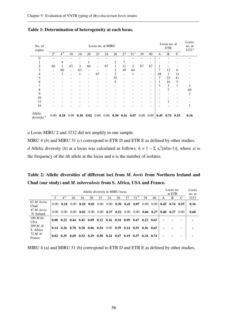
Chapter V: Evaluation of VNTR typing of Mycobacterium bovis strains __________________________________________________________________________________________
56
Table 1: Determination of heterogeneity at each locus.
Locus no. at MIRU Locus no. at
ETR
Locus no. at 3232 a
No. of copies
2a 4 b 10 16 20 23 24 26 27 31 c 39 40 A B C 0 - - - - - - - - - - - - - - - - 1 - 4 - - 1 - - 2 7 - - - - - - - 2 66 1 67 3 66 - 67 1 11 2 67 67 1 - - - 3 - 60 - 63 - - - 2 49 64 - - 7 12 6 - 4 - 2 - 1 - 67 - 2 - 1 - - 48 3 14 - 5 - - - - - - - 55 - - - - 7 25 41 - 6 - - - - - - - 5 - - - - 1 16 5 - 7 - - - - - - - - - - - - 3 3 1 3 8 - - - - - - - - - - - - - 7 - 60 9 - - - - - - - - - - - - - - - 2
10 - - - - - - - - - - - - - - - - 12 - - - - - - - - - - - - - 1 - - 16
-
-
-
-
-
-
-
-
-
-
-
-
-
-
-
1
Allelic diversity d
0.00 0.18 0.00 0.10 0.02 0.00 0.00 0.30 0.41 0.07 0.00 0.00 0.45 0.74 0.55 0.16
a Locus MIRU 2 and 3232 did not amplify in one sample.
MIRU 4 (b) and MIRU 31 (c) correspond to ETR D and ETR E as defined by other studies.
d Allelic diversity (h) at a locus was calculated as follows: h = 1 – Σ xi2[n/(n-1)], where xi is
the frequency of the ith allele at the locus and n is the number of isolates.
Table 2: Allelic diversities of different loci from M. bovis from Northern Ireland and
Chad (our study) and M. tuberculosis from S. Africa, USA and France.
Allelic diversity in MIRU locus Locus no.
at ETR 2 4 a 10 16 20 23 24 26 27 31 b 39 40 A B C
Locus no. at 3232
67 M. bovis Chad
0.00 0.18 0.00 0.10 0.02 0.00 0.00 0.30 0.41 0.07 0.00 0.00 0.45 0.74 0.55 0.16
47 M. bovis
N. Ireland 0.00 0.00 0.00 0.02 0.00 0.00 0.37 0.52 0.00 0.00 0.06 0.27 0.40 0.37 0.00 0.60
180 M.tbc USA
0.08 0.22 0.44 0.42 0.09 0.12 0.16 0.54 0.09 0.47 0.22 0.63 - - - -
209 M. tb S. Africa
0.14 0.26 0.70 0.28 0.06 0.54 0.00 0.59 0.14 0.55 0.36 0.65 - - - -
72 M. tb France
0.02 0.35 0.69 0.52 0.29 0.58 0.24 0.67 0.19 0.37 0.34 0.74 - - - -
MIRU 4 (a) and MIRU 31 (b) correspond to ETR D and ETR E as defined by other studies.
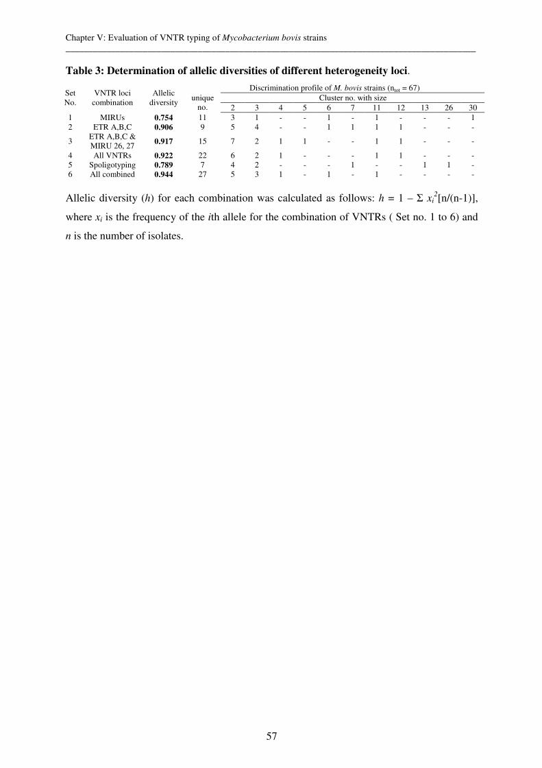
Chapter V: Evaluation of VNTR typing of Mycobacterium bovis strains __________________________________________________________________________________________
57
Table 3: Determination of allelic diversities of different heterogeneity loci.
Discrimination profile of M. bovis strains (ntot = 67) Cluster no. with size
Set No.
VNTR loci combination
Allelic diversity
unique no. 2 3 4 5 6 7 11 12 13 26 30
1 MIRUs 0.754 11 3 1 - - 1 - 1 - - - 1 2 ETR A,B,C 0.906 9 5 4 - - 1 1 1 1 - - -
3 ETR A,B,C & MIRU 26, 27
0.917 15 7 2 1 1 - - 1 1 - - -
4 All VNTRs 0.922 22 6 2 1 - - - 1 1 - - - 5 Spoligotyping 0.789 7 4 2 - - - 1 - - 1 1 - 6 All combined 0.944 27 5 3 1 - 1 - 1 - - - -
Allelic diversity (h) for each combination was calculated as follows: h = 1 – Σ xi
2[n/(n-1)],
where xi is the frequency of the ith allele for the combination of VNTRs ( Set no. 1 to 6) and
n is the number of isolates.
Chapter V: Evaluation of VNTR typing of Mycobacterium bovis strains __________________________________________________________________________________________
58
References
Cosivi et al., 1995 O. Cosivi, F.X. Meslin, C.J. Daborn and J.M. Grange, Epidemiology of Mycobacterium bovis infection in animals and humans, with particular reference to Africa, Rev. Sci. Tech. 14 (1995), pp. 733–746.
Cosivi et al., 1998 O. Cosivi, J.M. Grange, C.J. Daborn, M.C. Raviglione, T. Fujikura, D. Cousins, R.A. Robinson, H.F. Huchzermeyer, I. de Kontor and F.X. Meslin, Zoonotic tuberculosis due to Mycobacterium bovis in developing countries, Emerg. Infect. Dis. 4 (1998), pp. 59–70.
Cowan et al., 2002 L.S. Cowan, L. Mosher, L. Diem, J.P. Massey and J.T. Crawford, Variable-number-tandem repeat typing of mycobacterium tuberculosis isolates with low copy numbers of IS6110 by using mycobacterial interspersed repetitive units, J. Clin. Microbiol. 40 (2002), pp. 1592–1602.
FMO’Connell, 1998 R. Frothingham and W.A. Meeker-O’Connell, Genetic diversity in the Mycobacterium
tuberculosis complex based on variable numbers of tandem DNA repeats, Microbiology UK 144 (1998), pp.
Kamerbeek et al., 1997 J. Kamerbeek, L. Schouls, A. Kolk, M. van Agterveld, D. van Soolingen, S. Kuijper, A. Bunschoten, H. Molhuizen, R. Shaw, M. Goyal and J. van Embden, Simultaneous detection and strain differentiation of Mycobacterium tuberculosis for diagnosis and epidemiology, J. Clin. Microbiol. 35 (1997), pp. 907–914.
Kremer et al., 1999 K. Kremer, D. van Soolingen, R. Frothingham, W.H. Haas, P.W.M. Hermans, C. Martin, P. Palittapongarnpim, B.B. Plikaytis, L.W. Riley, M.A. Yakrus, J.M. Musser and J.D.A. van Embden, Comparison of methods based on different molecular epidemiological markers for typing of Mycobacterium tuberculosis complex strains: interlaboratory study of discriminatory power and reproducibility, J. Clin. Microbiol. 37 (1999), pp. 2607–2618.
Mazars et al., 2001 E. Mazars, S. Lesjean, A.L. Banuls, M. Gilbert, V. Vincent, B. Gicquel, M. Tibayrenc, C. Locht and P. Supply, High-resolution minisatellite-based typing as a portable approach to global analysis of Mycobacterium tuberculosis molecular epidemiology, Proc. Natl. Acad. Sci. U.S.A. 98 (2001), pp. 1901–1906.
Njanpop-Lafourcade et al., 2001 B.M. Njanpop-Lafourcade, J. Inwald, A. Ostyn, B. Durand, S. Hughes, M.F. Thorel, G. Hewinson and N. Haddad, Molecular typing of Mycobacterium bovis isolates from Cameroon, J.
Clin. Microbiol. 39 (2001), pp. 222–227.
O’Reilly and Daborn, 1995 L.M. O’Reilly and C.J. Daborn, The epidemiology of Mycobacterium bovis infections in animals and man: a review, Tuber. Lung Dis. 76 (1995) (Suppl 1), pp. 1–46.
Pritchard, 1988 D.G. Pritchard, A century of bovine tuberculosis 1888–1988: conquest and controversy, J.
Comp. Pathol. 99 (1988), pp. 357–399.
Roring et al., 2002 S. Roring, A. Scott, D. Brittain, I. Walker, G. Hewinson, S. Neill and R. Skuce, Development of variable-number tandem repeat typing of Mycobacterium bovis: comparison of results with those obtained by using existing exact tandem repeats and spoligotyping, J. Clin. Microbiol. 40 (2002), pp. 2126–2133.
Roring et al., 2004 S. Roring, A.N. Scott, H.R. Glyn, S.D. Neill and R.A. Skuce, Evaluation of variable number tandem repeat (VNTR) loci in molecular typing of Mycobacterium bovis isolates from Ireland, Vet. Microbiol. 101 (2004), pp. 65–73.
Savine et al., 2002 E. Savine, R.M. Warren, G.D. van der Spuy, N. Beyers, P.D. van Helden, C. Locht and P. Supply, Stability of variable-number tandem repeats of mycobacterial interspersed repetitive units from 12 loci in serial isolates of Mycobacterium tuberculosis, J. Clin. Microbiol. 40 (2002), pp. 4561–4566.
Selander et al., 1986 R.K. Selander, D.A. Caugant, H. Ochman, J.M. Musser, M.N. Gilmour and T.S. Whittam, Methods of multilocus enzyme electrophoresis for bacterial population genetics and systematics, Appl. Environ.
Microbiol. 51 (1986), pp. 873–884.

Chapter V: Evaluation of VNTR typing of Mycobacterium bovis strains __________________________________________________________________________________________
59
Serraino et al., 1999 A. Serraino, G. Marchetti, V. Sanguinetti, M.C. Rossi, R.G. Zanoni, L. Catozzi, A. Bandera, W. Dini, W. Mignone, F. Franzetti and A. Gori, Monitoring of transmission of tuberculosis between wild boars and cattle: genotypical analysis of strains by molecular epidemiology techniques, J. Clin. Microbiol. 37 (1999), pp. 2766–2771.
Skuce et al., 2002 R.A. Skuce, T.P. McCorry, J.F. McCarroll, S.M.M. Roring, A.N. Scott, D. Brittain, S.L. Hughes, R.G. Hewinson and S.D. Neill, Discrimination of Mycobacterium tuberculosis complex bacteria using novel VNTR–PCR targets, Microbiology-Sgm 148 (2002), pp. 519–528.
Supply et al., 2000 P. Supply, E. Mazars, S. Lesjean, V. Vincent, B. Gicquel and C. Locht, Variable human minisatellite-like regions in the Mycobacterium tuberculosis genome, Mol. Microbiol. 36 (2000), pp. 762–771.
van Soolingen, 2001 D. van Soolingen, Molecular epidemiology of tuberculosis and other mycobacterial infections: main methodologies and achievements, J. Intern. Med. 249 (2001), pp. 1–26.

Chapter VI: Population structure of Mycobacterium bovis from a high incidence country __________________________________________________________________________________________
61
Chapter VI: Population structure of Mycobacterium bovis from a
high incidence country: Implications for molecular epidemiology
and design of diagnostic candidates
Markus Hilty,1 Stephen V. Gordon3, Carmenchu Garcia-Pelayo3, Javier Nunez-Garcia3,
Colette Diguimbaye, 2 Simeon Cadmus,4 Esther Schelling,1 R. Glyn Hewinson3, and Jakob
Zinsstag1
Swiss Tropical Institute1, 4002 Basel, Switzerland,
Laboratoire de Recherches Vétérinaires et Zootechniques2 de Farcha, N’Djaména, Chad, and
Veterinary Laboratories Agency3, Weybridge, New Haw, Surrey, England
University of Ibadan4, Ibadan, Nigeria
Draft paper
Chapter VI: Population structure of Mycobacterium bovis from a high incidence country __________________________________________________________________________________________
62
Abstract
By using microarray-based comparative genomics, large genomic deletions define 63 and 4
M. bovis isolates from Chad as 2 different clones, respectively. The implications for the
molecular epidemiology and the choice of antigens used in a diagnostic mix for M.
tuberculosis complex are discussed.
Chapter VI: Population structure of Mycobacterium bovis from a high incidence country __________________________________________________________________________________________
63
Microarry based comparative analysis of members of the Mycobacterium tuberculosis
complex has defined a set of chromosomal deletions which mark all descendants of an
ancestral strain (1,8,9,12,14,19). This has allowed ‘diagnostic’ deletions to be identified that
allow an isolate to be unequivocally placed in a strain family. The value of genomic deletions
for finding large family groups has been found to be superior to all other genotyping methods
(e.g. Spoligotyping) (6). Additionally, knowledge of the genome organization across M.
tuberculosis strains allows us to inform the choice of antigens used in a diagnostic mix.
Based on previous molecular typing data that allowed us to define phylogenetic extremes (3),
the genomic DNA of 4 M. bovis strains from Chad with the most divergent spoligotypes
(Table 1) were applied to an M. tuberculosis amplicon-array and fluorescence scanned with
an Affymetrix 428 scanner as described (5). Data were analysed using GeneSpring 5.0
(Silicon Genetics, Redwood City,CA) and Mathematica (Wolfram Research). A cut-off for
the normalised test/control ratio of <2.0 was used and results were compared to the control to
create gene deletion lists.
Clone 1
For two M. bovis strains with spoligotypes 1 and 6 (Table 1), lacking spacer 30, microarray
analysis revealed one large genomic deletion, which is described for the first time in this
study. Subsequently, primers for amplification of the flanking regions of the deletion were
designed (664100 5’ actggaccggcaacgacctgg and 669951-5’cgggtgaccgtgaactgcgac). By
performing PCR reactions as described (1) for all 67 M. bovis strains from Chad and 15
strains from Nigeria (2) and following gel electrophoresis, the size of the amplicons could be
estimated by comparison with Size Marker VIII (Roche). PCR products of the two strains
analysed by microarray were sequenced with an ABI Prism 310 Genetic Analysis System.
Sequence characterization revealed a deletion of 5320 bp (664281-669600 bp), removing
Rv572c-574c and parts of Rv571c and Rv575c. The flanking regions of the deletion showed
no similarity to insertion sequences and occurred in a non variable region. The sequences of
the junction regions were exactly the same for the two strains analyzed.
63/67 and all the Nigerian strains lacked this deletion while this region wasn’t deleted for
4/67 M. bovis strains from Chad which lacked spacer 22-24. From this analysis and from the
fact that the population structure of M. bovis is strictly clonal (1), we conclude that most
(63/67) of the Chadian and all of the Nigerian M. bovis strains belong to the same clone and
therefore must have derived from a single, common cell in the past (18). Additionally, we
assume that also the M. bovis strains from Cameroon belong to this family as they also all
lack spacer 30 although we didn’t check these strains for the deletion. The reasons why there
Chapter VI: Population structure of Mycobacterium bovis from a high incidence country __________________________________________________________________________________________
64
is so little heterogeneity within strains of these countries can be many. For M. tuberculosis the
finding that one strain is causing most of the disease cases in a population would confirm the
suspicion of an outbreak and imply a failure to stop transmission (10). Indeed control
programs e.g. vaccination campaigns of test and slaughter policies to stop recent transmission
are basically inexistent in Chad, Cameroon and Nigeria. However, the existence of
homogeneity of these strains can also mean that there is little competition of other strain
families or that this particular strain family is especially good adapted to host and/or the
environment. In a recent study was shown, that Mbororo are more susceptible to the Chadian
strains than the Arabe breed (3).
The fact that most of the Chadian and Nigerian strains belong to one cluster raises important
implications for molecular epidemiological studies. Standard molecular typing tools may not
be discriminative enough to distinguish between epidemiologically unlinked strains. In a
recent study, a minimum of 5 VNTR loci was defined resulting in a sufficient discriminatory
power (7). However, spoligotyping of strains of Nigeria, Cameroon and Chad showed the
same genotypes for some of the strains. Although we know that the pastoralists in this area
migrate long distances we think that it is more likely that most of these strains have no direct
epidemiological link. If we exclude convergent evolution, we think a clonal expansion
scenario of some subclones as shown in a recent study from the UK (18), is most likely.
However, we cannot draw any conclusion which country is the origin and which is the
direction of expansion.
Clone 2
While it is known for M. bovis to have lost the ESAT-6 family proteins esxOP (RD 5) and
esxVW (RD 8) (Table 1) 2/4 of our M. bovis strains analyzed by comparative genomics had
additionally the deletions of Rv3019c/Rv3020c (esxSR) and Rv3890/3891 (esxCD). We
confirmed this finding by DNA sequencing and revealed deletions of 2439 bp (esxSR) and
8077 bp (esxCD) compared to H37Rv. Subsequent PCR reactions on all 67 strains isolated in
Chad showed the presence of these deletions in 4 strains.
ESAT-6 family proteins are encoded by 23 genes (esxA-W) and are promising targets for new
diagnostics and novel vaccine candidates. The hallmark members of this family, esxA (ESAT-
6) and esxB (CFP-10) are absent from M. bovis BCG (due to the RD1 deletion) and have
therefore been extensively studied for their role in virulence and for their diagnostic utility
(6,16). Additionally, proteins encoded by esxH (TB10.4) and its homologues esxR (TB10.3)
and esxQ (TB12.9) are strongly recognized T-cell antigens in humans and animals (17).
Chapter VI: Population structure of Mycobacterium bovis from a high incidence country __________________________________________________________________________________________
65
However, when choosing vaccine or diagnostic candidates it is important to survey clinical
isolates for variation in these targets. Deletion of esxSR has been previously described in M.
tuberculosis strains from France (EMBL accession AJ583832) (11) and the UK (15) and in
M. microti (EMBL accession AJ550619) (5). To our surprise, using the primers as described
(5), sequencing revealed that the deletion of esxSR in our M. bovis strains from Chad had
occurred at exactly the same position to that previously reported for M. tuberculosis and M.
microti, suggesting that this locus is prone to deletion events due to the highly repetitive
PE/PPE gene sequences that flank the region. However, we also uncovered a deletion of
esxCD which isn’t the first time found to be deleted too. However, the position of the deletion
characterized in our study doesn’t equal the one found (13). Adding these new deletions to
previous findings (Table 1) shows the high variability of ESAT-6 family members in the M.
tuberculosis complex.
In the study of Gao et al (4), transcriptome analysis across 10 clinical isolates of M.
tuberculosis revealed that esx genes vary in their expression levels between strains. The
deletion and variable expression of genes encoding ESAT members suggests that they may be
under strong selective pressures linked to immune escape. Hence while ESAT family
members are potent antigens, genome analysis has revealed that their encoding genes are
frequently deleted, suggesting caution in their use as stand-alone diagnostic reagents.
Knowledge of the genome organization across M. tuberculosis strains therefore allows us to
inform the choice of antigens used in a diagnostic mix.
Acknowledgments
We thank the technicians of the Swiss National Centre for Mycobacteria, the Swiss Tropical
Institute, and the “Laboratoire de Recherches Vétérinaires et Zootechniques de Farcha” who
have contributed to the project. NCCR North-South IP-4 is acknowledged for financial
support.
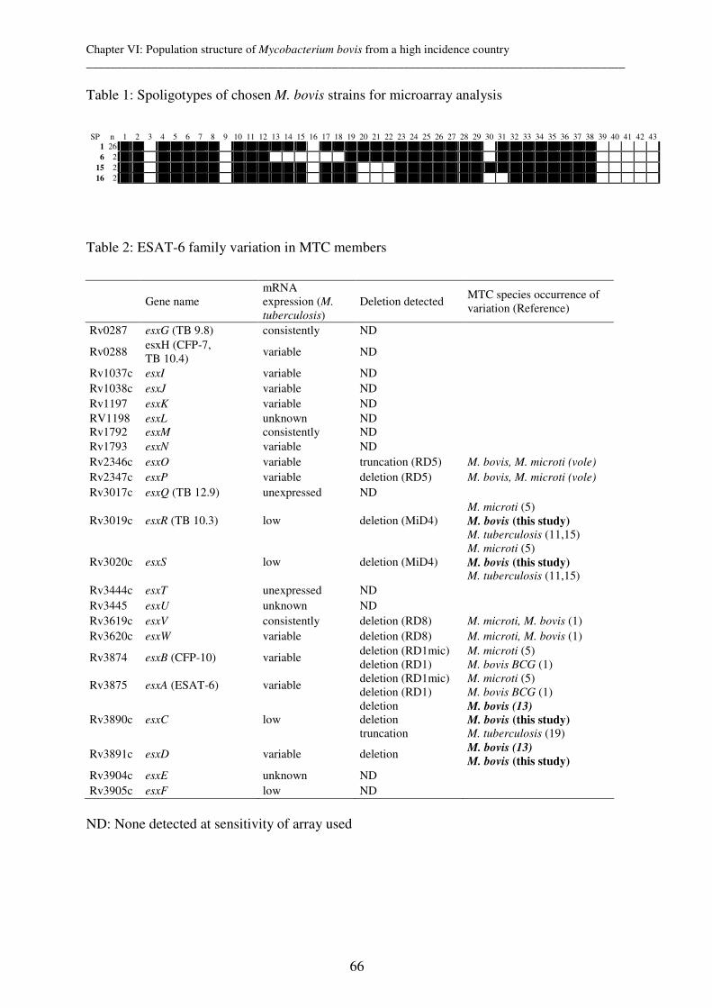
Chapter VI: Population structure of Mycobacterium bovis from a high incidence country __________________________________________________________________________________________
66
Table 1: Spoligotypes of chosen M. bovis strains for microarray analysis
Table 2: ESAT-6 family variation in MTC members
Gene name mRNA expression (M.
tuberculosis) Deletion detected
MTC species occurrence of variation (Reference)
Rv0287 esxG (TB 9.8) consistently ND
Rv0288 esxH (CFP-7, TB 10.4)
variable ND
Rv1037c esxI variable ND Rv1038c esxJ variable ND Rv1197 esxK variable ND RV1198 esxL unknown ND Rv1792 esxM consistently ND Rv1793 esxN variable ND Rv2346c esxO variable truncation (RD5) M. bovis, M. microti (vole)
Rv2347c esxP variable deletion (RD5) M. bovis, M. microti (vole)
Rv3017c esxQ (TB 12.9) unexpressed ND
Rv3019c esxR (TB 10.3) low deletion (MiD4) M. microti (5) M. bovis (this study) M. tuberculosis (11,15)
Rv3020c esxS low deletion (MiD4) M. microti (5) M. bovis (this study) M. tuberculosis (11,15)
Rv3444c esxT unexpressed ND
Rv3445 esxU unknown ND
Rv3619c esxV consistently deletion (RD8) M. microti, M. bovis (1) Rv3620c esxW variable deletion (RD8) M. microti, M. bovis (1)
Rv3874 esxB (CFP-10) variable deletion (RD1mic) deletion (RD1)
M. microti (5) M. bovis BCG (1)
Rv3875 esxA (ESAT-6) variable deletion (RD1mic) deletion (RD1)
M. microti (5) M. bovis BCG (1)
Rv3890c esxC low deletion deletion truncation
M. bovis (13)
M. bovis (this study) M. tuberculosis (19)
Rv3891c esxD variable deletion M. bovis (13)
M. bovis (this study)
Rv3904c esxE unknown ND Rv3905c esxF low ND
ND: None detected at sensitivity of array used
SP n 1 2 3 4 5 6 7 8 9 10 11 12 13 14 15 16 17 18 19 20 21 22 23 24 25 26 27 28 29 30 31 32 33 34 35 36 37 38 39 40 41 42 43 1 26 6 2
15 2 16 2
Chapter VI: Population structure of Mycobacterium bovis from a high incidence country __________________________________________________________________________________________
67
Reference List
1. Brosch, R., S. V. Gordon, M. Marmiesse, P. Brodin, C. Buchrieser, K. Eiglmeier, T. Garnier, C.
Gutierrez, G. Hewinson, K. Kremer, L. M. Parsons, A. S. Pym, S. Samper, D. van Soolingen, and S. T. Cole. 2002. A new evolutionary scenario for the Mycobacterium tuberculosis complex. Proc.Natl.Acad.Sci.U.S.A 99:3684-3689.
2. Cadmus, S., S. Palmer, M. Okker, J. Dale, K. Gover, N. Smith, K. Jahans, R. G. Hewinson, and S.
V. Gordon. 2006. Molecular Analysis of Human and Bovine Tubercle Bacilli from a Local Setting in Nigeria. Journal of Clinical Microbiology 44:29-34.
3. Diguimbaye-Djaibé C., Hilty M., Ngandolo R, Mahamat HH., Pfyffer G. E., Baggi F., Hewinson
G., Tanner M., Zinsstag J. and Schelling E. Mycobacterium bovis Isolates from Tuberculous Lesions in Chadian Zebu Carcasses. 2006. Emerg.Infect.Dis. 12(5):769-771
4. Gao, Q., K. E. Kripke, A. J. Saldanha, W. Yan, S. Holmes, and P. M. Small. 2005. Gene expression diversity among Mycobacterium tuberculosis clinical isolates. Microbiology 151:5-14.
5. Garcia-Pelayo, M. C., K. C. Caimi, J. K. Inwald, J. Hinds, F. Bigi, M. I. Romano, D. van
Soolingen, R. G. Hewinson, A. Cataldi, and S. V. Gordon. 2004. Microarray analysis of Mycobacterium microti reveals deletion of genes encoding PE-PPE proteins and ESAT-6 family antigens. Tuberculosis.(Edinb.) 84:159-166.
6. Hill, P. C., D. Jackson-Sillah, A. Fox, K. L. Franken, M. D. Lugos, D. J. Jeffries, S. A. Donkor, A. S. Hammond, R. A. Adegbola, T. H. Ottenhoff, M. R. Klein, and R. H. Brookes. 2005. ESAT-6/CFP-10 fusion protein and peptides for optimal diagnosis of mycobacterium tuberculosis infection by ex vivo enzyme-linked immunospot assay in the Gambia. J.Clin.Microbiol. 43:2070-2074.
7. Hilty, M., C. Diguimbaye, E. Schelling, F. Baggi, M. Tanner, and J. Zinsstag. 2005. Evaluation of the discriminatory power of variable number tandem repeat (VNTR) typing of Mycobacterium bovis strains. Vet.Microbiol. 109:217-222.
8. Hirsh, A. E., A. G. Tsolaki, K. DeRiemer, M. W. Feldman, and P. M. Small. 2004. Stable association between strains of Mycobacterium tuberculosis and their human host populations. Proc.Natl.Acad.Sci.U.S.A 101:4871-4876.
9. Kato-Maeda, M., J. T. Rhee, T. R. Gingeras, H. Salamon, J. Drenkow, N. Smittipat, and P. M.
Small. 2001. Comparing genomes within the species Mycobacterium tuberculosis. Genome Res. 11:547-554.
10. Malik, A. N. and P. Godfrey-Faussett. 2005. Effects of genetic variability of Mycobacterium tuberculosis strains on the presentation of disease. Lancet Infect.Dis. 5:174-183.
11. Marmiesse, M., P. Brodin, C. Buchrieser, C. Gutierrez, N. Simoes, V. Vincent, P. Glaser, S. T.
Cole, and R. Brosch. 2004. Macro-array and bioinformatic analyses reveal mycobacterial 'core' genes, variation in the ESAT-6 gene family and new phylogenetic markers for the Mycobacterium tuberculosis complex. Microbiology 150:483-496.
12. Mostowy, S., D. Cousins, J. Brinkman, A. Aranaz, and M. A. Behr. 2002. Genomic deletions suggest a phylogeny for the Mycobacterium tuberculosis complex. J.Infect.Dis. 186:74-80.
13. Mostowy, S., J. Inwald, S. Gordon, C. Martin, R. Warren, K. Kremer, D. Cousins, and M. A.
Behr. 2005. Revisiting the evolution of Mycobacterium bovis. J.Bacteriol. 187:6386-6395.
14. Nguyen, D., P. Brassard, D. Menzies, L. Thibert, R. Warren, S. Mostowy, and M. Behr. 2004. Genomic characterization of an endemic Mycobacterium tuberculosis strain: evolutionary and epidemiologic implications. J.Clin.Microbiol. 42:2573-2580.
Chapter VI: Population structure of Mycobacterium bovis from a high incidence country __________________________________________________________________________________________
68
15. Rajakumar, K., J. Shafi, R. J. Smith, R. A. Stabler, P. W. Andrew, D. Modha, G. Bryant, P. Monk, J. Hinds, P. D. Butcher, and M. R. Barer. 2004. Use of genome level-informed PCR as a new investigational approach for analysis of outbreak-associated Mycobacterium tuberculosis isolates. J.Clin.Microbiol. 42:1890-1896.
16. Ravn, P., M. E. Munk, A. B. Andersen, B. Lundgren, J. D. Lundgren, L. N. Nielsen, A. Kok-
Jensen, P. Andersen, and K. Weldingh. 2005. Prospective evaluation of a whole-blood test using Mycobacterium tuberculosis-specific antigens ESAT-6 and CFP-10 for diagnosis of active tuberculosis. Clin.Diagn.Lab Immunol. 12:491-496.
17. Skjot, R. L., I. Brock, S. M. Arend, M. E. Munk, M. Theisen, T. H. Ottenhoff, and P. Andersen. 2002. Epitope mapping of the immunodominant antigen TB10.4 and the two homologous proteins TB10.3 and TB12.9, which constitute a subfamily of the esat-6 gene family. Infect.Immun. 70:5446-5453.
18. Smith, N. H., J. Dale, J. Inwald, S. Palmer, S. V. Gordon, R. G. Hewinson, and J. M. Smith. 2003. The population structure of Mycobacterium bovis in Great Britain: clonal expansion. Proc.Natl.Acad.Sci.U.S.A 100:15271-15275.
19. Tsolaki, A. G., A. E. Hirsh, K. DeRiemer, J. A. Enciso, M. Z. Wong, M. Hannan, Goguet de la
Salmoniere YO, K. Aman, M. Kato-Maeda, and P. M. Small. 2004. Functional and evolutionary genomics of Mycobacterium tuberculosis: insights from genomic deletions in 100 strains. Proc.Natl.Acad.Sci.U.S.A 101:4865-4870.
Chapter VII: Genetic diversity in Mycobacterium ulcerans isolates from Ghana __________________________________________________________________________________________
69
Chapter VII: Genetic diversity in Mycobacterium ulcerans isolates
from Ghana revealed by a newly identified locus containing a
variable number of tandem repeats
Markus Hilty,1* Dorothy Yeboah-Manu,1,2* Daniel Boakye,2 Ernestina-Mensah-Quainoo,3
Simona Rondini,1 Esther Schelling,1 David Ofori-Adjei,2 Françoise Portaels4, Jakob Zinsstag1
and Gerd Pluschke1
Swiss Tropical Institute1, 4002 Basel, Noguchi Memorial Institute for Medical Research2,
Legon, Ghana, Tema Municipal Health Directorate, Tema, Ghana3, and Institute of Tropical
Medicine4, 2000 Antwerp, Belgium
Published in Journal of Bacteriology 2006 Feb;188(4):1462-5
*Contributed equally
Chapter VII: Genetic diversity in Mycobacterium ulcerans isolates from Ghana __________________________________________________________________________________________
70
Abstract
Molecular typing methods applied so far for Mycobacterium ulcerans isolates have not been
able to identify genetic differences among isolates from Africa. This apparent lack of genetic
diversity among M. ulcerans isolates is indicative for a clonal population structure. We
analysed the genetic diversity of 71 African isolates, including 57 strains from Ghana, by
variable number of tandem repeats (VNTR) typing based on a newly identified polymorphic
locus designated ST1 and the previously described locus, MIRU 1. Three different genotypes
were found in Ghana, demonstrating for the first time genetic diversity of M. ulcerans in an
African country. While the ST1/MIRU 1 allele combination BD/BAA seems to dominate in
Africa, it was only rarely found in isolates from Ghana, where the combination BD/B was
dominating and observed in all districts analysed. A third variant genotype (C/BAA) was
found only in the Amansie-West district. Results are indicative for the emergence and
spreading of new genetic variants of M. ulcerans within Ghana and support the potential of
VNTR-based typing for genotyping of M. ulcerans.
Chapter VII: Genetic diversity in Mycobacterium ulcerans isolates from Ghana __________________________________________________________________________________________
71
Introduction
Mycobacterium ulcerans, the causative agent of Buruli ulcer (BU) is an emerging pathogen
particularly in Sub-Saharan African countries, and is also found in tropical and sub-tropical
regions of Asia, the Western Pacific and Latin America (4). BU is characterized by chronic,
necrotic lesions of subcutaneous tissues. Due to the lack of an established effective
antimicrobial therapy, surgical excision and skin grafting is currently the recommended
treatment (27).
While it is known that proximity to slow flowing or stagnant water bodies is a risk factor for
M. ulcerans infection, the exact mode of transmission has remained an enigma (4). This is
partly because no molecular typing method is available that has sufficiently high resolution
for micro-epidemiological analyses. The apparent lack of genetic diversity of M. ulcerans
within individual geographical regions (7, 8, 16, 19-21) is indicative for a clonal population
structure. Genetic analyses suggest the recent divergence of M. ulcerans from M. marinum (5,
26), which is well known as fish pathogen and can cause limited granulomatous skin
infections in humans (10, 11, 13). One of the hallmarks of the emergence of M. ulcerans as a
more severe pathogen is the acquisition of a 174-kb plasmid bearing a cluster of genes
necessary for the synthesis of the macrolide toxin mycolactone responsible for the massive
tissue destruction seen in BU (22).
Variable number of tandem repeat (VNTR) typing is a PCR-based technique identifying
alleles of defined regions of DNA that contain a variable number of copies of short sequence
stretches. Resolution of the method is cumulative and can be increased by inclusion of
additional loci. Tandem repeats are easily identified from genome sequence data,
measurement of PCR fragment sizes is relatively straightforward and VNTR typing data can
be digitalized and compared between different laboratories. Availability of complete genomic
sequences has facilitated identification of repetitive genetic elements of M. tuberculosis (12,
15, 24, 25), M. bovis (14, 18) and M. avium (6), including short tandem repeats designated
exact tandem repeats (ETRs) and mycobacterial interspersed repetitive units (MIRUs). Strain
typing with these sets of polymorphic loci is developing into an important tool in the
epidemiological analysis of tuberculosis (9) and ordinary agarose gel electrophoretic
separation of PCR products is usually sufficient to estimate the number of repeat units in an
allele. In a study of M. bovis strains from Chad, VNTR-typing of a distinct number of loci is
most discriminative for strains of the same clone (14).
More recently, MIRUs and other VNTRs have also been described for M. ulcerans and M.
marinum (3, 23) typing. Most of the described sequences are orthologues of the M.
Chapter VII: Genetic diversity in Mycobacterium ulcerans isolates from Ghana __________________________________________________________________________________________
72
tuberculosis genome database and their resolution seems to be comparable to that of the
currently most discriminatory methods, the 2426 PCR analysis (19) and IS2404-Mtb2 PCR
(2) which discriminate among isolates from different geographical origin, but not among
strains from different endemic regions of Africa.
We describe in this report a new M. ulcerans specific VNTR locus (ST1) which together with
the previously described MIRU 1 (23) differentiated clinical isolates from Ghana into three
VNTR allele combinations.
Methods
Identification of the VNTR locus ST1: A tandem repeat finder software
(http://www.c3.biomath.mssm.edu/trf.thml) was used to screen the M. marinum sequence
data bank (www.sanger.ac.uk/projects/M_marinum) and identified a tandem repeat
containing locus which was designated ST1. This locus is present both in M. marinum and in
M. ulcerans (http://genopole.pasteur.fr/Mulc/BuruList.html), but not in M. tuberculosis
(http://www.sanger.ac.uk/Projects/M_tuberculosis). A forward (ctgaggggatttcacgaccag) and a
reverse primer (cgccacccgcggacacagtcg) located in the sequences flanking the identified locus
and yielding a PCR product of 423 bp was designed. Genomic sequences corresponding to
the primers were 100 % identical for M. marinum and M. ulcerans.
Bacterial strains: A panel of 11 M. ulcerans clinical isolates of human origin from diverse
geographical origin and of 6 M. marinum clinical isolates of human origin from Switzerland
was used to assess the polymorphism of ST1 (Table 1). The M. marinum isolates were from
patients living in the agglomeration of Zurich, except for M. marinum N119 that was isolated
from a patient living in Biel. The year of isolation was 1995 for strains 853 and 894, 1997 for
strains 8972 and 946 and 1998 for strains N119 and 3023. In order to analyse the diversity of
African M. ulcerans strains, 66 additional clinical isolates (12 from Benin and 54 from
Ghana) were included in this study. The Ghanaian strains were isolated (28) between 2001
and 2003 from patients being treated at the Amasaman Health Centre in the Greater Accra
Region of Ghana (48 isolates) or the Saint Martin Hospital Agroyesum in the Ashanti Region
of Ghana (6 isolates). The residential origin of the isolates is as indicated in the supporting
table.
DNA Extraction: DNA was extracted as described (17). Briefly, small bacterial pellets were
heated for 1 h at 95°C in 500 µl of an extraction mixture (50 mM Tris-HCl, 25 mM EDTA,
and 5% monosodium glutamate). One hundred microliters of a 50-mg/ml lysozyme solution
was then added and incubated for two hours at 37°C. 70 µl of proteinase K-10x buffer (100
mM Tris-HCl, 50 mM EDTA, 5% sodium dodecyl sulphate [pH 7.8]) and 10 µl of a 20-
Chapter VII: Genetic diversity in Mycobacterium ulcerans isolates from Ghana __________________________________________________________________________________________
73
mg/ml proteinase K solution was added. After incubation at 45°C overnight, 300 µl of 0.1-
mm-diameter zirconia beads (BioSpec Products) were added to each sample and vortexed at
full speed for 4 min. Beads and large debris were removed by brief centrifugation, and the
supernatants were transferred to fresh tubes for phenol-chloroform (Fluka) extraction. The
DNA contained in the upper phase was precipitated with ethanol and re-suspended in 100 µl
of water.
PCR analysis and sequencing of PCR products: PCR reaction mixtures contained 1x Taq
PCR buffer, deoxynucleoside triphosphates (0.2 mM each), 1 U of AmpliTaq Gold DNA
polymerase (Perkin-Elmer Applied Biosystems), a 0.5 µM concentration of the primer pair
and mycobacterial DNA in a final volume of 20 µl. Addition of 5 % DMSO to the reaction
mix improved the yield of PCR products. The reaction was carried out using a Perkin-Elmer
9600 cycler starting with a denaturing step of 10 min at 95°C. After denaturation, the PCR
was performed for 40 cycles of 0.5 min at 94°C, 0.5 min at 65°C and 1 min at 72°C. The
reactions were terminated by an incubation of 10 min at 72°C. PCR fragments were analysed
by agarose gel electrophoresis using 2% NuSieve agarose. The size of the amplicons was
estimated by comparison with Size Marker VIII (Roche). PCR products were directly
sequenced with an ABI Prism 310 Genetic Analysis System. PCR products for ST1 of 423 bp
and 369 bp corresponded to a copy number of 2 and 1, respectively. For MIRU 1, copy
numbers were assigned as described (23).
Results
Identification and characterization of a new VNTR locus found in M. ulcerans and M.
marinum
A new VNTR locus designated ST1, was identified by screening of the M. marinum sequence
data bank with a tandem repeat finder software. Orthologues of ST1 were found in M.
ulcerans, but not in M. tuberculosis. Its repeat length of 54 bp is suitable for size analysis by
standard agarose gel electrophoresis. In contrast to MIRUs (23), ST1 is not intergenic, but
part of a pseudogene and therefore of no known functional interest (personal communication).
When eleven M. ulcerans isolates of geographically diverse origin were typed by agarose gel
electrophoresis of PCR products, two different alleles of ST1 were identified (Table 1). While
eight strains had two repeats, two isolates, one from French Guyana and one of the two
analysed isolates from Ghana had only one repeat. Of seven M. marinum clinical isolates
tested, all five strains from patients in the agglomeration of Zurich (strains 8972, 946, 3023,
853 and 894) had three repeats, whereas strain N119 isolated from a different part of
Chapter VII: Genetic diversity in Mycobacterium ulcerans isolates from Ghana __________________________________________________________________________________________
74
Switzerland and the reference strain used for the M. marinum genome sequencing project,
both had two repeats. Sequence analysis of PCR products reconfirmed the size differences
observed by agarose gel electrophoresis and identified six sequence variants of the repeat unit
(designated A – F; Fig 1). Unlike many other VNTRs (1), ST1 showed no micro-deletions,
but only single nucleotide polymorphisms (SNPs) within the sequence variants. The M.
ulcerans strains from China and Japan turned out to have a different allele (CF; Table 1) than
the other M. ulcerans strains with two repeats (BD). A third allele with two repeats (AC) and
an allele with three repeats (ACE) was found in M. marinum (Table 1). Thus sequencing of
ST1 improved the discrimination power of M. ulcerans and M. marinum strains compared to
gel electrophoresis analysis alone and revealed distinctive genotypes for M. marinum
compared to M. ulcerans.
Diversity of M. ulcerans isolates from Ghana
Evidence for diversity of the ST1 locus in African isolates (Table 1) prompted us to analyse
additional collections of 12 disease isolates from Benin and 54 isolates from Ghana (Table 2).
All strains from Benin and the majority of Ghanaian strains had an ST1 allele (BD) with two
repeats. However, in most strains from the Amansie West district (including ITM-970359;
Table 1), a second allele with only one repeat (C) was identified (Table 2). When the
Ghanaian isolates were tested also for diversity in the loci MIRU 1 (23), VNTR 8, 9 and 19,
previously described as polymorphic within M. ulcerans strains of different geographical
origin (3), diversity was also found in locus MIRU 1. Sequence analysis of PCR products of
selected strains reconfirmed the size differences observed by agarose gel electrophoresis and
identified two sequence variants of the MIRU 1 repeat unit (designated A and B; Fig 1).
Altogether three VNTR allele combinations were found among the clinical isolates from
Ghana (Table 2, Fig. 2 and 3). While all isolates from the Ga, Akwapim South, Ahafo-Ano
North and Akim Abuakwa districts had the ST1/MIRU 1 allele combination 1 (BD/B), two
allele combinations, i.e. 1 and 2 (C/BAA) were found among the five isolates from the
Amansie-West district. A third allele combination (BD/BAA) was found in two strains
(Agy99 from the Ga district and ITM 97-0359 from the Ashanti region) isolated before 2000
in Ghana and in all other African isolates.

Chapter VII: Genetic diversity in Mycobacterium ulcerans isolates from Ghana __________________________________________________________________________________________
75
Discussion
Molecular typing methods such as multi-locus sequence typing, 16S rRNA sequencing,
restriction fragment length polymorphism and variable number of tandem repeats typing have
revealed a remarkable lack of genetic diversity of M. ulcerans and a clonal population
structure within given geographical regions. The discriminatory power of all these methods is
particularly insufficient to differentiate between African isolates. Innovative molecular
genetic fingerprinting methods are therefore required for local epidemiological studies aiming
to reveal transmission pathways and environmental reservoirs of M. ulcerans. First attempts
to use VNTR typing for M. ulcerans (3, 23) have identified variable loci suitable for
discrimination of disease isolates at continental level. In this study we used a newly identified
(ST1) and four previously described VNTRs to analyse genetic diversity within a collection
of 71 M. ulcerans strains from Africa, including 57 isolates from Ghana. Three of the
previously described VNTRs i.e. VNTR 8, 9 and 19 (3) were not able to discriminate among
the African strains. Yet MIRU 1 (23) and the newly identified locus (ST1) defined three
subgroups within the Ghanaian strains.
The fact that two allele combinations (BD/B and C/BAA) differing from the common African
combination (BD/BAA) were found within a recent (2001-2003) collection of Ghanaian
isolates is indicative for an ongoing microevolution of M. ulcerans and for the spreading of
new variants within Ghana. It is tempting to hypothesize that allele combination 3 (BD/BAA)
represents an ancestral like genotype and that the others evolved by reduction in the repeat
unit numbers in the ST1 locus (from BD to C) or in the MIRU 1 locus (from BAA to B),
respectively. While conversion of the MIRU 1 locus from BAA to B could be explained by
deletion of the two A repeat units, conversion of the ST1 locus from BD to C by a deletional
mechanism would require that a central sequence stretch of the BD repeat region comprising
portions of both the B and the D repeat unit would have been lost, yielding the hybrid repeat
unit C.
While allele combination 3 seems to be the most common in Africa, most of the M. ulcerans
strains from Ghana analysed, had the allele combination 1. This genotype was found in all
Ghanaian districts included in this study. Allele combination 2 dominated in the Amansie
West district, but was found exclusively there. Follow up of the temporal and spatial patterns
of emergence and spreading of genotypes may contribute in future to our understanding of the
transmission and epidemiology of Buruli ulcer. From the present data, we cannot draw any
conclusions why certain variant appear to be the more successful than others. The fact, that
we were not able to sub-group the 47 isolates from the Ga district by VNTR (with the only
Chapter VII: Genetic diversity in Mycobacterium ulcerans isolates from Ghana __________________________________________________________________________________________
76
exception of the ‘older’ isolate Agy99) reconfirms that M. ulcerans has a clonal population
structure associated with a low rate of genomic drift. Availability of the fully assembled and
annotated genome sequence of M. ulcerans in the near future will facilitate identification of
further polymorphic VNTR loci potentially contributing to further refinement of genetic
fingerprinting of M. ulcerans isolates.
Acknowledgements
We acknowledge Dr. Edwin Ampadu, of the Ghana National Buruli Ulcer Control program
for his assistances in clinical sample collection. NCCR North-South IP-4 is acknowledged for
financial support and Anthony Ablordey for critical review. Many thanks also to Franca
Baggi of the Swiss Centre of Mycobacteria for providing M. marinum strains and the heads of
the M. ulcerans and M. marinum genome sequencing projects for the permission to use the
sequence data.
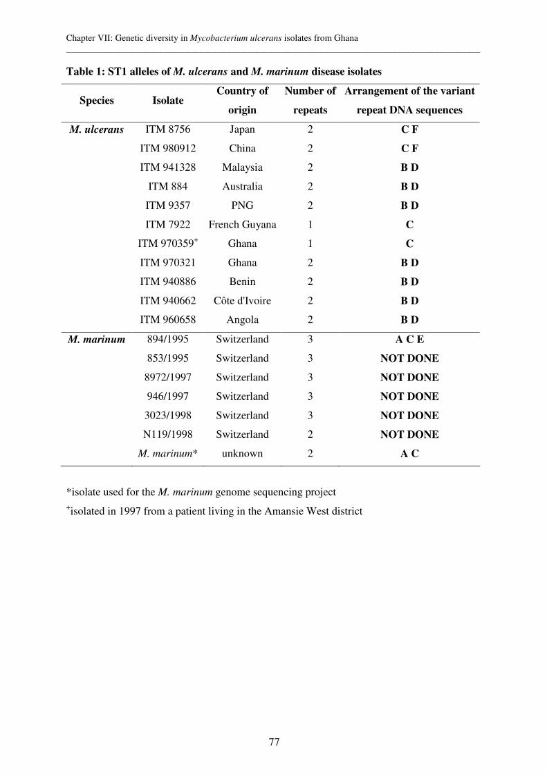
Chapter VII: Genetic diversity in Mycobacterium ulcerans isolates from Ghana __________________________________________________________________________________________
77
Table 1: ST1 alleles of M. ulcerans and M. marinum disease isolates
Species Isolate Country of
origin
Number of
repeats
Arrangement of the variant
repeat DNA sequences
M. ulcerans
ITM 8756
ITM 980912
ITM 941328
ITM 884
ITM 9357
ITM 7922
ITM 970359+
ITM 970321
ITM 940886
ITM 940662
ITM 960658
Japan
China
Malaysia
Australia
PNG
French Guyana
Ghana
Ghana
Benin
Côte d'Ivoire
Angola
2
2
2
2
2
1
1
2
2
2
2
C F
C F
B D
B D
B D
C
C
B D
B D
B D
B D
M. marinum
894/1995
853/1995
8972/1997
946/1997
3023/1998
N119/1998
M. marinum*
Switzerland
Switzerland
Switzerland
Switzerland
Switzerland
Switzerland
unknown
3
3
3
3
3
2
2
A C E
NOT DONE
NOT DONE
NOT DONE
NOT DONE
NOT DONE
A C
*isolate used for the M. marinum genome sequencing project +isolated in 1997 from a patient living in the Amansie West district

Chapter VII: Genetic diversity in Mycobacterium ulcerans isolates from Ghana __________________________________________________________________________________________
78
Table 2: ST1 and MIRU 1 allele combinations of M. ulcerans strains from Africa
Country of
origin
District Number
of strains ST1 allele MIRU 1 allele
Allele
combination
Ga district 47 2 (BD) 1 (B) 1
Ga district 1 2 (BD) 3 (BAA) 3
Akwapim South 1 2 (BD) 1 (B) 1
Akim Abuakwa 1 2 (BD) 1 (B) 1
Ahafo-Ano 1 2 (BD) 1 (B) 1
Amansie West 1 2 (BD) 1 (B) 1
Amansie West 4 1 (C ) 3 (BAA) 2
Ghana
unknown○ 1 2 (BD) 3 (BAA) 3
Benin 13 2 (BD) + 3* (BAA)+ 3
Côte d'Ivoire 1 2 (BD) 3* 3
Angola 1 2 (BD) 3* 3
VNTR copy numbers for ST1 and MIRU 1 were determined and allele combinations assigned
(1-3). Sequence profiles of M. ulcerans strains are shown in brackets. ○isolated at the Saint
Martin’s hospital in the Ashanti region; * MIRU 1 copy numbers as previously described
(23); +only one strain (ITM 94-0886) was analysed by sequencing (23).
Allele ST1 repeat unit sequences
A CCGGTTCTGTTTCGTCCGGTGCGACCGCTGGCACTGTCTCGACCGGTGCGACGA B .........................................G....C....... C .........................................G...........G D .......G...G.C...........................G...........G E -T.....G...G.C...........................G...........G F .......G...G.C....A......................G...........G
Allele MIRU 1 repeat unit sequences
A* ATGAGCCAGCCGGCGACGATGCAGAGCGAAGCGATGAGGAGGAGCGGCGCCAG
B G...A..C..T..........................................
Figure 1: Sequence variation of ST1 and MIRU 1 tandem repeat units
(-): Base deletions; (.): identical sequence positions.
*Allele A of MIRU 1 corresponds to variant A2 (23).
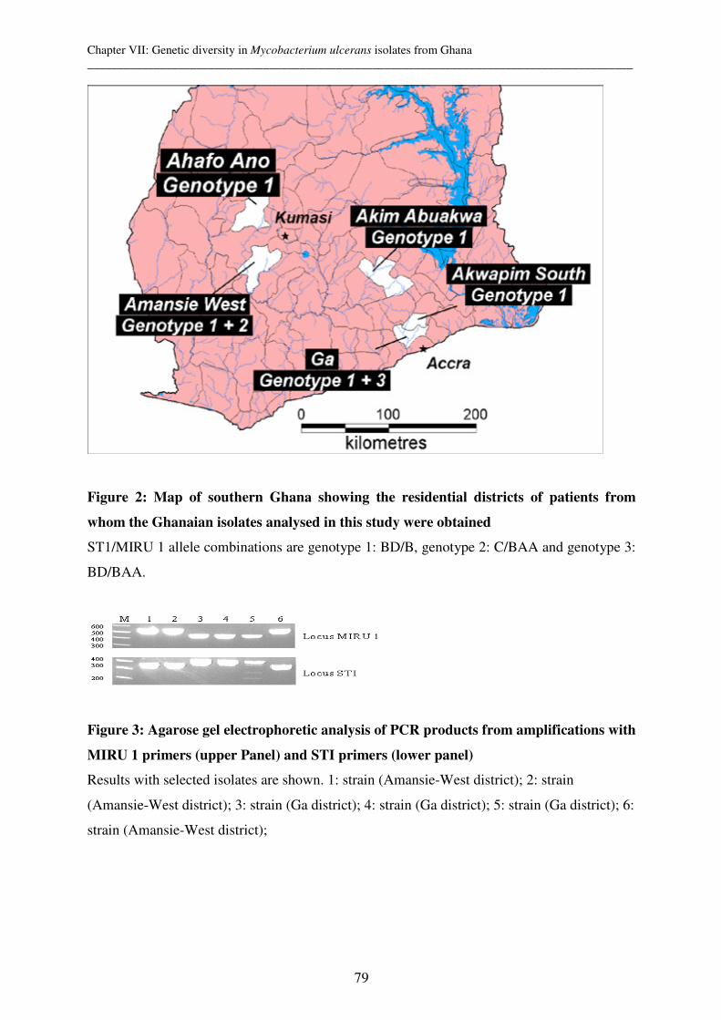
Chapter VII: Genetic diversity in Mycobacterium ulcerans isolates from Ghana __________________________________________________________________________________________
79
Figure 2: Map of southern Ghana showing the residential districts of patients from
whom the Ghanaian isolates analysed in this study were obtained
ST1/MIRU 1 allele combinations are genotype 1: BD/B, genotype 2: C/BAA and genotype 3:
BD/BAA.
Figure 3: Agarose gel electrophoretic analysis of PCR products from amplifications with
MIRU 1 primers (upper Panel) and STI primers (lower panel)
Results with selected isolates are shown. 1: strain (Amansie-West district); 2: strain
(Amansie-West district); 3: strain (Ga district); 4: strain (Ga district); 5: strain (Ga district); 6:
strain (Amansie-West district);

Chapter VII: Genetic diversity in Mycobacterium ulcerans isolates from Ghana __________________________________________________________________________________________
80
Reference List
1. Ablordey, A., M. Hilty, P. Stragier, J. Swings, and F. Portaels. 2005. Comparative Nucleotide Sequence Analysis of Polymorphic Variable-Number Tandem-Repeat Loci in Mycobacterium ulcerans. J.Clin.Microbiol. 43:5281-5284.
2. Ablordey, A., R. Kotlowski, J. Swings, and F. Portaels. 2005. PCR amplification with primers based on IS2404 and GC-rich repeated sequence reveals polymorphism in Mycobacterium ulcerans. J.Clin.Microbiol. 43:448-451.
3. Ablordey, A., J. Swings, C. Hubans, K. Chemlal, C. Locht, F. Portaels, and P. Supply. 2005. Multilocus variable-number tandem repeat typing of Mycobacterium ulcerans. J.Clin.Microbiol. 43:1546-1551.
4. Asiedu, K., R. Scherpbier, and M. Raviglione. 2000. Buruli ulcer - Mycobacterium ulcerans
infection. WHO document WHO/CDS/CPE/GBUI/2000.1.
5. Boddinghaus, B., T. Rogall, T. Flohr, H. Blocker, and E. C. Bottger. 1990. Detection and Identification of Mycobacteria by Amplification of Ribosomal-Rna. Journal of Clinical Microbiology 28:1751-1759.
6. Bull, T. J., K. Sidi-Boumedine, E. J. Mcminn, K. Stevenson, R. Pickup, and J. Hermon-Taylor. 2003. Mycobacterial interspersed repetitive units (MIRU) differentiate Mycobacterium avium subspecies paratuberculosis from other species of the Mycobacterium avium complex. Molecular and Cellular Probes 17:157-164.
7. Chemlal, K., K. De Ridder, P. A. Fonteyne, W. M. Meyers, J. Swings, and E. Portaels. 2001. The use of IS2404 restriction fragment length polymorphisms suggests the diversity of Mycobacterium ulcerans from different geographical areas. American Journal of Tropical Medicine and Hygiene 64:270-273.
8. Chemlal, K., G. Huys, P. A. Fonteyne, V. Vincent, A. G. Lopez, L. Rigouts, J. Swings, W. M.
Meyers, and F. Portaels. 2001. Evaluation of PCR-restriction profile analysis and IS2404 restriction fragment length polymorphism and amplified fragment length polymorphism fingerprinting for identification and typing of Mycobacterium ulcerans and M-marinum. Journal of Clinical Microbiology 39:3272-3278.
9. Crawford, J. T. 2003. Genotyping in contact investigations: a CDC perspective. International Journal of Tuberculosis and Lung Disease 7:S453-S457.
10. Dailloux, M., C. Laurain, R. Weber, and P. Hartemann. 1999. Water and nontuberculous mycobacteria. Water Research 33:2219-2228.
11. Dobos, K. M., F. D. Quinn, D. A. Ashford, C. R. Horsburgh, and C. H. King. 1999. Emergence of a unique group of necrotizing mycobacterial diseases. Emerging Infectious Diseases 5:367-378.
12. Frothingham, R. and W. A. Meeker-O'Connell. 1998. Genetic diversity in the Mycobacterium tuberculosis complex based on variable numbers of tandem DNA repeats. Microbiology-Uk 144:1189-1196.
13. Gluckman, S. J. 1995. Mycobacterium-Marinum. Clinics in Dermatology 13:273-276.
14. Hilty, M., C. Diguimbaye, E. Schelling, F. Baggi, M. Tanner, and J. Zinsstag. 2005. Evaluation of the discriminatory power of variable number tandem repeat (VNTR) typing of Mycobacterium bovis strains. Vet.Microbiol. 109:217-222.
15. Mazars, E., S. Lesjean, A. L. Banuls, M. Gilbert, V. Vincent, B. Gicquel, M. Tibayrenc, C. Locht,
and P. Supply. 2001. High-resolution minisatellite-based typing as a portable approach to global
Chapter VII: Genetic diversity in Mycobacterium ulcerans isolates from Ghana __________________________________________________________________________________________
81
analysis of Mycobacterium tuberculosis molecular epidemiology. Proceedings of the National Academy of Sciences of the United States of America 98:1901-1906.
16. Portaels, F., P. A. Fonteyne, H. DeBeenhouwer, P. DeRijk, A. Guedenon, J. Hayman, and W. M.
Meyers. 1996. Variability in 3' end of 16S rRNA sequence of Mycobacterium ulcerans is related to geographic origin of isolates. Journal of Clinical Microbiology 34:962-965.
17. Rondini, S., E. Mensah-Quainoo, H. Troll, T. Bodmer, and G. Pluschke. 2003. Development and application of real-time PCR assay for quantification of Mycobacterium ulcerans DNA. Journal of Clinical Microbiology 41:4231-4237.
18. Skuce, R. A., T. P. McCorry, J. F. McCarroll, S. M. M. Roring, A. N. Scott, D. Brittain, S. L.
Hughes, R. G. Hewinson, and S. D. Neill. 2002. Discrimination of Mycobacterium tuberculosis complex bacteria using novel VNTR-PCR targets. Microbiology-Sgm 148:519-528.
19. Stinear, T., J. K. Davies, G. A. Jenkin, F. Portaels, B. C. Ross, F. Oppedisano, M. Purcell, J. A.
Hayman, and P. D. R. Johnson. 2000. A simple PCR method for rapid genotype analysis of Mycobacterium ulcerans. Journal of Clinical Microbiology 38:1482-1487.
20. Stinear, T., B. C. Ross, J. K. Davies, L. Marino, R. M. Robins-Browne, F. Oppedisano, A. Sievers,
and P. D. R. Johnson. 1999. Identification and characterization of IS2404 and IS2606: Two distinct repeated sequences for detection of Mycobacterium ulcerans by PCR. Journal of Clinical Microbiology 37:1018-1023.
21. Stinear, T. P., G. A. Jenkin, P. D. R. Johnson, and J. K. Davies. 2000. Comparative genetic analysis of Mycobacterium ulcerans and Mycobacterium marinum reveals evidence of recent divergence. Journal of Bacteriology 182:6322-6330.
22. Stinear, T. P., A. Mve-Obiang, P. L. C. Small, W. Frigui, M. J. Pryor, R. Brosch, G. A. Jenkin, P.
D. R. Johnson, J. K. Davies, R. E. Lee, S. Adusumilli, T. Garnier, S. F. Haydock, P. F. Leadlay, and S. T. Cole. 2004. Giant plasmid-encoded polyketide synthases produce the macrolide toxin of Mycobacterium ulcerans. Proceedings of the National Academy of Sciences of the United States of America 101:1345-1349.
23. Stragier, P., A. Ablordey, W. M. Meyers, and F. Portaels. 2005. Genotyping Mycobacterium ulcerans and Mycobacterium marinum by using mycobacterial interspersed repetitive units. J.Bacteriol. 187:1639-1647.
24. Supply, P., J. Magdalena, S. Himpens, and C. Locht. 1997. Identification of novel intergenic repetitive units in a mycobacterial two-component system operon. Molecular Microbiology 26:991-1003.
25. Supply, P., E. Mazars, S. Lesjean, V. Vincent, B. Gicquel, and C. Locht. 2000. Variable human minisatellite-like regions in the Mycobacterium tuberculosis genome. Molecular Microbiology 36:762-771.
26. Tonjum, T., D. B. Welty, E. Jantzen, and P. L. Small. 1998. Differentiation of Mycobacterium ulcerans, M-marinum, and M-haemophilum: Mapping of their relationships to M-tuberculosis by fatty acid profile analysis, DNA-DNA hybridization, and 16S rRNA gene sequence analysis. Journal of Clinical Microbiology 36:918-925.
27. van der Werf, T. S., T. Stinear, Y. Stienstra, W. T. A. van der Graaf, and P. L. Small. 2003. Mycolactones and Mycobacterium ulcerans disease. Lancet 362:1062-1064.
28. Yeboah-Manu, D., T. Bodmer, E. Mensah-Quainoo, S. Owusu, D. Ofori-Adjei, and G. Pluschke. 2004. Evaluation of decontamination methods and growth media for primary isolation of Mycobacterium ulcerans from surgical specimens. J.Clin.Microbiol. 42:5875-5876.

Chapter VIII: Analysis of Polymorphic VNTR Loci in Mycobacterium ulcerans __________________________________________________________________________________________
83
Chapter VIII: Comparative Nucleotide Sequence Analysis of
Polymorphic Variable-Number Tandem-Repeat Loci in
Mycobacterium ulcerans
Anthony Ablordey,1 Markus Hilty,2 Pieter Stragier,1 Jean Swings,3,4 and Françoise Portaels1
Mycobacteriology Unit, Institute of Tropical Medicine, B-2000 Antwerp, Belgium,1
Swiss Tropical Institute, 4002, Basel, Switzerland,2
Laboratory of Microbiology,3 BCCM/LMG Culture Collection, University Ghent, B-9000
Ghent, Belgium4
Published in Journal of Clinical Microbiology 2005 Oct;43(10):5281-4

Chapter VIII: Analysis of Polymorphic VNTR Loci in Mycobacterium ulcerans __________________________________________________________________________________________
84
Abstract
We analyzed a set of variable-number tandem-repeat (VNTR) loci to assess their nucleotide
sequence diversity in isolates of three Mycobacterium ulcerans genotypes. Sequence variants
in two loci resulted in intraspecies resolution of Southeast Asian and Asian genotypes in
contrast to a homogenous sequence composition among African isolates. Nucleotide sequence
polymorphism in repeat units can enhance discrimination of VNTR loci.
Chapter VIII: Analysis of Polymorphic VNTR Loci in Mycobacterium ulcerans __________________________________________________________________________________________
85
Mycobacterium ulcerans causes Buruli ulcer, a necrotizing skin disease in tropical and
subtropical regions (4, 10). The epidemiology of Buruli ulcer is poorly understood, due in part
to the highly restricted genetic diversity in M. ulcerans, especially among isolates with
common geographic origins (1, 2, 5, 6, 11, 13, 14), and also to the difficulty in obtaining
cultures from environmental specimens (1, 10).
Tandem-repeat (TR) loci have enormous potential as highly evolving genomic regions
suitable for typing species with low genetic diversity. Their use in molecular epidemiology
studies have contributed significantly to the identification of sources of infection, a better
understanding of disease transmission, and strain-trait correlations (8, 9, 12).
To investigate the potential of TRs in providing highly discriminatory markers for studying
molecular diversity in M. ulcerans, we demonstrated allele-length polymorphism associated
with nine variable-number tandem-repeat (VNTR) loci. This allowed inter- and intraspecies
differentiation in a representative collection of Mycobacterium marinum and M. ulcerans (2).
Intraspecies discrimination in M. ulcerans was, however, limited among isolates within the
same geographic region (2). Different isolates from Africa, Southeast Asia, or Asia could not
be distinguished by allele-length analysis, after PCR amplification of nine VNTR loci. Such
isolates were also not distinguished by multilocus sequence typing (15), mycobacterial
interspersed repetitive unit-VNTR typing (16), and IS2404-restriction fragment length
polymorphism typing (5).
In this study, we carried out a comparative sequence analysis of the VNTR loci to further
assess the contribution of nucleotide sequence polymorphism to allelic diversity in isolates
belonging to the African, Southeast Asian and Asian M. ulcerans genotypes. The investigation
involved sequence analysis of nine VNTR loci in three isolates (including sequence strain)
belonging to the African genotype, and four loci (8, 9, 18, and 19) in isolates of the Asian and
Southeast Asian type (Table 1). The African isolates were of Angolan, Beninese, and
Ghanaian (sequence strain) origins. The Southeast Asian genotype comprises isolates of
Australian, Papua New Guinean, and Malaysian origins, while isolates from Japan and China
formed the Asian genotype. All M. ulcerans isolates were subcultured (from frozen stocks of
the collection of the Institute of Tropical Medicine, Antwerp [ITM]) onto Löwenstein-Jensen
medium and incubated at 32°C for 4 weeks. Isolates were further characterized
phenotypically and tested for the presence of IS2404 and IS2606 insertion sequences as
previously described (13, 18).
TR loci were bioinformatically identified by applying the TR Finder algorithm on M.
marinum genome sequences (available M. ulcerans genome sequences not accessible for TR
Chapter VIII: Analysis of Polymorphic VNTR Loci in Mycobacterium ulcerans __________________________________________________________________________________________
86
Finder analysis), which also generated a consensus pattern for each locus. Details of TR
discovery, DNA extraction, PCR primers, and amplification conditions have been previously
described (2). Purified PCR products were sequenced by using the ABI 310 genetic analysis
system.
For each locus, TR sequences of the different isolates were aligned and compared with the
consensus pattern. All nine loci were found to consist of heterogeneous arrays of repeat units
(variants) with deletions and/or nucleotide substitutions (Table 2). Locus 8 was the most
conserved in both species, with no nucleotide deletion and two substitutions in sequence
variants among all M. ulcerans isolates.
For each locus, the individual repeat variants were assigned designations (Table 2). While
some repeat variants were found exclusively either in M. ulcerans (e.g., G19, H19, or D18) or in
M. marinum (e.g., A18 or B19), others variant occurred in both species (e.g., A8 or A9).
Sequence profiles were generated at each locus for the isolates by combining these
designations. Comparison of the sequence profiles (which defines an allele at a given locus)
facilitates the identification of sequence types (Table 3).
Among the African isolates, corresponding loci featured 100% TR sequence identity;
consequently, intraspecies differentiation within this genotype was not possible. Among
Southeast Asian isolates, nucleotide sequence homology was complete in all except for loci 9
and 18, for which point mutations resulted in different allelic states. In locus 9, a single-
nucleotide deletion in a repeat variant in the Malaysian isolate (ITM 94-1328, with profile
A9A9C9) differentiated it from the Australian isolate (ITM 94-1324) and Papua New Guinean
isolate (ITM 94-1331), both with the A9A9D9 sequence profile. Each of the isolates, however,
harboured a unique sequence variant at locus 18 (D18, C18, and E18, respectively, for isolates
ITM 94-1328, ITM 94-1324, and ITM 94-1331), permitting the complete resolution of the
Southeast Asia genotype. Locus 18 also resolved the Asian type into China and Japan
genotypes (Table 3).
Although polymorphism at TR loci can occur either as a result of variation in the number of
repeat units (length polymorphism) or as a result of nucleotide sequence changes between
individual repeat units (sequence polymorphism) (12), the practical ease and lower cost of
analyzing length polymorphism (by agarose gel electrophoresis) over sequencing have
promoted the use of the former approach for routine typing purposes. Few studies on sequence
polymorphism in TR loci have yielded mixed results. While some studies have indicated
incremental gain in strain discrimination when length polymorphism data were complemented
with sequence analysis (3, 7), this has not been realized in others (9, 17).
Chapter VIII: Analysis of Polymorphic VNTR Loci in Mycobacterium ulcerans __________________________________________________________________________________________
87
In this study, we showed the occurrence of sequence polymorphism in two TR loci, which
exhibit no length polymorphism among isolates of two M. ulcerans genotypes. A general
trend of TR sequence conservation in isolates from the same geographic region was noticed.
This was most pronounced among the African isolates, which displayed complete sequence
homology across the nine VNTR loci. Consistent with previous data (1, 2, 5, 6, 11, 14-16), the
lack of sequence variants in this investigation further emphasizes the clonal homogeneity and
recent evolutionary origin and distribution of the African genotype (15).
In contrast, sequence analysis revealed three Southeast Asian alleles and two alleles within
the Asian genotype. Notably, the discrimination of these genotypes corroborates the data from
IS2404-Mtb2 PCR (which differentiates between the isolates from China and Japan and also
among the three Southeast Asian isolates) (1) and 2426 PCR (14), which discriminates among
the Southeast Asian but not between the Asian isolates. Isolates of these two genotypes show
limited differences in their repetitive-sequence-based PCR profiles. Differences in their
VNTR sequence profiles therefore are significant in further highlighting differences among
these isolates. A combination of the sequence and length polymorphism data results in a total
of 11 M. ulcerans alleles compared to 8 indexed by length polymorphism analysis alone and
10 alleles by IS2404-Mtb2 PCR on the same set of isolates. The conservation of TR loci in the
two Mycobacterium species and with much sequence degeneration in M. ulcerans is
consistent with the proposed origin of M. ulcerans from M. marinum through a reductive
genome evolution (15).
Sequence polymorphisms among M. ulcerans isolates involved single-nucleotide substitutions
and microdeletions. For clonal organisms, and also across VNTR loci, such point mutations
are often not considered major sources of genetic variation among isolates. However, data
accruing from whole-genome sequence analyses of a number of organisms and also from
sequence analysis of several genetic markers indicate that even in highly clonal species like
Mycobacterium tuberculosis, Bacillus anthracis, and Yersinia pestis, many thousands of point
mutations can be discovered when large portions of genomes are investigated (8). This theme
is thus further reinforced by sequence data from this investigation. Complementation of
sequence and length polymorphism data should potentially increase the discriminatory power
of the VNTR-typing method. It is envisaged that this approach would be more useful for
genotyping M. ulcerans and other highly monomorphic species.

Chapter VIII: Analysis of Polymorphic VNTR Loci in Mycobacterium ulcerans __________________________________________________________________________________________
88
Acknowledgments
This work was partly supported by grants from the Fund for Scientific Research, Flanders,
Belgium (F.W.O.-Vlaanderen, contract no. G.0301.01 and G.0471.03N). A.A. was supported
by a grant from the Damien Foundation (Brussels, Belgium).
We thank Pim de Rijk, Krista Fissette, and Cécile Uwizeye for the excellent technical work.
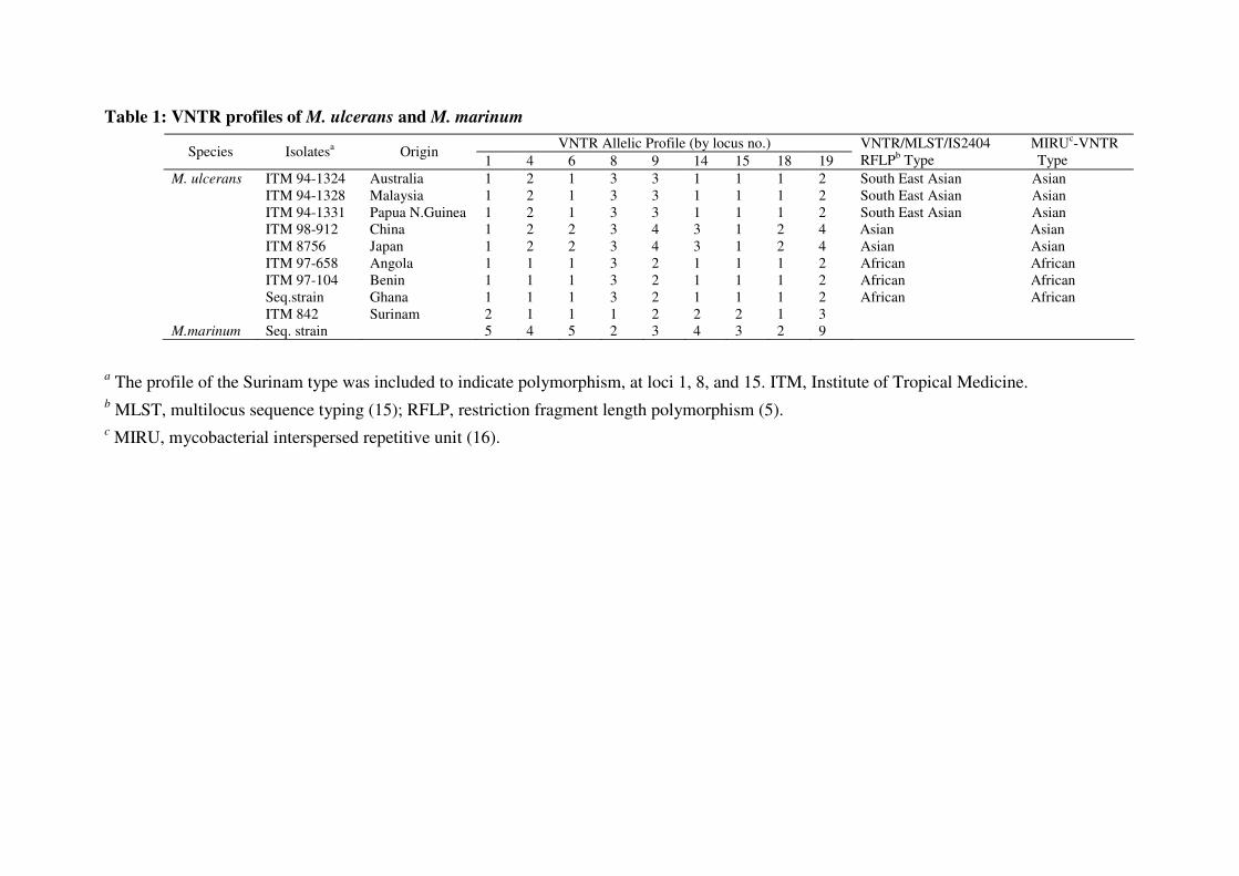
Table 1: VNTR profiles of M. ulcerans and M. marinum
VNTR Allelic Profile (by locus no.) Species Isolatesa Origin
1 4 6 8 9 14 15 18 19 VNTR/MLST/IS2404 MIRUc-VNTR RFLPb Type Type
M. ulcerans ITM 94-1324 Australia 1 2 1 3 3 1 1 1 2 South East Asian Asian ITM 94-1328 Malaysia 1 2 1 3 3 1 1 1 2 South East Asian Asian ITM 94-1331 Papua N.Guinea 1 2 1 3 3 1 1 1 2 South East Asian Asian ITM 98-912 China 1 2 2 3 4 3 1 2 4 Asian Asian ITM 8756 Japan 1 2 2 3 4 3 1 2 4 Asian Asian ITM 97-658 Angola 1 1 1 3 2 1 1 1 2 African African ITM 97-104 Benin 1 1 1 3 2 1 1 1 2 African African Seq.strain Ghana 1 1 1 3 2 1 1 1 2 African African ITM 842 Surinam 2 1 1 1 2 2 2 1 3 M.marinum Seq. strain 5 4 5 2 3 4 3 2 9
a The profile of the Surinam type was included to indicate polymorphism, at loci 1, 8, and 15. ITM, Institute of Tropical Medicine. b MLST, multilocus sequence typing (15); RFLP, restriction fragment length polymorphism (5). c MIRU, mycobacterial interspersed repetitive unit (16).
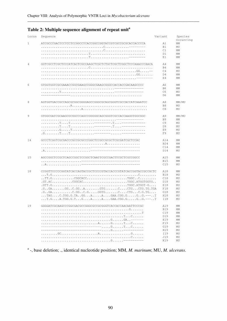
Chapter VIII: Analysis of Polymorphic VNTR Loci in Mycobacterium ulcerans __________________________________________________________________________________________
90
Table 2: Multiple sequence alignment of repeat unita
Locus Sequence Variant Species
Occurring
1 ATCGCCCGACTCCTCCTCCGGCCTCACCGGCCGGTATCGTCGCCGCGCACCACCCCA A1 MM
..................................C.............--------- B1 MU
..................................C...................... C1 MM
..........................T.............................. D1 MM
..........................T.....................--------- E1 MM
4 GGTCGCCTCGCTCCCATCACTCGCCAAGCTCGCTCTGCTCGCTCGGCTCCCAAACCCAACA A4 MM
..........................C.................................. B4 MM
....................................................GG.....-- C4 MU
....................................................GG....... D4 MM
....................................................--------- E4 MM
6 GTGGTGGTCGCGAAACCGGCGAAGCCGGGCGAAGCGGGCCACCACCGACAAGCCCC A6 MM
........................................---------------- B6 MM
..........T.............................---------------- C6 MU
----------------....................................... D6 MM
8 AGTGGTGACCGCCAGCGCGGCGGGGAGCCGGGCGCAGCGGGTCGCCACCATCAAATCC A8 MM/MU
................A......................................... B8 MU
......................A................................... C8 MU
9 GTGGCGATCGCAAGCGCGGCCCAGCCGGGGGCAGCGGGTCGCCACCAAGGTGGCGGC A9 MM/MU
.........................................--------------- B9 MM
..........T....T.............---------.T....------------- C9 MU
..........T....T.............---------.T....------------- D9 MU
..........G.....T........................................ E9 MU
.G........T....T.............---------......------------- F9 MU
14 GCCCTCGGTCGCGACCCGCCGCGCCCGGCTCCGCCGCGCTCGCGATCGCTCCAC A14 MM
...................................A.................. B24 MM
...........................--------------------------- C14 MM
.A.........................--------------------------- D14 MU
15 AGCCGGCTCCGCTCAGCCGGCTCCGGCTCAATTCGCCGACTTCGCTCGCCGGCC A15 MM
......................-------------------------------- B15 MM
..A...................-------------------------------- C15 MU
18 CCGGTTCCCCCGGTATCACCAGTACCGCTCCCCGTACCACCCGTATCACCGGTACCGCCGCTC A18 MM
...T.G...............................................C......... B18 MU
..TT.G............CGGCACC......................TGGC..C......... C18 MU
.GT.AC...........CGGCAC........................TGGC.ATGGTGGTG.. D18 MU
.GTT.G.........................................TGGC.ATGGT-G.... E18 MU
.G..GA.......GG..C.GG..A........GTG.......C....CTG...CTG.TG.TGA F18 MU
.G..GA...........C.GG..C.G.....GGTG.......C....CTG...C.G.TG.... G18 MU
...TAG....C.TGG.G.TA..GG...A.....A....GAA.CGG.G.....G..G.---..T H18 MU
...T.G....A.TGG.G.T...G....A.....A....GAA.CGG.G.....G..G.---..T I18 MU
19 GGGGATCGCAAGCCCGGCGACGCCGGGCGCCGCGGGTCACCACCAACAATTCCCGC A19 MM
................................................G....... B19 MM
.......................................................T C19 MM
.............................................T...C...... D19 MM
......................................G......GA..------- E19 MM
...............................A......G......T...C...... F19 MU
---------------------------------.....G......T...C...... G19 MU
---------------------------------.....G......----------- H19 MU
.........GC....................A.................G...... I19 MU
.................................................C...... J19 MU
......................................G......----------- K19 MU
a -, base deletion; ., identical nucleotide position; MM, M. marinum; MU, M. ulcerans.
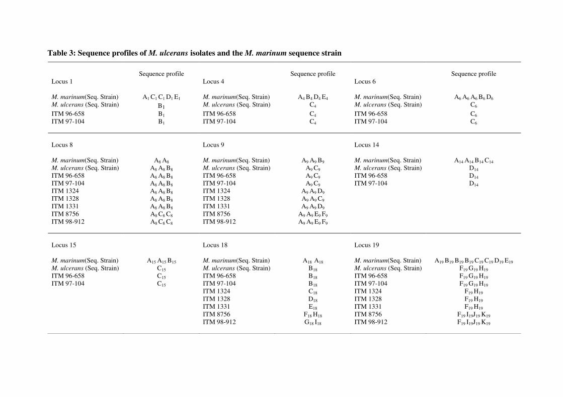
Table 3: Sequence profiles of M. ulcerans isolates and the M. marinum sequence strain
Sequence profile Sequence profile Sequence profile Locus 1 Locus 4 Locus 6 M. marinum(Seq. Strain) A1 C1 C1 D1 E1 M. marinum(Seq. Strain) A4 B4 D4 E4 M. marinum(Seq. Strain) A6 A6 A6 B6 D6 M. ulcerans (Seq. Strain) B1 M. ulcerans (Seq. Strain) C4 M. ulcerans (Seq. Strain) C6
ITM 96-658 B1 ITM 96-658 C4 ITM 96-658 C6 ITM 97-104 B1 ITM 97-104 C4 ITM 97-104 C6 Locus 8 Locus 9 Locus 14 M. marinum(Seq. Strain) A8 A8 M. marinum(Seq. Strain) A9 A9 B9 M. marinum(Seq. Strain) A14 A14 B14 C14 M. ulcerans (Seq. Strain) A8 A8 B8 M. ulcerans (Seq. Strain) A9 C9 M. ulcerans (Seq. Strain) D14 ITM 96-658 A8 A8 B8 ITM 96-658 A9 C9 ITM 96-658 D14 ITM 97-104 A8 A8 B8 ITM 97-104 A9 C9 ITM 97-104 D14 ITM 1324 A8 A8 B8 ITM 1324 A9 A9 D9 ITM 1328 A8 A8 B8 ITM 1328 A9 A9 C9 ITM 1331 A8 A8 B8 ITM 1331 A9 A9 D9 ITM 8756 A8 C8 C8 ITM 8756 A9 A9 E9 F9 ITM 98-912 A8
C8 C8 ITM 98-912 A9 A9 E9 F9 Locus 15 Locus 18 Locus 19 M. marinum(Seq. Strain) A15 A15 B15 M. marinum(Seq. Strain) A18 A18 M. marinum(Seq. Strain) A19 B19 B19 B19 C19 C19 D19 E19 M. ulcerans (Seq. Strain) C15 M. ulcerans (Seq. Strain) B18 M. ulcerans (Seq. Strain) F19 G19 H19 ITM 96-658 C15 ITM 96-658 B18 ITM 96-658 F19 G19 H19 ITM 97-104 C15 ITM 97-104 B18 ITM 97-104 F19 G19 H19 ITM 1324 C18 ITM 1324 F19 H19 ITM 1328 D18 ITM 1328 F19 H19 ITM 1331 E18 ITM 1331 F19 H19 ITM 8756 F18 H18 ITM 8756 F19 I19J19 K19 ITM 98-912 G18 I18 ITM 98-912 F19 I19J19 K19
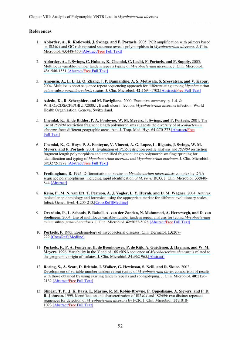
Chapter VIII: Analysis of Polymorphic VNTR Loci in Mycobacterium ulcerans __________________________________________________________________________________________
92
References
1. Ablordey, A., R. Kotlowski, J. Swings, and F. Portaels. 2005. PCR amplification with primers based on IS2404 and GC-rich repeated sequence reveals polymorphism in Mycobacterium ulcerans. J. Clin. Microbiol. 43:448-450.[Abstract/Free Full Text]
2. Ablordey, A., J. Swings, C. Hubans, K. Chemlal, C. Locht, F. Portaels, and P. Supply. 2005. Multilocus variable-number tandem repeats typing of Mycobacterium ulcerans. J. Clin. Microbiol. 43:1546-1551.[Abstract/Free Full Text]
3. Amonsin. A., L. L. Li, Q. Zhang, J. P. Bannantine, A. S. Motiwala, S. Sreevatsan, and V. Kapur. 2004. Multilocus short sequence repeat sequencing approach for differentiating among Mycobacteriun
avium subsp paratuberculosis strains. J. Clin. Microbiol. 42:1694-1702.[Abstract/Free Full Text]
4. Asiedu, K., R. Scherpbier, and M. Raviglione. 2000. Executive summary, p. 1-4. In W.H.O./CDS/CPE/GBUI/2000.1. Buruli ulcer infection: Mycobacterium ulcerans infection. World Health Organization, Geneva, Switzerland.
5. Chemlal, K., K. de Ridder, P. A. Fonteyne, W. M. Meyers, J. Swings, and F. Portaels. 2001. The use of IS2404 restriction fragment length polymorphisms suggests the diversity of Mycobacterium
ulcerans from different geographic areas. Am. J. Trop. Med. Hyg. 64:270-273.[Abstract/Free Full Text]
6. Chemlal, K., G. Huys, P. A. Fonteyne, V. Vincent, A. G. Lopez, L. Rigouts, J. Swings, W. M. Meyers, and F. Portaels. 2001. Evaluation of PCR-restriction profile analysis and IS2404 restriction fragment length polymorphism and amplified fragment length polymorphism fingerprinting for identification and typing of Mycobacterium ulcerans and Mycobacterium marinum. J. Clin. Microbiol. 39:3272-3278.[Abstract/Free Full Text]
7. Frothingham, R. 1995. Differentiation of strains in Mycobacterium tuberculosis complex by DNA sequence polymorphisms, including rapid identification of M. bovis BCG. J. Clin. Microbiol. 33:840-844.[Abstract]
8. Keim, P., M. N. van Ert, T. Pearson, A. J. Vogler, L. Y. Huynh, and D. M. Wagner. 2004. Anthrax molecular epidemiology and forensics: using the appropriate marker for different evolutionary scales. Infect. Genet. Evol. 4:205-213.[CrossRef][Medline]
9. Overduin, P., L. Schouls, P. Roholl, A. van der Zanden, N. Mahmmod, A. Herrewegh, and D. van Soolingen. 2004. Use of multilocus variable-number tandem repeat analysis for typing Mycobacterium
avium subsp. paratuberculosis. J. Clin. Microbiol. 42:5022-5028.[Abstract/Free Full Text]
10. Portaels, F. 1995. Epidemiology of mycobacterial diseases. Clin. Dermatol. 13:207-222.[CrossRef][Medline]
11. Portaels, F., P. A. Fonteyne, H. de Beenhouwer, P. de Rijk, A. Guédénon, J. Hayman, and W. M. Meyers. 1996. Variability in the 3' end of 16S rRNA sequence of Mycobacterium ulcerans is related to the geographic origin of isolates. J. Clin. Microbiol. 34:962-965.[Abstract]
12. Roring, S., A. Scott, D. Brittain, I. Walker, G. Hewinson, S. Neill, and R. Skuce. 2002. Development of variable-number tandem repeat typing of Mycobacterium bovis: comparison of results with those obtained by using existing tandem repeats and spoligotyping. J. Clin. Microbiol. 40:2126-2132.[Abstract/Free Full Text]
13. Stinear, T. P., J. K. Davis, L. Marino, R. M. Robin-Browne, F. Oppedisano, A. Sievers, and P. D. R. Johnson. 1999. Identification and characterization of IS2404 and IS2606: two distinct repeated sequences for detection of Mycobacterium ulcerans by PCR. J. Clin. Microbiol. 37:1018-1023.[Abstract/Free Full Text]
Chapter VIII: Analysis of Polymorphic VNTR Loci in Mycobacterium ulcerans __________________________________________________________________________________________
93
14. Stinear, T. P., J. K. Davis, G. A. Jenkins, F. Portaels, B. C. Ross, F. Oppedisano, M. Purcell, J Hayman, and P. D. R. Johnson. 2000. A simple PCR method for rapid genotype analysis of Mycobacterium ulcerans. J. Clin. Microbiol. 38:1482-1487.[Abstract/Free Full Text]
15. Stinear, T. P., G. A. Jenkins, P. D. R J. Johnson, and J. K. Davis. 2000. Comparative genetic analysis of Mycobacterium ulcerans and Mycobacterium marinum reveals evidence of recent divergence. J. Bacteriol. 182:6322-6330.[Abstract/Free Full Text]
16. Stragier, P., A. Ablordey, W. M. Meyers, and F. Portaels. 2005. Genotyping Mycobacterium
ulcerans and Mycobacterium marinum by using mycobacterial interspersed repetitive units. J. Bacteriol. 187:1639-1647.[Abstract/Free Full Text]
17. Supply, P., E. Mazars, S. Lesjean, V. Vincent, B. Gicquel, and C. Locht. 2000. Variable human minisatellite-like regions in the Mycobacterium tuberculosis genome. Mol. Microbiol. 39:3563-3571.
18. Vincent Levi-Frebault, V., and F. Portaels. 1992. Proposed minimal standards for the genus mycobacterium and description of new slowly growing mycobacteria species. Int. J. Syst. Bacteriol. 42:315-323.[Abstract]


Chapter IX: General discussion and conclusions __________________________________________________________________________________________
95
Chapter IX: General discussion and conclusions

Chapter IX: General discussion and conclusions __________________________________________________________________________________________
96
9.1 Abstract
The aim of this section is to review the studies whose evaluations of the discriminatory power
of the three popular typing methods for M. tuberculosis complex (MTC) strains reach the
conclusions that IS6110 RFLP has a higher discriminatory power than MIRU-VNTR which
itself is more discriminative than spoligotyping. In contrast, we additionally show that
spoligotyping can be more discriminative than MIRU-VNTR typing, depending on the
geographical location and genotype family chosen. Furthermore, we review that these
genotyping tools are stable enough over time thus justifying their usage although possible
convergent evolution and little heterogeneity may require the use of several different and
appropriate genotyping tools to exclude biases. This is particularly recommended in high
incidence countries where heterogeneity within strains is low and convergent evolution more
probable.
We also review the importance of factors such as the awareness of reinfection versus relapse
and mixed infections versus microevolution of a single strain to human tuberculosis control,
especially in high incidence, African countries. Furthermore, knowledge about the degree and
risk factors of ongoing tuberculosis transmission could reveal new target groups and result in
suggesting an improved Direct Observed Treatment Short course (DOTS). However, adapting
DOTS to distinct population groups cannot be suggested without knowing the variability of
locally perceived tuberculosis. Therefore the linking of social science with molecular
epidemiological studies is suggested.
The same molecular epidemiological methods prove additionally useful in investigating MTC
transmission within or between different animal species. In the case of M. bovis, some
animals were reviewed to act as maintenance and spillover hosts, respectively. The
knowledge of the natural reservoirs of M. bovis is important as it has potential to be
transmitted to humans. However, so far, no M. bovis has been found in human samples of
Chad and there are several possible explanations for this discussed.
We conclude this chapter with a short overview of the genotyping of a different
mycobacterium, M. ulcerans. In contrast to tuberculosis much less is known about the
transmission of Buruli Ulcer, the disease caused by M. ulcerans, but a new VNTR typing may
be very promising for micro epidemiological studies in the future.
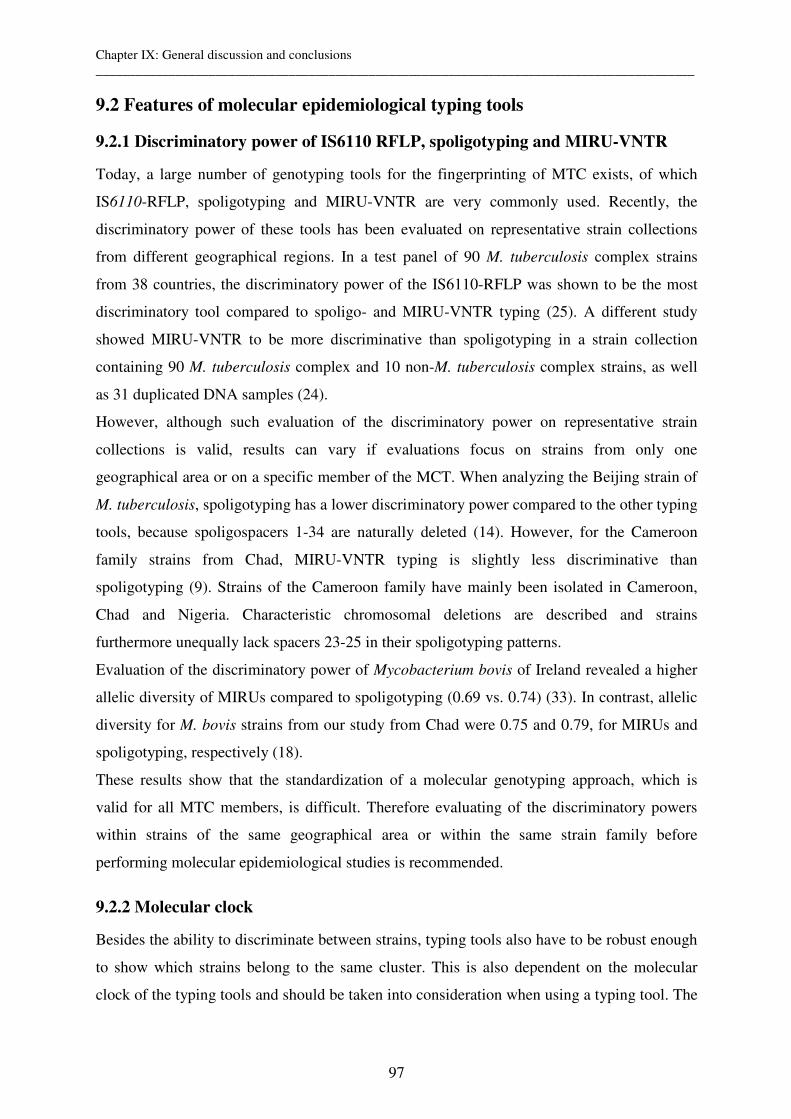
Chapter IX: General discussion and conclusions __________________________________________________________________________________________
97
9.2 Features of molecular epidemiological typing tools
9.2.1 Discriminatory power of IS6110 RFLP, spoligotyping and MIRU-VNTR
Today, a large number of genotyping tools for the fingerprinting of MTC exists, of which
IS6110-RFLP, spoligotyping and MIRU-VNTR are very commonly used. Recently, the
discriminatory power of these tools has been evaluated on representative strain collections
from different geographical regions. In a test panel of 90 M. tuberculosis complex strains
from 38 countries, the discriminatory power of the IS6110-RFLP was shown to be the most
discriminatory tool compared to spoligo- and MIRU-VNTR typing (25). A different study
showed MIRU-VNTR to be more discriminative than spoligotyping in a strain collection
containing 90 M. tuberculosis complex and 10 non-M. tuberculosis complex strains, as well
as 31 duplicated DNA samples (24).
However, although such evaluation of the discriminatory power on representative strain
collections is valid, results can vary if evaluations focus on strains from only one
geographical area or on a specific member of the MCT. When analyzing the Beijing strain of
M. tuberculosis, spoligotyping has a lower discriminatory power compared to the other typing
tools, because spoligospacers 1-34 are naturally deleted (14). However, for the Cameroon
family strains from Chad, MIRU-VNTR typing is slightly less discriminative than
spoligotyping (9). Strains of the Cameroon family have mainly been isolated in Cameroon,
Chad and Nigeria. Characteristic chromosomal deletions are described and strains
furthermore unequally lack spacers 23-25 in their spoligotyping patterns.
Evaluation of the discriminatory power of Mycobacterium bovis of Ireland revealed a higher
allelic diversity of MIRUs compared to spoligotyping (0.69 vs. 0.74) (33). In contrast, allelic
diversity for M. bovis strains from our study from Chad were 0.75 and 0.79, for MIRUs and
spoligotyping, respectively (18).
These results show that the standardization of a molecular genotyping approach, which is
valid for all MTC members, is difficult. Therefore evaluating of the discriminatory powers
within strains of the same geographical area or within the same strain family before
performing molecular epidemiological studies is recommended.
9.2.2 Molecular clock
Besides the ability to discriminate between strains, typing tools also have to be robust enough
to show which strains belong to the same cluster. This is also dependent on the molecular
clock of the typing tools and should be taken into consideration when using a typing tool. The
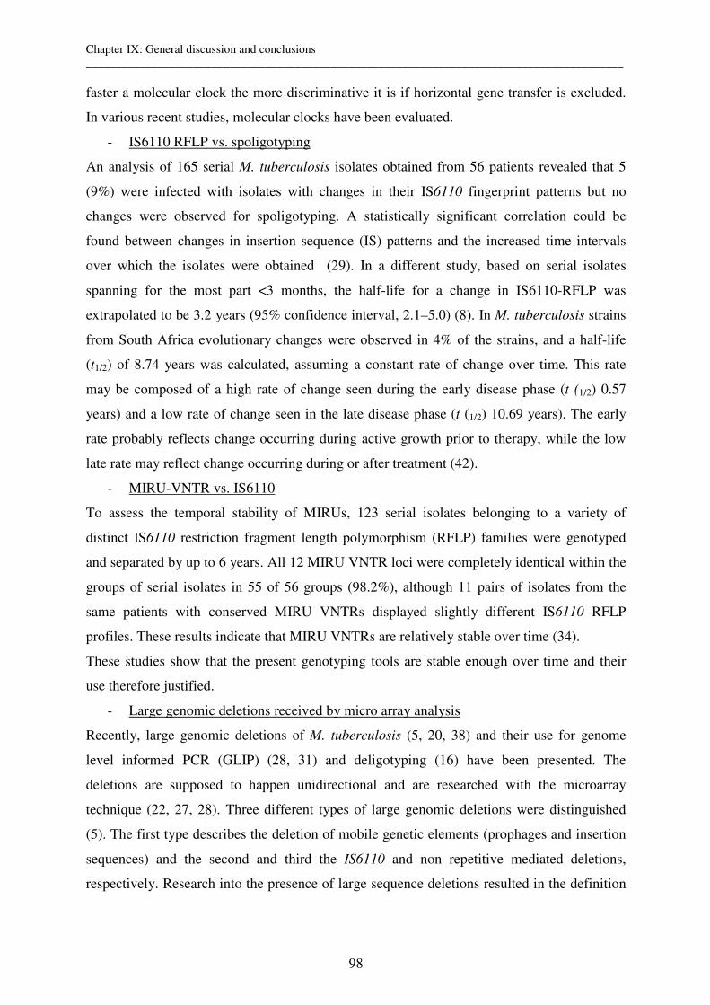
Chapter IX: General discussion and conclusions __________________________________________________________________________________________
98
faster a molecular clock the more discriminative it is if horizontal gene transfer is excluded.
In various recent studies, molecular clocks have been evaluated.
- IS6110 RFLP vs. spoligotyping
An analysis of 165 serial M. tuberculosis isolates obtained from 56 patients revealed that 5
(9%) were infected with isolates with changes in their IS6110 fingerprint patterns but no
changes were observed for spoligotyping. A statistically significant correlation could be
found between changes in insertion sequence (IS) patterns and the increased time intervals
over which the isolates were obtained (29). In a different study, based on serial isolates
spanning for the most part <3 months, the half-life for a change in IS6110-RFLP was
extrapolated to be 3.2 years (95% confidence interval, 2.1–5.0) (8). In M. tuberculosis strains
from South Africa evolutionary changes were observed in 4% of the strains, and a half-life
(t1/2) of 8.74 years was calculated, assuming a constant rate of change over time. This rate
may be composed of a high rate of change seen during the early disease phase (t (1/2) 0.57
years) and a low rate of change seen in the late disease phase (t (1/2) 10.69 years). The early
rate probably reflects change occurring during active growth prior to therapy, while the low
late rate may reflect change occurring during or after treatment (42).
- MIRU-VNTR vs. IS6110
To assess the temporal stability of MIRUs, 123 serial isolates belonging to a variety of
distinct IS6110 restriction fragment length polymorphism (RFLP) families were genotyped
and separated by up to 6 years. All 12 MIRU VNTR loci were completely identical within the
groups of serial isolates in 55 of 56 groups (98.2%), although 11 pairs of isolates from the
same patients with conserved MIRU VNTRs displayed slightly different IS6110 RFLP
profiles. These results indicate that MIRU VNTRs are relatively stable over time (34).
These studies show that the present genotyping tools are stable enough over time and their
use therefore justified.
- Large genomic deletions received by micro array analysis
Recently, large genomic deletions of M. tuberculosis (5, 20, 38) and their use for genome
level informed PCR (GLIP) (28, 31) and deligotyping (16) have been presented. The
deletions are supposed to happen unidirectional and are researched with the microarray
technique (22, 27, 28). Three different types of large genomic deletions were distinguished
(5). The first type describes the deletion of mobile genetic elements (prophages and insertion
sequences) and the second and third the IS6110 and non repetitive mediated deletions,
respectively. Research into the presence of large sequence deletions resulted in the definition

Chapter IX: General discussion and conclusions __________________________________________________________________________________________
99
of epidemiologically important clones (22, 28, 31). Furthermore, the deletions often hamper
or delete potential ORF and might therefore affect the feature of the pathogen.
The non repetitive mediated deletions (Type 3) are also called diagnostic deletions and allow
an isolate to be unequivocally placed in a strain family. The value of genomic deletions for
finding large family groups is considered to be superior as the turnaround time is slower than
for all other genotyping methods. Based on the assumption that all stains of a strain family
represent a cluster, the further use of genotyping methods leads to more refinement and
eventual subclustering.
9.2.3 Low heterogeneity and Convergence: the need for higher discriminatory
power
- Adding of VNTRs
Comparison of our data from Chad with Ireland (18, 33) showed a low discriminatory power
of MIRUs compared to similar studies on M. tuberculosis. However, the VNTR typing was
shown to become more discriminative when adding further VNTRs e.g. exact tandem repeats
(ETRs) to the 12 MIRUs. Adding ETR A, B, and C resulted in higher discrimination than the
use of all 12 MIRUs combined. Therefore a combination of different types of VNTRs is
suggested (18, 33).
- Convergence in VNTR typing because highly polymorphic loci lower sensitivity of
clustering
VNTR typing of MTC strains from different settings have shown different allelic diversities
at the VNTR loci. However, MIRU 40 and 26 seem to be very polymorphic for M.
tuberculosis. MIRU 26 is also polymorphic for M. bovis with, in contrast, ETR A and B much
more diverse than MIRU 40 (18).
The higher turnaround time of certain VNTR loci can result in a convergent evolution of
epidemiologically unlinked strains and therefore lower the sensitivity of clustering which
means that certain strains found in Nigeria (6) have the same VNTR type as strains from
Chad and convergence cannot be excluded. However, this problem can best be addressed by
the inclusion of a second genotyping method, e.g. spoligotyping and a sensitivity of clustering
of mostly 100% is common.
- Convergence Spoligotyping
Possible convergent evolution of spoligotyping has also been shown. Homologous
recombination between adjacent IS6110 elements leads to extensive deletion in the DR
region, again demonstrating a dependent evolutionary mechanism. Different isolates from the
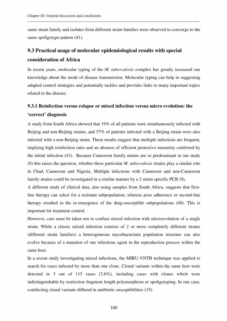
Chapter IX: General discussion and conclusions __________________________________________________________________________________________
100
same strain family and isolates from different strain families were observed to converge to the
same spoligotype pattern (41).
9.3 Practical usage of molecular epidemiological results with special
consideration of Africa
In recent years, molecular typing of the M. tuberculosis complex has greatly increased our
knowledge about the mode of disease transmission. Molecular typing can help in suggesting
adapted control strategies and potentially tackles and provides links to many important topics
related to the disease.
9.3.1 Reinfection versus relapse or mixed infection versus micro evolution: the
‘correct’ diagnosis
A study from South Africa showed that 19% of all patients were simultaneously infected with
Beijing and non-Beijing strains, and 57% of patients infected with a Beijing strain were also
infected with a non-Beijing strain. These results suggest that multiple infections are frequent,
implying high reinfection rates and an absence of efficient protective immunity conferred by
the initial infection (43). Because Cameroon family strains are so predominant in our study
(9) this raises the question, whether these particular M. tuberculosis strains play a similar role
in Chad, Cameroon and Nigeria. Multiple infections with Cameroon and non-Cameroon
family strains could be investigated in a similar manner by a 2 strain specific PCR (9).
A different study of clinical data, also using samples from South Africa, suggests that first-
line therapy can select for a resistant subpopulation, whereas poor adherence or second-line
therapy resulted in the re-emergence of the drug-susceptible subpopulations (40). This is
important for treatment control.
However, care must be taken not to confuse mixed infection with microevolution of a single
strain. While a classic mixed infection consists of 2 or more completely different strains
(different strain families) a heterogeneous mycobacterium population structure can also
evolve because of a mutation of one infectious agent in the reproduction process within the
same host.
In a recent study investigating mixed infections, the MIRU-VNTR technique was applied to
search for cases infected by more than one clone. Clonal variants within the same host were
detected in 3 out of 115 cases (2.6%), including cases with clones which were
indistinguishable by restriction fragment length polymorphism or spoligotyping. In one case,
coinfecting clonal variants differed in antibiotic susceptibilities (15).

Chapter IX: General discussion and conclusions __________________________________________________________________________________________
101
In another study, infections with different bacterial subpopulations were detected in samples
from eight patients (8.2%), with the frequency of detectable mixed infections in the study
population estimated to be 2.1%. Genotypic variations were found to be independent of drug
susceptibility, and the various molecular markers evolved independently in most cases (35).
In conclusion, we suggest that MIRU-VNTR typing is a potentially valuable tool to
investigate reinfection, mixed infection, microevolution or the relapse of tuberculosis. These
results may prove especially important for adapted human tuberculosis control in high
incidence countries such as Chad
9.3.2 Degree of ongoing transmission, global mycobacterial population structure
and outbreak investigations
While the basic principles of tuberculosis transmission are well understood, the advent and
use of molecular methods in epidemiological studies have shown that traditional contact
tracing may not always be accurate, leading to identification of previously unrecognized
source cases. The assessment of recent transmission and/or reactivation (44) remains of
particular interest in resource poor settings where scarce resources for control need to be
directed to transmission hotspots. In Chad, the overall heterogeneity using spoligotyping and
MIRU/ETR typing was high in 40 M. tuberculosis samples analyzed, however we also found
that a substantial proportion of strains (33 %) are part of the Cameroon family. Within this
family, strains evolved differently and therefore have slightly different VNTR and/or
spoligotypes (9). We propose that evolved strains, derived from a common ancestor should be
considered together with clustered strains to represent chains of ongoing transmission. In a
recent study, a data set which identifies newly evolved strains has been generated. Inclusion
of these evolved strains into various molecular epidemiological calculations significantly
increases the ability to estimate ongoing transmission in a particular high incidence study
setting (39).
In our study the Cameroon family was the most predominant group of M. tuberculosis strains
which is also most common for Nigeria, Cameroon and Chad (9). In the future, investigation
into the reasons why some strains are predominant may clarify which bacterial factors
contribute to disease. This knowledge has the potential to influence control and prevention
strategies for tuberculosis (26). Predominant strains should also be identified in developing
countries to study the differential pathogenesis between strains (12) and also to reveal the true
extent of genetic diversity of the pathogen (4).
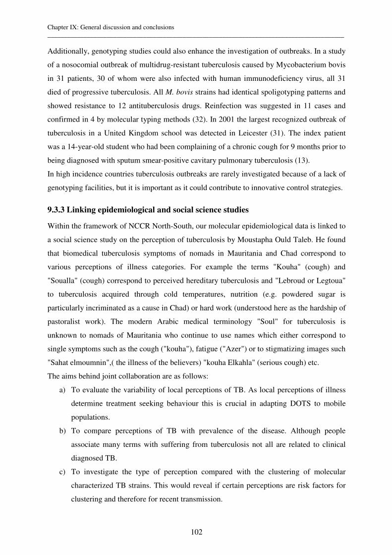
Chapter IX: General discussion and conclusions __________________________________________________________________________________________
102
Additionally, genotyping studies could also enhance the investigation of outbreaks. In a study
of a nosocomial outbreak of multidrug-resistant tuberculosis caused by Mycobacterium bovis
in 31 patients, 30 of whom were also infected with human immunodeficiency virus, all 31
died of progressive tuberculosis. All M. bovis strains had identical spoligotyping patterns and
showed resistance to 12 antituberculosis drugs. Reinfection was suggested in 11 cases and
confirmed in 4 by molecular typing methods (32). In 2001 the largest recognized outbreak of
tuberculosis in a United Kingdom school was detected in Leicester (31). The index patient
was a 14-year-old student who had been complaining of a chronic cough for 9 months prior to
being diagnosed with sputum smear-positive cavitary pulmonary tuberculosis (13).
In high incidence countries tuberculosis outbreaks are rarely investigated because of a lack of
genotyping facilities, but it is important as it could contribute to innovative control strategies.
9.3.3 Linking epidemiological and social science studies
Within the framework of NCCR North-South, our molecular epidemiological data is linked to
a social science study on the perception of tuberculosis by Moustapha Ould Taleb. He found
that biomedical tuberculosis symptoms of nomads in Mauritania and Chad correspond to
various perceptions of illness categories. For example the terms "Kouha" (cough) and
"Soualla" (cough) correspond to perceived hereditary tuberculosis and "Lebroud or Legtoua"
to tuberculosis acquired through cold temperatures, nutrition (e.g. powdered sugar is
particularly incriminated as a cause in Chad) or hard work (understood here as the hardship of
pastoralist work). The modern Arabic medical terminology "Soul" for tuberculosis is
unknown to nomads of Mauritania who continue to use names which either correspond to
single symptoms such as the cough ("kouha"), fatigue ("Azer") or to stigmatizing images such
"Sahat elmoumnin",( the illness of the believers) "kouha Elkahla" (serious cough) etc.
The aims behind joint collaboration are as follows:
a) To evaluate the variability of local perceptions of TB. As local perceptions of illness
determine treatment seeking behaviour this is crucial in adapting DOTS to mobile
populations.
b) To compare perceptions of TB with prevalence of the disease. Although people
associate many terms with suffering from tuberculosis not all are related to clinical
diagnosed TB.
c) To investigate the type of perception compared with the clustering of molecular
characterized TB strains. This would reveal if certain perceptions are risk factors for
clustering and therefore for recent transmission.
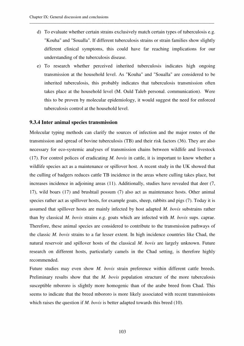
Chapter IX: General discussion and conclusions __________________________________________________________________________________________
103
d) To evaluate whether certain strains exclusively match certain types of tuberculosis e.g.
"Kouha" and "Soualla". If different tuberculosis strains or strain families show slightly
different clinical symptoms, this could have far reaching implications for our
understanding of the tuberculosis disease.
e) To research whether perceived inherited tuberculosis indicates high ongoing
transmission at the household level. As "Kouha" and "Soualla" are considered to be
inherited tuberculosis, this probably indicates that tuberculosis transmission often
takes place at the household level (M. Ould Taleb personal. communication). Were
this to be proven by molecular epidemiology, it would suggest the need for enforced
tuberculosis control at the household level.
9.3.4 Inter animal species transmission
Molecular typing methods can clarify the sources of infection and the major routes of the
transmission and spread of bovine tuberculosis (TB) and their risk factors (36). They are also
necessary for eco-systemic analyses of transmission chains between wildlife and livestock
(17). For control polices of eradicating M. bovis in cattle, it is important to know whether a
wildlife species act as a maintenance or spillover host. A recent study in the UK showed that
the culling of badgers reduces cattle TB incidence in the areas where culling takes place, but
increases incidence in adjoining areas (11). Additionally, studies have revealed that deer (7,
17), wild boars (17) and brushtail possum (7) also act as maintenance hosts. Other animal
species rather act as spillover hosts, for example goats, sheep, rabbits and pigs (7). Today it is
assumed that spillover hosts are mainly infected by host adapted M. bovis substrains rather
than by classical M. bovis strains e.g. goats which are infected with M. bovis sups. caprae.
Therefore, these animal species are considered to contribute to the transmission pathways of
the classic M. bovis strains to a far lesser extent. In high incidence countries like Chad, the
natural reservoir and spillover hosts of the classical M. bovis are largely unknown. Future
research on different hosts, particularly camels in the Chad setting, is therefore highly
recommended.
Future studies may even show M. bovis strain preference within different cattle breeds.
Preliminary results show that the M. bovis population structure of the more tuberculosis
susceptible mbororo is slightly more homogenic than of the arabe breed from Chad. This
seems to indicate that the breed mbororo is more likely associated with recent transmissions
which raises the question if M. bovis is better adapted towards this breed (10).

Chapter IX: General discussion and conclusions __________________________________________________________________________________________
104
9.3.5 Zoonotic transmission
Last but not least, molecular epidemiology can also investigate the transmission of
tuberculosis between animals and humans. M. bovis is considered to have zoonotic potential
and it is certainly very important to know the percentage of human tuberculosis caused by M.
bovis especially in high incidence countries (3). In Nigeria the rate of M. bovis from humans
was 4 M. bovis strains from 102 sputum samples (21) and 3 M. bovis strains cultured from 55
sputum samples (6). However, spoligo- and VNTR-typing in the latter study revealed that the
three M. bovis strains isolated from humans did not match the 15 M. bovis strains isolated
from cattle. This was despite the fact that they all had spoligotyping patterns similar to the
dominant pattern found in cattle. This surprising result may be due to the small number of
strains analyzed from cattle but it also raises questions about direct animal to human
transmission. Similarly, the spoligotypes of 4/5 M. bovis strains isolated from humans from
Tanzania did not match the types of 31 M. bovis strains from Tanzanian cattle (23). In
Cameroon, only a single M. bovis strain was isolated from 455 human specimens suggesting
the low importance of M. bovis in the overall burden of human tuberculosis in this country
(30).
To prove zoonotic transmission was one of the objectives of this thesis. It was thought that
the combination of high prevalence of M. bovis in cattle and the habit of drinking
unpasteurized milk and eating raw meat presented significant risks for transmission.
However, no M. bovis has so far been found in human samples of Chad. Here are several
possible explanations:
a) Insufficient numbers and incorrect study populations
The numbers of isolated MTC strains from humans and animals from Chad are very few but
are the very first ones isolated in this country (9, 10). As the first objective of the thesis of C.
Diguimbaye was to establish a mycobacterial laboratory in Chad, attention has not been given
to a high number or a careful selection of isolates. Only pulmonary and a few urine samples
of human patients from the main hospital of N’Djaména were included (9).
An ongoing study currently additionally analyzes extra pulmonary tuberculosis in children
from N’Djaména. However, preliminary results show that M. bovis is not present in these
samples. In future, the inclusion of extra pulmonary samples of adults is also planned.
Additionally, we plan to evaluate the burden of M. bovis infection within nomadic pastoralists
who have close contact with cattle and are therefore at high risk.
b) Reduced risk of transmission in semi-arid climates and in extensive livestock systems
Chapter IX: General discussion and conclusions __________________________________________________________________________________________
105
The sampling of tuberculosis specimens from Chad took place in N’Djaména where the
climate is dry and semi-arid. There are indications that zoonotic transmission is more
probable in humid areas and therefore inclusion of a more southerly region should be taken
into consideration. Furthermore, the extensive livestock systems used by the pastoralists does
not involve the use of stables. This may also reduce the risk of zoonotic transmission.
c) Less pathogenic strains
It is known that humans are not natural reservoirs for M. bovis, but can act as spillover hosts.
However, it is not known whether certain M. bovis strains are more pathogenic for humans
than others. As no M. bovis in humans has been found, this may mean that the Chadian strains
are less pathogenic for humans.
9.4 Genotyping in M. ulcerans
While there are many studies evaluating genotyping methods for MTC, the situation for M.
ulcerans is so far not as developed. The discriminatory power of genotyping methods is still
insufficient for performing micro epidemiological studies. Therefore new genotyping
methods have to be developed and evaluated.
As for MTC, VNTR typing of MIRUs (37) and other VNTRs (2) have been implemented for
M. ulcerans. Evaluations of the discriminatory power showed the ability to separate isolates
on a continental level but not within the contents. A study evaluating the sequences of VNTR
loci showed that sequencing does not enhance the discriminatory power (1). It is therefore of
special importance that a new VNTR-locus (designated STI) and a previously presented
MIRU locus have recently been shown to discriminate between M. ulcerans strain within
Ghana, and therefore within Africa, for the first time (19).
9.5 Ten key messages and recommendations of this thesis
1. MIRU/VNTR typing for M. tuberculosis strains from Chad is as discriminative as
spoligotyping. Recommendation: MIRU-VNTR typing is a very promising tool for
investigating mixed infection which might occur in high percentages in high incidence
countries like Chad (Chapter III).
2. Using microarry analysis, the Cameroon family was identified as a highly prevalent clone
and characterized 33 % of M. tuberculosis strains of Chad. Fortunately, this clone is not
currently associated with drug resistance. Recommendation: Evaluation of risk factors for
recent transmissions of Cameroon family members could lead to recommendations on how to
Chapter IX: General discussion and conclusions __________________________________________________________________________________________
106
interrupt the transmission of this clone and contribute to alternative control strategies
(Chapter III).
3. Although the risk of infection with M. bovis is high for humans in Chad, analysis of 40
pulmonary and urine samples of clinical suspected tuberculosis cases revealed that none of
the patients were infected by M. bovis (Chapter III). Recommendation: Evaluation of extra
pulmonary samples from risk population groups (e.g. pastoralists). It is also recommended to
look at M. bovis as a possible subpopulation of mixed infections in humans.
4. Drug resistance testing of 40 M. tuberculosis cases from Chad revealed a high percentage
of Isoniazid resistance (33 %) (Chapter III). Recommendation: Carry out molecular
epidemiological studies analyzing the risk factors of transmission of drug resistant strains.
Implementation of a method permitting faster detection of resistance is also desirable.
5. A study carried out in a slaughter house in Chad revealed that the mbororo breed is more
susceptible to M. bovis than the arabe breed. Recommendation: Investigate the
immunological and genetic consequences (Chapter IV).
6. Spoligo- and MIRU-VNTR typing revealed a remarkably homogenetic population structure
within M. bovis strains. However, the use of 2 MIRUs and 3 ETRs allows reasonably high
discrimination to take place. Recommendation: Use these 5 loci to investigate and prove
possible animal to human transmission and other molecular epidemiological objectives in the
future (Chapter V).
7. A high proportion of Chadian (63/67), Nigerian and Cameroon M. bovis strains are
members of the same clone defined by a large genomic deletion. There are indications that
subclones found in the different study sites are due to clonal expansion. Recommendation:
Investigate the possible spread of M. bovis strains from one country to the other (Chapter VI).
However, we also recommend looking at different hosts (camels, goats) in order to show if
other animal reservoirs (maintenance hosts) exist for this particular M. bovis clone.
8. A high proportion of Chadian strains showed deletion affecting ESAT-6 family members,
which are promising targets for new diagnostics and novel vaccine candidates.
Recommendation: Investigate genome organization across M. tuberculosis strains to inform
the choice of antigens used in a diagnostic mix (Chapter VI).
9. A new and a previously described VNTR locus revealed genetic diversity within African
M. ulcerans strains for the first time. Recommendation: Use these two VNTRs for extended
analysis on various panels of M. ulcerans. However, development of new genotyping tools
has to continue as the discriminatory power is still low. This could allow for micro
epidemiological studies in the future (Chapter VII).
Chapter IX: General discussion and conclusions __________________________________________________________________________________________
107
10. Sequencing of various VNTRs revealed that the discriminatory power of sequencing is
only slightly higher than that of Agarose Gel electrophoresis. Recommendation: Given that
sequencing does not significantly enhance the discriminatory power, and is more laborious
and expensive, it is not recommended for use, especially not within laboratories with
infrastructural constraints (Chapter VIII).
9.6 References of conclusion
1. Ablordey, A., M. Hilty, P. Stragier, J. Swings, and F. Portaels. 2005. Comparative Nucleotide Sequence Analysis of Polymorphic Variable-Number Tandem-Repeat Loci in Mycobacterium ulcerans. J.Clin.Microbiol. 43:5281-5284.
2. Ablordey, A., J. Swings, C. Hubans, K. Chemlal, C. Locht, F. Portaels, and P. Supply. 2005. Multilocus variable-number tandem repeat typing of Mycobacterium ulcerans. J.Clin.Microbiol. 43:1546-1551.
3. Ayele, W. Y., S. D. Neill, J. Zinsstag, M. G. Weiss, and I. Pavlik. 2004. Bovine tuberculosis: an old disease but a new threat to Africa. Int.J.Tuberc.Lung Dis. 8:924-937.
4. Borsuk, S., M. M. Dellagostin, S. G. Madeira, C. Lima, M. Boffo, I. Mattos, P. E. Almeida da
Silva, and O. A. Dellagostin. 2005. Molecular characterization of Mycobacterium tuberculosis isolates in a region of Brazil with a high incidence of tuberculosis. Microbes.Infect. 7:1338-1344.
5. Brosch, R., S. V. Gordon, M. Marmiesse, P. Brodin, C. Buchrieser, K. Eiglmeier, T. Garnier, C.
Gutierrez, G. Hewinson, K. Kremer, L. M. Parsons, A. S. Pym, S. Samper, D. van Soolingen, and S. T. Cole. 2002. A new evolutionary scenario for the Mycobacterium tuberculosis complex. Proc.Natl.Acad.Sci.U.S.A 99:3684-3689.
6. Cadmus, S., S. Palmer, M. Okker, J. Dale, K. Gover, N. Smith, K. Jahans, R. G. Hewinson, and S. V. Gordon. 2006. Molecular Analysis of Human and Bovine Tubercle Bacilli from a Local Setting in Nigeria. Journal of Clinical Microbiology 44:29-34.
7. Corner, L. A. 2005. The role of wild animal populations in the epidemiology of tuberculosis in domestic animals: How to assess the risk. Vet.Microbiol.
8. de Boer, A. S., M. W. Borgdorff, P. E. de Haas, N. J. Nagelkerke, J. D. van Embden, and D. van
Soolingen. 1999. Analysis of rate of change of IS6110 RFLP patterns of Mycobacterium tuberculosis based on serial patient isolates. J.Infect.Dis. 180:1238-1244.
9. Diguimbaye C., Hilty M. Ngandolo R., et al. 2006. Molecular characterization and drug resistance testing of Mycobacterium tuberculosis isolates from Chad. J.Clin.Microbio. Apr;44(4):1575-7
10. Diguimbaye-Djaibé C., Hilty M., Ngandolo R, Mahamat HH., Pfyffer G. E., Baggi F., Hewinson
G., Tanner M., Zinsstag J. and Schelling E. Mycobacterium bovis Isolates from Tuberculous Lesions in Chadian Zebu Carcasses. 2006. Emerg.Infect.Dis. 12(5):769-771
11. Donnelly, C. A., R. Woodroffe, D. R. Cox, F. J. Bourne, C. L. Cheeseman, R. S. Clifton-Hadley,
G. Wei, G. Gettinby, P. Gilks, H. Jenkins, W. T. Johnston, A. M. Le Fevre, J. P. McInerney, and W. I. Morrison. 2005. Positive and negative effects of widespread badger culling on tuberculosis in cattle. Nature .
12. Easterbrook, P. J., A. Gibson, S. Murad, D. Lamprecht, N. Ives, A. Ferguson, O. Lowe, P. Mason, A. Ndudzo, A. Taziwa, R. Makombe, L. Mbengeranwa, C. Sola, N. Rastogi, and F. Drobniewski.
Chapter IX: General discussion and conclusions __________________________________________________________________________________________
108
2004. High rates of clustering of strains causing tuberculosis in Harare, Zimbabwe: a molecular epidemiological study. J.Clin.Microbiol. 42:4536-4544.
13. Ewer, K., J. Deeks, L. Alvarez, G. Bryant, S. Waller, P. Andersen, P. Monk, and A. Lalvani. 2003. Comparison of T-cell-based assay with tuberculin skin test for diagnosis of Mycobacterium tuberculosis infection in a school tuberculosis outbreak. Lancet 361:1168-1173.
14. Ferdinand, S., G. Valetudie, C. Sola, and N. Rastogi. 2004. Data mining of Mycobacterium tuberculosis complex genotyping results using mycobacterial interspersed repetitive units validates the clonal structure of spoligotyping-defined families. Res.Microbiol. 155:647-654.
15. Garcia, d., V, R. N. Alonso, S. Andres, M. J. Ruiz Serrano, and E. Bouza. 2005. Characterization of clonal complexity in tuberculosis by mycobacterial interspersed repetitive unit-variable-number tandem repeat typing. J.Clin.Microbiol. 43:5660-5664.
16. Goguet de la Salmoniere YO, C. C. Kim, A. G. Tsolaki, A. S. Pym, M. S. Siegrist, and P. M.
Small. 2004. High-throughput method for detecting genomic-deletion polymorphisms. J.Clin.Microbiol. 42:2913-2918.
17. Hermoso, d. M., A. Parra, A. Tato, J. M. Alonso, J. M. Rey, J. Pena, A. Garcia-Sanchez, J.
Larrasa, J. Teixido, G. Manzano, R. Cerrato, G. Pereira, P. Fernandez-Llario, and d. M. Hermoso. 2005. Bovine tuberculosis in wild boar (Sus scrofa), red deer (Cervus elaphus) and cattle (Bos taurus) in a Mediterranean ecosystem (1992-2004). Prev.Vet.Med.
18. Hilty, M., C. Diguimbaye, E. Schelling, F. Baggi, M. Tanner, and J. Zinsstag. 2005. Evaluation of the discriminatory power of variable number tandem repeat (VNTR) typing of Mycobacterium bovis strains. Vet.Microbiol. 109:217-222.
19. Hilty, M. , D. Yeboah-Manu, D. Boakye, E. Mensah-Quainoo, S. Rondini, E. Schelling, D. Ofori-
Adjei, F. Portaels, J. Zinsstag and G. Pluschke. 2006. A new VNTR locus revealing genetic diversity in Mycobacterium ulcerans isolates from Ghana. J.Bacteriol. Feb;188(4):1462-5
20. Hirsh, A. E., A. G. Tsolaki, K. DeRiemer, M. W. Feldman, and P. M. Small. 2004. Stable association between strains of Mycobacterium tuberculosis and their human host populations. Proc.Natl.Acad.Sci.U.S.A 101:4871-4876.
21. Idigbe, E. O., C. E. Anyiwo, and D. I. Onwujekwe. 1986. Human pulmonary infections with bovine and atypical mycobacteria in Lagos, Nigeria. J.Trop.Med.Hyg. 89:143-148.
22. Kato-Maeda, M., J. T. Rhee, T. R. Gingeras, H. Salamon, J. Drenkow, N. Smittipat, and P. M.
Small. 2001. Comparing genomes within the species Mycobacterium tuberculosis. Genome Res. 11:547-554.
23. Kazwala, R. R., L. J. Kusiluka, K. Sinclair, J. M. Sharp, and C. J. Daborn. 2005. The molecular epidemiology of Mycobacterium bovis infections in Tanzania. Vet.Microbiol.
24. Kremer, K., C. Arnold, A. Cataldi, M. C. Gutierrez, W. H. Haas, S. Panaiotov, R. A. Skuce, P.
Supply, A. G. van der Zanden, and D. van Soolingen. 2005. Discriminatory Power and Reproducibility of Novel DNA Typing Methods for Mycobacterium tuberculosis Complex Strains. J.Clin.Microbiol. 43:5628-5638.
25. Kremer, K., D. van Soolingen, R. Frothingham, W. H. Haas, P. W. M. Hermans, C. Martin, P.
Palittapongarnpim, B. B. Plikaytis, L. W. Riley, M. A. Yakrus, J. M. Musser, and J. D. A. van Embden. 1999. Comparison of methods based on different molecular epidemiological markers for typing of Mycobacterium tuberculosis complex strains: Interlaboratory study of discriminatory power and reproducibility. Journal of Clinical Microbiology 37:2607-2618.
26. Malik, A. N. and P. Godfrey-Faussett. 2005. Effects of genetic variability of Mycobacterium tuberculosis strains on the presentation of disease. Lancet Infect.Dis. 5:174-183.
Chapter IX: General discussion and conclusions __________________________________________________________________________________________
109
27. Mostowy, S., D. Cousins, J. Brinkman, A. Aranaz, and M. A. Behr. 2002. Genomic deletions suggest a phylogeny for the Mycobacterium tuberculosis complex. J.Infect.Dis. 186:74-80.
28. Nguyen, D., P. Brassard, D. Menzies, L. Thibert, R. Warren, S. Mostowy, and M. Behr. 2004. Genomic characterization of an endemic Mycobacterium tuberculosis strain: evolutionary and epidemiologic implications. J.Clin.Microbiol. 42:2573-2580.
29. Niemann, S., E. Richter, and S. Rusch-Gerdes. 1999. Stability of Mycobacterium tuberculosis IS6110 restriction fragment length polymorphism patterns and spoligotypes determined by analyzing serial isolates from patients with drug-resistant tuberculosis. J.Clin.Microbiol. 37:409-412.
30. Niobe-Eyangoh, S. N., C. Kuaban, P. Sorlin, P. Cunin, J. Thonnon, C. Sola, N. Rastogi, V.
Vincent, and M. C. Gutierrez. 2003. Genetic biodiversity of Mycobacterium tuberculosis complex strains from patients with pulmonary tuberculosis in Cameroon. J.Clin.Microbiol. 41:2547-2553.
31. Rajakumar, K., J. Shafi, R. J. Smith, R. A. Stabler, P. W. Andrew, D. Modha, G. Bryant, P.
Monk, J. Hinds, P. D. Butcher, and M. R. Barer. 2004. Use of genome level-informed PCR as a new investigational approach for analysis of outbreak-associated Mycobacterium tuberculosis isolates. J.Clin.Microbiol. 42:1890-1896.
32. Rivero, A., M. Marquez, J. Santos, A. Pinedo, M. A. Sanchez, A. Esteve, S. Samper, and C.
Martin. 2001. High rate of tuberculosis reinfection during a nosocomial outbreak of multidrug-resistant tuberculosis caused by Mycobacterium bovis strain B. Clin.Infect.Dis. 32:159-161.
33. Roring, S., A. N. Scott, H. R. Glyn, S. D. Neill, and R. A. Skuce. 2004. Evaluation of variable number tandem repeat (VNTR) loci in molecular typing of Mycobacterium bovis isolates from Ireland. Vet.Microbiol. 101:65-73.
34. Savine, E., R. M. Warren, G. D. van der Spuy, N. Beyers, P. D. van Helden, C. Locht, and P.
Supply. 2002. Stability of variable-number tandem repeats of mycobacterial interspersed repetitive units from 12 loci in serial isolates of Mycobacterium tuberculosis. Journal of Clinical Microbiology 40:4561-4566.
35. Shamputa, I. C., A. L. Rigouts, and F. Portaels. 2004. Molecular genetic methods for diagnosis and antibiotic resistance detection of mycobacteria from clinical specimens. APMIS 112:728-752.
36. Skuce, R. A., S. W. McDowell, T. R. Mallon, B. Luke, E. L. Breadon, P. L. Lagan, C. M.
McCormick, S. H. McBride, and J. M. Pollock. 2005. Discrimination of isolates of Mycobacterium bovis in Northern Ireland on the basis of variable numbers of tandem repeats (VNTRs). Vet.Rec. 157:501-504.
37. Stragier, P., A. Ablordey, W. M. Meyers, and F. Portaels. 2005. Genotyping Mycobacterium ulcerans and Mycobacterium marinum by using mycobacterial interspersed repetitive units. J.Bacteriol. 187:1639-1647.
38. Tsolaki, A. G., A. E. Hirsh, K. DeRiemer, J. A. Enciso, M. Z. Wong, M. Hannan, Goguet de la
Salmoniere YO, K. Aman, M. Kato-Maeda, and P. M. Small. 2004. Functional and evolutionary genomics of Mycobacterium tuberculosis: insights from genomic deletions in 100 strains. Proc.Natl.Acad.Sci.U.S.A 101:4865-4870.
39. van der Spuy, G. D., R. M. Warren, M. Richardson, N. Beyers, M. A. Behr, and P. D. van Helden. 2003. Use of genetic distance as a measure of ongoing transmission of Mycobacterium tuberculosis. J.Clin.Microbiol. 41:5640-5644.
40. Van Rie, A., T. C. Victor, M. Richardson, R. Johnson, G. D. van der Spuy, E. J. Murray, N. Beyers, N. C. van Pittius, P. D. van Helden, and R. M. Warren. 2005. Reinfection and mixed infection cause changing Mycobacterium tuberculosis drug-resistance patterns. Am.J.Respir.Crit Care Med. 172:636-642.
Chapter IX: General discussion and conclusions __________________________________________________________________________________________
110
41. Warren, R. M., E. M. Streicher, S. L. Sampson, G. D. van der Spuy, M. Richardson, D. Nguyen,
M. A. Behr, T. C. Victor, and P. D. van Helden. 2002. Microevolution of the direct repeat region of Mycobacterium tuberculosis: implications for interpretation of spoligotyping data. J.Clin.Microbiol. 40:4457-4465.
42. Warren, R. M., G. D. van der Spuy, M. Richardson, N. Beyers, M. W. Borgdorff, M. A. Behr, and
P. D. van Helden. 2002. Calculation of the stability of the IS6110 banding pattern in patients with persistent Mycobacterium tuberculosis disease. J.Clin.Microbiol. 40:1705-1708.
43. Warren, R. M., T. C. Victor, E. M. Streicher, M. Richardson, N. Beyers, N. C. Gey van Pittius, and P. D. van Helden. 2004. Patients with active tuberculosis often have different strains in the same sputum specimen. Am.J.Respir.Crit Care Med. 169:610-614.
44. Wootton, S. H., B. E. Gonzalez, R. Pawlak, L. D. Teeter, K. C. Smith, J. M. Musser, J. R. Starke,
and E. A. Graviss. 2005. Epidemiology of pediatric tuberculosis using traditional and molecular techniques: Houston, Texas. Pediatrics 116:1141-1147.
Appendix 1: Variable host pathogen compatibility in Mycobacterium tuberculosis __________________________________________________________________________________________
111
Appendix 1: Variable host-pathogen compatibility in
Mycobacterium tuberculosis
Sebastien Gagneux*†‡, Kathryn DeRiemer†§, Tran Van†, Midori Kato-Maeda†¶, Bouke C. de
Jong†║, Sujatha Narayanan**, Mark Nicol††, Stefan Niemann‡‡, Kristin Kremer§§, M. Cristina
Gutierrez¶¶, Markus Hilty║║, Philip C. Hopewell¶, Peter M. Small*†††
*Institute for Systems Biology, Seattle, WA, USA, †Division of Infectious Diseases and
Geographic Medicine, Stanford University Medical Centre, Stanford, CA, USA, §University
of California, Davis, USA, ¶Division of Pulmonary and Critical Care Medicine, San
Francisco General Hospital and the University of California, San Francisco, CA, USA, ║MRC
Laboratories, Fajara, The Gambia, **Department of Immunology, Tuberculosis Research
Centre, Chennai, India, ††Institute of Infectious Disease and Molecular Medicine, University
of Cape Town, South Africa, ‡‡Forschungszentrum Borstel, National Reference Centre for
Mycobacteria, Borstel, Germany, §§Mycobacteria Reference Unit, National Institute of Public
Health and the Environment, Bilthoven, The Netherlands, ¶¶Centre National de Reference des
Mycobacteries, Institut Pasteur, Paris, France, ║║Swiss Tropical Institute, Basel, Switzerland, †††Bill and Melinda Gates Foundation, Seattle, WA, USA
Published in
Proceedings of the National Academy of Sciences of U S A 2006 Feb 21;103(8):2869-73
Appendix 1: Variable host pathogen compatibility in Mycobacterium tuberculosis __________________________________________________________________________________________
112
Abstract
Mycobacterium tuberculosis remains a major cause of morbidity and mortality worldwide.
Studies have reported human pathogens to have geographically structured population
genetics, some of which have been linked to ancient human migrations. However, no study
has addressed the potential evolutionary consequences of such longstanding human-pathogen
associations. Here we demonstrate that the global population structure of M. tuberculosis is
defined by six phylogeographical lineages, each associated with specific, sympatric human
populations. In an urban cosmopolitan environment, mycobacterial lineages were much more
likely to spread in sympatric than in allopatric patient populations. Tuberculosis cases that did
occur in allopatric hosts disproportionately involved high-risk individuals with impaired host
resistance. These observations suggest that mycobacterial lineages are adapted to particular
human populations. If confirmed, our findings have important implications for tuberculosis
control and vaccine development.
Appendix 1: Variable host pathogen compatibility in Mycobacterium tuberculosis __________________________________________________________________________________________
113
Introduction
Several studies have reported geographically structured populations in human pathogens (1-
4). Recently, the genetic population structure of Helicobacter pylori and Mycobacterium
leprae have been linked to ancient human migrations (1, 4, 5). Such long-standing host-
pathogen associations could lead to adaptive genetic changes between interacting host and
pathogen populations. Studies in invertebrate model systems have shown that pathogens can
adapt to specific host species (6). However, no example of host-specific pathogen adaptation
has yet been documented in pathogens affecting different human populations. The
observation of geographically structured populations of human pathogens implies that
particular strains and their corresponding patient populations can be classified as sympatric or
allopatric (6). Compatibility, defined as the ability of a given pathogen to infect a given host,
often differs in sympatric versus allopatric host-pathogen combinations with sympatric
combinations usually displaying a greater compatibility (6).
M. tuberculosis occurs world-wide and is still killing 2-3 million people each year (7). New
tools for tuberculosis control are urgently needed, including a more effective vaccine (8). A
series of genotyping tools for M. tuberculosis have been developed (9). Most of these make
use of mobile genetic elements or repetitive DNA. Even though these tools have been
invaluable for detecting ongoing tuberculosis transmission, the markers upon which they are
based change relatively rapidly, making it difficult to define deep phylogenetic relationships
(4). In contrast, large sequence polymorphisms (LSPs) represent unique event polymorphisms
that can be used to construct robust phylogenies for M. tuberculosis (10). An additional
advantage is that once LSPs have been identified (e.g. by comparative whole-genome
hybridization), simple PCR can be used to screen large numbers of strains in a high-
throughput fashion.
In this study, we used comparative genomic and molecular epidemiological tools to define the
global population structure of M. tuberculosis and to investigate its influence on the
transmission dynamics of M. tuberculosis in San Francisco during an eleven-year period.

Appendix 1: Variable host pathogen compatibility in Mycobacterium tuberculosis __________________________________________________________________________________________
114
Materials and Methods
Molecular epidemiology in San Francisco. In an ongoing population-based molecular
epidemiological study in San Francisco, California (11), 2807 tuberculosis patients were
enrolled between January 1991 and December 2001. Of these patients, 2382 (84.9%) had M.
tuberculosis isolated in culture. Demographic and epidemiological data, including place of
birth and self-defined ethnicity, were recorded for each patient and IS6110 restriction
fragment length polymorphism (RFLP) genotyping was performed on 2141 (89.9%) of the
bacterial isolates following standardized methods (11). Isolates with matching (clustered)
RFLP patterns were considered part of a chain of tuberculosis transmission. The protocols
and the procedures for the protection of human subjects were approved by Stanford
University and the University of California, San Francisco.
Global sample of M. tuberculosis. Fifty of the strains included in the global sample had
unique RFLP patterns and were isolated from US-and foreign-born patients from San
Francisco. We previously reported that these patients represented cases of reactivation of
infections acquired in their respective country of origin and that the genomic deletion profiles
of these strains were associated with the respective patient’s place of birth (10). Therefore,
the unique foreign-born cases from San Francisco could be used to sample the diversity of M.
tuberculosis. We validated this approach in 108 reference strains obtained from several
additional strain collections representative of specific geographic areas (Supplementary Table
1). These reference strains were selected because they represented the most common
genotypes in the corresponding geographic areas based on our previous molecular
epidemiological studies (9, 12) (B.D., S.Na., M.N., S.Ni., and M.H. unpublished). We then
screened an additional 709 unique strains isolated from US-and foreign-born patients from
San Francisco. Eight strains from different patient clusters comprising only US-born
individuals were also included.
Identification of large sequence polymorphisms. Comparative whole-genome hybridization
was performed using an Affymetrix DNA chip (Santa Clara, CA, USA) following procedures
described previously (13). Genomic regions putatively deleted in the test strains compared to
the sequenced reference strain H37Rv were identified using the DelScan software (AbaSci,
San Pablo, CA, USA). Putative deletions were confirmed by PCR and sequencing (13).

Appendix 1: Variable host pathogen compatibility in Mycobacterium tuberculosis __________________________________________________________________________________________
115
Lineage determination by PCR and multiplex real-time PCR. We used the
phylogenetically informative LSPs to screen by PCR and/or TaqMan multiplex real-time PCR
(Applied Biosystems, Foster City, CA, USA ) an additional 679 unique strains, as well as one
isolate representative of each of the 184 (97.7% of all) patient clusters that occurred in San
Francisco between 1991 and 2001. The screening results from the clustered isolates were
extrapolated to the remaining isolates of the respective clusters. The primer and probe
sequences used in this study are shown in Supplementary Tables 2 and 3. The Euro-American
lineage was defined based on a characteristic seven base pair deletion in pks15/1 (ref. (14))
or the ctg to cgg substitution at codon 463 of katG (ref. (15)), which are known to be
equivalent markers (14, 16).
Lineage-specific transmission in San Francisco. Of 2141 patients with available RFLP
data, 1849 (86.4%) were born in the US, China, The Philippines, Central America including
Mexico, and Vietnam. This set of 1849 patients represented our sampling frame. We
classified these patients as follows: all clustered patients, all cases with drug-resistance, all
patients born in Vietnam or in Central America for which DNA was available, and a random
selection of strains with unique RFLP patterns recovered from patients born in the US, China
or The Philippines (the three largest patient populations in San Francisco). Overall, 71.4% of
eligible patients (1321 patients) and their isolates were included in this part of the study,
comprising 66.6% (493/740) patients born in the US, 67.3% (301/447) patients born in China,
69.5% (251/361) patients born in The Philippines, 89.7% (140/156) patients born in Central
America, and 93.8% (136/145) patients born in Vietnam.
Statistical analysis. The number of secondary cases in each lineage was determined by
subtracting the number of RFLP clusters from the total number of clustered cases (17).
Because prevalent bacteria have a greater opportunity to transmit we translated the number of
secondary cases in each lineage into lineage-specific secondary case rates by dividing the
number of secondary cases in a lineage by the sum of all index cases (the number of clusters
plus all the unique cases) belonging to the same lineage. To compare transmission rates
between lineages, we then transformed the lineage-specific secondary case rates into
secondary case rate ratios by dividing the secondary case rate of the lineage of interest by the
secondary case rate of the other two lineages combined. To calculate the host population-
specific secondary case rate we made the simplifying assumption that any index case in San
Francisco, regardless of which host population he or she belonged, could have infected the
Appendix 1: Variable host pathogen compatibility in Mycobacterium tuberculosis __________________________________________________________________________________________
116
secondary case in question. Thus, we used as the denominator the sum of the number of
clusters plus unique cases for the whole of San Francisco. A total of 188 patient clusters with
604 secondary cases occurred in San Francisco between 1991 and 2001. For the analysis,
there were 184 clusters (97.9%), with their corresponding 596 (98.7% of all) secondary cases,
with at least one isolate with DNA available for screening. Overall, 1349 tuberculosis cases
with a unique RFLP pattern occurred during the same time period, 754 (55.9%) of which had
lineage information available (including 213 cases who were not part of the five main patient
populations). To account for the number of unique cases which were not screened for lineage-
defining markers, we weighted the denominator of the secondary case rate by multiplying the
number of unique cases in each lineage by 1.79 (1349 total unique cases/754 screened unique
cases).
In order to identify the risk determinants of transmission of allopatric strains in the US-born
population, we sought associations between the three lineages and patient characteristics
using univariate analyses with a 3 x 2 χ2 test of proportions with two degrees of freedom.
Variables with a p-value < 0.20 in the 3 x 2 comparison were further tested by individual 2 x
2 comparisons using the regular χ2 test of proportions or the Fisher’s two-tailed exact test. All
variables with a p-value < 0.20 in the 2 x 2 univariate analysis and biological plausibility
were considered for the multivariate logistic regression model. We performed forward
stepwise model construction and compared the log likelihood ratios of successive models
until the final, most parsimonious model was identified. We used the Hosmer-Lemeshow
goodness-of-fit test to validate the final models (18). Statistical analyses were performed with
Stata (version 7E; Stata Corporation, College Station, Texas, USA).
Appendix 1: Variable host pathogen compatibility in Mycobacterium tuberculosis __________________________________________________________________________________________
117
Results and Discussion
In order to define the global population structure of M. tuberculosis, we performed genomic
deletion analysis on a global sample of 875 strains originating from 80 countries (Table 1 and
Supplementary Table 1). This sample included strains isolated from foreign-born tuberculosis
patients in San Francisco who contracted the infection in their country of origin, and was
complemented with geographically representative strains from other reference collections.
We analyzed a subset of 111 strains by comparative whole-genome hybridization (13). The
results of 74 of these experiments were published elsewhere (13, 19, 20), and 37 are reported
here. We identified 19 phylogenetically informative and lineage-specific LSPs (Fig. 1a and
Supplementary Table 2). These LSPs were confirmed by sequencing, validated by PCR and
sequencing in 72 additional strains, and used to screen the remaining 692 strains by PCR or
multiplex real-time PCR (Supplementary Tables 2 and 3). We used as additional phylogenetic
markers the previously reported regions of difference (RD) TbD1 and RD9 (ref. (21)), the
seven base pair deletion in the pks15/1 locus (14), and the katG463 ctg to cgg substitution
(15).
The analysis of our global sample of 875 strains revealed six main lineages and 15
sub-lineages of M. tuberculosis (Fig. 1a, Table 1, Supplementary Tables 1 and 4). Some of
these lineages correspond to strain groupings that have previously been reported. For
example, the Indo-Oceanic lineage includes a group of strains that have been referred to as
‘ancestral’ due to the fact that they conserve the TbD1 genomic region, which is deleted in
‘modern’ strains of M. tuberculosis (21). The East-Asian lineage includes, but is not limited
to, the Beijing family of strains (20). The West-African lineages 1 and 2 correspond to strains
that have traditionally been named M. africanum (19), and the Euro-American lineage
regroups strains that have generally been described as principal genetic group 2 and 3 (ref.
(14-16)).
Besides confirming some of the mycobacterial groupings that have been described
previously, our analysis of an extended global strain collection revealed that the population
genetics of M. tuberculosis is highly geographically structured. Each of the six main lineages
was associated with particular geographical areas, and the lineage names reflect these
geographical associations (Fig. 1b and Table 1). For example, the East-Asian lineage is
dominant in many countries of the Far East, and the Indo-Oceanic lineage occurs all around
the Indian Ocean. The Euro-American lineage is clearly the most frequent lineage in Europe
and the Americas, but specific sublineages within the Euro-American lineage predominate
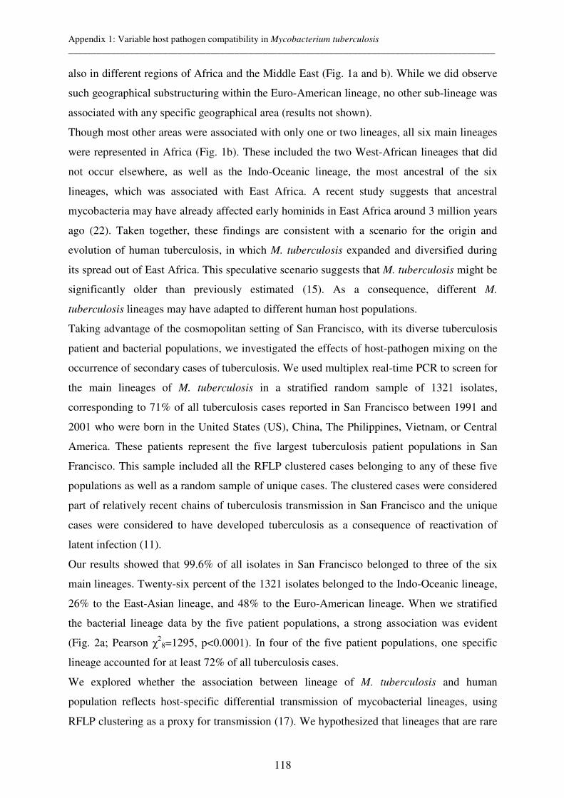
Appendix 1: Variable host pathogen compatibility in Mycobacterium tuberculosis __________________________________________________________________________________________
118
also in different regions of Africa and the Middle East (Fig. 1a and b). While we did observe
such geographical substructuring within the Euro-American lineage, no other sub-lineage was
associated with any specific geographical area (results not shown).
Though most other areas were associated with only one or two lineages, all six main lineages
were represented in Africa (Fig. 1b). These included the two West-African lineages that did
not occur elsewhere, as well as the Indo-Oceanic lineage, the most ancestral of the six
lineages, which was associated with East Africa. A recent study suggests that ancestral
mycobacteria may have already affected early hominids in East Africa around 3 million years
ago (22). Taken together, these findings are consistent with a scenario for the origin and
evolution of human tuberculosis, in which M. tuberculosis expanded and diversified during
its spread out of East Africa. This speculative scenario suggests that M. tuberculosis might be
significantly older than previously estimated (15). As a consequence, different M.
tuberculosis lineages may have adapted to different human host populations.
Taking advantage of the cosmopolitan setting of San Francisco, with its diverse tuberculosis
patient and bacterial populations, we investigated the effects of host-pathogen mixing on the
occurrence of secondary cases of tuberculosis. We used multiplex real-time PCR to screen for
the main lineages of M. tuberculosis in a stratified random sample of 1321 isolates,
corresponding to 71% of all tuberculosis cases reported in San Francisco between 1991 and
2001 who were born in the United States (US), China, The Philippines, Vietnam, or Central
America. These patients represent the five largest tuberculosis patient populations in San
Francisco. This sample included all the RFLP clustered cases belonging to any of these five
populations as well as a random sample of unique cases. The clustered cases were considered
part of relatively recent chains of tuberculosis transmission in San Francisco and the unique
cases were considered to have developed tuberculosis as a consequence of reactivation of
latent infection (11).
Our results showed that 99.6% of all isolates in San Francisco belonged to three of the six
main lineages. Twenty-six percent of the 1321 isolates belonged to the Indo-Oceanic lineage,
26% to the East-Asian lineage, and 48% to the Euro-American lineage. When we stratified
the bacterial lineage data by the five patient populations, a strong association was evident
(Fig. 2a; Pearson χ28=1295, p<0.0001). In four of the five patient populations, one specific
lineage accounted for at least 72% of all tuberculosis cases.
We explored whether the association between lineage of M. tuberculosis and human
population reflects host-specific differential transmission of mycobacterial lineages, using
RFLP clustering as a proxy for transmission (17). We hypothesized that lineages that are rare
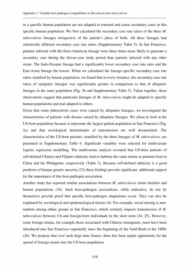
Appendix 1: Variable host pathogen compatibility in Mycobacterium tuberculosis __________________________________________________________________________________________
119
in a specific human population are not adapted to transmit and cause secondary cases in this
specific human population. We first calculated the secondary case rate ratios of the three M.
tuberculosis lineages irrespective of the patient’s place of birth. All three lineages had
statistically different secondary case rate ratios (Supplementary Table 5). In San Francisco,
patients infected with the Euro-American lineage were three times more likely to generate a
secondary case during the eleven-year study period than patients infected with any other
strain. The Indo-Oceanic lineage had a significantly lower secondary case rate ratio and the
East-Asian lineage the lowest. When we calculated the lineage-specific secondary case rate
ratios stratified by human population, we found that in every instance, the secondary case rate
ratios of sympatric lineages was significantly greater in comparison to that of allopatric
lineages in the same population (Fig. 2b and Supplementary Table 5). Taken together, these
observations suggest that particular lineages of M. tuberculosis might be adapted to specific
human populations and mal-adapted to others.
Given that some tuberculosis cases were caused by allopatric lineages, we investigated the
characteristics of patients with disease caused by allopatric lineages. We chose to look at the
US-born population because it represents the largest patient population in San Francisco (Fig.
2a) and that sociological determinants of transmission are well documented. The
characteristics of the US-born patients, stratified by the three lineages of M. tuberculosis, are
presented in Supplementary Table 6. Significant variables were selected for multivariate
logistic regression modelling. The multivariate analysis revealed that US-born patients of
self-defined Chinese and Filipino ethnicity tend to harbour the same strains as patients born in
China and the Philippines, respectively (Table 2). Because self-defined ethnicity is a good
predictor of human genetic ancestry (23) these findings provide significant additional support
for the importance of this host-pathogen association.
Another study has reported similar associations between M. tuberculosis strain families and
human populations (16). Such host-pathogen associations, while indicative, do not by
themselves provide proof that specific host-pathogen adaptations occur. They can also be
explained by sociological and epidemiological factors (6). For example, social mixing is non-
random among ethnic groups in San Francisco, which certainly impacts transmission of M.
tuberculosis between US-and foreign-born individuals in the short term (24, 25). However,
some foreign strains, for example those associated with Chinese immigrants, must have been
introduced into San Francisco repeatedly since the beginning of the Gold Rush in the 1800s
(26). We propose that over such large time frames, there has been ample opportunity for the
spread of foreign strains into the US-born population.

Appendix 1: Variable host pathogen compatibility in Mycobacterium tuberculosis __________________________________________________________________________________________
120
We cannot exclude the possibility that social factors contribute or even drive our observation
of lineage-specific association with particular human populations. However, further results
from our multivariate analysis support biological causality for the observed host-pathogen
association. US-born tuberculosis patients of non-Chinese and non-Filipino ethnicity infected
with allopatric strains (i.e. belonging to the Indo-Oceanic or East-Asian lineages) were more
likely to be HIV positive or homeless (Table 2). This suggests that although these lineages are
less adapted to transmit and cause disease in fully competent members of allopatric human
populations, they can do so in the context of impaired host resistance. Such differences in
host-pathogen compatibility or local adaptation have been associated with host-specific
pathogen adaptation and demonstrated in several invertebrate host-pathogen model systems
(6).
Overall, our findings demonstrate a global genetic population structure for M. tuberculosis,
and support the notion that this pathogen has adapted to specific human populations. These
results have implications for tuberculosis control efforts, especially for the development of
new vaccines. The importance of strain genetic variation for vaccine escape has been
documented in several bacterial species (27-29). In Bacille Calmette-Guerin (BCG), the
currently available tuberculosis vaccine, significant geographical variation in protective
efficacy has been observed (8). Environmental factors and differences in vaccine strain have
been invoked (8, 30-32), but our findings suggest that regional differences in host-pathogen
interactions could be partially responsible. Although recent progress has been made in the
development of new tuberculosis vaccines (33), the global population structure of M.
tuberculosis and host-specific pathogen adaptation may need to be considered when
engineering and evaluating new vaccine candidates.
Acknowledgements
We thank Bruce Levin and James H. Jones for their valuable comments on the manuscript,
and Colette Diguimbaye and Erik Post for providing strains. This research was supported by
the Swiss National Science Foundation and the Novartis Foundation (S.G.), the NIH (K.D.,
P.H.), and the Wellcome Trust (P.S.).

Table 1. Assignment of 875 strains of M. tuberculosis from 80 countries to six main phylogenetic lineages in eleven geographic regions
Geographic region (number of countries)
Total strains Indo-Oceanic lineage
East-Asian lineage
East-African-Indian lineage
Euro-American lineage
West-African lineage 1
West-African lineage 2
Americas (18) 207 6 7 194
Europe (14) 35 1 4 30
North Africa/Middle East (6) 10 10
West Africa (8) 28 12 9 7
Central Africa (5) 15 1 14
South Africa (1) 5 2 3
East Africa (6) 20 3 8 9
Indian Subcontinent (4) 17 3 1 10 3
Southeast Asia (9) 272 169 73 1 29
East Asia (7) 262 17 190 55
Pacific Islands (2) 4 4
Total (80) 875 199 277 20 363 9 7
Appendix 1: Variable host pathogen compatibility in Mycobacterium tuberculosis __________________________________________________________________________________________
122
Table 2. Risk factors independently associated with one of three M. tuberculosis lineages
in 490 US-born patients from San Francisco
Note: CI=confidence interval
M. tuberculosis lineage
Risk factor Adjusted odds ratio
(95% CI) P-value
East-Asian Chinese ethnicity 19.8 (4.6-84.2) <0.001
Homelessness 3.0 (1.4-6.2) 0.004
Indo-Oceanic Filipino ethnicity 43.2 (5.6-335) <0.001
Age ≥ 45 years 3.9 (1.5-10.1) 0.004
HIV positive 3.4 (1.3-8.7) 0.01
Euro-American Chinese ethnicity 0.18 (0.06-0.6) 0.004
Appendix 1: Variable host pathogen compatibility in Mycobacterium tuberculosis __________________________________________________________________________________________
123
Fig. 1. The global population structure and geographical distribution of M. tuberculosis. (a)
Large sequence polymorphisms (LSPs) define a global phylogeny for M. tuberculosis. The
name of the lineage-defining LSPs or regions of difference (RDs) are shown in rectangles.
The geographic regions associated with specific lineages are indicated. (b) The six main
lineages of M. tuberculosis are geographically structured. Each dot corresponds to one of 80
countries represented in the global strain collection. The colours of the dots relate to the six
main lineages defined in Fig. 1a and indicate the dominant lineage(s) in the respective
countries.
a b
711
702
7, 8, 10
9
239
750
105
12can
115
182
183
193
174
724
726
761
pks15/1∆7bp
H37Rv-like
122
TbD1
207
181
150
142
219
M. canettii
Indo-Oceanic
East-African-Indian
East-Asian
Euro-American
West-African-1
M. bovis lineage
West-African-2
AmericasEurope
West Africa
Middle East
South Africa
Central Africa
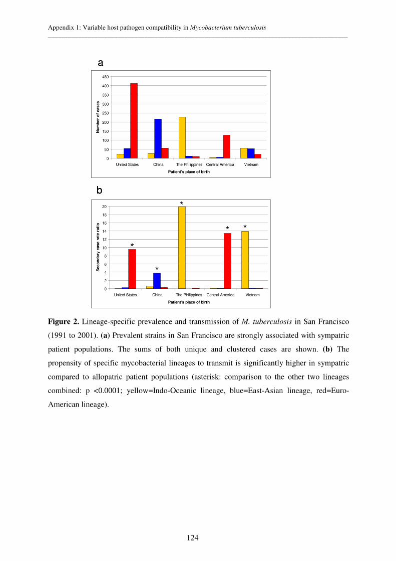
Appendix 1: Variable host pathogen compatibility in Mycobacterium tuberculosis __________________________________________________________________________________________
124
Figure 2. Lineage-specific prevalence and transmission of M. tuberculosis in San Francisco
(1991 to 2001). (a) Prevalent strains in San Francisco are strongly associated with sympatric
patient populations. The sums of both unique and clustered cases are shown. (b) The
propensity of specific mycobacterial lineages to transmit is significantly higher in sympatric
compared to allopatric patient populations (asterisk: comparison to the other two lineages
combined: p <0.0001; yellow=Indo-Oceanic lineage, blue=East-Asian lineage, red=Euro-
American lineage).
0
2
4
6
8
10
12
14
16
18
20
United States China The Philippines Central America Vietnam
Patient's place of birth
Seco
nd
ary
case r
ate
rati
o
*
* *
*
*
a
b
0
50
100
150
200
250
300
350
400
450
United States China The Philippines Central America Vietnam
Patient's place of birth
Nu
mb
er
of
cases
0
2
4
6
8
10
12
14
16
18
20
United States China The Philippines Central America Vietnam
Patient's place of birth
Seco
nd
ary
case r
ate
rati
o
*
* *
*
*
a
b
0
50
100
150
200
250
300
350
400
450
United States China The Philippines Central America Vietnam
Patient's place of birth
Nu
mb
er
of
cases
Appendix 1: Variable host pathogen compatibility in Mycobacterium tuberculosis __________________________________________________________________________________________
125
References
1. Falush, D., Wirth, T., Linz, B., Pritchard, J. K., Stephens, M., Kidd, M., Blaser, M. J., Graham, D. Y., Vacher, S., Perez-Perez, G. I., Yamaoka, Y., Megraud, F., Otto, K., Reichard, U., Katzowitsch, E., Wang, X., Achtman, M. & Suerbaum, S. (2003) Science 299, 1582-5.
2. Musser, J. M., Kroll, J. S., Granoff, D. M., Moxon, E. R., Brodeur, B. R., Campos, J., Dabernat, H., Frederiksen, W., Hamel, J. & Hammond, G. (1990) Rev. Infect. Dis. 12, 75-111.
3. Agostini, H. T., Yanagihara, R., Davis, V., Ryschkewitsch, C. F. & Stoner, G. L. (1997) Proc. Natl.
Acad. Sci. U S A 94, 14542-6. 4. Monot, M., Honore, N., Garnier, T., Araoz, R., Coppee, J. Y., Lacroix, C., Sow, S., Spencer, J. S.,
Truman, R. W., Williams, D. L., Gelber, R., Virmond, M., Flageul, B., Cho, S. N., Ji, B., Paniz-Mondolfi, A., Convit, J., Young, S., Fine, P. E., Rasolofo, V., Brennan, P. J. & Cole, S. T. (2005) Science 308, 1040-2.
5. Wirth, T., Wang, X., Linz, B., Novick, R. P., Lum, J. K., Blaser, M., Morelli, G., Falush, D. & Achtman, M. (2004) Proc. Natl. Acad. Sci. U S A 101, 4746-51.
6. Woolhouse, M. E., Webster, J. P., Domingo, E., Charlesworth, B. & Levin, B. R. (2002) Nat. Genet. 32, 569-77.
7. World Health Organization (2005) (WHO, Geneva). 8. Andersen, P. & Doherty, T. M. (2005) Nat. Rev. Microbiol. 9. Kremer, K., van Soolingen, D., Frothingham, R., Haas, W. H., Hermans, P. W., Martin, C.,
Palittapongarnpim, P., Plikaytis, B. B., Riley, L. W., Yakrus, M. A., Musser, J. M. & van Embden, J. D. (1999) J. Clin. Microbiol. 37, 2607-18.
10. Hirsh, A. E., Tsolaki, A. G., DeRiemer, K., Feldman, M. W. & Small, P. M. (2004) Proc. Natl. Acad.
Sci. U S A 101, 4871-6. 11. Jasmer, R. M., Hahn, J. A., Small, P. M., Daley, C. L., Behr, M. A., Moss, A. R., Creasman, J. M.,
Schecter, G. F., Paz, E. A. & Hopewell, P. C. (1999) Ann. Intern. Med. 130, 971-978. 12. Niobe-Eyangoh, S. N., Kuaban, C., Sorlin, P., Cunin, P., Thonnon, J., Sola, C., Rastogi, N., Vincent, V.
& Gutierrez, M. C. (2003) J. Clin. Microbiol. 41, 2547-53. 13. Tsolaki, A. G., Hirsh, A. E., DeRiemer, K., Enciso, J. A., Wong, M. Z., Hannan, M., Goguet de la
Salmoniere, Y. O., Aman, K., Kato-Maeda, M. & Small, P. M. (2004) Proc. Natl. Acad. Sci. U S A 101, 4865-70.
14. Marmiesse, M., Brodin, P., Buchrieser, C., Gutierrez, C., Simoes, N., Vincent, V., Glaser, P., Cole, S. T. & Brosch, R. (2004) Microbiology 150, 483-96.
15. Sreevatsan, S., Pan, X., Stockbauer, K. E., Connell, N. D., Kreiswirth, B. N., Whittam, T. S. & Musser, J. M. (1997) Proc. Natl. Acad. Sci. U S A 94, 9869-74.
16. Baker, L., Brown, T., Maiden, M. C. & Drobniewski, F. (2004) Emerg. Infect. Dis. 10, 1568-77. 17. Burgos, M., DeRiemer, K., Small, P. M., Hopewell, P. C. & Daley, C. L. (2003) J. Infect. Dis. 188,
1878-84. 18. Selvin, S. (1998) in Monographs in Epidemiology and Biostatistics (Oxford University Press, New
York), Vol. 28, pp. 385-391. 19. Mostowy, S., Onipede, A., Gagneux, S., Niemann, S., Kremer, K., Desmond, E. P., Kato-Maeda, M. &
Behr, M. (2004) J. Clin. Microbiol. 42, 3594-9. 20. Tsolaki, A. G., Gagneux, S., Pym, A. S., Goguet de la Salmoniere, Y. O., Kreiswirth, B. N., Van
Soolingen, D. & Small, P. M. (2005) J. Clin. Microbiol. 43, 3185-91. 21. Brosch, R., Gordon, S. V., Marmiesse, M., Brodin, P., Buchrieser, C., Eiglmeier, K., Garnier, T.,
Gutierrez, C., Hewinson, G., Kremer, K., Parsons, L. M., Pym, A. S., Samper, S., van Soolingen, D. & Cole, S. T. (2002) Proc. Natl. Acad. Sci. U S A 99, 3684-9.
22. Gutierrez, C., Brisse, S., Brosch, R., Fabre, M., Omais, B., Marmiesse, M., Supply, P. & Vincent, V. (2005) PLoS Pathogens 1, 1-7.
23. Rosenberg, N. A., Pritchard, J. K., Weber, J. L., Cann, H. M., Kidd, K. K., Zhivotovsky, L. A. & Feldman, M. W. (2002) Science 298, 2381-5.
24. Chin, D. P., DeRiemer, K., Small, P. M., de Leon, A. P., Steinhart, R., Schecter, G. F., Daley, C. L., Moss, A. R., Paz, E. A., Jasmer, R. M., Agasino, C. B. & Hopewell, P. C. (1998) Am. J. Respir. Crit.
Care Med. 158, 1797-1803. 25. Jasmer, R. M., Ponce, d. L., Hopewell, P. C., Alarcon, R. G., Moss, A. R., Paz, E. A., Schecter, G. F. &
Small, P. M. (1997) Int. J. Tuberc. Lung Dis. 1, 536-541. 26. http://en.wikipedia.org/wiki/Chinatown%2C_San_Francisco. 27. Gagneux, S. P., Hodgson, A., Smith, T. A., Wirth, T., Ehrhard, I., Morelli, G., Genton, B., Binka, F. N.,
Achtman, M. & Pluschke, G. (2002) J. Infect. Dis. 185, 618-26. 28. Van Loo, I. H. & Mooi, F. R. (2002) Microbiology 148, 2011-8.

Appendix 1: Variable host pathogen compatibility in Mycobacterium tuberculosis __________________________________________________________________________________________
126
29. Mbelle, N., Huebner, R. E., Wasas, A. D., Kimura, A., Chang, I. & Klugman, K. P. (1999) J. Infect.
Dis. 180, 1171-6. 30. Behr, M. A., Wilson, M. A., Gill, W. P., Salamon, H., Schoolnik, G. K., Rane, S. & Small, P. M.
(1999) Science 284, 1520-1523. 31. Rook, G. A., Dheda, K. & Zumla, A. (2005) Nat. Rev. Immunol. 5, 661-7. 32. Fine, P. E. (1995) Lancet 346, 1339-45. 33. McShane, H., Pathan, A. A., Sander, C. R., Keating, S. M., Gilbert, S. C., Huygen, K., Fletcher, H. A.
& Hill, A. V. (2004) Nat. Med. 10, 1240-4.
Appendix 2: Non-tuberculous mycobacteria from humans and cattle of Chad __________________________________________________________________________________________
127
Appendix 2: Species identification of non-tuberculous
mycobacteria from humans and cattle of Chad
C. Diguimbaye-Djaibé1, V. Vincent2, E. Schelling3, M. Hilty3, R. Ngandolo1, HH. Mahamat1,
G. Pfyffer4, F. Baggi5, M. Tanner3, J. Zinsstag3
1Laboratoire de Recherches Vétérinaires et Zootechniques de Farcha, N’Djaména, Chad, 2.Centre de Référence de Mycobactéries, Institut Pasteur, Paris, France and 3Swiss Tropical
Institute, Basel, 4 Department of Medical Microbiology, Kantonsspital Luzern, Luzern, 5
National Centre for Mycobacteria, University of Zurich, Switzerland
Published in Schweizerisches Archiv für Tierheilkunde 2006;148(5):251-6
Appendix 2: Non-tuberculous mycobacteria from humans and cattle of Chad __________________________________________________________________________________________
128
Summary
In Chad, during a study on tuberculosis in humans and cattle, 52 non-tuberculous
mycobacteria (NTM) strains were isolated. By means of INNO-LiPA, PRA-hsp65
amplification and sequencing of 16S rDNA, NTM species of 25/52 isolates were identified.
M. fortuitum complex (8) was the most frequent species, followed by M. nonchromogenicum
(4) and M. avium complex (4). PRA method could identify M. fortuitum 3rd variant among
isolates derived from cattle specimens. This finding could confirm the existence of farcy in
the Chadian cattle population as M. fortuitum 3rd variant and putitative pathogen M.
farcinogenes can’t be distinguished by the methods used in this study. Half of the NTM
isolates could not be specified and we considered them as contaminants from the
environment.
Keywords: non –tuberculous mycobacteria, Chad, molecular methods, INNO-LiPA assay,
PRA amplification, sequencing of the 16S gene
Zusammenfassung
Während einer Studie im Tschad, welche die Tuberkulose bei Menschen und Rindern
untersucht, wurden 52 nicht-Tuberkulse Mykobakterien (NTM) isoliert. Mit Hilfe von INNO-
LiPA tests, PRA-hsp65 Amplifizierung und Sequenzierung der 16S rDNS, konnten 25/52
Isolaten identifiziert werden. M. fortuitum complex war die häufigste Species, gefolgt von M.
nonchromogenicum (4) und M. avium complex (4). Die PRA Methode konnte bei Isolaten,
die von Kühen stammen, die dritte Variante von M. fortuitum identifizieren. Dieser Befund
könnte die Existenz von Farcy in der tschadischen Kuhpopulation bedeuten, weil die dritte
Variante von M. fortuitum vom vermuteten Farcy Erreger M. farcinogenes nicht mit den in
dieser Studie angewendeten Methoden unterschieden werden kann. Die Hälfte der NTM
Isolate konnten nicht identifiziert werden und wir betrachten diese als Kontaminationen aus
der Umwelt.
Schlüsselwörter: nicht-Tuberkulse Mykobakterien (NTM), Tschad, molekulare Methoden,
INNO-LiPA, PRA Amplifizierung, Sequenzierung des 16S Gens
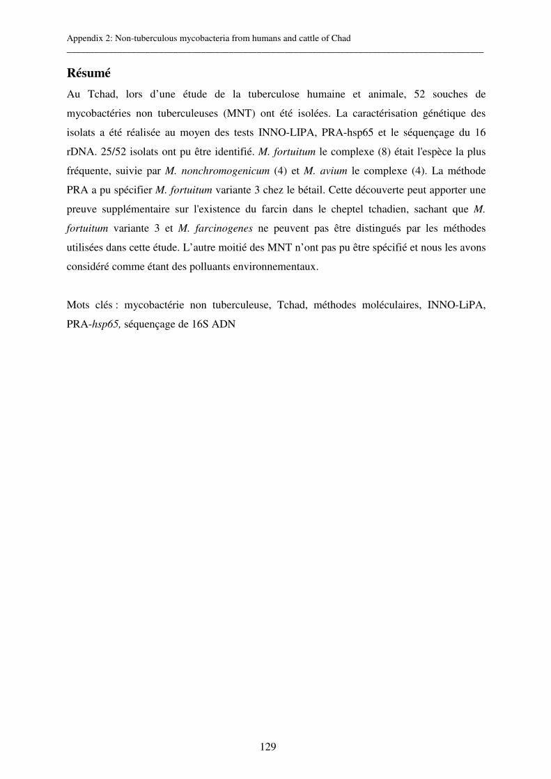
Appendix 2: Non-tuberculous mycobacteria from humans and cattle of Chad __________________________________________________________________________________________
129
Résumé
Au Tchad, lors d’une étude de la tuberculose humaine et animale, 52 souches de
mycobactéries non tuberculeuses (MNT) ont été isolées. La caractérisation génétique des
isolats a été réalisée au moyen des tests INNO-LIPA, PRA-hsp65 et le séquençage du 16
rDNA. 25/52 isolats ont pu être identifié. M. fortuitum le complexe (8) était l'espèce la plus
fréquente, suivie par M. nonchromogenicum (4) et M. avium le complexe (4). La méthode
PRA a pu spécifier M. fortuitum variante 3 chez le bétail. Cette découverte peut apporter une
preuve supplémentaire sur l'existence du farcin dans le cheptel tchadien, sachant que M.
fortuitum variante 3 et M. farcinogenes ne peuvent pas être distingués par les méthodes
utilisées dans cette étude. L’autre moitié des MNT n’ont pas pu être spécifié et nous les avons
considéré comme étant des polluants environnementaux.
Mots clés : mycobactérie non tuberculeuse, Tchad, méthodes moléculaires, INNO-LiPA,
PRA-hsp65, séquençage de 16S ADN
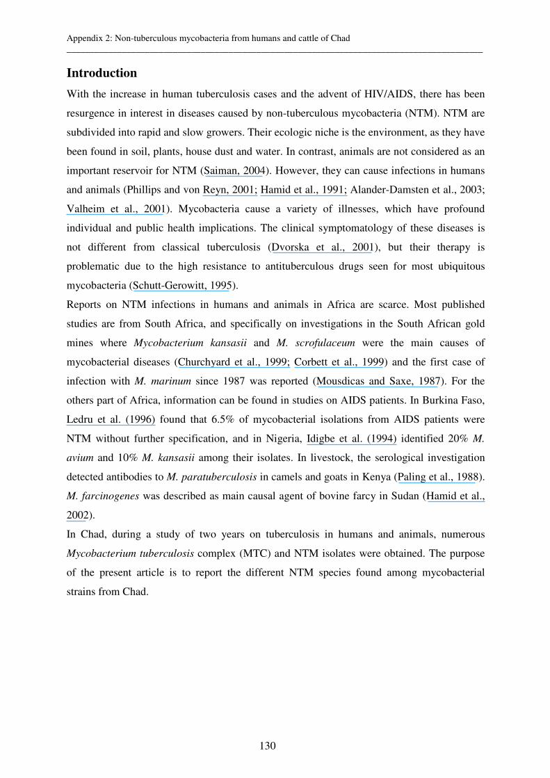
Appendix 2: Non-tuberculous mycobacteria from humans and cattle of Chad __________________________________________________________________________________________
130
Introduction
With the increase in human tuberculosis cases and the advent of HIV/AIDS, there has been
resurgence in interest in diseases caused by non-tuberculous mycobacteria (NTM). NTM are
subdivided into rapid and slow growers. Their ecologic niche is the environment, as they have
been found in soil, plants, house dust and water. In contrast, animals are not considered as an
important reservoir for NTM (Saiman, 2004). However, they can cause infections in humans
and animals (Phillips and von Reyn, 2001; Hamid et al., 1991; Alander-Damsten et al., 2003;
Valheim et al., 2001). Mycobacteria cause a variety of illnesses, which have profound
individual and public health implications. The clinical symptomatology of these diseases is
not different from classical tuberculosis (Dvorska et al., 2001), but their therapy is
problematic due to the high resistance to antituberculous drugs seen for most ubiquitous
mycobacteria (Schutt-Gerowitt, 1995).
Reports on NTM infections in humans and animals in Africa are scarce. Most published
studies are from South Africa, and specifically on investigations in the South African gold
mines where Mycobacterium kansasii and M. scrofulaceum were the main causes of
mycobacterial diseases (Churchyard et al., 1999; Corbett et al., 1999) and the first case of
infection with M. marinum since 1987 was reported (Mousdicas and Saxe, 1987). For the
others part of Africa, information can be found in studies on AIDS patients. In Burkina Faso,
Ledru et al. (1996) found that 6.5% of mycobacterial isolations from AIDS patients were
NTM without further specification, and in Nigeria, Idigbe et al. (1994) identified 20% M.
avium and 10% M. kansasii among their isolates. In livestock, the serological investigation
detected antibodies to M. paratuberculosis in camels and goats in Kenya (Paling et al., 1988).
M. farcinogenes was described as main causal agent of bovine farcy in Sudan (Hamid et al.,
2002).
In Chad, during a study of two years on tuberculosis in humans and animals, numerous
Mycobacterium tuberculosis complex (MTC) and NTM isolates were obtained. The purpose
of the present article is to report the different NTM species found among mycobacterial
strains from Chad.
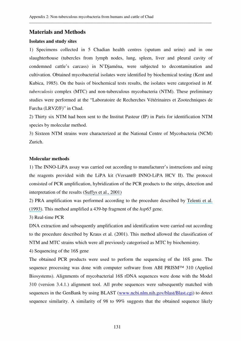
Appendix 2: Non-tuberculous mycobacteria from humans and cattle of Chad __________________________________________________________________________________________
131
Materials and Methods
Isolates and study sites
1) Specimens collected in 5 Chadian health centres (sputum and urine) and in one
slaughterhouse (tubercles from lymph nodes, lung, spleen, liver and pleural cavity of
condemned cattle’s carcass) in N’Djaména, were subjected to decontamination and
cultivation. Obtained mycobacterial isolates were identified by biochemical testing (Kent and
Kubica, 1985). On the basis of biochemical tests results, the isolates were categorised in M.
tuberculosis complex (MTC) and non-tuberculous mycobacteria (NTM). These preliminary
studies were performed at the “Laboratoire de Recherches Vétérinaires et Zootechniques de
Farcha (LRVZ/F)” in Chad.
2) Thirty six NTM had been sent to the Institut Pasteur (IP) in Paris for identification NTM
species by molecular method.
3) Sixteen NTM strains were characterized at the National Centre of Mycobacteria (NCM)
Zurich.
Molecular methods
1) The INNO-LiPA assay was carried out according to manufacturer’s instructions and using
the reagents provided with the LiPA kit (Versant® INNO-LiPA HCV II). The protocol
consisted of PCR amplification, hybridization of the PCR products to the strips, detection and
interpretation of the results (Suffys et al., 2001)
2) PRA amplification was performed according to the procedure described by Telenti et al.
(1993). This method amplified a 439-bp fragment of the hsp65 gene.
3) Real-time PCR
DNA extraction and subsequently amplification and identification were carried out according
to the procedure described by Kraus et al. (2001). This method allowed the classification of
NTM and MTC strains which were all previously categorised as MTC by biochemistry.
4) Sequencing of the 16S gene
The obtained PCR products were used to perform the sequencing of the 16S gene. The
sequence processing was done with computer software from ABI PRISM™ 310 (Applied
Biosystems). Alignments of mycobacterial 16S rDNA sequences were done with the Model
310 (version 3.4.1.) alignment tool. All probe sequences were subsequently matched with
sequences in the GenBank by using BLAST (www.ncbi.nlm.nih.gov/blast/Blast.cgi) to detect
sequence similarity. A similarity of 98 to 99% suggests that the obtained sequence likely
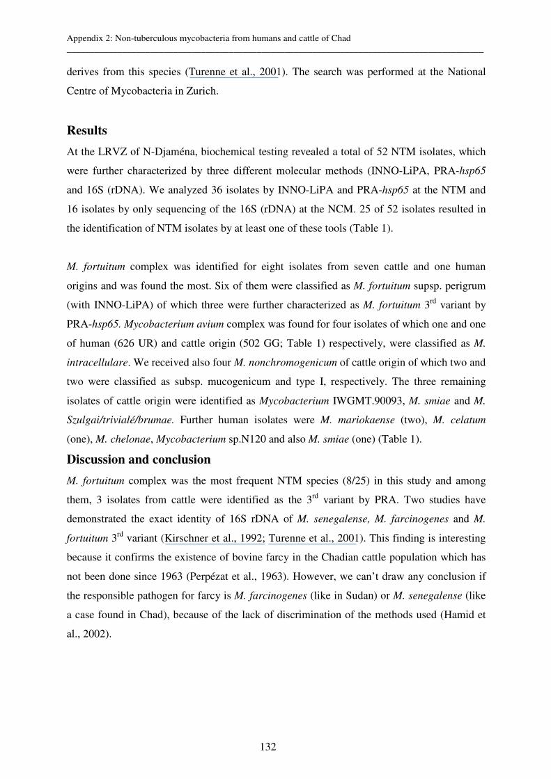
Appendix 2: Non-tuberculous mycobacteria from humans and cattle of Chad __________________________________________________________________________________________
132
derives from this species (Turenne et al., 2001). The search was performed at the National
Centre of Mycobacteria in Zurich.
Results
At the LRVZ of N-Djaména, biochemical testing revealed a total of 52 NTM isolates, which
were further characterized by three different molecular methods (INNO-LiPA, PRA-hsp65
and 16S (rDNA). We analyzed 36 isolates by INNO-LiPA and PRA-hsp65 at the NTM and
16 isolates by only sequencing of the 16S (rDNA) at the NCM. 25 of 52 isolates resulted in
the identification of NTM isolates by at least one of these tools (Table 1).
M. fortuitum complex was identified for eight isolates from seven cattle and one human
origins and was found the most. Six of them were classified as M. fortuitum supsp. perigrum
(with INNO-LiPA) of which three were further characterized as M. fortuitum 3rd variant by
PRA-hsp65. Mycobacterium avium complex was found for four isolates of which one and one
of human (626 UR) and cattle origin (502 GG; Table 1) respectively, were classified as M.
intracellulare. We received also four M. nonchromogenicum of cattle origin of which two and
two were classified as subsp. mucogenicum and type I, respectively. The three remaining
isolates of cattle origin were identified as Mycobacterium IWGMT.90093, M. smiae and M.
Szulgai/trivialé/brumae. Further human isolates were M. mariokaense (two), M. celatum
(one), M. chelonae, Mycobacterium sp.N120 and also M. smiae (one) (Table 1).
Discussion and conclusion
M. fortuitum complex was the most frequent NTM species (8/25) in this study and among
them, 3 isolates from cattle were identified as the 3rd variant by PRA. Two studies have
demonstrated the exact identity of 16S rDNA of M. senegalense, M. farcinogenes and M.
fortuitum 3rd variant (Kirschner et al., 1992; Turenne et al., 2001). This finding is interesting
because it confirms the existence of bovine farcy in the Chadian cattle population which has
not been done since 1963 (Perpézat et al., 1963). However, we can’t draw any conclusion if
the responsible pathogen for farcy is M. farcinogenes (like in Sudan) or M. senegalense (like
a case found in Chad), because of the lack of discrimination of the methods used (Hamid et
al., 2002).
Appendix 2: Non-tuberculous mycobacteria from humans and cattle of Chad __________________________________________________________________________________________
133
Only four isolates were identified as M. avium complex and none was M. kansasii, while
usually these two mycobacteria are the most common NTM in clinical specimens and
biopsies (Shih et al., 1997; Marras and Daley, 2002; Thorel, 1980; Pate et al., 2004).
Concerning the remaining mycobacteria found in our study, they are rarely isolated but most
of them were described as potential pathogens. Mycobacterium simiae is commonly found in
the environment and was rarely associated with human disease. However, some cases of
disease caused by M. simiae in AIDS and non-AIDS patients have been reported (Vandercam
et al., 1996; Huminer et al., 1993; Bell et al., 1983; Lavy and Yoshpe-Purer, 1982). M.
celatum was described as cause of fatal pulmonary infection in an old woman (Bux-Gewehr
et al., 1998) and of disseminated infection in domestic ferret (Valheim et al., 2001). M.
chelonae was frequently isolated from patients with cystic fibrosis (Hjelt et al., 1994;
Tomashefski et al., 1996; Fauroux et al., 1997). M. terrae complex is composed of M.
nonchromogenicum, M. terrae, and M. triviale. They are uncommon colonizers of human
epithelia and generally regarded as non-pathogenic (Lee et al., 2004). However, M.
nonchromogenicum may occasionally cause human disease such as pulmonary infection and
tenosynovitis (Peters and Morice, 1991).
We obtained many NTM usually found in the environment by sequencing because this
method is not focused on pathogen NTM strains. However, we have not confirmed our
sequencing results by another molecular method as suggested (Hafner et al. 2004). Generally,
NTM isolates should be further characterised because NTM infections can cause disease and
first line antituberculosis drug treatment may be not efficacious. In another hand, it will be
interesting for veterinarians to know about the importance of NTM in cattle, particularly in
interpretation of tuberculin test.
Acknowledgements
We thank the technicians of the “Centre de Reference des Mycobactéries” of Institut Pasteur,
the National Center for Mycobacteria, the Swiss Tropical Institute, and the “Laboratoire de
Recherches Vétérinaires et Zootechniques de Farcha” who have contributed to the project.
The Swiss National Science Foundation is acknowledged for financial support.
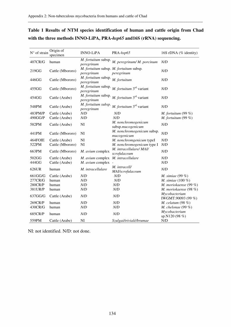
Appendix 2: Non-tuberculous mycobacteria from humans and cattle of Chad __________________________________________________________________________________________
134
Table 1 Results of NTM species identification of human and cattle origin from Chad
with the three methods INNO-LiPA, PRA-hsp65 and16S (rRNA) sequencing.
N° of strain Origin of specimen
INNO-LiPA PRA-hsp65 16S rDNA (% identity)
407CR/G human M. fortuitum subsp. peregrinum
M. peregrinum/ M. porcinum N/D
219GG Cattle (Mbororo) M. fortuitum subsp.
peregrinum
M. fortuitum subsp.
peregrinum N/D
446GG Cattle (Mbororo) M. fortuitum subsp.
peregrinum M. fortuitum N/D
455GG Cattle (Mbororo) M. fortuitum subsp.
peregrinum M. fortuitum 3rd variant N/D
454GG Cattle (Arabe) M. fortuitum subsp.
peregrinum M. fortuitum 3rd variant N/D
548PM Cattle (Arabe) M. fortuitum subsp.
peregrinum M. fortuitum 3rd variant N/D
483PM/P Cattle (Arabe) N/D N/D M. fortuitum (99 %) 490GG/P Cattle (Arabe) N/D N/D M. fortuitum (99 %)
582PM Cattle (Arabe) NI M. nonchromogenicum
subsp.mucogenicum N/D
441PM Cattle (Mbororo) NI M. nonchromogenicum subsp.
mucogenicum N/D
464FOIE Cattle (Arabe) NI M. nonchromgenicum typeI N/D
522PM Cattle (Mbororo) NI M. nonchromogenicum type I N/D
663PM Cattle (Mbororo) M. avium complex M. intracellulare/ MAI/
scrofulaceum N/D
502GG Cattle (Arabe) M. avium complex M. intracellulare N/D
444GG Cattle (Arabe) M. avium complex - N/D
626UR human M. intracellulare M. intracell/
MAI/scrofulaceum N/D
661GG/G Cattle (Arabe) N/D N/D M. simiae (99 %) 277CR/G human N/D N/D M. simiae (100 %) 280CR/P human N/D N/D M. moriokaense (99 %) 381UR/P human N/D N/D M. moriokaense (98 %)
637GG/G Cattle (Arabe) N/D N/D Mycobacterium
IWGMT.90093 (99 %) 269CR/P human N/D N/D M. celatum (98 %) 430CR/G human N/D N/D M. chelonae (99 %)
685CR/P human N/D N/D Mycobacterium sp.N120 (98 %)
559PM Cattle (Arabe) NI Szulgai/trivialé/brumae N/D
NI: not identified. N/D: not done.
Appendix 2: Non-tuberculous mycobacteria from humans and cattle of Chad __________________________________________________________________________________________
135
References
Alander-Damsten Y.K., Brander E.E., Paulin L.G.: Panniculitis, due to Mycobacterium smegmatis, in two Finnish cats. J.Feline.Med.Surg. 2003, 5:19-26.
Bell, R. C., J. H. Higuchi, W. N. Donovan, I. Krasnow, and W. G. Johanson, Jr.: Mycobacterium simiae. Clinical features and follow-up of twenty-four patients. Am.Rev.Respir.Dis. 1983, 127:35-38.
Bux-Gewehr, I., H. P. Hagen, S. Rusch-Gerdes, and G. E. Feurle.: Fatal pulmonary infection with Mycobacterium celatum in an apparently immunocompetent patient. J.Clin.Microbiol. 1998, 36:587-588.
Churchyard, G. J., I. Kleinschmidt, E. L. Corbett, D. Mulder, and K. M. De Cock.: Mycobacterial disease in South African gold miners in the era of HIV infection. Int.J.Tuberc.Lung Dis. 1999, 3:791-798.
Corbett, E. L., L. Blumberg, G. J. Churchyard, N. Moloi, K. Mallory, T. Clayton, B. G. Williams, R. E. Chaisson,
R. J. Hayes, and K. M. De Cock.: Nontuberculous mycobacteria: defining disease in a prospective cohort of South African miners. Am.J.Respir.Crit Care Med. 1999, 160:15-21.
Dvorska, L., M. Bartos, C. Martin, W. Erler, and I. Pavlik.: Strategies for differentiation, identification and typing of medically important species of Mycobacteria by molecular methods. Vet.Med.-Czech. 2001, 46:309-328.
Fauroux, B., B. Delaisi, A. Clement, C. Saizou, D. Moissenet, C. Truffot-Pernot, G. Tournier, and T. H. Vu.: Mycobacterial lung disease in cystic fibrosis: a prospective study. Pediatr.Infect.Dis.J. 1997, 16:354-358.
Hafner, B., H. Haag, H. K. Geiss, and O. Nolte.: Different molecular methods for the identification of rarely isolated non-tuberculous mycobacteria and description of new hsp65 restriction fragment length polymorphism patterns. Mol.Cell Probes 2004, 18:59-65.
Hamid, M. E., G. E. Mohamed, M. T. Abu-Samra, S. M. el Sanousi, and M. E. Barri.: Bovine farcy: a clinico-pathological study of the disease and its aetiological agent. J.Comp Pathol. 1991, 105:287-301.
Hamid, M. E., A. Roth, O. Landt, R. M. Kroppenstedt, M. Goodfellow, and H. Mauch.: Differentiation between Mycobacterium farcinogenes and Mycobacterium senegalense strains based on 16S-23S ribosomal DNA internal transcribed spacer sequences. J.Clin.Microbiol. 2002, 40:707-711.
Hjelt, K., N. Hojlyng, P. Howitz, N. Illum, E. Munk, N. H. Valerius, K. Fursted, K. N. Hansen, I. Heltberg, and
C. Koch.: The role of Mycobacteria Other Than Tuberculosis (MOTT) in patients with cystic fibrosis. Scand.J.Infect.Dis. 1994, 26:569-576.
Huminer, D., S. Dux, Z. Samra, L. Kaufman, A. Lavy, C. S. Block, and S. D. Pitlik.: Mycobacterium simiae infection in Israeli patients with AIDS. Clin.Infect.Dis. 1993, 17:508-509.
Idigbe, E. O., A. Nasidi, C. E. Anyiwo, C. Onubogu, S. Alabi, R. Okoye, O. Ugwu, and E. K. John.: Prevalence of human immunodeficiency virus (HIV) antibodies in tuberculosis patients in Lagos, Nigeria. J.Trop.Med.Hyg. 1994, 97:91-97.
Kent P.T., and G. P. Kubica.: Public health mycobacteriology- a guide for the level III laboratory. U.S. Departement of health and human Services publication, Atlanta, Ga. 1985.
Kirschner, P., M. Kiekenbeck, D. Meissner, J. Wolters, and E. C. Bottger.: Genetic heterogeneity within Mycobacterium fortuitum complex species: genotypic criteria for identification. J.Clin.Microbiol. 1992, 30:2772-2775.
Kraus, G., A. Cleary, N. Miller, R. Seivright, A. K. Young, G. Spruill, and H. J. Hnatyszyn.: Rapid and specific detection of the Mycobacterium tuberculosis complex using fluorogenic probes and real-time PCR. Mol.Cell.Probes 2001, 15:375-383.
Appendix 2: Non-tuberculous mycobacteria from humans and cattle of Chad __________________________________________________________________________________________
136
Lavy, A. and Y. Yoshpe-Purer.: Isolation of Mycobacterium simiae from clinical specimens in Israel. Tubercle. 1982, 63:279-285.
Ledru, S., B. Cauchoix, M. Yameogo, A. Zoubga, J. Lamande-Chiron, F. Portaels, and J. P. Chiron. : Impact of short-course therapy on tuberculosis drug resistance in South-West Burkina Faso. Tuber.Lung Dis. 1996, 77:429-436.
Lee, C. K., H. M. Gi, Y. Cho, Y. K. Kim, K. N. Lee, K. J. Song, J. W. Song, K. S. Park, E. M. Park, H. Lee, and
G. H. Bai.: The genomic heterogeneity among Mycobacterium terrae complex displayed by sequencing of 16S rRNA and hsp 65 genes. Microbiol.Immunol. 2004, 48:83-90.
Marras, T. K. and C. L. Daley.: Epidemiology of human pulmonary infection with nontuberculous mycobacteria. Clin.Chest Med. 2002, 23:553-567.
Mousdicas, N. and N. Saxe.: Fish-tank granuloma. The first reported case in South Africa. S.Afr.Med.J. 1987, 71:321-322.
Paling, R. W., S. Waghela, K. J. Macowan, and B. R. Heath.: The occurrence of infectious diseases in mixed farming of domesticated wild herbivores and livestock in Kenya. II. Bacterial diseases. J.Wildl.Dis. 1988, 24:308-316.
Pate, M., I. Zdovc, T. Pirs, B. Krt, and M. Ocepek.: Isolation and characterization of Mycobacterium avium and Rhodococcus equi from granulomatous lesions of swine lymph nodes in Slovenia. Acta Vet.Hung. 2004, 52:143-150.
Perpézat, A., F. Mariat and M. Thomé.: Importance du farcin chez le Zébu du Tchad. Bull.Soc.Path.Exot. 1963, 56:375-383.
Peters, E. J. and R. Morice.: Miliary pulmonary infection caused by Mycobacterium terrae in an autologous bone marrow transplant patient. Chest 1991, 100:1449-1450.
Phillips, M. S. and C. F. von Reyn.: Nosocomial infections due to nontuberculous mycobacteria. Clin.Infect.Dis. 2001, 33:1363-1374.
Saiman, L.: The mycobacteriology of non-tuberculous mycobacteria. Paediatr.Respir.Rev. 2004, 5 Suppl A:S221-S223.
Schutt-Gerowitt, H.: On the development of mycobacterial infections. I. A review concerning the common situation. Zentralbl.Bakteriol. 1995, 283:5-13.
Shih, J. Y., P. R. Hsueh, L. N. Lee, H. C. Wang, P. C. Yang, S. H. Kuo, and K. T. Luh.: Nontuberculous mycobacteria isolates: clinical significance and disease spectrum. J.Formos.Med.Assoc. 1997, 96:621-627.
Suffys, P. N., R. A. da Silva, M. de Oliveira, C. E. Campos, A. M. Barreto, F. Portaels, L. Rigouts, G. Wouters,
G. Jannes, G. van Reybroeck, W. Mijs, and B. Vanderborght.: Rapid identification of Mycobacteria to the species level using INNO-LiPA Mycobacteria, a reverse hybridization assay. J.Clin.Microbiol. 2001, 39:4477-4482.
Telenti, A., F. Marchesi, M. Balz, F. Bally, E. C. Bottger, and T. Bodmer.: Rapid identification of mycobacteria to the species level by polymerase chain reaction and restriction enzyme analysis. J.Clin.Microbiol. 1993, 31:175-178.
Thorel, M. F.: [Mycobacteria identified in a centre for veterinary research between 1973 and 1979 (author's transl)]. Ann.Microbiol.(Paris) 1980, 131:61-69.
Tomashefski, J. F., Jr., R. C. Stern, C. A. Demko, and C. F. Doershuk.: Nontuberculous mycobacteria in cystic fibrosis. An autopsy study. Am.J.Respir.Crit Care Med. 1996, 154:523-528.
Turenne, C. Y., L. Tschetter, J. Wolfe, and A. Kabani.: Necessity of quality-controlled 16S rRNA gene sequence databases: identifying nontuberculous Mycobacterium species. J.Clin.Microbiol. 2001, 39:3637-3648.

Appendix 2: Non-tuberculous mycobacteria from humans and cattle of Chad __________________________________________________________________________________________
137
Valheim, M., B. Djonne, R. Heiene, and D. A. Caugant.: Disseminated Mycobacterium celatum (type 3) infection in a domestic ferret (Mustela putorius furo). Vet.Pathol. 2001, 38:460-463.
Vandercam, B., J. Gala, B. Vandeweghe, J. Degraux, G. Wauters, L. Larsson, A. Bourlond, and F. Portaels.: Mycobacterium simiae disseminated infection in a patient with acquired immunodeficiency syndrome. Infection 1996, 24:49-51.


Appendix 3: Methods __________________________________________________________________________________________
139
Appendix 3: Methods
1. Ligation mediated PCR 1.1 Preparation of the DNA and linker
a) Extraction of DNA Place 100 µl of TE in 2 ml screw-cap tubes. Take sufficient colonies from the culture medium and suspend in the TE Incubate in the oven for 30 min at 100°C (to ensure that the bacteria are destroyed and DNA is extracted).
b) Digestion of the DNA with SalI For 1 reaction: DNA 17 µl (if TE) Neb 4 buffer 2 µl SalI 1 µl H2O qsp 20 µl Incubate for at least 1h at 37°C (dry water bath) - Check the digestion on a 0.8 % agarose gel. Warning: if no or partial digestion occurs, dialyse the samples for ½ h on a filter (0.05 µm pore size) and redigest the extract for 1 h at 37°C with 1 µl of SalI + 2 µl of buffer - Estimate the amount of DNA to be ligated on 0.8 % agarose gels by loading 2 µl of DNA/ SalI + 2 µl of loading buffer.
c) Ligation of DNA to the linker MIX 1 Reaction 20 Reactions Ligase T4 buffer10X 2 µl 40 µl Linker* 1 µl 20 µl Ligase T4 (1 U/µl) 1 µl 20 µl DNA As estimated above H2O qsp 20 µl final * see appendix ligation mediated PCR for preparation * always keep the linker on ice Ligation conditions: 5 min 5°C 1 h 16°C 10 min 65°C The reaction can be left at +4°C overnight.
d) Digestion of the ligation product with SalI Ligation product 20 µl Buffer H 2.5 µl SalI 0.5 µl H2O 2.0 µl - Incubate at 37°C for 15min - Dilute the DNA 1/5 (i.e. 25 µl of ligation Mix + 100 µl of H2O).

Appendix 3: Methods __________________________________________________________________________________________
140
1.2 Amplification of the ligation product with the SALGD linker Initial conc. 1 Reaction 20 Reactions Final conc. H2O 10 x 28.8 µl 576 µl - Taq buffer 5 µl 100 µl 1 x DMSO 5 µl 100 µl 10 % dNTP 10 mM (each) 1 µl 20 µl 200 µM IS2 50 µM 1 µl 20 µl 1 µM SALgd 50 µM 1 µl 20 µl 1 µM MgCl2 25mM 3 µl 60 µl 1.5 mM Taq Cold 5 U/µl 0.2 µl 4 µl 1 U Add 5 µl of DNA diluted 1 in 5 per reaction. PCR Programme Prog. 1 9 min – 94°C
Prog. 2 30 sec – 94°C 30 sec – 55°C 3 min – 72°C
35 cycles
Prog. 3 10 min – 72°C Separate the products by electrophoresis for 1h ½ at 100V in 2.5 % agarose gel. Load: 20 µl of the amplification product + 2 µl of loading buffer. Load: 2 µl of 100 bp marker in the end lanes 1.3 Interpretation of the results The number and size of fragments given by each sample is characteristic. If the results are ambiguous (different samples with very similar profiles), the technique should be repeated and/or another marker can be used. 1.4 Appendix ligation mediated PCR
a) Primers Salgd TAG CTT ATT CCT CAA GGC ACG AGC Salpt TCG AGC TCG TGC IS 2 ACC CCA TCC TTT CCA AGA AC
b) Preparation of the linker Reaction mix: Initial conc. 1 reaction Final conc. H2O - 148 µl - Taq buffer 10 x 20 µl 1 x Salgd 500 µM 10 µl 25 µM Salpt 500 µM 10 µl 25 µM MgCl2 25 mM 12 µl 1.5 mM Total volume of the MIX = 200 µl Hybridisation: Prog. 1 1 min at 80°C (denaturation) Prog. 2 -1°C per min from 80 °C to 4°C Divide the linker into 11 µl aliquots and store at -20°C.
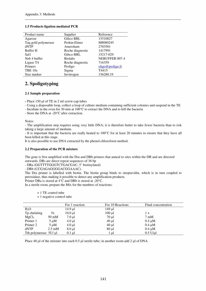
Appendix 3: Methods __________________________________________________________________________________________
141
1.5 Products ligation mediated PCR Product name Supplier Reference Agarose Gibco BRL 15510027 Taq gold polymerase Perkin-Elmer M8080245 dNTP Amersham 2703501 Buffer H Roche diagnostic 1417991 Sal1 Gibco-BRL 15217-029 Neb 4 buffer Biolabs NEBUFFER 007-4 Ligase T4 Roche diagnostic 716359 Primers Proligo [email protected] TBE 10x Sigma T4415 Size marker Invitrogen 156280.19
2. Spoligotyping
2.1 Sample preparation
- Place 150 µl of TE in 2 ml screw-cap tubes - Using a disposable loop, collect a loop of culture medium containing sufficient colonies and suspend in the TE - Incubate in the oven for 30 min at 100°C to extract the DNA and to kill the bacteria - Store the DNA at -20°C after extraction. Notes: - The amplification step requires using very little DNA; it is therefore better to take fewer bacteria than to risk taking a large amount of medium. - It is important that the bacteria are really heated to 100°C for at least 20 minutes to ensure that they have all been killed at this stage. It is also possible to use DNA extracted by the phenol-chloroform method. 2.2 Preparation of the PCR mixture The gene is first amplified with the Dra and DRb primers that anneal to sites within the DR and are directed outwards. DRs are direct repeat sequences of 36 bp - DRa (GGTTTTGGGTCTGACGAC, 5’ biotinylated) - DRb (CCGAGAGGGGACGGAAAC). The Dra primer is labelled with biotin. The biotin group binds to streptavidin, which is in turn coupled to peroxidase, thus making it possible to detect any amplification products. Primer DRa is stored at 4°C and DRb is stored at -20°C. In a sterile room, prepare the Mix for the numbers of reactions: + 1 TE control tube + 1 negative control tube For 1 reaction For 10 Reactions Final concentration H2O 14.9 µl 149 µl Tp chelating 5x 10.0 µl 100 µl 1 x MgCl2 50 mM 7.0 µl 70 µl 7 mM Primer 1 5 µM 4.0 µl 40 µl 0.4 µM Primer 2 5 µM 4.0 µl 40 µl 0.4 µM dNTP 2.5 mM 8.0 µl 80 µl 0.4 µM Tth polymerase 5U/ µl 0.1 µl 1 µl 0.5 U/µl Place 48 µl of the mixture into each 0.5 µl sterile tube; in another room add 2 µl of DNA

Appendix 3: Methods __________________________________________________________________________________________
142
PCR Programme: Prog. 1 3 min – 96°C
Prog. 2 1 min – 96°C 1 min – 55°C 30 sec – 72°C
30 cycles
Prog. 3 10 min – 72°C 2.3 Hybridisation The membranes must always be held by their edges.
1. Rinse the membrane twice in about 250 ml of 2xSSPE/0.1%SDS at 60°C for five minutes. 2. Place the membrane in the miniblotter that has previously been cleaned with soap and water and rinsed
thoroughly, on a support cushion (that acts as a joint) such that the side with the oligunucleotides (spacer) faces the slots, ant so that the slots are perpendicular to the lines of oligonucleotides (spacers) on the membrane (use the date in the bottom tight corner as a marker).
Note: The membrane must be placed so that it is exactly aligned with the two ink lines in the upper and lower slots, so that the slots cross all the lines of oligonucelotide (spacers) fixed to the membrane.
3. Tightly shut the sic plastic screws to close the press 4. Dilute 20 µl of each of the PCR products (maximum of 43 / membrane) in 150 µl of 2
2xSSPE/0.1%SDS buffer. Include one negative control (TE alone) and two positive controls (DNAs from the strain H37Rv and from M. bovis BCG).
5. Heat each tube at 100°C for 10minutes, then immediately place on ice. 6. Use a vacuum pump to remove all the residual liquid from each to the slots of the miniblotter. 7. Fill the first slot of the miniblotter with 150 µl of buffer containing 2xSSPE/0.1%SDS. 8. Fill the remaining slots one by one (max 43) with 150 µl (approximately) of the diluted PCR products,
kept at 4°C until loading. Note the loading order. Note: When filling the slots, take care not to introduce air bubbles (pipette slowly, in both directions, to avoid introducing bubbles). Each line must be completely filled, but care must be taken not to contaminate the slot underneath as this might lead to the contamination of neighbouring slots (use absorbent paper to eliminate excess).
9. Fill the last slot of the slot of the miniblotter with 150 µl of buffer containing 2xSSPE/0.1%SDS. The samples must always be surrounded by buffer to prevent leaking.
10. Incubate the membrane at 60 °C for one hour, keep the miniblotter horizontal throughout and do NOT shake to prevent overflow (and therefore contamination of one line by another).
11. Remove the samples from the slots by aspiration, in the same order as they were filled. 12. When all the slots are totally empty, dissemble the miniblotter and carefully remove the membrane. 13. Wash the membrane twice in250 ml of 2xSSPE/0.5%SDS at 60°Cfor 10minutesin a container with
shaking. (Possibly to interrupt the protocol It is possible to interrupt the protocol at step 13. overnight for example. In this case, proceed to step 13’. 13’. Wash the membrane twice in 250 ml of 2xSSPE at room temperature for 10 minutes, in a container with shaking. 13’’. Keep the membrane at 4°C until the following day, in a sealed plastic envelope or wrapped in Saran-wrap, to prevent it from drying out. The following day, if interrupted at step 13: 13’’’. Wash the membrane twice in 250 ml of 2xSSPE/0.5%SDS at 42°C for 10 minutes in a container with shaking. Continue the protocol at step 14. )
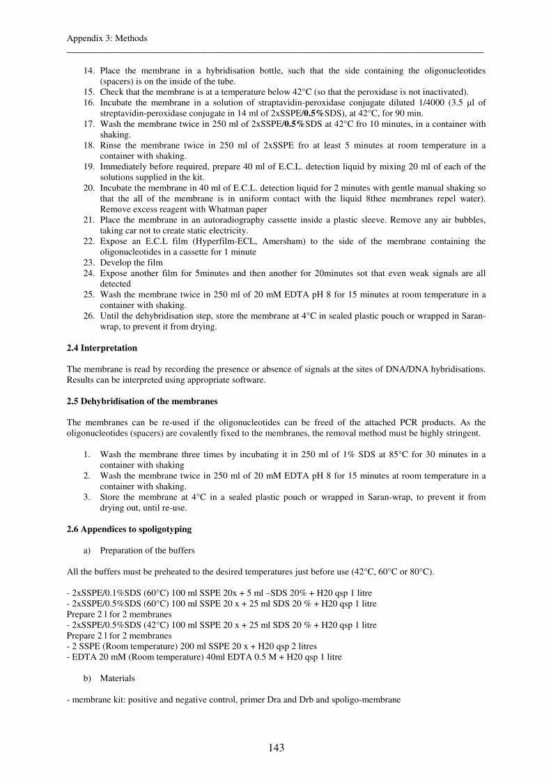
Appendix 3: Methods __________________________________________________________________________________________
143
14. Place the membrane in a hybridisation bottle, such that the side containing the oligonucleotides (spacers) is on the inside of the tube.
15. Check that the membrane is at a temperature below 42°C (so that the peroxidase is not inactivated). 16. Incubate the membrane in a solution of straptavidin-peroxidase conjugate diluted 1/4000 (3.5 µl of
streptavidin-peroxidase conjugate in 14 ml of 2xSSPE/0.5%SDS), at 42°C, for 90 min. 17. Wash the membrane twice in 250 ml of 2xSSPE/0.5%SDS at 42°C fro 10 minutes, in a container with
shaking. 18. Rinse the membrane twice in 250 ml of 2xSSPE fro at least 5 minutes at room temperature in a
container with shaking. 19. Immediately before required, prepare 40 ml of E.C.L. detection liquid by mixing 20 ml of each of the
solutions supplied in the kit. 20. Incubate the membrane in 40 ml of E.C.L. detection liquid for 2 minutes with gentle manual shaking so
that the all of the membrane is in uniform contact with the liquid 8thee membranes repel water). Remove excess reagent with Whatman paper
21. Place the membrane in an autoradiography cassette inside a plastic sleeve. Remove any air bubbles, taking car not to create static electricity.
22. Expose an E.C.L film (Hyperfilm-ECL, Amersham) to the side of the membrane containing the oligonucleotides in a cassette for 1 minute
23. Develop the film 24. Expose another film for 5minutes and then another for 20minutes sot that even weak signals are all
detected 25. Wash the membrane twice in 250 ml of 20 mM EDTA pH 8 for 15 minutes at room temperature in a
container with shaking. 26. Until the dehybridisation step, store the membrane at 4°C in sealed plastic pouch or wrapped in Saran-
wrap, to prevent it from drying.
2.4 Interpretation The membrane is read by recording the presence or absence of signals at the sites of DNA/DNA hybridisations. Results can be interpreted using appropriate software. 2.5 Dehybridisation of the membranes The membranes can be re-used if the oligonucleotides can be freed of the attached PCR products. As the oligonucleotides (spacers) are covalently fixed to the membranes, the removal method must be highly stringent.
1. Wash the membrane three times by incubating it in 250 ml of 1% SDS at 85°C for 30 minutes in a container with shaking
2. Wash the membrane twice in 250 ml of 20 mM EDTA pH 8 for 15 minutes at room temperature in a container with shaking.
3. Store the membrane at 4°C in a sealed plastic pouch or wrapped in Saran-wrap, to prevent it from drying out, until re-use.
2.6 Appendices to spoligotyping
a) Preparation of the buffers All the buffers must be preheated to the desired temperatures just before use (42°C, 60°C or 80°C). - 2xSSPE/0.1%SDS (60°C) 100 ml SSPE 20x + 5 ml –SDS 20% + H20 qsp 1 litre - 2xSSPE/0.5%SDS (60°C) 100 ml SSPE 20 x + 25 ml SDS 20 % + H20 qsp 1 litre Prepare 2 l for 2 membranes - 2xSSPE/0.5%SDS (42°C) 100 ml SSPE 20 x + 25 ml SDS 20 % + H20 qsp 1 litre Prepare 2 l for 2 membranes - 2 SSPE (Room temperature) 200 ml SSPE 20 x + H20 qsp 2 litres - EDTA 20 mM (Room temperature) 40ml EDTA 0.5 M + H20 qsp 1 litre
b) Materials - membrane kit: positive and negative control, primer Dra and Drb and spoligo-membrane
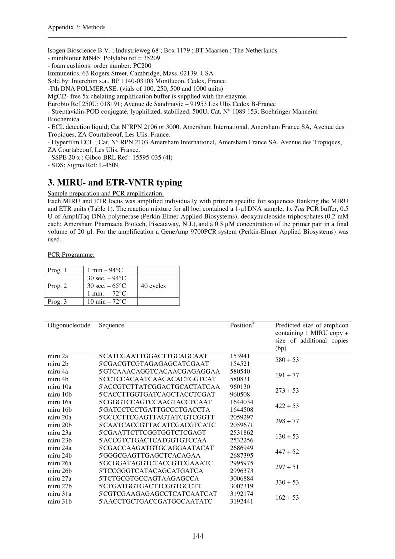
Appendix 3: Methods __________________________________________________________________________________________
144
Isogen Bioscience B.V. ; Industrieweg 68 ; Box 1179 ; BT Maarsen ; The Netherlands - miniblotter MN45: Polylabo ref = 35209 - foam cushions: order number: PC200 Immunetics, 63 Rogers Street, Cambridge, Mass. 02139, USA Sold by: Interchim s.a., BP 1140-03103 Montlucon, Cedex, France -Tth DNA POLMERASE: (vials of 100, 250, 500 and 1000 units) MgCl2- free 5x chelating amplification buffer is supplied with the enzyme. Eurobio Ref 250U: 018191; Avenue de Sandinavie – 91953 Les Ulis Cedex B-France - Streptavidin-POD conjugate, lyophilized, stabilized, 500U, Cat. N° 1089 153; Boehringer Manneim Biochemica - ECL detection liquid; Cat N°RPN 2106 or 3000. Amersham International, Amersham France SA, Avenue des Tropiques, ZA Courtabeouf, Les Ulis. France. - Hyperfilm ECL ; Cat. N° RPN 2103 Amersham International, Amersham France SA, Avenue des Tropiques, ZA Courtabeouf, Les Ulis. France. - SSPE 20 x ; Gibco BRL Ref : 15595-035 (4l) - SDS; Sigma Ref: L-4509
3. MIRU- and ETR-VNTR typing Sample preparation and PCR amplification: Each MIRU and ETR locus was amplified individually with primers specific for sequences flanking the MIRU and ETR units (Table 1). The reaction mixture for all loci contained a 1-µl DNA sample, 1x Taq PCR buffer, 0.5 U of AmpliTaq DNA polymerase (Perkin-Elmer Applied Biosystems), deoxynucleoside triphosphates (0.2 mM each; Amersham Pharmacia Biotech, Piscataway, N.J.), and a 0.5 µM concentration of the primer pair in a final
volume of 20 µl. For the amplification a GeneAmp 9700PCR system (Perkin-Elmer Applied Biosystems) was used. PCR Programme: Prog. 1 1 min – 94°C
Prog. 2 30 sec. – 94°C 30 sec. – 65°C 1 min. – 72°C
40 cycles
Prog. 3 10 min – 72°C Oligonucleotide Sequence Positiona Predicted size of amplicon
containing 1 MIRU copy + size of additional copies (bp)
miru 2a 5'CATCGAATTGGACTTGCAGCAAT 153941 miru 2b 5'CGACGTCGTAGAGAGCATCGAAT 154521
580 + 53
miru 4a 5'GTCAAACAGGTCACAACGAGAGGAA 580540 miru 4b 5'CCTCCACAATCAACACACTGGTCAT 580831
191 + 77
miru 10a 5'ACCGTCTTATCGGACTGCACTATCAA 960130 miru 10b 5'CACCTTGGTGATCAGCTACCTCGAT 960508
273 + 53
miru 16a 5'CGGGTCCAGTCCAAGTACCTCAAT 1644034 miru 16b 5'GATCCTCCTGATTGCCCTGACCTA 1644508
422 + 53
miru 20a 5'GCCCTTCGAGTTAGTATCGTCGGTT 2059297 miru 20b 5'CAATCACCGTTACATCGACGTCATC 2059671
298 + 77
miru 23a 5'CGAATTCTTCGGTGGTCTCGAGT 2531862 miru 23b 5'ACCGTCTGACTCATGGTGTCCAA 2532256
130 + 53
miru 24a 5'CGACCAAGATGTGCAGGAATACAT 2686949 miru 24b 5'GGGCGAGTTGAGCTCACAGAA 2687395
447 + 52
miru 26a 5'GCGGATAGGTCTACCGTCGAAATC 2995975 miru 26b 5'TCCGGGTCATACAGCATGATCA 2996373
297 + 51
miru 27a 5'TCTGCGTGCCAGTAAGAGCCA 3006884 miru 27b 5'CTGATGGTGACTTCGGTGCCTT 3007319
330 + 53
miru 31a 5'CGTCGAAGAGAGCCTCATCAATCAT 3192174 miru 31b 5'AACCTGCTGACCGATGGCAATATC 3192441
162 + 53
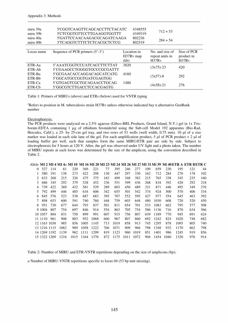
Appendix 3: Methods __________________________________________________________________________________________
145
miru 39a 5'CGGTCAAGTTCAGCACCTTCTACATC 4348555 miru 39b 5'CTCGGTGTTCCTTGAAGGTGGTTT 4349319
712 + 53
miru 40a 5'GATTCCAACAAGACGCAGATCAAGA 802236 miru 40b 5'TCAGGTCTTTCTCTCACGCTCTCG 802519
284 + 54
Locus name
Sequence of PCR primers (5’-3’)
Location in H37Rv map (kb)
No. and size of repeat units in H37Rv
Size of PCR product in H37Rv
ETR-Aa 5’AAATCGGTCCCATCACCTTCTTAT 3820 ETR-Ab 5’CGAAGCCTGGGGTGCCCGCGATTT
(3x75)-23 420
ETR-Ba 5’GCGAACACCAGGACAGCATCATG 4160 ETR-Bb 5’GGCATGCCGGTGATCGAGTGG
(3x57)-8 292
ETR-Ca 5’GTGAGTCGCTGCAGAACCTGCAG 1480 ETR-Cb 5’GGCGTCTTGACCTCCACGAGTG
(4x58)-21 276
Table 1: Primers of MIRUs (above) and ETRs (below) used for VNTR typing
aRefers to position in M. tuberculosis strain H37Rv unless otherwise indicated bay n alternative GenBank number
Electrophoresis: The PCR products were analyzed on a 2.5% agarose (Gibco-BRL Products, Grand Island, N.Y.) gel in 1x Tris-borate-EDTA containing 1 µg of ethidium bromide/ml using the Sub-cell Model 192 apparatus (Bio-Rad, Hercules, Calif.), a 25- by 25-cm gel tray, and two rows of 51 wells (well width, 0.75 mm). 10 µl of a size marker was loaded in each side lane of the gel. For each amplification product, 5 µl of PCR product + 2 µl of loading buffer per well, such that samples form the same MIRU/ETR pair are side by side. Subject to electrophoresis for 5 hours at 120 V. After, the gel was observed under UV light and a photo taken. The number of MIRU repeats at each locus was determined by the size of the amplicon, using the convention described in Table 2.
Allele MI 2 MI 4 MI 4a MI 10 MI 16 MI 20 MI 23 MI 24 MI 26 MI 27 MI 31 Mi 39 Mi 40 ETR A ETR BETR C
0 527 114 61 220 369 221 77 395 246 277 109 659 230 195 121 441 580 191 138 273 422 298 130 447 297 330 162 712 284 270 178 1022 633 268 215 326 475 375 183 499 348 383 215 765 338 345 235 1603 686 345 292 379 528 452 236 551 399 436 268 818 392 420 292 2184 739 422 369 432 581 529 289 603 450 489 321 871 446 495 349 2765 792 499 446 485 634 606 342 655 501 542 374 924 500 570 406 3346 845 576 523 538 687 683 395 707 552 595 427 977 554 645 463 3927 898 653 600 591 740 760 448 759 603 648 480 1030 608 720 520 4508 951 730 677 644 793 837 501 811 654 701 533 1083 662 795 577 5089 1004 807 754 697 846 914 554 863 705 754 586 1136 716 870 634 566
10 1057 884 831 750 899 991 607 915 756 807 639 1189 770 945 691 62411 1110 961 908 803 952 1068 660 967 807 860 692 1242 824 1020 748 68212 1163 1038 985 856 1005 1145 713 1019 858 913 745 1295 878 1095 805 74013 1216 1115 1062 909 1058 1222 766 1071 909 966 798 1348 932 1170 862 79814 1269 1192 1139 962 1111 1299 819 1123 960 1019 851 1401 986 1245 919 85615 1322 1269 1216 1015 1164 1376 872 1175 1011 1072 904 1454 1040 1320 976 914
Table 2: Number of MIRU and ETR-VNTR repetitions depending on the size of amplicons (bp); a Number of MIRU-VNTR repetitions specific to locus 04 (53 bp unit missing).
Appendix 3: Methods __________________________________________________________________________________________
146
4. IS6110-Restriction Fragment Length Polymorphism typing
4.1 Extraction of genomic DNA
Operations to be carried out in a Category 3 Laboratory - Collect an adequate number of colonies on Lowenstein-Jensen medium and suspend them in 500 µl of TE (see appendix IS6110-RFLP) - Place at 90°C for 30 min. Operations NOT to be carried out in a Category 3 Laboratory - Centrifuge the extracts - Discard the supernatant and add 50 µl or 100 µl of 20 mg/ml lysozyme (see appendix IS6110-RFLP) to the pellet, according to the size of the pellet. - Incubate for 1h at 37°C. - Add 70 µl of 10% SDS + 5 µl of 10 mg/ml proteinase K (see appendix IS6110-RFLP), or twice as much if the pellet is large. - Incubate for 1 h at 65 °C - Add 100 µl of 5 M NaCl Steps to be carried out under the Sorbonne flow hood - Add 1 volume of aquaphenol containing 8-beta-hydroxyquinoline to the extract, mix thoroughly and then centrifuge for 15 min in a micorcentrifuge at 149000 rpm at 4°C. - Transfer the aqueous phase into a clean Eppendorf tube. Note: the aqueous phase is transparent and is generally the upper phase. The phenolic phase is yellow and is generally the lower phase. The phases may be inversed if the interface is very large. In this case, or if the aqueous phase is cloudy, take the aqueous phase and repeat the phenol extraction step. - Add an equal volume of chloroform, mix and centrifuge for 1 min at 14900 rpm. - Transfer the aqueous supernatant to a clean Eppendorf tube. - divide the sample into two if its volume exceeds 600 µl. - Add 1.5 volumes of absolute ethanol (-20°C) to precipitate the nucleic acids. - Leave overnight at -20°C or for 30 min at -70°C → Possible to interrupt - Centrifuge for 15 min at 14900rpm and 4°C - Discard the supernatant by tipping the Eppendorf tube. - Wash the pellet with 100 µl of 70% ethanol at -20°C. - Centrifuge for 5 min at 14900 rpm and 4°C - Discard the supernatant and dry the pellet in a speed-vac for 15 min or under the flow hook. - Resuspend the DNA pellet in 20 µl of TE - Incubate for 20 min in a 60°C water bath. - Homogenise the sample with a pipette. - To estimate the concentration of the DNA, run an aliquot in a 0.8% agarose electrophoresis gel (2 µl of extract + 2 µl of loading buffer per well) 4.2 Digestion of genomic DNA - After estimating the concentration of the DNA, digest the equivalent of 2 µg of DNA with the restriction enzyme as follows (amount per reaction):
- X µl of DNA - 3 µl of enzyme buffer - 2 µl of PvuII - H2O (ultrapure) to a final volume of 30 µl
- Leave the enzyme to act for at least 1 h at 37°C or overnight at 37°C. → Possible to interrupt - Subject to electrophoresis in a 0.8% agarose gel in 1X TBE to check the digestion and to estimate the amount of DNA to load for the Southern blot.
Appendix 3: Methods __________________________________________________________________________________________
147
Note: If the bands nearest the top of the gel (high molecular weights) are less intense than the bands corresponding to lower molecular weights, the digestion has been satisfactory. (In the case of incomplete or no digestion: - Dialyse the sample with a Millipore filter (0.05 µm pore size): place the entire sample on the filter, leave contact with the water for 30 min and collect the DNA. - Repeat the digestion step with: - Dialysed DNA - 3 µl of enzyme buffer - 1 µl of PvuII - Leave to digest for at least 1 h at 37°C. - Check the digestion on a 0.8 % agarose gel.) 4.3 Electrophoresis - Load the equivalent of 2 µg of digested DNA + 2 µl of loading buffer, on a 1 % agarose gel in 1X TBE containing EtBr. - Do not load samples in the first or last wells. Load 10 µl of the lambda PstI external marker (appendix IS6110-RFLP) in the first lane to monitor the migration. - Every 5 wells, load the external molecular weight marker: DNA from M. tuberculosis strain MT 14323 digested with PvuII - Start the electrophoresis at 90V for 10 min (until the DNA has left the wells) and then continue overnight at 40V (i.e. 1-2 V/cm) → Possible to interrupt - The following day, stop the electrophoresis when the highest λ molecular weight marker (i.e. the 11500-bp band) is 2 cm from the wells and take a photo. 4.4 Treatment of gels a) Dupurination - Wash the gel with a solution of 0.25 M HCl (appendix IS6110-RFLP) for 10 min, for depurination - This treatment facilities the DNA transfer step by breaking up the large molecular weight molecules. Warning: Prolonged treatment can lead to excessive hydrolysis of the DNA and generate DNA fragments that are too small to adhere to the membrane correctly. - Rinse the gel with distilled water for 1-2 min. b) Denaturation -Cover the gel with the denaturing solution (appendix IS6110-RFLP) for 2 x 20 min periods with shaking. - Rinse the gel with distilled water for 1-2 min. c) Neutralisation Cover the gel with the neutralising solution (appendix IS6110-RFLP) for two 20 min periods with shaking. 4.5 Transfer (Southern Blot) of nucleic acids onto membranes a) Vacuum transfer - Cut a piece of Hybond N+ membrane and a piece of Whatman paper to the size of the gel. - Soak the Whatman with water and place it on the porous plate, then soak the Hybond N+ membrane with water and place on top, taking care not to form any air bubbles. - Place the gel in the middle of this pocket. - Adjust the clips on the apparatus - Fill the gel wells with 1 % agarose, ensuring that no bubbles are formed - Connect the vacuum pump. For the transfer of genomic DNA from an agarose gel, the recommended conditions are 45-60 mbar for 60-90min. - Cover the gel with 20x SSC buffer.

Appendix 3: Methods __________________________________________________________________________________________
148
b) After the transfer - Before removing the transferred gel, use a pencil to mark the positions of the first and last wells. - Remove the gel and collect the membrane - Wrap the membrane in Saran Wrap. - Fix the DNA to the membrane by exposing to UV light, either for 4 min at 365 nm or for 1 min at 254 nm (DNA side towards the UV light source) - Check the transfer efficiency by examining the gel under UV light and ensuring that all traces of DNA have disappeared - The fixed membrane can be stored at 4°C 4.6 Hybridisation and detection a) Prehybrdisation - Place the membrane in a glass hybridisation tube such that the DNA faces the inside of the tube. - Prehybridise the membrane for 45 to 60 min at 42°C with rotation in hybridisation buffer (appendix IS6110-RFLP). Voulume of buffer required = 0.125 ml / cm2 of membrane. b) Labelling the probe ECL Kit protocol: - Dilute the probe to a concentration of 10 ng/µl with the water supplied in the ECL Kit. - Heat to 100°C for 5 min, then leave at 0°C for 5 min - Add an equal volume of labelling reagent - Add the same volume of glutaraldehyde. - Incubate for 10 min at 37°C. - Keep the labelled probe on ice until use. Note; The labelled probe can be stored in 50% glycerol at -20°C for 6 months. c) Hybridisation - Transfer the hybridisation buffer into a clean tube and add the labelled probe. - Mix by rotating - Place the buffer + probe in the hybridisation tube with the membrane - Incubate overnight at 42°C with rotation d) Washes - The following day, discard the hybridisation buffer. - Wash the membrane with buffer I (appendix IS6110-RFLP) at 55°C with shaking: 2 x 10 min - Wash the membrane with buffer II (appendix IS6110-RFLP) at room temperature with shaking: 2 x 5 min e) Detection - With the ECL kit: mix 15 ml of detection reagent 1 with 15 ml of detection reagent 2 in a tube (in the dark). - Dry the membrane on a sheet of Whatman paper - Place the membrane in a container with the DNA side outwards - cover the membrane with the detection mixture and swirl manually for 1 min - Remove the membrane in an autoradiography film cassette and cover with a sheet of Saran Wrap - Place a film (Hyperfilm) on the membrane, shut the cassette and leave for 1 min - Remove the film and immediately replace with another. - Develop the first film and estimate the exposure time necessary for the second 4.7 Appendix IS6110-RFLP a) Preparation of the IS6110 probe
Appendix 3: Methods __________________________________________________________________________________________
149
The IS6110 probe is generated from a PCR product. An 868-bp fragment, corresponding to nucleotides 462 to 1330 of the IS6110 sequence of a strain belonging to the M. tuberculosis complex, is amplified (MT 14323). The oligonucleotides used as primers as follows: G1: 5’ CTGACCGAGCTGGGTGTGCC G2: 5’ TCTGATCTGAGACCTCAGCC Preparation of the MIX for 10 reactions REAGENT QUANTITIY FINAL CONCENTRATION H2O 235 µl Taq Buffer 10 x 50 µl 1 x DMSO 50 µl 10 % MgCl2 25 mM 30 µl 1.5 mM dNTP 10 mM 10 µl 200 µM (each) G1 50µM 10 µl 1 µM G2 50 µM 10 µl 1 µM Taq 5U/µl 5 µl 2.5 U - Place 40 µl of MIX into each PCR tube. - Add 10 µl of MT 14323 DNA diluted 1/100 (extracted by the Phenol/Chloroform method) PCR conditions: Prog. 1 6 min – 94°C
Prog. 2 1 min – 94°C 2 min – 55°C 2 min – 72°C
35 cycles
Prog. 3 10 min – 72°C - Subject the PCR product to electrophoresis on a 0.8 % agarose gel - Electrophoresis conditions with a 50 ml miniprep: ½ h at 90 V. - Load: 1 µl of DNA + 2 µl loading buffer - Prepare a 1 % gel with LMP agarose for a Miniprep apparatus, i.e. with 50 ml of 1 x TBE - Load 100 bp marker in the end lanes, and all of the amplified samples in the central lane, i.e. 500 µl of PCR product + 55 µl of loading buffer. - Leave for 30 min to 1h30 at 100 V. (Monitor the migration of the 800-bp band) - Identify the 800-bp band by use of the marker, cur the band out, taking care not to take too much agarose. - Put the 800-bp fragment in a Petri dish. - Weigh the agarose plug and place it in an eppendorf With the QIAGEN extraction kit (Qiaquick Gel Extraction Kit protocol) - Add 3 volumes of QG buffer to 1 volume of agarose (100 mg agarose = 100 µl) - Incubate at 50°C fro 10 min (the gel must be completely dissolved), vortex gently every 3 min during the incubation period - When the agarose is completely dissolved, the liquid becomes yellow - Add 1 volume of isopropanol and mix - Place 1Qiauick column on a 2 ml tube. - Place the melted gel containing the DNA to be purified on the column and centrifuge for 1 min at 13000 rpm - Discard the liquid in the 2 ml tube and place 0.5 ml of QG buffer on the column, centrifuge for 1 min at 13000 rpm - Wash the column with 0.75 ml of PE buffer and centrifuge for 1 min at 13000 rpm - Discard the liquid in the tube and centrifuge again. - Place the column on a 1.5 ml eppendorf tube and elute the DNA with 50 µl of the H2O provided in the ECL kit for labelling the probe (see below: labelling the probe) - Subject to electrophoresis in a 0.8 % agarose gel in 1 x TBE for 30 min at 90 V - Load: - lane n°1= 10 µl λ PstI + 2 µl of loading buffer
Appendix 3: Methods __________________________________________________________________________________________
150
- lane n°2= 1 µl IS6110 to be quantified + 10 µl H2O + 2 µl of loading buffer - lane n°3= 1 µl λ HindIII (10 ng) + 10 µl H2O + 2 µl of loading buffer - lane n°4= 5 µl λ HindIII (50 ng) + 10 µl H20 + 2 µl of loading buffer - lane n°5= 10 µl λ HindIII (100 ng) + 10 µl H20 + 2 µl of loading buffer Labelling the probe: After calculating the concentration of the purified probe, calculate the amount of probe to be labelled. Example: Amount of probe required for a 120 cm2 membrane Final concentration of the probe to be 1 ng/ cm2
Thus 120 ng for a 120 cm2 membrane If for example, the concentration of the probe is 10 ng/µl, 12 µl of probe is required For the labelling mix: 12 µl of the probe at a concentration of 10 ng/µl + 12 µl Labelling Reagent + 12 µl of Glutaraldehyde Total= 36 µl of labelled probe. b) Solutions and materials used Product name Supplier Reference Reference IP Tris-Trizma base SIGMA T 1503 52150 Boric Acid SIGMA B-6768 (1 kg) - ETA SIGMA E-5134 (1 kg) - Lysozyme SIGMA L-6876 (5kg) - Proteinase K ROCHE-Boehringer 1092766 (1 g) 90428 SDS SIGMA L-4509 (500 g) Sodium chloride (NaCl) PROLABO 27 810 295 (500 g) 48700 Phenol APPLIGENE 130181 (250 ml) - 8-beta-hydroxquinoline SIGMA H6878 - Chloroform CARLO ERBA 438601 49520 Ethanol MERCK 983 44656 PvuII GIBCO BRL 15 4120 18 95034 PstI GIBCO BRL 15 2150 23 90531 Hydrochloric acid (HCl) PROLABO 20 252 290 44160 Tri-Sodium citrate PROLABO 27 833 294 48740 Sodium hydroxide (NaOH) PROLABO 28 245 298 48880 Filter 0.05 µM/25 mm MILLIPORE VMWP02500 - Whatman paper 3 MM 3030 917 - Hybond-N+ membrane AMERSHAM RPN 303 B 90803 Hyperfilm ECL AMERSHAM RPN 2103 K 96098 ECL Kit AMERSHAM RPN 3000 97059 Qiaquick Gel Extraction Kit QIAGEN 28704 42792 Bromophenol Blue SIGMA B5525 - Agarose GIBCO BRL 15510027 52171 (500 g) Ethidium bromide (EtBr) EUROBIO 17589 99025 Taq PERIKNELMER M8080161 90085 dNTP AMERSHAM 2703501 90005 TBE 10 x SIGMA T4415 52155 (4 l) Lambda DNA size markers PHARMACIA 27-4111-01(500 µg/ml) - 100 bp size markers GIBCO-BRL 156280.19 90637 - Lysozyme: 20 mg/ml stock solution, divided into aliquots and frozen - Proteinase K: 10 mg/ml stock solution, divided into aliquots and frozen - SDS 10 % in distilled H2O - NaCl 5m - TE: 1 ml Tris 1 M (=> 89 mM), 0.2 ml EDTA 0.5 M (=>89mM), 100 ml H2O qsp - Phenol, saturated in water (pH 8) and coloured with 0.1 % 8-beta-hydroxyuinoline - TBE 10 x pH 8: Tris 89 mM, Boric Acid 89 mM, EDTA 2 mM - Bromophenol blue solution: 0.25 % bromophenol blue + 40 % sucrose - Preparation of lambda-PstI marker
Appendix 3: Methods __________________________________________________________________________________________
151
Digest 100 µl of lambda DNA with PstI in a final volume of 250 µl Check the digestion by agarose gel electrophoresis Add 75 µl of the bromophenol blue solution and 175 µl of TE buffer pH 8 Store the marker in 50 µl aliquots at + 4°C - Depurination solution: HCl 0.25 M = 13 ml HCl + 500 ml of H2O - Denaturation solution: NaCl 1.5 M / NaOH 0.5 M - Neutralising solution: NaCl 1.5 M / Tris 1 M pH 8 - SSC buffer 20 x: NaCl (=> 3 M) 175.3 g/l, Na-Citrate (=> 0.3 M) 88.2 g/l, H20 1 l qsp, Adjust the pH to 7 with 5M NaOH - Hybridisation buffer (Amersham Kit) Heat the hybridisation buffer supplied in the kit to 65°C. Adjust the concentration of NaCl to 0.5 M Add 5 % (w/v) blocking agent Mix, taking care not to allow any lumps to form, using a magnetic stirrer for 1 h at 65°C Store 30 ml aliquots of the buffer at – 20°C for up to 3 months For 200 ml of ECL buffer: 5.84 g of NaCl + 10 g of blocking agent - Washing buffer n°1 (SSC 0.5 x, SDS 0.4 %): 25 ml SSC 20 x, 20 ml SDS 20 %, 1 l H2O qsp - Washing buffer n°2 (SSC 2 x,): 100 ml SSC 20 x, 1 l H2O qsp.

Curriculum vitae
153
Curriculum vitae
Education
01/2003-03/2006 Swiss Tropical Institute (STI) of Basel, Switzerland
Ph.D., Microbiology, molecular-epidemiology
Thesis title: Molecular epidemiology of mycobacteria: Development
and refinement of innovative molecular typing tools to study
mycobacterial infections.
Supervisors: PD Dr. Jakob Zinsstag, Prof. Marcel Tanner
10/1996-05/2001 Swiss Federal Institute of Technology (ETH) of Zurich,
Switzerland
Master degree of natural Science, Bio- and Organic chemistry
Master thesis title: Benzophenones as photo activable surface markers
for Haemagluthinin (German).
Mark thesis: 5.5 (magna cum laude)
Supervisor: Prof. Josef Brunner
Post graduate research experience
01/2003-03/2006 Swiss Tropical Institute (STI) of Basel, Switzerland
Department of Public health and epidemiology
Supervisors: PD Dr. Jakob Zinsstag, Prof. Marcel Tanner
Name/Prename: Hilty Markus
Date of birth: 30.01.1976
Nationality: Liechtenstein
Address: Josefstrasse 192, 8005 Zürich, Switzerland
Phone number: +41 1 2715439
E-Mail: [email protected]

Curriculum vitae
154
10/2001 Swiss Federal Institute of Technology (ETH) of Zurich,
Switzerland
Institute of Biochemistry.
Post graduate research work title: Benzophenones as photo activable
surface markers for Haemagluthinin
07/2001 - 09/2001 University of Kiev, Ukraine
Institute of Biochemistry.
Practical work title: Solubilisation of fibrin clots by specific enzymes
Fieldwork and -experience
01/2003-03/2006 ‘Support en Santé Internationale (CSSI)’ and ‘Laboratoire de
Recherches Vétérinaires et Zootechniques de Farcha (LRVZ)’ of
N’Djaména, Chad
Collection and processing of samples within the Ph.D. research project.
Responsibility: Contribute to maintaining the mycobacterial unit of
LRVZ.
03/2002 – 10/2002 Total Community Mobilisation against HIV/AIDS of Francistown
and Kasane, Botswana
Employment within a non governmental organisation. Responsibilities:
Presenting of courses about ‘anti viral therapy’, ‘time management and
teamwork’ and ‘health and hygiene‘ and supervision of the work of
local collaborators.

Curriculum vitae
155
Meetings and Seminars attended
6th International Meeting on Microbial Epidemiological Markers. Molecular epidemiology
and transmission dynamics of human and bovine tuberculosis in Chad and Mauritania
(Poster). Les Diablerets, Switzerland. August 27 - 30, 2003
Course and practical work: Molecular tools and epidemiology of tuberculosis. Paris, France.
September 1-12, 2003
Swiss Meeting for Doctoral Students in Parasitology and Tropical Medicine. State of
surveillance of tuberculosis (TB) transmission in Chad (oral presentation). Müncheswiler,
Switzerland. October, 2003
63rd annual meeting of the Swiss Society for Microbiology. Molecular epidemiology and
transmission dynamics of human and bovine tuberculosis in Chad and Mauritania (poster).
Lugano, Switzerland. March 11-12, 2004
Annual Congress of the Swiss and the German Society of Tropical Medicine and
Parasitology. Molecular epidemiology and transmission dynamics of human and bovine
tuberculosis in Chad and Mauritania (poster). Würzburg, Germany. September 23-25, 2004
Mitgliederversammlung der Schweizerischen Vereinigung für Tierpathologie. Molecular
epidemiology of Mycobacterium bovis in Chad: A concern for humans? (oral presentation).
Bern, Switzerland. June 24, 2005
4th International Conference on Mycobacterium bovis. Evaluation of the discriminatory power
of Variable Number Tandem Repeat typing of Mycobacterium bovis strains from Chad
(poster). Dublin, Ireland. August 22-26, 2005
Annual Congress of the Swiss Society of Tropical Medicine and Parasitology.
- Evaluation of the discriminatory power of Variable Number Tandem Repeat typing of
Mycobacterium bovis strains from Chad (poster).
Curriculum vitae
156
- Epidemiology and economics of Mycobacterium bovis and its control (oral presentation).
Ascona, Switzerland. November 2-3, 2005
International Mini-symposium on human and animal health. Molecular epidemiology of
human and animal tuberculosis in the Sahel (oral presentation). Basel, Switzerland.
December, 2005.
Publications
Hilty, M. +, D. Yeboah-Manu,+ D. Boakye, E. Mensah-Quainoo, S. Rondini, E. Schelling, D.
Ofori-Adjei, F. Portaels, J. Zinsstag and G. Pluschke. Genetic diversity in Mycobacterium
ulcerans isolates from Ghana revealed by a newly identified locus containing a variable
number of tandem repeats. Journal of Bacteriology 2006 Feb;188(4):1462-5 + contributed equally
Diguimbaye-Djaibé C.+, Hilty M.+, Ngandolo R, Mahamat HH., Pfyffer G. E., Baggi F.,
Hewinson G., Tanner M., Zinsstag J. and Schelling E. Mycobacterium bovis Isolates from
Tuberculous Lesions in Chadian Zebu Carcasses. Emerging Infectious Diseases
2006;12(5):769-771 + contributed equally
Hilty, M., C. Diguimbaye, E. Schelling, F. Baggi, M. Tanner, and J. Zinsstag. Evaluation of
the discriminatory power of variable number tandem repeat (VNTR) typing of
Mycobacterium bovis strains. Veterinary Microbiology 2005 Aug;109(3-4): 217-222.
Colette Diguimbaye, Markus Hilty, Richard Ngandolo, Hassane H. Mahamat, Gaby E.
Pfyffer, Franca Baggi, Marcel Tanner, Esther Schelling, and Jakob Zinsstag. Molecular
characterization and drug resistance testing of Mycobacterium tuberculosis isolates from
Chad. Journal of Clinical Microbiology 2006 Apr;44(4):1575-7
Anthony Ablordey, Markus Hilty, Pieter Stragier, Jean Swings, and Françoise Portaels.
Comparative nucleotide sequence analysis of polymorphic variable-number tandem-repeat
Loci in Mycobacterium ulcerans. Journal of Clinical Microbiology 2005 Oct; 43(10):5281-
4
Curriculum vitae
157
C. Diguimbaye-Djaibé, V. Vincent, E. Schelling, M. Hilty, R. Ngandolo, HH. Mahamat, G.
Pfyffer, F. Baggi, M. Tanner, and J. Zinsstag. Species identification of non-tuberculous
mycobacteria from humans and cattle of Chad. Schweizerisches Archiv für Tierheilkunde
2006;148(5):251-6
Sebastien Gagneux, Kathryn DeRiemer, Tran Van, Midori Kato-Maeda, Bouke C. de Jong,
Sujatha Narayanan, Mark Nicol, Stefan Niemann, Kristin Kremer, M. Cristina Gutierrez,
Markus Hilty, Philip C. Hopewell and Peter M. Small. Variable host-pathogen compatibility
in Mycobacterium tuberculosis. Proceedings of the National Academy of Sciences of U S A
2006 Feb 21;103(8):2869-73

![Stephen Briggs [Compatibility Mode]](https://static.fdokumen.com/doc/165x107/6324c3005c2c3bbfa802dd10/stephen-briggs-compatibility-mode.jpg)


![BEE.ppt [Compatibility Mode] - Knowledge Exchange Platform](https://static.fdokumen.com/doc/165x107/631cba156c6907d368016fa0/beeppt-compatibility-mode-knowledge-exchange-platform.jpg)


![Bai07_Thiet ke CPU.ppt [Compatibility Mode]](https://static.fdokumen.com/doc/165x107/6337655dd63e7c790105c48c/bai07thiet-ke-cpuppt-compatibility-mode.jpg)


![0. Otwarcie Konferencji_[Compatibility Mode] - DAFA](https://static.fdokumen.com/doc/165x107/6326d44d6d480576770cfa67/0-otwarcie-konferencjicompatibility-mode-dafa.jpg)