Pathogenesis of Mycobacterium Tuberculosis, mode of action ...

Mycobacterium tuberculosis as viewedthrough a computerDenise Kirschner and Simeone Marino
Department of Microbiology and Immunology, University of Michigan Medical School, Ann Arbor, MI 48109-0260, USA
Mathematical models are emerging as important tools
in the study of microbiology. As an illustrative example,
we present results from several models each generated
to study the interaction of Mycobacterium tuberculosis
and the immune system. Different mathematical models
were formulated on the basis of assumptions regarding
system–component interactions, enabling us to explore
specific aspects at diverse biological scales (e.g. intra-
cellular, cell–cell interactions, and cell population
dynamics). In addition, we were able to examine both
temporal and spatial aspects. At each scale, there were
consistent themes that emerged as determinative in
infection outcome. Factors we identified include both
host and microbial characteristics. The use of the
models lies in generating hypotheses that can then be
tested experimentally. Here, we outline the primary host
and bacterial factors that we have identified as key
mechanisms that contribute to the success of
M. tuberculosis as a human pathogen. Our goal is to
stimulate experimentation and foster collaborations
between theoretical and experimental scientists.
Introduction
Mycobacterium tuberculosis is one of the oldest humanpathogens; evidence of tubercles has been found even inEgyptian mummies. The fantastic success of this organ-ism is highlighted by the fact that one-third of the world isinfected. Given its long association with humans, onemight expect that the study of this pathogen would haverevealed a range of virulence factors. Without becomingtrapped in a cycle of definitions regarding virulence, it isclear that standard notions do not apply forM. tuberculosis.This poses an interesting conundrum for microbiologists;namely, how can arguably the world’s most successfulpathogen lack traditional virulence factors? Is this just amatter of definitions, or has this bacteria evolved beyondour traditional concepts of such factors? To furtherconfound things, M. tuberculosis has a stable genome [1]and thus mutations or phase variations probably do notcontribute to its ability to evade the immune response.Here, we use a mathematical modeling approach to lendsupport to the idea that virulence strategies used byM. tuberculosis enable it to survive within macrophagesand evade host immunity. We identify both microbe andhost characteristics as determinative to its success.
Corresponding author: Kirschner, D. ([email protected]).Available online 24 March 2005
www.sciencedirect.com 0966-842X/$ - see front matter Q 2005 Elsevier Ltd. All rights reserve
A role for mathematical modeling in microbiology
Most modern research in microbial pathogenesis takesplace at the level of cellular and biochemical mechanismsgoverning host–parasite interaction; however, studies atlarger scales are undoubtedly needed for a deeper under-standing of infectious diseases. Components of host–pathogen systems are sufficiently numerous and theirinteractions sufficiently complex that intuition alone isinadequate to fully understand the dynamics of theinteractions. Here, mathematical modeling becomes animportant integrative experimental tool (Figure 1).Mathematical models provide a unique approach forrepresenting and studying the integrated behavior ofcomplex biological systems. A strength of the modelingprocess is that it can lend insight and clarification toexisting data and theories, as well as enabling one to com-pare and contrast existing hypotheses. We extensively relyon collaborators and the literature to define our modelstructure, to decidewhich biologicalmechanisms to include,and to determine what alternative hypothesis to test. Oncethe structure of the model is defined, parameters thatrepresent defined biological rates are derived from pub-lishedexperimentaldataaswell as thosegenerated fromourcollaborators (i.e. half-lives, infection rates and activationrates). We give weight to studies performed using humancells and M. tuberculosis primary and virulent laboratorystrains. For full details of the modeling process, please see,for example, Wiggington et al. [2].
Our mathematical models represent dynamics of ageneral adaptive immune response and microbial factorsspecific to M. tuberculosis. Data comparison and modelvalidation has been performed concurrently in a non-human primate (NHP) model. [3,4] When data are notavailable, we employ detailed statistical uncertainty andsensitivity analyses to estimate parameter values andevaluate how variations in their values contribute toinfection dynamics.
We have developed several mathematical modelsexploring the interaction between the human immunesystem and M. tuberculosis. They have been designed tocapture global dynamics regarding trafficking between thelung and its associated draining hilar lymph node [5,6](Box 1), as well as more local dynamics occurring within acell (Box 2), within a granuloma [7] or within lungs ofinfected individuals [2]. Mathematical models can be usedin a variety of ways to not only reproduce benchexperiments serving as validation of the model, but alsoto perform experiments not presently accessible in the
Opinion TRENDS in Microbiology Vol.13 No.5 May 2005
d. doi:10.1016/j.tim.2005.03.005
TRENDS in Microbiology
(a)
(b) (c)
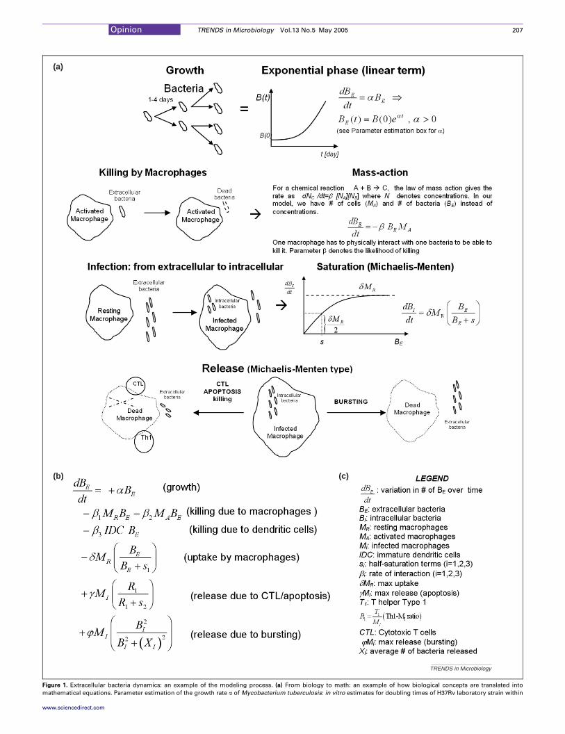
Figure 1. Extracellular bacteria dynamics: an example of the modeling process. (a) From biology to math: an example of how biological concepts are translated into
mathematical equations. Parameter estimation of the growth rate a of Mycobacterium tuberculosis: in vitro estimates for doubling times of H37Rv laboratory strain within
Opinion TRENDS in Microbiology Vol.13 No.5 May 2005 207
www.sciencedirect.com

macrophages ranged from 28 to 96 hours. In mouse lung tissue, H37Rv was estimated to have a doubling time of 63.2 hours. We can estimate the intracellular versus
extracellular growth rates from these values: rateZln2 / doubling time/aBIZ[0.007, 0.024], aBEZ0.011 (per day). (b) Differential equation for extracellular bacteria, BE. Each
equation of the model represents the incremental variation of a certain quantity over time (days): pg/ml (x 106 cells) for cytokine concentrations, cell/cm3 of tissue for cellular
variables and bacteria/cm3 of tissue for M. tuberculosis. (c) Table of symbols.
Box 1. How do we track infection in Mycobacterium tuberculosis?
Major infection outcomes in humans are latent infection (persistent
infection, w90% of infected) and active TB (w5% of infected).
Reactivation also occurs (w5–10% of latently infected) [28]. Reliable
markers of the status of M. tuberculosis infection in humans are not
presently available. A key challenge historically in the study of
tuberculosis has been identifying a truly representative animal
model for human latent TB. Mouse models are most often used,
however latent infection (the most common outcome in humans) is
not observed in the mouse and granuloma formation is less structured
with a different spatial pattern than in humans. Guinea pigs and
rabbits are also available models, but reagents for studying the
immune response to M. tuberculosis are scarce in these animals. Non-
human primates (NHP) more accurately reflect human disease but
these animals are expensive and must be maintained under Biosafety
Level 3 conditions. CFUs in tissue correlate with disease status in
NHPs [29] and in mice, where a bacterial burden (in whole lung)
greater than 108 translates to death [30].
On the basis of data obtained from animal studies, we chose
bacterial load as the most informative marker of TB progression in our
model simulations. We recognize, however, that the status of
M. tuberculosis infection is not reflected simply by the number of
bacteria or cell types present in the lung. Our models indicate that
latency is a state whereby bacterial numbers in the lung are low and
relatively stable; by contrast, during active TB, bacterial burdens
increase exponentially (Figure Ib). What should be emphasized is that,
owing to the non-linear nature of this biological system (and the
models developed to study it), there are multiple paths by which these
endpoints can be reached. In other words, by playing the parameters
off each other (and hence the processes they govern), the system can
either attain latent infection, active TB, reactivation or even clearance.
In this way, both the time it takes to reach latency and the levels of
bacteria present might also vary corresponding to these changes. This
has important implications for explaining differences between indi-
vidual host responses during infection.
TRENDS in Microbiology
0 100 200 300 400 500 600 700 800 900 1000
Dissemination
0 200 400 600 800 1000
Time (days) Time (days)
Control
Resting macrophagesT cellsBacteriaInfected macrophagesActivated macrophagesNecrotic areas
Solid granuloma Necrotic granuloma
Bac
teria
/cm
3 of
tiss
ue
10-1
100
100
102
104
106
108
101
102
103
104
105
106
Bac
teria
/cm
3 of
tiss
ue
Extracellular Bacteria-BEIntracellular Bacteria-BI
Extracellular Bacteria-BEIntracellular Bacteria-BI
(a)
(b)
Latent infection Active TB
Figure I. Model simulation results. (a) Simulations of a granuloma using an agent-based model. Shown are containment (left side) and dissemination (right side)
outcomes for a single granuloma. Both granulomas shown are at time points 360 days post infection and are each shown in a 2 mm!2 mm size window. (b) Global scale
model of lung and lymph node dynamics during Mycobacterium tuberculosis infection. Shown are simulations of bacterial levels in the lung during latency (left side) and
active TB (right side, log scale).
Opinion TRENDS in Microbiology Vol.13 No.5 May 2005208
www.sciencedirect.com
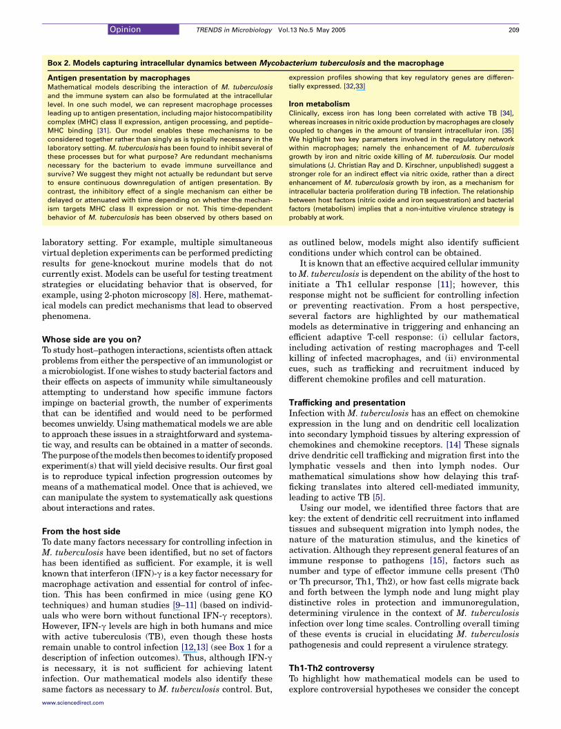
Box 2. Models capturing intracellular dynamics between Mycobacterium tuberculosis and the macrophage
Antigen presentation by macrophagesMathematical models describing the interaction of M. tuberculosis
and the immune system can also be formulated at the intracellular
level. In one such model, we can represent macrophage processes
leading up to antigen presentation, including major histocompatibility
complex (MHC) class II expression, antigen processing, and peptide–
MHC binding [31]. Our model enables these mechanisms to be
considered together rather than singly as is typically necessary in the
laboratory setting. M. tuberculosis has been found to inhibit several of
these processes but for what purpose? Are redundant mechanisms
necessary for the bacterium to evade immune surveillance and
survive? We suggest they might not actually be redundant but serve
to ensure continuous downregulation of antigen presentation. By
contrast, the inhibitory effect of a single mechanism can either be
delayed or attenuated with time depending on whether the mechan-
ism targets MHC class II expression or not. This time-dependent
behavior of M. tuberculosis has been observed by others based on
expression profiles showing that key regulatory genes are differen-
tially expressed. [32,33]
Iron metabolism
Clinically, excess iron has long been correlated with active TB [34],
whereas increases in nitric oxide production by macrophages are closely
coupled to changes in the amount of transient intracellular iron. [35]
We highlight two key parameters involved in the regulatory network
within macrophages; namely the enhancement of M. tuberculosis
growth by iron and nitric oxide killing of M. tuberculosis. Our model
simulations (J. Christian Ray and D. Kirschner, unpublished) suggest a
stronger role for an indirect effect via nitric oxide, rather than a direct
enhancement of M. tuberculosis growth by iron, as a mechanism for
intracellular bacteria proliferation during TB infection. The relationship
between host factors (nitric oxide and iron sequestration) and bacterial
factors (metabolism) implies that a non-intuitive virulence strategy is
probably at work.
Opinion TRENDS in Microbiology Vol.13 No.5 May 2005 209
laboratory setting. For example, multiple simultaneousvirtual depletion experiments can be performed predictingresults for gene-knockout murine models that do notcurrently exist. Models can be useful for testing treatmentstrategies or elucidating behavior that is observed, forexample, using 2-photon microscopy [8]. Here, mathemat-ical models can predict mechanisms that lead to observedphenomena.
Whose side are you on?
To study host–pathogen interactions, scientists often attackproblems from either the perspective of an immunologist oramicrobiologist. If one wishes to study bacterial factors andtheir effects on aspects of immunity while simultaneouslyattempting to understand how specific immune factorsimpinge on bacterial growth, the number of experimentsthat can be identified and would need to be performedbecomes unwieldy. Using mathematical models we are ableto approach these issues in a straightforward and systema-tic way, and results can be obtained in a matter of seconds.Thepurpose of themodels thenbecomes to identifyproposedexperiment(s) that will yield decisive results. Our first goalis to reproduce typical infection progression outcomes bymeans of a mathematical model. Once that is achieved, wecan manipulate the system to systematically ask questionsabout interactions and rates.
From the host side
To date many factors necessary for controlling infection inM. tuberculosis have been identified, but no set of factorshas been identified as sufficient. For example, it is wellknown that interferon (IFN)-g is a key factor necessary formacrophage activation and essential for control of infec-tion. This has been confirmed in mice (using gene KOtechniques) and human studies [9–11] (based on individ-uals who were born without functional IFN-g receptors).However, IFN-g levels are high in both humans and micewith active tuberculosis (TB), even though these hostsremain unable to control infection [12,13] (see Box 1 for adescription of infection outcomes). Thus, although IFN-gis necessary, it is not sufficient for achieving latentinfection. Our mathematical models also identify thesesame factors as necessary to M. tuberculosis control. But,
www.sciencedirect.com
as outlined below, models might also identify sufficientconditions under which control can be obtained.
It is known that an effective acquired cellular immunitytoM. tuberculosis is dependent on the ability of the host toinitiate a Th1 cellular response [11]; however, thisresponse might not be sufficient for controlling infectionor preventing reactivation. From a host perspective,several factors are highlighted by our mathematicalmodels as determinative in triggering and enhancing anefficient adaptive T-cell response: (i) cellular factors,including activation of resting macrophages and T-cellkilling of infected macrophages, and (ii) environmentalcues, such as trafficking and recruitment induced bydifferent chemokine profiles and cell maturation.
Trafficking and presentation
Infection with M. tuberculosis has an effect on chemokineexpression in the lung and on dendritic cell localizationinto secondary lymphoid tissues by altering expression ofchemokines and chemokine receptors. [14] These signalsdrive dendritic cell trafficking and migration first into thelymphatic vessels and then into lymph nodes. Ourmathematical simulations show how delaying this traf-ficking translates into altered cell-mediated immunity,leading to active TB [5].
Using our model, we identified three factors that arekey: the extent of dendritic cell recruitment into inflamedtissues and subsequent migration into lymph nodes, thenature of the maturation stimulus, and the kinetics ofactivation. Although they represent general features of animmune response to pathogens [15], factors such asnumber and type of effector immune cells present (Th0or Th precursor, Th1, Th2), or how fast cells migrate backand forth between the lymph node and lung might playdistinctive roles in protection and immunoregulation,determining virulence in the context of M. tuberculosisinfection over long time scales. Controlling overall timingof these events is crucial in elucidating M. tuberculosispathogenesis and could represent a virulence strategy.
Th1-Th2 controversy
To highlight how mathematical models can be used toexplore controversial hypotheses we consider the concept

Opinion TRENDS in Microbiology Vol.13 No.5 May 2005210
that there exists a Th1-Th2 switch in the immune responseto M. tuberculosis. Conflicting data exist in the literatureregarding T-cell populations during M. tuberculosis infec-tion in the lung. In the mouse and most humans, type 1cytokines are present at high levels during infection.However, an increase in type 2 cytokines in TB patientshas also been reported [16,17], but not consistently[18–20]. By contrast, our mathematical simulations [6]suggest that during latency, lymphocyte populations atthe site of infection are mainly of Th0 type, with very lowlevels of either Th1 or Th2 cells. Thus, we propose thatthere is not a strict Th1-Th2 switch, but that conflictingdata have probably arisen as a result of other factors. Th0cells produce both type 1 and type 2 cytokines. Therelative predominance of either Th1 or Th2 cells thusdepends on several factors, including the differentiationstage of Th0 cells at the time sampling, how long afterinfection a sample is drawn, and the sample site (bloodversus lung for example).
Recruitment
We also studied the effect of trafficking and host–pathogeninteractions on a smaller scale (cell–cell interactions)using a mathematical model of granuloma formation [21](Box 1). We are able to capture another important factor:localization of immune effector cells (macrophages andT cells) within the lung. In this setting where we capturespatial aspects of granuloma formation, the rate of cellularmovement in response to signals greatly influencesinfection outcome. Increased recruitment of restingmacrophages to the infection site is positively correlatedwith bacterial load, most likely by providing additionalcells for productive infection. This suggests the paradox-ical conclusion that inflammation might be detrimental,and further, that unless macrophages become activated,they serve to propagate infection rather than halt it. Thisis consistent with experimental data [22] suggesting thathigh levels of inflammation could be deleterious duringthe course of infection.
From the bacterial side
M. tuberculosis and other pathogenic mycobacteria arevery slow growing. Doubling times of 24–96 hours havebeen reported for M. tuberculosis [23] (Figure 1), and thisis striking when one considers that E. coli has a doublingtime of as low as 20 minutes.
In the mathematical models that included bacterialturnover, the growth rate ofM. tuberculosiswas one of thekey factors that determined whether the system achievedlatent infection or active TB. In the agent-based approach[21], where interactions are tracked at their moststochastic and discrete levels, we observed a strongcorrelation between the intracellular growth rate ofM. tuberculosis and granuloma size (or similarly, bacterialload). Using sensitivity analysis, we tracked this corre-lation during TB infection and we observed a shift frompositive to negative values. In the first 12 days of infection,higher growth rates are significantly more favorable forgranuloma growth (positive correlation), but between oneto three months, granulomas grow larger when growthrates are slowest (negative correlation). Finally, after
www.sciencedirect.com
three months, the correlation is again positive, whereslightly higher growth rates favor granuloma growth.
From these outcomes, it is clear that growth rate iscorrelated with granuloma formation, although the bio-logical basis for this result remains unclear. This counter-intuitive simulation outcome results from the non-lineardynamics that occur between T cells, macrophages andbacteria. Does the slow growth rate of M. tuberculosiscontribute to virulence? No experimental data are avail-able to support this finding, however our results are con-sistent with hypotheses drawn from earlier mathematicalmodels suggesting that persistence of M. tuberculosis atlow densities for extended periods in the face of immunepressure might be due to mechanisms that are associatedwith a very slow growth rate [24].
Extracellular and intracellular lifestyles
The status and location of bacteria during latent TBinfection has long been an area of controversy. A uniquefeature of the mathematical model is that we are able totrack at any given moment which bacteria are intra-cellular (within macrophages) and which are extracellular(not within macrophages). Although data on bacterialloads in murine models, for example, are usually given ascfu/gm of tissue, our mathematical models are able todistinguish total bacterial levels within these mutuallyexclusive compartments (Box 1).
In all but one of our cell-dynamic models (the exceptionbeing the agent-based model), levels of extracellular bac-teria arose as a marker of disease progression. If bacteriallevels could be contained intracellularly (within low levelsof infected macrophages), then infection could be con-trolled. Our results suggest that all of the bacteria areharbored within a few infected macrophages during latentinfection within the granuloma. New experiments by ourcollaborators are now in place to test this hypothesis. Bycontrast, when extracellular bacterial levels increaseuninhibited, this reflects an immune system that is unableto control infection.
Necrosis
As with most elements of this system, there is animportant balance regarding necrosis. Granulomas havea characteristic structure including a necrotic center,surrounded by macrophages, surrounded by T cells.Within the ring of macrophages these cells are lysedcontributing to centralized necrosis (this structure isobserved in our simulations of granulomas shown inBox 1). Our agent-based model [21] predicts that thisnecrotic center is a site within which extracellularbacteria reside, and unpublished data from the NHPmodel indicate that these granuloma are packed withbacteria in the necrotic centers (T. Reinhart, personalcommunication). This pattern of necrosis preventsimmune cells from reaching and eliminating thosetrapped bacteria; however, it also prevents bacteria fromspreading. Thus, in our simulations, this pattern ofnecrosis is a mechanism aiding stable containment ofinfection. Early studies on TB suggested that it might bepathology that helps limit infection spread [25,26].

Opinion TRENDS in Microbiology Vol.13 No.5 May 2005 211
From math to biology
Mathematics has long been relegated to the closet inmicrobiology studies. The emergence of new compu-tational tools and other technologies provides a perfectopportunity to dust off old notions of mathematics andwelcome it into the biological arena. Here, we show thatit can be applied in many ways to study relevant problemsin biology, using the example of M. tuberculosis infection.A key to successful conversations between theory andexperiment is the language. If theoreticians will put theirwork in the proper context of the problem being studied,then experimental biologists can more easily see howmathematics can be used as an additional tool to stimulateand address important questions in microbiology. Newexperimental technologies [8,27] will eventually producein vivo time series cell population dynamics data tosupport and validate mathematical model results andhypotheses. Combinations of experimental measurementsand mathematical models will ultimately yield funda-mental insights into biological phenomena. A new gener-ation of students is now being trained to this end.
AcknowledgementsWe are indebted to JoAnne Flynn, John Chan, Todd Reinhart and VicDiRita for helpful discussions and are grateful to J. Christian Ray for useof unpublished data.
References
1 Victor, T.C. et al. (1997) Genome and MIC stability in Mycobacteriumtuberculosis and indications for continuation of use of isoniazid inmultidrug-resistant tuberculosis. J. Med. Microbiol. 46, 847–857
2 Wigginton, J.E. and Kirschner, D. (2001) A model to predict cell-mediated immune regulatory mechanisms during human infectionwith Mycobacterium tuberculosis. J. Immunol. 166, 1951–1967
3 Flynn, J.L. et al. (2003) Non-human primates: a model for tuberculosisresearch. Tuberculosis (Edinb.) 83, 116–118
4 Capuano, S.V., 3rd. et al. (2003) Experimental Mycobacteriumtuberculosis infection of cynomolgus macaques closely resembles thevarious manifestations of human M. tuberculosis infection. Infect.Immun. 71, 5831–5844
5 Marino, S. et al. (2004) Dendritic cell trafficking and antigenpresentation in the human immune response to Mycobacteriumtuberculosis. J. Immunol. 173, 494–506
6 Marino, S. and Kirschner, D.E. (2004) The human immune response toMycobacterium tuberculosis in lung and lymph node. J. Theor. Biol.227, 463–486
7 Gammack, D. et al. (2004) Macrophage response to Mycobacteriumtuberculosis infection. J. Math. Biol. 48, 218–242
8 Miller, M.J. et al. (2002) Two-photon imaging of lymphocyte motilityand antigen response in intact lymph node. Science 296, 1869–1873
9 Cooper, A.M. et al. (1993) Disseminated tuberculosis in interferongamma gene-disrupted mice. J. Exp. Med. 178, 2243–2247
10 Flynn, J.L. et al. (1993) An essential role for interferon gamma inresistance to Mycobacterium tuberculosis infection. J. Exp. Med. 178,2249–2254
11 Flynn, J.L. and Chan, J. (2001) Immunology of tuberculosis. Annu.Rev. Immunol. 19, 93–129
12 Newport, M.J. et al. (1996) A mutation in the interferon-gamma-receptor gene and susceptibility to mycobacterial infection. N. Engl.J. Med. 335, 1941–1949
www.sciencedirect.com
13 Jouanguy, E. et al. (1996) Interferon-gamma-receptor deficiency in aninfant with fatal bacille Calmette-Guerin infection. N. Engl. J. Med.335, 1956–1961
14 Sallusto, F. et al. (1998) Rapid and coordinated switch in chemokinereceptor expression during dendritic cell maturation. Eur. J. Immunol.28, 2760–2769
15 Lanzavecchia, A. and Sallusto, F. (2000) Dynamics of T lymphocyteresponses: intermediates, effectors, and memory cells. Science 290,92–97
16 Bhattacharyya, S. et al. (1999) Dichotomy of cytokine profiles inpatients and high-risk healthy subjects exposed to tuberculosis. Infect.Immun. 67, 5597–5603
17 van Crevel, R. et al. (2000) Increased production of interleukin 4 byCD4C and CD8C T cells from patients with tuberculosis is related tothe presence of pulmonary cavities. J. Infect. Dis. 181, 1194–1197
18 Barnes, P.F. et al. (1993) Cytokine production at the site of disease inhuman tuberculosis. Infect. Immun. 61, 3482–3489
19 Jung, Y.J. et al. (2002) Evidence inconsistent with a negative influenceof T helper 2 cells on protection afforded by a dominant T helper 1response against Mycobacterium tuberculosis lung infection in mice.Infect. Immun. 70, 6436–6443
20 Lai, C.K. et al. (1997) Cytokine gene expression profile of circulatingCD4C T cells in active pulmonary tuberculosis. Chest 111, 606–611
21 Segovia-Juarez, J.L. et al. Identifying control mechanisms of granu-loma formation during M. tuberculosis infection using an agent basedmodel. J. Theor. Biol. (in press)
22 Mohan, V.P. et al. (2001) Effects of tumor necrosis factor alpha on hostimmune response in chronic persistent tuberculosis: possible role forlimiting pathology. Infect. Immun. 69, 1847–1855
23 Zhang, M. et al. (1998) Growth of virulent and avirulent Mycobacter-ium tuberculosis strains in human macrophages. Infect. Immun. 66,794–799
24 Antia, R. et al. (1996) Models of the within-host dynamics of persistentmycobacterial infections. Proc. R. Soc. Lond. B. Biol. Sci. 263, 257–263
25 Dannenberg, A.M., Jr. and Sugimoto, M. (1976) Liquefaction ofcaseous foci in tuberculosis. Am. Rev. Respir. Dis. 113, 257–259
26 Dannenberg, A.M., Jr. et al. (1968) The local nature of immunity intuberculosis, illustrated histochemically in dermal BCG lesions.J. Immunol. 100, 931–941
27 Cahalan, M.D. et al. (2002) Two-photon tissue imaging: seeing theimmune system in a fresh light. Nat. Rev. Immunol. 2, 872–880
28 Abu-Amero, K. (2002) Tuberculosis information on the Web. J. R. Soc.Health 122, 82–85
29 Langermans, J.A. et al. (2001) Divergent effect of bacillus Calmette-Guerin (BCG) vaccination on Mycobacterium tuberculosis infection inhighly related macaque species: implications for primate models intuberculosis vaccine research. Proc. Natl. Acad. Sci. U. S. A. 98,11497–11502
30 Bloom, B.R. (1994) Tuberculosis: pathogenesis, protection, and control,ASM Press
31 Chang, S. et al. A role for multiple mechanisms in the inhibition ofMHC class II-mediated antigen presentation by Mycobacteriumtuberculosis. Proc. Natl. Acad. Sci. U. S. A. (in press)
32 Haydel, S.E. and Clark-Curtiss, J.E. (2004) Global expression analysisof two-component system regulator genes during Mycobacteriumtuberculosis growth in human macrophages. FEMS Microbiol. Lett.236, 341–347
33 Graham, J.E. and Clark-Curtiss, J.E. (2000) Identifying myco-bacterium tuberculosis virulence determinants – new technologiesfor a difficult problem: response. Trends Microbiol. 8, 100
34 Lounis, N. et al. (2001) Iron andMycobacterium tuberculosis infection.J. Clin. Virol. 20, 123–126
35 Kim, S. and Ponka, P. (2002) Nitrogen monoxide-mediated control offerritin synthesis: implications for macrophage iron homeostasis.Proc. Natl. Acad. Sci. U. S. A. 99, 12214–12219
Copyright © 2022 FDOKUMEN