A recombinant Mycobacterium smegmatis induces potent bactericidal immunity against Mycobacterium...
18
A recombinant Mycobacterium smegmatis induces potent bactericidal immunity against Mycobacterium tuberculosis Kari A Sweeney 1,2 , Dee N Dao 1,2,6 , Michael F Goldberg 2,6 , Tsungda Hsu 1,2 , Manjunatha M Venkataswamy 2 , Marcela Henao-Tamayo 3 , Diane Ordway 3 , Rani S Sellers 4 , Paras Jain 1,2 , Bing Chen 1,2 , Mei Chen 1,2 , John Kim 1,2 , Regy Lukose 1,2 , John Chan 2,5 , Ian M Orme 3 , Steven A Porcelli 2,5 , and William R Jacobs Jr 1,2,5 1 Howard Hughes Medical Institute, Albert Einstein College of Medicine, Bronx, New York, USA 2 Department of Microbiology and Immunology, Albert Einstein College of Medicine, Bronx, New York, USA 3 Department of Microbiology, Immunology and Pathology, Colorado State University, Fort Collins, Colorado, USA 4 Department of Pathology, Albert Einstein College of Medicine, Bronx, New York, USA 5 Department of Medicine, Albert Einstein College of Medicine, Bronx, New York, USA Abstract We report the involvement of an evolutionarily conserved set of mycobacterial genes, the esx-3 region, in evasion of bacterial killing by innate immunity. Whereas high-dose intravenous infections of mice with the rapidly growing mycobacterial species Mycobacterium smegmatis bearing an intact esx-3 locus were rapidly lethal, infection with an M. smegmatis Δesx-3 mutant (here designated as the IKE strain) was controlled and cleared by a MyD88-dependent bactericidal immune response. Introduction of the orthologous Mycobacterium tuberculosis esx-3 genes into the IKE strain resulted in a strain, designated IKEPLUS, that remained susceptible to innate immune killing and was highly attenuated in mice but had a marked ability to stimulate bactericidal immunity against challenge with virulent M. tuberculosis. Analysis of these adaptive immune responses indicated that the highly protective bactericidal immunity elicited by IKEPLUS was dependent on CD4 + memory T cells and involved a distinct shift in the pattern of cytokine responses by CD4 + cells. Our results establish a role for the esx-3 locus in promoting mycobacterial virulence and also identify the IKE strain as a potentially powerful candidate vaccine vector for eliciting protective immunity to M. tuberculosis. © 2011 Nature America, Inc. All rights reserved. Correspondence should be addressed to W.R.J. ([email protected]). 6 These authors contributed equally to this work. Note: Supplementary information is available on the Nature Medicine website. AUTHOR CONTRIBUTIONS K.A.S. constructed bacterial strains, performed or contributed to the design of most experiments, and analyzed and interpreted data. D.N.D. carried out portions of the infection and challenge experiments. M.F.G. contributed to the T cell adoptive transfer studies and designed, performed and analyzed all flow cytometry analyses. T.H. participated in design and construction of bacterial strains and in the performance of mouse infection and challenge experiments. P.J. contributed to construction of bacterial strains. M.M.V. and M.H.- T. assisted with experiments analyzing responding T cell populations. R.S.S. analyzed and scored the histopathology samples. B.C., M.C., J.K. and R.L. carried out mouse infections, organ harvesting and quantification of bacilli in tissues. D.O., J.C., I.M.O., S.A.P. and W.R.J. Jr. designed and interpreted experiments. K.A.S., S.A.P. and W.R.J. Jr. wrote the manuscript. COMPETING FINANCIAL INTERESTS The authors declare no competing financial interests. NIH Public Access Author Manuscript Nat Med. Author manuscript; available in PMC 2012 January 3. Published in final edited form as: Nat Med. ; 17(10): 1261–1268. doi:10.1038/nm.2420. NIH-PA Author Manuscript NIH-PA Author Manuscript NIH-PA Author Manuscript
-
Upload
independent -
Category
Documents
-
view
0 -
download
0
Transcript of A recombinant Mycobacterium smegmatis induces potent bactericidal immunity against Mycobacterium...
A recombinant Mycobacterium smegmatis induces potentbactericidal immunity against Mycobacterium tuberculosis
Kari A Sweeney1,2, Dee N Dao1,2,6, Michael F Goldberg2,6, Tsungda Hsu1,2, Manjunatha MVenkataswamy2, Marcela Henao-Tamayo3, Diane Ordway3, Rani S Sellers4, Paras Jain1,2,Bing Chen1,2, Mei Chen1,2, John Kim1,2, Regy Lukose1,2, John Chan2,5, Ian M Orme3,Steven A Porcelli2,5, and William R Jacobs Jr1,2,5
1Howard Hughes Medical Institute, Albert Einstein College of Medicine, Bronx, New York, USA2Department of Microbiology and Immunology, Albert Einstein College of Medicine, Bronx, NewYork, USA3Department of Microbiology, Immunology and Pathology, Colorado State University, Fort Collins,Colorado, USA4Department of Pathology, Albert Einstein College of Medicine, Bronx, New York, USA5Department of Medicine, Albert Einstein College of Medicine, Bronx, New York, USA
AbstractWe report the involvement of an evolutionarily conserved set of mycobacterial genes, the esx-3region, in evasion of bacterial killing by innate immunity. Whereas high-dose intravenousinfections of mice with the rapidly growing mycobacterial species Mycobacterium smegmatisbearing an intact esx-3 locus were rapidly lethal, infection with an M. smegmatis Δesx-3 mutant(here designated as the IKE strain) was controlled and cleared by a MyD88-dependent bactericidalimmune response. Introduction of the orthologous Mycobacterium tuberculosis esx-3 genes intothe IKE strain resulted in a strain, designated IKEPLUS, that remained susceptible to innateimmune killing and was highly attenuated in mice but had a marked ability to stimulatebactericidal immunity against challenge with virulent M. tuberculosis. Analysis of these adaptiveimmune responses indicated that the highly protective bactericidal immunity elicited by IKEPLUSwas dependent on CD4+ memory T cells and involved a distinct shift in the pattern of cytokineresponses by CD4+ cells. Our results establish a role for the esx-3 locus in promotingmycobacterial virulence and also identify the IKE strain as a potentially powerful candidatevaccine vector for eliciting protective immunity to M. tuberculosis.
© 2011 Nature America, Inc. All rights reserved.Correspondence should be addressed to W.R.J. ([email protected]).6These authors contributed equally to this work.Note: Supplementary information is available on the Nature Medicine website.AUTHOR CONTRIBUTIONSK.A.S. constructed bacterial strains, performed or contributed to the design of most experiments, and analyzed and interpreted data.D.N.D. carried out portions of the infection and challenge experiments. M.F.G. contributed to the T cell adoptive transfer studies anddesigned, performed and analyzed all flow cytometry analyses. T.H. participated in design and construction of bacterial strains and inthe performance of mouse infection and challenge experiments. P.J. contributed to construction of bacterial strains. M.M.V. and M.H.-T. assisted with experiments analyzing responding T cell populations. R.S.S. analyzed and scored the histopathology samples. B.C.,M.C., J.K. and R.L. carried out mouse infections, organ harvesting and quantification of bacilli in tissues. D.O., J.C., I.M.O., S.A.P.and W.R.J. Jr. designed and interpreted experiments. K.A.S., S.A.P. and W.R.J. Jr. wrote the manuscript.COMPETING FINANCIAL INTERESTSThe authors declare no competing financial interests.
NIH Public AccessAuthor ManuscriptNat Med. Author manuscript; available in PMC 2012 January 3.
Published in final edited form as:Nat Med. ; 17(10): 1261–1268. doi:10.1038/nm.2420.
NIH
-PA Author Manuscript
NIH
-PA Author Manuscript
NIH
-PA Author Manuscript
Tuberculosis is a leading cause of global mortality due to infectious disease, and it is aworsening problem owing to HIV co-infection and the emergence of extremely drug-resistant strains of M. tuberculosis (Mtb)1,2. The only currently used vaccine, the attenuatedMycobacterium bovis strain bacille Calmette-Guérin (BCG), has been inconsistent inproviding protection from disease, with low or unmeasurable efficacy in many of thoseregions with the most cases1,3,4. Moreover, BCG can cause severe, disseminated infection inHIV-positive infants, further restricting its use in areas with high prevalence oftuberculosis5. Animal models of tuberculosis also demonstrate that BCG vaccinationgenerates at best partial immunity against Mtb that does not give complete control orclearance of infection6. Thus, new tuberculosis vaccines that are safer and more efficaciousthan BCG are clearly needed7–9.
Generation of more effective vaccines requires a better understanding of the mechanisms bywhich Mtb evades innate and adaptive immune responses6,10–14. Some of these mechanismsare known to involve the export of various effector molecules by specialized bacterialsecretion systems. For example, the well-studied mycobacterial type VII secretion systemknown as ESX-1 has been shown to be involved in a range of effects that promotemycobacterial virulence through secretion of the protein ESAT-6 and other effectorproteins15–25. Notably, the Mtb genome encodes four other potential secretion systems thatare paralogs of ESX-1 (designated ESX-2 through ESX-5), but their functions are mostlyunknown. Although all mycobacterial species studied to date have between two and five ofthe esx loci, only the esx-3 genes (comprising the genes Rv0282 to Rv0292 in Mtb andMs0615 to Ms0626 in M. smegmatis (Msmeg); Supplementary Fig. 1) are conserved in allcases26,27. This suggests a unique and crucial function for ESX-3. Expression of esx-3 genesis regulated by iron and zinc and is increased in the lungs of mice during persistent infectionwith Mtb28–31, suggesting it may have a key role in virulence. In addition, an intact esx-3locus has been shown to be essential for the growth of Mtb in culture32–34.
Because inactivation of esx-3 is incompatible with viability in Mtb, in the current study wehave used Msmeg, a mycobacterium that is able to grow without an intact esx-3 locus, tostudy the influence of this locus on the innate and adaptive immune systems of the host. Weobserved loss of the ability to evade bactericidal host innate immunity for the Msmeg strainwith deletion of esx-3, which we therefore designated by the acronym IKE to signify‘immune killing evasion’, which is lost in this strain. Although insertion of the esx-3 genesof Mtb into IKE did not reverse the susceptibility of the bacteria to innate immune killing, itdid produce a recombinant strain, designated IKEPLUS, that induced a previouslyunobserved level of bactericidal immunity against Mtb. This new immunization strategysuggests an approach to tuberculosis vaccine development that can potentially inducesterilizing immunity and also reveals a previously undocumented capacity for themammalian immune system to eliminate virulent mycobacteria from infected tissues.
RESULTSRole of esx-3 in evasion of innate immunity
A robust T helper type 1 (TH1) cell response has a key role in protective immunity to manyintracellular pathogens and may be crucial for effective immunity to Mtb35. To assessmechanisms by which mycobacteria may interfere with development of TH1 cell responses,we analyzed the cytokines induced in vivo in mice after infection with Msmeg mutants withdeletions of the esx-1 and esx-3 loci (Supplementary Fig. 2). We constructed these mutantsin Msmeg because of previous work indicating that esx-3 cannot be deleted in Mtb, probablybecause of its essential role in iron acquisition32,34. This is not the case for Msmeg becauseof the presence of a second siderophore system (exochelin) in that species. As expected, aΔesx-3 Msmeg strain (IKE) was readily isolated and grew normally in iron-replete or iron-
Sweeney et al. Page 2
Nat Med. Author manuscript; available in PMC 2012 January 3.
NIH
-PA Author Manuscript
NIH
-PA Author Manuscript
NIH
-PA Author Manuscript
deficient medium (Supplementary Fig. 3a). We infected mice intravenously with high dosesof either unmodified parental, Δesx-1 or IKE strains of Msmeg and measured serumcytokine concentrations. Whereas parental and Δesx-1 Msmeg induced low or undetectablelevels of the interleukin-12 (IL-12) subunit p40 and interferon-γ (IFN-γ), infection with IKEelicited robust secretion of these cytokines. In contrast, IL-6 was strongly induced byparental and Δesx-1 Msmeg but was barely detectable in IKE-infected mice (Fig. 1a).
Although Msmeg is a saprophytic organism and generally not considered pathogenic, wefound that a high intravenous dose (≥ 1 × 10⁷ colony-forming units (CFU)) of parental orΔesx-1 Msmeg was uniformly fatal in C57BL/6 mice within 7 d (Fig. 1b and data notshown). Administration of the same number of heat-killed or ultraviolet-irradiated bacteriadid not result in death (Supplementary Fig. 3b). In clear contrast, all C57BL/6 mice infectedwith the IKE mutant survived and cleared their infections without apparent sequelae (Fig.1b). The attenuation of Msmeg IKE was further underscored by its inability to killrecombination-activating gene-1–null mice (Rag1−/−), which lack T and B cells (Fig. 1b),suggesting control of infection by an innate immune response. To explore this possibility,we infected a panel of mouse strains deficient in various innate immune response genes(Supplementary Table 1). Among these, only mice lacking the adaptor protein MyD88 wereunable to clear the Msmeg IKE infection and died with kinetics similar to those observed inmice infected with parental Msmeg (Fig. 1b and Supplementary Table 1).
Whereas histological analysis of C57BL/6 mice infected with high-dose (5 × 10⁷ CFU)intravenous parental Msmeg showed substantial pathology in the brain, spleen and liver andparticularly in the kidney, mice infected with IKE showed no notable pathology in thesetissues (data not shown). To further study the attenuation of IKE, we determined tissuebacterial burdens after high-dose intravenous infection of C57BL/6, Rag1−/− and Myd88−/−
mice. For C57BL/6 and Rag1−/− mice, there was rapid clearance of IKE from the lungs andkidneys as early as day 3 after infection, with complete clearance by day 35, whereas theparental and Δesx-1 Msmeg strains showed increasing tissue bacterial burdens during the 3–6 d before death of the mice (Fig. 1c). In contrast, in Myd88−/− mice there was expansion ofIKE bacilli after inoculation (Fig. 1d). These findings indicated that a previouslyunappreciated function of the genes in the esx-3 region is to mediate evasion of a MyD88-dependent innate immune response that normally triggers bactericidal effects and hostresistance to Msmeg infection.
We performed a genetic complementation analysis by introducing an integrating cosmidencoding all genes of Msmeg esx-3 (Ms0609–Ms0637) into IKE to generate a strain thatshowed a partial restoration of virulence, as assessed by tissue bacterial burden(Supplementary Fig. 4a). The failure to completely complement the mutation may have beendue to aberrant regulation of the introduced esx-3 genes because of integration into a non-native location on the chromosome. We also infected mice with a strain of Msmeg that had adeletion in only a single esx-3 gene (Ms0615), which showed alterations in cytokine patternsand an attenuation of virulence that were similar to those of IKE (Supplementary Fig. 4b,c).Although we have not determined whether the deletion of Ms0615 has effects on other esx-3genes, it is noteworthy that complementation of this deletion with a plasmid encoding theentire Msmeg esx-3 region (Ms0600–Ms0624) fully reversed the phenotype of the mutant inRag1−/− mice (Supplementary Fig. 4b–d).
Generation and characterization of IKEPLUSIn contrast to our results with Msmeg and similar to previous studies, we were unable toisolate esx-3 deletion mutants of Mtb32,34. As an alternative approach to study the functionof Mtb esx-3, we assessed whether the orthologous Mtb genes could complement theMsmeg Δesx-3 phenotype. Thus, we introduced the orthologous locus of Mtb (Rv0282–
Sweeney et al. Page 3
Nat Med. Author manuscript; available in PMC 2012 January 3.
NIH
-PA Author Manuscript
NIH
-PA Author Manuscript
NIH
-PA Author Manuscript

Rv0292) into the IKE strain by transformation with cosmid pYUB1336 containing Mtbgenes Rv0278–Rv0303 (Supplementary Table 2), generating IKEPLUS. Despite the highhomology of these regions between Mtb and Msmeg (minimum of 44% and up to 85%homology at the protein level for the 11 ORFs), analysis of the immunological propertiesand virulence of IKEPLUS indicated that the Mtb esx-3 genes were unable to complementthe phenotype associated with deletion of the Msmeg esx-3 locus. Thus, measurements ofserum cytokine concentrations showed that IKEPLUS induced robust IL-12 p40, p70 andIFN-γ responses similar to those observed with the IKE mutant (Fig. 2a). In spite of thestable integration of the Mtb esx-3 genes into the IKE genome (Supplementary Fig. 5), theIKEPLUS strain was rapidly cleared from tissues (Fig. 2b) and was unable to kill eitherimmunocompetent or Rag1−/− mice but was lethal in MyD88-deficient mice (Fig. 2c).
Ability of IKEPLUS to elicit bactericidal immunity against MtbThe results presented above revealed properties of IKE and IKEPLUS that are desirable forantituberculosis vaccines. These mutants were highly attenuated for virulence in mice, thusfulfilling initial safety requirements, and they rapidly induced a cytokine milieu that wasfavorable to priming of TH1 responses (Fig. 2a). Furthermore, in the case of IKEPLUS, thisrecombinant Msmeg contained a cosmid (pYUB1336, Supplementary Table 2; also seeSupplementary Table 3) encoding 26 open reading frames of Mtb that had the potential toserve as specific antigens for eliciting adaptive immune responses that could be recalled bysubsequent Mtb infection. To examine its potential as an antituberculosis vaccine, we usedIKEPLUS to immunize mice (5 × 10⁷ CFU intravenously), which we then challenged 8weeks later with a high intravenous dose (1 × 10⁷ CFU) of Mtb (virulent strain H37Rv). Weimmunized control groups either with saline or subcutaneously with BCG (1 × 10⁷ CFU permouse). The average time to death after Mtb challenge was 54 d for sham-vaccinated miceand 65 d for BCG-immunized mice, whereas the IKEPLUS-immunized mice had a meansurvival time of 135 d (Fig. 3a).
Consistent with the extension of survival observed in a subset of IKEPLUS-immunizedmice, we found in three separate experiments that intravenous immunization of mice withIKEPLUS led to marked reductions in tissue bacterial burdens in mice surviving to latertime points after Mtb challenge (Fig. 3b). We observed this effect in lung, spleen and livertissue at several time points after the challenge. Notably, the bacterial burdens of all tissuesexamined in IKEPLUS-immunized mice showed a continuing decline over time in mice thatsurvived > 100 d. By 200 d after Mtb challenge, these declines exceeded by > 1,000-fold themaximum levels of CFU reduction achieved in those BCG-vaccinated mice that survived today 100. Furthermore, CFU measurements were below the limit of detection in the livers ofIKEPLUS-vaccinated and Mtb-challenged mice surviving >200 d, indicating a high level ofbactericidal and potentially sterilizing immunity. In contrast to the long-term survivalobserved in a subset of IKEPLUS-immunized mice, no BCG-immunized mice survivedmore than 173 d after challenge, and BCG-immunized mice that survived to 100 d had agreater bacterial burden in all organs than did IKEPLUS-immunized mice (Fig. 3b). TheMtb genes present in IKEPLUS were necessary to induce highly protective immuneresponses, as mice sham-vaccinated with the parental IKE strain showed survival andbacterial burden after Mtb challenge that were only modestly better than those of naive mice(Fig. 3c).
Histopathological evaluation also revealed evidence of robust immunity in IKEPLUS-vaccinated mice. At 100 d after Mtb challenge, all mice showed moderate-to-severe diffusegranulomatous pneumonia, and at this time point there were no apparent differences in thelung histology (Fig. 4a and Supplementary Fig. 6) or severity of pneumonia on the basis ofquantitative visual scoring (Fig. 4b) between mice immunized with saline, BCG orIKEPLUS. The severity of granulomatous inflammation in the spleens and livers from all
Sweeney et al. Page 4
Nat Med. Author manuscript; available in PMC 2012 January 3.
NIH
-PA Author Manuscript
NIH
-PA Author Manuscript
NIH
-PA Author Manuscript

treatment groups were likewise similar at day 100 after Mtb challenge (Fig. 4b and data notshown). However, lung tissues from IKEPLUS-immunized mice surviving >200 d after Mtbchallenge showed clear evidence of gradually resolving granulomatous inflammation ascompared to lung tissue at day 100, along with improvements in microscopic appearance(Fig. 4a) and significant reductions in the histological scores (Fig. 4b) in both lung and liversections. Notably, we continued to observe low numbers of acid-fast bacilli (AFB) in smallgranuloma-like structures in the livers of IKEPLUS-immunized mice killed at more than200 d after Mtb challenge (Fig. 4a), despite the lack of culturable bacilli in these tissues.
Efficacy of subcutaneously administered IKEPLUS as a vaccineThe unusually potent immune response elicited by intravenously administered IKEPLUS ledus to assess its potential as a subcutaneously administered vaccine against tuberculosis inmice. As a first approach, we immunized mice subcutaneously with BCG or IKEPLUS and8 weeks later challenged them with a high intravenous dose (1 × 10⁸ CFU) of Mtb.Compared to BCG-immunized mice, mice immunized subcutaneously with IKEPLUS hadsignificantly (P < 0.01) more serum IL-12 p40 upon challenge with Mtb (SupplementaryFig. 7a). Furthermore, mice immunized subcutaneously with IKEPLUS survivedsignificantly longer after Mtb challenge than did BCG-immunized mice (Fig. 5a).Quantification of tissue bacterial burdens showed that subcutaneous immunization of micewith IKEPLUS led to an ~3-log reduction of CFU in the lung at 20 d after Mtb challengecompared to lung tissue in saline-immunized mice, which was nearly 2 logs better than thelevel of protection from BCG immunization (Fig. 5a).
To assess the vaccine efficacy of IKEPLUS in a more physiological setting, we immunizedmice subcutaneously and then challenged them 1 month later by low-dose aerosol infectionwith Mtb (~100 CFU per mouse). Mice immunized subcutaneously with IKEPLUS showeda trend toward longer survival after Mtb challenge compared to BCG-immunized mice (Fig.5b; mean survival 301 d versus 267 d; P = 0.0898, log-rank test). IKEPLUS vaccinationresulted in significant reductions in CFU in lung and spleen tissues that were comparable tothose achieved with BCG at 4 weeks (Fig. 5b). However, at 25 weeks after Mtb challenge,the reduction in CFU for IKEPLUS-immunized mice was maintained, whereas bacterialburdens in BCG-immunized mice increased to the level observed in sham-vaccinated mice(Fig. 5b).
Central role for CD4+ T cells in immunity induced by IKEPLUSThe ability of IKEPLUS to enhance IL-12 p40 and IFN-γ production suggested enhancedTH1 cell responses as a mechanism for heightened protective immunity in IKEPLUS-immunized mice. This idea was supported by cytokine profiles elicited in the immunization-challenge model (Supplementary Fig. 7), as mice immunized with either IKE or IKEPLUSshowed rapid induction of IL-12 p40 and IFN-γ expression in the first 2 d after high-doseintravenous challenge with Mtb (Supplementary Fig. 7b). These data suggested that theprotective responses elicited by IKEPLUS immunization were associated with the inductionof TH1-type adaptive immunity. This hypothesis was further supported by our findings thatRag1−/− and IL-12 p40–deficient mice showed no significant change (P > 0.25) in their highsusceptibility to Mtb challenge after immunization with IKEPLUS (Supplementary Fig. 8a).
As adaptive T cell immunity requires priming through peptide antigen presentation by majorhistocompatibility complex (MHC) class I and MHC class II proteins, we analyzed therequirement for these molecules in the induction of protective immunity by IKEPLUS. Weimmunized MHC class I–deficient and MHC class II–deficient mice with IKEPLUS(intravenously), BCG (subcutaneously) or saline and then challenged them with a highintravenous dose (1 × 10⁷ CFU per mouse) of virulent Mtb. Determination of bacterial loads
Sweeney et al. Page 5
Nat Med. Author manuscript; available in PMC 2012 January 3.
NIH
-PA Author Manuscript
NIH
-PA Author Manuscript
NIH
-PA Author Manuscript
showed that MHC class I–deficient mice immunized with IKEPLUS had significantly higher(P < 0.05) protection compared to saline-immunized MHC class I–deficient mice uponchallenge with Mtb at both the 2-week and 4-week time points (Supplementary Fig. 8b). Incontrast, the bacterial loads in IKEPLUS-immunized MHC class II–deficient mice killed atboth 2 weeks and 4 weeks after challenge were not significantly different (P > 0.05) fromthose of saline-immunized MHC class II–deficient mice (Supplementary Fig. 8b). Thissuggests that the protective immunity elicited by IKEPLUS immunization is dependent onMHC class II presentation and therefore probably involves responses by CD4+ T cells.
To further examine the involvement of CD4+ T cells, we assessed the ability of purified Tcell populations from IKEPLUS-immunized mice to transfer protection against Mtbchallenge to naive mice. We immunized mice intravenously with IKEPLUS or saline orsubcutaneously with BCG. After 3–6 weeks, we purified CD4+ T cells from the spleens ofimmunized mice and then transferred them to naive mice. These mice were then challengedwith a high intravenous dose (1 × 10⁷ CFU per mouse) of Mtb. Lung CFU counts 3 weeksafter challenge showed that significant protection was transferred by CD4+ T cells fromIKEPLUS-vaccinated donors, with a reduction in CFU nearly equivalent to that in mice thathad been directly immunized with either intravenous IKEPLUS or subcutaneous BCG (Fig.6a). Survival was also significantly extended in mice receiving CD4+ T cells fromIKEPLUS-immunized donors (Fig. 6a). As expected, CD4+ T cells from sham-vaccinatedmice did not transfer protection in this experiment, nor did CD4+ T cells from BCG-vaccinated donors, consistent with earlier studies showing that transfer of BCG-inducedimmunity requires sublethal irradiation of recipients36. There was no transfer of protectionusing purified CD8+ T cells from any of the donor groups. Thus, the CD4+ T cell populationfrom IKEPLUS-immunized mice seemed to contain the main pool of memory T cellsresponsible for activating protective immunity to challenge with Mtb.
We examined the multifunctionality of the CD4+ T cell response in IKEPLUS-immunizedmice by analyzing cytokine production by CD4+ T cells infiltrating the lung at various timepoints after Mtb challenge (Fig. 6b–d and Supplementary Fig. 9). Upon re-stimulation invitro with a peptide corresponding to an MHC class II (I-Ab)-presented epitope of the TB9.8(esxG) protein (Supplementary Fig. 1), we observed a substantially different pattern ofcytokine production between BCG- and IKEPLUS-immunized mice (Fig. 6b). Usingsimultaneous analysis of three cytokines (IFN-γ, tumor necrosis factor (TNF) and IL-2), weobserved an increase of CD4+ T cells producing two or three of these cytokines inIKEPLUS-immunized compared with BCG-immunized mice (Fig. 6b,d) in response tostimulation by TB9.8. This was particularly evident at 2 or more weeks after Mtb challenge.In marked contrast, CD4+ T cells producing two or three of these cytokines accumulatedmore gradually in the IKEPLUS-immunized mice, but their levels were sustained to thelatest time points examined (that is, day 36). We observed a similar pattern in total lung-derived CD4+ T cells after re-stimulation in vitro with CD3-specific antibody (Fig. 6c,d).
DISCUSSIONIn the current study we have demonstrated a major role for the esx-3 locus of Msmeg inmodifying the mammalian host immune response, and in so doing we have generated a newand highly effective candidate vaccine for tuberculosis. Although Msmeg is a saprophyticorganism rarely reported as a cause of clinical infections37,38, our high-dose intravenousinoculation protocol in mice revealed its ability to establish a rapidly fatal experimentalinfection. This cryptic virulence of Msmeg was dependent on an intact ESX-3 secretionsystem, as deletion of the esx-3 genes generated a strain (IKE) that was rapidly clearedwithout sequellae. We observed clearance of IKE in mice lacking various components ofinnate and adaptive immunity, but not in MyD88-deficient mice. Overall, these findings are
Sweeney et al. Page 6
Nat Med. Author manuscript; available in PMC 2012 January 3.
NIH
-PA Author Manuscript
NIH
-PA Author Manuscript
NIH
-PA Author Manuscript
consistent with a role for ESX-3 in mediating resistance to MyD88-dependent bactericidaleffects in mice. As mice defective in Toll-like receptor 2 and IL-1R signaling retainedresistance to high-dose infection with IKE (Supplementary Table 1), these MyD88-dependent receptors are probably not involved.
How the esx-3 locus contributes to resistance to MyD88-dependent killing of mycobacteriaremains to be determined. It is possible that this effect is related to the iron-acquisitionfunction of ESX-3, although many other possibilities can be envisioned. For example,ESX-3–secreted substrates may directly interact with innate immune signaling or effectormolecules and modulate the response to intracellular infection. So far, only one secretedsubstrate, EsxH (TB10.4) has been directly documented for this system34. However, theCfp-10 paralog EsxG (TB9.8) is also likely to be secreted by ESX-3, and annotation of theesx-3 locus reveals genes for proteins belonging to the PE and PPE families, other membersof which are known to be exported by the ESX-5 system and to modulate macrophageresponses39. As in the case of ESX-1, the secreted substrates of ESX-3 may potentially beencoded by genes mapping to chromosomal sites outside the locus encoding the secretionsystem40.
Inoculation with IKE induced a more robust TH1-priming cytokine milieu and a more rapidand vigorous IFN-γ response as compared to inoculation with the parental Msmeg strain.Preliminary analysis also suggested that IKE stimulated increased phagosome maturationduring macrophage infection (K.A.S. and W.R.J. Jr., unpublished data), which could lead toincreased processing and presentation of bacterial antigens. When we added Mtb antigensfrom a cosmid encoding genes Rv0278–Rv0303 to IKE, we derived a strain (IKEPLUS) thatelicited a high level of protective immunity when used as a live attenuated vaccine againstMtb in mice. These findings contrast with many earlier studies examining Msmeg as avaccine vector delivered either intranasally or subcutaneously, which showed no significantimprovement in efficacy compared to BCG41–43. We hypothesize that the superior vaccinepotency of IKEPLUS is attributable to the loss of innate immune evasion mediated by esx-3.The modified innate immune response to Msmeg lacking esx-3 generates a cytokine milieuthat favors the priming of TH1 cell responses, as indicated by the enhanced systemic IL-12and IFN-γ levels observed. This, in turn, leads to an improved adaptive immunity, whichmay be directed against the Mtb antigens encoded by IKEPLUS.
Notably, the robust adaptive immune response induced by IKEPLUS was potent and long-lived, as indicated by the marked decline in Mtb bacilli in the tissues of IKEPLUS-immunized animals. To our knowledge, neither BCG nor any of the attenuated Mtb vaccinestrains described to date induces such obvious or prolonged bactericidal immunity. Severalreports have described modified-BCG or other novel tuberculosis vaccine regimens thatdeliver modestly better protective immunity against Mtb than does standard BCG (forexample, up to a 1-log improvement in reduction in CFU counts in the lungs)6,44–49.However, none of these ‘improved’ tuberculosis vaccines has been shown to produce thesustained decline or magnitude of reduction (>3 logs) in Mtb tissue burdens that weobserved in IKEPLUS-immunized mice.
Most notably, our observation of apparent sterility in the livers of IKEPLUS-immunizedanimals surviving past 200 d after Mtb challenge may represent the first documentedexample of the elimination of this organism from a tissue by immunological mechanismsalone. These findings reveal a capacity of the mammalian adaptive immune system toorchestrate potent bactericidal sterilization in Mtb-infected tissues. This result was mostapparent when we administered IKEPLUS intravenously, which is not a feasible route forstandard vaccination, and even with intravenous inoculation only a small fraction (10–20%)of IKEPLUS-immunized mice achieved long-term survival after Mtb challenge. Thus,
Sweeney et al. Page 7
Nat Med. Author manuscript; available in PMC 2012 January 3.
NIH
-PA Author Manuscript
NIH
-PA Author Manuscript
NIH
-PA Author Manuscript

further improvements will be needed to optimize the efficacy of IKEPLUS vaccination fortranslational development and implementation as a vaccine in humans. This may involvecombining IKEPLUS immunization with other types of immunogens in prime-boostregimens. Nevertheless, the substantial bactericidal immunity we observed aftersubcutaneous immunization with IKEPLUS in both the intravenous and aerosol Mtbchallenge models is encouraging. Although the magnitude of this immunity was not as greatas that observed after intravenous immunization, it was still substantially greater and longerlasting than the protection induced by standard BCG vaccination. We are currently in theprocess of determining which genes of the integrated cosmid in IKEPLUS are required forinduction of protective immunity and are carrying out further modifications with the intentof increasing the vaccine potency of IKEPLUS when administered by clinically acceptableroutes.
Determining of the mechanism of bacterial killing in IKEPLUS-immunized mice is clearly acrucial next step. Our finding that transfer of CD4+ T cells from IKEPLUS-immunized miceinto naive mice was sufficient to confer substantial protection strongly suggested that thismechanism involved generation of a unique memory CD4+ T cell population. To the best ofour knowledge, this is the first example in an Mtb infection model of adoptively transferredimmunity from immunized mice to naive mice without previous irradiation for lymphocytedepletion of the recipient mice36. This further suggests that the response stimulated byIKEPLUS is qualitatively different from that induced by BCG, or unusually potent. Aqualitative effect on the CD4+ T cell response was also indicated by increasedmultifunctionality of the cytokine-producing cells responding to the TB9.8 antigen in thelungs of IKEPLUS-immunized mice after Mtb challenge. Notably, we also observed thispattern for CD4+ T cells from IKEPLUS-vaccinated mice stimulated ex vivo by monoclonalanti-CD3 antibody, suggesting that this multifunctional program had been imprinted on thegreat majority of CD4+ T cells accumulating after Mtb challenge in the lungs of IKEPLUS-immunized but not BCG-immunized mice.
Although CD4+ T cells seem to be the crucial memory cells involved in initiating thebactericidal response, it is likely that these cells recruit additional effectors that mayultimately be partially or entirely responsible for the actual killing of the mycobacteria. Suchdownstream effectors could include various myeloid subsets and innate-like lymphocyteswith potent effector functions such as natural killer cells, NKT cells or γδ T cells50. Blymphocytes and secreted antibodies may also have a role, as preliminary experiments usingwestern blotting to assess antimycobacterial antibody responses in the sera of immunizedand challenged mice showed marked differences between IKEPLUS- and BCG-immunizedmice (K.A.S. and W.R.J. Jr., unpublished data). Determining the interplay between thesevarious immune effector mechanisms in bringing about clearance of newly introduced orestablished Mtb infections will be a major focus of further studies on the use of IKEPLUS asa candidate tuberculosis vaccine.
MethodsMethods and any associated references are available in the online version of the paper athttp://www.nature.com/naturemedicine/.
Supplementary MaterialRefer to Web version on PubMed Central for supplementary material.
Sweeney et al. Page 8
Nat Med. Author manuscript; available in PMC 2012 January 3.
NIH
-PA Author Manuscript
NIH
-PA Author Manuscript
NIH
-PA Author Manuscript

AcknowledgmentsWe acknowledge the support of US National Institutes of Health (NIH) grants AI063537, AI093649, AI092448,AI26170 and AI051519 (Einstein Center for AIDS Research), and the Bill and Melinda Gates FoundationCollaboration for AIDS Vaccine Discovery. K.A.S. acknowledges support from the Albert Einstein College ofMedicine’s Institutional AIDS Training Grant T32-AI007501. We thank C. Harding, L. Ramachandra, G. Lauvauand S. Morris for advice and suggestions, as well as C. Colon-Berezin and S. Tiwari for helpful editing of themanuscript. Flow cytometry studies were carried out using core facilities supported by the Einstein Cancer Center(NIH/National Cancer Institute CA013330) and the Einstein Center for AIDS Research (NIH AI051519).
References1. Dye C, Scheele S, Dolin P, Pathania V, Raviglione MC. Consensus statement. Global burden of
tuberculosis: estimated incidence, prevalence, and mortality by country. WHO Global Surveillanceand Monitoring Project. J. Am. Med. Assoc. 1999; 282:677–686.
2. Gandhi NR, et al. Extensively drug-resistant tuberculosis as a cause of death in patients co-infectedwith tuberculosis and HIV in a rural area of South Africa. Lancet. 2006; 368:1575–1580. [PubMed:17084757]
3. Colditz GA, et al. Efficacy of BCG vaccine in the prevention of tuberculosis. Meta-analysis of thepublished literature. J. Am. Med. Assoc. 1994; 271:698–702.
4. Corbett EL, et al. The growing burden of tuberculosis: global trends and interactions with the HIVepidemic. Arch. Intern. Med. 2003; 163:1009–1021. [PubMed: 12742798]
5. Hesseling AC, et al. The risk of disseminated bacille Calmette-Guerin (BCG) disease in HIV-infected children. Vaccine. 2007; 25:14–18. [PubMed: 16959383]
6. Hinchey J, et al. Enhanced priming of adaptive immunity by a proapoptotic mutant ofMycobacterium tuberculosis. J. Clin. Invest. 2007; 117:2279–2288. [PubMed: 17671656]
7. Orme IM. Preclinical testing of new vaccines for tuberculosis: a comprehensive review. Vaccine.2006; 24:2–19. [PubMed: 16139397]
8. Skeiky YA, Sadoff JC. Advances in tuberculosis vaccine strategies. Nat. Rev. Microbiol. 2006;4:469–476. [PubMed: 16710326]
9. Sander CR, et al. Safety and immunogenicity of a new TB vaccine, MVA85A, in M. tuberculosisinfected individuals. Am. J. Respir. Crit. Care Med. 2009; 179:724–733. [PubMed: 19151191]
10. Armstrong JA, Hart PD. Response of cultured macrophages to Mycobacterium tuberculosis, withobservations on fusion of lysosomes with phagosomes. J. Exp. Med. 1971; 134:713–740.[PubMed: 15776571]
11. Fulton SA, et al. Inhibition of major histocompatibility complex II expression and antigenprocessing in murine alveolar macrophages by Mycobacterium bovis BCG and the 19-kilodaltonmycobacterial lipoprotein. Infect. Immun. 2004; 72:2101–2110. [PubMed: 15039332]
12. Pai RK, et al. Prolonged toll-like receptor signaling by Mycobacterium tuberculosis and its 19-kilodalton lipoprotein inhibits gamma interferon-induced regulation of selected genes inmacrophages. Infect. Immun. 2004; 72:6603–6614. [PubMed: 15501793]
13. Dao DN, et al. Mycolic acid modification by the mmaA4 gene of M. tuberculosis modulates IL-12production. PLoS Pathog. 2008; 4 e1000081.
14. Baena A, Porcelli SA. Evasion and subversion of antigen presentation by Mycobacteriumtuberculosis. Tissue Antigens. 2009; 74:189–204. [PubMed: 19563525]
15. Mahairas GG, Sabo PJ, Hickey MJ, Singh DC, Stover CK. Molecular analysis of geneticdifferences between Mycobacterium bovis BCG and virulent M. bovis. J. Bacteriol. 1996;178:1274–1282. [PubMed: 8631702]
16. Behr MA, et al. Comparative genomics of BCG vaccines by whole-genome DNA microarray.Science. 1999; 284:1520–1523. [PubMed: 10348738]
17. Lewis KN, et al. Deletion of RD1 from Mycobacterium tuberculosis mimics bacille Calmette-Guerin attenuation. J. Infect. Dis. 2003; 187:117–123. [PubMed: 12508154]
18. Pym AS, et al. Recombinant BCG exporting ESAT-6 confers enhanced protection againsttuberculosis. Nat. Med. 2003; 9:533–539. [PubMed: 12692540]
Sweeney et al. Page 9
Nat Med. Author manuscript; available in PMC 2012 January 3.
NIH
-PA Author Manuscript
NIH
-PA Author Manuscript
NIH
-PA Author Manuscript
19. Stanley SA, Raghavan S, Hwang WW, Cox JS. Acute infection and macrophage subversion byMycobacterium tuberculosis require a specialized secretion system. Proc. Natl. Acad. Sci. USA.2003; 100:13001–13006. [PubMed: 14557536]
20. Hsu T, et al. The primary mechanism of attenuation of bacillus Calmette-Guerin is a loss ofsecreted lytic function required for invasion of lung interstitial tissue. Proc. Natl. Acad. Sci. USA.2003; 100:12420–12425. [PubMed: 14557547]
21. MacGurn JA, Cox JS. A genetic screen for Mycobacterium tuberculosis mutants defective forphagosome maturation arrest identifies components of the ESX-1 secretion system. Infect. Immun.2007; 75:2668–2678. [PubMed: 17353284]
22. de Jonge MI, et al. ESAT-6 from Mycobacterium tuberculosis dissociates from its putativechaperone CFP-10 under acidic conditions and exhibits membrane-lysing activity. J. Bacteriol.2007; 189:6028–6034. [PubMed: 17557817]
23. Xu J, et al. A unique Mycobacterium ESX-1 protein co-secretes with CFP-10/ESAT-6 and isnecessary for inhibiting phagosome maturation. Mol. Microbiol. 2007; 66:787–800. [PubMed:17908204]
24. Smith J, et al. Evidence for pore formation in host cell membranes by ESX-1-secreted ESAT-6 andits role in Mycobacterium marinum escape from the vacuole. Infect. Immun. 2008; 76:5478–5487.[PubMed: 18852239]
25. van der Wel N, et al. M. tuberculosis and M. leprae translocate from the phagolysosome to thecytosol in myeloid cells. Cell. 2007; 129:1287–1298. [PubMed: 17604718]
26. Gey Van Pittius NC, et al. The ESAT-6 gene cluster of Mycobacterium tuberculosis and other highG+C Gram-positive bacteria. Genome Biol. 2001; 2 research0044.
27. Stinear TP, et al. Insights from the complete genome sequence of Mycobacterium marinum on theevolution of Mycobacterium tuberculosis. Genome Res. 2008; 18:729–741. [PubMed: 18403782]
28. Maciag A, et al. Global analysis of the Mycobacterium tuberculosis Zur (FurB) regulon. J.Bacteriol. 2007; 189:730–740. [PubMed: 17098899]
29. Rodriguez GM, Voskuil MI, Gold B, Schoolnik GK, Smith I. ideR, an essential gene inMycobacterium tuberculosis: role of IdeR in iron-dependent gene expression, iron metabolism,and oxidative stress response. Infect. Immun. 2002; 70:3371–3381. [PubMed: 12065475]
30. Talaat AM, Lyons R, Howard ST, Johnston SA. The temporal expression profile ofMycobacterium tuberculosis infection in mice. Proc. Natl. Acad. Sci. USA. 2004; 101:4602–4607.[PubMed: 15070764]
31. Dubnau E, Chan J, Mohan VP, Smith I. Responses of Mycobacterium tuberculosis to growth in themouse lung. Infect. Immun. 2005; 73:3754–3757. [PubMed: 15908407]
32. Sassetti CM, Boyd DH, Rubin EJ. Genes required for mycobacterial growth defined by highdensity mutagenesis. Mol. Microbiol. 2003; 48:77–84. [PubMed: 12657046]
33. Sassetti CM, Rubin EJ. Genetic requirements for mycobacterial survival during infection. Proc.Natl. Acad. Sci. USA. 2003; 100:12989–12994. [PubMed: 14569030]
34. Siegrist MS, et al. Mycobacterial ESX-3 is required for mycobactin-mediated iron acquisition.Proc. Natl. Acad. Sci. USA. 2009; 106:18792–18797. [PubMed: 19846780]
35. Flynn JL, Chan J. Immunology of tuberculosis. Annu. Rev. Immunol. 2001; 19:93–129. [PubMed:11244032]
36. Orme IM, Collins FM. Protection against Mycobacterium tuberculosis infection by adoptiveimmunotherapy. Requirement for T cell-deficient recipients. J. Exp. Med. 1983; 158:74–83.[PubMed: 6602861]
37. Pierre-Audigier C, et al. Fatal disseminated Mycobacterium smegmatis infection in a child withinherited interferon gamma receptor deficiency. Clin. Infect. Dis. 1997; 24:982–984. [PubMed:9142806]
38. Kumar KJ, Chandra J, Mandal RN, Dutta R, Jain NK. Fatal pulmonary infection caused byMycobacterium smegmatis in an infant. Indian J. Pediatr. 1995; 62:619–621. [PubMed: 10829933]
39. Abdallah AM, et al. The ESX-5 secretion system of Mycobacterium marinum modulates themacrophage response. J. Immunol. 2008; 181:7166–7175. [PubMed: 18981138]
40. Raghavan S, Manzanillo P, Chan K, Dovey C, Cox JS. Secreted transcription factor controlsMycobacterium tuberculosis virulence. Nature. 2008; 454:717–721. [PubMed: 18685700]
Sweeney et al. Page 10
Nat Med. Author manuscript; available in PMC 2012 January 3.
NIH
-PA Author Manuscript
NIH
-PA Author Manuscript
NIH
-PA Author Manuscript
41. Yang C, et al. GLS/IL-12-modified Mycobacterium smegmatis as a novel anti-tuberculosisimmunotherapeutic vaccine. Int. J. Tuberc. Lung Dis. 2009; 13:1360–1366. [PubMed: 19861007]
42. Yi Z, et al. Recombinant M. smegmatis vaccine targeted delivering IL-12/GLS into macrophagescan induce specific cellular immunity against M. tuberculosis in BALB/c mice. Vaccine. 2007;25:638–648. [PubMed: 17000035]
43. Yeremeev VV, et al. The 19-kD antigen and protective immunity in a murine model oftuberculosis. Clin. Exp. Immunol. 2000; 120:274–279. [PubMed: 10792376]
44. Sun R, et al. Novel recombinant BCG expressing perfringolysin O and the over-expression of keyimmunodominant antigens; pre-clinical characterization, safety and protection against challengewith Mycobacterium tuberculosis. Vaccine. 2009; 27:4412–4423. [PubMed: 19500523]
45. Kita Y, et al. Novel recombinant BCG and DNA-vaccination against tuberculosis in a cynomolgusmonkey model. Vaccine. 2005; 23:2132–2135. [PubMed: 15755583]
46. van Dissel JT, et al. Ag85B-ESAT-6 adjuvanted with IC31 promotes strong and long-livedMycobacterium tuberculosis specific T cell responses in naive human volunteers. Vaccine. 2010;28:3571–3581. [PubMed: 20226890]
47. Williams A, et al. Boosting with poxviruses enhances Mycobacterium bovis BCG efficacy againsttuberculosis in guinea pigs. Infect. Immun. 2005; 73:3814–3816. [PubMed: 15908420]
48. Whelan KT, et al. Safety and immunogenicity of boosting BCG vaccinated subjects with BCG:comparison with boosting with a new TB vaccine, MVA85A. PLoS ONE. 2009; 4:e5934.[PubMed: 19529780]
49. Vordermeier HM, et al. Viral booster vaccines improve Mycobacterium bovis BCG-inducedprotection against bovine tuberculosis. Infect. Immun. 2009; 77:3364–3373. [PubMed: 19487476]
50. Bendelac A, Bonneville M, Kearney JF. Autoreactivity by design: innate B and T lymphocytes.Nat. Rev. Immunol. 2001; 1:177–186. [PubMed: 11905826]
Sweeney et al. Page 11
Nat Med. Author manuscript; available in PMC 2012 January 3.
NIH
-PA Author Manuscript
NIH
-PA Author Manuscript
NIH
-PA Author Manuscript
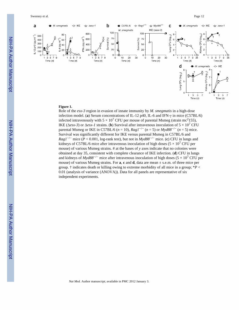
Figure 1.Role of the esx-3 region in evasion of innate immunity by M. smegmatis in a high-doseinfection model. (a) Serum concentrations of IL-12 p40, IL-6 and IFN-γ in mice (C57BL/6)infected intravenously with 5 × 10⁷ CFU per mouse of parental Msmeg (strain mc2155),IKE (Δesx-3) or Δesx-1 strains. (b) Survival after intravenous inoculation of 5 × 10⁷ CFUparental Msmeg or IKE in C57BL/6 (n = 10), Rag1−/− (n = 5) or Myd88−/− (n = 5) mice.Survival was significantly different for IKE versus parental Msmeg in C57BL/6 andRag1−/− mice (P < 0.001, log-rank test), but not in Myd88−/− mice. (c) CFU in lungs andkidneys of C57BL/6 mice after intravenous inoculation of high doses (5 × 10⁷ CFU permouse) of various Msmeg strains. # at the bases of y axes indicate that no colonies wereobtained at day 35, consistent with complete clearance of IKE infection. (d) CFU in lungsand kidneys of Myd88−/− mice after intravenous inoculation of high doses (5 × 10⁷ CFU permouse) of various Msmeg strains. For a, c and d, data are mean ± s.e.m. of three mice pergroup. † indicates death or killing owing to extreme morbidity of all mice in a group; *P <0.01 (analysis of variance (ANOVA)). Data for all panels are representative of sixindependent experiments.
Sweeney et al. Page 12
Nat Med. Author manuscript; available in PMC 2012 January 3.
NIH
-PA Author Manuscript
NIH
-PA Author Manuscript
NIH
-PA Author Manuscript

Figure 2.Characterization of innate immune responses against the Msmeg IKEPLUS strain. (a)Serum concentrations of IL-12 p40, IL-12p70 and IFN-γ in C57BL/6 mice infected withIKE or IKEPLUS (*P < 0.01, ANOVA with Bonferroni post-test). (b) Growth of bacteria(CFU) in the lungs (left) and kidneys (right) of Rag1−/− mice after intravenous inoculationof Msmeg parental, IKE or IKEPLUS strains. Differences in CFU were statisticallysignificant (P < 0.001, two-way ANOVA) for Msmeg versus either IKE or IKEPLUS at day3. # at the bases of y axes indicate that no colonies were obtained at day 35, consistent withcomplete clearance of IKE and IKEPLUS. (c) Average time to death of mice afterintravenous inoculation of IKEPLUS in C57BL/6 (n = 10), Rag1−/− (n = 5) and Myd88−/−
(n = 5) mice. Survival of wild-type C57BL/6 and Rag1−/− strains was significantly differentthan that of Myd88−/− mice (P < 0.05, log-rank test). All inoculations were done at a highintravenous dose (5 × 10⁷ CFU per mouse). Data from a and b are mean ± s.e.m. of threemice per group. Data for all panels are representative of four independent experiments.
Sweeney et al. Page 13
Nat Med. Author manuscript; available in PMC 2012 January 3.
NIH
-PA Author Manuscript
NIH
-PA Author Manuscript
NIH
-PA Author Manuscript
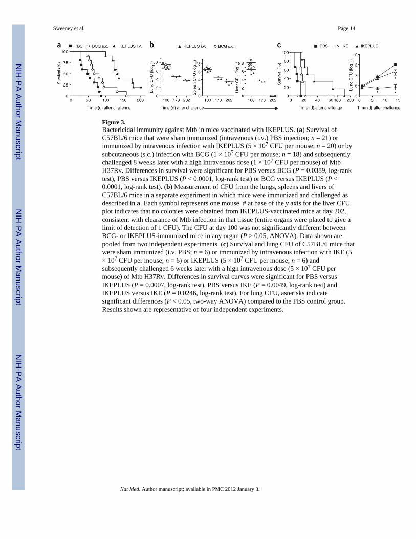
Figure 3.Bactericidal immunity against Mtb in mice vaccinated with IKEPLUS. (a) Survival ofC57BL/6 mice that were sham immunized (intravenous (i.v.) PBS injection; n = 21) orimmunized by intravenous infection with IKEPLUS (5 × 10⁷ CFU per mouse; n = 20) or bysubcutaneous (s.c.) infection with BCG (1 × 10⁷ CFU per mouse; n = 18) and subsequentlychallenged 8 weeks later with a high intravenous dose (1 × 10⁷ CFU per mouse) of MtbH37Rv. Differences in survival were significant for PBS versus BCG (P = 0.0389, log-ranktest), PBS versus IKEPLUS (P < 0.0001, log-rank test) or BCG versus IKEPLUS (P <0.0001, log-rank test). (b) Measurement of CFU from the lungs, spleens and livers ofC57BL/6 mice in a separate experiment in which mice were immunized and challenged asdescribed in a. Each symbol represents one mouse. # at base of the y axis for the liver CFUplot indicates that no colonies were obtained from IKEPLUS-vaccinated mice at day 202,consistent with clearance of Mtb infection in that tissue (entire organs were plated to give alimit of detection of 1 CFU). The CFU at day 100 was not significantly different betweenBCG- or IKEPLUS-immunized mice in any organ (P > 0.05, ANOVA). Data shown arepooled from two independent experiments. (c) Survival and lung CFU of C57BL/6 mice thatwere sham immunized (i.v. PBS; n = 6) or immunized by intravenous infection with IKE (5× 10⁷ CFU per mouse; n = 6) or IKEPLUS (5 × 10⁷ CFU per mouse; n = 6) andsubsequently challenged 6 weeks later with a high intravenous dose (5 × 10⁷ CFU permouse) of Mtb H37Rv. Differences in survival curves were significant for PBS versusIKEPLUS (P = 0.0007, log-rank test), PBS versus IKE (P = 0.0049, log-rank test) andIKEPLUS versus IKE (P = 0.0246, log-rank test). For lung CFU, asterisks indicatesignificant differences (P < 0.05, two-way ANOVA) compared to the PBS control group.Results shown are representative of four independent experiments.
Sweeney et al. Page 14
Nat Med. Author manuscript; available in PMC 2012 January 3.
NIH
-PA Author Manuscript
NIH
-PA Author Manuscript
NIH
-PA Author Manuscript
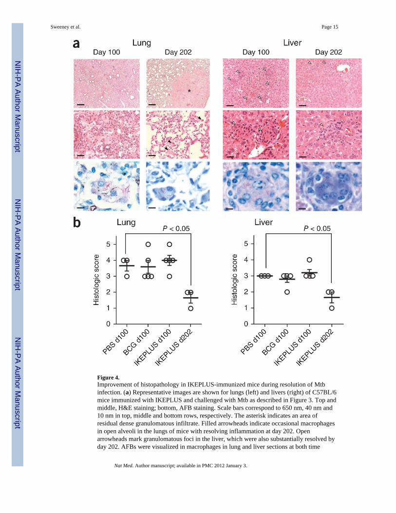
Figure 4.Improvement of histopathology in IKEPLUS-immunized mice during resolution of Mtbinfection. (a) Representative images are shown for lungs (left) and livers (right) of C57BL/6mice immunized with IKEPLUS and challenged with Mtb as described in Figure 3. Top andmiddle, H&E staining; bottom, AFB staining. Scale bars correspond to 650 nm, 40 nm and10 nm in top, middle and bottom rows, respectively. The asterisk indicates an area ofresidual dense granulomatous infiltrate. Filled arrowheads indicate occasional macrophagesin open alveoli in the lungs of mice with resolving inflammation at day 202. Openarrowheads mark granulomatous foci in the liver, which were also substantially resolved byday 202. AFBs were visualized in macrophages in lung and liver sections at both time
Sweeney et al. Page 15
Nat Med. Author manuscript; available in PMC 2012 January 3.
NIH
-PA Author Manuscript
NIH
-PA Author Manuscript
NIH
-PA Author Manuscript
points. (b) Quantitative scoring of histopathology confirming that all three groups showedsimilar pathology in the lungs and liver at day 100 (P > 0.05, two-way ANOVA). However,at day 202 the survivors from the IKEPLUS-immunized group showed significantly reducedpathology scores (P < 0.05 compared to all other groups at day 100; one-way ANOVA withTukey post-test).
Sweeney et al. Page 16
Nat Med. Author manuscript; available in PMC 2012 January 3.
NIH
-PA Author Manuscript
NIH
-PA Author Manuscript
NIH
-PA Author Manuscript
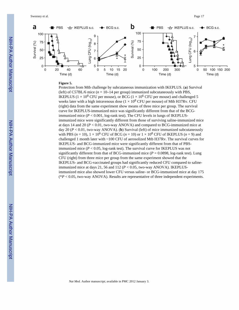
Figure 5.Protection from Mtb challenge by subcutaneous immunization with IKEPLUS. (a) Survival(left) of C57BL/6 mice (n = 10–14 per group) immunized subcutaneously with PBS,IKEPLUS (1 × 10⁸ CFU per mouse), or BCG (1 × 10⁶ CFU per mouse) and challenged 5weeks later with a high intravenous dose (1 × 10⁸ CFU per mouse) of Mtb H37Rv. CFU(right) data from the same experiment show means of three mice per group. The survivalcurve for IKEPLUS-immunized mice was significantly different from that of the BCG-immunized mice (P < 0.001, log-rank test). The CFU levels in lungs of IKEPLUS-immunized mice were significantly different from those of surviving saline-immunized miceat days 14 and 20 (P < 0.01, two-way ANOVA) and compared to BCG-immunized mice atday 20 (P < 0.01, two-way ANOVA). (b) Survival (left) of mice immunized subcutaneouslywith PBS (n = 10), 1 × 10⁶ CFU of BCG (n = 10) or 1 × 10⁸ CFU of IKEPLUS (n = 9) andchallenged 1 month later with ~100 CFU of aerosolized Mtb H37Rv. The survival curves forIKEPLUS- and BCG-immunized mice were significantly different from that of PBS-immunized mice (P < 0.05, log-rank test). The survival curve for IKEPLUS was notsignificantly different from that of BCG-immunized mice (P = 0.0898, log-rank test). LungCFU (right) from three mice per group from the same experiment showed that theIKEPLUS- and BCG-vaccinated groups had significantly reduced CFU compared to saline-immunized mice at days 21, 56 and 112 (P < 0.05, two-way ANOVA). IKEPLUS-immunized mice also showed lower CFU versus saline- or BCG-immunized mice at day 175(*P < 0.05, two-way ANOVA). Results are representative of three independent experiments.
Sweeney et al. Page 17
Nat Med. Author manuscript; available in PMC 2012 January 3.
NIH
-PA Author Manuscript
NIH
-PA Author Manuscript
NIH
-PA Author Manuscript
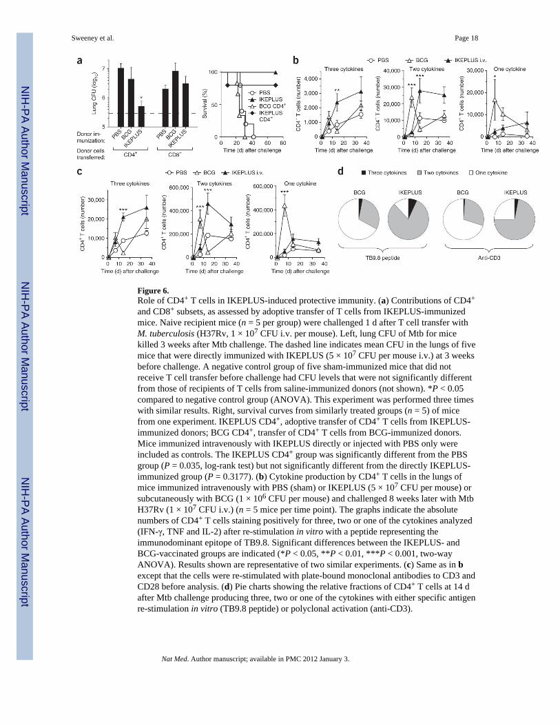
Figure 6.Role of CD4+ T cells in IKEPLUS-induced protective immunity. (a) Contributions of CD4+
and CD8+ subsets, as assessed by adoptive transfer of T cells from IKEPLUS-immunizedmice. Naive recipient mice (n = 5 per group) were challenged 1 d after T cell transfer withM. tuberculosis (H37Rv, 1 × 10⁷ CFU i.v. per mouse). Left, lung CFU of Mtb for micekilled 3 weeks after Mtb challenge. The dashed line indicates mean CFU in the lungs of fivemice that were directly immunized with IKEPLUS (5 × 10⁷ CFU per mouse i.v.) at 3 weeksbefore challenge. A negative control group of five sham-immunized mice that did notreceive T cell transfer before challenge had CFU levels that were not significantly differentfrom those of recipients of T cells from saline-immunized donors (not shown). *P < 0.05compared to negative control group (ANOVA). This experiment was performed three timeswith similar results. Right, survival curves from similarly treated groups (n = 5) of micefrom one experiment. IKEPLUS CD4+, adoptive transfer of CD4+ T cells from IKEPLUS-immunized donors; BCG CD4+, transfer of CD4+ T cells from BCG-immunized donors.Mice immunized intravenously with IKEPLUS directly or injected with PBS only wereincluded as controls. The IKEPLUS CD4+ group was significantly different from the PBSgroup (P = 0.035, log-rank test) but not significantly different from the directly IKEPLUS-immunized group (P = 0.3177). (b) Cytokine production by CD4+ T cells in the lungs ofmice immunized intravenously with PBS (sham) or IKEPLUS (5 × 10⁷ CFU per mouse) orsubcutaneously with BCG (1 × 10⁶ CFU per mouse) and challenged 8 weeks later with MtbH37Rv (1 × 10⁷ CFU i.v.) (n = 5 mice per time point). The graphs indicate the absolutenumbers of CD4+ T cells staining positively for three, two or one of the cytokines analyzed(IFN-γ, TNF and IL-2) after re-stimulation in vitro with a peptide representing theimmunodominant epitope of TB9.8. Significant differences between the IKEPLUS- andBCG-vaccinated groups are indicated (*P < 0.05, **P < 0.01, ***P < 0.001, two-wayANOVA). Results shown are representative of two similar experiments. (c) Same as in bexcept that the cells were re-stimulated with plate-bound monoclonal antibodies to CD3 andCD28 before analysis. (d) Pie charts showing the relative fractions of CD4+ T cells at 14 dafter Mtb challenge producing three, two or one of the cytokines with either specific antigenre-stimulation in vitro (TB9.8 peptide) or polyclonal activation (anti-CD3).
Sweeney et al. Page 18
Nat Med. Author manuscript; available in PMC 2012 January 3.
NIH
-PA Author Manuscript
NIH
-PA Author Manuscript
NIH
-PA Author Manuscript