Rapidly Growing Mycobacterium Species - MDPI
40
International Journal of Molecular Sciences Review Rapidly Growing Mycobacterium Species: The Long and Winding Road from Tuberculosis Vaccines to Potent Stress-Resilience Agents Mattia Amoroso 1 , Dominik Langgartner 1 , Christopher A. Lowry 2,3,4,5,6 and Stefan O. Reber 1, * Citation: Amoroso, M.; Langgartner, D.; Lowry, C.A.; Reber, S.O. Rapidly Growing Mycobacterium Species: The Long and Winding Road from Tuberculosis Vaccines to Potent Stress-Resilience Agents. Int. J. Mol. Sci. 2021, 22, 12938. https://doi.org/ 10.3390/ijms222312938 Academic Editors: Keith W. Kelley and Jennifer Felger Received: 2 November 2021 Accepted: 26 November 2021 Published: 29 November 2021 Publisher’s Note: MDPI stays neutral with regard to jurisdictional claims in published maps and institutional affil- iations. Copyright: © 2021 by the authors. Licensee MDPI, Basel, Switzerland. This article is an open access article distributed under the terms and conditions of the Creative Commons Attribution (CC BY) license (https:// creativecommons.org/licenses/by/ 4.0/). 1 Laboratory for Molecular Psychosomatics, Department of Psychosomatic Medicine and Psychotherapy, University of Ulm, 89081 Ulm, Germany; [email protected] (M.A.); [email protected] (D.L.) 2 Department of Integrative Physiology, Center for Neuroscience and Center for Microbial Exploration, University of Colorado Boulder, Boulder, CO 80309, USA; [email protected] 3 Department of Physical Medicine and Rehabilitation and Center for Neuroscience, University of Colorado Anschutz Medical Campus, Aurora, CO 80045, USA 4 Veterans Health Administration, Rocky Mountain Mental Illness Research Education and Clinical Center (MIRECC), The Rocky Mountain Regional Veterans Affairs Medical Center (RMRVAMC), Aurora, CO 80045, USA 5 Military and Veteran Microbiome: Consortium for Research and Education (MVM-CoRE), Aurora, CO 80045, USA 6 Senior Fellow, inVIVO Planetary Health, of the Worldwide Universities Network (WUN), West New York, NJ 07093, USA * Correspondence: [email protected] Abstract: Inflammatory diseases and stressor-related psychiatric disorders, for which inflammation is a risk factor, are increasing in modern Western societies. Recent studies suggest that immunoreg- ulatory approaches are a promising tool in reducing the risk of suffering from such disorders. Specifically, the environmental saprophyte Mycobacterium vaccae National Collection of Type Cul- tures (NCTC) 11659 has recently gained attention for the prevention and treatment of stress-related psychiatric disorders. However, effective use requires a sophisticated understanding of the effects of M. vaccae NCTC 11659 and related rapidly growing mycobacteria (RGMs) on microbiome–gut– immune–brain interactions. This historical narrative review is intended as a first step in explor- ing these mechanisms and provides an overview of preclinical and clinical studies on M. vaccae NCTC 11659 and related RGMs. The overall objective of this review article is to increase the compre- hension of, and interest in, the mechanisms through which M. vaccae NCTC 11659 and related RGMs promote stress resilience, with the intention of fostering novel clinical strategies for the prevention and treatment of stressor-related disorders. Keywords: immunoregulation; inflammation; Mycobacterium kyogaense; Mycobacterium vaccae; old friends; stress-associated disorders; stress resilience 1. Introduction 1.1. The “Old Friends” Hypothesis: A Biological Concept to Explain the Increasing Prevalence Rates of Stress-Associated Inflammatory Disorders in Modern Urban Societies The prevalence of many stress-associated somatic disorders including allergies [1] and autoimmune diseases [2–5] as well as mental pathologies such as depression and posttraumatic stress disorder (PTSD) [6] has increased over the past decades in Western- ized countries, overall representing a serious health and economic burden for our modern society. Although the mechanisms underlying both development and progression of these stress-associated disorders are not fully understood, and, consequently, prevention and treatment options for many of these disorders are still limited, a common feature of these disorders is a dysregulated immune system and increased inflammation [7]. As many of Int. J. Mol. Sci. 2021, 22, 12938. https://doi.org/10.3390/ijms222312938 https://www.mdpi.com/journal/ijms
-
Upload
khangminh22 -
Category
Documents
-
view
0 -
download
0
Transcript of Rapidly Growing Mycobacterium Species - MDPI

International Journal of
Molecular Sciences
Review
Rapidly Growing Mycobacterium Species: The Long andWinding Road from Tuberculosis Vaccines to PotentStress-Resilience Agents
Mattia Amoroso 1, Dominik Langgartner 1, Christopher A. Lowry 2,3,4,5,6 and Stefan O. Reber 1,*
�����������������
Citation: Amoroso, M.; Langgartner,
D.; Lowry, C.A.; Reber, S.O. Rapidly
Growing Mycobacterium Species: The
Long and Winding Road from
Tuberculosis Vaccines to Potent
Stress-Resilience Agents. Int. J. Mol.
Sci. 2021, 22, 12938. https://doi.org/
10.3390/ijms222312938
Academic Editors: Keith W. Kelley
and Jennifer Felger
Received: 2 November 2021
Accepted: 26 November 2021
Published: 29 November 2021
Publisher’s Note: MDPI stays neutral
with regard to jurisdictional claims in
published maps and institutional affil-
iations.
Copyright: © 2021 by the authors.
Licensee MDPI, Basel, Switzerland.
This article is an open access article
distributed under the terms and
conditions of the Creative Commons
Attribution (CC BY) license (https://
creativecommons.org/licenses/by/
4.0/).
1 Laboratory for Molecular Psychosomatics, Department of Psychosomatic Medicine and Psychotherapy,University of Ulm, 89081 Ulm, Germany; [email protected] (M.A.);[email protected] (D.L.)
2 Department of Integrative Physiology, Center for Neuroscience and Center for Microbial Exploration,University of Colorado Boulder, Boulder, CO 80309, USA; [email protected]
3 Department of Physical Medicine and Rehabilitation and Center for Neuroscience, University of ColoradoAnschutz Medical Campus, Aurora, CO 80045, USA
4 Veterans Health Administration, Rocky Mountain Mental Illness Research Education and ClinicalCenter (MIRECC), The Rocky Mountain Regional Veterans Affairs Medical Center (RMRVAMC),Aurora, CO 80045, USA
5 Military and Veteran Microbiome: Consortium for Research and Education (MVM-CoRE),Aurora, CO 80045, USA
6 Senior Fellow, inVIVO Planetary Health, of the Worldwide Universities Network (WUN),West New York, NJ 07093, USA
* Correspondence: [email protected]
Abstract: Inflammatory diseases and stressor-related psychiatric disorders, for which inflammationis a risk factor, are increasing in modern Western societies. Recent studies suggest that immunoreg-ulatory approaches are a promising tool in reducing the risk of suffering from such disorders.Specifically, the environmental saprophyte Mycobacterium vaccae National Collection of Type Cul-tures (NCTC) 11659 has recently gained attention for the prevention and treatment of stress-relatedpsychiatric disorders. However, effective use requires a sophisticated understanding of the effectsof M. vaccae NCTC 11659 and related rapidly growing mycobacteria (RGMs) on microbiome–gut–immune–brain interactions. This historical narrative review is intended as a first step in explor-ing these mechanisms and provides an overview of preclinical and clinical studies on M. vaccaeNCTC 11659 and related RGMs. The overall objective of this review article is to increase the compre-hension of, and interest in, the mechanisms through which M. vaccae NCTC 11659 and related RGMspromote stress resilience, with the intention of fostering novel clinical strategies for the preventionand treatment of stressor-related disorders.
Keywords: immunoregulation; inflammation; Mycobacterium kyogaense; Mycobacterium vaccae; oldfriends; stress-associated disorders; stress resilience
1. Introduction1.1. The “Old Friends” Hypothesis: A Biological Concept to Explain the Increasing PrevalenceRates of Stress-Associated Inflammatory Disorders in Modern Urban Societies
The prevalence of many stress-associated somatic disorders including allergies [1]and autoimmune diseases [2–5] as well as mental pathologies such as depression andposttraumatic stress disorder (PTSD) [6] has increased over the past decades in Western-ized countries, overall representing a serious health and economic burden for our modernsociety. Although the mechanisms underlying both development and progression of thesestress-associated disorders are not fully understood, and, consequently, prevention andtreatment options for many of these disorders are still limited, a common feature of thesedisorders is a dysregulated immune system and increased inflammation [7]. As many of
Int. J. Mol. Sci. 2021, 22, 12938. https://doi.org/10.3390/ijms222312938 https://www.mdpi.com/journal/ijms

Int. J. Mol. Sci. 2021, 22, 12938 2 of 40
these stress-associated disorders are further characterized by a compromised regulatory Tcell (Treg) compartment [8–10], a failure of immunoregulation has been hypothesized to beinvolved in promoting an over-reacting immune system, thus, predisposing an individualto disease development. Thus, anti-inflammatory and immunoregulatory approachesmight be a useful tool in prevention and treatment of stress-related disorders. Accordingto the “old friends” hypothesis, deficits in immunoregulation are due to reduced contactwith harmless microorganisms that accompanied mammalian evolution in high abundanceand had to be tolerated by an individual’s immune system to avoid damage caused bychronic inflammatory processes [11]. Interestingly, these “old friend” organisms promotetheir own survival and, as a beneficial side effect, the health of their host by facilitatingimmunoregulation. “Old friends” fall into three main categories: (1) microorganismsassociated with “old infections” that were common in human evolutionary past (helminths,Salmonella, Helicobacter pylori [12,13]); (2) microorganisms that were part of the humanmicrobiota (gut, airway, skin, genitourinary, oropharyngeal; [14–17]); and (3) harmlessmicroorganisms from the natural environment in water, air, and soil with which humansinevitably had regular contact (reviewed in [18,19]). Two such microorganisms attractingattention for their immunoregulatory effects are Mycobacterium vaccae National Collectionof Type Cultures 11659 (M. vaccae NCTC 11659) and Mycobacterium vaccae American TypeCulture Collection 15483 Typestrain (M. vaccae ATCC 15483T) [20–33]. In the current article,we aim to provide a narrative review of the research history of these two immunoregu-latory mycobacteria in a chronological way, starting with the first observational studieson their promising effects as tuberculosis (TB) vaccines up to the most recent studies indi-cating that these “old friends” have stress-protective effects and promote stress resilience.Special emphasis is given to the cellular and molecular mechanisms known so far to me-diate the effects of these rapidly growing mycobacteria (RGMs) including the recentlydiscovered 10(Z)-hexadecenoic acid (10(Z)-HDA), a free fatty acid synthetized by M. vaccaeNCTC 11659 that mediates its anti-inflammatory effects via enhancement of peroxisomeproliferator-activated receptor alpha (PPARα) signaling. Finally, the effectiveness of differ-ent routes of M. vaccae NCTC 11659 and M. vaccae ATCC 15483T administration and possiblestrategies to increase it are discussed. Important to note is that M. vaccae NCTC 11659(Colección Española de Cultivos Tipo (CECT) 9646T; DSMZ-Deutsche Sammlung vonMikroorganismen und Zellkulturen GmbH (DSM) 107316T) was reclassified in 2018 asMycobacterium kyogaense sp. nov. NCTC 11659T [34,35]. However, to avoid any confusionas this Mycobacterium strain has been referenced in many previously published articlesincluding our own [6,20,21,32,33] as M. vaccae NCTC 11659, we keep this nomenclatureconsistent and refer to this Mycobacterium strain in the current review article as M. vaccaeNCTC 11659 (please see Table 1 for alternative designations and different preparations andproduction processes of M. vaccae NCTC 11659). When discussing studies investigating theeffects of the M. vaccae type strain, we refer to it as M. vaccae (ATCC 15483T; DSM 43292T;NCTC 10916T), and to M. vaccae when the exact M. vaccae strain was not further specified inthe original articles. An overview of the exact nomenclature, preparation, and productiondetails and dose of the Mycobacterium species/strain used in each study discussed in thecurrent review article is provided in Tables 1–4.
Int. J. Mol. Sci. 2021, 22, 12938 3 of 40
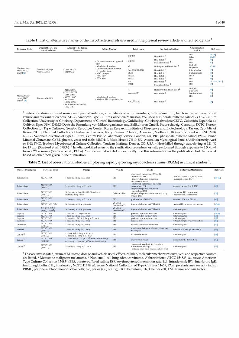
Table 1. List of alternative names of the mycobacterium strains used in the present review article and related details 1.
Reference Strain Original Source andYear of Isolation
Alternative CollectionNumbers Culture Medium Batch Name Inactivation Method Administration
Vehicle Reference
Mycobacteriumvaccae NCTC11659 [34]
Mud (lake Kyoga,Uganda), 1973
- DSM 107316- CECT 9646
- Peptone meat extract glycerolagar- Middlebrook medium- Löwenstein-Jensen medium- Tryptic Soy Agar- MB7H10 agar- PMG agar- GYM agar
SRP 299 Heat-killed 2 PBS [36–38]Saline [39,40]
SRL172 Heat-killed 2 BBS [41]Irradiation-killed 3,* BBS [42]
V7 Hydrolyzed and heat-killed 2 Oral pillexcipients [43,44]
Dar-901 (SRL 172) Heat-killed 2 Citrate buffer [45]MV07 Heat-killed 2 Culture media [46]MV001 Heat-killed 2 BBS * [47]MV007 Heat-killed 2 BBS [48]ENG1 Heat-killed 2 BBS [20,22,24,29,32]R877R Irradiation-killed 3 BBS [49]
Mycobacteriumvaccae ATCC15483T [50]
Bovine milk, 1964
- ATCC 23004- CCUG 21003T- DSM 43292- KCTC 19087- NCIB 9937- NCTC 10916- SN 920 (Bonicke SN920)- TMC 1526
- Middlebrook medium- Medium 55 for Mycobacterium
V7 Hydrolyzed and heat-killed 2 Oral pillexcipients [51]
M.vaccaeTM Heat-killed 2 Distilled water [52]
ATCC® 15483 Heat-killed 2
PBS [53]
BBS [29]
1 Reference strain, original source and year of isolation, alternative collection numbers, culture medium, batch name, administrationvehicle and relevant references. ATCC, American Type Culture Collection, Manassas, VA, USA; BBS, borate-buffered saline; CCUG, CultureCollection, University of Göteborg, Department of Clinical Bacteriology, Guldhedsg, Göteborg, Sweden; CETC, Colección Española deCultivos Tipo; DSM, DSMZ-Deutsche Sammlung von Mikroorganismen und Zellkulturen GmbH, Braunschweig, Germany; KCTC, KoreanCollection for Type Cultures, Genetic Resources Center, Korea Research Institute of Bioscience and Biotechnology, Taejon, Republic ofKorea; NCIB, National Collection of Industrial Bacteria, Torry Research Station, Aberdeen, Scotland, UK (incorporated with NCIMB);NCTC, National Collection of Type Cultures, Central Public Laboratory Service, London, UK; PBS, phosphate-buffered saline; PMG, PombeMinimal Glutamate; GYM, glucose, yeast and malt; MB7H10, Middlebrook 7H10; SN, Australian Mycological Panel (AMP; formerly Austor SN); TMC, Trudeau Mycobacterial Culture Collection, Trudeau Institute, Denver, CO, USA. 2 Heat-killed through autoclaving at 121 ◦Cfor 15 min (Stanford et al., 1990b). 3 Irradiation-killed refers to the sterilization procedure, usually performed through exposure to 2.5 Mradfrom a 60Co source (Stanford et al., 1990a). * indicates that we did not explicitly find this information in the publication, but deduced itbased on other facts given in the publication.
Table 2. List of observational studies employing rapidly growing mycobacteria strains (RGMs) in clinical studies 1.
Disease Investigated M. vaccae Strain Dosage Vehicle Effects Underlying Mechanisms Reference
Tuberculosis NCTC 11659 1 dose (i.d.; 1 mg in 0.1 mL) BBS
- improved clearance of TB bacilli- normalized ESR- improved sputum conversion- increased survival
- reduced serum IL-4, IL-10, TNF- increased serum IFNγ
[54–57]
Tuberculosis NCTC 11659(SRL172) 3 doses (i.d.; 1 mg in 0.1 mL) BBS
- improved clearance of TB bacilli- normalized ESR- improved sputum conversion
increased serum IL-4 & TNF [41]
Tuberculosis NCTC 11659(SRL172)
10 doses (p.o; day 0,7,14,21,28 and thenmonthly; 1 mg/dose) Gelatine tablet
- normalized ESR- improved sputum conversion and bodyweight gain
- increased Th1 parameters- decreased Th2 parameters [58]
Tuberculosis NCTC 11659(SRL172) 5 doses (i.d.; 1 mg in 0.1 mL) BBS proliferation of PBMCs increased IFNγ in PBMCs [42]
Tuberculosis NCTC 11659 (V7) 30 doses (p.o.; 10 µg/tablet) V7 tablet(Immunitor®) improved clearance of TB bacilli -reduced blood leukocyte number [43,44]
TuberculosisLongcom batch
(ATCC 15483T)30 doses (p.o.; 10 µg/tablet) V7 tablet
(Immunitor®) improved clearance of TB bacilli not investigated [51]
Leprosy NCTC 11659 1 dose (i.d.; 0.1 mg in 0.1 mL) BBS positive Leprosin A response not investigated [59,60]Leprosy NCTC 11659 1 dose (i.d.; 1 mg in 0.1 mL) BBS improved skin capillary flow not investigated [61]Leprosy NCTC 11659 3 doses (i.d.; 0.01, 0.1, 1 mg in 0.1 mL) BBS positive Leprosin A response not investigated [62]Psoriasis NCTC 11659 1 dose (i.d.; 1 mg in 0.1 mL) BBS reduced PASI reduced lymphocyte proliferation [63]
Dermatitis NCTC 11659(SRL172) 1 dose (i.d.; 3 mg in 0.3 mL) BBS reduced dermatitis lesion area not investigated [64]
Asthma NCTC 11659(SRL172) 1 dose (i.d.; 1 mg in 0.1 mL) BBS trend towards improved airway response
to allergen reduced IL-5 and IgE in PBMCs [65]
Cancer 2 NCTC 11659(SRL172)
- 1 dose (i.d.; 0.5 mg in 0.1 mL);- 3 doses (i.d.; 1 mg in 0.1 mL) BBS increased survival not investigated [66]
Cancer 2 NCTC 11659(SRL172)
- 1 dose (i.d.; 50 µL (5 × 10⁸ heat-killed bacilli));- 4 doses (i.d.; 100 µL (109 heat-killed bacilli))
BBS improved survival intracellular IL-2 induction [67]
Cancer 3 NCTC 11659(SRL172) 5 doses (i.d.; 1 mg in 0.1 mL) BBS
- improved quality of life (cognitivefunction and vitality);- reduced body pain, nausea and dyspnea
not investigated [68]
1 Disease investigated, strain of M. vaccae, dosage and vehicle used, effects, cellular/molecular mechanisms involved, and respective sourcesare listed. 2 Metastatic malignant melanoma. 3 Non-small-cell lung adenocarcinoma. Abbreviations: ATCC 15483T, M. vaccae AmericanType Culture Collection 15483T; BBS, borate-buffered saline; ESR, erythrocyte sedimentation rate; i.d., intradermal; IFN, interferon; IgE,immunoglobulin E; IL, interleukin; NCTC 11659, M. vaccae National Collection of Type Cultures 11659; PASI, psoriasis area severity index;PBMC, peripheral blood mononuclear cells; p.o, per os (i.e., orally); TB, tuberculosis; Th, T helper cell; TNF, tumor necrosis factor.

Int. J. Mol. Sci. 2021, 22, 12938 4 of 40
Table 3. List of observational studies employing rapidly growing mycobacteria strains (RGMs) in preclinical studies 1.
Disease Investigated M. vaccae Strain Dosage Vehicle Effects Underlying Mechanisms Reference
Negative consequences of stress NCTC 11659 3 doses (s.c.; 0.1 mg in 0.1 mL) BBS reduced stress-induced anxietyand colitis
- increased number of Treg (CD4+ CD25+ FoxP3+)- increased IL-10
[20,33]
Negative consequences of stress NCTC 11659 3 doses (i.n.; 0.1 mg in 0.012 mL) BBS reduced stress-induced colitis not investigated [32]
Negative consequences of stress NCTC 11659 3 doses (s.c.; 0.1 mg in 0.1 mL) BBSenhanced between- andwithin-session extinction infear-potentiated startle paradigm
alteration in serotonergic gene expression [25,27,28]
Negative consequences of stress NCTC 11659 3 doses (s.c.; 0.1 mg in 0.1 mL) BBSprevention of stress-inducedexaggeration of anxiety andmicroglial priming
- upregulated hippocampal IL4, Cd200r1 and Mrc1- downregulated hippocampal Nlrp3 and Nfkbia [24]
Negative consequences of stress NCTC 11659 3 doses (s.c.; 0.1 mg in 0.1 mL) BBS prevention of post-operativecognitive dysfunction
- upregulated hippocampal IL4, Arg1 and Foxp3- downregulated hippocampal NfκBbia and IL1β [22]
Negative consequences of stress NCTC 11659 3 doses (s.c.; 0.1 mg in 0.1 mL) BBS prevention of negative outcomesof a two-hit stress models
- prevention of stress-induced decreased Tph2 andSlc6a4 expression- prevention of stress-induced REM sleep disturbances
[23,26]
1 Disease investigated, strain of M. vaccae, dosage and vehicle used, effects, cellular/molecular mechanisms involved, and respectivesources are listed. Abbreviations: Arg1, arginase 1 gene; BBS, borate-buffered saline; CD, cluster of differentiation; FoxP3, forkhead box P3;IL, interleukin; i.n., intranasal; NCTC 11659, M. vaccae National Collection of Type Cultures 11659; NfκBbia: gene encoding nuclear factor ofkappa light polypeptide gene enhancer in B-cells inhibitor, alpha; Nlrp3, NLR family pyrin domain containing 3; s.c., subcutaneous; REM,rapid eye movement; Slc6a4, solute carrier family 6 member 4; Tph2, tryptophan hydroxylase 2; Treg, regulatory T cells.
Table 4. List of preclinical studies investigating the underlying mechanisms induced by rapidly growing mycobacteriastrains (RGMs) 1.
Cellular/ MolecularTarget Investigated M. vaccae Strain Dosage Vehicle Species Underlying Mechanisms Reference
Th1/Th2 balance NCTC 11659 (MV07) 1, 10, 100 µg/mL (in vitro) BBS human (DCs) - reduced IL-4- upregulated CD83 and CD86 [46]
Th1/Th2 balance NCTC 11659 (SRL172) 3 doses (s.c.; 106,107,108 bacteria in 0.1 mL) BBS mouse - reduced serum IgE, IL-4 and IL-5- reduced eosinophil count in BAL [69]
Th1/Th2 balance NCTC 11659 1 dose (s.c.; 107, 108 or 109 bacteria in 0.1 mL) BBS mouse - reduced serum IgE and IL-4- increased IL-2 in splenocytes [70]
γδ T cells NCTC 11659 (SRL172) 100 µg/mL (in vitro) BBS human (PBMCs) upregulated IFNγ, TNF and granzyme B [71]
CD11b+ myeloidcells
NCTC 11659 300 µg/mL (in vitro) BBS human (PBMCs)- downregulated CD62L- upregulated TLR2, TLR4, CD18, CD11a, CD14, CD36,CD44, CD45, CD54 CD58k, CD80, CD86, CD137L, CD206
[72]
CD11c+ APC NCTC 11659 (SRP299) 1 dose (s.c.; 0.1 mg in 0.1 mL) NaCl mouse - decreased cell number in BAL;- increased IL-10+ and TGFβ in lung DCs [39]
CD14+ monocytes ATCC 15483T (SN920)in vitro incubation with 1:10 ratiocells:mycobacteria Medium human (PBMCs) increased secretion of TNF and IL-12 [73]
CD8+ CTL ATCC 15483T 1 dose (i.p.; 1 mg in 0.1 mL) PBS mouse
- increased expression of IFNγ in TB-infectedmacrophages- increased cytotoxic activity of CTL against TB-infectedmacrophages
[74]
Tregs NCTC 11659 (SRP299) 1 dose (s.c.; 0.1 mg in 0.2 mL) NaCl mouse - increased number of Treg (CD4+ CD45RBLo)- suppressed airway inflammation upon Treg transfer
[40]
Tregs NCTC 11659 (SRP299) - 1 dose (i.g.; 0.1 mg in 0.1 mL)- 100, 200 or 400 µg/mL (in vitro) -H2O-NaCl mouse - reduced cellular infiltrate in lungs
- increased IL-10 in mesLNC [75]
Tregs NCTC 11659 3 doses (s.c.; 0.1 mg in 0.1 mL) BBS mouse- increased number of Treg (CD4+ CD25+ FoxP3+)- increased IL-10- reduced stress-induced anxiety and colitis
[33]
PPARα 10(Z)-HDA from NCTC11659 200 µM (in vitro) DMEM/F-12 mouse PPARα-dependent downregulation of pro-inflammatory
transcription factors, cytokines and chemokines [76]
Serotonergic neurons NCTC 11659 3 doses (s.c.; 0.1 mg in 0.1 mL) BBS mouse activation of serotonergic neurons in interfascicular partof dorsal raphe nucleus [21]
Microglia NCTC 11659 3 doses (s.c.; 0.1 mg in 0.1 mL) BBS rat
- increased expression of Il4 mRNA and IL-4-responsivegenes (Cd200r1, Mrc1)- reduced IL-1β secretion from freshly isolated andLPS-stimulated hippocampal microglia
[24]
Microglia NCTC 11659, ATCC 15483T 3 doses (s.c.; 0.1 mg in 0.1 mL) BBS rat prevention of stress-induced upregulation ofhippocampal Il6 mRNA expression [29]
Microglia NCTC 11659 3 doses (s.c.; 0.1 mg in 0.1 mL) BBS rat increased hippocampal Il4, Foxp3, Arg1, decreased Il1β,Il6 and Nfκbia mRNA expression [22]
1 Cellular/molecular target investigated, strain of M. vaccae, dosage and vehicle used, species, cellular/molecular mechanisms involved,and respective sources are listed. Abbreviations: Arg1, arginase 1 gene; ATCC 15483T, M. vaccae American Type Culture Collection 15483T;BAL, bronchoalveolar lavage; BBS, borate-buffered saline; CD, cluster of differentiation; DMEM, Dulbecco’s modified Eagle’s medium;FoxP3, forkhead box P3; HPC, human pancreatic carcinoma cell line, IFN, interferon; i.g., intragastric; IL, interleukin; i.p., intraperitoneal;Mrc1, gene encoding mannose receptor C-type 1; NCTC 11659, M. vaccae National Collection of Type Cultures 11659; NfκBbia: geneencoding nuclear factor of kappa light polypeptide gene enhancer in B-cells inhibitor, alpha; PBMC, peripheral blood mononuclear cells;PBS, phosphate-buffered saline; p.o, per os (i.e., orally); PPARα, peroxisome proliferator-activated receptor α; s.c., subcutaneous; TB,tuberculosis; TNF, tumor necrosis factor.
1.2. Mycobacterium vaccae NCTC 11659: General Information
M. vaccae NCTC 11659 is a rapidly growing, aerobic, Gram-positive, acid-alcohol-fast,rod-shaped soil saprophyte, which forms rough yellow pigmented colonies in culture [34].Mycobacterium strains can be ubiquitously found in water and soil as well as in man-ufactured water distribution systems [77,78] and are the dominant taxa in municipalshowerheads [79]. Although mycobacteria are not normally found in the human gut mi-crobiome, they are abundant in the human oral cavity (buccal mucosa and dental plaque)and upper respiratory tract (nostrils and oropharynx) [80]. Studies comparing the airwaymicrobiome in urban versus rural children in Denmark have found greater abundance ofmycobacteria in rural children versus urban children at three months of age [81]. Althoughno evidence of pathogenicity of M. vaccae NCTC 11659 has ever been shown, in 1996,

Int. J. Mol. Sci. 2021, 22, 12938 5 of 40
a non-identified strain of M. vaccae was for the first time reported to cause non-severeinfections in immunocompromised individuals [82]. More recently, another non-identifiedstrain of M. vaccae was reported to cause catheter-related sepsis in a patient with follicularnon-Hodgkin lymphoma [83]. Its name is derived from the Latin word for cow, “vacca”, asthe first discovered strain was isolated from cow dung in Austria [50] and known in theliterature under the designations ATCC 15483T, DSM 43292T, and NCTC 10916T [34,84].On the other hand, M. vaccae NCTC 11659 was first isolated from the mud of Lake Kyogain Uganda by Stanford and Paul [85]. Although incorrectly classified as M. vaccae NCTC11659, data based on 16S rRNA gene and genome sequencing provided a rationale for itsreclassification as M. kyogaense sp. nov [34]. This strain was originally grown on Sauton’smedium solidified with 1.5% agar at 32 ◦C, but showed optimal growth at 37 ◦C on glucose,yeast, and malt (GYM) agar, Middlebrook (MB) 7H10 agar, and proteose peptone-meatextract-glycerol (PMG) agar [34,86]. At the end of the period of logarithmic growth (aboutseven days), the bacterial growth is usually scraped from the surface of the medium,weighed, and suspended in borate-buffered saline (BBS) at a concentration of 10 mg of wetweight/mL [34]. Heat-killed preparations of M. vaccae NCTC 11659 can be prepared byautoclaving in BBS at 121 ◦C for 15 min [87].
Several lines of evidence have shown that a heat-killed preparation of M. vaccaeNCTC 11659 had remarkable immunomodulatory and, thus, health promoting proper-ties in both preclinical and clinical studies [88]. This is indicated by its beneficial effectsagainst infectious diseases such as leprosy and TB [85,87,89], chronic inflammatory dis-orders such as asthma [40,69,90], colitis [32,33] as well as various forms of cancer inhumans [66,68,71,91–95]. Strikingly, M. vaccae NCTC 11659 was also protective in animalmodels of inescapable stress, fear conditioning, post-operative cognitive impairment inaged rats, models of “two hit” stressors involving sleep deprivation or chronic disruptionof rhythms and social defeat, and chronic psychosocial stress [20–33]. The effects of M.vaccae NCTC 11659 in all of the above-referenced studies are discussed in a more detailedmanner in the following sections of this review article. Both M. vaccae NCTC 11659 [88]and M. vaccae ATCC 15483T [77] have peculiar immune modulating properties that arehypothesized to depend on the extraordinary complexity of the cell envelope, a feature char-acteristic for the whole Mycobacterium genus [96–99]. The envelope of mycobacteria adoptsa unique dual membrane structure with a waxy outer membrane rich in mycolic acidsand free lipids [100,101], a polysaccharide cell wall, and an inner cytoplasmic membrane(reviewed in [96–98]). Molecules in the outer membrane can be recognized by macrophagesand dendritic cells (DCs) through pattern recognition receptors (PRRs) such as Toll-likereceptors (TLRs), nucleotide-binding oligomerization domain (NOD)-like receptors (NLRs),and C-type lectin receptors (CLRs) (reviewed in [98]). These interactions influence DCmaturation and, as a consequence, modulate subsequent immune responses, for example,by promoting naïve T cells to develop either into classic T helper (Th) 1 and Th2 cells and,in the case of M. vaccae NCTC 11659, into Th1 cells [71,102,103] and Tregs [33,40,88].
Considering M. vaccae ATCC 15483T, it can form both rough and smooth colonies insolid culture, and the shift from smooth to rough colony type occurs at temperatures above30 ◦C [53]. Interestingly, while the rough colonies induced a Th1 response upon subcu-taneous (s.c.) injection in mice, as shown by increased interferon (IFN)γ and interleukin(IL)-12 (p40) production, the smooth variant induced a significantly weaker productionof the above-mentioned cytokines, but a higher IL-10 production instead [53]. Analyzingthe lipid profiles of the two colony variants, the authors identified a long-chain saturatedfatty acid polyester of estolide-like structure that is produced by the smooth, but notrough, M. vaccae ATCC 15483T colony variant. This substance, named RC by the authors(i.e., “red color”, as it stained red with anthrone), seems to be the putative substance thatexplains the differential immune-polarizing properties of the two kinds of M. vaccae ATCC15483T colonies [53].
The immunomodulatory properties of M. vaccae NCTC 11659 seem to be best retainedwhen the microorganism is heat-killed by autoclaving in BBS. In turn, M. vaccae NCTC

Int. J. Mol. Sci. 2021, 22, 12938 6 of 40
11659 autoclaved in phosphate-buffered saline (PBS) is much less effective, potentiallybecause autoclaving in a borate solution breaks down proteins into short amino acidchains, which are stably preserved [88,104]. Autoclaving M. vaccae NCTC 11659 in PBSis also considered to reduce the amount of the so called group I antigens [88], which arecommon among the whole Mycobacterium genus and can suppress inflammation given theirhigh homology with some heat shock proteins located in the mitochondria of eukaryoticcells, namely the heat-shock protein (hsp) 60 [105,106] and hsp70 [107,108]. AutoclavingM. vaccae NCTC 11659 in BBS ensures appropriate presentation of the amino acid chains ofthose antigens to naïve T cells, thereby resulting in a more stable product as opposed toautoclaving in PBS [88,103] or killing by exposing the bacterium to 60Co [109].
2. History of Mycobacterium vaccae NCTC 11659 Research
The history of M. vaccae NCTC 11659 research is strongly linked to the efforts to findan effective TB and leprosy vaccine. To date, Bacillus Calmette–Guèrin (BCG) remains theonly effective vaccine against TB available for human use [110]. BCG was developed at thebeginning of the 20th century as a suspension of live, attenuated Mycobacterium bovis bacilliisolated from a calf believed to be infected with the bovine form of TB, and was foundto protect children from active TB [111]. With respect to the underlying mechanisms, ithas been shown in BCG-vaccinated mice that the immune response against Mycobacteriumtuberculosis is characterized by an increased accumulation of effector T cells at the site ofactive infection as well as increased production of Th1 cytokines, leading to restrictedgrowth of the bacilli [112,113]. Of note, the immunotherapeutic efficacy of BCG seems torely on both cluster of differentiation (CD) 4+ and CD8+ T cell subsets, as depletion of eithercell type results in the failure of BCG therapy [114]. Both CD4+ and CD8+ T cell subsetsin turn are dependent on the enhanced survival and prolonged lifespan of DCs followingBCG injection, which are achieved through reduced rates of apoptosis [115]. However,BCG vaccination results in variable degrees of protection against TB and leprosy [116,117],being very effective in certain areas such as Uganda [85] and poorly effective in others suchas India [118] and Myanmar [119]. The reason behind the geography-dependent effects ofBCG vaccination was believed to be environmental in nature. Interestingly, in the searchfor an environmental factor that could explain the high success rate of BCG vaccination inUganda, Dr. John Stanford noticed that the mud in and around Lake Kyoga in Uganda wasparticularly rich in M. vaccae NCTC 11659 [85,120,121], while Mycobacterium scrofulaceumwas abundantly present in Myanmar [119]. Some years later, Stanford and colleaguescould indeed show that M. vaccae NCTC 11659 enhances the protective post-BCG immuneresponses in Uganda, while M. scrofulaceum blocks them in Myanmar [119].
In more detail, the geography-dependent effectiveness of BCG vaccination againstTB seems to be dependent, among other factors, on the environmental mycobacterialspecies present in the areas where studies on BCG were conducted. Rook and colleaguespostulated that the latter was due to the two types of cell-mediated immune responsesgenerally driven by M. tuberculosis infection [122]. The first, the “Koch-type”, initiallydescribed by Robert Koch in guinea pigs at the end of the 19th century [123,124] develops4–6 weeks after M. tuberculosis infection, as indicated by a positive tuberculin skin test, andis characterized by a mixed Th1 and Th2 immune response, promoting the necrotizingeffects of tumor necrosis factor (TNF) in the presence of the Th2 cytokines IL-4 and IL-5and immunoglobulin E (IgE) [125,126]. The second cell-mediated immune response, the“Listeria-type” [127], occurs within days following M. tuberculosis infection and is char-acterized by the appearance of macrophage-activating Th1 lymphocytes [128]. The term“Listeria-type” immune response has been coined by George Mackaness [129] investigatingthe immune response against Listeria monocytogenes in mice. In contrast to the “Koch-type”response, this response was not accompanied by necrosis and strongly correlated with pro-tection against M. tuberculosis and Mycobacterium leprae [130]. Interestingly, different speciesof mycobacteria have been demonstrated in animal models to induce these two types of im-mune responses, characterized by either an activation of Th1/Th2 (“Koch-type”) or solely
Int. J. Mol. Sci. 2021, 22, 12938 7 of 40
Th1 (“Listeria-type”) immune response, to varying degrees [122,131]. For instance, whilesome mycobacterial strains induce only the “Listeria-type” of response, others promoteonly the “Koch-type”. Therefore, given the influence of different mycobacterial species oncellular-mediated immune responses, the predominant immune response to BCG vaccina-tion found in a particular geographic region and, thus, the success rate of the BCG vaccineto protect individuals from M. tuberculosis-induced TB, was hypothesized to strongly de-pend on the environmental abundance of particular mycobacterial species as well as therelative abundances of different mycobacterial strains in the environment [119,122]. Fromthis point of view, Uganda turned out to be an ideal place for testing this hypothesis, asenvironmental mycobacteria vary in their individual abundance and general compositionfrom place to place, depending on the humidity and pH of the soil from where they areretrieved. In fact, Uganda is a country with a great variety of environmental conditionsincluding forests, grasslands, and both acid and alkaline swamplands that guaranteesthe optimal habitat for a variety of mycobacteria species [132]. Among the many speciesisolated, M. avium, M. nonchromogenicum, M. engbaekii, M. gordonae, M. fortuitum, M. vaccae,M. neoaurum, and M. kansasii were the most abundant [132].
In these studies, it turned out that M. vaccae NCTC 11659 can only induce a “Listeria-type” response [133,134], which, if pre-existing, markedly boosts the immune responsetoward the BCG vaccine and thereby enhances the capacity of an organism to recognizeand control further environmental mycobacterial species [119]. Thus, these data supportthe hypothesis that the high success rates of BCG vaccination against TB in particularareas of Uganda are due to the high environmental abundance of M. vaccae NCTC 11659and related strains [85]. The “Listeria-type” response induced by M. vaccae NCTC 11659thereby promotes the ability of the organism to induce a Th1 response; the Th1-polarizingeffects of M. vaccae NCTC 11659 were then demonstrated in studies in mice [70,90,135],humans [67,136,137], and in in vitro studies employing human DCs [46]. This is thoughtto counteract the pathological shift toward the detrimental M. tuberculosis-induced “Koch-type” immune response, which prevents clearance of pathogen-infected cells [74]. Inter-estingly, the ability of M. vaccae NCTC 11659 to shift the immune response from a Th2toward a Th1 response depends on the dose of M. vaccae NCTC 11659 administered. Whilea low-dose of M. vaccae NCTC 11659 (107 bacterial cells given subcutaneously in mice)induces a protective Th1 response, a high-dose of M. vaccae NCTC 11659 (109 bacterialcells) promotes a mixed Th1/Th2 response with detrimental effects for infection withM. tuberculosis [138]. These data are in agreement with previous findings of increasedpathogenicity of TB when a mixed Th1/Th2, rather than a pure Th1 immune response,ensues after M. tuberculosis infection [139,140]. In contrast, M. scrofulaceum can induceresponses of either the “Koch-“ or the “Listeria-type” depending on the frequency withwhich it and other environmental mycobacterial species are encountered [119]. Thus, thehigh amount of “Koch-type” reactions found in children in Myanmar, together with thehigh abundance of M. scrofulaceum present in the environment, may explain the low rateof success of BCG vaccination in Myanmar [141]. Following these early studies, manymore observational, and later also mechanistic, studies were conducted to investigatethe protective effects of M. vaccae NCTC 11659 in a variety of contexts. These studies arediscussed in a chronological way in the following sections.
2.1. Observational Studies on the Protective Effects of M. vaccae NCTC 11659 and M. vaccaeATCC 15483T: Chronological Evidence
The following section summarizes the relevant literature on M. vaccae NCTC 11659immunotherapy in the context of several conditions including TB, leprosy, psoriasis, der-matitis, asthma, and cancer. Specifically, the focus will be on observational studies (i.e., clin-ical trials in humans, and on the final outcomes of different formulations of M. vaccaeNCTC 11659 in the progression of the above-mentioned pathologies. We subsequentlyfocus on the cellular and molecular mechanisms of action of M. vaccae NCTC 11659. In asimilar way, studies employing M. vaccae ATCC 15483T will also be mentioned and discussed.

Int. J. Mol. Sci. 2021, 22, 12938 8 of 40
2.1.1. M. vaccae NCTC 11659 and TBSingle Intradermal M. vaccae NCTC 11659 Administration as an Adjunct Therapy forFirst-Line Drug Therapy for Treatment of TB
TB represents a global health problem that is further aggravated by malnutrition andpoor hygiene in developing countries, and is one of the most common co-infections andcauses of death among human immunodeficiency virus (HIV)-infected individuals [142].Moreover, certain M. tuberculosis strains are known to cause difficult-to-treat infectionssuch as multi-drug-resistant [143], extremely drug resistant, and totally drug resistantTB [96,144], overall generating high socioeconomic burden [43]. Therefore, there is a clearunmet need for developing novel and effective drugs for the prevention and treatmentof TB. Noteworthy, immune-based interventions employing M. vaccae NCTC 11659 asan adjunct therapy to standard anti-TB treatment have shown promising results in thiscontext. M. tuberculosis is an intracellular pathogen, and it can express hsps that are highlycross-reactive with the hsps of the host [145]. The inflammatory response directed againstthe hsps of M. tuberculosis can result in the production of a spectrum of autoantibodiessimilar to what is seen in rheumatoid arthritis patients [146]. Although the main target ofM. tuberculosis are phagocytic cells, in vitro studies have indicated that it can also infectother cell types [147,148]. Interestingly, infected endothelial cells and fibroblasts can onlyrarely be detected in vivo in histological sections of tissues. One possible explanation mightbe that in vivo these cells are killed very rapidly, which is supported by the observationthat cells containing M. tuberculosis are exquisitely sensitive to killing by TNF [147,148].Therefore, macrophages infected in vitro may be killed by their own production of TNF,while non-macrophage cells survive in vitro in the absence of TNF, but are rapidly killedin vivo since TNF is abundant in TB lesions [149]. As above-mentioned, the immuneresponse against M. tuberculosis can further promote necrosis of the infected tissue throughthe combined action of TNF [150], type 2 cytokines IL-4 and IL-5, and IgE [125,126,151].Studies in mice [138] revealed that M. vaccae NCTC 11659 (107 bacilli) induces a strong Th1immune response, activating infected macrophages to kill bacteria surviving in their phago-somes as well as promoting clearance of these infected macrophages by CD8+ cytotoxic Tlymphocyte (CTL), together ameliorating TB pathogenesis [152,153]. On the other hand, ahigher dose (109 bacilli) induces a mixed Th1/Th2 response with detrimental effects againsttuberculosis [138]. Based on these animal studies, several clinical trials were initiated inareas where TB is still endemic.
In a clinical study conducted in 1999 in Argentina by Dlugovitzky and colleagues [55],individuals with TB received a single intradermal injection of heat-killed M. vaccae NCTC11659 (SRL 172; batch A4, containing 10 mg wet-weight of bacilli per mL of M/l5 BBSat pH 8.0, equivalent to 109 bacilli per dose; injected volume: 0.1 mL) together withstandard immunotherapy for TB (isoniazid, also known as isonicotinic acid hydrazide(INH), rifampicin, and streptomycin for two months, followed by four months of INHand rifampicin alone). After one month, serum levels of IL-4, IL-10, and TNF decreased(p < 0.00l, p < 0.0l, and p < 0.01, respectively) while levels of IFNγ (p = 0.005) increasedmore in M. vaccae NCTC 11659-treated individuals than in those receiving drug therapyalone. Another randomized, placebo-controlled clinical trial conducted in Uganda included120 HIV-negative adults with newly diagnosed pulmonary TB, recruited from August 1995to February 1997 [56]. After screening, standard immunotherapy for TB began (two monthsof self-administered daily INH, rifampicin, pyrazinamide, and ethambutol, followed byfour months of daily INH and rifampicin with doses adjusted for body weight). In additionto drug therapy, individuals received either a single intradermal injection of 0.1 mL heat-killed M. vaccae NCTC 11659 (containing 109 organisms) in sterile BBS, or 0.1 mL sterileBBS placebo-excipient on the eighth day of anti-TB drug therapy. Heat-killed M. vaccaeNCTC 11659 was generally safe and well tolerated. The major finding of the study was thatthe number of individuals receiving M. vaccae NCTC 11659 and having negative sputumcultures after one month of anti-TB treatment was significantly higher than the number ofthose with sputum culture conversion in the placebo group (35% in the M. vaccae NCTC

Int. J. Mol. Sci. 2021, 22, 12938 9 of 40
11659 group vs. 14% in the placebo group; p = 0.01). The results from the above-mentionedstudies suggest that co-administration of M. vaccae NCTC 11659 favors a switch fromTh2 to Thl immune response during M. tuberculosis infection, and this is associated withfaster recovery and clinical benefits such as reduced recovery time from fever, improvedsmear conversion, and greater reduction in erythrocyte sedimentation rate (ESR). Theseresults are in accordance with other studies conducted in the 1990s employing M. vaccaeNCTC 11659 as an immunomodulatory agent in the treatment of TB in Nigeria [57] andRomania [54,154].
Repeated Intradermal M. vaccae NCTC 11659 Administrations as an Adjunct Therapy forFirst-Line Drug Therapy for Treatment of TB
In a follow-up study, Dlugovitzky and colleagues [41] administered Argentinianindividuals with newly diagnosed pulmonary TB between 18–70 years of age with a triple-dose immunotherapy with heat-killed M. vaccae NCTC 11659 (SRL 172) combined withdrug therapy for TB, consisting of daily INH, rifampicin, ethambutol, and pyrazinamide fortwo months followed by daily INH and rifampicin for a continuation phase of four months.M. vaccae NCTC 11659 was administered at days 1, 30, and 60 of drug therapy (10 mg ofheat-killed M. vaccae NCTC 11659 suspended in 1.0 mL of BBS (pH 8); placebo containedBBS alone; a volume of 0.1 mL of M. vaccae NCTC 11659 (equivalent to 109 bacilli perdose) or placebo was given by intradermal injection over alternating deltoid muscles).In confirmation of their previous study applying a single injection of M. vaccae NCTC11659 [55], individuals receiving M. vaccae NCTC 11659 repeatedly showed faster clearanceof tuberculous bacilli from sputum (p < 0.03), better radiological clearance of pulmonarycavities, and a faster fall in erythrocyte sedimentation rate (ESR; 63% vs. 35%; p < 0.001)compared to placebo-treated individuals. Serum TNF (p < 0.001) and IL-4 (p < 0.001) werelower in the group receiving M. vaccae NCTC 11659 vs. placebo.
Repeated Oral M. vaccae NCTC 11659 Administrations Promote Treatment of TB
A few years later, the same group conducted another clinical study to investigatewhether M. vaccae NCTC 11659 (SLR 172) has beneficial effects in 10 individuals aged16–52 with moderate to advanced pulmonary TB at Carrasco Hospital, Argentina, whenadministered via the non-invasive oral route [58]. All ten participants received two monthsof daily rifampicin, INH, ethambutol, and pyrazinamide followed by four months of dailyrifampicin and INH. M. vaccae NCTC 11659 was absorbed into a gelatin made from potatostarch/lactose powder (46 g starch to 184 g lactose) and encapsulated so that each gelatincapsule contained 1 mg of bacilli (109). Each patient swallowed a single capsule on thefirst day of drug therapy, then on days 7, 14, 21, and 28. Thereafter, the capsules weretaken at two fortnightly intervals, followed by monthly doses to the end of six months, fora total of ten doses. The results of this study indicated that M. vaccae NCTC 11659 is assuccessful when administered via the oral route as when given via intradermal injection inthe treatment of TB, as shown by the negative sputum conversion, normalization of theESR, recovery of body weight, increased IFNγ and IL-10 levels as well as decreased TNFlevels from in vitro-cultured peripheral blood mononuclear cells (PBMCs), respectively) inM. vaccae NCTC 11659-treated TB patients, suggesting that M. vaccae NCTC 11659 can alsoinduce its immunomodulatory effects via the mucosal immune system, where microfoldcells (M cells) [155] phagocytize mycobacteria and mycobacterial antigens and transportthem to macrophages in the epithelium [156].
More recently, a phase III clinical trial was conducted between 2014 and 2018, com-prised of an ethnically diverse population of Ukrainian and Mongolian TB patients [43].In this study, M. vaccae NCTC 11659 (V7, a hydrolyzed form of M. vaccae NCTC 11659)was administered in the form of an oral tablet containing 10 µg of hydrolyzed and heat-killed bacteria, administered once-daily for one month, in combination with standard TBdrug therapy consisting of daily doses of INH (300 mg), rifampicin (600 mg), ethambu-tol (1200 mg), pyrazinamide (2000 mg), and streptomycin (1000 mg). The results of thisphase III study indicate that when daily oral administrations of M. vaccae NCTC 11659

Int. J. Mol. Sci. 2021, 22, 12938 10 of 40
are combined with TB drug therapy, the M. tuberculosis clearance rate in the sputum issignificantly improved compared to the placebo group receiving TB drug therapy only, asis body weight gain (eight fold higher than placebo) and reduction in ESR (72% in the M.vaccae NCTC 11659-treated group vs. 53.8% in the placebo-treated group). These resultssupport the findings of two prior phase II trials comprising individuals with diverse formsof TB and using two different mycobacteria (i.e., M. vaccae NCTC 11659 [44] and M. vaccaeATCC 15483T (Longcom batch; No M20111124)) [51] administered with the same dose andformulation. Of note, in the here referenced clinical trials [43,44,51], M. vaccae NCTC 11659and M. vaccae ATCC 15483T (Longcom batch) were administered daily for one month at adose of 10 µg (equivalent to 107 bacilli per dose), which is 100-fold lower than the oral doseadministered earlier by Dlugovitzky and colleagues in a weekly, two-weekly, or monthlyfashion (ten doses of 1 mg each) [58].
Repeated Intradermal M. vaccae NCTC 11659 Administration Prevents TB in Persons withHIV Infection
HIV infection is a major contributor to the TB epidemic, and neither INH preventivetherapy (IPT) nor antiretroviral therapy (ART) is completely effective in reducing the in-fection risk [157–160]. Consequently, TB remains the major cause of death in most regionswhere TB and HIV coexist [157,158,161] and represents the most important opportunis-tic infection affecting HIV-positive people in the developing world [42,142]. Therefore,von Reyn and colleagues [42] conducted a randomized, placebo-controlled, double-blindclinical trial (DarDar trial) in Tanzania investigating the hypothesis that mycobacterialimmunity primed by childhood BCG immunization has to be boosted by mycobacterialre-exposure to provide protection against TB in patients with HIV infection. The authorsfurther hypothesized that a successful prime-boost strategy against TB in HIV infectionwould need to meet the following criteria: (1) given early in HIV infection for an optimalimmune response; (2) present multiple antigens because of the reduced T cell diversityin HIV infection; and (3) be well tolerated and, therefore, excluding the possibility ofadministering live mycobacteria. As timely administration of an inactivated whole-cellmycobacterial reagent would fulfill all these criteria, the authors employed the same M.vaccae NCTC 11659 formulation developed by Stanford and Rook (Strain R877R NCTC11659, 109 bacilli in 0.1 mL) [49]. Although single-dose studies [56,162] turned out to beunsuccessful, a phase II study [163] in Zambia indicated that five doses of heat-killedM. vaccae NCTC 11659 administered intradermally promoted mycobacteria-directed Tcell responses in HIV-infected participants and that responses were maximal in recipi-ents primed with BCG during childhood. Of note, studies conducted beforehand havedemonstrated the safety of a multiple-dose series of intradermal M. vaccae NCTC 11659in healthy adults and in HIV-infected adults and children [47,164,165]. In line with thesefindings, a subsequent, randomized, controlled, phase II trial [48] in Finland demonstratedthat five doses of intradermal M. vaccae NCTC 11659 (MV 007) were well-tolerated inHIV-infected participants and boosted mycobacteria-directed T cell responses in recipientsprimed with BCG. The above-mentioned DarDar trial [42] aimed at determining whetherrepeated administrations of inactivated whole cell M. vaccae NCTC 11659 could boostchildhood BCG vaccination to increase protection against TB and whether it could alsoprove successful in preventing HIV-associated TB among BCG-primed recipients in a TBendemic country such as Tanzania [45]. To be eligible for the study [42], participants had tobe HIV-positive, at least 18 years of age, with a CD4 T cell count of at least 200 cells/mL, avisible BCG scar from childhood immunization (sensitivity > 90%, as reported in a studyconducted in the Malawi region [166]), a negative pregnancy test, and no evidence ofactive TB. A total of 2013 individuals entered the study, were randomized (1006 to M.vaccae NCTC 11659 and 1007 to placebo), and followed [42]. Individuals in the M. vaccaeNCTC 11659 group received a five-dose series of 0.1 mL intradermal M. vaccae NCTC 11659(SRL 172, 1 mg, 109 colony-forming units in BBS; Immodulon, London, UK), while thosein the placebo group received BBS (same appearance, identical vial) at respective timepoints over the deltoid at 0, 2, 4, 6, and 12 months. Tuberculin skin tests were performed

Int. J. Mol. Sci. 2021, 22, 12938 11 of 40
every three months for a median of 3.3 years, and individuals with reactions of at least5 mm were administered INH for six months. Blood sampling was performed at baseline(prior to administering either BBS or M. vaccae NCTC 11659) and two months after thefinal (fifth) dose of treatment, in order to assess PBMC proliferation and IFNγ productionand serum IgG against lipoarabinomannan, a widely expressed mycobacterial lipopep-tide [167]. Other outcome measures were “disseminated (primary endpoint)”, “definite”,and “probable TB (secondary endpoints)”. In confirmation with the phase II study results,this phase III study demonstrated that a multiple-dose series of inactivated M. vaccaeNCTC 11659 given to BCG-primed recipients with HIV infection in Tanzania significantly(39%) reduced the risk of developing HIV-associated definite TB [42]. Overall, repeatedimmunizations were well-tolerated, with no adverse effect on CD4+ T cell counts or HIVviral load, and no increase in the rate of serious adverse events was recorded. Noteworthy,another study showed that PBMCs isolated from HIV-infected and BCG-vaccinated adultswith a CD4+ count ≥ 200 cells/mL administered with five intradermal doses of whole cellheat-inactivated M. vaccae NCTC 11659 further showed a boosted IFNγ production andproliferation when exposed in vitro to M. vaccae NCTC 11659 sonicated at a concentrationof 2 µg/mL over five days [45]; in addition, an increased serum antibody response tolipoarabinomannan, indicative of protective immunity against TB, was detected followingtreatment with M. vaccae NCTC 11659 in HIV-infected adults [45]. More recently, thesame authors showed similar BCG-boosting effects of M. vaccae NCTC 11659 in mice [168].Briefly, mice were vaccinated with BCG (TICE strain, 1 × 10⁵ colony forming units (CFU)in saline, intradermal (i.d.), week 0), administered with two doses of M. vaccae NCTC11659 (DAR-901; SRL 172; i.d., 1 mg/50 µL citrate buffer/dose on weeks 12 and 14) andinfected with aerosolized M. tuberculosis (H37Rv strain, 100 CFU/lung/mouse, week 20).The results show that two doses of DAR-901 (equivalent to 109 bacilli per dose) can boostthe efficacy of BGC vaccine, as shown by a significant reduction in the number of M.tuberculosis cells from lungs and spleen of infected mice in the group receiving BCG + twodoses of M. vaccae NCTC 11659 compared to the group receiving BCG alone. In addition,this effect was paralleled by increased IFNγ secretion in splenocytes of M. vaccae NCTC11659-treated mice [168].
The above-mentioned clinical studies are consistent with the first observational studiesin mice [119,169,170] showing that M. vaccae NCTC 11659 suppresses Th2 while boostingTh1 immune response in the host, resulting in significantly increased protection against,and clearance of, M. tuberculosis bacilli.
2.1.2. M. vaccae NCTC 11659 and Leprosy
Leprosy is a chronic granulomatous infection caused by the obligate intracellularorganism M. leprae, which primarily affects the skin and peripheral nerves [171,172] with apeculiar affinity for Schwann cells, resulting in demyelination and loss of axonal conduc-tance of peripheral nerves [173]. There are two major types of clinical leprosy. Tuberculoidleprosy is characterized by a vigorous cellular Th1 immune response to the bacterium,which limits the disease to a few well-defined skin patches or nerve trunks [174]. These areinfiltrated by IFNγ, TNF, IL-12, IL-15, and IL-18-secreting CD4+ T lymphocytes [175–178]forming granulomas containing multinucleated giant cells that prevent the bacterium fromspreading [175]. In contrast, lepromatous leprosy lacks a specific cellular immune responseand lesions are rich in cells secreting the Th2 cytokines IL-4 and IL-10 [175], allowinguncontrolled proliferation of leprosy bacilli with many lesions and extensive infiltration ofthe skin and nerves [173]. Most individuals have intermediate forms, which are clinicallyunstable and can shift toward either the tuberculoid or the lepromatous pole. Thus, thesefindings from persons infected with M. leprae suggest, similarly to what is the case for M.tuberculosis, that a Th1 rather than Th2 immune response is beneficial in containing the dis-ease. As previous studies have shown that M. vaccae NCTC 11659 modulates the immuneresponse via Th1 polarization [51,103,107], and as this immunomodulatory approach hasbeen shown to be effective in the prevention and treatment of TB [55,58,152,179], studies

Int. J. Mol. Sci. 2021, 22, 12938 12 of 40
were conducted in humans investigating whether the immunomodulatory properties ofM. vaccae NCTC 11659 would also be beneficial for people with leprosy. The first clinicaltrials in humans started in Spain between 1983–1985 amongst volunteers with long-treatedlepromatous leprosy to determine the dose of M. vaccae NCTC 11659 required to inducea positive skin test response to leprosin A [62]. The latter was originally isolated from M.leprae bacilli extracted from the tissues of experimentally infected armadillos. The rationalebehind the use of the leprosin A skin test in this study was that individuals with leproma-tous leprosy do not respond to leprosin A, whereas individuals with tuberculoid leprosyshow a positive response [180]. Thus, a positive skin response to leprosin A in individualswith lepromatous leprosy would indicate a favorable shift toward a less dangerous tuber-culoid type of leprosy. Moreover, studies conducted in India [181] and Malawi [182] haveshown that skin test positivity to leprosin A correlates with protection from subsequent de-velopment of lepromatous leprosy. In the study from Stanford and colleagues [62], leprosinA negative individuals with lepromatous leprosy were treated i.d. at yearly intervals withascending doses of M. vaccae NCTC 11659 (107, 108, 109, equivalent to 0.01, 0.1 and 1 mgwet weight of M. vaccae NCTC 11659, respectively) or BBS. Interestingly, one year after the109 dose, about one third of participants developed positive responses to leprosin A for thefirst time, suggesting that a shift from Th2 to Th1 cellular response against M. leprae requiresat least a 1 mg dose of M. vaccae NCTC 11659 and several months to develop. Anotherstudy showed that i.d. administration of M. vaccae NCTC 11659 (one single injection of 108
heat-killed bacilli in 0.1 mL BBS) to healthy individuals with regular contact with peoplewith leprosy increased immune responses against leprosin A, suggesting the use of M.vaccae NCTC 11659 as a potential vaccine against lepromatous leprosy [59]. Of note, so farand similarly to what has been shown for TB, only vaccination using BCG is consideredto be effective in reducing the risk for developing leprosy [183,184]. Thus, studies wereconducted to establish whether M. vaccae NCTC 11659 could also induce protective im-munity against leprosy, or boost the efficacy of BCG vaccination. In a study by Truoc andcolleagues [60] performed in Vietnam, children living in close contact with persons withleprosy were vaccinated with a single i.d. injection of BCG alone, BCG + 108 heat-killedM. vaccae NCTC 11659 (R877R), or 109 heat-killed M. vaccae NCTC 11659 (R877R) alone.The results showed that although all three vaccines significantly increased the number ofrecipients being skin-test positive to leprosin A, the best protection was seen in childrenreceiving BCG + M. vaccae NCTC 11659. Similar protective effects of M. vaccae NCTC 11659against leprosy were also found in other studies carried out in India [185], Iran [186–188],and Lebanon [189]. Another study showed that immunotherapy with M. vaccae NCTC11659 (i.d., 1 mg wet weight in 0.1 mL BBS) increased skin capillary blood flow, importantfor a proper transport of oxygen and nutrients [190,191] and increased skin temperature, amarker for leprosy severity (cooler tissues are more severely affected [192]), in individualswith chronic leprosy [61].
2.1.3. M. vaccae NCTC 11659 and Psoriasis
Psoriasis is a chronic autoimmune skin disease characterized by the production of ery-thematous squamous lesions with abnormal keratinocyte proliferation, vascular alterations,and dermal–epidermal inflammatory infiltrates [193], with plaque psoriasis being the mostcommon variant [194]. Although the pathogenesis of this disease remains poorly under-stood, lesions are likely to be mediated by activated Th cells releasing growth promotingand pro-inflammatory cytokines [195,196]. Interest in the use of M. vaccae NCTC 11659 fortreatment of psoriasis started with the observation that M. vaccae NCTC 11659 amelioratedpsoriasis in persons with and without comorbid leprosy [197]. In a clinical study conductedlater by Lehrer and colleagues [63], individuals with chronic plaque psoriasis were re-cruited. To clinically assess psoriasis, an index taking into account the extent of the affectedskin and the intensity of erythema, desquamation, and infiltration was used (psoriasis areaseverity index (PASI) [198]). Participants received a single dose of M. vaccae NCTC 11659(Batch A4, 1 mg, i.d.) or placebo (tetanus toxoid, Tetavax: Merieux, Institut Pasteur). As a

Int. J. Mol. Sci. 2021, 22, 12938 13 of 40
result, the recipients of M. vaccae NCTC 11659 showed a reduced PASI, indicating improvedskin lesions, and reduced blood lymphoproliferative response to concanavalin A whencompared with placebo recipients six months after treatment. Of note, tetanus toxoid waschosen as a placebo because it produces a small local response, helping to maintain studyblindness and had some benefit for the participants. Although the underlying mechanismsare not fully understood, the authors of the study suggested that a reduction in the toxiceffects of TNF, known to be high in psoriasis skin lesions [199,200], plays an important role.This hypothesis is also supported by other studies suggesting TNF to have higher toxicitywhen Th2 cytokines prevail over Th1 [140] and that the Th1-polarizing effect of M. vaccaeNCTC 11659 overall contributes to the clinical improvement in TB patients [58].
2.1.4. M. vaccae NCTC 11659 and Atopic Dermatitis
Atopic dermatitis is an inflammatory skin disorder characterized by intense itchingand recurrent eczematous lesions with usual age of onset in early childhood [201,202].The prevalence of atopic dermatitis has doubled during the last half century in Westernsociety [203] and this has been attributed to a reduced microbial exposure includingto infectious diseases such as TB [204–206]. Supporting the role of reduced exposureto TB in the increasing incidence rates of atopic dermatitis, it was shown that reducedexposure to mycobacteria is associated with an increased prevalence of atopic dermatitisand asthma [207]. Based on these observations, Arkwright and David hypothesizedin their clinical trial that immunizing atopic individuals with M. vaccae NCTC 11659ameliorates their disease [64]. This hypothesis is further supported by animal data showingthat M. vaccae NCTC 11659 induces anti-inflammatory responses in animal models ofatopy [70]. In their study, Arkwright and David immunized children aged 5–18 yearswith moderate-to-severe atopic dermatitis with an i.d. injection of 0.3 mL of a heat-killedpreparation of M. vaccae NCTC 11659 (SRL 172, 1010 organisms per mL). The severity ofthe children’s dermatitis was assessed just before treatment and then at one and threemonths after treatment with a score accounting for erythema, excoriation, exudation, andlichenification [208]. Serum total IgE concentration as well as absolute blood eosinophilcounts were measured at the same time points. The results of this study showed thatchildren with atopic dermatitis receiving M. vaccae NCTC 11659, relative to childrenreceiving BBS, showed reduced surface area of dermatitis lesions at one month as wellas three months after treatment, although no child showed a complete resolution of thedisease. On the other hand, no significant reduction in the amount of serum IgE or inthe absolute eosinophil count were measured. Although the cellular mechanisms bywhich M. vaccae NCTC 11659 exerted these effects were not the focus of the study, theauthors hypothesize a prominent role of Langerhans cells. These cells are the majorantigen-presenting cells in the skin and, compared to other types of DCs, express largeamounts of the non-classical major histocompatibility complex (MHC) receptor CD1a ontheir cell surface, which is known to present lipid antigens, especially those derived frommycobacteria [209]. Moreover, there is evidence that IL-10 and transforming growth factorbeta 1 (TGFβ1) are important mediators in inducing tolerance and preventing atopy [210],and it was shown that polymorphisms of the TGFB1 gene associated with low productionof this cytokine predisposes individuals to atopic dermatitis [67]. Thus, it is likely thatM. vaccae NCTC 11659 may restore adequate levels of those cytokines, a hypothesis thatis also supported by studies in mice showing increased numbers of Tregs secreting IL-10and TGFβ1 in a model of allergy [40]. Noteworthy, younger children aged 2–6 years withatopic dermatitis did not benefit from a subdermal single dose of M. vaccae NCTC 11659(SRP299; 1 mg in 0.1 mL) with respect to the surface area of dermatitis lesions [38]. Thiswas also confirmed in a large cohort study [37] involving male and female atopic children(aged five to 16 years) of 19 different centers between the United Kingdom and Croatiawith dermatitis severity required to be moderate to severe according to the six area, sixsign, atopic dermatitis score [208]. Participants in this study [37] received either M. vaccaeNCTC 11659 (SRP 299; 1 or 0.1 mg) or placebo (PBS) as a single 0.1 mL i.d. injection.
Int. J. Mol. Sci. 2021, 22, 12938 14 of 40
2.1.5. M. vaccae NCTC 11659 and Asthma
Bronchial asthma is a chronic airway inflammatory disease characterized by a predom-inant Th2 over Th1 immune response [211] with large production of IL-4 and IL-5, whichin turn promote airway eosinophilia and IgE synthesis [212]. Interestingly, the prevalenceof asthma is higher in developed, Westernized countries and relatively low in developingcountries [213]. Moreover, migration studies indicate that immigrants from countrieswith a lower asthma incidence than the natives of the host country show rising incidencerates with increasing length of residence, further suggesting that environmental factorsplay an important role in the etiopathogenesis of the disease [214,215]. Epidemiologicalstudies explain environment-dependent differences in asthma incidence with the “hygienehypothesis”, according to which the increased prevalence of atopic diseases has been, atleast partly, due to reduced early childhood exposure to environmental microbes, resultingin inadequate development of immunity against infectious agents and inappropriate in-flammation in response to harmless antigens [216–219]. M. vaccae NCTC 11659 is known toshift the immune response from Th2 to Th1 [4,103] and studies involving animal models ofallergic asthma revealed both a suppressive effect of M. vaccae NCTC 11659 on IL-5 and IgEsynthesis in ovalbumin (OVA)-sensitized mice [70] as well as an inductive effect on Tregs,which in turn downregulated Th2 responses [40]. Therefore, a placebo-controlled phase Iclinical trial was designed to test the hypothesis that M. vaccae NCTC 11659 is protectivein asthmatic humans [65]. In this clinical trial, a total of 24 asthmatic male volunteerswith mild-to-moderate disease received a single i.d. injection of M. vaccae NCTC 11659(SRL 172, 1 mg in 0.1 mL BBS, equivalent to 109 bacilli per dose) or BBS alone on day0 of the experimental protocol. A bronchial allergen challenge [220] was performed ondays −14 and 21. PBMCs were isolated right before M. vaccae NCTC 11659 injection onday 0 and subsequently on days 21 and 42, and cultured for 48 h with or without dust miteallergen extract from Dermatophagoides pteronyssinus. In support of an asthma-protectiveeffect of M. vaccae NCTC 11659, participants receiving the latter showed a by trend (p = 0.06)decreased area under the curve (AUC) for IL-5 concentration in the culture supernatantsconsidering the three time points investigated when compared with participants receivingthe placebo. As a similar trend was also seen in serum IgE (p = 0.07), these data suggestthat M. vaccae NCTC 11659 potentially has beneficial effects in asthmatic patients.
2.1.6. M. vaccae NCTC 11659 and Cancer
Interest in the use of mycobacteria in the treatment of cancer started about a hundredyears ago, when the first studies indicated lower cancer risk in TB patients [221,222]. Asa consequence, anti-tumor effects of BCG vaccine were tested [223], and proved to besuccessful against bladder carcinoma [224,225]. Moreover, BCG reduces the susceptibilityto develop malignant melanoma and increases successful treatment outcome, both by about50% [226]. Of note, contrary to M. vaccae NCTC 11659, BCG does not modulate from Th2toward Th1 maturation of T cells, which is an essential step in effective immunotherapyagainst cancer [103,223]. Therefore, the mechanisms through which BCG influences thedevelopment of certain cancers remains largely unknown. Studies were performed inwhich persons with a variety of different malignancies received repeated i.d. injectionsof M. vaccae NCTC 11659 (SRL 172, 1 mg in 0.1 mL BBS), resulting in a significant im-provement in quality of life scores [68], better tolerance of drug therapy side effects [94],and increased survival [95]. Other studies confirmed cancer protective effects of M. vaccaeNCTC 11659 (SRL 172), indicated, for instance, by an improved survival of participantswith melanoma [66,67] as well as advanced prostate cancer—the latter was paralleled by aswitch from Th2 to Th1 polarization [137]. O’Brien and colleagues performed a randomizedphase II trial in which M. vaccae NCTC 11659 (SRL 172) was administered once a week forthree weeks and then once a month for three to six months via i.d. injections (109 bacilli;1 mg in 0.1 mL BBS) together with intravenous injection of drug therapy in individuals withinoperable non-small-cell lung adenocarcinoma and mesothelioma. The combination ofdrug therapy with M. vaccae NCTC 11659 immunotherapy improved the participants’ me-

Int. J. Mol. Sci. 2021, 22, 12938 15 of 40
dian and one year survival, sleep, and appetite but did not affect the serum Th1 cytokinesIFNγ and TNF [93]. Although a beneficial interaction between drug therapy and M. vaccaeNCTC 11659 administration was confirmed in small cell lung cancer patients two yearslater [227], a follow-up phase III study from O’Brien and colleagues in 2004 revealed thatfive i.d. injections of M. vaccae NCTC 11659 (SRL 172, 1 mg in 0.1 mL BBS) once a monthfollowing standard drug therapy only by trend prolonged the survival of participants withadvanced non-small-cell lung adenocarcinoma. Interestingly, the latter trial found that M.vaccae NCTC 11659 co-administration significantly improved the participants’ cognitivefunctioning and vitality while reducing treatment-related adverse effects such as nausea,vomiting, peripheral neuropathy, body pain, and dyspnea [68]. Of note, re-analysis of thedata revealed that participants with better compliance also showed a strongly increasedsurvival rate [95], suggesting that the lack of a significant effect for cancer protection inthe initial study [68] could be explained by the poor compliance of study participants. Inline with cancer-protective effects of M. vaccae NCTC 11659, a phase II clinical trial furthershowed promising effects of a related species, M. obuense NCTC 13365 (IMM-101, six i.d.injections of 1 mg in 0.1 mL BBS; 3 doses administered every two weeks, followed by fourweeks rest; the remaining three doses were administered every four weeks) in advancedpancreatic ductal adenocarcinoma [228].
2.2. Mechanistic Studies on the Protective Effects of M. vaccae NCTC 11659 and M. vaccaeATCC 15483T
As extensively reported above, one of the most acknowledged mechanisms throughwhich M. vaccae NCTC 11659 exerts its immunomodulatory effects is by facilitating thedevelopment of naïve T cells into Th1 instead of Th2 cells, and this proved to be bene-ficial in the above-discussed pathologies characterized by an imbalanced Th2 over Th1immune response. However, the mechanisms of action of M. vaccae NCTC 11659 seemto be much more complex than that. Therefore, in the following sections, studies un-raveling further mechanistic details about the immunomodulatory effects of M. vaccaeNCTC 11659 are discussed and summarized in Figure 1. Studies investigating the mech-anisms of action of the closely related strain, M. vaccae ATCC 15483T, are also presented.In detail, besides the impact of both bacterial species on the Th1/Th2 immune profile,their effects on DCs, CD11b+ myeloid cells, γδ T cells, CD8+ CTL, and Tregs are alsooutlined. As measures of absolute and relative immune cell numbers, i.e., neutrophilsand monocytes, are particularly emerging as important predictors of anxiety-disorders,affective disorders, trauma and stressor-related disorders, and suicide, it is hoped thatunderstanding the impact of M. vaccae NCTC 11659 on peripheral immune function mayinform potential mechanisms through which M. vaccae NCTC 11659 promotes stress re-silience. For example, inflammation in general is thought to increase the risk of anxietydisorders [229], affective disorders [230,231], trauma and stressor-related disorders includ-ing PTSD [232], and suicide [233]. In support, increases in granulocyte:lymphocyte ratios,neutrophil cell counts, or neutrophil:lymphocyte ratios, thought to be a reliable marker ofchronic low-grade inflammation [234,235], have been associated with increases in bloodinflammatory markers, major depressive disorder (MDD) [236,237], impulsivity [238],and suicidal behavior [239–242]. In addition, an increase in the number of circulatingmonocytes is also thought to reflect chronic inflammation, and has been identified, amongthe white blood cell subtype counts, to be an independent predictor of cardiovasculardisease risk [243]. Recently, in a study of polygenic, epigenetic, metabolomic, endocrine,inflammatory, and routine clinical lab markers, computerized neurocognitive testing, andsymptom self-reports, machine learning models revealed that absolute numbers of mono-cytes measured prior to deployment of soldiers to Afghanistan were among the highestranking predictors of provisional PTSD diagnosis 90–180 days post-deployment [232].Finally, monocyte:lymphocyte ratios have also been shown to be predictive of a chronicinflammatory state [244], and the inflammatory state of monocytes has been linked todepression severity, childhood adversity, and suicide risk [230,245–247]. Given preclinical

Int. J. Mol. Sci. 2021, 22, 12938 16 of 40
studies suggesting that stress-mobilized IL-6-secreting inflammatory monocytes from thebone marrow traffic to the brain and mediate stress-induced anxiety- and depressive-likebehavioral responses [248–253], understanding the effects of M. vaccae NCTC 11659 onperipheral immune signaling may inform potential mechanisms through which M. vaccaeNCTC 11659 promotes stress resilience.
Figure 1. Pleiotropic effects of M. vaccae National Collection of Type Cultures (NCTC) 11659 on theimmune system, promoting immunoregulation. Recognition of M. vaccae NCTC 11659 by dendriticcells (DCs) results in the upregulation of Toll-like receptor (TLR)2, TLR4, and of the maturationmarkers cluster of differentiation (CD)83 and CD86. It also results in the polarization and proliferationof CD4+ T lymphocytes toward a T helper (Th)1 phenotype with production of Th1 cytokines likeinterferon gamma (IFNγ), tumor necrosis factor (TNF), interleukin (IL)6, and IL-12. M. vaccaeNCTC 11659-stimulated DCs also promote the differentiation of CD8+ cytotoxic T lymphocytes(CTL) and gammadelta (γδ) T cells with antitumor activity as well as of CD4+CD25+ forkheadbox P3 (FoxP3)+ regulatory T cells (Treg). M. vaccae NCTC 11659 upregulates anti-inflammatorygenes (i.e., Il4, Cd220r1, mannose receptor C-type 1 (Mrc1)) in hippocampal microglia, indicated byincreased secretion of IL-4, while in contrast reducing secretion of proinflammatory markers suchas IL-1β and IL-6. Finally, the M. vaccae NCTC 11659-produced lipid 10(Z)-hexadecenoic acid(10(Z)-HDA) induces anti-inflammatory responses in isolated peritoneal macrophages via activationof peroxisome proliferator-activated receptor alpha (PPARα) and downregulation of proinflammatorygenes (i.e., transcription factor nuclear factor-kappa B (Nfkb1, Nfkb2), Il1a, Il1b, Il6, Il11, Il12a, Il12b, Tnf ).Solid-line arrows represent direct effects of M. vaccae NCTC 11659 while dashed-line arrows representindirect effects. 3D image of 10(Z)-HDA retrieved from pubchem.ncbi.nlm.nih.gov.
2.2.1. M. vaccae NCTC 11659 Effects on DCs and Th1/Th2 Immune Profile
In their in vitro study, Le Bert and colleagues [46] investigated the effects of M. vaccaeNCTC 11659 on human DC maturation. PBMCs from healthy participants were usedto isolate CD14+ monocytes, which were subsequently differentiated into DCs during a4–day incubation with granulocyte-macrophage colony-stimulating factor (GM-CSF) andIL-4. DCs were then cultured for 24 h in the presence of different doses of heat-killedM. vaccae NCTC 11659 (batch MV07, 1 µg/mL, 10 µg/mL, 100 µg/mL), before they wereco-cultured with naïve CD4+ T cells. While M. vaccae NCTC 11659 in a dose-dependentmanner promoted maturation of DCs, indicated by upregulation of the co-stimulatory

Int. J. Mol. Sci. 2021, 22, 12938 17 of 40
molecule CD86 and the maturation marker CD83 [254], co-culturing of these M. vaccaeNCTC 11659-primed DCs with naïve T cells also reduced the number of IL-4+ T cells ina dose dependent manner, with the greatest reduction assessed at the 100 µg/mL dose.Together with the above referenced studies, these findings suggest that M. vaccae NCTC11659 is mediating the shift from Th2 to Th1 immunity and, thus, its allergy protectiveeffects [255,256], at least in part, via DCs. Of note, although this M. vaccae NCTC 11659-mediated inhibition of Th2 responses was critically dependent on TLR2 activation, and asother mycobacterial strains also activate TLR2 signaling [257–259], using a specific TLR2ligand alone (i.e., Pam3CSK4; (Pam3CysSerLys4), a synthetic triacylated lipopeptide and aTLR2/TLR1 ligand) failed to induce a Th2 polarization of T cells, suggesting that TLR2signaling alone is not sufficient to mediate this effect. Although further studies are certainlyrequired to fully understand the underlying mechanisms, the inhibiting effect of M. vaccaeNCTC 11659 on Th2 polarization of T cells was dependent on selective activation of thetranscription factor cAMP-response element binding protein (CREB) in DCs, which canantagonize the pro-inflammatory transcription factor nuclear factor-kappa B (NF-κB) andupregulate IL-10 [260]. Interestingly, mycobacteria have also been shown to induce theCREB signaling pathway in macrophages [261,262] and PBMCs [263]. Evidence for M.vaccae NCTC 11659 to promote a Th1 over Th2 polarization comes from mouse models ofOVA-induced asthma. In an early work from Wang and Rook [70], mice were sensitizedwith OVA (on day 0 and 21) and then a single s.c. dose of M. vaccae NCTC 11659 (107, 108,or 109 bacilli in saline solution) was administered 42 days after the first OVA challenge. M.vaccae NCTC 11659, irrespective of the concentration administered, significantly loweredthe levels of serum IgE compared to vehicle (Veh)-treated mice. Furthermore, splenocytescultured in vitro from M. vaccae NCTC 11659-treated mice showed decreased levels ofthe Th2 cytokine IL-4 and increased levels of the Th1 cytokine IL-2 in the supernatants.Similar results were also obtained by Smit and colleagues [69], who administered M. vaccaeNCTC 11659 (SRL 172; 106, 107, or 108 CFU in 0.1 mL BBS injected s.c.) immediately beforechallenging mice with OVA, although only the 107 dose of M. vaccae NCTC 11659 effectivelyprevented OVA-induced increases in eosinophil count and increases in IL-4 and IL-5 in theBAL fluid. In addition, M. vaccae NCTC 11659 significantly reduced the levels of IgE andIgG1 antibodies in the serum of mice after OVA challenge.
In support also of M. vaccae NCTC 11659-related strains affecting DC function, a studyby Strygin and colleagues [264] found effects of both heat-killed and sonicated M. vaccaeATCC 15483T (DSM 43292T) on DC function in co-cultures of DCs and CD4+ T cells fromboth human and murine sources. In their study, M. vaccae ATCC 15483T was cultured inMiddelbrook 7H9 medium, 1010 cells/mL were diluted in PBS, and either sonicated onice or autoclaved for 15 min at 120 ◦C. Mouse DCs were differentiated from bone marrowcells following erythrocyte lysis (eight days of culture in the presence of 100 ng/mLGM-CSF). DCs were stimulated with 10 µg/mL of heat-killed or lysate M. vaccae ATCC15483T (3.2 x 10⁵ DCs/mL, 24 h) and 104 DCs were co-incubated with 105 spleen-derivednaïve allogeneic CD4+CD62L+ T lymphocytes for five days. Human DCs and CD4+ Tlymphocytes were isolated from PBMCs of healthy donors using magnetic separation (CD4,CD14 microbeads). Isolated cells were cultured in the presence of 100 ng/mL GM-CSFand 50 ng/mL IL-4 with and without heat-killed/sonicated M. vaccae ATCC 15483T forfour days. Considering both human and murine DCs, heat-killed and sonicated M. vaccaeATCC 15483T both induced the upregulation of the co-stimulatory marker CD86 and ofthe maturation marker CD83, although sonicated M. vaccae ATCC 15483T increased theexpression of CD86 to a greater extent compared to heat-killed M. vaccae ATCC 15483T. Thesecretion of TNF and IL-10 was also significantly increased in the supernatants of lysate vs.heat-killed M. vaccae ATCC 15483T co-cultures, while no differences were detected betweenthe two bacterial preparations in terms of the supernatant cytokines IL-6, IL-12p70, IL-1β,IL-4, IL-5, IL-13, IFNγ, and IL-17.

Int. J. Mol. Sci. 2021, 22, 12938 18 of 40
2.2.2. M. vaccae NCTC 11659 Effects on γδ T Cells
The γδ T cells are characterized by the expression of the γ and δ chain in their TCR(T cell receptor) [265] and NKG2D (natural killer group 2D) [266]. They have cytotoxicactivity and show protective immunosurveillance against cancerous cells [267,268], beingable to recognize antigens independent of MHC I, which is often downregulated in avariety of cancers [269]. Interestingly, γδ T cells are highly reactive against mycobacterialantigens [270] and display vigorous cross-reactivity against tumor cells [271]. In order toinvestigate the mechanisms underlying the cancer protective effects of M. vaccae NCTC11659 immunotherapy as reported above, Fowler and colleagues [71] focused on γδ T cells.In detail, following overnight stimulation of PBMCs isolated from healthy donors withheat-killed M. vaccae NCTC 11659 (100 µg/mL in BBS), the percentage of IFNγ and TNFsecreting γδ T cells was increased. These Th1 cytokines have documented anti-tumor effectsincluding upregulation of MHC class I molecules on the surface of tumor cells promotingrecognition by cytotoxic CD8+ T cells [272,273], induction of cell cycle arrest and apoptosisin tumor cells [274,275], and facilitation of anti-tumor Th1 cell differentiation [276,277]. M.vaccae NCTC 11659 further upregulated the expression of granzyme B in γδ T cells, whichis an important effector molecule in γδ T cell-induced cytotoxicity [278]. Interestingly, theseeffects are mediated by the direct activation of type 1 myeloid DCs, which in turn activateγδ T cells via production of IL-12, IL-1β, and TNF.
2.2.3. M. vaccae NCTC 11659 Effects on CD11b+ Myeloid Cells
In order to gain more detailed insights into the modulatory effects of M. vaccae NCTC11659 on the immune system, human PBMCs were isolated from healthy volunteersand incubated with M. vaccae NCTC 11659 (300 µg/mL) for 3 h [72]. Interestingly, thedata revealed that the phagocytic cells, namely neutrophils and monocytes, are the maindirect targets of the bacterium. Specifically, with respect to surface markers involved inadhesion/trafficking, co-culturing of PBMCs with M. vaccae NCTC 11659 downregulatesCD62L and upregulates CD18, CD11a, CD44, CD54, and CD58k on monocytes. Amongthe co-stimulatory receptors and antigen presentation molecules, CD80, CD86, CD45, andCD137L were upregulated on monocytes following stimulation with M. vaccae NCTC 11659.Interestingly, a previous study suggests that increasing the expression of costimulatoryreceptors on monocytes facilitate anti-tumor immunity [91]. Finally, in vitro co-incubationof human PBMCs with M. vaccae NCTC 11659 further upregulates the PRRs TLR2, TLR4,CD14, CD36, and CD206. Therefore, although further functional studies are clearly requiredto fully understand the meaning of these changes, M. vaccae NCTC 11659 seems to havedirect effects on CD11b+ neutrophil and macrophage function and, consequently, to affectfirst-line defense provided by the innate immune system as well as antigen presentationrequired for an adequate adaptive immunity [279]. Support for M. vaccae NCTC 11659 toaffect the function of CD11b+ innate immune cells comes from the finding that stimulationof human whole blood cultures with M. vaccae NCTC 11659 for 24 h also resulted inincreased production of the typical myeloid cell line-derived cytokines IL-6, IL-10, andTNF [72]. Interestingly, the secretion of signature T cell cytokines IFNγ, IL-2, and IL-4by in vitro cultured human whole blood was not affected by M. vaccae NCTC 11659 co-incubation, at least not when stimulation was performed over 24 h, suggesting that M.vaccae NCTC 11659 specifically modulates the function and activity of CD11b+ myeloidcells of the innate immune system and, in an indirect process involving innate immunecells with antigen presenting function as well as the adaptive immune system and herespecifically T cells [72].
Although the mechanisms through which M. vaccae NCTC 11659 specifically modu-lates the function and activity of CD11b+ myeloid cells of the innate immune system arenot fully understood, recent studies have identified a mycobacteria-specific lipid, 1,2,3-tri[Z-10-hexadecenoyl] glycerol, isolated from M. vaccae NCTC 11659, that may play an im-portant role [76]. Studies using RNA-Seq analysis of mRNA expression in freshly isolatedmurine peritoneal macrophages have shown that the free fatty acid form of the lipid,

Int. J. Mol. Sci. 2021, 22, 12938 19 of 40
10(Z)-HDA, suppresses lipopolysaccharide (LPS)-induced inflammation. Genes that weredownregulated by 10(Z)-HDA included those encoding the transcription factor NF-κB (i.e.,Nfkb1, Nfkb2), Irf8, pro-inflammatory cytokines (i.e., Il1a, Il1b, Il6, Il11, Il12a, Il12b, and Tnf ),chemokine ligands, (i.e., Ccl2, Ccl3, Ccl4, Ccl7, Ccl6, Ccl17, Cxcl2 (a functional homologueof human Il8), Ccl22, and Cxcl3), and chemokine receptors (i.e., Cmklr1). Further studiesrevealed that the anti-inflammatory effects of 10(Z)-HDA are mediated by PPARα. Thus,following phagocytosis of heat-killed M. vaccae NCTC 11659, the mycobacterial lipid 10(Z)-HDA may mimic endogenous ligands of PPARα such as palmitoleic acid [280,281], and theendocannabinoid palmitoylethanolamide (PEA) [282,283], or increase/restore adequatePPARα expression to limit host inflammatory responses. Of note, recent findings sug-gest that the activation of PPARα may affect resilience, neuronal plasticity, and cognitivefunctioning in the aftermath of traumatic exposure, as is the case in PTSD [284].
2.2.4. M. vaccae ATCC 15483T Effects on CD8+ CTL
A study from Skinner and colleagues [74] aimed at analyzing the effects of M. vaccaeATCC 15483T on CD8+ CTL function against macrophages infected with M. tuberculosis.Therefore, M. vaccae ATCC 15483T was suspended in PBS at a concentration of 10 mg/mL(equivalent to 1010 bacilli per ml), autoclaved (15 min, 120 ◦C), and a single dose of1 mg was injected intraperitoneally (i.p.) in specific pathogen-free (SPF) BALB/c mice.CD8+ CTL were isolated from the spleen two, three, or four weeks after injection. Anotherset of syngeneic and non-treated mice were used to isolate peritoneal macrophages forin vitro infection with live M. tuberculosis (105 macrophages + 2 × 10⁵ bacilli overnight).CD8+ CTL were then co-cultured with infected macrophages to assess their cytotoxicactivity. Interestingly, co-culturing of CD8+ CTL isolated from spleens of M. vaccae ATCC15483T-treated mice with M. tuberculosis-infected macrophages revealed that CD8+ CTLisolated two weeks, but not three or four weeks following M. vaccae ATCC 15483T immu-nization, were specifically cytotoxic against M. tuberculosis–infected, but not uninfected,macrophages. In line with these findings, M. vaccae ATCC 15483T-primed CD8+ T cellsproduce more IFNγ and, consequently, enhance the production of IL-12 by M. tuberculo-sis-infected macrophages during in vitro co-incubation. This finding is of particular noteconsidering that the interplay between IFNγ and IL-12 is crucial in the M. tuberculosisclearance of infected macrophages [285]. Noteworthy, in vitro M. vaccae ATCC 15483T
pre-incubation of CD8+ cytotoxic T cells isolated later than two weeks following the initialin vivo immunization prior to co-incubation with M. tuberculosis-infected macrophagesre-activated their specific cytotoxic activity, suggesting that M. vaccae ATCC 15483T caninduce memory CD8+ T cells capable of recognizing and killing M. tuberculosis-infectedmacrophages upon re-stimulation.
2.2.5. M. vaccae NCTC 11659 Effects on Tregs
Similarly to TB, a shift from a Th2 to a Th1 immune response has proven to also bebeneficial against asthma, which is characterized by an increased Th2 over Th1 immuneresponse [211]. However, although s.c. administration of a single dose of M. vaccae NCTC11659 (SRP299, 0.1 mg in 200 µL saline) had protective effects in a mouse model of OVA-induced allergic asthma [40,286], it did not induce a shift from Th2 to Th1 immunity,indicated by the fact that the levels of the Th1 cytokines IFNγ, IL-2, and IL-12 were notdifferent between the groups. Interestingly, and in line with increased levels of IL-5 andIL-10 in cultured splenocytes of M. vaccae NCTC 11659T-treated mice [40], which correlatedwith decreased lung eosinophilia, numbers of IL-10 producing Tregs were also elevatedfollowing M. vaccae NCTC 11659 treatment [40]. As transfer of CD4+CD45RBLo Tregsfrom M. vaccae NCTC 11659-treated mice into asthmatic mice strongly suppressed allergen-induced eosinophilic lung inflammation in recipient mice [40], these studies were the firstdescribing a novel Treg-based mechanism by which M. vaccae NCTC 11659 modulatesimmune function.

Int. J. Mol. Sci. 2021, 22, 12938 20 of 40
Support for a role of Tregs in the allergy protective effects of M. vaccae NCTC 11659comes from Hunt and colleagues [75], who first showed higher IL-10, but not IFNγ andIL-12, concentrations in supernatants of mesenteric lymph node cells (mesLNCs) isolatedfrom naïve mice and cultured in vitro for 72 h in the presence of different doses of M.vaccae NCTC 11659 (100, 200, and 400 µg/mL) compared with respective PBS conditions.In follow-up experiments, M. vaccae NCTC 11659 (0.1 mg/100 µL of sterile water) wasadministered intragastric (i.g.) and mesLNCs and splenocytes were isolated one, four, orseven days after administration. After 72 h of culturing in the presence of M. vaccae NCTC11659 (300 µg/mL), splenocyte supernatants showed increased IL-10 and IFNγ, but notIL-12, while mesLNC supernatants did not show variations in those cytokines. Moreover,mice treated with M. vaccae NCTC 11659 i.g. (0.1 mg/100 µL) and then immunized withtwo i.p. injections of OVA after three weeks and challenged with intratracheal OVA after sixweeks from treatment showed decreased numbers of cell infiltrates and increased levels ofIL-10, but not IL-5, in the broncheoalveolar lavage (BAL) fluid. Interestingly, and opposedto what was observed in the BAL, cultured splenocytes showed increased IL-5 but notIL-10 following in vitro stimulation for 72 h with OVA.
In line with and extending the so far reported studies, Adams and colleagues showedthat a single administration of M. vaccae NCTC 11659 (s.c.; 100 µL; 1 mg/mL) vs. salineto mice prior (day-21) to OVA immunization (day 8) and OVA challenge (day 19; i.t.)reduced pulmonary inflammation, indexed by a decreased BAL total cell number andincreased IL-10 levels [39]. Interestingly, CD11c+ cells isolated from the lungs of M. vaccaeNCTC 11659-treated mice were characterized by increased levels of IL-10, TGFβ1, andIFNα mRNA expression, supporting the above-mentioned data indicating that M. vaccaeNCTC 11659 is able to facilitate the effect of antigen-presenting DCs to promote Tregdifferentiation [287,288].
Evidence that the protective effects of M. vaccae NCTC 11659 at least in part are medi-ated by the induction of Tregs also comes from our own studies, in which we administeredM. vaccae NCTC 11659 repeatedly via the s.c. route prior (days −21, −14, −7) to the start(day 1) of a mouse model of PTSD [33,289]. To induce this PTSD-like phenotype, thechronic subordinate colony housing (CSC) paradigm, which is based on the repeated psy-chosocial traumatization (i.e., social defeat) in combination with chronic subordination offour male CSC mice toward a dominant resident male conspecific, was used [289]. Briefly,compared with single-housed controls (SHC), CSC mice avoid trauma-related externalreminders, indicated by a lack of social preference toward unfamiliar male mice, anddevelop a long-lasting increase in general anxiety-related behavior and alcohol consump-tion/preference, hyperactivity, spontaneous colitis, and an aggravated dextran sulfatesodium (DSS)-induced colitis. CSC exposure is further associated with basal hypocorti-cism, increased dexamethasone suppression of adrenocorticotropic hormone (ACTH), andincreased hypothalamic–pituitary–adrenal (HPA) axis reactivity toward novel stressors.Importantly, CSC mice further showed functional glucocorticoid resistance of isolated andLPS-stimulated splenocytes and reduced mesLNC Treg counts, together contributing toan overall increased systemic inflammatory state [289]. Importantly, repeated s.c adminis-tration of a heat-killed preparation of M. vaccae NCTC 11659 (0.1 mg in 100 µL) induced ashift toward proactive stress coping, prevented/ameliorated CSC-induced anxiety, socialanxiety, spontaneous colitis, and aggravation of DSS-induced colitis [33]. As reported at thebeginning of this section in a mouse model of airway inflammation [40], M. vaccae NCTC11659 propagated its immunoregulatory and, thus, PTSD-protective effects via induction ofTregs and IL-10 secretion [33]. The latter was indicated by the fact that pretreatment withan anti-CD25 antibody, which depletes Tregs, but not pretreatment with a control-antibody,prevented the stress-protective effects of prior M. vaccae NCTC 11659 immunization.
2.2.6. M. vaccae NCTC 11659 Effects on Brain Microglia
In addition to the well-documented immunomodulatory effects of M. vaccae NCTC11659 on peripheral immune functioning, data suggest that immunization with M. vaccae

Int. J. Mol. Sci. 2021, 22, 12938 21 of 40
NCTC 11659 also has the potential to attenuate stress-induced neuroinflammation. Forexample, immunization of adult male Sprague Dawley rats with M. vaccae NCTC 11659(s.c., 0.1 mg in 0.1 mL sterile BBS) on days –21, –14, and –7, prevents inescapable stress-induced increases in anxiety-like defensive behavioral responses assessed 24 h followingstress exposure in a model of learned helplessness [24]. This effect is associated with M.vaccae NCTC 11659-induced increases in the expression of hippocampal IL-4 mRNA andprotein, which has anti-inflammatory effects in the central nervous system and has beenshown to induce anxiolytic and antidepressant-like behavioral responses following centraladministration [290,291]. Supporting effects of M. vaccae NCTC 11659 on anti-inflammatorysignaling in the central nervous system, immunization of rats with M. vaccae NCTC 11659increases expression of IL-4-responsive genes including Cd200r1 and mannose receptor C-type 1 (Mrc1; Cd206). Cd200r1 is the cognate receptor for Cd200, which inhibits microglialfunction. In line with a direct effect of M. vaccae NCTC 11659 on microglia, immunizationof rats with this “old friend” prevented stress-induced microglial priming [24]. Specifically,immunization with M. vaccae NCTC 11659 prevented stress-induced exaggeration of LPS-induced secretion of IL-1β from freshly isolated and cultured hippocampal microglia [24].Immunization with either M. vaccae NCTC 11659 or M. vaccae ATCC 15483T (both givens.c., 0.1 mg in 0.1 mL sterile BBS on days –21, –14, and –7) has been shown to prevent stress-induced increases in hippocampal Il6 mRNA expression in adult male Sprague Dawley rats,suggesting that both strains can induce anti-inflammatory signaling in the central nervoussystem [29]. Consistent with these findings, immunization with M. vaccae NCTC 11659(s.c., 0.1 mg in 0.1 mL sterile BBS on days –19, –12, and –5 prior to laparotomy) increaseshippocampal IL-4 and arginase 1 mRNA expression (a biological signature of alternativelyactivated, M2-like macrophages) as well as forkhead-box-protein (Foxp)3 mRNA expression(a marker of Tregs) while preventing surgery-induced increases in IL-1β mRNA and proteinand stress-induced cognitive impairment in a model of post-operative cognitive function,in aged (24 months) male F344XBN F1 rats [22].
An increasing body of evidence suggests that exaggerated neuroinflammation medi-ates stress-induced exaggeration of fear learning and stress-induced impairment of fearextinction [229,292–296]. Consistent with these findings, and consistent with the potentialfor M. vaccae NCTC 11659 in prevention of stress-induced impairment of fear extinction,repeated immunization of male Sprague Dawley rats with M. vaccae NCTC 11659, eitherbefore (s.c., 0.1 mg in 0.1 mL sterile BBS on days –35, –28, and –21 before baseline acousticstartle testing) or after (s.c., 0.1 mg in 0.1 mL sterile BBS on day 1, 8, and 15 following fearconditioning; –35, –28, and –21 before fear extinction training), enhances within-sessionand between-session fear extinction in the fear-potentiated startle model [25,27,28]. Finally,while sleep deprivation has been identified as an important risk factor for developmentof PTSD, repeated immunization of male C57BL/6N mice with M. vaccae NCTC 11659(s.c., 0.1 mg in 0.1 mL sterile BBS on day –18, –11, and –4 days before the onset of sleepdeprivation) has been shown to prevent a stress-induced sleep and behavioral phenotypethat shares features with human PTSD [23].
2.2.7. M. vaccae ATCC 15483T Effects on Gene Expression in the Context of TB Infection
In order to understand the protective effects of M. vaccae ATCC 15483T in the contextof TB reviewed in [297], Gong and colleagues aimed at analyzing the expression of an arrayof genes involved in inflammatory responses in a mouse model of tuberculosis [52]. Intheir study, mice were infected with live M. tuberculosis (strain H37Rv, 5 × 10⁵ CFU viathe caudal vein) on day 0, and received intramuscular (i.m.) injections of M. vaccae ATCC15483T (M. vaccaeTM, Longcom batch) or vehicle on day 7, 21, and 35 (22.5 µg in 100 µLdistilled water). Fifty-two days following the last injection, (day 87), lung and spleen werehomogenized and plated for four weeks to check for M. tuberculosis colony formation.To check for gene expression, total RNA from PBMCs was isolated. Interestingly, micethat received M. vaccae ATCC 15483T developed significantly less M. tuberculosis CFU inthe spleen and by trend less M. tuberculosis CFU in the lungs, suggesting that M. vaccae

Int. J. Mol. Sci. 2021, 22, 12938 22 of 40
ATCC 15483T hinders M. tuberculosis from establishing infections in those organs. Inparallel, M. vaccae ATCC 15483T induced the upregulation of genes associated with theTNF signaling pathway, NOD-like receptor signaling pathway, TLR signaling pathway, andmitogen-activated protein kinase (MAPK) signaling pathway. Specifically, the expressionof TLR2 was enhanced, accompanied by activation of the NF-κB and MAPK signalingpathways and upregulation of Th1 cytokines (TNF, IL-1, IL-6, IL-12, IL-18) and chemokines(C-X-C motif chemokine ligand 2 (CXCL2), monocyte chemoattractant protein (MCP)-1(also referred to as CC-chemokine ligand (CCL)2)), together promoting clearance of M.tuberculosis [298–300]. Furthermore, myeloid differentiation primary response 88 (MYD88)innate immune signal transduction adaptor (MyD88), an adaptor protein in the TLR2 andTLR4 signaling pathways [301] that plays a critical role in immune responses against M.tuberculosis infections [302,303], was also upregulated in response to M. vaccae ATCC 15483T
administration.
3. The Route of M. vaccae NCTC 11659 Administration Affects ItsImmunoregulatory Effects
During the past years, M. vaccae NCTC 11659 has been administered to humans andanimals via different routes. Although the i.d. route was preferably used in humans,and the s.c. route was preferably used in rodents, some studies employed non-invasivemucosal administration of M. vaccae NCTC 11659, e.g., oral (per os; p.o.) in humans aswell as i.g. and intranasal (i.n.) in rodents. The reason for dedicating a whole sectionof this review article to this topic is that different routes of administration are likely toresult in: (1) presentation of the antigen to different immune cell types; (2) different antigenconcentrations available to be presented to the respective immune cells; and (3) differenteffects on or at least kinetics of the subsequent immune response.
3.1. Invasive Route: s.c. Administration of M. vaccae NCTC 11659 and M. vaccae ATCC 15483T
The invasive s.c. route provides a stable and long-lasting presence of the injectedantigen, serving as a reservoir of bacterial material that is available for antigen presen-tation to tissue-resident Langerhans DCs for a longer period of time, thereby increasingits immunomodulatory function [32]. The immunoregulatory and anti-inflammatory ef-fects of s.c. administered M. vaccae NCTC 11659 are at least in part mediated via theinduction of CD4+CD45RBlow Type 1 regulatory (Tr1) Tregs or CD4+CD25+ forkhead boxP3 (FoxP3)+ Tregs. This was shown in a mouse model of asthma [40,286] as well as ina mouse model of chronic psychosocial stress [33]. Although it is not yet clear for thein vivo condition whether M. vaccae NCTC 11659 can increase Treg counts directly via af-fecting naïve T cells, in vitro studies support the hypothesis that this represents an indirectprocess involving M. vaccae NCTC 11659-primed immunoregulatory CD11c+ DCs, whichin turn promotes the differentiation of naïve T cells into Tregs [39]. In addition, data onthe use of s.c. M. vaccae NCTC 11659 in individuals with TB revealed a DC- and TLR2-dependent general switch from Th2 to Th1 immunity, resulting in improved clearance ofTB bacilli [126,127,131,142,150]. Studies in mice confirmed the Th2 to Th1 shift inducedby s.c. administration of M. vaccae NCTC 11659 or M. vaccae ATCC 15483T and extendedthese findings by showing that both mycobacterial strains administered via the s.c. routeenhanced the ability of M. tuberculosis-infected macrophages to eliminate these intracellularbacilli and enhance the ability of CD8+ CTLs to kill infected macrophages [74,138].
Invasive administration of M. vaccae NCTC 11659 also has beneficial effects on moodand cognition. For instance, participants diagnosed with non-small cell lung cancer [68,93]and treated with standard drug therapy in combination with i.d. administered M. vaccaeNCTC 11659 reported improved quality of life, indexed by improved cognitive functionand mood, reduced body pain, nausea, and peripheral neuropathy [68], besides bettertolerance of drug therapy side effects [94] and increased survival [95]. Follow-up studiesin rats revealed that s.c. administration of M. vaccae NCTC 11659 in M. vaccae NCTC11659-preimmunized mice is able to induce anti-depressive-like behavior, paralleled by an

Int. J. Mol. Sci. 2021, 22, 12938 23 of 40
activation of serotonergic neurons specifically in the interfascicular part of the dorsal raphenucleus 6 h after administration [21,30]. In line with these stress-protective effects, repeatedadministration of M. vaccae NCTC 11659 via the s.c. route prior to chronic psychosocialstress exposure promoted an active stress-coping style, and in a Treg-dependent mannerprevented stress-induced anxiety [20,33] and colitis [33]. Furthermore, repeated admin-istration of M. vaccae NCTC 11659 via the s.c. route prevents inescapable stress-inducedexaggeration of anxiety-like defensive behavioral responses in a model of learned help-lessness [24,29], and enhances fear extinction in the fear-potentiated startle model [25,27].Finally, repeated administration of M. vaccae NCTC 11659 via the s.c. route prevents devel-opment of a PTSD-like syndrome following a two-hit stressor of sleep deprivation followedby social defeat in C57BL/6N mice [23].
3.2. Non-Invasive Routes of Administration of M. vaccae NCTC 116593.2.1. i.n. Administration of M. vaccae NCTC 11659
In contrast, via both the i.g. and i.n. routes, the mycobacterial antigens are presentedto M cells and DCs in the mucosa [304,305]. The i.n. route, for instance, is well known for itstolerance promoting effects, mediated in the nose-draining lymph node microenvironment(i.e., cervical lymph nodes and pulmonary lymph nodes) via immunoregulatory DCspromoting Treg development [306,307]. In addition to Tregs, intraepithelial CD8+ γδ
T cells from the respiratory mucosa and from the small intestine are also involved inmucosal tolerance [308,309]. Noteworthy in this context is that antigens administered viathe mucosal route are not available for local immune cells for a long time, but are readilywashed away or degraded by mucosal enzymes, thereby potentially compromising theefficacy of the treatment.
Interestingly, while it could be shown that the non-invasive i.g. administration of M.vaccae NCTC 11659 promotes immunoregulation via an increased secretion of the typicalTreg cytokine IL-10 from mesLNC and splenocytes [75], repeated administration of M.vaccae NCTC 11659 (100 µg in BBS) via the i.n. route does not affect spleen and mesLNTreg counts in a model of chronic psychosocial stress (Reber et al., unpublished data).Although this was independent of whether M. vaccae NCTC 11659 was administered priorto (days –21, –14, –7) or during (days 2, 8, 15) chronic psychosocial stress exposure, it cannotbe excluded at the moment that Treg function or other immune cell types with regulatoryproperties are involved. In fact, several cell types, besides the CD4+CD25+Foxp3+ Tregsubset, have been recognized in mice for their regulatory function. Immune cells withimmunoregulatory potential include tissue-resident memory cells [310], IL-10-producingDCs [311], CD4+ Th2-like cells that produce IL-4 and IL-10, and antagonize the activity ofTh1 effector cells [312], CD4+CD45RBlow Tr1 cells that function through the production ofIL-10 [40,313], and CD4+ or CD8+ T cells producing TGFβ (Th3 cells) [314]. Furthermore,as above-mentioned, intraepithelial CD8+ γδ T cells from the respiratory mucosa and fromthe small intestine have also been suggested to be involved in mucosal tolerance [308,309].In summary, although i.n.-administered M. vaccae NCTC 11659 prevents the aggravatingeffects of stress on DSS-induced colitis when administered during chronic psychosocialstress exposure and shows at least mild stress protective effects when administered priorto stressor exposure [32], future studies are required to elucidate the exact underlyingmechanisms.
3.2.2. i.g./p.o. Administration of M. vaccae NCTC 11659
Studies employing i.g./p.o.-administered M. vaccae NCTC 11659 assessed its effects onTB in humans [43,58], pulmonary allergic inflammation in mice [75], and (using M. vaccaeATCC 15483T) anxiety-related behavior in mice [315]. TB participants receiving standarddrug therapy in addition to daily p.o. administration with M. vaccae NCTC 11659 forone month showed an ameliorated TB-associated weight loss and inflammation, reducedhepatotoxicity of TB drugs, and an improved clearance of sputum from M. tuberculo-sis [43]. Although the underlying mechanisms were not investigated in this phase III

Int. J. Mol. Sci. 2021, 22, 12938 24 of 40
trial, an improved sputum clearance in TB participants suggests that p.o. M. vaccae NCTC11659 administration promoted Th1-dependent intracellular killing of TB bacilli insidemacrophages [138]. In line with this, TB protective effects of p.o.-administered M. vaccaeNCTC 11659, either a total of 10 capsules [58] or 3 i.d. injections [41] of M. vaccae NCTC11659 together with TB drug therapy improved body weight, TB bacilli clearance, andnormalization of ESR to a comparable extent compared with TB drug therapy alone. Cul-tured monocytes from both p.o. and i.d. treatment groups revealed an increase in Th1 anda decrease in Th2 cytokines, once again confirming the effects of M. vaccae NCTC 11659on Th1/Th2 polarization. The only difference between the groups was that TB partici-pants administered M. vaccae NCTC 11659 via the p.o. route, as opposed to the i.d. route,showed no downregulation of TNF secretion from cultured monocytes [41,58]. Althoughnot investigated in detail, the authors suggest that the immunomodulatory effects of p.o.administered M. vaccae NCTC 11659 could be mediated by intestinal M cells, which phago-cytize mycobacteria and mycobacterial antigens and transport them to macrophages inthe epithelium [155,156]. In analogy, Hunt and colleagues [75] aimed at assessing whetherthe protective effects of i.g. and s.c. administered M. vaccae NCTC 11659 differed in amouse model of allergy. One single dose (0.1 mg/100 µL) of M. vaccae NCTC 11659 wasadministered either i.g. (prevention protocol, day –21) before both OVA sensitization (days0, 12; i.p.) and OVA challenge (days 19, 21; i.t.), or s.c. (treatment protocol, day 21) duringOVA sensitizations (days 0, 12, 42, 54; i.p.) but before OVA challenge (days 61, 63; i.t.).Interestingly and in line with the human data reported above, both the prevention (i.g.)and treatment (s.c.) protocol were comparably effective in reducing pulmonary inflamma-tion by restraining eosinophil infiltration and increasing IL-10 in the BAL. The cytokineenvironment in the BAL showed a bias toward increased IL-10 production, suggesting forthe first time an involvement of Tregs following i.g.-administered M. vaccae NCTC 11659,with potentially beneficial consequences for the treatment of allergy. Noteworthy, micetreated p.o. with M. vaccae ATCC 15483T via food pellets (4.5 × 10⁶ CFU/mL per foodpellet) on days –21 and –7 before behavioral testing in the Hebb-Williams-style complexmaze or the elevated zero-maze (EZM) [315] showed a faster maze run time and reducedexpression of anxiety-related behavior. Although the neurobiological mechanisms werenot elucidated, the authors speculate that the effects of M. vaccae ATCC 15483T might bedue to its influence on the serotonergic system in the midbrain and pontine raphe nuclei,as shown previously [21].
4. Summary and Conclusions
• Preparations of M. vaccae NCTC 11659 have been shown, regardless of their adminis-tration route, to have immunomodulatory properties (for summary see Figure 1).
• Preparations of M. vaccae NCTC 11659 have been shown to be beneficial in a plethoraof conditions such as TB, leprosy, psoriasis, dermatitis, allergy, asthma, and severalcancers as well as inescapable and chronic psychosocial stress.
• While invasive s.c. and non-invasive i.g. administration of M. vaccae NCTC 11659mediate their protective effects at least in part via induction of Tregs, the non-invasivei.n. administration of M. vaccae NCTC 11659 protects against the negative pro-inflammatory consequences of chronic psychosocial stress without affecting splenicand mesLN Treg counts.
5. Future Perspectives
Together, data covered in this historical narrative review suggest that combininginvasive or non-invasive administration of M. vaccae NCTC 11659 with other immunomod-ulatory substances known to additionally facilitate Treg counts or function in the futuremight be able to boost the immunoregulatory and, thus, stress-protective effects of M.vaccae NCTC 11659. Promising candidates would be Lactobacillus reuteri (e.g., WU and100-23 strains) and retinoic acid (RA). While both substances are well-known for theirfacilitating effect on the number of Tregs [316,317], RA can, similarly to M. vaccae NCTC

Int. J. Mol. Sci. 2021, 22, 12938 25 of 40
11659, induce tolerogenic DCs that facilitate the de novo conversion of Foxp3– CD4+ cellsinto Foxp3+ Treg cells [317,318]. In addition, in the presence of RA, TGFβ1 inhibits IL-6-dependent Th17 cell formation and promotes Treg development [319,320]. AnotherMycobacterium, M. obuense IMM-101 (NCTC 13365; [321]) has shown, similarly to M. vaccaeNCTC 11659, remarkable immunomodulatory properties. An interesting concept expressedby Kleen and colleagues [102] states that M. obuense IMM-101 as well as other mycobac-teria could have pleiotropic effects on the immune system. This concept, also known as“trained immunity”, can be defined as any contact with microbial stimuli that can inducelong-lasting epigenetic changes in innate immune cells, which can not only result in en-hanced response to a second challenge by the same microbe (immunological memory), butalso to unrelated microbial insults [322–324]. Interestingly, M. obuense IMM-101 has beendemonstrated to induce Th1 responses while counter-regulating Th2 responses and showedpromising results in clinical trials of melanoma and pancreatic cancer [228,325]. Mechanisti-cally, M. obuense IMM-101 enhanced antigen presentation in DCs [72,326], IFNγ productionby multiple cell types like natural killer (NK) cells and γδ T cells [71,321] and induced theactivation of CD4+ Th1 and CD8+ CTL [72,321,326,327]. The fact that M. obuense IMM-101has such pleiotropic activity on the innate and adaptive type 1 immune response makesits use an attractive candidate for a therapeutic agent against cancers [228,325] as well asviral infections such as severe acute respiratory syndrome coronavirus type 2 (SARS-CoV-2) [102]. This last statement is supported by data showing that M. obuense IMM-101 inducesthe DC-dependent generation of T cells that secrete the anti-viral molecules IFNγ, perforin,and granzyme B [327,328] and has led to the approval of a Phase 3 trial of immunizationwith M. obuense IMM-101 for the prevention of severe respiratory and coronavirus disease(COVID)-19 related infections in cancer patients [329]. Of note, just like M. vaccae NCTC11659, M. obuense IMM-101 is also a rapidly dividing environmental saprophyte [330] andits type strain (DSM 44075T, NCTC 10778T, ATCC 27023T; [330,331]) is highly related to M.vaccae NCTC 11659 (99.9% sequence similarity between 16S rRNA genes; [34]). Given theabove-mentioned report and the highly similar effects on the immune system between M.obuense IMM-101 and M. vaccae NCTC 11659, future studies should investigate the immune-training abilities of M. vaccae NCTC 11659 against viral infections such as SARS-CoV-2.Combining these two mycobacteria in the same formulation should be at least consideredto possibly benefit from their additive/synergistic immunoregulatory effects. Given theiranti-inflammatory and immunoregulatory properties, RGMs may be particularly useful foraddressing emergent psychiatric conditions associated with SARS-CoV-2 infection [332].
Although more work is needed to fully define the effects of RGM including M. vaccaeNCTC 11659 on microbiome–gut–immune–brain mechanisms relevant to stress-relatedpsychiatric disorders including PTSD, studies to date support continued research to definethe mechanisms involved. In addition, particularly given the extensive use of M. vaccaeNCTC 11659 in clinical trials and overall safety record, data support phase I or phaseI/phase II clinical trials evaluating M. vaccae NCTC 11659 for the prevention or treatment ofstress-related psychiatric disorders including PTSD. M. vaccae NCTC 11659 may have par-ticular promise in an inflammatory subset of individuals, or in individuals with comorbidinflammatory disease and psychiatric symptoms.
Author Contributions: Writing—original draft preparation, M.A.; Writing—review and editing, M.A.,D.L., C.A.L. and S.O.R. All authors have read and agreed to the published version of the manuscript.
Funding: Parts of our own work presented in this review article were supported by the Office ofNaval Research Global (ONRG; Project number: N00014-17-S-B001). M. vaccae NCTC 11659 researchof SOR is currently supported by the Deutsche Forschungsgemeinschaft (DFG, German ResearchFoundation, Project numbers: RE2911/21-1; RE2911/23-1).
Acknowledgments: Special thanks goes to Servier for providing parts of the artwork adapted andpresented in Figure 1 of the current manuscript (Servier Medical Art https://smart.servier.com,accessed on 28 July 2021). Servier Medical Art by Servier is licensed under a Creative CommonsAttribution 3.0 Unreported License.

Int. J. Mol. Sci. 2021, 22, 12938 26 of 40
Conflicts of Interest: C.A.L. serves on the Scientific Advisory Board of Immodulon Therapeutics,Ltd., is Cofounder and Chief Scientific Officer of Mycobacteria Therapeutics Corporation, and is amember of the faculty of the Integrative Psychiatry Institute, Boulder, Colorado. All other authorsdeclare no conflict of interest.
Abbreviations
10(Z)-HDA 10(Z)-hexadecenoic acidACTH adrenocorticotropic hormoneART antiretroviral therapyATCC American Type Culture Collection, Manassas, VA, USAAUC area under the curveBAL bronchoalveolar lavageBBS borate-buffered salineBCG Bacillus Calmette Guérin
CCL2C–C motif chemokine ligand 2, also referred to as monocytechemoattractant protein-1 (MCP-1)
CD cluster of differentiationCECT Colección Española de Cultivos TipoCFU colony-forming unitsCCUG Culture CollectionUniversity of Goteborg SwedenCLR C-type lectin receptorCOVID-19 coronavirus disease 19CREB cAMP-response element binding proteinCSC chronic subordinate colony housingCTL cytotoxic T lymphocyteCXCL2 C-X-C motif chemokine ligand 2DC dendritic cell
DSMDSMZ-Deutsche Sammlung von Mikroorganismen undZellkulturen GmbH
Braunschweig GermanyDSS dextran sulfate sodiumESR erythrocyte sedimentation rateEZM elevated zero-mazeFoxP3 forkhead box protein P3GM-CSF granulocyte-macrophage colony-stimulating factorGYM Glucose, yeast, and malt agarHIV human immunodeficiency virusHPA hypothalamic-pituitary-adrenalhsp heat-shock proteini.d. intradermalIFNγ interferon gammai.g. intragastricIg immunoglobulini.m. intramusculari.n. intranasalKCTC Korean Collection of Type CulturesIL interleukinINH isonicotinic acid hydrazide (isoniazid)

Int. J. Mol. Sci. 2021, 22, 12938 27 of 40
IPT INH preventative therapyi.t. intratrachealLPS lipopolysaccharideMAPK mitogen-activated protein kinaseMB7H10 middlebrook 7H10 agarM cells microfold cells
MCP-1monocyte chemoattractant protein-1, also referred to as C-C motifchemokine ligand 2 (CCL2)
MDD major depressive disordermesLN mesenteric lymph nodesmesLNC mesenteric lymph node cellsMHC major histocompatibility complexM. kyogaense Mycobacterium kyogaenseM. vaccae Mycobacterium vaccaeMyD88 MYD88 innate immune signal transduction adaptorNCIB National Collection of Industrial Bacteria
NCTCNational Collection of Type Cultures, Central Public Laboratory Service,London, UK
NF-κB nuclear factor-κBNK natural killer cellNKG2D natural killer group 2DNLR nucleotide-binding oligomerization domain (NOD)-like receptorsNOD nucleotide-binding oligomerization domainOVA ovalbuminPASI Psoriasis Area Severity IndexPBMC peripheral blood mononuclear cellsPBS phosphate-buffered salinePEA palmitoylethanolamidePMG proteose peptone-meat extract-glycerol agarp.o. per os (i.e., orally)PPARα peroxisome proliferator-activated receptor alphaPRR pattern recognition receptorPTSD posttraumatic stress disorderRA retinoic acidRGMs rapidly growing mycobacteriaSARS-CoV-2 severe acute respiratory syndrome coronavirus type 2s.c. subcutaneousSHC single-housed controlSN Australian Mycological PanelSPF specific pathogen-freeTB tuberculosisTCR T cell receptorTGFβ1 transforming growth factor beta 1Th T helper cellTLR Toll-like receptorTMC Trudeau Mycobacterial Culture CollectionTNF tumor necrosis factor, also referred to as tumor necrosis factor alphaTreg regulatory T cellsVeh vehicle

Int. J. Mol. Sci. 2021, 22, 12938 28 of 40
References1. Eder, W.; Ege, M.J.; von Mutius, E. The asthma epidemic. N. Engl. J. Med. 2006, 355, 2226–2235. [CrossRef]2. Bach, J.F. The effect of infections on susceptibility to autoimmune and allergic diseases. N. Engl. J. Med. 2002, 347, 911–920.
[CrossRef] [PubMed]3. Rook, G.A. Review series on helminths, immune modulation and the hygiene hypothesis: The broader implications of the
hygiene hypothesis. Immunology 2009, 126, 3–11. [CrossRef]4. Rook, G.A.; Stanford, J.L. Give us this day our daily germs. Immunol. Today 1998, 19, 113–116. [CrossRef]5. Stene, L.C.; Nafstad, P. Relation between occurrence of type 1 diabetes and asthma. Lancet 2001, 357, 607–608. [CrossRef]6. Langgartner, D.; Lowry, C.A.; Reber, S.O. Old Friends, immunoregulation, and stress resilience. Pflug. Arch. 2019, 471, 237–269.
[CrossRef]7. Rook, G.A.; Lowry, C.A. The hygiene hypothesis and psychiatric disorders. Trends Immunol. 2008, 29, 150–158. [CrossRef]8. Li, Y.; Xiao, B.; Qiu, W.; Yang, L.; Hu, B.; Tian, X.; Yang, H. Altered expression of CD4+ CD25+ regulatory T cells and its 5-HT(1a)
receptor in patients with major depression disorder. J. Affect. Disord. 2010, 124, 68–75. [CrossRef]9. Sommershof, A.; Aichinger, H.; Engler, H.; Adenauer, H.; Catani, C.; Boneberg, E.M.; Elbert, T.; Groettrup, M.; Kolassa, I.T.
Substantial reduction of naive and regulatory T cells following traumatic stress. Brain Behav. Immun. 2009, 23, 1117–1124.[CrossRef]
10. Jergovic, M.; Bendelja, K.; Vidovic, A.; Savic, A.; Vojvoda, V.; Aberle, N.; Rabatic, S.; Jovanovic, T.; Sabioncello, A. Patients withposttraumatic stress disorder exhibit an altered phenotype of regulatory T cells. Allergy Asthma Clin. Immunol. 2014, 10, 43.[CrossRef]
11. Rook, G.A. 99th Dahlem conference on infection, inflammation and chronic inflammatory disorders: Darwinian medicine and the‘hygiene’ or ‘old friends’ hypothesis. Clin. Exp. Immunol. 2010, 160, 70–79. [CrossRef]
12. Comas, I.; Coscolla, M.; Luo, T.; Borrell, S.; Holt, K.E.; Kato-Maeda, M.; Parkhill, J.; Malla, B.; Berg, S.; Thwaites, G.; et al.Out-of-Africa migration and Neolithic coexpansion of Mycobacterium tuberculosis with modern humans. Nat. Genet. 2013, 45,1176–1182. [CrossRef]
13. Wolfe, N.D.; Dunavan, C.P.; Diamond, J. Origins of major human infectious diseases. Nature 2007, 447, 279–283. [CrossRef]14. Hanski, I.; von Hertzen, L.; Fyhrquist, N.; Koskinen, K.; Torppa, K.; Laatikainen, T.; Karisola, P.; Auvinen, P.; Paulin, L.; Makela,
M.J.; et al. Environmental biodiversity, human microbiota, and allergy are interrelated. Proc. Natl. Acad. Sci. USA 2012, 109,8334–8339. [CrossRef]
15. Hoffmann, C.; Dollive, S.; Grunberg, S.; Chen, J.; Li, H.; Wu, G.D.; Lewis, J.D.; Bushman, F.D. Archaea and fungi of the human gutmicrobiome: Correlations with diet and bacterial residents. PLoS ONE 2013, 8, e66019. [CrossRef]
16. Naik, S.; Bouladoux, N.; Wilhelm, C.; Molloy, M.J.; Salcedo, R.; Kastenmuller, W.; Deming, C.; Quinones, M.; Koo, L.; Conlan, S.;et al. Compartmentalized control of skin immunity by resident commensals. Science 2012, 337, 1115–1119. [CrossRef]
17. Round, J.L.; Lee, S.M.; Li, J.; Tran, G.; Jabri, B.; Chatila, T.A.; Mazmanian, S.K. The Toll-like receptor 2 pathway establishescolonization by a commensal of the human microbiota. Science 2011, 332, 974–977. [CrossRef] [PubMed]
18. Rook, G.A.; Lowry, C.A.; Raison, C.L. Microbial ‘Old Friends’, immunoregulation and stress resilience. Evol. Med. Public Health2013, 2013, 46–64. [CrossRef]
19. Rook, G.A.; Raison, C.L.; Lowry, C.A. Microbial ‘old friends’, immunoregulation and socioeconomic status. Clin. Exp. Immunol.2014, 177, 1–12. [CrossRef]
20. Amoroso, M.; Bottcher, A.; Lowry, C.A.; Langgartner, D.; Reber, S.O. Subcutaneous Mycobacterium vaccae promotes resilience in amouse model of chronic psychosocial stress when administered prior to or during psychosocial stress. Brain Behav. Immun. 2020,87, 309–317. [CrossRef] [PubMed]
21. Lowry, C.A.; Hollis, J.H.; de Vries, A.; Pan, B.; Brunet, L.R.; Hunt, J.R.; Paton, J.F.; van Kampen, E.; Knight, D.M.; Evans, A.K.;et al. Identification of an immune-responsive mesolimbocortical serotonergic system: Potential role in regulation of emotionalbehavior. Neuroscience 2007, 146, 756–772. [CrossRef]
22. Fonken, L.K.; Frank, M.G.; D’Angelo, H.M.; Heinze, J.D.; Watkins, L.R.; Lowry, C.A.; Maier, S.F. Mycobacterium vaccae immuniza-tion protects aged rats from surgery-elicited neuroinflammation and cognitive dysfunction. Neurobiol. Aging 2018, 71, 105–114.[CrossRef] [PubMed]
23. Bowers, S.J.; Lambert, S.; He, S.; Lowry, C.A.; Fleshner, M.; Wright, K.P.; Turek, F.W.; Vitaterna, M.H. Immunization with aheat-killed bacterium, Mycobacterium vaccae NCTC 11659, prevents the development of cortical hyperarousal and a PTSD-likesleep phenotype after sleep disruption and acute stress in mice. Sleep 2021, 44, zsaa271. [CrossRef]
24. Frank, M.G.; Fonken, L.K.; Dolzani, S.D.; Annis, J.L.; Siebler, P.H.; Schmidt, D.; Watkins, L.R.; Maier, S.F.; Lowry, C.A. Immu-nization with Mycobacterium vaccae induces an anti-inflammatory milieu in the CNS: Attenuation of stress-induced microglialpriming, alarmins and anxiety-like behavior. Brain Behav. Immun. 2018, 73, 352–363. [CrossRef]
25. Fox, J.H.; Hassell, J.E., Jr.; Siebler, P.H.; Arnold, M.R.; Lamb, A.K.; Smith, D.G.; Day, H.E.W.; Smith, T.M.; Simmerman, E.M.;Outzen, A.A.; et al. Preimmunization with a heat-killed preparation of Mycobacterium vaccae enhances fear extinction in thefear-potentiated startle paradigm. Brain Behav. Immun. 2017, 66, 70–84. [CrossRef]
26. Foxx, C.L.; Heinze, J.D.; González, A.; Vargas, F.D.; Baratta, M.V.; Elsayed, A.I.; Stewart, J.R.; Loupy, K.M.; Arnold, M.R.; Flux,M.C.; et al. Effects of immunization with the soil-derived bacterium Mycobacterium vaccae on stress coping behaviors and cognitiveperformance in a” two hit” stressor model. Front. Physiol. 2020, 11, 1602. [CrossRef]

Int. J. Mol. Sci. 2021, 22, 12938 29 of 40
27. Hassell, J.E., Jr.; Fox, J.H.; Arnold, M.R.; Siebler, P.H.; Lieb, M.W.; Schmidt, D.; Spratt, E.J.; Smith, T.M.; Nguyen, K.T.; Gates, C.A.;et al. Treatment with a heat-killed preparation of Mycobacterium vaccae after fear conditioning enhances fear extinction in thefear-potentiated startle paradigm. Brain Behav. Immun. 2019, 81, 151–160. [CrossRef]
28. Loupy, K.M.; Arnold, M.R.; Hassell, J.E., Jr.; Lieb, M.W.; Milton, L.N.; Cler, K.E.; Fox, J.H.; Siebler, P.H.; Schmidt, D.; Noronha,S.; et al. Evidence that preimmunization with a heat-killed preparation of Mycobacterium vaccae reduces corticotropin-releasinghormone mRNA expression in the extended amygdala in a fear-potentiated startle paradigm. Brain Behav. Immun. 2019, 77,127–140. [CrossRef]
29. Loupy, K.M.; Cler, K.E.; Marquart, B.M.; Yifru, T.W.; D’Angelo, H.M.; Arnold, M.R.; Elsayed, A.I.; Gebert, M.J.; Fierer, N.; Fonken,L.K.; et al. Comparing the effects of two different strains of mycobacteria, Mycobacterium vaccae NCTC 11659 and M. vaccae ATCC15483, on stress-resilient behaviors and lipid-immune signaling in rats. Brain Behav. Immun. 2021, 91, 212–229. [CrossRef]
30. Siebler, P.H.; Heinze, J.D.; Kienzle, D.M.; Hale, M.W.; Lukkes, J.L.; Donner, N.C.; Kopelman, J.M.; Rodriguez, O.A.; Lowry, C.A.Acute administration of the nonpathogenic, saprophytic bacterium, Mycobacterium vaccae, induces activation of serotonergicneurons in the dorsal raphe nucleus and antidepressant-like behavior in association with mild hypothermia. Cell. Mol. Neurobiol.2018, 38, 289–304. [CrossRef]
31. Smith, Z.Z.; Kubiak, R.A.; Arnold, M.R.; Loupy, K.M.; Taylor, J.A.; Crist, T.G.; Bernier, A.E.; D’Angelo, H.M.; Heinze, J.D.;Lowry, C.A.; et al. Effects of immunization with heat-killed Mycobacterium vaccae on autism spectrum disorder-like behavior andepileptogenesis in a rat model of comorbid autism and epilepsy. Brain Behav. Immun. 2020, 88, 763–780. [CrossRef]
32. Amoroso, M.; Kempter, E.; Eleslambouly, T.; Lowry, C.A.; Langgartner, D.; Reber, S.O. Intranasal Mycobacterium vaccae admin-istration prevents stress-induced aggravation of dextran sulfate sodium (DSS) colitis. Brain Behav. Immun. 2019, 80, 595–604.[CrossRef]
33. Reber, S.O.; Siebler, P.H.; Donner, N.C.; Morton, J.T.; Smith, D.G.; Kopelman, J.M.; Lowe, K.R.; Wheeler, K.J.; Fox, J.H.; Hassell,J.E., Jr.; et al. Immunization with a heat-killed preparation of the environmental bacterium Mycobacterium vaccae promotes stressresilience in mice. Proc. Natl. Acad. Sci. USA 2016, 113, E3130–E3139. [CrossRef]
34. Nouioui, I.; Brunet, L.R.; Simpson, D.; Klenk, H.P.; Goodfellow, M. Description of a novel species of fast growing mycobacterium:Mycobacterium kyogaense sp. nov. a scotochromogenic strain received as Mycobacterium vaccae. Int. J. Syst. Evol. Microbiol. 2018, 68,3726–3734. [CrossRef]
35. Gupta, R.S.; Lo, B.; Son, J. Phylogenomics and comparative genomic studies robustly support division of the genus Mycobacteriuminto an emended genus Mycobacterium and four novel genera. Front. Microbiol. 2018, 9, 67. [CrossRef]
36. Breivik, T.; Rook, G.A. Oral treatment with SRP299 (killed Mycobacterium vaccae) inhibits experimental periodontal disease inWistar rats. J. Clin. Periodontol. 2003, 30, 931–936. [CrossRef]
37. Berth-Jones, J.; Arkwright, P.D.; Marasovic, D.; Savani, N.; Aldridge, C.R.; Leech, S.N.; Morgan, C.; Clark, S.M.; Ogilvie, S.;Chopra, S.; et al. Killed Mycobacterium vaccae suspension in children with moderate-to-severe atopic dermatitis: A randomized,double-blind, placebo-controlled trial. Clin. Exp. Allergy 2006, 36, 1115–1121. [CrossRef]
38. Arkwright, P.D.; David, T.J. Effect of Mycobacterium vaccae on atopic dermatitis in children of different ages. Br. J. Derm. 2003, 149,1029–1034. [CrossRef]
39. Adams, V.C.; Hunt, J.R.F.; Martinelli, R.; Palmer, R.; Rook, G.A.W.; Brunet, L.R. Mycobacterium vaccae induces a population ofpulmonary CD11c+ cells with regulatory potential in allergic mice. Eur. J. Immunol. 2004, 34, 631–638. [CrossRef]
40. Zuany-Amorim, C.; Sawicka, E.; Manlius, C.; Le Moine, A.; Brunet, L.R.; Kemeny, D.M.; Bowen, G.; Rook, G.; Walker, C.Suppression of airway eosinophilia by killed Mycobacterium vaccae-induced allergen-specific regulatory T-cells. Nat. Med. 2002, 8,625–629. [CrossRef]
41. Dlugovitzky, D.; Fiorenza, G.; Farroni, M.; Bogue, C.; Stanford, C.; Stanford, J. Immunological consequences of three doses ofheat-killed Mycobacterium vaccae in the immunotherapy of tuberculosis. Respir. Med. 2006, 100, 1079–1087. [CrossRef]
42. von Reyn, C.F.; Mtei, L.; Arbeit, R.D.; Waddell, R.; Cole, B.; Mackenzie, T.; Matee, M.; Bakari, M.; Tvaroha, S.; Adams, L.V.;et al. Prevention of tuberculosis in Bacille Calmette-Guerin-primed, HIV-infected adults boosted with an inactivated whole-cellmycobacterial vaccine. Aids 2010, 24, 675–685. [CrossRef]
43. Bourinbaiar, A.S.; Batbold, U.; Efremenko, Y.; Sanjagdorj, M.; Butov, D.; Damdinpurev, N.; Grinishina, E.; Mijiddorj, O.; Kovolev,M.; Baasanjav, K.; et al. Phase III, placebo-controlled, randomized, double-blind trial of tableted, therapeutic TB vaccine (V7)containing heat-killed M. vaccae administered daily for one month. J. Clin. Tuberc. Other Mycobact. Dis. 2020, 18, 100141. [CrossRef]
44. Butov, D.A.; Efremenko, Y.V.; Prihoda, N.D.; Zaitzeva, S.I.; Yurchenko, L.V.; Sokolenko, N.I.; Butova, T.S.; Stepanenko, A.L.;Kutsyna, G.A.; Jirathitikal, V.; et al. Randomized, placebo-controlled Phase II trial of heat-killed Mycobacterium vaccae (Immodulonbatch) formulated as an oral pill (V7). Immunotherapy 2013, 5, 1047–1054. [CrossRef]
45. Lahey, T.; Arbeit, R.D.; Bakari, M.; Horsburgh, C.R.; Matee, M.; Waddell, R.; Mtei, L.; Vuola, J.M.; Pallangyo, K.; von Reyn, C.F.Immunogenicity of a protective whole cell mycobacterial vaccine in HIV-infected adults: A phase III study in Tanzania. Vaccine2010, 28, 7652–7658. [CrossRef]
46. Le Bert, N.; Chain, B.M.; Rook, G.; Noursadeghi, M. DC priming by M. vaccae inhibits Th2 responses in contrast to specific TLR2priming and is associated with selective activation of the CREB pathway. PLoS ONE 2011, 6, e18346. [CrossRef]
47. Johnson, D.; Waddell, R.D.; Pelton, S.I.; Jaeger, A.S.; Modlin, J.F.; Yogev, R.; Morin, P.; Arbeit, R.D.; von Reyn, C.F. Randomisedtrial of intradermal Mycobacterium vaccae or intradermal hepatitis B immunisation in children with HIV infection. Vaccine 1999, 17,2583–2587. [CrossRef]

Int. J. Mol. Sci. 2021, 22, 12938 30 of 40
48. Vuola, J.M.; Ristola, M.A.; Cole, B.; Jarviluoma, A.; Tvaroha, S.; Ronkko, T.; Rautio, O.; Arbeit, R.D.; von Reyn, C.F. Immunogenicityof an inactivated mycobacterial vaccine for the prevention of HIV-associated tuberculosis: A randomized, controlled trial. Aids2003, 17, 2351–2355. [CrossRef]
49. Stanford, J.L.; Bahr, G.M.; Rook, G.A.; Shaaban, M.A.; Chugh, T.D.; Gabriel, M.; al-Shimali, B.; Siddiqui, Z.; Ghardani, F.;Shahin, A.; et al. Immunotherapy with Mycobacterium vaccae as an adjunct to chemotherapy in the treatment of pulmonarytuberculosis. Tubercle 1990, 71, 87–93. [CrossRef]
50. Boenickse, R.; Juhasz, E. [Description of the new species Mycobacterium vaccae N. Sp]. Zentralblatt fur Bakteriologie, Para-sitenkunde, Infektionskrankheiten und Hygiene. 1. Abt. Medizinisch-hygienische Bakteriologie, Virusforschung und Parasitolo-gie. Originale 1964, 192, 133–135.
51. Efremenko, Y.V.; Butov, D.A.; Prihoda, N.D.; Zaitzeva, S.I.; Yurchenko, L.V.; Sokolenko, N.I.; Butova, T.S.; Stepanenko, A.L.;Kutsyna, G.A.; Jirathitikal, V.; et al. Randomized, placebo-controlled phase II trial of heat-killed Mycobacterium vaccae (Longcombatch) formulated as an oral pill (V7). Hum. Vaccines Immunother. 2013, 9, 1852–1856. [CrossRef]
52. Gong, W.P.; Liang, Y.; Ling, Y.B.; Zhang, J.X.; Yang, Y.R.; Wang, L.; Wang, J.; Shi, Y.C.; Wu, X.Q. Effects of Mycobacterium vaccaevaccine in a mouse model of tuberculosis: Protective action and differentially expressed genes. Mil. Med. Res. 2020, 7, 25.[CrossRef]
53. Rodriguez-Guell, E.; Agusti, G.; Corominas, M.; Cardona, P.J.; Casals, I.; Parella, T.; Sempere, M.A.; Luquin, M.; Julian, E. Theproduction of a new extracellular putative long-chain saturated polyester by smooth variants of Mycobacterium vaccae interfereswith Th1-cytokine production. Antonie Van Leeuwenhoek 2006, 90, 93–108. [CrossRef] [PubMed]
54. Corlan, E.; Marica, C.; Macavei, C.; Stanford, J.L.; Stanford, C.A. Immunotherapy with Mycobacterium vaccae in the treatment oftuberculosis in Romania. 2. Chronic or relapsed disease. Respir. Med. 1997, 91, 21–29. [CrossRef]
55. Dlugovitzky, D.; Bottasso, O.; Dominino, J.C.; Valentini, E.; Hartopp, R.; Singh, M.; Stanford, C.; Stanford, J. Clinical andserological studies of tuberculosis patients in Argentina receiving immunotherapy with Mycobacterium vaccae (SRL 172). Respir.Med. 1999, 93, 557–562. [CrossRef]
56. Johnson, J.L.; Kamya, R.M.; Okwera, A.; Loughlin, A.M.; Nyole, S.; Hom, D.L.; Wallis, R.S.; Hirsch, C.S.; Wolski, K.; Foulds, J.; et al.Randomized controlled trial of Mycobacterium vaccae immunotherapy in non-human immunodeficiency virus-infected ugandanadults with newly diagnosed pulmonary tuberculosis. The Uganda-Case Western Reserve University Research Collaboration.J. Infect. Dis. 2000, 181, 1304–1312. [CrossRef] [PubMed]
57. Onyebujoh, P.C.; Abdulmumini, T.; Robinson, S.; Rook, G.A.; Stanford, J.L. Immunotherapy with Mycobacterium vaccae as anaddition to chemotherapy for the treatment of pulmonary tuberculosis under difficult conditions in Africa. Respir. Med. 1995, 89,199–207. [CrossRef]
58. Dlugovitzky, D.; Notario, R.; Martinel-Lamas, D.; Fiorenza, G.; Farroni, M.; Bogue, C.; Stanford, C.; Stanford, J. Immunotherapywith oral, heat-killed, Mycobacterium vaccae in patients with moderate to advanced pulmonary tuberculosis. Immunotherapy 2010,2, 159–169. [CrossRef]
59. Bottasso, O.; Merlin, V.; Cannon, L.; Cannon, H.; Ingledew, N.; Keni, M.; Hartopp, R.; Stanford, C.; Stanford, J. Studies ofvaccination of persons in close contact with leprosy patients in Argentina. Vaccine 1998, 16, 1166–1171. [CrossRef]
60. Truoc, L.V.; Ly, H.M.; Thuy, N.K.; Trach, D.D.; Stanford, C.A.; Stanford, J.L. Vaccination against leprosy at Ben San Leprosy Centre,Ho Chi Minh City, Vietnam. Vaccine 2001, 19, 3451–3458. [CrossRef]
61. Abbot, N.C.; Beck, J.S.; Feval, F.; Weiss, F.; Mobayen, M.H.; Ghazi-Saidi, K.; Dowlati, Y.; Velayati, A.A.; Stanford, J.L. Immunother-apy with Mycobacterium vaccae and peripheral blood flow in long-treated leprosy patients, a randomised, placebo-controlled trial.Eur. J. Vasc. Endovasc. Surg. 2002, 24, 202–208. [CrossRef]
62. Stanford, J.; de las Aguas, J.T.; Torres, P.; Gervasioni, B.; Ravioli, R.J.H.C.P. Studies on the effects of a potential immunotherapeuticagent in leprosy patients. Health Coop. Pap. 1987, 7, 201–206.
63. Lehrer, A.; Bressanelli, A.; Wachsmann, V.; Bottasso, O.; Bay, M.L.; Singh, M.; Stanford, C.; Stanford, J. Immunotherapy withMycobacterium vaccae in the treatment of psoriasis. Fems Immunol. Med. Microbiol. 1998, 21, 71–77. [CrossRef] [PubMed]
64. Arkwright, P.D.; David, T.J. Intradermal administration of a killed Mycobacterium vaccae suspension (SRL 172) is associated withimprovement in atopic dermatitis in children with moderate-to-severe disease. J. Allergy Clin. Immunol. 2001, 107, 531–534.[CrossRef]
65. Camporota, L.; Corkhill, A.; Long, H.; Lordan, J.; Stanciu, L.; Tuckwell, N.; Cross, A.; Stanford, J.L.; Rook, G.A.; Holgate, S.T.;et al. The effects of Mycobacterium vaccae on allergen-induced airway responses in atopic asthma. Eur. Respir. J. 2003, 21, 287–293.[CrossRef]
66. Cananzi, F.C.; Mudan, S.; Dunne, M.; Belonwu, N.; Dalgleish, A.G. Long-term survival and outcome of patients originally givenMycobacterium vaccae for metastatic malignant melanoma. Hum. Vaccines Immunother. 2013, 9, 2427–2433. [CrossRef]
67. Maraveyas, A.; Baban, B.; Kennard, D.; Rook, G.A.; Westby, M.; Grange, J.M.; Lydyard, P.; Stanford, J.L.; Jones, M.; Selby, P.;et al. Possible improved survival of patients with stage IV AJCC melanoma receiving SRL 172 immunotherapy: Correlationwith induction of increased levels of intracellular interleukin-2 in peripheral blood lymphocytes. Ann. Oncol. 1999, 10, 817–824.[CrossRef]
68. O’Brien, M.E.; Anderson, H.; Kaukel, E.; O’Byrne, K.; Pawlicki, M.; Von Pawel, J.; Reck, M.; Group, S.-O.-S. SRL172 (killedMycobacterium vaccae) in addition to standard chemotherapy improves quality of life without affecting survival, in patients withadvanced non-small-cell lung cancer: Phase III results. Ann. Oncol. 2004, 15, 906–914. [CrossRef]

Int. J. Mol. Sci. 2021, 22, 12938 31 of 40
69. Smit, J.J.; Van Loveren, H.; Hoekstra, M.O.; Schijf, M.A.; Folkerts, G.; Nijkamp, F.P. Mycobacterium vaccae administration duringallergen sensitization or challenge suppresses asthmatic features. Clin. Exp. Allergy 2003, 33, 1083–1089. [CrossRef] [PubMed]
70. Wang, C.C.; Rook, G.A. Inhibition of an established allergic response to ovalbumin in BALB/c mice by killed Mycobacteriumvaccae. Immunology 1998, 93, 307–313. [CrossRef]
71. Fowler, D.W.; Copier, J.; Wilson, N.; Dalgleish, A.G.; Bodman-Smith, M.D. Mycobacteria activate gammadelta T-cell anti-tumourresponses via cytokines from type 1 myeloid dendritic cells: A mechanism of action for cancer immunotherapy. Cancer Immunol.Immunother. 2012, 61, 535–547. [CrossRef]
72. Bazzi, S.; Modjtahedi, H.; Mudan, S.; Akle, C.; Bahr, G.M. Analysis of the immunomodulatory properties of two heat-killedmycobacterial preparations in a human whole blood model. Immunobiology 2015, 220, 1293–1304. [CrossRef] [PubMed]
73. Baran, J.; Baj-Krzyworzeka, M.; Weglarczyk, K.; Ruggiero, I.; Zembala, M. Modulation of monocyte-tumour cell interactions byMycobacterium vaccae. Cancer Immunol. Immunother. 2004, 53, 1127–1134. [CrossRef]
74. Skinner, M.A.; Yuan, S.; Prestidge, R.; Chuk, D.; Watson, J.D.; Tan, P.L. Immunization with heat-killed Mycobacterium vaccaestimulates CD8+ cytotoxic T cells specific for macrophages infected with Mycobacterium tuberculosis. Infect. Immun. 1997, 65,4525–4530. [CrossRef]
75. Hunt, J.R.; Martinelli, R.; Adams, V.C.; Rook, G.A.; Brunet, L.R. Intragastric administration of Mycobacterium vaccae inhibits severepulmonary allergic inflammation in a mouse model. Clin. Exp. Allergy 2005, 35, 685–690. [CrossRef]
76. Smith, D.G.; Martinelli, R.; Besra, G.S.; Illarionov, P.A.; Szatmari, I.; Brazda, P.; Allen, M.A.; Xu, W.; Wang, X.; Nagy, L.; et al.Identification and characterization of a novel anti-inflammatory lipid isolated from Mycobacterium vaccae, a soil-derived bacteriumwith immunoregulatory and stress resilience properties. Psychopharmacology 2019, 236, 1653–1670. [CrossRef]
77. de Carvalho, C.; Teixeira, R.; Fernandes, P. Mycobacterium vaccae adaptation to disinfectants and hand sanitisers, and evaluation ofcross-tolerance with antimicrobials. Antibiotics 2020, 9, 544. [CrossRef] [PubMed]
78. Hoisington, A.J.; Brenner, L.A.; Kinney, K.A.; Postolache, T.T.; Lowry, C.A. The microbiome of the built environment and mentalhealth. Microbiome 2015, 3, 60. [CrossRef]
79. Gebert, M.J.; Delgado-Baquerizo, M.; Oliverio, A.M.; Webster, T.M.; Nichols, L.M.; Honda, J.R.; Chan, E.D.; Adjemian, J.;Dunn, R.R.; Fierer, N. Ecological analyses of mycobacteria in showerhead biofilms and their relevance to human health. mBio2018, 9, e01614-18. [CrossRef] [PubMed]
80. Macovei, L.; McCafferty, J.; Chen, T.; Teles, F.; Hasturk, H.; Paster, B.J.; Campos-Neto, A. The hidden ‘mycobacteriome’ of thehuman healthy oral cavity and upper respiratory tract. J. Oral Microbiol. 2015, 7, 26094. [CrossRef]
81. Lehtimaki, J.; Thorsen, J.; Rasmussen, M.A.; Hjelmso, M.; Shah, S.; Mortensen, M.S.; Trivedi, U.; Vestergaard, G.; Bonnelykke,K.; Chawes, B.L.; et al. Urbanized microbiota in infants, immune constitution, and later risk of atopic diseases. J. Allergy Clin.Immunol. 2021, 148, 234–243. [CrossRef]
82. Hachem, R.; Raad, I.; Rolston, K.V.; Whimbey, E.; Katz, R.; Tarrand, J.; Libshitz, H. Cutaneous and pulmonary infections causedby Mycobacterium vaccae. Clin. Infect. Dis. 1996, 23, 173–175. [CrossRef]
83. Congedo, P.; Gardellini, A.; Corich, L.; Papa, A.; Turrini, M. The first case of Mycobacterium vaccae sepsis in a non-Hodgkinlymphoma patient: Biological understandings and clinical consequencies. Access Microbiol. 2020, 2, acmi000161. [CrossRef]
84. Publich Health England Culture Collections Bacteria Collection: Mycobacterium vaccae. Available online: https://www.phe-culturecollections.org.uk/products/bacteria/detail.jsp?refId=NCTC+10916&collection=nctc (accessed on 28 July 2021).
85. Stanford, J.L.; Paul, R.C. A preliminary report on some studies of environmental mycobacteria. Ann. Soc. Belg. Med. Trop. 1973,53, 389–393.
86. BacDive Mycobacterium kyogaense CECT 9646 Is an Aerobe, Mesophilic, Gram-Positive Bacterium That Was Isolated from Mud.Available online: https://bacdive.dsmz.de/search?search=NCTC+11659&submit (accessed on 28 July 2021).
87. Stanford, J.L.; Rook, G.A.; Bahr, G.M.; Dowlati, Y.; Ganapati, R.; Ghazi Saidi, K.; Lucas, S.; Ramu, G.; Torres, P.; Minh Ly, H.;et al. Mycobacterium vaccae in immunoprophylaxis and immunotherapy of leprosy and tuberculosis. Vaccine 1990, 8, 525–530.[CrossRef]
88. Stanford, J.; Stanford, C.; Stansby, G.; Bottasso, O.; Bahr, G.; Grange, J. The common mycobacterial antigens and their importancein the treatment of disease. Curr. Pharm. Des. 2009, 15, 1248–1260. [CrossRef] [PubMed]
89. Stanford, J.L.; Stanford, C.A. Immunotherapy of tuberculosis with Mycobacterium vaccae NCTC 11659. Immunobiology 1994, 191,555–563. [CrossRef]
90. Smit, J.J.; Van Loveren, H.; Hoekstra, M.O.; Van der Kant, P.A.; Folkerts, G.; Nijkamp, F.P. Therapeutic treatment with heat-killedMycobacterium vaccae (SRL172) in a mild and severe mouse model for allergic asthma. Eur. J. Pharmacol. 2003, 470, 193–199.[CrossRef]
91. Capece, D.; Verzella, D.; Fischietti, M.; Zazzeroni, F.; Alesse, E. Targeting costimulatory molecules to improve antitumor immunity.J. Biomed. Biotechnol. 2012, 2012, 926321. [CrossRef] [PubMed]
92. Eaton, J.D.; Perry, M.J.; Nicholson, S.; Guckian, M.; Russell, N.; Whelan, M.; Kirby, R.S. Allogeneic whole-cell vaccine: A phaseI/II study in men with hormone-refractory prostate cancer. Bju Int. 2002, 89, 19–26. [CrossRef]
93. O’Brien, M.E.; Saini, A.; Smith, I.E.; Webb, A.; Gregory, K.; Mendes, R.; Ryan, C.; Priest, K.; Bromelow, K.V.; Palmer, R.D.; et al. Arandomized phase II study of SRL172 (Mycobacterium vaccae) combined with chemotherapy in patients with advanced inoperablenon-small-cell lung cancer and mesothelioma. Br. J. Cancer 2000, 83, 853–857. [CrossRef] [PubMed]

Int. J. Mol. Sci. 2021, 22, 12938 32 of 40
94. Patel, P.M.; Sim, S.; O’Donnell, D.O.; Protheroe, A.; Beirne, D.; Stanley, A.; Tourani, J.M.; Khayat, D.; Hancock, B.; Vasey, P.; et al.An evaluation of a preparation of Mycobacterium vaccae (SRL172) as an immunotherapeutic agent in renal cancer. Eur. J. Cancer2008, 44, 216–223. [CrossRef]
95. Stanford, J.L.; Stanford, C.A.; O’Brien, M.E.; Grange, J.M. Successful immunotherapy with Mycobacterium vaccae in the treatmentof adenocarcinoma of the lung. Eur. J. Cancer 2008, 44, 224–227. [CrossRef] [PubMed]
96. Batt, S.M.; Minnikin, D.E.; Besra, G.S. The thick waxy coat of mycobacteria, a protective layer against antibiotics and the host’simmune system. Biochem J. 2020, 477, 1983–2006. [CrossRef]
97. Dulberger, C.L.; Rubin, E.J.; Boutte, C.C. The mycobacterial cell envelope—A moving target. Nat. Rev. Microbiol. 2020, 18, 47–59.[CrossRef] [PubMed]
98. Ishikawa, E.; Mori, D.; Yamasaki, S. Recognition of Mycobacterial Lipids by Immune Receptors. Trends Immunol. 2017, 38, 66–76.[CrossRef]
99. Brennan, P.J.; Nikaido, H. The envelope of mycobacteria. Annu. Rev. Biochem. 1995, 64, 29–63. [CrossRef]100. Zuber, B.; Chami, M.; Houssin, C.; Dubochet, J.; Griffiths, G.; Daffé, M. Direct visualization of the outer membrane of mycobacteria
and corynebacteria in their native state. J. Bacterial. 2008, 190, 5672–5680. [CrossRef]101. Christensen, H.; Garton, N.J.; Horobin, R.W.; Minnikin, D.E.; Barer, M.R. Lipid domains of mycobacteria studied with fluorescent
molecular probes. Mol. Microbiol. 1999, 31, 1561–1572. [CrossRef]102. Kleen, T.O.; Galdon, A.A.; MacDonald, A.S.; Dalgleish, A.G. Mitigating coronavirus induced dysfunctional immunity for at-risk
populations in COVID-19: Trained immunity, BCG and “New Old Friends”. Front. Immunol. 2020, 11, 2059. [CrossRef]103. Stanford, J.; Stanford, C. Mycobacteria and their world. Int. J. Mycobacteriol. 2012, 1, 3–12. [CrossRef]104. Stanford, J.L.; Grange, J.M. The meaning and structure of species as applied to mycobacteria. Tubercle 1974, 55, 143–152. [CrossRef]105. Burdon, R.H. Heat shock and the heat shock proteins. Biochem. J. 1986, 240, 313–324. [CrossRef]106. Dudani, A.K.; Gupta, R.S. Immunological characterization of a human homolog of the 65-kilodalton mycobacterial antigen. Infect.
Immun. 1989, 57, 2786–2793. [CrossRef]107. Deocaris, C.C.; Kaul, S.C.; Wadhwa, R. On the brotherhood of the mitochondrial chaperones mortalin and heat shock protein 60.
Cell Stress Chaperones 2006, 11, 116–128. [CrossRef]108. Garsia, R.J.; Hellqvist, L.; Booth, R.J.; Radford, A.J.; Britton, W.J.; Astbury, L.; Trent, R.J.; Basten, A. Homology of the 70-kilodalton
antigens from Mycobacterium leprae and Mycobacterium bovis with the Mycobacterium tuberculosis 71-kilodalton antigen and withthe conserved heat shock protein 70 of eucaryotes. Infect. Immun. 1989, 57, 204–212. [CrossRef] [PubMed]
109. Bahr, G.M.; Shaaban, M.A.; Gabriel, M.; al-Shimali, B.; Siddiqui, Z.; Chugh, T.D.; Denath, F.M.; Shahin, A.; Behbehani, K.;Chedid, L.; et al. Improved immunotherapy for pulmonary tuberculosis with Mycobacterium vaccae. Tubercle 1990, 71, 259–266.[CrossRef]
110. Andersen, P.; Doherty, T.M. The success and failure of BCG—Implications for a novel tuberculosis vaccine. Nat. Rev. Microbiol.2005, 3, 656–662. [CrossRef]
111. Grange, J.M.; Gibson, J.; Osborn, T.W.; Collins, C.H.; Yates, M.D. What is BCG? Tubercle 1983, 64, 129–139. [CrossRef]112. Goter-Robinson, C.; Derrick, S.C.; Yang, A.L.; Jeon, B.Y.; Morris, S.L. Protection against an aerogenic Mycobacterium tuberculosis
infection in BCG-immunized and DNA-vaccinated mice is associated with early type I cytokine responses. Vaccine 2006, 24,3522–3529. [CrossRef]
113. Irwin, S.M.; Izzo, A.A.; Dow, S.W.; Skeiky, Y.A.; Reed, S.G.; Alderson, M.R.; Orme, I.M. Tracking antigen-specific CD8 Tlymphocytes in the lungs of mice vaccinated with the Mtb72F polyprotein. Infect. Immun. 2005, 73, 5809–5816. [CrossRef]
114. Kawai, K.; Miyazaki, J.; Joraku, A.; Nishiyama, H.; Akaza, H. Bacillus Calmette-Guerin (BCG) immunotherapy for bladder cancer:Current understanding and perspectives on engineered BCG vaccine. Cancer Sci. 2013, 104, 22–27. [CrossRef]
115. Kumar, P.; John, V.; Gupta, A.; Bhaskar, S. Enhanced survival of BCG-stimulated dendritic cells: Involvement of anti-apoptoticproteins and NF-kappaB. Biol. Open 2018, 7, bio032045. [CrossRef] [PubMed]
116. Fine, P.E. BCG vaccination against tuberculosis and leprosy. Br. Med. Bull. 1988, 44, 691–703. [CrossRef] [PubMed]117. Setia, M.S.; Steinmaus, C.; Ho, C.S.; Rutherford, G.W. The role of BCG in prevention of leprosy: A meta-analysis. Lancet. Infect.
Dis. 2006, 6, 162–170. [CrossRef]118. Trial, T.P. Trial of BCG vaccines in south India for tuberculosis prevention: First report. J. Bull. World Health Organ. 1979, 57,
819–827.119. Stanford, J.L.; Shield, M.J.; Rook, G.A. How environmental mycobacteria may predetermine the protective efficacy of BCG.
Tubercle 1981, 62, 55–62. [CrossRef]120. Lord, R.; Naish, C.; Taylor, C.; Stanford, C.A.; Stanford, C.J.; Chacko, J.G.; Debanbu, V.; Samson, P.D.; Berchmans, J.; Surendran, D.;
et al. Skin test studies on close contacts of leprosy patients in India. Int. J. Lepr. Other Mycobact. Dis. 1989, 57, 801–809.121. Paul, R.C.; Stanford, J.L.; Misljenovic, O.; Lefering, J. Multiple skin testing of Kenyan schoolchildren with a series of new
tuberculins. J. Hyg. 1975, 75, 303–313. [CrossRef]122. Rook, G.A.; Bahr, G.M.; Stanford, J.L. The effect of two distinct forms of cell-mediated response to mycobacteria on the protective
efficacy of BCG. Tubercle 1981, 62, 63–68. [CrossRef]123. Koch, R. An address on bacteriological research. Br. Med. J. 1890, 2, 380–383. [CrossRef] [PubMed]124. Koch, R. A further communication on a remedy for tuberculosis. Br. Med. J. 1891, 1, 125. [CrossRef]

Int. J. Mol. Sci. 2021, 22, 12938 33 of 40
125. Rook, G.A.; Hernandez-Pando, R. Immunological and endocrinological characteristics of tuberculosis that provide opportunitiesfor immunotherapeutic intervention. Novartis Found. Symp. 1998, 217, 73–87; discussion 87–98. [PubMed]
126. Yong, A.J.; Grange, J.M.; Tee, R.D.; Beck, J.S.; Bothamley, G.H.; Kemeny, D.M.; Kardjito, T. Total and anti-mycobacterial IgE levelsin serum from patients with tuberculosis and leprosy. Tubercle 1989, 70, 273–279. [CrossRef]
127. Debré, R.; Bonnet, H. Surinfection du cobaye tuberculoux avant et après l’établissement de l’état allergique. C. R. Séances SociétéBiol. 1922, 87, 449.
128. Huebner, R.E. BCG vaccination in the control of tuberculosis. Curr. Top. Microbiol. Immunol. 1996, 215, 263–282.129. Mackaness, G.B. The Immunology of Antituberculous Immunity; American Lung Association: Chicago, IL, USA, 1968.130. Patel, P.J.; Lefford, M.J. Induction of cell-mediated immunity to Mycobacterium leprae in mice. Infect. Immun. 1978, 19, 87–93.
[CrossRef]131. Stanford, J.L.; Shield, M.J.; Rook, G.A.W. Mycobacterium leprae, other mycobacteria and a possible vaccine. Int. J. Lepr. 1979, 47,
357.132. Stanford, J.L.; Paul, R.C. A preliminary study of the effect of contact with environmental mycobacteria on the pattern of sensitivity
to a range of new tuberculins amongst Ugandan adults. Epidemiol. Infect. 1976, 76, 205–214. [CrossRef] [PubMed]133. Shepard, C.C.; Draper, P.; Rees, R.J.; Lowe, C. Effect of purification steps on the immunogenicity of Mycobacterium leprae. Br. J.
Exp. Pathol. 1980, 61, 376–379.134. Stanford, J.L.; Rook, G.A.; Samuel, N.; Madlener, F.; Khamenei, A.A.; Nemati, T.; Modabber, F.; Rees, R.J. Preliminary immunolog-
ical studies in search of correlates of protective immunity carried out on some Iranian leprosy patients and their families. Lepr.Rev. 1980, 51, 303–314. [CrossRef] [PubMed]
135. Abou-Zeid, C.; Gares, M.P.; Inwald, J.; Janssen, R.; Zhang, Y.; Young, D.B.; Hetzel, C.; Lamb, J.R.; Baldwin, S.L.; Orme, I.M.; et al.Induction of a type 1 immune response to a recombinant antigen from Mycobacterium tuberculosis expressed in Mycobacteriumvaccae. Infect. Immun. 1997, 65, 1856–1862. [CrossRef] [PubMed]
136. Grange, J.M.; Stanford, J.L.; Rook, G.A. Tuberculosis and cancer: Parallels in host responses and therapeutic approaches? Lancet1995, 345, 1350–1352. [CrossRef]
137. Hrouda, D.; Baban, B.; Dunsmuir, W.; Kirby, R.; Dalgleish, A.G. Immunotherapy of advanced prostate cancer: A phase I/II trialusing Mycobacterium vaccae (SRL172). Brit. J. Urol. 1998, 82, 568–573. [CrossRef]
138. Hernandez-Pando, R.; Pavon, L.; Arriaga, K.; Orozco, H.; Madrid-Marina, V.; Rook, G. Pathogenesis of tuberculosis in miceexposed to low and high doses of an environmental mycobacterial saprophyte before infection. Infect. Immun. 1997, 65, 3317–3327.[CrossRef]
139. Hernandez-Pando, R.; Orozcoe, H.; Sampieri, A.; Pavon, L.; Velasquillo, C.; Larriva-Sahd, J.; Alcocer, J.M.; Madrid, M.V.Correlation between the kinetics of Th1, Th2 cells and pathology in a murine model of experimental pulmonary tuberculosis.Immunology 1996, 89, 26–33.
140. Hernandez-Pando, R.; Rook, G.A. The role of TNF-alpha in T-cell-mediated inflammation depends on the Th1/Th2 cytokinebalance. Immunology 1994, 82, 591–595.
141. Bechelli, L.M.; Lwin, K.; Gallego Garbajosa, P.; Mg Mg, G.; Uemura, K.; Sundaresan, T.; Tamondong, C.; Matejka, M.; Sansarricq, H.;Walter, J. BCG vaccination of children against leprosy: Nine-year findings of the controlled WHO trial in Burma. Bull. WorldHealth Organ. 1974, 51, 93–99. [PubMed]
142. Bloom, B.R.; Atun, R.; Cohen, T.; Dye, C.; Fraser, H.; Gomez, G.B.; Knight, G.; Murray, M.; Nardell, E.; Rubin, E.; et al. Tuberculosis.In Major Infectious Diseases; Holmes, K.K., Bertozzi, S., Bloom, B.R., Jha, P., Eds.; The International Bank for Reconstructionand Development/The World Bank © 2021; The International Bank for Reconstruction and Development/The World Bank:Washington, DC, USA, 2017.
143. Nunn, A.J.; Phillips, P.P.J.; Meredith, S.K.; Chiang, C.Y.; Conradie, F.; Dalai, D.; van Deun, A.; Dat, P.T.; Lan, N.; Master, I.; et al. Atrial of a shorter regimen for rifampin-resistant tuberculosis. N. Engl. J. Med. 2019, 380, 1201–1213. [CrossRef]
144. Almeida Da Silva, P.E.; Palomino, J.C. Molecular basis and mechanisms of drug resistance in Mycobacterium tuberculosis: Classicaland new drugs. J. Antimicrob. Chemother. 2011, 66, 1417–1430. [CrossRef] [PubMed]
145. Roca, F.J.; Whitworth, L.J.; Redmond, S.; Jones, A.A.; Ramakrishnan, L. TNF Induces pathogenic programmed macrophagenecrosis in tuberculosis through a mitochondrial-lysosomal-endoplasmic reticulum circuit. Cell 2019, 178, 1344–1361. [CrossRef]
146. Shoenfeld, Y.; Isenberg, D.A. Mycobacteria and autoimmunity. Immunol. Today 1988, 9, 178–182. [CrossRef]147. Filley, E.A.; Bull, H.A.; Dowd, P.M.; Rook, G.A. The effect of Mycobacterium tuberculosis on the susceptibility of human cells to the
stimulatory and toxic effects of tumour necrosis factor. Immunology 1992, 77, 505–509.148. Filley, E.A.; Rook, G.A. Effect of mycobacteria on sensitivity to the cytotoxic effects of tumor necrosis factor. Infect. Immun. 1991,
59, 2567–2572. [CrossRef] [PubMed]149. Rook, G.; Stanford, J. The Koch phenomenon and the immunopathology of tuberculosis. In Tuberculosis; Springer:
Berlin/Heidelberg, Germany, 1996; pp. 239–262.150. Karopoulos, C.; Rowley, M.J.; Handley, C.J.; Strugnell, R.A. Antibody reactivity to mycobacterial 65 kDa heat shock protein:
Relevance to autoimmunity. J. Autoimmun. 1995, 8, 235–248. [CrossRef] [PubMed]151. Stanford, J.; Stanford, C.; Grange, J. Immunotherapy with Mycobacterium vaccae in the treatment of tuberculosis. Front. Biosci.
2004, 9, 1701–1719. [CrossRef]

Int. J. Mol. Sci. 2021, 22, 12938 34 of 40
152. Hernandez-Pando, R.; Pavon, L.; Orozco, E.; Rangel, J.; Rook, G. Interactions between hormone-mediated and vaccine-mediatedimmunotherapy for pulmonary tuberculosis in BALB/c mice. Immunology 2000, 100, 391–398. [CrossRef] [PubMed]
153. Marshall, B.G.; Chambers, M.A.; Wangoo, A.; Shaw, R.J.; Young, D.B. Production of tumor necrosis factor and nitric oxide bymacrophages infected with live and dead mycobacteria and their suppression by an interleukin-10-secreting recombinant. Infect.Immun. 1997, 65, 1931–1935. [CrossRef]
154. Corlan, E.; Marica, C.; Macavei, C.; Stanford, J.L.; Stanford, C.A. Immunotherapy with Mycobacterium vaccae in the treatment oftuberculosis in Romania. 1. Newly-diagnosed pulmonary disease. Respir. Med. 1997, 91, 13–19. [CrossRef]
155. Gebert, A.; Rothkotter, H.J.; Pabst, R. M cells in Peyer’s patches of the intestine. Int. Rev. Cytol. 1996, 167, 91–159.156. Fujimura, Y. Functional morphology of microfold cells (M cells) in Peyer’s patches–phagocytosis and transport of BCG by M cells
into rabbit Peyer’s patches. Gastroenterol. Jpn. 1986, 21, 325–335. [CrossRef]157. Golub, J.E.; Saraceni, V.; Cavalcante, S.C.; Pacheco, A.G.; Moulton, L.H.; King, B.S.; Efron, A.; Moore, R.D.; Chaisson, R.E.;
Durovni, B. The impact of antiretroviral therapy and isoniazid preventive therapy on tuberculosis incidence in HIV-infectedpatients in Rio de Janeiro, Brazil. Aids 2007, 21, 1441–1448. [CrossRef] [PubMed]
158. Havlir, D.V.; Getahun, H.; Sanne, I.; Nunn, P. Opportunities and challenges for HIV care in overlapping HIV and TB epidemics.JAMA 2008, 300, 423–430. [PubMed]
159. Lawn, S.D.; Myer, L.; Bekker, L.G.; Wood, R. Burden of tuberculosis in an antiretroviral treatment programme in sub-SaharanAfrica: Impact on treatment outcomes and implications for tuberculosis control. Aids 2006, 20, 1605–1612. [CrossRef]
160. Whalen, C.C.; Johnson, J.L.; Okwera, A.; Hom, D.L.; Huebner, R.; Mugyenyi, P.; Mugerwa, R.D.; Ellner, J.J. A trial of threeregimens to prevent tuberculosis in Ugandan adults infected with the human immunodeficiency virus. Uganda-Case WesternReserve University Research Collaboration. N. Engl. J. Med. 1997, 337, 801–808. [CrossRef] [PubMed]
161. Ansari, N.A.; Kombe, A.H.; Kenyon, T.A.; Hone, N.M.; Tappero, J.W.; Nyirenda, S.T.; Binkin, N.J.; Lucas, S.B. Pathology andcauses of death in a group of 128 predominantly HIV-positive patients in Botswana, 1997–1998. Int. J. Tuberc. Lung Dis. 2002, 6,55–63.
162. Immunotherapy with Mycobacterium vaccae in patients with newly diagnosed pulmonary tuberculosis: A randomised controlledtrial. Durban Immunotherapy Trial Group. Lancet 1999, 354, 116–119. [CrossRef]
163. Waddell, R.D.; Chintu, C.; Lein, A.D.; Zumla, A.; Karagas, M.R.; Baboo, K.S.; Habbema, J.D.; Tosteson, A.N.; Morin, P.; Tvaroha, S.;et al. Safety and immunogenicity of a five-dose series of inactivated Mycobacterium vaccae vaccination for the prevention ofHIV-associated tuberculosis. Clin. Infect. Dis. 2000, 30 (Suppl. 3), S309–S315. [CrossRef]
164. von Reyn, C.F.; Arbeit, R.D.; Yeaman, G.; Waddell, R.D.; Marsh, B.J.; Morin, P.; Modlin, J.F.; Remold, H.G. Immunization ofhealthy adult subjects in the United States with inactivated Mycobacterium vaccae administered in a three-dose series. Clin. Infect.Dis. 1997, 24, 843–848. [CrossRef]
165. von Reyn, C.F.; Marsh, B.J.; Waddell, R.; Lein, A.D.; Tvaroha, S.; Morin, P.; Modlin, J.F. Cellular immune responses to mycobacteriain healthy and human immunodeficiency virus-positive subjects in the United States after a five-dose schedule of Mycobacteriumvaccae vaccine. Clin. Infect. Dis. 1998, 27, 1517–1520. [CrossRef]
166. Floyd, S.; Ponnighaus, J.M.; Bliss, L.; Warndorff, D.K.; Kasunga, A.; Mogha, P.; Fine, P.E. BCG scars in northern Malawi: Sensitivityand repeatability of scar reading, and factors affecting scar size. Int. J. Tuberc. Lung Dis. 2000, 4, 1133–1142.
167. Nigou, J.; Gilleron, M.; Puzo, G. Lipoarabinomannans: From structure to biosynthesis. Biochimie 2003, 85, 153–166. [CrossRef]168. Lahey, T.; Laddy, D.; Hill, K.; Schaeffer, J.; Hogg, A.; Keeble, J.; Dagg, B.; Ho, M.M.; Arbeit, R.D.; von Reyn, C.F. Immunogenicity
and Protective Efficacy of the DAR-901 Booster Vaccine in a Murine Model of Tuberculosis. PLoS ONE 2016, 11, e0168521.[CrossRef]
169. Rook, G.A.; Stanford, J.L. The relevance to protection of three forms of delayed skin-test response evoked by m. leprae and othermycobacteria in mice. Correlation with the classical work in the guinea-pig. Parasite Immunol. 1979, 1, 111–123. [CrossRef]
170. Stanford, J.; Cordess, G.; Rook, G.; Barnass, S.; Lucas, S. Immunotherapy of tuberculosis in mice and guinea pigs. Bull. Int. UnionTuberc. Lung Dis. 1988, 62, 10–11.
171. Bhandari, J.; Awais, M.; Robbins, B.A.; Gupta, V. Leprosy. In StatPearls; StatPearls Publishing Copyright © 2021; StatPearlsPublishing LLC.: Treasure Island, FL, USA, 2021.
172. Mungroo, M.R.; Khan, N.A.; Siddiqui, R. Mycobacterium leprae: Pathogenesis, diagnosis, and treatment options. Microb. Pathog.2020, 149, 104475. [CrossRef] [PubMed]
173. Britton, W.J.; Lockwood, D.N. Leprosy. Lancet 2004, 363, 1209–1219. [CrossRef]174. Britton, W.J. Immunology of leprosy. Trans. R Soc. Trop Med. Hyg. 1993, 87, 508–514. [CrossRef]175. Yamamura, M.; Wang, X.H.; Ohmen, J.D.; Uyemura, K.; Rea, T.H.; Bloom, B.R.; Modlin, R.L. Cytokine patterns of immunologically
mediated tissue damage. J. Immunol. 1992, 149, 1470–1475. [PubMed]176. Jullien, D.; Sieling, P.A.; Uyemura, K.; Mar, N.D.; Rea, T.H.; Modlin, R.L. IL-15, an immunomodulator of T cell responses in
intracellular infection. J. Immunol. 1997, 158, 800–806. [PubMed]177. Little, D.; Khanolkar-Young, S.; Coulthart, A.; Suneetha, S.; Lockwood, D.N. Immunohistochemical analysis of cellular infiltrate
and gamma interferon, interleukin-12, and inducible nitric oxide synthase expression in leprosy type 1 (reversal) reactions beforeand during prednisolone treatment. Infect. Immun. 2001, 69, 3413–3417. [CrossRef]
178. García, V.E.; Uyemura, K.; Sieling, P.A.; Ochoa, M.T.; Morita, C.T.; Okamura, H.; Kurimoto, M.; Rea, T.H.; Modlin, R.L. IL-18promotes type 1 cytokine production from NK cells and T cells in human intracellular infection. J. Immunol. 1999, 162, 6114–6121.

Int. J. Mol. Sci. 2021, 22, 12938 35 of 40
179. Hernandez-Pando, R.; Aguilar, D.; Orozco, H.; Cortez, Y.; Brunet, L.R.; Rook, G.A. Orally administered Mycobacterium vaccaemodulates expression of immunoregulatory molecules in BALB/c mice with pulmonary tuberculosis. Clin. Vaccine Immunol.2008, 15, 1730–1736. [CrossRef] [PubMed]
180. Brennan, P.J. Skin test development in leprosy: Progress with first-generation skin test antigens, and an approach to the secondgeneration. Lepr. Rev. 2000, 71, S50–S54. [CrossRef]
181. Stanford, J.L. The history and future of vaccination and immunotherapy for leprosy. Trop. Geogr. Med. 1994, 46, 93–107. [PubMed]182. Fine, P.E.; Sterne, J.A.; Ponnighaus, J.M.; Rees, R.J. Delayed-type hypersensitivity, mycobacterial vaccines and protective immunity.
Lancet 1994, 344, 1245–1249. [CrossRef]183. Brown, J.A.; Stone, M.M.B.C.G. vaccination of children against leprosy: First results of a trial in Uganda. Br. Med. J. 1966, 1, 7–14.
[CrossRef] [PubMed]184. Fine, P.E. Variation in protection by BCG: Implications of and for heterologous immunity. Lancet 1995, 346, 1339–1345. [CrossRef]185. Ganapati, R.; Revankar, C.R.; Lockwood, D.N.; Wilson, R.C.; Price, J.E.; Ashton, P.; Ashton, L.A.; Holmes, R.M.; Bennett, C.;
Stanford, J.L.; et al. A pilot study of three potential vaccines for leprosy in Bombay. Int. J. Lepr. Other Mycobact. Dis. 1989, 57,33–37. [PubMed]
186. Ghazi Saidi, K.; Stanford, J.L.; Stanford, C.A.; Dowlati, Y. containing Mycobacterium vaccae and their use in the children of leprosypatients in Iran. Med. J. Islam. Repub. Iran 1994, 8, 87–91.
187. Ghazi Saidi, K.; Stanford, J.L.; Stanford, C.A.; Dowlati, Y.; Farshchi, Y.; Rook, G.A.; Rees, R.J. Vaccination and skin test studies onchildren living in villages with differing endemicity for leprosy and tuberculosis. Int. J. Lepr. Mycobact. Dis. Off. Organ. Int. Lepr.Assoc. 1989, 57, 45–53.
188. Stanford, J.L.; Stanford, C.A.; Ghazi Saidi, K.; Dowlati, Y.; Weiss, S.F.; Farshchi, Y.; Madlener, F.; Rees, R.J. Vaccination and skintest studies on the children of leprosy patients. Int. J. Lepr. Mycobact. Dis. Off. Organ. Int. Lepr. Assoc. 1989, 57, 38–44.
189. Bahr, G.M.; Stanford, J.L.; Rook, G.A.; Rees, R.J.; Abdelnoor, A.M.; Frayha, G.J. Two potential improvements to BCG and theireffect on skin test reactivity in the Lebanon. Tubercle 1986, 67, 205–218. [CrossRef]
190. Jonk, A.M.; Houben, A.J.; Schaper, N.C.; de Leeuw, P.W.; Serne, E.H.; Smulders, Y.M.; Stehouwer, C.D. Meal-related increasesin microvascular vasomotion are impaired in obese individuals: A potential mechanism in the pathogenesis of obesity-relatedinsulin resistance. Diabetes Care 2011, 34, S342–S348. [CrossRef]
191. Kraemer-Aguiar, L.G.; Laflor, C.M.; Bouskela, E. Skin microcirculatory dysfunction is already present in normoglycemic subjectswith metabolic syndrome. Metab. Clin. Exp. 2008, 57, 1740–1746. [CrossRef]
192. Shepard, C.C. Temperature optimum of Mycobacterium leprae in mice. J. Bacteriol. 1965, 90, 1271–1275. [CrossRef]193. Gottlieb, A.B. Immunologic mechanisms in psoriasis. J. Investig. Dermatol. 1990, 95, 18S–19S. [CrossRef]194. Armstrong, A.W.; Read, C. Pathophysiology, clinical presentation, and Treatment of psoriasis: A review. JAMA 2020, 323,
1945–1960. [CrossRef] [PubMed]195. Bata-Csorgo, Z.; Hammerberg, C.; Voorhees, J.J.; Cooper, K.D. Intralesional T-lymphocyte activation as a mediator of psoriatic
epidermal hyperplasia. J. Investig. Dermatol. 1995, 105, 89S–94S. [CrossRef]196. Wong, R.L.; Winslow, C.M.; Cooper, K.D. The mechanisms of action of cyclosporin A in the treatment of psoriasis. Immunol. Today
1993, 14, 69–74. [CrossRef]197. Ramu, G.; Prema, G.; Balakrishnan, S.; Shanker Narayan, N.; Stanford, J.J.I.M.G. A preliminary report on the immunotherapy of
psoriasis. Indian Med. Gaz. 1990, 124, 381–382.198. Fredriksson, T.; Pettersson, U.J.D. Severe psoriasis–oral therapy with a new retinoid. Dermatology 1978, 157, 238–244. [CrossRef]
[PubMed]199. Ettehadi, P.; Greaves, M.W.; Wallach, D.; Aderka, D.; Camp, R.D. Elevated tumour necrosis factor-alpha (TNF-alpha) biological
activity in psoriatic skin lesions. Clin. Exp. Immunol. 1994, 96, 146–151. [CrossRef] [PubMed]200. Nickoloff, B.J.; Karabin, G.D.; Barker, J.N.; Griffiths, C.E.; Sarma, V.; Mitra, R.S.; Elder, J.T.; Kunkel, S.L.; Dixit, V.M. Cellular
localization of interleukin-8 and its inducer, tumor necrosis factor-alpha in psoriasis. Am. J. Pathol. 1991, 138, 129–140.201. Langan, S.M.; Irvine, A.D.; Weidinger, S. Atopic dermatitis. Lancet 2020, 396, 345–360. [CrossRef]202. Williams, H.C. Atopic Dermatitis: The Epidemiology, Causes and Prevention of Atopic Eczema; Cambridge University Press: Cambridge, UK,
2000.203. Von Mutius, E. The environmental predictors of allergic disease. J. Allergy Clin. Immunol. 2000, 105, 9–19. [CrossRef]204. Romagnani, S. The role of lymphocytes in allergic disease. J. Allergy Clin. Immunol. 2000, 105, 399–408. [CrossRef]205. Stead, W.W.J.C.i.c.m. The origin and erratic global spread of tuberculosis: How the past explains the present and is the key to the
future. Clin. Chest Med. 1997, 18, 65–77. [CrossRef]206. Tocque, K.; Bellis, M.A.; Tam, C.M.; Chan, S.L.; Syed, Q.; Remmington, T.; Davies, P.D. Long-term trends in tuberculosis:
Comparison of age-cohort data between Hong Kong and England and Wales. Am. J. Respir. Crit. Care Med. 1998, 158, 484–488.[CrossRef]
207. Shirakawa, T.; Enomoto, T.; Shimazu, S.-I.; Hopkin, J.M. The inverse association between tuberculin responses and atopic disorder.Science 1997, 275, 77–79. [CrossRef]
208. Berth-Jones, J. Six area, six sign atopic dermatitis (SASSAD) severity score: A simple system for monitoring disease activity inatopic dermatitis. Br. J. Derm. 1996, 135, 25–30. [CrossRef]

Int. J. Mol. Sci. 2021, 22, 12938 36 of 40
209. Schaible, U.E.; Hagens, K.; Fischer, K.; Collins, H.L.; Kaufmann, S.H. Intersection of group I CD1 molecules and mycobacteria indifferent intracellular compartments of dendritic cells. J. Immunol. 2000, 164, 4843–4852. [CrossRef]
210. Strannegard, I.L.; Larsson, L.O.; Wennergren, G.; Strannegard, O. Prevalence of allergy in children in relation to prior BCGvaccination and infection with atypical mycobacteria. Allergy 1998, 53, 249–254. [CrossRef] [PubMed]
211. Robinson, D.S.; Hamid, Q.; Ying, S.; Tsicopoulos, A.; Barkans, J.; Bentley, A.M.; Corrigan, C.; Durham, S.R.; Kay, A.B. PredominantTH2-like bronchoalveolar T-lymphocyte population in atopic asthma. N. Engl. J. Med. 1992, 326, 298–304. [CrossRef]
212. Romagnani, S. Regulation of the development of type 2 T-helper cells in allergy. Curr. Opin. Immunol. 1994, 6, 838–846. [CrossRef]213. To, T.; Stanojevic, S.; Moores, G.; Gershon, A.S.; Bateman, E.D.; Cruz, A.A.; Boulet, L.P. Global asthma prevalence in adults:
Findings from the cross-sectional world health survey. Bmc Public Health 2012, 12, 204. [CrossRef] [PubMed]214. Cabieses, B.; Uphoff, E.; Pinart, M.; Anto, J.M.; Wright, J. A systematic review on the development of asthma and allergic diseases
in relation to international immigration: The leading role of the environment confirmed. PLoS ONE 2014, 9, e105347. [CrossRef][PubMed]
215. Papi, A.; Brightling, C.; Pedersen, S.E.; Reddel, H.K. Asthma. Lancet 2018, 391, 783–800. [CrossRef]216. Björkstén, B. Risk factors in early childhood for the development of atopic diseases. Allergy 1994, 49, 400–407. [CrossRef]
[PubMed]217. Holt, P.G. Environmental factors and primary T-cell sensitisation to inhalant allergens in infancy: Reappraisal of the role of
infections and air pollution. Pediatric Allergy Immunol. 1995, 6, 1–10. [CrossRef]218. Holt, P.G. Infections and the development of allergy. Toxicol. Lett. 1996, 86, 205–210. [CrossRef]219. Matricardi, P.M.; Rosmini, F.; Riondino, S.; Fortini, M.; Ferrigno, L.; Rapicetta, M.; Bonini, S. Exposure to foodborne and orofecal
microbes versus airborne viruses in relation to atopy and allergic asthma: Epidemiological study. BMJ 2000, 320, 412–417.[CrossRef] [PubMed]
220. Twentyman, O.P.; Finnerty, J.P.; Harris, A.; Palmer, J.; Holgate, S.T. Protection against allergen-induced asthma by salmeterol.Lancet 1990, 336, 1338–1342. [CrossRef]
221. Carlson, H.; Bell, E.T. A statistical study of the occurrence of cancer and tuberculosis in 11,195 postmortem examinations. J. CancerRes. 1929, 13, 126–135.
222. Pearl, R. Cancer and tuberculosis. Am. J. Hyg. 1929, 9, 97–159. [CrossRef]223. Grange, J.M.; Stanford, J.L. BCG vaccination and cancer. Tubercle 1990, 71, 61–64. [CrossRef]224. Alexandroff, A.B.; Jackson, A.M.; O’Donnell, M.A.; James, K. BCG immunotherapy of bladder cancer: 20 years on. Lancet 1999,
353, 1689–1694. [CrossRef]225. Krone, B.; Kölmel, K.F.; Henz, B.M.; Grange, J.M. Protection against melanoma by vaccination with Bacille Calmette-Guerin (BCG)
and/or vaccinia: An epidemiology-based hypothesis on the nature of a melanoma risk factor and its immunological control. Eur.J. Cancer 2005, 41, 104–117. [CrossRef]
226. Kölmel, K.; Grange, J.; Krone, B.; Mastrangelo, G.; Rossi, C.; Henz, B.; Seebacher, C.; Botev, I.; Niin, M.; Lambert, D. Priorimmunisation of patients with malignant melanoma with vaccinia or BCG is associated with better survival. An EuropeanOrganization for Research and Treatment of Cancer cohort study on 542 patients. Eur. J. Cancer 2005, 41, 118–125. [CrossRef]
227. Assersohn, L.; Souberbielle, B.E.; O’Brien, M.E.; Archer, C.D.; Mendes, R.; Bass, R.; Bromelow, K.V.; Palmer, R.D.; Bouilloux, E.;Kennard, D.A.; et al. A randomized pilot study of SRL172 (Mycobacterium vaccae) in patients with small cell lung cancer (SCLC)treated with chemotherapy. Clin. Oncol. 2002, 14, 23–27. [CrossRef]
228. Dalgleish, A.G.; Stebbing, J.; Adamson, D.J.; Arif, S.S.; Bidoli, P.; Chang, D.; Cheeseman, S.; Diaz-Beveridge, R.; Fernandez-Martos,C.; Glynne-Jones, R.; et al. Randomised, open-label, phase II study of gemcitabine with and without IMM-101 for advancedpancreatic cancer. Br. J. Cancer 2016, 115, 789–796. [CrossRef]
229. Michopoulos, V.; Powers, A.; Gillespie, C.F.; Ressler, K.J.; Jovanovic, T. Inflammation in fear- and anxiety-based disorders: PTSD,GAD, and beyond. Neuropsychopharmacology 2017, 42, 254–270. [CrossRef]
230. Miller, A.H.; Raison, C.L. The role of inflammation in depression: From evolutionary imperative to modern treatment target. Nat.Rev. Immunol. 2016, 16, 22–34. [CrossRef]
231. Raison, C.L.; Rook, G.W.; Miller, A.H.; Begay, T.K. Role of inflammation in psychiatric disease. In Neurobiology of Brain Disorders;Zigmond, M.J., Rowland, L.P., Coyle, J.T., Eds.; Academic Press: San Diego, CA, USA, 2015; pp. 396–421.
232. Schultebraucks, K.; Qian, M.; Abu-Amara, D.; Dean, K.; Laska, E.; Siegel, C.; Gautam, A.; Guffanti, G.; Hammamieh, R.; Misganaw,B.; et al. Pre-deployment risk factors for PTSD in active-duty personnel deployed to Afghanistan: A machine-learning approachfor analyzing multivariate predictors. Mol. Psychiatry 2021, 26, 5011–5022. [CrossRef] [PubMed]
233. Gould, T.D.; Georgiou, P.; Brenner, L.A.; Brundin, L.; Can, A.; Courtet, P.; Donaldson, Z.R.; Dwivedi, Y.; Guillaume, S.;Gottesman, I.I.; et al. Animal models to improve our understanding and treatment of suicidal behavior. Transl. Psychiatry 2017, 7,e1092. [CrossRef] [PubMed]
234. Bhat, T.; Teli, S.; Rijal, J.; Bhat, H.; Raza, M.; Khoueiry, G.; Meghani, M.; Akhtar, M.; Costantino, T. Neutrophil to lymphocyte ratioand cardiovascular diseases: A review. Expert Rev. Cardiovasc. Ther. 2013, 11, 55–59. [CrossRef]
235. Everds, N.E.; Snyder, P.W.; Bailey, K.L.; Bolon, B.; Creasy, D.M.; Foley, G.L.; Rosol, T.J.; Sellers, T. Interpreting stress responsesduring routine toxicity studies: A review of the biology, impact, and assessment. Toxicol. Pathol. 2013, 41, 560–614. [CrossRef]
236. Liang, M.; Du, B.; Zhang, H.; Lu, X.; Chen, C.; Fan, C.; Bi, X. NLR Is associated with geriatric depression in chinese women: Acommunity-based cross-sectional study in eastern China. Front. Psychol. 2019, 10, 2941. [CrossRef]

Int. J. Mol. Sci. 2021, 22, 12938 37 of 40
237. Lynall, M.E.; Turner, L.; Bhatti, J.; Cavanagh, J.; de Boer, P.; Mondelli, V.; Jones, D.; Drevets, W.C.; Cowen, P.; Harrison, N.A.; et al.Peripheral blood cell-stratified subgroups of inflamed depression. Biol. Psychiatry 2020, 88, 185–196. [CrossRef] [PubMed]
238. Sutin, A.R.; Milaneschi, Y.; Cannas, A.; Ferrucci, L.; Uda, M.; Schlessinger, D.; Zonderman, A.B.; Terracciano, A. Impulsivity-related traits are associated with higher white blood cell counts. J. Behav. Med. 2012, 35, 616–623. [CrossRef]
239. Ekinci, O.; Ekinci, A. The connections among suicidal behavior, lipid profile and low-grade inflammation in patients withmajor depressive disorder: A specific relationship with the neutrophil-to-lymphocyte ratio. Nord. J. Psychiatry 2017, 71, 574–580.[CrossRef]
240. Ivkovic, M.; Pantovic-Stefanovic, M.; Dunjic-Kostic, B.; Jurisic, V.; Lackovic, M.; Totic-Poznanovic, S.; Jovanovic, A.A.;Damjanovic, A. Neutrophil-to-lymphocyte ratio predicting suicide risk in euthymic patients with bipolar disorder: Moderatoryeffect of family history. Compr. Psychiatry 2016, 66, 87–95. [CrossRef]
241. Orum, M.H.; Kara, M.Z.; Egilmez, O.B. Relationship between immune cells and alcohol dependents and controls: What about thelymphocyte-related ratios? J. Immunoass. Immunochem. 2018, 39, 348–350. [CrossRef] [PubMed]
242. Velasco, A.; Rodriguez-Revuelta, J.; Olie, E.; Abad, I.; Fernandez-Pelaez, A.; Cazals, A.; Guillaume, S.; de la Fuente-Tomas, L.;Jimenez-Trevino, L.; Gutierrez, L.; et al. Neutrophil-to-lymphocyte ratio: A potential new peripheral biomarker of suicidalbehavior. Eur. Psychiatry 2020, 63, e14. [CrossRef]
243. Kim, J.H.; Lee, Y.J.; Park, B. Higher monocyte count with normal white blood cell count is positively associated with 10-yearcardiovascular disease risk determined by Framingham risk score among community-dwelling Korean individuals. Medicine2019, 98, e15340. [CrossRef] [PubMed]
244. Cherfane, C.E.; Gessel, L.; Cirillo, D.; Zimmerman, M.B.; Polyak, S. Monocytosis and a low lymphocyte to monocyte ratio areeffective biomarkers of ulcerative colitis disease activity. Inflamm. Bowel Dis. 2015, 21, 1769–1775. [CrossRef] [PubMed]
245. Nowak, W.; Grendas, L.N.; Sanmarco, L.M.; Estecho, I.G.; Arena, A.R.; Eberhardt, N.; Rodante, D.E.; Aoki, M.P.; Daray, F.M.;Carrera Silva, E.A.; et al. Pro-inflammatory monocyte profile in patients with major depressive disorder and suicide behaviour andhow ketamine induces anti-inflammatory M2 macrophages by NMDAR and mTOR. EBioMedicine 2019, 50, 290–305. [CrossRef]
246. Schiweck, C.; Claes, S.; Van Oudenhove, L.; Lafit, G.; Vaessen, T.; de Beeck, G.O.; Berghmans, R.; Wijkhuijs, A.; Muller, N.; Arolt,V.; et al. Childhood trauma, suicide risk and inflammatory phenotypes of depression: Insights from monocyte gene expression.Transl. Psychiatry 2020, 10, 296. [CrossRef]
247. Serafini, G.; Parisi, V.M.; Aguglia, A.; Amerio, A.; Sampogna, G.; Fiorillo, A.; Pompili, M.; Amore, M. A specific inflammatoryprofile underlying suicide risk? Systematic review of the main literature findings. Int. J. Environ. Res. Public Health 2020, 17, 2393.[CrossRef]
248. Foertsch, S.; Reber, S.O. The role of physical trauma in social stress-induced immune activation. Neurosci. Biobehav. Rev. 2020, 113,169–178. [CrossRef]
249. Hodes, G.E.; Pfau, M.L.; Leboeuf, M.; Golden, S.A.; Christoffel, D.J.; Bregman, D.; Rebusi, N.; Heshmati, M.; Aleyasin, H.; Warren,B.L.; et al. Individual differences in the peripheral immune system promote resilience versus susceptibility to social stress. Proc.Natl. Acad. Sci. USA 2014, 111, 16136–16141. [CrossRef]
250. McKim, D.B.; Patterson, J.M.; Wohleb, E.S.; Jarrett, B.L.; Reader, B.F.; Godbout, J.P.; Sheridan, J.F. Sympathetic release of splenicmonocytes promotes recurring anxiety following repeated social defeat. Biol. Psychiatry 2016, 79, 803–813. [CrossRef]
251. Niraula, A.; Witcher, K.G.; Sheridan, J.F.; Godbout, J.P. Interleukin-6 induced by social stress promotes a unique transcriptionalsignature in the monocytes that facilitate anxiety. Biol. Psychiatry 2019, 85, 679–689. [CrossRef]
252. Wohleb, E.S.; McKim, D.B.; Shea, D.T.; Powell, N.D.; Tarr, A.J.; Sheridan, J.F.; Godbout, J.P. Re-establishment of anxiety instress-sensitized mice is caused by monocyte trafficking from the spleen to the brain. Biol. Psychiatry 2014, 75, 970–981. [CrossRef][PubMed]
253. Wohleb, E.S.; Patterson, J.M.; Sharma, V.; Quan, N.; Godbout, J.P.; Sheridan, J.F. Knockdown of interleukin-1 receptor type-1on endothelial cells attenuated stress-induced neuroinflammation and prevented anxiety-like behavior. J. Neurosci. 2014, 34,2583–2591. [CrossRef]
254. Banchereau, J.; Briere, F.; Caux, C.; Davoust, J.; Lebecque, S.; Liu, Y.J.; Pulendran, B.; Palucka, K. Immunobiology of dendritic cells.Annu. Rev. Immunol. 2000, 18, 767–811. [CrossRef] [PubMed]
255. Bhatt, K.; Salgame, P. Host innate immune response to Mycobacterium tuberculosis. J. Clin. Immunol. 2007, 27, 347–362. [CrossRef][PubMed]
256. Flynn, J.L.; Chan, J. Immunology of tuberculosis. Annu. Rev. Immunol. 2001, 19, 93–129. [CrossRef]257. Drage, M.G.; Pecora, N.D.; Hise, A.G.; Febbraio, M.; Silverstein, R.L.; Golenbock, D.T.; Boom, W.H.; Harding, C.V. TLR2 and its
co-receptors determine responses of macrophages and dendritic cells to lipoproteins of Mycobacterium tuberculosis. Cell. Immunol.2009, 258, 29–37. [CrossRef]
258. Krutzik, S.R.; Ochoa, M.T.; Sieling, P.A.; Uematsu, S.; Ng, Y.W.; Legaspi, A.; Liu, P.T.; Cole, S.T.; Godowski, P.J.; Maeda, Y.; et al.Activation and regulation of Toll-like receptors 2 and 1 in human leprosy. Nat. Med. 2003, 9, 525–532. [CrossRef]
259. Sweet, L.; Schorey, J.S. Glycopeptidolipids from Mycobacterium avium promote macrophage activation in a TLR2- and MyD88-dependent manner. J. Leukoc. Biol. 2006, 80, 415–423. [CrossRef] [PubMed]
260. Alvarez, Y.; Municio, C.; Alonso, S.; Sanchez Crespo, M.; Fernandez, N. The induction of IL-10 by zymosan in dendritic cellsdepends on CREB activation by the coactivators CREB-binding protein and TORC2 and autocrine PGE2. J. Immunol. 2009, 183,1471–1479. [CrossRef] [PubMed]
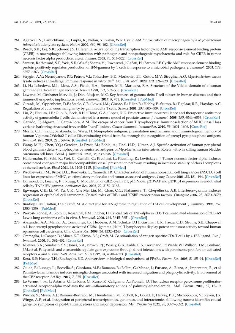
Int. J. Mol. Sci. 2021, 22, 12938 38 of 40
261. Agarwal, N.; Lamichhane, G.; Gupta, R.; Nolan, S.; Bishai, W.R. Cyclic AMP intoxication of macrophages by a Mycobacteriumtuberculosis adenylate cyclase. Nature 2009, 460, 98–102. [CrossRef]
262. Roach, S.K.; Lee, S.B.; Schorey, J.S. Differential activation of the transcription factor cyclic AMP response element binding protein(CREB) in macrophages following infection with pathogenic and nonpathogenic mycobacteria and role for CREB in tumornecrosis factor alpha production. Infect. Immun. 2005, 73, 514–522. [CrossRef]
263. Samten, B.; Howard, S.T.; Weis, S.E.; Wu, S.; Shams, H.; Townsend, J.C.; Safi, H.; Barnes, P.F. Cyclic AMP response element-bindingprotein positively regulates production of IFN-gamma by T cells in response to a microbial pathogen. J. Immunol. 2005, 174,6357–6363. [CrossRef]
264. Strygin, A.V.; Nesmiyanov, P.P.; Petrov, V.I.; Tolkachev, B.E.; Morkovin, E.I.; Gutov, M.V.; Strygina, A.O. Mycobacterium vaccaelysate induces anti-allergic immune response in vitro. Bull. Exp. Biol. Med. 2020, 170, 226–229. [CrossRef]
265. Li, H.; Lebedeva, M.I.; Llera, A.S.; Fields, B.A.; Brenner, M.B.; Mariuzza, R.A. Structure of the Vdelta domain of a humangammadelta T-cell antigen receptor. Nature 1998, 391, 502–506. [CrossRef]
266. Lawand, M.; Dechanet-Merville, J.; Dieu-Nosjean, M.C. Key features of gamma-delta T-cell subsets in human diseases and theirimmunotherapeutic implications. Front. Immunol. 2017, 8, 761. [CrossRef] [PubMed]
267. Girardi, M.; Oppenheim, D.E.; Steele, C.R.; Lewis, J.M.; Glusac, E.; Filler, R.; Hobby, P.; Sutton, B.; Tigelaar, R.E.; Hayday, A.C.Regulation of cutaneous malignancy by gammadelta T cells. Science 2001, 294, 605–609. [CrossRef]
268. Liu, Z.; Eltoum, I.E.; Guo, B.; Beck, B.H.; Cloud, G.A.; Lopez, R.D. Protective immunosurveillance and therapeutic antitumoractivity of gammadelta T cells demonstrated in a mouse model of prostate cancer. J. Immunol. 2008, 180, 6044–6053. [CrossRef]
269. Garrido, F.; Algarra, I.; García-Lora, A.M. The escape of cancer from T lymphocytes: Immunoselection of MHC class I lossvariants harboring structural-irreversible “hard” lesions. Cancer Immunol. Immunother. 2010, 59, 1601–1606. [CrossRef]
270. Morita, C.T.; Jin, C.; Sarikonda, G.; Wang, H. Nonpeptide antigens, presentation mechanisms, and immunological memory ofhuman Vgamma2Vdelta2 T cells: Discriminating friend from foe through the recognition of prenyl pyrophosphate antigens.Immunol. Rev. 2007, 215, 59–76. [CrossRef] [PubMed]
271. Wang, M.H.; Chen, Y.Q.; Gercken, J.; Ernst, M.; Bohle, A.; Flad, H.D.; Ulmer, A.J. Specific activation of human peripheralblood gamma/delta + lymphocytes by sonicated antigens of Mycobacterium tuberculosis: Role in vitro in killing human bladdercarcinoma cell lines. Scand. J. Immunol. 1993, 38, 239–246. [CrossRef]
272. Hallermalm, K.; Seki, K.; Wei, C.; Castelli, C.; Rivoltini, L.; Kiessling, R.; Levitskaya, J. Tumor necrosis factor-alpha inducescoordinated changes in major histocompatibility class I presentation pathway, resulting in increased stability of class I complexesat the cell surface. Blood 2001, 98, 1108–1115. [CrossRef] [PubMed]
273. Wroblewski, J.M.; Bixby, D.L.; Borowski, C.; Yannelli, J.R. Characterization of human non-small cell lung cancer (NSCLC) celllines for expression of MHC, co-stimulatory molecules and tumor-associated antigens. Lung Cancer 2001, 33, 181–194. [CrossRef]
274. Dormond, O.; Lejeune, F.J.; Ruegg, C. Modulation of cdk2, cyclin D1, p16INK4a, p21WAF and p27Kip1 expression in endothelialcells by TNF/IFN gamma. Anticancer Res. 2002, 22, 3159–3163.
275. Egwuagu, C.E.; Li, W.; Yu, C.R.; Che Mei Lin, M.; Chan, C.C.; Nakamura, T.; Chepelinsky, A.B. Interferon-gamma inducesregression of epithelial cell carcinoma: Critical roles of IRF-1 and ICSBP transcription factors. Oncogene 2006, 25, 3670–3679.[CrossRef]
276. Bradley, L.M.; Dalton, D.K.; Croft, M. A direct role for IFN-gamma in regulation of Th1 cell development. J. Immunol. 1996, 157,1350–1358. [PubMed]
277. Prevost-Blondel, A.; Roth, E.; Rosenthal, F.M.; Pircher, H. Crucial role of TNF-alpha in CD8 T cell-mediated elimination of 3LL-A9Lewis lung carcinoma cells in vivo. J. Immunol. 2000, 164, 3645–3651. [CrossRef]
278. Alexander, A.A.; Maniar, A.; Cummings, J.S.; Hebbeler, A.M.; Schulze, D.H.; Gastman, B.R.; Pauza, C.D.; Strome, S.E.; Chapoval,A.I. Isopentenyl pyrophosphate-activated CD56+ {gamma}{delta} T lymphocytes display potent antitumor activity toward humansquamous cell carcinoma. Clin. Cancer Res. 2008, 14, 4232–4240. [CrossRef]
279. Gramaglia, I.; Cooper, D.; Miner, K.T.; Kwon, B.S.; Croft, M. Co-stimulation of antigen-specific CD4 T cells by 4-1BB ligand. Eur. J.Immunol. 2000, 30, 392–402. [CrossRef]
280. Kliewer, S.A.; Sundseth, S.S.; Jones, S.A.; Brown, P.J.; Wisely, G.B.; Koble, C.S.; Devchand, P.; Wahli, W.; Willson, T.M.; Lenhard,J.M.; et al. Fatty acids and eicosanoids regulate gene expression through direct interactions with peroxisome proliferator-activatedreceptors α andγ. Proc. Natl. Acad. Sci. USA 1997, 94, 4318–4323. [CrossRef]
281. Kota, B.P.; Huang, T.H.; Roufogalis, B.D. An overview on biological mechanisms of PPARs. Pharm. Res. 2005, 51, 85–94. [CrossRef][PubMed]
282. Guida, F.; Luongo, L.; Boccella, S.; Giordano, M.E.; Romano, R.; Bellini, G.; Manzo, I.; Furiano, A.; Rizzo, A.; Imperatore, R.; et al.Palmitoylethanolamide induces microglia changes associated with increased migration and phagocytic activity: Involvement ofthe CB2 receptor. Sci Rep. 2017, 7, 375. [CrossRef]
283. Lo Verme, J.; Fu, J.; Astarita, G.; La Rana, G.; Russo, R.; Calignano, A.; Piomelli, D. The nuclear receptor peroxisome proliferator-activated receptor-alpha mediates the anti-inflammatory actions of palmitoylethanolamide. Mol. Pharm. 2005, 67, 15–19.[CrossRef] [PubMed]
284. Wuchty, S.; Myers, A.J.; Ramirez-Restrepo, M.; Huentelman, M.; Richolt, R.; Gould, F.; Harvey, P.D.; Michopolous, V.; Steven, J.S.;Wingo, A.P.; et al. Integration of peripheral transcriptomics, genomics, and interactomics following trauma identifies causalgenes for symptoms of post-traumatic stress and major depression. Mol. Psychiatry 2021, 26, 3077–3092. [CrossRef]

Int. J. Mol. Sci. 2021, 22, 12938 39 of 40
285. Flesch, I.E.; Hess, J.H.; Huang, S.; Aguet, M.; Rothe, J.; Bluethmann, H.; Kaufmann, S.H. Early interleukin 12 production bymacrophages in response to mycobacterial infection depends on interferon gamma and tumor necrosis factor alpha. J. Exp. Med.1995, 181, 1615–1621. [CrossRef]
286. Zuany-Amorim, C.; Manlius, C.; Trifilieff, A.; Brunet, L.R.; Rook, G.; Bowen, G.; Pay, G.; Walker, C. Long-term protective andantigen-specific effect of heat-killed Mycobacterium vaccae in a murine model of allergic pulmonary inflammation. J. Immunol.2002, 169, 1492–1499. [CrossRef]
287. Akbari, O.; DeKruyff, R.H.; Umetsu, D.T. Pulmonary dendritic cells producing IL-10 mediate tolerance induced by respiratoryexposure to antigen. Nat. Immunol. 2001, 2, 725–731. [CrossRef]
288. Akbari, O.; Freeman, G.J.; Meyer, E.H.; Greenfield, E.A.; Chang, T.T.; Sharpe, A.H.; Berry, G.; DeKruyff, R.H.; Umetsu, D.T.Antigen-specific regulatory T cells develop via the ICOS-ICOS-ligand pathway and inhibit allergen-induced airway hyperreactiv-ity. Nat. Med. 2002, 8, 1024–1032. [CrossRef]
289. Reber, S.O.; Langgartner, D.; Foertsch, S.; Postolache, T.T.; Brenner, L.A.; Guendel, H.; Lowry, C.A. Chronic subordinate colonyhousing paradigm: A mouse model for mechanisms of PTSD vulnerability, targeted prevention, and treatment-2016 Curt RichterAward Paper. Psychoneuroendocrinology 2016, 74, 221–230. [CrossRef]
290. Park, H.J.; Shim, H.S.; An, K.; Starkweather, A.; Kim, K.S.; Shim, I. IL-4 Inhibits IL-1beta-induced depressive-like behavior andcentral neurotransmitter alterations. Mediat. Inflamm. 2015, 2015, 941413. [CrossRef] [PubMed]
291. Wachholz, S.; Knorr, A.; Mengert, L.; Plumper, J.; Sommer, R.; Juckel, G.; Friebe, A. Interleukin-4 is a participant in the regulationof depressive-like behavior. Behav. Brain Res. 2017, 326, 165–172. [CrossRef]
292. Doenni, V.M.; Song, C.M.; Hill, M.N.; Pittman, Q.J. Early-life inflammation with LPS delays fear extinction in adult rodents. BrainBehav. Immun. 2017, 63, 176–185. [CrossRef]
293. Jones, M.E.; Lebonville, C.L.; Barrus, D.; Lysle, D.T. The role of brain interleukin-1 in stress-enhanced fear learning. Neuropsy-chopharmacology 2015, 40, 1289–1296. [CrossRef]
294. Parekh, S.V.; Paniccia, J.E.; Lebonville, C.L.; Lysle, D.T. Dorsal hippocampal interleukin-1 signaling mediates heroin withdrawal-enhanced fear learning. Psychopharmacology 2020, 237, 3653–3664. [CrossRef] [PubMed]
295. Quiñones, M.M.; Maldonado, L.; Velazquez, B.; Porter, J.T.J.B. Candesartan ameliorates impaired fear extinction induced byinnate immune activation. Brain Behav. Immun. 2016, 52, 169–177. [CrossRef] [PubMed]
296. Young, M.B.; Howell, L.L.; Hopkins, L.; Moshfegh, C.; Yu, Z.; Clubb, L.; Seidenberg, J.; Park, J.; Swiercz, A.P.; Marvar, P.J. A periph-eral immune response to remembering trauma contributes to the maintenance of fear memory in mice. Psychoneuroendocrinology2018, 94, 143–151. [CrossRef]
297. Huang, C.Y.; Hsieh, W.Y. Efficacy of Mycobacterium vaccae immunotherapy for patients with tuberculosis: A systematic reviewand meta-analysis. Hum. Vaccines Immunother. 2017, 13, 1960–1971. [CrossRef] [PubMed]
298. Giacomini, E.; Iona, E.; Ferroni, L.; Miettinen, M.; Fattorini, L.; Orefici, G.; Julkunen, I.; Coccia, E.M. Infection of humanmacrophages and dendritic cells with Mycobacterium tuberculosis induces a differential cytokine gene expression that modulates Tcell response. J. Immunol. 2001, 166, 7033–7041. [CrossRef]
299. Sugawara, I.; Yamada, H.; Mizuno, S.; Takeda, K.; Akira, S. Mycobacterial infection in MyD88-deficient mice. Microbiol. Immunol.2003, 47, 841–847. [CrossRef] [PubMed]
300. Wright, K.M.; Friedland, J.S. Regulation of monocyte chemokine and MMP-9 secretion by proinflammatory cytokines intuberculous osteomyelitis. J. Leukoc. Biol. 2004, 75, 1086–1092. [CrossRef] [PubMed]
301. Deguine, J.; Barton, G.M. MyD88: A central player in innate immune signaling. F1000prime Rep. 2014, 6, 97. [CrossRef]302. Cervantes, J.L. MyD88 in Mycobacterium tuberculosis infection. Med. Microbiol. Immunol. 2017, 206, 187–193. [CrossRef]303. Gu, X.; Gao, Y.; Mu, D.G.; Fu, E.Q. MiR-23a-5p modulates mycobacterial survival and autophagy during mycobacterium
tuberculosis infection through TLR2/MyD88/NF-kappaB pathway by targeting TLR2. Exp. Cell Res. 2017, 354, 71–77. [CrossRef]304. Coombes, J.L.; Powrie, F. Dendritic cells in intestinal immune regulation. Nat. Rev. Immunol. 2008, 8, 435–446. [CrossRef]
[PubMed]305. Kim, D.-Y.; Sato, A.; Fukuyama, S.; Sagara, H.; Nagatake, T.; Kong, I.G.; Goda, K.; Nochi, T.; Kunisawa, J.; Sato, S.; et al. The
airway antigen sampling system: Respiratory M cells as an alternative gateway for inhaled antigens. J. Immunol. 2011, 186,4253–4262. [CrossRef] [PubMed]
306. Ruane, D.; Brane, L.; Reis, B.S.; Cheong, C.; Poles, J.; Do, Y.; Zhu, H.; Velinzon, K.; Choi, J.H.; Studt, N.; et al. Lung dendritic cellsinduce migration of protective T cells to the gastrointestinal tract. J. Exp. Med. 2013, 210, 1871–1888. [CrossRef]
307. Unger, W.W.; Hauet-Broere, F.; Jansen, W.; van Berkel, L.A.; Kraal, G.; Samsom, J.N. Early events in peripheral regulatory T cellinduction via the nasal mucosa. J. Immunol. 2003, 171, 4592–4603. [CrossRef]
308. Huang, C.T.; Workman, C.J.; Flies, D.; Pan, X.; Marson, A.L.; Zhou, G.; Hipkiss, E.L.; Ravi, S.; Kowalski, J.; Levitsky, H.I.; et al.Role of LAG-3 in regulatory T cells. Immunity 2004, 21, 503–513. [CrossRef]
309. McMenamin, C.; Schon-Hegrad, M.; Oliver, J.; Girn, B.; Holt, P.G. Regulation of IgE responses to inhaled antigens: Cellularmechanisms underlying allergic sensitization versus tolerance induction. Int. Arch. Allergy Appl. Immunol. 1991, 94, 78–82.[CrossRef]
310. Perdomo, C.; Zedler, U.; Kuhl, A.A.; Lozza, L.; Saikali, P.; Sander, L.E.; Vogelzang, A.; Kaufmann, S.H.; Kupz, A. Mucosal BCGvaccination induces protective lung-resident memory T cell populations against tuberculosis. MBio 2016, 7, e01686-16. [CrossRef][PubMed]

Int. J. Mol. Sci. 2021, 22, 12938 40 of 40
311. Iwasaki, A.; Kelsall, B.L. Freshly isolated Peyer’s patch, but not spleen, dendritic cells produce interleukin 10 and induce thedifferentiation of T helper type 2 cells. J. Exp. Med. 1999, 190, 229–239. [CrossRef] [PubMed]
312. Polanski, M.; Melican, N.S.; Zhang, J.; Weiner, H.L. Oral administration of the immunodominant B-chain of insulin reducesdiabetes in a co-transfer model of diabetes in the NOD mouse and is associated with a switch from Th1 to Th2 cytokines.J. Autoimmun. 1997, 10, 339–346. [CrossRef] [PubMed]
313. Groux, H.; O’Garra, A.; Bigler, M.; Rouleau, M.; Antonenko, S.; de Vries, J.E.; Roncarolo, M.G. A CD4+ T-cell subset inhibitsantigen-specific T-cell responses and prevents colitis. Nature 1997, 389, 737–742. [CrossRef]
314. Chen, Y.; Kuchroo, V.K.; Inobe, J.; Hafler, D.A.; Weiner, H.L. Regulatory T cell clones induced by oral tolerance: Suppression ofautoimmune encephalomyelitis. Science 1994, 265, 1237–1240. [CrossRef] [PubMed]
315. Matthews, D.M.; Jenks, S.M. Ingestion of Mycobacterium vaccae decreases anxiety-related behavior and improves learning in mice.Behav. Process. 2013, 96, 27–35. [CrossRef]
316. Cervantes-Barragan, L.; Chai, J.N.; Tianero, M.D.; Di Luccia, B.; Ahern, P.P.; Merriman, J.; Cortez, V.S.; Caparon, M.G.; Donia,M.S.; Gilfillan, S.; et al. Lactobacillus reuteri induces gut intraepithelial CD4+ CD8alphaalpha+ T cells. Science 2017, 357, 806–810.[CrossRef]
317. Sun, C.M.; Hall, J.A.; Blank, R.B.; Bouladoux, N.; Oukka, M.; Mora, J.R.; Belkaid, Y. Small intestine lamina propria dendritic cellspromote de novo generation of Foxp3 T reg cells via retinoic acid. J. Exp. Med. 2007, 204, 1775–1785. [CrossRef] [PubMed]
318. Coombes, J.L.; Siddiqui, K.R.; Arancibia-Carcamo, C.V.; Hall, J.; Sun, C.M.; Belkaid, Y.; Powrie, F. A functionally specializedpopulation of mucosal CD103+ DCs induces Foxp3+ regulatory T cells via a TGF-beta and retinoic acid-dependent mechanism.J. Exp. Med. 2007, 204, 1757–1764. [CrossRef]
319. Mucida, D.; Cheroutre, H. TGFbeta and retinoic acid intersect in immune-regulation. Cell Adhes. Migr. 2007, 1, 142–144. [CrossRef]320. Mucida, D.; Park, Y.; Kim, G.; Turovskaya, O.; Scott, I.; Kronenberg, M.; Cheroutre, H. Reciprocal TH17 and regulatory T cell
differentiation mediated by retinoic acid. Science 2007, 317, 256–260. [CrossRef]321. Crooks, J.; Brown, S.; Gauthier, A. The effects of combination treatment of IMM-101, a heat-killed whole cell preparation of
Mycobacterium obuense (NCTC 13365) with checkpoint inhibitors in pre-clinical models. In Proceedings of the Annual Meetingof the SITC 2016, National Harbor, MD, USA, 11–13 November 2016; pp. 9–13.
322. Dominguez-Andres, J.; Joosten, L.A.; Netea, M.G. Induction of innate immune memory: The role of cellular metabolism. Curr.Opin. Immunol. 2019, 56, 10–16. [CrossRef]
323. Netea, M.G. Training innate immunity: The changing concept of immunological memory in innate host defence. Eur. J. Clin.Investig. 2013, 43, 881–884. [CrossRef]
324. Netea, M.G.; Quintin, J.; van der Meer, J.W. Trained immunity: A memory for innate host defense. Cell Host Microbe 2011, 9,355–361. [CrossRef] [PubMed]
325. Dalgleish, A.G.; Mudan, S.; Fusi, A. Enhanced effect of checkpoint inhibitors when given after or together with IMM-101:Significant responses in four advanced melanoma patients with no additional major toxicity. J. Transl. Med. 2018, 16, 227.[CrossRef]
326. Bazzi, S.; Modjtahedi, H.; Mudan, S.; Achkar, M.; Akle, C.; Bahr, G.M. Immunomodulatory effects of heat-killed Mycobacteriumobuense on human blood dendritic cells. Innate Immun. 2017, 23, 592–605. [CrossRef] [PubMed]
327. Crooks, J.; Brown, S.; Forss, C.; Phythian-Adams, A.; Cook, P.; Brunet, L.R.; MacDonald, A. The impact of Mycobacterium obuenseon innate and adaptive immunity. Eur. J. Cancer 2016, 1, S72. [CrossRef]
328. Galdon, A.; Crooks, J.; Brown, S.L.; Kampinga, J.; Brunet, L.R.; MacDonald, A.S. Defining the immunomodulatory effectsof IMM-101: A promising, novel co-therapy for cancer. In Proceedings of the CRI-CIMT-EATI-AACR International CancerImmunotherapy Conference, Paris, France, 25–28 September 2019.
329. ClinicalTrials.gov Immunization with IMM-101 vs. Observation for Prevention of Respiratory and Severe COVID-19 RelatedInfections in Cancer Patients at Increased Risk of Exposure (COV-IMMUNO): ClinicalTrials.gov Identifier: NCT04442048.Available online: https://clinicaltrials.gov (accessed on 28 July 2021).
330. Tsukamura, M.; Mizuno, S. Mycobacterium obuense, a rapidly growing scotochromogenic mycobacterium capable of forming ablack product from p-aminosalicylate and salicylate. J. Gen. Microbiol. 1971, 68, 129–134. [CrossRef] [PubMed]
331. Stone, B.B.; Nietupski, R.M.; Breton, G.L.; Weisburg, W.G. Comparison of Mycobacterium 23S rRNA sequences by high-temperaturereverse transcription and PCR. Int. J. Syst. Bacteriol. 1995, 45, 811–819. [CrossRef]
332. Postolache, T.T.; Benros, M.E.; Brenner, L.A. Targetable Biological Mechanisms Implicated in Emergent Psychiatric ConditionsAssociated with SARS-CoV-2 Infection. JAMA Psychiatry 2020. Available online: https://www.amedeolucente.it/public/Targetable%20Biological%20Mechanisms%20Implicated%20in%20Emergent%20Psychiatric%20Conditions%20Associated%20With%20SARS-CoV-2%20Infection.pdf (accessed on 28 July 2021).