Mycobacterium Tuberculosis-Associated Uveitis: Infection and ...
100
Mycobacterium Tuberculosis-Associated Uveitis: Infection and Autoimmunity Rina La Distia Nora
-
Upload
khangminh22 -
Category
Documents
-
view
3 -
download
0
Transcript of Mycobacterium Tuberculosis-Associated Uveitis: Infection and ...
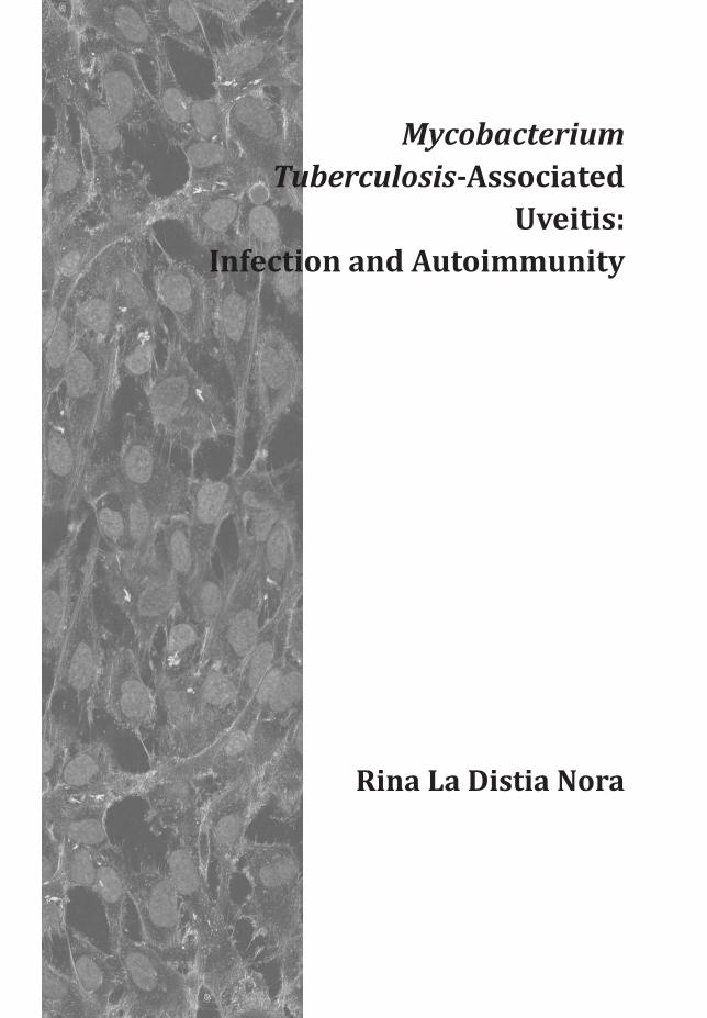
Mycobacterium Tuberculosis-Associated
Uveitis: Infection and Autoimmunity
Rina La Distia Nora

Mycobacterium Tuberculosis-Associated Uveitis: Infection and Autoimmunity
Mycobacterium tuberculosis-geassocieerde uveïtis:
infectie en auto-immuniteit
Proefschriftter verkrijging van de graad van doctor aan de
Erasmus Universiteit Rotterdam op gezag van de
rector magnificus
Prof.dr. R.C.M.E. Engels
en volgens besluit van het College voor Promoties.
De openbare verdediging zal plaatsvinden op woensdag 20 juni 2018 om 13.30 uur
Rina La Distia Norageboren te Banda Aceh, Indonesia
No parts of this thesis may be reproduced or transmitted in any form by any means, electronic or mechanical, including photocopying, recording or any information storage and retrieval system, without permission in writing from the author. The research for this thesis was performed within the framework of the Erasmus MC Postgraduate School Molecular Medicine. The studies described in this thesis were performed at the Laboratory for Medical Immunology, Department of Immunology, Erasmus MC, Rotter-dam, the Netherlands. Patients cohort was recruited in Ophthalmology Department Faculty of Medicine Universitas Indonesia/ Cipto Mangunku-sumo Hospital Kirana and Pulmonology Department Faculty of Medicine Universitas Indonesia/ Persahabatan Hospital, Jakarta, Indonesia.This PhD program was supported by the Indonesian Ministry of Research Technology and Higher Education.
ISBN: 978-94-91811-20-3Cover and invitation photo: Rina La Distia Nora and Mariëlle C. Haks
and Kimberley V. Walburg (the retinal pigment epithelium photo)Cover, invitation and thesis lay-out design: Rina La Distia Nora and Uti Nilam SariThesis lay-out: Daniëlle KorpershoekPrinting: Haveka BV, Hendrik-Ido-Ambacht
Copyright © 2018 by Rina La Distia Nora. All rights reserved. No part of this book may be reproduced, stored in a retrieval system or transmitted in any form or by any means, without prior permission of the author.

PROMOTIECOMMISSIE
Promotoren: Prof.dr. P.M. van Hagen Prof.dr. A. Rothova
Overige leden: Prof.dr. P.J. van der Spek Prof.dr. J.H. de Boer Prof.dr. T.H.M. Ottenhoff Copromotor: Dr. W.A. Dik
So verily, with every difficulty, comes ease. (the Quran 94:6)

CHAPTER 5 149Summary and discussion
APPENDIX 163Abbreviations 165Samenvatting 169Acknowledgments 173Curriculum Vitae 177PhD Portfolio 179Publications 183
CONTENTS
CHAPTER 1 9General introductionTuberculosis and its immunopathogenesisUveitis Uveitis in the spectrum of tuberculosis
CHAPTER 2 45Aims of the thesis
CHAPTER 3Uveitis related to Mycobcaterium tuberculosis in Indonesia and the Netherlands
3.1 Clinical manifestations of patients with intraocular inflammation 51 and positive QuantiFERON-TB gold in-tube test in a country non-endemic for tuberculosis Am J Ophthalmol 2014 Apr;157(4):754-61
3.2 Tuberculosis and other causes of uveitis in Indonesia 69 Eye 2018 Mar;32(3):546-554
CHAPTER 4Pathogenesis of Uveitis related to Mycobcaterium tuberculosis: infection or autoimmunity?
4.1 Retinal pigment epithelial cells control early Mycobacterium 91 tuberculosis infection via interferon signaling Invest Ophthalmol Vis Sci 2018; 59(3):1384-1395
4.2 Type 1 interferon-inducible gene expression in QuantiFERON 119 Gold TB-positive uveitis: a tool to stratify a high versus low risk of active tuberculosis? Submitted
4.3 Antiretinal antibodies in QuantiFERON Gold TB-positive patients 143 with unknown cause uveitis Accepted in Acta Ophthalmol


.
1ChapterGeneral Introduction
Tuberculosis and its immunopathogenesis Uveitis
Uveitis in the spectrum of tuberculosis

1
11
General Introduction
TUBERCULOSIS AND ITS IMMUNOPATHOGENESIS
Tuberculosis (TB) is an airborne infectious disease caused by microorganisms of the Mycobacterium tuberculosis (Mtb) complex, and mainly affects the lungs. Although primarily a pulmonary pathogen, Mtb can affect almost all organs. The World Health Organization (WHO) estimates that one-third of the world’s population is infected with Mtb, but only ten percent of infected persons develop clinical manifestations of TB. Of this 10%, 16%-27% have extrapulmonary TB involvement including the eye (Mtb-associated uveitis: 1.4% in the United States (US) before the 1990s and 18% in Spain recently).1, 2 Risk factors for extrapulmonary TB are: age over forty, female gender, and human immu-nodeficiency virus (HIV) infection.3 In many low-income and middle-income countries, TB continues to be a major cause of morbidity and mortality, and drug-resistant TB is a significant concern in many settings.
Clinical manifestations and diagnosisMtb mainly affects the lungs, therefore, an active pulmonary TB is characterized by
a chronic cough, fever, hemoptysis and general symptoms such as appetite loss, night sweats and sustained weight loss.4 Clinical manifestations of active TB infection in other organs will depend on the location of the infection.
The gold standard for diagnosing active TB infection is sputum smear microscopy and culture in liquid medium with subsequent drug-susceptibility testing. Sputum smear microscopy has many limitations, but it continues to be widely used in limited facility settings. Sputum smear microscopy will detect active TB from the first sputum smear test in only 75% in HIV-negative individuals and even in lower percentage in HIV-positive individuals (57%).5 Thus, the WHO recommended a PCR based test: Xpert MTB/RIF (Cepheid Inc., Sunnyvale, California, USA) which has better accuracy than sputum smear microscopy with a sensitivity of 90% in HIV-negative individuals and up to 77% in HIV-positive individuals.6
Radiography imaging of the chest (chest X-ray/CXR) is useful in diagnosing pulmo-nary TB in clinically suspected cases that could not be confirmed with microbiologi-cal testing or as a screening tool, especially in HIV positive individuals. As a screening tool, CXR has a higher sensitivity for pulmonary TB than screening for TB symptoms. However, CXR lacks specificity thus, the pulmonary TB diagnosis based on CXR needs to be followed up clinically and by microbiological tests.7
Exposure to Mtb will lead to a memory immune response by T lymphocytes (T cells). If a patient has memory T cells that have been exposed to Mtb previously, he/she will have an in vivo response to Mycobacterium antigen subcutaneously; the tuberculin skin test (TST) and in vitro from blood; as in QuantiERON-Gold TB tests (QFTs). However,
1
Chapter 1
12 13
General Introduction
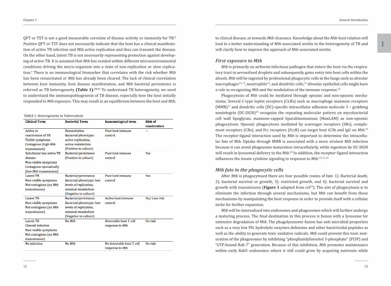
QFT or TST is not a good measurable correlate of disease activity or immunity for TB.8 Positive QFT or TST does not necessarily indicate that the host has a clinical manifesta-tion of active TB infection and Mtb active replication and thus can transmit the disease. On the other hand, latent TB is not necessarily representing protection against develop-ing of active TB. It is assumed that Mtb has resided within different microenvironmental conditions driving the micro-organism into a state of non-replication or slow replica-tion.9 There is no immunological biomarker that correlates with the risk whether Mtb has been resuscitated or Mtb has already been cleared. The lack of clinical correlation between host immunity, host disease manifestation, and Mtb bacterial persistence is referred as TB heterogeneity (Table 1).10,11 To understand TB heterogeneity, we need to understand the immunopathogenesis of TB disease, especially how the host initially responded to Mtb exposure. This may result in an equilibrium between the host and Mtb,
TABLE 1. Heterogeneity in Tuberculosis
to clinical disease, or towards Mtb clearance. Knowledge about the Mtb-host relation will lead to a better understanding of Mtb-associated uveitis in the heterogeneity of TB and will clarify how to improve the approach of Mtb-associated uveitis.
First exposure to MtbMtb is primarily an airborne infectious pathogen that enters the host via the respira-
tory tract in aerosolized droplets and subsequently gains entry into host cells within the alveoli. Mtb will be ingested by professional phagocytic cells in the lungs such as alveolar macrophages12, 13, neutrophils14, and dendritic cells.15 Alveolar epithelial cells might have a role in recognizing Mtb and the modulation of the immune response.16
Phagocytosis of Mtb could be mediated through opsonic and non-opsonic mecha-nisms. Several C-type leptin receptors (CLRs) such as macrophage mannose receptors (MMR)17 and dendritic cells (DC)-specific intracellular adhesion molecule 3 – grabbing nonintegrin (DC-SIGN)18 recognize the repeating molecular pattern on mycobacterial cell wall lipoglycan, mannose-capped liparabinomannan (ManLAM) as non-opsonic phagocytosis. Opsonic phagocytosis mediated by scavenger receptors (SRs), comple-ment receptors (CRs), and Fcγ receptors (FcγR) can target host iC3b and IgG on Mtb.19 The receptor-ligand interaction used by Mtb is important to determine the intracellu-lar fate of Mtb. Uptake through MMR is associated with a more virulent Mtb infection because it can avoid phagosome maturation intracellularly, while ingestion by DC-SIGN will result in lysosomal delivery to the Mtb.20 In addition, the receptor-ligand interaction influences the innate cytokine signaling in response to Mtb.19, 21, 22
Mtb fate in the phagocytic cellsAfter Mtb is phagocytosed there are four possible routes of fate: 1). Bacterial death,
2). bacterial survival or growth, 3). restricted growth, and 4). bacterial survival and growth with transmission (Figure 1 adapted from ref19). The aim of phagocytosis is to eliminate the infection through several mechanisms, but Mtb can benefit from those mechanisms by manipulating the host response in order to provide itself with a cellular niche for further expansion.
Mtb will be internalized into endosomes and phagosomes which will further undergo a maturing process. The final destination in this process is fusion with a lysosome for extensive degradation of Mtb. The phagolysosome fusion has anti-microbial properties such as a very low PH, hydrolytic enzymes defensins and other bactericidal peptides as well as the ability to generate toxic oxidative radicals. Mtb could prevent this toxic mat-uration of the phagosomes by inhibiting “phosphatidylinositol 3-phosphate” (PI3P) and “GTP-bound Rab 7” generation. Because of this inhibition, Mtb promotes maintenance within early Rab5 endosomes where it still could grow by acquiring nutrients while

1
Chapter 1
14 15
General Introduction
avoiding the acidic degradative environment.23 PI3P generation is also needed in auto-phagy, and its disruption will impair the clearance of Mtb by autophagy as well. However, appropriate phagolysosome formation will not always result in impaired bacterial repli-cation. Mtb can respond, resist, and even persist in the moderately acid environment of the phagosome or phagolysosome.24
Although Mtb could survive inside of macrophages, Mtb infected macrophages can also be activated to increase their killing capacity by specific cytokines such as IFNγ, tumor necrosing factor (TNF)α25, and other factors such as vitamin D.26-32 However, Mtb virulence factor 6 kDa early secretory antigenic target (ESAT-6) secretion system 1 (ESX1) enables escape from the phagosome and subsequent replication in the cyto-sol inducing cell death via necrosis.33-37 Cell necrosis will aid cell-to-cell spread of Mtb. Furthermore, Mtb infected macrophages can express DC markers and may actively exit the site of infection leading to hematogenous dissemination from the primary infection site in the early infection state.38, 39
Figure 1. Mtb fates in phagocytic cells.Ideally Mtb within Rab5 early endosome will mature to Rab7 phagolysosome and experience bacterial death. Several Mtb may survive and are able to grow within the phagolysosome. Mtb is able to prevent phagosome maturation by inhibiting phosphatidylinositol 3-phosphate (PI3P) generation in the phagosome and impairing recruitment of active GTP-bound Rab7. They will stay in Rab5 low PI3P and able to grow and further transmit Mtb. IFNγ, TNFα and vitamin D will enhance the killing activity within the cell and further induce bacterial death or restricted growth. Through Mtb virulence factor ESX1, Mtb could damage the membrane and access the cytosol. Mtb could escape and further grow and transmit or be resequestered by the ubiquitin system (adapted from ref19 with permission).
Innate immune response to MtbMtb is recognized by multiple pattern-recognition-receptors (PRRs), among them
are Toll-like receptors (TLRs) and CLRs on the cell membrane and intra-cellular and, the nucleotide-binding oligomerization domain (NOD)-like receptors/NLRs and reti-noic acid-inducible gene (RIG)-I-like receptors (RLRs) in the cytoplasm.40 These initial steps to phagocytosis are of evident importance, based on studies with toll-like recep-tor (TLR)2 and TLR9 single-knockout mice or TLR2/TLR9 double-knock out mice. The double-knock out mice are more susceptible to Mtb than the single knock-out mice.41 Moreover, in knock-out mice, second messengers that could coordinate upstream stimu-lation of TLRs, such as myeloid differentiation primary response 88 (MyD88), make the mice even more susceptible.42, 43
Downstream PRR activation will regulate innate immune responses and also initiates the adaptive immune response. PRR activation results in production of pro-inflammatory cytokines, including TNFα and interleukin (IL)-1β through activation of nuclear factor kappa-light-chain-enhancer of activated B cells (NF-κB). This process increases macro-phage effector functions including intracellular killing, and local/systemic immune cell mobilization and activation.44 Mtb has evolved multiple mechanisms to evade phagocyte killing and eventually subvert and delay the initiation of the adaptive immune response (as discussed below).
Type 1 InterferonsType 1 interferons (IFNs) are proteins that may “interfere” with intracellular infec-
tions. Their expression is primarily induced by cytoplasmic PRRs, and endosomal TLRs and can further activate certain interferon regulatory factors (IRFs) resulting in inter-feron-inducible gene expression.45 The role of type I IFN in Mtb infection has recently emerged by the findings of type 1 IFN inducible gene expression in active pulmonary TB patients, whose differential gene expression could differentiate them from healthy controls and other diseases such as sarcoidosis and pneumonia.46-49 This type 1 IFN-inducible gene expression (also referred to as Type 1 IFN signature) normalizes in response to successful anti-tuberculosis treatment (ATT).46, 47 Type 1 IFN signature also correlates with the risk of progression from latent TB infection to active TB infection.50
Despite mounting evidence about the role of type 1 IFNs in active TB, the exact mech-anism in the pathogenesis of TB is not fully understood. Mtb stimulated cytosolic PRRs induce type 1 IFNs, which lead to IFN-β production in an IRF3-dependent way. This is turn, induces CCL2/CCR2-dependent migration of Mtb permissive inflammatory macro-phages (iM) and inflammatory dendritic cells (iDC) to the lung.51 Within these cells, type 1 IFNs will change the arachidonic acid metabolism which renders the cells more sus-ceptible to necrotic death.52 Additionally, type 1 IFNs limits IL-1β mediated neutrophil

1
Chapter 1
16 17
General Introduction
activation to reduce excessive inflammation and associated damage. This type 1 IFN mediated inhibition acts through IL-10, which does however require that permissive cells have been primed by IFNγ.53, 54
Granuloma formationHistopathologically, Mtb is known to induce granuloma formation, these granulomas
are organized immune cell aggregates developing in response to persistent stimuli.55 It was previously thought that granuloma formation correlated with Mtb sequestration and elimination by host immune cells. Granuloma formation is the pathological basis for latent Mtb infection.56
A granuloma starts as a collection of mature macrophages in response to the per-sistent stimulus of Mtb. Mature macrophages are assumed to be more phagocytic and microbicidal with their increased cytoplasmic size, larger numbers of organelles, and ruffled cell membranes. These mature macrophages also underwent fusion into multinu-cleated giant cells. Some of the mature macrophages can transform into epithelioid cells which have tightly interdigitated cell membranes in zipper-like arrays that link adjacent cells. Within this cellular context are areas of necrosis due to cell death, including mac-rophages. In gross pathology studies, this is known as caseum. Other cells involved in granuloma formation are neutrophils, DCs, B cells, T cells, natural killer cells (NK-cells), fibroblasts, and epithelial cells.56 With these characteristics, it was long believed that granulomas play an important role in controlling the infection thereby protecting the host. But, studies in animal models show that in the early phase of granuloma formation Mtb growth is still rapid and reaches a plateau until adaptive immunity develops, gen-erating mature granulomas.57, 58 Moreover, in developing zebra fish with still a morpho-logically and functionally immature adaptive immunity, granuloma formation coincided with accelerated Mtb proliferation.59, 60 In a live imaging study in zebra fish, macrophage reaction depends on Mtb virulent factor ESX-1 secretion (encoded by region of difference (RD) 1 virulence locus). RD1 deficient Mtb will have fewer macrophages attracted to the granuloma, their morphology is more rounded, and they move more slowly. In conse-quence, this will coincide with poor granuloma formation, and limited Mtb proliferation and dissemination within the host. In contrast, in more RD1 positive virulent Mtb, there is rapid and continuous migration of the macrophages to the granuloma site. Besides the signal from RD1 positive Mtb, there will be a second signal generated by infected yet dying macrophages to attract additional macrophages to phagocytose the contents of dying infected macrophages. As a result higher numbers of infected cells will be found.61 Moreover, ESAT6 peptide secreted by ESX-1 positive Mtb, could induce macrophage-in-dependent MMP9 secretion from epithelial cells surrounding the granuloma to act as a chemoattractant for macrophages. MMP9-knockout mice have decreased macrophage
recruitment to the lung and poor granuloma formation upon Mtb infection, and con-sequently, it is associated with decreased bacterial loads.62 The host forms granulomas to concentrate the host defenses in an organized immune structure but Mtb alters it (facilitates) as a safe niche for its survival and growth and delay adaptive immunity.
Adaptive immune responses to Mtb and the “Immune Equilibrium”
Adaptive immune responses require the transport of live bacteria from lungs to the draining lymph nodes by dendritic cells (DCs) which takes 8-10 days after Mtb infection (in comparison, only 20 hours for influenza virus).15, 63 Besides the delay of presenting Mtb to T cells, adaptive immune responses are also hindered due to delayed arrival of effector T cells by regulatory T cells (Tregs) 64 and delayed activation of effector T cells65, 66 Therefore, it is possible that early granuloma formation is used by Mtb to evade the host immune system.56
The adaptive immune response is dependent on IL-12 (p40/p35) which is mainly secreted by Mtb-activated DCs through a TLR-dependent mechanism. In the lymph node, migrated DCs will drive naïve T cells to differentiate towards a T helper cells (Th) 1 phenotype. Protective antigen-specific Th1 cells will migrate back to the lung and pro-duce IFNγ a major cytokine in granuloma formation. IFNγ leads to activation, cytokine production, the induction of anti-microbial factors and bacterial control. This protective immune response is mainly mediated by CD4+ Th1 cells, but other cells also play a role in producing IFNγ; such as CD8+, NK cells, γδ T cells, and CD1-restricted cells.21
The adaptive immune response will arrest the progressive growth of the bacterial population but its ability to eliminate Mtb is limited. Immunocompetent mice which have been infected with virulent strains of Mtb will maintain a plateau population of bacteria with accumulation of effector CD4+ and CD8+ T cells in the lungs until the mice die.67 This phenomenon, together with the fact that CD4+ T cell-deficient mice or CD4+ T cell lymphopenic HIV patients are highly susceptible to TB, illustrates the crucial role of an appropriate immune response to maintain the immune equilibrium against Mtb activation.22
Reactivation of latent TB from immune equilibrium.As mentioned above, the best known mechanism for TB reactivation was observed
in the profound CD4+ T cell depletion in HIV/ acquired immune deficiency syndrome (AIDS) patients.68 Similar findings were found in simian immunodeficiency virus (SIV) infection in non-human primates and CD4+ T cells depleted-mice during the chronic stage of Mtb infection.65, 69 Recent knowledge was also obtained from the phe-nomenon of TB reactivation in patients treated with TNF neutralizing biologics. TNF-blocking treatment inhibits the induction of pro-inflammatory cytokines by blocking

1
Chapter 1
18 19
General Introduction
TNF-mediated NFκB activation and further diminishes anti-mycobacterial activity of macrophages.25, 70, 71 Medical conditions such as diabetes mellitus and treatment with corticosteroids are also associated with Mtb reactivation.72,73 Other possible mechanisms that might cause reactivation are 1) T cell exhaustion74-76, 2) altered antigen expression by Mtb 77, 78 , 3) decreased migratory capacity of specific myeloid and lymphoid cells 79, and 4) resuscitation-promoting factors from the Mtb itself 10.
UVEITIS
Uveitis is an inflammatory disease involving the uvea, the highly vascularized and pigmented middle layer of the eye located between the retina and sclera. The uvea is composed of the iris, ciliary body, and choroid (Figure 2). All the tissues surrounding the uveal tract, including the optic nerve, retina, vitreous, and sclera can also be involved in the inflammatory process, either secondary or primary, and therefore, the name uve-itis is usually used to indicate any type of intraocular inflammation.
Uveitis is considered as an uncommon disease with an incidence of approximately 52 persons per year per 100.000 individuals and prevalence of 115/100.000 person in developed countries.80 The incidence and prevalence of uveitis are probably much higher in developing countries, however there are no precise numbers available.
Uveitis is an important cause of visual impairment and blindness In the western world; uveitis results in visual impairment in approximately 35% and blindness in 5%-10% of cases.81 In developing countries the blindness due to uveitis reaches up to 25%.82
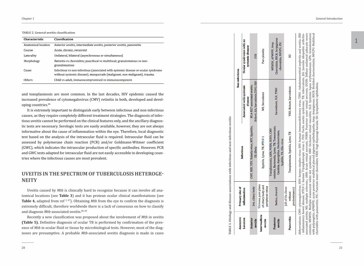
Uveitis can be classified in several ways as summarized in Table 2. Since the causal classification is not always feasible, especially in the initial phase of the inflammation, the anatomical uveitis classification gained most popularity. Anatomical classification of uveitis differentiates into anterior uveitis, intermediate uveitis and posterior uveitis. Furthermore, when uveitis comprises all parts of the uvea it is designated as panuveitis (Figure 2). Uveitis classification is important for diagnostic and therapeutic purposes as well as for the prognosis. Table 3 summarizes the relation of anatomical classification of uveitis with infectious and systemic non-infectious diseases and ocular syndromes (adapted from ref 83-85).
Worldwide, infectious uveitis is probably the most common uveitis entity. Infections are most frequent in the developing countries, with the prevalence of infectious uve-itis between 30% and 50%.81 In contrast, prevalence of infectious uveitis in the devel-oped countries is substantially lower, namely between 11% and 21%.81 Identification of the causative micro-organism is critical because the treatment with appropriate
antimicrobial agents might decrease inflammatory activity and even cures. In developing countries, the most common intraocular infections include tuberculosis and toxoplas-mosis. Other less common infections causing uveitis are leprosy, leptospirosis, oncho-cerciasis, cysticercosis, and trypanosomiasis.81 In developed countries, viral infections
Figure 2. Anatomical location of uveitis.The eye consists of three coats; outer (sclera and cornea), middle (uvea), and inner coat (retina). Inflammatory cells and localization Inflammation within the eye determine the “anatomic classification” diagnosis of uveitis. Anterior uveitis is predominantly affecting anterior chamber, intermediate uveitis is affecting predominantly vitreous and peripheral retina, and posterior uveitis is predominantly affecting retina and choroid. Panuveitis has no predominance and affect all part of the uvea. The structure adjacent to uvea; cornea, sclera, and optic nerve, are usually also affected either primarily or secondarily.
1
Chapter 1
20 21
General Introduction
and toxoplasmosis are most common. In the last decades, HIV epidemic caused the increased prevalence of cytomegalovirus (CMV) retinitis in both, developed and devel-oping countries.86
It is extremely important to distinguish early between infectious and non-infectious causes, as they require completely different treatment strategies. The diagnosis of infec-tious uveitis cannot be performed on the clinical features only, and the ancillary diagnos-tic tests are necessary. Serologic tests are easily available, however, they are not always informative about the cause of inflammation within the eye. Therefore, local diagnostic test based on the analysis of the intraocular fluid is required. Intraocular fluid can be assessed by polymerase chain reaction (PCR) and/or Goldmann-Witmer coefficient (GWC), which indicates the intraocular production of specific antibodies. However, PCR and GWC tests adapted for intraocular fluid are not easily accessible in developing coun-tries where the infectious causes are most prevalent.
UVEITIS IN THE SPECTRUM OF TUBERCULOSIS HETEROGE-NEITY
Uveitis caused by Mtb is clinically hard to recognize because it can involve all ana-tomical locations (see Table 3) and it has protean ocular clinical manifestations (see Table 4, adapted from ref 3, 87). Obtaining Mtb from the eye to confirm the diagnosis is extremely difficult, therefore worldwide there is a lack of consensus on how to classify and diagnose Mtb-associated uveitis.88, 89
Recently a new classification was proposed about the involvement of Mtb in uveitis (Table 5). Definitive diagnosis of ocular TB is performed by confirmation of the pres-ence of Mtb in ocular fluid or tissue by microbiological tests. However, most of the diag-noses are presumptive. A probable Mtb-associated uveitis diagnosis is made in cases
TABLE 2. General uveitis classification
TABL
E 3.
Eti
olog
y an
d di
seas
e-as
soci
atio
ns w
ith
infe
ctio
us a
nd n
on-in
fect
ious
uve
itis
Abbr
evia
tions
: CM
V: c
ytom
egal
ovir
us; H
SV: h
erpe
s si
mpl
ex v
irus
; HIV
: hum
an im
mun
odef
icie
ncy
viru
s; T
INU
: tub
uloi
nter
stiti
al n
ephr
itis
and
uvei
tis; I
BD
: inf
lam
mat
ory
bow
el d
isea
se; H
TLV-
1: h
uman
T-ly
mph
otro
pic
viru
s-1;
FUS
: Fuc
hs u
veiti
s sy
ndro
me;
TB;
tub
ercu
losi
s; JI
A: Ju
veni
le id
iopa
thic
art
hriti
s-as
soci
ated
uve
itis;
MS:
mul
tiple
scl
eros
is;;
ARN
: Acu
te r
etin
al n
ecro
sis;
POR
N: P
oste
rior
out
er r
etin
al n
ecro
sis;
DUS
N: D
iffus
e un
ilate
ral s
ubac
ute
neur
o re
tiniti
s; M
EWDS
: Mul
tiple
eva
nesc
ent
whi
te d
ot s
yndr
ome;
ORV
: Occ
lusi
ve r
etin
al v
ascu
litis
; SLE
: Sys
tem
ic lu
pus
eryt
hem
atos
us; G
PA: G
ranu
lom
atos
is
with
pol
yang
iitis
; APM
PPE:
acu
te p
oste
rior
mul
tifoc
al p
laco
id p
igm
ent e
pith
elio
path
y; A
mp:
Am
pigi
nous
; BSC
R: B
irds
hot c
hori
oret
initi
s; M
FCPU
: Mul
tifoc
al
chor
oidi
tis w
ith p
anuv
eitis
; PIC
: Pun
ctat
e in
ner c
horo
iditi
s; V
KH; V
ogt-
Koya
nagi
-har
ada;
SO:
Sym
path
etic
oph
thal
mia
.

1
Chapter 1
22 23
General Introduction
with immunological evidence of Mtb infection complemented with a microbiologically proven active Mtb infection from another organ or a chest radiographic imaging highly suspected of active TB (reviewed in 4, 90). A possible Mtb-associated uveitis diagnosis is made when uveitis is associated with a host immune response to Mtb antigen (QFT or TST positive latent TB infection without other evidence of clinically active TB; Table 5, adopted from ref91). In addition, possible Mtb-associated uveitis can be diagnosed only after other causes of uveitis were excluded. The infection of the eye by Mtb is thought to play a role in both definitive and probable TB-associated uveitis. The pathogenesis of possible Mtb-associated uveitis (uveitis of unknown cause with positive QFT and no clinical evidence of TB infection) is not yet clear. Depending on the geographical loca-tion, the association of IGRA with uveitis might be coincidental or might be either due to infection or immune reaction or both. Sometimes, the clinical response of uveitis to ATT (six until 12 months) is being used as additional criterion for the involvement of Mtb in the pathogenesis.87
In the clinical practice, a patient that fulfills the criteria of possible Mtb-associated uveitis poses a treatment dilemma to the clinician. As the ATT is characterized by its long-term use and might be associated with adverse effects, the decision to treat or not to treat should not be taken lightly. QFT and/or TST positivity indicates that the patient has been ever infected by Mtb; however, the level of QFT or induration area of TST are not associated with the probability of active TB disease nor with enhanced immunity against Mtb.8 Positive QFT and/or TST do not indicate that the host has active Mtb repli-cation. Unfortunately, so far, there is no marker that indicates the risk whether Mtb has been resuscitated or Mtb has already been cleared. Availability of such a measure would be of great value to guide optimal treatment for the above indicated problematic uveitis group.
TABLE 4. Possible clinical manifestations of uveitis associated with Mtb
TABL
E 5.
Cla
ssifi
cati
on o
f con
firm
ed o
cula
r TB
and
pre
sum
ed M
tb-a
ssoc
iate
d uv
eiti
s
* Rev
iew
ed in
4, 9
0

1
Chapter 1
24 25
General Introduction
EpidemiologyIn the western world, TB is an infrequent cause of ocular infection. Data of Mtb-
associated uveitis vary across the region and across time. Around the 1940s, TB was estimated to account for 80% of granulomatous uveitis cases. This decreased to 22% in the 1960s due to better knowledge of other possible causes of uveitis, including toxo-plasmosis and sarcoidosis.1, 92, 93 The percentage declined further in the decades after-ward to around 1.4% to 18%.2, 81, 94-102 However, recent increase of Mtb-associated uveitis was noted and is probably related to HIV infection, immigration from TB endemic areas, new immunosuppressive treatments, inadequate TB control, and increased drug resis-tance.87 Epidemiological data differ between TB endemic and non-endemic countries. In non-endemic countries such as the US, Italy, and Japan, the prevalence rates of Mtb-associated uveitis range between lower than 1% to 7%. In endemic countries such as in Asia and Africa, the prevalence rate of what ranges between 4% to 10%.81 In “middle” endemic countries such as Saudi Arabia and Singapore; the prevalence rate is still quite high: 6%-18%.95, 100, 102, 103 These differences are not only related to differences in disease burden but also due to variations in diagnostic strategies and classifications used.104 The prevalence of latent TB is high in endemic countries and association of latent TB and uveitis might be due to coincidence. Therefore, it is difficult to confirm whether a latent TB is related to the ocular inflammation and decide whether an individual patient should be treated.
Clinical manifestations of Mtb-associated uveitis Mtb-associated uveitis is considered a great mimicker of other uveitis entities and
may affect all anatomical locations. The typical clinical features involve granuloma formation located on iris or in the irido-corneal angle, ciliary body, choroid, occasionally leading to subretinal abscess formation, endophthalmitis, and panophthalmitis. All these features are assumed to be due to direct infection. However, there are several clinical manifestations that may be related to an immune-mediated mechanism such as the serpiginous or serpiginoid choroiditis, occlusive retinal vasculitis, and anterior uveitis.87 The described features and/or the most frequently reported clinical manifestations should prompt the clinician to check for TB. Also, an unspecific clinical manifestation with chronic or recurrent course, active despite the immunosuppressive treatment can be related to Mtb.
Anterior uveitis accounts for approximately 12%-36% of Mtb-associated uveitis.3 Besides the typical features of iris or angle granuloma and ciliary body tubercles, broad-based posterior synechiae were reported to be associated with Mtb-associated uveitis.105 Other ocular manifestations that have been reported in relation to TB are mutton fat keratic precipitates and less frequently hypopyon and iris atrophy. 3, 106, 107
Intermediate uveitis accounts for about 11% of Mtb-associated uveitis.3 The clini-cal manifestations usually comprise a low-grade, smoldering chronic vitritis, snow-ball opacities, snow banking, peripheral vascular sheathing and peripheral choroidal granuloma.87
Posterior uveitis is the most common clinical presentation, comprising 35%-42% cases of Mtb-associated uveitis.3 The clinical features in posterior Mtb-associated uveitis manifestations include multifocal choroiditis108, retinitis (usually concomitantly with choroiditis)87, serpiginous-like choroiditis109-112, tubercles113, 114, tuberculomas113, 115-120, subretinal abscesses (occur as a result of liquefaction necrosis in caseating granulomas and can develop in patients with disseminated TB),121 occlusive retinal vasculitis and other vasculopathies.122-124
Panuveitis is seen in 11%-20% of Mtb-associated uveitis cases.125 Mtb-associated panuveitis might primarily develop and have aspecific features or may progress from a severe form of posterior or intermediate uveitis.87 Generalized inflammation that has an acute onset and shows rapid progression with the destruction of intraocular tissues can manifest as endophthalmitis and panophthalmitis.87
PATHOGENESIS OF MTB-ASSOCIATED UVEITIS
The exact pathogenesis of “Mtb-associated uveitis” is so far unclear. Is it caused by bacterial spread to the eye and subsequent replication? And in what degree is the immune system involved? Several pathophysiological models were reported.126 We postulated four possible pathological mechanisms that may explain the pathogenesis of “Mtb-associated uveitis”.
1. Direct Mtb infection of the retina2. Mtb induces a specific anti-retinal immune process due to antigenic mimicry3. Latent Mtb immuno-surveillance induces a general higher level of immune cell
(monocytic and CD4+ T cell) activation that results in uveitis in predisposed individuals
4. Combination of the above postulated mechanismsPathogenesis of Mtb-associated uveitis is probably related to pathogenesis of hetero-
geneous manifestations of TB infection as indicated above (Table 1). In extreme cases of TB infection such as in lymphohematogenous dissemination of Mtb (miliary TB)127, there are reports of granulomas in choroid that responded to ATT like in other infected organs.128 There is a possibility that macrophages from Mtb-laden granuloma in foci in the lungs exit and spread and directly infected the eye.129 Guinea pigs infected with aerosolized Mtb developed uveal granulomatous lesions containing acid-fast staining

1
Chapter 1
26 27
General Introduction
positive organisms.130 The risk of ocular involvement is also increased in HIV-positive patients 2, and their clinical manifestations are granulomatous and are more likely to be caused by a direct infection with Mtb because as there was an evidence of infection in other organs as well.131
In latent TB however, the presence and/or activity of Mtb in the body might vary according to bacterial and immunological functions (Table 1). A histopathological study from eyes with proven presence of Mtb, revealed that systemic TB infection is not always found.132, 133 The authors reported that Mtb is distributed mainly in RPE cells and only one or two bacilli were found associated with a near giant cell or an area of necrosis.132 These lesions generated vascular endothelial growth factor (VEGF) expression in the neuroretina and retinal pigment epithelium (RPE).134 These studies showed RPE cells have an important role in Mtb-associated uveitis. An in vitro study showed that RPE cells were able to phagocytose Mtb with comparable efficacy as a monocytic cell line (THP-1) but with higher Mtb survival rates.135 This suggests that RPE cells could be a niche for Mtb and may be a locus where reactivation or reinfection could begin. RPE cells might have a role in sequestering Mtb as in granuloma formation in the lung or lymph node. RPE cells might also have a role in regulating the immune equilibrium thus controlling the inflammation. This hypothesis is supported by a study in which Mtb genome was found in sub-retinal fluids of rhegmatogenous retinal detachment patients that were without any signs of uveitis in either eye.136
Due to the difficulty to obtain Mtb from ocular tissues in presumed Mtb-associated uveitis cases, it has been hypothesized that Mtb-associated uveitis might be caused by autoimmune reactions rather than by an infection itself. Mtb has nearly 500 experimentally verified human T cell epitopes which may stimulate the immune system.137 There are numerous studies on autoimmune uveitis in animal models.138 The first experimental autoimmune uveitis (EAU), which could be consistently induced, was obtained by subcutaneous inoculation of inter-photoreceptor retinal binding protein (IRBP) emulsified in oil and complete Freund’s adjuvant (CFA).139, 140 CFA consist of dried extract Mtb strain H37Ra which will stimulate the PRRs and activate innate immune responses and drives differentiation of retina-specific T cells (by inter-photoreceptor retinoid binding protein, IRBP) in a pro-inflammatory type.141 This type of activated autoreactive T cells was found in peripheral blood, but these cells need to pass blood-retinal barrier to induce uveitis. In an experiment of adoptive transfer of naïve “hen egg lysozyme” (HEL)-specific CD4+ T-cells into transgenic mice expressing HEL, under a retina-specific promoter, showed proliferation of those specific T cells in the draining eye lymph nodes, without inducing any uveitis. However, if the same mice model was infected systemically with murine cytomegalovirus (MCMV) engineered to express HEL, there was proliferation of transferred naïve CD4+ T cells and induction of uveoretinitis, while
this was not observed when wild-type MCMV was used. Therefore, ocular autoantigens likely will not induce uveitis if not presented in the context of inflammation, which may be induced by infection.142
Autoreactive T cells toward retinal crude extract (RCE) were observed in vitreous samples of Mtb-associated uveitis, these cells were resistant to activation-induced cell death which might contribute to the inflammation. Even though the T-cells were poly-functional after stimulation with Mtb antigen ESAT6 and RCE (which was not found in non-Mtb-associated uveitis models), the population of retinal auto-reactive T-cells was different from the population of Mtb reactive T cells in terms of their sensitivity to acti-vation-induced cell death.143
Another study in a Bacille-Calmette-Guerin (BCG)-induced bilateral granulomatous anterior uveitis, peripheral blood T-cell proliferation upon exposure to retinal antigens and purified protein derivative (PPD) was found.144 The authors reported there was amino acid sequence homology between proteins from Mtb, BCG and retinal antigens, hence antigenic mimicry as the cause of uveitis may be possible.144
As a more general mechanism; it can be speculated that pulmonary TB or latent TB in an adult immunocompetent patient causes a state of a hyperactive anti-mycobacterial immune response. This may increase anti-retinal immunity in patients with a pre-exis-tent subclinical immune response against retina and may hypothetically induce uveitis. Increased delayed-type hypersensitivity to TB antigens was associated with a more likelihood to suffer from active disease or to develop it.145, 146 Increased IFNγ production was also found in the peripheral blood, lung tissue, bronchoalveolar lavage (BAL) fluid, pleural effusion and lymph nodes of active TB patients, which decreased upon treat-ment.147, 148 Moreover, several gene expression studies also implicate an increase in type I IFN activity and type 1 IFN regulated genes in active pulmonary TB patients or latent TB cases that eventually develop active pulmonary TB.46, 47, 149, 150 Therefore, in combination with antigenic mimicry, the hyperactive anti-mycobacterial immune response may also play a role.
“Immune recovery uveitis” occurs in HIV-positive patients with TB who develop immune reconstitution after starting antiretroviral therapy.151 The findings in “immune recovery uveitis” can vary widely and include anterior uveitis, hypopyon, vitritis, papil-litis, panuveitis, retinal or optic disc neovascularization, retinal detachment, cystoid macular edema, epiretinal membrane formation, vitreomacular traction, and macular hole.152 These findings are aspecific and can be observed in diverse uveitis entities.
Animal EAU demonstrates that uveitis is driven by Th17 or Th1 dominated immune response, the predominant pathway will be determined by the antigen exposure. CFA drives more Th17 response while in vitro antigen-pulsed DCs drives Th1 response.141 B cells contribute to the adaptive immune response towards TB and are abundant in

1
Chapter 1
28 29
General Introduction
tertiary lymphoid structure (TLS) formed during TB infection. TLS formation is further supported by IL23, and IL-17.153 B cell(-/-) mice display more severe immunopathology toward Mtb infection as evidenced by elevated recruitment of neutrophils, higher pro-duction of IL-10, and higher bacterial burden in the lung, which all can be inverted by adoptive transfer of B cells.154 Autoreactive B-cells might develop from TLS under the influence of type 1 IFN and B-cell activating factor (BAFF) in Th17 driven autoimmune disease.155 Although there were findings of intra-retinal TLS formation in EAU animal model156, generally little is known about B cell involvement in uveitis.154
Challenges in diagnosis and management of Mtb-associated uveitisThe diagnostic procedures for Mtb-associated uveitis are frequently not conclusive,
which implies the need to rule out other causes of uveitis. The identification of Mtb in ocular fluids represents a challenge; most techniques are unfortunately either unreli-able or not sensitive enough.
• Ocular Mtb staining and cultureObtaining Mtb directly from the eye as the gold standard for the diagnosis of Mtb-
associated uveitis is often not feasible because of the sensitivity limits of the staining and culture procedures. For positive microbiological conformation a certain minimal number of bacilli is required that is at least 1 x 10⁴. bacilli/ml for acid-fast staining of sputum and 1 x 10² bacilli/ml for bacterial culture.157 These are the requirements for the regular diagnostic tools for sputum in patients with active pulmonary TB. Yet, this amount is difficult to obtain in ocular TB due to the small volume and likely minimal bacterial load in intraocular fluids.
• Diagnostic PCRAlthough the latest nucleic acid amplification test through PCR can detect as mini-
mal as ten femtogram (fg) of DNA which equals to 2-3 tubercle bacilli, this test reveals variable results in intraocular fluid and is not commonly performed in diagnosing of ocular TB.157-159 The reasons for this unreliable PCR results in intraocular fluid include the non-uniform distribution of Mtb inside the eye and the paucibacillary nature of Mtb infection. Histopathological studies found that only 1-2 Mtb bacilli could be found in the retinal pigment epithelium layer in the posterior part of the eye and already cause severe panuveitis.133
There are several other caveats in using PCR as diagnosis tool for Mtb-associated uveitis. First, as already stated, the location of tissue sampling may also affect the results of PCR. A nested PCR approach for the gene target MPB-64 showed 78% sensitivity from epiretinal membranes as obtained by vitrectomy, while this decreased to 21% when the
test was performed from a vitreous biopsy.160, 161 PCR in anterior chamber fluid from possible and probable ocular TB cases obviously also showed a low yield of positivity.162 Second, different Mtb gene targets will give variable results. Some Mtb strains have zero or low copy numbers of certain gene targets (e.g. IS6110), therefore, a multi-tar-get gene (IS6110 + MPB-64 or IS6110 + 38 kDa + MPB-64) PCR approach will improve the sensitivity.158, 163 Lastly, PCR does not differentiate between the viable and nonviable microorganisms.
• Immunological testingAlthough active pulmonary Mtb infection is supposed to be easier to diagnose, there
is still a problem in sputum smear-negative TB with and without positive radiological findings. At least, exposure to Mtb can be tested by measuring the T-cell immunologi-cal response to Mtb through QFT or TST. Unfortunately, these tests cannot differentiate between an active and a latent Mtb infection. They have a low positive predictive value (PPV) and high negative predictive value (NPV) in sputum-negative, culture-proven active pulmonary TB.164-166 The low PPV and high NPV were also found in Mtb-associated uveitis cases (PPV 71% and NPV 95%).167 These Mtb-associated uveitis cases were based on criteria by Gupta et al., which include a 4-6 weeks ATT treatment observation.87 The low PPV of QFT does not solve the problem how to treat QFT-positive uveitis of unknown cause. There is a need for additional tests and biomarkers to further stratify that group of patients to optimize treatment choice.
Recent reports demonstrate a differential type-1 IFN signature in patients with active pulmonary Mtb infection and patients with latent infection.149 Both, the activation of the type-1 IFN cascade and its downstream genes present a characteristic pattern in immune cells. Until now, no studies are available that linked type-1 IFN-regulated gene expression in peripheral blood of patients with uveitis and Mtb infection.
Increasing number of studies reporting on the association between positive TST and/or QFT and uveitis were reported.168-176 Moreover, TST and/or QFT-positive uve-itis patients may benefit from complete ATT.177 The reason for this is not entirely clear, but might be related to minimal numbers of Mtb bacilli within the retina that cause retinitis.87 Alternatively, the immunological response to mycobacteria may generate an immune response that is autoreactive to retinal antigens.
As mentioned above, there is still a diagnostic problem in sputum smear-negative TB with and without positive radiological findings. A T-cell immunological response to Mtb peptides as measured by QFT or TST cannot differentiate active from latent TB infec-tion. Moreover, the tests can be positive in a certain proportion of healthy individuals; e.g., India (23%)178, Thailand (17%)179, USA (5%)180, Denmark (4.5%)181, and Tanzania (41%)182. Due to these ambiguities, the diagnosis and management of Mtb-associated

1
Chapter 1
30 31
General Introduction
uveitis may differ between different parts in the world. The choice of first-line diagnostic tools also differs: TST is preferable over QFT in low-income countries. Chest CT is often ordered in high-income countries to detect subtle changes and to detect other common non-infectious diseases such as sarcoidosis.
In conclusion, the evaluation of systemic and ocular signs and symptoms in patients
suspected from Mtb-associated uveitis represents a first important step in diagnosing Mtb-associated uveitis.159 In contrast to the classical findings of granulomatous inflam-mation, in latent TB, a broad spectrum of novel ocular manifestations was recently rec-ognized, which includes broad-based synechiae, serpiginous-like retinitis, and retinal occlusive vasculitis, features previously not linked to TB infection.105, 162, 174 There is an urgent need for an early and more conclusive diagnostic procedure for Mtb-associated uveitis.
MANAGEMENT
ATT is the mainstay treatment for Mtb-associated uveitis.87, 183, 184 However, there are no guidelines for the commencement and duration of ATT in patients with uveitis in the setting of positive QFT.88, 89 The present guidelines mainly suggest that confirmed ocular TB warrants ATT treatment. On the other hand, the diagnosis of Mtb-associated uveitis is presumptive in the majority of patients due to the lack of microbiological proof. In Mtb-associated uveitis, ATT administration is even a part of diagnosis criteria; a decrease of inflammation after 4-6 weeks of ATT was considered to support the diagnosis.87 Beside subsiding inflammation, reduced recurrences after the treatment will also facilitate the diagnosis. This management approach, is desirable for patients at risk of losing the vision as the diagnostic uncertainty and/or delay may lead to permanent vision loss.185
Recommended regimens for drug-susceptible pulmonary TB from the World Health Organization (WHO) is 6-month rifampicin-based regimen (2 months of isoniazid (H), rifampicin (R), pyrazinamide (Z) and ethambutol (E), and four months of H and R). Daily dosing is still recommended for uveitis patients in initial and continuation phase.186 However, there is a wide variation in the type of drugs, regimen, and duration of treat-ment for ocular TB or Mtb-associated uveitis. Initial RHZE regimens varied from a mini-mum of 2 months up to 3-4 months and subsequent HR regimens varied from 4 months up to 15 months.183 The problems with the multi-drug regimen and the long duration of ATT are the toxicity, poor compliance of the patient and possible drug-resistant Mtb induction and in some cases also the costs.187 There are many side effects reported from ATT, the most concerning one is toxic optic neuropathy associated with ethambutol.188
It is a rare but vision-threatening condition, which is ironic because the drug is used for regaining visual acuity from inflammation due to Mtb.
Variations also exist in the use of systemic corticosteroids and other immunosuppres-sive treatments. The use of oral corticosteroids in low-dose during the first 4-6 weeks along with ATT may be beneficial to limit tissue destruction. However, a systematic liter-ature review revealed no differences in clinical outcome of patients with and without use of corticosteroids 183 The use of corticosteroids alone, without ATT, was inconsistently reported as being both detrimental and beneficial.189, 190 Probably, the initial concomitant use of corticosteroids or other immunosuppressives could bias the true benefit of ATT as well. Some cases, such as for example serpiginoid choroiditis attributed to TB, were diffi-cult to manage by ATT solely, without addition of corticosteroids or immunosuppressive drugs.111 Even after the full ATT was finished, 15/136 (11%) of the patients needed addi-tional corticosteroid therapy.191 It was also shown that some patients suspected from Mtb-associated uveitis reached remission without ATT, and used only corticosteroids.190 The decision to use corticosteroids or corticosteroids-sparing agents must be made very carefully and monitored in well-equipped expert centers.
The beneficial effect of ATT is expected from its anti-microbial properties, which will eliminate Mtb as the inciting agent of uveitis. In a systematic review of 28 studies includ-ing 1917 patients suspected from Mtb-associated uveitis, it was concluded that the use of ATT benefits Mtb-associated uveitis with or without concomitant systemic corticoste-roids. However, there was no control group included, no standardized recruitment and treatment protocol.183 Additionally, there might be a publication bias toward the good results of ATT.
There is a possibility that ATT might have direct anti-inflammatory properties. In a study of experimental autoimmune encephalomyelitis (EAE), rifampicin treatment associated with suppression of inflammation acts on demyelination in spinal cords of EAE mice, through inhibition of Th17 cell differentiation.192 In another study with mice suffering from autoinflammatory atopic dermatitis induced by 1-chloro 2,4-dinitroben-zene (DNCB), rifampicin treatment tapered the dermatitis symptoms. Rifampicin was associated with the suppression of β-hexosaminidase and histamine from human mast cell (HMC)-1 cells, and therefore inhibited the secretion of inflammatory mediators by mast cells (TNF-α and PGD2). Though, there is no data yet how strong the anti-inflamma-tory properties of ATT are uveitis cases.
There are still many questions left about the immunopathogenesis of Mtb-associated uveitis. In the future, better knowledge on the pathophysiology will help the clinician to diagnose and to treat Mtb-associated uveitis patients in the most safe and effective way.

1
Chapter 1
32 33
General Introduction
REFERENCES
1. Donahue HC. Ophthalmologic experience in a tuberculosis sanatorium. Am J Ophthalmol 1967;64:742-
748.
2. Bouza E, Merino P, Munoz P, Sanchez-Carrillo C, Yanez J, Cortes C. Ocular tuberculosis. A prospective
study in a general hospital. Medicine (Baltimore) 1997;76:53-61.
3. Dalvin LA, Smith WM. Intraocular manifestations of mycobacterium tuberculosis: A review of the
literature. J Clin Tuberc Other Microbact Dis 2017;7:13-21.
4. Pai M, Behr MA, Dowdy D, et al. Tuberculosis. Nat Rev Dis Primers 2016;2:16076.
5. Leonard MK, Osterholt D, Kourbatova EV, Del Rio C, Wang W, Blumberg HM. How many sputum spec-
imens are necessary to diagnose pulmonary tuberculosis? Am J Infect Control 2005;33:58-61.
6. Dorman SE, Schumacher SG, Alland D, et al. Xpert MTB/RIF Ultra for detection of Mycobacterium
tuberculosis and rifampicin resistance: a prospective multicentre diagnostic accuracy study. Lancet
Infect Dis 2018;18:76-84.
7. World Health Organization. Chest radiography in tuberculosis detection — summary of current WHO
recommendations and guidance on programmatic approaches. Switzerland: WHO Press; 2016.
8. Petruccioli E, Scriba TJ, Petrone L, et al. Correlates of tuberculosis risk: predictive biomarkers for
progression to active tuberculosis. Eur Respir J 2016;48:1751-1763.
9. Barry CE, 3rd, Boshoff HI, Dartois V, et al. The spectrum of latent tuberculosis: rethinking the biology
and intervention strategies. Nat Rev Microbiol 2009;7:845-855.
10. Veatch AV, Kaushal D. Opening Pandora’s Box: Mechanisms of Mycobacterium tuberculosis Resuscita-
tion. Trends Microbiol 2018;26:145-157.
11. Cadena AM, Fortune SM, Flynn JL. Heterogeneity in tuberculosis. Nat Rev Immunol 2017;17:691-702.
12. Schlesinger LS. Entry of Mycobacterium tuberculosis into mononuclear phagocytes. Curr Top Micro-
biol Immunol 1996;215:71-96.
13. Skold M, Behar SM. Tuberculosis triggers a tissue-dependent program of differentiation and acquisi-
tion of effector functions by circulating monocytes. J Immunol 2008;181:6349-6360.
14. Eum SY, Kong JH, Hong MS, et al. Neutrophils are the predominant infected phagocytic cells in the
airways of patients with active pulmonary TB. Chest 2010;137:122-128.
15. Wolf AJ, Linas B, Trevejo-Nunez GJ, et al. Mycobacterium tuberculosis infects dendritic cells with high
frequency and impairs their function in vivo. J Immunol 2007;179:2509-2519.
16. Scordo JM, Knoell DL, Torrelles JB. Alveolar Epithelial Cells in Mycobacterium tuberculosis Infection:
Active Players or Innocent Bystanders? J Innate Immun 2016;8:3-14.
17. Rajaram MVS, Arnett E, Azad AK, et al. M. tuberculosis-Initiated Human Mannose Receptor Signaling
Regulates Macrophage Recognition and Vesicle Trafficking by FcRgamma-Chain, Grb2, and SHP-1. Cell
Rep 2017;21:126-140.
18. Tailleux L, Schwartz O, Herrmann JL, et al. DC-SIGN is the major Mycobacterium tuberculosis receptor
on human dendritic cells. J Exp Med 2003;197:121-127.
19. Philips JA, Ernst JD. Tuberculosis pathogenesis and immunity. Annu Rev Pathol 2012;7:353-384.
20. Schlesinger LS, Kaufman TM, Iyer S, Hull SR, Marchiando LK. Differences in mannose receptor-medi-
ated uptake of lipoarabinomannan from virulent and attenuated strains of Mycobacterium tubercu-
losis by human macrophages. J Immunol 1996;157:4568-4575.
21. Cooper AM. Cell-mediated immune responses in tuberculosis. Annu Rev Immunol 2009;27:393-422.
22. Ernst JD. The immunological life cycle of tuberculosis. Nat Rev Immunol 2012;12:581-591.
23. Jeschke A, Haas A. Deciphering the roles of phosphoinositide lipids in phagolysosome biogenesis.
Commun Integr Biol 2016;9:e1174798.
24. Vandal OH, Nathan CF, Ehrt S. Acid resistance in Mycobacterium tuberculosis. J Bacteriol
2009;191:4714-4721.
25. Clay H, Volkman HE, Ramakrishnan L. Tumor necrosis factor signaling mediates resistance to myco-
bacteria by inhibiting bacterial growth and macrophage death. Immunity 2008;29:283-294.
26. Adams LB, Dinauer MC, Morgenstern DE, Krahenbuhl JL. Comparison of the roles of reactive oxygen
and nitrogen intermediates in the host response to Mycobacterium tuberculosis using transgenic
mice. Tuber Lung Dis 1997;78:237-246.
27. Chan J, Xing Y, Magliozzo RS, Bloom BR. Killing of virulent Mycobacterium tuberculosis by reactive
nitrogen intermediates produced by activated murine macrophages. J Exp Med 1992;175:1111-1122.
28. Ding AH, Nathan CF, Stuehr DJ. Release of reactive nitrogen intermediates and reactive oxygen inter-
mediates from mouse peritoneal macrophages. Comparison of activating cytokines and evidence for
independent production. J Immunol 1988;141:2407-2412.
29. Yuk JM, Shin DM, Lee HM, et al. Vitamin D3 induces autophagy in human monocytes/macrophages via
cathelicidin. Cell Host Microbe 2009;6:231-243.
30. Liu PT, Stenger S, Li H, et al. Toll-like receptor triggering of a vitamin D-mediated human antimicrobi-
al response. Science 2006;311:1770-1773.
31. Martineau AR, Wilkinson KA, Newton SM, et al. IFN-gamma- and TNF-independent vitamin D-induc-
ible human suppression of mycobacteria: the role of cathelicidin LL-37. J Immunol 2007;178:7190-
7198.
32. Miyakawa Y, Ratnakar P, Rao AG, et al. In vitro activity of the antimicrobial peptides human and rab-
bit defensins and porcine leukocyte protegrin against Mycobacterium tuberculosis. Infect Immun
1996;64:926-932.
33. Pym AS, Brodin P, Brosch R, Huerre M, Cole ST. Loss of RD1 contributed to the attenuation of the
live tuberculosis vaccines Mycobacterium bovis BCG and Mycobacterium microti. Mol Microbiol
2002;46:709-717.
34. Champion PA, Cox JS. Protein secretion systems in Mycobacteria. Cell Microbiol 2007;9:1376-1384.
35. Molloy A, Laochumroonvorapong P, Kaplan G. Apoptosis, but not necrosis, of infected monocytes is
coupled with killing of intracellular bacillus Calmette-Guerin. J Exp Med 1994;180:1499-1509.
36. Keane J, Remold HG, Kornfeld H. Virulent Mycobacterium tuberculosis strains evade apoptosis of
infected alveolar macrophages. J Immunol 2000;164:2016-2020.
1
Chapter 1
34 35
General Introduction
37. Wong KW, Jacobs WR, Jr. Mycobacterium tuberculosis exploits human interferon gamma to stimulate
macrophage extracellular trap formation and necrosis. J Infect Dis 2013;208:109-119.
38. Balasubramanian V, Wiegeshaus EH, Taylor BT, Smith DW. Pathogenesis of tuberculosis: pathway to
apical localization. Tuber Lung Dis 1994;75:168-178.
39. Chackerian AA, Alt JM, Perera TV, Dascher CC, Behar SM. Dissemination of Mycobacterium tuber-
culosis is influenced by host factors and precedes the initiation of T-cell immunity. Infect Immun
2002;70:4501-4509.
40. Liu CH, Liu H, Ge B. Innate immunity in tuberculosis: host defense vs pathogen evasion. Cell Mol Im-
munol 2017;14:963-975.
41. Bafica A, Scanga CA, Feng CG, Leifer C, Cheever A, Sher A. TLR9 regulates Th1 responses and co-
operates with TLR2 in mediating optimal resistance to Mycobacterium tuberculosis. J Exp Med
2005;202:1715-1724.
42. Mayer-Barber KD, Barber DL, Shenderov K, et al. Caspase-1 independent IL-1beta production is crit-
ical for host resistance to mycobacterium tuberculosis and does not require TLR signaling in vivo. J
Immunol 2010;184:3326-3330.
43. Scanga CA, Bafica A, Feng CG, Cheever AW, Hieny S, Sher A. MyD88-deficient mice display a profound
loss in resistance to Mycobacterium tuberculosis associated with partially impaired Th1 cytokine
and nitric oxide synthase 2 expression. Infect Immun 2004;72:2400-2404.
44. Hertz CJ, Kiertscher SM, Godowski PJ, et al. Microbial lipopeptides stimulate dendritic cell maturation
via Toll-like receptor 2. J Immunol 2001;166:2444-2450.
45. Ivashkiv LB, Donlin LT. Regulation of type I interferon responses. Nat Rev Immunol 2014;14:36-49.
46. Berry MP, Graham CM, McNab FW, et al. An interferon-inducible neutrophil-driven blood transcrip-
tional signature in human tuberculosis. Nature 2010;466:973-977.
47. Ottenhoff TH, Dass RH, Yang N, et al. Genome-wide expression profiling identifies type 1 interferon
response pathways in active tuberculosis. PLoS One 2012;7:e45839.
48. Maertzdorf J, Weiner J, 3rd, Mollenkopf HJ, et al. Common patterns and disease-related signatures in
tuberculosis and sarcoidosis. Proc Natl Acad Sci U S A 2012;109:7853-7858.
49. Koth LL, Solberg OD, Peng JC, Bhakta NR, Nguyen CP, Woodruff PG. Sarcoidosis blood transcriptome
reflects lung inflammation and overlaps with tuberculosis. Am J Respir Crit Care Med 2011;184:1153-
1163.
50. Zak DE, Penn-Nicholson A, Scriba TJ, et al. A blood RNA signature for tuberculosis disease risk: a
prospective cohort study. Lancet 2016;387:2312-2322.
51. Antonelli LR, Gigliotti Rothfuchs A, Goncalves R, et al. Intranasal Poly-IC treatment exacerbates tuber-
culosis in mice through the pulmonary recruitment of a pathogen-permissive monocyte/macrophage
population. J Clin Invest 2010;120:1674-1682.
52. Mayer-Barber KD, Andrade BB, Oland SD, et al. Host-directed therapy of tuberculosis based on inter-
leukin-1 and type I interferon crosstalk. Nature 2014;511:99-103.
53. Mayer-Barber KD, Andrade BB, Barber DL, et al. Innate and adaptive interferons suppress IL-1alpha
and IL-1beta production by distinct pulmonary myeloid subsets during Mycobacterium tuberculosis
infection. Immunity 2011;35:1023-1034.
54. Mishra BB, Rathinam VA, Martens GW, et al. Nitric oxide controls the immunopathology of tuberculo-
sis by inhibiting NLRP3 inflammasome-dependent processing of IL-1beta. Nat Immunol 2013;14:52-
60.
55. Sakula A. Robert Koch: centenary of the discovery of the tubercle bacillus, 1882. Thorax 1982;37:246-
251.
56. Ramakrishnan L. Revisiting the role of the granuloma in tuberculosis. Nat Rev Immunol 2012;12:352-
366.
57. Swaim LE, Connolly LE, Volkman HE, Humbert O, Born DE, Ramakrishnan L. Mycobacterium mari-
num infection of adult zebrafish causes caseating granulomatous tuberculosis and is moderated by
adaptive immunity. Infect Immun 2006;74:6108-6117.
58. Ramakrishnan L. Images in clinical medicine. Mycobacterium marinum infection of the hand. N Engl
J Med 1997;337:612.
59. Volkman HE, Clay H, Beery D, Chang JC, Sherman DR, Ramakrishnan L. Tuberculous granuloma for-
mation is enhanced by a mycobacterium virulence determinant. PLoS Biol 2004;2:e367.
60. Davis JM, Clay H, Lewis JL, Ghori N, Herbomel P, Ramakrishnan L. Real-time visualization of mycobac-
terium-macrophage interactions leading to initiation of granuloma formation in zebrafish embryos.
Immunity 2002;17:693-702.
61. Davis JM, Ramakrishnan L. The role of the granuloma in expansion and dissemination of early tuber-
culous infection. Cell 2009;136:37-49.
62. Taylor JL, Hattle JM, Dreitz SA, et al. Role for matrix metalloproteinase 9 in granuloma formation
during pulmonary Mycobacterium tuberculosis infection. Infect Immun 2006;74:6135-6144.
63. Ho AW, Prabhu N, Betts RJ, et al. Lung CD103+ dendritic cells efficiently transport influenza virus to
the lymph node and load viral antigen onto MHC class I for presentation to CD8 T cells. J Immunol
2011;187:6011-6021.
64. Shafiani S, Tucker-Heard G, Kariyone A, Takatsu K, Urdahl KB. Pathogen-specific regulatory T cells
delay the arrival of effector T cells in the lung during early tuberculosis. J Exp Med 2010;207:1409-
1420.
65. Bold TD, Banaei N, Wolf AJ, Ernst JD. Suboptimal activation of antigen-specific CD4+ effector cells
enables persistence of M. tuberculosis in vivo. PLoS Pathog 2011;7:e1002063.
66. Egen JG, Rothfuchs AG, Feng CG, Horwitz MA, Sher A, Germain RN. Intravital imaging reveals lim-
ited antigen presentation and T cell effector function in mycobacterial granulomas. Immunity
2011;34:807-819.
67. Mogues T, Goodrich ME, Ryan L, LaCourse R, North RJ. The relative importance of T cell subsets in
immunity and immunopathology of airborne Mycobacterium tuberculosis infection in mice. J Exp
Med 2001;193:271-280.

1
Chapter 1
36 37
General Introduction
68. Kwan CK, Ernst JD. HIV and tuberculosis: a deadly human syndemic. Clin Microbiol Rev 2011;24:351-
376.
69. Diedrich CR, Mattila JT, Klein E, et al. Reactivation of latent tuberculosis in cynomolgus macaques
infected with SIV is associated with early peripheral T cell depletion and not virus load. PLoS One
2010;5:e9611.
70. Nadkarni S, Mauri C, Ehrenstein MR. Anti-TNF-alpha therapy induces a distinct regulatory T cell pop-
ulation in patients with rheumatoid arthritis via TGF-beta. J Exp Med 2007;204:33-39.
71. Bruns H, Meinken C, Schauenberg P, et al. Anti-TNF immunotherapy reduces CD8+ T cell-mediated
antimicrobial activity against Mycobacterium tuberculosis in humans. J Clin Invest 2009;119:1167-
1177.
72. Jick SS, Lieberman ES, Rahman MU, Choi HK. Glucocorticoid use, other associated factors, and the risk
of tuberculosis. Arthritis Rheum 2006;55:19-26.
73. Kumar Nathella P, Babu S. Influence of diabetes mellitus on immunity to human tuberculosis. Immu-
nology 2017;152:13-24.
74. Day CL, Abrahams DA, Lerumo L, et al. Functional capacity of Mycobacterium tuberculosis-specific T
cell responses in humans is associated with mycobacterial load. J Immunol 2011;187:2222-2232.
75. Reiley WW, Shafiani S, Wittmer ST, et al. Distinct functions of antigen-specific CD4 T cells during mu-
rine Mycobacterium tuberculosis infection. Proc Natl Acad Sci U S A 2010;107:19408-19413.
76. Barber DL, Mayer-Barber KD, Feng CG, Sharpe AH, Sher A. CD4 T cells promote rather than control
tuberculosis in the absence of PD-1-mediated inhibition. J Immunol 2011;186:1598-1607.
77. Rogerson BJ, Jung YJ, LaCourse R, Ryan L, Enright N, North RJ. Expression levels of Mycobacterium
tuberculosis antigen-encoding genes versus production levels of antigen-specific T cells during sta-
tionary level lung infection in mice. Immunology 2006;118:195-201.
78. Shi L, North R, Gennaro ML. Effect of growth state on transcription levels of genes encoding major se-
creted antigens of Mycobacterium tuberculosis in the mouse lung. Infect Immun 2004;72:2420-2424.
79. Chakravarty SD, Xu J, Lu B, Gerard C, Flynn J, Chan J. The chemokine receptor CXCR3 attenuates the
control of chronic Mycobacterium tuberculosis infection in BALB/c mice. J Immunol 2007;178:1723-
1735.
80. Gritz DC, Wong IG. Incidence and prevalence of uveitis in Northern California; the Northern California
Epidemiology of Uveitis Study. Ophthalmology 2004;111:491-500; discussion 500.
81. London NJ, Rathinam SR, Cunningham ET, Jr. The epidemiology of uveitis in developing countries. Int
Ophthalmol Clin 2010;50:1-17.
82. Rathinam SR, Cunningham Jr ET. Infectious causes of uveitis in the developing world. Int Ophthalmol
Clin 2000;40:137-152.
83. Jabs DA, Nussenblatt RB, Rosenbaum JT. Standardization of uveitis nomenclature for reporting clini-
cal data. Results of the First International Workshop. Am J Ophthalmol 2005;140:509-516.
84. Nussenblatt RB, Whitcup SM. Uveitis : fundamentals and clinical practice. 4th ed. Philadelphia, Pa.:
Mosby; 2010.
85. Jabs DA, Busingye J. Approach to the diagnosis of the uveitides. Am J Ophthalmol 2013;156:228-236.
86. Heiden D, Ford N, Wilson D, et al. Cytomegalovirus retinitis: the neglected disease of the AIDS pan-
demic. PLoS Med 2007;4:e334.
87. Gupta V, Gupta A, Rao NA. Intraocular Tuberculosis-An Update. Surv Ophthalmol 2007;52:561-587.
88. Ang M, Chee SP. Controversies in ocular tuberculosis Review. Br J Ophthalmol 2017;101:6-9.
89. Lou SM, Larkin KL, Winthrop K, et al. Lack of consensus in the diagnosis and treatment for ocular
tuberculosis among uveitis specialists. Ocul Immunol Inflamm 2015;23:25-31.
90. Jeong YJ, Lee KS. Pulmonary tuberculosis: up-to-date imaging and management. AJR Am J Roentgenol
2008;191:834-844.
91. Gupta A, Sharma A, Bansal R, Sharma K. Classification of intraocular tuberculosis. Ocul Immunol In-
flamm 2015;23:7-13.
92. Woods AC. Modern concepts of the etiology of uveitis. Am J Ophthalmol 1960;50:1170-1187.
93. Abrahams IW, Jiang YQ. Ophthalmology in China. Endogenous uveitis in a Chinese ophthalmological
clinic. Arch Ophthalmol 1986;104:444-446.
94. Abdulaal M, Antonios R, Barikian A, Jaroudi M, Hamam RN. Etiology and clinical features of ocular
inflammatory diseases in a tertiary center in Lebanon. Ocul Immunol Inflamm 2015;23:271-277.
95. Al Dhahri H, Al Rubaie K, Hemachandran S, et al. Patterns of Uveitis in a University-based Tertiary
Referral Center in Riyadh, Saudi Arabia. Ocul Immunol Inflamm 2015;23:311-319.
96. Al Dhibi HA, Al Shamsi HN, Al-Mahmood AM, et al. Patterns of Uveitis in a Tertiary Care Referral Insti-
tute in Saudi Arabia. Ocul Immunol Inflamm 2016;1-8.
97. Al-Baker ZM, Bodaghi B, Khan SA. Clinical Patterns and Causes of Uveitis in a Referral Eye Clinic in
Qatar. Ocul Immunol Inflamm 2016;1-10.
98. Islam SM, Tabbara KF. Causes of uveitis at The Eye Center in Saudi Arabia: a retrospective review.
Ophthalmic Epidemiol 2002;9:239-249.
99. Mercanti A, Parolini B, Bonora A, Lequaglie Q, Tomazzoli L. Epidemiology of endogenous uveitis in
north-eastern Italy. Analysis of 655 new cases. Acta Ophthalmol Scand 2001;79:64-68.
100. Siak J, Jansen A, Waduthantri S, Teoh CS, Jap A, Chee SP. The Pattern of Uveitis among Chinese, Malays,
and Indians in Singapore. Ocul Immunol Inflamm 2016;1-13.
101. Wakabayashi T, Morimura Y, Miyamoto Y, Okada AA. Changing patterns of intraocular inflammatory
disease in Japan. Ocul Immunol Inflamm 2003;11:277-286.
102. Yeo TK, Ho SL, Lim WK, Teoh SC. Causes of visual loss associated with uveitis in a singapore tertiary
eye center. Ocul Immunol Inflamm 2013;21:264-269.
103. Al Dhibi HA, Al Shamsi HN, Al-Mahmood AM, et al. Patterns of Uveitis in a Tertiary Care Referral Insti-
tute in Saudi Arabia. Ocul Immunol Inflamm 2016;1-8.
104. World Health Organization. Guidelines on the management of latent tuberculosis infection. Spain:
World Health Organization; 2015.
105. Gupta A, Bansal R, Gupta V, Sharma A, Bambery P. Ocular Signs Predictive of Tubercular Uveitis. Am J
Ophthalmol 2010;149:562-570.

1
Chapter 1
38 39
General Introduction
106. Velu J, Agarwal S, Gupta V, Sharma K, Sharma A, Gupta A. Hypopyon uveitis-a rare presentation of
intraocular tuberculosis. Ocul Immunol Inflamm 2013;21:251-253.
107. Rathinam SR, Rao NA. Tuberculous intraocular infection presenting with pigmented hypopyon: a
clinicopathological case report. Br J Ophthalmol 2004;88:721-722.
108. Gupta A, Gupta V. Tubercular posterior uveitis. Int Ophthalmol Clin 2005;45:71-88.
109. Gan WL, Jones NP. Serpiginous-like choroiditis as a marker for tuberculosis in a non-endemic area. Br
J Ophthalmol 2013;97:644-647.
110. Bansal R, Gupta A, Gupta V, Dogra MR, Sharma A, Bambery P. Tubercular serpiginous-like choroiditis
presenting as multifocal serpiginoid choroiditis. Ophthalmology 2012;119:2334-2342.
111. Gupta V, Bansal R, Gupta A. Continuous progression of tubercular serpiginous-like choroiditis after
initiating antituberculosis treatment. Am J Ophthalmol 2011;152:857-863.e852.
112. Gupta V, Gupta A, Arora S, Bambery P, Dogra MR, Agarwal A. Presumed tubercular serpiginouslike
choroiditis: Clinical presentations and management. Ophthalmology 2003;110:1744-1749.
113. Darrell RW. Nonmiliary tuberculosis presenting solely with a choroidal lesion. Ophthalmology
1986;93:276.
114. Tejada P, Mendez MJ, Negreira S. Choroidal tubercles with tuberculous meningitis. Int Ophthalmol
1994;18:115-118.
115. Cangemi FE, Friedman AH, Josephberg R. Tuberculoma of the choroid. Ophthalmology 1980;87:252-
258.
116. Levecq LJ, De Potter P. Solitary choroidal tuberculoma in an immunocompetent patient. Arch Ophthal-
mol 2005;123:864-866.
117. Mansour AM, Haymond R. Choroidal tuberculomas without evidence of extraocular tuberculosis.
Graefes Arch Clin Exp Ophthalmol 1990;228:382-383.
118. Mehta S, Chauhan V, Hastak S, Jiandani P, Dalal P. Choroidal tubercles in neurotuberculosis: preva-
lence and significance. Ocul Immunol Inflamm 2006;14:341-345.
119. Ohta K, Yamamoto Y, Arai J, Komurasaki Y, Yoshimura N. Solitary choroidal tuberculoma in a patient
with chest wall tuberculosis. Br J Ophthalmol 2003;87:795.
120. Sharma PM, Singh RP, Kumar A, Prakash G, Mathur MB, Malik P. Choroidal tuberculoma in miliary
tuberculosis. Retina 2003;23:101-104.
121. Demirci H, Shields CL, Shields JA, Eagle Jr RC. Ocular tuberculosis masquerading as ocular tumors.
Surv Ophthalmol 2004;49:78-89.
122. Chan HS, Pang J. Vasculitis in tuberculous infection. Chest 1990;98:511.
123. Gupta A, Gupta V, Arora S, Dogra MR, Bambery P. PCR-positive tubercular retinal vasculitis: clinical
characteristics and management. Retina 2001;21:435-444.
124. Yuksel E, Ozdek S. Unusual presentation of ocular tuberculosis: multiple chorioretinitis, retinal vas-
culitis and ischaemic central retinal vein occlusion. Clin Exp Optom 2013;96:428-429.
125. Khochtali S, Gargouri S, Abroug N, et al. The spectrum of presumed tubercular uveitis in Tunisia,
North Africa. Int Ophthalmol 2015;35:663-671.
126. Basu S, Wakefield D, Biswas J, Rao NA. Pathogenesis and Pathology of Intraocular Tuberculosis. Ocul
Immunol Inflamm 2015;23:353-357.
127. Sharma SK, Mohan A, Sharma A, Mitra DK. Miliary tuberculosis: new insights into an old disease.
Lancet Infect Dis 2005;5:415-430.
128. Annamalai R, Biswas J. Bilateral choroidal tuberculoma in miliary tuberculosis - report of a case. J
Ophthalmic Inflamm Infect 2015;5:4.
129. Krishnan N, Robertson BD, Thwaites G. The mechanisms and consequences of the extra-pulmonary
dissemination of Mycobacterium tuberculosis. Tuberculosis (Edinb) 2010;90:361-366.
130. Rao NA, Albini TA, Kumaradas M, Pinn ML, Fraig MM, Karakousis PC. Experimental ocular tuberculo-
sis in guinea pigs. Arch Ophthalmol 2009;127:1162-1166.
131. La Distia Nora R, Sitompul R, Susiyanti M, Edwar L, Sjamsoe S. Clinical characteristic and therapy re-
sults of presumed ocular tuberculosis and their relation to HIV status. Med J Indonesia 2012;21:214-
219.
132. Wroblewski KJ, Hidayat AA, Neafie RC, Rao NA, Zapor M. Ocular tuberculosis: A clinicopathologic and
molecular study. Ophthalmology 2011;118:772-777.
133. Rao NA, Saraswathy S, Smith RE. Tuberculous uveitis: distribution of Mycobacterium tuberculosis in
the retinal pigment epithelium. Arch Ophthalmol 2006;124:1777-1779.
134. Thayil SM, Albini TA, Nazari H, et al. Local ischemia and increased expression of vascular endo-
thelial growth factor following ocular dissemination of Mycobacterium tuberculosis. PLoS One
2011;6:e28383.
135. Nazari H, Karakousis PC, Rao NA. Replication of Mycobacterium tuberculosis in retinal pigment epi-
thelium. JAMA Ophthalmol 2014;132:724-729.
136. Bajgai P, Sharma K, Bansal R, Gupta N, Sharma A, Gupta A. Detection of Mycobacterium tuberculosis
Genome in Subretinal Fluid of Patients with Latent Tuberculosis Infection. Ocul Immunol Inflamm
2016;24:615-620.
137. Comas I, Chakravartti J, Small PM, et al. Human T cell epitopes of Mycobacterium tuberculosis are
evolutionarily hyperconserved. Nat Genet 2010;42:498-503.
138. Forrester JV, Klaska IP, Yu T, Kuffova L. Uveitis in mouse and man. Int Rev Immunol 2013;32:76-96.
139. Caspi RR, Chan CC, Wiggert B, Chader GJ. The mouse as a model of experimental autoimmune uveore-
tinitis (EAU). Curr Eye Res 1990;9 Suppl:169-174.
140. Gery I, Wiggert B, Redmond TM, et al. Uveoretinitis and pinealitis induced by immunization with
interphotoreceptor retinoid-binding protein. Invest Ophthalmol Vis Sci 1986;27:1296-1300.
141. Caspi RR. Understanding autoimmune uveitis through animal models. The Friedenwald Lecture. In-
vest Ophthalmol Vis Sci 2011;52:1872-1879.
142. Voigt V, Wikstrom ME, Kezic JM, et al. Ocular antigen does not cause disease unless presented in the
context of inflammation. Sci Rep 2017;7:14226.
143. Tagirasa R, Parmar S, Barik MR, Devadas S, Basu S. Autoreactive T Cells in Immunopathogenesis of
TB-Associated Uveitis. Invest Ophthalmol Vis Sci 2017;58:5682-5691.

1
Chapter 1
40 41
General Introduction
144. Garip A, Diedrichs-Mohring M, Thurau SR, Deeg CA, Wildner G. Uveitis in a patient treated with
Bacille-Calmette-Guerin: possible antigenic mimicry of mycobacterial and retinal antigens. Ophthal-
mology 2009;116:2457-2462 e2451-2452.
145. Comstock GW, Livesay VT, Woolpert SF. The prognosis of a positive tuberculin reaction in childhood
and adolescence. Am J Epidemiol 1974;99:131-138.
146. Doherty TM, Demissie A, Olobo J, et al. Immune responses to the Mycobacterium tuberculosis-specific
antigen ESAT-6 signal subclinical infection among contacts of tuberculosis patients. J Clin Microbiol
2002;40:704-706.
147. Tsao TC, Huang CC, Chiou WK, Yang PY, Hsieh MJ, Tsao KC. Levels of interferon-gamma and interleu-
kin-2 receptor-alpha for bronchoalveolar lavage fluid and serum were correlated with clinical grade
and treatment of pulmonary tuberculosis. Int J Tuberc Lung Dis 2002;6:720-727.
148. Verbon A, Juffermans N, Van Deventer SJ, Speelman P, Van Deutekom H, Van Der Poll T. Serum concen-
trations of cytokines in patients with active tuberculosis (TB) and after treatment. Clin Exp Immunol
1999;115:110-113.
149. Zak DE, Penn-Nicholson A, Scriba TJ, et al. A blood RNA signature for tuberculosis disease risk: a
prospective cohort study. Lancet 2016;387:2312-2322.
150. Maertzdorf J, Repsilber D, Parida SK, et al. Human gene expression profiles of susceptibility and resis-
tance in tuberculosis. Genes Immun 2011;12:15-22.
151. Rathinam SR, Lalitha P. Paradoxical worsening of ocular tuberculosis in HIV patients after antiretro-
viral therapy. Eye (Lond) 2007;21:667-668.
152. Urban B, Bakunowicz-Lazarczyk A, Michalczuk M. Immune recovery uveitis: pathogenesis, clinical
symptoms, and treatment. Mediators Inflamm 2014;2014:971417.
153. Khader SA, Guglani L, Rangel-Moreno J, et al. IL-23 is required for long-term control of Mycobacteri-
um tuberculosis and B cell follicle formation in the infected lung. J Immunol 2011;187:5402-5407.
154. Smith JR, Stempel AJ, Bharadwaj A, Appukuttan B. Involvement of B cells in non-infectious uveitis. Clin
Transl Immunology 2016;5:e63.
155. Mourik BC, Lubberts E, de Steenwinkel JEM, Ottenhoff THM, Leenen PJM. Interactions between Type
1 Interferons and the Th17 Response in Tuberculosis: Lessons Learned from Autoimmune Diseases.
Front Immunol 2017;8:294.
156. Kleinwort KJ, Amann B, Hauck SM, Feederle R, Sekundo W, Deeg CA. Immunological Characterization
of Intraocular Lymphoid Follicles in a Spontaneous Recurrent Uveitis Model. Invest Ophthalmol Vis Sci
2016;57:4504-4511.
157. Nema V. Tuberculosis diagnostics: Challenges and opportunities. Lung India 2012;29:259-266.
158. Mehta PK, Raj A, Singh N, Khuller GK. Diagnosis of extrapulmonary tuberculosis by PCR. FEMS Immu-
nol Med Microbiol 2012;66:20-36.
159. Agarwal A, Agrawal R, Gunasekaran DV, et al. The Collaborative Ocular Tuberculosis Study (COTS)-1
Report 3: Polymerase Chain Reaction in the Diagnosis and Management of Tubercular Uveitis: Global
Trends. Ocul Immunol Inflamm 2017;1-9.
160. Madhavan HN, Therese KL, Gunisha P, Jayanthi U, Biswas J. Polymerase chain reaction for detection
of Mycobacterium tuberculosis in epiretinal membrane in Eales’ disease. Invest Ophthalmol Vis Sci
2000;41:822-825.
161. Madhavan HN, Therese KL, Doraiswamy K. Further investigations on the association of Mycobacteri-
um tuberculosis with Eales’ disease. Indian J Ophthalmol 2002;50:35-39.
162. Ang M, Hedayatfar A, Zhang R, Chee SP. Clinical signs of uveitis associated with latent tuberculosis.
Clin Exp Ophthalmol 2012;40:689-696.
163. Sharma K, Gupta V, Bansal R, Sharma A, Sharma M, Gupta A. Novel multi-targeted polymerase chain
reaction for diagnosis of presumed tubercular uveitis. J Ophthalmic Inflamm Infect 2013;3:25.
164. Lee J, Lee SY, Yoo SS, et al. Clinical value of whole-blood interferon-gamma assay in patients with sus-
pected pulmonary tuberculosis and AFB smear- and polymerase chain reaction-negative bronchial
aspirates. Diagn Microbiol Infect Dis 2012;73:252-256.
165. Lui G, Lee N, Cheung SW, et al. Interferon gamma release assay for differentiating tuberculosis among
pneumonia cases in acute healthcare setting. The Journal of infection 2011;62:440-447.
166. Kik SV, Franken WP, Mensen M, et al. Predictive value for progression to tuberculosis by IGRA and TST
in immigrant contacts. Eur Respir J 2010;35:1346-1353.
167. Llorenç V, González-Martin J, Keller J, et al. Indirect supportive evidence for diagnosis of tuberculo-
sis-related uveitis: From the tuberculin skin test to the new interferon gamma release assays. Acta
Ophthalmol 2013;91:e99-e107.
168. Ahn SJ, Kim KE, Woo SJ, Park KH. The usefulness of interferon-gamma release assay for diagnosis of
tuberculosis-related uveitis in Korea. Korean J Ophthalmol 2014;28:226-233.
169. Ang M, Wong W, Ngan CCL, Chee SP. Interferon-gamma release assay as a diagnostic test for tubercu-
losis-associated uveitis. Eye 2012;26:658-665.
170. Ang M, Wong WL, Li X, Chee SP. Interferon γ release assay for the diagnosis of uveitis associated with
tuberculosis: A Bayesian evaluation in the absence of a gold standard. Br J Ophthalmol 2013;97:1062-
1067.
171. Figueira L, Fonseca S, Ladeira I, Duarte R. Ocular tuberculosis: Position paper on diagnosis and treat-
ment management. Rev Port Pneumol 2017;23:31-38.
172. Gineys R, Bodaghi B, Carcelain G, et al. QuantiFERON-TB gold cut-off value: Implications for the man-
agement of tuberculosis-related ocular inflammation. Am J Ophthalmol 2011;152:433-440.
173. Kurup SK, Buggage RR, Clarke GL, Ursea R, Lim WK, Nussenblatt RB. Gamma interferon assay as an
alternative to PPD skin testing in selected patients with granulomatous intraocular inflammatory
disease. Can J Ophthalmol 2006;41:737-740.
174. Mackensen F, Becker MD, Wiehler U, Max R, Dalpke A, Zimmermann S. QuantiFERON TB-Gold-A New
Test Strengthening Long-Suspected Tuberculous Involvement in Serpiginous-like Choroiditis. Am J
Ophthalmol 2008;146:761-766.
175. Urzua CA, Liberman P, Abuauad S, et al. Evaluation of the Accuracy of T-SPOT.TB for the Diagnosis of
Ocular Tuberculosis in a BCG-vaccinated, Non-endemic Population. Ocul Immunol Inflamm 2016;1-5.

1
Chapter 1
42 43
General Introduction
176. Zanetti S, Bua A, Molicotti P, Maiore I, Pinna A. Quantiferon-TB gold assay on plasma for confirmation
of presumed tuberculosis-related uveitis. J Clin Microbiol 2016;54:2175-2177.
177. Ang M, Hedayatfar A, Wong W, Chee SP. Duration of anti-tubercular therapy in uveitis associated with
latent tuberculosis: A case-control study. Br J Ophthalmol 2012;96:332-336.
178. Babu K, Satish V, Satish S, Subbakrishna DK, Abraham MP, Murthy KR. Utility of QuantiFERON TB gold
test in a south Indian patient population of ocular inflammation. Indian J Ophthalmol 2009;57:427-
430.
179. Reechaipichitkul W, Pimrin W, Bourpoern J, Prompinij S, Faksri K. Evaluation of the QuantiFERON?-TB
Gold In-Tube assay and tuberculin skin test for the diagnosis of Mycobacterium tuberculosis infection
in northeastern Thailand. Asian Pac J Allergy Immunol 2015;33:236-244.
180. Dorman SE, Belknap R, Graviss EA, et al. Interferon-gamma release assays and tuberculin skin testing
for diagnosis of latent tuberculosis infection in healthcare workers in the United States. Am J Respir
Crit Care Med 2014;189:77-87.
181. Hermansen T, Lillebaek T, Hansen AB, Andersen PH, Ravn P. QuantiFERON-TB Gold In-Tube test per-
formance in Denmark. Tuberculosis (Edinb) 2014;94:616-621.
182. Jensen AV, Jensen L, Faurholt-Jepsen D, et al. The prevalence of latent Mycobacterium tuberculosis
infection based on an interferon-gamma release assay: a cross-sectional survey among urban adults
in Mwanza, Tanzania. PLoS One 2013;8:e64008.
183. Kee AR, Gonzalez-Lopez JJ, Al-Hity A, et al. Anti-tubercular therapy for intraocular tuberculosis: A
systematic review and meta-analysis. Surv Ophthalmol 2016;61:628-653.
184. Agrawal R, Gunasekeran DV, Grant R, et al. Clinical Features and Outcomes of Patients With Tuber-
cular Uveitis Treated With Antitubercular Therapy in the Collaborative Ocular Tuberculosis Study
(COTS)-1. JAMA Ophthalmol 2017;135:1318-1327.
185. Patel SS, Saraiya NV, Tessler HH, Goldstein DA. Mycobacterial ocular inflammation: Delay in diagnosis
and other factors impacting morbidity. JAMA Ophthalmol 2013;131:752-758.
186. World Health Organization. Guidelines for treatment of drug-susceptible tuberculosis and patient
care. In: World Health Organization (ed). Geneva: World Health Organization; 2017.
187. Awofeso N. Anti-tuberculosis medication side-effects constitute major factor for poor adherence to
tuberculosis treatment. Bull World Health Organ 2008;86:B-D.
188. Chamberlain PD, Sadaka A, Berry S, Lee AG. Ethambutol optic neuropathy. Curr Opin Ophthalmol
2017;28:545-551.
189. Hamade IH, Tabbara KF. Complications of presumed ocular tuberculosis. Acta Ophthalmol
2010;88:905-909.
190. Vos AG, Wassenberg MWM, de Hoog J, Oosterheert JJ. Diagnosis and treatment of tuberculous uveitis
in a low endemic setting. Int J Infect Dis 2013;17:993-999.
191. Agrawal R, Gonzalez-Lopez JJ, Nobre-Cardoso J, et al. Predictive factors for treatment failure in pa-
tients with presumed ocular tuberculosis in an area of low endemic prevalence. Br J Ophthalmol
2016;100:348-355.
192. Ma K, Chen X, Chen JC, et al. Rifampicin attenuates experimental autoimmune encephalomyelitis by
inhibiting pathogenic Th17 cells responses. J Neurochem 2016;139:1151-1162.


2ChapterAims and scope of
the thesis

2
47
Aims and scope of the thesis
AIMS AND SCOPE OF THE THESIS
Mtb-associated uveitis has various clinical manifestations and represents a sight-threatening condition. At present, it is unknown whether “Mtb-associated uveitis” is an infection, an autoimmune disorder or a combination of both. In consequence, the correct treatment of this disorder is elusive.
The overall goal of this thesis is to increase our knowledge on the pathophysiology and clinical manifestations of Mtb-associated uveitis in order to improve its diagnosis and treatment.
• To achieve this, we formulated the following aims;• To examine ocular manifestations of TB and point out difficulties in the diagnosis
and management of Mtb-associated uveitis.• To examine clinical manifestations and treatment outcomes of uveitis associated
with QFT-positive outcomes in regions endemic and non-endemic for TB.• To examine whether RPE can be infected with Mtb and how RPE cells respond to
this infection.• To examine occurrence of anti-retinal and anti-nuclear antibodies in patients
with Mtb-associated uveitis.• To determine whether a peripheral blood type 1 IFN signature is associated with
Mtb-associated uveitis and may serve as biomarker for Mtb-associated uveitis risk stratification in OFT positive patients with uveitis of unknown cause.
In the discussion, we summarize and interpret the results of our investigations, and speculate about further research directions based on the findings in this thesis.


Chapter 3 Uveitis related to
Mycobcaterium tuberculosis in Indonesia and the Netherlands

3.1ChapterClinical manifestations of patients with intraocular inflammation and positive
QuantiFERON–TB Gold In-Tube Test in a country nonendemic for tuberculosis
Rina La Distia Nora1,2, Mirjam E.J. van Velthoven5, Ninette H. Ten Dam- van Loon6, Tom Misotten5,
Marleen Bakker3, P. Martin van Hagen4, and Aniki Rothova2,6
1Department of Ophthalmology, University of Indonesia & Cipto Mangunkusumo Hospital Kirana, Jakarta, Indonesia,
2Department of Ophthalmology, 3Department of Pulmonary Diseases, 4Department of Clinical Immunology,
Internal Medicine, Erasmus Medical Center, Rotterdam, the Netherlands, 5Rotterdam Eye Hospital, Rotterdam,
the Netherlands, 6Department of Ophthalmology University Medical Center Utrecht, Utrecht, the Netherlands.
Am J Ophthalmol 2014 Apr;157(4):754-61
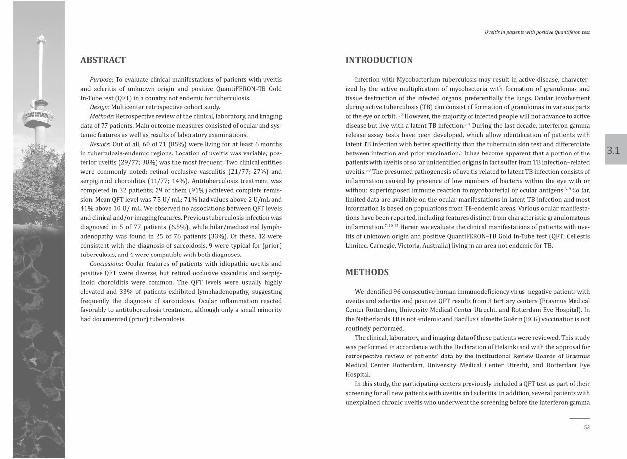
3.1
53
Uveitis in patients with positive Quantiferon test
INTRODUCTION
Infection with Mycobacterium tuberculosis may result in active disease, character-ized by the active multiplication of mycobacteria with formation of granulomas and tissue destruction of the infected organs, preferentially the lungs. Ocular involvement during active tuberculosis (TB) can consist of formation of granulomas in various parts of the eye or orbit.1, 2 However, the majority of infected people will not advance to active disease but live with a latent TB infection.3, 4 During the last decade, interferon gamma release assay tests have been developed, which allow identification of patients with latent TB infection with better specificity than the tuberculin skin test and differentiate between infection and prior vaccination.5 It has become apparent that a portion of the patients with uveitis of so far unidentified origins in fact suffer from TB infection–related uveitis.6-8 The presumed pathogenesis of uveitis related to latent TB infection consists of inflammation caused by presence of low numbers of bacteria within the eye with or without superimposed immune reaction to mycobacterial or ocular antigens.3, 9 So far, limited data are available on the ocular manifestations in latent TB infection and most information is based on populations from TB-endemic areas. Various ocular manifesta-tions have been reported, including features distinct from characteristic granulomatous inflammation.7, 10-15 Herein we evaluate the clinical manifestations of patients with uve-itis of unknown origin and positive QuantiFERON–TB Gold In-Tube test (QFT; Cellestis Limited, Carnegie, Victoria, Australia) living in an area not endemic for TB.
METHODS
We identified 96 consecutive human immunodeficiency virus–negative patients with uveitis and scleritis and positive QFT results from 3 tertiary centers (Erasmus Medical Center Rotterdam, University Medical Center Utrecht, and Rotterdam Eye Hospital). In the Netherlands TB is not endemic and Bacillus Calmette Guérin (BCG) vaccination is not routinely performed.
The clinical, laboratory, and imaging data of these patients were reviewed. This study was performed in accordance with the Declaration of Helsinki and with the approval for retrospective review of patients’ data by the Institutional Review Boards of Erasmus Medical Center Rotterdam, University Medical Center Utrecht, and Rotterdam Eye Hospital.
In this study, the participating centers previously included a QFT test as part of their screening for all new patients with uveitis and scleritis. In addition, several patients with unexplained chronic uveitis who underwent the screening before the interferon gamma
ABSTRACT
Purpose: To evaluate clinical manifestations of patients with uveitis and scleritis of unknown origin and positive QuantiFERON–TB Gold In-Tube test (QFT) in a country not endemic for tuberculosis.
Design: Multicenter retrospective cohort study.Methods: Retrospective review of the clinical, laboratory, and imaging
data of 77 patients. Main outcome measures consisted of ocular and sys-temic features as well as results of laboratory examinations.
Results: Out of all, 60 of 71 (85%) were living for at least 6 months in tuberculosis-endemic regions. Location of uveitis was variable; pos-terior uveitis (29/77; 38%) was the most frequent. Two clinical entities were commonly noted: retinal occlusive vasculitis (21/77; 27%) and serpiginoid choroiditis (11/77; 14%). Antituberculosis treatment was completed in 32 patients; 29 of them (91%) achieved complete remis-sion. Mean QFT level was 7.5 U/ mL; 71% had values above 2 U/mL and 41% above 10 U/ mL. We observed no associations between QFT levels and clinical and/or imaging features. Previous tuberculosis infection was diagnosed in 5 of 77 patients (6.5%), while hilar/mediastinal lymph-adenopathy was found in 25 of 76 patients (33%). Of these, 12 were consistent with the diagnosis of sarcoidosis, 9 were typical for (prior) tuberculosis, and 4 were compatible with both diagnoses.
Conclusions: Ocular features of patients with idiopathic uveitis and positive QFT were diverse, but retinal occlusive vasculitis and serpig-inoid choroiditis were common. The QFT levels were usually highly elevated and 33% of patients exhibited lymphadenopathy, suggesting frequently the diagnosis of sarcoidosis. Ocular inflammation reacted favorably to antituberculosis treatment, although only a small minority had documented (prior) tuberculosis.
3.1
Chapter 3.1
54 55
Uveitis in patients with positive Quantiferon test
the logMAR results were converted back to Snellen VA and are presented as such in this report. In patients with unilateral involvement, visual acuity of the affected eye was included; to prevent the bias of bilateral disease, only the right eye was included in bilat-eral cases.
RESULTS
The general characteristics of the 77 patients (44 male and 33 female) are given in Table 1. The mean age at onset was 46 years and the median follow-up time was 3 years. Out of all patients, 24 of 71 (32%) were born in the Netherlands, of whom 13 stayed longer than 6 months in a country endemic for TB; 47 of 71 (66%) came from and stayed longer than 6 months in a country endemic for TB (demographic data could not be traced for 6 patients).
The anatomic location of intraocular inflammation was diverse, but posterior uveitis was most frequent (Table 2). Anterior uveitis without posterior segment involvement was observed in 19 of 77 patients (25%). Anatomic location of uveitis did not differ between the patients born in the Netherlands and those born in an endemic country or between the patients who never visited an endemic country and those who were born in or stayed longer than 6 months in an endemic country.
The clinical manifestations of inflammation varied widely; however, 2 explicit clinical posterior segment entities were commonly noted, specifically occlusive retinal vasculi-tis (21/77; 27%) and serpiginoid choroiditis (11/77, 14%). Occlusive retinal vasculitis manifested preferentially in male subjects (P < 0.001) and was associated with younger age (P = 0.04, Table 1). Intraocular granulomas were not observed. Corneal opacities (other than band keratopathy) were noted in 7 of 77 patients (9%). Granulomatous ker-atic precipitates were present in 10 of 77 patients (13%; 10/19 with anterior uveitis, of whom 4 were bilateral and 6 unilateral). Vitreous inflammation was present in 46 of 77 patients (60%) and vitreous hemorrhage in 6 of 77 (8%), and 2 patients exhibited retinal fibro- vascular tumors. Vasculitis (arterial, venous, or both) was noted in 34 of 77 patients (44%) and 21 of the 34 (62%) exhibited vascular occlusions and retinal isch-emic areas. The most common ocular complications included macular edema, glaucoma, and cataract (Table 3). Vitreous hemorrhage and retinal neovascularizations were most common in retinal occlusive vasculitis compared to other types of uveitis (P = 0.001, Table 3). The number of patients with 1 or more complications was 55 of 77 (71%). Out of 77 patients, 35 (45%) had to undergo 1 or more intraocular surgical procedures (n = 29) and/or laser treatment (n = 8).
release assay era were also tested. All our new uveitis patients received the QFT and tuberculin skin tests simultaneously during the screening; in several patients the tuber-culin skin test was performed previously, but in all cases more than 6 months before the QFT tests were performed. The QFT cutoff value was 0.35 U/mL.
A specific cause of uveitis and/or associated systemic disorder was determined in 19 patients (ocular toxoplasmosis, n = 4; Behçet disease, n = 2; human leukocyte antigen B27–associated acute anterior uveitis, n = 2; viral anterior uveitis, n = 5; masquerade syndromes, n = 3; Vogt-Koyanagi-Harada disease, n = 1; Wegener disease n = 1; and pre-sumed ocular histoplasmosis syndrome, n = 1). Uveitis in these patients was considered unrelated to latent TB infection and these cases were excluded from the final analysis. Four patients with biopsy-proven sarcoidosis and positive QFT results were included in our series because both diseases might share common pathogenic pathways and the differential diagnosis of sarcoidosis and TB can be difficult.16 All 77 included patients had been subjected to uveitis screening that included erythrocyte sedimentation rate, blood counts, serum angiotensin converting enzyme level, serology for syphilis, and, in those with anterior uveitis, also human leukocyte antigen B27 testing. Chest radiograph examination was performed in 69 patients; out of the remaining 8 patients, 7 had a com-puterized tomography (CT) scan primarily performed and in 1 patient the imaging data could not be retrieved.
The classification of uveitis was performed according to current criteria.17 In this study, the term intraocular inflammation included uveitis and scleritis. The diagnosis of serpiginoid chorioretinitis was performed in patients with multiple serpiginous-like chorioretinal lesions that exhibited multiple areas of activity and were not located adjacent to the optic disc.18, 19 Immunosuppressive treatment was defined as systemic treatment with corticosteroids, sometimes in combination with methotrexate, azathi-oprine, or cyclosporine A. An anti-TB treatment was defined as a course of at least 3 antibiotics—isoniazid (INH), rifampin, pyrazinamide—in the initial 2-month phase of treatment followed by a 4-month consolidation phase using rifampin and INH (in total, 6 months).20 Three patients from one of the centers (Utrecht) received a longer consol-idation phase (7 months, encompassing a total treatment of 9 months). In the absence of signs of pulmonary and/or systemic TB in the majority of patients, the decision to treat the patients with anti-TB drugs was based on the severity of the ocular disease and the patient’s preference in case of non-sight-threatening uveitis. The presence of mac-ular edema was confirmed by optical coherence tomography in all clinically suspected patients.
Data were entered and analyzed using SPSS software version 20.0 (IBM Inc, Chicago, Illinois, USA). For all calculations with visual acuity (VA) data, Snellen VA was converted to the logarithm of the minimal angle of resolution (logMAR). For easier understanding,

3.1
Chapter 3.1
56 57
Uveitis in patients with positive Quantiferon test
Mean QFT value for the whole series was 7.49 U/ mL (median 5.75 U/mL, Table 1); 52 of 73 patients (71%) had levels above 2 U/mL and 30 (41%) above 10 U/mL. No associations were found between the levels of QFT and various clinical manifestations, specifically when the following groups of patients were compared: patients with and without anterior uveitis (mean 7.9 U/mL vs 7.4 U/ mL, P = 0.757), with and without occlusive retinal vasculitis (mean 6.3 U/mL vs 7.9 U/mL, P = 0.329), with and without serpiginoid choroiditis (mean 5.6 U/mL vs 7.8 U/mL, P = 0.261), and with and without hilar and/or mediastinal lymphadenopathy (mean 8.3 U/mL vs 7.1 U/mL, P= 0.437). An elevated serum angiotensin converting enzyme level was noted in 16 of 68 patients (24%). A positive association was noted between elevated serum angiotensin convert-ing enzyme and QFT levels (P= 0.04). A positive tuberculin skin test was present in 51 of 55 patients (93%), out of which 15 (15/51; 29%) had a test result exceeding 20 mm. An association between highly positive tuberculin skin test result and clinical and/or laboratory characteristics was not observed. There were 4 patients with negative tuber-culin skin test (despite positive QFT results). Two patients were previously diagnosed with active TB, of whom 1 was still on anti-TB treatment, and 3 were diagnosed with TB during the assessment for uveitis. There was no difference in the levels of QFT for patients born in the Netherlands vs not born in the Netherlands (P = 0.78), nor was there an association with age noted (P = 0.095). Contact with a TB patient was recalled more TA
BLE
1. G
ener
al C
hara
cter
isti
cs o
f Qua
ntiF
ERO
N–T
B Go
ld T
est–
Posi
tive
Pati
ents
wit
h U
veit
is a
nd S
cler
itis
CT
= co
mpu
ted
tom
ogra
phy;
N.A
. = n
ot a
pplic
able
; TB
= tu
berc
ulos
is; T
ST =
tube
rcul
in sk
in te
st.
a Dat
a w
ere
not a
vaila
ble
for 6
pat
ient
s.b A
nter
ior u
veiti
s occ
urre
d m
ore
freq
uent
ly in
fem
ale
subj
ects
(P =
0.0
1).
c Occ
lusi
ve re
tinal
vas
culit
is o
ccur
red
mor
e fr
eque
ntly
in m
ale
subj
ects
(P =
0.0
01).
Patie
nts
with
occ
lusi
ve re
tinal
vas
culit
is w
ere
youn
ger (
P =
0.04
) com
pare
d to
the
rem
aind
er.
d Of t
hese
25
patie
nts,
12 w
ere
cons
ider
ed c
ompa
tible
with
the
diag
nosi
s of s
arco
idos
is (s
ymm
etric
al c
onfig
urat
ion
of ly
mph
nod
es a
nd a
bsen
ce o
f old
ca
lcifi
catio
ns),
9 w
ith tu
berc
ulos
is (a
sym
met
rical
pre
sent
atio
n, c
alci
ficat
ion,
and
old
scar
s), a
nd 4
wer
e co
nsis
tent
with
bot
h di
agno
ses.
TABLE 2. Ocular Characteristics of QuantiFERON–TB Gold Test–Positive Patients With Uveitis and Scleritis
aFluorescein angiography and/or appropriate clinical information were not available for 4 patients.

3.1
Chapter 3.1
58 59
Uveitis in patients with positive Quantiferon test
(PCR), and Ziehl- Neelsen stain, but 3 (out of 12) other biopsies were positive for M tuberculosis (2 by culture, of which 1 was also positive by PCR and 1 by Ziehl-Neelsen stain only); these 3 patients had occlusive retinal vasculitis. Two out of these 3 patients were previously treated with immunosuppressive agents compared to 1 out of 9 with negative biopsies. Four additional patients underwent bronchoalveolar lavage, but none tested positive for M tuberculosis by staining, PCR, or culture. In addition, 4 patients had no thoracic lymph-adenopathy but had enlarged lymph nodes elsewhere (in the pan-creas, liver, spleen, and neck).
Anti-TB treatment was initiated in 42 patients. Eight patients stopped the medication prematurely (2 patients because of liver toxicity, 1 because of general malaise, and 5 for nonmedical reasons); 2 were still on treatment at the time of data collection (Table 4). Thirty-two patients finished a full course of anti-TB treatment (10 in combination with corticosteroids, an additional 12 patients after a course with corticosteroids and/or other immunosuppressive drugs) and had an average follow-up after anti-TB treat-ment of 2.1 years (standard deviation: 3.2 months). Out of 32 patients who received a full anti-TB treatment course, 29 (91%) exhibited gradual decrease of inflammatory activity with complete disappearance of inflammation even after withdrawing all other (local and/or systemic) treatment. Two showed persisting intraocular inflammation; in 1 patient intraocular inflammation recurred 3 years after completion of anti-TB treat-ment. Development of VA during the various treatment regimens is shown in Table 4. Mean VA at 1-year follow-up (after the initiation of anti- TB treatment) was improved in patients who finished a full course of anti-TB treatment (whether or not in combination with immunosuppressive medication) (P = 0.004;Table 4). Patients who did not receive anti-TB treatment had a better visual acuity at onset compared to those with anti-TB
often in the group who never visited an endemic country (7/11; 64% vs 10/60; 17%, P = 0.003).
Hilar and/or mediastinal adenopathy was diagnosed in 25 of 76 patients (33%) (10 by chest radiograph, 13 by CT scan and 2 by both imaging methods; Table 1) and was characterized as compatible with the diagnosis of sarcoidosis in 12 of 25 (48%); in 9 of 25 (36%) the adenopathy was compatible with the diagnosis of TB (asymmetrical con-figuration, calcifications, and/or presence of lung parenchyma lesions suggestive of TB) and in 4 (16%) was compatible with both diagnoses. Out of 12 patients with bilateral adenopathy on chest radiograph, 4 also had CT scans done, confirming the chest findings in 2; in 2 patients, however, the CT scan revealed no abnormalities. Out of 15 patients with hilar adenopathy on CT scan, 9 had concurrent chest radiographs available, of which 2 were concordant, but in 7 no abnormalities were seen on chest radiograph. No correlation was observed between the presence of hilar/mediastinal adenopathy and general characteristics such as sex, age, BCG status, QFT levels, diameter of tuberculin skin test induration, serum angiotensin converting enzyme level, location of uveitis, and presence of specific ocular characteristics. Biopsy of enlarged chest lymph nodes was performed in 12 of 25 patients (48%) with hilar and/or mediastinal adenopathy. Microscopic evaluation revealed only 1 patient with granuloma and associated necrosis; 2 showed nonspecific inflammatory infiltrates and the remainder exhibited granulomas without necrosis consistent with the diagnosis of sarcoidosis. The patient with necrotic granuloma in the lung showed negative results in culture, polymerase chain reaction
TABLE 3. Ocular Complications of QuantiFERON–TB Gold Test–Positive Patients with Uveitis and Scleritis
aData were not available for 6 patients. bIncludes hypotony, band keratopathy, hyphema, macular atrophy, macular pucker, tractional retinal detachment, and subretinal fibrosis.
TABLE 4. Visual Acuity and Treatment Regimens of QuantiFERON–TB Gold Test–Positive Patients With Uveitis and Scleritis
ATT = antituberculosis treatment (for definitions see Methods section).Visual acuity was calculated using the logarithm of the minimal angle of resolution (logMAR). For easier understanding the logMAR results were converted back to Snellen visual acuity and presented here. In bilateral cases, only the right eye is included to prevent a bias of similar bilateral presentation. In patients without ATT, visual acuity before and 1 year after the onset of any kind of treatment was included.
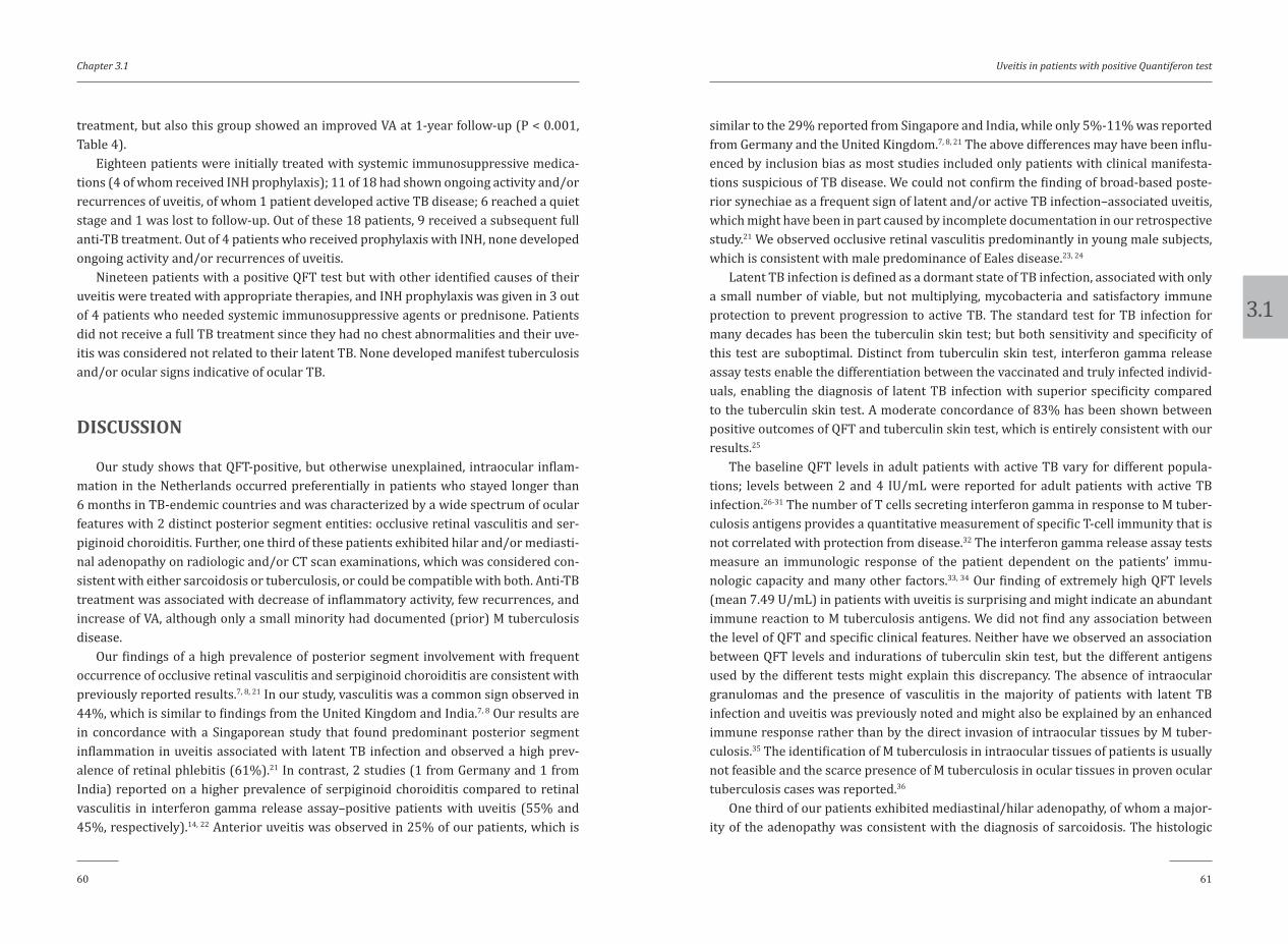
3.1
Chapter 3.1
60 61
Uveitis in patients with positive Quantiferon test
similar to the 29% reported from Singapore and India, while only 5%-11% was reported from Germany and the United Kingdom.7, 8, 21 The above differences may have been influ-enced by inclusion bias as most studies included only patients with clinical manifesta-tions suspicious of TB disease. We could not confirm the finding of broad-based poste-rior synechiae as a frequent sign of latent and/or active TB infection–associated uveitis, which might have been in part caused by incomplete documentation in our retrospective study.21 We observed occlusive retinal vasculitis predominantly in young male subjects, which is consistent with male predominance of Eales disease.23, 24
Latent TB infection is defined as a dormant state of TB infection, associated with only a small number of viable, but not multiplying, mycobacteria and satisfactory immune protection to prevent progression to active TB. The standard test for TB infection for many decades has been the tuberculin skin test; but both sensitivity and specificity of this test are suboptimal. Distinct from tuberculin skin test, interferon gamma release assay tests enable the differentiation between the vaccinated and truly infected individ-uals, enabling the diagnosis of latent TB infection with superior specificity compared to the tuberculin skin test. A moderate concordance of 83% has been shown between positive outcomes of QFT and tuberculin skin test, which is entirely consistent with our results.25
The baseline QFT levels in adult patients with active TB vary for different popula-tions; levels between 2 and 4 IU/mL were reported for adult patients with active TB infection.26-31 The number of T cells secreting interferon gamma in response to M tuber-culosis antigens provides a quantitative measurement of specific T-cell immunity that is not correlated with protection from disease.32 The interferon gamma release assay tests measure an immunologic response of the patient dependent on the patients’ immu-nologic capacity and many other factors.33, 34 Our finding of extremely high QFT levels (mean 7.49 U/mL) in patients with uveitis is surprising and might indicate an abundant immune reaction to M tuberculosis antigens. We did not find any association between the level of QFT and specific clinical features. Neither have we observed an association between QFT levels and indurations of tuberculin skin test, but the different antigens used by the different tests might explain this discrepancy. The absence of intraocular granulomas and the presence of vasculitis in the majority of patients with latent TB infection and uveitis was previously noted and might also be explained by an enhanced immune response rather than by the direct invasion of intraocular tissues by M tuber-culosis.35 The identification of M tuberculosis in intraocular tissues of patients is usually not feasible and the scarce presence of M tuberculosis in ocular tissues in proven ocular tuberculosis cases was reported.36
One third of our patients exhibited mediastinal/hilar adenopathy, of whom a major-ity of the adenopathy was consistent with the diagnosis of sarcoidosis. The histologic
treatment, but also this group showed an improved VA at 1-year follow-up (P < 0.001, Table 4).
Eighteen patients were initially treated with systemic immunosuppressive medica-tions (4 of whom received INH prophylaxis); 11 of 18 had shown ongoing activity and/or recurrences of uveitis, of whom 1 patient developed active TB disease; 6 reached a quiet stage and 1 was lost to follow-up. Out of these 18 patients, 9 received a subsequent full anti-TB treatment. Out of 4 patients who received prophylaxis with INH, none developed ongoing activity and/or recurrences of uveitis.
Nineteen patients with a positive QFT test but with other identified causes of their uveitis were treated with appropriate therapies, and INH prophylaxis was given in 3 out of 4 patients who needed systemic immunosuppressive agents or prednisone. Patients did not receive a full TB treatment since they had no chest abnormalities and their uve-itis was considered not related to their latent TB. None developed manifest tuberculosis and/or ocular signs indicative of ocular TB.
DISCUSSION
Our study shows that QFT-positive, but otherwise unexplained, intraocular inflam-mation in the Netherlands occurred preferentially in patients who stayed longer than 6 months in TB-endemic countries and was characterized by a wide spectrum of ocular features with 2 distinct posterior segment entities: occlusive retinal vasculitis and ser-piginoid choroiditis. Further, one third of these patients exhibited hilar and/or mediasti-nal adenopathy on radiologic and/or CT scan examinations, which was considered con-sistent with either sarcoidosis or tuberculosis, or could be compatible with both. Anti-TB treatment was associated with decrease of inflammatory activity, few recurrences, and increase of VA, although only a small minority had documented (prior) M tuberculosis disease.
Our findings of a high prevalence of posterior segment involvement with frequent occurrence of occlusive retinal vasculitis and serpiginoid choroiditis are consistent with previously reported results.7, 8, 21 In our study, vasculitis was a common sign observed in 44%, which is similar to findings from the United Kingdom and India.7, 8 Our results are in concordance with a Singaporean study that found predominant posterior segment inflammation in uveitis associated with latent TB infection and observed a high prev-alence of retinal phlebitis (61%).21 In contrast, 2 studies (1 from Germany and 1 from India) reported on a higher prevalence of serpiginoid choroiditis compared to retinal vasculitis in interferon gamma release assay–positive patients with uveitis (55% and 45%, respectively).14, 22 Anterior uveitis was observed in 25% of our patients, which is

3.1
Chapter 3.1
62 63
Uveitis in patients with positive Quantiferon test
treated with immunosuppressive drugs showed ongoing activity and, in the long-term follow-up, decrease of VA, and 9 of these patients showed a satisfactory effect on their inflammatory activity after subsequent anti-TB treatment.
The drawbacks of our study include the limitations of retrospective studies; there was no systematic data collection, diagnostic procedures differed, and no systematic treatment criteria were applied. However, even in this limited series the beneficial effect of anti-TB treatment was clear.
In conclusion, we show a favorable outcome of anti-TB treatment in QFT-positive patients with a uveitis of undetermined cause. Our results show the absence of intraoc-ular granulomas and confirm the presence of occlusive retinal vasculitis and serpiginoid choroiditis, as previously observed in patients positive for interferon gamma release assay tests in endemic populations. Further, we report on highly elevated QFT levels in patients with intraocular inflammation and point out the limited number of patients with documented (prior) TB disease. We identify the association of QFT-positive uveitis with hilar and/or mediastinal lymphadenopathy suggestive of the diagnosis of sarcoid-osis. Future studies are required to elucidate the exact pathogenesis of QFT-associated intraocular inflammation and lymphadenopathy, and to show whether viable mycobac-teria are present in affected tissues or whether the inflammation represents an immune reaction or a combination of both processes. In the view of the multitude of infected individuals in the world and the associated visual morbidity, intensive research on this topic is needed.
REFERENCES
1. Deschenes J, Wade NK, Lalonde R. Tuberculosis and Atypical Mycobacteria. In: Tasman W, Jaeger EA
(eds), Duane’s Ophthalmology. Philadelphia: Lippincott Williams & Wilkins; 2006.
2. Alvarez GG, Roth VR, Hodge W. Ocular tuberculosis: diagnostic and treatment challenges. Int J Infect
Dis 2009;13:432-435.
3. Tufariello JM, Chan J, Flynn JL. Latent tuberculosis: mechanisms of host and bacillus that contribute
to persistent infection. Lancet Infect Dis 2003;3:578-590.
4. Horsburgh CR, Jr., Rubin EJ. Clinical practice. Latent tuberculosis infection in the United States. N Engl
J Med 2011;364:1441-1448.
5. Menzies D, Pai M, Comstock G. Meta-analysis: new tests for the diagnosis of latent tuberculo-
sis infection: areas of uncertainty and recommendations for research. Annals of internal medicine
2007;146:340-354.
6. Ang M, Hedayatfar A, Wong W, Chee SP. Duration of anti-tubercular therapy in uveitis associated with
latent tuberculosis: a case-control study. The British journal of ophthalmology 2012;96:332-336.
characteristics as well as the negative results of staining, cultures, and PCR for M tuber-culosis in 9 of 12 lymph node biopsies were considered consistent with the diagnosis of sarcoidosis. One could speculate either that our diagnostic tests were not sensitive enough to reveal mycobacteria or that this was attributable to a sampling error since the presence of mycobacteria in lymph node tissue might be scarce.36 In contrast, our find-ings might also indicate that a specific type of sarcoid reaction could occur triggered by TB infection. Our findings emphasize the possible relation- ship between infection with M tuberculosis and development of enlarged hilar and/or mediastinal lymph nodes con-sistent with the diagnosis of sarcoidosis. The association of tuberculosis and sarcoidosis has been repeatedly reported and various studies suggest that mycobacterial antigens might represent the inciting agent in a proportion of patients with sarcoidosis.37-39 DNA of M tuberculosis was observed in granulomas of patients with sarcoidosis and spe-cific antigens (catalase-peroxidase and the 70-kDa mycobacterial heat shock protein) were pointed out as possible inciting agents.38, 39 QFT results also seem to be reliable in patients with sarcoidosis, in contrast to their anergy to tuberculin skin test.40, 41
So far, systematic assessments of radiologic and histologic findings in a large number of patients with uveitis and positive QFT test results are not available. Mediastinal lymph-adenopathy was also observed in 4 of 21 cases (19%) with presumed intraocular TB in the United Kingdom.8 The symmetrical radiologic appearance and histologic findings as well as negative results of various tests for TB in the biopsies of our 15 patients might also indicate that mycobacteria are not (anymore) present and that a sarcoid reaction has developed, possibly triggered by mycobacterial antigens. It is not yet clear whether uveitis in our QFT-positive population can be considered as an active ocular infection (probably with slowly multiplying mycobacteria) or just as a purely immunologic reac-tion to previous infection or a combination of both phenomena. The positive reaction to anti-TB treatment in our series favors the presence of infection, and it might be feasible that anti-TB treatment eliminates M tuberculosis either in the eye or elsewhere in the body and stops a perpetual immune response that is leading to uveitis.
In the absence of a diagnostic test for ocular TB, a therapeutic trial was consid-ered justified in patients with severe sight-threatening intraocular inflammation and latent TB infection.42 The indication for anti-TB treatment in our study was biased by the severity of ocular disease; predominantly, patients with active uveitis and poor VA were treated. Our study documents a very good overall effect of anti-TB treatment, as 29 of 32 patients (90%) with completed anti-TB treatment exhibited a disappear-ance of intraocular inflammation in contrast to patients who did not receive anti-TB treatment. The improvement of VA in patients who did not receive anti-TB treatment can be explained by a natural course of their uveitis as well as initial favorable effect of immunosuppressive therapy. Interestingly, 11 out of 19 patients who were initially

3.1
Chapter 3.1
64 65
Uveitis in patients with positive Quantiferon test
23. Das T, Pathengay A, Hussain N, Biswas J. Eales’ disease: diagnosis and management. Eye (Lond)
2010;24:472-482.
24. Biswas J, Sharma T, Gopal L, Madhavan HN, Sulochana KN, Ramakrishnan S. Eales disease--an update.
Surv Ophthalmol 2002;47:197-214.
25. Mazurek GH, LoBue PA, Daley CL, et al. Comparison of a whole-blood interferon gamma assay with
tuberculin skin testing for detecting latent Mycobacterium tuberculosis infection. JAMA : the journal
of the American Medical Association 2001;286:1740-1747.
26. Rose MV, Kimaro G, Nissen TN, et al. QuantiFERON(R)-TB gold in-tube performance for diagnosing
active tuberculosis in children and adults in a high burden setting. PLoS One 2012;7:e37851.
27. Chee CB, Barkham TM, Khinmar KW, Gan SH, Wang YT. Quantitative T-cell interferon-gamma respons-
es to Mycobacterium tuberculosis-specific antigens in active and latent tuberculosis. European jour-
nal of clinical microbiology & infectious diseases : official publication of the European Society of Clinical
Microbiology 2009;28:667-670.
28. Lee SW, Lee CT, Yim JJ. Serial interferon-gamma release assays during treatment of active tuberculosis
in young adults. BMC Infect Dis 2010;10:300.
29. Kurup SK, Buggage RR, Clarke GL, Ursea R, Lim WK, Nussenblatt RB. Gamma interferon assay as an
alternative to PPD skin testing in selected patients with granulomatous intraocular inflammatory
disease. Can J Ophthalmol 2006;41:737-740.
30. Mackensen F, Becker MD, Wiehler U, Max R, Dalpke A, Zimmermann S. QuantiFERON TB-Gold--a new
test strengthening long-suspected tuberculous involvement in serpiginous-like choroiditis. Am J Oph-
thalmol 2008;146:761-766.
31. Cordero-Coma M, Calleja S, Torres HE, et al. The value of an immune response to Mycobacterium
tuberculosis in patients with chronic posterior uveitis revisited: utility of the new IGRAs. Eye (Lond)
2010;24:36-43.
32. Mack U, Migliori GB, Sester M, et al. LTBI: latent tuberculosis infection or lasting immune responses
to M. tuberculosis? A TBNET consensus statement. Eur Respir J 2009;33:956-973.
33. Mazurek GH, Jereb J, Vernon A, LoBue P, Goldberg S, Castro K. Updated guidelines for using Interferon
Gamma Release Assays to detect Mycobacterium tuberculosis infection - United States, 2010. MMWR
Recommendations and reports : Morbidity and mortality weekly report Recommendations and reports
/ Centers for Disease Control 2010;59:1-25.
34. Kobashi Y, Mouri K, Yagi S, et al. Usefulness of the QuantiFERON TB-2G test for the differential diag-
nosis of pulmonary tuberculosis. Intern Med 2008;47:237-243.
35. Llorenc V, Gonzalez-Martin J, Keller J, et al. Indirect supportive evidence for diagnosis of tuberculo-
sis-related uveitis: from the tuberculin skin test to the new interferon gamma release assays. Acta
Ophthalmol 2013;91:e99-e107.
36. Wroblewski KJ, Hidayat AA, Neafie RC, Rao NA, Zapor M. Ocular tuberculosis: a clinicopathologic and
molecular study. Ophthalmology 2011;118:772-777.
7. Gupta A, Bansal R, Gupta V, Sharma A, Bambery P. Ocular signs predictive of tubercular uveitis. Amer-
ican journal of ophthalmology 2010;149:562-570.
8. Manousaridis K, Ong E, Stenton C, Gupta R, Browning AC, Pandit R. Clinical presentation, treatment,
and outcomes in presumed intraocular tuberculosis: experience from Newcastle upon Tyne, UK. Eye
(Lond) 2013;27:480-486.
9. Bansal R, Gupta A, Gupta V, Dogra MR, Bambery P, Arora SK. Role of anti-tubercular therapy in uveitis
with latent/manifest tuberculosis. American journal of ophthalmology 2008;146:772-779.
10. De Luigi G, Mantovani A, Papadia M, Herbort CP. Tuberculosis-related choriocapillaritis (multifo-
cal-serpiginous choroiditis): follow-up and precise monitoring of therapy by indocyanine green angi-
ography. Int Ophthalmol 2012;32:55-60.
11. Zhang M, Zhang J, Liu Y. Clinical presentations and therapeutic effect of presumed choroidal tubercu-
losis. Retina 2012;32:805-813.
12. Davis EJ, Rathinam SR, Okada AA, et al. Clinical spectrum of tuberculous optic neuropathy. J Ophthal-
mic Inflamm Infect 2012;2:183-189.
13. Gupta V, Gupta A, Arora S, Bambery P, Dogra MR, Agarwal A. Presumed tubercular serpiginouslike
choroiditis: clinical presentations and management. Ophthalmology 2003;110:1744-1749.
14. Sudharshan S, Ganesh SK, Balu G, et al. Utility of QuantiFERON(R)-TB Gold test in diagnosis and man-
agement of suspected tubercular uveitis in India. Int Ophthalmol 2012;32:217-223.
15. Mahyudin M, Choo MM, Ramli NM, Omar SS. Ocular Tuberculosis Initially Presenting as Central Reti-
nal Vein Occlusion. Case Rep Ophthalmol 2010;1:30-35.
16. Gupta D, Agarwal R, Aggarwal AN, Jindal SK. Sarcoidosis and tuberculosis: the same disease with dif-
ferent manifestations or similar manifestations of different disorders. Current opinion in pulmonary
medicine 2012;18:506-516.
17. Jabs DA, Nussenblatt RB, Rosenbaum JT. Standardization of uveitis nomenclature for reporting clinical
data. Results of the First International Workshop. American journal of ophthalmology 2005;140:509-
516.
18. Bansal R, Gupta A, Gupta V, Dogra MR, Sharma A, Bambery P. Tubercular serpiginous-like choroiditis
presenting as multifocal serpiginoid choroiditis. Ophthalmology 2012;119:2334-2342.
19. Vasconcelos-Santos DV, Rao PK, Davies JB, Sohn EH, Rao NA. Clinical features of tuberculous serpig-
inouslike choroiditis in contrast to classic serpiginous choroiditis. Arch Ophthalmol 2010;128:853-
858.
20. Centers for Disease Control and Prevention. Treatment of Tuberculosis, American Thoracic Society,
CDC and Infectious Diseases Society of America. 2003:36-41.
21. Ang M, Hedayatfar A, Zhang R, Chee SP. Clinical signs of uveitis associated with latent tuberculosis.
Clin Experiment Ophthalmol 2012;40:689-696.
22. Doycheva D, Deuter C, Hetzel J, et al. The use of positron emission tomography/CT in the diagnosis of
tuberculosis-associated uveitis. The British journal of ophthalmology 2011;95:1290-1294.

3.1
Chapter 3.1
66 67
Uveitis in patients with positive Quantiferon test
37. Gideon HP, Flynn JL. Latent tuberculosis: what the host “sees”? Immunologic research 2011;50:202-
212.
38. Song Z, Marzilli L, Greenlee BM, et al. Mycobacterial catalase-peroxidase is a tissue antigen and target
of the adaptive immune response in systemic sarcoidosis. J Exp Med 2005;201:755-767.
39. Dubaniewicz A, Kampfer S, Singh M. Serum anti-mycobacterial heat shock proteins antibodies in sar-
coidosis and tuberculosis. Tuberculosis (Edinb) 2006;86:60-67.
40. Gupta D, Kumar S, Aggarwal AN, Verma I, Agarwal R. Interferon gamma release assay (QuantiFER-
ON-TB Gold In Tube) in patients of sarcoidosis from a population with high prevalence of tuberculosis
infection. Sarcoidosis, vasculitis, and diffuse lung diseases : official journal of WASOG / World Associa-
tion of Sarcoidosis and Other Granulomatous Disorders 2011;28:95-101.
41. Milman N, Soborg B, Svendsen CB, Andersen AB. Quantiferon test for tuberculosis screening in sar-
coidosis patients. Scandinavian journal of infectious diseases 2011;43:728-735.
42. van Daele PL, Bakker M, van Hagen PM, Baarsma GS, Kuijpers RW. TB or not TB: treat to see. Med J
Aust 2006;185:178-179.

3.2ChapterTuberculosis and other causes
of uveitis in Indonesia
Rina La Distia Nora1,5, Ratna Sitompul1, Marleen Bakker4, Made Susiyanti1,
Lukman Edwar1, Soedarman Sjamsoe1, Gurmeet Singh2, P. Martin van Hagen5,
Aniki Rothova3
1Department of Ophthalmology, 2Respirology and Critical Illness Division Department of Internal Medicine,
University of Indonesia &Cipto Mangunkusumo Hospital Kirana, Jakarta, Indonesia, 3Department of Ophthalmology,
4Department of Pulmonary Diseases, 5Department of Clinical Immunology, Internal Medicine, Erasmus Medical Center,
Rotterdam, the Netherlands.
Eye 2018 Mar;32(3):546-554

3.2
71
TB and other causes of uveitis in Indonesia
ABSTRACT
Purpose: To assess the causes of uveitis in Indonesia and determine the importance of tuberculosis (TB) as a cause o uveitis.
Methods: Prospective cohort study examining 146 consecutive new HIV-negative patients with active uveitis between June 2014 and May 2015. We assessed the anatomic locations and specific causes of uve-itis as well as associations with infectious and non-infectious systemic diseases. We determined the prevalence of positive Quantiferon-G test (QFT) results in Indonesian patients with uveitis and calculated the number of patients with active systemic TB.
Results: Posterior and panuveitis were the most common anatomic entities (38% each). Infections represented the most frequent cause of uveitis (33%); the most prevalent were toxoplasmosis (19%) and active systemic TB (8%). The majority of patients were QFT-positive (61%). A specific diagnosis could not be established in 45% of the patients. At first presentation to the ophthalmologist, the majority of patients (66%) had a visual acuity of less than finger counting at 3 m and already exhib-ited various complications of uveitis. When classifying the QFT-positive patients with unexplained uveitis into a TB-related group, the percent-age of “TB-associated” uveitis cases increased from 8% to 48%. Highly elevated QFT levels were observed in patients with uveitis of unknown cause and no signs of active systemic TB.
Conclusion: In Indonesia, infectious uveitis was the most common type of uveitis, and the leading causes consisted of toxoplasmosis and TB. The association observed between highly elevated QFT results and uve-itis of otherwise unexplained origins indicates that a link exists between the latent TB infection and the development of uveitis.
Keywords: uveitis in Indonesia, tuberculosis-related uveitis
INTRODUCTION
Uveitis is an ocular disorder that is associated with multiple infectious and non-in-fectious causes and a major cause of blindness worldwide. Determining the cause of uveitis in each patient is essential for disease management.1 Tuberculosis (TB) has his-torically been considered a major cause of uveitis, but the importance of TB as a primary cause of uveitis has gradually decreased in the last decades, although it has recently re-emerged.2-4
TB is an infectious disease caused by Mycobacterium tuberculosis (M. tuberculosis). One-third of the world’s population is thought to be infected with M. tuberculosis, and in 2015,10.4 million new infections occured.5 TB is typically viewed as a poverty-re-lated disease, and immunodeficiency, including infection with human immunodeficiency virus (HIV), is an important risk factor. The typical risk factors for extrapulmonary TB including eye involvement are HIV infection, young age, female gender and non-white race.6
Ocular involvement has prevalence between 1.4% and 18% in pulmonary TB patients.7, 8 The diagnosis of ocular TB is difficult to achieve because access to intraocular tissues is limited, and few mycobacteria invade the intraocular environment. Previously, if a patient had unexplained uveitis, evidence of active systemic TB elsewhere in the body justified treatment for ocular TB.9 In recent years, the introduction of Interferon Gamma Release Assay (IGRA) tests has made it possible to identify patients with latent TB infections. Identifying uveitis with active and/or latent TB is important because it can lead to substantial visual loss or even blindness if left untreated.10, 11 Previous reports have emphasized the presence of granulomas as a pathognomonic feature of ocular TB, whereas recent publications have shown that serpiginoid choroiditis and occlusive reti-nal vasculitis are ocular manifestations attributable to latent TB infection.12
There are no precise data regarding ocular TB and other potential causes of uveitis in Indonesia, a country with a high prevalence of all three TB categories (i.e., TB, TB/HIV and multi-drug-resistant TB) and that will carry a high disease burden for the years 2016-2020 according to the World Health Organization (WHO).5 In 2016, the preva-lence rate of TB was estimated at 395 per 100,000.5 In the current prospective study, we determined the anatomical location and causes of uveitis in Indonesia and evaluated the prevalence of active and latent TB infections in the uveitis population using standard screening examinations including radiologic chest imaging and concurrent tuberculin skin tests (TSTs) and QuantiFERON-Tb Gold tests (QFTs).
3.2
Chapter 3.2
72 73
TB and other causes of uveitis in Indonesia
SUBJECTS AND METHODS
We performed a prospective cohort study that included all 247 consecutive new patients with uveitis who were referred to the Infection and Immunology Division of the Ophthalmology Department of the Medical Faculty Universitas Indonesia/Cipto Mangunkusumo Hospital, Jakarta Indonesia. Our study occurred from June 2014 until May 2015 and was approved by the local medical ethics committee. All included patients provided informed consent. In all, 146 of the 247 enrolled patients were included in the final analysis. We excluded patients unwilling to participate and/or patients who did not complete the screening work-up and those who did not attend follow-up examinations (n=72). Additional 29 HIV-positive patients with uveitis were seen in the same time-frame; they are reported separately.
A medical history was obtained, and a full ophthalmological examination, including slit-lamp biomicroscopy and indirect ophthalmoscopy, was performed for all included patients. Color photographs were obtained, and fluorescein angiography, optical coher-ence tomography (OCT) and visual field tests were performed according to standard clin-ical practices. Uveitis cases were classified and graded according to the Standardization of Uveitis Nomenclature (SUN) system.
All patients were submitted to standard screening protocols for uveitis to obtain the erythrocyte sedimentation rate and red and white blood cell counts. Additionally, sero-logical testing for HIV, syphilis and toxoplasmosis, urine analysis and radiological chest examination were performed. TSTs (RT23 SSI-2 T.U/0.1 mL Statens Serum Institute, Copenhagen, Denmark) and QFTs (Cellestis Inc., Carnegie, Australia) were also per-formed. In all cases, the QFT was performed before the TST. Because of a temporary lack of tuberculin supply, the TST was performed in only 115/146 (79%) of the patients. In Indonesia, a TST result with an induration larger than 10 mm in diameter is considered positive.13 The QFT cut-off value was 0.35 IU/mL.14 In HIV positive patients, however, QFT and TST tests were not systematically applied because we had very limited supply of tests and the outcomes of laboratory tests in HIV-infected patients are not reliable. In addition, some HIV-positive patients (13/29, 45%) were previously diagnosed with systemic TB and treated, and their QFT and TST outcomes would not be contributory.
A radiological examination; standard postero-anterior chest X ray (CXR) was per-formed in all patients. When needed, additional imaging examinations, including apico-lordotic CXR (n=5) and chest computed tomography (CT) scans (n=16), were conducted. Cases involving iris and/or choroidal granulomas, sub retinal abscesses, serpiginoid choroiditis and retinal vasculitis of unknown cause (n=16) underwent chest CT scanning because this modality can be useful for diagnosing granulomatous uveitis.15
Human leucocyte antigen (HLA) B27 testing was not available at our institution at the time of this study.
Microbiological examinations (i.e., sputum culture (n=1), Ziehl Nielsen (ZN) staining of sputum (n=21), and polymerase chain reaction (PCR) for M. tuberculosis in sputum (N=10)) were performed in selected cases. Additional PCRs of intraocular fluids was per-formed in 44 patients with a high suspicion of infectious uveitis etiology and no response to standard treatments with corticosteroids including herpes simplex virus (HSV, n=10), varicella zoster virus (VZV, n=3), cytomegalovirus (CMV, n=18), Toxoplasma (n=5), and M. tuberculosis (n=37). PCR for M. tuberculosis was performed on samples isolated using a QIAamp DNA and Blood Mini Kit and primers specific for IS 1660 in a thermal cycler. The products were further analyzed using gel electrophoresis.
Diagnoses of pulmonary TB were based on the Indonesian Society of Respirology (ISR)TB guidelines (see the algorithm illustrated in supplemental Figure 1).16, 17 Briefly, diagnoses of active pulmonary TB were based on a general clinical examination and microbiological and/or radiological findings. A review of chest X-ray and CT scan data was performed by two pulmonologists who are specialists in TB (one from Indonesia and one from the Netherlands), and cases were classified as active TB, prior but inactive TB and abnormalities other than TB.
Clinical diagnoses, such as Behcet’s disease, Vogt Koyanagi Harada (VKH) syndrome, and Fuchs uveitis syndrome (FUS), were diagnosed according to current diagnostic cri-teria. All diagnoses of sarcoidosis were achieved according to current diagnostic criteria for patients with negative TB tests. Patients showing no evidence of active systemic TB but who had Mtb-PCR-positive intraocular fluids were not classified as having active sys-temic TB and were separately indicated. Diagnoses of ocular toxoplasmosis were based on clinical manifestations including focal active chorioretinitis located adjacent to a cho-rioretinal scar in combination with positive serological results and a good response to antiparasitic drugs. We registered all demographic data and the results of all diagnostic and ophthalmological examinations.
All statistical analyses were performed using chi-squared tests and one-way ANOVA tests. Statistical significance was defined as a p value less than 0.05. We performed Bonferroni post-hoc analyses, and all p values shown were adjusted. Multiple regres-sion analysis was used to compare all clinical and laboratory characteristics related to QFT values. For all calculations with visual acuity data, we used the logarithm of the Minimum Angle of Resolution (logMAR). In bilateral cases, we used the average logMAR visual acuity; in unilateral cases, the visual acuity of the affected eye was used.
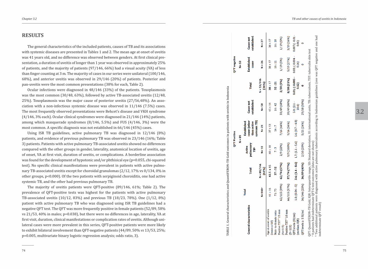
3.2
Chapter 3.2
74 75
TB and other causes of uveitis in Indonesia
RESULTS
The general characteristics of the included patients, causes of TB and its associations with systemic diseases are presented in Tables 1 and 2. The mean age at onset of uveitis was 41 years old, and no difference was observed between genders. At first clinical pre-sentation, a duration of uveitis of longer than 1 year was observed in approximately 25% of patients, and the majority of patients (97/146, 66%) had a visual acuity (VA) of less than finger counting at 3 m. The majority of cases in our series were unilateral (100/146, 68%), and anterior uveitis was observed in 29/146 (20%) of patients. Posterior and pan-uveitis were the most common presentations (38% for each, Table 2).
Ocular infections were diagnosed in 48/146 (33%) of the patients. Toxoplasmosis was the most common (30/48, 63%), followed by active TB-associated uveitis (12/48, 25%). Toxoplasmosis was the major cause of posterior uveitis (27/56,48%). An asso-ciation with a non-infectious systemic disease was observed in 11/146 (7.5%) cases. The most frequently observed presentations were Behcet’s disease and VKH syndrome (4/146, 3% each). Ocular clinical syndromes were diagnosed in 21/146 (14%) patients, among which masquerade syndromes (8/146, 5.5%) and FUS (4/146, 3%) were the most common. A specific diagnosis was not established in 66/146 (45%) cases.
Using ISR TB guidelines, active pulmonary TB was diagnosed in 12/146 (8%) patients, and evidence of previous pulmonary TB was observed in 23/146 (16%; Table 3) patients. Patients with active pulmonary TB-associated uveitis showed no differences compared with the other groups in gender, laterality, anatomical location of uveitis, age of onset, VA at first visit, duration of uveitis, or complications. A borderline association was found for the development of hypotonic and/or phthisical eye (p=0.055, chi-squared test). No specific clinical manifestations were prevalent in patients with active pulmo-nary TB-associated uveitis except for choroidal granulomas (2/12, 17% vs 0/134, 0% in other groups, p<0.000). Of the two patients with serpiginoid choroiditis, one had active systemic TB, and the other had previous pulmonary TB.
The majority of uveitis patients were QFT-positive (89/146, 61%; Table 2). The prevalence of QFT-positive tests was highest for the patients with active pulmonary TB-associated uveitis (10/12, 83%) and previous TB (18/23, 78%). One (1/12, 8%) patient with active pulmonary TB who was diagnosed using ISR TB guidelines had a negative QFT test. The QFT was more frequently positive in female patients (52/89, 58% vs 21/53, 40% in males; p=0.038), but there were no differences in age, laterality, VA at first visit, duration, clinical manifestations or complication rates of uveitis. Although uni-lateral cases were more prevalent in this series, QFT-positive patients were more likely to exhibit bilateral involvement than QFT-negative patients (44/89, 50% vs 13/53, 25%; p=0.005, multivariate binary logistic regression analysis; odds ratio, 3). TA
BLE
1. G
ener
al c
hara
cter
isti
cs a
nd Q
uant
iFER
ON
–TB
Gold
out
com
es in
pat
ient
s w
ith
uvei
tis
in In
done
sia
QFT:
Qua
ntiF
ERON
–TB
Gold
, IQR
: Int
erqu
artil
e ra
nge,
SD:
Sta
ndar
d de
viat
ion,
IU: i
nter
natio
nal u
nits
, TB:
tube
rcul
osis
, TST
: tub
ercu
lin sk
in te
sta 4
pat
ient
s had
inde
term
inat
e QF
T-G
test
out
com
es (d
ata
are
not s
how
n)b T
wo
addi
tiona
l pat
ient
s w
ere
diag
nose
d w
ith a
ctiv
e pu
lmon
ary
tube
rcul
osis
acc
ordi
ng to
Indo
nesi
an g
uide
lines
(on
e w
as Q
FT n
egat
ive
and
one
had
inde
term
inat
e QF
T re
sult)
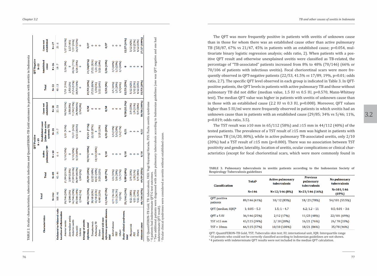
3.2
Chapter 3.2
76 77
TB and other causes of uveitis in Indonesia
TABL
E 2.
Ocu
lar
char
acte
rist
ics,
tube
rcul
osis
infe
ctio
n an
d Q
uant
iFER
ON
–TB
Gold
out
com
es in
pat
ient
s w
ith
uvei
tis
in In
done
sia
QFT:
Qua
ntiF
ERON
–TB
Gold
, TB:
tube
rcul
osis
, VKH
: Vog
t Koy
anag
i Har
ada,
FUS
: Fuc
hs u
veiti
s syn
drom
ea 4
pat
ient
s had
inde
term
inat
e QF
T-G
test
out
com
es.
b Tw
o ad
ditio
nal p
atie
nts
wer
e di
agno
sed
with
act
ive
pulm
onar
y tu
berc
ulos
is a
ccor
ding
to In
done
sian
gui
delin
es (o
ne w
as Q
FT n
egat
ive
and
one
had
inde
term
inat
e QF
T re
sult)
.c O
cula
r clin
ical
synd
rom
es w
ere
cons
ider
ed a
s uve
itis w
ithou
t est
ablis
hed
caus
e.
The QFT was more frequently positive in patients with uveitis of unknown cause than in those for whom there was an established cause other than active pulmonary TB (58/87, 67% vs 21/47, 45% in patients with an established cause; p=0.054, mul-tivariate binary logistic regression analysis; odds ratio, 2). When patients with a pos-itive QFT result and otherwise unexplained uveitis were classified as TB-related, the percentage of “TB-associated” patients increased from 8% to 48% (70/146) (66% or 70/106 of patients with infectious uveitis). Focal chorioretinal scars were more fre-quently observed in QFT-negative patients (22/53, 41.5% vs 17/89, 19%, p=0.01; odds ratio, 2.7). The specific QFT level observed in each group is indicated in Table 3. In QFT-positive patients, the QFT levels in patients with active pulmonary TB and those without pulmonary TB did not differ (median value, 1.5 IU vs 0.5 IU, p=0.570, Mann-Whitney test). The median QFT value was higher in patients with uveitis of unknown cause than in those with an established cause (2.2 IU vs 0.3 IU, p=0.008). Moreover, QFT values higher than 5 IU/ml were more frequently observed in patients in which uveitis had an unknown cause than in patients with an established cause (29/85; 34% vs 5/46; 11%, p=0.019; odds ratio, 3.5).
The TST result was ≥10 mm in 65/112 (58%) and ≥15 mm in 44/112 (40%) of the tested patients. The prevalence of a TST result of ≥15 mm was highest in patients with previous TB (16/20, 80%), while in active pulmonary TB-associated uveitis, only 2/10 (20%) had a TST result of ≥15 mm (p=0.000). There was no association between TST positivity and gender, laterality, location of uveitis, ocular complications or clinical char-acteristics (except for focal chorioretinal scars, which were more commonly found in
QFT: QuantiFERON–TB Gold, TST; Tuberculin skin test; IU; international unit, IQR: Interquartile rangea 10 patients who could not be correctly classified according to Indonesian guidelines are not shown.b 4 patients with indeterminate QFT results were not included in the median QFT calculation.
TABLE 3. Pulmonary tuberculosis in uveitis patients according to the Indonesian Society of Respirology Tuberculosis guidelines

3.2
Chapter 3.2
78 79
TB and other causes of uveitis in Indonesia
TST-negative patients: 23/68 or 34% of patients had negative TST results vs 5/44 or 11% of patients with positive TST results; p=0.008). All patients with active pulmonary TB had positive TST results (≥10 mm). In accordance with the QFT results, a TST value of ≥15 mm was more frequently observed in patients with uveitis of unknown origin than in those with an established diagnosis (34/68, 50% vs 8/34, 23.5%; p=0.011).
The QFT and TST results were highly correlated (p<0.000), and this relationship was even stronger in patients with QFT levels ≥5 IU/ml (TST ≥15 mm, p=0.000; TST ≥10 mm, p=0.001). Out of the 74 QFT-positive patients, 42/74 (57%) had a TST result ≥15 mm, and 57/74 (77%) had a TST result ≥10 mm.
Out of 37 PCR runs to assess the presence of M. tuberculosis in intraocular fluid, 4 yielded a positive result. These four patients were all QFT-positive, and one was classi-fied as having active pulmonary TB (positive sputum for M. tuberculosis determined by PCR). Two patients had anterior uveitis, and two had pan-uveitis. Small nodules were observed on the iris in two of these patients, but no other similarities in clinical mani-festations were observed.
TST: Tuberculin skin test QFT: QuantiFERON–TB Gold, TB: tuberculosis, VKH: Vogt Koyanagi Harada, FUS: Fuchs uveitis syndrome
TABLE 4. Characteristics in Human Immunodeficiency Virus (HIV)-positive patients with uveitis
The characteristics of 29 HIV-positive uveitis patients are provided in Table 4. The majority of the HIV-associated uveitis cases was of infectious origin (18/29, 62%), with CMV retinitis being most common (9/18, 50%) followed by toxoplasmosis (4/18, 22%). Association of active systemic TB infection with uveitis at the time of presentation was found in 3/29 (10%) cases.
DISCUSSION
This prospective study demonstrates that the known etiology of uveitis is most commonly infectious in Indonesia. Furthermore, we show that the majority of patients consulted an ophthalmologist when uveitis had entered the late stage and they already had poor VA and complications. This late presentation limits the visual prognosis and precludes the recognition of typical clinical manifestations, which impairs the correct diagnosis of uveitis. We demonstrate that active pulmonary TB remains a frequent cause of uveitis in Indonesia (8% of all uveitis cases) and ranks second after toxoplasmosis among infectious causes of uveitis. We also found that a majority of uveitis patients were QFT-positive (61%) and observed an excess of highly positive QFT results in patients with uveitis of unknown cause and without any signs and symptoms of active systemic TB. This finding suggests that QFT-positive uveitis might be associated with previous or latent TB, as has been previously suggested.7, 18, 19 Interestingly, QFT levels were much higher in patients with uveitis associated with latent and previous TB than in patients with active systemic TB-associated uveitis. The majority of patients with uveitis had a positive QFT result, illustrating the limited diagnostic value of this test in endemic pop-ulations such as ours.
In western countries, infections account for approximately 16% of all uveitis cases. This percentage is higher in developing countries (range, 20% to 30%).3, 20 Because only proven infections are classified as infectious cases, it is likely that in reality these per-centages are much higher. In our series, infectious causes accounted for 33% of all cases. The majority were caused by toxoplasmosis, followed by active systemic TB-associated uveitis. Our findings are similar to those described in reports from South America, Thailand and Nepal. In contrast, reports have shown that in other countries in Asia (including India, Singapore and Myanmar), uveitis is more often TB-associated than caused by toxoplasmosis.21-23 However, these reports classified patients with uveitis and positive QFT and/or TST results (without additional evidence of systemic TB) as having TB. While this may explain the higher prevalence of TB-associated cases, it also prevents comparisons with our series. If the QFT-positive and unexplained cases of uveitis in our series were all classified as TB-associated, the percentage of “TB-associated” cases of

3.2
Chapter 3.2
80 81
TB and other causes of uveitis in Indonesia
uveitis would increase from 8% to 48%, which illustrates the importance of classifica-tion methods. Similar disparities in the prevalence of (presumed) ocular TB can also be found in studies performed in western and middle-eastern countries (range, from under 1% to 18%).3, 24, 25These large variations might be caused by genuine geographical dif-ferences or potentially by a lack of appropriate classification criteria for TB-associated uveitis.
It is currently unknown whether uveitis associated with a positive IGRA test in the absence of signs of active TB represents a real ocular infection with M. tuberculosis, an immune reaction, or a combination of both. The presence of M. tuberculosis in ocular tissues has occasionally been demonstrated in active systemic TB-associated uveitis, but a histopathological study demonstrated that very few bacilli are required to cause uveitis.26, 27 In India, patients with uveitis and positive IGRA outcomes had positive PCR results in the absence of systemic signs of TB infection.28 These findings show that a low-grade intraocular infection with M. tuberculosis is possible even in patients show-ing no other systemic signs of TB. The diagnostic role of performing PCR on intraocular fluids to identify TB is not yet clear, and the results vary according to the selected gene targets.12, 18, 29 Because patients can have negative PCR findings in ocular fluid while showing uveitis and active systemic TB or positive PCR findings while showing no evi-dence of active systemic TB,29-32 dividing patients into those with either active or latent TB infection is inadequate. These results indicate that there is a broad spectrum of dis-ease severity.
Leprosy is a mycobacterial disease that is endemic in Indonesia, and the possibility of cross-reactivity in the QFT must be taken into account. The peptides ESAT6 and CFP10, which are included in the QFT, were reported to show positive results after infection M. leprae.33, 34 One study conducted in the USA, a non-endemic country, reported that 5/10 (50%) of patients with tuberculoid leprosy patients had positive QFT results, whereas none with lepromatous leprosy (n=40) were QFT-positive.35 As the leprosy is still preva-lent in Indonesia (yearly incidence in 2015 was 7 leprosy cases per 100.000 population versus 395 TB cases per 100.000 population), we cannot exclude that some QFT posi-tivity might be caused by infection with M. leprae.5, 36 However, isolated uveitis without any systemic signs of leprosy is exceedingly rare. In addition, our study included only one patient with a history of treated leprosy, and the patient’s QFT result was negative.
In our series, patients with uveitis and signs of previous pulmonary TB had very high QFT levels, as did QFT-positive patients with idiopathic uveitis but no signs of systemic TB. These results are consistent with those described in previous reports.37-39 High QFT levels might reflect the induction of additional immune reactions by M. tuberculosis antigens in (genetically) susceptible patients.38The link between uveitis and a positive IGRA test is not entirely clear and the specific pathogenesis of uveitis accompanied by
a positive IGRA test remains unknown. In this study, the lack of associations between ocular features and manifestations of TB may in part be have resulted from the large number patients with end-stage uveitis; it was not feasible to recognize specific clinical features in these patients.
Although this study had a prospective design, it has several limitations. First, our center provides tertiary care, and it is possible that our population of patients was biased towards more severe and chronic cases. The prevalence of patients with anterior uveitis (20%) was, however, very similar to that in other series performed at tertiary centers worldwide. In Indonesia, peripheral ophthalmologists commonly lack the appropriate methods to diagnose patients with uveitis, and the socioeconomic status of patients frequently precludes a timely medical consultation. For example, most of the patients in our study presented with complications and already had very limited VA. This cre-ates a complex and challenging situation for organizations in the ophthalmology health system in Indonesia. Ophthalmologists in Indonesia should be aware of the high preva-lence TB-and other infection-associated uveitis. Close collaboration between infectious diseases specialists and pulmonologists is needed to distinguish between infectious and non-infectious uveitis and to thereby enable the selection of an appropriate treatment. In addition, we did not evaluate the QFT value in HIV-positive patients due funding lim-itations and the unreliability of these tests in the HIV-infected population. Indeed, QFT in HIV-positive patients is reported to have low sensitivity and to frequently exhibit inde-terminate results.40 Therefore, the prevalence of positive QFT outcomes in HIV-positive patients could not be compared to HIV-negative patients; nonetheless, the proportion of uveitis associated with active TB infection was similar for HIV-positive and HIV-negative patients with uveitis (3/29, 10% vs 12/146, 8%, respectively)
In conclusion, infections are the leading cause of uveitis in Indonesia, and most patients consult with an ophthalmologist when their VA is already severely compro-mised, and their clinical complications are apparent. Infectious uveitis is potentially treatable, and blindness is therefore preventable in these patients. Our findings will hopefully increase the early recognition of uveitis etiologies and play a role in preventing unnecessary blindness.
SUMMARY
What was previously known:• Active ocular TB has historically been an important cause of uveitis. Recently, in
western countries, active ocular TB has been found to primarily occur in immu-nosuppressed patients.

3.2
Chapter 3.2
82 83
TB and other causes of uveitis in Indonesia
• The introduction of Interferon Gamma Release Assay/Quantiferon-G tests (QFTs) made it possible to identify patients with latent TB infections, and it seems prob-able that some types of uveitis are associated with latent TB infection.
• Data related to the causes of uveitis in Indonesia, a country in which TB is endemic, are entirely lacking.
Novel findings of this study:
• Infectious uveitis was identified as the most common type of uveitis in Indonesia (33% of all cases). The leading causes were toxoplasmosis and uveitis associated with active pulmonary TB.
• Latent TB was present in 40% of patients with uveitis of unknown origin, and when we classified cases of uveitis of unknown cause and latent TB as TB-associated uveitis, the percentage of TB-associated uveitis increased from 8% to 48% of all uveitis cases.
• Extremely high QFT levels were found in patients with uveitis of otherwise unex-plained origin, indicating a clear link between uveitis and latent TB. These data suggest the involvement of an associated (auto)immune reaction to TB antigens.
• The proportion of cases associated with active systemic TB was similar in HIV-positive and HIV-negative patients with uveitis.
FINANCIAL DISCLOSURE
No authors had any conflicts of interest in conducting this study. The first author was supported by the Directorate General of Higher Education, Indonesian Ministry of Research Technology and Higher Education during her PhD studies in Erasmus MC, the Netherlands.
ACKNOWLEDGEMENT AND DISCLOSURE OF POTENTIAL CONFLICT OF INTEREST
All authors reported no conflict of interest in this study. This project was supported by the Directorate General of Higher Education, Indonesian Ministry of Research Technology and Higher Education. Contributions of authors: design (A.R., R.L.D.N.) and conduct the of the study (A.R., R.L.D.N., M.B., G.S.); collection (R.L.D.N, M.S., L.E., S.S., G.S.), management (R.L.D.N., R.S.), analysis (R.L.D.N., A.R.), and interpretation of the data
(R.L.D.N., A.R., M.B., G.S.); preparation (R.L.D.N., A.R.), review (R.L.D.N., A.R., M.B., M.P.v.H., R.S., M.S., L.E., S.S., G.S.), and approval of the manuscript (R.L.D.N., A.R., M.B., M.P.v.H., R.S., M.S., L.E., S.S., G.S.).
The authors thank Yulia Aziza, Kiki Nauli, Azalia Latuasan, Dina Lestari and Ikhwanuliman Putera for their help in collecting the data of the patients.
REFERENCES
1. Suttorp-Schulten MS, Rothova A. The possible impact of uveitis in blindness: a literature survey. Br J
Ophthalmol 1996;80:844-848.
2. Das D, Bhattacharjee H, Das K, et al. The changing patterns of uveitis in a tertiary institute of North-
east India. Indian J Ophthalmol 2015;63:735-737.
3. Jones NP. The Manchester Uveitis Clinic: The first 3000 patients--epidemiology and casemix. Ocul
Immunol Inflamm 2015;23:118-126.
4. Luca C, Raffaella A, Sylvia M, et al. Changes in patterns of uveitis at a tertiary referral center in North-
ern Italy: analysis of 990 consecutive cases. Int Ophthalmol 2017.
5. Global Tuberculosis Programme., World Health Organization. Global tuberculosis report. Geneva,
Switzerland: World Health Organisation; 2016.
6. Yang Z, Kong Y, Wilson F, et al. Identification of risk factors for extrapulmonary tuberculosis. Clinical
infectious diseases : an official publication of the Infectious Diseases Society of America 2004;38:199-
205.
7. Donahue HC. Ophthalmologic experience in a tuberculosis sanatorium. Am J Ophthalmol 1967;64:742-
748.
8. Bouza E, Merino P, Munoz P, Sanchez-Carrillo C, Yanez J, Cortes C. Ocular tuberculosis. A prospective
study in a general hospital. Medicine (Baltimore) 1997;76:53-61.
9. Helm CJ, Holland GN. Ocular tuberculosis. Survey of ophthalmology 1993;38:229-256.
10. Bansal R, Gupta A, Gupta V, Dogra MR, Bambery P, Arora SK. Role of Anti-Tubercular Therapy in Uve-
itis With Latent/Manifest Tuberculosis. Am J Ophthalmol 2008;146:772-779.e772.
11. Ang M, Hedayatfar A, Wong W, Chee SP. Duration of anti-tubercular therapy in uveitis associated with
latent tuberculosis: A case-control study. Br J Ophthalmol 2012;96:332-336.
12. La Distia Nora R, van Velthoven ME, Ten Dam-van Loon NH, et al. Clinical manifestations of patients
with intraocular inflammation and positive QuantiFERON-TB gold in-tube test in a country nonen-
demic for tuberculosis. American journal of ophthalmology 2014;157:754-761.
13. Triasih R, Robertson C, Duke T, Graham SM. Risk of infection and disease with Mycobacterium tuber-
culosis among children identified through prospective community-based contact screening in Indo-
nesia. Trop Med Int Health 2015;20:737-743.
3.2
Chapter 3.2
84 85
TB and other causes of uveitis in Indonesia
14. Harada N, Higuchi K, Yoshiyama T, et al. Comparison of the sensitivity and specificity of two whole
blood interferon-gamma assays for M. tuberculosis infection. The Journal of infection 2008;56:348-
353.
15. Ganesh SK, Roopleen, Biswas J, Veena N. Role of High-resolution Computerized Tomography (HRCT)
of the chest in granulomatous uveitis: A tertiary uveitis clinic experience from india. Ocul Immunol
Inflamm 2011;19:51-57.
16. Indonesian Society of Respirology. Diagnosis and Treatment Guidelines of Tuberculosis (TB) in Indone-
sia (TB consensus). Jakarta: Indonesian Society of Respirology; 2011:55.
17. The Directorate General of Disease Control and Environmental Health Ministry of Health Republic of
Indonesia. National Guidelines of Tuberculosis Control. Jakarta: Ministry of Health Republic of Indone-
sia; 2014.
18. Ang M, Hedayatfar A, Zhang R, Chee SP. Clinical signs of uveitis associated with latent tuberculosis.
Clinical & experimental ophthalmology 2012;40:689-696.
19. Manousaridis K, Ong E, Stenton C, Gupta R, Browning AC, Pandit R. Clinical presentation, treatment,
and outcomes in presumed intraocular tuberculosis: experience from Newcastle upon Tyne, UK. Eye
2013;27:480-486.
20. Manandhar A. Patterns of Uveitis and Scleritis in Nepal: A Tertiary Referral Center Study. Ocular im-
munology and inflammation 2016;1-9.
21. Yeo TK, Ho SL, Lim WK, Teoh SC. Causes of visual loss associated with uveitis in a singapore tertiary
eye center. Ocular immunology and inflammation 2013;21:264-269.
22. Singh R, Gupta V, Gupta A. Pattern of uveitis in a referral eye clinic in north India. Indian J Ophthalmol
2004;52:121-125.
23. Win MZ, Win T, Myint S, Shwe T, Sandar H. Epidemiology of Uveitis in a Tertiary Eye Center in Myan-
mar. Ocular immunology and inflammation 2016;1-6.
24. Al Dhahri H, Al Rubaie K, Hemachandran S, et al. Patterns of Uveitis in a University-based Tertiary
Referral Center in Riyadh, Saudi Arabia. Ocul Immunol Inflamm 2014;1-9.
25. Jakob E, Reuland MS, Mackensen F, et al. Uveitis subtypes in a german interdisciplinary uveitis cen-
ter--analysis of 1916 patients. J Rheumatol 2009;36:127-136.
26. Rao NA, Saraswathy S, Smith RE. Tuberculous uveitis: distribution of Mycobacterium tuberculosis in
the retinal pigment epithelium. Arch Ophthalmol 2006;124:1777-1779.
27. Wroblewski KJ, Hidayat AA, Neafie RC, Rao NA, Zapor M. Ocular tuberculosis: a clinicopathologic and
molecular study. Ophthalmology 2011;118:772-777.
28. Bhuibhar SS, Biswas J. Nested PCR-positive tubercular ampiginous choroiditis: a case report. Ocular
immunology and inflammation 2012;20:303-305.
29. Balne PK, Modi RR, Choudhury N, et al. Factors influencing polymerase chain reaction outcomes in
patients with clinically suspected ocular tuberculosis. Journal of ophthalmic inflammation and infec-
tion 2014;4:10.
30. Arora SK, Gupta V, Gupta A, Bambery P, Kapoor GS, Sehgal S. Diagnostic efficacy of polymerase chain
reaction in granulomatous uveitis. Tuber Lung Dis 1999;79:229-233.
31. Bajgai P, Sharma K, Bansal R, Gupta N, Sharma A, Gupta A. Detection of Mycobacterium tuberculosis
Genome in Subretinal Fluid of Patients with Latent Tuberculosis Infection. Ocular immunology and
inflammation 2016;24:615-620.
32. Scheepers MA, Lecuona KA, Rogers G, Bunce C, Corcoran C, Michaelides M. The value of routine poly-
merase chain reaction analysis of intraocular fluid specimens in the diagnosis of infectious posterior
uveitis. TheScientificWorldJournal 2013;2013:545149.
33. Geluk A, van Meijgaarden KE, Franken KL, et al. Immunological crossreactivity of the Mycobacterium
leprae CFP-10 with its homologue in Mycobacterium tuberculosis. Scand J Immunol 2004;59:66-70.
34. Geluk A, van Meijgaarden KE, Franken KL, et al. Identification and characterization of the ESAT-6
homologue of Mycobacterium leprae and T-cell cross-reactivity with Mycobacterium tuberculosis.
Infect Immun 2002;70:2544-2548.
35. Rendini T, Levis W. Quantiferon-Gold Tuberculosis Test Cannot Detect Latent Tuberculosis in Patients
With Leprosy. Clinical infectious diseases : an official publication of the Infectious Diseases Society of
America 2015;61:1439-1440.
36. World Health Organization. Global Health Observatory data repository: neglected tropical disease
Leprosy. 2015.
37. Gineys R, Bodaghi B, Carcelain G, et al. QuantiFERON-TB gold cut-off value: implications for the
management of tuberculosis-related ocular inflammation. American journal of ophthalmology
2011;152:433-440 e431.
38. Jakob E, Max R, Zimmermann S, et al. Three years of experience with QuantiFERON-TB gold testing in
patients with uveitis. Ocular immunology and inflammation 2014;22:478-484.
39. Agrawal R, Gonzalez-Lopez JJ, Nobre-Cardoso J, et al. Predictive factors for treatment failure in pa-
tients with presumed ocular tuberculosis in an area of low endemic prevalence. The British journal of
ophthalmology 2016;100:348-355.
40. Aabye MG, Ravn P, PrayGod G, et al. The impact of HIV infection and CD4 cell count on the perfor-
mance of an interferon gamma release assay in patients with pulmonary tuberculosis. PLoS One
2009;4:e4220.

3.2
Chapter 3.2
86 87
TB and other causes of uveitis in Indonesia
SUPPLEMENTAL DATA
Clinical examination suggestive for TB
Sputum microbiological testing
a. Bacterial acid fast staining (3 times)b. GeneXpert/PCR
c. Culture
Active pulmonary TB Sputum positive or negative
CXR examination
Reassessment
a. Non ATT antibiotic treatment
b. Microbiological testingc. Radiological testing
Review
a.History of previous TB disease or TB contactb. CXR abnormalities
c. TST positivityd. Clinical, microbiological and radiological evidence of active
TB disease
No active pulmonary TBPrevious pulmonary TB
TB suspect
Positive Negative or not available
a. Respiratory symptomsProductive cough >2 weeks, hemopthysis, dyspnea, and/or
chest painb. Systemic symptoms
Weight loss, night sweat, malaise and unexplained feverc. Physical examination
Ronchi in lung auscultation
Suggestive for active TB
Doubtful No signs of active TB
Suggestive for active TB
Not suggestive foractive TB
Supplemental Figure 1. Indonesian Society of Respirology (ISR) TB diagnostic guideline.Scheme depicting the TB diagnostic algorithm in Indonesia. The guideline starts with evaluation of clinical signs suggestive for tuberculosis (TB). In patients with signs suggestive of pulmonary TB infection, an acid-fast staining examination from the sputum is required and a positive result leads to diagnosis of active pulmonary TB. If the culture examinations from the sputum are negative, radiologic imaging is reviewed and classified When imaging is considered to exhibit signs of active pulmonary TB, the patient is diagnosed with active pulmonary TB. Therefore, active pulmonary TB will be either acid-fast bacillus (AFB) positive or AFB negative. The diagnosis of AFB-positive active pulmonary TB is based on positive culture or positive GeneXpert outcomes, and AFB-negative TB is based on radiologic signs of active pulmonary TB and clinical improvement after anti-tuberculosis antibiotic treatment. TB: tuberculosis, AFB: acid-fast bacillus, GeneXpert: a molecular test that detects bacterial DNA from Mycobacterium tuberculosis, PCR: polymerase chain reaction, CXR: chest X-ray, TST: tuberculin skin tes


Chapter 4 Pathogenesis of Uveitis
related to Mycobacterium tuberculosis:
infection or autoimmunity?

4.1ChapterRetinal pigment epithelial cells control
early Mycobacterium tuberculosis infection via interferon signaling
Rina La Distia Nora1,7*, Kimberley V. Walburg2*,P. Martin van Hagen1,3, Sigrid M. A. Swagemakers4,
Peter J. van der Spek4, Edwin Quinten2,Mirjam van Velthoven5, Tom H. M. Ottenhoff2**,
Willem A. Dik1,6**, Mariëlle C. Haks2**
Department of 1Immunology, 3Internal Medicine,section Clinical Immunology, 4Bioinformatics,
Erasmus Medical Center, Rotterdam, The Netherlands.2Department of Infectious Diseases, Leiden University
Medical Center, Leiden, The Netherlands.5Rotterdam Eye Hospital, Rotterdam, The Netherlands.
6Laboratory Medical Immunology, Erasmus Medical Center, Rotterdam, The Netherlands.
7Department of Ophthalmology, University of Indonesia & Cipto Mangunkusumo Hospital Kirana, Jakarta, Indonesia.
* Combined first authorship** Combined last authorship
Invest Ophthalmol Vis Sci 2018; 59(3):1384-1395
4.1
93
RPE cells control early Mtb infection via IFN signaling
INTRODUCTION
Tuberculosis (TB) is a global health issue with one-fourth of the world population being latently infected. It is the world’s leading cause of death from infectious diseases as 1.8 million people died from TB in 2015 alone.1 TB primarily infects (alveolar) mac-rophages in the lungs, and its causative agent Mycobacterium tuberculosis (Mtb) spreads via airborne droplets from active pulmonary TB patients to other susceptible individu-als.2 The infected host first initiates an innate immune response to limit Mtb infection which is followed by an adaptive immune response and formation of caseating granulo-mas.3, 4 However, Mtb exhibits immune evasion strategies to survive and replicate within macrophages and disseminate from the primary focus throughout the body via blood and lymphatic system.5
Mtb can also infect other cell types, including epithelial cells, endothelial cells, fibro-blasts, adipocytes and neuronal cells.5, 6 Persistence of Mtb in such cell types distant from the lungs may constitute reservoirs that can reactivate TB disease resulting in extrapul-monary tuberculosis.5 Notably, several histopathological studies have described uveitis cases that, due to a lack of systemic symptoms, were not suspected to be active TB cases but obviously did contain Mtb in the retinal pigment epithelium (RPE).7, 8 Although very few Mtb bacilli were detected in RPE cells, necrosis or severe granulomatous inflamma-tion around the RPE cells was observed, suggesting a role for Mtb-infected RPE cells in driving these responses. Therefore, knowledge of the host-pathogen interaction between Mtb and RPE cells and the immune responses induced by Mtb-infected RPE cells will provide critical pathophysiological insights into how Mtb can cause uveitis. Importantly, RPE cells have been described to phagocytose Mtb, resulting in increased expression of Toll-like receptor (TLR)2 and TLR4, which are major pattern recognition receptors (PRRs) for sensing Mtb.9, 10 Moreover, chemical inhibition of TLR2 and TLR4 on RPE cells reduced the number of intracellular Mtb bacilli, suggesting an essential role for these receptors in phagocytosis of Mtb by RPE cells9. Yet, limited data are available on the response of RPE cells to Mtb infection and how this response compares to the response elicited in human macrophages, the predominant target cell of Mtb in the lungs.
In this study, we infected primary human RPE cells (OZR1), the human RPE cell line ARPE-19, and primary human monocyte-derived pro-inflammatory (M1) and anti-in-flammatory (M2) macrophages with Mtb to investigate the host-pathogen interaction between these cells and Mtb as well as the host response induced in these cells to infec-tion with Mtb. Transcriptomics and cytokine secretion data were analyzed to discover crucial signaling pathways/networks and biological effector functions regulated in Mtb-infected RPE cells and macrophage subtypes. Our findings suggest that while M2 cells have a plethora of responses, including IFN signaling, to control Mtb infection,
ABSTRACT
Purpose: Mycobacterium tuberculosis (Mtb) bacilli have been found in retinal pigment epithelial (RPE) cells from uveitis patients without signs of systemic tuberculosis (TB) infection. RPE cells are important for the ocular immune privilege and uveitis development.
Methods:To address a potential role for Mtb-infected RPE cells in the development of uveitis, we delineated the response to Mtb infection in human RPE cells and primary human macrophages, the main target cell of Mtb. Primary human RPE cells, the human RPE cell line ARPE-19 and monocyte-derived pro-inflammatory M1 and anti-inflammatory M2 macrophages were infected with DsRed-expressing Mtb strain H37Rv. Infection rates and clearance were addressed along with RNA sequenc-ing analysis, a confirmation analysis by dual-color reverse-transcriptase multiplex ligation-dependent probe amplification (dcRT-MLPA) and cytokine secretion.
Results: RPE cells robustly controlled intracellular outgrowth of Mtb early after infection. The response in RPE cells to control Mtb survival was dominated by interferon (IFN)-signaling and further characterized by prominent regulation of cell death/survival-associated genes and low-level production of Th1-associated cytokines. In contrast, macrophages engaged a plethora of responses including IFN signaling and communi-cation between innate and adaptive immune cells to induce granuloma formation.
Conclusions: Together, our data demonstrate that RPE cells display a strong response to Mtb infection that appears however incomplete in comparison to the macrophage response to Mtb. The RPE response might reflect a balance between mechanisms aimed at Mtb eradication and mechanisms that limit retinal inflammation.
Keywords: Tuberculosis, Retinal Pigment Epithelial (RPE) cells, Macrophages, IFN signaling, Immune profiling.

4.1
Chapter 4.1
94 95
RPE cells control early Mtb infection via IFN signaling
inoculated with 100 µl of the bacterial suspension, centrifuged (3 minutes, 129g) and incubated (60 minutes: 37°C, 5% CO2). Plates were then washed with culture medium containing 30 µg/ml gentamicin sulfate (Lonza BioWhittaker, Basel, Switzerland) and incubated at 37°C and 5% CO2 in medium containing 5 µg/ml gentamicin until readout by flow cytometry, confocal microscopy or colony forming unit (CFU) assay as described previously.14
Confocal microscopy27x104 cells were grown overnight on 35mm glass bottom microwell dishes (MatTek
corporation, Ashland, Maryland, USA) and infected as described above. Samples were stained with Phalloidin-Alexa Fluor® 488 (Life Technologies) in PBS to visualize F-actin. Samples were subsequently fixed for 30 minutes at RT with 4% paraformaldehyde (PFA), embedded in VectaShield with 4’6-diamidino-3-phenylindole (DAPI, Brunschwig Chemie, Amsterdam, The Netherlands). Microscopy slides were examined on a Leica TCS SP5 confocal microscope (Leica Microsystems B.V., Endhoven, The Netherlands).
PhagocytosisTo quantify the phagocytic capacity of cells, fluorescent polysterene particles of 2
μm in diameter with carboxylate coating (Fluoresbrite®YGcarboxylate microspheres, Polyscience, Eppelheim, Germany) were used as described.15 For details see supplemen-tary information.
RNA isolation and Dual-color Reverse-Transcriptase Multiplex Ligation-dependent Probe Amplification (dcRT-MLPA)
RNA isolation and dcRT-MLPA were performed as described elsewhere.16 For details see supplementary information. Trace data were analyzed using GeneMapper software 5 package (Applied Biosystems, Warrington, UK). The areas of each assigned peak (in arbitrary units) were exported for further analysis in Microsoft Excel spreadsheet soft-ware. Data were normalized to GAPDH and signals below the threshold value for noise cutoff in GeneMapper (log2 transformed peak area 7.64) were assigned the threshold value for noise cutoff. Finally, the normalized data were log2 transformed for statistical analysis.
RNA-sequencing and pathway and network analysisRNA-Sequencing was performed by ZF-Screens B.V (Leiden, The Netherlands). For
detail see supplementary information. Differential gene expression results were ana-lyzed using Partek (Partek® Genomics Suite® software, version 6.6 Copyright ©; 2014 Partek Inc., St. Louis, MO, USA.), Ingenuity Pathway Analysis (IPA; QIAGEN Redwood City,
RPE cells primarily depend on IFN signaling to control the early phases of Mtb infection successfully.
MATERIALS AND METHODS
Cell culturePrimary human RPE cells (OZR1)11 cells and the human RPE cell line ARPE-19 (CRL-
2302; American Type Culture Collection ATCC) were maintained in Iscove’s Modified Dulbecco’s Medium (IMDM) supplemented with 10% fetal bovine serum (FBS; Greiner Bio-One, Alphen aan den Rijn, Netherlands). For experiments, OZR1 cells were used between 6th-10th and ARPE-19 cells between 25th-39th passage.
Pro-inflammatory M1 and anti-inflammatory M2 macrophages were generated from monocytes isolated from whole blood of healthy donors by FICOLL gradient separation and CD14 MACS sorting (Miltenyi Biotec, Teterow, Germany) followed by differentiation for 6 days in the presence of either 5 ng/ml recombinant granulocyte macrophage-col-ony stimulating factor (GM-CSF; Life Technologies, Bleiswijk, The Netherlands) or 50 ng/ml recombinant macrophage-colony stimulating factor (M-CSF; R&D Systems, Abingdon, United Kingdom), respectively, as previously reported.12 Macrophages were maintained in Gibco Roswell Park Memorial Institute (RPMI) 1640 medium (Life Technologies) sup-plemented with 10% FBS (Greiner Bio-One), 100 U/ml penicillin and 100 mg/ml strep-tomycin (Life Technologies).
Mycobacterial cultureDsRed-expressing Mtb strain H37Rv was cultured in Difco Middlebrook 7H9 broth
(Becton Dickinson, Breda, The Netherlands) supplemented with 10% albumin dex-trose catalase (ADC; Becton Dickinson), 0.5% Tween-80 (Sigma-Aldrich Chemie B.V., Zwijndrecht, The Netherlands) and 50 µg/ml Hygromycin B (Life Technologies). DsRed was expressed from expression plasmid pSMT3 and expression was under the control of the mycobacterial heat shock promoter Phsp60.13
Mycobacterial infectionMycobacterial cultures were diluted to pre-log phase density with appropriate anti-
biotics one day before infection (optical density at 600 nm (OD600) of 0.4). Immediately before infection, bacterial density was determined, and the bacterial suspension was diluted to 30x106 bacteria/ml in cell culture medium without antibiotics (Multiplicity of infection (MOI) = 10). The accuracy of bacterial density was verified by colony forming unit (CFU) assay. RPE cells, seeded 24h prior to infection at 30x104 cells/24-well, were
4.1
Chapter 4.1
96 97
RPE cells control early Mtb infection via IFN signaling
24h after infection (Figure 1E). Comparable with M1 and M2 macrophages, a significant reduction in CFUs was observed in ARPE-19 and OZR1 cells over time, indicating that RPE cells are able to reduce the intracellular survival of Mtb and control the mycobac-terial infection. However, OZR1 cells showed more Mtb load to begin with (t =1h) than ARPE-19 cells, which is in line with the higher proportion of phagocytic cells observed for OZR1 in comparison to ARPE-19 (Figure 1C).
Characterization of the host response at the transcriptomic level in RPE cells and macrophages infected with Mtb
Next, we characterized the host response induced in RPE cells and macrophages upon Mtb infection to (1) identify crucial host signaling pathways and networks reg-ulating intracellular bacterial survival and (2) address whether similar regulatory net-works are operational in RPE cells and macrophages to control outgrowth of Mtb. Global transcriptional changes induced in primary human RPE cells OZR1 and human anti-in-flammatory M2 macrophages following Mtb infection were determined in triplicate samples by RNA-sequencing. M2 macrophages were selected for RNA-sequencing since they share many characteristics with alveolar macrophages, the predominant target of Mtb in vivo in the human lung. Principal component analysis (PCA) analysis indicated profound differences between the overall gene expression patterns of Mtb-infected cells and their uninfected counterparts, with the largest changes found between infected and uninfected M2 macrophages (Figure 2A). This could either reflect a stronger regulation of commonly altered genes in M2 compared to OZR1 cells and/or regulation of a larger and more diverse gene repertoire in macrophages compared to RPE cells.
To first identify individual genes that were differentially expressed following Mtb infection, cut-off values for fold-change >1.5 and p-value ≤0.01 were applied to the data set. 390 and 1638 genes were differentially expressed in Mtb-infected OZR1 and M2 cells compared to their uninfected controls, respectively, of which 130 genes were commonly regulated by OZR1 and M2 cells in response to Mtb infection (Figure 2B and Supplementary Table 1). Notably, not only does the host response to Mtb infection in M2 macrophages involve the regulation of an extended set of genes compared to OZR1 cells, but the magnitude of the response also appears superior in M2 cells compared to OZR1 as indicated by a larger overall fold induction of regulated genes (Figure 2C).
Identification of signaling pathways and networks regulated in RPE cells and macrophages upon infection with Mtb
Next, genes that were found to be differentially expressed in OZR1 and M2 cells fol-lowing Mtb infection were fed into Ingenuity Pathway Analysis to identify critical signal-ing pathways and networks involved in the host response of RPE cells and macrophages
CA, USA), DAVID (open access Database for Annotation, Visualization and Integrated Discovery DAVID Bioinformatics Resources, Frederick, MD, USA) and Instem/OmniViz Treescape (version 6.1.13.0; Instem scientific, Staffordshire, UK) software tools.
Cytokine, chemokine, and growth factor assay analysisCulture supernatants were analyzed using the Bio-Plex Pro Human Cytokine,
Chemokine, and Growth Factor Assay (BioRad, Hercules, CA, USA) for: IL-1β, IL-1Ra, IL-2, IL-4, IL-5, IL-6, IL-7, IL-8/CXCL8, IL-9, IL-10, IL-12 (p70), IL-13, IL-15, IL-17, bFGF, eotaxin, granulocyte colony-stimulating factor (G-CSF), GM-CSF, IFNg, interferon gam-ma-induced protein (IP)-10/CXCL10, monocyte chemotactic protein (MCP)-1/CCL2, macrophage inflammatory protein (MIP)-1α/CCL3, MIP-1β/CCL4, PDGF-BB, regulated on activation, normal T cell expressed and secreted (RANTES/CCL5), TNFα and VEGFA. The assay was performed according to the manufacturer’s instructions.
Statistical analysisMtb bacterial load, cytokine/chemokine/growth factors, and dcRT-MLPA data
were analyzed for statistical significance using a two-tailed unpaired Student’s t-test. A p-value ≤0.05 was considered significant. For the RNA-Sequencing data set, p-values were adjusted for multiple testing using the Benjamini-Hochberg procedure, which con-trols the false discovery rate (FDR). The statistical computation was performed using GraphPad 7 (La Jolla, California, USA) and SPSS 22 software (IBM Corp, Armonk, NY, US).
RESULTS
Macrophages and RPE cells infected with Mtb control intracellular bacterial growth
To validate previous findings that Mtb can be phagocytosed by RPE cells,9 ARPE-19 and OZR1 cells were infected with DsRed-expressing Mtb strain H37Rv. Confocal micros-copy and Z-scan analyses clearly revealed intracellular localization of Mtb bacilli, con-firming that Mtb can infect RPE cells (Figure 1A). The percentage of Mtb-infected cells was markedly higher in pro-inflammatory M1 and anti-inflammatory M2 macrophages than in the RPE cells ARPE-19 and OZR1 (Figure 1B). Mtb requires active phagocytosis by host cells and the differences in infection rate between macrophage subsets and RPE cells indeed correlated with their phagocytic capacity (Figure 1C) and expression of phagocytosis-associated cell surface markers (Figure 1D). To address whether RPE cells, like macrophages, control Mtb infection, intracellular bacterial loads were determined immediately after infection with Mtb (t=1h) and compared to intracellular bacterial load
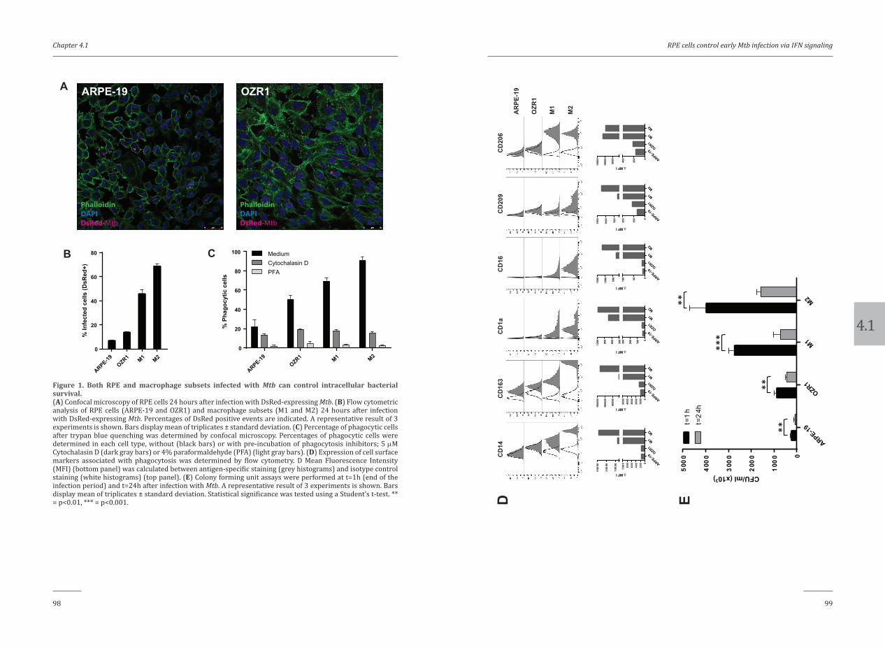
4.1
Chapter 4.1
98 99
RPE cells control early Mtb infection via IFN signaling
ARPE-19OZR1 M1 M2
0
20
40
60
80
100
% P
hago
cytic
cel
ls
MediumCytochalasin DPFA
ARPE-19OZR1 M1 M2
0
20
40
60
80
% In
fect
ed c
ells
(DsR
ed+)
A
B
ARPE-19 OZR1
PhalloidinDAPIDsRed-Mtb
PhalloidinDAPIDsRed-Mtb
C
Figure 1. Both RPE and macrophage subsets infected with Mtb can control intracellular bacterial survival.(A) Confocal microscopy of RPE cells 24 hours after infection with DsRed-expressing Mtb. (B) Flow cytometric analysis of RPE cells (ARPE-19 and OZR1) and macrophage subsets (M1 and M2) 24 hours after infection with DsRed-expressing Mtb. Percentages of DsRed positive events are indicated. A representative result of 3 experiments is shown. Bars display mean of triplicates ± standard deviation. (C) Percentage of phagocytic cells after trypan blue quenching was determined by confocal microscopy. Percentages of phagocytic cells were determined in each cell type, without (black bars) or with pre-incubation of phagocytosis inhibitors; 5 µM Cytochalasin D (dark gray bars) or 4% paraformaldehyde (PFA) (light gray bars). (D) Expression of cell surface markers associated with phagocytosis was determined by flow cytometry. D Mean Fluorescence Intensity (MFI) (bottom panel) was calculated between antigen-specific staining (grey histograms) and isotype control staining (white histograms) (top panel). (E) Colony forming unit assays were performed at t=1h (end of the infection period) and t=24h after infection with Mtb. A representative result of 3 experiments is shown. Bars display mean of triplicates ± standard deviation. Statistical significance was tested using a Student’s t-test. ** = p<0.01, *** = p<0.001. D E
ARPE-19
OZR1
M1
M2
0
100
0
200
0
300
0
400
0
500
0
CFU/ml (x103)
t=1
ht=
24h
****
**
***
CD
163
CD
1aC
D16
CD
209
CD
206
AR
PE-1
9
OZR
1
M1
M2
CD
14
ARPE-19OZR1
M1M2
0
100
200
300
300
600
900
1200
∆ MFI
1000
00
1500
00 ARPE-19OZR1
M1M2
010
0020
0030
0040
0050
00
5000
0
∆ MFI
1000
1500 ARPE-19
OZR1M1M2
050100
500
∆ MFI
ARPE-19OZR1
M1M2
0
100
200
500
1000
1500
∆ MFI
ARPE-19OZR1
M1M2
0
200
400
5000
1000
0
1500
0
∆ MFI
02
004
006
008
001
000
100
00
300
00
500
00
∆ MFI
ARPE-19OZR1
M1M2
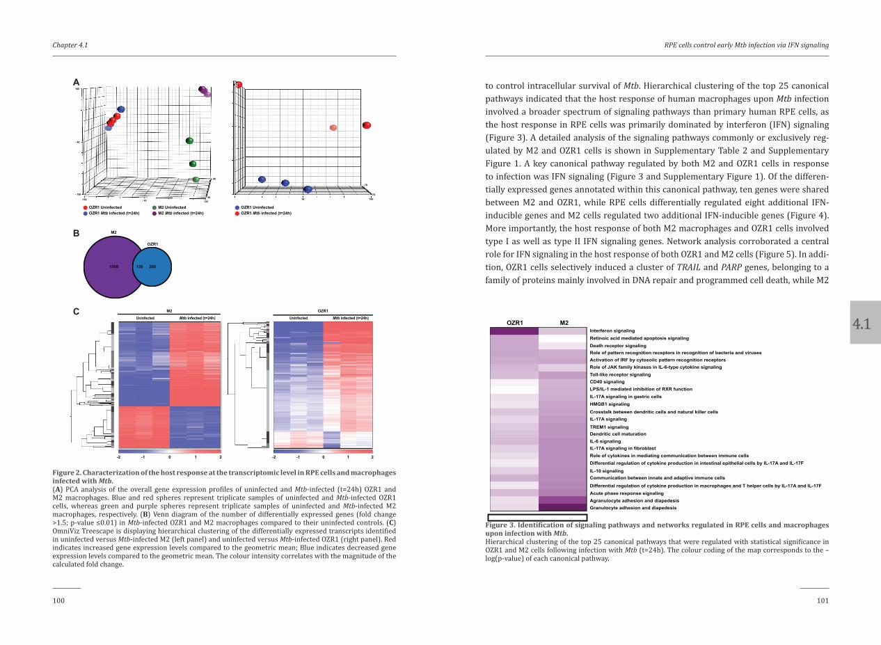
4.1
Chapter 4.1
100 101
RPE cells control early Mtb infection via IFN signaling
to control intracellular survival of Mtb. Hierarchical clustering of the top 25 canonical pathways indicated that the host response of human macrophages upon Mtb infection involved a broader spectrum of signaling pathways than primary human RPE cells, as the host response in RPE cells was primarily dominated by interferon (IFN) signaling (Figure 3). A detailed analysis of the signaling pathways commonly or exclusively reg-ulated by M2 and OZR1 cells is shown in Supplementary Table 2 and Supplementary Figure 1. A key canonical pathway regulated by both M2 and OZR1 cells in response to infection was IFN signaling (Figure 3 and Supplementary Figure 1). Of the differen-tially expressed genes annotated within this canonical pathway, ten genes were shared between M2 and OZR1, while RPE cells differentially regulated eight additional IFN-inducible genes and M2 cells regulated two additional IFN-inducible genes (Figure 4). More importantly, the host response of both M2 macrophages and OZR1 cells involved type I as well as type II IFN signaling genes. Network analysis corroborated a central role for IFN signaling in the host response of both OZR1 and M2 cells (Figure 5). In addi-tion, OZR1 cells selectively induced a cluster of TRAIL and PARP genes, belonging to a family of proteins mainly involved in DNA repair and programmed cell death, while M2
-140-80
20
120
OZR1 UninfectedOZR1 Mtb infected (t=24h)
1601070
-70
- 40
130- 10-150
- 180
100
90
- 90
OZR1 UninfectedOZR1 Mtb infected (t=24h)
M2 UninfectedM2 Mtb infected (t=24h)
A
B
C
M2
OZR1
1508 130 260
M2
Mtb infected (t=24h)Uninfected
-2 -1 0 1 2 -2 -1 0 1 2
OZR1
Mtb infected (t=24h)Uninfected
Figure 2. Characterization of the host response at the transcriptomic level in RPE cells and macrophages infected with Mtb.(A) PCA analysis of the overall gene expression profiles of uninfected and Mtb-infected (t=24h) OZR1 and M2 macrophages. Blue and red spheres represent triplicate samples of uninfected and Mtb-infected OZR1 cells, whereas green and purple spheres represent triplicate samples of uninfected and Mtb-infected M2 macrophages, respectively. (B) Venn diagram of the number of differentially expressed genes (fold change >1.5; p-value ≤0.01) in Mtb-infected OZR1 and M2 macrophages compared to their uninfected controls. (C) OmniViz Treescape is displaying hierarchical clustering of the differentially expressed transcripts identified in uninfected versus Mtb-infected M2 (left panel) and uninfected versus Mtb-infected OZR1 (right panel). Red indicates increased gene expression levels compared to the geometric mean; Blue indicates decreased gene expression levels compared to the geometric mean. The colour intensity correlates with the magnitude of the calculated fold change.
0 24.008-log(p-value)
Interferon signalingRetinoic acid mediated apoptosis signalingDeath receptor signalingRole of pattern recognition receptors in recognition of bacteria and virusesActivation of IRF by cytosolic pattern recognition receptorsRole of JAK family kinases in IL-6-type cytokine signalingToll-like receptor signalingCD40 signalingLPS/IL-1 mediated inhibition of RXR functionIL-17A signaling in gastric cellsHMGB1 signalingCrosstalk between dendritic cells and natural killer cellsIL-17A signaling
TREM1 signalingDendritic cell maturationIL-6 signalingIL-17A signaling in fibroblastRole of cytokines in mediating communication between immune cellsDifferential regulation of cytokine production in intestinal epithelial cells by IL-17A and IL-17FIL-10 signalingCommunication between innate and adaptive immune cells
Differential regulation of cytokine production in macrophages and T helper cells by IL-17A and IL-17FAcute phase response signalingAgranulocyte adhesion and diapedesisGranulocyte adhesion and diapedesis
OZR1 M2
Figure 3. Identification of signaling pathways and networks regulated in RPE cells and macrophages upon infection with Mtb.Hierarchical clustering of the top 25 canonical pathways that were regulated with statistical significance in OZR1 and M2 cells following infection with Mtb (t=24h). The colour coding of the map corresponds to the –log(p-value) of each canonical pathway.
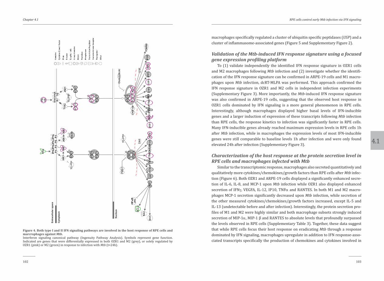
4.1
Chapter 4.1
102 103
RPE cells control early Mtb infection via IFN signaling
macrophages specifically regulated a cluster of ubiquitin specific peptidases (USP) and a cluster of inflammasome-associated genes (Figure 5 and Supplementary Figure 2).
Validation of the Mtb-induced IFN response signature using a focused gene expression profiling platform
To (1) validate independently the identified IFN response signature in OZR1 cells and M2 macrophages following Mtb infection and (2) investigate whether the identifi-cation of the IFN response signature can be confirmed in ARPE-19 cells and M1 macro-phages upon Mtb infection, dcRT-MLPA was performed. This approach confirmed the IFN response signature in OZR1 and M2 cells in independent infection experiments (Supplementary Figure 3). More importantly, the Mtb-induced IFN response signature was also confirmed in ARPE-19 cells, suggesting that the observed host response in OZR1 cells dominated by IFN signaling is a more general phenomenon in RPE cells. Interestingly, although macrophages displayed higher basal levels of IFN-inducible genes and a larger induction of expression of these transcripts following Mtb infection than RPE cells, the response kinetics to infection was significantly faster in RPE cells. Many IFN-inducible genes already reached maximum expression levels in RPE cells 1h after Mtb infection, while in macrophages the expression levels of most IFN-inducible genes were still comparable to baseline levels 1h after infection and were only found elevated 24h after infection (Supplementary Figure 3).
Characterization of the host response at the protein secretion level in RPE cells and macrophages infected with Mtb
Similar to the transcriptomic response, macrophages also secreted quantitatively and qualitatively more cytokines/chemokines/growth factors than RPE cells after Mtb infec-tion (Figure 6). Both OZR1 and ARPE-19 cells displayed a significantly enhanced secre-tion of IL-6, IL-8, and MCP-1 upon Mtb infection while OZR1 also displayed enhanced secretion of IFNγ, VEGFA, IL-12, IP10, TNFα and RANTES. In both M1 and M2 macro-phages MCP-1 secretion significantly decreased upon Mtb infection, while secretion of the other measured cytokines/chemokines/growth factors increased, except IL-5 and IL-13 (undetectable before and after infection). Interestingly, the protein secretion pro-files of M1 and M2 were highly similar and both macrophage subsets strongly induced secretion of MIP-1α, MIP-1 β and RANTES to absolute levels that profoundly surpassed the levels observed in RPE cells (Supplementary Table 3). Together, these data suggest that while RPE cells focus their host response on eradicating Mtb through a response dominated by IFN signaling, macrophages upregulate in addition to IFN response-asso-ciated transcripts specifically the production of chemokines and cytokines involved in
Nuc
leus
Cyt
opla
sm
Extr
acel
lula
r spa
ce
Figure 4. Both type I and II IFN signaling pathways are involved in the host response of RPE cells and marcrophages against Mtb.Interferon signaling canonical pathway (Ingenuity Pathway Analysis). Symbols represent gene function. Indicated are genes that were differentially expressed in both OZR1 and M2 (grey), or solely regulated by OZR1 (pink) or M2 (green) in response to infection with Mtb (t=24h).

4.1
Chapter 4.1
104 105
RPE cells control early Mtb infection via IFN signaling
Inte
rfer
on s
igna
ling
Apo
ptos
is a
nd d
eath
re
cept
or s
igna
ling
A
Figure 5. Network analysis of the host response in RPE cells and macrophages following Mtb infection.Ingenuity based pathway analysis of genes that were differentially expressed between (A) uninfected and Mtb-infected OZR1 cells and (B) uninfected and Mtb-infected M2 macrophages (t=24h). Shown are the top networks identified in both cell types. Type I and II IFN-inducible genes are highlighted in red, apoptosis and death receptor signaling genes are highlighted in blue, protein ubiquitination-associated genes are indicated in green, and inflammasome-associated genes are shown in yellow.
B
Inte
rfer
on s
igna
ling
Prot
ein
ubiq
uina
tion
Infla
mm
asom
e
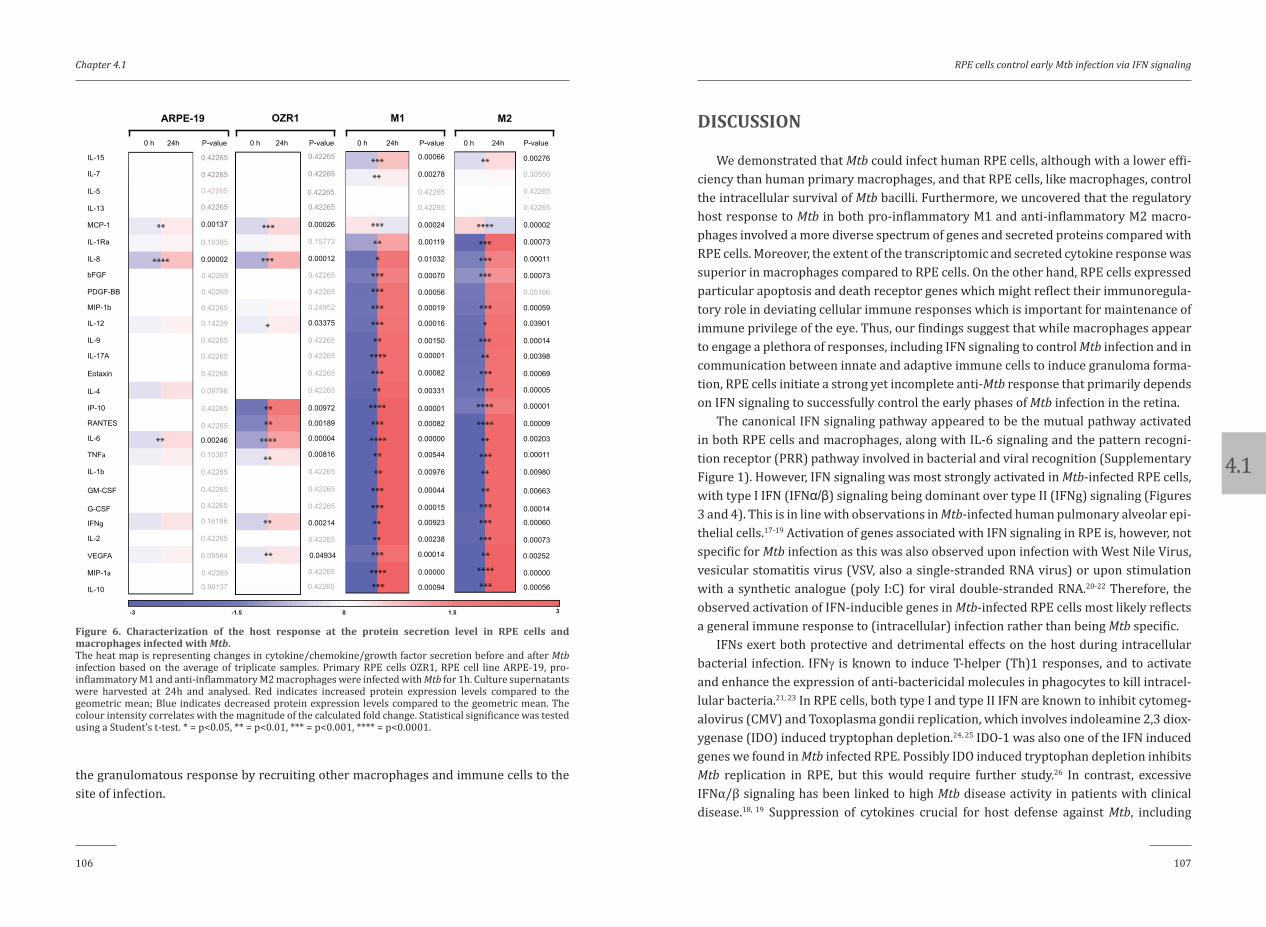
4.1
Chapter 4.1
106 107
RPE cells control early Mtb infection via IFN signaling
DISCUSSION
We demonstrated that Mtb could infect human RPE cells, although with a lower effi-ciency than human primary macrophages, and that RPE cells, like macrophages, control the intracellular survival of Mtb bacilli. Furthermore, we uncovered that the regulatory host response to Mtb in both pro-inflammatory M1 and anti-inflammatory M2 macro-phages involved a more diverse spectrum of genes and secreted proteins compared with RPE cells. Moreover, the extent of the transcriptomic and secreted cytokine response was superior in macrophages compared to RPE cells. On the other hand, RPE cells expressed particular apoptosis and death receptor genes which might reflect their immunoregula-tory role in deviating cellular immune responses which is important for maintenance of immune privilege of the eye. Thus, our findings suggest that while macrophages appear to engage a plethora of responses, including IFN signaling to control Mtb infection and in communication between innate and adaptive immune cells to induce granuloma forma-tion, RPE cells initiate a strong yet incomplete anti-Mtb response that primarily depends on IFN signaling to successfully control the early phases of Mtb infection in the retina.
The canonical IFN signaling pathway appeared to be the mutual pathway activated in both RPE cells and macrophages, along with IL-6 signaling and the pattern recogni-tion receptor (PRR) pathway involved in bacterial and viral recognition (Supplementary Figure 1). However, IFN signaling was most strongly activated in Mtb-infected RPE cells, with type I IFN (IFNα/β) signaling being dominant over type II (IFNg) signaling (Figures 3 and 4). This is in line with observations in Mtb-infected human pulmonary alveolar epi-thelial cells.17-19 Activation of genes associated with IFN signaling in RPE is, however, not specific for Mtb infection as this was also observed upon infection with West Nile Virus, vesicular stomatitis virus (VSV, also a single-stranded RNA virus) or upon stimulation with a synthetic analogue (poly I:C) for viral double-stranded RNA.20-22 Therefore, the observed activation of IFN-inducible genes in Mtb-infected RPE cells most likely reflects a general immune response to (intracellular) infection rather than being Mtb specific.
IFNs exert both protective and detrimental effects on the host during intracellular bacterial infection. IFNγ is known to induce T-helper (Th)1 responses, and to activate and enhance the expression of anti-bactericidal molecules in phagocytes to kill intracel-lular bacteria.21, 23 In RPE cells, both type I and type II IFN are known to inhibit cytomeg-alovirus (CMV) and Toxoplasma gondii replication, which involves indoleamine 2,3 diox-ygenase (IDO) induced tryptophan depletion.24, 25 IDO-1 was also one of the IFN induced genes we found in Mtb infected RPE. Possibly IDO induced tryptophan depletion inhibits Mtb replication in RPE, but this would require further study.26 In contrast, excessive IFNα/β signaling has been linked to high Mtb disease activity in patients with clinical disease.18, 19 Suppression of cytokines crucial for host defense against Mtb, including
the granulomatous response by recruiting other macrophages and immune cells to the site of infection.
MCP-1
IL-5
IL-13
IL-15
IL-1Ra
IL-8
IL-7
MIP-1b
PDGF-BB
IL-4
bFGF
IP-10
IFNg
IL-12
IL-2
IL-9
MIP-1a
IL-17A
Eotaxin
GM-CSF
TNFa
G-CSF
IL-1b
IL-10
VEGFA
IL-6
RANTES
ARPE-19
0 h 24h
OZR1
0 h 24h
M1
0 h 24h
M2
0 h 24h
****
**
**
***
****
**
**
**
**
**
**
**
**
***
***
****
***
***
******
****
******
**
***
**
****
***
***
**
*****
***
***
***
***
***
****
***
****
****
*
***
***
***
***
*
**
***
***
***
***
****
***
***
***
****
****P-value P-value P-valueP-value
0.00137
0.42265
0.42265
0.42265
0.42265
0.42265
0.42265
0.42265
0.42265
0.42265
0.42265
0.42265
0.42265
0.42265
0.42265
0.42265
0.42265
0.42265
0.42265
0.24952
0.03375
0.42265
0.00972
0.00189
0.00004
0.00816
0.42265
0.42265
0.00214
0.10395
0.00002
0.10367
0.09584
0.16186
0.42265
0.14239
0.42265
0.42265
0.99137
0.09796
0.00246
0.42265
0.00026
0.15773
0.00012
0.42265
0.42265
0.42265
0.42265
0.42265
0.42265
0.04934
0.42265
0.42265
0.00278
0.42265
0.42265
0.00056
0.00019
0.00016
0.00331
0.00001
0.00082
0.00000
0.00544
0.00976
0.00015
0.00923
0.00066
0.00024
0.00119
0.01032
0.00070
0.00150
0.00001
0.00082
0.00044
0.00238
0.00014
0.00000
0.00094
0.30550
0.42265
0.42265
0.05166
0.00059
0.03901
0.00005
0.00001
0.00009
0.00203
0.00011
0.00980
0.00014
0.00060
0.00276
0.00002
0.00073
0.00011
0.00073
0.00014
0.00398
0.00069
0.00663
0.00073
0.00252
0.00000
0.00056
-3 -1.5 0 1.5 3
****
*
**
****
**
***
***
*
****
**
**
**
***
**
***
**
*
******
***
***
**
****
***
**
****
***
****
**
**
***
***
**
*****
*******
**
****
***
***
***
***
*
***
**
***
****
****
****
**
***
**
**
***
***
***
******
***
****
Figure 6. Characterization of the host response at the protein secretion level in RPE cells and macrophages infected with Mtb.The heat map is representing changes in cytokine/chemokine/growth factor secretion before and after Mtb infection based on the average of triplicate samples. Primary RPE cells OZR1, RPE cell line ARPE-19, pro-inflammatory M1 and anti-inflammatory M2 macrophages were infected with Mtb for 1h. Culture supernatants were harvested at 24h and analysed. Red indicates increased protein expression levels compared to the geometric mean; Blue indicates decreased protein expression levels compared to the geometric mean. The colour intensity correlates with the magnitude of the calculated fold change. Statistical significance was tested using a Student’s t-test. * = p<0.05, ** = p<0.01, *** = p<0.001, **** = p<0.0001.

4.1
Chapter 4.1
108 109
RPE cells control early Mtb infection via IFN signaling
where only short distance inter-cellular communication is required as opposed to that of macrophages that need to instruct other (immune) cells over longer distances. Mtb infection was associated with increased IL-12 production by RPE cells, although it was upregulated to a lesser extent than seen in macrophages. It is tempting to hypothesize that IL-12 derived from Mtb-infected RPE cells is involved in local Th1-lymphocyte acti-vation and uveitis-like disease. Also, production of RANTES and IP-10 by Mtb-infected RPE cells may further amplify local T-lymphocyte recruitment and activation. This is further supported by observations in a primed mycobacterial uveitis model in rats that was associated with increased production of IP-10.37 Besides local Th1-lymphocyte acti-vation, it is possible that RPE function in maintaining immune privilege is compromised after a certain degree of infection. Mtb-infected RPE cells produced IL-6 that antagonizes TGF-β which has an important regulatory role in ocular immune privilege.38 Further insight into the involved pathways may contribute to a better immunopathologic under-standing of Mtb-associated uveitis, which may also be of benefit for finding new treat-ment modalities.
In conclusion, we demonstrated that Mtb can infect RPE cells and that RPE cells can control the intracellular survival of Mtb bacilli similar to macrophages. However, RPE cells phagocytosed Mtb less effectively than macrophages. RPE cells displayed a clear yet restricted anti-Mtb response that primarily used IFN signaling to control early phases of Mtb infection while macrophages engage a more diverse repertoire of responses includ-ing IFN signaling and production of cytokines/chemokines to facilitate communication between innate and adaptive immune cells to induce granuloma formation. RPE cells more strongly activated death receptor and retinoic acid-mediated apoptosis signaling pathways which are postulated to represent an immuno-suppressive mechanism to pro-tect the retina from excessive inflammation and damage.
CONFLICT OF INTEREST
The authors declare that they have no conflicting and competing financial interests.
FUNDING
This project was funded by the Indonesian Ministry of Research Technology and Higher Education. We also gratefully acknowledge the support of the Netherlands Organization for Health Research and Development (ZonMw-TOP grant 91214038). The
IL-1α, IL-1β, IL-12 and TNFα, as well as repression of innate cell responsiveness to IFNg have been proposed as important mechanisms of IFNα/β-mediated immunosup-pression during Mtb infection.4, 20, 21, 27-31 Moreover, RPE-derived IFNβ can down-regu-late CXCL9 and intercellular adhesion molecule 1 (ICAM-1) expression by RPE in an autocrine manner. This potentially limits retinal T-lymphocyte/ natular killer (NK) cell recruitment and has been proposed as an immuno-suppressive mechanism that protects the retina from excessive inflammation, for instance in case of RPE infection.20 Our data suggest that in RPE cells, at least during the early stages of Mtb infection, IFN signaling may have a beneficial effect by inhibiting the outgrowth of intracellular Mtb. However, it could be speculated that the (prolonged) high induction of IFNα/β could potentially be detrimental at later stages of infection and thereby contributes to Mtb-mediated uveitis due to chronic infection or Mtb-latency in the RPE cells. Clearly, unraveling the exact role or balance of IFN signaling and Th1 induction in RPE in Mtb-mediated uveitis requires further investigation.
Molecules related to protein ubiquitination and inflammasome pathways were amongst the genes more specifically identified in the M2 regulatory network (Figure 5B). These findings are in line with other in vitro studies demonstrating that Mtb-infected macrophages exhibit innate immune functions (including inflammasome acti-vation and ubiquitin-mediated autophagy) to kill intracellular mycobacteria but also to induce adaptive immunity.3, 32 A canonical pathway more prominently regulated in RPE upon Mtb infection compared to M2 macrophages was related to cell death and survival (Figures 3 and 5A). One of the apoptosis-related genes that increased in Mtb-infected RPE (TNFSF10/TRAIL/APO2L; Supplementary Figure 2) encodes the pro-apoptotic membrane expressed molecule TNF-related apoptosis-inducing ligand (TRAIL). TRAIL is expressed by many ocular tissues, including RPE, and contributes to ocular immune privilege, most likely by inducing apoptosis of infiltrating inflammatory cells that express TRAIL-receptor 2 (TRAIL-R2/DR5/TNFRSF10B).33 Type I IFNs are known to enhance TRAIL expression, for instance on dendritic cells, which enables these cells to induce T-lymphocyte apoptosis.34 Induction of TRAIL on RPE may thus represent a way to deviate the cellular immune response in order to protect the retina from excessive inflammation and to limit the immunopathologic damage. On the other hand, human RPE cells express TRAIL-R2 (this study and33) and necrotic Mtb-positive RPE cells have been observed in human eyes,8 suggesting that TRAIL-TRAIL-R2 interactions between RPE cells may induce death of RPE cells and possibly contribute to inflammation.
RPE cells are well-known producers of mediators that attract and activate different types of leukocytes and as such are considered important initiators/regulators of retinal inflammation.35, 36 Yet Mtb-infected RPE secreted substantial lower amounts of cytokines than Mtb-infected macrophages did. This might reflect the local ocular function of RPE

4.1
Chapter 4.1
110 111
RPE cells control early Mtb infection via IFN signaling
9. Nazari H, Karakousis PC, Rao NA. Replication of Mycobacterium tuberculosis in retinal pigment epi-
thelium. JAMA Ophthalmol 2014;132:724-729.
10. Means TK, Wang S, Lien E, Yoshimura A, Golenbock DT, Fenton MJ. Human toll-like receptors mediate
cellular activation by Mycobacterium tuberculosis. Journal of immunology 1999;163:3920-3927.
11. van Bilsen K, van Hagen PM, Bastiaans J, et al. The neonatal Fc receptor is expressed by human retinal
pigment epithelial cells and is downregulated by tumour necrosis factor-alpha. The British journal of
ophthalmology 2011;95:864-868.
12. Verreck FA, de Boer T, Langenberg DM, van der Zanden L, Ottenhoff TH. Phenotypic and function-
al profiling of human proinflammatory type-1 and anti-inflammatory type-2 macrophages in re-
sponse to microbial antigens and IFN-gamma- and CD40L-mediated costimulation. J Leukoc Biol
2006;79:285-293.
13. Korbee CJ, Heemskerk MT, Kocev D, et al. Combined chemical genetics and data-driven bioinformatics
approach identifies receptor tyrosine kinase inhibitors as host-directed antimicrobials. Nat Commun
2018;9:358.
14. Jett BD, Hatter KL, Huycke MM, Gilmore MS. Simplified agar plate method for quantifying viable bac-
teria. Biotechniques 1997;23:648-650.
15. Leclerc L, Boudard D, Pourchez J, et al. Quantification of microsized fluorescent particles phagocyto-
sis to a better knowledge of toxicity mechanisms. Inhal Toxicol 2010;22:1091-1100.
16. Joosten SA, Goeman JJ, Sutherland JS, et al. Identification of biomarkers for tuberculosis disease using
a novel dual-color RT-MLPA assay. Genes Immun 2012;13:71-82.
17. Mvubu NE, Pillay B, Gamieldien J, Bishai W, Pillay M. Canonical pathways, networks and transcription-
al factor regulation by clinical strains of Mycobacterium tuberculosis in pulmonary alveolar epithelial
cells. Tuberculosis 2016;97:73-85.
18. Berry MP, Graham CM, McNab FW, et al. An interferon-inducible neutrophil-driven blood transcrip-
tional signature in human tuberculosis. Nature 2010;466:973-977.
19. Ottenhoff TH, Dass RH, Yang N, et al. Genome-wide expression profiling identifies type 1 interferon
response pathways in active tuberculosis. PLoS One 2012;7:e45839.
20. Hooks JJ, Nagineni CN, Hooper LC, Hayashi K, Detrick B. IFN-beta provides immuno-protection in the
retina by inhibiting ICAM-1 and CXCL9 in retinal pigment epithelial cells. J Immunol 2008;180:3789-
3796.
21. McNab F, Mayer-Barber K, Sher A, Wack A, O’Garra A. Type I interferons in infectious disease. Nat Rev
Immunol 2015;15:87-103.
22. Munoz-Erazo L, Natoli R, Provis JM, Madigan MC, King NJ. Microarray analysis of gene expression in
West Nile virus-infected human retinal pigment epithelium. Mol Vis 2012;18:730-743.
23. Flynn JL, Chan J, Triebold KJ, Dalton DK, Stewart TA, Bloom BR. An essential role for interferon gam-
ma in resistance to Mycobacterium tuberculosis infection. The Journal of experimental medicine
1993;178:2249-2254.
funders had no role in study design, data collection, and analysis, decision to publish, or preparation of the manuscript.
ACKNOWLEDGEMENTS
Mycobacterial reporter constructs were kindly provided by Dr. J. Bestebroer (VUMC, Amsterdam, The Netherlands).
AUTHOR CONTRIBUTIONS
P.M.v.H. W.A.D., M.v.V., T.H.M.O., and M.C.H. conceptualized and designed the study. R.L.D.N., W.A.D., S.M.A.S., P.J.vd.S., and M.C.H. analyzed the transcriptomic and secreted protein data and wrote the manuscript. K.V.W. designed and performed the Mtb infection experiments and analyzed the data. E.Q. and R.L.D.N. executed the dcRT-MLPA experi-ments. M.C.H. had primary responsibility for the final content of the manuscript; and all authors have read and approved the final manuscript.
REFERENCES
1. World Health Organization. Global tuberculosis report. Geneva, Switzerland: World Health Organisa-
tion; 2016.
2. Turner RD, Bothamley GH. Cough and the transmission of tuberculosis. The Journal of infectious dis-
eases 2015;211:1367-1372.
3. Watson RO, Manzanillo PS, Cox JS. Extracellular M. tuberculosis DNA targets bacteria for autophagy
by activating the host DNA-sensing pathway. Cell 2012;150:803-815.
4. O’Garra A, Redford PS, McNab FW, Bloom CI, Wilkinson RJ, Berry MP. The immune response in tuber-
culosis. Annu Rev Immunol 2013;31:475-527.
5. Barrios-Payan J, Saqui-Salces M, Jeyanathan M, et al. Extrapulmonary locations of mycobacterium
tuberculosis DNA during latent infection. The Journal of infectious diseases 2012;206:1194-1205.
6. Randall PJ, Hsu NJ, Quesniaux V, Ryffel B, Jacobs M. Mycobacterium tuberculosis infection of the
‘non-classical immune cell’. Immunol Cell Biol 2015;93:789-795.
7. Wroblewski KJ, Hidayat AA, Neafie RC, Rao NA, Zapor M. Ocular tuberculosis: A clinicopathologic and
molecular study. Ophthalmology 2011;118:772-777.
8. Rao NA, Saraswathy S, Smith RE. Tuberculous uveitis: distribution of Mycobacterium tuberculosis in
the retinal pigment epithelium. Arch Ophthalmol 2006;124:1777-1779.

4.1
Chapter 4.1
112 113
RPE cells control early Mtb infection via IFN signaling
SUPPLEMENTAL DATA24. Bodaghi B, Goureau O, Zipeto D, Laurent L, Virelizier JL, Michelson S. Role of IFN-gamma-induced
indoleamine 2,3 dioxygenase and inducible nitric oxide synthase in the replication of human cyto-
megalovirus in retinal pigment epithelial cells. J Immunol 1999;162:957-964.
25. Nagineni CN, Pardhasaradhi K, Martins MC, Detrick B, Hooks JJ. Mechanisms of interferon-induced
inhibition of Toxoplasma gondii replication in human retinal pigment epithelial cells. Infect Immun
1996;64:4188-4196.
26. Blumenthal A, Nagalingam G, Huch JH, et al. M. tuberculosis induces potent activation of IDO-1, but
this is not essential for the immunological control of infection. PLoS One 2012;7:e37314.
27. de Paus RA, van Wengen A, Schmidt I, et al. Inhibition of the type I immune responses of human
monocytes by IFN-alpha and IFN-beta. Cytokine 2013;61:645-655.
28. McNab FW, Ewbank J, Howes A, et al. Type I IFN induces IL-10 production in an IL-27-independent
manner and blocks responsiveness to IFN-gamma for production of IL-12 and bacterial killing in
Mycobacterium tuberculosis-infected macrophages. Journal of immunology 2014;193:3600-3612.
29. Teles RM, Graeber TG, Krutzik SR, et al. Type I interferon suppresses type II interferon-triggered
human anti-mycobacterial responses. Science 2013;339:1448-1453.
30. Mayer-Barber KD, Andrade BB, Oland SD, et al. Host-directed therapy of tuberculosis based on inter-
leukin-1 and type I interferon crosstalk. Nature 2014;511:99-103.
31. Mayer-Barber KD, Barber DL, Shenderov K, et al. Caspase-1 independent IL-1beta production is crit-
ical for host resistance to mycobacterium tuberculosis and does not require TLR signaling in vivo.
Journal of immunology 2010;184:3326-3330.
32. van Crevel R, Ottenhoff TH, van der Meer JW. Innate immunity to Mycobacterium tuberculosis. Clin
Microbiol Rev 2002;15:294-309.
33. Hueber A, Aduckathil S, Kociok N, et al. Apoptosis-mediating receptor-ligand systems in human reti-
nal pigment epithelial cells. Graefes Arch Clin Exp Ophthalmol 2002;240:551-556.
34. Leplina OY, Tyrinova TV, Tikhonova MA, Ostanin AA, Chernykh ER. Interferon alpha induces gener-
ation of semi-mature dendritic cells with high pro-inflammatory and cytotoxic potential. Cytokine
2015;71:1-7.
35. Bastiaans J, van Meurs JC, van Holten-Neelen C, et al. Factor Xa and thrombin stimulate proinflam-
matory and profibrotic mediator production by retinal pigment epithelial cells: a role in vitreoretinal
disorders? Graefes Arch Clin Exp Ophthalmol 2013;251:1723-1733.
36. Holtkamp GM, Kijlstra A, Peek R, de Vos AF. Retinal pigment epithelium-immune system interactions:
cytokine production and cytokine-induced changes. Prog Retin Eye Res 2001;20:29-48.
37. Pepple KL, Rotkis L, Van Grol J, et al. Primed Mycobacterial Uveitis (PMU): Histologic and Cyto-
kine Characterization of a Model of Uveitis in Rats. Investigative ophthalmology & visual science
2015;56:8438-8448.
38. Ohta K, Yamagami S, Taylor AW, Streilein JW. IL-6 antagonizes TGF-beta and abolishes immune privi-
lege in eyes with endotoxin-induced uveitis. Invest Ophthalmol Vis Sci 2000;41:2591-2599.
-log(p-value)
Granulocyte adhesion and diapedesis
Acute phase response signaling
Agranulocyte adhesion and diapedesis
Communication between innate and adaptive immune cells
Crosstalk between dendritic cells and natural killer cells
Dendritic cell maturation
Differential regulation of cytokine production in macrophages and T helper cells by IL-17A and IL-17F
Differential regulation of cytokine production in intestinal epithelial cells by IL-17A and IL-17F
IL-10 signaling
LPS/IL-1 mediated inhibition of RXR function
Role of cytokines in mediating communication between immune cells
TREM1 signaling
IL-6 signaling
Interferon signaling
Role of pattern recognition receptors in recognition of bacteria and viruses
Interferon signaling
Retinoic acid mediated apoptosis signaling
Death receptor signaling
z score1.27 3.31
M2
M2 and OZR1
OZR1
0 2 4 6 8 10 12 14 16 18 20
Supplementary Figure 1. Molecular signatures of Mtb infection in OZR1 and M2 cells identified by Ingenuity Pathway Analysis differ in intensity and quality. Bar charts represent canonical pathways that were common (middle panel) or exclusively regulated by either M2 (top panel) or OZR1 cells (lower panel) upon infection with Mtb. The color coding of the bars corresponds to the z-score of each canonical pathway.

4.1
Chapter 4.1
114 115
RPE cells control early Mtb infection via IFN signaling
-log(p-value)0.00E00 22.2
Gene expression log ratio-4.029 9.781
Death receptor signaling pathways Retinoic acid mediated apoptosis signaling pathways
M2 OZR1-log(p-value)0.00E00 22.2
Gene expression log ratio-4.029 9.781
M2 OZR1
TRAILTNFAPARP9PARP14PARP10PARP12CASP7TL1NFKB2NFKBIA
TRAILIFNB1IRF1PARP9IFNGPARP14PARP12PARP10FLIPTNFRSF10A
Supplementary Figure 2. Heatmaps of genes belonging to the death receptor and retinoic acid-mediated apoptosis signaling pathways that were differentially expressed upon Mtb infection.Shown are the –log(p-values) of the death receptor and retinoic acid-mediated apoptosis signaling pathways in M2 and OZR1 cells, indicating both pathways were regulated more profoundly in OZR1 than in M2 following infection with Mtb. The log ratio of the top 10 differentially expressed genes within each pathway is also indicated. Green defines lower gene expression levels, and red defines higher gene expression levels compared to the uninfected control.

4.1
Chapter 4.1
116 117
RPE cells control early Mtb infection via IFN signaling
Supplementary Table 1. List of differentially expressed genes in OZR1 and M2 cells following infection with Mtb.
Supplementary Table 2. List of all canonical pathways differentially regulated in OZR1 and M2 cells upon Mtb infection.
Supplementary Table 3. Expression levels of secreted proteins in the supernatant of OZR1 and M2 cells following infection with Mtb.
Available for download at:http://iovs.arvojournals.org/article.aspx?articleid=2674883
BLR
1K
IF1B
BM
P6
IFI1
6IL
1RN
IRF1
FCG
R3B
FCG
R2A
FCG
R1A
AR
PE-1
9
0h24
h
OZR
1
0h24
h
M1
0h24
h
M2
0h24
h 1h
1h1h
1h
FOX
P3
HP
RT
CX
CR
4B
IRC
3C
D3E
ISG
15PA
RP
10S
PATA
13B
CL6
CD
19C
X3C
L1
EG
FA
PO
BE
C3G
DD
X17
IFN
B1
VE
GF
IFIT
5D
SE
IRF7
STA
T2H
CK
IFI3
5A
SA
P1
TNIP
1IF
ITM
3IL
7RIL
13IF
NA
1D
RA
P1
IFI2
7C
D74
CD
14A
AK
1IL
12B
RS
AD
2S
OC
S1
IFI4
4L
LYN
IFIH
1O
AS
3
OA
S1
STA
T1IFI6IL6
IFIT
2O
AS
2C
XC
L10
CX
CL9
CD
274
TAP
2IF
IT3
IND
OG
BP
1C
CL1
1G
BP
5G
BP
2C
CL4
CC
L3
SLA
MF7
TAP
1
IFI4
4
-1-0
.50
0.5
1
P-v
alue
0h
vs
1hP
-val
ue
1h v
s 24
h
0.00
349
0.08
019
0.07
142
0.00
504
0.05
895
0.44
643
0.00
352
0.00
050
0.00
224
0.89
587
0.01
319
0.16
755
0.00
727
0.42
265
0.42
265
0.37
390
0.05
626
0.54
107
0.18
996
0.24
320
0.07
078
0.42
265
0.00
299
1.00
000
0.42
265
0.42
265
0.66
800
0.75
156
0.37
390
0.42
265
0.10
680
0.37
052
0.11
290
0.44
520
0.50
369
0.67
575
0.55
385
0.22
774
0.40
275
0.15
305
0.42
265
0.54
735
0.66
726
0.46
337
0.42
265
0.50
103
0.42
265
0.42
265
1.00
000
0.00
344
0.00
004
0.00
002
1.00
000
0.42
265
0.13
976
0.61
438
0.42
265
0.00
000
0.08
404
0.02
740
0.42
265
0.42
265
0.42
265
0.42
265
1.00
000
1.00
000
1.00
000
1.00
000
1.00
000
1.00
000
1.00
000
1.00
000
1.00
000
1.00
000
0.20
127
0.00
067
0.00
004
0.00
085
0.42
265
1.00
000
1.00
000
1.00
000
1.00
000
1.00
000
1.00
000
1.00
000
1.00
000
1.00
000
0.00
046
0.00
050
0.00
036
0.00
041
0.00
019
0.00
041
0.00
000
0.00
032
0.00
009
0.00
960
0.00
052
0.00
037
0.00
000
0.00
000
0.00
038
0.00
182
0.00
005
0.00
003
0.00
013
0.00
038
0.00
026
0.00
004
0.00
002
0.00
000
0.00
005
0.00
000
0.00
130
0.00
027
0.00
002
0.00
005
0.63
002
0.00
178
0.70
602
0.00
196
0.00
024
0.00
001
0.00
001
0.00
048
0.00
007
0.00
050
0.00
003
0.00
000
0.00
011
1.00
000
0.00
021
0.00
032
0.00
079
0.00
000
0.00
003
0.00
005
0.00
020
0.00
639
1.00
000
0.42
265
0.42
265
0.42
265
0.42
265
0.26
933
0.00
000
0.02
722
0.00
111
0.00
036
0.00
000
0.01
925
0.00
023
0.01
027
0.00
002
0.00
004
0.42
265
0.00
002
0.00
144
0.00
020
0.00
064
0.00
519
0.00
114
0.00
054
0.00
025
0.00
097
0.00
099
0.00
038
0.00
120
0.00
003
0.00
097
0.00
000
0.00
027
0.00
037
0.94
600
0.00
989
0.00
062
0.00
352
0.00
001
0.00
066
0.00
553
0.00
018
0.00
041
0.00
009
0.00
129
0.00
002
0.00
022
0.00
020
0.00
066
0.00
000
0.00
693
0.00
230
0.96
007
0.00
348
1.00
000
0.42
265
0.42
265
0.42
265
0.42
265
1.00
000
1.00
000
1.00
000
1.00
000
1.00
000
1.00
000
1.00
000
1.00
000
1.00
000
0.54
233
0.02
713
0.20
321
0.00
101
0.00
031
0.12
163
0.01
488
0.46
134
0.24
653
0.61
787
0.00
004
0.13
975
0.97
948
0.42
265
0.16
692
0.42
265
0.89
847
0.42
265
0.42
265
0.42
265
0.42
265
0.42
265
0.00
002
0.02
782
0.42
265
0.42
265
0.42
265
0.42
265
0.05
200
0.09
612
0.07
349
0.05
939
0.98
085
0.31
427
0.58
282
0.78
190
0.42
265
0.42
265
0.42
265
0.42
265
0.42
265
0.00
613
0.00
000
0.00
001
0.41
347
0.15
408
0.81
236
0.42
265
0.60
630
0.24
268
0.24
529
0.57
292
0.76
055
0.03
453
0.91
593
0.56
466
0.58
233
0.09
095
0.54
080
0.42
265
0.10
931
0.42
265
0.42
265
1.00
000
1.00
000
1.00
000
1.00
000
1.00
000
1.00
000
1.00
000
1.00
000
1.00
000
1.00
000
0.00
156
0.00
722
0.00
009
0.19
181
0.71
274
0.26
597
0.89
217
1.00
000
1.00
000
1.00
000
0.14
397
0.00
254
0.14
913
0.33
207
0.76
440
0.34
792
0.36
945
0.14
449
0.04
931
0.01
127
0.58
259
0.55
626
0.00
069
0.00
719
0.01
763
0.01
101
0.55
485
0.11
915
0.31
296
0.00
055
0.47
275
0.00
494
1.00
000
1.00
000
1.00
000
1.00
000
1.00
000
1.00
000
1.00
000
1.00
000
1.00
000
1.00
000
1.00
000
1.00
000
1.00
000
1.00
000
0.89
671
0.00
309
1.00
000
1.00
000
0.00
072
0.00
000
0.00
122
0.09
507
0.00
209
0.56
687
0.00
249
0.72
160
0.42
265
0.86
422
0.32
355
0.00
067
0.05
609
0.04
325
0.03
135
0.35
563
0.27
467
0.71
192
0.85
522
0.92
104
0.64
675
0.16
526
0.57
453
0.21
467
0.96
433
0.87
260
0.69
186
0.61
409
0.34
132
0.96
819
0.74
121
0.62
930
0.30
678
0.84
657
0.43
083
0.14
392
0.09
534
0.53
780
0.87
080
0.00
030
0.00
000
0.00
001
0.00
017
0.00
032
0.00
000
0.00
004
0.00
069
0.00
000
0.00
020
0.00
040
0.00
080
0.00
481
0.00
476
0.00
010
0.00
008
0.02
712
0.00
054
0.00
002
0.00
008
0.00
002
0.00
519
0.00
004
0.00
119
0.00
009
0.00
001
1.00
000
1.00
000
1.00
000
1.00
000
1.00
000
1.00
000
1.00
000
1.00
000
0.02
123
0.00
380
0.00
361
0.86
611
1.00
000
1.00
000
1.00
000
1.00
000
1.00
000
1.00
000
1.00
000
1.00
000
1.00
000
0.42
265
1.00
000
0.91
536
0.30
913
0.93
126
0.00
649
0.06
972
0.77
799
0.58
138
1.00
000
0.37
390
0.02
303
0.51
833
0.80
640
0.05
980
0.00
017
0.06
671
0.00
001
0.09
209
0.16
135
0.09
171
0.19
462
0.49
563
0.05
707
0.09
057
0.30
413
0.21
971
0.15
164
0.14
669
0.98
409
0.10
443
0.65
034
0.31
512
0.79
100
0.08
508
0.19
880
0.16
882
0.18
473
0.24
766
0.13
705
0.14
455
0.28
403
1.00
000
0.37
390
0.37
390
0.00
079
0.00
283
0.37
390
1.00
000
0.88
938
0.00
054
0.01
028
0.05
351
0.00
036
0.07
977
0.37
390
0.73
510
1.00
000
0.37
390
0.37
390
1.00
000
1.00
000
1.00
000
1.00
000
1.00
000
1.00
000
1.00
000
1.00
000
1.00
000
1.00
000
0.00
413
0.16
265
0.30
198
1.00
000
0.10
642
0.10
221
0.43
227
1.00
000
1.00
000
0.86
702
P-v
alue
0h
vs
1hP
-val
ue
1h v
s 24
hP
-val
ue
0h v
s 1h
P-v
alue
1h
vs
24h
P-v
alue
0h
vs
1hP
-val
ue
1h v
s 24
h
1.00
000
0.0
1054
0.4
8791
0.1
9653
1.0
0000
1.0
0000
1.0
0000
1.0
0000
1.0
0000
1.0
0000
1.0
0000
1.0
0000
1.0
0000
1.0
0000
1.0
0000
1.0
0000
0.2
0013
0.0
5258
0.0
0148
0.00
444
0.0
0000
0.0
0000
0.00
021
0.00
026
0.00
534
0.0
0000
0.00
029
0.18
155
0.42
265
0.68
695
0.00
089
0.00
001
0.00
033
0.00
360
0.00
492
0.00
027
0.00
001
0.00
085
0.00
230
0.00
206
0.00
000
0.00
001
0.01
283
0.00
094
0.00
092
0.00
083
0.00
246
0.00
000
0.00
024
0.00
009
0.00
621
0.00
163
0.00
063
0.00
001
0.00
003
0.00
001
1.00
000
0.42
265
1.00
000
0.00
081
0.42
265
0.64
093
0.00
174
**** ** *******
***
**
**** ***
******
*
** *******
***
***
***
****** **** ***
**** ***** ***
***
***
*****
***
******** ***
****
****
********* ***
****
**** *****
***
*****
****
****
****
**** ****
*
**** ***
**
**** ***
***** ***
*
****
****
*
***
**** ***
***** **
***
**
***
***
**** ** ****
*******
****
****
*
** **** ***
**
***
****
** * ****
******
*** **
***
** ***
***
***
***
******
** ***
****
*****
***** *** **** ***
***
**** *****
***
** ** ****
******* **** ***
****
****
***** ** *** *****
***** **** ***
****
***** ***
**** ***
***
***
***
****
****
****
***
**
***
****
****
*******
***
*
***
****
***
****
* **
********
****
****
***
**** ***
**** ***
**** ***
****
****
****
****
******
**** ***
***
*******
**
*******
****
**** *******
****
****
****** ****
****** ***
***
Supplementary Figure 3. Validation of the host response against infection with Mtb in RPE cells and macrophage subsets using dcRT-MLPA.Relative expression levels are depicted of selected genes in uninfected ARPE-19, OZR1, M1 and M2 cells or cells infected with Mtb for 1h (end of the infection period) or evaluated 24h after infection. Red indicates increased gene expression levels compared to the geometric mean; Blue indicates decreased gene expression levels compared to the geometric mean. The colour intensity with the magnitude of the calculated fold change. Statistical significance between uninfected and 1h infected cells, and between 1 h and 24h infected cells was tested using a Student’s t-test. * = p<0.05, ** = p<0.01, *** = p<0.001, **** = p<0.0001.


.
4.2ChapterType 1 interferon-inducible geneexpression in QuantiFERON Gold
TB-positive uveitis: a tool to stratify a high versus low risk of active tuberculosis?
Rina La Distia Nora, MD1,10*; Ratna Sitompul, MD, PhD1; Mar-leen Bakker, MD, PhD5; Marjan A. Versnel, PhD9;
Sigrid M. A. Swagemakers6,7; Peter J. van der Spek, PhD6,7; Made Susiyanti, MD1; Lukman Edwar, MD1;
Soedarman Sjamsoe, MD1; Gurmeet Singh, MD2; RR Diah Handayani, MD3; Aniki Rothova, MD, PhD4;
P. Martin van Hagen, MD, PhD8,9¶; and Willem A. Dik, PhD9,10
Departments of 1Ophthalmology, 2Internal Medicine, Respirology and Critical Illness Division, University of Indone-
sia & Cipto Mangunkusumo Hospital Kirana, Jakarta, Indonesia. 3Department of Pulmonology, Persahabatan Hos-
pital, Jakarta, Indonesia. Departments of 4Ophthalmology, 5Pulmonary Diseases, 6Bioinformatics, 7Pathology, 8Internal
Medicine Section Clinical Immunology and 9Immunology, 10Laboratory of Medical Immunology, Erasmus Medical Center,
Rotterdam, the Netherlands. *Corresponding author
E-mail: [email protected] and [email protected] ¶ These authors contributed equally to this work as a com-
bined last authorship
Submitted
4.2
121
Type I IFN gene signature : a tool to stratify high vs low-risk TB uveitis?
INTRODUCTION
Tuberculosis (TB) is one of the major health problems worldwide. TB associated uveitis represents a major cause of infectious uveitis in Indonesia and in other countries endemic for TB.1-5 The diagnosis of TB uveitis is mainly based on microbiological proof of active TB infection in the eye or a positive culture in other organs, most frequently the lungs. However, the diagnosis of TB uveitis is challenging in the absence of clinically apparent pulmonary disease because ocular tissue examinations are not readily avail-able and are troublesome to perform.6
The advent of the IFN-γ release assay (IGRA) or QuantiFERON Gold TB (QFT) test enabled the identification of individuals with a prior Mycobacterium tuberculosis (Mtb) infection. However, although the QFT test provides evidence of an immune response to Mtb, it lacks the specificity to distinguish between active and latent TB.7 Indeed, whether QFT-positive uveitis is the result of a direct infection of the retina, an anti-retinal immune response in QFT-positive individuals or a combination of both is unclear.8 Therefore, using QFT as a diagnostic test for active TB is not feasible in cases suspect of active TB uveitis.
Histopathological studies in patients with uveitis, but without pathological lung findings, documented a Mtb infection of the retinal pigment epithelium (RPE). This finding shows that ocular infection may be present even in patients without signs of active systemic TB.9, 10 Anti-tuberculous therapy (ATT) treatment can be very suc-cessful in QFT-positive uveitis of unknown cause patients, but the results are not conclusive.11, 12 Thus, whether a patient has uveitis due to active TB infection, retinal autoimmunity or a combination of both is unclear. Discrimination of active TB infection from latent TB infection-associated uveitis is thus an important goal for adjusting ther-apy and optimizing visual outcomes in patients with QFT-positive uveitis.
Recent reports have indicated that active TB is associated with a specific activa-tion pattern of the type 1 IFN signaling cascade.13-16 Active pulmonary TB can be dis-criminated from latent TB by the expression of type 1 IFN-inducible genes in immune cells.13, 15 In this study, we explore whether the expression of type 1 IFN-inducible genes in peripheral blood cells from patients with QFT-positive uveitis can serve as a tool to stratify these patients into groups with a high or low likelihood of having active TB uveitis.
ABSTRACT
QuantiFERON-Gold TB (QFT)-positive patients with undetermined uveitis are problematic in terms of whether to diagnose and treat them for tuberculosis (TB). Here, we investigated whether peripheral blood expression of type 1 interferon (IFN)-inducible genes may be of use to stratify QFT-positive patients with uveitis into groups of high versus low risk of having active TB-associated uveitis. We recruited all new uveitis patients in Cipto Mangunkusumo Hospital, Jakarta, Indonesia for one year. We included 12 patients with uveitis and clinically diagnosed active pulmonary TB, 58 QFT-positive patients with uveitis of unknown cause, 10 sputum positive active pulmonary TB patients without uveitis and 23 QFT-negative healthy controls. Expression of 35 type 1 IFN-inducible genes was measured in peripheral blood cells from active pulmonary TB patients without uveitis and healthy controls. Differentially expressed genes were identified and used for further clustering analyses of the uveitis groups. A type-1 IFN gene signature score was calculated and the optimal cut-off value for this score to differentiate between active pulmo-nary from healthy controls was determined and applied to QFT-positive patients with uveitis of unknown cause. Ten type 1 IFN-inducible genes were differentially expressed between active pulmonary TB and healthy controls. Expression of these 10 genes in QFT-positive patients with uveitis of unknown cause revealed three groups: 1). patients resembling active pulmonary TB, 2). patients resembling healthy controls, and 3). patients displaying an in-between gene expression pattern. A type 1 IFN gene signature score ≥5.61 displayed high sensitivity (100%) and spec-ificity (91%) for identification of active TB. Application of this score to QFT-positive patients with uveitis of unknown yielded two groups with expected different likelihood (high vs. low) of having active-TB uveitis, and therefore may be useful to stratify into groups of high or low likeli-hood of being active TB uveitis.
4.2
Chapter 4.2
122 123
Type I IFN gene signature : a tool to stratify high vs low-risk TB uveitis?
microbiological and radiological findings. The review of the chest X-rays and computed tomography (CT) scans was performed by two independent pulmonologists specialized in TB (one from Indonesia (GS) and one from the Netherlands (MB)). The results were classified as being compatible with 1) active TB, 2) prior TB or 3) abnormalities other than TB. The study was approved by the Faculty of Medicine University of Indonesia (FMUI) medical ethics committee, and written informed consent was obtained.
Whole blood collectionBlood was collected in PAXgene tubes (PreAnalytix, Hombrechtikon, Switzerland)
according to the manufacturer’s protocol. The collection was performed at the patient’s first visit to our clinic along with a routine blood draw for the standard uveitis work-up. The collected blood was stored at -80 °C for further processing.
Real-time quantitative PCRTotal RNA was extracted from the whole blood samples using the Blood RNA
Extraction Kit (PreAnalytix) and subsequently reverse-transcribed into complementary DNA (cDNA) according to the manufacturer’s instructions. The expression levels of 35 type 1 IFN-inducible genes selected based on previous reports13-15, 21, 22 (CCL2, CCL7, CX3CR1, CXCL10, DDX58, FCGR1B, GBP1, GBP4, IFI16, IFI27, IFI44, IFI44L, IFIH1, IFIT1, IFIT2, IFIT3, IFITM1, IFNA1, IFNB1, IL15RA, IL1B, IRF7, ISG15, LY6E, MyD88, MxA, OAS1, OAS2, RSAD2, SERPING1, STAT1, TBK1, TLR8, UBE2L6, and USP18) were determined. To calculate the relative expression levels, all the samples were normalized to the expres-sion of the housekeeping gene ABL (ΔCt).23
Statistical analysisDescriptive statistics (mean and standard deviation (SD) or median and interquar-
tile range (IQR)) were used to summarize demographic factors, age, gender, and the TB-related test results (QFT, sputum smear, and TST). Then, the groups were compared using the non-parametric Kruskal-Wallis test or chi-square test. Post hoc analysis was performed with the Bonferroni test.
To identify which of the 35 type 1 IFN-inducible genes differed significantly between the patients with active pulmonary TB without uveitis and the healthy controls, the Mann-Whitney U test was performed. Due to significant age and gender differences between the groups, the analysis was adjusted for age and gender using binary logistic regression.
All statistical analyses were performed in SPSS, and a P value <0.05 indicated statis-tical significance. Unsupervised hierarchical clustering analysis of the identified genes was performed using OmniViz version 6.1.1.13.0 (Instem scientific, Inc.)
PATIENTS AND METHODS
PatientsPatients with uveitis were selected from a prospective uveitis cohort study (June
2014 to May 2015) in which we consecutively recruited 247 patients (after receiving informed consent) with new uveitis referred to the Infection and Immunology Division of the Ophthalmology Department of the Medical Faculty Universitas Indonesia/Cipto Mangunkusumo Hospital, Jakarta, Indonesia. The whole clinical study is recently in press.1 For the present study, we excluded 177 of these 247 uveitis patients for the fol-lowing reasons: 72 had an incomplete screening workup, 29 were human immunode-ficiency virus (HIV) positive, 53 were QuantiFERON-Gold TB (QFT) negative, 2 had an indeterminate QFT value without evidence of clinically active TB, and 21 QFT-positive patients had another established cause of uveitis. In total, we included 12 cases with uveitis with clinically diagnosed active pulmonary TB (of whom two were Mtb spu-tum-positive and 10 were Mtb sputum-negative) and 58 patients with uveitis who were QFT positive without any signs of active TB and in whom no alternative cause for uveitis could be established.
As a positive control group, we additionally included 10 newly diagnosed Mtb spu-tum-positive active pulmonary TB patients (HIV negative) without uveitis or a history of ATT from the outpatient clinic of Persahabatan Hospital, East Jakarta, Indonesia. We also included 23 Indonesian healthy controls who were QFT negative, had no history of uveitis and did not use any medication at the time of the study.
All included patients underwent a full ophthalmic examination. The uveitis classi-fication and grading were performed according to standardized uveitis nomenclature (SUN).17 The diagnosis and workup were performed as described previously.1 QFT was performed for all included uveitis patients with the QuantiFERON-Tb Gold (QFT; Cellestis Inc., Carnegie, Australia) test using a positive cut-off value of >0.35 U/mL.1 The tuberculin skin test (TST) (RT23 SSI-2 T.U/0.1 mL, Statens Serum Institute, Copenhagen, Denmark) was performed in 59/70 uveitis patients due to a temporary unavailability of tuberculin. The TST was considered positive in patients with an induration larger than 10 mm in diameter.18 The QFT was always performed before the TST, and the TST was not performed in the healthy controls or the active pulmonary TB patients without uveitis.
Diagnosis of pulmonary TBPulmonary TB was diagnosed according to the Tuberculosis Guidelines from the
Indonesian Society of Respirology (the algorithm is depicted in S1 Fig).19, 20 Briefly, the diagnosis of active pulmonary TB was based on a clinical examination with the support of
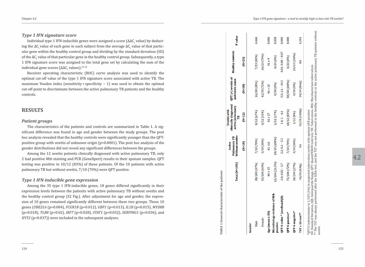
4.2
Chapter 4.2
124 125
Type I IFN gene signature : a tool to stratify high vs low-risk TB uveitis?
Type 1 IFN signature scoreIndividual type 1 IFN-inducible genes were assigned a score (ΔΔCt value) by deduct-
ing the ΔCt value of each gene in each subject from the average ΔCt value of that partic-ular gene within the healthy control group and dividing by the standard deviation (SD) of the ΔCt value of that particular gene in the healthy control group. Subsequently, a type 1 IFN signature score was assigned to the total gene set by calculating the sum of the individual gene scores (ΔΔCt values).22-24
Receiver operating characteristic (ROC) curve analysis was used to identify the optimal cut-off value of the type 1 IFN signature score associated with active TB. The maximum Youden index (sensitivity + specificity − 1) was used to obtain the optimal cut-off point to discriminate between the active pulmonary TB patients and the healthy controls.
RESULTS
Patient groupsThe characteristics of the patients and controls are summarized in Table 1. A sig-
nificant difference was found in age and gender between the study groups. The post hoc analysis revealed that the healthy controls were significantly younger than the QFT-positive group with uveitis of unknown origin (p<0.0001). The post hoc analysis of the gender distributions did not reveal any significant differences between the groups.
Among the 12 uveitis patients clinically diagnosed with active pulmonary TB, only 2 had positive Mtb staining and PCR (GeneXpert) results in their sputum samples. QFT testing was positive in 10/12 (83%) of these patients. Of the 10 patients with active pulmonary TB but without uveitis, 7/10 (70%) were QFT positive.
Type 1 IFN-inducible gene expressionAmong the 35 type 1 IFN-inducible genes, 18 genes differed significantly in their
expression levels between the patients with active pulmonary TB without uveitis and the healthy control group (S2 Fig.). After adjustment for age and gender, the expres-sion of 10 genes remained significantly different between these two groups. These 10 genes (UBE2L6 (p=0.004), FCGR1B (p=0.012), GBP1 (p=0.013), IL1B (p=0.015), MYD88 (p=0.018), TLR8 (p=0.02), IRF7 (p=0.028), STAT1 (p=0.032), SERPING1 (p=0.036), and IFIT2 (p=0.037)) were included in the subsequent analyses.
*QFT
-G in
dete
rmin
ate
in 1
/103
(1%
) in
susp
ecte
d tu
berc
ulou
s uve
itis w
ith sp
utum
AFB
pos
itive
SD: S
tand
ard
Devi
atio
n, IQ
R: In
terq
uart
ile R
ange
, QFT
: Qua
ntiF
ERON
Gol
d te
st, T
B: T
uber
culo
sis,
Mtb
: Myc
obac
teri
um tu
berc
ulos
is**
The
TST
was
alw
ays
perf
orm
ed a
fter
the
IGRA
test
, and
the
TST
was
not
per
form
ed in
the
heal
thy
cont
rols
or
the
activ
e pu
lmon
ary
TB p
atie
nts
with
out
uvei
tis.
TABL
E 1
Gene
ral c
hara
cter
isti
c of
the
pati
ents
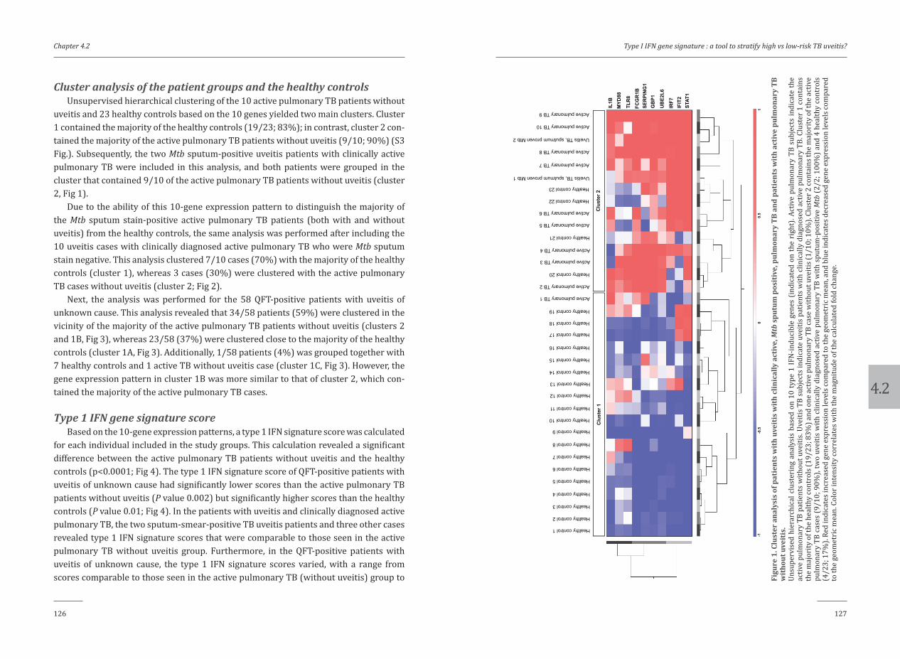
4.2
Chapter 4.2
126 127
Type I IFN gene signature : a tool to stratify high vs low-risk TB uveitis?
Cluster analysis of the patient groups and the healthy controlsUnsupervised hierarchical clustering of the 10 active pulmonary TB patients without
uveitis and 23 healthy controls based on the 10 genes yielded two main clusters. Cluster 1 contained the majority of the healthy controls (19/23; 83%); in contrast, cluster 2 con-tained the majority of the active pulmonary TB patients without uveitis (9/10; 90%) (S3 Fig.). Subsequently, the two Mtb sputum-positive uveitis patients with clinically active pulmonary TB were included in this analysis, and both patients were grouped in the cluster that contained 9/10 of the active pulmonary TB patients without uveitis (cluster 2, Fig 1).
Due to the ability of this 10-gene expression pattern to distinguish the majority of the Mtb sputum stain-positive active pulmonary TB patients (both with and without uveitis) from the healthy controls, the same analysis was performed after including the 10 uveitis cases with clinically diagnosed active pulmonary TB who were Mtb sputum stain negative. This analysis clustered 7/10 cases (70%) with the majority of the healthy controls (cluster 1), whereas 3 cases (30%) were clustered with the active pulmonary TB cases without uveitis (cluster 2; Fig 2).
Next, the analysis was performed for the 58 QFT-positive patients with uveitis of unknown cause. This analysis revealed that 34/58 patients (59%) were clustered in the vicinity of the majority of the active pulmonary TB patients without uveitis (clusters 2 and 1B, Fig 3), whereas 23/58 (37%) were clustered close to the majority of the healthy controls (cluster 1A, Fig 3). Additionally, 1/58 patients (4%) was grouped together with 7 healthy controls and 1 active TB without uveitis case (cluster 1C, Fig 3). However, the gene expression pattern in cluster 1B was more similar to that of cluster 2, which con-tained the majority of the active pulmonary TB cases.
Type 1 IFN gene signature scoreBased on the 10-gene expression patterns, a type 1 IFN signature score was calculated
for each individual included in the study groups. This calculation revealed a significant difference between the active pulmonary TB patients without uveitis and the healthy controls (p<0.0001; Fig 4). The type 1 IFN signature score of QFT-positive patients with uveitis of unknown cause had significantly lower scores than the active pulmonary TB patients without uveitis (P value 0.002) but significantly higher scores than the healthy controls (P value 0.01; Fig 4). In the patients with uveitis and clinically diagnosed active pulmonary TB, the two sputum-smear-positive TB uveitis patients and three other cases revealed type 1 IFN signature scores that were comparable to those seen in the active pulmonary TB without uveitis group. Furthermore, in the QFT-positive patients with uveitis of unknown cause, the type 1 IFN signature scores varied, with a range from scores comparable to those seen in the active pulmonary TB (without uveitis) group to
FCG
R1B
SER
PIN
G1
GB
P1
IRF7
IL1B
IFIT
2
MYD
88
TLR
8
UB
E2L6
STAT
1
Healthy control 1
Healthy control 2
Healthy control 3
Healthy control 4
Healthy control 6
Healthy control 5
Healthy control 17
Healthy control 16
Healthy control 15
Healthy control 14
Healthy control 13
Healthy control 12
Healthy control 11
Healthy control 10
Healthy control 9
Healthy control 8
Healthy control 7
Healthy control 20
Healthy control 19
Healthy control 18
Healthy control 22
Healthy control 21
Healthy control 23
Active pulmonary TB 1
Active pulmonary TB 2
Active pulmonary TB 3
Active pulmonary TB 4
Active pulmonary TB 5
Active pulmonary TB 6
Active pulmonary TB 7
Active pulmonary TB 8
Active pulmonary TB 9
Active pulmonary TB 10
Uveitis TB, sputmum proven Mtb 1
Uveitis TB, sputmum proven Mtb 2
-1-0
.50
0.5
1
Clu
ster
1C
lust
er 2
Figu
re 1
. Clu
ster
ana
lysi
s of
pat
ient
s w
ith
uvei
tis
wit
h cl
inic
ally
act
ive,
Mtb
spu
tum
pos
itiv
e, p
ulm
onar
y TB
and
pat
ient
s w
ith
activ
e pu
lmon
ary
TB
wit
hout
uve
itis
. Un
supe
rvis
ed h
iera
rchi
cal c
lust
erin
g an
alys
is b
ased
on
10 ty
pe 1
IFN
-indu
cibl
e ge
nes
(indi
cate
d on
the
righ
t). A
ctiv
e pu
lmon
ary
TB s
ubje
cts
indi
cate
the
activ
e pu
lmon
ary
TB p
atie
nts
with
out u
veiti
s. Uv
eitis
TB
subj
ects
indi
cate
uve
itis
patie
nts
with
clin
ical
ly d
iagn
osed
act
ive
pulm
onar
y TB
. Clu
ster
1 c
onta
ins
the
maj
ority
of t
he h
ealth
y co
ntro
ls (1
9/23
; 83%
) and
one
act
ive
pulm
onar
y TB
case
with
out u
veiti
s (1/
10; 1
0%).
Clus
ter 2
cont
ains
the
maj
ority
of t
he a
ctiv
e pu
lmon
ary
TB c
ases
(9/1
0; 9
0%),
two
uvei
tis w
ith c
linic
ally
dia
gnos
ed a
ctiv
e pu
lmon
ary
TB w
ith s
putu
m-p
ositi
ve M
tb (2
/2; 1
00%
) and
4 h
ealth
y co
ntro
ls
(4/2
3; 1
7%).
Red
indi
cate
s inc
reas
ed g
ene
expr
essi
on le
vels
com
pare
d to
the
geom
etri
c mea
n, a
nd b
lue
indi
cate
s dec
reas
ed g
ene
expr
essi
on le
vels
com
pare
d to
the
geom
etri
c mea
n. C
olor
inte
nsity
corr
elat
es w
ith th
e m
agni
tude
of t
he ca
lcul
ated
fold
chan
ge.

4.2
Chapter 4.2
128 129
Type I IFN gene signature : a tool to stratify high vs low-risk TB uveitis?
Figu
re 3
. Clu
ster
ana
lysi
s of
QFT
-pos
itiv
e pa
tien
ts w
ith
uvei
tis
of u
nkno
wn
caus
e, p
atie
nts
wit
h ac
tive
pulm
onar
y TB
wit
hout
uve
itis
and
hea
lthy
co
ntro
ls.
Unsu
perv
ised
hie
rarc
hica
l clu
ster
ing
anal
ysis
bas
ed o
n 10
type
1 IF
N-in
duci
ble
gene
s (in
dica
ted
on th
e ri
ght)
. Act
ive
pulm
onar
y TB
sub
ject
s in
dica
te th
e ac
tive
pulm
onar
y TB
pat
ient
s w
ithou
t uve
itis.
Uvei
tis T
B su
bjec
ts in
dica
te u
veiti
s pa
tient
s w
ith c
linic
ally
dia
gnos
ed a
ctiv
e pu
lmon
ary
TB C
lust
er 1
(A+B
+C)
cont
ains
the
maj
ority
of t
he h
ealth
y co
ntro
ls (1
9/23
; 83%
), cl
uste
r 1A
con
tain
s 11
hea
lthy
cont
rols
(11/
23; 4
8%) a
nd 2
3 pa
tient
s w
ith u
veiti
s of
unk
now
n ca
use
wer
e QF
T po
sitiv
e (2
3/58
; 40%
), cl
uste
r 1B
cont
ains
1 h
ealth
y co
ntro
l (1/
23; 4
%),
2 pa
tient
s w
ith a
ctiv
e pu
lmon
ary
TB w
ithou
t uve
itis
(2/2
0; 2
0%)
and
16 p
atie
nts w
ith u
veiti
s of u
nkno
wn
caus
e w
ere
QFT
posi
tive
(16/
58; 2
8%) a
nd cl
uste
r 1C
cont
ains
7 h
ealth
y co
ntro
ls (7
/23;
30%
), 1
patie
nt w
ith a
ctiv
e pu
lmon
ary
TB w
ithou
t uve
itis (
1/10
; 10%
) and
1pa
tient
with
uve
itis o
f unk
now
n ca
use
wer
e QF
T po
sitiv
e (1
/58;
2%
). Cl
uste
r 2 co
ntai
ns th
e m
ajor
ity o
f the
ac
tive
pulm
onar
y TB
cas
es w
ithou
t uve
itis
(9/1
0; 9
0%),
4 he
alth
y co
ntro
ls (4
/23;
17%
) and
18
patie
nts
with
uve
itis
of u
nkno
wn
caus
e w
ere
QFT
posi
tive
(18/
58; 3
1%).
Red
indi
cate
s in
crea
sed
gene
exp
ress
ion
leve
ls w
hen
com
pare
d to
the
geom
etri
c m
ean,
and
blu
e in
dica
tes
decr
ease
d ge
ne e
xpre
ssio
n le
vels
w
hen
com
pare
d to
the
geom
etri
c mea
n. C
olor
inte
nsity
corr
elat
es w
ith th
e m
agni
tude
of t
he ca
lcul
ated
fold
chan
ge.
FCG
R1B
SER
PIN
G1
GB
P1
IRF7
IL1B
MYD
88TL
R8
UB
E2L6
STAT
1
Healthy control 1Healthy control 2Healthy control 3Healthy control 4
Healthy control 6Healthy control 5
Healthy control 17
Healthy control 16Healthy control 15Healthy control 14Healthy control 13
Healthy control 12Healthy control 11Healthy control 10
Healthy control 9
Healthy control 8Healthy control 7
Healthy control 20
Healthy control 19
Healthy control 18
Healthy control 22
Healthy control 21
Healthy control 23
Active pulmonary TB 1
Active pulmonary TB 2Active pulmonary TB 3Active pulmonary TB 4
Active pulmonary TB 5
Active pulmonary TB 6
Active pulmonary TB 7
Active pulmonary TB 8Active pulmonary TB 9Active pulmonary TB 10
QFT positive unknown cause 1
QFT positive unknown cause 2QFT positive unknown cause 3
QFT positive unknown cause 8QFT positive unknown cause 7QFT positive unknown cause 6QFT positive unknown cause 5QFT positive unknown cause 4
QFT positive unknown cause 9QFT positive unknown cause 10
QFT positive unknown cause 15QFT positive unknown cause 14QFT positive unknown cause 13QFT positive unknown cause 12QFT positive unknown cause 11
QFT positive unknown cause 37
QFT positive unknown cause 16
QFT positive unknown cause 21
QFT positive unknown cause 20QFT positive unknown cause 19QFT positive unknown cause 18QFT positive unknown cause 17
QFT positive unknown cause 22
QFT positive unknown cause 27QFT positive unknown cause 26QFT positive unknown cause 25QFT positive unknown cause 24QFT positive unknown cause 23
QFT positive unknown cause 28
QFT positive unknown cause 33QFT positive unknown cause 32QFT positive unknown cause 31
QFT positive unknown cause 30QFT positive unknown cause 29
QFT positive unknown cause 36
QFT positive unknown cause 35QFT positive unknown cause 34
QFT positive unknown cause 41
QFT positive unknown cause 40QFT positive unknown cause 39QFT positive unknown cause 38
QFT positive unknown cause 42
QFT positive unknown cause 45QFT positive unknown cause 44
QFT positive unknown cause 43
QFT positive unknown cause 46
QFT positive unknown cause 51QFT positive unknown cause 50QFT positive unknown cause 49QFT positive unknown cause 48
QFT positive unknown cause 47
QFT positive unknown cause 53
QFT positive unknown cause 57
QFT positive unknown cause 56QFT positive unknown cause 55QFT positive unknown cause 54
QFT positive unknown cause 52
QFT positive unknown cause 58
IFIT
2
-1-0
.50
0.5
1
Clu
ster
2C
lust
er 1
AC
lust
er 1
BC
lust
er 1
C
Figu
re 2
. Clu
ster
ana
lysi
s of
pat
ient
s w
ith
uvei
tis
who
wer
e sp
utum
Mtb
neg
ativ
e bu
t wer
e di
agno
sed
wit
h ac
tive
TB in
fect
ion
on c
linic
al g
roun
ds,
pati
ents
wit
h ac
tive
pulm
onar
y TB
wit
hout
uve
itis
and
hea
lthy
con
trol
s.
Unsu
perv
ised
hie
rarc
hica
l clu
ster
ing
anal
ysis
bas
ed o
n 10
type
1 IF
N-in
duci
ble
gene
s (in
dica
ted
on th
e ri
ght)
. Act
ive
pulm
onar
y TB
sub
ject
s in
dica
te th
e ac
tive
pulm
onar
y TB
pat
ient
s w
ithou
t uve
itis.
Uvei
tis T
B su
bjec
ts in
dica
te u
veiti
s pa
tient
s w
ith c
linic
ally
dia
gnos
ed a
ctiv
e pu
lmon
ary
TB. C
lust
er 1
con
tain
s th
e m
ajor
ity o
f the
hea
lthy
cont
rols
(19
/23;
83%
) an
d on
e ac
tive
pulm
onar
y TB
cas
e w
ithou
t uve
itis
(1/1
0; 1
0%).
Clus
ter
2 co
ntai
ns th
e m
ajor
ity o
f the
ac
tive
pulm
onar
y TB
case
s (9/
10; 9
0%) a
nd 4
hea
lthy
cont
rols
(4/2
3; 1
7%).
Ten
uvei
tis p
atie
nts h
ad cl
inic
ally
dia
gnos
ed a
ctiv
e pu
lmon
ary
TB in
fect
ions
but
ne
gativ
e M
tb s
putu
m te
sts.
Only
3 p
atie
nts
(3/1
0; 3
0%) w
ere
grou
ped
in c
lust
er 2
, whi
ch p
rim
arily
con
tain
ed c
lust
ered
act
ive
pulm
onar
y TB
pat
ient
s. Re
d in
dica
tes i
ncre
ased
gen
e ex
pres
sion
leve
ls c
ompa
red
to th
e ge
omet
ric
mea
n, a
nd b
lue
indi
cate
s dec
reas
ed g
ene
expr
essi
on le
vels
com
pare
d to
the
geom
etri
c m
ean.
Col
or in
tens
ity co
rrel
ates
with
the
mag
nitu
de o
f the
calc
ulat
ed fo
ld ch
ange
.
FCG
R1B
SER
PIN
G1
GB
P1
IRF7
IL1B
IFIT
2
MYD
88TL
R8
UB
E2L6
STAT
1
Healthy control 1
Healthy control 2
Healthy control 3
Healthy control 4
Healthy control 6
Healthy control 5
Healthy control 17
Healthy control 16
Healthy control 15
Healthy control 14
Healthy control 13
Healthy control 12
Healthy control 11
Healthy control 10
Healthy control 9
Healthy control 8
Healthy control 7
Healthy control 20
Healthy control 19
Healthy control 18
Healthy control 22
Healthy control 21
Healthy control 23
Active pulmonary TB 1
Active pulmonary TB 2
Active pulmonary TB 3
Active pulmonary TB 4
Active pulmonary TB 5
Active pulmonary TB 6
Active pulmonary TB 7
Active pulmonary TB 8
Active pulmonary TB 9
Active pulmonary TB 10
Uveitis TB, sputmum negative 1
Uveitis TB, sputmum negative 2
Uveitis TB, sputmum negative 7
Uveitis TB, sputmum negative 6
Uveitis TB, sputmum negative 5
Uveitis TB, sputmum negative 4
Uveitis TB, sputmum negative 3
Uveitis TB, sputmum negative 9
Uveitis TB, sputmum negative 8
Uveitis TB, sputmum negative 10
-1-0
.50
0.5
1
Clu
ster
1C
lust
er 2
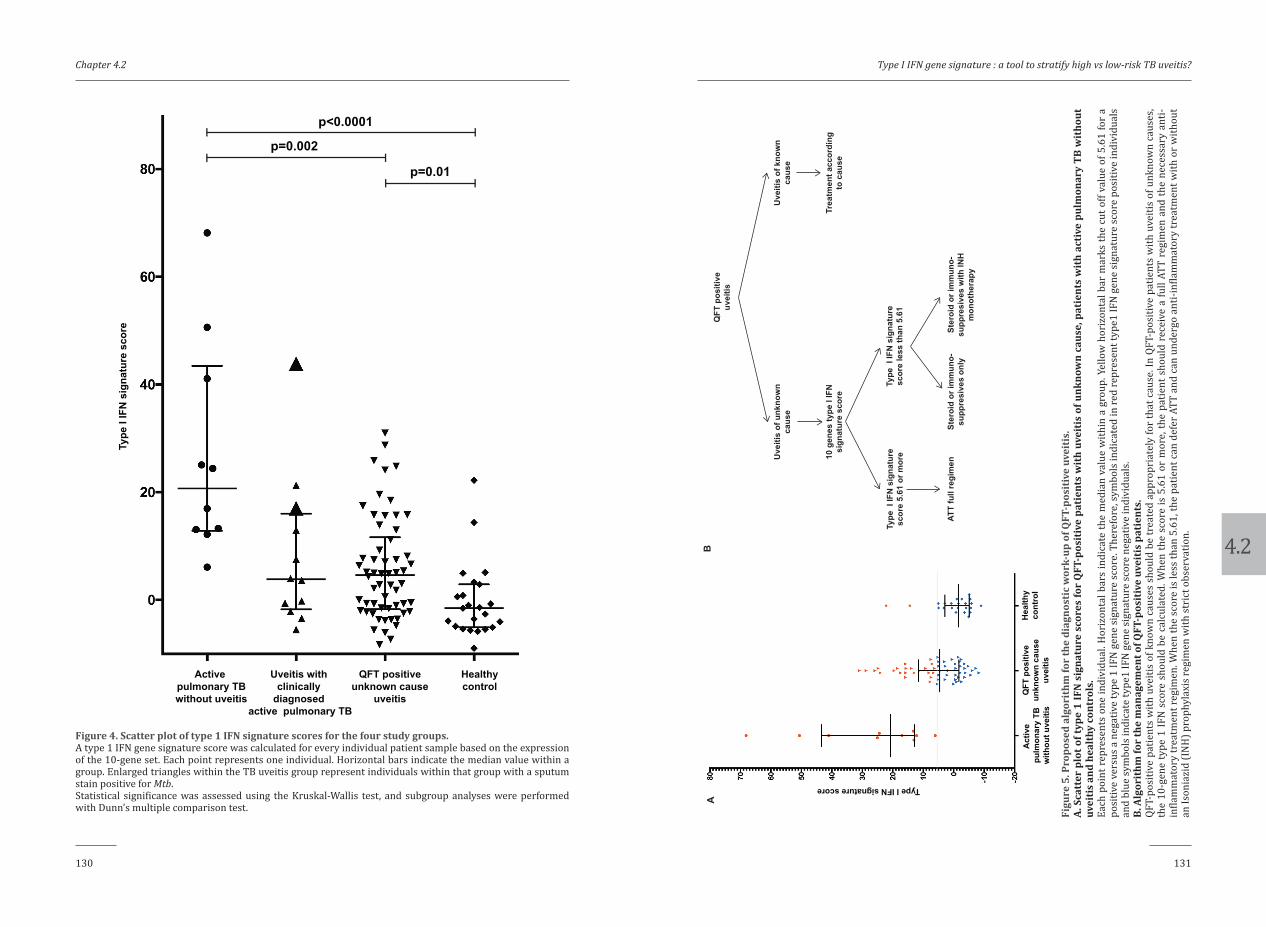
4.2
Chapter 4.2
130 131
Type I IFN gene signature : a tool to stratify high vs low-risk TB uveitis?
QFT
pos
itive
uv
eitis
Uve
itis
of k
now
n ca
use
Trea
tmen
t acc
ordi
ng
to c
ause
Uve
itis
of u
nkno
wn
caus
e
10 g
enes
type
I IF
N
sign
atur
e sc
ore
Type
I IF
N s
igna
ture
sc
ore
5.61
or m
ore
Type
I IF
N s
igna
ture
sc
ore
less
than
5.6
1
ATT
full
regi
men
Ster
oid
or im
mun
o-su
ppre
sive
s on
lySt
eroi
d or
imm
uno-
supp
resi
ves
with
INH
m
onot
hera
py
AB
Type I IFN signature score
Act
ive
pulm
onar
y TB
with
out u
veiti
s
QFT
pos
itive
unkn
own
caus
euv
eitis
Hea
lthy
cont
rol
Figu
re 5
. Pro
pose
d al
gori
thm
for
the
diag
nost
ic w
ork-
up o
f QFT
-pos
itiv
e uv
eiti
s.A.
Sca
tter
plo
t of t
ype
1 IF
N s
igna
ture
sco
res
for
QFT
-pos
itiv
e pa
tien
ts w
ith
uvei
tis
of u
nkno
wn
caus
e, p
atie
nts
wit
h ac
tive
pulm
onar
y TB
wit
hout
uv
eiti
s an
d he
alth
y co
ntro
ls.
Each
poi
nt r
epre
sent
s on
e in
divi
dual
. Hor
izon
tal b
ars
indi
cate
the
med
ian
valu
e w
ithin
a g
roup
. Yel
low
hor
izon
tal b
ar m
arks
the
cut o
ff va
lue
of 5
.61
for
a po
sitiv
e ve
rsus
a n
egat
ive
type
1 IF
N g
ene
sign
atur
e sc
ore.
The
refo
re, s
ymbo
ls in
dica
ted
in re
d re
pres
ent t
ype1
IFN
gen
e si
gnat
ure
scor
e po
sitiv
e in
divi
dual
s an
d bl
ue sy
mbo
ls in
dica
te ty
pe1
IFN
gen
e si
gnat
ure
scor
e ne
gativ
e in
divi
dual
s. B.
Alg
orit
hm fo
r th
e m
anag
emen
t of Q
FT-p
osit
ive
uvei
tis
pati
ents
. QF
T-po
sitiv
e pa
tient
s w
ith u
veiti
s of
kno
wn
caus
es s
houl
d be
trea
ted
appr
opri
atel
y fo
r th
at c
ause
. In
QFT-
posi
tive
patie
nts
with
uve
itis
of u
nkno
wn
caus
es,
the
10-g
ene
type
1 IF
N s
core
sho
uld
be c
alcu
late
d. W
hen
the
scor
e is
5.6
1 or
mor
e, th
e pa
tient
sho
uld
rece
ive
a fu
ll AT
T re
gim
en a
nd th
e ne
cess
ary
anti-
infla
mm
ator
y tr
eatm
ent r
egim
en. W
hen
the
scor
e is
less
than
5.6
1, th
e pa
tient
can
def
er A
TT a
nd c
an u
nder
go a
nti-i
nfla
mm
ator
y tr
eatm
ent w
ith o
r with
out
an Is
onia
zid
(IN
H) p
roph
ylax
is re
gim
en w
ith st
rict
obs
erva
tion.
Active pulmonary TBwithout uveitis
Uveitis with clinically
diagnosed active pulmonary TB
QFT positiveunknown cause
uveitis
Healthycontrol
Type
I IF
N s
igna
ture
sco
rep<0.0001
p=0.002
p=0.01
Figure 4. Scatter plot of type 1 IFN signature scores for the four study groups. A type 1 IFN gene signature score was calculated for every individual patient sample based on the expression of the 10-gene set. Each point represents one individual. Horizontal bars indicate the median value within a group. Enlarged triangles within the TB uveitis group represent individuals within that group with a sputum stain positive for Mtb. Statistical significance was assessed using the Kruskal-Wallis test, and subgroup analyses were performed with Dunn’s multiple comparison test.

4.2
Chapter 4.2
132 133
Type I IFN gene signature : a tool to stratify high vs low-risk TB uveitis?
scores overlapping those seen in the healthy control group (Fig 4). Further, we deter-mined a type 1 IFN signature score cut-off value using ROC curve analysis that yielded an area under the curve (AUC) of 0.961 with a P value <0.00001 (S4 A Fig.); a cut-off value ≥5.61 displayed the highest sensitivity (100%), specificity (91%) and Youden index (0.913) for the identification of active TB (S4 B Fig. and Fig 5 A).
DISCUSSION
In this study, we demonstrate that a blood transcriptional signature of 10 type 1 IFN-inducible genes differentiates QFT-positive uveitis patients into distinct different groups.
Type 1 IFN (IFNα/β)-inducible gene expression signatures in peripheral blood cells have consistently been reported as a potential biomarker for active pulmonary TB compared with signatures in healthy controls or patients with other diseases.13 Berry et al. first reported the association between active pulmonary TB and the expression of numerous type 1 IFN-inducible genes in peripheral blood cells in regions with both an intermediate TB burden (London, UK) and a high TB burden (South Africa).13 Importantly, these type 1 IFN-inducible gene transcripts differed from those observed in systemic autoimmune diseases, including systemic lupus erythematosus (SLE), which is among a group of systemic autoimmune diseases that contains subgroups of patients display-ing a type 1 IFN-inducible gene expression pattern.13, 21, 25, 26 Several other studies have corroborated the use of type 1 IFN-inducible gene expression in the peripheral blood to distinguish active from latent TB infection.14-16, 27, 28 Moreover, a rapid normalization of the type 1 IFN gene expression pattern upon successful ATT was reported.13, 15 Together, these observations underscore the role of type 1 IFN signaling in the pathogenesis of TB and the potential usefulness of the peripheral blood type 1 IFN transcriptome as a biomarker and monitoring tool in TB diagnostics and management. However, no studies of type 1 IFN-inducible blood transcriptome have been conducted in uveitis cases in relation to TB infection.
In our study, we identified a peripheral blood cell transcriptome consisting of 10 type 1 IFN-inducible genes that were strongly associated with active pulmonary TB with-out uveitis. When a type 1 IFN signature score was applied to this 10-gene set, a score ≥5.61 displayed the optimal sensitivity and specificity for distinguishing active pulmo-nary (Mtb sputum smear-positive) TB patients without uveitis from healthy controls. In line with this result, the two TB uveitis cases diagnosed with active pulmonary TB and having positive Mtb sputum smear displayed a type 1 IFN signature score >5.61. This finding indicates that microbiologically proven active pulmonary TB with or with-out uveitis is associated with high expression of type 1 IFN-inducible genes. However,
several additional uveitis cases, who were QFT-positive, displayed a positive type 1 IFN signature despite being Mtb sputum negative.
Over-diagnosis of uveitis TB due to the lack of a gold standard diagnostic test (i.e., laboratory and radiological investigations) is a recognized problem that may result in overzealous treatment of uveitis with TB drugs.29 Within the uveitis group diagnosed with pulmonary TB on clinical grounds solely, we found 7 cases with a type 1 IFN sig-nature score <5.61. In these cases, the uveitis was probably not related to active TB. Importantly, the type 1 IFN signature scores revealed two subgroups within the QFT-positive patients with uveitis of unknown cause. We propose that QFT-positive patients with uveitis of unknown cause and a type 1 IFN signature score <5.61 are unlikely to have uveitis related to active TB infection. This conclusion is supported by the high con-cordance within the set of 10 type 1 IFN-inducible genes identified in our study and the type-1 IFN inducible genes identified in other studies that did compare active with latent TB. Furthermore, this finding is in agreement with other studies that compared patients with active and latent TB and reported a rapid decline in peripheral blood cell expression of type 1 IFN-inducible genes in active TB patients treated with a full ATT regimen.13, 15 Consequently, we expect that patients within the QFT-positive group with uveitis of unknown cause who display a positive type 1 IFN signature score (≥5.61) will have a higher likelihood of having uveitis due to active TB infection. Our series, how-ever, lacks the results of type 1 IFN signature in QFT-positive patients without uveitis. Nevertheless, we propose that measurement of a peripheral blood type 1 IFN gene sig-nature score can aid to the diagnostic workup and choice of treatment in patients with QFT positive uveitis, as is indicated in Fig 5 B.
Elevated expression of type 1 IFN-inducible genes in peripheral blood cells is not specific to active TB disease. Type 1 IFN gene signatures are also associated with sys-temic autoimmune diseases, such as in Sjögren’s syndrome cases (~55%), systemic lupus erythematosus (~50%), and systemic sclerosis (~30%), and are also present in healthy control subjects (~5%).21, 25, 26 However, differential expression of type 1 IFN-inducible genes between TB and systemic autoimmune diseases is clear. MxA is a type 1 IFN-inducible gene that encodes an important mediator of the early innate immune defense against viruses.22 Elevated MxA expression is part of the peripheral blood type 1 IFN signature in systemic autoimmune disease patients.22 Yet in our study elevated MxA gene expression was not found in peripheral blood from patients with active pulmonary TB compared to healthy control subjects (S2 Fig.). Therefore, our data support differen-tial involvement of type 1 IFN-inducible genes among different diseases associated with type 1 IFN activity.13
In conclusion, we identified a blood transcriptional signature of 10 type 1 IFN-inducible genes highly expressed in active pulmonary TB patients. Although further
4.2
Chapter 4.2
134 135
Type I IFN gene signature : a tool to stratify high vs low-risk TB uveitis?
studies are required, our data suggest that this peripheral blood type 1 IFN gene signa-ture has the potential to stratify patients with suspected active TB infection-associated uveitis into groups with either a low or high risk of having uveitis due to TB. We expect that this stratification forms a constructive basis for future diagnostic and treatment studies in QFT/ TST positive uveitis patients.
FINANCIAL SUPPORT STATEMENT
1. This project was funded by the Indonesian Ministry of Research Technology and Higher Education for the first author’s PhD program in Erasmus MC.
2. The research was partially funded by the Global Research Initiative Program Universitas Indonesia (BOPTN UI) in the year 2014
3. The funding organization had no had no role in the design or conduct of this research
4. The authors declare that they have no conflicting and competing financial interests.
ACKNOWLEDGEMENTS
The patient recruitment was kindly assisted by Yulia Aziza, Kiki Nauli, Azalia Latuasan, Dina Lestari and Ikhwanuliman Putera. Early design and statistical method was discussed with Sopiyudin Dahlan.
AUTHOR CONTRIBUTIONS
R.L.D.N , P.M.v.H. and W.A.D., conceptualized and designed the study. R.L.D.N, R.S, M.S, L.E, S.S, G.S, and RR.D.H collected the patient data. R.L.D.N did the RT PCR experiments. M.A.V, supervised and analyzed the MxA EIA experiment. R.L.D.N, S.M.A.S and P.J.v.d.S. analyzed the RT PCR data. R.L.D.N and A.R analyzed the clinical data. R.L.D.N and W.A.D wrote the manuscript. W.A.D. had primary responsibility for the final content of the manuscript; and all authors have read and approved the final manuscript.
REFERENCES
1. La Distia Nora R, Sitompul R, Bakker M, et al. Tuberculosis and other causes of uveitis in Indonesia.
Eye (Lond) 2017.
2. Manandhar A. Patterns of Uveitis and Scleritis in Nepal: A Tertiary Referral Center Study. Ocul Immu-
nol Inflamm 2016;1-9.
3. Win MZ, Win T, Myint S, Shwe T, Sandar H. Epidemiology of Uveitis in a Tertiary Eye Center in Myan-
mar. Ocul Immunol Inflamm 2016;1-6.
4. Yeo TK, Ho SL, Lim WK, Teoh SC. Causes of visual loss associated with uveitis in a singapore tertiary
eye center. Ocul Immunol Inflamm 2013;21:264-269.
5. Singh R, Gupta V, Gupta A. Pattern of uveitis in a referral eye clinic in north India. Indian J Ophthalmol
2004;52:121-125.
6. Helm CJ, Holland GN. Ocular tuberculosis. Survey of ophthalmology 1993;38:229-256.
7. Sester M, Sotgiu G, Lange C, et al. Interferon-gamma release assays for the diagnosis of active tuber-
culosis: a systematic review and meta-analysis. Eur Respir J 2011;37:100-111.
8. La Distia Nora R, Van Velthoven MEJ, Ten Dam-Van Loon NH, et al. Clinical manifestations of patients
with intraocular inflammation and positive QuantiFERON-TB gold in-tube test in a country nonen-
demic for tuberculosis. Am J Ophthalmol 2014;157:754-761.
9. Rao NA, Saraswathy S, Smith RE. Tuberculous uveitis: distribution of Mycobacterium tuberculosis in
the retinal pigment epithelium. Arch Ophthalmol 2006;124:1777-1779.
10. Wroblewski KJ, Hidayat AA, Neafie RC, Rao NA, Zapor M. Ocular tuberculosis: A clinicopathologic and
molecular study. Ophthalmology 2011;118:772-777.
11. Vos AG, Wassenberg MW, de Hoog J, Oosterheert JJ. Diagnosis and treatment of tuberculous uveitis in
a low endemic setting. Int J Infect Dis 2013;17:e993-999.
12. Gineys R, Bodaghi B, Carcelain G, et al. QuantiFERON-TB gold cut-off value: implications for the man-
agement of tuberculosis-related ocular inflammation. Am J Ophthalmol 2011;152:433-440 e431.
13. Berry MP, Graham CM, McNab FW, et al. An interferon-inducible neutrophil-driven blood transcrip-
tional signature in human tuberculosis. Nature 2010;466:973-977.
14. Maertzdorf J, Repsilber D, Parida SK, et al. Human gene expression profiles of susceptibility and resis-
tance in tuberculosis. Genes Immun 2011;12:15-22.
15. Ottenhoff TH, Dass RH, Yang N, et al. Genome-wide expression profiling identifies type 1 interferon
response pathways in active tuberculosis. PLoS One 2012;7:e45839.
16. Zak DE, Penn-Nicholson A, Scriba TJ, et al. A blood RNA signature for tuberculosis disease risk: a
prospective cohort study. Lancet 2016;387:2312-2322.
17. Jabs DA, Nussenblatt RB, Rosenbaum JT. Standardization of uveitis nomenclature for reporting clini-
cal data. Results of the First International Workshop. Am J Ophthalmol 2005;140:509-516.

4.2
Chapter 4.2
136 137
Type I IFN gene signature : a tool to stratify high vs low-risk TB uveitis?
18. Triasih R, Robertson C, Duke T, Graham SM. Risk of infection and disease with Mycobacterium tuber-
culosis among children identified through prospective community-based contact screening in Indo-
nesia. Trop Med Int Health 2015;20:737-743.
19. Indonesian Society of Respirology. Diagnosis and Treatment Guidelines of Tuberculosis (TB) in Indone-
sia (TB consensus). Jakarta: Indonesian Society of Respirology; 2011:55.
20. The Directorate General of Disease Control and Environmental Health Ministry of Health Republic of
Indonesia. National Guidelines of Tuberculosis Control. Jakarta: Ministry of Health Republic of Indone-
sia; 2014.
21. Brkic Z, Maria NI, van Helden-Meeuwsen CG, et al. Prevalence of interferon type I signature in CD14
monocytes of patients with Sjogren’s syndrome and association with disease activity and BAFF gene
expression. Ann Rheum Dis 2013;72:728-735.
22. Maria NI, Brkic Z, Waris M, et al. MxA as a clinically applicable biomarker for identifying systemic
interferon type I in primary Sjogren’s syndrome. Ann Rheum Dis 2014;73:1052-1059.
23. Livak KJ, Schmittgen TD. Analysis of relative gene expression data using real-time quantitative PCR
and the 2(-Delta Delta C(T)) Method. Methods 2001;25:402-408.
24. Kirou KA, Lee C, George S, et al. Coordinate overexpression of interferon-alpha-induced genes in sys-
temic lupus erythematosus. Arthritis Rheum 2004;50:3958-3967.
25. Brkic Z, Corneth OB, van Helden-Meeuwsen CG, et al. T-helper 17 cell cytokines and interferon type I:
partners in crime in systemic lupus erythematosus? Arthritis Res Ther 2014;16:R62.
26. Brkic Z, van Bon L, Cossu M, et al. The interferon type I signature is present in systemic sclerosis be-
fore overt fibrosis and might contribute to its pathogenesis through high BAFF gene expression and
high collagen synthesis. Ann Rheum Dis 2016;75:1567-1573.
27. Jacobsen M, Repsilber D, Gutschmidt A, et al. Candidate biomarkers for discrimination between infec-
tion and disease caused by Mycobacterium tuberculosis. J Mol Med (Berl) 2007;85:613-621.
28. Mistry R, Cliff JM, Clayton CL, et al. Gene-expression patterns in whole blood identify subjects at risk
for recurrent tuberculosis. J Infect Dis 2007;195:357-365.
29. Gupta A, Sharma A, Bansal R, Sharma K. Classification of intraocular tuberculosis. Ocul Immunol In-
flamm 2015;23:7-13.
Clinical examination suggestive for TB
Sputum microbiological testing
a. Bacterial acid fast staining (3 times)b. GeneXpert/PCR
c. Culture
Active pulmonary TB Sputum positive or negative
CXR examination
Reassessment
a. Non ATT antibiotic treatment
b. Microbiological testingc. Radiological testing
Review
a.History of previous TB disease or TB contactb. CXR abnormalities
c. TST positivityd. Clinical, microbiological and radiological evidence of active
TB disease
No active pulmonary TBPrevious pulmonary TB
TB suspect
Positive Negative or not available
a. Respiratory symptomsProductive cough >2 weeks, hemopthysis, dyspnea, and/or
chest painb. Systemic symptoms
Weight loss, night sweat, malaise and unexplained feverc. Physical examination
Ronchi in lung auscultation
Suggestive for active TB
Doubtful No signs of active TB
Suggestive for active TB
Not suggestive foractive TB
SUPPLEMENTAL DATA
Supplemental Figure 1. Indonesian Society of Respirology (ISR) TB diagnostic guideline. Scheme depicting the TB diagnostic algorithm in Indonesia. The guideline starts with evaluation of clinical signs suggestive for tuberculosis (TB). In patients with signs suggestive of pulmonary TB infection, an acid-fast staining examination from the sputum is required and a positive result leads to diagnosis of active pulmonary TB. If the culture examinations from the sputum are negative, radiologic imaging is reviewed and classified When imaging is considered to exhibit signs of active pulmonary TB, the patient is diagnosed with active pulmonary TB. Therefore, active pulmonary TB will be either acid-fast bacillus (AFB) positive or AFB negative. The diagnosis of AFB-positive active pulmonary TB is based on positive culture or positive GeneXpert outcomes, and AFB-negative TB is based on radiologic signs of active pulmonary TB and clinical improvement after anti-tuberculosis antibiotic treatment. TB: tuberculosis, AFB: acid-fast bacillus, GeneXpert: a molecular test that detects bacterial DNA from Mycobacterium tuberculosis, PCR: polymerase chain reaction, CXR: chest X-ray, TST: tuberculin skin test

4.2
Chapter 4.2
138 139
Type I IFN gene signature : a tool to stratify high vs low-risk TB uveitis?
Healthy control 1
Healthy control 2
Healthy control 3
Healthy control 4
Healthy control 6
Healthy control 5
Healthy control 17
Healthy control 16
Healthy control 15
Healthy control 14
Healthy control 13
Healthy control 12
Healthy control 11
Healthy control 10
Healthy control 9
Healthy control 8
Healthy control 7
Healthy control 20
Healthy control 19
Healthy control 18
Healthy control 22
Healthy control 21
Healthy control 23
Active pulmonary TB 1
Active pulmonary TB 2
Active pulmonary TB 3
Active pulmonary TB 4
Active pulmonary TB 5
Active pulmonary TB 6
Active pulmonary TB 7
Active pulmonary TB 8
Active pulmonary TB 9
Active pulmonary TB 10
IFIT
2
SER
PIN
G1
GB
P1
IRF7
IL1B
MYD
88
TLR
8
UB
E2L6
STAT
1
FCG
R1B
-1-0
.50
0.5
1
Supp
lem
enta
l Fig
ure
3. C
lust
er a
naly
sis
of a
ctiv
e pu
lmon
ary
TB p
atie
nts
wit
hout
uve
itis
and
hea
lthy
con
trol
s ba
sed
on ty
pe 1
IFN
-indu
cibl
e ge
ne
expr
essi
on p
atte
rns.
Un
supe
rvis
ed h
iera
rchi
cal c
lust
erin
g an
alys
is b
ased
on
10 ty
pe 1
IFN
-indu
cibl
e gen
es (i
ndic
ated
on
the r
ight
). Ac
tive p
ulm
onar
y TB
subj
ects
indi
cate
the a
ctiv
e pu
lmon
ary
TB p
atie
nts w
ithou
t uve
itis.
Clus
ter 1
cont
ains
the
maj
ority
of t
he h
ealth
y co
ntro
ls (1
9/23
; 83%
) and
one
act
ive
pulm
onar
y TB
case
with
out u
veiti
s (1
/10;
10%
). Cl
uste
r 2 co
ntai
ns th
e m
ajor
ity o
f the
act
ive
pulm
onar
y TB
case
s with
out u
veiti
s (9/
10; 9
0%) a
nd 4
hea
lthy
cont
rols
(4/2
3; 1
7%).
Red
indi
cate
s in
crea
sed
gene
expr
essi
on le
vels
whe
n co
mpa
red
to th
e geo
met
ric m
ean,
and
blue
indi
cate
s dec
reas
ed g
ene e
xpre
ssio
n le
vels
whe
n co
mpa
red
to th
e geo
met
ric
mea
n. C
olor
inte
nsity
corr
elat
es w
ith th
e m
agni
tude
of t
he ca
lcul
ated
fold
chan
ge.
Supp
lem
enta
l Fig
ure
2. T
ype
1 IF
N-in
duci
ble
gene
s wit
h si
gnifi
cant
diff
eren
ces i
n ex
pres
sion
bet
wee
n ac
tive
pulm
onar
y TB
pat
ient
s wit
hout
uve
itis
an
d he
alth
y co
ntro
ls.
Of a
tota
l of 3
5 ty
pe 1
IFN
-indu
cibl
e ge
nes m
easu
red
in th
e pe
riph
eral
blo
od, 1
8 ge
nes d
iffer
ed si
gnifi
cant
ly in
thei
r exp
ress
ion
betw
een
the
activ
e pu
lmon
ary
TB w
ithou
t uve
itis p
atie
nts a
nd th
e he
alth
y co
ntro
ls w
ithou
t cor
rect
ion
for a
ge d
iffer
ence
s bet
wee
n th
e gr
oups
. Sta
tistic
al si
gnifi
canc
e w
as a
sses
sed
usin
g th
e M
ann-
Whi
tney
U te
st.
****
P v
alue
<0.
0001
, ***
P v
alue
<0.
001,
** P
val
ue <
0.01
, * P
val
ue <
0.05

4.2
Chapter 4.2
140 141
Type I IFN gene signature : a tool to stratify high vs low-risk TB uveitis?
Sens
itivi
ty
1 - S
peci
ficity
RO
C c
urve
AB
Supp
lem
enta
l Fig
ure
4. R
OC
curv
e an
alys
is a
nd Y
oude
n In
dex
calc
ulat
ion
for
dete
rmin
ing
the
opti
mal
cut
-off
valu
e of
type
1 IF
N s
igna
ture
sco
re.
A. T
he R
OC c
urve
ana
lysi
s and
are
a un
der t
he c
urve
(AUC
) B. Y
oude
n in
dex
anal
yses
indi
cate
that
a ty
pe 1
IFN
sign
atur
e sc
ore
cut-
off v
alue
≥5.
61 re
pres
ents
th
e op
timal
cut
-off
valu
e fo
r dis
tingu
ishi
ng a
ctiv
e pu
lmon
ary
TB p
atie
nts
with
out u
veiti
s fr
om h
ealth
y co
ntro
ls w
ith 1
00%
sen
sitiv
ity, 9
1% s
peci
ficity
and
a
Youd
en in
dex
of 0
.913
.

.
4.3ChapterAntinuclear and antiretinal antibodies
in uveitis associated with active and latent tuberculosis
Rina La Distia Nora1, Josianne C. ten Berge2, Aniki Rothova2, Marco W.J. Schreurs3
1Department of Ophthalmology, Erasmus University Medical Center, Rotterdam, Netherlands; Department of Ophthalmology, University of Indonesia & Cipto
Mangunkusumo Hospital Kirana, Jakarta, Indonesia; 2Department of Ophthalmology, Erasmus University Medical
Center, Rotterdam, The Netherlands; 3Department of Immunology, Erasmus University Medical
Center, Rotterdam, The Netherlands.
Accepted in Acta Ophthalmol
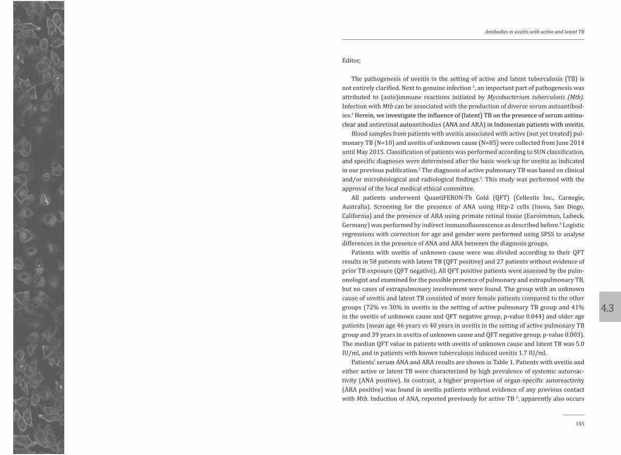
4.3
145
Antibodies in uveitis with active and latent TB
Editor,
The pathogenesis of uveitis in the setting of active and latent tuberculosis (TB) is not entirely clarified. Next to genuine infection 1, an important part of pathogenesis was attributed to (auto)immune reactions initiated by Mycobacterium tuberculosis (Mtb). Infection with Mtb can be associated with the production of diverse serum autoantibod-ies.2 Herein, we investigate the influence of (latent) TB on the presence of serum antinu-clear and antiretinal autoantibodies (ANA and ARA) in Indonesian patients with uveitis.
Blood samples from patients with uveitis associated with active (not yet treated) pul-monary TB (N=10) and uveitis of unknown cause (N=85) were collected from June 2014 until May 2015. Classification of patients was performed according to SUN classification, and specific diagnoses were determined after the basic work-up for uveitis as indicated in our previous publication.3 The diagnosis of active pulmonary TB was based on clinical and/or microbiological and radiological findings.3. This study was performed with the approval of the local medical ethical committee.
All patients underwent QuantiFERON-Tb Gold (QFT) (Cellestis Inc., Carnegie, Australia). Screening for the presence of ANA using HEp-2 cells (Inova, San Diego, California) and the presence of ARA using primate retinal tissue (Euroimmun, Lubeck, Germany) was performed by indirect immunofluorescence as described before.4 Logistic regressions with correction for age and gender were performed using SPSS to analyse differences in the presence of ANA and ARA between the diagnosis groups.
Patients with uveitis of unknown cause were was divided according to their QFT results in 58 patients with latent TB (QFT positive) and 27 patients without evidence of prior TB exposure (QFT negative). All QFT positive patients were assessed by the pulm-onologist and examined for the possible presence of pulmonary and extrapulmonary TB, but no cases of extrapulmonary involvement were found. The group with an unknown cause of uveitis and latent TB consisted of more female patients compared to the other groups (72% vs 30% in uveitis in the setting of active pulmonary TB group and 41% in the uveitis of unknown cause and QFT negative group, p-value 0.044) and older age patients (mean age 46 years vs 40 years in uveitis in the setting of active pulmonary TB group and 39 years in uveitis of unknown cause and QFT negative group, p-value 0.003). The median QFT value in patients with uveitis of unknown cause and latent TB was 5.0 IU/ml, and in patients with known tuberculosis induced uveitis 1.7 IU/ml.
Patients’ serum ANA and ARA results are shown in Table 1. Patients with uveitis and either active or latent TB were characterized by high prevalence of systemic autoreac-tivity (ANA positive). In contrast, a higher proportion of organ-specific autoreactivity (ARA positive) was found in uveitis patients without evidence of any previous contact with Mtb. Induction of ANA, reported previously for active TB 5, apparently also occurs

146
4.3
Chapter 4.3
146 147
Antibodies in uveitis with active and latent TB
COMPETING INTERESTS
The authors declare that they have no competing interests.
REFERENCES
1. Wroblewski KJ, Hidayat AA, Neafie RC, Rao NA, Zapor M. Ocular tuberculosis: A clinicopathologic and
molecular study. Ophthalmology 2011;118:772-777.
2. Shen CY, Hsieh SC, Yu CL, Wang JY, Lee LN, Yu CJ. Autoantibody prevalence in active tuberculosis:
reactive or pathognomonic? BMJ Open 2013;3.
3. La Distia Nora R, Sitompul R, Bakker M, et al. Tuberculosis and other causes of uveitis in Indonesia.
Eye (Lond) 2017.
4. Ten Berge JC, Schreurs MW, Vermeer J, Meester-Smoor MA, Rothova A. Prevalence and clinical impact
of antiretinal antibodies in uveitis. Acta Ophthalmol 2016;94:282-288.
5. Elkon K, Casali P. Nature and functions of autoantibodies. Nat Clin Pract Rheumatol 2008;4:491-498.
6. Kubicka-Trzaska A, Wilanska J, Romanowska-Dixon B, Sanak M. Serum anti-endothelial cell antibod-
ies in patients with age-related macular degeneration treated with intravitreal bevacizumab. Acta
Ophthalmol 2016;94:e617-e623.
in latent TB. Interestingly, organ-specific autoreactivity (ARA) appears to be suppressed in both active and latent TB, however, the local production of ARA in ocular fluid sam-ples was not investigated in this series. The presence of serum autoantibodies, directed against endothelial cells, and their decrease following treatment was reported in age-re-lated macular degeneration.6 Unfortunately, we have no samples of our patients after they completed the treatment. Further studies are warranted to dissect the pathogene-sis of this selective systemic induction of autoreactivity in uveitis patients as a result of Mtb infection and its implication on disease course.
ACKNOWLEDGEMENTS
This research was supported by ‘Stichting Lijf en Leven’, ‘Rotterdamse Blindenbelangen’, and by the Directorate General of Higher Education, Indonesian Ministry of Research Technology and Higher Education.
TABLE 1. Prevalence of antinuclear and antiretinal antibodies in tuberculosis induced uveitis and uveitis of unknown cause with positive or negative QuantiFERON–TB Gold outcomes
Legend: Serum ANA were more prevalent in patients with TB-induced uveitis (50%) than in patients with uveitis of unknown cause with latent TB (21%) or without latent TB (4%; p=0.023; Table 2). The prevalence of ARA was higher in QFT negative patients with unknown uveitis cause (62%) than in QFT positive uveitis cases (p=0.049). Prevalence of ARA or ANA did not differ between TB-induced uveitis and QFT positive uveitis of unknown cause. Abbreviations: ANA= antinuclear antibodies, ARA = antiretinal antibodies, OR = odds ratio, QFT = QuantiFERON–TB Gold test


5ChapterSummary and discussion

5
151
Summary and discussion
SUMMARY
The pathophysiology of Mycobacterium tuberculosis (Mtb)-associated uveitis is not entirely understood, and in consequence, the diagnosis and the treatment of Mtb-associated uveitis are challenging. The studies included in this thesis aim to increase our knowledge of this debilitating ocular disorder and aim to develop specific biomarkers for its diagnosis as defined in chapter 2.
Chapter 1 provides a general introduction to tuberculosis (TB) disease and its immunology and Mtb-associated uveitis and summarizes our current knowledge of this topic. Uveitis is a potentially sight-threatening disease, and therefore, early recognition of its cause is of great importance as the correct treatment might prevent unnecessary deterioration of visual acuity. Especially in patients with infectious uveitis, the disease may rapidly progress and lead to a permanent visual loss. The definitive diagnosis of “Mtb-associated uveitis” is frequently not feasible and the diagnosis is in the majority of cases presumptive (“probable or possible Mtb-associated uveitis”).
Chapter 3.1 reports a retrospective study conducted on QuantiFERON–TB Gold In-Tube test (QFT)-positive patients with an otherwise undetermined cause of uveitis in the Netherlands. In this study, the ocular features of patients with uveitis of unde-termined cause, but positive QFT were diverse, yet two distinct clinical entities could be specifically identified: retinal occlusive vasculitis occurring predominantly in young males, and serpiginoid choroiditis. The QFT levels of patients with occlusive vasculitis were highly elevated. Further, 25/76 (33%) of the patients exhibited lymphadenopathy, suggestive for the diagnosis sarcoidosis. Moreover, in 10 out of 12 lymph node biop-sies, the presence of granulomas was found, but Mtb culture was positive in only 3 of them. The role of Mtb in sarcoidosis is an ever-recurring subject, as Mtb antigens were previously found in sarcoid biopsies.1 Ocular inflammation reacted favorably to anti-tu-berculosis treatment (ATT), but in 3 cases corticosteroid therapy was started because of relapse of disease after ATT was completed.
In Chapter 3.2 the results of a prospective study of consecutive patients with uveitis in Indonesia showed that infectious uveitis was the most common type of uveitis. The leading causes of infectious uveitis included toxoplasmosis and TB. At the first presen-tation to the ophthalmologist, the majority of patients had a visual acuity of less than finger counting and already exhibited various complications of uveitis. When classifying the “QFT-positive patients with unexplained uveitis” into a TB-related group, the per-centage of ‘TB-associated’ uveitis cases increased from 8 to 48%. Further, an excess of patients with “uveitis of undetermined cause” was found within the group of patients positive for QFT, which suggests a link between latent Mtb infection and the development of uveitis. The QFT levels in otherwise healthy patients with “uveitis of undetermined

5
Chapter 5
152 153
Summary and discussion
of this score to QFT-positive patients with “uveitis of unknown cause” may help to strat-ify patients within this group into those with high versus low likelihood of having uveitis due to active TB.
High score type 1 IFN gene signature in peripheral blood and the ability for Mtb to infect RPE suggest that an infectious origin is probably the most important factor in the pathogenesis of Mtb-associated uveitis. However, it has been shown that Mtb-associated uveitis cases sometimes require an immunosuppressive treatment even in cases which have been treated with a long-term ATT.6-8 These findings support the possibility of an autoimmune component in the pathogenesis. In Chapter 4.3, a cohort study was per-formed to investigate whether humoral autoimmunity plays a role in Mtb-associated uveitis. The presence of antinuclear antibody (ANA) and antiretinal antibody (ARA) were determined in QFT positive and negative patients. It was shown that patients with
origin” were higher than QFT levels of patients suffering from active pulmonary TB. This suggests that these patients have a “high immune response” as measured by IFNγ pro-duction, however we cannot rule out T- lymphocyte exhaustion in the active TB group. Meanwhile, the study in the Netherlands, a non-endemic TB country, showed that QFT positive patients exhibited an association with certain ocular features in contrast to patients in Indonesia. This might be in part related to late presentation of patients to the ophthalmologist in Indonesia.
Mtb bacilli have been found in retinal pigment epithelium (RPE) cells from uveitis patients without signs of systemic TB infection.2, 3 RPE cells are essential for mainte-nance of the retina and are involved in both the ocular immune privilege and uveitis development. In Chapter 4.1 the cellular response of RPE cells after Mtb infection was studied and compared to that of macrophages (the main target cell of Mtb). RPE cells were found to robustly control intracellular outgrowth of Mtb early after infection. This control of Mtb was associated with prominent activation of interferon (IFN)-signaling pathways as well as altered expression of cell death/survival-associated genes and low-level production of T helper (Th)1-associated cytokines. The RPE response upon Mtb infection differed from that in macrophages, with the latter displaying a plethora of responses including IFN signaling and communication between innate and adaptive immune cells to induce granuloma formation. These data demonstrate that RPE cells display a strong response to Mtb infection that appears however incomplete in compar-ison to the macrophage response. The RPE response might reflect a balance between mechanisms aimed at Mtb eradication and mechanisms that limit retinal inflammation.
As type 1 IFN-inducible genes are prominent expressed in active systemic TB and even locally in Mtb infected RPE cells the possibility whether a type 1 IFN gene signature could stratify the problematic group “QFT positive uveitis of unknown cause” was stud-ied in Chapter 4.2. This revealed ten type 1 IFN-inducible genes that were differentially expressed between active pulmonary TB and healthy controls; UBE2L6, FCGR1B, GBP1, IL1B, MYD88, TLR8, IRF7, STAT1, SERPING1, and IFIT2. Exploration of the peripheral blood expression pattern of these ten type 1 IFN-inducible genes (type 1 IFN gene sig-nature) in QFT-positive patients with uveitis of unknown cause revealed three groups: 1). patients with a type 1 IFN gene signature resembling that of patients with active pulmonary TB (without and with uveitis), 2). patients with a type 1 IFN gene signature resembling that of healthy controls, and 3). patients displaying a type 1 IFN gene sig-nature in-between the other two patterns indicated above. Subsequently, a type 1 IFN gene signature score was calculated as previously described in the context of systemic autoimmune diseases.4, 5
A type 1 IFN gene signature score ≥ 5.61 displayed high sensitivity (100%) and spec-ificity (91%) for identification of patients with active TB. It is proposed that application
Pro-in�lammatory cytokines and chemokines
- Mtb virulence factors: ESAT6, IDO inhibition?- Interferon-α signalling
Infection
Mtb
Macrophage/ APC
T-cell B-cell
Auto-reactive/Mtb antigenspeci�ic T-cells
Auto-immunity
Auto-antibodies
RPE Cells
Bruch’s membrane
Figure 1. Hypothetical simplified concept of the pathogenesis of Mtb-associated uveitis.RPE cells represent central players in the pathogenesis of Mtb-associated uveitis as they can be infected by Mtb and induce a local inflammatory response. It is speculated that Mtb induces T-cell depended retinal autoimmunity that may result or contribute to uveitis. Our studies showed that anti-retinal antibodies are not a common finding in Mtb-associated uveitis and thus not mandatory in the pathogenesis.

5
Chapter 5
154 155
Summary and discussion
uveitis and either active or latent systemic TB were characterized by a high prevalence of ANA indicating systemic auto-reactivity. In contrast, a higher proportion of ARA, sug-gesting organ-specific autoreactivity, was observed in patients with uveitis who had no evidence of any previous contact with Mtb.
Induction of ANA was previously reported in active TB, but apparently also occurs in latent TB. Interestingly, organ-specific autoreactivity determined by presence of ARA appears to be suppressed in both, active and latent TB. It seems that autoimmunity through a humoral immune response to retinal antigens does not play an important role in Mtb-associated uveitis. A simplified hypothetical concept of the pathogenesis of Mtb-associated uveitis is presented in Figure 1.
DISCUSSION
TB is an airborne disease, but is heterogeneous in disease manifestations, in Mtb rep-lication state, and in host immunological state. Active TB can start from latent infection at the moment the immune system cannot prevent Mtb replication anymore, the host develops clinical manifestations. From its primary focus in the lung, Mtb might dissem-inate haematogenously into many organs including the eye. Therefore, Mtb-associated uveitis can potentially be caused by direct invasion of Mtb into the retina. Apart from macrophages, Mtb is able to infect epithelial cells, and RPE cells are thus potential targets. Moreover, RPE has biologic similarities with macrophages, i.e., phagocyto-sis, pro-inflammatory cytokine production and expression of HLA class 2 molecules.9 Macrophages may destruct or hide Mtb. In that sense, it is intriguing if Mtb is able to hide in RPE, facilitated by the production of molecules that hamper the cellular defense of the RPE. Mtb derived molecules such as 6 kDa early secretory antigenic target (ESAT6) are able to manipulate the host cell defense.10 On the other hand, Mtb may induce in a certain stage of development a “RPE inflammatory state” by inducing pro-inflammatory cytokine/chemokine release and subsequent T-cell infiltration, a process that will result in local tissue damage which by itself accelerates local inflammation.
We need more insights into the contribution of RPE cells within the spectrum of Mtb infection, whether and how RPE cells eliminate Mtb and how they are involved in hiding Mtb. Previous studies reported Mtb within RPE cells, it is however unknown whether if Mtb is sequestered in RPE due to a supposed role as immune privilege site or is it in fact facilitated Mtb evasion. RPE differential gene expression after Mtb infection supports the possibility of both, therefore we need further studies to elaborate the mechanism and kinetics.
With the dominance of IFN signaling in RPE cells after early Mtb infection, the levels of IFNγ, IFNα, and IFNβ intraocularly are of interest. In CMV infected RPE cells, IFNγ and IFNβ are known to inhibit cytomegalovirus (CMV) and Toxoplasma gondii replication. 11, 12 This inhibition acts by inducing the indoleamine 2,3 dioxygenase (IDO) pathway which would convert tryptophan to kynurenine. Tryptophan deprivation will inhibit CMV replication and will be reversed by tryptophan addition in this model. It is not known yet whether Mtb will also be inhibited by IDO-1 induced tryptophan depletion as observed in CMV and Toxoplasma gondii. On the other hand, IDO-1 exerts an immune-dampening effect by suppressing the response of macrophages and effec-tor T cells, IDO-1 expression by RPE cells may in part explain RPE-mediated immune suppressive effects.13 Whether the IDO pathway is involved in either Mtb tolerance or destruction is subject for further studies. Previously, a clinical study reported the bene-ficial effect of IFN alpha-2a in presumed intraocular TB. In this study, patients finished complete ATT regimen and experienced recurrence of inflammation despite the ATT and corticosteroid treatment.14
In our Mtb infected RPE cultures, we found production of interleukins, T cell chemo-kines, and adhesion molecules, potentially enabling the infected RPE cells to facilitate a uveitis condition. Upregulation of TRAIL gene expression was observed, and this might have an important role in uveitis pathophysiology as it can be important in ocular immune privilege. The role of TRAIL needs further elaboration in Mtb infected RPE.
The RPE infection studies were performed after 24 hours culture. There is a need to investigate the survival of Mtb in RPE and the cellular response of RPE in long-term cul-tures. Drug sensitivity of intracellular Mtb is another important subject which has to be studied in long-term cultures, sensitivity of RPE phagocytosed Mtb to ATT is unknown. These items are subjects for future research, for which these long term Mtb-infected RPE cultures are warranted. It is not known whether the RPE response depends on a particular Mtb strain. RPE infection with various strains may identify strains that have a more pro-inflammatory characteristic while other Mtb strains that hide may produce facilitating factors. At this moment the role of the type of Mtb strain in Mtb associated uveitis needs further exploration.
Another issue that needs attention: can other retinal cells (such as glial cells, i.e., Müller cells, microglia, and astroglia) be infected by Mtb? Microglia cells are the most interesting candidates: these are phagocytic cells required for neuronal homeostasis and innate immune defense. The immunological potential of microglia is comparable with blood monocytes and macrophages,15 so these cells may destroy phagocytosed Mtb or even may hide Mtb..
There are no studies yet showing whether RPE cells can contribute to choroid granuloma formation comparable to alveolar epithelial cells in the lung.16 Interestingly,

5
Chapter 5
156 157
Summary and discussion
increased MMP9 gene expression was observed in Mtb infected RPE which was not found in the macrophages (data is not shown). MMP9 is an important factor for homing of macrophages and is produced by alveolar epithelial cells and supports granuloma formation. Mtb infected RPE cells have the capability to produce this factor, it is tempting to hypothesize that MMP9 contributes to Mtb induced choroidal granuloma formation as previously reported.17, 18
Important granuloma inducing or stimulating cytokines, i.e., IFNγ and TNFα are pro-duced by Mtb infected RPE cells. The role of IFNγ is well established in tuberculosis and granuloma formation, on the contrary, no Mtb-associated uveitis has been described yet in patients with neutralizing anti– IFNγ autoantibodies, these patients develop recur-rent non-tuberculous mycobacterial infections or disseminated TB.19 In Mtb infections, several questions can be addressed according to the host factors or who develops Mtb-associated uveitis? From many host factors, it can be speculated that they contribute to susceptibility to develop disease. More understanding came from genetic studies in severe TB. In particular, Mendelian susceptibility to mycobacterial diseases (MSMDs) studies, which usually involves weak virulent mycobacterial species, such as BCG and environmental mycobacteria.20 Several mutations were described including germline mutations in seven autosomal (IFNGR1, IFNGR2, IL12B, IL12RB1, STAT1, IRF8, ISG15) and two X-linked (NEMO, CYBB) genes. These genes are related to functionality of IFN γ, cellular responses or intracellular microbe degradation such as in chronic granuloma-tous disease.21
GWAS studies identified an association between four polymorphisms in the TLR8 gene and pulmonary TB.22 Moreover, previous candidate gene studies identified variants in purinergic receptor P2X7,23 vitamin D receptor,24 IL-1β,25 LTA,26 IL-10,27 IFNγ,27-30 and TLR2 in extrapulmonary disease.31 This supports the hypothesis of a genetic predispo-sition for extrapulmonary tuberculosis. Up to now no genetic variants were described concerning Mtb associated uveitis. Genetic studies may help to identify genetic variants in patients who are at risk to develop Mtb associated uveitis.
The association between uveitis and positive TST or QFT in otherwise healthy patients are frequently reported. As described above, these patients may benefit from complete ATT as it is assumed that minimum numbers of bacteria reach the retina and cause the inflammation. However, a substantial number of patients (~21%) developed recurrent disease after full ATT therapy, without proof of reinfection or multi-resistant Mtb. These patients recover with immunosuppression without a systemic outbreak of systemic TB.7
Immunotherapy with Bacille-Calmette-Guerin (BCG) instillation in patients with bladder cancer can be complicated by uveitis as well.32-34 It is unclear if BCG can infect the retina, but an autoimmune reaction to the retina is expected because no signs of a
peripheral BCGitis was described. Reactive polyarthritis (Poncet’s disease) occurring in TB patients manifests in the absence of detectable mycobacteria in the joint spaces35 Indicating an autoimmune reaction in non-articular TB. Moreover, complete Freund’s adjuvants (CFA) induce autoimmunity in animal models including uveitis, even without co-administration of retinal antigens.36 Retinal autoantibodies are seemingly not play-ing a role in Mtb associated uveitis as described in this thesis. CD4+ T lymphocytes are supposed to be the major players in autoimmune uveitis, and obviously also in TB, as human immunodeficiency virus (HIV) resulted in an epidemic increase in patients with TB. The contribution of cellular autoimmunity in Mtb associated uveitis, in particular, autoreactive T-lymphocytes needs further investigation. Interestingly, autoreactive T cells directed against retinal crude extract (RCE) were observed in vitreous samples of Mtb-associated uveitis cases, and these were not found in other uveitis cases. The phe-notypes include both T-central memory lymphocytes and T-effector memory T lympho-cytes37 ; they found T-effector memory cell preponderance while the cytokine response was significantly stronger in T-central memory lymphocytes. The mechanisms underly-ing these phenomena are not known yet, especially whether the T cells were activated in the periphery or locally in the eye. If a specific type autoreactive T cell can be identified in peripheral blood or the eye through Mtb epitopes, it may contribute to the diagnosis and monitoring of Mtb-associated uveitis 36 Single nucleotide polymorphisms (SNPs) that associate with TB severity also associate with autoimmune disease, for example, CTLA-4 autoimmunity associated genotype contributes to TB severity.38 An in-depth T-lymphocyte analysis in patients with Mtb associated uveitis may give more insights in the immune process and may deliver additional monitoring tools.
An immunological response to mycobacteria may also generate an immune response to retinal antigens; both responses might play a role in the development of uveitis. It is further of interest to measure the type 1 IFN gene signature in these “autoimmune” patients (without known active TB disease) to stratify them in the group latent TB or active TB to adjust the most effective therapy. Comparison with the type 1 IFN gene sig-nature of other autoimmune diseases such as M. Sjögren’s or lupus erythematosus is of interest to identify gene expressions typical for autoimmunity or other infection besides Mtb infection (CMV or Toxoplasma gondii). Another hypothesis for the ATT success and recurrent inflammation in high QFT and high type-1 IFN gene signature is whether patients with latent TB have a higher state of immune surveillance; this may stimulate the development of uveitis in predisposed individuals. A small, short-term preliminary follow-up (in the Indonesian cohort) retrospective study suggests an association of high QFT value with ATT treatment success and an association of high type-1 IFN gene signa-ture score (≥5.61) to persistent uveitis inflammation or recurrence in non-ATT treated patients (unpublished data).

5
Chapter 5
158 159
Summary and discussion
In the near future, there is a need to perform a randomized controlled trial of ATT treatment in patients with “QFT-positive unknown cause uveitis” based on the type 1 IFN gene signature score with long follow up of the clinical manifestations and moni-toring of the type 1 IFN scores during the course of treatment in order to draw further conclusions.
CONCLUSIONS
Infectious uveitis was the most common type of uveitis in Indonesia and uveitis in the setting of systemic TB was one of its major causes. The studies performed in this thesis further indicate that Mtb infection plays a crucial role in a significant proportion of patients “with uveitis of undetermined cause” but positive QFT reaction. Two specific clinical manifestations were recognized in patients with uveitis of undetermined cause and positive QFT in the Netherlands, a country not endemic to Mtb. QFT positive patients with uveitis might benefit from ATT, despite the absence of systemic signs of active TB. Further, Mtb infected RPE cells dominantly showed interferon signaling canonical path-way and network. In the peripheral blood, type 1 IFN gene signature score stratified Mtb-associated uveitis in three types. These findings might form the basis for a prospec-tive trial with ATT and/or corticosteroids. Lastly, we found no evidence that antibodies against retinal antigens are involved in the pathogenesis in Mtb-associated uveitis, and we assume that -if autoimmune processes are involved- a cellular response is more likely.
REFERENCES
1. Brownell I, Ramirez-Valle F, Sanchez M, Prystowsky S. Evidence for mycobacteria in sarcoidosis. Am J
Respir Cell Mol Biol 2011;45:899-905.
2. Rao NA, Saraswathy S, Smith RE. Tuberculous uveitis: distribution of Mycobacterium tuberculosis in
the retinal pigment epithelium. Arch Ophthalmol 2006;124:1777-1779.
3. Wroblewski KJ, Hidayat AA, Neafie RC, Rao NA, Zapor M. Ocular tuberculosis: a clinicopathologic and
molecular study. Ophthalmology 2011;118:772-777.
4. Brkic Z, Maria NI, van Helden-Meeuwsen CG, et al. Prevalence of interferon type I signature in CD14
monocytes of patients with Sjogren’s syndrome and association with disease activity and BAFF gene
expression. Ann Rheum Dis 2013;72:728-735.
5. Brkic Z, van Bon L, Cossu M, et al. The interferon type I signature is present in systemic sclerosis be-
fore overt fibrosis and might contribute to its pathogenesis through high BAFF gene expression and
high collagen synthesis. Ann Rheum Dis 2016;75:1567-1573.
6. Agrawal R, Gupta B, Gonzalez-Lopez JJ, et al. The role of anti-tubercular therapy in patients with pre-
sumed ocular tuberculosis. Ocul Immunol Inflamm 2015;23:40-46.
7. Agrawal R, Gonzalez-Lopez JJ, Nobre-Cardoso J, et al. Predictive factors for treatment failure in pa-
tients with presumed ocular tuberculosis in an area of low endemic prevalence. Br J Ophthalmol
2016;100:348-355.
8. Basu S, Nayak S, Padhi TR, Das T. Progressive ocular inflammation following anti-tubercular therapy
for presumed ocular tuberculosis in a high-endemic setting. Eye 2013;27:657-662.
9. Holtkamp GM, Kijlstra A, Peek R, de Vos AF. Retinal pigment epithelium-immune system interactions:
cytokine production and cytokine-induced changes. Progress in retinal and eye research 2001;20:29-
48.
10. Cambier CJ, Falkow S, Ramakrishnan L. Host evasion and exploitation schemes of Mycobacterium
tuberculosis. Cell 2014;159:1497-1509.
11. Bodaghi B, Goureau O, Zipeto D, Laurent L, Virelizier JL, Michelson S. Role of IFN-gamma-induced
indoleamine 2,3 dioxygenase and inducible nitric oxide synthase in the replication of human cyto-
megalovirus in retinal pigment epithelial cells. J Immunol 1999;162:957-964.
12. Nagineni CN, Pardhasaradhi K, Martins MC, Detrick B, Hooks JJ. Mechanisms of interferon-induced
inhibition of Toxoplasma gondii replication in human retinal pigment epithelial cells. Infect Immun
1996;64:4188-4196.
13. Park CY, Yang SH, Chuck RS, Gehlbach PL, Park CG. The role of indoleamine 2,3-dioxygenase in retinal
pigment epithelial cell-mediated immune modulation. Ocul Immunol Inflamm 2010;18:24-31.
14. Invernizzi A, Iannaccone F, Marchi S, et al. Interferon Alpha-2a for the Treatment of Post-Infectious
Uveitis Secondary to Presumed Intraocular Tuberculosis. Ocul Immunol Inflamm 2018;1-8.
15. Fu R, Shen Q, Xu P, Luo JJ, Tang Y. Phagocytosis of microglia in the central nervous system diseases. Mol
Neurobiol 2014;49:1422-1434.
16. Scordo JM, Knoell DL, Torrelles JB. Alveolar Epithelial Cells in Mycobacterium tuberculosis Infection:
Active Players or Innocent Bystanders? J Innate Immun 2016;8:3-14.
17. Rao NA, Albini TA, Kumaradas M, Pinn ML, Fraig MM, Karakousis PC. Experimental Ocular Tuberculo-
sis in Guinea Pigs. Arch Ophthalmol 2009;127:1162-1166.
18. Thayil SM, Albini TA, Nazari H, et al. Local ischemia and increased expression of vascular endo-
thelial growth factor following ocular dissemination of Mycobacterium tuberculosis. PLoS One
2011;6:e28383.
19. Browne SK, Burbelo PD, Chetchotisakd P, et al. Adult-onset immunodeficiency in Thailand and Tai-
wan. N Engl J Med 2012;367:725-734.
20. Al-Muhsen S, Casanova JL. The genetic heterogeneity of mendelian susceptibility to mycobacterial
diseases. J Allergy Clin Immunol 2008;122:1043-1051; quiz 1052-1043.
21. Abel L, El-Baghdadi J, Bousfiha AA, Casanova JL, Schurr E. Human genetics of tuberculosis: a long and
winding road. Philos Trans R Soc Lond B Biol Sci 2014;369:20130428.

5
Chapter 5
160 161
Summary and discussion
22. Davila S, Hibberd ML, Hari Dass R, et al. Genetic association and expression studies indicate a role of
toll-like receptor 8 in pulmonary tuberculosis. PLoS Genet 2008;4:e1000218.
23. Fernando SL, Saunders BM, Sluyter R, et al. A polymorphism in the P2X7 gene increases susceptibility
to extrapulmonary tuberculosis. Am J Respir Crit Care Med 2007;175:360-366.
24. Wilkinson RJ, Llewelyn M, Toossi Z, et al. Influence of vitamin D deficiency and vitamin D receptor
polymorphisms on tuberculosis among Gujarati Asians in west London: a case-control study. Lancet
2000;355:618-621.
25. Wilkinson RJ, Patel P, Llewelyn M, et al. Influence of polymorphism in the genes for the interleukin
(IL)-1 receptor antagonist and IL-1beta on tuberculosis. J Exp Med 1999;189:1863-1874.
26. Taype CA, Shamsuzzaman S, Accinelli RA, Espinoza JR, Shaw MA. Genetic susceptibility to different
clinical forms of tuberculosis in the Peruvian population. Infect Genet Evol 2010;10:495-504.
27. Tso HW, Ip WK, Chong WP, Tam CM, Chiang AK, Lau YL. Association of interferon gamma and interleu-
kin 10 genes with tuberculosis in Hong Kong Chinese. Genes Immun 2005;6:358-363.
28. Mosaad YM, Soliman OE, Tawhid ZE, Sherif DM. Interferon-gamma +874 T/A and interleukin-10
-1082 A/G single nucleotide polymorphism in Egyptian children with tuberculosis. Scand J Immunol
2010;72:358-364.
29. Ansari A, Talat N, Jamil B, et al. Cytokine gene polymorphisms across tuberculosis clinical spectrum
in Pakistani patients. PLoS One 2009;4:e4778.
30. Henao MI, Montes C, Paris SC, Garcia LF. Cytokine gene polymorphisms in Colombian patients with
different clinical presentations of tuberculosis. Tuberculosis (Edinb) 2006;86:11-19.
31. Dalgic N, Tekin D, Kayaalti Z, et al. Arg753Gln polymorphism of the human Toll-like receptor 2 gene
from infection to disease in pediatric tuberculosis. Hum Immunol 2011;72:440-445.
32. Garip A, Diedrichs-Mohring M, Thurau SR, Deeg CA, Wildner G. Uveitis in a patient treated with
Bacille-Calmette-Guerin: possible antigenic mimicry of mycobacterial and retinal antigens. Ophthal-
mology 2009;116:2457-2462 e2451-2452.
33. Wertheim M, Astbury N. Bilateral uveitis after intravesical BCG immunotherapy for bladder carcino-
ma. Br J Ophthalmol 2002;86:706.
34. Parafita-Fernandez A, Parafita MA. Bilateral Iritis after Vaccine for Bladder Cancer. Optom Vis Sci
2015;92:e368-370.
35. Elkington P, Tebruegge M, Mansour S. Tuberculosis: An Infection-Initiated Autoimmune Disease?
Trends Immunol 2016;37:815-818.
36. Forrester JV, Klaska IP, Yu T, Kuffova L. Uveitis in mouse and man. Int Rev Immunol 2013;32:76-96.
37. Tagirasa R, Parmar S, Barik MR, Devadas S, Basu S. Autoreactive T Cells in Immunopathogenesis of
TB-Associated Uveitis. Invest Ophthalmol Vis Sci 2017;58:5682-5691.
38. Pagan AJ, Ramakrishnan L. Immunity and Immunopathology in the Tuberculous Granuloma. Cold
Spring Harb Perspect Med 2014;5.

Appendix Abbreviations
Samenvatting
Acknowledgments
Curriculum Vitae
PhD Portfolio
Publications
A
165
Abbreviations
ABBREVIATIONS
AFB Acid-fast bacillus
AIDS Acquired immune deficiency syndrome
Amp Ampiginous
APMPPE Acute posterior multifocal placoid pigment epitheliopathy
ARN Acute retinal necrosis
ATT Anti-tuberculosis treatment
AUC Area under the curve
BAFF B-cell activating factor
BCG Bacille-Calmette-Guerin
BSCR Birdshot chorioretinitis
CD Cluster of differentiation
CFA Complete Freund’s adjuvant
CLR C-type leptin receptors
CMV Cytomegalovirus
CR Complement receptors
CT Computerized/ computed tomography
CXR Chest X-Ray
DC Dendritic cells
DC-SIGN Dendritic-cells specific intracellular adhesion molecule 3 – grabbing nonintegrin
DNA Deoxyribonucleic acid
DNCB 1-chloro 2,4-dinitrobenzene
DUSN Diffuse unilateral subacute neuro retinitis
E Ethambutol
EAU Experimental autoimmune uveitis
ESAT-6 6 kDa early secretory antigenic target
ESX1 ESAT-6 secretion system 1
FcγR Fcγ receptors
FMUI Faculty of Medicine University of Indonesia
FUS Fuchs uveitis syndrome
GPA Granulomatosis with polyangiitis
GWC Goldmann-Witmer coefficient
H (or INH) Isoniazid
HEL Hen egg lysozyme
HIV Human immunodeficiency virus
HMC Human mast cells
166
A
Appendix
167
Abbreviations
HSV Herpes simplex virus
HTLV Human T-lymmphotropic virus
IBD Inflammatory bowel disease
iDC Inflammatory dendritic cells
IFN Interferon
IGRA Interferon-γ release assay
IL Interleukin
iM Inflammatory macrophages
INH (or H) Isoniazid
IQR Interquartile range
IRBP Inter-photoreceptor retinoid binding protein
IRF Interferon regulatory factor
ISR Indonesian Society of Respirology
IU International units
JIA Juvenile idiopathic arthritis
kDa kilo Dalton
logMAR Logarithm of the minimum angle of resolution
M. leprae Mycobacterium leprae
ManLAM Mannose-capped liparabinomannan
MEWDS Multiple evanescent white dot syndrome
MFCPU Multifocal choroiditis with panuveitis
MMR Macrophage mannose receptors
MS Multiple sclerosis
Mtb Mycobacterium tuberculosis
MyD88 Myeloid differentiation primary response 88
NFκB nuclear factor kappa-light-chain-enhancer of activated B cells
NK-cells Natural killer cells
NLR NOD-like receptors
NOD Nucleotide-binding oligomerization domain
NPV Negative predictive value
OCT Optical coherence tomography
ORV Occlusive retinal vasculitis
PCR Polymerase chain reaction
PGD Prostaglandin
PI3P Phosphatidylinositol 3-phosphate
PIC Punctate inner choroiditis
PORN Posterior outer retinal necrosis
PPD Purified protein derivative
PPV Positive predictive value
PRR Pattern-recognition-receptor
QFT QuantiFERON-Gold TB
R Rifampicin
RCE Retinal crude extract
RD Region of difference
RHZE Rifampicin, Isoniazid, Pyrazinamide, Ethambutol
RIG Retinoic acid-inducible gene
RLR RIG-I-like receptors
RNA Ribonucleic acid
ROC Receiver operating characteristic
RPE Retinal pigment epithelium
SD Standard of deviation
SIV Simian immunodeficiency virus
SLE Systemic lupus erythematosus
SO Sympathetic ophthalmia
SR Scavenger receptor
SUN Standardization of uveitis nomenclature
T cells T lymphocytes
TB Tuberculosis
Th T helper cells
TINU Tubulointerstitial nephritis and uveitis
TLR Toll-like receptor
TLS Tertiary lymphoid structure
TNF Tumor necrosis factor
Tregs Regulatory T cells
TST Tuberculin skin test
US United States of America
VA Visual acuity
VEGF Vascular endothelial growth factor
VKH Vogt Koyanagi Harada
VZV Varicella zoster virus
WHO World Health Organization
Z Pyrazinamide
ZN Ziehl Nielsen

A
169
SamenvattingAppendix
168
SAMENVATTING
De pathogenese van uveitis, geassocieerd met een infectie door Mycobacterium tuberculosis (Mtb), de zogenaamde Mtb-geassocieerde uveїtis, wordt niet volledig begrepen. Als gevolg hiervan zijn de diagnose en de behandeling van Mtb-geassocieerde uveїtis een uitdaging. Mtb-geassocieerde uveitis kan het gezichtsvermogen verminderen en tot blindheid leiden. De studies opgenomen in dit proefschrift hebben tot doel de kennis over Mtb-geassocieerde uveїtis te vergroten en zijn vooral gericht op de ontwikkeling van specifieke biomarkers voor diagnose en therapie, zoals gedefinieerd in de “Aims” in hoofdstuk 2.
Hoofdstuk 1 geeft een algemene inleiding over tuberculose (TB) en de immunologie hiervan. Daarnaast beschrijft dit hoofdstuk de indeling en oorzaken van uveitis en de huidige kennis over Mtb-geassocieerde uveitis. Uveїtis is een potentieel gezichtsvermogen bedreigde ziekte. Vroegtijdige herkenning van uveitis alsmede de identificatie van de oorzaak is daarom van groot belang omdat de juiste behandeling onnodige (irreversibele) verslechtering van het gezichtsvermogen kan voorkomen. Vooral bij patiёnten met een infectieuze uveїtis, kan de ziekte snel leiden tot ernstig verlies van gezichtsvermogen. De definitieve diagnose “Mtb-geassocieerde uveitis” is vaak moeilijk te stellen daarom krijgt de diagnose in de meeste gevallen het predicaat “vermoedelijk” (dit wordt weer onderverdeeld in “waarschijnlijke” of “mogelijke” Mtb-geassocieerde uveїtis).
In hoofdstuk 3.1 wordt een retrospectieve studie gepresenteerd die werd uitgevoerd bij QuantiFERON-TB test (QFT)–positieve patiёnten met een onverklaarde uveїtis in Nederland. In deze studie waren de oogsymptomen divers, maar toch konden vooral twee verschillende klinische entiteiten herkend worden: retinale occlusieve vasculitis (voornamelijk bij jonge mannen) en serpigineuze choroїditis. De QFT-niveaus van patiёnten met occlusieve vasculitis waren opvallend hoog. Verder hadden 25/76 (33%) van de patiёnten lymfadenopathie, klinisch suggestief voor de diagnose sarcoїdose. Bovendien werd in 10 van de 12 lymfeklierbiopten de aanwezigheid van granulomen gevonden, maar de Mtb-kweek was slechts positief in 3 lymfeklieren. De rol van Mtb bij sarcoїdose is overigens een steeds terugkerend onderwerp, aangezien Mtb–componenten werden aangetroffen in sarcoїd granulomen. De uveїtis reageerde gunstig op behandeling met tuberculostatica (ATT), maar in 3 gevallen werd behandeling met corticosteroїden gestart vanwege nieuwe ziekte-activiteit nadat de ATT behandeling was voltooid.
In hoofdstuk 3.2 worden de resultaten gepresenteerd van een prospectieve studie van patiёnten met uveitis die de oogheelkunde kliniek van de “University of Indonesia” in Jakarta consulteerden. Er werd aangetoond dat aldaar, infectieuze uveїtis het meest frequent voorkomt. De belangrijkste oorzaken van infectieuze uveїtis waren;

170
A
Appendix
171
Samenvatting
toxoplasmose en TB. Bij de eerste presentatie aan de oogarts had de meerderheid van de patiёnten een gezichtsscherpte van minder dan “aantal vingers tellen” en er bestonden ook al verschillende complicaties van de uveїtis. Bij het classificeren van de QFT-positieve patiёnten met onverklaarde uveїtis in de TB-gerelateerde groep, nam het percentage TB-geassocieerde uveїtis gevallen toe van 8 tot 48 %. Er werd een oververtegenwoordiging gevonden van “onverklaarde uveїtis” binnen de QFT positieve patiënten, dit suggereert een verband tussen latente Mtb–infectie en de ontwikkeling van uveїtis.
Mtb-bacilli zijn gevonden in retinale pigmentepitheelcellen (RPE) van uveïtispatiënten die verder geen tekenen van systemische Mtb-infectie vertoonden. RPE-cellen zijn essentieel voor het behoud van het netvlies en zijn betrokken bij zowel de oculaire immuniteitsprivilege als bij de ontwikkeling van uveïtis. In hoofdstuk 4.1 worden de resultaten van de cellulaire respons van RPE-cellen na Mtb-infectie gepresenteerd. Deze respons werd vergeleken met die van macrofagen (de belangrijkste doelcel van Mtb). RPE-cellen bleken in staat om de intracellulaire groei van Mtb te beïnvloeden. Intracellulair Mtb was geassocieerd met een prominente activering van de interferon (IFN)–signaleringsroutes en verder veranderde expressie van celdood/overleving-geassocieerde genen en productie van, weliswaar in geringe mate, Th1-geassocieerde cytokines. De RPE-respons als reactie op Mtb-infectie verschilde met die van macrofagen, waarbij de laatsten overmatige activatie-responsen lieten zien waaronder IFN-signalering en interactie tussen de innate en adaptieve immuniteit, die een rol speelt bij granuloomvorming. Deze bevindingen tonen aan dat RPE-cellen een sterke reactie vertonen op een Mtb-infectie maar dat deze reactie onvolledig is in vergelijking met de reactie van macrofagen. De cellulaire RPE-reactie kan een weerspiegeling zijn van de balans tussen mechanismen die gericht zijn op Mtb-degradatie en de mechanismen die netvlies ontsteking beperken.
Aangezien type 1 IFN-induceerbare genen significant tot expressie komen in actieve systemische TB en ook lokaal in Mtb geïnfecteerde RPE-cellen, werd de mogelijkheid bestudeerd of type 1 IFN-genexpressie (IFN-handtekening) in bloedcellen de moeilijk definieerbare groep “onverklaarde QFT-positieve uveïtis “ zou kunnen stratificeren. De resultaten worden in hoofdstuk 4.2 gepresenteerd. Er werden 10 type 1 IFN-induceerbare genen geïdentificeerd, die voornamelijk tot expressie werden gebracht bij actieve pulmonale tuberculose; UBE2L6, FCGR1B, GBP1, IL1B, MYD88, TLR8, IRF7, STAT1, SERPING1 en IFIT2. Onderzoek van deze type 1 IFN-handtekening in QFT-positieve patiënten met onverklaarde uveïtis leverde 3 groepen op: 1). patiënten met een type 1 IFN-handtekening die lijkt op die van patiënten met actieve pulmonale tuberculose (onafhankelijk van het voorkomen van uveïtis), 2). patiënten met een type 1 IFN-handtekening die lijkt op die van gezonde controles en 3). patiënten die een type
1 IFN-handtekening vertonen met kenmerken van zowel groep 1 als groep 2. Vervolgens werd een type 1 IFN-handtekening score gemeten zoals eerder beschreven in de context van systemische auto-immuunziekten. Een type 1 IFN-handtekening score van ≥ 5,61 toonde een hoge sensitiviteit (100%) en specificiteit (91%) voor patiënten met actieve TB. Toepassing van deze score op QFT-positieve patiënten met “onverklaarde uveïtis “ leidde tot stratificatie van uveïtispatiënten met een hoge of lage overeenkomst met actieve TB. Deze resultaten vormen een basis voor verder prospectief therapeutisch onderzoek.
Een hoge type 1 IFN-handtekening score in perifeer bloed en het vermogen van Mtb om RPE te infecteren suggereert dat een infectie met Mtb waarschijnlijk de belangrijkste rol speelt in de pathogenese van Mtb-geassocieerde uveïtis. Er is echter met regelmaat gerapporteerd dat patiënten met Mtb-geassocieerde uveïtis soms een immunosuppressieve behandeling nodig hebben, zelfs patiënten die langdurig zijn behandeld met ATT. Deze bevindingen ondersteunen de mogelijkheid dat tevens een auto-immuuncomponent bijdraagt aan de pathogenese. In hoofdstuk 4.3 wordt een cohortonderzoek gepresenteerd waarin onderzocht werd of humorale auto-immuniteit een rol speelt bij Mtb-geassocieerde uveïtis. De aanwezigheid van antinucleaire antistoffen (ANA) en anti-retinale antistoffen (ARA) werd bepaald bij QFT-positieve en -negatieve patiënten met en zonder uveïtis. Er werd gevonden dat patiënten met uveïtis en actieve of latente systemische TB werden gekenmerkt door een hoge prevalentie van ANAs wat systemische autoreactiviteit suggereert. Inductie van ANAs werd eerder gerapporteerd bij actieve TB, maar komt blijkbaar ook voor bij latente TB. Daarentegen werd een hogere frequentie ARAs, wat orgaanspecifieke autoreactiviteit representeert, waargenomen bij patiënten met uveïtis die geen bewijs hadden van enig eerder contact met Mtb. Interessant in deze is, dat orgaanspecifieke autoreactiviteit, weergegeven door de aanwezigheid van ARAs, lijkt te zijn onderdrukt in het geval van zowel actieve als latente TB. De resultaten suggereren dat een humorale auto-immuunrespons tegen retinale antigenen geen dominante rol speelt bij Mtb-geassocieerde uveïtis.
CONCLUSIES
Infectieuze uveïtis is de meest voorkomende vorm van uveïtis in Indonesië, waarbij uveïtis ten gevolge van systemische tuberculose behoort tot één van de belangrijkste oorzaken. De studies in dit proefschrift geven verder aan dat een Mtb-infectie een cruciale rol speelt bij een aanzienlijk deel van de patiënten “met onverklaarde uveïtis”. Twee specifieke klinische manifestaties werden vooral gezien bij QFT-positieve patiënten met “uveïtis van onbekende origine” in Nederland: retinale occlusieve

A
Appendix
172 173
Acknowledgments
vasculitis en serpigineuze choroїditis. Bij deze twee vormen van uveïtis moet men dus alert zijn op de mogelijkheid van een Mtb-geassocieerde uveïtis. QFT-positieve patiënten met uveïtis kunnen baat hebben van behandeling met ATT, ondanks de afwezigheid van systemische kenmerken van actieve TB. Mtb geïnfecteerde RPE-cellen reageerden met een sterke interferon-signalering en in het perifere bloed werd een specifieke type 1 IFN-handtekening score gemeten die geassocieerd is met actieve TB (met en zonder uveitis). Op basis van deze perifeer bloed type 1 IFN-handtekening kan bij QFT-positieve patiënten met een onverklaarde uveïtis, een patiënt-stratificatie plaatsvinden. Deze laatste bevinding kan de basis vormen voor een prospectieve therapeutische studie. Ten slotte werd er geen evident bewijs gevonden dat auto-antistoffen tegen retinale antigenen betrokken zijn in de pathogenese van Mtb-geassocieerde uveïtis. Indien auto-immuunprocessen een rol spelen in Mtb-geassocieerde uveїtis lijkt een cellulaire T-cel respons waarschijnlijker.
ACKNOWLEDGMENTS
I completed this part of the thesis at the last moment. I thought it would be easy to write and acknowledge people who have helped me through my life journey in finishing my PhD It turned out to be a very difficult process, without being emotional and rem-inisce about everything. Moreover, the list of people I would like to acknowledge was popping up in my mind endlessly; I didn’t know where to start.
But of course, I could not get until this point without the tremendous help of my promotors and co-promotor in Erasmus MC; Prof. Martin van Hagen, Prof. Aniki Rothova and dr. Willem A. Dik. Prof. Martin is my all-inclusive mentor; he gave guidance not only for my PhD project but also for my life. Someone I can go to for all kinds of question in life, as long as there is a Heineken on the table. Thank you for all the dinners you and Ning held for me. Prof. Aniki, the name I only read through uveitis papers. I felt so lucky when Prof. Martin first introduced me to her, and I was very excited upon knowing that she would help me with my PhD project. The excitement didn’t stop there; I felt lucky that I could get to see how she managed her uveitis patients. Thank you, Prof. Aniki, for showing me how passion and care for others could lead to excellent patient care and good scientific papers that can help others. When I started my work in Immunology Department, I had a different mindset in understanding basic immunology science. Wim had a significant role in helping me understand immunology and teaching me how to write a paper on the subject. Thank you for your patience in discussing my projects until it became scientific papers.
Dr. Ratna Sitompul, she is the one who made this possible for me from the begin-ning, and she also supported all my endeavor to make this PhD project come through. She always said to me “This is a journey, so enjoy the process.” My enormous grati-tude is also addressed to my other colleagues in the Infection and Immunology divi-sion of Ophthalmology Department Faculty of Medicine Universitas Indonesia/Cipto Mangunkusumo Hospital (FKUI/RSCM) Kirana. Dr. Soedarman Sjamsoe, he is a very inspiring teacher who guided me through my early uveitis cases management. He is the kind of teacher that would encourage you to learn more out of your care to a patient and out of your curiosity. Dr. Made Susiyanti, thank you for your full support during my PhD, patiently backing up all of us during the out- and hospitalized-patient service, surgery rooms, and management meetings. You taught me so many things, especially in surgery rooms. Dr. Lukman Edwar, thank you for your wonderful support, I enjoy our scientific discussion in that corner of the staff room and of course the coffee especially brewed by you; the famous Kirana’s barista. Dr. Yulia Aziza, thank you for your sweet support during my PhD program. It is your turn now, little sister! I wish you the best of luck, and as I was told, “This is a journey, so enjoy the process!”

A
Appendix
174 175
Acknowledgments
I would like to thank Prof. Steven Lamberts and Raoul Tan for introducing me to Prof. Martin. I remember the interview they held in the Dean’s meeting room, and from the get-go they immediately thought that I would have instant connection with Prof. Martin, and they were right. Mas Raoul also introduced me to his lovely wife, Enno, and I have enjoyed their hospitality with a dinner in their home. Beside them, I also owe Judy and Kiki van Bilsen for my first days staying in the Netherlands. Judy and her amazing secretarial capacity had helped me with many paper works, signatures and stamps. They were my crucial success for my PhD program.
Home is where your heart is and the first long-term apartment I rent together with dr. Yulia Rosa Saharman (Lili) was entirely a home for me in Rotterdam. Thank you dr. Lili, you have been a friend and even a sister during our PhD journey. We had fun in that apartment, and we were lucky to have great landlords, Stephan Hoogwerf and Monique. We felt welcome and in one way or another, it provided a significant support in our PhD process. You can find the picture of our lovely apartment on the cover of this book. The photo was taken when I was on one of my afternoon run and saw a beautiful view of the sunset from across the water. I rediscovered my love for running during my stay in Rotterdam. I even joined Rotterdam ladies run (my first 5K run) with my neighbor Siskha and Dowty. Looking back, my early PhD years in that apartment left such fond memories.
My PhD project would not be as it is without my collaborators; I would like to convey my great gratitude to Mirjam van Velthoven, Ninette H. Ten Dam-van Loon, Tom Misotten, Marleen Bakker, Gurmeet Singh, R.R. Diah Handayani, Mariëlle C. Haks, Kimberley V. Walburg, Edwin Quinten, Tom H.M. Ottenhoff, Peter J. van der Spek, Sigrid M.A. Swagemakers, Marjan A. Versnel, Cornelia G. van Helden-Meeuwsen, Josianne C. ten Berge, and Marco W.J. Schreurs. I would also like to convey my gratitude to other experts who helped me and were willing to share their great mind through our discussion; Pieter Leenen, Hemmo A. Drexhage, Sopiyudin Dahlan, Ahmad Fuadi, and Setya Permana.
This thesis would not be as artistic and beautiful without the help of Uti Nilam Sari, a bright medical doctor slash medical illustrator. Thank you, Uti, for listening to my stories and looking at all my pictures and decide to mash them together as we have seen in this book. I would like to thank Daniëlle for her fast and beautiful work in layouting the book. You keep me on pace week by week, and always delivered an excellent layout afterward.
I would like to thank all colleague PhD and master students in Immunology depart-ment: Sita Virakul (Prayer), Jeroen Bastiaans, Narissara Suratannon (May), Naomi Maria, Marieke Timmermans, Kiki van Bilsen, Willem Jan Fokkink, Christoper Stairiker, Kornvalee Meesilpavikkai (Dew), Tirza, Vilma, Noor Tams, Judith, Iris, Fabian, Alice, Christina, Jenna, Jorn, Liza, Malou, Marjolein, Prisca, Simar, Wendy, Wida, Wouter, Mahnaz, and Jamie. Prayer, you have helped me to survive during my stay in Rotterdam,
thank you for all your help in the lab and all the dinners and the staying overs. I wish you a lot of success in Thailand and looking forward to collaborating with you in the future, since Bangkok-Jakarta will not be much a distance than our city to Rotterdam. Jeroen Bastiaans, I don’t think my life in Immunology will be more colorful without you in it, I wish you success in all your endeavors and more sweet and sour food in the future. Christopher, I still believe you are a Korean descendant, so I look forward to having you in Asia in the future. Dew, thank you for all the talks we had in the corner of my room, I owe you and Chris big time for helping me turn on my computer every time I was away in Indonesia. Willem Jan, thank you for looking after me in the old corner of my room, I wish you the best to come with Melissa. Naomi and Marieke, thank you so much for your help with my research and ethical clearance paper works. Although I came and went in such an erratic timing, your friendship and kind support from all of you have helped me a lot to enjoy my PhD journey.
I would like to thank my PhD colleague, residents, and ophthalmologists in Ophthalmology Department Erasmus MC. Shannon, Josianne, and Fahriye thank you for all your help during the uveitis clinic with Prof. Aniki. I would like to thank Prof. Hans Vingerling, Joeri Hoog, Alberta Thiadens, and Emine Kilic for the opportunity to observe your clinic and to discuss all about ophthalmology with you.
Lab work is not what I was apt to do, but I had so many helps from these wonderful people. Nicoline, Jeroen, Prayer, thank you for helping me during my cell culture exper-iments. Conny, the ELISA queen, thank you for guiding me during my ELISAs. Nicole, thank you for helping me during flow cytometry and for being my paranimf. Thank you Jacolien for the sushi and your help with the ANAs and ARAs. Thank you, Inge and Corine, for helping me with the PAXgene tube RNA extraction and type-1 IFN genes expression. I would like to thank my friends in Room Na 1101 (where my favorite corner flex plaats was): Angelique, Harm, Annemarie, and Thomas. Thank you for all the support from all the staff and technician in Immunology department: Benjamin, Marion, Marja, Corrien, Dicky, Diana, Jac, Cindy, Elham, Sanae, Wouter, Annet, Marie-José, Marjan, Stefan, Yvonne, Rianne, Maaike, Patricia, Monique, and Gellof.
I would like to thank also the staff and technician in Universitas Indonesia integrated lab: Bapak Heri Wibowo, ibu Dra. Neneng, mbak Sri Supriati, mbak Astriani Pertiwi dan mbak Dachniar Hajar. Thank you mas Imam and the nurses in Infection and Immunology division FKUI/RSCM Kirana. I won’t be able to do my PhD in the Netherlands without the support of all the staffs in FKUI/RSCM Kirana, the human resources in both Faculty of Medicine (mbak Dina and mbak Sari) and Universitas Indonesia (mbak Maila and mbak Dini). Thank you for mbak Ceria who has helped me with many administrative works. I would like to thank also the team of medical records in RSCM Kirana: Mas Ihsan, Mas Rahmat, Mbak Marinem, Mas Hendra, Mas Roni, Pak Awi, Mas Soni, Mbak Elvi, Mbak

176
A
Appendix
177
Curriculum Vitae
Nurul, Mas Brian, Mbak Rizkanita, mas Taryo, mbak Fajrotun Nisa, mas Saripudin, mas Awang, Mbak Fara, Mas Kustiawan dan mas Sadam.
During my PhD program, I was so lucky to be supported by other medical staff of Ophthalmology Department FKUI/RSCM Kirana. Dr. Widya Artini was the head of Ophthalmology department when I was starting my PhD program. Dr. Andi Arus Victor is the present head of department and is also always supporting me. My greatest grati-tude also to Prof. Nila F. Moeloek, Prof. Rita Sitorus, dr. Tjahjono D. Gondhowiardjo, dr. M. Sidik, dr. Hernawita, dr. Bondan Harmani, (late) dr. Rossalyn Sandra, dr. Elvioza, dr. Gusti Gede S, dr. Yunia Irawati, dr. Gitalisa Andayani, dr. Tri Rahayu, dr. Ari Djatikusumo, dr. Neni Anggraini, dr. Virna Dwi Oktarina, dr. Anna Puspitasari Bani, dr. Umar Mardianto, dr. Syntia Nusanti, dr. Amir Shidik, dr. Yeni Dwi Lestari, dr. Astrianda, (late) dr. Achmad Juandy, dr. Dian Estu, dr. Mutmainah, dr. Jessica, dr. Diella, and dr. Salma.
After I finished the contract with my first apartment, I moved to a temporary apart-ment and afterward for my short stays, I was lucky I had friends who were able to give me a temporary place to stay at their home. Thank you to Fatih Anfasa, Salma Oktaria, and their beautiful daughter Safaa Nafisa Anfasa. Three of them are lovely hosts and wonderful discussion partners (that includes Safaa). Thank you for the Dowty and kang Opik, for letting me stay in their beautiful apartment near the Erasmus Bridge. I would like to thank also for Ahmad Fuady dan Dina, Widagdo, Adhit, and Bri for the time we spent there in Rotterdam.
My biggest success in my studies was also largely contributed by my research assis-tants: Kiki Nauli, Azalia Latuasan, Dina, Iwan, Merry, Gisna, Anin, Andrew, Prilly, Syifa, Erika, Nichelle, Nazmun, Santika, Riezki Amalia, and Lily. I thank you for all your help and your friendship, I hope that your time spent with me was beneficial for you and something useful for your future.
Thank you to all my big family in Indonesia Admar Anwar, Zurnel Hanifah, Muhammad Andre, Yulia Miftahul Janna, Agita Sesara, Ahmar Abyadh, Husna Syahlan, Riyadh Firdaus, Kathrin Nada, Fauzan, and Alfi Muna. Papa, Admar Anwar, thank you for always being there for me, and you are the one who made me the way I am. Your motto; “always choose for the best”, stays with me forever. Mama, Zurnel Hanifah, you are my rock, although we do not always see eye to eye in everything, you have been my compass in life, you always knew what is best for me before I realized it, your mindset is ahead in the future than anyone else. Thank you for taking care of me up until now. I guess it is right what they say, “Kasih ibu sepanjang jalan, kasih anak sepanjang galah.” Lastly, thank you, my two lovely sons, Adnil Hanif Abrar and Akhtar Naim. You are my whole world; I thank you for all the understanding you gave me for leaving you during my PhD program. You two have to know that both you gave me the strength to face all the difficult things in the world, thank you for being there for me.
CURRICULUM VITAE
Rina La Distia Nora was born on the 7th November 1979 in Banda Aceh, Indonesia. She grew up in Jakarta and graduated from high school at 8 Senior High School, Jakarta in 1998. She entered Faculty of Medicine Universitas Indonesia in 1998, and she obtained her medical degree in 2004. She was accepted for ophthalmology residency in Faculty of Medicine Universitas Indonesia/ Cipto Mangunkusumo Hospital (FKUI/RSCM), Jakarta, Indonesia in 2005 and graduated as an ophthalmologist in 2009. She was recruited to become a medical staff in Infection and Immunology division in Ophthalmology Department FKUI/RSCM. Consequently, she underwent an internal fellowship program in that division for two years and officially started as medical staff in February 2011. In December 2013, she officially began her Ph.D. study described in this thesis under the supervision of Prof. dr. P. Martin van Hagen (Clinical Immunology, Internal Medicine) and Prof. dr. A. Rothova (Ophthalmology).

178
A
179
PhD PortfolioAppendix
PHD PORTFOLIO
Name PhD student: Rina La Distia NoraErasmus MC Department: Immunology PhD period: December 2013 – June 2018Promotors: Prof. dr. P. Martin van Hagen, Prof. dr. A. Rothova
Courses and workshops2014 Basic Flow Cytometry & Advanced Flow Cytometry, Immunology
Department, Erasmus MC, Rotterdam2014 Photoshop and Illustrator CS6 course for PhD-students and other
researcher, Postgraduate School Molecular Medicine, Erasmus MC, Rotterdam
2014 Molecular Diagnostic IX, Postgraduate School Molecular Medicine, Erasmus MC, Rotterdam
2014 Next Generation Sequencing (NGS) in DNA Diagnostic Course, The Postgraduate School Molecular Medicine, Erasmus MC, Rotterdam
2014 Biomedical Research Techniques XII, Postgraduate School Molecular Medicine, Erasmus MC, Rotterdam
2014 Research Management for PhD-students, Postgraduate School Molecular Medicine, Erasmus MC, Rotterdam
2014 Molecular Genetics and Therapeutics of Ophthalmic Disorders Seminar and Practical workshop on DNA Isolation, PCR, and RT-PCR in Ophthalmology Department FKUI/RSCM, Jakarta, Indonesia, in collaboration with Radboud University Nijmegen, the Netherlands.
2015 Partek Course on Microarray and Next Generation Sequencing, Postgraduate School Molecular Medicine, Erasmus MC, Rotterdam
2015 English Biomedical Writing and Communication, Erasmus MC, Rotterdam
2015 Good Clinical Practice Course & Workshop, by Clinical Study Unit, Faculty of Medicine, Universitas Indonesia
2015 Advanced Immunology Course, by Immunology Department and the Postgraduate School Molecular Medicine, Erasmus MC, The Netherlands
2015 Genetic for Dummies, by the Postgraduate School Molecular Medicine, Erasmus MC, The Netherlands
2015 11th Vienna Ophthalmic Wetlab Course, Vienna, Austria

180
A
Appendix
181
PhD Portfolio
2015 SNP Course XII: SNPs and Human Disease, The Postgraduate School Molecular Medicine, Erasmus MC, The Netherlands
2016 Workshop Ingenuity Pathway Analysis (IPA), The Postgraduate School Molecular Medicine, Erasmus MC, The Netherlands
2016 Course On R, by the Postgraduate School Molecular Medicine, Erasmus MC, The Netherlands
2016 Research Integrity, Department Medical Ethics and Philosophy of Medicine, Erasmus MC, The Netherlands
2017 CPO Course 2017 Patient Oriented Research: Design, Conduct, and Analysis, Erasmus MC, The Netherlands
2017 Panelist in Global Ocular Inflammation Workshop (GOIW), Asia Pacific Intraocular Inflammation Study Group (APIISG), Indonesian Infection and Immunology Society Meeting (INOIIS), Bali, Indonesia
2017 Leadership for Effective Team Management, Medical Faculty Universitas Indonesia (FKUI), Jakarta
2017 Training of trainers, held by the Royal College of Ophthalmologist and Kolegium Oftalmologi Indonesia (KOI), Bandung, Indonesia.
2017 Bayesians statistics and JASP, the Postgraduate School Molecular Medicine, Erasmus MC, The Netherlands
2018 Microsoft access basic, the Postgraduate School Molecular Medicine, Erasmus MC, The Netherlands
Presentations2013 2nd Dutch Ophthalmology PhD Students’ (DOPS) conference
meeting, Nijmegen, the Netherlands. Title:QuantiFERON Positive Uveitis in the Netherlands?
2014 Meet the expert in Kursus Penyegar dan Penambah Ilmu Kedokteran (KPPIK) FKUI 2013, Jakarta, Indonesia. Title:The Red Eye, Current Concept for Primary Care Decision: Microbial conjunctivitis, to treat or not to treat?
2014 Indonesian Radio/ Radio Republik Indonesia (RRI) about the publication: Clinical characteristic and therapy results of pre-sumed ocular tuberculosis and their relation to HIV status. (Med J Indones. 2012;21:214-9)
2014 12th International Ocular Inflammation Society Congress, Valencia, Spain. Title:1. Clinical Manifestations of Patients with Intraocular Inflammation and Positive QuantiFERON-TB Gold in-Tube Test in Country Non-endemic for Tuberculosis2. Intracameral Levofloxacin for Intra Operative Prophylaxis of Acute Endophthalmitis Post Cataract Surgery: Safety Profile3. E-poster: A rare case of Blau Syndrome from Indonesia4. E-poster: Intracameral levofloxacin for intra operative pro-phylaxis of acute endophthalmitis post cataract surgery: efficacy profile
2015 40th Annual Scientific Meeting of Indonesian Ophthalmologist, Bandung, Indonesia. Title:Tuberculosis Related Uveitis in Indonesia
2015 13th International Ocular Inflammation Society Congress, in San Fransisco, California, the United States. Title:Tuberculosis Related Uveitis in Indonesia
2016 41st Annual Scientific Meeting of Indonesian Ophthalmologist, Jakarta, Indonesia. Title: Tuberculous Uveitis
2017 Symposium Bandung Eye Center. Title: Ocular surface problem Related to systemic diseases
2017 41st Annual Scientific Meeting of Indonesian Ophthalmologist, Malang, Indonesia. Title: Ocular manifestation of skin diseases
2017 Symposium Childhood Eye Disorder, Departemen Medik Mata RSCM Kirana. Title:Pediatric uveitis
2017 ASEAN Ophthalmology Society Congress 2017, Jakarta, Indonesia. Title: Challenges in microbiological proven etiology from intraoc-ular fluid in uveitis patients
2017 14th Congress of The International Ocular Inflammation Society (IOIS) and The 4th International Assembly Ocular Inflammation Societies, the Topic was Prognostic Clinical Features of Mycobacterium Tuberculosis Intraocular Inflammation, Lausanne, Switzerland
2018 Poster in Molecular medicine day, the Postgraduate School Molecular Medicine, Erasmus MC, The Netherlands.

182
A
Appendix
183
Publications
(Inter)national conferences2013 2nd Dutch Ophthalmology PhD Students’ (DOPS) conference meet-
ing, Nijmegen, the Netherlands2014 ARVO-Ned, Utrecht, The Netherlands2015 5th Dutch Ophthalmology PhD Students’ (DOPS) conference meet-
ing, Nijmegen, the Netherlands2015 European Society of Ophthalmology Congress, Vienna, Austria2015 40th Annual Scientific Meeting of Indonesian Ophthalmologist,
Bandung, Indonesia2015 13th International Ocular Inflammation Society Congress, in San
Fransisco, California, the United States2016 8th WID Expert Meeting, Santpoort, the Netherlands2016 9th International Symposium of Uveitis, International Uveitis
Study Group (IUSG), Dublin, Ireland2016 ‘Eilanddagen Oogheelkunde’ Congress, Schiermonnikoog, The
Netherlands2016 41st Annual Scientific Meeting of Indonesian Ophthalmologist,
Jakarta, Indonesia2017 Indonesian Primary Immunodeficiency Clinical Care Meeting,
Jakarta, Indonesia2017 42nd Annual Scientific Meeting of Indonesian Ophthalmologist,
Malang, Indonesia2017 14th Congress of The International Ocular Inflammation Society
(IOIS) and The 4th International Assembly Ocular Inflammation Societies, Lausanne, Switzerland
Teaching activities2013 Supervising research master 4th year medical student of Erasmus
MC to do retrospective descriptive study on Uveitis and scleritis in Indonesia.
2011- current Lecturer in Ophthalmology Department Universitas Indonesia for medical students and ophthalmology residents.
Other academic activitiesApril- September 2016
Weekly clinical work at uveitis outpatient department of ophthal-mology, Erasmus MC
April- September 2016
Weekly clinical ophthalmology-immunology seminars, Erasmus MC
PUBLICATIONS
1. Sitompul R, La Distia Nora R. Glaucoma and dry eye disease: the role of preser-vatives in glaucoma medications. Med J Indones 2011;20:302-305.
2. La Distia Nora R, Sitompul R, Susiyanti M, Edwar L, Sjamsoe S. Clinical charac-teristic and therapy results of presumed ocular tuberculosis and their relation to HIV status. Med J Indonesia 2012;21:214-219.
3. La Distia Nora R, Van Velthoven MEJ, Ten Dam-Van Loon NH, et al. Clinical man-ifestations of patients with intraocular inflammation and positive QuantiFER-ON-TB gold in-tube test in a country nonendemic for tuberculosis. Am J Ophthal-mol 2014;157:754-761.
4. Asroruddin M, La Distia Nora R, Edwar L, Sjamsoe S, Susiyanti M. Various factors affecting the bacterial corneal ulcer healing: A 4-years study in referral tertiary eye hospital in Indonesia. Med J Indonesia 2015;24:150-155.
5. Cut Putri S, La Distia Nora R, Susiyanti M. Response of plasminogen deficiency associated ligneous conjunctivitis to topical fresh frozen plasma with heparin. Journal of Case Reports 2015;5:132-136.
6. Ratnasari M, La Distia Nora R, Susiyanti M. Folliculitis extending into preseptal cellulitis and abscess in children. Journal of Case Reports 2015;5:45-52.
7. Rhendy R, La Distia Nora R, Susiyanti S. Challenging diagnosis of atypical toxoplasmic neuroretinitis in children: a case series. Journal of Case Reports 2015;5:564-571.
8. La Distia Nora R, Sitompul R, Bakker M, et al. Tuberculosis and other causes of uveitis in Indonesia. Eye (Lond) 2017.
9. La Distia Nora R, Walburg KV, van Hagen PM, et al. Retinal Pigment Epithelial Cells Control Early Mycobacterium tuberculosis Infection via Interferon Signal-ing. Invest Ophthalmol Vis Sci 2018;59:1384-1395.