
Comparative Genomic Analysis of Mycobacterium tuberculosis Drug Resistant Strains from Russia
13
RESEARCH ARTICLE Open Access Comparative genomic analysis of Mycobacterium tuberculosis clinical isolates Fei Liu 1† , Yongfei Hu 1† , Qi Wang 1† , Hong Min Li 2 , George F Gao 1 , Cui Hua Liu 1* and Baoli Zhu 1* Abstract Background: Due to excessive antibiotic use, drug-resistant Mycobacterium tuberculosis has become a serious public health threat and a major obstacle to disease control in many countries. To better understand the evolution of drug-resistant M. tuberculosis strains, we performed whole genome sequencing for 7 M. tuberculosis clinical isolates with different antibiotic resistance profiles and conducted comparative genomic analysis of gene variations among them. Results: We observed that all 7 M. tuberculosis clinical isolates with different levels of drug resistance harbored similar numbers of SNPs, ranging from 1409–1464. The numbers of insertion/deletions (Indels) identified in the 7 isolates were also similar, ranging from 56 to 101. A total of 39 types of mutations were identified in drug resistance-associated loci, including 14 previously reported ones and 25 newly identified ones. Sixteen of the identified large Indels spanned PE-PPE-PGRS genes, which represents a major source of antigenic variability. Aside from SNPs and Indels, a CRISPR locus with varied spacers was observed in all 7 clinical isolates, suggesting that they might play an important role in plasticity of the M. tuberculosis genome. The nucleotide diversity (Л value) and selection intensity (dN/dS value) of the whole genome sequences of the 7 isolates were similar. The dN/dS values were less than 1 for all 7 isolates (range from 0.608885 to 0.637365), supporting the notion that M. tuberculosis genomes undergo purifying selection. The Л values and dN/dS values were comparable between drug-susceptible and drug-resistant strains. Conclusions: In this study, we show that clinical M. tuberculosis isolates exhibit distinct variations in terms of the distribution of SNP, Indels, CRISPR-cas locus, as well as the nucleotide diversity and selection intensity, but there are no generalizable differences between drug-susceptible and drug-resistant isolates on the genomic scale. Our study provides evidence strengthening the notion that the evolution of drug resistance among clinical M. tuberculosis isolates is clearly a complex and diversified process. Keywords: Mycobacterium tuberculosis, Drug resistance, Single nucleotide polymorphisms, Whole genome sequencing, Evolution Background The emergence and transmission of drug-resistant M. tuberculosis strains, especially Multidrug-resistant (MDR) and extensively drug-resistant (XDR) strains pose signifi- cant clinical, economic, as well as societal challenges. Ac- cording to WHO report, there were an estimated 8.6 million incident cases of TB worldwide in 2012. Most of the estimated number of cases in 2012 occurred in Asia (58%) and the African Region (27%). The five countries with the largest number of incident cases include: India (2.0–2.4 million, in 2012), China (0.9–1.1 million, in 2012), South Africa (0.4–0.6 million, in 2012), Indonesia (0.4–0.5 million, in 2012) and Pakistan (0.3–0.5 million, in 2012). India and China alone accounted for 26% and 12% of global cases, respectively. In addition, the global esti- mate of the burden of MDR-TB was 300, 000 cases among notified TB patients in 2012. India and China were the two countries estimated to have the largest numbers of MDR-TB patients (both over 50,000) [1]. The latest na- tionwide baseline survey for TB drug resistance carried out in China for the 2007 and 2008 reported that 8.32% of pulmonary TB patients in China suffered from MDR- TB and 0.68% from XDR-TB. In 2007, there were an * Correspondence: [email protected]; [email protected] † Equal contributors 1 CAS key Laboratory of Pathogenic Microbiology and Immunology, Institute of Microbiology, Chinese Academy of Sciences, Beijing, China Full list of author information is available at the end of the article © 2014 Liu et al.; licensee BioMed Central Ltd. This is an Open Access article distributed under the terms of the Creative Commons Attribution License (http://creativecommons.org/licenses/by/2.0), which permits unrestricted use, distribution, and reproduction in any medium, provided the original work is properly credited. Liu et al. BMC Genomics 2014, 15:469 http://www.biomedcentral.com/1471-2164/15/469
-
Upload
independent -
Category
Documents
-
view
0 -
download
0
Transcript of Comparative Genomic Analysis of Mycobacterium tuberculosis Drug Resistant Strains from Russia

RESEARCH ARTICLE Open Access
Comparative genomic analysis of Mycobacteriumtuberculosis clinical isolatesFei Liu1†, Yongfei Hu1†, Qi Wang1†, Hong Min Li2, George F Gao1, Cui Hua Liu1* and Baoli Zhu1*
Abstract
Background: Due to excessive antibiotic use, drug-resistant Mycobacterium tuberculosis has become a seriouspublic health threat and a major obstacle to disease control in many countries. To better understand the evolutionof drug-resistant M. tuberculosis strains, we performed whole genome sequencing for 7M. tuberculosis clinicalisolates with different antibiotic resistance profiles and conducted comparative genomic analysis of gene variationsamong them.
Results: We observed that all 7M. tuberculosis clinical isolates with different levels of drug resistance harbored similarnumbers of SNPs, ranging from 1409–1464. The numbers of insertion/deletions (Indels) identified in the 7 isolates werealso similar, ranging from 56 to 101. A total of 39 types of mutations were identified in drug resistance-associated loci,including 14 previously reported ones and 25 newly identified ones. Sixteen of the identified large Indels spannedPE-PPE-PGRS genes, which represents a major source of antigenic variability. Aside from SNPs and Indels, a CRISPRlocus with varied spacers was observed in all 7 clinical isolates, suggesting that they might play an important role inplasticity of the M. tuberculosis genome. The nucleotide diversity (Л value) and selection intensity (dN/dS value) of thewhole genome sequences of the 7 isolates were similar. The dN/dS values were less than 1 for all 7 isolates (rangefrom 0.608885 to 0.637365), supporting the notion that M. tuberculosis genomes undergo purifying selection. The Лvalues and dN/dS values were comparable between drug-susceptible and drug-resistant strains.
Conclusions: In this study, we show that clinical M. tuberculosis isolates exhibit distinct variations in terms of thedistribution of SNP, Indels, CRISPR-cas locus, as well as the nucleotide diversity and selection intensity, but there areno generalizable differences between drug-susceptible and drug-resistant isolates on the genomic scale. Our studyprovides evidence strengthening the notion that the evolution of drug resistance among clinical M. tuberculosis isolatesis clearly a complex and diversified process.
Keywords: Mycobacterium tuberculosis, Drug resistance, Single nucleotide polymorphisms, Whole genome sequencing,Evolution
BackgroundThe emergence and transmission of drug-resistant M.tuberculosis strains, especially Multidrug-resistant (MDR)and extensively drug-resistant (XDR) strains pose signifi-cant clinical, economic, as well as societal challenges. Ac-cording to WHO report, there were an estimated 8.6million incident cases of TB worldwide in 2012. Most ofthe estimated number of cases in 2012 occurred in Asia(58%) and the African Region (27%). The five countries
with the largest number of incident cases include: India(2.0–2.4 million, in 2012), China (0.9–1.1 million, in2012), South Africa (0.4–0.6 million, in 2012), Indonesia(0.4–0.5 million, in 2012) and Pakistan (0.3–0.5 million, in2012). India and China alone accounted for 26% and 12%of global cases, respectively. In addition, the global esti-mate of the burden of MDR-TB was 300, 000 cases amongnotified TB patients in 2012. India and China were thetwo countries estimated to have the largest numbers ofMDR-TB patients (both over 50,000) [1]. The latest na-tionwide baseline survey for TB drug resistance carriedout in China for the 2007 and 2008 reported that 8.32%of pulmonary TB patients in China suffered from MDR-TB and 0.68% from XDR-TB. In 2007, there were an
* Correspondence: [email protected]; [email protected]†Equal contributors1CAS key Laboratory of Pathogenic Microbiology and Immunology, Instituteof Microbiology, Chinese Academy of Sciences, Beijing, ChinaFull list of author information is available at the end of the article
© 2014 Liu et al.; licensee BioMed Central Ltd. This is an Open Access article distributed under the terms of the CreativeCommons Attribution License (http://creativecommons.org/licenses/by/2.0), which permits unrestricted use, distribution, andreproduction in any medium, provided the original work is properly credited.
Liu et al. BMC Genomics 2014, 15:469http://www.biomedcentral.com/1471-2164/15/469

estimated 110,000 incident cases of MDR-TB and 8,200incident cases of XDR-TB [2]. Furthermore, most cases ofMDR- and XDR-TB were shown to be the result of pri-mary transmission, suggesting that many of the new TBcases suffer from the most intractable types of highlydrug-resistant M. tuberculosis strains [2,3]. Antibiotic sus-ceptibility profiles and the corresponding resistance deter-minants of M. tuberculosis have been extensively reported.However, the genome variations and evolution of drug re-sistance in M. tuberculosis are still not well explained. De-termining the genome components and variations withinnatural populations of M. tuberculosis isolates with differ-ent antibiotic susceptibility profiles may provide a novelperspective on the evolution of drug resistance in M. tu-berculosis and enable us to better understand and controldrug-resistant TB.The Mycobacterium tuberculosis complex (MTBC) lin-
eages were considered to be monomorphic, but moreand more studies have confirmed the extensive geneticdiversity and genome plasticity of the mycobacterialgenome through molecular typing techniques such asIS6110-RFLP, spoligotyping, and MIRU-VNTR [4-6].With the advent of high throughput Next GenerationSequencing technologies (NGS), multiple genome se-quences from different strains of a single species canprovide comprehensive information for exploring therelationship between genotypes and phenotypes withunprecedented resolution. In this study, we used theIllumina GAIIx sequencing platform to generate a high-quality and annotated draft genome for 7M. tuberculosisclinical isolates with different antibiotic resistance phe-notypes in order to better understand the evolution ofdrug resistance in M. tuberculosis isolates in a clinicalcontext. Comparative genomic analyses of these 7 strains
as well as 7 other previously published M. tuberculosisgenomes have revealed some genomic variations whichmight underlie diverse phenotypes among those strains,but no generalizable differences were identified betweendrug-susceptible and drug-resistant isolates on the gen-omic scale. Our study adds some new knowledge ongenomic variability and evolution of drug-resistant M.tuberculosis.
ResultsWhole genome sequencing statisticsThe detailed epidemiologic and clinical data of theselected M. tuberculosis isolates were summarized inTable 1. The basic whole genome sequencing statisticsare shown in Additional file 1: Table S1. The coverageranged between 200× and 560×, and the completion was97.43-97.82%. By comparing the sequenced M. tubercu-losis clinical isolates to H37Rv, we observed that all 7isolates with different levels and profiles of drug resist-ance harbored similar numbers of SNPs, ranging from1409–1464. The numbers of insertion/deletions (Indels)identified in the 7 isolates were also similar, rangingfrom 56 to 101.
SNP clustering and distribution in the M. tuberculosisgenomesFurther comparative genomic analysis identified a totalof 1871 non repetitive SNPs, among which a commonpool of 1102 SNPs were shared by the 7 isolates. Moredetailed information on total SNPs as well as SNPsin each isolate relative to H37Rv are summarized inAdditional file 2: Table S2. To identify regions of SNPclustering, SNP density was estimated throughout thegenomes using a sliding window of 5 kb. The resulting
Table 1 Epidemiologic and clinical data of clinical M. tuberculosis isolates
Isolates Type Drug resistance profilesa Age,years
Gender Geographiclocation
Year ofisolation
Treatmenthistory
Clinicaloutcome
24 locus MIRU-VNTR profiles
Mtb562 Susceptible None 22 Male Liaoning 2010 New Cure 223224163533-454334682431
Mtb526 MDR INH, RMP, STR 39 Male Shanxi 2011 Retreated Cure 233224163533-454344672432
Mtb194 Pre-XDR INH, RMP, STR, EMB, OFX, LVX 21 Female Beijing 2010 New Cure 213224163433-243344572422
Mtb293 Pre-XDR INH, RMP, OFX, LVX, PAS, ETH 35 Female Heilongjiang 2009 n.a. n.a. 233324163523-454344682432
Mtb940 Pre-XDR INH, RMP, STR, EMB, PAS, OFX, LVX,ETH
63 Male Hebei 2010 New Cure 233324143533-254344672432
Mtb984 XDR INH, RMP, STR, EMB, OFX, LVX, KAN 72 Male Anhui 2011 Retreated Cure 233424173534-254344482432
Mtb43 XDR INH, RMP, STR, EMB, PZA, OFX, LVX,KAN, CAP, AMK, PAS, ETH
47 Male Henan 2009 Retreated Died 232224153433-454344582432
n.a. = not available.aINH, isoniazid; RMP, rifampicin; STR, streptomycin; EMB, ethambutol; PZA, pyrazinamide; OFX, ofloxacin; LVX, levofloxacin; KAN, kanamycin; CAP, capreomycin;AMK, amikacin; PAS, para-amino salicylic acid; ETH, ethionamide.
Liu et al. BMC Genomics 2014, 15:469 Page 2 of 13http://www.biomedcentral.com/1471-2164/15/469

SNP density map shows a non-random distribution ofSNPs, with 25 regions having statistically significantclusters (red bars in Figure 1). The detailed informationon the 25 regions with significantly high SNP density isshown in Additional file 2: Table S2. We further ana-lyzed the distribution of SNPs according to the differentclasses of the Clusters of Orthologous Groups (COG)[7-9]. We found that SNPs were significantly under-
represented in genes belonging to secondary metabolitesbiosynthesis, transport, and catabolism (class Q), whilegenes whose functions were unknown (class S) were sig-nificantly enriched in SNPs (p < 0.01) (Additional file 3:Figure S1). SNPs were also slightly over-represented ingenes belonging to several other classes such as classM (Cell wall/membrane/envelope biogenesis), class R(General function), class V (Defense mechanisms), class
Figure 1 SNP density map constructed using Circos. Note: Green bars means the density of SNPs in non-overlapping 5-kb regions; Red barsmeans the regions with significantly high SNP density.
Liu et al. BMC Genomics 2014, 15:469 Page 3 of 13http://www.biomedcentral.com/1471-2164/15/469

J (Translation, ribosomal structure and biogenesis), classK (Transcription), class T (Signal transduction mecha-nisms), and class N (Cell motility).
Genomic insertions and deletionsWe further analyzed large insertions and deletions (des-ignated as those insertions or deletions are of 20 basepair long or above) in clinical M. tuberculosis relative toM. tuberculosis H37Rv. In total, 1 non strain-specificand 29 strain-specific large insertions as well as 2 nonstrain-specific and 61 strain-specific large deletions wereidentified. Sixteen of those Indels spanned PE-PPE-PGRS genes, which have been considered a major sourceof antigenic variability [10]. Many Indels were identifiedboth in drug-susceptible and drug-resistant strains.
CRISPR distribution in the M. tuberculosis genomesCRISPRfinder was used to identify putative CRISPR lociin the genomes of the 7M. tuberculosis isolates. In con-trast to the M. tuberculosis lab strain H37Rv, which waspredicted to have two CRISPR loci, all the 7 clinical M.tuberculosis isolates sequenced in this study as well astwo other previously sequenced clinical M. tuberculosisisolates (including CCDC5079 and CCDC5180) werepredicted to have only one of the two CRISPR loci.While the spacers in the CRISPR were identical among 5clinical isolates including CCDC5079, CCDC5180 andthree of our clinical isolates (Mtb562, Mtb 526 andMtb43), other isolates had high variability in the spacers(Additional file 4: Figure S2). No correlation between
antibiotic resistance and the presence of CRISPR-caslocus was observed.
Gene mutations associated with drug resistance inM. tuberculosisThe detailed information on mutations identified in drugresistance-associated loci of the 7 Chinese clinical iso-lates is summarized in Table 2 and 3. A total of 39types of mutations were identified in drug resistance-associated loci, including 14 previously reported onesand 25 newly identified ones. The levels of correlationbetween phenotypic drug resistance and drug resistance-associated mutations varied greatly for different drugs,ranging from 0% (for para-aminosalicylic acid andethionamide) to 100% (isoniazid). We also identified20 known or putative drug efflux pumps with non-synonymous SNPs in MDR, pre-XDR and XDR M. tu-berculosis isolates but not in H37Rv strain (Additionalfile 5: Table S3). We further over expressed the mutateddrug efflux pump genes in the drug-susceptible refer-ence H37Rv strain and determined MICs of those re-combinant strains. No increased drug resistance wasobserved for all examined strains over expressing mu-tated drug efflux pump genes. We also performedgenetic studies by creating point mutations in the sus-ceptible reference strain H37Rv using the pJV53K sys-tem for some other potential drug resistance-associatedmutations identified in this study [11], but also couldnot confirm their function in causing drug resistance(data not shown).
Table 2 SNPs and Indels identified in antibiotic resistance-associated regions in M. tuberculosis isolates
Mutations in target gene or intergenicregions (corresponding drugs)a
Isolates Rv1483(mabA)b
(INH)
Rv1484(inhA)(INH)
Rv1592c(INH)
Rv1908c(katG) (INH)
Rv2247(accD6) (INH)
Rv2428(ahpC) (INH)
Rv2846c(efpA) (INH)
Rv0667(rpoB)c (RMP)
Rv0682(rpsL) (STR)
Mtb562 None None T70(del),E321e,f,I322Vf,g None None None None A1075e,f None
Mtb526 None None T70(del),E321e,f,I322Vf,g S315Td,g,R463Lg D200e,f,D229Gg S40Nf,g None L511Pd,g,A1075e,f
K43Rd,g,K121e,f
Mtb194 None None T70(del),E321e,f,I322Vf,g R463Lg D200e,f,D229Gg None None A1075e,f K121e,f
Mtb293 None None T70(del),E321e,f,I322Vf,g C171Gf,g,R463Lg D200e,f,D229Gg None None A1075e,f K121e,
Mtb940 None None T70(del),E321e,f,I322Vf,g R463Lg D200e,f,D229Gg None None A1075e,f K121e,f
Mtb984 None G3 e,f T70(del),E321e,f,I322Vf,g S315Td,g,R463Lg D200e,f,D229Gg None F128e,f L511Pd,g,S512Gg,D516Gg,A1075e,f
K43Rd,g,K121e,f
Mtb43 T-8Cd G3 e,f T70(del),E321e,f,I322Vf,g S315Td,g,R463Lg D200e,f,D229Gg None None S531Ld,g K43Rd,g,K121e,f
a“R”, resistance of isolates to the corresponding anti-TB drug; “S”, sensitivity of isolates to the corresponding anti-TB drug; “del”, deletion; INH, isoniazid; RMP,rifampicin; STR, streptomycin; EMB, ethambutol; PZA, pyrazinamide; OFX, ofloxacin; LVX, levofloxacin; KAN, kanamycin; CAP, capreomycin; AMK, amikacin;ETH, ethionamide.bintergenic regions.cnucleotide mutational position is relative to Mycobacterium tuberculosis H37Rv rpoB, and amino acid position is relative to Escherichia coli numbering.ddrug resistance-asssociated mutations with high confidence.esynonymous.fnewly identified mutations.gnon-synonymous.
Liu et al. BMC Genomics 2014, 15:469 Page 4 of 13http://www.biomedcentral.com/1471-2164/15/469

Table 3 SNPs and identified in antibiotic resistance-associated regions in M. tuberculosis isolates
Mutations in target gene or intergenic regions (corresponding drugs)a
Isolates Rv3919c (gidB)(STR)
Rv3793 (embC)(EMB)
Rv3794 (embA)(EMB)
Rv3795 (embB)(EMB)
Rv2043c (pncA)(PZA)
Rv0006 (gyrA) (OFX,LVX)
Rvnr01 (rrs) (KAN,CAP, AMK)
Rv1694 (tlyA) (KAN,CAP, AMK)
Rv3854c (ethA)(ETH)
Mtb562 S100Fg R927e None None None S95Tg None None Q360Hg
Mtb526 E92Dg,S100Fg,A205e V885Mf,g,R927e C76e None None E21Qg,S95Tg,G668Df,g None L11e Q360Hg
Mtb194 E92Dg,S100Fg,A205e V885Mf,g,R927e C76e None None E21Qg,S95Tg,G668Df,g None L11e Q360Hg
Mtb293 E92Dg,S100Fg,A205e V885Mf,g,R927e C76e None None E21Qg,S95Tg,G668Df,g None L11e Q360Hg
Mtb940 E92Dg,S100Fg,A205e V885Mf,g,R927e C76e None None E21Qg,S95Tg,G668Df,g None L11e Q360Hg
Mtb984 E92Dg,S100Fg,A205e V885Mf,g,R927e G5Sf,g,C76e G406Sd,g F94Sf,g E21Qg,D94Gd,g,S95Tg, G668Df,g None L11e Q360Hg
Mtb43 E92Dg,S100Fg,A205e V885Mf,g C76e M306Vd,g T76Ig E21Qg,D94Gd,g,S95Tg,G668Df,g G1332A,A1401G L11e P164Lf,g,Q360Hg
a“R”, resistance of isolates to the corresponding anti-TB drug; “S”, sensitivity of isolates to the corresponding anti-TB drug; “del”, deletion; INH, isoniazid; RMP,rifampicin; STR, streptomycin; EMB, ethambutol; PZA, pyrazinamide; OFX, ofloxacin; LVX, levofloxacin; KAN, kanamycin; CAP, capreomycin; AMK, amikacin; ETH, ethionamide.bintergenic regions.cnucleotide mutational position is relative to Mycobacterium tuberculosis H37Rv rpoB, and amino acid position is relative to Escherichia coli numbering.ddrug resistance-asssociated mutations with high confidence.esynonymous.fnewly identified mutations.gnon-synonymous.
Liuet
al.BMCGenom
ics2014,15:469
Page5of
13http://w
ww.biom
edcentral.com/1471-2164/15/469
Genetic diversity and selection intensity in the M.tuberculosis genomesWe used the whole genome sequences of the M. tuber-culosis isolates for genetic diversity and selection inten-sity analysis and the data were shown in Additional file6: Table S4. The nucleotide diversity (Л value) for thewhole genome sequences of the 7 newly sequencedclinical isolates were similar, ranging from 0.00033 to0.00036. There was no significant differences in Л valuesbetween drug-susceptible isolates and drug-resistant iso-lates (0.00024 versus 0.00021), while the Л value wassignificantly higher among clinical isolates (0.00033) ascompared with lab strains (0.00004). The dN/dS valuesfor the whole genome sequences were similar amongisolates with different drug resistance profiles, rangingamong 0.608885 to 0.637365. There was no significantdifferences in dN/dS values between drug-susceptible iso-lates and drug-resistant isolates (0.66891 versus 0.687259),while the dN/dS value was significantly lower among clin-ical isolates (0.66018) as compared with lab strains(0.765664). We observed significant differences in Лvalues between our 7 clinical isolates and 5 previously de-scribed clinical isolates (0.00008 versus 0.00057). But whenwe analyzed our 7 isolates together with the two Beijinglineage strains (CCDC5079 and CCDC5080) from the 5previously described clinical isolates, the Л value increasedfrom 0.00008 to 0.00028.
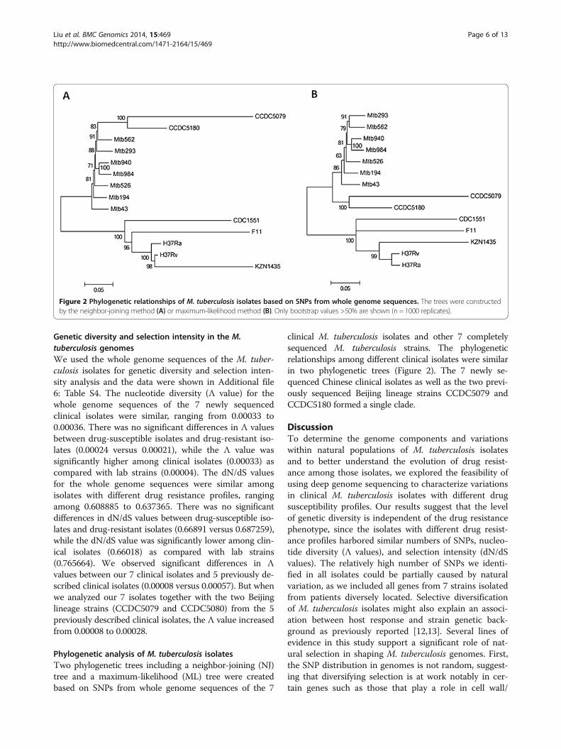
Phylogenetic analysis of M. tuberculosis isolatesTwo phylogenetic trees including a neighbor-joining (NJ)tree and a maximum-likelihood (ML) tree were createdbased on SNPs from whole genome sequences of the 7
clinical M. tuberculosis isolates and other 7 completelysequenced M. tuberculosis strains. The phylogeneticrelationships among different clinical isolates were similarin two phylogenetic trees (Figure 2). The 7 newly se-quenced Chinese clinical isolates as well as the two previ-ously sequenced Beijing lineage strains CCDC5079 andCCDC5180 formed a single clade.
DiscussionTo determine the genome components and variationswithin natural populations of M. tuberculosis isolatesand to better understand the evolution of drug resist-ance among those isolates, we explored the feasibility ofusing deep genome sequencing to characterize variationsin clinical M. tuberculosis isolates with different drugsusceptibility profiles. Our results suggest that the levelof genetic diversity is independent of the drug resistancephenotype, since the isolates with different drug resist-ance profiles harbored similar numbers of SNPs, nucleo-tide diversity (Л values), and selection intensity (dN/dSvalues). The relatively high number of SNPs we identi-fied in all isolates could be partially caused by naturalvariation, as we included all genes from 7 strains isolatedfrom patients diversely located. Selective diversificationof M. tuberculosis isolates might also explain an associ-ation between host response and strain genetic back-ground as previously reported [12,13]. Several lines ofevidence in this study support a significant role of nat-ural selection in shaping M. tuberculosis genomes. First,the SNP distribution in genomes is not random, suggest-ing that diversifying selection is at work notably in cer-tain genes such as those that play a role in cell wall/
Figure 2 Phylogenetic relationships of M. tuberculosis isolates based on SNPs from whole genome sequences. The trees were constructedby the neighbor-joining method (A) or maximum-likelihood method (B). Only bootstrap values >50% are shown (n = 1000 replicates).
Liu et al. BMC Genomics 2014, 15:469 Page 6 of 13http://www.biomedcentral.com/1471-2164/15/469

membrane/envelope biogenesis (class M) and in generalfunction (class R), which tend to accumulate an excessof SNPs [7,12,14,15]. Second, in the SNP density map,many genes located in the regions with significantly highSNP density are involved in host–pathogen interactionsand may contribute to strain-specific virulence attri-butes. For example, one region corresponds to a previ-ously reported virulence operon including the genesRv0986–Rv0988 that are present in one of horizontalgenetic transfer (HGT) regions [7,16,17]. Another regionwith high density of SNPs was found in the ESX-1 locus(RD1 region), which includes a type VII secretion system[18]. But in the absence of the information about strain-specific differences in virulence, the high number ofSNPs could also be the result of lateral gene transfer.Third, the dN/dS values were less than 1 for the genomesof all 7 isolates analyzed, consistent with genome-widepurifying selection. We have previously shown that thedN/dS values for coding regions of drug resistance-associated genes in MDR and XDR isolates were higherthan 1, suggesting that exposure to drugs is among themajor forces driving the high dN/dS ratios in those drugresistance-associated genes [19]. But as suggested in thisstudy, on the genome-wide scale, the clinical M. tubercu-losis strains with different drug resistance profiles undergosimilar levels of purifying selection. Consistently, resultsfrom a recent study suggest that the dominant effect of se-lection on natural M. tuberculosis population is removal ofnovel variants, with exceptions in certain group of genessuch as those involved in defense [20].Indels have a wide range of effects as a very important
cause of phenotypic variability. The acquisition and lossof certain genes could provide pathogens with some ad-vantages during infection and transmission. Thus, theIndel loci identified in this study are candidates for drugresistance or virulence-associated factors that may repre-sent evolutionary signatures during the co-evolution ofhumans and pathogens. For example, the deletion of apolyketide synthase gene (pks5) with high homology tomycocerosic acid synthase is particularly intriguing be-cause the product of this gene may be involved in theproduction of multimethylated branched lipids [21]. Inaddition, the pks5 mutant strain of M. tuberculosisH37Rv was shown to display severe growth defects inmice [22]. It is also worth noting that sixteen of thoseIndels spanned PE-PPE-PGRS genes, which have beenconsidered a major source of antigenic variability [10].In addition, two of those unique proteins code forputative membrane proteins (including MmpL1 andMmpL4) and may directly alter the interactions betweenpathogens and their hosts [23]. Since we identifiedmany Indels including some of those above-mentionedvirulence-associated genes within both drug-susceptibleand drug-resistant strains, our results suggest that drug
resistance in M. tuberculosis is not necessarily an indica-tion of increased virulence. Our findings are consistentwith the notion that the virulence of individual clinicalM. tuberculosis isolate is dependent on multiple factorsincluding strain genetic background and host immuneresponses [24].A highly significant inverse correlation between the
presence of CRISPR-cas locus and acquired antibiotic re-sistance was observed in E. faecalis, suggesting that anti-biotic use inadvertently selects for enterococcal strainswith compromised genome defense [25]. But in thisstudy, no functional genes were identified in CRISPRlocus and no correlation between antibiotic resistanceand the presence of CRISPR-cas locus was observed inclinical M. tuberculosis isolates.Using our previously established method of automatic
TBDReaMDB-coupled analysis for drug resistance-associatedmutations in M. tuberculosis isolates [19], we detected 25types of unreported mutations, as well as 20 known or pu-tative drug efflux pumps with non-sense SNPs in MDR,pre-XDR and XDR M. tuberculosis isolates, but we couldnot establish the association between over expression ofthose mutated drug efflux pumps with increased drug re-sistance in M. tuberculosis. It was reported previously thatmutations or overexpression of Rv0194 and Rv2686c areassociated with increased resistance to multiple drugs inM. tuberculosis [26,27]. But according to another recentstudy which aimed to compare the differences of the ex-pression of 15 putative multidrug efflux pump genes inclinically isolated drug sensitive and MDR M. tuberculosisisolates, all the tested putative multidrug efflux pumpgenes in the drug-sensitive and MDR M. tuberculosis iso-lates have similar rates of expression [28]. Thus, the exist-ence of mutations and over expression of the efflux pumpgenes might not be necessarily associated with increaseddrug resistance.By closely examining the correlation of the phenotypic
drug susceptibility profiles of the strains with mutationsidentified in their drug resistance-associated genes, weidentified a few potential new genetic determinantsof drug resistance. For example, while 5 (Mtb194,Mtb293, Mtb940, Mtb984, Mtb43) of the 7 strains exhib-ited phenotypic resistance to ofloxacin and levofloxacin,only 2 of them (Mtb984 and Mtb43) had gyrA D94Gmutation known to confer resistance to fluoroquino-lones. The other 3 had the same gyrA E21Q, G668D,and S95T mutations seen in fluoroquinolone susceptiblestrain Mtb526, indicating that these mutations arenot the source of fluoroquinolone resistance. Similarly,among the 6 strains showing phenotypic resistance to ri-fampicin, 3 (Mtb194, Mtb293, Mtb940) only had therpoB A1075 mutation, which was also present in thesusceptible strain, suggesting the presence of other un-known mechanisms for rifampicin resistance in them.
Liu et al. BMC Genomics 2014, 15:469 Page 7 of 13http://www.biomedcentral.com/1471-2164/15/469

Since we identified no mutations by further examiningother drug resistance-associated genes such as gyrB,gidB and eis in those strains [29], we then performedgenetic studies for those newly identified potentialdrug resistance-associated mutations, but failed to con-firm their function in causing drug resistance either(data not shown). Thus, our observations demonstratedthat though certain drug resistance-associated mutationssuch as rpoB S531L, katG S315T, gyrA D94G, embBM306V, rpsL K43R, and rrs A1401G could serve as use-ful markers for rapid detection of resistance in the clin-ical M. tuberculosis isolates, the accuracy and sensitivityof genetic-based drug resistance assays still need to beincreased by further elucidation of unknown mecha-nisms of drug resistance, especially for second-line drugs[29-31]. It should also be pointed out that confirmingdrug resistance-associated mutations by genetic studycould only examine the function of individual genemutation without taking into consideration the wholegenetic background of the strain, while based on thewhole genome sequencing studies by us and a fewothers, there might be no common causes of drug resist-ance to multiple drugs. Rather, the MDR and XDR phe-notypes could result from a combination of mutations inthe genomes [15,32,33].The phylogenetic relationships among different clinical
isolates were similar in two phylogenetic trees based onwhole genome SNPs. The whole genome sequencing hasbeen proposed as a sort of “gold standard” for strain typ-ing in M. tuberculosis since it clarifies previous straintyping approaches used for phylogenetic and epidemio-logic studies and provides more detailed genomic vari-ation information. The observation that MDR, pre-XDR,and XDR isolates were located sporadically on differentbranches in phylogenetic trees based on SNPs fromwhole genome sequences of the 14M. tuberculosis iso-lates further confirms our previous observation that theyhave evolved and acquired mutations independently onmultiple occasions. The observation that isolates fromChina were phylogenetically distant from the isolatesfrom other regions such as the KZN strain from SouthAfrica in the phylogenetic trees also confirmed ourprevious observation that drug-resistant M. tuberculosisstrains from different geographic regions have distinctevolutionary pathways [19]. The close phylogenetic re-latedness among the 7 clinical M. tuberculosis isolatescould also be best supported by the analysis of specificSNPs in drug resistance-associated genes. The presenceof identical uncommon mutations in many of thosegenes among the 7 strains (e.g. I322V in Rv1592c, R463Lin katG, A1075 in rpoB, G668D in gyrA etc.) is indicativeof a single cluster of strains circulating in the popula-tion. The finding of high levels of clustering and min-imal strain diversity among MDR/XDR M. tuberculosis
strains within a population has been described previ-ously [34].This study has several limitations. Firstly, since the 7
clinical M. tuberculosis isolates included in the analysisall belonged to the Beijing lineage, thus it is possible thatsimilarities and differences between different straingroups may be explained by phylogenetic lineages, ratherthan phenotypic differences. By comparing our 7 clinicalisolates with 5 previously described clinical isolates fromdiverse lineages and countries of origin, we did observesignificantly higher Л value for those 5 previously de-scribed clinical isolates. However, when we analyzed our7 Beijing lineage isolates together with two previouslydescribed Beijing lineage isolates (CCDC5180: resistantto four first-line drugs; CCDC5079: susceptible strain),the Л value increased significantly, we thus suggest thatgenomic variations we observed among different groupsof isolates are unlikely caused completely by phylogen-etic lineages, but rather associated with diverse pheno-types of the isolates. Secondly, this study was limited bythe relatively small number of isolates included in theanalysis. It is likely that a larger sample with diverse line-ages and countries of origin would probably reveal moreinformation on genomic variations and evolution ofdrug-resistant M. tuberculosis strains.
ConclusionsIn this study, by performing whole genome sequencingstudy, we show that though clinical M. tuberculosis iso-lates have a certain degree of similarity in their geneticmake-up, they exhibit distinct variations in terms of thedistribution of SNP, Indels, CRISPR-cas locus, as wellas the nucleotide diversity and selection intensity. Nogeneralizable differences were identified between drug-susceptible and drug-resistant isolates on the genomicscale. Our study provides evidence strengthening the no-tion that the evolution of drug resistance among clinicalM. tuberculosis isolates is clearly a complex and diversi-fied process. Several questions remain further in-depthinvestigations, such as whether drug susceptibility is af-fected by the deletion of specific genes and disabling ofspecific metabolic pathways. In addition, further studiesusing a larger sampling of M. tuberculosis isolates fromdiverse lineages are warranted to better understand theevolution of drug-resistant M. tuberculosis strains.
MethodsSelection of strains for genome sequencing andcomparative genomic analysisSeven M. tuberculosis clinical strains used for wholegenome sequencing in this study were obtained from aTB referral hospital in Beijing, China during the period2009–2011 [3]. The epidemiologic and clinical data ofthe patients were extracted from the subjects’ medical
Liu et al. BMC Genomics 2014, 15:469 Page 8 of 13http://www.biomedcentral.com/1471-2164/15/469

records. The selected M. tuberculosis clinical isolateshad different antibiotic susceptibility profiles (including1 susceptible isolate, 1 MDR isolate, 3 pre-XDR isolatesand 2 XDR isolate). The median age of the 7 patientswere 39.02 (range: 21–72) years. All 7 patients wereHIV-negative adults. All 7 isolates have the Beijingspoligotype (000000000003771) based on the virtual spo-ligotyping analysis results. For comparative analysis,genome sequences of two lab strains including H37Rv(NC_000962) and H37Ra (NC_009525) as well as otherfive previously sequenced clinical isolates includingKZN_1435 (NC_012943), F11 (NC_009565), CDC1551(NC_002755), CCDC5079 (NC_017523), and CCDC5180 (NC_017522) were downloaded from the NCBIwebsite (fttp://ftp.ncbi.nih.gov/genomes/Bacteria/). Thisstudy was approved by the Ethics Committee of the309 Hospital and the Institute of Microbiology, ChineseAcademy of Sciences, Beijing, China.
Cultures and drug susceptibility testingCultures and drug susceptibility testing (DST) were con-ducted as described previously [19]. Briefly, sputumspecimens were collected, treated and cultured accord-ing to the manufacturer’s instructions using the BAC-TEC MGIT 960 system (Becton Dickinson DiagnosticSystems, Sparks, MD, USA). Cultures positive for growthwere examined by microscopy for the presence of acid-fast bacilli after Ziehl-Neelsen staining. Identification ofM. tuberculosis was performed using p-nitrobenzoic acidand thiophene carboxylic acid hydrazine resistance testsas well as PCR tests. M. tuberculosis isolates werefurther confirmed by 16S rDNA sequencing. DST wasconducted using the indirect proportion method onMiddlebrook 7H10 agar containing 10% oleic acid-albumin-dextrose-catalase (Difco) and 0.5% glycerol ac-cording to the WHO guidelines. The concentrations ofthe drugs used were as follows: isoniazid (0.2 ug/mL),rifampicin (1 ug/mL), ethambutol (5 ug/mL), strepto-mycin (2 ug/mL), pyrazinamide (100 ug/mL), ofloxacin(2 ug/mL), levofloxacin (2 ug/mL), kanamycin (5 ug/mL),capreomycin (10 ug/mL), amikacin (1 ug/mL), ethion-amide (5 ug/mL), para-aminosalicylic acid (2 ug/mL).Quality control was performed during susceptibility test-ing using the reference strains provided by the Nationalinstitute for the control of pharmaceutical and biologicalproducts (China). All drugs were obtained from Sigma LifeScience Company (USA).
Genotyping of M. tuberculosis isolatesIn silico MIRU-VNTR genotyping of the M. tuberculosisisolates was conducted. To predict the number of re-peats at each locus of MIRUs, 24 VNTR sequences fromH37Rv genome were aligned to each assembled genome.The Tandem Repeat Finder algorithm was also used to
predict the MIRU-VNTR type of each strain [35]. The insilico MIRU-VNTR results were confirmed by perform-ing experiments following the 24 locus MIRU-VNTRgenotyping protocol described by Supply et al. [6]. Vir-tual spoligotyping was performed by aligning (withoutgaps) all the reads obtained for each strain against eachof the 43 spacer sequences (26-bp oligos) from the directrepeats (DR) regions. The number of matching reads foreach spacer was counted, considering both forward andreverse-complemented sequences, and accepting up to 1nucleotide mismatch. Spacers with 0 matches were inter-preted as missing. In addition, we also used SpolPred[36], a well-established genotyping technique based onthe presence of unique DNA sequences in M. tubercu-losis, to predict the spoligotype of each strain.
DNA preparation and whole genome sequencingA single colony from 7H10 plate was transferred into7H9 liquid medium supplemented with OADC andTween-80, cultured to 0.5 at OD600, harvested by centri-fugation and resuspended in TE pH8.0 [0.01 M Tris–HCl, 0.001 M EDTA (pH 8.0)]. Genomic DNA wasextracted with phenol/chloroform/isoamyl alcohol (25:24: 1, v/v), precipitated with isopropanol, washed with75% ethanol and finally resuspended in TE pH8.0. Gen-ome sequencing was performed by BerryGenomics(Beijing, China). We used a whole genome shotgunsequencing strategy and Illumina Genome Analysersequencing technology. A 100 bp paired-end run wasperformed with the seven M. tuberculosis strains in twolanes. Genomic DNA was sheared by a nebulizer to gen-erate DNA fragments for the Illumina Paried-EndSequencing method. DNA libraries (15–30 ng/μl) wereconstructed by ligating the specific oligonucleotides(Illumina adapters) designed for PE sequencing to bothends of DNA fragments with the TA cloning method.The ligated DNA was then size selected on a 2% agarosegel. DNA fragments of about 500 bp were excised fromthe gel. DNA was then recovered using a Qiagen gel ex-traction kit and was PCR amplified to produce the finalDNA library. Five picomoles of DNA from each strainwere loaded onto two lanes of the sequencing chip, andthe clusters were generated on the cluster generationstation of the GAIIx using the Illumina cluster gener-ation kit. Bacteriophage ×174 DNA was used as a con-trol. In the case of paired-end reads, distinct adaptorsfrom Illumina were ligated to each end with PCRprimers that allowed reading of each end as separateruns. The sequencing reaction was run for 100 cycles(tagging, imaging, and cleavage of one terminal base at atime), and four images of each tile on the chip weretaken in different wavelengths for exciting each base-specific fluorophore. For paired-end reads, data werecollected as two sets of matched 100-bp reads. Reads for
Liu et al. BMC Genomics 2014, 15:469 Page 9 of 13http://www.biomedcentral.com/1471-2164/15/469

each of the indexed samples were then separated using acustom Perl script (Additional file 7: Script 1). Imageanalysis and base calling were done using the IlluminaGA Pipeline software.
Genome assembly and annotationShort reads were assembled using SOAPdenovo (http://soap.genomics.org.cn), a genome assembler developedspecifically for next-generation short-read sequences. Asthe algorithm is sensitive to sequencing errors, low-quality reads were filtered, and high-quality reads wereused for de novo assembly. Sequences were filteredfor low quality reads using the DynamicTrim andLengthSort Perl scripts within SolexaQA. These scriptstrimmed each read to the longest contiguous read seg-ment for which the quality score at each base wasgreater than p = 0.05 (approximately equivalent to aPhred score of 13), and then removed sequence readsshorter than 25 bp respectively. Where one sequence ofa pair was removed, the remaining sequence was putinto a separate file and used as a singleton during denovo assembly. The SOAP GapCloser was also used toclose gaps where possible after assembly.The protein-coding genes were predicted using Glim-
mer 3.02 [37], while tRNAscan-SE [38] and RNAmmer[39] were used to identify tRNA and rRNA, respectively.The genome sequence was also uploaded into RapidAnnotation using Subsystem Technology (RAST) [40]to check the annotated sequences. The functions ofpredicted protein-coding genes were then annotatedthrough comparisons with the databases of NCBI-NR,COG, and KEGG.
Nucleotide sequence accession numbersWhole genome sequencing projects for 7 clinical M.tuberculosis isolates Mtb562, Mtb194, Mtb293, Mtb526,Mtb940, Mtb984, and Mtb43 have been depositedin GenBank under accession numbers AUTG00000000,AUNH00000000, AUPX00000000, AUTF00000000, AUTX00000000, AUTY00000000, and AUPO00000000,respectively.
SNP detection and analysisFor the sequenced genomes, SOAPsnp (http://soap.gen-omics.org.cn/soapsnp.html) was used to score SNPsfrom aligned reads [41]. The short reads were alignedonto the H37Rv genome reference using the SOAP2program [18]. To obtain reliable alignment hits, at mosttwo mismatches were allowed between the read and thereference. The alignments with the least number of dif-ferences were defined as “best hits.” If there was onlyone single best hit for a read, then the read was taken asuniquely placed; a read with multiple equal best hits was
taken as repeatedly placed. For paired-end reads, tworeads belonging to a pair were aligned together withboth in the correct orientation and with a proper spansize on the reference. The 100-bp reads that were gener-ated for each strain were mapped against H37Rv as areference sequence via ungapped alignments allowing upto two mismatches. For reads that mapped to multiplelocations, one was chosen at random. For paired-enddata, mapping locations of each read were restricted tosites within 300 bp of mapping locations of its partner.SOAPsnp results were filtered as follows: 1) The readcoverage of the SNP site was more than five; 2) The Illu-mina quality score of either allele was more than 30; 3)The count of all mapped best base is more than twotimes the count of all mapped second best base. Inaddition, BWA 0.6.2 [42] and SAMtools 0.1.18 [43] wereused to confirm our results. The Illumina reads werefirst aligned by BWA with default parameters for eachsample. The aligned results were piped to SAMtools forconversion of BWA output format to BAM format andto perform SNP analysis. For the other genomes, all spe-cific SNPs for each strain were manually inspected bytaking into account if SNPs were detected by the twoaligners including MAUVE [44] and MUMmer 3.2 [45].From all SNPs identified in the sequenced genome se-quences, the density of SNPs was calculated throughoutthe M. tuberculosis H37Rv genome using a sliding-window size of 5 kb (step of the sliding window = 5 kb).This analysis led to the construction of a SNP clusteringmap using Circos [46].
Insertion and deletion (Indel) analysisThree different methods were used to detect Indels:1) Multiple alignment of genomic sequences was per-formed by using Mauve multiple alignment software andthe progressive alignment option. The output file pro-duced by Mauve was parsed by using a custom Perlscript to retrieve multiple aligned sequences for Indelloci (Additional file 8: Script 2); 2) For each genome-wide Ilumina sequence dataset, the sequence reads werealigned against the reference genome sequence usingBWA 0.6.2 [42]. Then SAMTOOLS 0.1.18 [43], which isbased on a Bayesian model for Indel calling, was used toperform the analysis using the default Indel detectionparameters, with a small increase in the coverage thresh-old (−D 200); 3) Indel from paired-end mapping datawere identified and visualized with inGAP-SV [47],which uses read depth and read pair data to detect andvisualize large and complex sequence variation.
CRISPR locus identificationFor published genome sequences, CRISPR loci were re-trieved from the CRISPRdb database [48]. Alternatively,the detection of CRISPR loci in our 7 draft genome
Liu et al. BMC Genomics 2014, 15:469 Page 10 of 13http://www.biomedcentral.com/1471-2164/15/469
sequences was achieved using CRISPRFinder [48].BLAST was used for similarity searches between CRISPRspacer sequences and existing sequences in the GenBankdatabase limited to Bacteria (taxid: 2) or Viruses (taxid:10239) entries. Only matches showing 100% identity overthe complete CRISPR spacer sequences were retained, andmatches to sequences found within CRISPR loci wereignored.
Identification of gene mutations associated with drugresistanceMutations in M. tuberculosis antibiotic resistance-associatedgenes and inter-genic regions were downloaded from theTB Drug Resistance Mutation Database (TBDReaMDB)[49], a comprehensive database providing all reported muta-tions associated with TB drug resistance through a publiclyaccessible web site: http://www.tbdreamdb.com, to provideinformation for comparison analysis of drug resistance-associated mutation profiles for the 7 sequencedM. tubercu-losis isolates. To confirm the association between specificgene mutations and drug resistance, we amplified the puta-tive drug resistance-associated genes with mutations fromthe genomic DNA of clinical M. tuberculosis isolates byPCR, and cloned them into the plasmid pMV261, a myco-bacterial replicating vector, then electroporated the recom-binant vectors into the drug-susceptible reference H37Rvstrain for drug susceptibility testing. All the experimentswere repeated at least 3 times.
Genetic diversity and selection intensity analysisThe program DnaSP software version 5.10 was used toinvestigate the genetic diversity of the whole genome se-quences of the M. tuberculosis isolates [50]. The geneticdiversity were measured by haplotype (H), diversity ofhaplotype (Hd), nucleotide diversity (p), and the averagenumber of nucleotide differences (K). The sequences ofthe coding regions from each isolate were concatenatedand the resulting sequences were used to determine thenumber of non-synonymous (dN) and synonymous (dS)substitutions per site. To test the selection intensity, theratios of dN/dS were calculated for each pairwise com-parison, and two-sided Z-test was used to determine thelevel of significance.
Phylogenetic analysisThe neighbor-joining (NJ) and maximum-likelihood (ML)phylogenetic trees were constructed in MEGA5 [51] basedon SNPs from whole genome sequences. The reliability ofeach node was estimated from 1000 random bootstrapresamplings of the data. The phylogenetic data have beendeposited in TreeBase under the accession number 15638(http://purl.org/phylo/treebase/phylows/study/TB2:S15638).
Additional files
Additional file 1: Table S1. Sequencing statistics of M. tuberculosis isolates.
Additional file 2: Table S2. Regions with significantly high SNP density.
Additional file 3: Figure S1. Distribution of SNPs according to theClusters of Orthologous Groups (COG) classification. (U) Intracellular traffickingand secretion; (V) Defense mechanisms; (D) Cell cycle control, mitosis, andmeiosis; (F) Nucleotide transport and metabolism; (O) Post-translationalmodification, protein turnover, chaperones; [O] Posttranslational modification,protein turnover, chaperones; [J] Translation, ribosomal structure and biogen-esis; (H) Coenzyme transport and metabolism; [M] Cell wall/membrane/enve-lope biogenesis; [S] Function unknown; [K] Transcription; (P) Inorganic iontransport and metabolism; (T) Signal transduction mechanisms; (G) Carbohy-drate transport and metabolism; (N) Cell motility; (C) Energy production andconversion; (L) Replication, recombination, and repair; [E] Amino acid trans-port and metabolism; (I) Lipid transport and metabolism; (R) General function;.(Q) Secondary metabolites biosynthesis, transport, and catabolism. (*) Classwith significant over-representation and less-representation of SNPs (p < 0.01).
Additional file 4: Figure S2. Overview of the CRISPR loci in M. tuberculosisstrains. Spacers are shown as diamonds and repeats as rectangles. In eachCRISPR, spacers with identical sequence in the studied genomes are shown inthe same color.
Additional file 5: Table S3. Known or putative drug efflux pumps withnon-synonymous SNPs in MDR, pre-XDR and XDR M. tuberculosis isolatesbut not in H37Rv strain.
Additional file 6: Table S4. DNA diversity and selection intensityanalysis for the whole genome sequences of M. tuberculosis isolates.
Additional file 7: Script 1. The custom Perl script used to separateeach of the indexed samples from raw reads.
Additional file 8: Script 2. The custom Perl script used to retrieve Indelloci from output file (.xmfa) produced by Mauve.
Competing interestsThe authors declare that they have no competing interests.
Authors’ contributionsCHL and BZ conceived and designed the study; CHL, QW and HML collectedand characterized the isolates that are used in this study; CHL and QWperformed laboratory experiments; CHL, FL and YH performed the dataanalysis; QW, HML, GFG and BZ assisted in the data analysis; CHL wrote themanuscript with assistance from other authors; FL, YH and QW equallycontributed to the work. All authors read and approved the final manuscript.
AcknowledgementsThis work was supported by the National Basic Research Program of China(2014CB744400 and 2012CB518700), National Natural Science Foundation ofChina (81371769), the Ministry of Health and the Ministry of Science andTechnology, China (2013ZX10003006 and 2012ZX10005007-011), and theChinese Academy of Sciences (KJZD-EW-L02), and the Beijing MunicipalScience & Technology Development Program.
Author details1CAS key Laboratory of Pathogenic Microbiology and Immunology, Instituteof Microbiology, Chinese Academy of Sciences, Beijing, China. 2Institute forTuberculosis Research, the 309th Hospital, Beijing, China.
Received: 27 August 2013 Accepted: 10 June 2014Published: 13 June 2014
References1. World Health Organization (WHO): Global tuberculosis report 2013. Geneva:
WHO; 2013. Available from: http://apps.who.int/iris/bitstream/10665/91355/1/9789241564656_eng.pdf.
2. Zhao Y, Xu S, Wang L, Chin DP, Wang S, Jiang G, Xia H, Zhou Y, Li Q, Ou X,Pang Y, Song Y, Zhao B, Zhang H, He G, Guo J, Wang Y: National survey ofdrug-resistant tuberculosis in China. N Engl J Med 2012, 366(23):2161–2170.
3. Liu CH, Li L, Chen Z, Wang Q, Hu YL, Zhu B, Woo PC: Characteristics andtreatment outcomes of patients with MDR and XDR tuberculosis in a
Liu et al. BMC Genomics 2014, 15:469 Page 11 of 13http://www.biomedcentral.com/1471-2164/15/469

TB referral hospital in Beijing: a 13-year experience. PLoS One 2011,6(4):e19399.
4. Otal I, Martin C, Vincent-Levy-Frebault V, Thierry D, Gicquel B: Restrictionfragment length polymorphism analysis using IS6110 as an epidemio-logical marker in tuberculosis. J Clin Microbiol 1991, 29(6):1252–1254.
5. Kamerbeek J, Schouls L, Kolk A, van Agterveld M, van Soolingen D, KuijperS, Bunschoten A, Molhuizen H, Shaw R, Goyal M, van Embden J:Simultaneous detection and strain differentiation of Mycobacteriumtuberculosis for diagnosis and epidemiology. J Clin Microbiol 1997,35(4):907–914.
6. Supply P, Allix C, Lesjean S, Cardoso-Oelemann M, Rüsch-Gerdes S, Willery E,Savine E, de Haas P, van Deutekom H, Roring S, Bifani P, Kurepina N,Kreiswirth B, Sola C, Rastogi N, Vatin V, Gutierrez MC, Fauville M, Niemann S,Skuce R, Kremer K, Locht C, van Soolingen D: Proposal for standardizationof optimized mycobacterial interspersed repetitive unit-variable-numbertandem repeat typing of Mycobacterium tuberculosis. J Clin Microbiol 2006,44(12):4498–4510.
7. Namouchi A, Didelot X, Schock U, Gicquel B, Rocha EP: After thebottleneck: Genome-wide diversification of the Mycobacteriumtuberculosis complex by mutation, recombination, and natural selection.Genome Res 2012, 22(4):721–734.
8. Tatusov RL, Koonin EV, Lipman DJ: A genomic perspective on proteinfamilies. Science 1997, 278(5338):631–637.
9. Tatusov RL, Fedorova ND, Jackson JD, Jacobs AR, Kiryutin B, Koonin EV,Krylov DM, Mazumder R, Mekhedov SL, Nikolskaya AN, Rao BS, Smirnov S,Sverdlov AV, Vasudevan S, Wolf YI, Yin JJ, Natale DA: The COG database:an updated version includes eukaryotes. BMC Bioinformatics 2003, 4:41.
10. Sampson SL: Mycobacterial PE/PPE proteins at the host-pathogeninterface. Clin Dev Immunol 2011, 2011:497203.
11. van Kessel JC, Hatfull GF: Recombineering in Mycobacterium tuberculosis.Nat Methods 2007, 4(2):147–152.
12. Deitsch KW, Moxon ER, Wellems TE: Shared themes of antigenic variationand virulence in bacterial, protozoal, and fungal infections. Microbiol MolBiol Rev 1997, 61(3):281–293.
13. Di Pietrantonio T, Correa JA, Orlova M, Behr MA, Schurr E: Joint effects ofhost genetic background and mycobacterial pathogen on susceptibilityto infection. Infect Immun 2011, 79(6):2372–2378.
14. Kennemann L, Didelot X, Aebischer T, Kuhn S, Drescher B, Droege M,Reinhardt R, Correa P, Meyer TF, Josenhans C, Falush D, Suerbaum S:Helicobacter pylori genome evolution during human infection. Proc NatlAcad Sci U S A 2011, 108(12):5033–5038.
15. Wu W, Zheng H, Zhang L, Wen Z, Zhang S, Pei H, Yu G, Zhu Y, Cui Z, Hu Z,Wang H, Li Y: A genome-wide analysis of multidrug-resistant andextensively drug-resistant strains of Mycobacterium tuberculosis Beijinggenotype. Mol Genet Genomics 2013, 288(9):425–436.
16. Rosas-Magallanes V, Deschavanne P, Quintana-Murci L, Brosch R, Gicquel B,Neyrolles O: Horizontal transfer of a virulence operon to the ancestor ofMycobacterium tuberculosis. Mol Biol Evol 2006, 23(6):1129–1135.
17. Veyrier F, Pletzer D, Turenne C, Behr MA: Phylogenetic detection ofhorizontal gene transfer during the step-wise genesis of Mycobacteriumtuberculosis. BMC Evol Biol 2009, 9:196.
18. Bitter W, Houben EN, Bottai D, Brodin P, Brown EJ, Cox JS, Derbyshire K,Fortune SM, Gao LY, Liu J, Gey van Pittius NC, Pym AS, Rubin EJ, ShermanDR, Cole ST, Brosch R: Systematic genetic nomenclature for type VIIsecretion systems. PLoS Pathog 2009, 5(10):e1000507.
19. Liu CH, Li HM, Lu N, Wang Q, Hu YL, Yang X, Hu YF, Woo PC, Gao GF, ZhuB: Genomic sequence based scanning for drug resistance-associated mu-tations and evolutionary analysis of multidrug-resistant and extensivelydrug-resistant Mycobacterium tuberculosis. J Infect 2012, 65(5):412–422.
20. Pepperell CS, Casto AM, Kitchen A, Granka JM, Cornejo OE, Holmes EC,Birren B, Galagan J, Feldman MW: The role of selection in shapingdiversity of natural M. tuberculosis Populations. Plos Pathog 2013,9(8):e1003543.
21. Etienne G, Malaga W, Laval F, Lemassu A, Guilhot C, Daffe M: Identificationof the Polyketide Synthase Involved in the Biosynthesis of theSurface-Exposed Lipooligosaccharides in Mycobacteria. J Bacteriol 2009,191(8):2613–2621.
22. Rousseau C, Sirakova TD, Dubey VS, Bordat Y, Kolattukudy PE, Gicquel B,Jackson M: Virulence attenuation of two Mas-like polyketide synthasemutants of Mycobacterium tuberculosis. Microbiol-Sgm 2003,149:1837–1847.
23. Wells RM, Jones CM, Xi ZY, Speer A, Danilchanka O, Doornbos KS, Sun PB,Wu FM, Tian CL, Niederweis M: Discovery of a Siderophore export systemessential for virulence of Mycobacterium tuberculosis. Plos Pathog 2013,9(1):e1003120.
24. Portevin D, Gagneux S, Comas I, Young D: Human Macrophage responsesto clinical isolates from the Mycobacterium tuberculosis complexdiscriminate between ancient and modern lineages. Plos Pathog 2011,7(3):e1001307.
25. Palmer KL, Gilmore MS: Multidrug-resistant enterococci lack CRISPR-cas.MBio 2010, 1(4):e00227. 10.
26. Danilchanka O, Mailaender C, Niederweis M: Identification of a novelmultidrug efflux pump of Mycobacterium tuberculosis. Antimicrob AgentsChemother 2008, 52(7):2503–2511.
27. Pasca MR, Guglierame P, Arcesi F, Bellinzoni M, De Rossi E, Riccardi G: Rv2686c-Rv2687c-Rv2688c, an ABC fluoroquinolone efflux pump in Mycobacteriumtuberculosis. Antimicrob Agents Chemother 2004, 48(8):3175–3178.
28. Calgin MK, Sahin F, Turegun B, Gerceker D, Atasever M, Koksal D, Karasartova D,Kiyan M: Expression analysis of efflux pump genes among drug-susceptibleand multidrug-resistant Mycobacterium tuberculosis clinical isolates andreference strains. Diagn Microbiol Infect Dis 2013, 76(3):291–297.
29. Jnawali HN, Hwang SC, Park YK, Kim H, Lee YS, Chung GT, Choe KH, Ryoo S:Characterization of mutations in multi- and extensive drug resistanceamong strains of Mycobacterium tuberculosis clinical isolates in Republicof Korea. Diagn Microbiol Infect Dis 2013, 76(2):187–196.
30. Poudel A, Maharjan B, Nakajima C, Fukushima Y, Pandey BD, Beneke A,Suzuki Y: Characterization of extensively drug-resistant Mycobacteriumtuberculosis in Nepal. Tuberculosis (Edinb) 2013, 93(1):84–88.
31. Imperiale BR, Zumarraga MJ, Di Giulio AB, Cataldi AA, Morcillo NS: Molecularand phenotypic characterisation of Mycobacterium tuberculosis resistant toanti-tuberculosis drugs. Int J Tuberc Lung Dis 2013, 17(8):1088–1093.
32. Zhang H, Li D, Zhao L, Fleming J, Lin N, Wang T, Liu Z, Li C, Galwey N,Deng J, Zhou Y, Zhu Y, Gao Y, Wang T, Wang S, Huang Y, Wang M, ZhongQ, Zhou L, Chen T, Zhou J, Yang R, Zhu G, Hang H, Zhang J, Li F, Wan K,Wang J, Zhang XE, Bi L: Genome sequencing of 161 Mycobacteriumtuberculosis isolates from China identifies genes and intergenic regionsassociated with drug resistance. Nat Genet 2013, 45(10):1255–1260.
33. Warner DF, Mizrahi V: Complex genetics of drug resistance inMycobacterium tuberculosis. Nat Genet 2013, 45(10):1107–1108.
34. Gandhi NR, Brust JC, Moodley P, Weissman D, Heo M, Ning Y, Moll AP,Friedland GH, Sturm AW, Shah NS: Minimal diversity of drug-resistantMycobacterium tuberculosis strains, South Africa(1.). Emerg Infect Dis2014, 20(3):394–401.
35. Benson G: Tandem repeats finder: a program to analyze DNA sequences.Nucleic Acids Res 1999, 27(2):573–580.
36. Coll F, Mallard K, Preston MD, Bentley S, Parkhill J, McNerney R, Martin N,Clark TG: SpolPred: rapid and accurate prediction of Mycobacteriumtuberculosis spoligotypes from short genomic sequences. Bioinformatics2012, 28(22):2991–2993.
37. Delcher AL, Bratke KA, Powers EC, Salzberg SL: Identifying bacterial genesand endosymbiont DNA with Glimmer. Bioinformatics 2007, 23(6):673–679.
38. Lowe TM, Eddy SR: tRNAscan-SE: a program for improved detection of transferRNA genes in genomic sequence. Nucleic Acids Res 1997, 25(5):955–964.
39. Lagesen K, Hallin P, Rodland EA, Staerfeldt HH, Rognes T, Ussery DW:RNAmmer: consistent and rapid annotation of ribosomal RNA genes.Nucleic Acids Res 2007, 35(9):3100–3108.
40. Aziz RK, Bartels D, Best AA, DeJongh M, Disz T, Edwards RA, Formsma K,Gerdes S, Glass EM, Kubal M, Meyer F, Olsen GJ, Olson R, Osterman AL,Overbeek RA, McNeil LK, Paarmann D, Paczian T, Parrello B, Pusch GD, ReichC, Stevens R, Vassieva O, Vonstein V, Wilke A, Zagnitko O: The RAST Server:rapid annotations using subsystems technology. BMC Genomics 2008, 9:75.
41. Li R, Li Y, Fang X, Yang H, Wang J, Kristiansen K, Wang J: SNP detectionfor massively parallel whole-genome resequencing. Genome Res 2009,19(6):1124–1132.
42. Li H, Durbin R: Fast and accurate long-read alignment with Burrows-Wheeler transform. Bioinformatics 2010, 26(5):589–595.
43. Li H, Handsaker B, Wysoker A, Fennell T, Ruan J, Homer N, Marth G, AbecasisG, Durbin R, Genome Project Data Processing S: The Sequence Alignment/Map format and SAMtools. Bioinformatics 2009, 25(16):2078–2079.
44. Darling AC, Mau B, Blattner FR, Perna NT: Mauve: multiple alignment ofconserved genomic sequence with rearrangements. Genome Res 2004,14(7):1394–1403.
Liu et al. BMC Genomics 2014, 15:469 Page 12 of 13http://www.biomedcentral.com/1471-2164/15/469
45. Kurtz S, Phillippy A, Delcher AL, Smoot M, Shumway M, Antonescu C,Salzberg SL: Versatile and open software for comparing large genomes.Genome Biol 2004, 5(2):R12.
46. Krzywinski M, Schein J, Birol I, Connors J, Gascoyne R, Horsman D, Jones SJ,Marra MA: Circos: an information aesthetic for comparative genomics.Genome Res 2009, 19(9):1639–1645.
47. Qi J, Zhao F: inGAP-sv: a novel scheme to identify and visualizestructural variation from paired end mapping data. Nucleic Acids Res 2011,39(Web Server issue):W567–W575.
48. Grissa I, Vergnaud G, Pourcel C: CRISPRFinder: a web tool to identifyclustered regularly interspaced short palindromic repeats. Nucleic AcidsRes 2007, 35(Web Server issue):W52–W57.
49. Sandgren A, Strong M, Muthukrishnan P, Weiner BK, Church GM, Murray MB:Tuberculosis drug resistance mutation database. PLoS Med 2009, 6(2):e2.
50. Librado P, Rozas J: DnaSP v5: a software for comprehensive analysis ofDNA polymorphism data. Bioinformatics 2009, 25(11):1451–1452.
51. Tamura K, Peterson D, Peterson N, Stecher G, Nei M, Kumar S: MEGA5:Molecular Evolutionary Genetics Analysis using maximum likelihood,evolutionary distance, and maximum parsimony methods. Mol Biol Evol2011, 28(10):2731–2739.
doi:10.1186/1471-2164-15-469Cite this article as: Liu et al.: Comparative genomic analysis ofMycobacterium tuberculosis clinical isolates. BMC Genomics 2014 15:469.
Submit your next manuscript to BioMed Centraland take full advantage of:
• Convenient online submission
• Thorough peer review
• No space constraints or color figure charges
• Immediate publication on acceptance
• Inclusion in PubMed, CAS, Scopus and Google Scholar
• Research which is freely available for redistribution
Submit your manuscript at www.biomedcentral.com/submit
Liu et al. BMC Genomics 2014, 15:469 Page 13 of 13http://www.biomedcentral.com/1471-2164/15/469