Mycobacterium - Europe PMC
156
BACTERIOLOGICAL Rzvizws, Mar. 1977, p. 217-372 Vol. 41, No. 1 Copyright © 1977 American Society for Microbiology Printed in U.S.A. Mycobacterium LANE BARKSDALE'* AND KWANG-SHIN KIM Department of Microbiology, New York University School of Medicine and Medical Center, New York, New York 10016 INTRODUCTION ................................... 219 TAXONOMY: IDENTIFYING MYCOBACTERIA . ................... 222 THE MYCOBACTERIAL CELL ............................................. 225 Acid-Fastness ...................... ....................... 227 Mycobacterial acid-fastness ......................................... ... 227 Ziehl-Neelsen stain ........... .............................. 230 Interaction of dye and mycolic acid ......................................... 231 Ropelike structures, the integrity of the rigid layer of the cell walls, and acid- 231 fastness ......................................... 235 Fluorochrome staining of mycobacteria ..................................... 235 Acid-stable binding and gram staining of mycobacteria ........ .............. 235 Non-acid-fast mycobacteria . ......................................... 238 Conditions of Growth ......................................................... 238 The Mycobacterial Nucleoid .................... 239 Mycobacterial Ribosomes .................... 241 Rifampin, DNA-directed RNA polymerase, and the breakdown of ribosomes ... 241 Genetics of Mycobacteria (Origins of H37Rv) ........ .......................... 242 Autolysis, Protoplasts, and Mycobacterial L-Forms ....... ..................... 242 LIPID BIOSYNTHESIS ....................................................... 242 Fatty Acids in General . ....................................................... 250 Diglycerides, Triglycerides, Lipid Globules (Fat Bodies), and Intracellular Storage of Lipid ................................................................ 245 Phospholipids and Glycolipids .............. .................................. 246 Cardiolipin ................................................................ 249 Phosphatidylethanolamines and Diglycerides ........ .......................... 249 Mannophosphoinositides ......... . .......................................... 249 Biosynthesis of Mannophosphoinositides ......... ............................. 250 Immunizations with Cardiolipins, Phosphatidylethanolamine, and Phosphatidyl- inositol Mannosides ........................ 251 Lipids of Transfer, Carrier Lipids ........................ 251 Mycobacterial Methylations ........................ 252 Lipid Syntheses and Stages (Phases) of Mycobacterial Growth ..... ............ 253 Mycolic Acids ............................................................... 253 Isonicotinic Acid Hydrazide Synthesis of Mycolates ....... ..................... 255 Acylglucose and Acyltrehalose .............. .................................. 255 Cord Factor(s) ............................................................... 256 The real cord factor ........................................................ 258 Cord factor ganulomas . ..................................................... 258 Cord factor inhibition of tumors ............ ................................ 258 Sulfolipids ................................................................ 258 THE CYTOPLASMIC MEMBRANE SYSTEM ................ ................... 259 Mesosomes, Cytochromes, Iron-Chelating Compounds ...... ................... 259 Carotenoid Pigments of Mycobacteria ......................................... 259 Biology of Pigmentation .................... ................................. 261 Pigmentation and Virulence ................. ................................. 262 Biosynthesis of Mycobacterial Carotenoids ......... ........................... 262 Induction of carotenoid synthesis ........... ................................ 263 Protection Against Photoinduced Cell Death ........ .......................... 264 Carotenoids and Photosensitization of Animals .......... . ...................... 265 Cellular Locations of Carotenoids and Endogenous Sensitizers ..... ............ 265 Sensitivity of Mycobacteria to Ultraviolet Irradiation ....... ................... 266 Evolutionary Implications of Patterns of Carotenoid Biosynthesis ..... ......... 266 THE MYCOBACTERIAL CELL WALL: THE MUREIN-ARABINOGALACTAN- MYCOLATE . ............................................................ 267 Arabinogalactan and Arabinogalactan-Mycolate ....... ........................ 268 1 Reprint requests from the United States and the Commonwealth of Puerto Rico should be accompanied by the appropriate postage for return mail. 217
-
Upload
khangminh22 -
Category
Documents
-
view
1 -
download
0
Transcript of Mycobacterium - Europe PMC

BACTERIOLOGICAL Rzvizws, Mar. 1977, p. 217-372 Vol. 41, No. 1Copyright © 1977 American Society for Microbiology Printed in U.S.A.
MycobacteriumLANE BARKSDALE'* AND KWANG-SHIN KIM
Department of Microbiology, New York University School of Medicine and Medical Center, New York,New York 10016
INTRODUCTION ................................... 219TAXONOMY: IDENTIFYING MYCOBACTERIA .................... 222THE MYCOBACTERIAL CELL ............................................. 225
Acid-Fastness ...................... ....................... 227Mycobacterial acid-fastness ......................................... ... 227Ziehl-Neelsen stain ........... .............................. 230Interaction of dye and mycolic acid ......................................... 231Ropelike structures, the integrity of the rigid layer of the cell walls, and acid- 231
fastness ......................................... 235Fluorochrome staining of mycobacteria ..................................... 235Acid-stable binding and gram staining of mycobacteria ........ .............. 235Non-acid-fast mycobacteria.......................................... 238
Conditions of Growth ......................................................... 238The Mycobacterial Nucleoid .................... 239Mycobacterial Ribosomes .................... 241Rifampin, DNA-directed RNA polymerase, and the breakdown of ribosomes ... 241
Genetics of Mycobacteria (Origins of H37Rv) ........ .......................... 242Autolysis, Protoplasts, and Mycobacterial L-Forms ....... ..................... 242
LIPID BIOSYNTHESIS ....................................................... 242Fatty Acids in General........................................................ 250Diglycerides, Triglycerides, Lipid Globules (Fat Bodies), and Intracellular Storage
of Lipid ................................................................ 245Phospholipids and Glycolipids .............. .................................. 246Cardiolipin ................................................................ 249Phosphatidylethanolamines and Diglycerides ........ .......................... 249Mannophosphoinositides .................................................... 249Biosynthesis of Mannophosphoinositides ......... ............................. 250Immunizations with Cardiolipins, Phosphatidylethanolamine, and Phosphatidyl-
inositol Mannosides ........................ 251Lipids of Transfer, Carrier Lipids........................ 251Mycobacterial Methylations ........................ 252Lipid Syntheses and Stages (Phases) of Mycobacterial Growth ..... ............ 253Mycolic Acids ............................................................... 253Isonicotinic Acid Hydrazide Synthesis of Mycolates ....... ..................... 255Acylglucose and Acyltrehalose .............. .................................. 255Cord Factor(s) ............................................................... 256The real cord factor........................................................ 258Cord factor ganulomas...................................................... 258Cord factor inhibition of tumors ............ ................................ 258
Sulfolipids ................................................................ 258THE CYTOPLASMIC MEMBRANE SYSTEM ................ ................... 259Mesosomes, Cytochromes, Iron-Chelating Compounds ...... ................... 259Carotenoid Pigments of Mycobacteria......................................... 259Biology of Pigmentation .................... ................................. 261Pigmentation and Virulence ................. ................................. 262Biosynthesis of Mycobacterial Carotenoids ......... ........................... 262
Induction of carotenoid synthesis ........... ................................ 263Protection Against Photoinduced Cell Death ........ .......................... 264Carotenoids and Photosensitization of Animals .......... . ...................... 265Cellular Locations of Carotenoids and Endogenous Sensitizers ..... ............ 265Sensitivity of Mycobacteria to Ultraviolet Irradiation ....... ................... 266Evolutionary Implications of Patterns of Carotenoid Biosynthesis ..... ......... 266
THE MYCOBACTERIAL CELL WALL: THE MUREIN-ARABINOGALACTAN-MYCOLATE............................................................. 267
Arabinogalactan and Arabinogalactan-Mycolate ....... ........................ 2681 Reprint requests from the United States and the Commonwealth of Puerto Rico should be accompanied by the
appropriate postage for return mail.
217

218 BARKSDALE AND KIM BACTERIOL. REV.
Teichoic Acids ............................................................... 269Early Studies on Fragments of the Mycobacterial Cell Wall ...... .............. 269Beyond the Arabinogalactan-Mycolate, "Nonpeptidoglycan" Amino Acids, and
Surface Peptidoglycolipid ............... ................................. 270Ropelike Patterns: the Surface or Subsurface Glycolipids, Peptidoglycolipids, and
Peptidolipids ............................. 271Glycolipids, Peptidoglycolipids (Mycosides), and Peptidolipids of the Outer Enve-
lope ................................................ ................ 277Other Molecular Species Associated with the Mycobacterial Cell Surface: Phos-
phatidylinositol Oligomannosides .......... ............................... 279MYCOBACTERIA GROWING IN VIVO (PHE I) AND IN VITRO (PHE II) ....... 279GRANULOMAS............281GRANULOMAS~~~~~ . . . . . . . . . . . . . . . . . . . . . . . . . . . . . . . . . . . . . . . . .............
The Tubercle and Granulomatagenesis .......... .............................. 281The Bentonite Granuloma .................................................... 282Experimental Granulomatous Systems .......... ............................... 283Giant Cell Formation In Vitro................................................ 283Giant Cells in Beryllium Granulomas .......... ............................... 283Cord Factor Granulomas ..................... ................................ 283Are FBG and HG Poles Apart? .............. ................................. 284
MYCOBACTERIUM AS ANTIGEN ............................ ; 285Agglutination ............................... 285Serology ofM. avium ............................. 285Soluble Antigens (Immunodiffusion) .... ........................ 287Agglutination of Particles Coated with Soluble Mycobacterial Antigens ..... .... 289The Soluble-Antigen Fluorescent-Antibody Test ........ ....................... 290Tuberculins and Other Mycobacterial Elicitins ........ ........................ 290
Skin test reactions .......................................................... 290PPD-S, OT, and their tuberculin-active components ....... .................. 291
THE IMMUNE RESPONSE TO MYCOBACTERIUM ....... ................... 293"Humoral Immunity" (HI), the Plasma Cell Arm of the Immune System ........ 293Antibody Responses in Tuberculous Infection ........ ......................... 293
"Cytophilic" and other antibody activities associated with tuberculous infection. 294Binding of guinea pig cytophilic antibody (y2-immunoglobulin) to mycobacterialglycopeptide .......................................... ; .................. 295
Cytotoxic antibody.......................................................... 296Antibodies reactive with purified protein derivative ....... ................... 296
Delayed Hypersensitivity, the Jones-Mote Reaction (Cutaneous Basophil Hyper-sensitivity), Contact Sensitivity, and Suppressor B Cells ...... ............. 296
Delayed Hypersensitivity and Cell-Mediated Immunity ...... ................... 298Humoral Immunity, Delayed Hypersensitivity, and Cell-Mediated Immunity in
Mycobacterial Infections ................ ................................. 300Lymphocytes, Macrophages, and Mycobacteriostasis ....... .................... 300Migration Inhibition Factor and Delayed Hypersensitivity in Immunized Guinea
Pigs .................................................................. 302Development of Autoantibodies in Mycobacterial Infections ...... .............. 303Adjuvant Effects on the Cellular Components of the Immune System ..... ...... 303CFA and antigenic competition ............ ................................. 305CFA and the depression of DH and HI ......... ............................. 305CFA, PPD, and tuberculin anergy ........... ............................... 306Intramacrophagic phospholipase A and adjuvants ............................ 306Adjuvant disease........................................................... 307The ultimate mycobacterial adjuvant .......... ............................. 307
Mycobacterial Vaccines...................................................... 309Living BCG ............................................................... 309Residues of methanol-extracted BCG (MER) .... . ............................ 311Mycobacterium microti and the vole bacillus vaccine ...... ................... 312Cross-protection studies with mycobacterial vaccines ...... .................. 313Cell wall vaccines.......................................................... 313
MYCOBACTERIOPHAGES .................................................... 313Serology ................................................................... 317Chloroform-Sensitive Mycobacteriophages ..................................... 318Lysogeny and Pseudolysogeny ............... ................................. 319Suitability of Mycobacteriophages as "Typing" Phages......................... 319Phage-Typing of Mycobacteria Such as M. tuberculosis and M. fortuitum ....... 321Host Cell Receptors and Mycobacteriophage-Induced Receptor-Destroying En-
zymes ............................................................... 321
MYCOBACTERIUM 219
MYCOBACTERIOCINS ........................................................SOME ABBREVIATIONS USED IN THE TEXT ................................SUMMARY ...................................................................The Mycobacterial Cell ......................................................
Ultrastructure .............................................................Biosyntheses ..............................................................Taxonomy ................................................................
Mycobacteriophages and Genetics of Mycobacteria ............................Interaction with the Animal Host.............................................Macrophages ..............................................................Immune response ..........................................................Components inducing specific host responses................................
LITERATURE CITED.........................................................
322322324324324324325325325325325326326
INTRODUCTIONUp until about 10 years ago, our concern with
mycobacteria was occasional and related toidentifying organisms from clinical materials.Then, when pondering the species forma, My-cobacterium leprae (95), we found a need toknow more about Mycobacterium. An inspec-tion of Tables 1 and 2 will remind the readerthat 50% of all mycobacterial species were notrecognized until after 1950, despite the fact thatM. tuberculosis had been known since 1882.Although these "new" species of mycobacteriahad been around since prehistoric times, theiracceptance by mycobacteriologists, especiallymedical mycobacteriologists, was slow due tobeliefs and prejudices that held sway followingthe establishment of M. tuberculosis as thecause of tuberculosis. Expressions such as"atypical mycobacteria" and beliefs such as"chromogenic mycobacteria do not cause pul-monary disease" gained currency during theperiod 1900 to 1920 when the approach to clini-cal material was often "to rule in or out" M.tuberculosis. Now that the infective potentialofmany mycobacteria is appreciated (see Table4) and their identification is easily accom-plished (see Table 3), there is a need to gobeyond mere identification ofmycobacteria andassemble data concerning the biology of theseversatile microorganisms. The present reviewis aimed at defining Mycobacterium from twogeneral standpoints: (i) their biology, includingtheir biosynthetic capacities, the shapes theyassume, the basis for their own peculiar acid-fastness, etc. (original work of ours reportedherein deals with the biology of mycobacteria)and (ii) the reactions they and/or their variousproducts elicit in animal hosts. The immuneresponse of animals to mycobacteria providesus with a further means of characterizing theorganisms and their products. Our coverage ofimmune responses to mycobacteria is some-what diffuse. To a degree, this reflects the cur-rent proliferation of reports attempting to de-fine the components of the immune system andtheir interaction.
Mammalian immune mechanisms comprisemulticomponent systems whose primary selec-tive advantage may have come from their ca-pacity to successfully control growth and differ-entiation. One aspect of this control may in-volve surveillance (1152) by adaptive (206) andnonadaptive immune killing (469a) of elements(endogenous or exogenous) foreign to the host.When foreign antigens stimulate an immunesystem with seemingly exquisite specificity,they do so as alien agents carrying non-selfmarkers (selfincludes major histocompatibilityantigens [MHC; 77a], ABO blood group anti-gens, and any as yet to be discovered surfaceantigens of host phenotypes) toward which thehost animal is equipped to respond. When adju-vant-active molecules enhance or depress a hostresponse, they are exhibiting a capacity to reg-ulate an activity of the host. Perhaps, in somecases, their capacity to function as regulatorsstems from their likeness to regulator mole-cules ofthe host. This likeness seemingly wouldrequire a neatness of fit such as that whichexists between enzymes and their substrates.The accident of such fits no doubt involves thekind of accidents of similarity that account forserological cross-reactions. An interesting ex-ample of a bacterial product serving as a regu-lator molecule in a mammalian immune sys-tem is N-acetylmuramyl-L-alanyl-i-isoglutam-ine which, when administered to animals,turns on both antibody synthesis and the devel-opment of delayed hypersensitivity to an other-wise "poor" antigen. It has been establishedthat, whereas N-acetylmuramic acid can be re-placed by N-glycolyl-muramic acid in thissugar dipeptide, the y-glutamyl function of iso-glutamine is essential for the adjuvant action ofthe compound. Does the sugar dipeptide, syn-thetic or derived from bacterial cell walls (of awide range of species of bacteria), do its jobbecause it mimics a regulator molecule of theimmune system?The mammalian immune system does not
always reject that which is foreign. For exam-ple, there is accommodation to foreign-ness in
VOL. 41, 1977
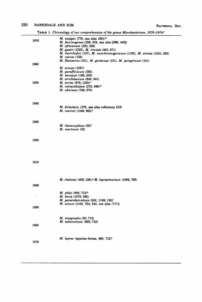
TABLE 1. Chronology ofour comprehension of the genus Mycobacterium, 1870-1974a
1970 M. szulgai (776, see also 1001)bM. farcinogenes (229; 228, see also [699, 440])M. africanum (225; 226)M. gastri (1225), M. triviale (561; 671)M. diernhoferi (157), M. nonchromogenicum (1162), M. simiae (1241; 582)M. vaccae (156)M. flavescens (151), M. gordonae (151), M. peregrinum (151)
1960M. xenopi (1037)M. paraffinicum (292)M. kansasii (198; 500)M. scrofulaceum (940; 941)
1950 M. terrae (978; 1225)cM. intracellulare (275; 996)dM. ukerans (746; 370)
1940M. fortuitum (276, see also reference 519)M. microti (1246; 963)e
1930M. thamnopheos (53)'M. marinum (52)
1920
1910
M. chelonei (403; 126),g M. lepraemurium (1094; 769)1900
M. phlei (842; 713)hM. bovis (1070; 585)M. paratuberculosis (552, 1188; 126)YM. avium (1104, 754; 244, see also [717])
1890
M. smegmatis (30; 713)M. tuberculosis (623; 712)
1800
M. keprae (species forma, 489, 712)J1870
220 BARKSDALE AND KIM BACTERIOL. REV.

TABLE 1-Continueda Species are arranged chronologically according to the date of the initial description. With two excep-
tions, the mycobacterial species listed in this table are those having species status in Bergey's Manualedition 8 (196). Proposed mycobacterial taxa that are not recognized as having species status according toBergey's Manual (196) and for which no synonymy has been proposed include: M. anabanti (130), M.asiaticum (1245), M. aurum (1175, 1164), M. chitae (1166), M. duvaiii (1086), M. engbaeki (647), M. gilvum(1086), M. lactis (647), M. obuense (1170), M. parafortuitum (1174), M. rhodesiae (1171), M. shimoidei (1173a),and M. thermoresistibile (1163, 1164). Other as yet unsubstantiated taxa (see references 196 and 499)include: M. album, M. azot-absorptum, M. brevicale, M. butanitrificans, M. cuneatum, M. gallinarum, M.hyalinum, M. methanicum, M. petroleophilum, M. rubrum, and M. sarni.
b The first number(s) following each specific epithet is (are) the reference(s) describing that organismand/or its isolation. The reference just after the semicolon is that which establishes the presently acceptedtaxon.
¢ Common name: radish bacillus.d Common name: Battey bacillus.e Common name: vole bacillus." Lechevalier et al. (698), on the basis of an analysis for mycolic acids of the type strain, suggest that this
species is probably a nocardia.9 See Table 2.h Common names: timothy and hay bacilli.i Common name: Johne's bacillus.J Common name: Hansen's bacillus.
TABLE 2. Certain accepted species of mycobacteria and their synonyms
M. chelonei (abscessus 849/1084, 1087borstelense 154/1084, 1087friedmanii 403; 1272/1084runyonii 151/1084, 1087)a
M. chelonei subsp. abscessus (abscessus 849/196, 666 runyonii 151/1172)M. chelonei subsp. chelonei (borstelense 154/196, 666, 1173)M. flavescens (acapulcensis 151/911, 1231)M. fortuitum (giae 282/449
minetti 823, 918/449, 1248peregrinum 151/196, 666, 912ranae 676; 126/519, 912, 1085salmoniphilum 447; 992/attributed to Gordon [499])
M. gordonae (aquae sensu B6nicke [153] 417; 762, see also reference 418/1226, 1000)M. intracellulare (Nocardia intracellularis 275/996
brunense 604/196, 1002)M. kansasii (luciflavum 815; 768/196)M. marinum (balnei 882; 728/150
platypoecilus 85/150, 806)M. microti (muris 1246; 1065/196)M. nonchromogenicum (terrae Tsukamura 1164/1165)M. paratuberculosis (Johnei 552; 392/196)M. phlei (moelleri 842; 244/196)M. scrofulaceum (marianum 1115, see also 917/1228, 1232, 1234
paraffinicum 292/196, 1002)M. simiae (budapestae 1244; 1009/1009
habana 1196/1242, 1245a)M. smegmatis(aquae 417; 762, see also 418/449
butyricum 923; 126/448friburgensis 644; 244/448lacticola 713/448)
M. terrae (novum 1167/196)M. ulcerans (buruli 251; 250/1002)M. xenopi (littorale 777; 771/771, 778)
a References preceding bar (/): the first number(s) following each specific epithet is (are) the reference(s)describing that organism or its isolation or both; the reference following the semicolon is that whichestablishes the presently accepted taxon. The reference(s) following the bar (/) documents the synonym.
MYCOBACTERIUM 221VOL. 41, 1977

222 BARKSDALE AND KIM
the carrying of a fetus and in the bearing oftumors. Although the specific mechanisms ofaccommodation or tolerance in these cases isnot fully understood, they probably involveblocking antibodies (987a) and/or specific anti-gen-antibody complexes (1060a), either ofwhich could interfere with the cytotoxic actionof effector cells. The first stage in this process,the generation of toleragenic factors, occursduring protracted exposure to antigen, as in thecase of animals immunized with completeFreund adjuvant (CFA) (26). It would appearreasonable to assume that the induction of tol-erance might often be a necessary concomitantof mycobacterial infections.
It has recently been reported that many hu-man subjects possess antibodies to M. tubercu-losis. Was the production of these antibodiesstimulated by M. tuberculosis? Or is their ap-parent specificity due to a cross-reaction? Doesthe arabinogalactan of wheat flour elicit anti-bodies cross-reactive with the arabinogalactansof M. tuberculosis, Corynebacterium diphthe-riae, and Nocardia asteroides? Could such ac-count for the reported widespread presence ofantibodies against tubercle bacilli?Over the years, a number ofexperiments have
been designed to find out whether or not anti-bodies aid in recovery from tuberculous infec-tion. One experiment reported herein (964) in-dicates that passively administered antibodiesdo not enhance recovery from experimental tu-berculous infection. That experiment may alsoindicate that the introduction of an allogeneicserum into animals prior to and/or during tu-berculous infection does not negatively affecttuberculosis in those animals. It is clear from anumber of papers discussed by us that oneshould not equate "humoral immunity" withisolated immunoglobulins. What is importantin the immune system is the interdependence ofits components. Thus, the plasma cell arm, de-rived from the B component, not only producesantibodies and cytokines but also can exert asuppressor effect on the development ofdelayedhypersensitivity. On the other hand, the T-cellcomponent of the immune system, required forthe development of delayed hypersensitivity,exerts regulatory effects on the synthesis ofantibodies. Interacting with each of these armsis the macrophage. Kostiala (648-650) has of-fered a prototype of the kinds of experimentsthat might reveal the workings of the immunesystem in the progress of, and in the recoveryfrom, tuberculosis. Those experiments, coupledwith data derived from such in vitro studies as(i) T cell, B cell, and macrophage behavior,using a Mishell and Dutton system (829), and(ii) delineation of the role of antibodies and
lymphokines in phagosome-lysosome fusionand other intramacrophagic differentiativeprocesses (Armstrong and Hart [51]), etc., couldgo a long way towards giving us more insightinto mycobacterial infections.
It is hoped that future reviews of Mycobacte-rium may detail many mycobacterial productswhose structures are known, as well as thestructures of the host molecules whose appear-ance those products induce. In the present re-view, only a few such mycobacterial productshave been reported. To date, other than immu-noglobulins, few host response molecules havebeen characterized, and even the means bywhich the recovering animal disposes of myco-bacteria remains to be discovered.
TAXONOMY: IDENTIFYINGMYCOBACTERIA
Since this paper was submitted for publica-tion, there has been published an exhaustivereview, "Taxonomic Criteria for Mycobacteriaand Nocardiae," by Bradley and Bond (177).The reader interested in the fundamental prin-ciples of mycobacterial taxonomy is referred tothat paper.
In acquainting ourselves with the problemsinvolved in sorting out strains of mycobacteria,we were impressed with the order brought tothe genus Mycobacterium by the common senseof Runyon over a period of more than two dec-ades (994-1002). His illuminating work hasbeen augmented by that of Juhlin (568), of Tsu-kamura et al. (1172) and, more recently, ofseveral groups and international cooperativecommittees (666, 669, 670, 1231). Already in1968, Tsukamura and Mizuno (1169) had exam-ined 97 characters in 754 strains of mycobacte-ria and concluded that a "hypothetical meanMycobacterium" (HMM) could be prepared foreach species using a numerical classificationsystem. They proposed to define a species as agroup of strains showing a mean S value [Svalue between two strains is a simple matchingcoefficient: S(%) = (NsINd + Ns) x 100, whereNd is the number of characters showing differ-ent code symbols (+ -), andNs is the number ofcharacters showing like code symbols (+ + or--)] of 90% or more to, for example, HMMa,and showing mean S values of 89% or less toother HMMs such as HMMC, HMMd, etc. Bythis method M. tuberculosis and M. bovis areone species. The authors present their dataclearly, acknowledge the contributions of oth-ers to their handling of problems in numericaltaxonomy and discuss the enormity of the taskof handling many strains and the usefulness ofthe hypothetical mean concept in such a task.A recent paper by Tsukamura offers valuable
BACTRIUOL. REV.
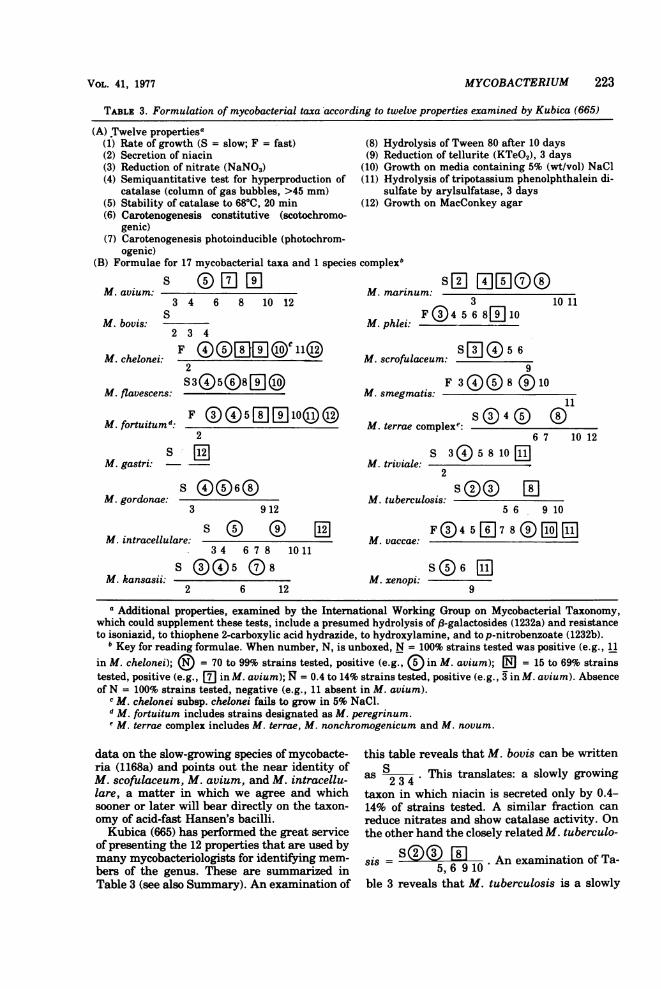
TABLE 3. Formulation of mycobacterial taxa according to twelve properties examined by Kubica (665)
(A) Twelve properties0(1) Rate of growth (S = slow; F = fast)(2) Secretion of niacin(3) Reduction of nitrate (NaNO3)(4) Semiquantitative test for hyperproduction of
catalase (column of gas bubbles, >45 mm)(5) Stability of catalase to 6800, 20 min(6) Carotenogenesis constitutive (scotochromo-
genic)(7) Carotenogenesis photoinducible (photochrom-
ogenic)(B) Formulae for 17 mycobacterial taxa and 1 species
S OWl[E1M. avium:3 4 6 8 10 12
SM. bovis:
2 3 4
M. chelonei:2
M. flavescens: S30508W[j]
M. fortuitumd: F ( i5[I 10(l (0
2
M. gastri: - -
M. gordonae:3 912
M. intracellulare:
M. kansasii:
1l2
34 6 7 8 1011
S 6(5 182 6 12
(8) Hydrolysis of Tween 80 after 10 days(9) Reduction of tellurite (KTeO2), 3 days
(10) Growth on media containing 5% (wt/vol) NaCl(11) Hydrolysis of tripotassium phenolphthalein di-
sulfate by arylsulfatase, 3 days(12) Growth on MacConkey agar
complexb
sxWWWO0M. marinum:3
M.phlei:F 4 68[]110 11
M. scrofulaceum: S W (iD 5 69
F 300(S80)10M. smegmatis: 1111
M. terrae complexe.S040 06 7 10 12
S 3(i)5810 1M. triviale: -
2
M. tuberculosis: - -1
56 910
M. vaccae:
SQ 6 E1M. xenopi:
a Additional properties, examined by the International Working Group on Mycobacterial Taxonomy,which could supplement these tests, include a presumed hydrolysis of f3-galactosides (1232a) and resistanceto isoniazid, to thiophene 2-carboxylic acid hydrazide, to hydroxylamine, and to p-nitrobenzoate (1232b).
b Key for reading formulae. When number, N, is unboxed, N = 100% strains tested was positive (e.g., 11in M. chelonei); @ = 70 to 99% strains tested, positive (e.g.,IE in M. avium); @|J = 15 to 69% strainstested, positive (e.g., mD in M. avium); R = 0.4 to 14% strains tested, positive (e.g., 3 in M. avium). Absenceof N = 100% strains tested, negative (e.g., 11 absent in M. avium).
c M. chelonei subsp. chelonei fails to grow in 5% NaCl.d M. fortuitum includes strains designated as M. peregrinum.e M. terrae complex includes M. terrae, M. nonchromogenicum and M. novum.
data on the slow-growing species of mycobacte-ria (1168a) and points out the near identity ofM. scofulaceum, M. avium, and M. intracellu-lare, a matter in which we agree and whichsooner or later will bear directly on the taxon-omy of acid-fast Hansen's bacilli.Kubica (665) has performed the great service
of presenting the 12 properties that are used bymany mycobacteriologists for identifying mem-bers of the genus. These are summarized inTable 3 (see also Summary). An examination of
this table reveals that M. bovis can be written
as S This translates: a slowly growing234 '
taxon in which niacin is secreted only by 0.4-14% of strains tested. A similar fraction canreduce nitrates and show catalase activity. Onthe other hand the closely related M. tuberculo-
S © (~sis = An examination of Ta-5,b6 9 10
ble 3 reveals that M. tuberculosis is a slowly
MYCOBACTERIUM 223VOL. 41, 1977

TABLE 4. Mycobacteria reported to cause disease in human subjectsMycobacterium Related disease(s) Sourcea
M. africanumM. avium
M. bovis
M. bovis BCG
M. chelonei
M. fortuitum
M. intracellulare
M. kansasii
M. leprae (species forma)
M. lepraemurium
M. marinum
M. scrofulaceum
M. simiae (habana)M. szulgai
M. terrae
M. triviale
PulmonaryPulmonaryExtrapulmonary:DisseminatedLymphadenitisAssociated with silicosisMeningitisOcular
G. R.bPulmonaryExtrapulmonary:DisseminatedLymphadenitisMeningitisBone and jointMiscellaneous
G. R.Extrapulmonary:Disseminated
PulmonaryExtrapulmonary:
Injection and abrasion ab-scesses
DisseminatedLymphadenitis (cervical)
PulmonaryExtrapulmonary:CornealDisseminatedInjection and abrasion ab-
scessesMiscellaneous
PulmonaryExtrapulmonary:DisseminatedLymphadenitis
PulmonaryExtrapulmonary:DisseminatedMiscellaneous
G. R.ExtrapulmonaryTuberculoid * lepromatous
leprosyExtrapulmonaryNodular infections
or skin and lymph nodesExtrapulmonary:Cutaneous granulomas (in-
cluding "swimming poolgranulomas")
SporotrichoidMiscellaneous
PulmonaryExtrapulmonary:Lymphadenitis (cervical)Miscellaneous
PulmonaryPulmonaryExtrapulmonary:Lymphadenitis (cervical)Joint
Extrapulmonary:Disseminated
Extrapulmonary:Arthritis
224
225, 226, 474, 527173, 245, 312, 346, 515, 772
360, 683, 1032312, 332, 345, 615, 726, 772543, 1278312, 8871222366, 67288, 472, 584, 589, 692, 716, 755
480, 584230460230584230, 367, 472, 907, 1259
766, 774475, 1173
160, 419, 535, 849, 913
468854, 85592, 263, 320, 877, 950, 1208
720, 1186, 1187, 1288, 1311363115, 214, 252, 276, 487, 892, 901, 1053, 1199,
1248504, 1062262, 269, 368, 608, 719, 1293
275, 294, 628, 719, 1010, 1210693, 726139, 235, 236, 551, 608, 719, 927, 1303
348, 473, 484, 803, 1281431, 434, 503, 526, 719, 745, 902, 12231281
514, 565
770
98, 254, 264, 729, 847, 852, 928, 1005, 1075,1116, 1215, 1310
1, 11, 212, 299, 378, 558, 1277382, 461, 558, 580, 1264, 12731275
133, 942, 1275, 12768481195, 1242776
776776
247
344

TABLE 4-Continued
Mycobacterium Related disease(s) SourceaM. tuberculosis G. R. 477, 973M. ulcerans (buruli) Extrapulmonary:
Chronic skin 28, 94, 197, 250, 251, 259, 260,ulcerations 307, 361, 540, 687, 737, 746, 925, 965,
1069, 1160, 1221M. xenopi Pulmonary 149, 296, 311, 343, 347, 372, 778, 805, 980
a References cited are those in which specific cases were examined by the author(s). Only general reviewsare given for the widely investigated species, M. tuberculosis and M. leprae. References were selected todemonstrate the variety of disease involvement reported in the literature and are not meant to be inclusive.
b G. R., General references. Other general references include 234, 235, 284, 365, 366, 383, 391, 515, 726,773, 926, 973, 994, 995, 998, 1001, 1056, 1186, 1254, 1276.
growing taxon, 70 to 99% of whose memberssecrete niacin, 100% produce catalase, etc. Theuninitiated reader might ask: Are these 12properties peculiar to mycobacteria? Of coursenot. Their presence or absence can equally wellbe determined for Corynebacterium, Nocardia,and a wide variety of other bacteria. However,for nonsporeforming, gram-positive bacteriaexhibiting superficial ropelike structures (seeFig. 6) and mycobacterial acid-fastness (as op-posed to other types of acid-fastness), these 12properties serve to delineate the species listedin Table 1.
In Table 4 are listed those mycobacteria that,at one time or another, have been associatedwith human disease. Many of them are notprimarily found in man. One entry, M. leprae-murium, has been included because it is from aprovocative paper that beautifully points up thedifficulties in dealing with so-called noncultiva-ble species. The data in Table 4 are taken froma more comprehensive table we have preparedon the host ranges ofmycobacteria. The use of aterm such as "host range" is a reflection of thezoocentric (mostly anthropocentric) view thatinteracting with animals is a prime function ofmycobacteria. In truth, these microbes, of re-markable biosynthetic capacities, have in theirdispersions down through time got caught up innooks, crannies, and niches where several ofthem have lost, to varying degrees, their inde-pendence. However, studying them only as pro-ducers of disease would yield as narrow a viewof their capabilities as would studying themonly for the 12 properties listed in Table 3.
Eventually, taxonomists will probably findthat the Corynebacterium, Mycobacterium,and Nocardia (CMN) group of microorganismsis best treated as a single genus. This decisionwill no doubt come as a result of insights gainedby an examination of the comparative biologyof CMN. Such will require (i) direct examina-tion of the ranges of colonial and cellular mor-phology, (ii) growth and metabolism (best ex-
amined through comparison of mutant strains,where possible, augmented by recombinationalgenetics), and (iii) various biosynthetic capaci-ties leading to the synthesis of distinctive prod-ucts. Information so gained can be supple-mented with (iv) studies of the interaction ofparticular bacteria and their viruses and (v)their interaction with animal hosts as sourcesof antigen (structural components such as mu-reins or peptidoglycans, enzymes, toxins, etc.).The present review supplies much of this infor-mation for Mycobacterium.
THE MYCOBACTERIAL CELLMuch is known about the chemistry of var-
ious compounds produced by mycobacteria, andmicrobial anatomists, microbial biochemists,and geneticists now need to put this all togetherin terms of a growing Mycobacterium. An ul-trathin section of actively growing M. tubercu-losis H37Rv is shown in Fig. 1. Mycobacteriaare nonmotile, nonsporeforming, pleomorphic,gram-positive bacilli. Mycobacteria, growingon solid media, pile up to form smooth or rough,often wrinkled, soft and buttery, powdery towaxy (see Fig. 16), transparent to opaque col-onies (e.g., see Fig. 1 of reference 1025 andFig. 1 of reference 898). Sometimes, in oldcultures, mycobacteria show an inhibition ofpostdivisional cell separation that is apparentas filamentous growth and, less commonly,as branching. Runyon has pointed out that, onthe basis of their tendencies to form aerial fila-ments, mycobacteria range from those, such asM. tuberculosis and M. bovis, which show nofilamentation to those forming rudimentary fil-aments (M. xenopi), to those that show fila-mentous extensions from their colonies (M. for-tuitum), with fragmentation to the obviouslyfilamentous M. farcinogenes, which rarelyshows fragmentation (999).
Forty-five years ago, Kahn, in an elegantseries of experiments using microdrops, showedthat the ancestral strain of M. tuberculosis,
VOL. 41, 1977 MYCOBACTERIUM 225

226 BARKSDALE AND KIM BACTERIOL. REV.a- i S*U r s4 tl ~eF
".0-,;,W I.
I Y .l
-,
Pr1.
h,61L-J. I
r,
., Iz;., I.
,C' "XRM"r
llo ---. 4r; " ,Ai Alluarlm, -.Y ..
-.V1. $'v
..-;, -Iv..-

MYCOBACTERIUM 227
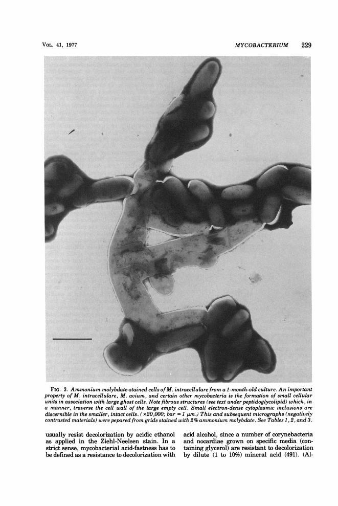
H37Rv, underwent fragmentation to coccalforms, and rods developed from some of thesecocci (570). In Fig. 2B is shown a cell of H37Rvfrom an actively growing culture in which twoof the compartments show no signs of viability.The terminal one, however, obviously hashealthy nucleic acid, dense cytoplasm, poly-somes, and a fatty inclusion. The overall di-mensions of this cytoplast, 1.0 by 0.3 ,um, areequivalent to those of Kahn's coccal bodies. Insome species, such as M. avium, growth ischaracterized by an increase in the number ofcytoplasts within a common cell wall, with pe-riodic fragmentation to release coccoid cells.Maffucci (754) observed these peculiarities ofM. avium in 1892. They were rediscovered andcarefully described as a "life cycle" for M. av-ium by Brieger and Fell some three decades ago(185). McCarthy (800) has reported the use oftwo mutants of M. avium for studying cellgrowth and division, employing photomicrogra-phy, electronic counting and size distributionanalyses, and viability and protein determina-tions. The mutants were colonial types termedtransparent (T) and opaque (0). T was virulent;O was not (898), 1025; see also [48a]). T wasresistant to more drugs than 0 and formed asmaller cell mass than O. The initial inoculumwas sized by passage through a 1.2-,um-pore-size filter (type MF, Millipore Corp.), and syn-chronous growth was obtained. This very beau-tiful paper establishes that growth ofM. aviuminvolves an increase in deoxyribonucleic acid(DNA), protein, and cell mass, concomitantwith a lengthening of the cell body. With frag-mentation, the DNA and protein decreased topreelongation ratios. This fluctuation betweencoccal forms and pleomorphic rods is not un-common in the CMN group (e.g., in the genusNocardia [114] and in C. diphtheriae [293]).One of the most interesting findings of Mc-Carthy was that the virulent T mutant pro-duced one-twentieth as many viable units asthe 0 mutant. Would these units, nonviable invitro, have been viable in vivo? In Fig. 3 areshown small cells of M. intracellulare (a closerelative ofM. avium) in association with longerghost cells.
Although most of the studies of transitionsfrom rods to coccal forms and vice versa inmycobacteria have been with facultatively par-asitic mycobacteria, Chang and Andersen havenoted the occurrence of chains of tiny acid-fastcoccal forms during the growth of the obligatelyparasitic M. lepaemurium in mouse macro-phages (231).
Acid-FastnessAcid-fastness is the capacity of biological ma-
terials to form acid-stable complexes with cer-tain arylmethane dyes. Such materials containdye complexes that are not decolorized follow-ing exposure to acidic ethanol or mineral acids.This general property exists in a variety ofentities, including spores of a number of fungi(1294), the spores ofBacillus cereus (1297), hu-man sperm (125), the embryophores of Taeniasaginata (914), the hooklets of Taenia echino-coccus (194), corynebacteria and/or certain oftheir inclusions (929a, and our unpublisheddata), tubercle bacilli (623), leprosy bacilli(1151), keratin (C. A. Fisher, unpublisheddata), nuclear DNA (as in the Feulgen reac-tion), and chitin following exposure, in situ, tomild oxidation (916). In each case the biologicalpoduct responsible for stably combining withthe dye is apparently different: for example, thecapacity for acid-fastness of the spores of B.cereus is associated with f3-hydroxybutyrateand can be removed by extracting the sporeswith chloroform (1297); that of leprosy bacillican be extracted with pyridine whereas that oftubercle bacilli cannot (377). The capacity foracid-fastness of various mycobacteria can beremoved with alkaline ethanol (377). It is clear,then, that the term acid-fastness, in order totake on meaning, needs be qualified accordingto the chemistry of each potentially acid-fastmaterial.
Mycobacterial acid-fastness. Mycobacterialcells that have taken up fuchsin (triaminotri-phenylmethane chloride; pararosanilin), crys-tal violet (hexamethylpararosanilin chloride),or auramine 0 (tetramethyldiaminodiphenylketoimine) in phenol-water (as carbol fuchsin,carbol crystal violet, or carbol auramine 0)
FIG. 1. Sections of cells of actively growing M. tuberculosis H37Rv 102. (A) Mesosome within nucleoidshowing central homogeneous matrix of intralamellar spaces. See sections, The Mycobacterial Cell andNucleoid in text. (B) A completed cross wall showing a distance of about 26 nm in width with an electron-transparent layer separating two cells which are still connected to the mother cell wall. Note membrane-boundvesicle and metachromatic granule. (C) A cross wall halfway completed showing continuity with plasmamembrane and a large vesiculated mesosome. (D) The initiation of cross wall formation, showing largemesosomes at both sides of the cell. Note the vertical position of the mesosomes in relation to the initial crosswall. (All micrographs, x78,000; bar = 0.5 pm.) Cells were fixed with OS04, dehydrated in ethanol, andembedded in liquid epoxy resin, D.E.R. 736 (The Dow Chemical Co.). The following micrographs (sectionedmaterials) were prepared in same way. See also Tables 1, 2, and 3.
VOL. 41, 1977

FIG. 2. (A) Section of M. aurum cell showing vacuole-like fat inclusions enclosed by a membranousstructure. (B) Section ofan actively growing cell ofM. tuberculosis H37Rv 102 showing viable compartmentwithin lysed old mother cell. Note lack of classical cross wall formation. (C) and (D) Sections of activelygrowing cells ofM. tuberculosis H37Rv 102 showing vesiculated type of mesosomes. Continuity ofcytoplasmicmembrane is clearly visible. (All micrographs, x78,000; bar = 0.5 gm.) See also discussion ofThe Mycobac-terial Cell. See Tables 1, 2, and 3.
228
MYCOBACTERIUM 229
FIG. 3. Ammonium molybdate-stained cells ofM. intracellulare from a 1-month-old culture. An importantproperty of M. intracellulare, M. avium, and certain other mycobacteria is the formation of small cellularunits in association with large ghost cells. Note fibrous structures (see text under peptidoglycolipid) which, ina manner, traverse the cell wall of the large empty cell. Small electron-dense cytoplasmic inclusions arediscernible in the smaller, intact cells. (x20,OOO; bar = 1 uim.) This and subsequent micrographs (negativelycontrasted materials) were pepared from grids stained with 2% ammonium molybdate. See Tables 1, 2, and 3.
usually resist decolorization by acidic ethanolas applied in the Ziehl-Neelsen stain. In astrict sense, mycobacterial acid-fastness has tobe defined as a resistance to decolorization with
acid alcohol, since a number of corynebacteriaand nocardiae grown on specific media (con-taining glycerol) are resistant to decolorizationby dilute (1 to 10%) mineral acid (491). (Al-
VOL. 41, 1977

230 BARKSDALE AND KIM
though it is often stated that all basic fuchsinsare mixtures of pararosanilin [triaminotri-phenylmethane chloride], rosanilin [triamino-tolyldiphenylmethane chloride], and magentaII [triaminoditolylphenylmethane chloride],the Stain Commission has found many of the
H2N / CC NH2+ Cl
NH2
Pararosanilin (Fuchsin)
(CH3) 2N+CV
(CH3)2N / \ N(CH3)2
Crystal Violet
(CH3)2: N (CH3)2
Auramine 0
commercial basic fuchsins to be fairly purepararosanilin [725].)The capacity of these anionic compounds to
interact with various naturally occurring mo-
lecular species, including a number of poly-meric ones, has long been known (725, 813,899). The resultant acid-resistant cells appearred (fuchsin retained) or purple (crystal violetretained) or exhibit yellow-green fluorescence(auramine 0 retained). This is mycobacterialacid-fastness, regarded by many as the hall-mark of mycobacteria. Fuchsin-stained, acid-fast mycobacteria exhibit a marked brillianceof color. Mycobacterial cells which have beenbroken open and then subjected to the acid-faststain are only weakly acid-fast, lack brillianceof color and, undoubtedly because ofthe obscur-ing counter stain (124), have long been callednon-acid-fast (624). If the outer walls of myco-
bacteria are removed with alkaline ethanol,there is left an intact cell which shows its char-acteristic shape but which is non-acid-fast.Thus, the integrity of the rigid layer does notassure acid-fastness. To be acid-fast, the myco-bacterial cell must possess its lipid rich outercoat (L2 of Fig. 4; see also Fig. 5). The firstperson to suggest that mycobacterial acid-fast-ness was of a dual nature was Berg (124). Wehave come to a similar conclusion, but the twoparts of the duality, as we see them, are differ-ent from those ofBerg, and their demonstrationdoes not require a change from the Ziehl-Neel-sen decolorization with hydrochloric acid-ethanol to decolorization with acetic acid-ethanol so important to Berg's explanation ofacid-fastness (122). As we see it, the intact my-cobacterial cell (i) takes carbol fuchsin into itsinterior and (ii) also binds fuchsin to the my-colic acid residues of the peptidoglycolipids ofthe outer cell wall (Fig. 4 and 5). Free mycolicacids bind fuchsin. on a mole-for-mole basis(123), and the bonding is acid stable. Once themycolic acid of the cell is complexed with anarylmethane dye, the cell surface becomes ex-tremely hydrophobic. After the fuchsin-repleteand mycolate-fuchsin-coated mycobacterial cellhas been subjected to decolorization with hydro-chloric acid-ethanol, two states obtain: (i) thefuchsin taken into the "interior" of the cell re-mains there and supplies a brilliant enhance-ment to (ii) the lightly staining fuchsin-myco-late complex ofthe outer wall. Isolated prepara-tions of mycolic acids, which certainly contain agreater density of molecules than are to befound in peptidoglycolipids, present a weaklypink color following decolorization (59, p. 256).Thus, the acid-fastness of mycobacteria de-pends for its brilliance on trapped fuchsin, andthe trapping is ensured by the fuchsin-mycolateof the outer peptidoglycolipid (see L2 of Fig. 4).Although Corynebacterium and Nocardia pro-duce plenty of mycolic acids that resist decolori-zation by dilute mineral acids (59, 111), theirmycolic acid-bound fuchsin does not preventdecolorization by acidic ethanol. Hence, they donot exhibit mycobacterial acid-fastness. Theconfiguration oftheir surface peptidoglycolipidsis different from that of mycobacteria (see Fig.22-26), and this leads us to suggest that thechemical constituents of the unique ropelikestructures of the outer mycobacterial cell wallare essential for mycobacterial acid-fastness.
Ziehl-Neelsen stain. Most of what is knownof the chemistry of the acid-fast reaction inmycobacteria has been derived from the studyof the Ziehl-Neelsen stain, in which the dyeemployed for acid-stable binding is carbol
BACTERIOL. REV.

MYCOBACTERIUM 231
jl'% .,I
FIG. 4. The mycobacterial cell wall as dia-grammed from electron micrographic data consid-ered in this review. Beginning with the plasma mem-
brane (Pm) and coming past the murein or peptido-glycan (Mur), the underlying fibrous ropelike struc-tures ofthe cell wall are shown as L3 merging into themore wrinkled and superficial L2 with which it iscontinuous. This material lies immediately under thesheetlike surface glycolipid or peptidoglycolipid (L,).Contained within the Pm are shown a growing sep-
tum (S) with its vesiculated mesosome (M) associatedwith DNA of the nucleoid, metachromatic granule(Met), lipoidal bodies (L), and dense granules (Dg)(see Fig. 10). For biological activity associated withLs see Tuberculins and Other Mycobacterial Elici-tins, as well as Delayed Hypersensitivity; for discus-sion of chemistry of these layers, see From the Cyto-plasmic Membrane to the Peptidolipid and parts ofMycobacterium as Antigen. In Fig. 5, L3, L2, and L,,as well as the murein or peptidoglycan layer, havebeen rendered visible by negative staining (5A) andfreeze-etching (5B). Note: Although macromolecularstructures such as L2 and L3 can be rendered visible,other surface components such as trehalose dimyco-late (cord factor), acyl glucoses, etc., cannot.
fuchsin. Lartigue and Fite (694) establishedthe fact that, in carbol fuchsin, fuchsin andphenol are independently associated (no chemi-cal modification of either compound) and con-firmed the contention of Lamanna (684) thatphenol (and, for them, detergents as well)enhances the penetration of fuchsin into lipidsby rendering the fuchsin more lipid soluble andless water soluble. Since the time of Stodola etal. (1103), it has been held that mycolic acidsper se were acid-fast. However, Richards (975)and Rich (972), as well as Yegian and Vander-linde (1294), have regarded that as untrue,perhaps because thin films of mycolic acidwere only faintly pink following exposure toacidic ethanol. The faintness of color was due,no doubt, (i) to the low degree of binding ofdye when the lipoidal receptors for that dye
were dispersed rather than ordered in theultrastructure of the Mycobacterium and (ii)to the absence of the background color pro-vided by intracellular dye (see above). The im-portance to acid-fastness of the way and extentto which mycolic acids are ordered about themycobacterial cell wall is suggested by the re-port of Murohashi et al., who detected no corre-lation between mycolate yields and degree ofacid-fastness of strains (865).
Interaction of dye and mycolic acid. In re-viewing the properties essential for the acid-fastness of mycolic acids, Asselineau haspointed out that the mycolic acid must possess afree carboxyl group (methyl mycolate and my-colic alcohol were not acid-fast). The hydroxylgroup must be present, but it could be esteri-fied, as in acetates of mycolic acid (59, p. 255).Berg tested the interaction of a mycolic acid,leprosinic acid, with crystal violet after theblocking of its two carboxyl groups by methyla-tion. No dye complexing occurred. In fact,complex formation was prevented even whenthe two carboxyls were free, but one of thehydroxyls had been blocked by acetylation.Free mycolic acid was found by Berg to bindcrystal violet on a mole-for-mole basis. The dye-acid complex has a characteristic peak at 350/im (123). This is a very different peak fromthat found with crystal violet and agar or nu-cleic acid (813). The mycolic acid-dye complex,as well as mycolic acid, was soluble in xylene,although the dye alone was not (123).
Ropelike structures, the integrity of therigid layer of the cell wall, and acid-fastness.In this paper, we give the first documenta-tion that all species of mycobacteria appear topossess surface ropelike structurs of peptido-glycolipid and that, from species to species, thepattern assumed by this material is similar(Fig. 6). The ropelike peptidoglycolipid seemsto be an essential part of the duality (124) ofmycobacterial acid-fastness. It furnishes the re-acting mycolic acid residues required for pro-ducing the acid-fast arylmethane mycolate es-sential for blocking the exit of fuchsin takeninto the cell during the staining process. Thefollowing findings indicate that a ropelike con-figuration per se is not necessary to the func-tioning of this peptidoglycolipid in acid-fast-ness. Incubation of cells of actively growingstrain 607 (in Penassay medium [Difco] plusTween 80) in the presence of ethylenediamine-tetraacetic acid (EDTA) (200 jig/ml) and lyso-zyme (100 ,ug/ml) for 15 h permits the elonga-tion of the cells showing a marked absence ofobservable ropelike structures. These cells,nevertheless, are strongly acid-fast (see Fig.
VOL. 41, 1977

FIG. 5. Accumulation of outermost surface component (L,) by still-grown Mycobacterium sp. NQ. (A)Negatively stained cell showing outer encasing material (see text under peptidoglycolipid) which extendsbeyond the cell as ribbon-like appendages. Note that characteristic ropelike structures are covered up,although polyphosphate granules may be seen within the cell. (x65,000; bar = 0.5 Aum.) (B) Micrograph offreeze-etched Mycobacterium sp. NQ. When the ribbon-like structures shown in (A) are shaken away from thecell, ropelike structures are exposed. These can be visualized by negative staining as shown in Fig. 24I. Theyare characteristic surface structures of mycobacteria and differ in size from analogous structures found inNocardia (Fig. 26) and Corynebacterium (Fig. 25). The uncircled arrow indicates a fracture through L,revealing in profile the ropelike structures ofL2 and L3 adhering to the rigid murein layer, which is exposed asa smooth background. The direction of the shadowing is indicated by the circled arrow. (x140,000; bar =0.25 aum.) For a discussion of this component of mycobacterium as receptor for bacteriophage see Host CellReceptors and Bacteriophage-Induced Receptor-Destroying Enzymes. For general chemical structures seePeptidoglycolipids or Mycosides. For details regarding production of mycosidic mycobacterial casements,see reference 614.
232
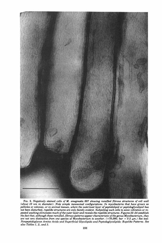
FIG. 6. Negatively stained cells of M. smegmatis 607 showing ramified fibrous structures of cell wall(about 10 nm in diameter). Note simple mesosomal configurations. In mycobacteria that have grown aspellicles or colonies, or in animal tissues, where the outermost layer ofpeptidolipid or peptidoglycolipid hasnot been disturbed, ropelike structures are only faintly evident. Subjecting such cells to sonic vibration or re-peated washing eliminates much ofthe outer layer and reveals the ropelike structures. Figures 22-24 establishthe fact that, although these ramified, fibrous patterns appear characteristic ofthe genus Mycobacterium, theyare not very distinctive from one species of Mycobacterium to another. (x78,000; bar = 0.5 .um.) See text:Nonpeptidoglycan Amino Acids and Superficial Glycolipids and Peptodoglycolipids: Ropelike Patterns. Seealso Tables 1, 2, and 3.
233

tC.P
FIG. 7. Alterations of the surface organization of the cell walls of actively growing (Penassay broth plusTween 80) M. smegmatis 607 in relation to acid-fastness and pyridine-extractable components. (A) EDTA(200 pglml) and lysozyme (100 pg/ml) were added to the growth medium, and incubation was continued for15 h. Negatively stained cell shows an absence offibrous structures. These cells were acid-fast at 15 h but non-acid-fast at 24 h (see text). ( x27,000; bar = 1 ,um.) (B) Cells treated with 0.5% (wtlvol) KOH-ethanol (see alsoFig. 8B) for 4 h lose their acid-fastness. ( x 78,000; bar = 0.5 tum.) (C) Cells were suspended in pyridine for 4h and then prepared for negative staining. Most ofthe ramified structures disappeared. Many ringlike surfaceparticles are apparent. Cells are acid-fast. Their enhanced permeability to fuchsin is discussed in the sectionon phosphatidylinositol polymannosides. (x78,000; bar = 0.5 ,tm.) See Tables 1, 2, and 3.
234
r-e.
.11 A.
MYCOBACTERIUM 235
7A). Further prolonged incubation (total, 24 h)results in a loss of acid-fastness. Extraction ofsurface lipids with 0.5 to 1% (wt/vol) KOH-ethanol for 4 h results in complete loss of acid-fastness and of ropelike structures (Fig. 7B and8B). This probably means that, although theropelike configuration per se is not essential toacid-fastness, the substance which assumesthat configuration is essential.Although Koch's early demonstration that
acid-fastness could be destroyed by grinding thedried bacilli in an agate mortar (624) suggestedthat the intact bacterial cell was needed foracid-fastness, cells that contain open channelsthrough the cell wall to the cytoplasmic mem-brane (pyridine-extracted cells [Fig. 8A]) re-main acid-fast. This, plus the fact that delipi-dated cells are not acid-fast, leaves little doubtthat in the dual nature of acid-fastness (i) thebrilliance of the stain is attributable to trappedintracellular fuchsin and (ii) the trapping ofthefuchsin is the result of a barrier furnished bymycolate-fuchsin complexes of the peptidogly-colipid of the outer cell wall.
Interference with the synthesis of mycolicacids as, for example, in the treatment of myco-bacteria with isonicotinic acid hydrazide (INH)leads to non-acid-fastness (see section, Isonico-tinic Acid Hydrazide and Mycolic Acid Synthe-sis).Fluorochrome staining of mycobacteria.
Over the past 15 years it has been establishedthat fluorescent staining of Mycobacterium tu-berculosis (482) with tetramethyldiaminodi-phenylketoimine (auramine 0) gives a higheryield of positive findings than does stainingwith carbol fuchsin. This proves true both forbacilli in sputa (979) and in histological sections(466, 1289). In fact, a comparison betweenZiehl-Neelsen staining, auramine 0 staining,and cultivation of material from infectedguinea pigs has shown that "diagnosis" by cul-tivation and by fluorescence of acid-stablebound auramine 0 were almost equivalent(1268). One is led to speculate that the structureof the diphenyl-containing auramine 0 is bettersuited for penetrating and stacking along thereceptor macromolecules of the mycobacterialpeptidolipid and/or peptidoglycolipid than isfuchsin. Aurmaine 0 also penetrates biologicalmaterials very well. The relative fluorescenceof auramine 0 bound, for example, to DNAdepends upon concentration of the polymer(899). The intracellular concentration possiblewith auramine 0 may be greater than withfuchsin and this may account, in part, for itsreputed superiority as a stain for acid-fast my-cobacteria.
Acid-stable Binding and Gram staining ofmycobacteria. The gram-positive state isequated with the capacity to retain a complexof crystal violet and iodine. It has long beenheld that "mycobacteria cannot be classified asgram positive or gram negative by the Gramstaining technique because, once they havebeen stained by basic dyes, they cannot bedecolorized by alcohol regardless of whether ornot they have been treated with iodine" (816,p. 491). This observation stems from the capac-ity of mycobacterial walls to bind crystal violetin an acid-stable state. However, if one re-moves the lipoidal portion of the mycobacterialwall with alkaline ethanol (1% KOH in abso-lute alcohol, wt/vol), there is left an intactbacterial cell which is non-acid-fast but grampositive (L. Barksdale, K.-S. Kim, and C. A.Fisher, unpublished data) i.e., retains the crys-tal violet-iodine complex. Thus, the apparent"gram-positiveness of tubercle bacilli [myco-bacteria] is independent of the mordant effectof iodine and appears to depend upon the samefactors which are responsible for acid-fastness"(1064, p. 510). Removal of the structures re-sponsible for acid-fastness enables one to estab-lish the inherent gram-positivity of mycobac-teria.
Non-acid-fast mycobacteria. The stages ofgrowth of mycobacteria may be associated withan apparent lack of acid-fastness. It may beabsent in a large proportion of actively growingpopulations of mycobacteria, and it may not bedemonstrable in so-called chromophobic pheno-types, which withstand the penetration offuchsin and other dyes (891). For example, inour hands, actively growing cells of strain 607are acid-fast only when exposed to hot carbolfuchsin, and then no more than 10% of the cellsshow good retention of the dye. Cells fromLowenstein-Jensen slants show a similar lackof acid-fastness. Under the discussion of phos-phatidylinositol oligomannosides and the myco-bacterial cell wall, we point out that pyri-dine-extracted cells (Fig. 8A and 9) contain tun-nels to the cytoplasmic membrane, tunnelsonce occupied by pyridine-extracted lipids. Fur-ther, pyridine extraction greatly modifies theorganization of the cell surface and revealssome ringlike structures (Fig. 7C). Such pyri-dine-extracted cells can be shown to take upfuchsin much more readily than the unextractedcell and, although they are riddled, they retaintheir acid-fastness. In other words, the normalcell (in this case, Mycobacterium smegmatis) isrelatively chromophobic. Pyridine extractionlowers the resistance to the penetration of fuch-sin without any negative effect upon acid-fast-
VOL. 41, 1977

236 BARKSDALE AND KIM
riV~ -.
~~~~~~
K
.1;.
I--1
It
BACTERIOL. REV.

VOL. 41, 1977
ness. In fact, the enhanced uptake by pyridine-extracted cells results in a higher count of acid-fast bacteria than is found in unextracted sam-ples from the same population of mycobacteria.Nyka (889, 890) has recommended the use of
oxidants for demonstrating chromophobic acid-fast bacilli in tissues. The products of this tech-
MYCOBACTERIUM 237
nique, although useful for showing up addi-tional bacilli, should not be regarded as exam-ples of a mycobacterial acid-fast stain. Aspointed out by Fisher (377) even Escherichiacoli can become "acid-fast" under such condi-tions. Fisher noted that of the arylmethanedyes only carbol fuchsin, acidified basic fuch-
FIG. 9. Pyridine-extracted material from M. smegmatis 607. Cells were treated as in Fig. 8A. Thesupernatant pyridine was evaporated to dryness in a Rotavapor and negatively stained. Note laminatedstructures consisting of fibrils of about 8 nm in diameter, suggestive of ramified fibrous structures seen inmycobacterial cell walls. The removal ofthis material resulted in the changes shown in Fig. 7C. Note that theextracted cells retained their acid-fastness. (x78,000; bar = 0.2 pm.) For discussion, see section on Phosphati-dylinositol Mannosides. See Tables 1, 2, and 3.
FIG. 8. Alterations in cell wall profiles ofpyridine-extracted (A) and KOH-ethanol-treated (B) cells ofM.smegmatis, 607. (A) Cells were extracted with pyridine while shaking for 15 h, 350C. Note that thepeptidoglycolipid layer and the murein (Mur = peptidoglycan) are intact. Here and there are openings (Pyr)created by removal of pyridine-soluble components (see text). The double thickness of the peptidoglycolipid(PL), about 26 nm in width, is evident between two adjacent cells. The remarkable aggregation ofthese cellssuggests that the PL layer has become sticky. Leakage from the cytoplasm is indicated by an absence of thecytoplasmic membrane and mesosomes. The integrity of the ribosomes has been lost. These cells, despite thechanges noted, remain acid-fast. (x78,000; bar = 0.5 pim.) See sections on Acid-Fastness and Phospholipids.(B) Section ofcells ofM. smegmatis 607 shaken in KOH-ethanol (0.5%, wtlvol) for 15 h. Note numerous fibrousstructures extending from the cytoplasm and the murein layer. There is no observable cytoplasmic membrane.The peptidoglycolipid layer has been removed. The cells show no tendency to aggregate. They are non-acid-fast. See section, Acid-Fastness. (x78,000; bar = 0.5 pim.) See Tables 1, 2, and 3.

238 BARKSDALE AND KIM
sin, or aqueous pararosaniline yielded an acid-stable complex with the oxidized bacteria. Heattributed this chemical conversion to acid-fast-ness to the formation of Schiff bases betweenbasic dyes (primary amines) and aldehydes gen-erated by the periodate oxidation of cell walland membrane components. Kiyoshi Haradahas taken the Nyka procedure much furtherand recommended an acid-fast stain employingan oxidation step involving potassium perman-ganate (490; see also reference 586). As he putsit, "oxidants which attack ethylenes to formaldehydes were effective in increasing acid-fastness of mycobacteria." He further pointedout that the oxidized mycobacteria reacted sowell with methylene blue as to have theirnewly acquired acid-stable fuchsinophilia ob-scured. It should be clear from the foregoingdiscussion that acid-stable fuchsinophilia, acid-fastness of Schiff bases, is not related to myco-bacterial acid-fastness.
Conditions of GrowthFor the most part, mycobacteria have simple
growth requirements as compared, for exam-ple, with C. diphtheriae. Throughout the CMNgroup, the content of the medium greatly af-fects the overall syntheses that take place. Inthe presence of glucose, for example, the majorsoluble lipids made by M. smegmatis and C.diphtheriae are acylglucoses in which the hex-ose is esterified in the 6 position. The majorfatty acid (FA) of these acylglucoses is aC,5H3,-CH-CH-COOH corynomycolic acid.
OH C14H29In the absence of glucose, acylglucoses fail to bemade by either of these representatives of theCMN group (183). Acylglucoses may play a rolein glucose transport (1049). Quite early, Frouin(409) and Long and Finner (732) found that thecomposition of the medium in which M. tuber-culosis, M. bovis, and M. avium are grownmarkedly affects the quantitative lipid compo-sition of these organisms. The latter authorsfound that organisms grown with 12.5% glyc-erol were much more intensely acid-fast thanthose grown with 0.5% glycerol. Glucose hasbeen included in media for mycobacteria. In theCMN group, glucose, however, is not always apreferred carbon source. For example, C. diph-theriae gives greater yields on maltose thanglucose (96, p. 395). The utilization of glycerolby M. tuberculosis is markedly inhibited byglucose, and the inhibition is by some mecha-nism other than catabolite repression. Gluta-mate is a preferred nutrient for initiation ofgrowth by this Mycobacterium as compared, for
example, with the traditionally employed as-paragine. In a medium containing glycerol andglutamate, there is preferential prior consump-tion of glutamate. Citrate, commonly employedin mycobacterial media, is not utilized by M.tuberculosis H37Rv. The apparent growth-en-hancing effects of citrate are undoubtedly, assuggested by Dubos and Middlebrook, attribut-able to the capacity of citrate to chelate cal-cium, iron, and magnesium, thus preventingtheir precipitation from liquid media (322).Shaking is a preferred method of cultivation,and small inocula are to be avoided (162). Car-bon dioxide, 6% (872) to 8% (476), enhancesgrowth and is essential for growth from certainsmall inocula. Antoine and Tepper (47) havedemonstrated an effect of limitation of nitrogenor sulfur on accumulation of glycogen and lipidin M. phlei. Tanaka and associates (1139) havereported an effect of nutrient (n-alkanes versusglucose) on the fragility of cells ofM. smegma-tis. Their electron micrographs revealed thatthe more fragile hydrocarbon-grown cells wereround and possessed a comparatively ill-definedcell membrane. David has shown that when apopulation of H37Rv is subjected to a nutritionalshift-down by transfer from a medium supple-mented with amino acids to a basal medium(liquid or solid), a large number of cells fail tosurvive the transfer. Similar disparities innumber of surviving M. tuberculosis are ob-tained when inocula from in vivo sources areplated out on Middlebrook 7H10 agar and themore enriched Middlebrook 7H11 agar. Thefailure to survive has been ascribed to an ina-bility of the cells to adapt from a state of endproduct repression (285). Other examples of ef-fects of environment on mycobacterial bio-syntheses are scattered throughout this paper.We have previously pointed out the remarka-
ble capacity of certain members of the CMNgroup to be stimulated by pyruvic acid (96). In1967, Dixon and Cuthbert pointed out that re-covery ofM. tuberculosis from sputa was mark-edly enhanced in egg media in which glycerolhad been replaced by pyruvate. Earlier obser-vations on the enhancement of mycobacterialgrowth by pyruvate are alluded to by theseauthors (306).
The Mycobacterial NucleoidThe DNA of the nucleoid of H37Rv (Fig. 1)
occurs in association with a lamellated concen-tration of some of the cytoplasmic membranesystem, a mesosome. The size of the genome ofthis particular Mycobacterium has been esti-mated to be 2.5 x 10⁹ daltons. This is approxi-mately the same amount of nuclear DNA as isfound in M. avium (2.9 x 10⁹), M. bovis BCG
BACTERIOL. REV.

MYCOBACTERIUM 239
(2.8 x 10⁹), and M. intracellulare (2.5 x 10⁹),but less than that comprising the genomes ofM. kansasii (3.8 x 10⁹), M. marinum (3.9 x
109), and M. smegmatis 405 (4.5 x 10⁹) (175).The guanine plus cytosine (G+C) contents of
mycobacteria have been examined by a numberof workers (384, 483, 1061, 1140). Randomlychosen percent values for G+C from Wayneand Gross follow: M. tuberculosis H37Rv, 65.0;M. kansasii, 65.7; M. marinum, 65.0; M. xen-
opi, 65.5; M. fortuitum, 65.1; M. flavescens,65.4; M. avium, 68.5; M. intracellulare, 67.3; M.phlei, 67.4 (1233). Optical measurements ofreassociation of various mycobacterial DNAswith reference DNA from M. bovis BCG andM. farcinica 436 have been carried out byBradley (175; see also reference 176). Puzzlingamong these findings was a zero reassociationbetween M. marinum and both BCG and strain436 DNAs (175) under conditions where DNAsfrom M. phlei and M. smegmatis 461 gave 23and 22% reassociation, respectively, with BCG.By this same method, the percent "optical reas-
sociation homology" between M. marinumDNA and DNA from M. smegmatis 405 gave avalue of 19% when DNAs from M. bovis BCG,M. intracellulare, M. fortuitum, and M. phleiwere, respectively, 12, 26, 37, and 45%. Bradleyhas pointed out the limitations of such qualita-tive studies (176). Although DNA is the geneticheart of the matter, there are problems thatlimit the value of hybridization studies for taxo-nomic purposes. The difficulties partly restwith problems in purifying DNA preparationsfrom certain species. Hill et al. have discussedthe matter of the contamination of DNA fromBCG, presumably with arabinogalactan. Theyfound the association between polysaccharideand DNA to be tight and to exert a real effect onthe accuracy of optical determinations of G+Ccontent. However, the presence of the polysac-charide did not adversely affect thermal dena-turation values (513). Mizuguchi and Tokunaga(839) have described a gentle method for obtain-ing DNA from M. smegmatis 607 that uses
glycine and lysozyme.
Mycobacterial RibosomesThe ribosomes of H37Rv, shown in Fig. 1(see
also Fig. 10 for M. smegmatis 607), have beenstudied by Worcel et al. as 70S units and havebeen separated into their component 50S and30S subunits (1282). Youmans and Youmanshave pioneered in the use ofmycobacterial ribo-somes for immunity studies (1300, 1301).Theseauthors have reviewed sedimentation proper-ties of ribosomal ribonucleic acid (RNA) fromH37Ra in relation to immunogenicity and adju-vant action (1302). Recently, Neiburger et al.
(873) have studied the effects of ribosomal frac-tion, termed myc RNA, which is capable ofgiving protection to mice challenged intrave-nously with H37Rv. This protection was. equal tothat afforded by vaccination with H37Ra. How-ever, whereas vaccination with H37Ra led to thedevelopment of delayed hypersensitivity (DH)as measured by specific macrophage migrationinhibition (MIF) techniques, vaccination withmyc RNA did not. These findings have beensaid to again raise the question of the meaningofDH to tuberculin in immunity to tuberculousinfection and recall the work of Raffel, in whichDH could be separated from humoral immunity(HI), and only those animals showing DH gavea "modified Koch reaction" to intracutaneousinoculation with virulent tubercle bacilli (953),as well as the work of Fong et al., which showsthat cellular resistance ("immune monocytes")could exist in the absence of DH (385). (Forrelated details, see section DH and HI in Myco-bacterium as Antigen and Tuberculin Anergy.)Baker et al. (87), using immunoelectropho-
retic methods, have begun an examination ofrelatedness of ribosomal antigens from BCGand H37Rv ribosomes and the relatedness ofboth sets of ribosomal antigens to those fromcytoplasmic fractions and culture filtrates ofthe two bacterial species. The ribosomal anti-gen-antibody systems from the two mycobacte-ria shared many interactants with one anotherbut few with systems of antigen-antibody devel-oped from cytoplasm and from culture filtrates.Here, as with all comparative assessmentsbased on immunodiffusion, well-characterizedreference antigens and their antibodies are es-sential. Once these become available, compara-tive evaluations can be made of ribosomal anti-gens of a variety of mycobacteria, as well as ofthose from other members of the CMN group.
In mouse protection tests (using intravenouschallenge) ribosomal antigens from Histo-plasma capsulatum (plus adjuvant) have givenadequate protection. This suggests that ribo-somal vaccines, effective within such a mousesystem, can be prepared from any of a numberof disease-producing organisms. Although theexact molecular configurations functioning asantigen in ribosomal preparations remain to bedescribed, there seem to be certain structuresthat cannot be violated without rendering in-effective the ribosomal antigen. Thus, ribonu-clease (RNase), trypsin, and Pronase treatmentreduced effectiveness in the case ofH. capsula-tum by 85, 50, and 55%, respectively (364). Theeffectiveness of ribosomal vaccines may simplyderive from their content of contaminating im-munogens. A recent critical study of Salmo-nella ribosomal vaccines indicated that, indeed,
VOL. 41, 1977

240 BARKSDALE AND KIM BACTERIOL. REV.
I

MYCOBACTERIUM 241
contaminating 0 antigens were responsible forthe effectiveness of the so-called ribosomal vac-cine (336).Rifampin, DNA-directed RNA polymerase
and the breakdown of ribosomes. Rifampin, asemisynthetic antibiotic derived from rifamy-cin B (a fermentation product of Streptomycesmediterranei) affects bacteria and other suscep-tible organisms by specifically blocking DNA-dependent RNA polymerase (see review byWehrli and Staehelin [1237]). Similarly, DNA-dependent RNA polymerase isolated from a ri-fampin-sensitive strain ofM. smegmatis (1253)and ofM. bovis BCG were blocked by rifampin(830). Although 1,ug/ml blocked the activity ofthe polymerase from sensitive M. smegmatis,enzyme from a resistant mutant was unaf-fected. At least two forms of resistance appearto occur: (i) those involving resistant enzymeand (ii) those in which uptake of the antibioticis reduced or inhibited (830). The overall effectof rifampin on a susceptible cell is one of totalbreakdown, as shown by Konno et al. (642).These investigators have published a series ofultrathin sections of H37Ra subjected to 10 pg ofrifampin per ml for periods of 6, 12, 24, and 48h. Breakdown of cellular organization was evi-dent at 12 h: e.g., although the nucleus, thecytoplasmic membrane, and the cell wall re-mained intact, ribosomes disappeared, the cyto-plasm became vacuolated, and mesosomalstructure seemed affected. Thus, the more ob-vious effects were those associated with mes-senger RNA, protein, and ribosomal bio-syntheses.
Genetics of Mycobacteria(Origins of H37Rv)
Petroff and Steenken (924), in reviewing theliterature of "microbic dissociation" up to 1930,made it clear that nonrecombinational geneticsamong mycobacteria, i.e., genetic variation ormutation, followed a pattern already estab-lished for a number of bacterial species. Theselection of the bile-tolerant mutant of M.bovis BCG had been established 22 years ear-lier (213). Present-day descendants ofM. tuber-
culosis strain H37 (isolated by E. R. Baldwin in1905 from the sputum of a patient, age 19,suffering from chronic pulmonary tuberculo-sis), the mutants H37Rv and H37Ra, were iso-lated, characterized, and reported in two papersby Steenken et al. (1092, 1093). Steenkenstated, "for clarity it seems advisable to discon-tinue this usage of 'R' and 'S' and to employthem in the usual sense as indicative of roughand smooth colony structure. To indicate viru-lence or avirulence the letters (v) or (a) areappended thus 'Rv' and 'Ra'. Since a smoothvariant of H37 which manifests a typical mor-phology and virulence has not been obtained,this terminology has been adopted to cover thevirulent and avirulent strains having an 'R'morphology" (1092). What is meant here is thatR will no longer stand for Resistant to an inimi-cal environment (Gentian violet-egg medium,pH 6.1), and S will no longer stand for Sensitiveto an inimical environment. By making thechange, these authors also avoided confusionwith the R and S of the pneumococci and cer-tain other microorganisms where smoothness isassociated with virulence. Petroff and Steenkenestablished the fact that old cultures of M. tu-berculosis accumulate mutants (Resistant topH 6.1, bile, etc., and Sensitive to the same),and the Resistant mutants may be selected onthose media which do not favor the growth ofSensitive strains. Their continued cultivationon the selective media tends to suppress thedevelopment of populations of the sensitive mu-tants. Other conditions were described whichfavored the outgrowth of the sensitive but notthat of the resistant strains. The association ofdistinctive colonial morphology and degrees ofvirulence with Rv and Ra was indeed fortui-tous. Today, drug-resistant strains of mycobac-teria are not uncommon and, in time, drugresistance markers will be useful in mycobacte-rial genetics. Undoubtedly plasmids such asresistance transfer factors occur in mycobacte-ria and probably often are related to the drugresistance patterns found in various species. Todate, however, there is only the most circum-stantial evidence for their existence (563). A
FIG. 10. (A) Section of M. smegmatis 607 cells showing a mesosome-like structure (minus extractedinternal substances) adjacent to newly completed cross wall. Membrane-associated lipid inclusions arefrequently observed in actively growing cells. In 'aging" cells larger, vacuole-like inclusions are common.(x65,000; bar = 0.5 pim.) (B) Section ofactively growing cells ofM. smegmatis 607 showing nucleus in centerand polysomes in cytoplasm. An electron-transparent peptidoglycolipid layer (PL, about 13 nm in width) islocated external to the murein layer (Mur). Several granules (probably polyphosphate) appear in thismicrograph. Their sizes vary from 25 nm to 65 in diameter. (x78,000; bar = 0.5 PM.) (C) Section ofMycobacterium sp., strain NQ showing many densely stained inclusions (average 30 nm in diameter) in thecytoplasm. For some, only a limiting peripheral vesicle remains. (x 75,000; bar = 0.2 pin.) In text (A), (B),and (C) are discussed under The Mycobacterial Cell, Biosynthesis ofLipids, and Murein-Arabinogalactan.See Tables 1, 2, and 3.
VOL. 41, 1977

242 BARKSDALE AND KIM
search for such plasmids will probably be veryfruitful.
It has been well established that ultravioletUV irradiation (640, 837), ethyl methane sulfo-nate (641), nitrous acid (952), N-methyl-N'-ni-tro-N-nitrosoguanidine, and phenethyl alcohol(835, 1312) are suitable mutagens for mycobac-teria. At least two transducing mycobacterio-phages have been reported (425, 426, 658, 955,956). One of these is illustrated in Fig. 31C andD. It is remarkable for a CMN phage, for it hasan elaborate tail apparatus suggestive of thecoliphages T2, T4, and T6 (42) and belongs toBradley's group A (174). The existence of suita-ble mutagens and a pair of transducing phagesaugurs well for progress in the genetics of my-cobacteria. Genetic recombination has alreadybeen demonstrated to occur between M. smeg-matis and M. smegmatis (lacticola) (840) wherethe donor bacterium, M. smegmatis (lacticola)(for synonymy, see Table 2), provided only afew genes (836) in the formation of the recombi-nant merozygotes (542). This is suggestive ofrecombination in E. coli (501) and Streptomycescoelicolor (520), and between Nocardia cani-cruria and N. erythropolis (193). In this revieware listed many well-established geneticmarkers of mycobacteria. It would seem thatthe markers and the methodologies needed instudies of mycobacterial genetics are at hand,and that all that is required now is a search fornew mating pairs, interest in the subject, andthe use of a modicum of caution. For example,Hartwigk and Hummel (498), using single-cell,isolation techniques, point up the fact thatsometimes cultures of mycobacteria are mixed.They found the Nassal strain 396 ofM. tubercu-losis to be a mixed culture of M. tuberculosisand M. avium.The future of mycobacterial genetics seems
assured, and the genetics of control systemsgoverning the biosynthesis of mycolic acids, theassemblage of mycobacterial cardiolipins(CLs), arabinogalactans, arabinomannans,phosphatidylinositol oligomannosides, etc., of-fers exciting territory for tomorrow's moleculargenetics.
Autolysis, Protoplasts, and Mycobacterial L-Forms
Old cultures of mycobacteria often undergoconsiderable autolysis, the initiation of whichcan result from any of several conditions lead-ing to unbalanced metabolism. The depletion ofthe nitrogen supply in the presence of an amplecarbon source (1027) and severe oxygen limita-tion in the presence of ample triose (1229) havebeen studied as conditions that bring on autoly-
sis of mycobacterial cultures. Cultures in whichautolysis has been induced by oxygen limita-tion, upon adequate oxygenation, yield a burstofnew growth (1229). Thus, there are a numberof viable units that survive in preponderantlyautolyzed cultures. Old broth cultures (andsome old slants) of mycobacteria are sources ofmycobacterial cells of coccal shapes and alteredstaining properties (1167, p. 541; 270, 1096).Csillag (270) has shown that clones of coccalforms, derived by streaking on nutrient agarfrom aged broth cultures of a streptomycin-resistant strain of M. phlei, themselves carrythe streptomycin-resistant marker. This andother experiments of Csillag are very importantfor understanding cell form variants reportedin mycobacteria from the time ofMuch (1167, p.541). In this connection, see Imaeda and Rieber(532) and Krassilnikov (661, 662).Clasener (253) has reviewed the pathogenic-
ity of L-phases of a number of bacteria andconcluded that L-phases per se are probablyincapable of establishing an infection. Muchhas been written about "wall-less mycobacte-ria" and their possible role in infections. Arecent paper by Korsak (646) reviews the sub-ject and points up the problems in assessing theimportance of such forms. The presence ofproven mycobacterial L-forms certainly indi-cates the presence of a mycobacterial genomefor which there is a reasonable chance of muta-tion to the infectious walled organism. See sec-tion, Mycobacteria Growing In Vivo and In Vi-tro.With regard to L-forms and granulomas,
Spector has reported an intriguing observation:"whereas an A strain hemolytic streptococcuscauses, on injection, acute inflammation fol-lowed by fibrosis, the L-form variant of thesame organism causes a persistent granuloma"(1078).
LIPID BIOSYNTHESISFatty Acids in General
Two general systems for the biosynthesis ofFAs have been discovered during the last twodecades (178, 743, 1194, 1213): (i) a stable mul-tienzyme complex effecting at least seven spe-cific enzymatic reactions is found in mammals,birds, and yeasts. Each reaction in the yeastsystem involves the covalent bonding of thesubstrate and intermediates to the FA synthe-tase complex (744; see also [66]). (ii) The second"system" involves enzymes associated with thesoluble fraction from such cells as E. coli.These, in the presence of 4'-phosphopantoth-eine acyl carrier protein (ACP), carry out FAbiosynthesis (410, 764, 1211). In both systems,
BACTERIOL. REzV.

MYCOBACTERIUM 243
the thiol that is esterified to the growing fattyacyl chain is a part of 4'-phosphopantotheine-containing ACP. The overall process involves(i) initiation with CH3-C0-SCoA to yield (ii)COOH
I0H2-CO-SCoA + CH3-[CH2-CH2]11CO-S-ACP and elongation through to (iii) CH3-[CH2-CH2]n+1-CO-SCoA and termination.Nicotinamide adenine dinucleotide phosphate(NADP+) and flavin mononucleotide (FMN)are involved in the middle steps of elongation.Konrad Bloch and his associates have shownthat M. phlei possesses a high-molecular-weight (1.39 x 10⁶) multienzyme FA synthetase(1198), originally named FA synthetase type I(187) (FAS I) and distinguishable from the typeII synthetases (nonaggregated), FAS II, whichrequire for their activity the addition of pro-tein-bound 4'-phosphopantotheine ACP and thepresence of each of the readily separable cata-lysts in the multistep pathway. Employing asystem (FAS I ofM. phlei plus acetyl-coenzymeA [CoA] and malonyl-CoA) yielding triaceticacid lactone in the presence of either NAD+ orNADP+, White et al. showed that NADP+ wasthe specific coenzyme for the 13-ketoacyl reduc-tase step, whereas NAD+ was preferentiallyutilized in a-f3-enoyl reduction (1250).
In the M. phlei FAS I system, the presence ofFMN and of mycobacterial polysaccharides con-taining either 3-0-methylmannose (MMP) or(lipo)polysaccharides containing 6-0-methyl-glucose results in maximal synthesis (see Fig.11 and reference 529). CoA and CoA derivativesof C16, C18, C20, and C22 FAs form tight com-plexes with these polysaccharides, and the com-plexes travel together on Sephadex columns. Inthe case of the longer-chain FA, the complexeswere composed of equal molar amounts of acyl-CoA derivatives and polysaccharide (750). Al-pha-, beta-, and gamma-cyclodextrins, in thatorder of effectiveness, were also found to stimu-late (as 6-0-methylglucose- and MMP-contain-ing polysaccharides) FAS I of M. phlei. 2,6-Di-0-methyl-a- and -f3-dextrins were severaltimes more stimulatory (749). The classes ofFAproduced by FAS I of M. phlei are of two,groups, one of shorter chains (C14 to C18) andone of longer chains (C20 to C24). The relativeproportions of palmitate and tetracosanoate, onthe one hand, to myristate, stearate, arachi-donate, and behenate, on the other, can bevaried by: (i) raising the acetyl-CoA to malonyl-CoA ratio, (ii) the addition of one of the stimu-latory 0-methylhexose-containing polysacchar-ides, or (iii) the addition of bovine serum albu-min (BSA). Each of these changes or additions
FIG. 11. A proposed helical structure (1071) forthe 3-0-methyl-D-glucose-containing lipopolysaccha-ride [predominantly a-(1 -- 4)-linked] of M. phlei(MGLP-IV) containing 10 molecules of 6-0-methyl-D-glucose, 1 molecule of3-0-methyl-D-glucose, and 7molecules of D-glucose. It is acylated by acetyl, pro-pionyl, isobutyryl, octanoyl, and succinyl groups andis linked to D-glyceric acid. It thus has a hydrophilicside where are concentrated most of the hydroxylgroups and a hydrophobic side where acyl groupsand methyl groups (0) predominate. It has beensuggested that the dual nature of MGLP (hydro-philic/hydrophobic) suits it to function at the inter-face between membranes and cytosol. MGLP hasbeen shown to be capable ofactivating FAS I (see textand [529]). Further, the juxtaposed carboxyl groupsof D-glyceric acid and one of the two monoesters ofsuccinate (or the two adjacent succinate carboxylgroups) could allow for metal ion (M) chelation. Inartificially prepared membranes, chelation ofCa2+ byMGLP has been demonstrated (607). See Tables 1, 2,3. Reprinted with permission ofthe authors andpub-lishers (470).
brings about a shift in favor of shorter-chainacids (381). [Lee and Ballou (708) first noted the6-0-methylglucose-containing polysaccharidesfrom M. tuberculosis H37Rv and M. phlei, andthese were defined by Lee (707) and furthercharacterized in reports by Saier and Ballou(1006-1008). Ferguson is reported to have iso-lated methylglucose-containing lipopolysaccha-rides (MGLP) from the cytoplasm of sonicallydisrupted M. phlei (607). An intracellular local-ization for the 6-0-methyl-D-glucose-containingsoluble polysaccharide of M. tuberculosisH37Ra, which differed slightly from the polysac-charide described by Lee, has been suggestedby Lornitzo and Goldman (735). Keller and Bal-lou (607) have resolved a 6-0-methylglucose-containing lipopolysaccharide from M. phlei(see also references 467, 1006-1008) into fourcomponents with zero to three monoesterifiedsuccinate residues. Each component containedfour additional acyl groups: acetate, propio-
VOL. 41, 1977
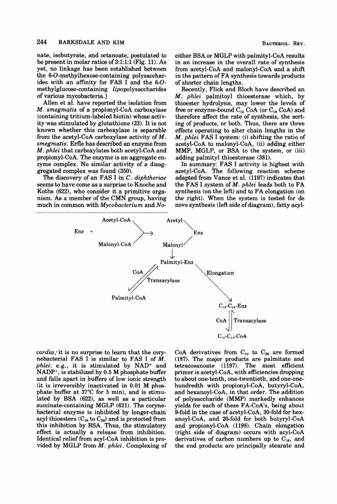
244 BARKSDALE AND KIM
nate, isobutyrate, and octanoate, postulated tobe present in molar ratios of 3:1:1:1 (Fig. 11). Asyet, no linkage has been established betweenthe 6-O-methylhexose-containing polysacchar-ides with an affinity for FAS I and the 6-0-methylglucose-containing lipopolysaccharidesof various mycobacteria.]Allen et al. have reported the isolation from
M. smegmatis of a propionyl-CoA carboxylase(containing tritium-labeled biotin) whose activ-ity was stimulated by glutathione (23). It is notknown whether this carboxylase is separablefrom the acetyl-CoA carboxylase activity of M.smegmatis. Erfle has described an enzyme fromM. phlei that carboxylates both acetyl-CoA andpropionyl-CoA. The enzyme is an aggregate en-zyme complex. No similar activity of a disag-gregated complex was found (350).The discovery of an FAS I in C. diphtheriae
seems to have come as a surprise to Knoche andKoths (622), who consider it a primitive orga-nism. As a member of the CMN group, havingmuch in common with Mycobacterium and No-
BACTERIOL. REV.
either BSA or MGLP with palmityl-CoA resultsin an increase in the overall rate of synthesisfrom acetyl-CoA and malonyl-CoA and a shiftin the pattern ofFA synthesis towards productsof shorter chain lengths.
Recently, Flick and Bloch have described anM. phlei palmitoyl thioesterase which, bythioester hydrolysis, may lower the levels offree or enzyme-bound C,6 CoA (or C18 CoA) andtherefore affect the rate of synthesis, the sort-ing of products, or both. Thus, there are threeeffects operating to alter chain lengths in theM. phlei FAS I system: (i) shifting the ratio ofacetyl-CoA to malonyl-CoA, (ii) adding eitherMMP, MGLP, or BSA to the system, or (iii)adding palmityl thioesterase (381).
In summary: FAS I activity is highest withacetyl-CoA. The following reaction schemeadapted from Vance et al. (1197) indicates thatthe FAS I system of M. phlei leads both to FAsynthesis (on the left) and to FA elongation (onthe right). When the system is tested for denovo synthesis (left side of diagram), fatty acyl-
Acetyl-CoA
Enz +
Malonyl-CoA
CoATran
Palmityl-CoA
cardia,- it is no surprise to learn that the co:nebacterial FAS I is similar to FAS I of.phlei: e.g., it is stimulated by NAD+ aNADP+, is stabilized by 0.5 M phosphate bufand falls apart in buffers of low ionic streng(it is irreversibly inactivated in 0.01 M phphate buffer at 370C for 5 min), and is stirrlated by BSA (622), as well as a particu]succinate-containing MGLP (621). The coryrbacterial enzyme is inhibited by longer-cheacyl thioesters (C16 to C20) and is protected fr(this inhibition by BSA. Thus, the stimulatceffect is actually a release from inhibiti(Identical relief from acyl-CoA inhibition is pvided by MGLP from M. phlei. Complexing
Acetyl\Enz
Malonyl/
Palmityl-Enz
Elongationsacylase
C,S-C24-Enz
CoA |Transacylase
C,8-C24-CoA
ry- CoA derivatives from C14 to C26 are formedM. (187). The major products are palmitate andLnd tetracosanoate (1197). The most efficientFer primer is acetyl-CoA, with efficiencies droppingYth to about one-tenth, one-twentieth, and one-one-os- hundredth with propionyl-CoA, butyryl-CoA,iu- and hexanoyl-CoA, in that order. The additionlar of polysaccharide (MMP) markedly enhancesne- yields for each of these FA-CoA's, being aboutain 9-fold in the case of acetyl-CoA, 30-fold for hex-Dm anoyl-CoA, and 20-fold for both butyryl-CoA)ry and propionyl-CoA (1198). Chain elongationDn. (right side of diagram) occurs with acyl-CoAtro- derivatives of carbon numbers up to C16, andof the end products are principally stearate and

MYCOBACTERIUM 245
tetracosanoate. When purified FAS I is used,MMP or MGLP is required for chain elonga-tion. Longer chains (elongation of decanoyl-CoA, for example, are not attained with muchefficiency unless there is added an elongationfactor, a partially purified protein associatedwith the long-chain acyl-CoA thioesterase. Ap-parently, elongation factor (inseparable fromlong-chain acyl-CoA thioesterase) releases freeCoA from substrates (1197).When Brindley et al. first reported FAS I
from M. phlei, they also reported the presence
of free ACP in this organism, and for this pres-ence they offered two explanations: (i) that themultienzyme complex dissociates in vitro, re-leasing free ACP, or (ii) that the two FASs (Iand II) operate separately in M. phlei. Indeed,they did find a second system, of average molec-ular weight less than 250,000, which was de-pendent on added ACP and used palmitoyl-CoAor stearoyl-CoA, but neither octanoyl-CoA nor
acetyl-CoA, for chain initiation (187).ACPM. phlei was subsequently found to have amolecular weight of 10,600 and an amino acidcomposition slightly different from that ofACPE. coli (796): four prolines (one in ACPE. coli)and no histidine (one in ACPE. coli). ACPM. phelisupports malonyl-CoA-CO2 exchange in E. coliextracts with about half the activity found withhomologous ACPE. coli. It is unable to replaceACPE. coli in the E. coli FAS II system. It ac-tually inhibits the reaction, despite the factthat in the transformation of stearoyl-CoA tolonger-chain FA by M. phlei both ACPM. phleiand ACPE. coli can serve as active acyl carriers(795). This chain-lengthening activity is abso-lutely dependent on added supply of ACP (796).So far, there has been no report of a FAS I inother mycobacteria, nor has there been addi-tional information on the FAS II ofM. phlei.
Fourteen years ago, the Kusunoses et al.demonstrated the incorporation of label from[14C]acetate and [14C]malonate into FA in solu-ble extracts of M. avium (677). With added[14C]malonate, most ofthe label appeared in FAabove C16, especially C2,. Bicarbonate was animportant component of the system. Goldmanand associates have examined FA-elongatingand -synthesizing soluble extracts from M. tu-berculosis H37Ra (576, 929, 1219). Recently, Be-sert and Etemadi (128, 129) have shown thatthe palmityl-CoA-ACP transacylase (molecularweight, 100,000), the malonyl-CoA-ACP trans-acylase (molecular weight, 32,000), and the ace-tyl-CoA-ACP transacylase (molecular weight,85,000) activities, derived by ammonium sul-fate fractionation of 105,000 x g supernatants ofM. smegmatis sonically disrupted (in 0.1 Mphosphate buffer, pH 7, containing 0.0001 M
ethylenediaminetetraacetic acid [EDTA] and0.01 M mercaptoethanol), are completely sepa-rable. ACP from E. coli functioned in this FASII system. These authors apparently found noevidence for a multienzyme FAS I in M. smeg-matis. They have discussed in some detail thepossible meaning of their findings in terms ofan FAS I system, as reported for M. phlei, andthe FAS II system, which functions in E. coli. IfM. phlei is the archetypal MycobacteriumTwort and Ingram have thought it to be (1189),it may have far more elaborate machinery forthe synthesis of lipids than some of its derivedcousins. Since, however, Bloch and associateshave found FAS I in C. diphtheriae, it would besurprising not to find such activity in mycobac-teria other than M. phlei.
Diglycerides, Triglycerides, Lipid Globules,Fat Bodies, and Intracellular Storage of
LipidBurdon (205) was impressed with the inclu-
sions revealed in members of the CMN groupby staining with Sudan black B. In Fig. 2 areshown side-by-side sections of a cell of M. au-rum and a cell of M. tuberculosis H37Rv show-ing areas once occupied by fat bodies. Schaeferand Lewis (1026) have demonstrated that ap-parently similar inclusions can be induced toappear in cells ofM. kansasii whose growth hasbeen stimulated with detoxified oleic acid (asoleate-BSA-fraction V complex) or its ester,polyoxyethylene sorbitan mono-oleate. Uptakeof oleate was rapid. First the cells becamecoated with lipid (which modified their opticalproperties), to which Sudan black B fixed. Theintracellular accumulations of lipid appearedunder phase microscopy as bright globules. At4 to 7 h postincubation with lipid, the globulescould be seen at the ends ofthe cells and, at 18 h,their numbers had increased to six or seven percell. These fatty inclusions corresponded withthe Sudan black B-stained inclusions demon-strable in the same cells. Electron micrographsof 19-h-old cells revealed inclusions morphologi-cally similar to those shown in Fig. 2A and B(for initial lipid accumulation associated withcytoplasmic membrane, see Fig. 10A). The au-thors showed that synthetic triolein, elaidicacid, lecithin, cephalin, polyoxyethelene sorbi-tan monolaurate, and sodium laurate, as wellas high concentrations of human, bovine, rab-bit, and guinea pig sera, stimulated the appear-ance of the globular inclusions. Whereas M.kansasii and M. marinum responded to botholeic acid and esterified oleate, as Tween 80, M.avium formed globules in the presence of oleicacid but not Tween 80. This finding was corre-lated with the inability of M. avium to hydro-
VOL. 41, 1977

246 BARKSDALE AND KIM
lyze Tween 80. The extent to which the FAscontained in lipid globules are in some waymodified was not determined in these experi-ments. McCarthy (801) has reported pH-sensi-tive, temperature-dependent uptake of[1'4Cpalmitic acid by M. avium. Uptake pro-ceeds with saturation kinetics and, within 5min after the cells are brought into contactwith the FA, the latter is incorporated intotriglycerides of the cell. In turn, some of thetriglyceride is rapidly utilized, as indicated bythe evolution of 14CO2. Gas-liquid chroma-tographic analyses of these triglycerides of M.avium indicated the FAs in greatest concentra-tion to be palmitic, palmitoleic, and oleic acids.FAs above C18 and below 016 were also present.In fully grown M. avium, not starved for FA,the level of triglyceride was found to be close to5% of the dry weight of the cells. For otherrelationships between M. avium and palmiticacid, see McCarthy (802). It has been shown byAntoine and Tepper that nitrogen-limited M.tuberculosis undergoing postexponential growthin a glycerol-containing medium accumulatedconsiderable lipid (48). Although this lipid wasnot identified, McCarthy has suggested thatit was triglyceride (801). Early demonstrationsof triglycerides in mycobacteria (142, 814) didnot localize these compounds in the cell. Theirexistence in Mycobacterium has long beenknown, and Asselineau has reviewed their dis-tribution among some members of this genus(59, p. 157). Walker and co-workers have re-ported that the triglycerides of BCG and M.smegmatis are esterified mainly with C,8-re-lated FA in position 1 (18:0, 18:1, and 19 BR),016 FA in position 2, and C20 and higher FAin position 3 (1216). They found a marked dif-ference between the C20+ FA at position 3 in thetriglycerides of BCG (76%) as compared withM. smegmatis (43%). More recently Weir et al.(1239) have studied uptake and incorporationof oleate into triglycerides of M. smegmatis inrelation to the appearance in the cells of lipoidalbodies. They suggested that early oleate enter-ing the cell ended up in triglycerides and thatonce the acceptors (diglycerides?) were used up,remaining oleate accumulated as lipoidal drop-lets. Their reasoning was: "the initial rapid in-corporation of oleate into triglycerides may bethe result of esterification of the small amountsof free diglycerides present. When the endoge-nous diglyceride is exhausted, the incorpora-tion rate decreases, slower incorporation occur-ring as diglyceride is made available bymetabolism and by lipase hydrolysis of phos-phatidic acid and other phospholipids."McCarthy's results indicate that stored tri-glycerides offer a ready source of energy to the
cell, as well as a means of keeping intracellularfree FA at nontoxic levels. Her findings, takenwith those of Schaefer and Lewis, suggest thata number of different FAs may provide mole-cules for tri- and phosphoglyceride stockpiles.For example, we have found that cells ofMyco-bacterium sp. 1081, growing in the footpads ofmice, at 2 months postinfection show remark-able accumulation of lipoidal bodies (Fig. 12).Glycosyldiglycerides have been found in no-
cardias (612), and a cell-free system that formsthem from uridine 5'-diphosphate (UDP)-galac-tose and UDP-glucose has been isolated fromM. smegmatis (1035), where it has been sug-gested that they may function as reservoirs andin the transport of hexoses across the mem-brane, or both (see also [1049]).
Sometimes, the areas ofaccumulated lipid aresurrounded by polysomes (Fig. 13). Such lipidstorage areas appear to be one of the hallmarksof the postexponential mycobacterial cell. Wehave previously pointed out aspects of lipoidalbodies of other members ofthe CMN group (96).That the role of triglycerides in storage may bevery different from their role in the activelymetabolizing cell is suggested by the work ofBrennan et al. (184), whose results indicated ahigh rate of turnover of triglycerides. This sug-gested to them that triglycerides could not bethought of as metabolically stable storage com-pounds. They pointed out that there is a highrate oflabeling of triglycerides in Chlorella vul-garis. What may be important here with regardto triglyceride turnover versus storage is thephysiological state of the cells being studied.Throughout this review, there are reported ex-amples ofmarked differences in biosyntheses inlog-phase cells as compared with cells not un-dergoing exponential growth.
Phospholipids and GlycolipidsThe phospholipids of mycobacteria include:
phosphatidylethanolamines (PE):RCOOCH2R'COO H O
H2C-O-P-OCH2CH2+NHs0-
diphosphatidylglycerides (CLs), and the myo-inositol phospholipids (20, 91, 709),
0
R 2 P-P-O-CH2I3O HCOCOR'
HH2COCOR
BACTRIUOL. REV.

MYCOBACTERIUM 247
FIG. 12. Lipoidal inclusions in in vivo-grown Mycobacterium sp. 1081. Bacilli found in different sectionsfrom infected mouse footpads show lipoidal inclusions occurring as single (A) or lobed (C) units. The cellwalls and the cytoplasmic membranes are intact but most of the cellular organization is gone (A-C). (x40,000;bar = 0.5 urm.) See discussion, Intracellular Storage ofLipids and Stages of Mycobacterial Growth.
VOL. 41, 1977

FIG. 13. Section of old cells of Mycobacterium sp. strain ICRC, showing large fat inclusions, polysomes,and densely staining DNA (around the inclusions). See also Kolbel (629) for fat inclusions. (x 78,000; bar =
0.5 iAm.) See discussion of Intracellular Storage of Lipids.248

MYCOBACTERIUM 249
where R(I) = H, R(II) = mannosyl, R(III) =
dimannosyl, R(IV) = trimannosyl, R(V) = te-tramannosyl, and R(VI) = pentamannosyl(710). These phosphatidylinositol oligomanno-sides will be referred to as PI-Man,. Accordingto Lee and Ballou (709), the dimannosides aremannosylated at C2 and C6. All further manno-syl additions are at C6. Therefore, the penta-mannoside, formula VI, has the structure:
M-M-M-M
0
20I--0-CH2
HCOCOR'H
H20OCOR"
It has subsequently been suggested by Brennanand Ballou (180) that, among the mannosylderivatives of M. phlei, some are acylated onthe mannose or myoinositol portion ofthe mole-cule. This is in addition to the acyl groups (R'and R") on the glycerol moiety ofthese phospho-lipids. The chemistry of mycobacterial phospho-lipids has been reviewed by Asselineau (59),Pangborn (905), Lederer (704), and Goren (453).Phosphatidic acid levels in mycobacteria can beassumed to be low, of rapid turnover, and con-fined to the cytosol. Although the major phos-pholipids are reviewed here in conventionalcatalog fashion, their actual state is certainlydynamic and ever-changing in the biosyntheticprocesses of the actively growing mycobacterialcell. This is suggested by their changing cellu-lar distribution as populations move from loga-rithmic to interphasic states of growth. Fromthe laboratory of Nojima have come findingsrelating the three mycobacterial phospholipidsto the anatomy of the mycobacterial cell (20-22).
CardiolipinIn mycobacterial CL, the distribution of FAs
was nonrandom: the unsaturated acids, octade-cenoic and tuberculostearic, were located at thea-position, whereas palmitic and other satu-rated FAs were located mainly at the (8-posi-tion. There was no evidence for the presence inCL of such long-chain FAs as phthienoic, myco-cerosic, or mycolic. The patterns of the FA(acyl) substituents in the CLs differed from spe-
cies to species (M. tuberculosis Aoyama B, M.bovis BCG, M. phlei Penso, and M. butyricum[896]). The turnover rate of CL in M. phlei, logphase, was high (22). CL seemea most concen-trated in the cytoplasmic membrane fraction(21). No phospholipid of any class was detectedin the fluid cytoplasm (minus membranes andribosomes; P content of this fraction was 1.1%).
Phosphatidylethanolamines and GlycosylDiglycerides
The distribution of PE was more difficult topinpoint (21). It was most concentrated in thecytoplasmic membrane fraction and in fractionsthat were mixtures of ribosomes and cytoplas-mic membranes. Minnikin et al. (824) havesuggested a possible interchangeability of PEand diglycosyldiglycerides in the membranes ofsome members of the genus Bacillus. Whetherthis means that the two serve similar functionsremains to be discovered.
Schultz and Elbein (1035) have reported thetransfer, by a particulate system from M. smeg-matis, of [14C]glucose from UDP-[P4C]glucoseand [14C]galactose from UDP-[14C]galactoseinto mycobacterial acceptors (endogenous) toyield monoglycosyldiglycerides and diglyco-syldiglycerides. The major FAs of these glycer-ides were palmitic and oleic. Some stearic acidwas also present. The authors suggest thatthese diglycerides are membrane bound andtherefore may serve as carbohydrate reservoirsor function in the transport of hexose residuesacross the membrane (see also [1049]).
MannophosphoinositidesThe PI-Manx's ofM. phlei were most concen-
trated in the cell wall fraction. In growing M.phlei, they seemed slow to turnover. Yet, mea-sured as mannose of PI-Manx, the phosphatidy-linositol mannoside family of compounds isalso found, though at much lower concentra-tions, in the membrane fraction(s). This wouldbe expected if shorter precursor molecules wereassociated during biosynthesis mostly with thecytoplasmic membrane fraction or if PI-Manx'salways were in continuum with their points oforigin. Homogeneous layers are probably theexception in bacterial organization. There areprobably always intergradations and intrusionsfrom one layer into another. It seems reasona-ble to consider phosphatidylinositol oligoman-nosides as essential cell wall components ofmycobacteria (see also Motomiya et al. [858]regarding the location of PI-Manx in the wallsof Mycobacterium sp. P-6). Kotani's earlyhunch that they might function as a matrix inthe wall (653) seems reasonable, and proof ofsuch a function would be welcome. Nojima (879)isolated from a "wax D" of M. bovis BCG, aphosphatidylinositol polymannoside which, ondeacylation, gave a mixture of glycerophos-phorylinositol oligomannosides with two, four,and five mannosyl residues. This is strong addi-tional evidence that PI-Manx is a cell wall com-ponent. Goldman reports from studies withH37Rv that phosphatidyl-myo-inositol diman-noside occurred in the cytoplasm, cytoplasmic
VOL. 41, 1977

250 BARKSDALE AND KIM
membrane, and cell wall. However, he reportedthat the cell wall contained the greatest concen-tration of di- and pentamannosides (436). ThePI-Manx's can be extracted with pyridine (905).Evidence for a pyridine-extractable matrix-likematerial extending from points just beneathand through the murein layer into the outermembrane of M. smegmatis 607 is provided inFig. 8A. Cells, after extraction with pyridine,become sticky and tend to agglutinate. Theyretain their acid-fastness. Not only are theyacid-fast, but also, carbol fuchsin penetratesthem more readily than it penetrates theirunextracted counterparts. As shown in Fig. 8B,extraction with alkaline ethanol removes thepeptidolipid layer. Such cells no longer are acid-fast. Pyridine extraction leaves the peptidolipidlayer essentially intact except for randomareas, where it removes pyridine-soluble mate-rial that had extended through the peptidoli-pid. The pyridine-extracted substance(s) isshown in Fig. 9. Kataoka and Nojima havesuggested that PI-Manx may be very specialphospholipids of the Actinomycetales (588).They have pointed out that the phospholipids ofC. diphtheriae (58, 439), N. asteroides and N.brasiliensis (690), M. bovis BCG, M. phlei(905), and M. smegmatis (681, 1119), and Strep-tomyces griseus and Microbisporangiumchromogenes (588) all contain PI-Manx.
Biosynthesis of MannophosphoinositidesBrennan and Ballou (179, 180) have ad-
dressed themselves to the biosynthesis of thedimannophosphoinositides in M. phlei. Phos-phatidyl-myo-inositol can act as an acceptor ina test tube system where mannose is donated asguanosine 5'-diphosphate mannose (GDP-man)(512). The requisite enzymes occurred in (boththe soluble and) the particulate fractions of M.phlei. The dimannophosphoinositides weremultiply acylated (two fatty acids were at-tached to the glycerol moiety and additionalFAs were esterified to available hydroxyls onthe mannose or myo-inositol portions). The acyl-ation step required CoA and adenosine 5'-tri-phosphate (ATP) but could be effected by fattyacyl-CoA. The specificity of the enzyme was notlimited to fatty acids found in M. phlei. It couldadd on not only palmitic and 10-methylstearic(tuberculostearic) acids but also myristic,stearic, and oleic acids. This latter informationmay be of considerable interest when compara-tive lipid biosynthesis is assessed. The pathwaydiscussed by these authors offered two possibleroutes from phosphatidyl-myo-inositol to di-mannophosphoinositide: (i) monomannophos-phatidylinositol accepts mannose from GDP-
man at C6 of the inositol and is then acylated tobecome dimannophosphoinositide or (ii) mono-mannophosphatidylinositol becomes acylatedin the inositol ring, after which it becomesmannosylated in position 6 on the myo-inositolring, yielding a dimannophosphoinositide acyl-ated in the inositol part. Takayama and Gold-man (1121) found that a particulate fractionfrom H37Ra incorporated radioactive label from[14C]mannose only at position 6 of the dimanno-side and at position 2 in the monomannoside.Two reactions seemed to occur:
(i) phosphatidyl-myo-inositol+ GDP--[1-j4C]mannose
myo-inositol mono-[1-14C]mannoside + GDP
(ii) phosphatidyl-myo-inositol monomannoside+ GDP--[1-14C]mannose
- phosphatidyl-myo-inositoldi-[1-14C]mannoside + GDP
To these authors, this finding of label at C2 inthe monomannoside and only at C6 in the di-mannoside precluded the possibility that themonomannoside might be the direct precursorof the dimannoside. However, they suggestedthat once monomannosides were acylated onthe myo-inositol ring they might serve as accep-tors at C6. These findings fit in with the gen-eral information about PI-man,. The work ofLee and Ballou (90, 709, 710), Hill and Ballou(512), and Pangborn and McKinney (906) hadindicated that M. phlei and M. tuberculosisproduced a variety of dimannosides that dif-fered as to the number of their fatty acylgroups. Dimannosides having 4, 3, and 2 mol ofFA per mol of phosphate have been found inboth M. tuberculosis and M. phlei. Probablyroute ii above (acylation in the myo-inositol ringof the monomannoside being a prerequisite formannosylation at position 6) is central to theformation of the more acylated and mannosy-lated PI-Man,'s. Different species of mycobacte-ria may possess mannosyltransferases andfatty acylating enzymes of varying specificitiesand these, plus intracellular restrictions as tothe kinds of FAs available for addition, couldaccount for the wide variety of PI-Manx's syn-thesized by different mycobacterial species (invivo), as well as for limitations on in vitrosystems. Takayama and Armstrong have foundthe cell-free system of M. smegmatis to yieldPI-Man,, PI-Man2, PI-Man3, PI-Man4, and PI-Man5, but PI-Man4 was most abundant, and PI-Man2 was least abundant. This contrastedwith the cell-free system ofM. tuberculosis andM. phlei which, as indicated above, yieldedonly PI-Man2 (1119).Oka et al. (894) have agreed with the findings
BACTERIOL. REV.
BARKSDALE AND KIM
of others regarding the cellular distribution ofphospholipids, but they have inferred that theconstitutively (scoto-) chromogenic strain, P-6from Runyon, has as its major phosphatidyl-inositol mannoside not PI-Man, but phosphati-dylinositol monomannoside, thus implying aninherent difference regarding the lipids of "un-classified" mycobacteria (Runyon group II).Khuller and Brennan (611) have been quick todemonstrate that monomannophosphoinosi-tides were in fact absent in strain P-6, whichthey found to have a preponderance of diman-nophosphoinositides. So far, then, dimanno-phosphoinositides seem to be of general distri-bution among the mycobacteria.Kataoka and Nojima advance the idea that
the proportion and kinds of the phospholipidtriad, CL, PE, and PI-Man,, in the Actinomyce-tales are unique. Although phosphatidylinositolmannoside and CL are found in corynebacteria,Brennan and Lehane report that PE is absentin C. diphtheriae (182), although Khuller andBrennan have reported it in an organism re-ferred to as Corynebacterium aquaticum (610),and Komura and associates have found tracesof PE in C. diphtheriae (632).
Immunizations with Cardiolipins, Phosphati-dylethanolamine, and Phosphatidylinositol
MannosidesIn studies of an unclassified constitutively
chromogenic mycobacterium, P-6 (obtainedfrom E. H. Runyon), Motomiya et al. (859)showed that the CL fraction from P-6 behavedas a satisfactory antigen in the floccula-tion test for syphilis and adsorbed onto latexparticles employed in an agglutination test us-ing sera from patients with lepromatous lep-rosy. CL, PE, and PI-Man, fractions were sepa-rately used for immunizing guinea pigs, andthese animals were subsequently challengedwith living H37Rv (7.5 x 10⁴ bacteria). No pro-tection was afforded by either of the three im-munizing antigens. Although the capacity ofleukocytes derived from animals immunizedwith CL, PE, or PI-Man, to phagocytize H37Rvwas examined in slide culture, there was nosignificant difference between cells derivedfrom the control and the immunized animals(857). Pigretti et al. have reported that two PI-Man, fractions from M. tuberculosis, whenused to immunize mice, failed to extend thesurvival time after challenge with M. tubercu-losis (931). Khuller and Subrahmanyam havereported that phosphatidylinositol mannosides,when combined with incomplete Freund adju-vant (IFA), elicit antibody formation in rabbits(613). Employing the kaolin agglutination test
of Takahashi, Subramanyam and Singhvi(1105) have shown that phosphatidic antigens(identified by them as "PI-Mans") reactive withhuman tuberculous sera occur in H37Rv, H37Ra,M. avium, M. phlei, and strain 607, and theseseem very closely related or indistinguishablefrom the antigen of Takahashi.
Portelance and Asselineau have called atten-tion to the fact that fractions of phospholipidsfrom BCG and H37Rv characterized as phospha-tidylethanolamine and mannosides of phospha-tidylinositiol were contaminated with lipidscontaining amino acids (sometimes devoid ofphosphorous), lipopolysaccharides, and pep-tides (934). The biological properties of thesecontaminants might be confusing. Purity ofpreparations with name designations such aspeptidoglycans, glycopeptides, peptidolipids,etc. (e.g., see section on Antigens), pose seriousproblems throughout mycobacteriology.
Lipids of Transfer, Carrier LipidsThe murein of the mycobacterial wall has
much in common with other bacterial peptido-glycans (see section, Cytoplasmic Membrane toPeptidolipid). It is therefore to be expected that,in its biosynthesis, mechanisms common to anumber of other bacteria should operate. Im-portant in the biosynthesis of bacterial cellwalls are membrane carrier lipids such as un-
CH3I
decaprenol phosphate, H-(CH2-C = CH-CH2)11-PO (38, 511, 794), which, through itsphosphate, accepts P-MurNAc-pentapeptidefrom UDP-MurNAc-pentapeptide. Similar car-rier lipid function is involved in the biosyn-thesis of lipopolysaccharides of gram-negativebacteria (1238, 1284, 1285). Schultz and Elbein(1034) have examined the biosynthesis of man-nosyl- and glycosyl-polyprenol phosphates byparticulate enzymes from M. phlei. The authorshave suggested that such lipids may have a rolein the buildup of arabinomannans of the myco-bacterial cell wall. Takayama and Goldman(1122) have found (using H37Ra) that 60% of themannophospholipids formed by a particulatetransmannolase acting with labeled mannoseand mycobacterial phospholipid was [14C]man-nosyl-1-phosphoryl-decaprenol. Two alkali-sta-ble mannophospholipids from M. smegmatishave been investigated by Takayama and Arm-strong (1119). More recently, Takayama et al.(1124) have characterized these transfer lipidsas mannosyl-1-phosphoryl-octahydrohepta-prenol and -decaprenol. The importance of iso-prenoid phosphates in the translocation of othermicrobial membrane subunits such as teichoic
BACTERIOL. REv. 251

252 BARKSDALE AND KIM
acids has been discussed by Baddiley and co-workers (39, 1224). They have reviewed specificcases in vitro where carrier lipids derived fromgram-positive organisms function in the trans-fer of capsular polysaccharides in systems fromgram-negative organisms. Similarly, carriersfrom Aerobacter have been shown to function inthe synthesis of mannans by crude extractsfrom Micrococcus lysodeikticus (682). Eventhough the lipid intermediates are known toexhibit minor differences as, for example, inthe linkages of hexose units, it appears thattheir polyisoprenol residues are identical (682).It is possible, therefore, that carrier lipids frommycobacteria might function in in vitro sys-tems from either gram-positive or gram-nega-tive organisms.
Mycobacterial Methylations
Mycobacterial products possessing substitu-ent methyl groups include methyl branches onFAs (i) produced via methylmalonyl-CoA or (ii)donated from S-adenosylmethionine. Methyl-ated sugars are not uncommon among myco-bacterial lipopolysaccharides. The restrictionand modification systems, known to exist inmycobacteria (see section, Mycobacterio-phages) but as yet undefined, undoubtedly in-volve the methylation of DNA. Cantoni (216)has reiterated his proposal of two decades agothat S-adenosylmethionine is the sole donor ofmethyl groups in methyl transfer reactions,with the possible exception of N5-methyltetra-hydrofolate involvement in the methylation ofcatechols and indoleamines in animals. Mudd(861) has suggested that, since the de novo syn-thesis of methionine requires methyl-B12, ex-amples may yet be found where the methylgroup from B12 (which originates from the (3-carbon of serine or other C1 sources) itselfmightbe donated directly rather than via S-adenosyl-methionine.
Detailed information has long been availableconcerning certain methylation reactions inmycobacteria. Lennarz et al. have found thepattern of tuberculostearic (10-methylstearic)acid synthesis in M. phlei to go from stearate tooleate to 10-methylstearate. The conversion ofstearate to oleate is oxygen dependent. Whenlabeled oleic acid was provided to growing cellsof M. phlei, it served as a "precursor" of 10-methylstearic acid, and the methyl groups wereshown to derive from labeled methionine (715).The formation of the methyl side chain in 10-methylstearic acid apparently resulted from ad-dition of a methyl group to unsaturated C10 ofoleic acid. Jaur6guiberry, et al. (546) examined
the 10-methylstearic acid synthesized by M.smegmatis grown in the presence of methio-nine-methyl-D3, and found no mass spectromet-ric evidence for trideuterated FA methyl esters.Molecular peaks corresponding to dideuteratedcompounds were observed. This suggested thatduring the transfer of the D3-methyl groups, aproton was lost. Subsequently, Jaureguiberryet al., employing extracts of M. phlei, foundevidence for an intermediate in the synthesis of10-methylstearic acid, 10-methylenestearicacid, and they postulated that the loss of theproton in going from -CD3 -+-CD2 involvedthe migration of a hydrogen between C10 andC9 of oleate (548; see also [547]), with the subse-quent loss of a methyl hydrogen in the forma-tion of the methylenic side group (as in Fig. 14).The migration of the methyl hydrogen was fur-ther studied by mass spectrometry of deuter-ated compounds provided to M. phlei (714). Led-erer has reviewed, in some detail, these andother methylation reactions (701; see also Jaur-6guiberry et al. [547] and Law [697]).
In looking into the details of the reactionleading to the methylation of tuberculostearicacid at C10, Akamatsu and Law have soughtthe endogenous lipid acceptor, the actualmethyl donor, and the hydrogen donor for thereduction of the 10-methylene group to amethyl group. They have used the crude en-zyme system (supernatant from 100,000 x gextract of sonically disrupted cells) involved inthe synthesis (including methylation) of 10-methylstearic acid by M. phlei (17, 18). Thecleanest syntheses were obtained with acetone-treated preparations, which were relativelyfree of endogenous lipid substrates. In this cell-free system, oleic acid failed to stimulate thesynthesis of 10-methylstearate derivatives.
CH3-[H2]7- CH2- CH2- [CH2]7-COOH
Stearic (octadeconoic) acid
CH3-{CH2]7\ 4H2]7-COOHH -7IH > c- ce
/ ll-AOleic acid OH3 CH3 H
Cf HHHHX /
CH3CH2]7-CH - CH012- [CH2]7-COOH<-C CH2CH3 CH2
10- Methylstearic acid 10- Methylene stearic acid
FIG. 14. Synthesis of 10-methylstearic acid, afterAkamatsu and Law (18). R = methyl donor.
BACTERIOL. REV.

VOL. 41, 1977
Olefinic fatty acyl phospholipids were required.The chain was alkylenated at the C10 to give amethylene group; this was discovered byemploying S-[methyl-14C]adenosylmethionine(methionine did not function as an effectivemethyl donor when added to the cell-free sys-tem), and this methylene group was subse-quently reduced to a methyl group. Althoughthe necessary methodologies for unraveling theexact role of the phospholipids were not availa-ble, the overall reaction appeared to encompasstwo steps:
(i) (olefinic fatty acyl) phospholipid + S-ad-enosylmethionine -- (methylene acyl) phospho-lipid + S-adenosylhomocysteine
(ii) (methylene acyl) phospholipid +NADPH + H+ -- (methyl acyl) phospholipid +NADP+Phosphatidylglycerol, phosphatidylinositol,and PE could serve as substrates. Although theenzyme system was associated with the-solublefraction, there was enough activity in the mem-brane fraction to suggest that the intact systemmight be membrane associated. It has previ-ously been pointed out that tuberculostearicacid and cyclopropane FAs accumulate in thestationary phase of growth (715). This has sug-gested to Akamatsu and Law that both of theseprocesses involve enzymes that act upon themembrane phospholipids, alkylenating themafter their deposition by phospholipid synthe-tases. Tuberculostearic acid may occur in anumber of macromolecules of the mycobacterialcell. For example, Pigretti et al. reported thatthe phosphatidylinositol pentamannoside ofM.tuberculosis contained 43% of this 10-methyl-stearic acid, along with large amounts of pal-mitic and small amounts of oleic and stearicacids (931).Akamatsu and Law have also recovered a
soluble enzyme from M. phlei that transfersmethyl groups from S-adenosylmethionine tothe carboxyl group of FAs. The role of thisalkylation of carboxyl groups to yield FAmethyl esters in the biosynthetic processes ofM. phlei is not known (19).
Lornitzo (734) has reported protein bound 1-O-methyl-sn-glycerol-3-phosphate as a proba-ble intermediate in a transmethylation reac-tion occurring in H37Ra, leading to the forma-
MYCOBACTERIUM 253
In Fig. 11 is shown a lipopolysaccharide,MGLP IV, which contains an a-(1 -- 4)-r-glu-co-oligosaccharide methylated in position 6 ofeach of the glucose units. A polysaccharidemethyltransferase that brings about the trans-fer of these methyl groups from S-adenosylme-thionine has been found in extracts of M. phlei(371, 470). Oligosaccharides of 7 to 10 glucoseunits in a-(1-3 4) linkage are suitable sub-strates; analogous structures in f3-(1 -- 4) link-age are not. In fact, the latter inhibited thereaction. The presence of acyl groups on thepolysaccharide (Fig. 11) enhanced methyltransfer (607, 869, 1179).
Lipid Syntheses and Stages (Phases) of Myco-bacterial Growth
Here and there in this review it is noted thatmetabolic products characteristic of aging pop-ulations are different from those of populationsin log phase of growth. Donets et al. (310) havenoted an increase in phospholipids ofM. albumfrom 9.3% towards the end of log phase to 15%in stationary phase; for M. rubrum, phospho-lipids increased from 15.5 to 19.2%. For bothspecies triglycerides dropped from a peak (M.album, 20.4 to 15.4%; M. rubrum, 36.6 to 26.6%)at 6 days to a lower value at 12 days. The high-est levels of free FAs were found associatedwith end-phase populations. Amounts of digito-nin-precipitable lipids were higher in log-phasecells, reduced to 50% in late log, and increasedat the onset of stationary phase. Although theauthors refer to the latter as sterols, no databeyond their digitonin precipitability are of-fered to support this conclusion.
Mycolic Acids
The characterization of the corynomycolicacids (706, 949) and studies on their in vivosynthesis using [1-14C]palmitate has indicatedthat parts of two molecules of palmitic acidenter into the biosynthesis of one molecule ofcorynomycolate, since label was found in C1and C3 of the latter (421, 422). Et6madi andLederer (356) have provided labeled tetracosa-noic acid, C23H47-*COOH, to cultures of M.smegmatis, growing in Sauton medium, andthen isolated from those cultures:
OH
CH3-(CH2)ni-CH=-CH-(CH2),,2-CH=CH -CH-(CH2)1,7 -CH-CH-*COOH
UH3 C22H45
tion of 1-0-methyl-glycerol from S-adenosylme-thionine and sn-glycerol-3-phosphate. n, = 15-19; n2 = 12-16

254 BARKSDALE AND KIM
(a-smegmamycolic acid labeled in the carbon ofthe carboxyl group). To other growing M. smeg-matis they supplied [ methyl-'4C]methionineand found that the carbon of the methyl sidechain was derived from the labeled methionine.These experiments, with their elegant suppor-tive degradation studies, indicate that the finalstage in the synthesis of a-smegmamycolic acidinvolves a condensation of the carboxyl termi-nal of a methyl branched FA (synthesized byM. smegmatis) with the a-carbon of the labeledtetracosanoic acid (supplied in the medium). Asa result of this event, the carboxyl carbon dur-ing the process of condensing becomes the hy-droxylated ,8-carbon of a-smegmamycolic acid.As mentioned above, the origin of the radioac-tive methyl group in the long-chain FA moiety(synthesized by M. smegmatis) was labeled me-thionine. Lederer has reviewed some of thesemethylation reactions (700, 701, 703).Using [14C]palmitic acid in combination with
a cell-free extract prepared (in 0.05 M phos-phate buffer, pH 7.0, plus 3 x 10⁻³ M mercapto-ethanol and 0.01 M disodium EDTA) from anontoxinogenic derivative of C. diphtheriaegravis, Halifax strain, Walker et al. (1217) re-cently have reported a Claisen sort of condensa-tion of 2 mol of the labeled acid to yield 2-tetradecyl-3-keto-octadecanoic acid. WithNaBH4, these investigators were able to quan-titatively reduce the 83-keto acids, yielding amixture of corynomycolic acids. Whether or nota buffer of greater ionic strength might haveexpanded the biosynthetic capabilities of theseextracts is not known. (In this connection, seediscussion of FAS I of C. diphtheriae.) Morerecently, Prome et al. (944) have found thatunder slightly modified conditions, from 2 molof palmitic acid, the C. diphtheriae system syn-thesized a monotetradecyl-2-keto-3-octadeca-noate of trehalose. This finding suggested tothe authors that trehalose-lipid is an interme-diate in the synthesis of corynemycolate.
It would seem that the in vivo experiments ofEt6madi and Lederer could be extended in thefollowing way to give more information aboutthe biosynthesis of mycolic acids in the CMNgroup. The close interrelations of the group aregenerally acknowledged (59, 159, 271, 441, 446,653, 698, 699, 704) and have previously beendiscussed by one of us from the standpoint of
the type species of the genus Corynebacterium,C. diphtheriae (96). A gradient of organismsfrom C. diphtheriae through N. rubra and N.asteroides to M. bovis, M. kansasii, M. intra-cellulare, M. smegmatis, and M. phlei could bearranged on the basis of the relative complexi-ties of their mycolic acids (in this connection,see the findings of Maurice et al. concerningvariations in mycolic acids of nocardias [798]and discussions of Et6madi on phylogenetic im-plications of various mycolic acids [352-354]).Since each organism in the list produces a-alkyl, j3-hydroxy acids (mycolic acids), each iscapable ofjoining the two branches. IfM. smeg-matis or C. diphtheriae were supplied labeledC25H51-*COOH synthesized by M. tuberculosis,would they produce mycolic acids with aC24H49- a-branch? Presumably, they would.Both C. diphtheriae and M. phlei possess FASsI and II. The chain lengths of the 3-hydroxyl-ated moiety of their mycolic acids are very dif-ferent (59). Further comparative study of thesetwo systems would enhance our understandingof mycolic acid biosynthesis.Etemadi and Gasche have reviewed FA prod-
ucts of mycobacteria that might represent in-termediates in the biosynthesis of mycolic acidsand have considered the origin of the compo-nents of the dicarboxylic mycolic acids found inM. avium and certain other species (355). Theysuggested that precursors of the dicarboxylicacids might be keto mycolic acids and that thefinal intermediate might be an ester resultingfrom the introduction of an oxygen between theketo carbon and its neighbor (see arrow in theformula of the wax of Laneelle and Laneelle,below). Hydrolysis would then yield the dicar-boxylic mycolic acid and a carbinol. This wouldmean that the number of carbons in the dicar-boxylic acid would be less (than that of theprecursor) by the number of carbons in thecarbinol component removed by hydrolysis. La-neelle and Laneelle (691) have addressed them-selves to the problem of finding the keto inter-mediate of the dicarboxylic mycolic acidsamong the great mixture of mycolic acids pro-duced by Johne's bacillus (M. paratubercu-losis). They have found (i) keto acids with 80 to85 carbons, (ii) dicarboxylic mycolic acids with58 to 65 carbons, and (iii) the proposed dimy-colic acid intermediate of 78 to 85 carbons:
%\jHOH'CH3-(CH2)17 -CH O-CO-(CnH21-2)-CHOH-CH-COOH
CH3 CH22H45
Laneelles' wax n = 32, 34, 36, 37, 38, or 39
BACTERIOL. REzV.

MYCOBACTERIUM 255
which they called a wax. The latter existed as amonoglyceride. The proposed formula for thedicarboxylic mycolic acid is:
HOCO- (CnH2n2)-CHOH-CH-COOHC22'-45
n= 32, 34, 36, 37, 38, or 39
Matching arrows in the chain of the dimycolicacid with that of the ester intermediate indi-cate the proposed origin of the acid. There isleft the residual methyl carbinol. A precedentfor the kind of oxidation, which renders theketo mycolic acid into the wax form, is found inthe utilization of 2-tridecanone by Pseudomo-nas aeruginosa. Molecules of CH3-(CH2)9-CH2-C-CH3 undergo two distinct changes: (i)
It0
some are reduced to form 2-tridecanol [CH3-(CH2)9-CH2-CH-CH3] and (ii) some are oxi-
OHdized to form an ester, undecyl acetate [CH3-(CH2)9-CH2-O-C-CH3]. The latter is split
110
into two moieties: 1-undecanol [CH3-(CH2)9-CH2OH] and acetate (388). For a discussion ofthe possible role of 5-enoic acids (C22, C24, andC26) in the synthesis of mycolic acids, see dis-cussion by Asselineau and associates (57).
Isonicotinic Acid Hydrazide and Synthesis ofMycolates
Isonicotinic acid hydrazide (INH) exerts someunspecified effect on mycolic acid synthesis inmycobacteria (1125, 1220, 1270) as indicated bydecreases in a-mycolates, methoxymycolate,and 8-mycolate within 60 min after exposure.Viability of the cells also slowly decreases.About 5.2 pmol of INH per 109 cells serves toinhibit mycolic acid synthesis by about 50%.Scanning electron microscopy (SEM) of H37Racells exposed to an amount of INH (0.5 jig per5 x 108 bacteria/ml) that completely inhibitsmycolic acid synthesis in 60 min revealed adramatic change in the cells over a 24-h period,with a complete loss of outer membrane areasL, and L2 (Fig. 4) and the development of thinspots in the cell wall, with concomitant bulging.Takayama et al., the authors responsible forthe foregoing data, have cautiously stated: "theresults of this study did not allow us to deter-mine whether or not the morphological changesobserved are caused by the INH inhibition ofmycolic acid synthesis" (1126). Of course, SEM
will pick up only the more gross changes oc-curring in INH-treated bacteria. The elimina-tion, for example, of the L1 peptidoglycolipid isan alteration in fine structure that can bepicked up only under very special conditions(Fig. 5).
In reviewing the efforts at elucidating themode of action of INH, Youatt (1298) has sug-gested that INH acts specifically to combinewith an enzyme peculiar to INH-susceptiblebacteria. Ifthe enzyme is not one that is crucialto either transcription or translation, removalofINH should allow for recovery ofthe cell fromthe INH effect. Takayama et al. (1120) havebeen able to show that, up to a critical time(prior to 10 h postexposure), washing cells ex-posed to INH (0.5 pg/ml) allows the cells torecover the capacity to synthesize mycolicacids. The restoration of the capacity was aslow process, involving an 8-h lag period andrequiring about 24 h of subsequent incubation.Without washing, INH-treated cells are able toundergo one division (1118). Further, Taka-yama et al. have obtained evidence that duringthe first 12 h of exposure to INH, the contentswithin the cytoplasmic membrane of the cellincrease at a normal rate without an equalincrease in cell volume (1123). The pigmentsproduced by M. tuberculosis in the presence ofINH must still be fitted into the story of themechanism of INH-induced bacterial death(1299). For that matter, the hypothetical targetenzyme, "mycolic acid synthetase" (1120), re-mains to be characterized. It has long beenknown that, following INH treatment, INH-sensitive mycobacteria become non-acid-fast,whereas INH-resistant mutants retain theiracid-fastness (627).
Acylglucose and AcyltrehaloseWinder et al. (1271) have found that M.
smegmatis, under balanced conditions ofgrowth with glucose as its carbon source,turned over free trehalose at a rate about 3times that of its net formation, whereas thesugars of acylglucose and acyltrehalose areturned over at 13 and 8 times, respectively,their net formation. In the introductory section,we pointed out that acylglucoses are producedin large amounts only when glucose is providedas the sole carbon source. They have been de-tected in Corynebacterium xerosis (181), C.diphtheriae, and M. smegmatis (183). The ma-jor FA of acylglucoses from C. diphtheriae andM. smegmatis is corynomycolic acid. Brennanand his associates (1271) have suggested, fromresults obtained with radioactive glucose that,because of the turnover of acylated forms ofglucose and trehalose (about 10 times per bacte-
VOL. 41, 1977

256 BARKSDALE AND KIM
rial generation) under conditions of activegrowth, these sugars probably are not storagecompounds. On the other hand, they do notseem to be structural compounds or, if they arestructural compounds, they have some meta-bolic turnover as well. It seems to us that sincemycobacteria under natural conditions restsometimes and grow sometimes, always in thepresence of their metabolic and catabolic prod-ucts, it is possible that the mycolic acids andthe sugars as acylated sugars may be in a se-questered state for use when needed. It is diffi-cult to think of any natural environment inwhich 1% glucose would exist. Thus, we wouldfavor regarding acylated sugars as storage com-pounds. Even the dimycolates of trehalose,which occur at the surface of cells and whichbehave as detergents, would probably serve asa source of energy when needed.
Cord Factor(s)The toxic glycolipids containing trehalose,
originally discovered by "Bloch in petroleumether extracts of M. tuberculosis H37Rv (141)and structurally defined by Noll et al. as dimy-colates of trehalose (881), occur with distinctivemodifications in members of the genera Cory-nebacterium (537), Mycobacterium (881), andNocardia (536). The occurrence of these dies-ters of trehalose (see Fig. 15) is another com-mon bond between members of the CMN group.It is interesting that, in N. asteroides and var-
0II
CH2-O-C R
HO H H
HO 0LOH- OH
H , O H
R-C-O-CH20
A. Cord Factor
ious true corynebacteria, the trehaloses are es-terified with corynomycolic acids (C28HMO.3 toC36H6mO3), whereas in Mycobacterium (Nocar-dia) rhodochrous, the cord factors contain no-cardic acids (C38H7603 to C16H.O3). In N. aster-oides, mycolic acids of the higher carbon num-bers are found in the bound lipids of the cellwall but not in cord factors (536). Thus, fromCorynebacterium to Mycobacterium via Nocar-dia, there is a gradient in total carbon numberof these a-ramified, f3-hydroxylated long-chainFAs from C28 to C90 and a gradual change in themacromolecules of the cell to which they arecharacteristically conjugated. (For exceptionssee section on Acylglucoses.) A mycolate pre-cursor, the 13-keto C32 product of the condensa-tion oftwo molecules ofpalmitate in the coryne-bacterial system (944), becomes converted intoa monotetradecyl-2-keto-3-octadecanoate of tre-halose. Winder et al. have found acyltrehaloseundergoing rapid turnover in actively growingcells of M. smegmatis, suggesting to them,among other possibilities, that acylation mighttake place in the cell wall or membrane (1271).Thus, trehalose esters such as cord factor maybe essential for the transfer of mycolic acidsinto the mycobacterial ultrastructure. Cord fac-tors behave as detergents and this, coupledwith the fact that they are located about thesurface of the outer cell wall, suggests for thema role in facilitating the inward movement ofcertain molecules important to the growth of
0
CHZ-0-C-R I
HO,
HO0
-0-C-RIH
B. Sulfollpid I
RI- C- O-CH2
0
OH
R: CH-GCH -
c + C H60 24 49
OH CH3 CH3R:C I IHR cH31 -CH - [CH -CH2]7 CH
CH3 CH3R2 C 1 5H 3 1 CH2 [ CH _ CH2] 6.I .2 s 1 2-[CH[c CH]2 -H~
3 15 31
FIG. 15. Two acyltrehaloses from Mycobacterium tuberculosis (after Goren [453]). For data concerning thespontaneous desulfation of sulfolipids, see (452). See also Tables 1, 2 and 3.
BACTERIOL. REV.
0.H

MYCOBACTERIUM 257
the cell. Comparative uptake studies using mu-tants blocked and/or leaky in the synthesis oftrehalose dimycolates could shed some light onthe role of cord factor for CMN cells.M. tuberculosis H37Rv elaborates the cord
factor 6,6'-dimycolyl-a,a'-n-trehalose which, inmicrogram amounts, kills mice (601). The lateMasahiko Kato has shown that this killing isassociated with destruction of mitochondrialmembranes and concomitant damage of themembrane-associated respiration and phospho-rylation (592). Toida has found a side effect ofcord factor intoxication upon the microsomalenzymes pyrazinamide deamidase and amino-pyrine demethylase (1155). In contrast to mice,cord factor is relatively nontoxic for the rat(1013). (It has recently been discovered thatthere also accumulates around H37Rv a mono-mycolate of trehalose that is less toxic for micethan the dimycolate [600].) Asselineau andKato have been interested in the essential re-
quirements for cord factor activity. Early on, itappeared that ester linkages at the 6,6' positionoftrehalose with mycolic acids and the presenceof hydroxyl groups of the sugar in the moleculewere essential to toxicity of cord factor (60, 63,933). Mycolic acid alone or with trehalose, tre-halose alone (591), acetylated cord factor, and6, x -di- (3' - acetoxy, x - methoxymycolanoyl) -N-acetyl-D-glucosamine were shown to be non-
toxic for mice and nondestructive for mouseliver mitochondria. On the other hand, 6-(3'-acetoxy, x-methoxymycolanoyl)-N-acetyl-i)-glu-cosamine and methyl 6-(3',x-sulfitomycolan-oyl)-a-n-glucoside were toxic for mice andcaused in vivo a decrease in the contents ofmitochondrial protein, total lipids, and lipidphosphorous and a decline in the phosphoryla-tive activity coupled to the oxidation of succi-nate in mouse liver mitochondria (592). Theselatter compounds appear to exert a detergent-like action on mitochondrial membranes. Itwas shown that two glycolipids prepared in thelaboratory, methyl-6-mycoloyl-a-D-glucopyran-oside and 6,6'-dimycoloyl sucrose, quantita-tively retained the toxicity of cord factor (meanlethal doses [LDo's] of 187 and 161 ,g, com-
pared with 50 pg for the cord factor 6,6'-dimyco-loyl-a-D-trehalose), and this indicated the pos-
sibility of constructing four stereoisomers ofmethyl-6-mycoloyl-a-n-glucopyranoside and ex-
amining their relative toxicity (598).The subsequent study of Asselineau and
Kato (61) has shown that the steric dispositionof the hydroxyl groups at C2, C3, and C4 isessential for the toxicity of methyl-6-mycoloyl-glucoside, whereas the glycosidic linkage at C1has no steric specificity. Peracetylated methyl6-mycoloyl-a-D-glucopyranoside, like peracety-
lated cord factor, was without mouse toxicityand without effect on mitochondrial mem-branes as tested in vitro. (The acetylation ofthemycolic acids of cord factor [58, 880] or those ofcord factor analogues [59] had previously beenshown not to affect toxicity.)
Since the tying up ofthe free hydroxyl groupson the trehalose fragment, either by methyla-tion or acetylation, completely blocks toxicity,various investigators have suggested that theeffect of the trehalose hydroxyls may be uponthe fit of the glycolipid to the sensitive site onthe mitochondrial membrane. This explanationfinds an analogy in the toxic fungal octapeptidea-amanitin, which causes death through theinactivation of eukaryotic DNA-directed RNApolymerase II (379). Among the analogues of a-amanitin that are not toxic is amanullin (1255),a cyclic octapeptide having a second isoleucinein place of the y-hydroxylated side chain foundin a-amanitin. Here, again, the final fitness forbiological activity involves specific hydroxylgroup(s). Cord factor has been found to sup-press pyrazinamide deamidase in livers of in-toxicated mice but has no effect on the enzymesystem in vitro, thus suggesting an effect onenzyme biosynthesis, probably secondary to de-struction of mitochondrial membranes (1154).Cord factor is not immunogenic, but it serves
as a functional "hapten" when complexed withmethylated BSA (MBSA). Cord factor-MBSA,in WFA, used as a vaccine, protected miceagainst both the toxic action of cord factor andinfection with M. tuberculosis H37Rv (593). Pro-tection was not afforded cord factor-MBSA-vac-cinated mice challenged with either Listeriamonocytogenes, Brucella abortus, Salmonellaenteritidis, or S. typhimurium. This type ofexperiment had also been reported in 1973(595). Antibody prepared in rabbits againstcord factor-MBSA passively protected miceagainst the toxic action of cord factor. Cordfactor-anti-cord factor antibody mixtures werenontoxic for mice. Anti-cord factor serum re-acted almost as well with the trehalose-6,6'-dicorynomycolate of C. diphtheriae as it didwith its homologous trehalose-6,6'-dimycolatefrom M. tuberculosis. It was nonreactive withsucrose-6,6'-dimycolate, methyl-6-mycoloyl-a-D- glucopyranoside, or 6, x - di - (3'- acetoxy, x -methoxymycoloyl)-N-acetyl-n-glucosamine. In"haptenic" inhibition studies, a-D-trehalose in-hibited to almost 50% precipitation betweencord factor-MBSA and its antibody. Precipi-tates were determined by both radioactivityand total protein, and the inhibition curveswere almost superimposable one upon theother. Rabbits immunized with cord factor-MBSA remain sensitive to the injection of cord
VOL. 41, 1977

258 BARKSDALE AND KIM
factor, and this complicates skin test reactionsrelated to cord factor in rabbits. Rabbits immu-nized with cord factor-MBSA showed no DH totuberculin. (There was some reaction toMBSA.) BCG-vaccinated rabbits were tubercu-lin positive (594). It has subsequently beendemonstrated that cord factor bound to neutral-izing antibody loses all of its effects on mito-chondrial processes but, once dissociated fromimmune complexes, it is again toxic (597). Katoexamined the sera of mhan and animals infectedwith tubercle bacilli and found no antibody ca-pable of precipitating cord factor from an emul-sion. Similarly, mice and rabbits vaccinatedwith BCG or heat-killed H37Rv failed to developprecipitating antibody. Both mice and rabbitsvaccinated with cord factor-MBSA respondedwith the formation of precipitating antibody.The rabbit antibody, which could be destroyedby mercaptoethanol, was recovered from im-mune precipitates and shown, by immunodiffu-sion against (i) goat anti-rabbit serum, (ii) goatanti-rabbit immunoglobulin M (IgM), and (iii)goat anti-rabbit IgG, to be IgM antibody (596).The real cord factor. Goren has rued the fact
that trehalose-6,6'-dimycolate has never beenshown to be responsible for cording (453). Al-though the correlation between the capacity ofM. tuberculosis to grow as serpentine cords andits ability to kill guinea pigs is very well estab-lished (283, 819), there is some question as towhether or not trehalose-6,6'-dimycolate is re-sponsible for cording (454). Perhaps geneticstudies could resolve this problem. A mediumhas been described which employs TritonWR1339 to give maximum expression ofcording(733). Nitrosoguanidine has been shown to be asuitable mutagen for mycobacteria (639; seealso section, Genetics). Since a loss of cordingshows up as a change in colonial morphology,selection of hyper- and hypocording mutantsshould be simple. If it turns out that there is nodirect relationship between cording and totalyields of trehalose-6,6'-dimycolate, then thesearch for the real cord factor will certainly beundertaken.Cord factor granulomas. The granulomas
induced by cord factors are discussed in thesection on Mycobacterium as Antigen.Cord factor inhibition of tumors. Yarkoni et
al. (1292) have reported that either trehalose-6,6-dimycolate, trehalose-6,6-dipalmitate, su-crose-6,6-dimycolate, or trehalose monopalmi-tate, when administered to mice subsequentlychallenged with Ehrlich ascites tumor cells,brought about a significant inhibition of tumordevelopment (see also [117] regarding thesuppression of urethan-induced lung adenomas
BACTERIOL. REV.
in mice treated with cord factor). This inductionof antitumor activity on the part of cord factoris suggestive of similar effects attributed towhole BCG (89, 148, 522, 696, 781, 991, 1029,1055, 1308, 1309). Since the host response com-mon to BCG infections and cord factor injec-tions is the formation ofgranulomas (infectiousagent granulomas in the case of BCG and for-eign body granulomas in the case of cord fac-tor), the antitumor activity, as has alreadybeen suggested (850), may well be in some wayassociated with the mobilized macrophage.
In the section, Ultimate Mycobacterial Adju-vant, it is pointed out that the unit, N-acetyl-muramyl-L-alanyl-1-isoglutamine, in water-in-oil emulsion with ovalbumin, can enhance theproduction of specific antibody and the develop-ment of specific DH (655). Cell wall skeletonsproduce these effects and, in addition, wheninjected with neoplastic cells, promote the de-velopment of T effector cells cytotoxic, in thetest tube, for the neoplastic cells (69). Recently,the adjuvant activity of highly purified cordfactor (455) has been reinvestigated by an inter-national group consisting of Saito, Tanaka, Su-giyama, Azuma, Yamamura, Kato, and Goren(1012), and it is clear that trehalose dimycolatein a water-in-oil emulsion does enhance bothantibody production and the development ofDH to egg albumin, hamster erythrocytes, andsheep erythrocytes (SRBC). Why this group didnot also investigate the enhancement by puri-fied cord factor of the development of cells cyto-toxic for a specific neoplastic cell is not clear.
SulfolipidsGoren has shown that the neutral red-bind-
ing strain H37Ra (817) produces families ofstructurally related sulfolipids (SLs) of whichSb-I is a complex glycolipid ester having a mo-lecular weight of about 2,386 and an empiricalformula of C145H283020NS (Fig. 15). It is a 2, 3, 6,6' tetraester of trehalose, which can be writtenas 2, 3, 6, 6'-tetraacyl-a, a'-trehalose-2'-sulfate(450, 451, 456). Goren et al. have suggested thatneutral red binding may involve both the SLsand the phospholipids (Pb) (457). The role of SLin the virulence of mycobacteria has been amajor concern of Goren and associates. Katoand Goren have found that Sb-I is nontoxic formice. In vitro SbI caused disruption of themembranes of mitochondria from mouse liverand inhibited mitochondrial oxidative phospho-rylation. Various animal sera were found toneutralize these effects of SL-I in vitro. Pre-sumably, they are capable ofthe same neutrali-zation in vivo. The administration of S-I, in

MYCOBACTERIUM 259
combination with cord factor, in vivo producedan enhanced damage of mitochondria over thatof cord factor alone. Thus, mycobacterial sulfa-tides appear capable of augmenting the toxicityof cord factor (599). An examination of certainof the Mitchison strains of M. tuberculosis(which show reduced virulence for the guineapig) for their combined capacity to produce SLand PL has suggested to Goren et al. (457) thatthere exists a "statistically very significant"correlation between virulence for the guineapig and the capacity to produce SL and PL. Afraction of the attenuated strains, however,were hyperproducers of SL. Their capacity toproduce other products such as cord.factor wasnot measured. Although the H37Rv strain wasused as a control in these experiments, theavirulent strain, H37Ra, was not used. Perhapsin the near future we will have data comparingthe cord factor/sulfatide ratio of selected viru-lent and avirulent strains. The spontaneousdesulfation of purified SLs has been reported(452).
THE CYTOPLASMIC MEMBRANESYSTEM
Mesosomes, Cytochromes, Iron-ChelatingCompounds
A recent review of the literature of meso-somes leaves no doubt that these apparent or-ganelles are in fact manifestations of the cyto-plasmic membrane system (469). (The meso-some is visual evidence of excess membrane.Throughout nature, more-than-enough is themechanism by which systems work: from themillions of sperm produced in excess of ova tobe fertilized, to the excess of genes in termi-nally redundant phage genomes, such is thecase. The emergent aspect of these excesses,whether in the findings of carbon balance stud-ies of bacterial growth or the production of earsof corn in a field of corn plants, is an apparentexactness of systems.) It has previously beenpointed out that in actively growing cells of theCMN group there is very little accumulation ofexcess cytoplasmic membrane as mesosomalstructures (96), but that such accumulationdoes occur in late-log-phase cells. Asano et al.report this to be the case for cells of M. phlei(54). These workers found 97% of total cyto-chromes b, c, and a + a3 in a membrane "ghost"preparation from M. phlei. Although oxidativephosphorylation in ghosts was only slightlylower than that found in electron transport par-ticles (ETP; see review of intermediary metabo-lism by Rhamakrishnan et al. [958] and that ofGoldman [435] regarding enzyme systems of
mycobacteria), ghosts did not couple phospho-rylation to oxidation. Most adenosine triphos-phatase (ATPase) activity was associated withghosts, as was malate-vitamin K reductase.The authors advance arguments for dual ori-entation characteristic of ghost membranes: (i)some properties suggestive of the inner mem-branes of mitochondria and (ii) permeabilitycharacteristics suggestive of a cytoplasmicmembrane. Iron transport in Mycobacteriumand Nocardia involves special transport mole-cules, nocardamin (609) and mycobactins. Thelatter have been the subject of an extensivereview by Snow and White (1072; see also [96]).Ratledge and co-workers have suggested thatthe lipophilic mycobactin molecule is located onthe "boundary layers" of the mycobacteriumand that "it transports iron across the thicklipoidal layers of the mycobacterial cell" (748,960) as ferric ion, and that the ferrous form isthen released from its carrier by the action ofNADP-linked reductase (192).Macham and Ratledge (747) have also de-
scribed a class of partly characterized water-soluble iron-binding compounds that they havetermed "exochelins." These are reported to holdiron in solution at physiological pH values, arefreely diffusible in the desferric and ferricforms, and can rob iron from ferritin. They can,thus, reverse the inhibition of mycobacterialgrowth by serum (625). Macham et al. (748)propose that iron in the extracellular milieu issolubilized by exochelins which, upon contactwith the mycobacterial cell wall, are taken ontomycobactin, which then carries the ironthrough the mycobacterial wall (L1, L2, and L3of Fig. 4) to the cytoplasmic membrane wherethe NAD-linked ferrimycobactin reductasemakes the ferrous ion available to the cyto-plasm.
Recently, McCullough and Merkal have de-scribed the isolation of an iron-chelating sec-ondary hydroxamate, L-a-asparaginyl-L-a-N-hydroxyasparagine, from cultures of M. aviumand have reported the isolation of structurallyrelated compounds from "corynebacteria"(802b). The enzymes associated with bacterialmembranes have recently been reviewed bySalton (1014). The component of membranesespecially pertinent to this review consists ofthe mycobacterial carotenoid pigments, whichare covered in the next section.
Carotenoid Pigments of MycobacteriaThe polyene pigments, the carotenoids, are
found in all groups of living things. They aresynthesized only by plants, including microor-ganisms. Animals derive their carotenoids from
VOL. 41, 1977

260 BARKSDALE AND KIM
food. Although all photosynthesizing orga-nisms contain these pigments, the distributionof carotenoids among fungi and nonphotosyn-thetic bacteria is far from universal (442, 1090).The genus Mycobacterium illustrates this point
very well. Some species produce colorful po-lyenes (see Fig. 16), whereas others do not.Whether or not all mycobacteria have the ca-pacity to produce the colorless phytoene (seeFig. 17), or earlier precursors of carotenoids, is
FIG. 16. (A) Colony ofMycobacterium sp. growing on Dubos albumin agar. This mycobacterium has beencalled M. duvalii, see (1086). (B) Crystal of fcarotene. Kodachrome transparency by courtesy of Otto Isler(see 1236) and Hoffman-La Roche (Basel). Crystals of this carotenoid pigment accumulate around thecolonies ofcertain strains ofM. kansasii. For biosynthesis, see Fig. 17. For a discussion ofthe probable'role ofcarotenoids in mycobacterial survival see section on Carotenoid Pigments. (C) Growth of a constitutivelychromogenic mycobacterium on Lowenstein-Jensen medium. (D) Enhancement of color of strain in Cfollowing 2 weeks ofexposure to light. Reproduced with permission from (1203) using transparencies kindlysupplied by George P. Kubica. See Tables 1, 2, and 3.
BACTERIOL. RiEV.

MYCOBACTERIUM 261
HZ~~~/g,-o-nND--H
TwoGeranylgeranyl Pyrophosphates
(head tahead)
Prephytoene Pyrophosphate
Phytoenea
-2H |
b a and b accumulate in prePhytofluene of Diphenylamine (984)
1 -2H
esence
-2H C- CaroteneNeurosporene
I -2H:~~~~Lycopene
Ring
8-Ca
Ring
d-Ca
C accumulates in presence
of Nicotine (523)
clasure f~ydroxylation; Ring closure
ratene Epoxidotion! ~~~ytelds Y-Carotene
Xnhphylle Ring closurerotene
CaratCene
FIG. 17. Probable biosynthetic pathway for (3-car-otene in Mycobacterium marinum. M. marinum alsomakes small amounts of a-carotene (106). The dottedlines indicate branch points which, in different spe-cies, lead to the synthesis of other carotenoids (e.g.,a-zeacarotene) and xanthophylls. See Porter and An-derson (936), Batra (106) and Goodwin (444). Sincethis figure was prepared, David (287) has presenteddata concerning the "biogenesis" of carotenoid pig-ments in M. kansasii. He has chromatographic evi-dence to support the sythesis by M. kansasii of 4-carotene, neurosporene, lycopene, leprotene, -y-caro-
tene, b-carotene, a-carotene, and (8-carotene (288).Little is known concerning stereoisomerism andfunctional carotenoids of mycobacteria. According toWeedon, phytoene from carrots, tomatoes and thegreen alga, Chlorella vulgaris, have a 15-cis struc-ture. The phytoene that accumulates in diphenyla-mine-inhibited Flavobacterium dehydrogenans ap-pears to be a trans isomer; such is also the case withMycobacterium sp. (469b). For a discussion ofprob-lems in stereoisomerism ofcarotenoids see Beytia andRilling (130a) and Weedon (1236). See also Fig. 16and section concerning Carotenoid Pigments of My-cobacterium. See Tables 1, 2, and 3.
not known. Although colored polyenes wereidentified in mycobacteria some 40 years ago(238, 239, 534), their exploitation for mycobacte-rial taxonomy (568, 647, 994, 995) and the use ofmycobacteria for elucidating the route of theirown biosyntheses (106, 782, 982) is more recent.
Biology of PigmentationAmong mycobacteria, pigmentation is a sta-
ble genetic property (568, 631, 878, 893, 994,995, 1141, 1168, 1177, 1178). The carotenes ofrepresentatives of a single species appear to bealike. Tarnok and T6rnok (1141) have pre-sented preliminary data showing this to be thecase for the carotenoids of M. phlei (threestrains), M. avium (two strains), M. intracellu-lare (eight strains), M. aurum (four strains),and perhaps M. thamnopheos (one strain).They found that the products of M. marinum(balnei, nine strains) and M. kansasii (sevenstrains) traveled as identical spots on thin-layerchromatography (TLC) plates. The same wastrue for those of M. gordonae (aquae, ninestrains) and M. scrofulaceum (one strain). Pig-mentation may be a constitutive attribute of agiven strain (i.e., pigment is made under most,if not all, conditions suitable for growth) or pig-mentation may be inducible (i.e., pigment pro-duction occurs only under conditions or follow-ing an event that leads to induction, e.g., ex-posure to light). Photoinduction of bacterialpigmentation was probably first reported byProve in 1887 in an organism he called Micro-coccus ochrolecus (945). In 1938, Baker demon-strated that incubation of M. marinum (pla-typoecilus) and 24 of 185. other strains of myco-bacteria in subdued visible light resulted in anenhancement of their pigmentation (84). Thepresence or absence in a given species of Myco-bacterium of the photoinducible trait has beensuccessfully exploited in the taxonomy of thespecies (also called photochromogens) M. kan-sasii, M. marinum (balnei and platypoecilus),and M. vaccae (568, 994, 995, 1235), separatingthem from the constitutively pigmented strains(also called scotochromogens) and those speciesin which carotenoids are colorless or apparentlyabsent.
Wild-type M. kansasii is photoinducible and,under certain conditions, accumulates, extra-cellularly, large amounts of crystalline ,8-caro-tene. A crystal of 13-carotene is shown in Fig.16B. Runyon has described constitutive (nonin-ducible) mutants ofM. kansasii that also accu-mulate large amounts of crystalline 13-carotene(995, see also [997]). In studying a number ofstrains of M. kansasii, M. marinum, and M.vaccae, Runyon has come to the conclusion
VOL. 41, 1977
-;l
262 BARKSDALE AND KIM
that, in any clonally derived strain ofMycobac-terium, color mutants (color versus colorless,inducible versus noninducible) are rare and sta-ble. In other words, 'mycobacterial genes ex-hibit the universal stability of genetic matter.He has pointed out (994), as has Juhlin (568),that controlled conditions of illumination, oxy-genation (no tightly capped tubes), and incuba-tion are requisite to obtaining reproducible re-sults regarding chromogenicity of strains.Thus, in order to exploit, for taxonomic pur-poses, carotenogenesis of mycobacteria, preciseconditions relating to the biosynthesis of caro-tenes is a sine qua non. These have variedgreatly from laboratory to laboratory (e.g., ref-erence 878).Tsukamura and Tarnok and Tarnok have
given serious consideration to the effect of nu-trition on carotenoid pigmentation in mycobac-teria (1141, 1168). They have concluded thatneither the source ofnitrogen nor that ofcarbonaffects pigmentation. Some media, such asthose containing egg, give richer colors thansolid Sauton medium. Of course, if the orga-nism being tested gives poor growth on a givenmedium, its pigmentation will show up poorly.Tsukamura reports that old cultures of certainscotochromogens give increased coloration inresponse to the presence of sodium thioctate inthe medium (1168).
Pigmentation and VirulenceWhen pigmentless mutants of pigmented
strains were tested for their capacity to producedisease, it seemed that mutation to the color-less state was without effect on virulence of theorganism or, for that matter, on other proper-ties by which the organism(s) was identified(1177). Pigmentless mutants of Staphylococcusaureus exhibit no alterations in virulence. Anexamination of Tables 3 and 4 reveals severalpigmented strains that produce disease in hu-mans. Apparently, nonpigmented mutants ofany of these strains would have the same dis-ease-producing potential.
Biosynthesis of Mycobacterial CarotenoidsThe lipid-soluble carotenes and xanthophylls
typically contain eight isoprene residues of 40carbon atoms. In 1950, Porter and Lincoln (937),studying carotenogenesis in mutant tomatoplants, suggested that, in the biosynthesis ofcarotenoids, there was perhaps a progressionfrom saturated to relatively desaturated mole-cules such as is shown in Fig. 17. Porter andAnderson (935) revised their scheme in 1962 ata time when strong support for it came from theinvestigations of Liaaen-Jensen, Cohen-Bazire,
and Stanier in relation to the purple photosyn-thetic bacteria (724). Porter and Anderson re-viewed additional data in 1967 (936) and sum-marized support for the probable pathways ofthe biosynthesis of carotenes from acetate tomevalonic acid to isopentenyl pyrophosphate togeranylgeranyl pyrophosphate. More recently,other aspects of carotenoids have been re-viewed: their isolation (reactions) by Liaaen-Jensen (723), their total syntheses by Mayerand Isler (799), their synthesis in nonphotosyn-thetic plants by Batra (106), and their overallbiosynthesis by Goodwin (443, 444).The pathway shown in Fig. 17 for the bio-
synthesis of ,8-carotene by M. marinum prob-ably applies equally well to M. kansasii andother carotenoid-synthesizing mycobacteria.Whether or not all mycobacteria have the ma-chinery for making the precursors of carote-noids is not known. Presumably, synthesis be-gins with the ATP-linked decarboxylation of 5-pyrophosphomevalonic acid to yield isopentenylpyrophosphate (IPP) and, through the action ofisopentenyl pyrophosphate isomerase, 3,3-di-methylallyl pyrophosphate (DMAPP). Sequen-tial head-to-tail condensation of these terpenes(DMAPP and IPP) yields, successively, geranylpyrophosphate, farnesyl pyrophosphate, andgeranylgeranyl pyrophosphate. The head-to-head condensation of two of the latter poly-prenyl phosphates involves the formation of aC. cyclopropylcarbinyl pyrophosphate ester,prephytoene pyrophosphate. The relatively sat-urated molecule, phytoene, is presumed to beformed directly from prephytoene pyrophos-phate (29). By a series of desaturation steps,the increasingly unsaturated sequence, phyto-fluene, c-carotene, and neurosporene, leads tothe formation of the tomato-red pigment, lyco-pene. In bacteria in which open-chain caroteno-genesis has been induced with visible light,diphenylamine inhibits the desaturation stepsbeyond phytoene and phytofluene, resulting inthe accumulation of these compounds. Rillinghas suggested that diphenylamine and the di-phenylketone, benzophenone, both of whichblock carotenogenesis in Mycobacterium sp.1312, act by binding to dehydrogenating en-zymes involved in the desaturation steps essen-tial to the formation of open-chain carotenoids(984). Howes and Batra have discovered thatthe addition of 5 mM nicotine to photoinducedcultures of M. marinum results in an accumu-lation of lycopene and no 18-carotene. When theconcentration of nicotine was lowered to 0.5mM, lycopene, some y-carotene, and some (3-carotene were formed. This strongly suggeststhat nicotine serves to block the cyclizationsneeded to yield the final major product, the
BACTERIOL. REV.

MYCOBACTERIUM 263
bicyclic polyene, (3-carotene (523). Kleinig hasreported that D- and L-nicotine are equally ef-fective in bringing about the accumulation oflycopene in the case ofMyxococcus fulvus (617).Once lycopene has accumulated, its conversionto (3-carotene requires neither oxygen (107, 804)nor new enzyme protein synthesis (107). Re-cently Batra et al (107) have shown that 2-(4-chlorophenylthio)-triethylamine hydrochloride(CPTA) also blocks the cyclizations but, unlikenicotine, CPTA cannot be removed by washingof treated cells.Runyon provided Tarnok with a mutant of
M. kansasii, 3053, that accumulates lycopene.Tarnok and Tarnok (1142), working with 3053,have felt constrained to show that its lycopeneis identical with lycopene from tomatoes (seeFig. 18). They have also shown that a red mu-tant of M. kansasii, Runyon's 3347A, formsmostly lycopene but produces traces of (3-caro-
tene. Both of these mutants would appear to beblocked in cyclization reaction(s). Number3347A seems, in addition, to be a leaky mutant.A white mutant ofM. kansasii (also from Run-yon) examined by Tarnok and Tarnok accumu-lated phytofluene. Their preliminary studiesindicate that mycobacterial mutants could beuseful for delineating certain pathways of ca-rotenoid biosynthesis. For example, the simplescheme shown in Fig. 17 offers the pathway ofcarotenogenesis in a Mycobacterium thatmakes one major pigment. A more complexscheme (see dotted lines) is required to accom-modate to the capacities ofM. phlei whose ma-
jor products, the phlei-xanthophylls, requirevarious oxygenating steps for their completion.
01.
- 6moo mi. OO
WAVE LENOTH(mm)
FIG. 18. (A) Upper line, spectrum of lycopene
from a mutant of M. kansasii. Lower (broken line),spectrum of lycopene from tomatoes. Solvent: petro-leum ether. (B) Upper line, spectrum of f3-carotenefrom M. marinum. Lower (broken line), spectrum ofsynthetic 83-carotene. See also Fig. 16B and 17, andsection on Carotenoid Pigments of Mycobacteria.Adapted from Tdrnok and Tarnok (1142). See Tables1,2,and 3.
One of these, phlei-xanthophyll (V), is a mono-cyclic, hydroxylated terpene (empirical for-mula, C4I407) and is a tertiary glycoside of D-glucose (506). Conceivably, data on a variety ofsteps in carotenogenesis in mycobacteria in-volving such capacities as the ability to add onoxygens and hydroxyl groups, initiate cycliza-tions and isomerizations, incorporate glycosidicresidues, etc. (all reflected in minor and majorcarotenes formed), could be ofuse in the chemo-taxonomy of the genus.
Induction of carotenoid synthesis. The pho-toinducible strains differ from the noninduciblestrains in that, following exposure to visiblelight (400 to 700 nm), their level ofcarotenoid(s)biosynthesis is severalfold greater than theirbasal level. For example, Ta6rnok and Tarnok(1141) have shown that M. kansasii and M.marinum, growing in the dark, yield traceamounts of four different carotenoids. Photoin-duced cultures, however, yielded large amounts(90% of the total carotenoids) of (3-carotene.Constitutive (scotochromogenic) strains showslight stimulation by light.Below saturating doses, the yield of carote-
noids is directly related to the dose of illumina-tion. A given dose of light leads to the sameresponse independent of whether it is adminis-tered in fractions or in one continuous expo-sure. The amount of light required for induc-tion varies from species to species. For exam-ple, Rilling (982) found Mycobacterium sp. toreach saturation by exposure for 1 to 1.5 min to700 ft-c of light whereas, at the same light inten-sity, M. marinum was subsequently found torequire 10 to 15 times as much illumination(983). Batra has reckoned the actual radiantenergy required for a half-saturation responseto be 5.3 x 10⁴ ergs/cm2 at 445 nm for Mycobac-terium sp. and 3.6 x 10⁵ ergs/cm2 at 404 nm forM. marinum (106). The most effective wave-lengths (action spectra) for the photoreactionvary from species to species. For M. marinum,Mathews reported important maxima at 410and 439 nm and smaller ones at 502, 528, and560 nm (782). For a discussion of these maximain relation to susceptibility of cells to photo-killing see the section on Protection AgainstPhotoinduced Cell Death.Zalokar showed that the synthesis of carote-
noids in the red bread mold, Neurosporacrassa, could be stimulated by light (1306), thatoxygen was required for synthesis of carote-noids, and that diphenylamine inhibited carot-enogenesis (1307). Acknowledging this infor-mation as a stimulus, Rilling, in 1962, reporteda series of experiments with the photoinduciblemycobacterium, Mycobacterium sp. 1312 (ob-tained from E. H. Runyon), in which he found a
VOL. 41, 1977

264 BARKSDALE AND KIM
pattern of photoinduction that closely paral-leled that ofN. crassa. In this and a subsequentpaper (982, 983) he established that: (i) thephotoinducible step required oxygen and lightbut was independent of temperature. Photoin-duction did not occur in a nitrogen atmosphere(see also Howes et al. [525] and Tsukamura[1168]). (ii) There was a characteristic actionspectrum for photoinduction, with maxima at365 and 460 nm. (iii) The photoinductive stepappeared to be a photooxidation requiring morethan one quantum of light per bacterium, andthe photoproduct was postulated to be capableof inducing a carotenogenic enzyme. Photoin-duction took place at temperatures of 0 to 150Cand below. After storage of photoinduced cellsat -15'C for 3 months, carotenogenesis oc-curred upon incubation in the dark at tempera-tures suitable for growth. Therefore, the initialphotoproduct seemed very stable. (iv) Duringincubation after photoinduction, there was asequential appearance of newly synthesized ca-rotenoids; phytoene and phytofluene appearedfirst, followed by more highly unsaturated ca-rotenoids. The addition of chloramphenicol atvarious points postinduction led to a differen-tial inhibition of synthesis, which resembledthe time sequence of appearance of the progres-sion from saturated to less-saturated carote-noids (Fig. 17). These results led Rilling topostulate that the photogenerated inducermight (i) be a key enzyme that led to the se-quential induction of other carotenogenic en-zymes or (ii) derepress an operon encompassingthe genes of carotenogenesis. Johnson et al.(553) have recently shown that prephytoene py-rophosphate synthetase is totally photoinducedin Mycobacterium sp. and that the constitutivelevels ofgeranylgeranyl pyrophosphate synthe-tase found in dark-grown cells are increasedafter photoinduction. On the other hand, thelevels of enzymes concerned with the synthesisof C35 to C40 polyprenyl pyrophosphates andisopentenyl pyrophosphate isomerase were un-affected by photoinduction.
Batra et al. have discovered (107) that a com-ponent (either A1, A2, A3 or A4 [109]) of theantimycin A complex induces carotenogenesisin M. marinum but not Mycobacterium sp.1312. The induction is different from, and addi-tive with, induction by light.Protection Against Photoinduced Cell DeathThe carotenoid pigments of mycobacteria
have been shown to exert a protective effectagainst lethal photoproducts such as those asso-ciated with photodynamic killing by the dyestoluidine blue and eosin (782). This function ofcarotenoids in the neutralization of products of
photodynamic action appears to be generalamong photosynthetic (331, 1058, 1090), and anumber of nonphotosynthetic, bacteria (786).For example, white mutants of Sarcina lutea,blocked at some step in carotenogenesis, arerapidly killed by exposure to sunlight, whereasthe fully pigmented wild type is not (788). Longago, it was discovered that diphenylamine insmall amounts would suppress the synthesis ofcarotenoids (1182, 1183), presumably at a stepprior to the formation of phytoene (Fig. 17).Mathews exploited this diphenylaminesuppression to show that a population of M.marinum, exposed to 10-4 M diphenylamine,1.25 x 10-5 M toluidine blue, and 1,000 ft-c oflight, was killed, whereas populations exposedonly to (i) light and diphenylamine, (ii) lightand toluidine blue, or (iii) diphenylamine andtoluidine blue, under conditions ofdarkness, allsurvived and grew (782). Thus, carotenoids ap-pear to protect M. marinum from the lethaleffects of photodynamically activated dyes.Using various carotenoid mutants ofS. lutea
as experimental organisms, Mathews-Roth andKrinsky (790) have shown that, in protectionagainst photodynamic action (toluidine blue),there are at least two critical factors: (i) theactual amount of carotenoid synthesized percell and (ii) the kind of carotenoids produced bythe cell. Carotenoids having fewer than nineconjugated double bonds seem to be incapableof protecting against photodynamic killing.These data regarding conjugated double-bondcontent and protection for nonphotosyntheticorganisms confirm earlier observations byStanier (1089), Claes and Nakayama (249), andCrounse et al. (268) in photosynthetic bacteria.
Photokilling and photoinduction (of carote-noids) appear to be interlinked, possibly com-peting, processes. Data that strongly supportedthe idea that light-induced carotenogenesis andphotokilling are competing processes catalyzedby a common endogenous sensitizer came fromstudies of photolysis of Myxococcus xanthus.Burchard and Dworkin (202) reported that M.xanthus grown in the light developed an orangecarotenoid after the cells entered the stationaryphase of growth; pigment content increasedwith age. (Mathews-Roth and Krinsky [790]also found more pigment per cell in stationary-phase cells than in cells in the log phase ofgrowth.) Cells grown in the dark did not de-velop carotenoid(s) and could be photolysed byrelatively low-intensity light (only during thestationary phase). The rate of photolysis in-creased with age. The action spectrum for pho-tolysis of M. xanthus proved to be strikinglysimilar to the absorption spectrum for protopor-phyrin IX (203). Purified protoporphyrin IX ex-
BACTERIOL. REV.
MYCOBACTERIUM 265
tracted from stationary-phase cells (which con-tained 16 times the porphyrin found in log-phase cells) was capable of sensitizing log-phase cells to photolysis (202). Burchard andHendriks (204) have shown the maxima for theaction spectrum for carotenogenesis in M. xan-thus to be at 405 and 410 nm, with subsidiarymaxima at 512, 533, 548, 585, and 635 nm.These are reasonably matched to the absorp-tion spectrum of the tetrapyrrole, protopor-phyrin IX; peaks in 25% HCl are at 409, 556,and 601 unm (959). Here, then, protoporphyrinIX is the endogenous photosensitizer. In loga-rithmically growing cells, the level ofthe tetra-pyrrole is low. Exposure of such cells to lightinitiates the synthesis of photoprotective caro-tenes. Burchard and Hendriks have suggestedthat these highly unsaturated molecules mayserve as a substrate for photooxidation. Theiraccumulation in light-grown Myxococcus isproportional to that ofthe photosensitizer (202).Dark-grown cells in late-stationary phase havean accumulation of protoporphyrin IX and noaccumulation of photoprotective pigments. Onexposure of these cells to light, photoexcitedoxygen could increase to levels greater thanthat used in carotenoid synthesis, with a result-ant diversion to the competing photoprocess,which leads to lysis (204).
Carotenoids and Photosensitization ofAnimals
In 1964 Mathews showed that ,8-carotenewould afford protection to mice that had beenlethally sensitized to visible light by injectionsof hematoporphyrin (783). Human beings whoaccumulate porphyrins in the condition knownas erythropoietic porphyria (EPP) are ex-tremely sensitive to sunlight. In a trial treat-ment of 53 patients suffering with EPP 3-caro-tene was found by Mathews and her associatesto markedly increase the tolerance of 49 of thepatients to sunlight and to artificial light (791).
Cellular Locations of Carotenoids andEndogenous Sensitizers
The cellular location of carotenoids appearsto be about the cytoplasmic membrane (787,789, 1015). After induction, the newly synthe-sized carotenoids in M. marinum seem to beelaborated or deposited on preformed mem-brane (782, 784). It appears that several sites inthe electron transport chain of Sarcina mem-branes (malate dehydrogenase, succinoxidase,and the terminal oxidase) are sensitive to thephotodynamic action of toluidine blue. As hasbeen pointed out by Prebble and Huda (938), anexogenous sensitizer such as toluidine blue is of
general distribution in the cell. It might beexpected that the endogenous sensitizer(s) re-sponsible for the effects of light on carotenoid-less mutants would be of limited distribution inthe cell and localized in the neighborhood ofconcentrations of carotenoids. The dangers tothe nonphotosynthetic cell from products of en-dogenous sensitizers at light intensities used inphotodynamic dye experiments are negligiblebut, with increasing light intensities, killingdoes occur (788).The action spectra of mycobacteria suggest
that, in some of them, the endogenous photo-sensitizer might be related to a prophyrin and,in others, to a flavin. Batra has stressed thedifferences in the action spectra of Mycobacte-rium sp. 1312 and M. marinum (106). (Thisraises the possibility that action spectra mightbe useful for examining interrelationshipsamong mycobacteria.) For example, Batra andRilling (110) found M. marinum to exhibitmaxima at 404, 493, and 577 nm, indicative of aporphyrin-like action spectrum, in contrast tothe updated maxima found by Howes and Batrafor Mycobacterium sp. 1312, in which peaks arefound at 280 to 285, 365 to 370, 443 to 448, and465 to 470 um, suggestive of a flavin-like actionspectrum (524). In the case ofM. xanthus, pro-toporphyrin IX was associated in early studieswith the endogenous sensitizer (202, 204) (seeProtection Against Photoinduced Cell Death).Circumstantial evidence has suggested thatsinglet oxygen may be critical to the lethalphotooxidation that activates the endogenoussensitizer, and this has been discussed in somedetail by Krinsky (664). In a recent compara-tive study of photodynamic oxidation and ra-diofrequency-discharged 102 oxidation of gua-nine, Kornhauser et al. (645) have suggestedthat, whereas two oxidation mechanisms ap-pear to operate in the photodynamic dye sys-tem, only one of these is operative in the ex-cited-sensitizer-oxygen-produced singlet oxy-gen reactions.Mathews-Roth et al. have expressed the opin-
ion that the general correlation between protec-tion against light and efficiency of 102 quench-ing by the carotenoids of S. lutea argues formediation by singlet oxygen of light-induceddamage in that organism (792). However, theirexamination of a mutant strain producing acarotenoid with only eight conjugated doublebonds has led them to conclude that, besidesquenching ability, other factors such as concen-tration and location of pigment in relation tophotosensitizer and to sensitive cellular sitemay well be important in determining the pro-tectiveness of a given carotenoid pigment (792).Recently Prebble et al. (939) have suggested
VOL. 41, 1977

266 BARKSDALE AND KIM
that an object of photodamage in S. lutea is therespiratory quinone, menaquinone. Menaqui-nones are important respiratory molecules inmycobacteria and other members of the CMNgroup (96).
Sensitivity of Mycobacteria to UltravioletIrradiation
Tsukamura has considered the danger ofUVlight to mycobacteria (1178). In a series of sixstrains of mycobacteria (two constitutivelychromogenic, two inducible but not induced,and two nonchromogenic strains) exposed toUV irradiation, the sonstitutive strains weremore resistant, the nonchromogenic strainswere somewhat less resistant, and the unin-duced inducible strains were the most sensitive.No information on the lysogenic status of thesestrains was given. The conclusion was reachedthat carotenoid pigments protect against UVirradiation. David has examined the UV sensi-tivity of apparently nonpigmented, constitu-tively pigmented, and photoinducible strains(286). Each was grown in the dark prior to irra-diation. Killing curves were obtained in theusual way by determining the number of colonyformers as the surviving fraction. A UV-sensi-tive strain of Escherichia coli was included.Taking the sensitivity of the E. coli strain as1.0, the following order of sensitivities was ob-tained: M. tuberculosis, 0.4; M. fortuitum, 0.25;M. avium-intracellulare, 0.22; M. marinum,0.19; M. kansasii, 0.18; M. smegmatis, 0.16; M.flavescens, 0.11. M. flavescens is a constitutivelychromogenic strain and exhibited one-tenth thesensitivity of the E. coli strain and one-fourththat of M. tuberculosis. The lysogenic status ofthese strains was not given. In general, theirradiation response was said to be correlatedwith the size of their genomes, their cellularcontent of carotenoids, and their capacity forDNA repair.The correlation found between the presence
of carotenoid pigments in mycobacteria and de-creased UV sensitivity agrees with the findingofMorris and Subden (856) that albino mutantsof N. crassa and colored strains inhibited incarotenoid synthesis by 83-ionone showed agreater sensitivity to UV irradiation (LD50, 4.35X 10³ ergs/mm2) than the wild type (LD50, 7.30X 10³ ergs/mm2), whereas all strains exhibitedsimilar y-ray survival curves. The authorspoint out that, although the carotenoids do notabsorb in the UV region, their double-bond sys-tem is capable of quenching photosensitized orexcited compounds. As a case in point they citeFoote's (387) demonstration that singlet oxygenexcited from NaOCl plus H202 (NaOCl + H202
NaCl + H2 02 + 102) undergoes quenching inthe presence of 83-carotene (see also [664] and[793]).These results regarding UV sensitivities in
relation to the presence or absence of carote-noids do not agree with the conclusion of Kuni-sawa and Stanier (675) that nonpigmented mu-tants of Corynebacterium poinsettiae are killedby UV at the same rate as the pigmented wildtype. A similar conclusion was reached by Ma-thews and Krinsky (785) regarding the killingof a strain of Sarcina lutea and its colorlessmutant by UV. It is difficult to assess theseconclusions, since survival curves were not pre-sented and details of UV irradiation were notmentioned. The latter authors gave survivalsat two times of exposure to UV. At an exposureof 3 min, 0.95% of the wild type had survived incontrast to 0.65% of the colorless mutant. At 5min, 0.32% of the wild type and 0.36% of themutant had survived.
Evolutionary Implications of Patterns ofCarotenoid Biosynthesis
M. phlei has been suggested as an archetypalMycobacterium (1189). If that be true, the oc-currence in M. phlei of a number of distinctivecarotenoids might indicate that, early in theevolutionary tree of mycobacteria, pathwaysfor the biosynthesis of a multiplicity of thesepigments existed (505). To what extent has thisability been lost in strains of mycobacteria longassociated with the darker recesses of animals?Does the white M. bovis synthesize phytoene orphytofluene? Does a lack of carotenoids make itmore difficult for such bacteria to survive out-side hosts? Does the fact that M. tuberculosisforms yellow growth on aged bovine serum(1266, p. 519) indicate the possibility that, pro-vided certain intermediates, it could synthesizecarotene(s), or is serum only affecting theamount of flavin produced? M. kansasii ap-pears in man, at least, not to be a contagiousorganism. It has been isolated rarely from dust(Jeffries, as reported in [667]) and perhaps morecommonly from tap water (82, 199, 773). Doesits photoinduced hypersynthesis of carotenoidsaid in its survival in a terrestrial or lacustrineenvironment? Would a constitutive mutant ofM. kansasii (scotochromogen), because of thefact that its carotenoid synthesis is alwaysturned on, have more chance of surviving insunlight?
[Note: 2-(p-diethylaminoethoxybenzal)-p-methoxyacetophenone has been described as aderepressor of carotenoid biosynthesis in a widevariety of plants, in Phycomyces, and in Rho-dospirillum rubrum with resultant 10- to 15-
BACTERIOL. REV.
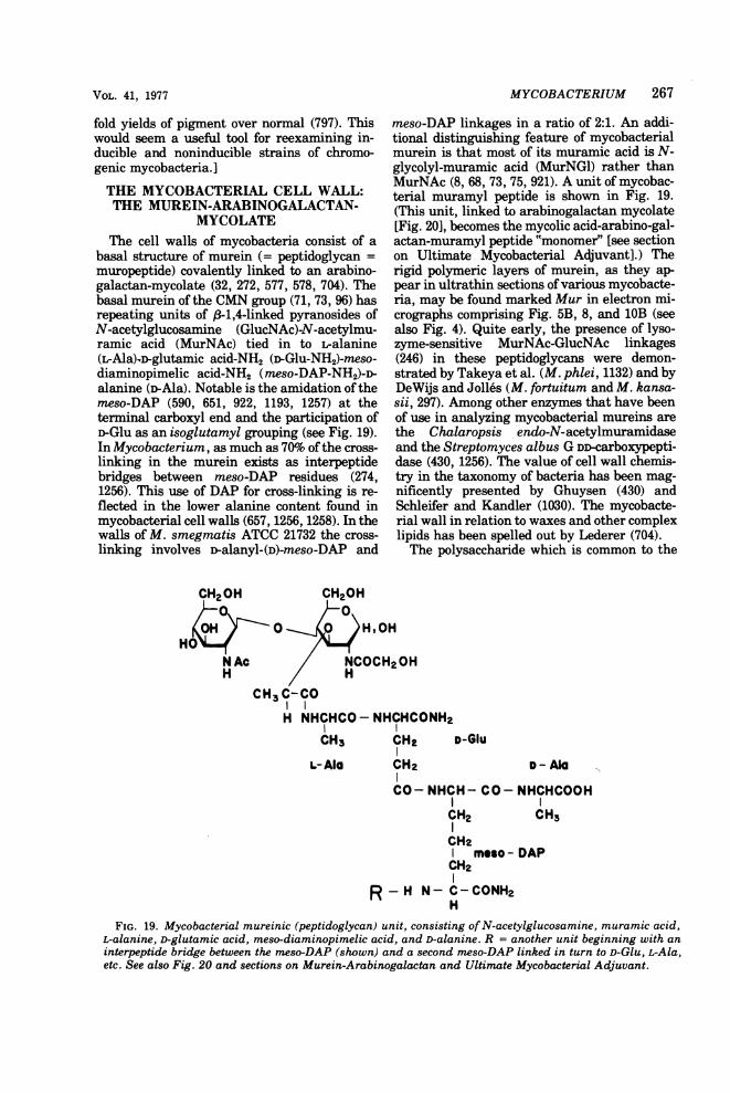
VOL. 41, 1977
fold yields of pigment over normal (797). Thiswould seem a useful tool for reexamining in-ducible and noninducible strains of chromo-genic mycobacteria.]
THE MYCOBACTERIAL CELL WALL:THE MUREIN-ARABINOGALACTAN-
MYCOLATEThe cell walls of mycobacteria consist of a
basal structure of murein (= peptidoglycan =muropeptide) covalently linked to an arabino-galactan-mycolate (32, 272, 577, 578, 704). Thebasal murein of the CMN group (71, 73, 96) hasrepeating units of (-1,4-linked pyranosides ofN-acetylglucosamine (GlucNAc)-N-acetylmu-ramic acid (MurNAc) tied in to Lralanine(L-Ala).D-glutamic acid-NH2 (D-Glu-NH2)-meso-diaminopimelic acid-NH2 (meso-DAP-NH2)-n-alanine ()-Ala). Notable is the amidation of themeso-DAP (590, 651, 922, 1193, 1257) at theterminal carboxyl end and the participation ofD-Glu as an isoglutamyl grouping (see Fig. 19).In Mycobacterium, as much as 70% ofthe cross-linking in the murein exists as interpeptidebridges between meso-DAP residues (274,1256). This use of DAP for cross-linking is re-flected in the lower alanine content found inmycobacterial cell walls (657, 1256, 1258). In thewalls of M. smegmatis ATCC 21732 the cross-linking involves D-alanyl-(D)-meso-DAP and
CH2OH/0\
HON AcH
MYCOBACTERIUM 267
meso-DAP linkages in a ratio of 2:1. An addi-tional distinguishing feature of mycobacterialmurein is that most of its muramic acid is N-glycolyl-muramic acid (MurNGl) rather thanMurNAc (8, 68, 73, 75, 921). A unit of mycobac-terial muramyl peptide is shown in Fig. 19.(This unit, linked to arabinogalactan mycolate[Fig. 20], becomes the mycolic acid-arabino-gal-actan-muramyl peptide "monomer" [see sectionon Ultimate Mycobacterial Adjuvant].) Therigid polymeric layers of murein, as they ap-pear in ultrathin sections ofvarious mycobacte-ria, may be found marked Mur in electron mi-crographs comprising Fig. 5B, 8, and 10B (seealso Fig. 4). Quite early, the presence of lyso-zyme-sensitive MurNAc-GlucNAc linkages(246) in these peptidoglycans were demon-strated by Takeya et al. (M. phlei, 1132) and byDeWijs and Jolles (M. fortuitum and M. kansa-sii, 297). Among other enzymes that have beenof use in analyzing mycobacterial mureins arethe Chalaropsis endo-N-acetylmuramidaseand the Streptomyces albus G DD-carboxypepti-dase (430, 1256). The value of cell wall chemis-try in the taxonomy of bacteria has been mag-nificently presented by Ghuysen (430) andSchleifer and Kandler (1030). The mycobacte-rial wall in relation to waxes and other complexlipids has been spelled out by Lederer (704).The polysaccharide which is common to the
CH20H
o
00 ; HOH
:I / NCOCH2OHH
CH3 C-CO
H NHCHCO- NHCHCONH2CH3 CH2 1
L-AIG CH?
D-GIu
D - Ala
CO- NHCH- CO- NHCHCOOH
CH2 CH3
CH2moso - DAP
CH2I
R-H N-C- CONH2
FIG. 19. Mycobacterial mureinic (peptidoglycan) unit, consisting of N-acetylglucosamine, muramic acid,L-alanine, D-glutamic acid, meso-diaminopimelic acid, and D-alanine. R = another unit beginning with aninterpeptide bridge between the meso-DAP (shown) and a second meso-DAP linked in turn to D-Glu, L-Ala,etc. See also Fig. 20 and sections on Murein-Arabinogalactan and Ultimate Mycobacterial Adjuvant.

*~~~~~~~~~~~~~t7 .5)-a -D-Araf( 1- _ 5) _a-D-Araf(l 1 4)-15-D-Gal ( 1 4)-P-D-Gal ( 1- 4)-P-ID-Gal (I 1-
3p p
3
a _-D-Araf2
[ a - D.-Araj]I
LR-CO-- a -D-Araf
n> 3
D-Ara, = D-Arabinofuranose
D-Gal = D-Galactopyranosep
R-CO = Mycolic acid residue
FIG. 20. Possible structure of the mycobacterial arabinogalactan unit, adapted from Misaki et al. (826).Kanetsuna (578) has suggested that the arabinogalactan mycolate may be tied into the muramyl peptide at the* (upper left), sometimes by phosphate bridges, sometimes via GlucNAc and MurNAc. Several mycobacterialarabinogalactan preparations examined by Misaki et al. interacted with antisera prepared against cell wallsof Corynebacterium diphtheriae and Nocardia asteroides. The close similarity of the arabinogalactans of theCMN group has been further examined by Azuma et al. (68). See section on Arabinogalactans in text. Forfurther information on biological activity of arabinogalactans, see Birnbaum and Affronti in section onSoluble Antigens and section on Delayed Hypersensitivity and Fig. 19. t Vilkas et al. (1204) have obtained adigalactoside from the arabinogalactan of cell walls and wax D of M. tuberculosis identified as 6-O-f3-D-galactofuranosyl-D-galactose. The presence of D-galactofuranosyl units in the arabinogalactan suggests thatfor those saccharides a 1 -* 4 linkage between arabinofuranose and galactofuranose does not obtain. Misaki etal. point out that such units "may originate from . . . secondary galactosidic linkages."
cell walls of the CMN group is an arabinogalac-tan. In Mycobacterium it consists of 5 arabinoseto 2 galactose units linked as 1 -* 5-D-arabino-furanose and 1 -* 4-i)-galactopyranose (32, 33,77, 826, 827) (for the presence of 6-O-,3-D-galac-tofuranosyl-D-galactose, see Fig. 20). This ara-binogalactan polymer links the murein of theinner wall to the mycolic acids of the outerenvelope (Fig. 19).
Arabinogalactan and Arabinogalactan-Mycolate
The a-branched, 83-hydroxylated long-chainlaa
fatty acids of the CMN group, R-CH--CH-
OH RICOOH, discovered by Anderson (40, 41) andelucidated by Asselineau (59), Asselineau andLederer (62), Lederer and Pudles (706), Pudlesand Lederer (948), and Etemadi et al. (357) areamong the hallmarks of the CMN group. Theyrange in carbon numbers from C28 to C40 (cory-noipycolic acids) to C40 to C60 (nocardic acids) toC60 to C. (mycolic acids). Although some of theshorter-chain mycolic acids, such as this C32 H64
OHI
03 corynomycolic acid, CH3-(CH2)14--CH-CH-COOH, are made by C. diphtheriae andIC 14H29C. ovis (298, 706, 949), those genera that syn-
thesize longer-chain mycolic acids are also ca-pable of producing some corynomycolic acids.The mycolic acids are found as mixtures ofhomologues. From species to species there arewide variations in these structures resultingfrom differences in the lengths of their chainsand the localization of groupings of ketone, hy-droxyl, cyclopropane, carboxyl, etc., especiallyon the main chain. The occurrence of thesesubstituents depends on the biosynthetic capa-bilities peculiar to different species of mycobac-teria. Et6madi has discussed the uniqueness ofcertain mycolic acids in relation to the taxon-omy of mycobacteria and to their phylogeny(352-354) (see Krassilnikov et al. regarding my-colic acids in chromogenic paraffin-oxidizingmycobacteria [663]). Azuma and Yamamuramade the valuable observation that mycolatesrecovered from the bound lipids of walls of M.tuberculosis Aoyama B were mycolates of ara-binose (74, 76). Since then, ample evidence hasaccumulated to confirm their suggestion thatsome mycolic acids of the envelope were linkedthrough their carboxyl groups to the 5-hydroxylof arabinofuranose units that terminatebranches of the arabinogalactan of the cell wall(32, 33, 75, 77, 825, 827).Continuing the investigations of Misaki et
al. (77, 827), Misaki et al. (826) have found therepeating units of the arabinogalactans of M.bovis BCG, M. tuberculosis H37Rv, H37Ra, andAoyama B, M. phlei, M. smegmatis, and Myco-bacterium sp. P1 to consist of ramified struc-
268 BARKSDALE AND KIM BACTERIOL. REV.

VOL. 41, 1977
tures of 11 to 16 sugar residues of a-(1 -- 5)-,major, and a-(1 -. 2)-, minor, n-arabinofurano-side linkages and /3-(1 -- 4)-r-galactopyrano-sidic [orfi3-(1 -- 5)-furanosidic] linkages. Theside chains terminate in arabinofuranose resi-dues and are attached to the main chains at C3of arabinose and perhaps at C6 of the galactoseresidues (Fig. 20) (see also [33] and [652]). Ara-binose mycolates have been further studied byAcharya et al. (4), using mass spectroscopy,and evidence has been obtained for specificstructures having carbon numbers rangingfrom about Cm to 091. These were derived fromarabinomycolates in the envelopes of strains ofBCG and M. kansasii. Kanetsuna and San Blas(578) have proposed a structure for the intactmurein(peptidoglycan)-arabinogalactan-myco-late from the walls of M. bovis BCG and M.smegmatis ATCC 14468, where mycolic acidsare linked at carboxyl ends to D-arabinofura-nose (5-OH) and through them to the arabino-galactan polymer. The arabinogalactan is thentied into the basement peptidoglycan througheither (i) GlucNAc, (ii) MurNGl, or (iii) a phos-phodiester bridge joining MurNGl. After thefinding of muramic acid-6-phosphate in thewalls of Mycobacterium butyricum by Liu andGotschlich (731), Kanetsuna demonstrated itspresence in the walls ofBCG (577); Cunto et al.reported it in the walls of M. smegmatis (274),and Kotani et al. gave evidence for its presencein the walls ofM. tuberculosis H37Rv (657). (Foran analysis of the chemical composition of thecell wall ofM. tuberculosis H37Ra, see Acharyaand Goldman [3].) Amar and Vilkas have re-cently reported the isolation of an alkali-stablearabinose phosphate from the walls of H37Ra(31). Thus, bases for the linkages shown in Fig.20 exist. (Monomycolates of arabinose havebeen found in fractions from cell walls of M.tuberculosis [74], M. bovis, and M. kansasii [4],and a dimycolate of arabinose [C147H21101J] wasfound in extracts of M. scrofulaceum[marianum]. One of the mycolic acids is mon-oethylenic and dicarboxylic; the other has onecarboxyl group and two cyclopropane rings.Through their carboxyl ends, they are esterlinked at the 3-hydroxyl of arabinose [195].)Arabinofuranosyl side chains of mycobacterialarabinogalactans are, apparently, primarily re-sponsible for the immunological cross-reactionsbetween mycobacterial polysaccharides, per se,and those of C. diphtheriae and N. asteroides(826).
Isolated and purified arabinogalactan reactswith the jack bean lectin, concanavalin A (ConA), and the interacting sites appear to be at theC2, C3, and C5 hydroxyls of the a-D-arabinofur-anosyl residues (438). A survey of the Con A
MYCOBACTERIUM 269
agglutinability -of filtrates from several speciesof mycobacteria indicated that strains of morethan one species released arabinogalactan(and, perhaps, some arabinomannan) into themedium, whereas other strains of those samespecies, as well as certain other species, re-leased no reactive material (279).
Arabinogalactans are of wide distribution innature. The arabinogalactan peptide from theendosperm of wheat is agglutinated by castorbean lectin, and this agglutination is stronglyinhibited by galactose and methyl B-D-galacto-side. In the wheat arabinogalactan peptide, thearabinogalactan units are tied via their galacto-syl ends into the peptide core through hydroxy-proline residues. Most likely, the linkages be-tween the arabinose and galactose are not ,8-1,4(374).
Teichoic AcidsUp to now there have been no reports of
polyol-phosphate chains, teichoic acids, in thewalls of members of the CMN group (oursearch, plus personal communication from J.Baddiley). In analyses of delipidated cell wallsof corynebacteria, mycobacteria, and nocardiascarried out in this laboratory by S.B. Ardenand B.L. Beaman, plus additional studies byB.L. Beaman elsewhere (personal communica-tion), total cell wall (delipidated) phosphorus ofmore than 0.1% was not found. This augursagainst the presence of teichoic acids in thesebacteria.
Early Studies on Fragments of theMycobacterial Cell Wall
Ether-soluble, acetone-insoluble, chloroform-extractable peptidoglycolipid components de-rived from old cultures of M. tuberculosis wereearly designated as "waxes" (waxes D: Ander-son [40, 41], Asselineau [59], Jolles et al. [555,556], Aebi et al. [12]).
Asselineau (59, p. 241) has said, "Waxes D ofhuman strains are essentially composed of pep-tidoglycolipids, i.e. esters of mycolic acids witha polysaccharide containing D-galactose, D-ar-abinose, and D-mannose and glucosaminelinked through galactosamine residues to pep-tides formed of alanine, glutamic acid, and a--diaminopimelic acid; muramic acid has alsobeen detected," and he cites the detectors,Stewart-Tull and White (1099). This finding ofcomponents common to wax D and the rigidlayer of the mycobacterial cell wall (Fig. 19)suggested that wax D either was a part of thewall or was material synthesized in excess ofthat needed for insertion into the wall. Kotaniet al. (651) showed that wax D could be got out

270 BARKSDALE AND KIM
of intact cell walls ("bound" wax D) with suita-ble enzymes. Kanetsuna has discussed the mu-ral location of wax D in relation to variousprocedures used for its extraction (577). Inter-ference with the synthesis of cell walls in M.tuberculosis H37Rv by cycloserine causes theaccumulation in the medium of arabinogalac-tan-galactosamine-DAP-mycolate (289). If oneuses "waxes D" as a term to designate parts ofthe backbone of the mycobacterial cell wall(Fig. 19), then wax D-active or -cross-reactingmaterial could be generated by disordered cellwall synthesis (either genetically or antibi-otically induced) singly by specific or in con-cert by several autolytic enzymes. The accumu-lation of excess cell wall material duringgrowth, the induction of unfinished cell wallproducts (precursors) by antibiotics, and therelease of cell wall components by enzymaticand acid hydrolysis are well-known events fora number of bacteria (137).The mycobacterial cell wall is an elaborate
structure. A section of it will contain materialfrom each of the three layers shown graphicallyin Fig. 4 and actually in Fig. 5: mucopeptide-arabinogalactan-mycolate and componentswhich make up L, and L2 of Fig. 4. Dependingon the CMN bacterial strains being considered,other polysaccharides will also be found in"waxes": e.g., glucans, mannans, and arabino-mannans are found in a range of CMN speciesincluding M. bovis, M. smegmatis, and M. phlei,as well as in C. diphtheriae and N. asteroides(67). Lederer has suggested that bound waxD, liberated by enzymatic hydrolysis, be con-sidered a "monomer" of the cell wall (704). Cer-tainly, as Kanetsuna's work suggested, "waxD" is an early designation for a fragment of themycobacterial wall that could be extracted in aparticular way. Undoubtedly, autolysis occurr-ing in old cultures contributed to its extracta-bility. For recent coverage of other informationconcerning wax D, see the excellent review byGoren (453).The adjuvant activity of mycobacterial
"waxes" has been much investigated (see sec-tion, Adjuvant Action). Stewart-Tull and White(1100) have made the valuable observation thatthe time of harvesting of mycobacteria mark-edly affects the yields of adjuvant-active pepti-doglycolipids obtainable from them. They usedthe following protocol: washed cells were ex-tracted with alcohol-ether (1:1) for 30 days. Or-ganisms were then collected on a filter andreextracted with chloroform. Pooled chloroformextracts were concentrated by distillation, andthe crude wax obtained was extracted with hotmethanol and boiling acetone, repeatedly, untilno more material dissolved. The residue was
called purified wax D. The aim of the study wasto examine the importance of the age of theculture upon the production of waxes D by M.phlei, M. balnei, M. fortuitum, and M. smeg-matis, with control studies of M. avium, M.kansasii, M. tuberculosis, and M. tuberculosisBCG. The more rapidly growing organisms(e.g., M. phlei and M. smegmatis), when har-vested at 7 days, yielded markedly more pepti-doglycolipid than at 28 days. Although the pep-tidoglycolipid from young cultures possessedadjuvant activity, the predominantly glycolipidextract from old cultures did not. The authorspoint out that the hydrosoluble moiety of thepeptidoglycolipid (missing from the glycolipidextracted from old cultures) contains thoseamino acids, hexosamines, and sugars found inthe murein of the mycobacterial cell wall andthey suggest that, in the absence of nutrient inthe culture medium, mycobacterial growth ismaintained by autonutrition (45) from pepti-doglycolipid. Their results make it clear thatall naturally occurring mycobacteria, whetherthey are called "saprophytic" or "parasitic,"produce adjuvant-active peptidoglycolipids.
Beyond the Arabinogalactan-Mycolate,"Nonpeptidoglycan" Amino Acids, and
Surface Peptidoglycolipid
Sohler et al. (1074) noted an absence of pro-line- and sulfur-containing amino acids fromcell walls ofN. rubra 3639, in which they foundmore than trace amounts of arginine, asparticacid, glycine, lysine, serine, threonine, valine,phenylalanine, leucine, and isoleucine. Snyderet al. (1073) confirmed the presence of minoramounts of several amino acids in thoroughlywashed and unextracted cell walls of nocardias.Cummins (271, 273) and Yamaguchi (1290) ob-served that alkaline ethanol removed these"background amino acids." Some investigatorshad suggested that the amino acids found insmall quantities in cell wall hydrolysates werethe result of contamination with cytoplasmiccomponents. Beaman, by carrying out compar-ative, quantitative analyses of (i) cell wallsdigested with trypsin and pepsin, (ii) cell wallsdigested with trypsin and pepsin and extractedwith alkaline ethanol, and (iii) whole cells ex-tracted with alkaline ethanol, prior to render-ing them into cell wall preparations and treat-ment as in (i), has demonstrated rather conclu-sively that these nonpeptidoglycan amino acidsare associated with the lipoidal component ofthe surface of the nocardial cell (113). (It isinteresting in this respect that Misaki et al.[828], using entirely different methods, foundaspartic acid, valine, leucine, isoleucine, and
BACTERIOL. REV.

MYCOBACTERIUM 271
serine associated with the murein of BCG priorto treatment with Pronase.) Beaman has sinceextended his findings to Corynebacterium andMycobacterium and related the amino acids (aspeptides) and lipids (peptidolipids) to patternsfound on the surfaces of members of the CMNgroup (112). A catalog of electron micrographsof these patterns of the L2 outer envelopes ofmycobacteria, with, for reference, two coryne-bacterial and two nocardial species, comprisesFig. 22 to 26.
Wietzerbin-Falszpan et al. (1258) coined theterm "nonpeptidoglycan" amino acids and haveattributed to them 15% of the weight of crudedelipidated BCG cell walls. About half of theseamino acids could be removed with hot dodecylsulfate, suggesting that they were from lipopro-teins or glycolipoproteins noncovalently linkedto the arabinogalactan-mycolate-peptidogly-can. The fraction of material not removed byhot dodecyl sulfate treatment may representmaterial that is covalently linked to one of thebasal structures. The nonpeptidoglycan aminoacids, then, to a great extent go to make uppeptidolipids and peptidoglycolipids of theouter envelope, which will be considered cate-gorically in the following sections.As shown in Fig. 4, the outer lipoidal mate-
rial of the mycobacterial cell wall can be re-solved into two layers (L, and L2-L3). Figures 5and 21 show the outermost lipoidal coating ofMycobacterium sp. NQ and M. lepraemurium,respectively. This material has been called acapsule of mycoside C by Draper and Rees (315).In Figures 22 through 26 are shown the L2-L3patterns of reference strains of the CMN group.
Ropelike Patterns: the Surface or SubsurfaceGlycolipids, Peptidoglycolipids, and
PeptidolipidsThe nonpeptidoglycan amino acids just dis-
cussed are associated with the lipoidal surfaceof mycobacteria. Exactly how, and as parts ofwhat kinds of molecular units, is far from com-pletely known. The ropelike structures respon-sible for the markings shown on the variousnegatively stained mycobacteria in Fig. 22 to 24have been said to be of glycolipid or peptidogly-colipid composition (113, 445, 530, 1018, 1131,1132, 1134, 1135).
Visualization of the subsurface of the glyco-lipids of members of the CMN group can beaccomplished by the technique of negativestaining (see also 313). This material assumes aropelike appearance in mycobacteria, as isshown in Fig. 22 to 24. Sometimes, demonstrat-
FIG. 21. Freshly grown cells of M. lepraemurium were prepared for electron microscopy after being oncewashed with phosphate buffer (pH 7.2). Note two interconnected organisms completely surrounded bynonstaining substances (arrows). Note also sonicated cells for the comparison (Fig. 22A). (x28,000; bar = 1Am.) See section on Mycobacteria Growing In Vivo and In Vitro. See Tables 1, 2, and 3.
VOL. 41, 1977
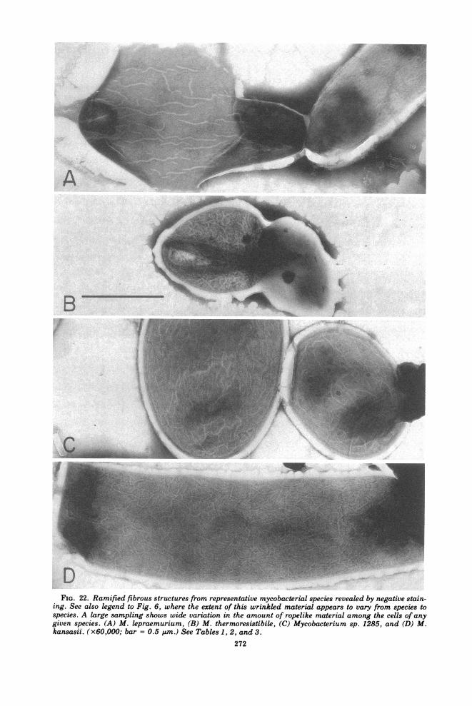
FIG. 22. Ramified fibrous structures from representative mycobacterial species revealed by negative stain-ing. See also legend to Fig. 6, where the extent of this wrinkled material appears to vary from species tospecies. A large sampling shows wide variation in the amount of ropelike material among the cells of anygiven species. (A) M. lepraemurium, (B) M. thermoresistibile, (C) Mycobacterium sp. 1285, and (D) M.kansasii. (x60,OOO; bar = 0.5 Am.) See Tables 1, 2, and 3.
272

FIG. 23. (E) M. tuberculosis H37Rv 102 (x40,000), (F) M. smegmatis 607 (x78,000), (G) Mycobacteriumsp. I (x86,000), (H) M. ulcerans (x88,000). (All bars = 0.5 ptm.) See also Tables 1, 2, and 3.
273

K.-
FIG. 24. (I) Mycobacterium sp. NQ (x21,000), (J) M. phlei (x39,000), (K) Mycobacterium sp. 1582(x39,000), (L) M. intracellulare (x34,000). (All bars = 0.5 Szm.) See also Tables 1, 2, and 3.
274
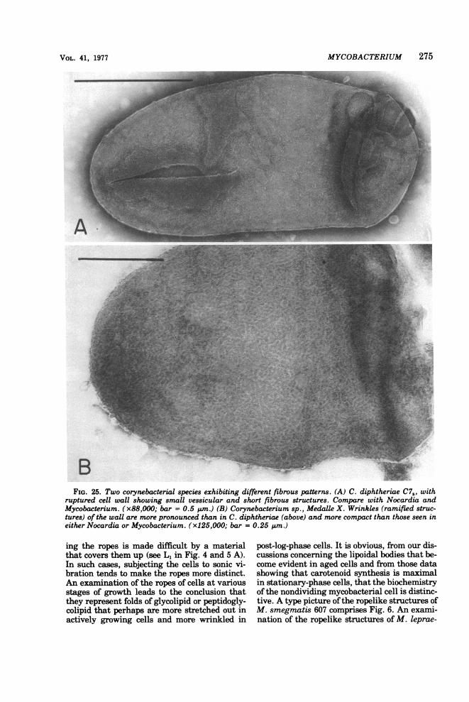
MYCOBACTERIUM 275
FIG. 25. Two corynebacterial species exhibiting different fibrous patterns. (A) C. diphtheriae C7S, withruptured cell wall showing small vessicular and short fibrous structures. Compare with Nocardia andMycobacterium. (x88,000; bar = 0.5 pum.) (B) Corynebacterium sp., Medalle X. Wrinkles (ramified struc-tures) of the wall are more pronounced than in C. diphtheriae (above) and more compact than those seen ineither Nocardia or Mycobacterium. (x125,000; bar = 0.25 pm.)
ing the ropes is made difficult by a materialthat covers them up (see L1 in Fig. 4 and 5 A).In such cases, subjecting the cells to sonic vi-bration tends to make the ropes more distinct.An examination of the ropes of cells at variousstages of growth leads to the conclusion thatthey represent folds of glycolipid or peptidogly-colipid that perhaps are more stretched out inactively growing cells and more wrinkled in
post-log-phase cells. It is obvious, from our dis-cussions concerning the lipoidal bodies that be-come evident in aged cells and from those datashowing that carotenoid synthesis is maximalin stationary-phase cells, that the biochemistryof the nondividing mycobacterial cell is distinc-tive. A type picture of the ropelike structures ofM. smegmatis 607 comprises Fig. 6. An exami-nation of the ropelike structures of M. leprae-
VOL. 41, 1977

276 BARKSDALE AND KIM
FIG. 26. Negatively stained cells oftwo strains ofNocardia asteroides showing the organization ofsurfacefibrous materials. The ramified fibrous configurations are smaller (2 to 8 nm in diameter) than those ofmycobacteria and offer a comparatively irregular pattern. (A) N. asteroides 92. Note irregular mass ofmaterial from the outer wall and smaller fibrous structures; also, metachromatic granule at the point ofbranching. (x150,000; bar = 0.2 ,um.) (B) N. asteroides 133. Compare this fine ramified pattern with those ofmycobacteria. (x150,000; bar = 0.2 gm.) (C) N. asteroides 133. Fibrous pattern is obscured by sheathlike(peptidoglycolipid) material that lies external to it. To a lesser degree, this obscuring is evident in Fig. 26B.See text under peptidoglycolipid for a discussion of outer sheathlike materials. (x150,000; bar = 0.2 gm.)
BACTERIOL. REV.

VOL. 41, 1977
murium (Fig. 22A), M. thermoresistibile (Fig.22B), Mycobacterium sp. 1285 (Fig. 22C), M.kansasii (Fig. 22D), M. tuberculosis H37Rv(Fig. 23E), M. smegmatis 607 (Fig. 23F), Myco-bacterium sp. ICRC (Fig. 23G), M. ulcerans(Fig. 23H), Mycobacterium sp. NQ (Fig. 24I),M. phlei (Fig. 24J), Mycobacterium sp. 1582(Fig. 24K), and M. intracellulare (Fig. 24L)indicates the range of patterns and dimensionsfound in these foldings. Similar surface struc-tures of C. diphtheriae C7, Corynebacteriumsp., Medalle X, and N. asteroides 92 and 133 areto be found in Fig. 25 and 26, respectively.Specific sequences comprising the peptidolipidsand peptidoglycolipids of mycobacteria are dis-cussed in the following section.
Glycolipids, Peptidoglycolipids (Mycosides),and Peptidolipids of the Outer EnvelopeOver the past two decades there has been
recovered from mycobacteria a series of glyco-lipids and peptidoglycolipids; in many cases,these have in common terminal saccharidemoieties containing rhamnoses that are 0-methylated in various positions. These com-pounds are named mycosides, and they werereported to be species specific (1066, 1067, 1068).Lederer (702) has thoroughly reviewed the lit-erature of mycosides up to 1967 (see also Goren[453]), pointing out that they fall into two gen-eral categories.Category I. Here are included the mycosides
that are lipidic aglycones, consisting of glyco-sides of p-phenols having a branched glycolicchain whose hydroxyls are esterified withstraight- and branched-chain fatty acids. Gas-tambide-Odier and Sarda (423), employing spe-cial methods of oxidation, have established thestructures of the ethers of the phenol-glycols ofmycoside A (from M. kansasii) and mycoside B(from M. bovis) through a combination of paperand gas chromatography and mass spectrogra-phy. Their interesting paper contains suppor-tive data for concluding that these two myco-sides have the following common backbone:
MYCOBACTERIUM 277
coside [424].) Mycoside B from M. bovis is sim-pler: R = 2-O-methylrhamnose and n = 14, 15,16, 17, 18. (Mycosides G and G' from, for exam-ple, M. marinum also fall into this category[420, 1207].) Earlier, it was thought that thephenol of these lipid aglycones originated fromtyrosine (702). The authors discuss the origin ofthe phenolic group mostly in relation to incor-poration oflabel from sodium [3-14C]propionate,and they suggest that the phenol originates viashikimate. Goren et al. (458) have examinedcertain lipids of a group of strains of M. tuber-culosis from East Africa, Burma, and the re-gion of South India around Madras. This samegroup of bacteria had been studied by Mitchi-son and his associates for their relative viru-lence (831) and for their general differencesfrom other strains ofM. tuberculosis, includingtheir sensitivity to H202, their catalase activ-ity, and their isoniazide sensitivity (832, 833).In extracts of a group of these M. tuberculosisstrains, which Mitchison had found to be oflowered virulence for the guinea pig, Gorenand his associates were able to identify a pheno-lic phthioceryl diester indistinguishable fromthe phenolic O-methyl ether of the aglyconemoiety of mycosides A and B. This methyl
ether, CH30 / occurs naturally in
the strains showing attenuated virulence forthe guinea pig. In these mutants, the sugars (2-0-methylfucose, 2-O-methylrhamnose, 2,4-di-O-methylrhamnose, in the case of mycoside A,and 2-O-methylrhamnose in the case of myco-side B) have been substituted by an 0-methylether.Category II. The second group of mycosides
is made up of peptidoglycolipids consisting of asugar moiety, a short peptide, and FA, asshown in Fig. 27. Mycoside C1, isolated from M.avium and characterized by Joll6s et al., was ofthis type (554). Structurally distinct mycosides
OCH3RO / (CH2)n-CH-CH2-CH-(CH2)4-CH-CH-CH2-CH3-RI
OR' OR' CH
where R' = the acyl substituents, palmitic andmycocerosic acids. In the case ofthe mycoside Aof M. kansasii, R = a trisaccharide of 2-0-methylfucose, 2-O-methylrhamnose, and 2,4-di-O-methylrhamnose; n = 16, 17, 18, 19, 20. (Sub-sequently, 3-O-methylrhamnose and 3-0-meth-ylfucose have also been reported from this my-
of category II have been described for Mycobac-terium sp. 1217 (688), Mycobacterium farcino-genes (Nocardia farcinica) (689), and other my-cobacterial species (59, 453). The mycoside C2,synthesized by M. avium, has been shown byVoiland et al. (1209) to be linked on a pentapep-tide of n-phenylalanine-D-allothreonine-D-Ala-

278 BARKSDALE AND KIM
H3C- HC- CH- CO-I I
HO NHCO C6H5I I
CHi- (CHp),;~CO- NH - CH-CHp
CH3CH-CO- NHI I
-CO-NH CH-CH3
H3C-
C Hs- CCH3 CO-O
R a H or CH3Mycoside C2
FIG. 27. Structure proposed by Voiland et al. for mycoside C2 from Mycobacterium avium (1209), unusualin that the pentapeptide portion of a molecule of this. class is the aminoalcohol L-alaninol to which is linked3,4-di-O-methyl-L-rhamnose. The sugar terminal to allothreonine is 6-deoxytalose or its 3-0-methyl deriva-tive. The lipid moiety of this mycoside consists ofa complex mixture of fatty acids containing palmitic acid.Additional described mycosides of category II fall into two subclasses. (A) The peptidoglycolipids of thissubclass are comprised ofD-phenylalanine-D-allothreonine-D-alanine-L-alaninol conjugated to one or more ofthe following sugar moieties as indicated: C' = di- or mono-0-methyl rhamnose in M. farcinogenes (689); Cb1= di-O-acetyl-6-deoxytalose and tri-0-methyl rhamnose in M. smegmatis (butyricum) (1206); CQ = di-0-acetyl-6-deoxytalose and 3,4-di-0-methyl rhamnose in M. scrofulaceum (1205); C1217 = di-0-acetyl-6-deoxyta-lose, tri-0-methyl rhamnose, and 3,4-di-0-methyl rhamnose, trace amounts, in Mycobacterium sp. 1217(688). (B) The peptidoglycolipids of this subclass are comprised of D-phenylalanine-[D-allothreonine-D-alanine]2 conjugated to the following sugar moieties: C1 = di-O-acetyl-6-deoxytalose, 3-0-methyl-6-deoxyta-lose, and 3,4-di-0-methyl rhamnose in M. avium 802 (554); and of D-phenylalanine-[D-allothreonine-D-alanine]3 conjugated to the following sugar moieties: Cm = di-O-acetyl-6-deoxytalose and 3,4-di-0-methylrhamnose in M. scrofulaceum (marianum) (237). Note: It is unfortunate that reference materials for most ofthese compounds are no longer available (personal communications from E. Lederer, G. Michel, M. Gastam-bide-Odier, and others). See sections on Myeosides, on Tuberculins and Other Mycobacterial Elicitins, andon Mycobacteriophage Receptors. See also Tables 1, 2, and 3.
D-allothreonine-D-Ala-L-alaninol. To the ami-noalcohol, alaninol, is linked 3,4-di-0-methyl-L-rhamnose. To the second molecule of threo-nine is linked 6-deoxy-L-talose or 3-0-methyl-deoxy-L-talose. The complex mixture of FAs,including palmitic acid, are linked to phenyl-alanine. This proposed structure is shown inFig. 27. In this paper we are concerned with thefunction and cellular location of various speciesof macromolecules derived from mycobacteria.It is assumed that the peptidoglycolipids occupya superficial position in the mycobacterial cell.In the electron micrographs of Mycobacteriumsp. NQ in Fig. 5, surface structures are clearlyshown.Fourteen years ago, Fregnan and Smith (395)
and Randall (396) attempted to relate the pres-ence and absence of mycosides to colonial mu-tants (cells exhibiting different surface proper-ties from that of the wild or ancestral type andtherefore piling up to form identifiably differ-ent colonies). They attributed "type specificity"to the mycosides. Recently, support for the sur-face location of mycoside C has come from stud-ies by Furuchi and Tokunaga (412) using bacte-riophage D4 and the mycoside C of the D4-
sensitive M. smegmatis, termed mycoside CsmThis component in isolated form blocked theadsorption of phage to the bacterium by whichit was synthesized. M. smegmatis D4, a mutantresistant to D4, failed to adsorb D4 and failed tosynthesize mycoside Csm. Goren and associateshave independently reached similar conclu-sions concerning D4 and mycosides C,217 andC8crofulaceum and have published beautiful pic-tures of D4 phages fixed onto filamentousneedles of C mycosides derived from M. smeg-matis and Mycobacterium sp. 1217 (459). Thesestudies leave no doubt that the receptors for D4are in the mycoside and, therefore, indicatethat in D4-adsorbing strains of Mycobacteriumthe mycoside is at the surface. Bar mutantsthat are resistant to D4 may be oftwo kinds: (i)those that no longer synthesize receptors for D4and (ii) those that elaborate, at their surfaces,substances that cover up the phage receptors(10). Some years ago, Lan6elle and Asselineau,in discussing the possible function of mycosideC0217, with its polar portions and its nonpolarportions, suggested that mycosides might belocated at the periphery of the mycobacterialcell (688; see also [315]). It now seems that
BACTERIOL. REV.

VOL. 41, 1977
mycoside C,217 is located peripherally but itsfunction remains to be discovered. Serving as aphage receptor is not fulfilling a microbial func-tion. Very likely, these mycobacterial lipid-sac-charide-peptide moieties serve for inwardtransport in a wet environment and, in con-junction with mycolates, sulfolipids, etc., serveto prevent loss of water from the cell duringperiods of excessive drying. The advantage theygive to some cells in invading host animals maybe similar to that provided more virulentstrains of Salmonella typhimurium by specificterminal sugars on their 0-antigenic sidechains. For example, S. typhimurium serotype1, 4, 12, which has the 0 antigen lipopolysac-charide side chains terminate in the dideoxy-hexose, tyvelose, is more virulent for mice thanS. typhimurium 1, 9, 12, which has, instead oftyvelose, the dideoxyhexose, abequose, as theterminal sugar (765).Category III. Peptidolipids. If the surface
antigens of mycobacteria generally consist ofpeptidolipids with saccharide moieties or othersubstituents, one would expect to find strains(mutants) lacking one or more of the capacitiesneeded to introduce the sugar moieties intothe peptidoglycolipids. Species of Mycobacte-rium are known which produce only peptido-lipids (687a, 689a). In the case of mycosideC, for example, Laneelle has the impressionthat bacteria which produce this mycoside donot produce detectable lipopeptides, and myco-bacteria which produce lipopeptides do notseem to yield mycosides (personal communica-,tion). An investigation of the genetics of myco-side synthesis may well furnish us with infor-mation on the biosynthesis ofthese compounds.They would seem in many ways to be analogousto the lipopolysaccharides of E. coli where theelucidation of the regions I (lipopolysaccha-ride), II (core), and III (lipid A) stemmed fromconcerted biochemical and genetic efforts (97).Other Molecular Species Associated with the
Mycobacterial Cell Surface:Phosphatidylinositol Oligomannosides
We have suggested that dikes of phosphati-dylinositol oligomannoside may extend fromunder the peptidoglycan layer to the surface ofthe cell. Evidence for or against this notion,which originated with Kotani (653), could beobtained by haptenic inhibition and other sero-logical studies.
MYCOBACTERIA GROWING IN VIVO(Phe I) AND IN VITRO (Phe II)
The adaptive processes of microbes wouldlead one to expect in vivo-grown organisms todiffer to some degree from those grown in vitro.
MYCOBACTERIUM 279
The matter of differences existing between invivo- versus in vitro-grown mycobacteria hasbeen under consideration for some time (seeTable 1 in reference 973). The pinpointing ofdifferences between populations of H37Rvgrown in vivo (in the lungs of mice) and popula-tions grown in vitro was begun by Segal andBloch (1040), who demonstrated that the in vivophenotype (Phe I) and the in vitro phenotype(Phe II) of H37Rv (shown to be qualitativelycomparable as to dry weight and viable count)differed significantly in their capacities to re-duce 2,3,5-triphenyltetrazolium chloride in aTween-albumin medium with 0.5% glucose andin the extent of the stimulation of their meanQo2 values over endogenous rates by glu-cose, glycerol, lactate, acetate, pyruvate, ben-zoate, benzaldehyde, salicylate, n-heptanoate,octanoate, and oleate. The endogenous rates ofrespiration for Phe I and Phe II were similar.Phe II was 2.7 times as active as Phe I inhydrogen transfer and showed a marked in-crease in Qon with 11 of 11 added energysources, whereas Phe I was stimulated by only4 of 11. These four (see above) were salicylateand the three FAs. Both phenotypes were stim-ulated by extracts from infected mouse lungsbut not by extracts from normal lungs. Cul-tures (in vitro) of H37Rv derived from Phe Ibehaved in all respects like those of H37Rv (PheII). Thus, there are marked differences in thetwo phenotypes and the differences must in-volve barring of some of the energy moleculesfrom Phe I.In a subsequent paper (1041), Segal and
Bloch recounted a visible difference betweensuspensions of phenotypes I and II. The mouse-grown cells showed far less tendency to formlarge clumps, yet they were less able to passthrough filter paper (Whatman no. 12, fluted)than in vitro-grown cells. Phe II, as a phenol-killed vaccine, was a better immunogen thanPhe I, but Phe I proved to be more virulent formice than Phe II. Segal later extended thesestudies of the immunogenicity of the two phe-notypes by examining (i) protection againstinfection (in CF1, 6-week-old, female mice), (ii)capacity to elicit DH (in randomly bred, albino,tuberculous guinea pigs), and (iii) capacity toinduce DH (in white New Zealand rabbits).Whereas phenol-killed vaccines had previouslybeen used, heat-killed cells (with and withoutIFA) and sonically disrupted cells were em-ployed as three separate means of immuniza-tion. In the mouse protection experiments, aswell as in the elicitation and induction of DH,Phe II was a better immunogen than Phe I.Segal has stated that various preparations ofPhe I appear to exhibit the same biological

280 BARKSDALE AND KIM
properties whether derived from one passage or25 serial passages through mice (1038). Sincethe shift Phe I - Phe II is readily reversible, itis obvious that the genome of H37Rv (the wolf)remains the same, though properties related totransport are temporarily modified (sheep'sclothing). The fact that Phe I lacks the neutralred-binding capacity (528, 817) of Phe II cer-tainly suggests a real modification in the sur-face of the former (1039). Further, Kanai hasfound that Phe I is resistant to 4% NaOH at370C for 4 h, whereas the viability of Phe II islowered 2 logs by such treatment (571, 572, 573).The most reasonable working hypothesis isthat the Phe I bacilli are coated so that thesurface features associated with Phe II are una-vailable to react with neutral red. Segal andMiller obtained evidence for a quantitative dif-ference between the lipids of Phe I and Phe II(1042). Kondo and Kanai have demonstratedthat Phe, I and Phe.ll of th Ravew, l train ofM. bovis behave with regar to DH and mouseprotection much as do their homologues ofH37Rv (634, 637). They further provided evi-dence that Phe I contains cord factor, a surfacecomponent of Phe II. Kondo et al. (638) showedthat 10-methylstearic acid (tuberculostearicacid) was present in Phe I ofM. bovis. Kanai etal. (575) demonstrated the presence of phthi-ocerol dimycocerosate and mycolic acids in PheI of M. bovis Ravenel. The surface component,mycoside B (see section on Peptidolipids), wasnot found in extracts of Phe I bacilli of theRavenel strain.The presence of Phe I of H37Rv within J-111
cells (derived from leukocytes of a case of hu-man leukemia) did not prevent cell division orcause vacuolation as long as the numbers ofbacilli were small (2 to 10/cell). When the num-bers of bacilli per cell were large, excessivecording was evident. If cord factor were a majorcontributor to death of the cells, then that fromintracellular bacilli must have been responsiblefor death. Experiments were carried out withadded cord factor and with numerous bacillicording extracellularly. Under these condi-tions, the J-111 cells remained healthy. Thisfinding led the authors to conclude that extra-cellular cord factor was tolerated by the cellsunder study (190).
Rabbits infected with M. bovis, M. avium,and Pasteurella pseudotuberculosis show ele-vated levels of serum cholesterol, phospholipid,and triglyceride (1151a). Experiments using [2-14C]mevalonic acid suggest that mechanisms forthe disposal of serum cholesterol are impairedin infected (M. bovis) rabbits (1151b). Amongthe host's lipids that are closely associated withharvested bacilli of Phe I are esters of choles-
terol (636, 638). Unsaturated octadecanoic acidsaccount for most of the esterification (637).Kondo and Kanai carried out a series of lipidanalyses on casein-induced (and uninduced)harvested macrophages and demonstrated thatthe induced macrophages contained a higherproportion of phospholipid and cholesterol esterthough a lower level of total lipid than theuninduced cells (635). Thus, there is probablyavailable to the macrophage-engulfed mycobac-terium a large amount of cholesterol. Bacteriain general, when in the presence of cholesterol,take it up (e.g., the uptake of [4-14C]cholesterolby washed Streptobacillus moniliformis was17,000 cpm/mg of cell protein, whereas its L-form took up 19,820 cpm/mg, M. phlei took up125 cpm/mg, and C. diphtheriae took up 1,725cpm/mg [961]). Is the difference between Phe Iand Phe II a layer acquired in the host? Is thelayer primarily lipoidal? Does the layer containsome protein, some antibody? The report thatagklutinins active against M. tuberculosis havea ubiquitous distribution among human beings(93) would seem to make it inevitable that Phe Itubercle bacilli would attach some such anti-body to their surfaces.Brown et al. (191) have pointed out that obli-
gately parasitic M. lepraemurium is sur-rounded by an electron-transparent zone (ETZ),which separates the bacterium from the cyto-plasmic components of rat fibroblasts in whichthey grow. They have suggested that this zoneoffers protection from the host (see also [314]).They equate these areas with those seen insections of granulomas induced in hamsterswith heat-killed H37Rv (325) and, presumably,with those surrounding living intraphagosomalM. microti (495). There is no doubt that, for alarge number of bacteria, there exists an outertransparent zone (OTZ of reference 394). TheOTZs found in most figures in this review areprobably equivalent to L, (and L2, to a limitedextent) shown in Fig. 4 and 5. In sections ofmycobacteria within animal cells, however, theexaggerated OTZ of Brown et al. seems to be aresult of fixation and embedding effects uponone biological entity within another. This israther obvious in the hamster pictures (325) ofintracellular H37Rv. Thus, one probably cannotrender visible the differences between Phe Iand Phe II. The data accumulated by Kanai(571, 572) and Kanai and Kondo (574) do enableus to attribute some of the differences betweenPhe I and Phe II to the result of an intimateassociation of Phe I with macromolecules of thehost. Kanai (571, 572) showed that in vivo-grown H37Rv and BCG had associated withthem murine acid phosphatase (MAP). Whenliving in vitro-grown bacilli were exposed to
BACTERIOL. REV.

VOL. 41, 1977
MAP, they bound it, and its presence seemed toblock the activity of mycobacterial acid phos-phatase. The accumulation ofenough host mac-romolecules about the surface of Phe I shouldrender it somewhat indistinguishable from thehost's selfMost Phe I appear to have an intact cell wall.
This is interesting in light of the fact thatWillet and Thacore found that either acid phos-phatase or phospholipase C could render H37Rasusceptible to lysozyme, with subsequent proto-plast formation (1263) and, with high enoughnumbers of bacteria, even L-form development(1262). The addition oflysozyme to cell culturesof J-111 human monocytes has made possiblethe formation of protoplasts in these host cells(1147). Perhaps host agents act to destroy thesewall-less types of mycobacteria? For an inter-estingly documented discussion ofthe plasticityof in vivo M. tuberculosis, see N. A. Schmelevon "Polymorphism of Mycobacteria" (1031). Seealso the section Autolysis, Protoplasts, and My-cobacterial L-Forms. Salient features of Phe Iand Phe II are shown in Table 5. Examples ofaging Phe I comprise Fig. 12.
GRANULOMASThe Tubercle and GranulomatagenesisPresumably, one of the goals of the last 100
years of mycobacterial research has been tounderstand the pathogenesis of the chronicgranulomatous disease, tuberculosis. Thechronicity ofthe granulomatous diseases seemsdirectly related to the capacity of the infectingmicroorganisms (protozoa, fungi, bacteria, andothers) to survive in the macrophages of the
TABLz 5. A few ofthe known differences between invivo and in vitro phenotypes of Mycobacterium
tuberculosis H37RvaPhe I = in vivob
Qo, elevated by 4/licStick together when har-
vested5x more virulent for micePoor immunogenDoes not bind netural redBound cholesterol and anti-body
Murine MAPd
Phe II = in vitro
Qo, elevated by 11/11Not
so sticky5x less virulent for miceGood immunogenBinds neutral redNo bound cholesterol;no bound antibody
None: but will bind
a Probably any mycobacterium causing a chronic infec-tion in any given animal must be able to exist as an in vivophenotype in the macrophages of that animal (see alsoCollins [255]).
b See text. Note that the Phe I of M. bovis Ravenel hasbeen shown by Kondo and Kanai to produce tuberculopro-tein in the lungs of mice. This product moves like PPD indisc gel electrophoresis and has good tuberculin activity inskin tests (634).
c For substrates used, see text.d MAP, Murine acid phosphatase.
MYCOBACTERIUM 281
host. Robbins (985, p. 335) has given the follow-ing textbook description of a tuberculous gran-uloma: "the center of the microscopic granu-loma is occupied by a small nest or aggregate ofplump, rounded mononuclear cells that va-guely resemble epithelial cells and are there-fore designated as epithelioid cells.... The ep-ithelioid cells are thought to be mononuclearphagocytes that have ingested intact or frag-mented tubercle bacilli. In the margin of thiscluster of epithelioid cells and sometimeswithin the center ofthe cluster, there are large,multinucleate giant cells.... About this clus-ter of epithelioid cells and accompanying giantcells, there is a peripheral zone or collar ofplump fibroblasts and lymphocytes." This is thehard tubercle. Subsequent necrosis of the cen-tral zone, caseation necrosis, provides what thepathologist regards as the most characteristicfeature of the tubercle. The biology of the gran-ulomatous inflammatory exudate has beenclearly presented by Spector (1078).
For extensive discussions of the developmentof tuberculous granulomas, see Rich (973) andLurie (740). Over the last decade Lurie andDannenberg and Dannenberg and his associ-ates have used a variety of techniques forstudying tuberculosis in rabbits and for exam-ining cellular reactions in tuberculous (BCG,H37Rv) infections (741, 1052). The incubation invitro of excised dermal and pulmonary tubercu-lous lesions with tritiated thymidine ([3H]T)under conditions ofoxygenation provided mate-rial for quick freezing and cryostatic manipula-tion and sectioning. Autoradiography, stainingfor f3-galactosidase (an inducible enzyme usedby the authors as an indication of the activatedstate of a macrophage), staining for acid-fast-ness, and counterstaining with hematoxylinprepared the experimental material for assess-ment (741). The authors found that macro-phages undergoing epithelioidization still in-corporated [3H]T. "Fully mature" epithelioidcells were rich in S3-galactosidase (4+) butshowed no [3H]T incorporation. The authorsstated that "tubercle bacilli did not stimulatemacrophage(s)" to divide. In fact, bacilli-ladenmacrophages, which by the criteria of the au-thors were activated, instead of dividing, died,taking their place in the caseous centers. Thus,cell division seemed not to be the way thesemacrophages reduced their bacillary load. Itwas the authors' impression that dividing mac-rophages were those recently arrived (in the
'lesion) from the bloodstream, unsurfeited andhungry. These authors subsequently (44, 281)found that in the first 2 weeks of developmentof dermal (rabbit) BCG-induced lesions, mono-nuclear cells entered at a rate roughly in pro-

282 BARKSDALE AND KIM
portion to growth (increase in size) of the le-sion. Lesions of reinfection developed fasterand, presumably, migration into them was atan increased rate. As previously, the authorsconcluded that most of the divisions ofmononu-clear cells in the lesions occurred early afterarrival. Since lesions in reinforced animalsshowed an accelerated development, the au-thors suggested that DH influences the overallprocess "by increasing a) the number of mono-nuclear cells entering the tuberculous lesions,b) their local rate of division, c) their death rateand d) their rate of activation" (44). In thesepapers the dynamics of the developing tuberclehas been acknowledged. The assumption thatthe gorged macrophage is a dying one might betested further. For, although these macro-phages do not incorporate [3H]T, it has not beenestablished that they could not be stimulated todo so in the presence of adequate levels of lym-phokines. (Despite the established dogma thatmature macrophages do not divide [1202], thereis good evidence to the contrary [217, 874].) It isnot known whether intracellular phenotypes ofmycobacteria liberate mitogenic materials thatreach the extracellular milieu. One wouldguess that they do not (see discussion ofIn Vivoand In Vitro Phenotypes). It would be worth-while to know whether or not the injection ofpurified protein derivative (PPD) into the kindsof lesions studied by Dannenberg and his co-workers would influence nucleic acid biosyn-thesis in those cells. If the answer were no, themoribund state ofgorged macrophages destinedfor the "caseous center" would be more firmlyestablished. Although Ando (43) has examinedcytologically the tuberculin response in second-infection animals, he offers no information con-cerning the gorged macrophage. He does, how-ever, point out that, in rabbits with reinfectionlesions (animals with specific PPD-associatedDH), there is an increased activation of youngmononuclear cells in (skin test) areas injectedwith PPD.
The Bentonite GranulomaA number of immunologists have taken the
position that granulomas formed in chronic in-fectious processes probably do not relate di-rectly to hypersensitivity of the delayed type.Uhr, in 1966, noted that "the relationship be-tween delayed hypersensitivity and the forma-tion of granulomata is not known" (1190). Withregard to establishing such a relationship, Ep-stein has indicated that "the most urgent prob-lem is to devise suitable laboratory models ofgranulomatous hypersensitivity. In the pastthe main stumbling block has been separating
colloidal foreign body reactions and allergicgranulomas. At this time it seems wise to ex-tend information about the organized epithe-lioid cell granuloma as a prototype of granu-lomatous hypersensitivity" (349). Three yearslater, Reid clearly and succinctly brought thematter into more recent focus, proposing that"the immunologic granuloma is largely a cellmediated reaction occurring under certain par-ticular physicochemical conditions" (966). Theexperiments ofBoros and Warren (161) bear outthis statement and spell out most of the sug-gested "physicochemical conditions." These areconditions that relate directly to the tubercle(see above), the Mitsuda reaction (834), and theclearance of mycobacteria in the competency forclearing bacilli test of Convit and associates(261).Bentonite (a colloidal aluminum silicate)
particles are capable of inducing in experimen-tal animals granulomas of the foreign bodytype (FBG). Boros and Warren (161) have pro-duced in the lungs of mice (Swiss albino CF1and C3H/HeJ) FF`Gs with bentonite. Further,they have employed bentonite to which hadbeen adsorbed soluble antigens derived fromSchistosoma mansoni, M. tuberculosis H37Ra,and Histoplasma capsulatum for eliciting spe-cific hypersensitivity-type granulomas (HG) inanimals infected with the very organisms fromwhich these antigens were derived. The sameantigen-coated particles induced only FBGs inanimals carrying a heterologous infection. Theauthors also showed that, in infected ani-mals receiving naked bentonite (no adsorbedantigen), only FBGs developed. Moreover, theydemonstrated that infectious granulomatousreactivity could be transferred with immunelymphoid cells but not with serum. They wereable to demonstrate that infectious-type granu-lomas (HGs) would develop around either na-ked bentonite particles or particles carryingheterologous antigens when antigen-adjuvant-sensitized or infected animals received systemicinjections of homologous antigen along withthe particles. The occurrence of HG correlatedwell with the elicitation by homologous anti-gens of a delayed swelling in the footpads of thesensitized animals.
Dunsford et al. (326) have used both benton-ite and latex (styrene divinyl benzene copoly-mer) particles in conjunction with soluble schis-tosoma egg antigen (SEA) for examining gran-uloma formation in relation to DH in themouse. Although the latex was a particle bettersuited to histological procedures, bentoniteproved superior in that it absorbed more anti-gen. In the footpads of sensitized mice, SEA
BACTERIOL. REV.
MYCOBACTERIUM 283
elicited an early acute inflammatory responsefollowed by DH dermal response lasting 2 to 4days. SEA adsorbed onto bentonite or latexparticles elicited a histologically typical granu-lomatous response in the sensitized mouselung. The peak of the reaction was between 2and 4 days and lasted 16 days for bentonite and8 days for latex. Peritoneal macrophages fromthe sensitized mice were blocked in their migra-tion after addition of SEA. These experimentsshow that the response of the sensitized animalto diffuse and particle-sequestered antigen isdifferent and, in essence, the difference is themanner in which the antigen has been pre-sented (Mdiuse versus sequestered). The au-thors found that, although most of the antigenwas released from particles within 60 min, im-munofluorescence indicated that it persisted inlung granulomas for at least 24 h.
Experimental Granulomatous SystemsGalindo and Myrvik (416) made the observa-
tion that rabbits sensitized subcutaneouslywith heat-killed BCG-in-oil emulsion failed todevelop pulmonary granulomas but did developdermal sensitivity to tuberculin. Alveolar mac-rophages procured from such animals were notconsistently inhibited by PPD in the migrationinhibition test. On the other hand, the majorityof animals sensitized with the same 100-,ug doseofBCG given intravenously responded with theformation of pulmonary granulomas with in-creased ratios of lung weight to body weightand increased populations of alveolar cells. Al-though the majority of such animals did notdevelop dermal sensitivity to tuberculin, alveo-lar cells procured from those showing an exten-sive granulomatous response were inhibited byPPD in a migration inhibition test.
Giant Cell Formation In VitroGalindo has exploited the pulmonary granu-
lomatous response of rabbits toward gainingsome insight into the requirements for giantcell formation (see above). A strong pulmonarygranulomatous response could be elicited inthese' animals 4 weeks after the intravenousadministration of H37Ra suspended in mineraloil. Harvested alveolar cells from such rabbits,following 12-h incubation with heat-killedH37Ra, fused to form multinucleated giant cells(30 to 700 nuclei per cell). Products such as (i)heat-killed E. coli, (ii) heat-killed B. subtilis,(iii) latex particles, ovalbumin, or phytohemag-glutinin failed to stimulate fusion and giantcell formation. The addition of immune serumenhanced the formation of giant cells, and su-pernatant fluids from alveolar cells of sensi-
tized animals incubated with H37Ra provokedgiant cell formation by alveolar cells from non-sensitized animals (414). More recently, Gal-indo et al. (415) found that cell-free fluids fromBCG-sensitized lymph node cells (6 x 10⁶/ml),incubated with 5 pg of heat-killed BCG per ml,induced extensive giant cell formation amongalveolar macrophages from nonimmunizd(normal) rabbits. Although neither E. coli norB. subtilis could serve as an inducing antigenin this system, Nocardia braziliensis couldelicit the production of macrophage fusion fac-tor in the same lymphoid cells (from BCG-im-munized animals). It would be worthwhile toknow whether the elicitation of fusion factor ispossible with just any member of the CMNgroup.
Giant Cells in Beryllium GranulomasBeryllium granulomas develop after the in-
jection of beryllium (BeO) into sensitized ani-mals. Sensitivity to beryllium is transferableby cells, and there is a good correlation betweenthe formation of migration inhibition factor(MIF), lymphocytotoxin, and sensitivity asmeasured by skin tests. The production, in re-sponse to PPD, ofMIF by cells from tuberculin-negative berylliosis patients under steroidtreatment remains an unresolved phenomenonworthy offurther investigation (779). Black andEpstein (138) tested the ability of giant cells, inepithelioid granulomas induced with zirconiumand beryllium salts, to incorporate [3H]T. Theyinterpreted the failure of the nuclei of thesecells to show tritium labeling 40 min after expo-sure to mean that "normal nuclear division"does not occur in giant cells. They postulatedthat epithelioid cells containing vesicles devel-oped "in damaged and necrotic areas, and thatmainly this type of epithelioid cell fuses toform giant cells."
Cord Factor GranulomasBekierkunst et al. (116) have reported that
intravenous injection into mice of "amounts oftrehalose-6,6-dimycolate as small as 1 to 5,gg ... induce[s] in the lungs of mice the for-mation of tubercles in which the cellular com-position is indistinguishable from that in tuber-cles formed after infection with living BCG ba-cilli." Obviously, the authors in this statementdid not regard populations of M. bovis BCG(absent from these aggregates) as cells, nor didthey consider empty macrophages differentfrom macrophages that had engulfed BCG.Further, they made no distinction between for-eign body-type granulomas and infectiousagent granulomas (see results of Moore et al.,
VOL. 41, 1977

284 BARKSDALE AND KIM
just following). These authors also reported anincreased protection of cord factor-treated miceagainst challenge with H37Rv. More recently,Bekierkunst and Yarkoni have stated that"Cord factor in the form of an emulsion is un-able to sensitize mice to react with a moreextensive granulomatous response to a subse-quent challenge with the same substance"(119). They have reported that mice infectedwith BCG become sensitized to cord factor andthat this DH is different from that elicitablewith PPD. Further, whereas PPD administeredto BCG-infected mice did not elicit a granu-lomatous response, the administration of cordfactor to such mice resulted in extensive granu-loma formation.Moore et al. (850) have used a quantitative
migration inhibition test for investigating therole of cord factor in tuberculous granulomata-genesis. Their migration unit was equal to thenumber of 5-mm2 areas occupied by cells mi-grating in a chamber over a 30-h period at 370C.The migration index = (migration units of cellsin presence of Agn)/(migration units of cellswithout Agn) x 100. The animals were injectedaccording to protocols that yielded comparativedata on the capacities of 100 Mg of trehalose-6,6'-dimycolate (oil-in-water emulsion) and ofequivalent amounts of killed BCG (i) to elicitchronic granuloma formation after intravenousinjection, as well as pulmonary DH, (ii) to elicitpulmonary DH in rabbits sensitized with killedBCG, and (iii) to sensitize rabbits to undergoacute granulomatous response upon challenge(at 3 weeks) with whole (5 mg/ml of 0.15 MNaCl) BCG. Although lungs from the cord fac-tor-sensitized animals contained numerous mi-croscopic granulomas, the volume of packedcells (macrophages) that could be collected fromthe lungs was scarcely more than was found innormal lungs and about 1 to 2% of the packedvolume of cells collected from lungs of BCG-sensitized rabbits. The results of the migrationinhibition test for DH in cells from cord factor-injected rabbits were nil. The capacity of cordfactor to function in the migration inhibitiontest employing cells from BCG-sensitized rab-bits was nil in the face of significant inhibitionby PPD and BCG. Animals sensitized withkilled BCG responded to challenge with wholeBCG by an acute granulomatous response. Ani-mals sensitized with cord factor did not producea response detectable by the methods used.
Are FBG and HG Poles Apart?Spector (1078) has applicably termed the
granuloma an outpost ofthe reticuloendothelialsystem enjoying "the constant renewal and re-inforcement of similar cell collections else-
where" as, for example, the spleen or bone mar-row. Following on the heels of the diapedeticpolymorphonuclear leukocytes (PMNs) at atime no earlier than 4 h, the mononuclear infil-tration begins. The emigration of these cells(mostly from the bone marrow) is followed bysome cell transformation and some cell divi-sion. The sustaining of the infiltrate is ascribedto (i) longevity of some constituent cells, (ii)recruitment of fresh mononuclear cells, and(iii) mitotic division of macrophages. The na-ture of the overall reaction found in granu-lomas initiated by beryllium or zirconium (inhumans), bentonite, carrageenan or IFA, onthe one hand, and B. pertussis, CFA, BCG,etc., on the other, are different. The former, theforeign body-type granuloma (FBG), and thelatter, the epithelioid or the hypersensitivitygranuloma (HG) (see Epstein [349]), are seem-ingly poles apart. In FBG, there is a low turn-over of the mononuclear phagocytes and theyshow little tendency to divide. Spector feelsthat maintenance of such lesions is due in largepart to the longevity of the macrophages, andhe suggests that this may be a special propertyof those macrophages peculiar to FBG. The HGlesion shows a reduction in the bacterial load,constant arrival of new macrophages, the de-velopment of giant cells and epithelioid cells,and the accumulation of large numbers of lym-phocytes. Variations in these two polar typesmay occur as the result of additional reactantscontributed by the host animal: e.g., the devel-opment of antigen-antibody complexes withinthe lesion, the coating of the bacterial cell withsome soluble self component acquired from themacrophage (see Glynn [433]; see section, My-cobacteria Growing In Vivo and In Vitro), etc.These are modifications that tend to shift theresponse from HG towards FBG. Convit (261)has provided experimental proof for such amodified HG in lepromatous leprosy. A suppor-tive finding for the clinical diagnosis of lep-romatous leprosy, the most disfiguring form ofthe disease, is a negative lepromin reaction.Lepromin is an autoclaved preparation of hu-man leprosy bacilli contaminated with a "mini-mal amount" of human tissue. Whereas per-sons with lepromatous leprosy (by definition)give a negative response to lepromin, personssuffering from tuberculoid leprosy respond tothe injection of lepromin by giving an HG re-sponse. Convit has shown that, whereas thelepromatous patient forms an FBG in responseto lepromin, that patient gives an HG responseto each of several other mycobacteria. The fac-tor responsible for this peculiar response to hu-man leprosy bacilli, as opposed to other myco-bacteria, has not yet been found. It might be an
BACTERIOL. REV.

MYCOBACTERIUM 285
antibody to a human antigen; it might resultfrom a very singular idiosyncrasy with regardto antigen recognition, etc. It certainly is notthe result of general anergy (1184) or generalimmunosuppression (1036).The fact that macrophages from cases of lep-
romatous leprosy give an HG reaction to a vari-ety of mycobacteria but an FBG reaction tolepromin would seem to mitigate against theidea (1078) that the macrophages found in HGare qualitatively different from the macro-phages found in FBG.
MYCOBACTERIUM AS ANTIGENAgglutination
Although mycobacteria are effective in stim-ulating overall immune responses, serologicalanalyses of them have lagged far behind thosedone on other bacteria. Their hydrophobic na-ture (1019) and the inagglutinability of a num-ber of mammalian strains (411) have limitedthe use ofagglutination reactions for their gen-eral identification. Recognizing these difficul-ties, Wilson (1265) and Griffith (471) independ-ently, in 1925, concluded, through the use ofantibody absorption techniques, that M. tuber-culosis and M. avium could be serologicallydistinguished one from the other, but that M.tuberculosis and M. bovis could not.Although heat-labile surface antigens (the
K antigens of Lautrop [695]) have not beencharacterized for mammalian strains, their ex-istence is indicated by the fact that inagglutin-able strains (i) may stimulate antibodies reac-tive with agglutinable strains, (ii) may adsorbthese same antibodies, and (iii) may be ren-dered agglutinable by heat at 65 to 100'C (411).
Serology ofM. aviumFurth (411) exploited both agglutination and
complement fixation tests in the antigenicanalysis of strains ofM. avium with referenceto M. tuberculosis and M. bovis. He found thatavian strains fell into three subtypes. He feltthat the bacilli isolated by Kedrowsky (605) andDuval (328) were also avian strains. In 1935,Schaefer began a serological study of avianstrains. The early part of this work and itsconfirmation by others were summarized in1965 (1019). Stable suspensions of M. aviumcells (antigen) are obtainable; the agglutina-tion is clear-cut, and absorbed antisera againstthree distinct types (775), used in proper dilu-tion, give no confusing cross-reactions betweentypes. Some constitutively chromogenic strainsand nonchromogenic strains have been found tobe identical as to serological type. Further,some chromogenic strains have been found to
be fully virulent for chickens, whereas othershave lost this virulence. Thus, there is no cor-relation between the capacity to synthesizecarotenoids and virulence (for additional evi-dence on this point see section on Carotenoids).Avian types I and I both have been the causeof human disease. Although both types infectbirds and swine, type II seems to occur morefrequently as a cause of infections in chickens,other birds, cattle, swine, and man (1022).Until recently, the differentiation ofM. aviumand M. intracellulare was based on a patho-genicity test in chickens; M. avium was said tobe pathogenic; M. intracellulare, not (1295).Schaefer extended his serological grouping of
mycobacteria to strains of M. kansasii and tocertain other strains falling into Runyon'sgroups 11,11, and IV (1020). By examining thecapacity to absorb agglutinins and to be agglu-tinated, certain cross-reactions among thesemycobacteria were analyzed, and the numberof serotypes was thereby extended (1021). Theserological limits for M. avium at present ap-pear to be set at three serotypes (1279). Four-teen of eighteen serotypes other than M. aviumwere encountered not only in man but also incattle and swine. Those strains isolated fromcervical lymph node infections in children (Ta-ble 4) fell into either of two serotypes. Thus, by1967, it was evident that serotyping of thesemycobacteria furnished essential adjunct infor-mation for clinical diagnosis of mycobacterialinfections. In fact, Fischer et al. made this clearin their presentation of 50 of 1,185 consecutiveadmissions (to the Tuberculosis Service at Na-tional Jewish Hospital) who continually ex-creted mycobacteria other than M. tuberculo-sis, M. bovis, or M. avium. Of these, 29 wereM. kansasii and the remaining organisms fellinto 10 separate serotypes (376). Birn et al., indealing with the M. avium-M. intracellularecomplex, have expressed their frustration withserotyping in relation to the test for pathogenic-ity in the chicken by using such terms as "op-portunist[icr" mycobacteria, making them syn-onymous with nameless mycobacteria and sug-gesting "a broad division" of such bacteriato be defined as "dysgonic nonchromogens."They found that chromatographic patterns oflipids were of some help in distinguishing cer-tain avian groups (134). Jenkins et al. haveattempted to further delineate some of the rap-idly growing mycobacteria by chromatographi-cally examining their lipids in relation to theiragglutination reactions (550). Although thelipid analyses of these authors amount to nomore than resolution by TLC of spots withthree solvents, (i) propanol-ammonia-water(75:3:22, vol/vol), (ii) butanol-acetic acid-water
VOL. 41, 1977
286 BARKSDALE AND KIM
(60:20:20, vol/vol), and (iii) propanol-isopropylether-water-ammonia (45:45:9.5:0.5, vol/vol),distinctive patterns have been obtained, andthese correlate well with patterns of agglutina-tion by absorbed specific sera. It would be agreat advance to know the chemical nature ofthese lipids.
In a study of endemic tuberculosis in birds(1023), expected lipid patterns did not alwaysoccur in extracts from strains of a specific sero-type. In some cases, a lipid pattern suggestiveof M. avium was found, but the cultures weretoo rough to permit reading of agglutinations.This paper illustrates the importance of carefulmicrobiology in the early stages of isolation ofthe mycobacteria, and the authors state that"much labour will be saved in the typing ofM.avium strains by first plating on oleic acid-albumin agar medium. The selection ofa trans-parent colony for the examination will give thebest chance of successful typing by agglutina-tion or lipid analysis" (1023). Actual characteri-zation of the lipids responsible for the patternsfound would advance considerably our under-standing of the structure of the bacteria. Infor-mation regarding the serology of delipidatedstrains might help in the long-term objective ofequating mycobacterial serotypes and chemo-types.
Schaefer et al. have shown the value of sero-typing for analyzing 109 cases of M. avium-M.intracellulare infections (89 were genuine, ofwhich 32 were M. avium) in England andWales (1024). Reznikov et al. have confirmedthe utility of the serotypes of Shaefer et al. andhave employed them to relate strains of the M.intracellulare-M. scrofulaceum group isolatedfrom house dust to those isolated from sputa(969). Reznikov and Leggo have suggested cer-tain modifications in the procedure for serotyp-ing oforganisms ofthe M. avium-M. intracellu-lare-M. scrofulaceum complex (968), and Rezni-kov and Dawson have since found additionalserotypes (967). The overall efforts of the inves-tigators just discussed have produced a growinglist of serologically distinguishable mycobacte-rial strains among the intergradations thatconstitute the M. avium complex (M. avium,M. intracellulare, and M. scrofulaceum). Wol-insky and Schaefer have recently suggested ascheme for simplifying the designations of theantigens of this complex so that each antigenwould be represented by an arabic numeral: M.avium, 1, 2, 3; M. intracellulare, 4, 5. . . 19, 20;M. scrofulaceum, 41, 42, 43 ... (1279).By dint ofhard work, then, over a period of 14
years, the long-studied M. avium has offered usthe first mycobacterial species readily identifia-
ble by simple agglutination procedures. Unfor-tunately, there are some drawbacks. For exam-ple, the pathogenicity ofM. avium for chickensis not correlated with serological type. Somestrains ofM. avium may be chromogenic. Bothpathogenic (for the chicken) and nonpathogenicchromogenic strains have been found. Al-though type I and type II strains cause diseasein man, cattle, swine, and birds, type II strainsare the most infectious and pathogenic of aviantubercle bacilli (1020). The feasibility of sero-typing ofM. avium strains has led to a basis fora workable system of serological identificationfor M. kansasii, M. intracellulare, M. scrofula-ceum, and related mycobacteria. Anz et al.have successfully carried out serological studiesof avian strains isolated in West Germany byemploying a number of reference antigens in-cluding M. scrofulaceum, M. intracellulare, M.kansasii, and M. marinum, as well as standardavian strains (49). Aho et al. have employedantigens prepared from nine strains of M. in-tracellulare for screening for circulating agglu-tinins in patients and healthy persons in Fin-land. Agglutinins occurred in 40 to 67% of thepopulation and without any correlation withillness. The antibody spectrum varied from per-son to person, suggesting that a particular per-son's antibodies were the result of "immuniza-tion by the mycobacterial type(s) used for sero-logical testing or a closely related type" (16).Schr6der and Magnuson have demonstratedthe usefulness of agglutination as the final stepin distinguishing M. kansasii from other pho-toinducible chromogenic mycobacteria and forthe identification of strains of M. kansasiiblocked in the biosynthetic step from lycopeneto (3-carotene (see section on Carotenoids) aswell as colorless mutants of that mycobacterialspecies (1033).
Schaefer et al. (1028) have shown the value ofagglutination and absorption in assemblingrepresentatives of a species which they havedesignated Mycobacterium szulgai. They re-ported that antisera against M. szulgai agglu-tinated only M. szulgai and one other mycobac-terium which, however, was unable to removeby adsorption to any degree the specific M.szulgai agglutinins. M. szulgai has one para-doxical property. It is constitutively chromo-genic at 3000 but requires light for formation ofpigment at 2500.
Juhlin and Winblad have evolved a mycobac-terial serotyping technique that is designed toconserve antiserum (consumes one-tenth thatused in tube agglutinations) and avoids prob-lems with unstable antigen suspensions andthe need to deal with living mycobacteria. The
BACTERIOL. REV.

MYCOBACTERIUM 287
two interactants are (i) a strain of S. aureuskilled and coated with serologically active anti-mycobacterial antibody and (ii) killed mycobac-teria as the mycobacterial antigen. It has beenknown for sometime that the A-protein of S.aureus (produced by about 98.9% of coagulase-positive strains and by about 2% of coagulase-negative strains [389]) has the capacity to fixthe Fc-end of about 45% of the IgG molecules(390, 1269), leaving the Fab portion free to reactwith antigen. In the method under discussion,specific spot agglutinations were effected using3 drops of a mycobacterial suspension (heatkilled) and 3 drops of the suspension of anti-body-coated staphylococci (plus a saline controlof mycobacterial antigen). The results were re-ported to be clear-cut and easy to read (569).
Soluble Antigens (Immunodiffusion)In 1958, Parlett and Youmans (908) studied
the antigenic relatedness of a number of myco-bacterial species by using agar gel diffusion.They confirmed the independent findings ofWilson (1265) and Griffith (471) that M. tuber-culosis and M. bovis are antigenically veryclosely related, and they made the astute obser-vation that constitutively chromogenic strains,photoinducible chromogens, and nonchromo-genic strains were associated with no singleserological group. In the same year Castel-nuovo et al. (222) combined immunodiffusion(ID) and immunoelectrophoresis for examiningmycobacterial antigens. Since then, the needfor characterizing the antigens being detected,i.e., knowing what they are, has been well ap-preciated but seldom realized. More recently,soluble antigens have been separated frommycobacterial cells, fractionated by a variety ofprocedures, and then employed in precipitinreactions, commonly in agar gels, or adsorbedonto particles and used in agglutination reac-tions. Yoneda and Fukui (1296) have reportedand reviewed the isolation and purification oftwo protein antigens from H37Rv, which theytermed a and (3. Although they found weakcatalase activity associated with the (3 antigen,they ascribed enzyme activity to neither ofthese proteins. Employing anti-a and anti-,8antisera, they have found homologous antigensin M. bovis strains Ravenel and BCG, but notin M. kansasii, M. phlei, M. smegmatis, or M.fortuitum. By now, a large literature has grownup from surveys for relatedness among myco-bacteria by examining reactions between theirantigens and homologous antibodies in agargels. See, for example, 60,000 individual testsreported by Kwapinski et al. (680). Extensiveimmunodiffusion studies of M. avium in rela-
tion to M. kansasii, M. simiae, M. marinum(balnei), M. smegmatis, and M. phlei havebeen reported by Weiszfeiler et al. (1243; seealso [583]). Bacteriophage lysates of mycobac-teria have been recommended as a ready sourceof precipitins for analysis by ID (405). Turcottehas pointed out that the antigen profiles ofbacterial extracts vary with time of harvest inrelation to age of cultures and that, sometimes,known antigens fail to make their appearancein the culture filtrate or in extracts of groundcells (1180). Turcotte and Boulanger (1181)have used cross-absorption of antisera followedby immunoelectrophoresis as a means of exam-ining the number of antigens detectable inH.Rv, Ravenel, and M. avium (Sheard), andthe avirulent homologues of each. Each of thevirulent strains produced at least two antigensnot found in the homologous avirulent strains.What are these antigens? As has been found offand on for the past 50 years, M. tuberculosisand M. bovis were antigenically close, whereaseach was rather distinct (antigenically) fromM. avium.Lind et al. have found that incubation tem-
perature quantitatively affects the antigeniccomposition of a variety of mycobacterial cells,as well as that of their associated (culture)filtrates (727). Norlin and Ernevad (884) havedemonstrated the feasibility of separation ofmycobacterial antigens by gel filtration andzone electrophoresis. First-run fractions werescreened by ID. Sometimes more than one anti-gen came off in the same fraction. Subse-quently, each of these was separated. Throughvarious modifications of their procedures anumber of "purified" antigens was obtained.For example, the following antigens were re-covered in a serologically pure state: from M.avium, a, 3, y, and d; from M. marinum (bal-nei), a aild b; and from M. fortuitum, e and (8.Obviously, relatively homogeneous antigensand absorbed monospecific antisera are usefulreference materials needed in carrying outthese valuable adjunct methods for the identifi-cation of mycobacteria. In this work, only sin-gle strains ofM. avium, M. marinum (balnei),and M. fortuitum were used. When more repre-sentative samples ofM. avium (from both sero-types 1 and 2) were employed in a subsequentstudy concerning only M. avium in relation toM. intracellulare (serotypes 3 to 7), very differ-ent distributions of the key antigens (a, (,y, d,and e) were found. In this work, Kubin et al.(673) employed the supernatants derived fromcells broken for analysis by immunodiffusion.They found three strains of serotypes 1 and 2which lacked antigen d, one which failed to
VOL. 41, 1977
288 BARKSDALE AND KIM
show any y activity, and four strains whichcontained e antigen. These results were verydifferent from their early reports based on sin-gle strains. The authors acknowledged that ag-glutination reactions served to better delineateM. avium andM. intracellulare strains than didtheir analysis of precipitinogens by ID. This isunderstandable since, in their work, most ofthe tightly bound antigens ofthe mycobacterialcell surface were eliminated during centrifuga-tion of fractured cells to produce the soluble(intracellular) material used in their ID analy-ses.Wayne (1227) has found that antigens, proba-
bly from the surface ofM. kansasii, M. gastri,and M. marinum can be extracted in phenol.Such phenol-extractable antigens had the ca-pacity to absorb agglutinins specific for the spe-cies under study, thus suggesting that phenolextraction might be a useful first step in theisolation and characterization ofsome mycobac-terial antigens. Aifronti and associates havebrought the application of two-dimensionalpolyacrylamide gel electrophoresis (1286),two-dimensional immunoelectrophoresis (2D-JEP) (986), and discontinuous pore gradient gelelectrophoresis (15) for establishing patterns ofmolecular migration characteristic of variousmycobacteria. Using these methods, the au-thors have uncovered a wide variety of compo-nents categorized as protein, carbohydrate,lipid, and nucleic acid. The interaction of someof these components with antisera has beenstudied, and at least one "band" common toseveral mycobacteria was found. Whether ornot this represented the common mycobacterialantigen found by Castelnuovo et al. (224) re-mains to be ascertained. The need for standardreference mycobacterial antigens has been feltand, towards this end, "reference culture fil-trates" (RCF) and reference antisera have beenproduced under the aegis of the United States-Japan Tuberculosis Panel (545). Daniel and Af-fronti (278) have made a comparative immuno-electrophoretic analysis of the U.S.-Japan RCFwith fractions of H37Ra prepared by the methodearlier used by Seibert (1043). By reaction iden-tity, Seibert's five fractions could be related toRCF antigens as follows: Seibert protein A =RCF 1, 2, 5, 6; protein B = RCF 1, 2, 5, 6, 7;protein C = 2, 6, 7; Seibert polysaccharide I =RCF 1, 2; polysaccharide II = RCF 3. Years.earlier, Birnbaum and Affronti (135, 136) hadshown that tuberculopolysaccharide I of Sei-bert was undoubtedly arabinogalactan from thecell wall (see section on Cell Wall) and thatthere was a common identity by ID between thearabinogalactans of H37Ra, M. kansasii, andM. intracellulare (see also legend to Fig. 20).
The technique of 2D-IEP, in which a complexof antigens is subjected to electrophoresisthrough an agarose matrix in one direction andthen through an antiserum-agarose matrix atright angles to the first direction, was success-fully applied by Roberts et al. in a comparativestudy of antigens and antigen-antibody precipi-tates of M. tuberculosis, M. bovis BCG, M.scrofulaceum, and M. phlei. The superiority of2D-IEP was evidenced by the fact that it re-vealed 36 precipitin peaks for M. tuberculosis,whereas one-dimensional EEP yielded only 11 to13 immunoprecipitates (986). Wright and Rob-erts (1287) have found that it is possible by 2D-IEP to employ one standard reference serum(prepared in goats against H37Rv) for use incomparative studies of extracts (prepared bysonic treatment of cells) from several differentspecies of mycobacteria.
Ridell and Norlin have shown the usefulnessof mycobacterial reference precipitinogens forexamining serological relatedness between My-cobacterium and Nocardia. They have giventheir own a, (3, y, and 8 designations for anti-gens demonstrated by immunodifsion. Theyhave found that 24 of 56 nocardias possessed themycobacterial antigen a, a different 30 of 56produced mycobacterial antigen partial a (pa)and 29 of 56 produced mycobacterial antigen (8.Significantly, only certain strains ofN. farcin-ica (5 of 8) produced (3 antigen, and thesestrains the authors considered for several rea-sons to be mycobacteria (981; see also [228]).
Stanford and various associates have em-ployed immunodiffusion methods for the "anti-genic analysis" of strains of M. fortuitum, M.kansasii, M. phlei, M. smegmatis, and M. tu-berculosis (1083); they have used similar meth-ods, in conjunction with fermentation reactionsand other bacteriological procedures, for stud-ies of M. friedmannii (see Table 2 [1084]), M.acapulcense (see Table 2), M. flavescens, andcertain "new" species (1086), M. chelonei (1087),M. gordonae, M. scrofulaceum (marianum),M. avium (479), and M. fortuitum (ranae)(1085). In many of these studies and in papersdevoted to M. ulcerans (1081) and to M. leprae-murium (1082), a number of different strains ofeach taxon have been examined, thus greatlyenhancing the value of the results.
It is very difficult in the search of immuno-logical analyses of mycobacteria to find a studyof antigens associated with an enzymic activityor a particular molecular structure such as aterminal O-methylfucose on a surface peptidoli-pid (see section on Peptidolipids). Yet, as earlyas 1964, Cann and Willox showed some promisefor the exploitation of esterases separated bystarch gel electrophoresis in the characteriza-
BAC-r]OL. Rzv.
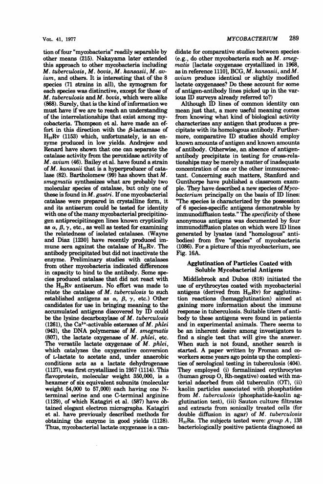
MYCOBACTERIUM 289
tion offour "mycobacteria" readily separable byother means (215). Nakayama later extendedthis approach to other mycobacteria includingM. tuberculosis, M. bovis, M. kansasii, M. av-ium, and others. It is interesting that of the 8species (71 strains in all), the zymogram foreach species was distinctive, except for those ofM. tuberculosis and M. bovis, which were alike(868). Surely, that is the kind ofinformation wemust have ifwe are to reach an understandingof the interrelationships that exist among my-cobacteria. Thompson et al. have made an ef-fort in this direction with the (-lactamase ofH37Rv (1153) which, unfortunately, is an en-zyme produced in low yields. Andrejew andRenard have shown that one can separate thecatalase activity from the peroxidase activity ofM. avium (46). Bailey et al. have found a strainof M. kansasii that is a hyperproducer of cata-lase (82). Bartholomew (99) has shown that M.smegmatis synthesizes what are probably twomolecular species of catlase, but only one ofthese is found in M. gastri. Ifone mycobacterialcatalase were prepared in crystalline form, itand its antiserum could be tested for identitywith one ofthe many mycobacterial precipitino-gen antiprecipitinogen lines known crypticallyas a, (3, y, etc., as well as tested for examiningthe relatedness of isolated catalases. (Wayneand Diaz [1230] have recently produced im-mune sera against the catalase of H37Rv. Theantibody precipitated but did not inactivate theenzyme. Preliminary studies with catalasesfrom other mycobacteria indicated differencesin capacity to bind to the antibody. Some spe-cies produced catalase that did not react withthe H37Rv antiserum. No effort was made torelate the catalase of M. tuberculosis to suchestablished antigens as a, 3, y, etc.) Othercandidates for use in bringing meaning to theaccumulated antigens discovered by ID couldbe the lysine decarboxylase of M. tuberculosis(1261), the Ca2+-activable esterases ofM. phlei(943), the DNA polymerase of M. smegmatis(807), the lactate oxygenase of M. phlei, etc.The versatile lactate oxygenase of M. phlei,which catalyzes the oxygenative conversionof -lactate to acetate and, under anaerobicconditions acts as a lactate dehydrogenase(1127), was first crystallized in 1957 (1114). Thisflavoprotein, molecular weight 350,000, is ahexamer of six equivalent subunits (molecularweight 54,000 to 57,000) each having one N-terminal serine and one C-terminal arginine(1129), of which Katagiri et al. (587) have ob-tained elegant electron micrographs. Katagiriet al. have previously described methods forobtaining the enzyme in good yields (1128).Thus, mycobacterial lactate oxygenase is a can-
didate for comparative studies between species(e.g., do other mycobacteria such as M. smeg-matis [lactate oxygenase crystallized in 1968,as in reference 1110], BCG, M. kansasii, and M.avium produce identical or slightly modifiedlactate oxygenases? Do these account for someof antigen-antibody lines picked up in the var-ious ID surveys already referred to?)Although ID lines of common identity can
mean just that, a more useful meaning comesfrom knowing what kind of biological activitycharacterizes any antigen that produces a pre-cipitate with its homologous antibody. Further-more, comparative ID studies should employknown amounts ofantigen and known amountsof antibody. Otherwise, an absence of antigen-antibody precipitate in testing for cross-rela-tionships may be merely a matter ofinadequateconcentration of one or the other immunoreac-tant. Concerning such matters, Stanford andGunthorpe have published a classroom exam-ple. They have described a new species ofMyco-bacterium principally on the basis of ID lines:'The species is characterized by the possessionof 6 species-specific antigens demonstrable byimmunodiffusion tests." The specificity of theseanonymous antigens was documented by fourimmunodiffusion plates on which were ID linesgenerated by lysates (and "homologous" anti-bodies) from five "species" of mycobacteria(1086). For a picture of this mycobacterium, seeFig. 16A.
Agglutination of Particles Coated withSoluble Mycobacterial Antigens
Middlebrook and Dubos (818) initiated theuse of erythrocytes coated with mycobacterialantigens (derived from H37Rv) for agglutina-tion reactions (hemagglutination) aimed atgaming more information about the immuneresponse in tuberculosis. Suitable titers of anti-body to these antigens were found in patientsand in experimental animals. There seems tobe an inherent desire among investigators tofind a single test that will give the answer.When such is not found, another search isstarted. A paper written by Froman and co-workers some years ago points up the complexi-ties of serological testing in tuberculosis (404).They employed (i) formalinized erythrocytes(human group 0, Rh-negative) coated with ma-terial adsorbed from old tuberculin (OT), (ii)kaolin particles associated with phosphatidesfrom M. tuberculosis (phosphatide-kaolin ag-glutination test), (iii) Sauton culture filtratesand extracts from sonically treated cells (fordouble diffusion in agar) of M. tuberculosisH37Ra. The subjects tested were: group A, 138bacteriologically positive patients diagnosed as
VOL. 41, 1977

290 BARKSDALE AND KIM
tuberculous; group B, 94 patients diagnosed astuberculous from whom M. tuberculosis couldnot be isolated; and group C, a control group of47 tuberculin-negative adults. Positive reac-tions to one or more of the tests occurred in 85%of group A, 75% of group B, and 6% of group C.More than 50% of group A patients were posi-tive for two or more of the tests, and an addi-tional 30% were positive to only one ofthe threetests; ofthe group B patients, 38% were positiveto two or more tests, and an additional 38%were positive for one of the three tests. In thecontrol group, those positive (6%) were positiveto one only of the tests. Although the data donot indicate that the test has been found, theydo point up an interesting difference among thebacteriologically positive patients. One wouldlike to know more about the 15% nonreactors.The authors raised the question as to whetheror not this hard core of nonreactors (i) mighthave responded to other tests not employed bythem, (ii) might have contained antigen-anti-body complexes, or (iii) might have had block-ing levels of circulating incomplete (blocking)antibody. The possibility of an immunologicalunresponsiveness shared by this 15% was notsuggested as the basis for their failure to makedetectable amounts of antibody.The results ofIshibashi et al. (538) employing
the hemagglutination reaction between wax Dadsorbed on erythrocytes and antisera directedagainst M. tuberculosis Aoyama B indicatedthat the (lipo) "polysaccharide antigen" of Mid-dlebrook and Dubos (818) shared determinantswith those of wax D.
Daniel (277) used hemagglutination as ameans of measuring the immune response ofNew Zealand white rabbits to OT, OT in incom-plete adjuvant, alum-precipitated PPD, andkilled H37Rv. A measurable primary responseoccurred in each of the animals. The authorsfound only IgM formed in the animals immu-nized with soluble OT and alum-precipitatedPPD. OT in adjuvant and killed H37Rv elicitedthe formation of both IgM and IgG. Ten weekspostimmunization, all IgM had disappeared,and there were residual levels of IgG in thoseanimals in which it had been formed. When theanimals were reinoculated at 13 weeks, noneshowed a secondary response, probably becauseof still-persisting antibody available for the for-mation of immunosuppressive immune com-plexes.
The Soluble-Antigen Fluorescent-AntibodyTest
The soluble-antigen fluorescent-antibody(SAFA) test for circulating antimycobacterial
antibodies employs cellulose acetate filter disksimpregnated with proteins A and C (14), as wellas a partially characterized arabinogalactan(from cell walls, see Birnbaum and Affronti[135, 136]), all from H37Ra (373; see also [1159]).The moist, impregnated disks are reacted withdilutions of sera to be tested. Following storagein the cold, disks are washed, rendered rela-tively dry, and then exposed to fluorescein-la-beled antiglobulin antibody (anti-human, anti-monkey, etc., according to subject animal). Fi-nal reading for level of antibody in the immu-nological sandwich (antigen-antibody-fluoresc-ing anti-antibody) is carried out in a fluorome-ter. Affronti et al. (13), using a modified SAFAtest, have shown that 11 of 12 monkeys havingfatal tuberculous infections developed a detect-able antibody response, and 10 of these re-sponses were detected by day 42 postinfection.The SAFA test was positive earlier than thetuberculin skin tests (both intradermal palpe-bral and intradermal abdominal injectionswere carried out on each animal). The SAFAtest was also positive for 25 tuberculous pa-tients hospitalized for chemotherapeutic treat-ment. Furthermore, these patients showed asignificant increase in SAFA-detectable anti-bodies within 30 to 90 days posthospitalization.
Tuberculins and Other MycobacterialElicitins
Skin test reactions. Magnusson (756) hassuggested the term "sensitin" for "a nonanti-genic substance, prepared from a microorga-nism (virus, bacterium, or fungus), capable ofrevealing sensitivity ofthe delayed type evokedby the organism." Since these products elicitskin reactions but do not sensitize for such reac-tions, "elicitin" would seem a more apt termthan "sensitin." Human and avian tuberculin,histoplasmin, and coccidiodin are examples ofeliciting. The model of this kind of immunologi-cal detection is found in the guinea pig experi-mentally infected with M. tuberculosis andskin tested with PPD. Since the number (kinds)of sensitizations induced by the infecting orga-nism is usually multiple, the elicitation of spe-cific response in the skin of that animal isdependent on the purity of the eliciting re-agent, lack of cross-reactions, etc. (see Table 6).By 1957, at the State Serum Institute in Co-
penhagen, the following protocol was in use forthe analysis of interrelationships among myco-bacteria based on the degrees of specificity inskin tests. Sensitization was induced by theintradermal injection of dried, heat-killed my-cobacteria suspended in paraffin oil. Follow-upskin tests were carried out 3 to 4 weeks later
BACTERIOL. REV.
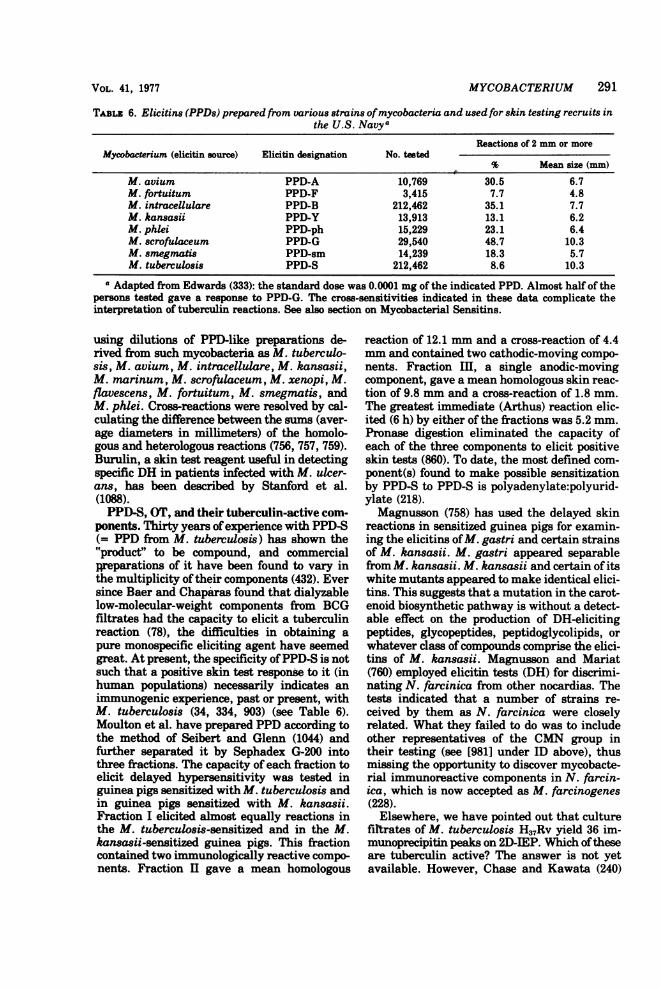
TABL.z 6. Elicitins (PPDs) prepared from various strains ofmycobacteria and used for skin testing recruits inthe U.S. Navya
Reactions of 2 mm or moreMycobacterium (elicitin source) Elicitin designation No. tested
% Mean size (mm)M. avium PPD-A 10,769 30.5 6.7M. fortuitum PPD-F 3,415 7.7 4.8M. intracellulare PPD-B 212,462 35.1 7.7M. kansasii PPD-Y 13,913 13.1 6.2M. phlei PPD-ph 15,229 23.1 6.4M. scrofulaceum PPD-G 29,540 48.7 10.3M. smegmatis PPD-sm 14,239 18.3 5.7M. tuberculosis PPD-S 212,462 8.6 10.3
a Adapted from Edwards (333): the standard dose was 0.0001 mg of the indicated PPD. Almost half of thepersons tested gave a response to PPD-G. The cross-sensitivities indicated in these data complicate theinterpretation of tuberculin reactions. See also section on Mycobacterial Sensitins.
using dilutions of PPD-like preparations de-rived from such mycobacteria as M. tuberculo-sis, M. avium, M. intracellulare, M. kansasii,M. marinum, M. scrofulaceum, M. xenopi, M.flavescens, M. fortuitum, M. smegmatis, andM. phlei. Cross-reactions were resolved by cal-culating the difference between the sums (aver-age diameters in millimeters) of the homolo-gous and heterologous reactions (756, 757, 759).Burulin, a skin test reagent useful in detectingspecific DH in patients infected with M. ulcer-ans, has been described by Stanford et al.(1088).PPD-S, OT, and their tuberculin-active com-
ponents. Thirty years ofexperience with PPD-S(= PPD from M. tuberculosis) has shown the"product" to be compound, and commercialpreparations of it have been found to vary inthe multiplicity oftheir components (432). Eversince Baer and Chaparas found that dialyzablelow-molecular-weight components from BCGfiltrates had the capacity to elicit a tuberculinreaction (78), the difficulties in obtaining apure monospecific eliciting agent have seemedgreat. At present, the specificity ofPPD-S is notsuch that a positive skin test response to it (inhuman populations) necessarily indicates animmunogenic experience, past or present, withM. tuberculosis (34, 334, 903) (see Table 6).Moulton et al. have prepared PPD according tothe method of Seibert and Glenn (1044) andfurther separated it by Sephadex G-200 intothree fractions. The capacity of each fraction toelicit delayed hypersensitivity was tested inguinea pigs sensitized with M. tuberculosis andin guinea pigs sensitized with M. kansasii.Fraction I elicited almost equally reactions inthe M. tuberculosis-sensitized and in the M.kansasii-sensitized guinea pigs. This fractioncontained two immunologically reactive compo-nents. Fraction II gave a mean homologous
reaction of 12.1 mm and a cross-reaction of 4.4mm and contained two cathodic-moving compo-nents. Fraction III, a single anodic-movingcomponent, gave a mean homologous skin reac-tion of 9.8 mm and a cross-reaction of 1.8 mm.The greatest immediate (Arthus) reaction elic-ited (6 h) by either of the fractions was 5.2 mm.Pronase digestion eliminated the capacity ofeach of the three components to elicit positiveskin tests (860). To date, the most defined com-ponent(s) found to make possible sensitizationby PPD-S to PPD-S is polyadenylate:polyurid-ylate (218).Magnusson (758) has used the delayed skin
reactions in sensitized guinea pigs for examin-ing the eliciting ofM. gastri and certain strainsof M. kansasii. M. gastri appeared separablefrom M. kansasii. M. kansasii and certain of itswhite mutants appeared to make identical elici-tins. This suggests that a mutation in the carot-enoid biosynthetic pathway is without a detect-able effect on the production of DH-elicitingpeptides, glycopeptides, peptidoglycolipids, orwhatever class ofcompounds comprise the elici-tins of M. kansasii. Magnusson and Mariat(760) employed elicitin tests (DH) for discrimi-nating N. farcinica from other nocardias. Thetests indicated that a number of strains re-ceived by them as N. farcinica were closelyrelated. What they failed to do was to includeother representatives of the CMN group intheir testing (see [981] under ID above), thusmissing the opportunity to discover mycobacte-rial immunoreactive components in N. farcin-ica, which is now accepted as M. farcinogenes(228).Elsewhere, we have pointed out that culture
filtrates of M. tuberculosis H37Rv yield 36 im-munoprecipitin peaks on 2D-IEP. Which oftheseare tuberculin active? The answer is not yetavailable. However, Chase and Kawata (240)
MYCOBACTERIUM 291VOL. 41, 1977

292 BARKSDALE AND KIM
have begun a rational approach to the problemof sorting tuberculin-active mycobacterial anti-gens. They used a set of IgG, antimycobacterialsera for ascertaining the antigenic complexityof "tuberculins" (tuberculin-active prepara-tions) through a carefully quantified techniqueof passive cutaneous anaphylaxis (PCA) carriedout in guinea pigs. Animals lacking immuneexperience with mycobacterial antigens are in-tracutaneously prepared at various sites withdifferent batches of IgG, immune sera. (Seracorresponding to at least 10 antigen-antibodysystems were used. Reactivities of differentsera were adjusted to equivalent concentrations.A range of concentrations was employed to ac-commodate to variations in dermal behaviorfrom animal to animal.) The prepared animalsreceived, intravenously, antigens under inves-tigation. The firing, or not, ofPCA reactions atdifferent sites provided comparative data for animmunological analysis of the various PPD-Ssand OTs being tested. An examination ofbatches of PPD-S (prepared in several ways:e.g., by precipitation with ammonium sulfate,trichloroacetic acid, or benzoic acid) from sev-eral sources indicated the presence of at leastfive different antigens (certain unheated prepa-rations contained more). OT could be shown tocontain one dominant antigen not present in allPPD-S. OT seemed to lack one dominant anti-gen found in all PPD-S. The dominant antigenof OT was not found in trichloroacetic acid-PPDs but was present to some degree in mostPPD-Ss prepared by ammonium sulfate precipi-tation. The authors conclude that infectionwith M. tuberculosis leads to the acquisition ofseveral delayed-type hypersensitivities. Theysuggest that OTs and certain PPDs test fordifferent DHs and that at least five types oftubercle bacilli-induced DH exist. It is interest-ing to note that OT detects a particular DHsensitivity. A number ofyears ago, one ofus (L.B. in collaboration with E. L. Hopphani and T.Rei) found that a much-earlier-signal DH forsacrificing guinea pigs that were developingtuberculosis from clinical materials could beobtained by testing with OT than with variousstrengths of PPD. It would be interesting toknow now whether the distinctive tuberculin-active component found in OT by Chase andKawata can be related to a sequence of DHs(early, middle, late) that develop following tu-berculous infection in the guinea pig and othersensitive animals.The need for highly purified tuberculin-ac-
tive peptide has long been felt. Morisawa et al.reported in 1960 (853) the crystallization fromculture filtrates of H37Rv (grown in Sauton me-dium) a heat-stable, basic peptide capable of
eliciting DH in human subjects in amounts of0.1 /ig. More recently, Kuwabara has reportedthe purification and properties of a tuberculin-active protein, molecular weight 9,700, ob-tained in crystalline form from acetone-powderpreparations of heated cells of M. tuberculosisAoyama B. Cell-free extracts were exposed toDNase and RNase following treatment withstreptomycin. Redissolved precipitate from 75%saturated ammonium sulfate was passedthrough diethylaminoethyl (DEAE)-cellulosecolumns and then through Sephadex G-200.Crystallization was from eluates of the latter.The final product, a crystalline protein, con-tained an estimated specific activity of 6.33 x10⁹ tuberculin units per mg of protein nitrogenfor guinea pigs and 6.33 x 10¹¹ tuberculin unitsper mg of protein nitrogen for sensitized humansubjects (678). Subsequently, the amino acidsequence was determined. Among the seventryptic peptides found, only one, consisting ofAsn-Gly-Ser-Gln-Met-Arg, exhibited tubercu-lin activity. No information was given regard-ing the immunogenic capacity of the protein(679).Although the growing need for chemically
pure and well-characterized eliciting sub-stances for exploiting the exquisite specificitypossible in tuberculin-type reactions has beenfelt by many investigators, model systems wor-thy of the term "reference systems" are stillscarce. One such model could derive from re-cent studies of M. avium and M. paratubercu-losis. It has been on record since the report ofHagan and Zeissig (481) and the opinion ofPaterson (909) that some animals infected withM. paratuberculosis (johnei) respond to elici-tins from M. avium. In fact, the reactions in theskin caused by these two preparations havebeen said to be indistinguishable (909). Single-ton et al. (1057) have examined the capacities ofpeptidoglycolipid fractions from M. avium andfrom M. paratuberculosis to elicit DH reactionsin guinea pigs sensitized to M. avium and to M.paratuberculosis. By this test, the active prin-cipal in the two fractions seemed identical. Thissimilarity of behavior was paralleled by anoverall similarity in the infrared spectra of thetwo. Although the authors termed the materi-als with which they worked as peptidoglycoli-pid, implying a mycosidic nature for the com-pounds involved, the material actually con-sisted of murein and polysaccharide from themycobacterial cell wall (reported [991a] as glu-tamic acid, alanine, meso-diaminopimelic acid,glucosamine, galactose, mannose, arabinose,aspartic acid, and traces of glycine and phenyl-alanine) and considerable lipid. Consequently,their results only mean that their fractions
BACTERIOL. REV.

MYCOBACTERIUM 293
from M. paratuberculosis and M. avium hadmuch in common. They were not dealing withthe specific, superficial mycosides or peptidog-lycolipids. Laneelle (687a, 689a) has demon-strated that M. paratuberculosis produces pep-tidolipid but not peptidoglycolipid (see sectionon Glycolipids, Peptidoglycolipids, etc.).Kubica et al. (668) have shown that mice
sensitized by the injection of viable mycobacte-ria into one of the hind footpads responded (3 or4 weeks later) to a second injection in the con-tralateral footpad with an accelerated responsethat was exaggerated over that of the first in-jection. Tuberculin shock following testing isone of the hazards of the procedure. The au-thors suggest that this can be obviated by using10 mice per test and making the sensitizing andchallenging doses around 1,000,000 viable unitsper mouse.
THE IMMUNE RESPONSE TOMYCOBACTERIUM
"Humoral Immunity" (HI), the Plasma CellArm of the Immune System
The immune response to chronic infectionsbegins with inflammation and involves the en-tire gamut of the defenses of the host. Theeffectiveness of these defenses varies accordingto the genetic makeup of the host animal (740)and that of the invading agent. The regulationofthe mechanisms ofdefense may be affected invarious ways according to the offending agent.In rabbits, guinea pigs, and other susceptibleanimals, experimental tuberculous infection,like so many other bacterial infections, beginswith a pouring in of polymorphonuclear leuko-cytes (PMNs) at the site of injection (305) andwith subsequent ingestion of the bacilli bythese cells. The bacteria-laden granulocytessubsequently die, and their remnants and con-tents are engorged by macrophages (973).There is little or no evidence to suggest that,before dying, these PMNs have had a lethaleffect on their engulfed mycobacteria (973, 488),despite the hydrogen peroxide such PMNs ap-parently liberate (616).During tuberculous infection, antibodies are
formed (818), and immunoglobulins reactivewith M. tuberculosis are reported to be commonamong "normal" human subjects (93, 393). Theimportance of antibodies in recovery from tu-berculosis has long been questioned. Someyears ago, Raffel reported experiments whichsuggested that infected guinea pigs passivelytreated with repeated doses of antituberculoussera (as well as whole blood) developed a tuber-culous infection comparable to that in control
animals receiving normal serum, normal wholeblood, or nothing (954). Recently, Reggiardoand Middlebrook have reported that immunerabbit sera passively administered 1 day priorto, and on days 5, 11, 18, and 25 following, aero-genic challenge with M. bovis Ravenel failed tomodify the number of viable bacilli recoverablefrom the animals at 6 weeks (964). These au-thors have been careful to point out that theirresults do not accord with: (i) those of Rowley etal. (993), who found in mouse typhoid (Salmo-nella typhimurium) that the specific aspect ofimmunity was related to a cytophylic antibodycapable of fixing to macrophages and affordingpassive protection; (ii) those of Lurie (738, 739),confirmed by Tsuji et al. (1161), demonstratingin immune sera of guinea pigs and rabbits ahumoral antibacterial component; and (iii)those of Fong et al. (386), showing that a factorin sera from Salmonella-immune rabbits in vi-tro nonspecifically protected macrophages fromthe necrotizing action of virulent M. tuberculo-sis H37Rv. The additional finding of the latterinvestigators that "monocytes" from animalsimmunized with BCG and cultivated in anti-BCG serum were able to cope with H37Rv,whereas "monocytes" from Salmonella-im-mune rabbits under the same conditions failedto withstand H37Rv infection, pointed to a rolefor both serum factors and mononuclear cells inthe immune response. This archetypal finding,after being many times rediscovered, nowseems to be generally accepted.
Antibody Responses in Tuberculous InfectionThe mammalian immune system and its con-
trols are complex and include mechanisms yetto be understood. These controls appear to in-volve complicated interactions between twoclasses derived from lymphoid stem cells: T,which have had an association with the thymusor thymosin (437, 633), and B, which have hadan extrathymic maturation (335). The B lym-phocyte is characterized by at least three sur-face receptors: (i) receptors that bind the Fcregion of Ig molecules and provide a site forantibody molecules, antigen-antibody com-plexes or aggregated Ig (102, 103, 104, 300), (ii)surface Ig receptors for specific antigens (886),and (iii) receptors for the C3 component of com-plement (131, 888). These bone marrow-derivedlymphocytes can be stimulated to produce thelymphokine, monocyte chemotactic factor, (i) bymitogens carrying multiple binding sites, e.g.,lipopolysaccharide or polymerized flagellin (see[369]), (ii) by antigen-antibody complexes oraggregated gamma globulin interacting withthe Fc receptor, and (iii) by interaction of anti-
VOL. 41, 1977

294 BARKSDALE AND KIM
gen-antibody-complement complexes at the C3receptor. The bridging or cross-linking of recep-tors is thought by some to be the key to stimula-tion, and such stimulation is affected by nonmi-togenic as well as mitogenic molecules as longas they can cross-link the receptors (1212). (Forother theories as to the basis for B cell activa-tion, see [266a].) A B lymphocyte with an im-munoglobulin having an affinity for a specificantigen, upon binding that antigen, may bestimulated to divide, giving rise to a clone ofplasma cells (monoclonal stimulation) capableof synthesizing immunoglobulin of specific an-tibody activity(ies). B cells may also be gener-ally triggered to mitosis by certain species ofmolecules operationally defined as mitogens(e.g., certain plant lectins [polyclonal stimula-tion] [265, 266]), as well as by C'3 (323) andlipopolysaccharides (846). Compounds capableof polyclonal stimulation have also been termedPBA or polyclonal B cell activators (266a), andmany of them have long been known as adju-vants. Classes of adjuvants that have beentermed "B cell oriented" (319) include Borde-tella pertusis, Propionibacterium parvum,and E. coli lipopolysaccharide (24). IgM pro-duced in mice in response to lipopolysaccharideis of broad affinity and low avidity (319a).Although the B lymphocyte prior to antigenicstimulation possesses Fc and C'3 receptors, thecells of clones (plasma cells) derived from thatB lymphocyte have lost or have covered up theC'3 receptors (104, 845, 1050). Certain, if notall, of the IgG's synthesized by plasma cells arecytophilic; i.e., they fix by their Fc end to mac-rophages (see following discussion of CytophilicAntibodies). Thus, a major component of HI orB cell(s)-related immunity can interact withmacrophages.Adjuvants classed as "T cell oriented" and
including beryllium, CFA, lentinan, polyade-nylic acid * polyuridylic acid [poly(A) * poly(U)],and retinol are effective in the stimulation ofantibody only in animals unimpaired as to com-plement of thymus-associated T lymphocytes.For example, Cone and Wilson (258). found a 10-fold amplification of T rosette-forming cells inmice receiving antigen (sheep erythrocytes[SRBC]) plus poly(A) * poly(U). T lymphocytes,probably with macrophages, are the major bio-synthetic machinery for DH and are also essen-tial for maximal B cell response to certain anti-gens. Through experiments employing T and Bcells in separate chambers linked by Nucleo-pore membranes, Feldmann (368a) has demon-strated that T and B cell cooperation can occurthrough soluble factors. Thus there appears tobe no requirement for cell-to-cell contact. Fur-
ther, he has suggested that complexes of IgMfrom T cells with antigen become bound to mac-rophages, and it is with this macrophage-boundcomplex that B cells interact and become stim-ulated to produce antibody. Finding this inter-action between the T and B components of theimmune system has led to the suggestion that Tcells function as regulators: (i) sometimes assuppressors of an immune response (428, 429,507, 508, 895, 1117) and (ii) sometimes as en-hancers of an immune response (86). In addi-tion, there is the killer T cell (KTC). The studyof lentinan's (a fungal polysaccharide, a linear,3-(1-3)-glucan polymer) antitumor activity hasrevealed that this T cell adjuvant acts on thehelper function, causing T cell enhancement ofB cell antibody synthesis, but does not increasethe sensitization of KTCs in an allogeneic sys-tem (295, see also [368b]). Silver and Benacer-raf (1054) have shown a comparable dissocia-tion of T cell helper function and DH usingdeaggregated protein carrier and its hapten.Using the former as a tolerogen in low doses,they could show the inhibition of helper func-tion in the face of retained DH. Thus, the regu-lation of antibody synthesis extends beyond theB cell function per se. (Recently, Munro andTaussig have offered support for the thesis that,in mice, antibody production is controlled bytwo functionally distinct cistrons in the I regionof the major histocompatibility complex. Onecontrols [i] T cell recognition of antigen and [ii]T cell mediators of cell cooperation; the othercontrols the B cell response to the T cell media-tors [864]. Further, the controls that lead totolerance, e.g., tolerance induced by antigen-antibody complexes, may be T cell mediated[Taylor and Basten in reference 3181.)"Cytophilic" and other antibody activities
associated with tuberculous infection. Over adecade ago Boyden and Sorkin described anantibody that was cytophilic for the spleen cellsof rabbits immunized with human serum albu-min (HSA; 170, 171, 1076). Guinea pig cyto-philic antibody produced in animals immunizedwith SRBC mixed with CFA was found to selec-tively bind to homologous macrophages, ren-dering them capable of adsorbing SRBC (169).Berken and Benacerraf showed that this cyto-philic activity for macrophages was a propertyof the complement-binding 7S, y2-class ofguinea pig immunoglobulin (IgG). The anti-body-binding site for the macrophages was lo-cated on the Fc fragment and thus is a part ofthe H chains. Cytophilic activity is not comple-ment dependent. Cytophilic activity was shownto be a property of opsonizing antibody, whichallows the fixation of antibody to the cell mem-
BAC-rzMOL. Rzv.

MYCOBACTERIUM 295
brane in an orientation optimal for phagocyto-sis. Cytophilic antibodies were found in im-mune sera from rabbits and mice vaccinatedwith SRBC. Sensitization of macrophages wasmaximal with homologous antibodies, butcross-sensitization between species did occur(127).Amos et al. (35) found that the sera of guinea
pigs, sensitized to tuberculin (PPD) using PPDin Freund adjuvant (or better, BCG followed byPPD in Freund adjuvant) as well as to (3-lacto-globulin, contained cytophilic antibodies capa-ble of fixing to macrophages and renderingthem sensitive to migration inhibition by eitherPPD or (3-lactoglobulin. This migration inhibi-tion could, of course, be passively transferredwith serum and was not to be confused with thelymphokines (290, 324) and other nonantibodymediators of cellular immunity generated afterlymphocyte activation. Heise et al. (502), usingBCG-sensitized guinea pigs and PPD, showedthat cytophilic antibody could render macro-phages sensitive to inhibition by PPD. Mix-tures of PPD and immune serum were inhibi-tory to macrophage migration but not cytotoxicfor mouse L cells. Cytophilic antibodies tend tocome off macrophages at 370C (127), and thishas raised some question as to their importancein vivo (335).Binding of guinea pig cytophilic antibody
(y2-immunoglobulin) to mycobacterial glyco-peptide. Stewart-Tull et al. (using the samemethod Seibert employed for the isolation of"polysaccharide" I of M. tuberculosis [1043]),have isolated from culture filtrates ofM. tuber-culosis a glycopeptide (containing arabinose,galactose, glucosamine, muramic acid, alanine,glutamic acid, a,e-diaminopimelic acid, andsmall amounts of aspartic acid and glycine).They have shown that this glycopeptide ("poly-saccharide" I of Seibert) has an immunologicalcloseness (near identity) with the peptidoglyco-lipid wax D (see section, From the CytoplasmicMembrane to the Peptidolipid). They noted(1102) an affinity of the glycopeptide for guineapig immunoglobulin (formed in animals receiv-ing CFA and egg albumin) and subsequentlyshowed (1101) that it is capable of binding toboth the Fc and Fab fragments of y2-globulin(y2G). The ratio of Fc-binding to Fab-bindingwas 4:2. Sincey2G from guinea pigs immunizedwith bentonite-ovalbumin complexes as well asthat from guinea pigs receiving repeated injec-tions of incomplete adjuvant also fixed to glyco-peptide, the authors concluded that specificimmunization with mycobacteria (in adjuvant)was not related to the effect. They examinedother glycopeptide fractions for their y2-binding
capacity. It appeared that, for satisfactory bind-ing, muramic acid and arabinose were essentialcomponents of the glycopeptide. Recently, Dav-ies and Stewart-Tull have shown that, whenSRBC become coated with glycopeptide, theyhave an affinity for guinea pig y2G. In fact,SRBC-glycopeptide-y2G complexes were agglu-tinable by rabbit-antiguinea pig globulin, thusestablishing a dual affinity of the glycopeptidefor cell membranes and for y2G (291). Humansera from active cases of tuberculosis (from 2months to 7 years duration) showed no affinityfor mycobacterial glycopeptide (1101). The au-thors have not indicated whether or not theglycopeptide can bind to the surfaces of B or Tlymphocytes or macrophages. The glycopeptideshares with a number of mediators of immuneresponses the property of being bifunctional.Waksman and Namba (1213a) have pointed outthat the concept inherent in the term "ambo-ceptor" is bifunctionality, a property common toopsonic IgG, to cytophilic IgG in its binding byits Fc end to the macrophage and by its Fab endto antigen, and to IgE in its binding to mastcells or basophils. Are the bifunctional mole-cules of mycobacterial origin capable of effect-ing similar binding in the animal host? Wouldthe presence of such molecules exert a regula-tory effect on an immune response?
In 1963, Benacerraf et al. reported thatguinea pigs immunized intraperitoneally withegg albumin or hapten conjugates of proteinantigens responded with the synthesis of IgG,antibodies (120). However, when the same anti-gens were administered with CFA, both IgG,and IgG2 antibodies were produced. IgG2 anti-bodies could fix complement, whereas IgG, an-tibodies could not (144). More recently, Stew-art-Tull et al. (1097) have electrofocused globu-lins from normal and CFA-plus-ovalbumin-stimulated guinea pigs and determined the ra-tio of IgG2:IgG, by polyacrylamide disc gel elec-trophoresis and densitometry combined withE280l.spectroscopy. They concluded that, in-deed, significant differences exist between IgG,and IgG2 populations in anti-ovalbumin serumof animals stimulated by CFA and those ofnonimmune, normal guinea pigs. It seems in-escapable that the difference must be attrib-uted to CFA. Wilkinson, giving due credit toStewart-Tull and White, in 1966 observed thatthe development of granulomas in guinea pigsfollowing injection of antigen-adjuvant mix-tures (CFA or bentonite) was correlated with arise in IgG2 antibodies (1260). Kotani and asso-ciates have referred to this latter as evidencethat a statistically significant relationship ex-ists between the production of IgG2 and the
VOL. 41, 1977

296 BARKSDALE AND KIM
development of DH (654). They have estab-lished a similar correlation between the devel-opment ofDH and IgG2 in animals immunizedwith water-in-oil emulsions consisting of oval-bumin as antigen and cell walls or their water-soluble fractions from a variety of gram-posi-tive bacteria. They and Ellouz et al. (342) haveindependently shown that an ultimate adju-vant for inducing this effect is N-acetylmura-myl-L-alanyl-n-isoglutamine (655). Is there anyrelationship here between the affinity of IgG2for peptidoglycolipid-containing N-acetylmu-ramic acid and the increase in IgG2 antibodies?Cytotoxic antibody. Recently it has been
found that humoral antibody (IgG) can induceand/or inhibit an in vitro lymphocyte-mediatedcytotoxicity by thymus-independent cells (re-viewed by Perlmann et al. [920; see also refer-ence 1060]). In this lymphocyte-mediated kill-ing, normal lymphoid cells are cytotoxic forparticular target cells in the presence of anti-body to the latter. The Fc portion of the anti-body molecule is essential for cytotoxicity.Lymphocytes in contact with antibody-treatedtarget cells showed increased mobility, as indi-cated by their infiltration of the monolayers ofthe latter. The activation of these infiltratinglymphocytes may come from interaction withantigen-antibody complexes on the surface ofthe target cells. Antigen-antibody complexesare known to stimulate lymphocytes to increaseDNA synthesis (145, 844). Although the exactnature of the effector or killing lymphocyteis not known, it seems not to be a T cell(1060). However, as Biberfeld et al. have pointedout, there is a possibility that antibody-depend-ent lymphocytolysis and phytohemagglutinin(PHA)-induced cytolysis (T cell-mediated kill-ing) both operate through the activation of acommon cell type (132). It may therefore beconcluded that some cells important for lym-phocytolysis, in vitro, have a requirement forantibody attachment to the target cell. Mac-Lennan (753) has suggested the term "cytotoxicB cell" for the non-glass-adherent, nonphagocy-tic mononuclear cells that kill target cells sen-sitized with IgG. He regards such cells asathymic in development and postulates thatthey do not release lymphokines. (It is nowknown that B cells do release certain lympho-kines.) If this cytotoxic lymphocyte is indeed aB cell, then here again is evidence that, in the"normal" immune system, the functions of theB component (HI) and the T component (DH)are interdependent.
Antibodies reactive with purified proteinderivative. Kostalia has followed guinea pigsimmunized with 2.4 mg of heat-killed M. tuber-
culosis in adjuvant (Bayol F, 90%; Arlacel A,10%) from 4 days to 1 year postimmunization.She examined the capacity of the serum immu-noglobulins produced to (i) cause the hemagglu-tination of PPD-coated erythrocytes, (ii) effectthe passive immune hemolysis of those anti-body-coated erythrocytes (648), and (iii) renderperitoneal exudate cells able to form rosetteswith PPD-coated SRBC (macrophage-bound cy-tophilic antibody) (650). The results regardingcytophilic antibodies (on macrophages and insera) indicated that they were present in smallamounts in a few animals at the end of 1 monthpostimmunization but, at the end of 1 year,were present in a larger number ofanimals andin higher concentrations. (The possible contri-bution of these immunoglobulins to measure-ment of MIF in migration inhibition tests isconsidered under the section, MIF.) Hemagglu-tinating antibodies appeared as early as 5 days,whereas immediate reactions (4-h reactions,Arthus type, elicited in response to 10 ,ug ofPPD) appeared in a few animals as early as 10to 14 days. Tests for each of these responseswere uniformly positive at 2, 4, 6, and 12months postinfection. The highest agglutinintiters (1:5,120) were observed 28 and 35 dayspostsensitization. These persisted for 1 year.They were 2-mercaptoethanol sensitive and, so,could thereby be distinguished from 7S anti-body (127).
Delayed Hypersensitivity, the Jones-MoteReaction (Cutaneous Basophil
Hypersensitivity), Contact Sensitivity, andSuppressor B Cells
When DH reactions are recorded here, theymay well be either Jones-Mote reactions or typ-ical DH, for until recently distinctions betweenthe two were seldom made. Cutaneous basophilhypersensitivity (CBH; the Jones-Mote reac-tion [560]) is a transient dealyed skin reaction,occurring in guinea pigs sensitized with insolu-ble antigen-antibody complexes (in IFA) andwith the contact allergen dinitrofluorobenzene(DNFB), in which the infiltrate contains largenumbers of basophilic leukocytes in addition tothe round cell component characteristic of DH(330, 977). Like classic DH, CBH reactions de-pend on lymphocyte function but are distin-guished by a lack of induration. When cellsfrom CBH-positive animals were transferred tononimmune recipients, subsequent skin testingelicited small reactions (averaging 6.1 mm indiameter), which histologically were weakCBH reactions. The calculated number of baso-phils in the passive transfer was no more than300,000 of 3 x 10⁸ viable lymphoid cells. Passive
BACTERIOL. REV.

VOL. 41, 1977
transfer of serum brought about no positivereactions in the recipients (see Askenase et al.,just following). Dvorak et al. (330) have furtherpointed out that basophils in substantial num-bers have been identified in contact allergy (inguinea pigs and in man), in hypersensitivity tovaccinia, and in allograft rejection, as well as inCBH reactions. They occur to a lesser extent inthe "classic" DH that follows immunizationwith protein antigens in CFA. Their function inthese various reactions is not clear. Stadeckerand Leskowitz (1080) have found that skin reac-tions, elicited in normal guinea pigs in responseto PHA, Con A, and pokeweed mitogen but notto the B cell mitogen, E. coli lipopolysaccha-ride, were rich in basophils. This has suggestedto them that T cells release a factor chemotacticfor basophils. Richerson has suggested a rela-tion between CBH and passive cutaneous ana-phylaxis (homocytotropic) antibody (976). Dvo-rak et al., however, have shown that circulat-ing basophils from animals primed for CBHlack demonstrable specificity for sensitizing an-tigen. They feel that homocytotropic antibodyhas no obvious role in CBH. They conclude thatCBH and DH have an analogous lymphocyte-rhediated pathogenesis (329). Askenase et al.have reported the transfer by serum of CBH tokeyhole limpet hemocyanin (KLH). Further-more, they have shown that skin testing per seinduces CBH. This important observation theyhave discussed in relation to interpretationsgiven to specific immune reactivity followingtransfer of serum, cells, or cell extracts (56).
Recently, Turk et al. and Parker (1184) havedefined two forms of DH in guinea pigs receiv-ing ovalbumin. (i) In CFA, one form leads to areaction that bears "a close similarity to theclassical tuberculin reaction" (DH: skin reac-tivity developing within 7 days and reachingmaximum intensity at 24 to 30 h post-testing,with persistence of inflammation after 48 to 72h). (ii) In IFA, the other form is detectable 5 to10 days after inoculation, reaches its maximumintensity between 24 and 30 h, and fades at ±48h; it was defined by Turk as CBH. At 14 daysthis sensitization becomes an "Arthus or imme-diate-type" skin reactivity. The authors furtherestablished the fact that treatment of guineapigs (or mice) with cyclophosphamide (CY), 300mg/kg, "selectively depleted" lymphocytes fromthe lymph follicles, the corticomedullary junc-tion in lymph nodes, -and from analogous non-thymus-dependent areas of the spleen, Thislowering in the populations of rapid-turnoverlymphocytes (presumably B cells) was reflectedin the immunized animal by lowered levels ofIgG,, homocytotropic antibody. When the re-
MYCOBACTERIUM 297
sponses were compared between control ani-mals and CY-pretreated animals, both receiv-ing ovalbumin in IFA, they were found to beclearly different (see Fig. 28). Those animalspretreated with CY (3 days before sensitiza-tion), and therefore experiencing a depletion inpresumed B cells, developed typical DH reac-tions (as defined above). The control animals,on the other hand, exhibited a CBH type ofresponse. Here, then, B cell elimination hasenhanced DH. The authors offer evidence froma number of ancillary experiments to make itclear that suppressor B cells may regulate theactivity ofT cells. In fact, their report confirmsand extends that of Neta and Salvin (875, 876).Turk et al. also found that, in the B cell-
depleted animals, the amount of basophil infil-tration was markedly reduced. Thus, a so-called cell-mediated response is regulated byboth T cells and B cells. The authors furthershowed that, in CY-treated animals sensitizedto DNFB, there was a somewhat comparableshift from larger and longer-lived skin test re-actions in the B cell-depleted animals (24 to 96h) to less-intense and shorter-lived reactions inthe control animals (24 to 48 h). One other
,E 6E0
U)U)
-2
0
4 24 48 72 96
Time after skin test (hours)FIG. 28. Average increase in skin thickness after
skin testing with 100 pg of egg albumin in guineapigs immunized with egg albumin in IFA 7 daysearlier. (A) Reactions (DH) occurring in animalspretreated with cyclophosphamide (CY) 3 days priorto sensitization. (B) Reactions (CBH) occurring inuntreated control animals. When untreated animalsare immunized with egg albumin in CFA, they give areaction essentially like that in curve (A) (DH), butwhen the protein antigen is administered with IFAthe reaction of curve (B) (CBH) obtains. Thus, CYpretreatment modifies the CBH reaction expectedwith egg albumin in IFA, rendering it equivalent totheDH reaction normally obtained with egg albuminin CFA. Adapted from Turk et al. (1185).

298 BARKSDALE AND KIM
singular finding in this report was the fact that,in animals made tolerant to dinitrochloroben-zene, either by feeding or by intravenous dos-ages, tolerance could easily be broken in CY-treated animals. The authors conclude that thedifferences observed in classical DH, CBH, andcontact sensitivity are reflections of degrees ofinteraction of effector (T) and suppressor (B)elements, particularly at the reaction site.
Delayed Hypersensitivity and Cell-MediatedImmunity
From the foregoing it is clear that the termcell-mediated immunity (CMI) is difficult forthe beginning student since all immunity is cellmediated. Isolated immunoglobulin (humoralimmunity) and isolated lymphokines (acti-vation factors from T and B lymphocytes) arecell products and their action in the immunesystem must be understood at the cellular level.DH, commonly equated with CMI, is alsopoorly understood. The triad of the immunesystem, the B cell component (primarily immu-noglobulin synthesis), the T cell component,and macrophages, interacts in the wake of in-flammatory responses to control chronic infec-tions. Limitations in understanding the behav-ior of this system are related to difficulties indealing with each of its isolated components.We have, in the preceding section, given somegleanings regarding the behavior of the B cellcomponent in mycobacterial infections. Cyto-philic antibodies made by B cells could effectcytotoxic killing by macrophages. The level ofantibody synthesized by B cells in response tocertain antigens could be strongly influencedby T cells (see Katz and Benacerraf [602, 603]).Thus, helper T cells seem critical in the hu-moral immune response. There is a question asto whether or not some of them synthesize im-munoglobulin (for a discussion of this point seereference 1221a). DH is thought not to occur inthe absence ofT cells. They have a primary butnot exclusive role in DH. Some of the lympho-kines attributed to them are also produced by Bcells (e.g., in lymphocytes from BCG-immuneanimals, PPD induces interferon production[1218], MIF, and other products discussed be-low). Thus, Mackaness has suggested that therole of T cells in resistance to bacterial infec-tiotis is as "molecular mediators" possibly re-sponsible for the structural changes that char-acterize activated macrophages (752). (These Thelper cells and cytotoxic T cells, KTC [effectorcells], are thought to be separable on the basisof sedimentation velocity profiles [340].) Bloomhas pointed up the difficulty in detecting specif-ically sensitized cells (the mediators of DH) by
their ability to produce lymphokines. Whereasone can examine the ability of B cells to pro-duce antibody (e.g, in the Jerne plaque assay),the amount of factors such as MIF/cell or inter-feron/cell is very little "and activity may not bedetected even at small distances from targetcells" (146). The function(s) associated with theT cell component of the immune system thatcan be well assessed in vitro is antigen-inducedlymphocyte cytotoxicity, reviewed by Cerotiniand Brunner (227). The prototype of the killerlymphocyte (cytotoxic lymphocyte, KTO), de-rived from antigen-reactive precursors, was alymphocyte induced in graft-versus-host reac-tion, in which the specific inducing antigenoccurred on allogeneic cells which themselvesbecame the targets (cells) of lymphocyte kill-ing. Killing reached completion within 1 h andwas modulated by intracellular levels of cyclicadenosine monophosphate (AMP). Antibodydirected against the surface antigens of the tar-get cells protected the latter from KTC killing.This role of the KTC seems well adapted to thejob ofimmune surveillance (206, 1152), as in therejection of grafts and in the destruction ofneoplastic cells. KTC seems not to be a celldesigned for killing bacteria. Could its innatecapacity for destroying foreign cells when theirforeign-ness is due to subtle differences in sur-face antigens (e.g., HLA antigens) complicateDH reactions involving bacterial antigens; e.g.,chicken RBC, which have been coated withPPD, are susceptible to cytolysis by lympho-cytes from BCG-sensitized donors (919)? Willcells from tuberculin-positive individuals,coated with PPD during a skin test, be liable tokilling by KTC and/or other K cells (920)?The same sort of question can be asked re-
garding armed macrophages. Evans and Alex-ander (359) have shown that the killing speci-ficity of macrophages involves their being"armed" against specific tumor cells [SL2(DBA/2) and TLX9(C57/B1) lymphomas]. Armingmay be accomplished by (i) deriving macro-phages from peritoneal exudate cells (PEc) ofspecifically immunized mice, (ii) arming in vi-tro by association of nonimmune macrophageswith spleen cells from hyperimmune mice, or(iii) arming in vitro by exposure of nonimmunemacrophages to cell-free supernatant obtainedwhen spleen cells from immunized mice arecultured with specific antigen. All such macro-phages, following incubation with specific anti-gen, undergo activation to killing of targetcells. The killing follows contact of the targetcell membranes with the macrophages. Phago-cytosis occurs only after the target cell hasbegun to disintegrate. Macrophages from BCG-immunized animals kill neither type of tumor
BAC-rRIOL. REV.

MYCOBACTERIUM 299
cells. However, if such macrophages are incu-bated with PPD, they now are activated to killeither of the antigenically distinct tumor cells.A careful examination of the data in Table 7reveals that immunization of animals with spe-cific antigen (specific tumor cells) yields a mac-rophage armed to kill that particular cell and,further, that incubation of macrophage popula-tions (taken 10 days after primary immuniza-tion) from any of several specifically hyperim-munized animals (armed macrophages") withthe specific immunizing antigen will activatethem to kill cells bearing other specific anti-gens upon their surfaces (Table 7). Thus, BCG"armed macrophages" can be activated in thepresence of BCG to kill SL2 cells and TLX9cells. They cannot be so activated by antigensother than the immunizing antigen. Will"armed macrophages" in a tuberculin-positiveindividual kill cells that have bound PPD? Themacrophage-arming factor, which is releasedby lymphocytes, will undoubtedly turn out tobe a cytokine such as MIF (358), and armingand activation will become synonymous withthe enhanced-capacity macrophage discoveredby Elberg (338) and rediscovered and expandedupon by Mackaness (751, 752). Germain et al.have confirmed the general findings of Evansand Alexander (359). For an assessment of tu-
TABLE 7. Cytotoxicity of macrophages fromimmunized micea
Cytotoxicityc atMacrophages 48 h to:CBA mice immunized
with: incubatedwith&: SL2 TLX9(%) (%)
SL2 lymphoma (DBA/ No antigen 99 <102)
TLX9 lymphoma No antigen <10 99(C57B1)
BCG No antigen <10 <10SL2 lymphoma SL2 cells 99 79
TLX9 99 <10PPD 99 <10
TLX9 lymphoma TLX9 cells 75 99SL2 cells <10 99PPD <10 99
BCG PPD 92 84SL2 cells <10 <10TLX9 cells <10 <10
Adapted from Evans and Alexander (359).PEc taken at 10 days after second immunization
showed specific cytotoxicity. However, if a further dose ofspecific antigen was given and macrophages were collected24 to 72 h thereafter, those macrophages killed both SL2and TLX9 cells. This was the case for macrophages fromeither SL2-, TLX9-, or BCG-immune mice. Ten days afterthe third immunization, however, the activated macro-phages had disappeared, and the macrophages found in thePEc possessed immunologically specific cytotoxicity shownabove. For further details, see text.
I Cytotoxicity expressed as percent growth inhibitionin relation to growth in normal or control CBA cultures.
mor cell inhibition, they have measured theinhibition ofDNA synthesis (IDS), as indicatedby the uptake of [3H]T by tumor cells in thepresence and absence of effector cells (presum-ably macrophages):cpm (effector + tumor)
- cpm (effector alone) x 100cpm (tumor alone)
and compared these results with those obtainedby measuring 51Cr release (1264a). When theseauthors (427a) examined that fraction of thePEc that was non-glass-adherent, they discov-ered another population of lymphoid cells capa-ble of specifically lysing tumor cells. Althoughthe details of their characterization of the cellswere necessarily limited, those cells are proba-bly most akin to the natural killer lymphocytes(NK) of Herberman (503a, 503b) and of Kies-sling (613a, 613b) (see following).Wolfe et al. (1274a) have recently reported
the amplification in PE of a mouse cell typecapable of lysing both syngeneic EL-4 thymomaand allogenic P815-X2 mastocytoma target cellsfollowing intraperitoneal (i.p.) infection of micewith BCG. The cells were non-glass. (polysty-rene, in this case)-adherent. Their killing activ-ity was enhanced rather than diminished whenthe population was exposed to 200 mg of car-bonyl iron per 2 x 10⁷ cells and subjected to amagnetic field that removed 60% of the popula-tion. (i) This would seem to mitigate against theidea that the cells are macrophages. (ii) Treat-ment of the population with anti-E serum andcomplement (under conditions where identicaltreatment removed 91% of the T-cell mediated[KTC] cytotoxic activity of spleen populationsfrom C57BL/6 mice immunized with P815[DBA/2] alloantigen) failed to lower the lyticactivity, thus suggesting that lytic activitycould not be ascribed to T cells. (iii) When thenumbers of the population having Ig on theirsurfaces were lowered from 12 to 2.4%, therewas no effect on lytic activity. Thus, the B cellsin the population seemed not involved in thelysis under study. (iv) Fc-receptor-requiring,trypsin-resistant, antibody-dependent cytolyticactivity was also ruled out. The authors con-clude that they were dealing with a "naturalkiller" cell previously described by Herberman(503a, 503b) and by Kiessling (613a, 613b).When PEc were raised with thioglycolate orproteose peptone, no lytic activity for P815-X2was found. Following i.p. immunization withBCG, cytolytic cells appeared. The authorshave suggested that BCG might: (i) induce thecytotoxic population ab initio; (ii) cause theexpansion of clones of cytotoxic cells existing inundetectable numbers; (iii) recruit cytotoxic
VOL. 41, 1977
300 BARKSDALE AND KIM
cells to the peritoneal cavity from other areas;or (iv) activate an exudate cell population tocytotoxicity.
Humoral Immunity, DelayedHypersensitivity, and Cell-Mediated
Immunity in Mycobacterial InfectionsIn three solicited editorials on "Delayed Hy-
persensitivity and Immunity in Tuberculosis"appearing in the American Review ofRespira-tory Diseases, the following conclusions wereexpressed: (i) " . . . reactions of tuberculin hy-persensitivity play little, if any, role in ac-quired immunity to tuberculosis" (1304); (ii) atpresent the conditions and reagents employedfor studying DH on the one hand (soluble anti-gens) and CMI on the other (living bacteria)sometimes suggest a correlation and sometimesdo not. "More extensive standardization of anti-gens and assay procedures must be developed.With this additional information and proce-dures, the data that now seem contradictorymay actually be complementary" (1016). (iii)"The evidence for the essential indivisibility ofDTH and immunity is circumstantial and reliesheavily on the strong association between thetwo phenomena." The author concludes from amass of "disparate data that lymphokine pro-duction, blast transformation, DTH, and im-munity are predominantly, if not exclusively, Tcell functions. Thus the T cells are the commondenominator that link DTH, immunity, andtheir in vitro analogs." He further suggests thatto establish a separation between DH and im-munity an immunizing agent should be foundwhich is nonallergenic, admonishing that "noclaim should be made for such agents withoutexhaustively testing several parameters of cel-lular hypersensitivity so as to ensure that thematerials are truly nonsensitizing" (711).DH, then, appears to be a necessary adjunct
to the interactions that lead to CMI. It is de-structive for the animal and is thought to con-tribute to the development ofthe disease processin tuberculosis. In the experiments of Turk,cells associated with the HI component of theimmune system (presumed B cells) exerted asuppressive effect on the extent ofDH inducibleby specific eliciting. Presumably, this suppres-sive effect would be proportional to the numberof suppressor cells available. If that is true,then the HI component of the immune system,including any specifically amplified populationthereof, would be important in recovery fromtuberculosis. In this regard the suppressor ef-fect of B cells would have a benefit for the hostcomparable, to a degree, to that reputedly asso-ciated with desensitization (549, 1291).
Elsewhere, we have pointed out that the eli-citin, PPD-S, is nonpriming per se but, whengiven to an animal along with poly(A) *poly(U),DH develops. Since this cannot happen withoutthe release of lymphokines, it necessarily in-volves the activation of macrophages via MIF,etc. Since many cell types including B and Tlymphocytes produce MIF, an as yet limitedlycharacterized cytokine (see Cohen in reference885), since B cells exert a suppressor actionin DH, and since B cells are linearly related toantibody-producing plasma cells, HI and DHwould appear to be more, not less, inextricablyrelated than ever. For a general discussion ofDH in relation to CMI, see McCluskey andCohen (802a).
Lymphocytes, Macrophages, andMycobacteriostasis
Dienes and Mallory showed that new lesions,produced by injecting M. tuberculosis into tu-berculous (already hypersensitive) guinea pigs,were characterized for the first 24 to 48 h by amarked accumulation of granulocytes, conges-tion, and serous exudation, a reaction sugges-tive of any pyogenic infection. However, withgreat regularity, beginning on day 3 and be-coming well marked on day 4, "collections oflarge mononuclear phagocytes begin to appearas cuffs about blood vessels and nerves, to infil-trate the stroma first at some distance fromorganisms and leucocytes but gradually to con-dense about them to form a wall several cellsthick" (305). It is an old story that these cuffsconsist largely of macrophages and lympho-cytes (see section on Granuloma).A role for armed and activated macrophages
in resistance to tuberculous infection has cometo be accepted (140, 740, 1048), and similar mac-rophage involvement has been demonstrated ininfections with Salmonella typhi (1011), Liste-ria monocytogenes (820), and Brucella abortus(517), as well as certain protozoa and viruses(397). Since Elberg's observation in 1957 (338),it has become clear that macrophages from aspecifically infected animal, on subsequentchallenge, show an enhanced capacity to copewith the original infecting organism, as well asany one of several unrelated organisms but, todate, the mechanism by which the bacteria arekilled is not known (885).
It is a long time since Rich and Lewis madethe observation that cells from spleens andlymph nodes of tuberculin-sensitive animalswere inhibited in their migration by tuberculin(974). The present textbook picture of the speci-ficity of migration inhibition (146, 335) datesfrom the experiments of George and Vaughan
BACTrERIOL. REV.

VOL. 41, 1977
(427), in which the migration of peritoneal exu-date cells was shown to be inhibited followingthe addition of tuberculin or egg albumin, andthe development of sensitivity to inhibition par-alleled a development ofdelayed skin reactivityto egg albumin or tuberculin (see review byDavid [2901). T lymphocytes derived from ani-mals rendered tuberculin sensitive by vaccina-tion with BCG respond to tuberculin by releas-ing (among other lymphokines [3241) a factorthat inhibits the migration ofmacrophages (mi-gration inhibition factor, MIF) from capillarytubes (147). Macrophages from immunized aswell as nonimmunized animals nonspecificallyrespond to MIF. MIF can be produced by nondi-viding lymphocytes (987). The activities ofMIFmay be complex (871) and are probably affectedby the levels of cyclic AMP in macrophages(643) at the time MIF reaches them. Althoughthe production of lymphokines such as MIF hasat first seemed to be a specific attribute of Tcells, this apparently is not so: human B cellsfrom tuberculin-sensitive (as well as from Can-dida-sensitive and streptokinase-streptodor-nase-sensitive) donors produce MIF upon expo-sure to their respective elicitins, e.g., PPD.Whereas T cells, after exposure to PPD, un-dergo proliferation, B cells do not. Only T cells,following exposure to specific antigen, producedthe lymphokine, lymphocyte mitogenic factor(LMF). This LMF induces [3HIT incorporationinto both T and B cells obtained from donorslacking sensitivity to the antigens used to elicitthe factor. Thus, both MIF and LMF, althoughinduced by specific antigens, act upon cellsfrom unprimed animals (988). These findingsindicate that, for the most part, although MIFproduction is associated with DH, it is not asso-ciated with, or indicative of, activity by any onecell type. Further, the T component and the Bcomponent of the immune system must be com-posed of a number of subtypes. It is evidentfrom the above that, in experiments designed todetermine the portion of tuberculin that caninteract with T cells or B cells, one needs notonly chemically well-characterized mycobacte-rial products, but also relatively homogeneouspopulations of T and B lymphocytes. Previousexperiments showing the induction of blastoge-nesis in lymphocytes by nondialyzable proteinsfrom "tuberculin" (92a, 233, 544) will take onmore meaning when repeated with better-char-acterized populations of lymphocytes.
Soluble factors capable of inactivating, in vi-tro, S. faecalis, S. aureus, P. aeruginosa, andCandida albicans and of suppressing hemato-poiesis in bone marrow explants were demon-strated in the sera of mice initially infected
MYCOBACTERIUM 301
intravenously (i.v.) with BCG and challengedi.v. 3 weeks later with OT (1016a). Flower et al.have described a soluble factor, formed by lym-phocytes in response to streptococcal filtrate orphytohemagglutinin (PHA), which could re-duce the viability ofE. coli grown under specialconditions (381a). The implication in these pa-pers is that some cytokines may act directly onbacteria.Mackaness and his colleagues suggested an
antigen-specific role for an immunologicallycommitted lymphocyte in inducing, in vivo, en-hanced macrophage resistance to homologouschallenge (751). Patterson and Youmans haveshown that splenic lymphocytes from mice im-munized with living H37Ra released (a) sub-stance(s) which inhibited the multiplication ofH37Rv in peritoneal macrophages from normalmice (910). Krahenbuhl and Remington, usingToxoplasma gondii and L. monocytogenes, suc-ceeded in demonstrating that lymphocytes fromspecifically sensitized inbred strain XIII guineapigs could induce in normal peritoneal macro-phages resistance to heterologous ("nonspe-cific") as well as homologous challenge (659).Youmans and associates, in a series of papers,have examined products of lymphocytes thatactivate macrophages to enhanced bacteriosta-sis. They conclude that a growth inhibitoryfactor (GIF) is formed in lymphocytes grown inthe presence ofany one of several bacteria (e.g.,L. monocytogenes, M. tuberculosis, etc.). GIFcan be induced by bacteria added to lympho-cytes from immunized as well as nonimmu-nized mice (620). When isolated preparations ofGIF are added to macrophages there is an ar-rest of the intracellular growth of the infectingorganism. GIF is said to be separable from MIF(618). GIF is neither species nor genus specific,for GIF induced by H37Ra or L. monocytogenesis effective in blocking the intracellular growthof both H37Rv and L. monocytogenes (559, 620).Although GIF is effective against intracellularH37Ra and L. monocytogenes, it is without ef-fect on these microbes grown extracellularly(619). Thus, GIF is probably an inducer of afactor of growth inhibition in macrophages.Lymphocytes could be stimulated to produceGIF in vitro. While Con A stimulated the pro-duction of GIF, PHA did not (620). GIF seems,then, to be produced in response to a class ofmitogens rather than specific organisms. Thus,H37Ra, L. monocytogenes, and Con A all stimu-late the production of GIF in normal as well as"immune" lymphocytes. It remains to be shownthat GIF is not equivalent to the macrophageactivation factor (latent period of 3 days fordevelopment following antigenic stimulation)

302 BARKSDALE AND KIM
of Nathan et al. (870) or even MIF. GIF isprobably a glycoprotein. It is of low molecularweight (see also Kiihner and David [674]), issensitive to pH 2.0 (see also Salvin et al. [1017]),and is sensitive to chymotrypsin (see also[674]). It is nondialyzable, and its synthesis isblocked by inhibitors of protein synthesis (209,210, 211). Salvin et al. have shown, with regardto the difficulties involved in sorting low-molec-ular-weight activation factors, how similarMIF and interferon are (1017, 1305). Whetherthe primary effect of GIF on macrophages is atthe level of macrophage lysosomal activity re-
mains to be decided.Hart and Armstrong (494) have recently re-
ported a comparative study of phagolysosome(fusion of secondary lysosomes) formation incultured macrophages in response to H37Rv andH37Ra. It was not possible to carry out theexperiment in such a way that both bacterialpopulations were treated identically. In week 1,there was a drop in the number of viable H37Raas compared with H37Rv, and this roughly cor-related with a higher level of fusion betweenintact organism-containing phagosomes andferritin-prelabeled lysosomes. Later, the num-bers of H37Ra did increase. From this trend theauthors conclude that H37Ra is an attenuatedbut not a truly avirulent strain. Hart and Arm-strong (51) have found that pretreatment oftubercle bacilli (H37Rv) with specific antiserumprior to their ingestion by macrophages led tofusion of bacilli-laden phagosomes with ferri-tin-labeled lysosomes as quantitatively as-
sessed using electron microscopy. Bacterial via-bility studies indicated that serum pretreat-ment was itself not bactericidal. No differencein the intracellular growth rates of serum-treated and untreated bacilli was found. Elec-tron microscopic monitoring of this materialrevealed that bacilli remained intact and multi-plied, both in phagolysosomes and in unfusedphagosomes. It was concluded that turning on
of "phagosome-lysosome fusion in normal mac-rophages did not influence the outcome of infec-tion with virulent M. tuberculosis; lysosomecontents ... failed to exercise an antibacterialeffect on this organism. Nevertheless the possi-bility remains that the lysosomes of specificimmune macrophages have antituberculous po-tentiality. In that case the experimental 'turn-ing on or off of fusion could be a decisive factorin the outcome of a virulent challenge. Shouldit not be, the antibacterial capabilities of im-mune cells would need to be ascribed to factorsother than lysosomal attack, the latter beingessentially for disposal of dead organisms."This scholarly and provocative paper contains a
useful review of the effects of serum on the
outcome of macrophage-"parasite" interaction,which differs from system to system. For exam-ple, in the case of Toxoplasma gondii, reportedexperiments of Jones et al. showed that,whereas T. gondii could fluorish in the phago-somes of macrophages in the absence of antise-rum, following pretreatment with antibodythere was ample evidence provided by electronmicrographic monitoring of an increase in pha-golysosomes enclosing organisms in an obviousstate of degeneration. See also Dannenberg etal. (281), Kanai and Kondo (574), and the sec-tion on Granuloma Formation. Whether GIFbrings on the production of the bacteriostaticheptane-soluble fatty acids called HEF by Ko-chan and Golden (626; see also references 337,496, 638) or the induction of an enzyme thatcleaves such FA from more complex mycobacte-rial FA is intriguing, since the sensitivity ofcertain mycobacteria to long-chain fatty acidshas long been known (321).
(Recently, Hahn [485] and Hahn andBierther [486] have shown that the polymerdextran sulfate 500 [DS 500], in doses of 50 mg/kg of mouse, brings about the autolysis of anumber of macrophages, thus limiting theamount of phagocytosis the macrophage popu-lation can effect. In experimental infections inmice with L. monocytogenes, DS 500 adminis-tered anytime [see reference 158] during thefirst 3 days led to an immediate increase inbacterial counts and, in many cases, death.Additional experiments indicated that, in thepresence of DS 500, mice were unable to de-velop antilisterial immunity following eitheractive immunization or passive immunizationthrough the administration of spleen cells fromListeria-immune donors [485, see also refer-ences 401 and 1051]. These data offer circum-stantial evidence to support the general thesisthat the inactivation of macrophages would re-sult in effective impairment of cell-mediatedimmunity [CMI]. According to Bonventre [per-sonal communication], DS 500 also induces adrop in polymorphonuclear cells in mice. Thus,DS 500 appears to inhibit not one but twoclasses of phagocytic cells.)Migration Inhibition Factor and Delayed
Hypersensitivity in Immunized Guinea PigsKostiala (648) found that delayed skin reac-
tivity (10 ,ug of PPD per ml, eliciting dose; 24-hreading) developed a little bit earlier (4 to 6days, depending upon peculiarities of the ani-mals) than migration inhibition responses to 25.ug of PPD per ml (7 to 8 days). She subse-quently showed that, whereas the stimulationof the formation of MIF by PPD required 25 ,ugof PPD per ml at 7 days, only 0.01 ,ug/ml was
BACTERIOL. REV.

MYCOBACTERIUM 303
required at 6 months. Kostiala reasoned thatthis increased sensitivity to stimulation mayhave resulted from (i) increasing affinity of thelymphocyte receptors for PPD, (ii) the activa-tion of lymphocytes by antigen-antibody com-plexes, and/or (iii) the presence of cytophilicantibody on the surface of macrophages (649).The capacity to bring about migration inhibi-tion was much more marked at the end of 1 yearthan at the end of 1 month. Passively sensitized(with antibody) guinea pig sera showed littlechange in effect on migration of macrophages.Since MIF is generally measured as an effect oncells rather than as a specific activity, there isalways the possibility that more than one sub-stance may inhibit the migration of macro-phages.
Development of Autoantibodies inMycobacterial Infections
It has been known for over 50 years that serafrom patients with chronic pulmonary tubercu-losis often have the capacity to precipitate sa-line extracts of normal human lung, liver, etc.Thewaini Ali and Oakley have reviewed theliterature on the association of the productionof autoantibodies with chronic infections as anintroduction to their experiments, which estab-lish unquestionably that the production ofauto-antibodies in the host is not an uncommonevent in rabbits chronically infected with Pas-teurella pseudotuberculosis and M. tuberculo-sis (1150). Sera from uninfected animals or serataken from animals prior to infection containedno autoantibodies. Following infection, aboutone-quarter of the animals developed antibod-ies against antigens from rabbit liver, kidney,spleen, lymph-gland, lung, and heart. Rabbitsimmunized with phenol-sterilized pseudotuber-culous rabbit organ homogenate in IFA devel-oped autoantibodies faster than rabbits immu-nized with normal rabbit organs in IFA. Seracontaining high titers of autoantibody weremarkedly cytotoxic for liver, spleen, and kidneycells in culture but not for lymphocytes or mac-rophages.The various experiments of these authors
support the thesis that developing infectionsalter normal high-molecular-weight tissuecomponents in such a way as to render themantigenic, with the result that antibodies pro-duced against these modified tissues react withnormal tissue components. It was pointed outsome years ago that all autoantibodies do notproduce deleterious effects and that Ehrlich'sconcept of"horror autotoxicus" being generatedby such antibodies could only partially be justi-fied (675a). Examples of innocuous autoanti-bodies are those involved in clearing up cellular
debris or acting as "transporteurs" for animalcells, or both, and have been discussed in somedetail by Grabar (461a). There exist a numberof resumes of autoimmune disease and criteriarequired for establishing autoimmune etiolo-gies (e.g., 335, p. 585-588, and the laboratorytests for autoimmune diseases by Nakamura etal. [867a]). Information on the classes of anti-body molecules involved in autoimmune proc-esses is often lacking in reviews of such disor-ders as thyroiditis (1013a, 1053a). Recent pa-pers do attempt to define the class of antibodybeing studied. For example, autoantibodies act-ing as complement-dependent, serum-demye-lating factor in autoimmune encephalomyelitisin the guinea pig have been shown to be IgG2and neither IgG, nor IgM (697a). An indirectidentification by antiglobulin consumptiontests of incomplete platelet autoantibodies inidiopathic thrombocytic purpura, systemic lu-pus erythematosis, and autoimmune hemolyticanemia indicated the presence of IgG and theabsence ofIgM in 31 of33 cases (359a). Antibod-ies that block the uptake of vitamin B12 (block-ing antibodies) by gastric intrinsic factor havebeen shown to be IgG (1211b), as have some ofthe antiacetylcholine receptor factors in sera ofpatients suffering from myasthenia gravis(834a). The autoantibodies most characteristicof rheumatoid arthritis (RA) are anti-IgG anti-bodies or rheumatoid factors (RF): IgG RF andIgM RF. Complexes of IgG * IgG RF apparentlyplay an important role in the pathogenesis ofRA (1310a). The pros and cons of a role for IgMRF in rheumatoid arthritis have recently beenreviewed by Wager (1211a). An anticoagulantactivity (a presumed antiprothrombinase)found in a case of chronic lymphocytic leukemiawas classed as an IgM antibody (521a). Al-though the role of immunoglobulins has longbeen considered in autoimmune disorders, onlymore recently has the interaction of T and Blymphocytes in such conditions been investi-gated (e.g., reference 261a), and the use of theinduction of specific immune unresponsivenessbeen considered as a means of preventing dele-terious autoimmune effects (42a). Studies ofautoimmune reactions as concomitants of my-cobacterial infections are few (1150, and re-viewed in [1184]) and rarely have they definedantibody activities in terms of classes of immu-noglobulin (853a).Adjuvant Effects on the Cellular Components
of the Immune SystemIn 1924, Lewis and Loomis (721) made accept-
able an old idea about the general refractori-ness to infection of people with tuberculosis bymaking the discovery that tuberculous infec-
VOL. 41, 1977

304 BARKSDALE AND KIM
tion raises the "allergic irritability" of animals.They (722) showed that there is a marked en-hancement in antibody formation by guineapigs injected with living tubercle bacilli in theperitoneal cavity a few days prior to receivingantigens such as SRBC or killed S. typhi.Dienes confirmed and pointed up the generalapplication of these findings by showing thatinduced hypersensitivity to egg albumin andtimothy pollen differed in the tuberculous andthe nontuberculous guinea pig (302, 303, 304).When Freund tried to repeat Dienes' experi-ments, using killed tubercle bacilli, he had nosuccess. Out of this no success eventually camethe discovery that dead M. tuberculosis in wa-ter-in-oil emulsions (mannide monooleate, 1.5parts, to paraffin oil, 8.5 parts, vol/vol) behavelike living M. tuberculosis in enhancing anti-body production to cointroduced antigens, aswell as inducing specific DH to tuberculin (398;see also BCG, just following). [As pointed out inthe section on Elicitins, the best example of achemically defined adjuvant for use in sensitiz-ing animals to PPD-S is the polynucleotidepoly(A) * poly(U).]Freund and his co-workers found that other
mycobacteria (e.g., M. smegmatis [butyricum ],as well as Nocardia asteroides) served as goodadjuvants. Freund subsequently showed thatlipoidal extracts from N. asteroides were alsoeffective as adjuvant. Animals sensitized withN. asteroides gave positive skin reactions to OT(399). It has been presumed that water-in-oilemulsion provides a depot from which antigencan be slowly released. Such slow release has incommon with chronic infection the protractedavailability of antigen. Oil droplets containingantigen have been found in cervical lymphnodes 3 weeks after the injection of IFA (1251).The enhancement of antibody production byCFA is correlated with the formation of a gran-uloma at the site of injection and hyperplasia inthe regional lymph nodes (375). It has beensuggested that this hyperplasia occurs in areasof T cell proliferation (335; see also [25, 317,1144]). Although the specific mycobacterialproduct(s) functioning in this stimulation is(are) not known, the mycobacterial cell wallcomponent, wax D, was early used as a substi-tute for mycobacterium in CFA (1252; see also[243, 8121). Freund long ago showed that, on aweight basis, whole mycobacterium was 100times more effective in CFA than wax D instimulating, for example, DH to tuberculin(400).Donald and Pound have shown'that a single
i.v. injection into rabbits of a "hard wax" pre-pared from M. tuberculosis caused an overalldecrease in the circulating (blood) mononuclear
cells for the first 8 h postinjection, followed byan increase between 24 and 48 h. The number ofreticuloendothelial cells in spleen, liver, andlung that were able to incorporate [3H]T intotheir nuclei increased after 24 h. Tritium-tagged monocytes in the lungs were related todeveloping miliary granulomas. A single injec-tion of lipid into the footpad caused intensehyperplasia of the reticuloendothelial cells(REC) of the sinusoids of the draining lymphnodes. The REC hyperplasia reached a peak at36 h and lasted until 72 h. Multiple injections oflipid caused a marked increase in the numberofREC in the spleen and liver and a doubling inthe weight of the spleen (309). The results ofthese experiments are in keeping with findingsregarding migration and association of cells ofREC in CFA-stimulated animals. For example,Taub et al. (1144) and Taub and Gershon (1143)have shown that in the adjuvant-stimulatednode the early increase in cells is due to thearrival of cells, an event which occurs beforethe increase in mitoses. This arrival and se-questering of lymphoid cells within an antigen-stimulated lymphoid organ did not occur "in T-cell-depleted" animals (thymectomized, irradi-ated, and bone marrow reconstituted). Frostand Lance (408) have proposed that the capac-ity to bring about the sequestration of lympho-cytes in lymphoid organs ("lymphocyte trap-ping") is an important but not exclusive prop-erty of adjuvants. Adjuvant-induced trapping(e.g., CFA) lasts over an extended period oftime. Prior sensitization of animals potentiatestrapping. Circumstantial evidence points to themacrophage as a cell central to the phenome-non.The effects of adjuvants at the level of indi-
vidual types of cells (e.g., macrophages, T lym-phocytes, and B lymphocytes) has recently beenreviewed by Allison and others (1280).
It appears that adjuvants and antigens, fixedto macrophages, are a part ofan important firststep in successful antibody formation. Thetransfer of macrophages with associated anti-gen and adjuvant to syngeneic mice results inincreased antibody formation over that found inmice receiving macrophages and antigen only.Treatment of lymphocytes with adjuvant priorto using them for reconstituting irradiated re-cipients was without effect on the immune re-sponse (1079). The macrophage-associated anti-gen and/or adjuvant is probably located on theplasma membrane. This is suggested by thefact that treatment with trypsin or with anti-body specific for the antigen results in an evi-dent depression of the immune response thatfollows in the recipient animal (1192). Macro-phage-plasma-membrane-bound antigen has
BACTERIOL. REV.

MYCOBACTERIUM 305
been termed a "superantigen" (1213a).It has already been noted above that the
"lymphocyte trapping' induced by adjuvant isdiminished or does not occur in T cell-limitedanimals. Unanue (1191) has found that doses ofberyllium sulfate which normally bring about a10-fold increase in the immune response ofmiceto certain doses of keyhole limpet hemocyanin(KLH) have no enhancing effect on the immuneresponse of mice that had been thymectomized,irradiated, and reconstituted with bone marrowcells. Additional evidence for a key role of Tlymphocytes in the effectiveness of certainother adjuvants comes from experiments of Al-lison and Davies (25) and of Cone and Johnson(257). The latter investigators found thatpoly(A) poly(U), classed as a T cell-orientedadjuvant by Allison (24), gives its fullest adju-vant effect in the presence of critical numbers ofT cells but not under conditions where T cellsare in excess. Allison has indicated that macro-phages are necessary in the poly(A) -poly(U)augmentation of immune responses (antibodysynthesis) in mixed lymphocyte cultures andthat elimination ofT cells through the action ofanti-B serum and complement results in an in-hibition of overall thymidine incorporation(24). Friedman et al. (402) have found thatpoly(A) * poly(U) increased the incorporation ofthymidine in lymphocytes from tuberculin-pos-itive subjects when those lymphocytes were ex-
posed in vitro to tuberculin.Mishell and Dutton (829) devised a system for
the in vitro synthesis of antibody by mouse
spleen cells. Using heterologous erythrocytesas antigen, they could elicit a primary antibodyresponse in cultured spleen cells from normalmice. The number of spleen cells making anti-body could be measured by the hemolyticplaque assay. This system lends itselfto a studyof the influence upon antibody production ofcell types and their products. Thus, Dutton andhis associates have found a soluble productfrom T cells, either stimulated in the mixedlymphocyte reaction (83, 730) or in response toantigen, which can replace T cells in augment-ing the primary in vitro response manifested byspleen cells exposed to antigen (327). Maillardand Bloom (763) have employed a similar sys-tem for examining the in vitro primary re-
sponse of spleen cells derived from animalsprimed with B. pertussis and M. tuberculosis.When these cells in culture were exposed to a
third unrelated antigen, SRBC, they producedantibody. The levels of antibody were muchenhanced when either PPD or B. pertussis wasadded along with the SRBC. Supernatants de-rived from cell cultures after the exposure ofadjuvant-primed cells to adjuvant in vitro con-
tained a factor that enhanced the immune re-sponse of normal spleen cells to SRBC. Treat-ment of the cells with anti-B serum and com-plement yielded suspensions whose superna-tants failed to stimulate the primary responseof spleen cells. Some T cells, then, appear torespond to such adjuvants as CFA and myco-bacteria by the production of a soluble factor(s)which act(s) upon antibody-synthesizing cells(B cells) and, perhaps, macrophages.CFA and antigenic competition. Taussig
has shown that CFA can alter the outcome ofan antibody response. When mice were immu-nized with mixtures ofgamma globulin (rabbit)and albumin (rabbit), about 95% of the anti-body was found to be anti-globulin, and 5% wasanti-albumin. (The discovery of such antigeniccompetition has been ascribed to Michaelis in acomprehensive review by Taussig [1145].) Whensimilar stocks of mice were immunized usingCFA combined with albumin and globulin, theproportions were significantly altered: 40% wasanti-globulin, and 60% was anti-albumin.Taussig has suggested the following generalexplanation for these results: "(a) CFA en-hances antibody responses by its action on coop-erating T cell function; (b) this increased T cellactivity can in turn lead to the ability of anantigen to compete successfully against otherantigens administered at the same time (anti-genic competition); and (c) as a result, a changein specificity of the immune response may oc-cur, compared with the response to the sameantigens given without adjuvant" (1146).CFA and the depression ofDH and HI. Ash-
erson and Allwood have found that the admin-istration ofCFA to guinea pigs 10 days prior toimmunization with bovine gamma globulin(BGG) in CFA resulted in depression 14 dayslater in the mean diameters of skin reactionsobtained in response to BGG (mean diameterswere 22.2 mm in animals receiving no pretreat-ment and 18.0 mm in pretreated animals). Sim-ilar experiments employing HSA gave a de-pression from 23.2 to 6.0 mm; arsanil-N-acetyl-tyrosine gave 12.6 to 7.1 mm of depression.Propionibacterium parvum adjuvant behavedsimilarly. Further, pretreatment with CFA de-pressed DH induced by P. parvum adjuvant(with the antigen), and the converse appearedto be true: definite depression occurred in one oftwo experiments. Pretreatment also depressedthe reactions obtained in contact sensitivity ex-periments in mice with picryl chloride (pre-treatment in footpads; test measured mean in-crements in ear thickness at 24 h). Four-hourindurations (Arthus) in guinea pigs were alsodepressed by pretreatment with CFA. Pretreat-ment with IFA or with silica did not lead to a
VOL. 41, 1977

306 BARKSDALE AND KIM
depression in DH or HI (55). The authors havetested two general hypotheses regarding themechanism(s) by which pretreatment withCFA depresses the DH: (i) pretreatment de-presses the inflammatory responses that are
essential for DH skin reactions, and (ii) pre-treatment interferes with induction, mainte-nance, or performance of those specifically sen-
sitized cells concerned with DH. Experimentswere carried out to determine the effect ofpretreatment on responses in (i) animalspassively immunized with sera from hyper-immune animals and (ii) others passivelyimmunized with peritoneal exudate cells (PEc)from immune animals as well as the responseof normal animals passively immunized withPEc from CFA-pretreated and immunized ani-mals (pretreated at day -10; immunized at day1 and PEc harvested at day 14). Suitable controlanimals were included. It was found that pre-treatment of guinea pigs with CFA depressedpassively transferred 4-h (Arthus) skin reac-tions. This was interpreted as an effect on theinflammatory response. Pretreatment also de-pressed passively transferred 24-h DH skin re-actions. Further, PEc from guinea pigs treatedand subsequently immunized with BGG inCFA showed a depressed ability to transfer DH.This finding that CFA reduced DH by depress-ing inflammatory responses and the function-ing of some cells of the immune system (26)may apply generally to antibody responses, asshown by the delay of immune-induced regres-sion of hepatoma in guinea pigs after pretreat-ment with CFA (808). These effects of pretreat-ment with CFA probably represent the genera-tion of suppressor T cells and the effect theirpresence adds to a continually changing im-mune response. As Asherson and Zembala (in318, p. 158) have said: "each immune responseis likely to favor certain other immune re-
sponses and to decrease the chance of othersoccurring."CFA, PPD, and tuberculin anergy. Myco-
bacteria and mycobacterial products such asPPD are known to sometimes induce anergicstates. Such depressed DH responses arise dur-ing some mycobacterial (973 [p. 447], 718, 780)and some viral infections (189, 579), duringcertain cancerous states (1095), during infec-tion with B. pertussis (930), and during closelysequenced retesting of tuberculin-positive sub-jects with PPD (1149). Thestrup-Pedersen hasexamined the behavior of the lymphocytesof tuberculin-positive subjects showing a de-pressed skin response after testing with PPDand found a significant depression in the blas-togenic response of such lymphocytes follow-ing exposure to PPD and to PHA. This tran-
BACTERIOL. REV.
sient suppression of DH in response to smallamounts of PPD (0.02 pug) occurs in normalindividuals and does not seem related to circu-lating antibody (1148). It may well relate tothe immunosuppressive factor(s) found to bereleased by PPD-stimulated dividing lympho-cytes from BCG-immunized animals by Outter-bridge and Lepper and regarded by them asantigen-antibody complexes (900). These ef-fects of CFA and PPD are aspects of immuno-logical tolerance, a condition tied into the in-tricate interactions of the T and B componentsofthe immune system. Diena et al. have shownthat, during massive tuberculous infection,there is a shutoff in antibody synthesis. Inanimals moderately infected, there was evi-dence that a sustained antibody response re-quired sustained input of antigen (301). In astudy by Zeitz and co-workers (1309a), 6 of 458patients with active tuberculosis were anergicby skin test at the time of diagnosis. In five, nocause for anergy could be identified. All fivehad precipitating antibodies to tuberculopro-tein, and their lymphocytes responded poorly inculture to PHA and not at all to tuberculopro-tein (PPD). During treatment, humoral anti-body titers dropped, skin tests became positive,and lymphocyte reactivity in culture ap-proached that of positive control subjects. Someof the possible mechanisms underlying theturning off of the immune system, with result-ant immunological unresponsiveness, have re-cently been the subject of extensive discussionson immunological tolerance edited by Katz andBenacerraf (603) and, more recently, the paperson immunological tolerance edited for the Bri-tish Medical Bulletin by Dresser (318). Theseinclude considerations of the effects of suppres-sor T and B lymphocytes, of'blocking" antibod-ies, anti-idiotype antibodies, and soluble im-mune complexes.
Intramacrophagic phospholipase A and ad-juvants. Munder and Modolell (862) have foundassociated with macrophages from adjuvant-stimulated animals an increased formation oflysophosphatides. This they showed to be theresult of the action of an intramacrophagicphospholipase A that seemed stimulable by ad-juvants. The major lysophosphatide producedwas lysolecithin. Lysolecithin itself turned outto be a good adjuvant. In experiments designedto test the effect of adjuvants upon the produc-tion of lysophosphatides by macrophages in vi-tro, the following protocol was employed forproducing PEc: macrophages were produced bythe i.p. injection of 2 mg of lysolecithin (1-acyl-glycero-3-phosphorylcholine) in saline intomice, which were subsequently sacrificed; theirPEc were harvested, washed, and incubated

VOL. 41, 1977
with [1-'4C]oleic acid (2 nmol per 2 x 10⁷ PEc).Under these conditions [1-"4C]oleic acid was in-corporated into cellular phospholipids and li-pids as follows: 0.5 to 1% lysolecithin, 4 to 6%sphingomyelin, 55 to 60% lecithin, 8 to 12%cephalin, 2 to 4% oleic acid, and 15 to 20% neu-tral lipids. When 25 x 106 labeled PEc wereincubated with adjuvant, e.g., Propionibacte-rium parvum, rapid degradation of labeled cel-lular lecithin occurred within the 1st h, with aconcomitant accumulation of [1-4Choleic acid(863). This remarkable induction ofintramacro-phagic phospholipase A was shown to be ef-fected by CFA (as well as each of its compo-nents given separately), by retinol (vitamin A),P. parvum, B. pertussis, Salmonella minnesotaR 595, saponin, bentonite, Al(OH)3, A1203,BeSO4, and Ca3(P04)2, but not by endotoxin or"Staphylococcus albus." Thus, both T-orientedand B-oriented adjuvants (24) produced the ef-fect in vitro upon PEc prelabeled with [1-14C]-oleic acid. Exogenous lysolecithin acts as anadjuvant in vivo. The authors showed that theinjection of 50 to 2,000 pg of lysolecithin (andsome of its analogues) prior to antigen (BGG,BSA, and SRBC) produced a 10- to 100-fold in-crease in antibody titers in mice, as measuredby hemagglutination, plaque-forming cells,antigen elimination, or antigen-binding capac-ity (862). These authors report experiments ofWestphal et al., which showed the adjuvant ef-fect on a weight basis to have been even greaterwhen nonmetabolizable analogues of lysoleci-thin were given.
It is encouraging to think that some of thedisparate molecular species which act as adju-vants may do so principally by their commonability to induce similar responses in macro-phages. It may be worth noting that most adju-vants are administered with mineral oil. Eventhe adjuvant action of poly(A) * poly(U) was es-tablished using an oil vehicle (218, 219). Lysole-cithin used for priming mice in the experimentsreported above was given in saline. With re-gard to adjuvants that do not include an oilvehicle, the authors refer to Dresser's findingthat free FAs show some adjuvant effect. Forexample, oleic acid plus BGG was a more effec-tive combination than mineral oil and BGG(316).Adjuvant disease. In rats inoculated with
CFA, there occurs (in 10 to 16 days) an arthritisthat is a part of a generalized disease thatproduces lesions in a variety of tissues includ-ing the skin, mucous membranes, connectivetissues, and the eye. Reinoculation with CFAafter subsidence of the disease results in anaccelerated return. There is a genetic predispo-sition to the disease, as indicated by the varia-
MYCOBACTERIUM 307
tion in susceptibility among litter mates. Sys-temic injection of OT into diseased animalscauses a transient increase in the severity ofthe joint involvement. Repeated injections ofmycobacteria or mycobacterial fractions (with-out mineral oil) subcutaneously or i.p. (or veryearly in life, in the footpad) results in a loss ofability to develop the disease. Passive transferof cells from sensitized donor rats (along withtuberculin) results in production of mild buttypical lesions (1214). As would be expected,nocardias (including Corynebacterium rub-rum) in oil induced the disease. Washed tho-racic duct cells from diseased rats could trans-fer skin reactivity to PPD but not the disease.However, unwashed thoracic duct cells didtransfer the disease. It has been concluded thatadjuvant disease results from prolonged DHreactions to mycobacterial components depos-ited at sites of inflammation (915). Wax D in oilalso produces adjuvant disease (951). Jolles etal. and Migliore (152) have confirmed the find-ing of Tanaka (1138) that acetylation of wax Deliminates its arthritogenic properties andhave made the further observation that ani-mals primed with acetylated wax D in oil areprotected against subsequent challenge againstnonacetylated wax D in oil. Cozine et al. (267)have used an adjuvant disease model in Holtz-man rats in which the inducing dose is given at28 days. They have found that pretreatment ofthe animals with Mycobacterium smegmatis(butyricum) in mineral oil (either 12.5- or 50-,g doses) protected them against induction ofadjuvant disease 28 days later. Pretreatmentwith mineral oil alone afforded moderate butsignificant protection. Pretreatment with min-eral oil failed to protect against induction at 14days. There was moderate protection by 50 1Agof the complete adjuvant for the 14-day chal-lenge. It is indeed provocative to find mineraloil alone suppressing induction of arthritogene-sis. The authors have suggested that if mineraloil caused the production of suppressor T lym-phocytes, these might inhibit induction of thedisease.Adjuvant arthritogenesis provides a measure
of potential side effects of mycobacterial vac-cines when they consist of mycobacterial prod-ucts in oil (see section on BCG and Mycobacte-rial Vaccines).The ultimate mycobacterial adjuvant. As
early as 1964 Jolles et al. looked into the basicrequirements for adjuvant activity in the myco-bacterial cell wall fragment, then known aswax D. They concluded that the amino sugarsin the peptidoglycolipid were important for ad-juvant action (557). Ishibashi and his associateshave shown that wax D, alone, enhances the

308 BARKSDALE AND KIM
immune response to ovalbumin (539). Azuma etal. (70) indicated that the walls ofM. smegma-tis, M. phlei, M. tuberculosis Aoyama B, M.bovis BCG, and M. kansasii were all potentadjuvants. Kotani has also long been interestedin determining the location of the immunopo-tent configuration(s) responsible for adjuvantaction in the CMN group. Japanese and Frenchworkers concerned with this matter have maderemarkable progress over the last 5 years. Infocusing on the nature of adjuvant-active mate-rial, two approaches have been used.From Lederer's laboratory (6) has come a
report of a water-soluble adjuvant (WSA) de-rived from purified mycobacterial cell walls(pretreated with trypsin and pepsin) by treat-ment with lysozyme. WSA consists of at leastcell wall oligomeres of Ala-Glu-meso-DAP-GlucNAc-MurNAc (Fig. 19) of average molecu-lar weight 20,000. WSA, substituted in theplace of mycobacteria in CFA (WSA-FA), ex-hibited a stronger immunopotentiating effectthan an equal amount of mycobacteria. Chedidand associates have shown that WSA-FA doesnot elicit hyper-reactivity to endotoxins,lymphoid hyperplasia in immunized animals,or polyarthritis in rats. Further, they havefound that it greatly enhances the production ofantiviral antibodies against Columbia SK virus(6). Subsequently, from this laboratory hascome the description of a neo-WSA (from M.smegmatis and N. opaca) that enhances anti-body response to ovalbumin (in guinea pigs)but does not produce DH to tuberculin or other"side effects" such as an increased susceptibilityto histamine, hyper-reactivity to endotoxin, or
hypertrophy of spleen and liver (7). Thus,WSA-FA lacks the side effects associated withCFA (see also [1098]).
Modolell et al. have recently shown thatWSA-FA acts in vitro to increase the immuneresponse of mouse lymphoid cells againstSRBC, dinitrophenyl (DNP)-dextran, andDNP-edestin (hemp seed globulin). The pri-mary action of WSA-FA seemed to be uponmacrophages rather than a direct B cell-stimu-lating effect. There was no evidence for its adju-vant action being T cell mediated. Fetal calfserum gave much less effective results as asupplement for the antibody response in cellculture than did mouse PE fluid (841). Ledererand associates have reported that WSA con-tains: 12 to 15% amino sugars (GlcNAc andMurNAc in equal proportions); 60 to 70% arabi-nogalactan (with arabinose and galactose in a2:1 ratio); 12 to 15% amino acids (principallyAla, Glu, and a,e-DAP in a molar ratio of1.3:1:1 and traces of other amino acids); less
BACTERIOL. REV.
than 5% FAs; and no evidence for contaminat-ing lysozyme. The authors further suggest thatno mycolic acids are present (6).This general approach to the production of
WSA-FA from delipidated organisms has beenextended (248) toNocardia corallina, N. rubra,and N. opaca, with interesting results. For ex-ample, all fractions enhanced antibody produc-tion. All fractions were mitogenic except onelacking murein. Since that one did enhanceantibody production, the enhancement musthave been stimulated by a component otherthan murein, or undetectable traces of mureinmust have been present.From Osaka have come two approaches to
localizing adjuvant activity of cell wall skele-tons and fractions of mycobacteria. (i) Azumaand associates (69) have examined the adjuvantbehavior of cell wall skeletons (CWS; mycolicacid-arabinogalactan-murein) from BCG, No-cardia asteroides 131, and C. diphtheriae PW8,each suspended in IFA. Each was capable ofenhancing the formation of circulating anti-body and DH to BSA, SRBC, and sulfanylazo-BSA. More extensive studies were carried outwith the BCG-derived CWS, which, in additionto the already mentioned enhancement of anti-body formation both to carrier and hapten(BSA versus sulfanylazo-BSA), stimulated Tcell-mediated cytotoxicity when administeredalong with target cell antigen. Neither arabino-semycolate nor arabinogalactan from BCG-CWS showed immunopotentiation. A signifi-cant side observation in these experiments wasthat oil droplets themselves, in combinationwith a dose of 105 mastocytoma P185-X2 cells,enhance cell-mediated cytotoxicity. On theother hand, CWS in buffered saline along withmastocytoma P185-X2 antigen (cells) did stimu-late lymphocytes (specific "T effector cells") ca-pable of causing the cytolysis of allogenic mas-tocytoma cells. Thus, although water-in-oilemulsion seems required for adjuvant enhance-ment of HI and DH, either oil droplets or CWSin buffered saline serve to enhance the develop-ment of specific lymphocytotoxicity. (ii) Kotaniand associates (654) have used various enzymesfor liberating and solubilizing moieties of thecell wall peptidoglycan from M. smegmatis, C.diphtheriae PW8, Actinomyces viscosus, Bacil-lus megaterium, Lactobacillus plantarum,Streptococcus pyogenes (group A, type 6), S.salivarius, S. mutans, S. faecalis, Staphylo-coccus aureus, S. epidermidis, and Micrococ-cus lysodeikticus. With the exception of mate-rial derived from the last two named species,preparations from each of the mureinic deriva-tives, administered in a water-in-oil emulsion,

MYCOBACTERIUM 309
showed a capacity to enhance the antibody re-sponse to ovalbumin as well as the developmentof DH. The authors had thought that the fail-ure of mureinic fragments from M. lysodeikti-cus and S. epidermidis to behave as adjuvantmight have been related to their lysozyme sus-
ceptibility, with consequent degradation in thehost animal. However, as they point out, thestrain of S. epidermidis used was lysozymeresistant. It is possible that these two strainsmay contain, in their mureinic fragments, oneor more immunosuppressive components thatcounter any adjuvant effect.The direction of all of this research has been
to delineate that portion (or one of those por-tions) of the mycobacterial wall that is (are)essential for adjuvant action. Adam et al. (5)have found that the mycobacterial cell wallunit, N-acetylglucosaminyl-N-glycolylmuram-Y1-L-alanyl-D-'isoglutaminyl-meso-diaminopi-melyl-D-alanine, prepared by partial enzymaticdegradation, gel filtration, and chromatogra-phy, when administered along with ovalbuminin IFA to Hartley female guinea pigs producedboth an enhanced antibody response and DH.Material from E. coli cell walls similarly pre-
pared, e.g., a mixture ofdisaccharide tetra- andtripeptides in IFA, elicited similar responses inguinea pigs. Further studies (342) with shorterfragments indicated that the N-acetylglucosa-mine and the carboxy-terminal -Ala could beeliminated without loss of adjuvant activity.MurNAc tripeptides and a synthetic MurNAcdipeptide obtained from "Sinay et al." (811)were also active (see also reference 656).Kotani and associates, in collaboration withgroup associated with Shiba (655), have pre-
pared several N-acetylmuramyl peptides (andN-acetylmuramyl-linked amino acids) by con-
densation of benzyl N-acetyl-4,6-O-benzyli-dene-a-muramide with various benzyl esters ofpeptide (or amino acid) employing the dicyclo-hexylcarbodiimide-N-hydroxysuccinimide or
the ethylchlorocarbonate-N-methylmorpholinemethod, with subsequent removal of protectinggroups by hydrogenolysis. N-acetylmuramyl-L-alanyl-D-isoglutamine was found to be thesmallest unit showing the adjuvant activity as-
cribed to bacterial cell walls. In guinea pigs,the minimal amount of this N-acetylmuramyldipeptide capable of enhancing synthesis of an-tibodies specific for, and DH to, crystalline eggalbumin was 12.5 to 25 ,g in a water-in-oilemulsion.Up to the present, it has seemed that water-
in-oil emulsion is a necessary adjunct for adju-vant action of any of a number of bacterialfractions. Recently, however, Audibert et al.
have shown that there are exceptions to thisrule (65). They have found that when antigen(BSA) is given in saline with MurNAc-L-Ala-D-iso-Gin or MurNAc-L-Ala-n-Glu there is amarked enhancement of HI. However, they of-fer no evidence that DH develops under theseconditions. Furthermore, even with IFA andovalbumin, MurNAc-L-Ala-D-Glu induces nei-ther HI nor DH. Under similar conditions,MurNAc-L-Ala-n-iso-Gln induces both HI andDH. Thus, the y-glutamyl function seems veryimportant in the adjuvant action of theMurNAc dipeptide and especially so when IFAis used. When MurNAc-L-Ala-n-iso-Gln (5 jIg)and MurNAc-L-Ala-n-Glu (5 )ig) were usedalong with the controls, PPD (50 IU), OT (50IU), and WSA (5 jig), they failed to elicit DH inanimals sensitized with CFA. Animals givenIFA plus MurNAc-L-Ala-n-iso-Gln (50 jig) andIFA plus MurNAc-L-Ala-D-Glu (50 tig) failed tobecome sensitized to any of the three elicitins.
Mycobacterial VaccinesLiving BCG. Freund's early confirmation of
the fact that living M. tuberculosis exhibits theadjuvant properties of dead M. tuberculosis inCFA indicated that a living attenuated strainof M. tuberculosis, as a vaccine, might havebroad immunogenic effects. The bile-tolerant,attenuated strain of M. bovis isolated 68 yearsago by Calmette and Guerin (213) meets justthese requirements. A number ofBCG vaccina-tion programs have been carried out in humanpopulations in various parts of the world, andcareful records ofthem point up the virtues andthe shortcomings of BCG. BCG-induced tuber-culin positivity persists for several years asshown by Comstock et al. (256) and is compara-ble to hypersensitivity induced by M. tubercu-losis. Sutherland (1112) has pointed out thatthe actual protection afforded by BCG has var-ied from almost none to a remarkable 80%. Thishas puzzled a number ofobservers and has beenattributed to possible variations in prepara-tions of BCG vaccines (493) and to the regionaloccurrence of low-grade infections caused byother mycobacteria, such as M. intracellulare,with consequent cross-protection not signifi-cantly modified by BCG (904). Sutherland haslooked at the relation between the incidence oftuberculosis in unvaccinated controls of partic-ular regional BCG trials and the percentage ofprotection from BCG. Table 8 is adapted fromhis compilations. Sutherland's arrangement oftrial data indicates the likelihood that benefitsfrom BCG will be most evident in groups atspecial risk of exposure to M. tuberculosis. Asan outstanding example, he cites the U.S.
VOL. 41, 1977

310 BARKSDALE AND KIM
troops sent to Vietnam (1113). The risk of tu-berculosis there was high, and the infectionsdeveloping in a group of Marines was as mighthave been anticipated, 5.3%, in an unvacci-nated group being moved into a high-risk area(341). Furthermore, conversions to tuberculinpositivity was, in a larger group of Americansin Vietnam, 4.9%. Of these, caucasians had a3.4% conversion rate, whereas 17.1% of negroesconverted (1077). For relevant studies on risk oftuberculosis disease in children in France, Po-land, Switzerland, and Yugoslavia during theyears 1961 to 1966, see reference (736).
It is a general truth that infection with mostmycobacteria, including M. tuberculosis, canlead to tuberculin positivity (333, 1063). In fact,
TABLE 8. Efficacya, b ofBCG vaccination as relatedto the incidence of tuberculosis
Tuberculo-sis per %b protec-
Trial vaccination 1,000/yr tion fromamong un- BCGvaccinated
North American Indians 15.6 80Chicago infants 2.2 75British schoolchildren 1.3 78South Indians, rural 0.86 60Puerto Rican children 0.43 31Georgia, Alabama 0.13 14Georgia, schoolchildren 0.11 nil
aAfter Sutherland (1112).b "By relating the percentage efficacy to the inci-
dence of tuberculosis in the unvaccinated groupalone, any relationship between efficacy and theamount of tuberculosis will be obscured to a greateror lesser extent by a spurious correlation. This is theintrinsic correlation between any compound varia-ble (in this instance the ratio of the rates in thevaccinated and unvaccinated groups) and one of itsparts (the rate in the unvaccinated group). In thepresent instance a chance fluctuation which in-creases the unvaccinated rate above its true valuewill reduce the ratio of the vaccinated to the unvac-cinated rate, and vice versa; the effect of the chancevariability of the unvaccinated rate is consequentlyto create a correlation which will be superimposedon, and which will therefore obscure, a genuinerelationship between the efficacy of vaccination andthe incidence of tuberculosis in the area.... Themisleading effects of this correlation can be avoidedby relating the percentage efficacy instead to anaverage (namely the geometric mean) of the inci-dences in the vaccinated and unvaccinated groups.When this is done the close concordance betweenefficacy and the incidence of tuberculosis in theseven controlled trials remains. Regarding the twotrials in Georgia as one, the chance of as close aconcordance is still only 1 in 60" (1113).
it has been found that school children inQueensland generally showed greater reactionsto PPD-A (from M. avium) than to PPD-S.Even after vaccination with BCG, this is thecase. However, a group of children who hadbeen vaccinated with BCG shortly after birthand tested with PPD-A and PPD-S showed apreponderance of greater reactions with PPD-S(for at least 16 years after vaccination). Abra-hams has called this "original mycobacterialsin" and has suggested that the first mycobacte-rial infection may set the "antigenic reactionpattern" (2). Thus, superinfection of BCG-vac-cinated individuals should enhance the DH ofthese subjects, much as repeated tuberculintesting may prevent the waning of BCG-engen-dered immunity (897). Sutherland appears tobe among the first to question the importance ofsuperinfection (mycobacterial infections en-countered after BCG vaccination) upon the du-ration of BCG-induced immunity.The standards for BCG dried vaccine were
issued by the World Health Organization 10years ago (1283). The most common danger thatcould introduce variability in trials is that ofnot using the vaccine soon enough after hydra-tion, with consequent loss in viability, e.g.,following storage at room temperature. When aBCG strain has been maintained by serial sub-culture for several years, changes in geneticproperties have been observed (509). Thus,maintenance of all stocks in the lyophilizedstate is indicated. When vaccines from two sep-arate countries, strains Copenhagen 1331 andGlaxo 1077, were compared in a trial withschool children, they were found to be equallyeffective (186; see also [232]). In other cases,differences have developed in vaccine strains.For example, differences have been found (i) insensitizing capacity of various strains (946) and(ii) in the overall lipid content of comparedvaccine strains of BCG and strains of M. mi-croti (1108). These changes were associatedwith certain periods when stocks underwentlyophilization.Asselineau and Portelance (64) have made a
comparative study of the free lipids of eightsublines of BCG: Danish COP 3, Japanese,Montreal I.M.H., Moreau (Brazil), Paris I.P.,Russian, Swedish D-3, and Tice 946 BL. Thetotal free lipids ranged from 21 to 31% ofthe dryweight of the bacterial cells. Fractions fromethanol-ether extracts were: fats, with a rangeof from 9 to 18%; phospholipids, 4.3 to 6.7; waxA, 3 to 11.5. Fractions from chloroform extractswere: wax B, range 0.1 to 0.4%; wax C, 0.04 to2.7; wax D, 0.02 to 0.2. Free FAs (extracted inan aqueous solution of Na2CO3) varied from 8.5
BACTERIOL. REV.

MYCOBACTERIUM 311
to 49.2% of the total fat fraction. The authorshave stressed the importance of differencesfound in amounts of cord factor and amounts ofwax D in the different strains. They have indi-cated that both the Montreal and Moreaustrains give good results in tuberculosis vacci-nation, whereas the Tice strain is less effective.The former two strains contained three timesas much cord factor as the latter. However, thewax D content of the three strains was equiva-lent. These data were presented at a sympo-sium on cancer therapy, and the authors con-clude with the following statement: "the pro-duction by a particular BCG daughter strain oflarge amounts of cord factor and of waxes Drich in peptidoglycolipid might be a factor toconsider in the choice of a strain for tumorimmunotherapy." It should be noted that theimplied modifications in the BCG strains haveoccurred over a long period of time, involvingmany transfers on a variety of media. It is to behoped that the chemical expertise ofsuch inves-tigators as those in the Toulouse group willsoon be directed towards the study of specific,genetically related, mutants selected for theirmanifestation of mutant lipid patterns. At theonset, N-methyl-N'-nitro-N-nitrosoguanidine-induced mutants could be selected on the basisof colonial morphology. In the case of mycobac-teria, there is good reason to expect the appear-ance of such mutant colonies to be related tochanges in products of lipid biosynthesis.A regional lymphadenitis (BCG-itis) has
been found in some trials to be a side effect in asmany as 1.9% of vaccinated children. A con-trolled study of such a condition in Panamasuggested that it was associated with the use ofvaccine initially prepared as a fluid suspensionand that, in 2,000 children inoculated fromfreeze-dried preparations, BCG-itis did not oc-cur (241).Some years ago Horwitz and Meyer prepared
a scholarly treatise on the safety record ofBCGvaccination. They set the normal reaction tothe intracutaneous injection of BCG:
During the first 14 days after the vaccination nolocal reaction appears at the site of injection.Following this period an infiltration slowly de-velops which later ulcerates. It reaches its max-imum from 31/2 to 6 weeks after vaccination andshould not measure more than 11 mm diameterat this time. The ulceration should heal no laterthan 4 months after the vaccination. The re-gional lymph glands should not be suppuratingor become adherent to the skin. Other clinicalsymptoms should not appear. This clinicalcourse must be considered the response of thenormal, non-allergic [established by prior tu-
berculin testing] organism to intracutaneous in-jection ofBCG in the doses generally used. Ifthereaction is appreciably stronger, and/or otherclinical symptoms appear, the course of theBCG vaccination must be considered compli-cated (521).
In places where tuberculin testing prior to BCGvaccination is an added burden, it has beenreported (from Mongolia) that vaccinationwithout prior tuberculin testing does not causeuntoward reactions even in tuberculin-positiveindividuals (242).The adjuvant effects ofBCG upon an immune
system already challenged with "cancer" havebeen the subject of many papers prior to, andduring this, the seventh decade of BCG. Thepossibilities and limitations of BCG for im-munoprophylaxis and immunotherapy of can-cer have recently been reviewed by Bast et al.(101).Living BCG or BCG cell walls in oil when
given in conjunction with, for example, the as-cites form of line 10 of the hepatocellular carci-noma of guinea pigs, induce tumor rejectionimmunity (100). Whether or not there are anti-gens shared by BCG and the carcinoma cells isnot known. Shared antigens have been demon-strated between human malignant cells andBCG (822). B. pertussis, when given along withBCG and the mouse ascites tumor cells, sup-presses the immune response (100). In anothersystem employing killed BCG or delipidatedand deproteinized H37Ra and tumors in guineapigs, tumor regression could be much enhancedby the addition of cord factor to the bacterialvaccine (118). Thus, whereas the T cell-ori-ented adjuvant B. pertussis suppresses the de-velopment oftumor rejection, trehalose dimyco-late enhances rejection.
It is clear from the discussion in UltimateMycobacterial Adjuvant that a variety of fiag-ments of mycobacterial cell walls will have thecapacity to enhance three aspects of the im-mune response: HI, DH, and lymphocyte cyto-toxicity (see also Cord Factor Inhibition of Tu-mors). While it is beyond the scope of this re-view to cover all of the papers on this generaltopic, the interested reader should see papersfrom the group working with Ribi (e.g., refer-ences 72, 463). This group has carried out anumber of studies concerning the adjuvanticityof a fraction, termed P3 (a mixture of trehalosemycolates [463, 705]) from M. tuberculosis. P3plus PPD served to stimulate DH to PPD.
Residues of methanol-extracted BCG(MER). Weiss and various associates havefound that the residue from methanol-extracted
VOL. 41, 1977

312 BARKSDALE AND KIM
cells ofBCG (MER), when administered in IFA(MER-FA), markedly enhanced the immune re-sponse to such poor antigens as DNP conjugatesof guinea pig globulin and human serum albu-min. When antigen and MER-FA were givenjointly, both HI responses (measured as hemag-glutinin) and DH responses (skin test positiv-ity) were elicited. MER-saline did not serve asan adjuvant. However, MER-saline, unlikeMER-FA, could be used in pretreatments (noantigen) which, depending on (i) time of pre-treatment, (ii) subsequent immunizing anti-gen, and (iii) amount of that antigen, couldsomewhat modulate the immune response toDH or HI (121). MER (0.5 to 1.5 mg) is reportedto have markedly curtailed a naturally occur-ring epidemic of Pasteurelkl pseudotubercu-losis among a large population ofguinea pigs, afraction of which just happened to have re-ceived MER pretreatment. MER in the correctdoses also has been shown to be beneficial intumor rejection. Weiss prefers to categorizeMER as an immunological modulator ratherthan as an adjuvant. He notes that, althoughMER is a macromolecular lipopolysaccharide-protein complex, it behaves very differentlyfrom gram-negative endotoxin lipopolysaccha-ride: e.g., it is not pyrogenic and it lacks thetoxic properties associated with the activationof complement and various kinins (1240).Mycobacterium microti and the vole bacil-
lus vaccine. In 1937, Wells made the first re-port of tuberculosis in Microtus agrestis, thevole. Mycobacterium microti proved to bedeadly for guinea pigs and produced large locallesions in rabbits. One month after inoculation,both guinea pigs and rabbits became tuberculinpositive (1246). Wells became interested in de-veloping a vaccine from an attenuated strain ofM. microti to be used in humans for protectionagainst tuberculosis. He has described the de-velopment of his vaccine strain. The followingare his deliberations about this strain and theapparently fully attenuated strain of M. mi-croti employed by Sula and a Czechoslovakiangroup (1107):
The fact that the vole bacillus is a natural path-ogen of voles and its virulence for voles can bemaintained by passage through its natural hostis a considerable theoretical advantage overBCG, which is a bovine type oftubercle bacillusattenuated to the point where it is no longerpathogenic to laboratory animals. The viru-lence ofthe vole bacillus for voles diminishes, asdoes that of the great majority of pathogenicmycobacteria, with repeated subculture on eggmedia. By passage through laboratory-bredvoles after each tenth subculture on egg me-dium the vaccine strain of vole bacillus has,
however, retained its full virulence for volesafter nineteen years. The same strain, but notpassaged through voles, is now almost avirulentfor its natural host.
This is the appropriate place to consider whythe strain of vole bacillus used by the Czechworkers has given so low a complication rateand so small a local reaction compared with thevole bacillus vaccine used in Great Britain,while still conferring a high degree of tubercu-lin hypersensitivity on the vaccinated. It ap-pears from the literature that oula's strain hasnot been passaged through voles for nine years.It was subcultured in a synthetic liquid mediumto which ascitic fluid was added, and later in thesynthetic medium without ascitic fluid. Duringthis time it is clear that considerable attenua-tion must have taken place, as the dose injectedintradermally in man is 0.05 mg. bacilli "semi-dry weight". Newborn babies receive twice thisdosage. Wells and Wylie (1249) have found that adose of 0.0001 mg. moist weight produces a localreaction in man comparable with the usual dos-age of the Copenhagen strain of BCG. Gaisford(413) has also reported that a dose of vole bacillifar lower than the dose of BCG used by him innewborn babies produces a brisk reaction withan appreciable complication rate. The question,therefore, arises whether a degree of attenua-tion in the vole bacillus for the preparation of avaccine for use in man is desirable. It certainlyseems that the vaccine used by the Czech work-ers has most desirable qualities. It produces asmaller local reaction than BCG, the complica-tion rate is lower and the tuberculin hypersen-sitivity is greater. But how is this level of viru-lence to be maintained? The answer to thisquestion is not clear (1247).
From a trial carried out in 1950 with certainbatches of vole bacillus vaccine, untoward reac-tions developed which became obvious 2 to 3years or more after vaccination and which per-sisted as a skin condition termed lupus muri-nus (188, 761). Over a 15-year period, some ofthese lesions had grown to cover a wide area ofthe upper arm. Bacilli isolated from these le-sions were incapable of initiating infections in12 field voles (Microtus agrestis). They wereinoculated directly with biopsy material frompatients (761). As early as 1957, Hart et al.gave an assessment of tuberculosis vaccinesused in adolescents in Great Britain and con-cluded that "although both BCG and vole bacil-lus vaccines have so far produced a similardegree of protection, lupus has been observed todevelop at the site of vaccination in some of theparticipants who were given vole bacillus vac-cine, but not following BCG" (497). Wells diedin 1956 (1274). Some of the difficulties withWells' strains were publicized in the BritishMedical Journal under an anonymous "leading
BACTrzFOL. REV.

MYCOBACTERIUM 313
article": "Of Voles and Men" (188). Since thefiully attenuated strains used in Czechoslovakiahave been a success, the lupus problem withonly three of Wells' substrains would seem tooffer no insurmountable hurdles in perfectingWells' living vaccine from Mycobacterium mi-croti. In fact, just such a recommendation hasbeen made in an editorial appearing recently inThe Lancet (686).
Cross-protection studies with mycobacte-rial vaccines. Collins (255) has shown thatpathogen-free CD-1 mice vaccinated intrave-nously with 106 living M. tuberculosis Erdman;M. bovis BCG-Tice; M. avium serotype 1; M.kansasii strain Forbes or Brownell; or M. in-tracellulare D673 (serotype Chance) were satis-factorily protected against subsequent chal-lenge with M. tuberculosis Erdman or with M.bovis BCG. However, mice immunized with M.bovis BCG SMW; M. kansasii Bostrum; M.scrofulaceum; M. intracellulare strains Tru-deau 1403 (serotype Boone), 1406 (serotypeYandle), and 1467 (serotype Altman); M. terraeor M. fortuitum were not protected. The prop-erty common to these latter ineffective strainswas an inability to survive in the mouse. Thisproperty operationally served to render themboth nonimmunogenic and nonallergenic (inthe mouse). The finding ofthe ability to survivein the mouse in only one of four strains of M.intracellulare emphasizes the importance of ex-amining representative strains in reaching con-
clusions about virulence (as well as any otherproperty) of mycobacteria. Collins has attrib-uted the non-immunogenicity of these strainsto their inability to survive in macrophages.This would seem to be really an inability toform in vivo phenotypes. (Collins has also dealtwith the question of the interference of onemycobacterial infection [primary] with a sec-
ond infection [vaccination]. His conclusions andthose of Palmer and Long [904] indicate that aconcurrent mycobacterial infection is not likelyto interfere with the development of-adequatelevels of antituberculous resistance in responseto vaccination with BCG [255].)
Cell wall vaccines. In a number of papers,Ribi and associates have tested the protectiveefficacy of vaccines prepared from oil-treatedmycobacterial cell walls (970), employing Rhe-sus monkeys and airborne infection. In a studyof cross-protection between BCG and H37Rv inmice, Ribi et al. have emphasized the specific-ity of immunity demonstrated by the aerosolchallenge as compared with i.v. challenge(971). This finding may have important impli-cations concerning the assessment of themouse-protection test (employing i.v. chal-
lenge) in relation to immunity to infectionswhich, in nature, are airborne.
Alsaadi and Smith have shown that inguinea pigs infected by the respiratory routeand followed for 18 weeks: (i) H37Rv rapidlyincreased 100,000-fold and maintained thislevel in the lungs for the period of observation;(ii) H37Ra increased 1,000-fold up to week 3 andslowly underwent an elimination from thelungs, completed by week 12 (both H37Rv andH37Ra were recovered from lymph nodes andspleens); (iii) in contrast, M. smegmatis failedto multiply in the lungs of guinea pigs follow-ing inhalation of 2,000 viable bacilli (27). Thus,route of exposure is of paramount importancein infections, and the natural route is the onlyone that can make sense relative to contractionof disease under natural conditions.
MYCOBACTERIOPHAGESRedmond has written a review of mycobac-
teriophage studies up to 1963 (962). In Fig. 29,30, and 31 are illustrated several bacterio-phages active on mycobacteria. The scant infor-mation available indicates that these are alldouble-stranded DNA phages (809) with a G+Ccontent close to that of mycobacteria (660).Most of the mycobacteriophages illustrated inthis paper belong to class B ofBradley (174), theexception being I3. Bradley included no myco-bacteriophages in his landmark review. GroupB phages are characterized by a hexagonalhead and a long noncontractile tail. Kolbel andMohelska have presented an informative studyof four of these phages of group B (AG1, GS4E,D29 and BK1) and suggested that the tail baseis a pentahedral structure with fibrous append-ages (630). In Fig. 32 is illustrated a compositeanalysis of a number of group B mycobacterio-phages that we have studied. Evidence to sup-port the tail structures we have inferred can befound by careful examination of Fig. 29 and 30.Additional information concerning tail struc-tures is given in the legends to Fig. 31 and 32.Phage I3, a mycobacteriophage of group A, isremarkable as a bacterial virus active on mem-bers of the CMN group, since it has a tailstructure that includes a baseplate, collar, anda contractile tail such (except for its lack of tailfibers) as are found in the T-even coliphages(10, 658).A diagram of the overall structure of myco-
bacteriophage MC3 is shown in Fig. 32. Itsdistinctive features include a hexagonal head(63 by 70 nm) and a tail which, at its middle, isabout 11 nm across and which tapers to 8 nm atthe head end and 8 nm at the end bearing thebasal structure and tip. The basal structure has
VOL. 41, 1977

FIG. 29. (A) Mycobacteriophage MC-1. Somewhat smaller in dimensions but strikingly similar in struc-ture to MC-3. Note the distinct cross-striations and the single fiber extending from the tail plates. (x300,000;bar = 0.1 am.) (B) Mycobacteriophage MC-4. Hexagonal head structure is not clearly demonstrated. Notedistinct cross-striations, three to four ringlike baseplates, and fibers. (x300,000; bar = 0.1 um.)
314

FIG. 30. (A) Mycobacteriophage MC-3. As with most mycobacteriophages, the symmetrical hexagonalhead suggests an icosahedron. The long, noncontractile tail exhibits distinct cross-striations and ends in oneor more fibers extended from two parts of the baseplate. (x300,000; bar = 01 ptm.) (B) MycobacteriophageD29 exhibits geometrical structures similar to those of MC-3. Its tail has distinctive baseplates suggestive ofan umbrella or lamp shade. In some the spikes appear with no interconnecting webbing. no tail fiber has beenobserved. (x300,000; bar = 0.1 pm.)
315
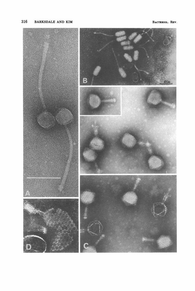
316 BARKSDALE AND KIM BACTERIOL. REV.

MYCOBACTERIUM 317
70
150
B/N
ca4
17
\20
FIG. 32. Schematic drawing ofa typical mycobac-teriophage (MC-3) revealed in ammonium molyb-date-stained micrographs. Five capsomeres appear tobe arranged on each face of the hexagonal sides. Thelong, flexible tail consists of about 34 units (cross-striations), two distinct baseplates, and a fiber. (A)and (B) are baseplate models of phage D29. (C) isthat ofphage MC-4. All dimensions are representedin nanometers.
the appearance of an expansion valve with anapical fiber. Examination of the negativelystained electron micrographs ofMC3 reveals 34subunits in the tail and the constricted termi-nal swelling with its fiber. Along the edge ofthe faces of the head, one can count five capso-meres. Phage D29 (Fig. 30B) differs from MC3.Its tail structure sometimes suggests struc-ture A in the diagram and sometimes struc-ture B. Our diagram of the terminal apparatusof MC4 appears as C in Fig. 32. In Fig. 31A isshown phage C-1 of Mankiewicz (767), the hex-agonal heads of which contrast with the ovalheads of phage F-K of Buraczewska et al. (Fig.31B). Table 9 offers a list of representative my-cobacteriophages with their major structuralfeatures.
SerologyAs with other viruses, the simplest technique
for the serological characterization of mycobac-teriophages is the neutralization of infectivitywith specific antisera. Phages of the CMNgroup are said to be poor antigens (163, 685).However, there are published examples ofphages that induced the formation of relativelygood neutralizing sera (164, 838, 1045) and oth-ers where the titers of the antisera were low(166, 380, 660). Bowman has compared the ca-pacity of sera prepared against phages R1, D29,and Leo to neutralize heterologous infectivityand found no cross-neutralization; i.e., the fix-ing and adsorption apparati of each of thesephages seem serologically distinct (164). Cas-telnuovo et al. (223) have examined the proteinantigens of phage phlei liberated by KOH hy-drolysis, and Castelnuovo et al. (220) have stud-ied, by immunodiffusion and immunoelectro-phoresis, the antigens of whole and disruptedphage phlei. Using antiphage phlei sera theyhave looked for lines of identity with antigensfrom six other mycobacteriophages. Two sharedlines were found between phage minetti andphage phlei, but no sharing was apparent withantigens from the other five phages, which in-cluded D12, D29, and D35. Since phage minetticame into being (by recombination betweenresident prophage and phage phlei?) followingthe growth of phage phlei in the minetti strainof M. fortuitum, some antigenic sharing be-tween the two might be expected. What is im-pressive is the lack of serological relatednessbetween six of the seven phages. Thus, theexperience of several investigators indicatesthat mycobacteriologists have at their disposala number of serologically identifiable and dis-tinct phages.
Lipoidal antigens of mycobacteriophageshave not yet been reported (166, 1046). As willbe seen from the next section, lipoidal compo-nents are essential for the infectivity of somemycobacteriophages. That these lipids func-tions as antigens remains to be established.Phages phlei have been employed in studies
of the in vitro synthesis of antibodies by rabbitspleen cells (339, 510).
FIG. 31. (A) Mycobacteriophage C1. Hexagonal head structure is not clearly shown. Cross-striations andbaseplates are noticeably missing. The diameter ofthe tail at itsjuncture with the head is 7 nm. Its diameter atthe end is 13 nm. This phage offers an exaggeration of the tail entasis typical of mycobacteriophages.(x300,000; bar = 01 jam.) (B) Mycobacteriophage F-K. (From reference 200, by permission ofthe author andpublisher.) Shows an oval head and long, noncontractile tail with bulbous baseplate. (C) MycobacteriophageI3. (From reference 658, by permission of the author and publisher.) This is the only mycobacteriophage todate which belongs to Bradley's group A. Note the large hexagonal head, the contractile sheath, and thehollow canaliculus spent of its DNA. (x180,000.) (D) Mycobacteriophage I3 (our preparation), showingdisrupted phage head with closely packed globular capsomers (9.5 nm in diameter, and center-to-centerdistance is 13 nm) in hexagonal arrays. (x264,000.)
VOL. 41, 1977

TABLE 9. Anatomy of representative mycobacteriophageSa
Mycobacterio- Dimensions (nm) Triatil Tail fiberMycobacterio- Terminutal tail (number, Propagating Mycobacterium Referencephage Headb Tail structure nm)
MC-1 Icosahedron,d 50 125 x 10 2 Disks 1, 20 M. smegmatis, 607 K and BeMC-3 Icosahedron'd 63 150 x 10 2 Disks 1, 20 M. smegmatis, 607 K and BMC-4 Icosahedrond 45 125 x 10 4 Rings 3-4, 20 M. smegmatis, 607 K and BD28 Hexagonal, 65 170 x f Bulbous M. lacticola, F13 1047D29 Icosahedron,d 63 135 x 10 Plate with spikes M. smegmatis, 607 K and BD32 Hexagonal, 80 220 x Bulbous Mycobacterium sp. F119 1047D34 Hexagonal, 65 250 x Bulbous Mycobacterium sp. F130 1047C1 Icosahedron,d 50 200 x 13 Swollen club N.O.9 M. smegmatis, 607 K and BC2 Icosahedron, 60 200 x 7.5 Platelike M. smegmatis, 607 810AG1 Hexagonal, 65 120 x 10 Bulbous M. kansasii 200BG2 Icosahedron, 80 280 x 15 2 Disks 2, 70 M. smegmatis, 607 200BK1 Octahedral, 80 280 x 16 Bulbous 2, 70 M. smegmatis, 607 200GS7 Hexagonal, 83" 135 x 12" Bulbous M. smegmatis, 607 53367 Hexagonal, 60 180 x 15 2 Disks 2, 70 M. pellegrino 200MyF3P/59a Hexagonal, 52 123 x M. fortuitum (minetti) 843B1 Octahedral, 50 145 x 9 Plate with spikes Mycobacterium sp. Jucho 1130B2, B3, A2, Hexagonal, 70 140-170 x Bulbous Mycobacterium sp., Jucho 1133A3, A4, A5 10
Y7, Y10 Octahedral, 80 260 x 15 Bulbous Short fi- Mycobacterium sp., Jucho 200bers
3111-D Oval, 90 x 45 150 x 15 Bulbous Short fi- M. smegmatis, 607 200bers
3215-D Oval, 90 x 45 190 x 14 Bulbous Short fi- M. smegmatis, 607 200bers
F-K Oval, 95 x 50 215 x 16 Bulbous N.O. M. smegmatis, 607 200F-S Oval, 95 x 50 235 x 16 Bulbous N.O. M. smegmatis, 607 200R1 Oval, 100 x 60 200 x 20 Bulbous Short fi- M. butyricum 168
bersI3 Hexagonal, 85 85 x 12 Platelike M. smegmatis, 607 or.SN2 K and B,
658
aSee Tables 1, 2, and 3.bHexagonal and octahedral: refer to outline of head of phage.c Tail type: among the phages listed here, only I3 exhibits a tail having a contractile sheath. It is the only one of these
mycobacteriophages belonging to Bradley's group A (174). All others belong to Bradley's group B.d Most likely structure is an icosahedron.eK and B, Kim and Barksdale, unpublished data.' Blank spaces indicate that information is not available.o N.O., Not observed.hEstimated from data in reference.
Chloroform-Sensitive Mycobacteriophages
Some mycobacteriophages are exceptionallysensitive to chloroform (165, 380, 1046); othersare not (955). Exposure ofphages D4, D29, D28,and D32 in broth to 30% (vol/vol) chloroform for1 h at 3700 followed by removal of solvent andevaporation to dryness (1 h), resulted in a dropin titer of 7 logs (1046). N-butyl alcohol wasalmost as effective. Ethyl ether produced amore marked effect on D4 and D28 than on D32and D29. It was effective in bringing aboutinactivation even when lowered to 10% (vol!vol). Methanol and benzene varied according tothe phage being examined. These results sug-gested that the integrity of the adsorption andinjection apparati ofthese phages involved neu-tral and phospholipids. Bowman et al. (167)have examined highly purified, chloroform-sen-sitive mycobacteriophage DS6A grown onH37Rv and found 11.7% of its dry weight to be
lipid. Of the phage lipid, approximately 53.8%(6.2% of the dry weight) was phospholipid. Acomparison by TLC of H37Rv lipids and thelipids of phage DS6A revealed similar, but farfrom identical, patterns. Jones et al. (564) havereported that phage D29 contains no phospho-lipid. No phospholipid has been reported inphage R1 (362, 380). Thus, of the chloroform-sensitive mycobacteriophages, R1 (362, 380),Leo, D29, DS6A (165), D4, D28, and D32 (1046),only DS6A is known to have an essential phos-pholipid component. The only other bacterio-phage known to have a major phospholipidcomponent is the marine, ether- and chloro-form-sensitive, tail-less, spiked pseudomonadphage, PM2, in which 75% of the total lipid hasbeen reported to be phospholipid (351). Subse-quently, in a paper concerning the molecularultrastructure of PM2, preliminary data weregiven which established the percent phospho-lipid of total lipid as greater than 90 (492). The
318 BARKSDALE AND KIM BACTLzMOL. REV.

VOL. 41, 1977
mycobaceriophages, then, should be interestingto classify, for in addition to the usual proper-ties of overall size, shape, DNA content, hostrange, and serological peculiarities, they maypossess an essential lipid component, either aneutral lipid or phospholipid, or both or nei-ther. These are characteristics that can easilybe screened for by exposure to lipid solventsfollowed by assay for viral activity.
Lysogeny and PseudolysogenyThe greatest number of wild-type phages in
nature exist in lysogenic bacteria (97). Amongcommon examples of lysogenic mycobacteriaare M. tuberculosis H37Rv (1111), M. bovis(531), M. marinum (1059, p. 54), M. fortuitum(883), M. smegmatis (155, 532), and others (201,308, 1200). An impressive study concerning thelysogenization of mycobacteria was carried outby Russell et al. in 1960 (1003). They were ableto establish lysogenic strains of M. tuberculo-sis, M. smegmatis, M. phlei, and M. fortuitum.Investigators of lysogeny in mycobacteria haveoften rediscovered the fact that the demonstra-tion of lysogeny is much facilitated when thereis available a suitably sensitive (indicator) bac-terium. Many papers about experimental lyso-geny in mycobacteria have in fact been aboutthe phage carrier state or pseudolysogeny (97).Baess has reviewed pseudolysogeny and pre-sented a case in point (80). Russell et al. (1003),in their studies, were well aware ofthe possibleconfusion of lysogeny with pseudolysogeny andwent to considerable lengths to obviate suchconfusion. If corynebacteriophages can betaken as models of the CMN group, then it canbe said that most lysogenic mycobacteria areresistant to phages homologous to their carriedprophages (518, 742) and rare exceptions to thisgeneralization (97) appear to be of little conse-quence outside the experimental genetics labo-ratory. This resistance, lysogenic immunity(742, 947), results from the synthesis of a spe-cific prophage-encoded repressor which shutsoff the expression of viral functions in the car-ried phage genome. Russell et al. (1004) werethe first to do a controlled study ofthe effects oflysogenic immunity on the response of myco-bacteria to typing phages. The use of lysogenicimmunity in phage typing is discussed furtheron.
Suitability of Mycobacteriophages as"Typing" Phages
In 1973, there was issued the following state-ment by the WHO Cooperative Study on thephage typing of mycobacteria:
Although phage-typing of some pathogenic mi-
MYCOBACTERIUM 319
crobes-e.g., Escherichia coli and the generaSalmonella, Staphylococcus, and Strepto-myces- is in routine use, little progress has yetbeen made in the phage-typing ofmycobacteria,chiefly because mycobacteriophages possess apolyvalence that prevents the reliable identifi-cation of individual species of mycobacterium.Furthermore, laboratories cannot comparetheir results because they employ differentmethods and culture media and also because thephages used cannot be accurately identified. Ithas been shown that internationally reproduci-ble results can be obtained for M. tuberculosisby selecting suitable mycobacteriophages andsensitive culture media, and by using a stand-ardized technique of phage-typing. Under theseconditions, phage lysis may be utilized to clas-sify strains of M. tuberculosis (1109).
Despite the negative tone ofthis report, written20 years after Lwoffs review on lysogeny (742),14 years after the publication of Adams' Bacte-riophages (10), 9 years after the appearance ofthe first edition of Hayes' The Genetics ofBac-teria and Their Viruses (501), 20 years afterAnderson and Felix presented accumulatedfacts about the Vi-type determining phages ofS. typhi (37), and 16 years after Anderson hadspelled out the broad effects of bacteriophagesin bacterial ecology (36), there is every reasonto believe that a satisfactory phage-typingscheme can be worked out for the mycobacteria.Mycobacteriophages are essentially like otherphages. For example, there exists a large num-ber of mycobacteriophages capable of replicat-ing in a variety of mycobacterial hosts (567,1004).
In Fig. 29, 30, and 31 are shown representa-tive mycobacteriophages that vary in size andshape and complexity of tail structure. So far aswe know, the genomes of these phages arefound to be double-stranded DNA (809). RNA-containing mycobacteriophages and mycobac-teriophages characterized by single-strandedDNA remain to be discovered. The isolationand purification of the phages do not seemterribly difficult, since Pokorny and associatesreport ultrafiltrates of phage D29 (Fig. 30B) of4.2 x 1015 particles per ml (932). The prepara-tion of functional DNA from mycobacterio-phages has been accomplished in successfultransfection experiments by Tokunaga andSellers (1158) and by Nakamura (867). Myco-bacteriophages respond to the mutagenic ef-fects of N-methyl-N'-nitro-N-nitrosoguanidine(838). As already pointed out, mycobacterio-phages range from good inducers of specificphage-neutralizing antibody to those behavingas moderately good to poor antigens.
It has long been known that certain mycobac-

320 BARKSDALE AND KIM
terial host strains permit the replication of cer-tain mycobacteriophages at one range of tem-peratures. For example, 6 of 9 different strainsof M. avium resisted the destructive effects ofsix phages when growth occurred at 370C.When growth occurred at 420C, there was adifferential pattern of susceptibility: somephages grew in some strains and not in others(406). The discoverers of this example of tem-perature-sensitive permissiveness in mycobac-terial phage-host systems pointed out its poten-tialities for lending additional discriminatorypowers to a set of typing phages. Phage geneti-cists use temperature-sensitive mutants (tsmutants) of phages as sources of markers forexploitation in genetic mapping. Mizuguchiand Sellers have used matings between ts mu-tants of mycobacteriophages D29 and D29A forestablishing a rudimentary genetic map ofphage D29 (838). V
Not only can mycobacteriophages be readilymutagenized, any phage stock of 1010 or moreparticles will contain a number of sponta-neously occurring mutant particles includingmutants that attack additional hosts (ex-tended-host-range mutants: see [10, 1045, 406]).All that is needed for the selection of suchmutants are sensitive host cells (host cells hav-ing suitable phage receptors and no inimicableendonucleases). Just as a high-titer stock ofphage is a source ofmutant particles, so a denseculture ofbacteria is a source ofphage-resistantbacterial mutants (mutants which fail to makea particular receptor). As is well known, a high-titer stock of phage is also a powerful tool foreliminating the predominant sensitive mem-bers of the bacterial indicator population andselecting out the rare resistant (mutant) cells.Cultures ofthese resistant clones in turn can beused in a reversed procedure for selecting hostrange mutants of phage. For isolating new my-cobacteriophages from the wild, Grant (464) hasdesigned membrane filter sandwiches of spe-cific mycobacteria, on which phages aretrapped prior to enrichment through incubationwith the desired host strain.
In many phage-host systems, the host cellhas the capacity to destroy the incoming phageDNA through the agency of an endonucleaselocated near the cytoplasmic membrane (50,172). Such host cells also have the capacity tomodify entering DNA that has escaped thecell's protective endonuclease. When occasionalphage (DNA) genomes become so modified,stocks of them can be prepared which bringabout the lysis of a previously unsusceptible(restricting) bacterium. Thus, the combinationofhost restriction and modification also offers a
means of expanding the lytic potential of aparticular phage and, therefore, its host range.The authors, in collaboration with Pollice, havefound restricting and modifying systems to becommon among mycobacteria and nocardias.This was to be expected, since such endonuclea-ses and DNA-modifying systems are commonamong corynebacteria (96, 685). Millman (821)was probably dealing with restriction and mod-ification in 1958 when his 607-grown phagefailed to lyse either H37Rv, H37Ra, or BCG,whereas H37Ra-grown phage did lyse both theM. tuberculosis and the M. -bovis strains.Nordstr6m and Grange (883) have exploited thephage-modifying capacities of M. fortuitum inelaborating a phage-typing system for that spe-cies of mycobacterium. Sometimes the processof apparent modification (adaptation) may ac-tually result from recombination between su-perinfecting phage and the resident phage ge-nome, which is itself induced by the superin-fecting phage to assume the vegetative pheno-type (541; for the operation ofsuch a recombina-tional system in the CMN group, see [517]).The existence of the lysogenic state offers
additional means for adding specificity tophage-typing schemes. Sometimes, on oldslants of lysogenic strains, areas of lysis ap-pear. These result from the lytic activity ofvirulent mutants of the carried prophage. Suchmutants have lost the capacity to respond torepressor. They, too, can be used for expandinga typing scheme.
Laboratories given to phage typing are usu-ally dealing with two kinds of lysis: (i) non-productive lysis, murolysis. In many phagestocks there is an accumulation of murolyticenzymes that lyse bacterial cells by destroyingportions of their cell walls. This nonproductivelysis is unrelated to phage multiplication. Suchenzymatic lysis was used by Millman for pre-paring protoplasts of M. tuberculosis and M.bovis (821 see also [990]). (ii) Productive lysis.The specific phage-induced lysis involves phagereplication, so beautifully illustrated at the cel-lular level by the pictures of Kolbel (a la Kel-lenberger, 606) showing the growth of mycobac-teriophage Bo5 in Mycobacterium vaccae (629),where the destruction of the cell coincides withthe liberation of a burst of newly formed bacte-riophages. Each of these two mechanisms oflysis can offer useful information in the identi-fication of unknown mycobacteria.With some understanding of phage-host in-
teraction, the preceding devices for tailoringthe specificity of phages as to host receptors,host endonucleases, lysogenic immunity, per-missive temperatures for replication, plaque
BACTERIOL. REV.

MYCOBACTERIUM 321
morphology (including halos versus no halos[6601), and lysis due to cell wall destructionversus productive lysis due to phage replicationcan be exploited for the phage typing of myco-bacteria. Probably enough mycobacteriophagesalready exist for accomplishing the job. Mucheffort must be expended, however, for produc-ing a workable collection of typing phages.Phage stocks are not indefinitely stable. Prac-ticing laboratories will need to get used to reti-tering stocks almost weekly and to substituteactual numbers of plaque-forming particles forRTD (routine test dilution, 1109). Growing so-called slow-growing mycobacteria such asH37Rv in shake culture so that actively growingcells are available needs to become routine. Inour laboratory plaques can be read on H37Rv, soprepared, in 2 days, and that is the length oftime it takes corynebacteriophages to developplaques on the Park-Williams number 8 strainof C. diphtheriae. Mycobacteria grown onTween-supplemented media are freed of theTween (by dilution or washing) prior to usingthem in phage assays. Thus, plenty of evidenceexists to indicate that, among mycobacterio-phages, there is an adequate source of materialfor designing a system for the definitive typingofmycobacteria. Although theWHO committeehas attributed much of their difficulties withphage typing of mycobacteria to the "polyva-lency" of mycobacteriophages, there seems, infact, little or no justification for such an excuse.
Phage Typing of Mycobacteria such as M.tuberculosis and M. fortuitum
Baess (79) has shown the possibilities of bac-teriophage typing for sorting the strains of M.tuberculosis and, in so doing, has reviewed theefforts in this same direction of earlier workers,including Froman et al. (407), Takeya and asso-ciates (1137), Murohashi and his group (866),and Sugita et al. (1106). The materials andmethods section of Baess' paper clearly spellsout how one uses existing phage stocks for lo-cating host range mutants. She shows that thephage type of M. tuberculosis is, as with otherbacteria, a property of real constancy and asstable as any other inherited trait. Her conclu-sions as regards the usefulness ofphage typingin the epidemiology of tuberculous disease at-tributable to M. tuberculosis agrees with thefindings of Tokunaga et al. (1157). Baess' dis-cussion of the pitfills of phage typing of M.tuberculosis indicates the importance to suchwork of an understanding of the methods ofbacterial virology, including single-plaque iso-lation, adsorption experiments, production ofstocks of high titer, etc. Bates and Mitchison
(105; see also [1201] with regard to phage type ofstrains of M. tuberculosis from Hungary) haveexamined the geographic distribution of M. tu-berculosis according to phage types. Their dataindicate that (i) M. tuberculosis from differentregions tended to be different phage types; (ii)phage type was a constant property, and (iii)the mutation to drug resistance did not involvea change in phage type. Phage type as a stablegenetic marker seems well established for M.tuberculosis. Stable markers can be of use insuch problems of epidemiology as decidingwhether reinfection tuberculosis is endogenous(1091) or exogenous. Both modes certainly oper-ate. Raleigh and Wichelhausen (957) have re-ported a third clinical episode of pulmonarytuberculosis in a patient in whom the finalinfecting strain ofM. tuberculosis was distinctas to drug resistance, urease production, andphage type. A close associate of the patient wasinfected with M. tuberculosis of this samephage type. If one assumes that the firequencyof the mutations in drug resistance pattern,urease activity, and phage type are as high as10-7 each, then the chance that the strain caus-ing the final infection was directly related tothat causing the second infection seems veryremote (i.e., 10-21).
Baess and Bentson, extending the earlier ef-forts of Kappler (581), Rodda (989), Tokunagaand associates (1156), and Juhasz and Bonicke(566), have offered a basis for the developmentof a phage-typing scheme that could distin-guishM. phlei, M. smegmatis, M. fortuitum, M.vaccae and, by extension, other rapidly growingmycobacteria (81). Among the additionalmarkers investigated were (i) growth at 37, 45,and 520C, (ii) ability to deamidate some of 13amides, and (iii) ability to ferment some of 12carbohydrates. Nordstr6m and Grange haveemployed host-induced modification for devel-oping a set of phage capable of discriminatingdifferences in strains of M. fortuitum (883).These authors also have established a correla-tion between inositol utilization by strains ofM. fortuitum and their susceptibility to a par-ticular phage, BK4 (462). Gunnels and Bateshave shown that 'typing" phages can be usedfor identifying M. xenopi (478).
Host Cell Receptors and Mycobacteriophage-Induced Receptor-Destroying EnzymesImaeda and San Blas found that the recep-
tors for a phage, GS-7, were located in a lipoidalcomponent of the cell wall ofM. smegmatis 607(533). The glycolipids and peptidoglycolipidsare surface components of the mycobacterialcell, and the first act of the infecting virus is to
VOL. 41, 1977

322 BARKSDALE AND KIM
fix to the surface. One would expect to encoun-ter mycobacteriophages capable of attaching toa variety of mycobacterial surface components.The virus does not distinguish isolated recep-tors from in situ receptors. Some viruses, afteradsorption to isolated receptors, "inject" theirDNA into the milieu. The fixation to isolatedreceptors effectively eliminates the infectivityof the virus. Thus, the receptor suitability of acell component can be demonstrated by thesame general techniques used for demonstrat-ing the neutralizing capacity of antiviral anti-body. The mixing of small amounts of prepara-tions of mycosides C (see section on Peptidogly-colipids) with phage D4 effectively neutralizedthe infectivity of it but not that of unrelatedphages. Castelnuovo et al. recovered a glyco-lipid (lacking any ninhydrin-positive compo-nent) from extracts of the cell envelopes of M.phlei which inactivates phage phlei, causing adrop in infectivity of4 logs in 60 min (221). Oncea number of specific, distinct, and chemicallycharacterized receptor substances becomeavailable they can be used to classify the recep-tor apparatus of a particular phage simply bytesting their capacity to neutralize the infectiv-ity of that phage. This would enable laborato-ries to gain such information without resortingto the sophisticated chemistry needed for char-acterizing mycobacterial receptor lipids.
Bacteriophages of many classes of bacteriahave associated with their replication the syn-thesis of enzymes capable of degrading thosesites on the bacterial surface to which thephage attaches. Such catalysts can conven-iently be referred to as receptor-destroying en-zymes (RDE; 208). Although the original desig-nation, RDE, referred to the destruction ofneuraminidase-sensitive receptors on mamma-lian cells (207), the term is here used to coverall receptor-destroying enzymes. The plaques ofphages that produce RDE usually are sur-rounded by a halo. Gratia (465) long agoshowed that from the center ofsuch plaques onecould recover phage, whereas from the zone ofthe halo or beyond no phage was to be found. Inthe lyses that produced the phages at the centerof the plaque, RDE was also produced; the en-zymes then diffused to the edge of the halo,changing the surface properties of the bacterialcjlls.Such bacteria, lacking receptors, are pheno-
typically resistant to the bacteriophage respon-sible for receptor-destroying activity. In theCMN group, it is sometimes difficult to see thehalos of plaques because the amount of sub-strate produced may so overwhelm the enzymethat no optical difference is apparent between
BACTERIOL. REV.
the bacteria within the zone of RDE and thosebeyond. This matter can be obviated by allow-ing the phages and bacteria to interact prior toplating out in a lawn of bacteria. The concen-tration of bacteria should be minimal. After theplaques have developed, a second lawn of sensi-tive bacteria is layered over the first. Lysisproceeds, and the accumulated, plus current,yields ofRDE produce a visible halo. The halo-producing property ofphage Bol growing on M.smegmatis 607 has been documented by Kraisset al. (660). Pictures of halo-forming corynebac-teriophages are illustrated on p. 406 of refer-ence 96. A collection of RDE should be of greatvalue to those laboratories devoted to the studyof the chemistry of mycobacterial lipids.Jones and David (562) have made a prelimi-
nary report of the production (during one stepofgrowth ofphage D29 on M. smegmatis 607) ofa lipase that liberates lauric acid from Tween20. Similar lipase activity was found in cells ofM. fortuitum infected with D29, but not in un-infected cells ofeitherM. fortuitum orM. smeg-matis 607. A function for the lipase in thephage-infected cell has not yet been sought.
MYCOBACTERIOCINSAlthough low-molecular-weight bacteriocins
(174) produced by mycobacteria and inhibitoryfor other mycobacteria have been known since1958 (851), these dialyzable antibiotics have notyet been used in a system for ordering relation-ships among mycobacteria. Additional myco-bacteriocins have been described (9, 1136), andTakeya and Tokiwa have had some successwith preliminary sorting of M. tuberculosis(nine strains) with 11 bacteriocins (1136). Thetyping of new isolates from the same patientsover a period oftime showed the infecting typesto remain the same. We have encountered noreports of high-molecular-weight bacteriocins(174) active on mycobacteria. Since the high-molecular-weight bacteriocins occur for a widevariety of bacteria, one would expect some to befound which are active on mycobacteria. Even-tually, it should be possible to combine phagesand bacteriocins into one typing scheme.
SOME ABBREVIATIONS USED IN THETEXT
ACP1)-AlaL-AlaAMPATPB cells or B lym-phocytes
Acyl carrier proteins-AlanineL-AlanineAdenosine 5'-monophosphateAdenosine 5'-triphosphateLymphoid stem cells whichhave had an extrathymicmaturation

MYCOBACTERIUM 323
BCG
BCG-itis
BGGBSAC'3 or C3
CBH
CCB
CF
CFACLCMICMN
CoA or CoASHCon ACPIA
CW
CWS
CYDAP (DPM)DHDNADNFBDMAPP
DS 500EDTA
EMEPPETPETZFAFab or Fab frag-ment, two suchfragments
Fc or Fc fragment
ft-c
M. bovis strain BCG, bacilleCalmette Guerin
Regional lymphadenitis some-times resulting from vac-cination
Bovine gamma globulinBovine serum albuminThe third component of com-plement (Cl-9)
Cutaneous basophil hyper-sensitivity
Competency for Clearing Ba-cilli test
Cord factor(s), dimycolates oftrehalose
Complete Freund adjuvantCardiolipin(s)Cell-mediated immunityCorynebacterium, Mycobacte-rium, Nocardia group
Coenzyme AConcanavalin A2- (4-Chlorophenylthio)-tri-ethylamine hydrochloride
Cell wallCell wall skeleton: residual
cell walls (one-third ofstarting material) obtainedfollowing freeing of proteinwith proteinase and of freelipids with neutral organicsolvents (69)
CyclophosphamideDiaminopimelic acidDelayed hypersensitivityDeoxyribonucleic acidDinitrofluorobenzene3,3-Dimethylallyl pyrophos-
phateDextran sulfate 500Ethylenediaminetetraacetic
acidElectron micrographErythropoietic porphyriaElectron transport particlesElectron-transparent zoneFatty acidConsisting of one heavy andone light chain, each con-taining one antibody com-bining site. Are derived bypapain hydrolysis as in fol-lowing.
Crystallizable fragment ob-tained by papain hydroly-sis of molecules of immu-noglobulin: e.g., IgG Fcconsists of C-terminalhalves of two heavy chainslinked by disulfide bonds(molecular weight, 50,000).Has no antibody activity,can fix to complement.
Footcandles
FAS IFAS IIFBG
FMN (FMNH2)
G+CGDP-man
GIFGlnD-GluGlucNAcH37Ra
H37Rv
HEFHG
HIHSAIDIFAIgGIgMIPP'UKLHKTCLMFLPS
MAPMER
MER-FAMFMGLP
MIFMMP
MurNAcMurNGlmyc RNA
NAD+ (NADH)
NADP+ (NADPH)
OTOTZPCA
PEPEcPHAPhe I
Phe II
Fatty acid synthetase type IFatty acid synthetase type IIForeign body-type granu-loma
Flavin mononucleotide (re-duced form); same as ribo-flavin phosphate
Guanine + cytosine contentGuanosine diphosphoman-nose
Growth inhibitory factorGlutamineD-Glutamic acidN-acetylglucosamineM. tuberculosis attenuated
strain derived from H37RvM. tuberculosis, standard
virulent strain. For originssee text.
Heptane-soluble fatty acidsHypersensitivity-type granu-loma
Humoral immunityHuman serum albuminImmunodiffusionIncomplete Freund adjuvantImmunoglobulin GImmunoglobulin MIsopentenyl pyrophosphateInternational test unitKeyhole limpet hemocyaninKiller T cell (lymphocyte)Lymphocyte mitogenic factorLipopolysaccharide fromgram-negative bacteria
Murine acid phosphataseMethanol-extracted residue
of cells of BCGMER in IWAMillipore filterMethylglucose-containing li-
popolysaccharidesMigration inhibition factor3.0-methyl mannose poly-
saccharideN-acetylmuramic acidN-glycolylmuramic acidRibosomal fractions of (from)mycobacteria
Nicotinamide adenine dinu-cleotide (reduced form)
Nicotinamide adenine dinu-cleotide phosphate (re-duced form)
Old tuberculinOuter transparent zonePassive cutaneous anaphy-
laxisPhosphatidylethanolaminePeritoneal exudate cellsPhytohemagglutininPhenotype I, an in vivo phe-notype
Phenotype II, an in vitro phe-
VOL. 41, 1977

324 BARKSDALE AND KIM
PI-Man.
PLPMN
poly(A) * poly(U)
PPDPPD-SRCFRDERECRNARTD607
SAFA
SEASLSRBCT
TLCts
2D-IEP
UDPUVWHOWSAWSA-FA
notypePhosphatidylinositol oligo-mannoside
PhospholipidPolymorphonuclear leuko-
cytesPolyadenylic acid -polyuri-
dylic acidPurified protein derivativePPD from M. tuberculosisReference culture filtratesReceptor-destroying enzymesReticuloendothelial cellsRibonucleic acidRoutine test dilutionMycobacterium smegmatisATCC 607
Soluble-antigen fluorescentantibody
Shistosoma egg antigenSulfolipidSheep erythrocytesLymphoid stem cells whichhave had an associationwith thymus or thymosin
Thin-layer chromatographyTemperature-sensitive mu-
tantsTwo-dimensional immunoe-
lectrophoresisUridine 5'-diphosphateUltraviolet lightWorld Health OrganizationWater-soluble adjuvantWSA substituted' for myco-
bacteria in CFA, i.e., WSAplus IFA
SUMMARYThe Mycobacterial Cell
Mycobacteria are gram-positive, nonmotile,nonsporeforming, pleomorphic rods. The cellu-lar morphology of several species is not easilydistinguished from that ofother members oftheCMN group, Corynebacterium and Nocardia.Some mycobacteria show a tendency to formaerial filaments: they range from nonfilamen-tous M. tuberculosis to M. fortuitum, with lim-ited filamentation and fragmentation, to fila-mentous M. farcinogenes.Mycobacterium can be distinguished from
the genera Corynebacterium and Nocardia onthe basis of certain ropelike structures oftenfound as a superficial "layer" (see Fig. 6, 25,and 26). Mycobacteria exhibit an acid-fastnessdistinct from the acid-fastness of Corynebacte-rium, Nocardia, bacterial spores, fingal spores,human sperm, etc. Mycobacterial acid-fastnessis lost upon removal of the ropelike structures.Mycobacterial acid-fastness is compoundedfrom two events: (i) the formation of aryl-
methane-mycolates between fuchsin and themycolic acids of the ropelike structures and (ii)the trapping of intracellular fuchsin by the dye-complexed ropelike structures in conjunctionwith other components of the outer cell wall.Mycobacteria may be non-acid-fast (and gener-ally chromophobic) when they produce sub-stances that cover up their ropelike structures.
Ultrastructure. Electron micrographs of ac-tively growing mycobacteria, negativelystained, freeze fractured, metal shadowed, orultrathin sectioned and stained, indicate thatbeyond the cytoplasmic membrane there is arigid layer, the murein, upon which is super-posed three discernible layers: L3, L2, and L,(Fig. 4 and 5). At the macromolecular level,these are apparent (only) layers deriving frominterconnecting structures stretching outwardfrom the basement muramyl peptidoglycan, aspredominantly arabinogalactan-mycolate withsome arabinomannan and some peptidolipid,the ropelike structures of peptidoglycolipid (L2)terminating in ribbons at the cell surface (L,).Interspersed in these same outer areas are my-colates of trehalose, including sulfolipids andcord factors.The average size of the DNA of the mycobac-
terial nucleoid is around 3.0 x 10⁹ daltons, andits G+C content is about 65%. Sections ofmyco-bacterial cells in logarithmic growth exhibitfibrillar DNA associated with mesosomal ex-tensions of the cytoplasmic membrane system.The cytoplasm is rich with densely packed ribo-somes. Cells in which metabolism is not atmaximum rate contain lipoidal bodies, meta-chromatic granules, and small, dense granulesof unknown composition. The ultrastructure ofthe mycobacterial cell (see foregoing) indicatesthat it is capable of a variety of biosyntheticprocesses.
Biosyntheses. Mycobacterium, Corynebacte-rium, and Nocardia are remarkable for thelipids they synthesize. M. phlei (and C. diph-theriae) possesses a stable, multienzyme com-plex, FAS I, carrying its own ACP, which iscapable of catalyzing the synthesis of long-chain FAs as follows:CH3-CO-SCoA + nHOOC-CH2-CO-SCoA + xTPNH+ yDPNH + (x + y)H+ -* CH3-[CH2-CH2]1-CO-SCoA + nCoASH + nCO2 + xTPN+ + yDPN+ +
nH20
(143). In addition, this complex catalyzes theelongation of acyl-CoA derivatives with carbonchains up to C20. Special mycobacterial methyl-ated polysaccharides and lipopolysaccharidesare capable of markedly activating FAS I. M.phlei, also, contains a FAS II (malonyl-CoAincorporation), which requires added ACP.
BAC-rRIOL. REV.
MYCOBACTERIUM 325
Among the major phospholipids of mycobacte-ria are PEs, diphosphatidyl glycerides, and my-oinositol phospholipids, including a phosphati-dylinositol pentamannoside. When providedfree FA as palmitate or in serum or from lipidsavailable in tissue, mycobacteria produce visi-ble intracellular globules of lipid, probablystored as di- and triacylglycerides (see Fig. 13).The acylating capabilities of the CMN groupare also rapidly turned on in the presence ofexcess glucose, and acylglucose is produced.The carotenoid pigments of mycobacteria, ex-
ploited for taxonomic purposes, are synthesizedconstitutively by some strains, inducibly bysome, and not at all by others (see Fig. 16 and17). The protection these polyene compoundsafford the mycobacterial cell relates to the sen--sitivity of mycobacteria to toxic photoproductsinitiated by white (perhaps, also UV) light. Pro-toporphyrin IX or undesignated flavins or all ofthese appear required for the light-induced for-mation of these toxic substances. Whether res-piratory quinones are destroyed upon exposureof mycobacteria to light, as in Sarcina lutea,has not been determined. Menaquinones areimportant respiratory molecules in mycobacte-ria.Taxonomy. The antigenic analysis of myco-
bacteria is, after 50 years of intensive effort,still in its infancy. Valuable advances havebeen made regarding the serology ofM. avium.As is so often the case, no correlation betweenvirulence and serological type has been estab-lished. Soluble antigens of mycobacteria havebeen examined by a variety of means, oftenwith interesting results. Among these havebeen attempts to establish relatedness throughimmunodiffusion patterns. The interpretationofsuch patterns is markedly limited by the lackof either well-characterized antigens or well-characterized antibodies. In this connection,there has been generally admitted a need formeaningful reference antigens and antisera.The delineation of species within Mycobacte-
rium is accomplished through the use of adozen or more tests including: presence or ab-sence of enzymes such as catalase and arylsul-fatase, rates of growth, capacity to produce ca-rotenoid pigments, the secretion of niacin, sen-sitivity to sodium chloride, the reduction of tel-lurite, and the hydrolysis of polyoxyethylenesorbitan monooleate.
Mycobacteriophages and Geneticsof Mycobacteria
A number of mycobacteriophages exhibitinga wide range of ultrastructure are describedherein (Fig. 29 through 32) and await exploita-
tion for the taxonomy and the genetics ofMyco-bacterium. Genetic recombination has beeneffected between two strains of M. smegmatis.
Interaction with the Animal HostMacrophages. Many mycobacteria are capa-
ble of surviving in macrophages of animalhosts. Apparently, the phagosome-lysosome fu-sion associated with the killing of bacteria isnot very evident in macrophages that have en-gulfed, for example, M. tuberculosis H37Rv.Even when such fusion has been induced innormal macrophage, H37Rv is not killed. Thosemycobacteria that survive in macrophages ofanimal hosts assume a phenotype (Phe I) differ-ing in distinct ways from their homologous invitro (Phe II) phenotype: e.g., in metabolizingcertain energy sources, in producing detectablesulfolipid, or in immunogenicity. 'Clones" ofPhe I of M. tuberculosis occur principally intuberculous granulomas, adventitious coloniesfrom the reticuloendothelial system, consistingof macrophages gorged with tubercle bacilli,epithelioid cells, and multinucleated giantcells, all surrounded by fibroblasts and lympho-cytes. New macrophages constantly enter thegranulomas, phagocytosing bacilli and frag-ments from dying macrophages. There is evi-dence to support the assumption that in statesof specific delayed hypersensitivity related toPPD, there is an enhanced activity of macro-phages within granulomas. A factor found inBCG-immune serum (rabbit) induces giant cellformation in macrophages from nonimmunerabbits when incubated with heat-killed BCG.Nocardia braziliensis can be substituted forBCG in this induction, suggesting that perhapsother members of the CMN group might func-tion similarly.Immune response. In experimental infec-
tions with M. tuberculosis in guinea pigs and inhuman tuberculosis, there is a turning on ofboth "humoral" (HI) and "cellular" immunity(DH and CMI), all mediated by cells. Folklorehas long held that tubercular individuals areless prone to the common run of infections thannontuberculars. The laboratory contribution tothis opinion derives from the establishment ofthe adjuvant action of mycobacteria. Livingmycobacteria administered to animals alongwith antigen X bring about an enhancement inthe production of antibodies versus antigen X.Although dead mycobacteria cannot substitutefor living mycobacteria as adjuvants, dead my-cobacteria in a water-in-oil emulsion (CFA) arewell-known adjuvants. Immunization with an-tigen X in CFA can lead to (i) enhanced produc-tion of antibody versus antigen X and (ii) DH
VOL. 41, 1977

326 BARKSDALE AND KIM
versus antigen X. (iii) When administeredwith certain tumor antigens, Y, or when saidtumor antigens, Y, are administered withCFA, there develop lymphocytes cytotoxic invitro for Y tumor cells. (iv) In addition, adju-vant arthritis and general adjuvant diseasemay develop in a number of animals receivingCFA. Variations on the time of administrationand amounts of doses of CFA can suppress theimmune response. Under certain conditions the'full development ofDH may require suppressorB cells. When CFA is administered with twodistinct antigens, the outcome of the antibodyresponse may be altered: the phenomenon, an-tigenic competition. Various side effects ofCFAmake it unsuitable for immunization of hu-mans. Living BCG and living M. microti arethe mycobacterial vaccines most studied inman. They, too, in a small number of subjects,give untoward reactions. Many concerned withdeveloping mycobacterial vaccines hope to findmycobacterial components capable of immuniz-ing and/or acting as adjuvants without produc-ing damaging side effects. In a number of hu-man subjects suffering from chronic tuberculo-sis, as well as in about 25% of rabbits sufferingfrom experimental chronic infections includingtuberculosis, there develop autoantibodies thatare cytotoxic for the animals' cells growing intissue culture.
Virulences of mycobacteria are no more andno less complicated than virulences of Histo-plasma, Leishmania, or Candida. Virulence inM. avium appears to be associated with a par-ticular surface antigen. Virulence in M. tuber-culosis is associated with those molecular spe-cies that make possible "cording" of colonies. Inrabbits and man, there are also genetic factorsof the host that affect the outcome of infections.The extent to which these relate to antigenscommon to host and bacterium is just nowbeing questioned.Components inducing specific host re-
sponses. Fruitful results in localizing the bio-logical activity(ies) of mycobateria to certainsubcellular components follow.
(i) Cord factor. The dimycolate(s) of treha-lose, cord factor(s), (a) are toxic for mitochon-drial membranes, (b) when conjugated to asuitable carrier serve as functional haptens, and(c) exhibit adjuvant activity when administered(as a mixture designated P3) with PPD. Toxicityof these dimycolates depends upon certain freehydroxyl groups on the molecules of trehalose.A related sulfur-containing molecule, mycobac-terial sulfolipid, appears capable of enhancingthe toxicity of cord factor.
(ii) Nonmurein peptides, tuberculin-activepeptides. Many mycobacterial elicitins (tuber-
culin-active peptides) appear to originate fromthe mycobacterial cell wall. Tuberculin re-mains the most widely studied such mycobacte-rial product. As OT and various commerciallyavailable PPDs, it is comprised of at least fivedistinct elicitins. Although PPD itself does notsensitize animals to a state of tuberculin posi-tivity, PPD plus poly(A) -poly(U) does effectsensitization.
(iii) Muramic acid-linked dipeptides. Thesugar dipeptide, N-acetylmuramyl-L-alanyl-D-isoglutamine, administered in mineral oil withantigen X, brings about an enhancement ofspecfic antibody synthesis and the developmentof DH to antigen X.
ACKNOWLEDGEMENTSWe are much indebted to Marjorie Christiansen
Pollice for expert help with the manuscript, its liter-ature and related mycobacteria. We thank YoshieHervey for her various contributions; S. Bruce Ar-den and Arthur Stanley Eckman for work leading tothe production of Tables 1, 2, 3, and 4, MicheleneMathews-Roth for her comments concerning the sec-tion devoted to carotenoid pigments, and Ruth Gor-don for helpful prescriptions during periods of taxo-nomic colic. We thank John Foo Wong for many dis-cussions about mice, macrophages, MIFs, andMAFs. The help ofthe editorial board of this journaland its ad hoc referees, particularly that ofMayer B.Goren, is gratefully acknowledged. Gilbert Claus-man, Elizabeth Decker and Eleanor Pasmik (NewYork University Medical Center Library) were un-stinting in their aid of our pursuit of "outside" jour-nals. We are grateful to C. Harman, G. Davy, andG. Ojakian for help in the use of the Balzer freezeetching apparatus and to Geraldine Hodgson andEllena Kappa for producing the typescript.The original work reported here was supported by
the Andrew Mellon Education Fund, Public HealthService General Research Support Program, PublicHealth Service grant AI-01071 from the NationalInstitute of Allergy and Infectious Diseases and agrant from the World Health Organization (contrib-uted by Emmaus-Suisse). One of us, L. B., was therecipient of a fellowship from The John Simon Gug-genheim Foundation, which made possible thegroundwork for this review and its sequel concern-ing etiology of leprosy.
Note Added In PressThe substitution of "et al." for names of investi-
gators referred to herein results from a policy ofBacteriological Reviews.
LITERATURE CITED1. Aaronson, C. M., and C. H. Park. 1974. Spo-
rotrichoid infection due to Mycobacteriummarinum: lesion exacerbated by corticoste-roid infiltration. South. Med. J. 67:117-118.
2. Abrahams, E. W. 1970. "Original mycobacte-rial sin." Tubercle 51:316-321.
3. Acharya, P. V. N., and D. S. Goldman. 1970.
BACTERIOL. REV.

MYCOBACTERIUM 327
Chemical composition of the cell wall of theH37Ra strain of Mycobacterium tuberculo-sis. J. Bacteriol. 102:733-739.
4. Acharya, P. V. N., M. Senn, and E. Lederer.1967. Sur la presence et la structure de my-colates d'arabinose dans les lipides lies dedeux souches de Mycobacteries. C.R. Acad.Sci. Ser. C 264:2173-2176.
5. Adam, A., R. Ciorbaru, F. Ellouz, J.-F. Petit,and E. Lederer. 1974. Adjuvant activity ofmonomeric bacterial cell wall peptidogly-cans. Biochem. Biophys. Res. Commun.56:561-567.
6. Adam, A., R. Ciorbaru, J.-F. Petit, and E.Lederer. 1972. Isolation and properties of amacromolecular, water-soluble, immuno-adjuvant fraction from the cell wall ofMyco-bacterium smegmatis. Proc. Natl. Acad.Sci. U.S.A. 69:851-854.
7. Adam, A., R. Ciorbaru, J.-F. Petit, E. Led-erer, L. Chedid, A. Lamensans, F. Parant,M. Parant, J. P. Rosselet, and F. M. Ber-ger. 1973. Preparation and biological prop-erties of water-soluble adjuvant fractionsfrom delipidated cells of Mycobacteriumsmegmatis and Nocardia opaca. Infect. Im-mun. 7:855-861.
8. Adam, A., J.-F. Petit, J. Wietzerbin-Fal-szpan, P. Sinay, D. W. Thomas, and E.Lederer. 1969. L'acide N-glycolyl-mura-mique, constituant des parois de Mycobacte-rium smegmatis: identification par spectro-metrie de masse. FEBS Lett. 4:87-92.
9. Adamek, L., L. Trnka, P. Mison, and M. Gu-tova. 1968. Hemmstoffe vom Bakteriocin-Typus bei schnell wachsenden saprophy-tischen Stammen der Mycobakterien.Beitr. Klin. Tuberk. Spezifischen Tuberk.Forsch. 138:51-55.
10. Adams, M. H. 1959. Bacteriophages, p. 592.Interscience Publishers, New York.
11. Adams, R. M., J. S. Remington, J. Steinberg,and J. S. Seibert. 1970. Tropical fish aquar-iums. A source of Mycobacterium marinuminfections resembling sporotrichosis. J.Am. Med. Assoc. 211:457-461.
12. Aebi, A., J. Asselineau, and E. Lederer. 1953.Sur les lipides de la souche humaine "bre-vannes" de Mycobacterium tuberculosis.Bull. Soc. Chim. Biol. 35:661-684.
13. Affronti, L. F., E. H. Fife, and L. Grow. 1973.Serodiagnostic test for tuberculosis. Am.Rev. Respir. Dis. 107:822-825.
14. Affronti, L. F., and F. B. Seibert. 1965. Myco-bacterial antigen fractionations. Someearly investigations of Mycobacterium tu-berculosis. Am. Rev. Respir. Dis. 92(part 2):1-8.
15. Affronti, L. F., G. L. Wright, Jr., and M.Reich. 1972. Characterization and compari-son of mycobacterial antigens by discontin-uous pore gradient gel electrophoresis. In-fect. Immun. 5:474-481.
16. Aho, K., E. Brander, and T. Estola. 1973.Agglutinins against group III atypical my-
cobacteria. Acta Pathol. Microbiol. Scand.Sect. B 81:589-592.
17. Akamatsu, Y., and J. H. Law. 1968. Enzy-matic synthesis of 10-methylene stearic acidand tuberculostearic acid. Biochem. Bio-phys. Res. Commun. 33:172-176.
18. Akamatsu, Y., and J. H. Law. 1970. Enzy-matic alkylenation of phospholipid fattyacid chains by extracts of Mycobacteriumphlei. J. Biol. Chem. 245:701-708.
19. Akamatsu, Y., and J. H. Law. 1970. The enzy-matic synthesis of fatty acid methyl estersby carboxyl group alkylation. J. Biol.Chem. 245:709-713.
20. Akamatsu, Y., and S. Nojima. 1965. Separa-.tion and analyses ofthe individual phospho-lipids of mycobacteria. J. Biochem. 57:430-439.
21. Akamatsu, Y., Y. Ono, and S. Nojima. 1966.Phospholipid patterns in subcellular frac-tions of Mycobacterium phlei. J. Biochem.59:176-182.
22. Akamatsu, Y., Y. Ono, and S. Nojima. 1967.Studies on the metabolism of phospholipidin Mycobacterium phlei. 1. Difference inturnover rates of individual phospholipids.J. Biochem. 61:96-102.
23. Allen, S. H. G., B. F. Jacobson, and R.Stjernholm. 1964. Biocytin as a constituentof methylmalonyl-oxaloacetic transcarbox-ylase and propionyl CoA carboxylase of bac-terial origin. Arch. Biochem. Biophys.105:494-500.
24. Allison, A. C. 1973. Effects of adjuvants ondifferent cell types and their interactions inimmune responses, p. 73-99. In G. E. W.Wolstenholme and J. Knight (ed.), Immu-nopotentiation. Ciba Foundation Sympo-sium 18 (new series). Associated ScientificPublishers, Amsterdam.
25. Allison, A. C., and A. J. S. Davies. 1971. Re-quirement of thymus-dependent lympho-cytes for potentiation by adjuvants of anti-body formation. Nature (London) 233:330-332.
26. Allwood, G. G., and G. L. Asherson. 1971.Depression of delayed hypersensitivity bypretreatment with Freund-type adjuvants.II. Mechanism of the phenomenon. Clin.Exp. Immunol. 9:259-266.
27. Alsaadi, A-I., and D. W. Smith. 1973. The fateof virulent and attenuated mycobacteria inguinea pigs infected by the respiratoryroute. Am. Rev. Respir. Dis. 107:1041-1046.
28. Alsop, D. G. 1972. The Bairnsdale ulcer. Aust.N. Z. J. Surg. 41:317-319.
29. Altman, L. J., L. Ash, R. C. Kowerski, W. W.Epstein, B. R. Larsen, H. C. Rilling, F.Muscio, and D. E. Gregonis. 1972. Prephy-toene pyrophosphate. New intermediate inthe biosynthesis of carotenoids. J. Am.Chem. Soc. 94:3257-3259.
30. Alvarez and Tavel. 1885. Recherches sur lebacille de Lustgarten. Arch. Physiol. Norm.Pathol. 6:303-321.
VOL. 41, 1977

328 BARKSDALE AND KIM
31. Amar, C., and E. Vilkas. 1973. Isolement d'unphosphate d'arabinose Apartir des parois deMycobacterium tuberculosis, H37Ra. C.R.Acad. Sci. Ser. D 277:1949-1951.
32. Amar-Nacasch, C., and E. Vilkas. 1969.etude des parois d'une souche humaine vir-ulente de Mycobacterium tuberculosis. 1.Preparation et analyse chimique. Bull. Soc.Chim. Biol. 51:613-620.
33. Amar-Nacasch, C., and E. Vilkas. 1970.etude des parois de Mycobacterium tubercu-losis. II. Mise en evidence d'un mycolated'arabinobiose et d'un glucane dans les pa-rois de M. tuberculosis H37Ra. Bull. Soc.Chim. Biol. 52:145-151.
34: American Lung Association. 1974. Diagnosticstandards and classification of tuberculosisand other mycobacterial diseases, p. 41.American Lung Association, New York.
35. Amos, H. E., B. W. Gurner, R. J. Olds, and R.R. A. Coombs. 1967. Passive sensitization oftissue cells. II. Ability of cytophilic antibodyto render the migration of guinea pig perito-neal exudate cells inhibitable by antigen.Int. Arch. Allergy Appl. Immunol. 32:496-505.
36. Anderson, E. S. 1957. The relations of bacteri-ophages to bacterial ecology, p. 189-217. InR. E. D. Williams and C. C. Spicer (ed.),Microbial ecology, 7th Symposium of theSociety ofGeneral Microbiology. UniversityPress, Cambridge.
37. Anderson, E. S., and A. Felix. 1953. The Vitype-determining phages carried by Salmo-nella typhi. J. Gen. Microbiol. 9:65-88.
38. Anderson, J. S., M. Matsuhashi, M. A. Has-kin, and J. L. Strominfer. 1965. Lipid-phos-phoacetylmuramyl-pentapeptide and lipid-phosphodisaccharide-pentapeptide: pre-sumed membrane transport intermediatesin cell wall synthesis. Proc. Natl. Acad. Sci.U.S.A. 53:881-889.
39. Anderson, R. G., H. Hussey, and J. Baddiley.1972. The mechanism of wall synthesis inbacteria. The organization of enzymes andisoprenoid phosphates in the membrane.Biochem. J. 127:11-25.
40. Anderson, R. J. 1939. The chemistry of thelipoids of the tubercle bacillus and certainother microorganisms. Fortschr. Chem.Org. Naturst. 3:145-202.
41. Anderson, R. J. 1943. The chemistry of thelipids of the tubercle bacillus. Yale J. Biol.Med. 15:311-345.
42. Anderson, T. F. 1953. The morphology andosmotic properties of bacteriophage sys-tems. Cold Spring Harbor Symp. Quant.Biol. 18:197-203.
42a. Andersson, L. C., H. Binz, and H. Wigzell.1976. Specific unresponsiveness to trans-plantation antigens induced by autoimmu-nisation with syngeneic, antigen-specific Tlymphoblasts. Nature (London) 264:778-780.
43. Ando, M. 1973. Macrophage activation in tu-
berculin reactions of rabbits with primaryBCG infection and reinfection. J. Reticulo-endothel. Soc. 14:132-145.
44. Ando, M., A. M. Dannenberg, Jr., and K.Shima. 1972. Macrophage accumulation, di-vision, maturation and digestive and mi-crobicidal capacities in tuberculous lesions.II. Rate at which mononuclear cells enterand divide in primary BCG lesions andthose of reinfection. J. Immunol. 109:8-19.
45. Andrejew, A. 1948. L'utilisation par oxydationde quelques substances azotees par le ba-cille de Koch. Ann. Inst. Pasteur Paris74:464-477.
46. Andrejew, A., and A. Renard. 1968. Essai deseparation des activit6s catalasique et per-oxydasique chez les mycobacteries. Ann.Inst. Pasteur Paris 115:3-10.
47. Antoine, A. D., and B. S. Tepper. 1969. Envi-ronmental control of glycogen and lipid con-tent of Mycobaterium phlei. J. Gen. Micro-biol. 55:217-226.
48. Antoine, A. D., and B. S. Tepper. 1969. Envi-ronmental control of glycogen and lipid con-tent of Mycobacterium tuberculosis. J. Bac-teriol. 100:538-539.
48a. Anz, W., and G. Meissner. 1972. Verglei-chende Virulenzprufungen am Huhn vontransparenten und opaken Kolonien ausStammen der aviaren Mykobakterien-gruppe verschiedener Serotypen. Zentralbl.Bakteriol. Parasitenkd. Infektionskr. Hyg.Abt. I. Orig. Reihe A 221:334-342.
49. Anz, W., G. Meissner and U. Rider. 1969.Serologische Untersuchungen an atyp-ischen Mycobakterien mittels der Agglutin-ationsmethode. Zentralbl. Bakteriol. Par-asitenkd. Infektionskr. Hyg. Abt. I Orig.211:530-550.
50. Arber, W., and S. Linn. 1969. DNA modifica-tion and restriction. Annu. Rev. Biochem.38:467-500.
51. Armstrong, J. A., and P. D. Hart. 1975. Pha-gosome-lysosome interactions in culturedmacrophages infected with virulent tuber-cle bacilli. Reversal of the usual nonfusionpattern and observations on bacterial sur-vival. J. Exp. Med. 142:1-16.
52. Aronson, J. D. 1926. Spontaneous tuberculosisin salt water fish. J. Infect. Dis. 39:315-320.
53. Aronson, J. D. 1929. Spontaneous tuberculosisin snakes; n. sp. Mycobacterium thamno-pheos. J. Infect. Dis. 44:215-223.
54. Asano, A., N. S. Cohen, R. F. Baker, and A.F. Brodie. 1973. Orientation of the cellmembrane in ghosts and electron transportparticles of Mycobacterium phlei. J. Biol.Chem. 248:3386-3397.
55. Asherson, G. L., and G. G. Allwood. 1971.Depression of delayed hypersensitivity bypretreatment with Freund type adjuvants.I. Description of the phenomenon. Clin.Exp. Immunol. 9:249-258.
56. Askenase, P. W., J. D. Haynes, D. Tauben,and R. De Bernardo. 1975. Specific basophil
BACTIERIOL. REV.

VOL. 41, 1977
hypersensitivity induced by skin testingand transferred using immune serum. Na-ture (London) 256:52-54.
57. Asselineau, C. P., C. S. Lacave, H. L. Mon-trozier, and J.-C. Prome. 1970. Relationsstructurales entre les acides mycoliques in-satures et lea acides inf6rieurs insaturdssynthetisds par Mycobacterium phlei. Impli-cations mdtaboliques. Eur. J. Biochem.14:406-410.
58. Asselineau, J. 1961. Sur la composition deslipides de Corynebacterium diphtheriae.Biochim. Biophys. Acta 54:359-360.
59. Asselineau, J. 1966. The bacterial lipids. Hol-den-Day, Inc., San Francisco.
60. Asselineau, J., H. Bloch, and E. Lederer.1954. Synthesis of cord factor-active sub-stances. Biochim. Biophys. Acta 15:136-137.
61. Asselineau, J., and M. Kato. 1973. Chemicalstructure and biochemical activity of cordfactor analogs. II. Relationships betweenactivity and stereochemistry of the sugarmoiety. Biochimie 55:559-568.
62. Asselineau, J., and E. Lederer. 1951. Sur laconstitution chimique des acides myco-liques de deux souches humaines virulentesde Mycobacterium tuberculosis. Biochim.Biophys. Acta 7:126-145.
63. Asselineau, J., and E. Lederer. 1955.Syntheses de substances ayant de struc-tures analogues a celle du "cord factor":esters mycoliques de sucres; esters etamides mycoliques de sucres amines. Bull.Soc. Chim. Fr., p. 1232-1240.
64. Asselineau, J., and V. Portelance. 1974. Com-parative study of the free lipids of eightBCG daughter strains. Recent Results Can-cer Res. 47:214-220.
65. Audibert, F., L. Chedid, P. Lefrancier, and J.Choay. 1976. Distinctive adjuvanticity ofsynthetic analogs of mycobacterial water-soluble components. Cell. Immunol. 21:243-249.
66. Ayling, J., R. Pirson, and F. Lynen. 1972.Participation of covalently linked fatty acylcoenzyme A products in the action of yeastfatty acid synthetase. Biochemistry 11:526-532.
67. Azuma, I., M. Ajisaka, and Y. Yamamura.1970. Polysaccharides of Mycobacteriumbovis Ushi 10, Mycobacterium smegmatis,Mycobacterium phlei, and atypical Myco-bacterium P1. Infect. Immun. 2:347-349.
68. Azuma, I., F. Kanetsuna, Y. Tanaka, M.Mera, Y. Yanagihara, I. Mifuchi, and Y.Yamamura. 1973. Partial chemical charac-terization of the cell wall ofNocardia aster-oides strain 131. Jpn. J. Microbiol. 17:154-159.
69. Azuma, I., F. Kanetsuna, T. Taniyama, Y.Yamamura, M. Hori, and Y. Tanaka. 1975.Adjuvant activity of mycobacterial frac-tions. I. Purification and in vivo adjuvantactivity of cell wall skeletons of Mycobacte-
MYCOBACTERIUM 329
rium bovis BCG, Nocardia asteroides 131and Corynebacterium diphtheriae PW8. Bi-ken J. 18:1-13.
70. Azuma, I., S. Kishimoto, Y. Yamamura, andJ.-F. Petit. 1971. Adjuvanticity of mycobac-terial cell walls. Jpn. J. Microbiol. 15:193-197.
71. Azuma, I., A. Ohuchida, T. Taniyama, Y.Yamamura, K. Shoji, M. Hori, Y. Tanaka,and E. Ribi. 1974. The mycolic acids ofMy-cobacterium rhodochrous and Nocardia cor-allina. Biken J. 17:1-9.
72. Azuma, I., E. E. Ribi, T. J. Meyer, and B.Zbar. 1974. Biologically active componentsfrom mycobacterial cell walls. I. Isolationand composition of cell wall skeleton andcomponent P3. J. Natl. Cancer Inst. 52:95-101.
73. Azuma, I., D. W. Thomas, A. Adam, J-M.Ghuysen, R. Bonaly, J.-F. Petit, and E.Lederer. 1970. Occurrence of N-glycolyl-muramic acid in bacterial cell walls.Biochim. Biophys. Acta 208:444-451.
74. Azuma, I., and Y. Yamamura. 1962. Studieson the firmly bound lipids of human tuber-cle bacillus. I. Isolation of arabinose myco-late. J. Biochem. (Tokyo) 52:200-206.
75. Azuma, I., and Y. Yamamura. 1963. Studieson the firmly bound lipids of human tuber-cle bacillus. II. Isolation of arabinose myco-late and identification of its chemical struc-ture. J. Biochem. (Tokyo) 53:275-281.
76. Azuma, I., Y. Yamamura, and K. Fukushi.1968. Fractionation of mycobacterial cellwall. Isolation of arabinose mycolate andarabinogalactan from cell wall fraction ofMycobacterium tuberculosis strain AoyamaB. J. Bacteriol. 96:1885-1887.
77. Azuma, I., Y. Yamamura, and A. Misaki.1969. Isolation and characterization of ara-binose mycolate from firmly bound lipids ofmycobacteria. J. Bacteriol. 98:331-333.
77a. Bach, F. H., and J. J. van Rood. 1976. Themajor histocompatibility complex-geneticsand biology. N. Engl. J. Med. 295:806-813,872-878, 927-936.
78. Baer, H., and S. D. Chaparas. 1963. The im-munology and chemistry of tuberculin. I.The isolation of dialyzable and nondialyza-ble tuberculin-active components from un-heated BCG culture filtrates. Am. Rev.Respir. Dis. 88:28-32.
79. Baess, I. 1969. Subdivision ofM. tuberculosisby means of bacteriophages. With specialreference to epidemiological studies. ActaPathol. Microbiol. Scand. 76:464 474.
80. Baess, I. 1971. Report on a pseudolysogenicmycobacterium and a review of the litera-ture concerning pseudolysogeny. Acta Pa-thol. Microbiol. Scand. Sect. B 79:428-434.
81. Baess, I., and M. W. Bentzon. 1969. Rapidly-growing mycobacteria. Susceptibility tobacteriophages, reactions in the amidasetest, production of acid from carbohydrates,and growth at various temperatures. Acta

330 BARKSDALE AND KIM
Pathol. Microbiol. Scand. 75:331-347.82. Bailey, R. K., S. Wyles, M. Dingley, F.
Hesse, and G. W. Kent. 1970. The isolationof high catalase Mycobacterium kansasiifrom tap water. Am. Rev. Respir. Dis.101:430-431.
83. Bain, B., M. R. Vas, and L. Lowenstein.1964. The development of large immaturemononuclear cells in mixed lymphocyte cul-tures. Blood 23:108-116.
84. Baker, J. A. 1938. Light as a factor in theproduction of pigment by certain bacteria.J. Bacteriol. 35:625-631.
85. Baker, J. A., and W. A. Hagan. 1942. Tuber-culosis ofthe Mexican platyfish (Platypoeci-lus maculatus). J. Infect. Dis. 70:248-252.
86. Baker, P. J., N. D. Reed, P. W. Stashak, D.F. Amsbaugh, and B. Prescott. 1973. Regu-lation of the antibody response to type IIIpneumococcal polysaccharide. J. Exp. Med.137:1431-1441.
87. Baker, R. E., W. E. Hill, and C. L. Larson.1973. Ribosomes of acid-fast bacilli: immu-nogenicity, serology, and in vitro correlatesof delayed hypersensitivity. Infect. Immun.8:236-244.
88. Baldwin, J. H. 1968. Pulmonary bovine tuber-culosis in an owner and in his dairy herd.Cornell Vet. 58:81-87.
89. Baldwin, R. W., and M. V. Pimm. 1971. Influ-ence of BCG infection on growth of 3-methylcholanthrene-induced rat sarcomas.Eur. J. Clin. Biol. Res. 16:875-881.
90. Ballou, C. E., and Y. C. Lee. 1964. The struc-ture of a myoinositol mannoside from Myco-bacterium tuberculosis glycolipid. Biochem-istry 3:682-685.
91. Ballou, C. E., E. Vilkas, and E. Lederer.1963. Structural studies on the myo-inositolphospholipids of Mycobacterium tuberculo-sis (var. bovis, strain BCG). J. Biol. Chem.238:69-76.
92. Banerjee, R., R. Hall, and G. R. V. Hughes.1970. Pulmonary Mycobacterium fortuituminfection in association with achalasia ofthe aesophagus. Case report and review ofthe literature. Br. J. Dis. Chest 64:112-118.
92a. Baram, P., L. Soltysik, and W. Condoulis.1971. The in vitro assay oftuberculin hyper-sensitivity in Macaca mulatta sensitizedwith bacille Calmette Guerin cell wall vac-cine and/or infected with virulent Mycobac-terium tuberculosis. Lab. Anim. Sci. 21:727-733.
93. Bardana, E. J., Jr., J. K. McClatchy, R. S.Farr,and P. Minden. 1973. Universal occur-rence ofantibodies to tubercle bacilli in serafrom non-tuberculous and tuberculous indi-viduals. Clin. Exp. Immunol. 13:65-77.
94. Barker, D. J. 1971. Buruli disease in a districtof Uganda. J. Trop. Med. Hyg. 74:260-264.
95. Barksdale, L. 1965. Discussion of Dr. B. R.Chatterjee's paper. Int. J. Lepr. 33:560-562.
96. Barksdale, L. 1970. Corynebacterium diphthe-riae and its relatives. Bacteriol. Rev.
34:378-422.97. Barksdale, L., and S. B. Arden. 1974. Persist-
ing bacteriophage infections, lysogeny, andphage conversions. Annu. Rev. Microbiol.28:265-299.
98. Barrow, G. I., and M. Hewitt. 1971. Skininfection with Mycobacterium marinumfrom a tropical fish tank. Br. Med. J. 2:505-506.
99. Bartholomew, W. R. 1968. Multiple catalaseenzymes in two species of mycobacteria.Am. Rev. Respir. Dis. 97:710-712.
100. Bartlett, G. L., and B. Zbar. 1973. Adjuvantsfor a tumor-specific vaccine: Bordetella per-tussis or mycobacterial cell walls attachedto oil droplets. J. Natl. Cancer Inst.50:1385-1390.
101. Bast, R. C., Jr., B. Zbar, T. Borsos, and H. J.Rapp. 1974. BCG and cancer. N. Engl. J.Med. 290:1413-1420, 1458-1469.
102. Basten, A., J. F. A. P. Miller, J. Sprent, andJ. Rye. 1972. A receptor for antibody on Blymphocytes. I. Method of detection andfunctional significance. J. Exp. Med.135:610-626.
103. Basten, A., J. Sprent, and J. F. A. P. Miller.1972. Receptor for antibody-antigen com-plexes used to separate T cells from B cells.Nature (London) New Biol. 235:178-180.
104. Basten, A., N. L. Warner, and T. Mandel.1972. A receptor for antibody on B lym-phocytes. II. Immunochemical and electronmicroscopy characteristics. J. Exp. Med.135:627-642.
105. Bates, J. H., and D. A. Mitchison. 1969. Geo-graphic distribution of bacteriophage typesof Mycobacterium tuberculosis. Am. Rev.Respir. Dis. 100:189-193.
106. Batra, P. P. 1971. Mechanism oflight-inducedcarotenoid synthesis in nonphotosyntheticplants, p. 47-76. In A. C. Giese (ed.), Photo-physiology, vol. VI. Academic Press Inc.,New York.
107. Batra, P. P., R. M. Gleason, Jr., and J. Jen-kins. 1969. Mechanism of photo-induced andantimycin A-induced carotenoid synthesisin Mycobacterium marinum. Requirementsfor carotenogenesis and further evidence forprotein synthesis following induction.Biochim. Biophys. Acta 177:124-135.
108. Batra, P. P., R. M. Gleason, Jr., and J. W.Louda. 1973. Cyclization of lycopene in thebiosynthesis of a-carotene. Photochemistry12:1309-1313.
109. Batra, P. P., T. L. Harbin, C. D. Howes, andS. C. Bernstein. 1971. A study of the rela-tionship of structure and activity of antimy-cin A in the induction of carotenoid synthe-sis in Mycobacterium marinum. J. Biol.Chem. 246:7125-7130.
110. Batra, P. P., and H. C. Rilling. 1964. On themechanism of photoinduced carotenoid syn-thesis: aspects of the photoinductive re-action. Arch. Biochem. Biophys. 107:485-492.
BACTrz]OL. Rzv.

MYCOBACTERIUM 331
111. Beaman, B. L., and J. Burnside. 1973. Pyri-dine extraction of nocardial acid fastnessAppl. Microbiol. 26:426-428.
112. Beaman, B. L., K-S. Kim, M-A. Laneelle, andL. Barksdale. 1974. Chemical characteriza-tion of organisms isolated from leprosy pa-tients. J. Bacteriol. 117:1320-1329.
113. Beaman, B. L., K-S. Kim, M. R. J. Salton,and L. Barksdale. 1971. Amino acids of thecell wall of Nocardia rubra. J. Bacteriol.108:941-943.
114. Beaman, B. L., and D. M. Shankel. 1969.Ultrastructure ofNocardia cell growth anddevelopment on defined and complex agarmedia. J. Bacteriol. 99:876-884.
115. Beck, A. 1965. Mycobacterium fortuitum inabscesses of man. J. Clin. Pathol. 18:307-313.
116. Bekierkunst, A., I. S. LevU, E. Yarkoni, E.Vilkas, A. Adam, and E. Lederer. 1969.Granuloma formation induced in mice bychemically defined mycobacterial fractions.J. Bacteriol. 100:95-102.
117. Berkerkunst, A., I. S. Levij, E. Yarkoni, E.Vilkas, and E. Lederer. 1971. Suppressionof urethan-induced lung adenomas in micetreated with trehalose-6,6'-dimycolate (cordfactor) and living Bacillus CalmetteGu6rin. Science 174:1240-1242.
118. Bekierkunst, A., L. Wang, R. Toubiana, andE. Lederer. 1974. Immunotherapy of cancerwith nonliving BCG and fractions derivedfrom mycobacteria: role of cord factor (tre-halose-6,6'-dimycolate) in tumor regres-
sion. Infect. Immun. 10:1044-1050.119. Bekierkunst, A., and E. Yarkoni. 1973. Gran-
ulomatous hypersensitivity to trehalose-6,6'-dimycolate (cord factor) in mice in-fected with BCG. Infect. Immun. 7:631-638.
120. Benacerraf, B., Z. Ovary, K. J. Bloch, and E.C. Franklin. 1963. Properties of guinea pig7S antibodies. I. Electrophoretic separationof two types of guines pig 7S antibodies. J.Exp. Med. 117:937-949.
121. Ben-Efraim, S., M. Constantini-Sourojon,and D. W. Weiss. 1973. Potentiation andmodulation of the immune response ofguinea pigs to poorly immunogenic protein-hapten conjugates by pretreatment with theMER fraction ofattenuated tubercle bacilli.Cell. Immunol. 7:370-379.
122. Berg, J. W. 1953. Acid-fastness as a histo-chemical test. J. Histochem. Cytochem.1:436-441.
123. Berg, J. W. 1953. Chemistry of acid-fastness.Proc. Soc. Exp. Biol. Med. 84:196-198.
124. Berg, J. W. 1953. The dual nature of acid-fastness. Yale J. Biol. Med. 26:215-223.
125. Berg, J. W. 1954. An acid-fast lipid from sper-
matozoa. Arch. Pathol. 57:115-120.126. Bergey, D. H., F. C. Harrison, R. S. Breed,
B. W. Hammer, and F. M. Huntoon. 1923.Bergey's manual of determinative bacteri-ology. 1st ed. Williams & Wilkins, Balti-more.
127. Berken, A., and B. Benacerraf. 1966. Proper-ties of antibodies cytophilic for macro-phages. J. Exp. Med. 123:119-144.
128. Berset, C., and A. H. Etemadi. 1973. Acyl-CoA-ACP-transacylases of Mycobacteriumsmegmatis. An analytical study. Biochimie55:1381-1394.
129. Berset, C., and A-H. Etemadi. 1973. Separa-tion complete des activites palmityl-CoA-ACP-transacylasique et malonyl-CoA-ACP-transacylasique des homogenats de Myco-bacterium smegmatis. C.R. Acad. Sci. Ser.D 297:1389-1392.
130. Besse, P. 1949. Epizootie a bacilles acido-r&-sistants chez les poissons exotiques. Bull.Acad. Vet. Fr. 22:151-154.
130a. Beytia, E. D., and J. W. Porter. 1976. Bio-chemistry of polyisoprenoid biosynthesis.Annu. Rev. Biochem. 45;113-142.
131. Bianco, C., R. Patrick, and V. Nussenzweig.1970. A population oflymphocytes bearing amembrane receptor for antigen-antibody-complement complexes. I. Separation andcharacterization. J. Exp. Med. 132:702-720.
132. Biberfeld, P., G. Biberfeld, P. Perlmann, andG. Holm. 1973. Cytological observations onthe cytotoxic interaction between lympho-cytes and antibody-coated monolayer cells.Cell. Immunol. 7:60-72.
133. Biggs, T. H. 1971. Cervical lymphadenitiscaused by atypical mycobacterium. J. K.Med. Assoc. 69:438-439.
134. Birn, K. J., W. B. Schaefer, P. A. Jenkins, T.Szulga, and J. Marks. 1967. ClassificationofMycobacterium avium and related oppor-tunist mycobacteria met in England andWales. J. Hyg. 65:575-589.
135. Birnbaum, S. E., and L. F. Affronti. 1968.Chemical and serological relationships be-tween the heteropolysaccharides of Myco-bacterium tuberculosis and Mycobacteriumkansasii. J. Bacteriol. 95:559-564.
136. Birnbaum, S. E., and L. F. Afronti. 1969.Mycobacterial polysaccharides. II. Compar-ison of polysaccharides from strains of fourspecies of mycobacteria. J. Bacteriol.100:58-63.
137. Bishop, D. G., and E. Work. 1965. An extra-cellular glycolipid produced by Escherichiacoli grown under lysine-limiting conditions.Biochem. J. 96:567-576.
138. Black, M. M., and W. L. Epstein. 1974. For-mation of multinucleate giant cells in orga-nized epithelioid cell granulomas. Am. J.Pathol. 74:263-274.
139. Back, W. A., and H. Tries. 1972. Severe Myco-bacterium kansasii infection of the lung.Can. J. Public Health 63:523-526.
140. Blanden, R. V., M. J. Lefford, and G. B.Mackaness. 1969. The host response to Cal-mette-Guerin bacillus infection in mice. J.Exp. Med. 129:1079-1101.
141. Bloch, H. 1950. Studies on the virulence oftubercle bacilli. Isolation and biologicalproperties of a constituent of virulent orga-
VOL. 41, 1977

332 BARKSDALE AND KIM
nisms. J. Exp. Med. 91:197-218.142. Bloch, H., J. Defaye, E. Lederer, and H. Noll.
1957. Constituents of a "toxic-lipid" ob-tained from Mycobacterium tuberculosis.Biochim. Biophys. Acta 23:312-321.
143. Bloch, K. 1975. Fatty acids synthases fromMycobacterium phlei, p. 84-90. In S. P.Colowick and N. 0. Kaplan (ed.), Methodsin enzymology, vol. 35. Lipids, part B. Aca-demic Press Inc., New York.
144. Bloch, K. J., F. M. Kourilsky, Z. Ovary, andB. Benacerraf. 1963. Properties of guineapig 7S antibodies. III. Identification of anti-bodies involved in complement fixation andhemolysis. J. Exp. Med. 117:965-981.
145. Bloch-Shtacher, N., K. Hirschhorn, and J. W.Uhr. 1968. The response of lymphocytesfrom non-immunized humans to antigen-antibody complexes. Clin. Exp. Immunol.3:889-899.
146. Bloom, B. R. 1971. In vitro approaches to themechanism of cell-mediated immune reac-tions. Adv. Immunol. 13:101-208.
147. Bloom, B. R., and B. Bennett. 1966. Mecha-nism of a reaction in vitro associated withdelayed-type hypersensitivity. Science153:80-82.
148. Bluming, A. Z., C. L. Vogel, J. L. Ziegler, N.Mody, and G. Kamya. 1972. Immunologicaleffects of BCG in malignant melanoma.Two modes of administration compared.Ann. Intern. Med. 76:405-411.
149. Boisvert, H. 1965. Mycobacterium xenopei(Marks et Schwabacher 1965), mycobacteriescotochromogene, thermophile, dysgo-nique, 6ventuellement pathogene pourl'homme. Ann. Inst. Pasteur Paris 109:447-453.
150. Bojalil, L. F. 1959. Estudio comparativo entreMycobacterium marinum y Mycobacteriumbalnei. Rev. Latinoam. Microbiol. 2:169-174.
151. Bojalil, L. F., J. Cerb6n, and A. Trujillo.1962. Adansonian classification of mycobac-teria. J. Gen. Microbiol. 28:333-346.
152. Bonhomme, F., C. Boucheron, D. Migliore,and P. Jolles. 1966. Effet de l'acetylationsur les propiet6s arthrogenes d'une fractionde cire D de Mycobacterium tuberculosisvar. hominis. C.R. Acad. Sci. Ser. D 263:1422-1425.
153. Bonicke, R. 1961. Die Bedeutung der Acylam-idasen fur die Identifizierung und Differen-zierung der verschiedenen Arten der Gat-tung Mycobacterium. Jahresber. Borstel5:7-87.
154. Bonicke, R. 1965. Beschreibung der neuenSpecies Mycobacterium borstelense n. sp.Zentralbl. Bakteriol. Parasitenkd. Infek-tionskr. Hyg. Abt. I Orig. 196:535-538.
155. Bonicke, R. 1969. Lysogeny among mycobac-teria. Folia Microbiol. (Prague) 14:297-304.
156. Bonicke, R., and S. E. Juhasz. 1964. Bes-chreibung der neuen Species Mycobacte-rium vaccae n. sp. Zentralbl. Bakteriol.
Parasitenkd. Infektionskr. Hyg. Abt. IOrig. 192:133-135.
157. Bonicke, R., and S. E. Juhasz. 1965. Myco-bakterium diernhoferi n. sp., eine in derUmgebung des Rindes haufig vorkom-mende neue Mycobakterium-Species. Zen-tralbl. Bakteriol. Parasitenkd. Infektionsk.Hyg. Abt. I Orig. 197:292-294.
158. Bonventre, P. E., and B. Black-Schaffer.1965. The effect of neutral and acidic poly-saccharides on natural resistance of mice tobacterial challenge. J. Infect. Dis. 115:413-420.
159. Bordet, C., M. Karahjoli, 0, Gateau, and G.Michel. 1972. Cell walls of nocardiae andrelated actinomycetes: identification of thegenusNocardia by cell wall analysis. Int. J.Syst. Bacteriol. 22:251-259.
160. Borghans, J. G. A., and J. L. Stanford. 1973.Mycobacterium chelonei in abscesses afterinjection of diphtheria-pertussis-tetanus-polio vaccine. Am. Rev. Respir. Dis. 107:1-8.
161. Boros, D. L., and K. S. -Warren. 1973. Thebentonite granuloma. Characterization of amodel system for infectious and foreignbody granulomatous inflammation usingsoluble mycobacterial, histoplasma andschistosoma antigens. Immunology 24:511-529.
162. Bowles, J. A., and W. Segal. 1965. Kinetics ofutilization of organic compounds in thegrowth of Mycobacterium tuberculosis. J.Bacteriol. 90:157-163.
163. Bowman, B. U., Jr. 1958. Quantitative studieson some mycobacterial phage-host systems.J. Bacteriol. 76:52-62.
164. Bowman, B. U. 1968. Antigenicity of myco-bacteriophages R1, D29 and Leo in rabbits.Proc. Soc. Exp. Biol. Med. 128:441-445.
165. Bowman, B. U. 1969. Effect of chloroform onmycobacteriophages. Microbios 1:347-352.
166. Bowman, B. U. 1969. Properties of mycobac-teriophage DS6A. I. Immunogenicity inrabbits. Proc. Soc. Exp. Biol. Med. 131:196-200.
167. Bowman, B. U., H. A. I. Newman, J. M.Moritz, and R. M. Koehler. 1973. Propertiesof mycobacteriophage DS6A. II. Lipid com-position. Am. Rev. Respir. Dis. 107:42-49.
168. Bowman, B. U., Jr., and W. B. Redmond.1959. A temperate bacteriophage from My-cobacterium butyricum. Am. Rev. Respir.Dis. 80:232-239.
169. Boyden, S. V. 1964. Cytophilic antibody inguinea-pigs with delayed-type hypersensi-tivity. Immunology 7:474-483.
170. Boyden, S. V., and E. Sorkin. 1960. The ad-sorption of antigen by spleen cells previ-ously treated with antiserum in vitro. Im-munology 3:272-283.
171. Boyden, S. V., and E. Sorkin. 1961. The ad-sorption of antibody and antigen by spleencells in vitro. Some further experiments.Immunology 4:244-252.
BACTERIOL. Rzv.
MYCOBACTERIUM 333
172. Boyer, H. W. 1971. DNA restriction and modi-fication mechanisms in bacteria. Annu.Rev. Microbiol. 25:153-176.
173. Bradbury, F. C. S. 1946. Human pulmonarytuberculosis due to avian tubercle bacilli(pathological report by Young, J. A.). Lan-cet 250(1):89-91.
174. Bradley, D. E. 1967. Ultrastructure of bacte-riophages and bacteriocins. Bacteriol. Rev.31:230-331.
175. Bradley, S. G. 1972. Reassociation of deoxyri-bonucleic acid from selected Mycobacteriawith that from Mycobacterium bovis andMycobacterium farcinica. Am. Rev. Respir.Dis. 106:122-124.
176. Bradley, S. G. 1973. Relationships among my-cobacteria and nocardiae based upon deoxy-ribonucleic acid reassociation J. Bacteriol.113:645-651.
177. Bradley, S. G., and J. S. Bond. 1974. Taxo-nomic criteria for mycobacteria and nocar-diae. Adv. Appl. Microbiol. 18:131-190.
178. Brady, R. 0. 1958. The enzymatic synthesis offatty acids by aldol condensation. Proc.Natl. Acad. Sci. U.S.A. 44:993-998.
179. Brennan, P., and C. E. Ballou. 1967. Biosyn-thesis of mannophosphoinositides by Myco-bacterium phlei. The family of dimanno-phosphoinositides. J. Biol. Chem. 242:3046-3056.
180. Brennan, P., and C. E. Ballou. 1968. Biosyn-thesis of mannophosphoinositides by Myco-bacterium phlei. Enzymic acylation of thedimannophosphoinositides. J. Biol. Chem.243:2975-2984.
181. Brennan, P. J., and D. P. Lehane. 1969. Acyl-glucoses in corynebacteria. Biochim. Bio-phys. Acta 176:675-677.
182. Brennan, P. J., and D. P. Lehane. 1971. Thephospholipids of corynebacteria. Lipids6:401-409.
183. Brennan, P. J., D. P. Lehane, and D. W.Thomas. 1970. Acylglucoses of the coryne-bacteria and mycobacteria. Eur. J. Bio-chem. 13:117-123.
184. Brennan, P. J., S. A. Rooney, and F. G. Win-der. 1970. The lipids of Mycobacterium tu-berculosis BCG: fractionation, composition,turnover and the effects of isoniazid. Ir. J.Med. Sci. 3:371-390.
185. Brieger, E. M., and H. B. Fell. 1945. Warm-stage observations on the initial develop-ment of the avian bacillus cultivated in em-bryo extract. J. Hyg. 44:158-169.
186. Brindle, T. W., M. I. Griffiths, T. Holme, K.Stalker, W. L. Burland, G. A. Coates, andP. W. Muggleton. 1972. A trial to comparereactions and responses to BCG vaccine pre-pared from Copenhagen 1331 and Glaxo1077 strains. Tubercle 53:100-105.
187. Brindley, D. N., S. Matsumura, and K.Bloch. 1969. Mycobacterium phlei fatty acidsynthetase; a bacterial multienzyme com-plex. Nature (London) 244:666-669.
188. British Medical Journal, Leading Articles.
1969. Of voles and men. Br. Med. J. 1:527-528.
189. Brody, J. A., T. Overfield, and L. M.Hammes. 1964. Depression of the tubercu-lin reaction by viral vaccines. N. Engl. J.Med. 271:1294-1296.
190. Brosbe, E. A., P. G. Fox, and J. H. Bates.1970. Cytotoxic effect of Mycobacterium tu-berculosis on J-111 cells. Tex. Rep. Biol.Med. 28:197-202.
191. Brown, C. A., P. Draper, and P. D. Hart.1969. Mycobacteria and lysosomes: a para-dox. Nature (London) 221:658-660.
192. Brown, K. A., and C. Ratledge. 1975. Irontransport in Mycobacterium smegmatis: fer-rimycobactin reductase (NAD(P)H:ferri-mycobactin oxidoreductase), the enzyme re-leasing iron from its carrier. FEBS Lett.53:262-266.
193. Brownell, G. H., and J. N. Adams. 1967. Link-age and segregation of unselected markersin matings ofNocardia erythropolis andNo-cardia canicruria. J. Bacteriol. 94:650-659.
194. Brundelet, P. J. 1973. Acid-fast staining ofhooklets of Taenia echinococcus. Lanceti:678.
195. Bruneteau, M., and G. Michel. 1968. Struc-ture d'un dimycolate d'arabinose isol6 deMycobacterium marianum. Chem. Phys.Lipids 2:229-239.
196. Buchanan, R. E., and N. E. Gibbons. 1974.Bergey's manual of determinative bacteri-ology, 8th ed. Williams & Wilkins Co.,Baltimore.
197. Buckle, G. 1972. Notes on Mycobacterium ul-cerans. Aust. N. Z. J. Surg. 41:320-323.
198. Buhler, V. B., and A. Pollak. 1953. Humaninfection with atypical acid-fast organisms.Report of two cases with pathologic find-ings. Am. J. Clin. Pathol. 23:363-374.
199. Bullin, C. H., E. I. Tanner, and C. H. Collins.1970. Isolation of Mycobacterium xenopeifrom water taps. J. Hyg. 68:97-100.
200. Buraczewska, M., B. Kwiatkowski, W. Man-owski, and H. Rdultowska. 1972. Ultra-structure ofsome mycobacteriophages. Am.Rev. Respir. Dis. 105:22-29.
201. Buraczewska, M., W. Manowska, and H.Rdultowska. 1971. Induction ofphages fromlysogenic mycobacteria by means ofultravi-olet irradiation. Am. Rev. Respir. Dis.104:760-762.
202. Burchard, R. P., and M. Dworkin. 1966.Light-induced lysis and carotenogenesis inMyxococcus xanthus. J. Bacteriol. 91:535-545.
203. Burchard, R. P., S. A. Gordon, and M. Dwor-kin. 1966. Action spectrum for the photo-lysis of Myxococcus xanthus. J. Bacteriol.91:896-897.
204. Burchard, R. P., and S. B. Hendricks. 1969.Action spectrum for carotenogenesis inMyxococcus xanthus. J. Bacteriol. 97:1165-1168.
205. Burdon, K. L. 1946. Fatty material in bacte-
VOL. 41, 1977

334 BARKSDALE AND KIM
ria and fungi revealed by staining dried,fixed slide preparations. J. Bacteriol.52:665-678.
206. Burnet, F. M. 1970. The concept of immuno-logical surveillance. Prog. Exp. Tumor Res.13:1-27.
207. Burnet, F. M., J. F. McCrea, and J. D. Stone.1946. Modification of human red cells byvirus action. I. The receptor gradient forvirus action in human red cells. Br. J. Exp.Pathol. 27:228-236.
208. Burnet, F. M., and J. D. Stone. 1947. Thereceptor-destroying enzyme of V. cholerae.Aust. J. Exp. Biol. Med. Sci. 25:227-233.
209. Cahall, D. L., and G. P. Youmans. 1975. Con-ditions for production, and some character-istics, of mycobacterial growth inhibitoryfactor produced by spleen cells from miceimmunized with viable cells of the atten-uated H37Ra strain of Mycobacterium tu-berculosis. Infect. Immun. 12:833-840.
210. Cahall, D. L., and G. P. Youmans. 1975. Mo-lecular weight and other characteristics ofmycobacterial growth inhibitory factor pro-duced by spleen cells obtained from miceimmunized with viable attenuated myco-bacterial cells. Infect. Immun. 12:841-850.
211. Cahall, D. L., and G. P. Youmans. 1975. Mac-rophage migration inhibitory activity ofmycobacterial growth inhibitory factor andthe effect ofa number offactors on mycobac-terial growth inhibitory factor activity. In-fect. Immun. 12:851-857.
212. Cahn, B. J., and D. T. Smith. 1972. Sporotri-choid Mycobacterium marinum infection:adjunct therapy with hyposensitization andx-ray. Cutis 9:486490.
213. Calmette, A., and C. Guerin. 1908. Surquelques proprietes du bacille tuberculeuxcultive sur la bile. C.R. Acad. Sci. 147:1456-1459.
214. Canilang, B., and D. Armstrong. 1968. Subcu-taneous abscesses due to Mycobacteriumfortuitum. Am. Rev. Respir. Dis. 97:451-454.
215. Cann, D. C., and M. E. Willox. 1964. Analy-sis of multimolecular enzymes as an aidto the identification of certain rapidlygrowing mycobacteria, using starch gelelectrophoresis. J. Appl. Bacteriol. 28:165-173.
216. Cantoni, G. L. 1975. Biological methylation:selected aspects. Annu. Rev. Biochem.44:435-451.
217. Carr, I. 1973. The macrophage, a review ofultrastructure and function, p. 18. Aca-demic Press Inc., London.
218. Casavant, C. H., and G. P. Youmans. 1975.The adjuvant activity of mycobacterialRNA preparations and synthetic polynucle-otides for induction of delayed hypersensi-tivity to purified protein derivative inguinea pigs. J. Immunol. 114:1014-1022.
219. Casavant, C. H., and G. P. Youmans. 1975.The induction of delayed hypersensitivity
on guinea pigs to poly U and Poly A:U. J.Immunol. 114:1506-1509.
220. Castelnuovo, G., G. Bellezza, and F. Borrelli.1970. Antigens of a mycobacteriophage:phlei phage. Ann. Inst. Pasteur (Paris)118:214-219.
221. Castelnuovo, G., G. Bellezza, and S. Yaman-aka. 1970. Phage inhibiting substances ofMycobacteria. I. The nature of phage in-hibiting substances ofMycobacterium phlei.Ann. Inst. Pasteur Paris 119:302-311.
222. Castelnuovo, G., A. Gaudiano, M. Morellini,and M. Polizzi-Sciarrone. 1958. Differen-ziazione antigenica dei micobatteri studiatacon le techniche di diffusione in agar, im-munoelettroforesi e deviazione del comple-mento. Riv. Tuberc. Mal. Appar. Respir.6:303-308.
223. Castelnuovo, G., H. I. Giuliani, and E. L. deGiuliani. 1966. Protein components of a my-cobacteriophage. J. Gen. Virol. 4:253-257.
224. Castelnuovo, G., S. Yamanaka, M. Zoppis,and E. Dotti. 1969. The protein componentsofMycobacteriumphlei: fractionation proce-dures. Tubercle (London) 50:194-202.
225. Castets, M., H. Boisvert, F. Grumbach, M.Brunel, and N. Rist. 1968. Les bacilles tu-berculeux de type Africain. Note prelimi-naire. Rev. Tuberc. Pneumol. 32:179-184.
226. Castets, M., N. Rist, and H. Boisvert. 1969.La variete africaine du bacille tuberculeuxhumain. Bull. Soc. Med. Afr. Noire Lang.Fr. 16:321-322.
227. Cerottini, J.-C., and K. T. Brunner. 1974.Cell-mediated cytotoxicity, allograft rejec-tion, and tumor immunity. Adv. Immunol.18:67-132.
228. Chamoiseau, G. 1973. Mycobacterium farcino-genes agent causal du farcin du boeuf enAfrique. Ann. Microbiol. (Paris) 124A:215-222.
229. Chamoiseau, G. and J. Asselineau. 1970. Exa-men des lipides d'une souch de Nocardiafarcinica: presence d'acides mycoliques.C. R. Acad. Sci. 270:2603-2604.
230. Chang, C. S. 1933. Human and bovine infec-tion in extrapulmonary tuberculosis. N.Engl. J. Med. 209:690-692.
231. Chang, Y. T., and R. N. Andersen. 1969. Mor-phological changes of Mycobacterium le-praemurium grown in cultures of mouseperitoneal macrophages. J. Bacteriol.99:867-875.
232. Chaparas, S. D., and S. R. Hedrick. 1973.Comparison of strains of BCG. I. Antigenicanalysis and tuberculin reactivity. Infect.Immun. 7:777-780.
233. Chaparas, S. D., D. E. Thor, and S. R. Hed-rick. 1971. Comparison of lymphocytetransformation, inhibition of macrophagemigration and skin tests using dialyzableand nondialyzable tuberculin fractions fromMycobacterium bovis (BCG). J. Immunol.107:149-153.
234. Chapman, J. S. (ed.). 1960. The anonymous
BAC-rRIOL. REV.

MYCOBACTERIUM 335
mycobacteria in human disease. Charles CThomas, Springfield, ill.
235. Chapman, J. S. 1967. Atypical mycobacterialinfections. Pathogenesis, clinical manifes-tations and treatment. Med. Clin. NorthAm. 51:503-517.
236. Chapman, J. S., M. Dyerly, and S. Spohn.1968. Epidemiological notes on Mycobacte-rium kansasii disease. Arch. Environ.Health 16:673-678.
237. Chaput, M., G. Michel, and E. Lederer. 1962.Structure du mycoside Cm, peptido-glyco-lipide de Mycobacterium marianum.Biochim. Biophys. Acta 63:310-326.
238. Chargaff, E. 1933. Sur les carotinoides desbacteries. C. R. Acad. Sci. 197:946-948.
239. Chargaff, E., and E. Lederer. 1935. Sur lespigments carotenoides de deux bactariesacido-r6sistantes. Ann. Inst. Pasteur Paris54:383-388.
240. Chase, M. W., and H. Kowata. 1975. Multiplemycobacterial antigens in diagnostic tuber-culins. Dev. Biol. Stand. 29:308-330.
241. Chaves-Carballo, E., and G. A. Sanchez.1972. Regional lymphadenitis followingBCG vaccination (BCGitis). Clinical com-ments based upon 25 instances among 1295childhood vaccinees. Clin. Pediatr. (Phila-delphia) 11:693-697.
242. Chavganc, J., R. Hanak, and H. G. Ten Dam.1969. Epidemiological significance of the lo-cal reaction to direct BCG vaccination asassessed from a study in Mongolia. Bull.WHO 41:45-62.
243. Chedid, L., A. Lamensans, F. Parant, M.Parant, A. Adam, J.-F. Petit, and E. Led-erer. 1973. Protective effect of delipidatedmycobacterial cells and purified cell wallsagainst Ehrlich carcinoma and a syngeneiclymphoid leukemia in mice. Cancer Res.33:2187-2195.
244. Chester, F. D. 1901. A manual of determina-tive bacteriology. MacMillan PublishingCo., Inc., New York.
245. Cheung, 0. T., and H. Konst. 1963. Pulmo-nary tuberculosis apparently caused by theavian tubercle bacillus. Can. Med. Assoc. J.89:116-120.
246. Chipman, D. M., and N. Sharon. 1969. Mech-anism of lysozyme action. Science 165:454-465.
247. Cianciulli, F. D. 1974. The radish bacillus(Mycobacterium terrae): saprophyte orpathogen? Am. Rev. Respir. Dis. 109:138-141.
248. Ciorbaru, R., A. Adam, J.-F. Petit, E. Led-erer, C. Bona, and L. Chedid. 1975. Isola-tion of mitogenic and adjuvant active frac-tions from various species ofNocardiae. In-fect. Immun. 11:257-264.
249. Claes, H., and T. 0. M. Nakayama. 1959. Dasphotoxydative Ausbleichen von Chlorophyllin vitro in Gegewart von Carotinen mitverschiedenen chromophoren Gruppen. Z.Naturforsch. 14b:746-747.
250. Clancey, J. K. 1964. Mycobacterial skin ulcersin Uganda: description of a new mycobacte-rium (Mycobacterium buruli). J. Pathol.Bacteriol. 88:175-187.
251. Clancey, J. K., 0. G. Dodge, H. F. Lunn, andM. L. Oduori. 1961. Mycobacterial skin ul-cers in Uganda. Lancet 2:951-954.
252. Clapper, W. E., and J. Whitcomb. 1967. Myco-bacterium fortuitum abscess at injectionsite. J. Am. Med. Assoc. 202:550.
253. Clasener, H. 1972. Pathogenicity of the L-phase of bacteria. Annu. Rev. Microbiol.26:55-79.
254. Cleveland, D. E. H. 1951. Possible tubercu-lous skin infection from a swimming pool.Report of four cases. Acta Derm. Venereol.31:147-152.
255. Collins, F. M. 1971. Immunogenicity of var-ious mycobacteria and the correspondinglevels of cross-protection developed betweenspecies. Infect. Immun. 4:688-696.
256. Comstock, G. W., L. B. Edwards, and H.Nabangxang. 1971. Tuberculin sensitivityeight to fifteen years after BCG vaccina-tion. Am. Rev. Respir. Dis. 103:572-575.
257. Cone, R. E., and A. G. Johnson. 1971. Regula-tion ofthe immune system by synthetic pol-ynucleotides. III. Action on antigen-reac-tive cells of thymic origin. J. Exp. Med.133:665-676.
258. Cone, R. E., and J. D. Wilson. 1972. Adjuvantaction of poly A:U on T and B rosette-form-ing cells in SRBC-immunized mice. Int.Arch. Allergy Appl. Immunol. 43:123-130.
259. Connor, D. H., and H. F. Lunn. 1965. Myco-bacterium ulcerans infection (with com-ments on pathogenesis). Int. J. Lepr. 33:698-705.
260. Connor, D. H., and H. F. Lunn. 1966. Buruliulceration, a clinicopathologic study of 38Ugandans with Mycobacterium ukcerans ul-ceration. Arch. Pathol. 81:183-199.
261. Convit, J., J. L. Avila, M. Goihman, and M.E. Pinardi. 1972. A test for the determina-tion of competency in clearing bacilli in lep-rosy patients. Bull. WHO 46:821-826.
261a. Cooper, M. D., R. G. Keightley, and S. R.Webb. 1975. T- and B-cell interactions inautoimmune syndromes. Ann. N. Y. Acad.Sci. 256:105-116.
262. Corpe, R. F. 1964. Clinical aspects, medicaland surgical, in the management of Battey-type pulmonary disease. Dis. Chest 45:380-382.
263. Corpe, R. F., E. S. Smith, and I. Stergus.1961. Death due to Mycobacterium fortui-tum. J. Am. Med. Assoc. 177:262-263.
264. Cortez, L. M., and G. A. Pankey. 1973. Myco-bacterium marinum infections of the hand.Report ofthree cases and review ofthe liter-ature. J. Bone Jt. Surg. 55A:363-370.
265. Coutinho, A., E. Gronowicz, W. W. Bullock,and G. Moller. 1974. Mechanism of thymusindependent immunocyte triggering. Mito-genic activation ofB cells results in specific
VOL. 41, 1977

336 BARKSDALE AND KIM
immune responses. J. Exp. Med. 139:73-92.266. Coutinho, A., and G. Moller. 1973. B cell mi-
togenic properties of thymus-independentantigens. Nature (London) New Biol.245:12-14.
266a. Coutinho, A., and G. Moller. 1975. Thymus-independent B-cell induction and paralysis.Adv. Immunol. 21:113-236.
267. Cozine, W. S., Jr., A. B. Stanfield, C. A. L.Stephens, Jr., and M. T. Mazur. 1973. Adju-vant disease-prevention with completeFreund's adjuvant and with mineral oilalone. Proc. Soc. Exp. Biol. Med. 143:528-530.
268. Crounse, J. B., R. P. Feldman, and R. K.Clayton. 1963. Accumulation of polyeneprecursors of neurosporene in mutantstrains of Rhodopseudomonas spheroides.Nature (London) 198:1227-1228.
269. Crow, H. E., C. T. King, C. E. Smith, R. F.Corpe, and I. Stergus. 1957. A limited clini-cal, pathologic, and epidemiologic study ofpatients with pulmonary lesions associatedwith atypical acid-fast bacilli in the spu-tum. Am. Rev. Tuberc. Pulmon. Dis.75:199-222.
270. Csillag, A. 1970. A simple method to obtainthe mycococcus form of Mycobacteriumphlei. J. Gen. Microbiol. 62:251-259.
271. Cummins, C. S. 1962. Chemical compositionand antigenic structure of cell walls ofCory-nebacterium, Mycobacterium, Nocardia,Actinomyces, and Arthrobacter. J. Gen. Mi-crobiol. 28:35-50.
272. Cummins, C. S. 1965. Chemical and antigenicstudies on cell walls of mycobacteria, cory-nebacteria and nocardias. Am. Rev. Respir.Dis. 92:63-72.
273. Cummins, C. S., and H. Harris. 1958. Studieson the cell-wall composition and taxonomyof Actinomycetales and related groups. J.Gen. Microbiol. 18:173-189.
274. Cunto, G., F. Kanetsuna, and T. Imaeda.1969. Chemical analysis of the mucopeptideofMycobacterium smegmatis. Biochim. Bio-phys. Acta 192:358-360.
275. Cuttino, J. T., and A. M. McCabe. 1949. Puregranulomatous nocardiosis: a new fungusdisease distinguished by intracellular para-sitism. A description of a new disease inman due to a hitherto undescribed orga-nism, Nocardia intracellularis, n. sp., in-cluding a study of the biologic and patho-genic properties of this species. Am. J. Pa-thol. 25:1-48.
276. da Costa Cruz, J. 1938. "Mycobacterium for-tuitum." Um novo bacilo acido-resistentepatogenico paro o homen. Acta Med. (Rio deJaneiro) 1:297-301.
277. Daniel, T. M. 1965. Observations of the anti-body response of rabbits to mycobacterialantigens. J. Immunol. 95:100-108.
278. Daniel, T. M., and L. F. Affronti. 1973. Im-munoelectrophoretic analysis of antigenicconstituents of Seibert fractions ofmycobac-
terial culture filtrates. Identification withthe proposed United States-Japan referencenomenclature. Am. Rev. Resp. Dis.108:1244-1248.
279. Daniel, T. M., and J. J. Wisnieski. 1970. Thereaction of concanavalin-A with mycobacte-rial culture filtrates. Am. Rev. Resp. Dis.101:762-764.
280. Dannenberg, A. M. 1975. Macrophages in in-flammation and infection. N. Engl. J. Med.293:489-493.
281. Dannenberg, A. M., Jr., M. Ando, and K.Shima. 1972. Macrophage accumulation, di-vision, maturation, and digestive and mi-crobicidal capacities in tuberculous lesions.III. The turnover of macrophages and itsrelation to their activation and antimicro-bial immunity in primary BCG lesions andthose of reinfection. J. Immunol. 109:1109-1121.
282. Darzifis, E. 1950. Tuberculose des gias (Lepto-dactylus pentadactylus). Arq. IBIT (Inst.Bras. Invest. Tuberc.) 9:29-37.
283. Darzifis, E., and G. Fahr. 1956. Cord-formingproperty, lethality and pathogenicity ofmy-cobacteria. Dis. Chest 30:642-648.
284. Davey, M. E. 1967. Some clinical aspects andtreatment of anonymous mycobacterial in-fections. Med. J. Aust. 2:438- 441.
285. David, H. L. 1972. Response ofMycobacteriumtuberculosis to environmental changes.Survival after transfer from amino acid-supplemented medium to basal medium.Am. Rev. Resp. Dis. 105:944-953.
286. David, H. L. 1973. Response of mycobacteriato ultraviolet light radiation. Am. Rev.Resp. Dis. 108:1175-1185.
287. David, H. L. 1974. Biogenesis of (3-carotene inMycobacterium kansasii. J. Bacteriol.119:527-533.
288. David, H. L. 1974. Carotenoid pigments ofMycobacterium kansasii. Appl. Microbiol.28:696-699.
289. David, H. L., D. S. Goldman, and K. Takay-ama. 1970. Inhibition of the synthesis ofwax D peptidoglycolipid of Mycobacteriumtuberculosis by D-cycloserine. Infect. Im-mun. 1:74-77.
290. David, J. R. 1973. Lymphocyte mediators andcellular hypersensitivity. N. Engl. J. Med.288:143-149.
291. Davies, M., and D. E. S. Stewart-Tull. 1973.The dual affinity of a mycobacterial glyco-peptide for sheep erythrocyte membranesand guinea-pig gamma-globulin. Immunol-ogy 25:1-9.
292. Davis, J. B., H. H. Chase, and R. L. Ray-mond. 1956. Mycobacteriumparaffinicum n.sp., a bacterium isolated from soil. Appl.Microbiol. 4:310-315.
293. Davis, J. C., and S. Mudd. 1954. The cytologyof a strain of Corynebacterium diphtheriae.J. Bacteriol. 69:372-386.
294. Davis, S. D., W. M. M. Kirby, and J. C. Sher-ris. 1966. Disseminated osteomyelitis due to
BACTEZRIOL. Rzv.

VOL. 41, 1977
"Battey" mycobacteria. Am. Rev. Resp.Dis. 93:269-274.
295. Dennert, G., and D. Tucker. 1973. Antitumorpolysaccharide lentinan-a T cell adjuvant.J. Natl. Cancer Inst. 51:1727-1729.
296. Desbordes, J., Savigny, J. Liaudet, G.-M.Lelieure, and G. Fouye. 1971. La xeno-piose. Mycobactdriose Havraise? Nor-mande? Maritime? Arch. Med. Normand.5:343-360.
297. DeWijs, H., and P. Jolles. 1964. Cell walls ofthree strains of mycobacteria (Mycobacte-rium phlei, Mycobacterium fortuitum, andMycobacterium kansasii). Preparation,analysis and digestion by lysozymes of dif-ferent origins. Biochim. Biophys. Acta83:326-332.
298. Diara, A., and J. Pudles. 1959. Sur les lipidesde Corynebacterium ovis. Bull. Soc. Chim.Biol. 41:481486.
299. Dickey, R. F. 1968. Sporotrichoid mycobacter-iosis caused by M. marinum (balnei). Arch.Dermatol. 98:385-391.
300. Dickler, H. B. and H. G. Kunkel. 1972. Inter-action of aggregated y-globulin with B lym-phocytes. J. Exp. Med. 136:191-196.
301. Diena, B. B., R. Wallace, J. Carriere, and L.Greenberg. 1971. Observations on the anti-body response to Mycobacterium tuberculo-sis in laboratory animals. Am. Rev. Resp.Dis. 104:757-759.
302. Dienes, L. 1928. Further observations con-cerning sensitization of tuberculous guineapigs. J. Immunol. 15:153-174.
303. Dienes, L. 1928. The immunological signifi-cance of the tuberculous tissue. J. Immu-nol. 15:141-152.
304. Dienes, L. 1929. Technic of producing the tu-berculin type of sensitization with eggwhite in tuberculous guinea pigp. J. Immu-nol. 17:531-538.
305. Dienes, L., and T. B. Mallory. 1937. The in-fluence of allergy on the development ofearly tuberculous lesions. Am. J. Pathol.13:897-910.
306. Dixon, J. M. S., and E. H. Cuthbert. 1967.Isolation of tubercle bacilli from uncentri-fuged sputum on pyruvic medium. Am.Rev. Resp. Dis..96:119-122.
307. Dodge, 0. G. 1964. Mycobacterial skin ulcersin Uganda: histopathological and experi-mental aspects. J. Pathol. Bacteriol.88:167-174.
308. Doke, S. 1959. Lysogenic strains and bacterio-phages active against atypical mycobacte-ria. Med. Biol. (Tokyo) 53:253-256. (in Jap-anese)
309. Donald, K. J., and A. W. Pound. 1973. Prolif-eration and migration of reticuloendothe-lial cells following injection of a tuberclebacillary lipid. Br. J. Exp. Pathol. 54:79-89.
310. Donets, A. T., N. M. Gerasimova, V. V. Kote-lev, and M. N. Bekhtereva. 1970. Dynamicsof various fractions in the lipids during thedevelopment ofMycobacterium rubrum and
MYCOBACTERIUM 337
Mycobacterium album. Microbiology 39:675-677.
311. Doyle, W. M., L. C. Evander, and H. Gruft.1968. Pulmonary disease caused by Myco-bacterium xenopei. Am. Rev. Resp. Dis.97:919-922.
312. Dragsted, J. 1949. Avian tuberculosis in man.Lancet 257(2):103-105.
313. Draper, P. 1974. The mycoside capsule ofMy-cobacterium avium 357. J. Gen. Microbiol.83:431-433.
314. Draper, P., and R. J. W. Rees. 1970. Electron-transparent zone of mycobacteria may be adefense mechanism. Nature (London)228:860-61.
315. Draper, P., and R. J. W. Rees. 1973. Thenature ofthe electron-transparent zone thatsurrounds Mycobacterium lepraemuriuminside host cells. J. Gen. Microbiol. 77:79-87.
316. Dresser, D. W. 1961. Effectiveness of lipid andlipidophilic substances as adjuvants. Na-ture (London) 191:1169-1171.
317. Dresser, D. W. 1972. The role of T cells andadjuvant in the immune response of mice toforeign erythrocytes. Eur. J. Immunol.2:50-57.
318. Dresser, D. W. (ed.) 1976. Immunological tol-erance. Br. Med. Bull. 32:99-185.
319. Dresser, D. W., and J. M. Phillips. 1973. Thecellular targets for the action of adjuvants:T-adjuvants and B-adjuvants, p. 3-28. In G.E. W. Wolstenholme and J. Knight (ed.).Immunopotentiation. Ciba Found. Symp.18 (new ser.). Associated Scientific Publish-ers, Amsterdam.
319a. Dresser, D. W., and A. M. Popham. 1976.Induction of an IgM anti-(bovine)-IgG re-sponse in mice by bacterial lipopolysaccha-ride. Nature (London) 264:552-554.
320. Dross, I. C., A. A. Abbatiello, F. S. Jenney,and A. C. Cohen. 1964. Pulmonary infectiondue to M. fortuitum. Am. J. Resp. Dis.89:923-925.
321. Dubos, R. J. 1947. The effect of lipids andserum albumin on bacterial growth. J. Exp.Med. 85:9-22.
322. Dubos, R. J., and G. Middlebrook. 1947. Me-dia for tubercle bacilli. Am. Rev. Tuberc.56:334-345.
323. Dukor, P., and K. U. Hartmann. 1973. BoundC3 as the second signal for B-cell activation.Cell. Immunol. 7:349-356.
324. Dumonde, D. C., R. A. Wolstencroft, G. S.Panayi, M. Matthew, J. Morley, and W. T.Howson. 1969. "Lymphokines": nonanti-body mediators of cellular immunity gener-ated by lymphocyte activation. Nature(London) 224:38-42.
325. Dumont, A., and H. Sheldon. 1965. Changesin the fine structure of macrophages in ex-perimentally produced tuberculous granu-lomas in hamsters. Lab. Invest. 14:2034-2055.
326. Dunsford, H. A., H. L. Lucia, B. L. Doughty,

338 BARKSDALE AND KIM
and F. von Lichtenberg. 1974. Artificialgranulomas from bentonite and latex car-rier particles. Am. J. Trop. Med. Hyg.23:203-217.
327. Dutton, R. W., R. Falkoff, J. A. Hirst, M.Hoffmann, J. W. Kappler, J. R. Kettman,J. F. Lesley, and D. Vann. 1971. Is thereevidence for a non-antigen specific diffusa-ble chemical mediator from the thymus-de-rived cell in the initiation of the immuneresponse? p. 355-368. In B. Amos (ed.),Progress in immunology. 1st InternationalCongress of Immunology. Academic PressInc., New York.
328. Duval, C. W. 1910. The cultivation of the lep-rosy bacillus and the experimental produc-tion of leprosy in the Japanese dancingmouse. J. Exp. Med. 12:649-665.
329. Dvorak, H. F., R. B. Colvin, and W. H.Churchill. 1975. Specificity ofbasophils andlymphocytes in cutaneous basophil hyper-sensitivity. J. Immunol. 114:507-511.
330. Dvorak, H. F., B. A. Simpson, R. C. Bast, Jr.,and S. Leskowitz. 1971. Cutaneous basophilhypersensitivity. III. Participation of thebasophil in hypersensitivity to antigen-an-tibody complexes, delayed hypersensitivityand contact allergy. Passive transfer. J. Im-munol. 107:138-148.
331. Dworkin, M. 1958. Endogenous photosensiti-zation in a carotenoidless mutant ofRhodo-pseudomonas spheroides. J. Gen. Physiol.41:1099-1112.
332. Edmond, E., A. J. Keay, and A. Wallace.1968. Avian tuberculosis. Scott. Med. J.13:379-381.
333. Edwards, L. B. 1963. Current status of thetuberculin test. Ann. N. Y. Acad. Sci.106:32-42.
334. Edwards, L. B., L. Hopwood, and C. E. Pal-mer. 1965. Identification of mycobacterialinfections. Bull. WHO 33:405-412.
335. Eisen, H. N. 1974. Immunology: an introduc-tion to molecular and cellular principles ofthe immune responses, 2nd ed. Harper andRow, Publishers, Hagerstown, Md.
336. Eisenstein, T. K. 1975. Evidence for 0 anti-gens as the antigenic determinants in "ribo-somal" vaccines prepared from Salmonella.Infect. Immun. 12:364-377.
337. Eisler, D. M., and R. J. Heckly. 1968. Possiblemechanisms of action of an anti-Pasteurellapestis factor. J. Bacteriol. 96:1977-1981.
338. Elberg, S. S., P. Schneider, and J. Fong.1957, Cross-immunity between Brucellamelitensis and Mycobacterium tuberculosis.Intracellular behavior of Brucella meliten-sis in monocytes from vaccinated animals.J. Exp. Med. 106:545-554.
339. Elek, G., E. Vandra, and I. Foldes. 1969.Production of antibodies against Mycobacte-rium phlei phage. Acta Microbiol. Acad.Sci. Hung. 16:69-75.
340. Elliott, B. E., and J. S. Haskill. 1974. Separa-tion ofT effector cells in humoral and cellu-
lar immunity. Nature (London) 252:607-608.341. Elliott, R. C., W. H. Spaur, and J. W. Soko-
lowski. 1969. The incidence of tuberculosisamong Marine Corps and naval personnelin Vietnam. Transactions of the 28th VA-AF Pulmonary Disease Research Confer-ence, p. 6.
342. Ellouz, F., A. Adam, R. Ciorbuar, and E.Lederer. 1974. Minimal structural require-ments for adjuvant activity ofbacterial pep-tidoglycan derivatives. Biochem. Biophys.Res. Commun. 59:1317-1325.
343. Elston, H. R., and J. P. Duffy. 1973. Mycobac-terium xenopi and mycobacteriosis. Am.Rev. Resp. Dis. 108:944-949.
344. Elting, J. J., and W. 0. Southwick. 1974.Acute infantile septic arthritis of the hipdue to Mycobacterium triviale. J. Bone Jt.Surg. 56A:184-186.
345. Engbaek, H. C. 1964. Lymph gland processescaused by atypical mycobacteria and M. av-ium. Acta Tuberc. Scand. 44:108-137.
346. Engbaek, H. C. 1964. Three cases in the samefamily of fatal infection with M. avium.Acta Tuberc. Scand. 45:105-117.
347. Engbaek, H. C., B. Vergmann, I. Baess, andD. W. Will. 1967. M. xenopei. A bacteriolog-ical study of M. xenopei including case re-ports of Danish patients. Acta Pathol. Mi-crobiol. Scand. 69:576-594.
348. Engstrom, P. F., G. C. Dewey, and 0. Bar-rett, Jr. 1972. Disseminated Mycobacteriumkansasii infection. Successful treatment ofa patient with pancytopenia. Am. J. Med.52:533-537.
349. Epstein, W. L. 1967. Granulomatous hyper-sensitivity. Prog. Allergy 11:36-88.
350. Erfle, J. D. 1973. Acetyl-CoA and propionyl-CoA carboxylation by Mycobacterium phlei.Partial purification and some properties ofthe enzyme. Biochim. Biophys. Acta316:143-155.
351. Espejo, R. T., and E. S. Canelo. 1968. Proper-ties of bacteriophage PM2: a lipid contain-ing bacterial virus. Virology 34:738-747.
352. Etemadi, A.-H. 1965. Sur l'interet taxino-mique et la signification phylogenetiquedes acides mycoliques. Mem. Soc. Bot. Fr.p. 47-71.
353. Etemadi, A.-H. 1967. Correlations structur-ales et biogenetiques des acides mycoliquesen rapport avec la phylogenese de quelquesgeneres d'Actinomycetales. Bull. Soc.Chim. Biol. 49:695-706.
354. Etemadi, A.-H. 1967. Les acides mycoliquesstructure, biogenese et interet phylogene-tique. Exp. Annu. Biochim. Med. 28, p. 77-109.
355. Etemadi, A.-H., and J. Gasche. 1967. Surl'origine biogenetique du 2-eicosanol et du2-octadecanol de Mycobacterium avium.Bull. Soc. Chim. Biol. 47:2095-2104.
356. Etemadi, A.-H., and E. Lederer. 1965. Bio-synthese de l'acide a-smegma-mycolique.Biochim. Biophys. Acta 98:160-167.
BACTERIOL. REV.-

MYCOBACTERIUM 339
357. Etemadi, A.-H., R. Okuda, and E. Lederer.1964. Sur la structure de l'acide a-smegma-mycolique. Bull. Soc. Chim. Fr., p. 868-870.
358. Evans, R., and P. Alexander. 1971. Renderingmacrophages specifically cytotoxic by a fac-tor released from immune lymphoid cells.Transplantation 12:227-229.
359. Evans, R., and P. Alexander. 1972. Mecha-nism of immunologically specific killing oftumour cells by macrophages. Nature (Lon-don) 236:168-170.
359a. Fagiolo, E. 1976. Immunochemical study ofincomplete platelet autoantibodies by theanti-globulin consumption test (AGCT)with specific anti-immunoglobulin sera. J.Immunol. Methods 9:225-229.
360. Falk, G. A., S. J. Hadley, F. E. Sharkey, M.Liss, and C. Muschenheim. 1973. Mycobac-terium avium infections in man. Am. J.Med. 54:801-810.
361. Farber, E. R., and A. Tsang. 1967. Mycobac-terial ("Buruli") ulcer in a Peace Corpsworker. Arch. Surg. (Chicago) 95:297-300.
362. Fay, D. D., and B. U. Bowman. 1971. Charac-terization of mycobacteriophage R1. Bacte-riol. Proc. 1971:174.
363. Feinberg, K. A., and S. S. Schneierson. 1969.Disseminated infection by Mycobacteriumfortuitum. Mt. Sinai J. Med. N. Y. 36:375-379.
364. Feit, C., and R. P. Tewari. 1974. Immunogen-icity of ribosomal preparations from yeastcells ofHistoplasma capsulatum. Infect. Im-mun. 10:1091-1097.
365. Feldman, R. A., and E. Hershfield. 1974. My-cobacterial skin infection by an unidenti-fied species. A report of 29 patients. Ann.Intern. Med. 80:445-452.
366. Feldman, W. H. 1938. Avian tuberculosis in-fections. The Williams & Wilkins Co., Bal-timore.
367. Feldman, W. H. 1947. Animal tuberculosisand its relationship to the disease in man.Ann. N. Y. Acad. Sci. 48:469-505.
368. Feldman, W. H., and R. E. Ritts. 1963. Patho-genicity studies of group III (Battey) myco-bacteria from pulmonary lesions of man.Dis. Chest 43:26-33.
368a. Feldmann, M. 1972. Cell interactions in theimmune response in vitro. V. Specific col-laboration via complexes of antigen andthymus-derived cell immunoglobulin. J.Exp. Med. 136:737-760.
368b. Feldmann, M. 1973. Induction ofB cell toler-ance by antigen specific T cell factor. Na-ture (London) New Biol. 242:82-84.
369. Feldmann, M., and A. Basten. 1971. The rela-tionship between antigenic structure andthe requirement for thymus-derived cells inthe immune response. J. Exp. Med.134:103-119.
370. Fenner, F. 1950. The significance of the incu-bation period in infectious diseases. (Theoriginal designation, M. ukcerans, iscredited to MacCallum, Tolkurst and Buc-
kle, herein). Med. J. Aust. 2:813-818.371. Ferguson, J. A., and C. E. Ballou. 1970. Bio-
synthesis of a mycobacterial lipopolysac-charide. Properties of the polysaccharidemethyltransferase. J. Biol. Chem. 245:4213-4223.
372. Fields, B. T., Jr., M. C. Bishop, E. A. Brosbe,and J. H. Bates. 1969. Pulmonary diseasecaused by Mycobacterium xenopei and His-toplasma capsulatum: a case report. Am.Rev. Resp. Dis. 99:590-594.
373. Fife, E. H., Jr., R. H. Kruse, A. J. Toussaint,and E. V. Staab. 1970. Serodiagnosis of sim-ian tuberculosis by soluble antigen fluores-cent antibody (SAFA) tests. Lab. Anim.Care 20:969-978.
374. Fincher, G. B., W. H. Sawyer, and B. A.Stone. 1974. Chemical and physical proper-ties of an arabinogalactan-peptide fromwheat endosperm. Biochem. J. 139:535-545.
375. Fischel, E. E., E. A. Kabat, H. C. Stoerk, andA. E. Bezer. 1952. The role of tubercle ba-cilli in adjuvant emulsions on antibody pro-duction to egg albumin. J. Immunol.69:611-618.
376. Fischer, D. A., W. Lester, and W. B. Schae-fer. 1968. Infections with atypical mycobac-teria. Five years' experience at the Na-tional Jewish Hospital. Am. Rev. Resp. Dis.98:29-34.
377. Fisher, C. A., and L. Barksdale. 1973. Cyto-chemical reactions of human leprosy bacilliand mycobacteria: ultrastructural implica-tions. J. Bacteriol. 113:1389-1399.
378. Fisher, D. A., and R. S. Lyss. 1974. Mycobac-terium marinum infection. Arch. Dermatol.109:571.
379. Fiume, L., and T. Wieland. 1970. Amanitins.Chemistry and action. FEBS Lett. 8:1-5.
380. Fleer, M. A., and B. U. Bowman. 1973. Par-tial characterization of mycobacteriophageR1 particles surviving chloroform treat-ment. Microbios 7:37-44.
381. Flick, P. K., and K. Bloch. 1974. In vitroalterations ofthe product distribution ofthefatty acid synthetase from Mycobacteriumphlei. J. Biol. Chem. 249:1031-1036.
381a. Flower, R. L. P., K. J. Turner, and M. P.Alpers. 1975. Mechanism of an action of anantibacterial murine lymphokine. Nature(London) 254:459-460.
382. Flowers, D. J. 1970. Human infection due toMycobacterium marinum after a dolphinbite. J. Clin. Pathol. 20:475-477.
383. Fogan, L. 1970. Atypical mycobacteria. Theirclinical, laboratory, and epidemiologicalsignificance. Medicine 49:243-255.
384. Foldes, I., and P. Somogyi. 1968. Basenana-lyse der Nukleinsauren von Mykobakter-ien. Z. Tuberk. 128:151-155.
385. Fong, J., D. Chin, H. J. Akiyama, and S. S.Elberg. 1959. Studies on tubercle bacillus-monocyte relationship. III. Conditions af-fecting the action of serum and cells. Modi-fications of bacilli in an immune system. J.
VOL. 41, 1977
340 BARKSDALE AND KIM
Exp. Med. 109:523-543.386. Fong, J., P. Schneider, and S. S. Elberg.
1957. Studies on tubercle bacillus-monocyterelationship. II. Induction of monocyte de-generation by bacteria and culture filtrate:specificity ofserum and monocyte effects onresistance to degeneration. J. Exp. Med.105:25-37.
387. Foote, C. S. 1968. Mechanisms of photosensi-tized oxidation. Science 162:963-970.
388. Forney, F. W., and A. J. Markovetz. 1968.Oxidative degradation of methyl ketones.II. Chemical pathway for degradation of 2-tridecanone byPseudomonas aeruginosa. J.Bacteriol. 96:1055-1064.
389. Forsgren, A. 1970. Significance of protein Aproduction by staphylococci. Infect. Immun.2:672-673.
390. Forsgren, A., and J. Sjoquist. 1966. "ProteinA" from S. aureus. I. Pseudo-immune reac-tion with human y-globulin. J. Immunol.97:822-827.
391. Fox, T. G. 1972. Non-tuberculous mycobacte-rial infections. N. Z. Med. J. 76:93-97.
392. Francis, J. 1943. Infection of laboratory ani-mals with Mycobacterium johnei. J. Comp.Pathol. Ther. 53:140-150.
393. Freedman, S. 0., R. Turcotte, A. J. Fish, andA. H. Sehon. 1963. The in vitro detection of"cell-fixed" hemagglutinating antibodies totuberculin purified protein derivative(PPD) in humans. J. Immunol. 90:52-59.
394. Freer, J., K. S. Kim, M. R. Krauss, L. Bea-man, and L. Barksdale. 1969. Ultrastruc-tural changes in bacteria isolated fromcases of leprosy. J. Bacteriol. 100:1062-1075.
395. Fregnan, G. B., and D. W. Smith. 1962. De-scription of various colony forms of myco-bacteria. J. Bacteriol. 83:819-827.
396. Fregnan, G. B., D. W. Smith, and H. M.Randall. 1962. A mutant of a scotochromo-genic Mycobacterium detected by colonymorphology and lipid studies. J. Bacteriol.83:828-836.
397. Frenkel, J. K. 1967. Adoptive immunity tointracellular infection. J. Immunol.98:1309-1319.
398. Freund, J. 1956. The mode of action of immu-nologic adjuvants. Adv. Tuberc. Res. 7:130-148.
399. Freund, J., and M. M. Lipton. 1948. Poten-tiating effect ofNocardia asteroides on sen-sitization to picryl chloride and on produc-tion of isoallergic encephalomyelitis. Proc.Soc. Exp. Biol. Med. 68:373-377.
400. Freund, J., and S. H. Stone. 1959. The effec-tiveness of tuberculo-glycolipid as an adju-vant in eliciting allergic encephalomyelitisand aspermatogenesis. J. Immunol. 82:560-567.
401. Friedberg, D., and M. Shilo. 1965. Infection-promoting activity of high molecularweight microbial polysaccharides on mouseperitoneal infections with Pasteurella pes-tis. J. Infect. Dis. 115:263-270.
402. Friedman, H. M., A. G. Johnson, and P. Pan.1969. Stimulatory effect of polynucleotideson short term leukocyte cultures. Proc. Soc.Exp. Biol. Med. 132:916-918.
403. Friedmann, F. F. 1903. Spontane Lungentu-berkulose bei Schildkr6ten und die Stellungdes Tuberkelbazillus im System. Mit einerUbersicht uber die Lehre. von der Kaltblu-tertuberkulose. Z. Tuberk. Erkr. Thoraxor-gane 4:439-457.
404. Froman, S., R. Burge, M. Gedebou, and M. J.Pickett. 1968. Serologic testing for tubercu-losis. Am. Rev. Resp. Dis. 97:201-205.
405. Froman, S., M. D. Lechtman, L. Scammon,D. W. Will, and B. R. Eckmann. 1961. My-cobacteriophage lysates as serologic anti-gens. Am. Rev. Resp. Dis. 83:901-902.
406. Froman, S., and L. Scammon. 1964. Effect oftemperature on the bacteriophage suscepti-bility of strains of Mycobacterium aviumisolated from fowl. Am. Rev. Resp. Dis.89:236-239.
407. Froman, S., D. W. Will, and E. Bogen. 1954.Bacteriophage active against virulent My-cobacterium tuberculosis. I. Isolation andactivity. Am. J. Public Health 44:1326-1333.
408. Frost, P., and E. M. Lance. 1973. The relationof lymphocyte trapping to the mode of ac-tion of adjuvants, p. 29-45. In G. E. W.Wolstenholme and J. Knight (ed.), Immu-nopotentiation. Ciba Found. Symp. no. 18(new ser.). Associated Scientific Publishers,Amsterdam.
409. Frouin, A. 1921. Sur la teneur en matieresgrasses des bacilles tuberculeux des typeshumain, bovin, aviaire. C. R. Soc. Biol.84:606-608.
410. Fulco, A. J. 1974. Metabolic alterations offatty acids. Annu. Rev. Biochem. 43:215-241.
411. Furth, J. 1926. On the serological relationshipof acid-fast bacilli. J. Immunol. 12:273-292.
412. Furuchi, A., and T. Tokunaga. 1972. Natureof the receptor substance of Mycobacteriumsmegmatis for D4 bacteriophage adsorption.J. Bacteriol. 111:404-411.
413. Gaisford, W. 1955. The protection of infantsagainst tuberculosis. Br. Med. J. 2:1164-1171.
414. Galindo, B. 1972. Antigen-mediated fusion ofspecifically sensitized rabbit alveolar mac-rophages. Infect. Immun. 5:583-594.
415. Galindo, B., J. Lazdins, and R. Castillo. 1974.Fusion of normal rabbit alveolar macro-phages induced by supernatant fluids fromBCG-sensitized lymph node cells after elici-tation by antigen. Infect. Immun. 9:212-216.
416. Galindo, B., and Q. N. Myrvik. 1970. Migra-tory response of granulomatous alveolarcells from BCG-sensitized rabbits. J. Immu-nol. 105:227-237.
417. Galli-Valerio, B. 1912. etudes sur les actino-mycetes. Zentralbl. Bakteriol. Parasitenkd.Infektionskr. Hyg. Abt. 1 Orig. 63:555-564.
BACTrz]OL. REV.

VOL. 41, 1977
418. Galli-Valerio, B., and M. Bornand. 1927. LeMycobacterium aquae Galli-Valerio et sonaction pathogene. Zentralbl. Bakteriol.Parasitenkd. Infektionskr. Hyg. Abt. 1Orig. 101:182-193.
419. Gangadharam, P. R., and K. H. K. Hsu. 1972.Mycobacterium abcessus infection in a punc-ture wound. Am. Rev. Resp. Dis. 106:275-277.
420. Gastambide-Odier, M. 1973. Variantes de my-cosides caract6risee par des residus glycosi-diques substitues par des chaines acyles.Nature mycolique des mycosides G'. Eur. J.Biochem. 33:81-86.
421. Gastambide-Odier, M., and E. Lederer. 1959.Biosynthesis ofcorynemycolic acid from twomolecules ofpalmitic acid. Nature (London)184:1563-1564.
422. Gastambide-Odier, M., and E. Lederer. 1960.Biosynthese de l'acide corynemycolique apartir de deux mol6cules d'acide palma-tique. Biochem. Z. 333:285-295.
423. Gastambide-Odier, M., and P. Sarda. 1970.Contribution a l'acide de la structure et dela biosynthese de glycolipides specifiquesisoles de mycobacteries: les mycosides A etB. Pneumonologie 142:241-255.
424. Gastambide-Odier, M., and C. Ville. 1970.Desoxysucres isoles du mycoside A: identifi-cation des d6riv6s acetyles des m6thyl 2,4-di-O-methyl-rhamnopyranoside, 2-O-m6thyl-rhamnofuranoside, 3-O-methyl-rhamnofur-anoside, 2-O-m6thyl-fucopyranoside et 3-0-methyl-fucofuranoside. Bull. Soc. Chim.Biol. 52:679-693.
425. Gelbart, S. M., and S. E. Juhasz. 1970. Ge-netic transfer in Mycobacterium phlei. J.Gen. Microbiol. 64:253-254.
426. Gelbart, S. M., and S. E. Juhasz. 1973. Trans-duction in Myobacterium phlei. Antonie vanLeeuwenhoek; J. Microbiol. Serol. 39:1-10.
427. George, M., and J. H. Vaughan. 1962. In vitrocell migration as a model for delayed hyper-sensitivity. Proc. Soc. Exp. Biol. Med.111:514-521.
427a. Germain, R. N., R. M. Williams, and B.Benacerraf. 1975. Specific and nonspecificantitumor immunity. II. Macrophage-me-diated nonspecific effector activity inducedby BCG and similar agents. J. Natl. CancerInst. 54:709-720.
428. Gershon, R. K. 1974. Lack of activity of con-tra-suppressor T cells as a mechanism oftolerance, p. 441-469. In D. H. Katz and B.Benacerraf (ed.), Immunological tolerance;mechanisms and potential therapeutic ap-plications. Academic Press Inc., New York.
429. Gershon, R. K. 1974. T cell control of antibodyproduction, p. 1-40. In M. D. Cooper and N.L. Warner (ed.), Contemporary topics inimmunobiology, vol. 3. Plenum Press, NewYork.
430. Ghuysen, J.-M. 1968. Use of bacteriolytic en-zymes in determination of wall structureand their role in cell metabolism. Bacteriol.Rev. 32:425 464.
MYCOBACTERIUM 341
431. Girard, D. E., G. C. Bagby, Jr., and J. R.Walsh. 1973. Destructive polyarthritis sec-ondary to Mycobacterium kansasii. Arthri-tis Rheum. 16:665-669.
432. Glenchur, H., R. E. Burge, and S. Froman.1973. Tuberculin activity of mycobacterialfractions obtained by chromatography.Appl. Microbiol. 25:5-10.
433. Glynn, L. E. 1968. The chronicity of inflam-mation and its significance in rheumatoidarthritis. Ann. Rheum. Dis. 27:105-121.
434. Godwin, M. C. 1965. Infection of knee joint byMycobacterium kansasii. J. Am. Med. As-soc. 194:88-89.
435. Goldman, D. S. 1961. Enzyme systems in my-cobacteria. A review. Adv. Tuberc. Res.11:144.
436. Goldman, D. S. 1970. Subcellular localizationof individual mannose-containing phospho-lipids in Mycobacterium tuberculosis. Am.Rev. Respir. Dis. 102:543-555.
437. Goldstein, A; L., A. Guha, M. L. Howe, andA. White. 1971. Ontogenesis of cell-me-diated immunity in murine thymocytes andspleen cells and its acceleration by thymo-sin, a thymic hormone. J. Immunol.106:773-780.
438. Goldstein, I. J., and A. Misaki. 1970. Interac-tion of concanavalin A with an arabinoga-lactan from the cell wall of Mycobacteriumbovis. J. Bacteriol. 103:422-425.
439. Gomes, N. F., T. Ioneda, and J. Pudles. 1966.Purification and chemical constitution ofthe phospholipids from Corynebacteriumdiphtheriae P.W. 8. Nature (London)211:81-82.
440. Goodfellow, M. 1971. Numerical taxonomy ofsome nocardioform bacteria. J. Gen. Micro-biol. 69:33-80.
441. Goodfellow, M., D. E. Minnikin, P. V. Patel,and H. Mordarska. 1973. Free nocardomy-colic acids in the classification of nocardiasand strains of the "rhodochrous" complex.J. Gen. Microbiol. 74:185-188.
442. Goodwin, T. W. 1959. The biosynthesis andfunction of the carotenoid pigments. Adv.Enzymol. 21:295-368.
443. Goodwin, T. W. 1971. Biosynthesis, p. 577-636. In 0. Isler (ed.), Carotenoids. Birkhdu-ser, Basel.
444. Goodwin, T. W. 1971. Biosynthesis of carote-noids and plant triterpenes. Biochem. J.123:293-329.
445. Gordon, J., and R. G. White. 1971. Surfacepeptido-glycolipid filaments on Mycobacte-rium leprae. Clin. Exp. Immunol. 9:539-547.
446. Gordon, R. E. 1966. Some strains in search ofa genus-Corynebacterium, Mycobacte-rium, Nocardia or what? J. Gen. Microbiol.43:329-343.
447. Gordon, R. E., and J. M. Mihn. 1959. A com-parison of four species of mycobacteria. J.Gen. Microbiol. 21:736-748.
448. Gordon, R. E., and M. M. Smith. 1953. Rap-

342 BARKSDALE AND KIM
idly growing, acid fast bacteria. I. Species'descriptions of Mycobacterium phlei Leh-mann and Neumann and Mycobacteriumsmegmatis (Trevisan) Lemann and Neu-mann. J. Bacteriol. 66:41-48.
449. Gordon, R. E., and M. M. Smith. 1955. Rap-idly growing, acid fast bacteria. II. Speciesdescription of Mycobacterium fortuitumCruz. J. Bacteriol. 69:502-507.
450. Goren, M. B. 1970. Sulfolipid I of Mycobacte-rium tuberculosis, strain H37Rv. I. Purifica-tion and properties. Biochim. Biophys. Acta210:116-126.
451. Goren, M. B. 1970. Sulfolipid I of Mycobacte-rium tuberculosis, strain H37Rv. II. Struc-tural studies. Biochim. Biophys. Acta210:127-138.
452. Goren, M. B. 1971. Mycobacterial sulfolipids:spontaneous desulfation. Lipids 6:40-46.
453. Goren, M. B. 1972. Mycobacterial lipids: se-lected topics. Bacteriol. Rev. 36:33-64.
454. Goren, M. B. 1975. Cord factor revisited: atribute to the late Dr. Hubert Bloch. Tuber-cle 56:65-71.
455. Goren, M. B., and 0. Brokl. 1974. Separationand purification of cord factor (6,6' dimyco-loyl trehalose) from wax C or from mycolicacids. Recent Results Cancer Res. 47:251-258.
456. Goren, M. B., 0. Broki, B. C. Das, and E.Lederer. 1971. Sulfolipid I of Mycobacte-rium tuberculosis, strain H37Rv. Nature ofthe acyl substituents. Biochemistry 10:72-81.
457. Goren, M. B., 0. Brokl, and W. B. Schaefer.1974. Lipids of putative relevance to viru-lence in Mycobacterium tuberculosis: corre-lation ofvirulence with elaboration of sulfa-tides and strongly acidic lipids. Infect. Im-mun. 9:142-149.
458. Goren, M. B., 0. Brokl, and W. B. Schaefer.1974. Lipids of putative relevance to viru-lence in Mycobacterium tuberculosis: phthi-ocerol dimycocerosate and the attenuationindicator lipid. Infect. Immun. 9:150-158.
459. Goren, M. B., J. K. McClatchy, B. Martens,and 0. Brokl. 1972. Mycosides C: behavioras receptor site substance for mycobacterio-phage D4. J. Virol. 9:999-1003.
460. Gosling, R., and J. Montanus. 1924. Directcultivation of tubercle bacilli from spinalfluids. Occurrence of human and bovinetypes. J. Med. Res. 44:513-519.
461. Gould, W. M., D. R. McMeekin, and R. D.Bright. 1968. Mycobacterium marinum(balnei) infection. Report of a case with cu-taneous and-laryngeal lesions. Arch. Derma-tol. 97:159-162.
461a. Grabar, P. 1975. Hypothesis. Auto-antibod-ies and immunological theories: an analyti-cal review. Clin. Immunol. Immunopathol.4:453-466.
462. Grange, J. M., and G. Nordstrom. 1973. Bac-teriophage typing of Mycobacterium ranae(fortuitum). The correlation of lysis by my-
cobacteriophage BK4 and inositol utilisa-tion. Acta Pathol. Microbiol. Scand. B.81:408-412.
463. Granger, D. L., K.-I. Yamanoto, and E. Ribi.1976. Delayed hypersensitivity and granu-lomatous response after immunization withprotein antigens associated with a mycobac-terial glycolipid and oil droplets. J. Immu-nol. 116:482-488.
464. Grant, J. 1971. Selective mycobacteriophageisolation with membrane filters. Appl. Mi-crobiol. 21:1091.
465. Gratia, A. 1937. Le phenomene du halo et lasynergie des bacteriophages. C.R. Soc. Biol.126:418-421.
466. Gray, D. F. 1953. Detection ofsmall numbers ofmycobacteria in sections by fluorescencemicroscopy. Am. Rev. Tuberc. 68:82-95.
467. Gray, G. R., and C. E. Ballou. 1972. The 6-0methylglucose-containing lipopolysaccha-rides of Mycobacterium phlei. Locations ofthe acyl groups. J. Biol. Chem. 247:8129-8135.
468. Graybill, J. R., J. Silva, Jr., D. W. Fraser, R.Lordon, and E. Rogers. 1974. Disseminatedmycobacterioses due to Mycobacterium ab-scessus in two recipients of renal homo-grafts. Am. Rev. Respir. Dis. 109:4-10.
469. Greenawalt, J. W., and T. L. Whiteside. 1975.Mesosomes: membranous bacterial organ-elles. Bacteriol. Rev. 39:405463.
469a. Greenberg, A. H., and M. Greene. 1976. Non-adaptive rejection of small tumour inoculaas a model of immune surveillance. Nature(London) 264:356-359.
469b. Gregonis, D. E., and H. C. Rilling. 1974. Thestereochemistry of tmns-phytoene synthe-sis. Some observations on lycopersene as acarotene precursor and a mechanism for thesynthesis of cis- and trans-phytoene. Bio-chemistry 13:1538-1542.
470. Grellert, E., and C. E. Ballou. 1972. Biosyn-thesis of a mycobacterial lipopolysaccha-ride. Evidence for an acylpolysaccharidemethyltransferase. J. Biol. Chem. 247:3236-3241.
471. Griffith, A. S. 1925. The serological classifica-tion of mammalian and avian tubercle ba-cilli. Tubercle 6:417436.
472. Griffith, A. S. 1937. Bovine tuberculosis inman. Tubercle (London) 18:529-543.
473. Grillo-Lopez, A. J., E. Rivera, M. Castillo-Staab, and N. Maldonado. 1971. Dissemi-nated M. kansasii infection in a patientwith chronic granulocytic leukemia. Cancer28:476-481.
474. Grosset, J., G. Decroix, and C. Sors. 1971. Lestuberculoses a Mycobacterium africanumchez les nois africains de la region Pari-sienne. Rev. Tuberc. Pneumonol. 35:430-436.
475. Gruft, H., and H. G. Henning. 1972. Pulmo-nary mycobacteriosis due to rapidly grow-ing acid-fast bacillus, Mycobacterium chelo-nei. Am. Rev. Respir. Dis. 105:618-620.
BACTrBRIOL. Rzcv.

MYCOBACTERIUM 343
476. Gruft, H., and A. Loder. 1971. Enhancingeffects of carbon dioxide on the primary iso-lation of acid-fast bacilli in a modified Low-enstein-Jensen medium. Appl. Microbiol.22:944-945.
477. Grunberg, E. (ed.) 1963. Current status oftuberculosis. Ann. N. Y. Acad. Sci. 106:1-156.
478. Gunnels, J. J., and J. H. Bates. 1972. Charac-terization and mycobacteriophage typing ofMycobacterium xenopi. Am. Rev. Respir;Dis. 105:388-396.
479. Gunthorpe, W. J., and J. L. Stanford. 1972. Astudy of Mycobacterium gordonae and My-cobacterium marianum (scrofulaceum). Br.J. Exp. Pathol. 53:665-671.
480. Habib, N. I., and F. C. Warring, Jr. 1966. Afatal case of infection due to Mycobacteriumbovis. Am. Rev. Respir. Dis. 93:804-810.
481. Hagan, W. A., and W. A. Zeissig. 1929.Johnin versus avian tuberculin as a diag-nostic agent for paratuberculosis (Johne'sdisease) of cattle. J. Am. Vet. Med. Assoc.74:985-1003.
482. Hagemann, P. K. H. 1937. Fluoreszenzmi-kroskopische Untersuchungen uber Virus-und andere Mikroben. Zentralbl. Bakteriol.Parasitenkd. Infektionskr. Hyg. Abt. 1Beihft. 140:184-187.
483. Hagihara, Y., and K. Shikada. 1969. The basecomposition of deoxyribonucleic acid iso-lated from mycobacteria (in Japanese).Kekkaku 44:321-324.
484. Hagmar, B., J. Kutti, P. Lundin, M. Norlin,A. Weinfeld, and P. Wahlen. 1969. Dissemi-nated infection caused by Mycobacteriumkansasii. Report of a case and brief reviewof the literature. Acta Med. Scand. 186:93-99.
485. Hahn, H. 1974. Effects of dextran sulfate 500on cell-mediated resistance to infectionwithListeria monocytogenes in mice. Infect.Immun. 10:1105-1109.
486. Hahn, H., and M. Bierther. 1974. Morphologi-cal changes induced by dextran sulfate 500in mononuclear phagocytes of Listeria-in-fected mice. Infect. Immun. 10:1110-1119.
487. Hand, W. L., and J. P. Sanford. 1970. Myco-bacterium fortuitum-a human pathogen.Ann. Intern. Med. 73:971-977.
488. Hanks, J. H., and B. Evans. 1940. The actionof serum, cells and blood or acid-fast bacte-ria in vitro. I. Absence of in vitro bacteri-cidal power against human tubercle bacillior timothy bacilli in the serum-leucocytemixtures or the blood of normal and immu-nized rabbits. Am. Rev. Tuberc. 41:605-619.
489. Hansen, G. A. 1955. Spedalskhadens arsagen(causes of leprosy), reprinted in translationfrom Norsk Magazin for Laegevidenskaben4 (1874), p. 76-79. Int. J. Lepr. 23:307-309.
490. Harada, K. 1973. Effect of prior oxidation onthe acid-fastness of mycobacteria. StainTechnol. 48:269-273.
491. Harrington, B. J. 1966. A numerical taxonom-
ical study of some corynebacteria and re-lated organisms. J. Gen. Microbiol. 45:31-40.
492. Harrison, S. C., D. L. D. Caspar, R. D. Ca-merini-Otero, and R. M. Franklin. 1971.Lipid and protein arrangement in bacterio-phage PM2. Nature (London) New Biol.229:197-201.
493. Hart, P. D. 1967. Efficacy and applicability ofmass B. C. G. vaccination in tuberculosiscontrol. Br. Med. J. 1:587-592.
494. Hart, P. D., andJ. A. Armstrong. 1974. Strainvirulence and the lysosomal response inmacrophages infected with Mycobacteriumtuberculosis. Infect. Immun. 10:742-746.
495. Hart, P. D., J. A. Armstrong, C. A. Brown,and P. Draper. 1972. Ultrastructural studyof the behavior of macrophages toward par-asitic mycobacteria. Infect. Immun. 5:803-807.
496. Hart, P. D., A. H. Gordon, and P. J. Jacques.1969. Suggested role of lysosomal lipid inthe contrasting effects of "Triton WR-1339"and dextran on tuberculous infection. Na-ture (London) 222:672-673.
497. Hart, P. D., T. M. Pollock, and I. Sutherland.1957. Assessment of the first results of theMedical Research Council's trial oftubercu-losis vaccines in adolescents in Great Brit-ain. Adv. Tuberc. Res. 8:171-189.
498. Hartwigk, H., and P. Hummel. 1967. Prifungvon Mykobakterien auf VariabilitAt nachSelektion mit der Einzellkultur. Zentralbl.Bakteriol. Parasitenkd. Infektionskr. Hyg.I Abt. Orig. 202:487-496.
499. Hatt, H. D., E. F. Lessel, W. A. Clark, E. E.Davis, S. C. Jong, R. G. Zieg, and M. T.Alexander. 1974. American Type CultureCollection catalogue of strains, 11th ed.Rockville, Md.
500. Hauduroy, P. 1955. Derniers aspects dumonde des mycobacteries. Masson et Cie,Paris.
501. Hayes, W. 1968. The genetics of bacteria andtheir viruses. Studies in basic genetics andmolecular biology, 2nd ed. Blackwell Scienti-fic Publications. Oxford.
502. Heise, E. R., S. Han, and R. S. Weiser. 1968.In vitro studies on the mechanism of macro-phage migration inhibition in tuberculinsensitivity. J. Immunol. 101:1004-1015.
503. Hepper, N. G. G., A. G. Karlson, F. J. Leary,and E. H. Soule. 1971. Genitourinary infec-tion due to Mycobacterium kansasii. Proc.Mayo Clin. 46:387-390.
503a. Herberman, R. B., M. E. Nunn, H. T. Hol-den and D. H. Lavrin. 1975. Natural cyto-toxic reactivity of mouse lymphoid cellsagainst syngeneic and allogeneic tumors II.Characterization of effector cells. Int. J.Cancer 16:230-239.
503b. Herberman, R. B., M. E. Nunn, and D. H.Lavrin. 1975. Natural cytotoxic reactivity ofmouse lymphoid cells against syngeneicand allogeneic tumors. I. Distribution of
VOL. 41, 1977

344 BARKSDALE AND KIM
reactivity and specificity. Int. J. Cancer16:216-229.
504. Herndon, J. H., D. R. Dantzker, and A. M.Lanoue. 1972. Mycobacterium fortuitum in-fections involving the extremities. Report ofthree cases. J. Bone Jt. Surg. 54A:1279-1282.
505. Hertzberg, S., and S. Liaaen-Jensen. 1966.Bacterial carotenoids. XIX. The carotenoidsof Mycobacterium phlei strain Vera. 1. Thestructure of the minor carotenoids. ActaChem. Scand. 20:1187-1194.
506. Hertzberg, S., and S. Liaaen-Jensen. 1967.Bacterial carotenoids. XX. The carotenoidsof Mycobacterium phlei strain Vera. 2. Thestructures of the phlei-xanthophylls-twonovel tertiary glucosides. Acta Chem.Scand. 21:15-41.
507. Herzenberg, L. A., and L. A. Herzenberg.1974. Short term and chronic allotypesuppression in mice, p. 41-75. In M. D.Cooper and N. L. Warner (ed.), Contempo-rary topics in immunobiology, vol. 3.Plenum Press, New York.
508. Herzenberg, L. A., C. M. Metzler, and L. A.Herzenberg. 1974. Mechanism of allotypesuppression in mice, p. 519-529. In D. H.Katz and B. Benacerraf (ed.), Immunologi-cal tolerance; mechanisms and potentialtherapeutic applications. Academic PressInc., New York.
509. Hesselberg, I. 1972. Drug resistance in theSwedish/Norwegian BCG strain. BullW.H.O. 46:503-507.
510. Hetey, L., E. Vandra, and I. Foldes. 1968. Invitro synthesis of antibody to phages ofMy-cobacterium phlei. Acta Microbiol. Acad.Sci. Hung. 15:125-127.
511. Higashi, Y., J. L. Strominger, and C. C.Sweeley. 1967. Structure of a lipid interme-diate in cell wall peptidoglycan synthesis: aderivative of a C. isoprenoid alcohol. Proc.Natl. Acad. Sci. U.S.A. 57:1878-1884.
512. Hill, D. L., and C. E. Ballou. 1966. Biosyn-thesis of mannophospholipids by Myocbac-terium phlei. J. Biol. Chem. 241:895-902.
513. Hill, E. B., L. G. Wayne, and W. M. Gross.1972. Purification of mycobacterial deoxyri-bonucleic acid. J. Bacteriol. 112:1033-1039.
514. Hill, G. J. 1970. Leprosy in five young men.Colorado Associated University Press,Boulder.
515. Hobby, G. L., W. B. Redmond, E. H. Run-yon, W. B. Schaefer, L. G. Wayne, and R.H. Wickelhausen. 1967. A study on pulmo-nary diseases associated with mycobacteriaother than Mycobacterium tuberculosis:identification and characterization of themycobacteria. XVIII. Rep. Vet. Admin.-Armed Forces Coop. Study 95:954-971.
516. Holland, J. J., and M. J. Pickett. 1958. Acellular basis of immunity in experimentalbrucella infection. J. Exp. Med. 108:343-359.
517. Holmes, R. K., and L. Barksdale. 1969. Ge-netic analysis oftox+ and tox- bacteriophagesof Corynebacterium diphtheriae. J. Virol.3:586-598.
518. Holmes, R. K., and L. Barksdale. 1970. Com-parative studies with tox+ and tox- coryne-bacteriophages. J. Virol. 5:783-794.
519. Holt, J. G., secietary, Judicial Commission.1974. Opinion 51. Conservation of the epi-thet fortuitum in the combination Mycobac-terium fortuitum da Costa Cruz. Int. J.Syst. Bacteriol. 24:552.
520. Hopwood, D. A., K. F. Chater, J. E. Dowding,and A. Vivian. 1973. Advances in Strepto-myces coelicolor genetics. Bacteriol. Rev.37:371405.
521. Horwitz, O., and J. Meyer. 1957. The safetyrecord of BCG vaccination and untowardreactions observed after vaccination. Adv.Tuberc. Res. 8:245-271.
521a. Houbouyan, L., J. Soria, C. Soria, and A.Goguel. 1974. Analysis of an autoimmuneantibody (IgM class) in a case of chroniclymphocytic leukaemia. Haemostasis3:171-182.
522. Houchens, D. P., A. I. Goldberg, M. R. Gas-ton, M. Kende, and A. Goldin. 1973. Studiesof the effects of Bacillus Calmette-Gugrin onMoloney sarcoma virus-induced tumors innormal and immunosuppressed mice. Can-cer Res. 33:685-690.
523. Howes, C. D., and P. P. Batra. 1970. Accumu-lation of lycopene and inhibition of cycliccarotenoids in Mycobacterium in the pres-ence of nicotine. Biochim. Biophys. Acta222:174-179.
524. Howes, C. D., and P. P. Batra. 1970. Mecha-nism of photoinduced carotenoid synthesis.Further studies on the action spectrum andother aspects of carotenogenesis. Arch. Bio-chem. Biophys. 137:175-180.
525. Howes, C. D., P. P. Batra, and C. F. Blake-ley. 1969. Absolute requirement for oxygenduring illumination for photoinduced carot-enoid synthesis. Biochim. Biophys. Acta189:298-299.
526. Huempfner, H. R., W. R. Kingsolver, and K.W. Deuschle. 1966. Tuberculous meningitiscaused by both Mycobacterium tuberculosisand atypical mycobacteria. Am. Rev. Res-pir. Dis. 94:612-614.
527. Huet, M., N. Rist, G. Boube, and D. Potier.1971. Etude bacteriologique de la tubercu-lose au Cameroun. Rev. Tuberc. (Paris)35:413426.
528. Hughes, D. E., E. S. Moss, M. Hood, and M.Henson. 1954. Virulence of Mycobacteriumtuberculosis. Evaluation of a test, usingneutral red indicator. Am. J. Clin. Pathol.24:621-625.
529. Ilton, M., A. W. Jevans, E. D. McCarthy, D.Vance, H. B. White III, and K. Bloch. 1971.Fatty acid synthetase activity in Mycobacte-rium phlei: regulation by polysaccharides.Proc. Natl. Acad. Sci. U.S.A. 68:87-91.
530. Imaeda, T. 1965. Electron microscopy. Ap-
BACTERIOL. REV.

MYCOBACTERIUM 345
proach to leprosy research. Int. J. Lepr.33:669-688.
531. Imaeda, T., and M. Rieber. 1968. MitomycinC-induced phage-like particles in a mutantofMycobacterium tuberculosis BCG. J. Bac-teriol. 96:557-559.
532. Imaeda, T., and M. Rieber. 1970. Defectivecell-wall formation associated with lyso-geny in mycobacteria, p. 189-203. In S. E.Juhasz and G. Plummer (ed.), Host-virusrelationships in Mycobacterium, Nocardiaand Actinomyces. Charles C Thomas, Spring-field, Ill.
533. Imaeda, T., and F. San Blas. 1969. Adsorptionof mycobacteriophage on cell-wall compo-nents. J. Gen. Virol. 5:493-498.
534. Ingraham, M. A., and H. Steenbock. 1935.The relation of microorganisms to carote-noids and vitamin A. II. The production ofcarotenoids by Mycobacterium phlei. Bio-chem. J. 29:2553-2562.
535. Inman, P. M., A. Beck, A. E. Brown, and J.L. Stanford. 1969. Outbreak of injection ab-scesses due to Mycobacterium abscessus.Arch. Dermatol. 100:141-147.
536. Ioneda, T., E. Lederer, and J. Rozanis. 1970.Sur la structure des diesters de trehalose("cord factors") produits parNocardia aster-oides et Nocardia rhodocrous. Chem. Phys.Lipids 4:375-392.
537. Ioneda, T., M. Lenz, and J. Pudles. 1963.Chemical constitution of a glycolipid fromC. diphtheriae PW-8. Biochem. Biophys.Res. Commun. 13:110-114.
538. Ishibashi, T., Y. Fujiwara, A. Tanaka, and K.Sugiyama. 1969. Antigenicity ofwax D. IV.
Its relationship to other hemagglutinationreactions. Int. Arch. Allergy Appl. Immu-nol. 36:506-514.
539. Ishibashi, T., D. Hagimoto, S. Shinozaki, A.Tanaka, and K. Sugiyama. 1971. Immuno-logical adjuvants. V. A critical evaluationof minimal requirements for mycobacterialadjuvants to induce delayed hypersensitiv-ity. Jpn. J. Exp. Med. 41:95-102.
540. Iturribarria, F. M., and G. Middlebrook.1953. Un caso de infection humana por My-cobacterium ulcerans en el hemisfero occi-dental nota previa. Int. J. Lepr. 21:469-476.
541. Jacob, F., and E. L. Wollman. 1955. etudegenetique d'un bacteriophage tempered'Escherichia coli. m. Effect du rayonne-ment ultraviolet sur la recombinaison g6ne-tique. Ann. Inst. Pasteur (Paris) 88:724-749.
542. Jacob, F., and E. L. Wollman. 1961. Sexual-ity and the genetics of bacteria. AcademicPress Inc., New York.
543. Jahn, R. P. 1959. Silicosis and avian tubercu-losis. Am. Rev. Respir. Dis. 80:78-84.
544. Janicki, B. W., S. A. Aron, G. P. Schechter,and W. McFarland. 1972. Tuberculin anti-gens active in human lymphocyte blastoge-nesis. Proc. Soc. Exp. Biol. Med. 141:809-813.
545. Janicki, B. W., S. D. Chaparas, T. M. Daniel,
G. P. Kubica, G. L. Wright Jr., and G. S.Yee. 1971. A reference system for antigensof Mycobacterium tuberculosis. Am. Rev.Respir. Dis. 104:602-604.
546. Jaureguiberry, G., J. H. Law, J. A. Mc-Closkey, and E. Lederer. 1965. Studies onthe mechanism of biological carbon alkyla-tion reactions. Biochemistry 4:347-353.
547. Jaureguiberry, G., M. Lenfant, B. C. Das,and E. Lederer. 1966. Sur le m6canisme desC-m6thylations biologiques par la methio-nine. Tetrahedron 22(Suppl. 8):27-32.
548. Jaureguiberry, G., M. Lenfant, R. Toubiana,R. Azerad, and E. Lederer. 1966. Biosyn-thesis of tuberculostearic acid in a cell-freeextract. Identification of 10-methylene-stearic acid as an intermediate. Chem.Commun., p. 855-857.
549. Jenkins, D. E., and E. Wolinsky. 1974. Myco-bacterial diseases of the lung and bronchialtree: treatment of active pulmonary tuber-culosis, p. 323-365. In G. L. Baum (ed.),Textbook of pulmonary diseases, 2nd ed.Little, Brown and Co., Boston.
550. Jenkins, P. A., J. Marks, and W. B. Schaefer.1971. Lipid chromatography and seroagglu-tination in the classification of rapidlygrowing mycobacteria. Am. Rev. Respir.Dis. 103:179-187.
551. Johanson, W. G., Jr., and D. P. Nicholson.1969. Pulmonary disease due to Mycobacte-rium kansasii. An analysis of some factorsaffecting prognosis. Am. Rev. Respir. Dis.99:73-85.
552. Johne, H. A., and L. Frothingham. 1895. Eineigenthumlichen Fall von Tuberculosebeim Rind. Deutsch. Z. Tiermed. Vergl. Pa-thol. 21:438-454.
553. Johnson, J. H., B. C. Reed, and H. C. Rilling.1974. Early photoinduced enzymes of pho-toinduced carotenogenesis in a Myocbacter-ium species. J. Biol. Chem. 249:402-406.
554. Jolles, P., F. Bigler, T. Gendre, and E. Led-erer. 1961. Sur la structure chimique du"mycoside C", peptidoglycolipide de Myco-bacterium avium. Bull. Soc. Chim. Biol.43:177-192.
555. Jolles, P., D. Samour, and E. Lederer. 1962.Analytical studies on wax D, a macromolec-ular peptido-glycolipid fraction from humanstrains of Mycobacterium tuberculosis.Arch. Biochem. Biophys. 99:283-289.
556. Jolles, P., D. Samour, and E. Lederer. 1963.Isolement de fractions peptido-glycolipi-diques A partir des cires D de mycobacteriesbovines, atypiques, aviaires et saprophytes.Biochim. Biophys. Acta 78:342-350.
557. Jolles, P., D. Samour-Migliore, H. De Wis,and E. Lederer. 1964. Correlation of adju-vant activity and chemical structure of my-cobacterial wax D fractions: the importanceof amino sugars. Biochim. Biophys. Acta83:361-363.
558. Jolly, H. W., and J. H. Seabury. 1972. Infec-tions with Mycobacterium marinum. Arch.Dermatol. 106:32-36.
VOL. 41, 1977

346 BARKSDALE AND KIM
559. Jones, T., and G. P. Youmans. 1974. Nonspe-cific inhibition of growth of intracellularListeria monocytogenes by lymphocyte cul-ture products. Infect. Immun. 9:472-474.
560. Jones, T. D., and J. R. Mote. 1934. The phasesof foreign protein sensitization in humanbeings. N. Engl. J. Med. 210:120-123.
561. Jones, W. D., Jr., V. D. Abbot, A. L. Vestal,and G. P. Kubica. 1966. A hitherto unde-scribed group of nonchromogenic mycobac-teria. Am. Rev. Respir. Dis. 94:790-795.
562. Jones, W. D., Jr., and H. L. David. 1970.Biosynthesis of a lipase by Mycobacteriumsmegmatis ATCC 607 infected by mycobac-teriophage D29. Am. Rev. Respir. Dis.102:818-820.
563. Jones, W. D., and H. L. David. 1972. Prelimi-nary observations on the occurrence of astreptomycin R-factor in Mycobacteriumsmegmatis ATCC 607. Tubercle 53:35-42.
564. Jones, W. D., H. L. David, and R. E. Beam.1970. The occurrence of lipids in mycobac-teriophage D29 propagated in Mycobacte-rium smegmatis ATCC 607. Am. Rev. Res-pir. Dis. 102:814-817.
565. Jopling, W. H. 1971. Handbook of leprosy.William Heinemann Medical Books, Ltd.,London.
566. Juhasz, S. E., and R. Bonicke. 1965. Possibleclassification of rapidly growing mycobacte-ria on the basis of their phage susceptibil-ity. Can. J. Microbiol. 11:235-241.
567. Juhasz, S. E., and G. Plummer (ed.). 1970.Host-virus relationships in Mycobacterium,Nocardia and Actinomyces. Charles CThomas, Springfield, Ill.
568. Juhlin, I. 1967. Contribution to the classifica-tion of mycobacteria and nocardias. ActaPathol. Microbiol. Scand. 70(Suppl. 189):1-120.
569. Juhlin, I., and S. Winblad. 1973. Sero-typingof mycobacteria by a new technique usingantibody globulin absorbed to staphylococ-cal protein A. Acta Pathol Microbiol.Scand. B. 81:179-180.
570. Kahn, M. C. 1929. A developmental cycle ofthe tubercle bacillus as revealed by single-cell studies. Am. Rev. Tuberc. 20:150-200.
571. Kanai, K. 1967. Affinity of lysosomal acidphosphatase to the surface of the tuberclebacillus. Jpn. J. Med. Sci. Biol. 20:401-411.
572. Kanai, K. 1967. Detection of host-originatedacid phosphatase on the surface of "in vivogrown tubercle bacilli." Jpn. J. Med. Sci.Biol. 20:73-90.
573. Kanai, K. 1967. Resistance to sodium hydrox-ide treatment of "in vivo grown tuberclebacilli." Jpn. J. Med. Sci. Biol. 20:91-96.
574. Kanai, K., and E. Kondo. 1970. A suggestedrole of the lysosomal membrane as a part ofthe defence mechanism against tuberculousinfection. Jpn. J. Med.'Sci. Biol. 23:295-302.
575. Kanai, K., E. Wiegeshaus, and D. W. Smith.1970. Demonstration of mycolic acid andphthiocerol dimycocerosate in "in vivo
grown tubercle bacilli." Jpn. J. Med. Sci.Biol. 23:327-333.
576. Kanemasa, Y., and D. S. Goldman. 1965. Di-rect incorporation of octanoate into long-chain fatty acids by soluble enzymes ofMy-cobacterium tuberculosis. Biochim. Bio-phys. Acta 98:476-485.
577. Kanetsuna, F. 1968. Chemical analyses ofmy-cobacterial cell walls. Biochim. Biophys.Acta 158:130-143.
578. Kanetsuna, F., and G. San Blas. 1970. Chemi-cal analysis of a mycolic acid-arabinogalac-tan-mucopeptide complex of mycobacterialcell wall. Biochim. Biophys. Acta 208:434-443.
579. Kantzler, G. B., S. F. Lauteria, C. L. Cusu-mano, J. D. Lee, R. Ganguly, and R. H.Waldman. 1974. Immunosuppression dur-ing influenza virus infection. Infect. Im-mun. 10:996-1002.
580. Kaplan, H., and M. Clayton. 1969. Carpaltunnel syndrome secondary to Mycobacte-rium kansasii infection. J. Am. Med. Assoc.208:1186-1188.
581. Kkppler, W. 1961. Uber die Lysotypie von sap-rophytaren Mycobakterien. Naturwissen-schaften 48:630-631.
582. Karassova, V., J. G. Weiszfeiler, and E.Krasznay. 1965. Occurrence of atypical my-cobacteria in Macacus rhesus. Acta Micro-biol. Acad. Sci. Hung. 12:275-282.
583. Karczag, E., and J. G. Weiszfeiler. 1973. An-tigenic structure ofM. simiae and M. asiati-cum, p. 175-186. In J. G. Weiszfeiler (ed.),Atypical mycobacteria. Akademiai Kiado,Budapest.
584. Karlson, A. G., and D. T. Carr. 1970. Tuber-culosis caused by Mycobacterium bovis. Re-port of six cases: 1954-1968. Ann. Intern.Med. 73:979-983.
585. Karlson, A. G., and E. F. Lessel. 1970. Myco-bacterium bovis nom. nov. Int. J. Syst. Bac-teriol. 20:273-282.
586. Kasten, F. H. 1960. The chemistry of Schiff'sreagent. Int. Rev. Cytol. 10:1-100.
587. Katagiri, M., S. Takemori, T. Oda, and T.Matsumoto. 1974. A lactate oxygenase fromMycobacterium pheli. HI. Evidence for thesubunit structure. Electron microscopicstudies-"morphology." Arch. Biochem.Biophys. 160:295-303.
588. Kataoka, T., and S. Nojima. 1967. The phos-pholipid composition of some actinomy-cetes. Biochim. Biophys. Acta 144:681-683.
589. Kataria, Y. P. 1969. Observations on humaninfection with Mycobacterium bovis. Tuber-cle (London) 50:14-21.
590. Kato, K., J. L. Strominger, and S. Kotani.1968. Structure of the cell wall of Coryne-bacterium diphtheriae. I. Mechanism of hy-drolysis by the L-3 enzyme and the struc-ture of the peptide. Biochemistry 7:2762-2773.
591. Kato, M., 1968. Studies of a biochemical lesionin experimental tuberculosis in mice. VIII.
BACTERIOL. REcV.

MYCOBACTERIUM 347
Effect of derivatives and chemical ana-logues of cord factor on structure and func-tion of mouse liver mitochondria. Am. Rev.Respir. Dis. 98:668-676.
592. Kato, M. 1970. Site II-specific inhibition ofmitochondrial oxidative phosphorylation bytrehalose-6,6'-dimycolate (cord factor) ofMycobacterium tuberculosis. Arch. Bio-chem. Biophys. 140:379-390.
593. Kato, M. 1971. The role ofcord factor in patho-genesis of tuberculosis. Jpn. J. Med. Sci.Biol. 24:47-51.
594. Kato, M. 1972. Antibody formation to treha-lose-6,6'-dimycolate (cord factor) of Myco-bacterium tuberculosis. Infect. Immun.5:203-212.
595. Kato, M. 1973. Effect of anti-cord factor anti-body on experimental tuberculosis in mice.Infect. Immun. 7:14-21.
596. Kato, M. 1973. Immunochemical properties ofanti-cord factor antibody. Infect. Immun.7:9-13.
597. Kato, M. 1974. Further study of neutraliza-tion of biochemical activity of cord factor byanti-cord factor antibody. Infect. Immun.10:277-279.
598. Kato, M., and J. Asselineau. 1971. Chemicalstructure and biochemical activity of cordfactor analogs. 6,6'-Dimycoloyl sucrose andmethyl 6-mycoloyl-a-D-glucoside. Eur. J.Biochem. 22:364-370.
599. Kato, M., and M. B. Goren. 1974. Synergisticaction of cord factor and mycobacterial sul-fatides on mitochondria. Infect. Immun.10:733-741.
600. Kato, M., and J. Maeda. 1974. Isolation andbiochemical activities of trehalose-6-mono-mycolate ofMycobacterium tuberculosis. In-fect. Immun. 9:8-14.
601. Kato, M., and A. Tanaka. 1967. Studies of abiochemical lesion in experimental tubercu-losis in mice. V. Further study on a toxiclipid fraction in firmly bound lipids. Am.Rev. Respir. Dis. 96:460-468.
602. Katz, D. H., and B. Benacerraf. 1972. Theregulatory influence of activated T cells onB cell responses to antigen. Adv. Immunol.15:1-94.
603. Katz, D. H., and B. Benacerraf (ed.). 1974.Immunological tolerance. Mechanisms andpotential therapeutic applications. Aca-demic Press Inc., New York.
604. Kazda, J. 1967. Mycobacterien im Trinkwas-ser als Ursache der Parallergie gegenuberTuberkulinen bei Tieren. m. Mitteilung:Taxonomische Studie einiger rasch wach-sender Mykobakterien und Beschreibungeiner neuen Art: Mycobacterium brunensen. sp. Zentralbl. Bakteriol. Parasitenkd. In-fektionskr. Hyg. I. Abt. Orig. 203:199-211.
605. Kedrowski, W. J. 1901. Ueber die Cultur derLepraerreger. Z. Hyg. Infektionskr. 37:52-69.
606. Kellenberger, E. 1961. Vegetative bacterio-phage and the maturation of the virus par-
ticles, p. 1-61. In K. M. Smith and M. A.Lauffer (ed.), Advances in virus research,vol. 8. Academic Press Inc., New York.
607. Keller, J. M., and C. E. Ballou. 1968. The 6-0-methylglucose-containing lipopolysaccha-ride of Mycobacterium phlei. Identificationof the lipid components. J. Biol. Chem.243:2905-2910.
608. Keller, R. H., and E. H. Runyon. 1964. Myco-bacterial diseases. Am. J. Roentgenol. Rad-ium Ther. Nucl. Med. 92:528-539.
609. Keller-Schierlein, W., and V. Prelog. 1961.Uber das Ferrioxamin E; ein Beitrag zurKonstitution des Nocardamins. Helv.Chim. Acta 44:1981-1985.
610. Khuller, G. K., and P. J. Brennan. 1972. Fur-ther studies on the lipids of corynebacteria.The mannolipids of Corynebacterium aqua-ticum. Biochem. J. 127:369-373.
611. Khuller, G. K., and P. J. Brennan. 1972. Themannophosphoinositides of the unclassifiedMycobacterium P6. Am. Rev. Respir. Dis.106:892-896.
612. Khuller, G. K., and P. J. Brennan. 1972. Thepolar lipids of some species of Nocardia. J.Gen. Microbiol. 73:409-412.
613. Khuller, G. K., and D. Subrahmanyam. 1971.Antigenicity of phosphatidyl inositomanno-sides ofMycobacterium tuberculosis. Immu-nochemistry 8:251-256.
613a. Kiessling, R., E. Klein, H. Pross, and H.Wigzell. 1975. "Natural" killer cells in themouse. II. Cytotoxic cells with specificityfor mouse Moloney leukemia cells. Charac-teristics of the killer cell. Eur. J. Immunol.5:117-121.
613b. Kiessling, R., G. Petranyi, K. Karre, M.Jondal, D. Tracey, and H. Wigzell. 1976.Killer cells: a functional comparison be-tween natural, immune T-cell and anti-body-dependent in vitro systems. J. Exp.Med. 143:772-780.
614. Kim, K.-S., M. R. J. Salton, and L. Barks-dale. 1976. Ultrastructure of superficial my-cosidic integuments ofMycobacterium sp. J.Bacteriol. 125:739-743.
615. Kincade, G. F. 1959. Avian tuberculosis in thehuman. Can. Med. Assoc. J. 80:32-34.
616. Klebanoff, S. J. 1970. Myeloperoxidase: con-tribution to the microbicidal activity of in-tact leukocytes. Science 169:1095-1097.
617. Kleinig, H. 1974. Inhibition of carotenoid syn-thesis in Myxococcus fulvus (Myxobactales).Arch. Mikrobiol. 97:217-226.
618. Klun, C. L., R. G. Neiburger, and G. P. You-mans. 1973. Relationship between mousemycobacterial growth-inhibitory factor andmouse migration-inhibitory factor in super-natant fluids from mouse lymphocyte cul-tures. J. Reticuloendothel. Soc. 13:310-318.
619. Klun, C. L., and G. P. Youmans. 1973. Theeffect of lymphocyte supernatant fluids onthe intracellular growth of virulent tuber-cle bacilli. J. Reticuloendothel. Soc. 13:263-274.
VOL. 41, 1977

348 BARKSDALE AND KIM
620. Klun, C. L., and G. P. Youmans. 1973. Theinduction of Listeria monocytogenes andplant mitogens of lymphocyte supernatantfluids which inhibit the growth of Myobac-terium tuberculosis within macrophages invitro. J. Reticuloendothel. Soc. 13:275-285.
621. Knoche, H., T. W. Esders, K. Koths, and K.Bloch. 1973. Palmityl coenzyme A inhibi-tion of fatty acid synthesis. Reliefby bovineserum albumin and mycobacterial polysac-charides. J. Biol. Chem. 248:2317-2322.
622. Knoche, H. W., and K. E. Koths. 1973. Char-acterization of a fatty acid synthetase fromCorynebacterium diphtheriae. J. Biol.Chem. 248:3517-3519.
623. Koch, R. 1882. Die Aetiologie der Tuberku-lose. Berl. Klin. Wochenschr. 19:221-230.
624. Koch, R. 1897. Ueber neue Tuberkulinprapar-ate. Dtsch. Med. Wochenschr. 23:209-213.
625. Kochan, I. 1973. The role of iron in bacterialinfections, with special consideration ofhost-tubercle bacillus interaction. Curr.Top. Microbiol. Immunol. 60:1-30.
626. Kochan, I., and C. A. Golden. 1974. Immuno-logical nature of antimycobacterial phe-nomenon in macrophages. Infect. Immun.9:249-254.
627. Koch-Weser, D., R. H. Ebert, W. R. Barclay,and V. S. Lee. 1953. Studies on the meta-bolic significance of acid-fastness of tuber-cle bacilli. J. Lab. Clin. Med. 42:828-829.
628. Koenig, M. G., R. D. Collins, and R. M. Heys-sel. 1966. Disseminated mycobacteriosiscaused by Battey type mycobacteria. Ann.Intern. Med. 64:145-154.
629. Kolbel, H. 1970. Phage multiplication inmycobacterium as a function of time andmultiplicity of infection: an electron micro-scopic study, p. 29-38. In S. E. Juhasz andG. Plummer (ed.), Host-virus relationshipsin Mycobacterium, Nocardia and Actino-myces. Charles C Thomas, Springfield, Ill.
630. Kolbel, H. K., and H. Mohelska. 1971. Ver-suche zur morphologischen Charakterisi-erung einiger Mykobakteriophagen (AG1,GS4E, D29, BK1). Zentralbl. Bakteriol.Parasitenkd. Infektionskr. Hyg. Abt. 1Orig. A 217:507-528.
631. Kolmanova, A., J. Hochmannova, and I. Ma-lek. 1970. Carotenoids synthesized by UV-induced mutants of a non-acid-fast strain ofMycobacterium phlei. Folia Microbiol.(Prague) 15:426-430.
632. Komura, I., K. Yamada, S.-I. Otsuka, and K.Komagata. 1975. Taxonomic significance ofphospholipids in coryneform and nocardi-oform bacteria. J. Gen. Appl. Microbiol.21:251-261.
633. Komuro, K., and E. A. Boyse. 1973. In-vitrodemonstration of thymic hormone in themouse by conversion of precursor cells intolymphocytes. Lancet 1:740-743.
634. Kondo, E., and K. Kanai. 1972. Separation oftuberculin-active proteins from the infectedmouse lung tissue. Jpn. J. Med. Sci. Biol.
25:249-257.635. Kondo, E., and K. Kanai. 1974. A com-
parative observation on cholesterol estercontents of uninduced and induced mouseperitoneal cells. Jpn. J. Med. Sci. Biol.27:67-79.
636. Kondo, E., and K. Kanai. 1974. Further stud-ies on the increase in cholesterol ester con-tent of the lungs of tuberculous mice. Jpn.J. Med. Sci. Biol. 27:59-65.
637. Kondo, E., and K. Kanai. 1974. Some immu-nological and toxic properties of mycobacte-ria grown in vivo. Jpn. J. Med. Sci. Biol.27:53-57.
638. Kondo, E., K. Kanai, K. Nishimura, and T.Tsumita. 1970. Analysis of host-originatedlipids associating with "in vivo grown tu-bercle bacilli." Jpn. J. Med. Sci. Biol.23:315-326.
639. Konickova-Radochova, M., and I. Malek.1969. The mutagenic effect of nitrosoguani-dine on Mycobacterium phlei PA. Folia Mi-crobiol. (Prague) 14:201-207.
640. Konickova-Radochova, M., and I. Malek.1969. The induction of auxotrophic mutantsof Mycobacterium phlei PA by ultravioletirradiation. Folia Microbiol. (Prague)14:251-255.
641. Konickova-Radochova, M., and I. Malek.1969. The use of ethyl methanesulfonate forthe induction of mutants in Mycobacteriumphlei strain PA. Folia Microbiol. (Prague)14:470-474.
642. Konno, K., K. Oizumi, F. Arii, J. Yamagu-chi, and S. Oka. 1973. Mode of action ofrifampin on mycobacteria. 1. Electron mi-croscopic study of the effect of rifampin onMycobacterium tuberculosis. Am. Rev.Resp. Dis. 107:1002-1005.
643. Koopman, W. J., M. H. Gillis, and J. R.David. 1973. Prevention of MIF activity byagents known to increase cellular cyclicAMP. J. Immunol. 110:1609-1614.
644. Korn, 0. 1899. Zur Kenntnis der SaurefestenBakterien. Zentralbl. Bakteriol. Parasi-tenkd. Infektionskr. Hyg. Abt. 1 Orig.25:532-541.
645. Kornhauser, A., N. I. Krinsky, P.-K. C.Huang, and D. C. Clagett. 1973. A compara-tive study of photodynamic oxidation andradio frequency-discharge-generated 102oxidation of guanosine. Photochem. Photo-biol. 18:63-69.
646. Korsak, T. 1975. Occurrence of L-forms in acase of generalized mycobacteriosis due toMycobacterium scrofulaceum. Acta Tuberc.Pneumol. Belg. 66:445-469.
647. Korsak, T., and H. Boisvert. 1972. Mycobac-teries a pigment rose. Ann. Inst. PasteurParis 122:31-41.
648. Kostiala, A. A. I. 1971. Delayed hypersensi-tivity in the guinea pig immunized withkilled tubercle bacilli in adjuvant. 1. Devel-opment of peritoneal cell migration inhibi-tion, skin reactions and antibodies to tuber-
BACTEIUOL. REV.

MYCOBACTERIUM 349
culin purified protein derivative. Acta Pa-thol. Microbiol. Scand. Sect. B 79:275-280.
649. Kostiala, A. A. I. 1971. Delayed hypersensi-tivity in the guinea pig immunized withkilled ttbercle bacilli in adjuvant. 2. Theeffect of tuberculin purified protein deriva-tive con entration on the inhibition of peri-toneal cell migration. Acta Pathol. Micro-biol. Scand. Sect. B 79:281-284.
650. Kostiala, A. A. I., and T. U. Kosunen. 1972.Delayed hypersensitivity in the guinea pigimmunized with killed tubercle bacilli inadjuvant. III. Peritoneal cell migration in-hibition and macrophage cytophilic anti-bodies to tuberculin purified protein deriva-tive. Acta Pathol. Microbiol. Scand. Sect. B80:1-9.
651. Kotani, S., S. Hashimoto, T. Matsubara, K.Kato, K. Harada, J. Kogami, T. Kitaura,and A. Tanaka. 1963. Lysis of isolated BCGcell walls with enzymes. II. Demonstrationof "bound wax D" as a component of BCGcell walls. Biken J. 6:181-196.
652. Kotani, S., T. Kato, T. Matsuda, K. Kato, andA. Misaki. 1971. Chemical structure of theantigenic determinants of cell wall polysac-charide of Mycobacterium tuberculosisstrain H37Rv. Biken J. 14:379-387.
653. Kotani, S., T. Kitaura, T. Hirano, and S.Tanaka. 1959. Isolation and chemical com-position of the cell walls of BCG. Biken J.2:129-141.
654. Kotani, S., T. Narita, D. E. S. Stewart-Tull,T. Shimono, Y. Watanabe, K. Kato, and S.Iwata. 1975. Immunoadjuvant activities ofcell walls and their water-soluble fractionsprepared from various gram-positive bacte-ria. Biken J. 18:77-92.
655. Kotani, S., Y. Watanabe, F. Kinoshita, T.Shimono, I. Morisaki, T. Shiba, S. Kusu-moto, Y. Tarumi, and K. Ikenaka. 1975.Immunoadjuvant activities of synthetic N-acetylmuramylpeptides or -amino acids. Bi-ken J. 18:105-111.
656. Kotani, S., Y. Watanabe, T. Shimono, F. Ki-noshita, T. Narita, K. Kato, D. E. S. Stew-art-Tull, I. Morisaki, K. Yokogawa, and S.Kawata. 1975. Immunoadjuvant activitiesof peptidoglycan subunits from the cellwalls ofStaphylococcus aureus and Lactoba-cillus plantarum. Biken J. 18:93-103.
657. Kotani, S., I. Yanagida, K. Kato, and T. Mat-suda. 1970. Studies on peptides, glycopep-tides and antigenic polysaccharide-glyco-peptide complexes isolated from an L-11 en-zyme lysate of the cell walls of Mycobacte-rium tuberculosis strain H37Rv. Biken J.13:249-275.
658. Kozloff, L. M., C. V. S. Raj, R. N. Rao, V. A.Chapman, and S. DeLong. 1972. Structureof a transducing mycobacteriophage. J. Vi-rol. 9:390-393.
659. Krahenbuhl, J. L., and J. S. Remington. 1971.In vitro induction of nonspecific resistancein macrophages by specifically sensitized
lymphocytes. Infect. Immun. 4:337-343.660. Kraiss, J. P., S. M. Gelbart, and S. E. Juhasz.
1973. A comparison of three mycobacterio-phages. J. Gen. Virol. 20:75-87.
661. Krasilnikov, N. A. 1934. History of the devel-opment of soil mycobacteria (in Russianand German). Dokl. Akad. Nauk SSSR2:577-583.
662. Krassilnikov, N. A. 1959. Diagnostik der Bak-terien und Actinomyceten, p. 202-208. Gus-tav Fischer, Jena.
663. Krassilnikov, N. A., T. V. Koronelli, B. V.Rozinov, and T. V. Kaliuzhnaya. 1973. My-colic acids from coloured paraffin-oxidizingmycobacteria (in Russian). Mikrobiologiya42:240-243.
664. Krinsky, N. I. 1971. Function, p. 669-716. In0. Isler (ed.), Carotenoids. Birkhauser,Basel.
665. Kubica, G. P. 1973. Differential identificationofmycobacteria. VII. Key features of identi-fication of clinically significant mycobac-teria. Am. Rev. Resp. Dis. 107:9-21.
666. Kubica, G. P., I. Baess, R. E. Gordon, P. A.Jenkins. J. B. G. Kwapinski, C. Mc-Durmont, S. R. Pattyn, H. Saito, V. Silcox,J. L. Stanford, K. Takeya, and M. Tsuka-mura. 1972. A cooperative numerical analy-sis of rapidly growing mycobacteria. J. Gen.Microbiol. 73:55-70.
667. Kubica, G. P., P. E. Beam, and J. W. Palmer.1963. A method for the isolation of unclassi-fied acid-fast bacilli from soil and water.Am. Rev. Resp. Dis. 88:718-720.
668. Kubica, G. P., F. P. Dunbar, and T. H.Kim. 1973. Response of hypersensitive miceto the footpad injection ofliving homologousor heterologous mycobacteria: preliminaryreport. Appl. Microbiol. 25:718-723.
669. Kubica, G. P., and V. A. Silcox. 1973. Numer-ical taxonomic analysis of some slowlygrowing mycobacteria using hypotheticalmedian strain patterns. J. Gen. Microbiol.74:149-158.
670. Kubica, G. P., V. A. Silcox, and E. Hall. 1973.Numerical taxonomy of selected slowlygrowing mycobacteria. J. Gen. Microbiol.74:159-167.
671. Kubica, G. P., V. A. Silcox, J. 0. Kilburn, R.W. Smithwick, R. E. Beam, W. D. Jones,Jr., and K. D. Stottmeier. 1970. Differentialidentification of mycobacteria. VI. Myco-bacterium triviale Kubica sp. nov. Int. J.Syst. Bacteriol. 20:161-174.
672. Kubin, M., J. Kruml, Z. Horak, J. Lukavsky,and C. Vanek. 1966. Pulmonary and non-pulmonary disease in humans due to avianmycobacteria. I. Clinical and epidemiologicanalysis of nine cases observed in Czecho-slovakia. Am. Res. Respir. Dis. 94:20-30.
673. Kubin, M., A. Lind, E. Matuskova, and M.Norlin. 1971. Studies on the precipitino-genic relationships between different sero-types of M. avium and M. intracellulare.Acta Pathol. Microbiol. Scand. Sect. B
VOL. 41, 1977

350 BARKSDALE AND KIM
79:850-854.674. Kuhner, A. L., and J. R. David. 1976. Partial
characterization ofmurine migration inhib-itory factor (MIF). J. Immunol. 116:140-145.
675. Kunisawa, R., and R. Y. Stanier. 1958. Stud-ies on the role of carotenoid pigments in achemoheterotrophic bacterium, Corynebac-terium poinsettiae. Arch. Mikrobiol. 31:146-156.
675a. Kunkel, H. G., and E. M. Tan. 1964. Auto-antibodies and disease, p. 351-395. In F. J.Dixon, Jr., and J. H. Humphrey (ed.), Ad-vances in immunology, vol. 4. AcademicPress Inc., New York.
676. Kuster, E. 1905. Ueber Kaltblutertuberku-lose. Muench. Med. Wochenschr. 52:57-59.
677. Kusunose, E., M. Kusunose, Y. Kowa, and Y.Yamamura. 1960. The synthesis of fattyacids in the cell-free extract of Mycobacte-rium avium. J. Biochem. (Tokyo) 47:689-693.
678. Kuwabara, S. 1975. Purification and proper-ties of tuberculin-active protein from Myco-bacterium tuberculosis. J. Biol. Chem.250:2556-2562.
679. Kuwabara, S. 1975. Amino acid sequence oftuberculin-active protein from Mycobacte-rium tuberculosis. J. Biol. Chem. 250:2563-2568.
680. Kwapinski, J. B. H., A. Alcasid, E. H. Kwa-pinski, and V. Nairn. 1972. The immunol-ogy of cytoplasmic antigens of mycobacte-ria. Can. J. Microbiol. 18:1201-1211.
681. LaBelle, Y.-S. L., and R. W. Walker. 1972.The phospholipids of Mycobacterium smeg-matis. Am. Rev. Resp. Dis. 105:625-628.
682. Lahav, M., T. H. Chiu, and W. J. Lennarz.1969. Studies on the biosynthesis ofmannanin Micrococcus lysodeikticus. II. The enzy-matic synthesis of mannosyl-1-phosphoryl-undecaprenol. J. Biol. Chem. 244:5890-5898.
683. Lakshminarayan, S., and S. Sahn. 1973. Dis-seminated infection caused by Mycobacte-rium avium. Report of a case with associ-ated leukopenia. Am. Rev. Resp. Dis.108:123-126.
684. Lamanna, C. 1946. The nature ofthe acid-faststain. J. Bacteriol. 52:99-103.
685. Lampidis, T., and L. Barksdale. 1971. Park-Williams number 8 strain of Corynebacte-rium diphtheriae. J. Bacteriol. 105:77-85.
686. Lancet, The (Editorial). 1972. B. C. G. vacci-nation. Lancet 2: 168-169.
687. Lane, D. 1964. Mycobacterium ulcerans infec-tion in Queensland. Med. J. Austr. 1:124-125.
687a. Laneelle, G., and J. Asselineau. 1962. Isole-ment de peptidolipides a partir de Mycobac-terium paratuberculosis. Biochim. Biophys.Acta 59:731-732.
688. Laneelle, G., and J. Asselineau. 1968. Struc-ture d'un glycoside de peptidolipide isoled'une mycobacterie. Eur. J. Biochem.
5:487-491.689. Laneelle, G., J. Asselineau, and G. Chamoi-
seau. 1971. Presence de mycosides C'(formes simplifiees de mycoside C) dans lesbacteries isol6es de bovins atteints du far-cin. FEBS Lett. 19:109-111.
689a. Laneelle, G., J. Asselineau, W. A. Wolsten-holme and E. federer. 1965. Determinationof amino acid sequences in oligopeptides bymass spectrometry. III. Structure of a lipo-peptide from Mycobacterium johnei. Bull.Soc. Chim. Fr., p. 2133-2134.
690. Laneelle, M-A., J. Asselineau, and G. Castel-nuovo. 1965. etudes sur les mycobacterieset les nocardiae. IV. Composition des lip-ides de Mycobacterium rhodocrous, M. pel-legrino sp., et de quelques souches de nocar-diae. Ann. Inst. Pasteur Paris 108:69-82.
691. Laneelle, M-A., and G. Laneelle. 1970. Struc-ture d'acides mycoliques et d'un interme-diare dans la biosynthese d'acides myco-liques dicarboxyliques. Eur. J. Biochem.12:296-300.
692. Langerova, M., and Z. Zavadilova. 1966. Inci-dence of Mycobacterium bovis in tuberculo-sis patients of the Kolin county. Am. Rev.Respir. Dis. 93:136.
693. L'Archeveque, A., P. Dionne, and J.-M.Klein. 1970. Un cas d'adenite sous-maxil-lare et preauriculaire gauche a mycobac-terie de Battey (M. intracellulare). UnionMed. Can. 99:1625-1629.
694. Lartigue, D. J., and G. L. Fite. 1962. Thechemistry ofthe acid-fast reaction. J. Histo-chem. Cytochem: 10:611-618.
695. Lautrop, H. 1950. Studies on antigenic struc-ture ofCorynebacteria diphtheriae. Acta Pa-thol. Microbiol. Scand. 27:443-447.
696. Lavrin, D. H., S. A. Rosenberg, R. J. Connor,and W. D. Terry. 1973. Immunoprophylaxisof methylcholanthrene-induced tumors inmice with Bacillus Calmette-Guerin andmethanol-extracted residue. Cancer Res.33:472-477.
697. Law, J. H. 1971. Biosynthesis of cyclopropanerings. Acc. Chem. Res. 4:199-203.
697a. Lebar, R., J-M. Boutry, C. Vincent, R. Ro-bineau, and G. A. Voisin. 1976. Studies onautoimmune encephalomyelitis in theguinea pig. II. An in vitro investigation onthe nature, properties, and specificity oftheserum-demyelinating factor. J. Immunol.116:1439-1446.
698. Lechevalier, M. P., A. C. Horan, and H.Lechevalier. 1971. Lipid composition in theclassification of nocardiae and mycobacte-ria. J. Bacteriol. 105:313-318.
699. Lechevalier, M. P., H. Lechevalier, and A. C.Horan. 1973. Chemical characteristics andclassification of Nocardiae. Can. J. Micro-biol. 19:965-972.
700. Lederer, E. 1964. The origin and function ofsome methyl groups in branched-chain fattyacids, plant sterols and quinones. Biochem.J. 93:449-468.
BACTERIOL. REmV.

VOL. 41, 1977
701. Lederer, E. 1965. C-methylations in biologicalsystems. Isr. J. Med. Sci. 1:1129-1147.
702. Lederer, E. 1967. Glycolipids of mycobacteriaand related microorganisms. Chem. Phys.Lipids 1:294-315.
703. Lederer, E. 1969. Some problems concerningbiological C-alkylation reactions and phy-tosterol biosynthesis. Q. Rev. 23:453-481.
704. Lederer, E. 1971. The mycobacterial cell wall.Pure Appl. Chem. 25:135-165.
705. Lederer, E., A. Adam, R. Ciorbaru, J. F. Pe-tit, and J. Wietzerbin. 1975. Cell walls ofmycobacteria and related organisms; chem-istry and immunostimulant properties.Mol. Cell. Biochem. 7:87-104.
706. Lederer, E., and J. Pudles. 1951. Surl'isolement et la constitution chimique d'unhydroxy-acide ramifie du bacille diphte-rique. Bull. Soc. Chim. Biol. 33:1003-1011.
707. Lee, Y. C. I965. Isolation and characteriza-tion of lipopoi'ysaccharides containing 6-0-methyl-D-glucase from Mycobacterium spe-cies. J. Biol. Cmem. 241:1899-1908.
708. Lee, Y. C., and C. E. Ballou. 1964. 6-0-methyl-D-glucose from mycobacteria. J.Biol. Chem. 239:3602-3603.
709. Lee, Y. C., and C. E. Ballou. 1964. Structuralstudies on the myo-inositol mannosidesfrom the glycolipids of Mycobacterium tu-berculosis and Mycobacterium phlei. J. Biol.Chem. 239:1316-1327.
710. Lee, Y. C., and C. E. Ballou. 1965. Completestructure ofthe glycophospholipids of myco-bacteria. Biochemistry 4:1395-1404.
711. Lefford, M. J. 1975. Delayed hypersensitivityand immunity in tuberculosis. Am. Rev.Respir. Dis.-111:243-246.
712. Lehmanrn, K. B., and R. Neumann. 1896. Leh-mann's Medezin. Handatlanten. Band X.Atlas und Grundriss der Bakteriologie undLehrbuch der speciellen bacteriologischenDiagnostik, Tell II. Minchen.
713. Lehmann, K. B., and R. 0. Neumann. 1899.Lehmann's Medezin. Handatlanten. BandX. Atlas und Grundriss der Bakteriologieund Lehrbuch der speziellen bakteriolo-gischen Diagnostik, Teil II. 2. Auflage.Munchen.
714. Lenfant, M., H. Audier, and E. Lederer. 1966.Sur une migration d'hydrogene au cours dela biosynthese de l'acide tuberculostea-rique. Bull. Soc. Chim. Fr., p. 2775-2777.
715. Lennarz, W. J., G. Scheuerbrandt, and K.Bloch, with a note by R. Ryhage. 1962. Thebiosynthesis of oleic and 10-methylstearicacids in Mycobacterium phlei. J. Biol. Chem.237:664-671.
716. Leslie, I. W., K. Magnus, and C. J. Stewart.1972. The prevalence of bovine type tuber-culous infection in the English rural popu-lation. Tubercle 53:198-204.
717. Lessel, E. F. (Editorial Secretary for the Ju-dicial Commission of the InternationalCommittee on Systematic Bacteriology).1973. Opinion 47. Conservation of the spe-
MYCOBACTERIUM 351
cific epithet avium in the scientific name ofthe agent of avian tuberculosis. Int. J. Syst.Bacteriol. 23:472.
718. Lester, C. F., and R. J. Atwell. 1958. Thetuberculin skin test reaction in active pul-monary tuberculosis. Am. Rev. Tuberc.Pulmon. Dis. 78:399402.
719. Lester, W. 1966. Unclassified mycobacterialdiseases. Annu. Rev. Med. 17:351-360.
720. Levenson, D. S., C. H. Harrison. 1966. Myco-bacterium fortuitum corneal ulcer. Arch.Ophthalmol. 75:189-191.
721. Lewis, P., and D. Loomis. 1924. Allergic irrit-ability. The formation of anti-sheep hemo-lytic amboceptor in the normal and tuber-.culous guinea pig. J. Exp. Med. 40:503-515.
722. Lewis, P., and D. Loomis. 1925. Allergic irra-tibility. II. Anaphylaxis in the guinea pigas affected by the inheritance. J. Exp. Med.41:327-335.
723. LiaaenJensen, S. 1971. Isolation, reactions,p. 61-188. In 0. Isler (ed.), Carotenoids.Birkhauser, Basel.
724. Liaaen-Jensen, S., G. Cohen-Bazire, and R.Y. Stanier. 1961. Biosynthesis of carote-noids in purple bacteria: a reevaluationbased on considerations of chemical struc-ture. Nature (London) 192:1168-1172.
725. Lillie, R. D. 1969. H. J. Conn's biologicalstains, 8th ed. The Williams& Wilkins Co.,Baltimore.
726. Lincoln, E. M., and L. A. Gilbert. 1972. Dis-ease in children due to mycobacteria otherthan Mycobacterium tuberculosis. Am. Rev.Respir. Dis. 105:683-714.
727. Lind, A., M. Norlin, and 0. Ouchterlony.1970. Studies on the precipitonogenic pat-tern of mycobacteria cultivated at varioustemperatures. Pneumonologie 142:283-289.
728. Linell, L., and A. Norden. 1952. Hudinfek-tioner i simhall genom ny art av-Mycobacte-rium. Nord. Med. 47:888-891.
729. Linell, F., and A. Norden. 1954. Mycobacte-rium balnei, a new acid-fast bacillus occur-ring in swimming pools and capable of pro-ducing skin lesions in humans. Acta Tub-erc. Scand. Suppl. 33:1-85.
730. Ling, W. R. 1968. The mixed lymphocyte reac-tion, p. 117-145. Lymphocyte stimulation.North-Holland Publishing Co., Amsterdam;John Wiley & Sons, Inc., New York.
731. Liu, T.-Y., and E. Gotschlich. 1967. Muramicacid phosphate as a component ofthe muco-peptide of gram-positive bacteria. J. Biol.Chem. 242:471-476.
732. Long, E. R., and L. I. Finner. 1927. Therelation of glycerol in culture media to thegrowth and chemical composition of tuber-cle bacilli. Am. Rev. Tuberc. 16:523-529.
733. Lorian, V. 1969. Direct cord reading agar inroutine mycobacteriology. Appl. Microbiol.17:559-562.
734. Lornitzo, F. A. 1972. A protein-bound phos-phorylated product as an intermediate inthe biosynthesis of O-methyl glycerol by

352 BARKSDALE AND KIM
Mycobacterium tuberculosis. Biochim. Bio-phys. Acta 268:733-740.
735. Lornitzo, F. A., and D. S. Goldman. 1968.Intracellular localization of a 6-0-methyl-D-glucose containing soluble polysaccha-ride from Mycobacterium tuberculosis.Biochim. Biophys. Acta 158:329-335.
736. Lotte, A. 1972. Tuberculosis in children: acooperative study in Europe. W.H.O.Chronicle 26:550-554.
737. Lunn, H. F. 1965. Effects of climate on thelocalization and geographic distribution ofskin ulcers. Dermatol. Intern. 4:111-114.
738. Lurie, M. B. 1936. On the mechanism of im-munity in tuberculosis. The host-parasiterelationship under the conditions of a local-ized agar focus of infection and the generali-zation of the disease in normal and immu-nized rabbits. J. Exp. Med. 63:923-946.
739. Lurie, M. B. 1939. Studies on the mechanismof immunity in tuberculosis. The r6le ofextracellular factors and local immunity inthe fixation and inhibition of growth of tu-bercle bacilli. J. Exp. Med. 69:555-578.
740. Lurie, M. B. 1964. Resistance to tuberculosis:experimental studies in native and ac-quired defense mechanisms. Harvard Uni-versity Press, Cambridge, Mass.
741. Lurie, M. B., and A. M. Dannenberg, Jr. 1965.Macrophage function in infectious diseasewith inbred rabbits. Bacteriol. Rev. 29:466-476.
742. Lwoff, A. 1953. Lysogeny. Bacteriol. Rev.17:269-337.
743. Lynen, F. 1961. Biosynthesis of saturatedfatty acids. Fed. Proc. 20:941-951.
744. Lynen, F. 1967. The role of biotin-dependentcarboxylations in biosynthetic reactions.Biochem. J. 102:381-400.
745. Maberry, J. D., J. F. Mullins, and 0. J. Stone.1965. Cutaneous infection due to Mycobacte-rium kansasii. J. Am. Med. Assoc.194:1135-1137.
746. MacCallum, P., J. C. Tolhurst, G. Buckle,and H. A. Sissons. 1948. A new mycobacte-rial infection in man. J. Pathol. Bacteriol.60:93-122.
747. Macham, L. P., and C. Ratledge. 1975. A newgroup of watersoluble iron-binding com-pounds from mycobacteria: the exochelins.J. Gen. Microbiol. 89:379-382.
748. Macham, L. P., C. Ratledge, and J. C. Noc-ton. 1975. Extracellular iron acquisition bymycobacteria: role of the exochelins and ev-idence against the participation ofmycobac-tin. Infect. Immun. 12:1242-1251.
749. Machida, Y., R. Bergeron, P. Flick, and K.Bloch. 1973. Effects of cyclodextrins onfatty acid synthesis. J. Biol. Chem.248:6246-6247.
750. Machida, Y., and K. Bloch. 1973. Complexformation between mydobacterial polysac-charides and fatty acyl-CoA derivatives.Proc. Natl. Acad. Sci. U.S.A. 70:1146-1148.
751. Mackaness, G. B. 1969. The influence of im-
BACTERIOL. Rzv.
munologically committed lymphoid cells onmacrophage activity in vivo. J. Exp. Med.129:973-992.
752. Mackaness, G. B. 1971. Delayed hypersensi-tivity and the mechanism of cellular resist-ance to infection, p. 413-424. In B. Amos(ed.), Progress in immunology. Interna-tional Congress of Immunology, 1st. Aca-demic Press Inc., New York.
753. MacLennan, I. C. M. 1972. Antibody in theinduction and inhibition of lymphocyte cy-totoxicity. Transplant. Rev. 13:67-90.
754. Maffucci, A. 1892. Die HIhnertuberculose.Experimentelle Untersuchungen. Z. Hyg.Infektionskr. 11:445-486.
755. Magnus, K. 1966. Epidemiological bases of tu-berculosis eradication. III. Risk of pulmo-nary tuberculosis after human and bovineinfection. Bull. W.H.O. 35:483-508.
756. Magnusson, M. 1961. Specificity of mycobac-terial sensitins. I. Studies in guinea pigswith purified "tuberculin" prepared frommammalian and avian tubercle bacilli, My-cobacterium balnei, and other acid-fast ba-cilli. Am. Rev. Resp. Dis. 83:57-68.
757. Magnusson, M. 1967. Identification of speciesof mycobacterium on the basis of the speci-ficity ofthe delayed type reaction in guinea-pigs. Z. Tuberk. Erk. Thoraxorgane. 127:55-56.
758. Magnusson, M. 1971. A comparative study ofMycobacterium gastri and Mycobacteriumkansasii by delayed type skin reactions inguinea pigs. Am. Rev. Respir. Dis. 104:377-384.
759. Magnusson, M., H. C. Engbaek, and M. WeisBentzon. 1961. Specificity of mycobacterialsensitins. II. Studies in guinea pigs withpurified sensitin prepared from unclassifiedacidfast bacilli. Am. Rev. Respir. Dis. 83:69-84.
760. Magnusson, M., and F. Mariat. 1968. Deline-ation ofNocardia farcinica by delayed typeskin reactions on guinea pigs. J. Gen. Mi-crobiol. 51:151-158.
761. Maguire, A. 1968. Lupus murinus. The discov-ery, diagnosis and treatment of seventeencases of lupus murinus. Br. J. Dermatol.80:419423.
762. Maie, S. 1922. Experimentelle Versuche beiGoldfischen (Carassius auratus) mit sauer-festen Bazillen. Zentralbl. Bakteriol. Par-asitenkd. Infektionskr. Abt. 1 Orig. 88:28-38.
763. Maillard, J., and B. R. Bloom. 1972. Immu-nological adjuvants and the mechanism ofcell cooperation. J. Exp. Med. 136:185-190.
764. Majerus, P. W., and P. R. Vagelos. 1967.Fatty acid biosynthesis and the role of theacyl carrier protein. Adv. Lipid Res. 5:1-33.
765. Makela, P. H., V. V. Valtonen, and G. Valto-nen. 1973. Role of 0-antigen (lipopolysac-charide) factors in the virulence of Salmo-nella, p. 73-77. In E. H. Kass and S. M.Wolff (ed.), Bacterial lipopolysaccharides.

MYCOBACTERIUM 353
The University of Chicago Press, Chicago.766. Mande, R. 1968. BCG vaccination, p. 140-158.
Dawsons of Pall Mall, London.767. Mankiewicz, E. 1972. Bacteriophage types of
mycobacteria. Can. J. Public Health63:342-354.
768. Manten, A. 1957. Antimicrobial susceptibilityand some other properties of photochromo-genic mycobacteria associated with pulmo-nary disease. Antonie van Leeuwenhoek; J.Microbiol. Serol. 23:357-363.
769. Marchoux, E., and F. Sorel. 1912. Recherchessur la l6pre. La l6pre des rats (lepramu-rium). Ann. Inst. Pasteur Paris 26:675-700.
770. Marchoux, M. E. 1922. La lpre du rat est peut-etre transmissible a l'homme. Bull. Acad.Med. (Paris) 87:545-547.
771. Marks, J. 1964. Aspects of the epidemiology ofinfection by "anonymous" mycobacteria.Proc. R. Soc. Med. 57:479-480.
772. Marks, J., and K. J. Birn. 1963. Infection dueto Mycobacterium avium. Br. Med. J.2:1503-1506.
773. Marks, J., and P. A. Jenkins. 1971. The oppor-tunist mycobacteria-a 20-year retrospect.Postgrad. Med. J. 47:705-709.
774. Marks, J., P. A. Jenkins, G. S. Kilpatrick, H.C. Engbaek, and B. Vergmann. 1971. Pul-monary tuberculosis due to B.C.G. Br. Med.J. 3:229-230.
775. Marks, J., P. A. Jenkins, and W. B. Schaefer.1969. Identification and incidence of a thirdtype of Mycobacterium avium. Tubercle50:394-395.
776. Marks, J., P. A. Jenkins, and M. Tsukamura.1972. Mycobacterium szulgai - a new patho-gen. Tubercle 53:210-214.
777. Marks, J., and M. Richards. 1962. Classifica-tion of the anonymous mycobacteria as aguide to their significance. (A preliminarystudy). Mon. Bull. Minist. Health PublicHealth Lab. Serv. 21:200-208.
778. Marks, J., and H. Schwabacher. 1965. Infec-tion due to Mycobacterium xenopei. Br.Med. J. 1:32-33.
779. Marx, J. J., Jr., and R. Burrell. 1973. Delayedhypersensitivity to beryllium compounds.J. Immunol. 111:590-598.
780. Mascher, W. 1951. Tuberculin-negative tu-berculosis. Am. Rev. Tuberc. 63:501-525.
781. Mastrangelo, M. J., Y. H. Kim, R. S. Born-stein, D. 0. Chee, H. L. Sulit, J. W. Yar-bro, and R. T. Prehn. 1974. Clinical andhistologic correlation of melanoma regres-sion after intralesional BCG therapy: a casereport. J. Natl. Cancer Inst. 52:19-24.
782. Mathews, M. M. 1963. Studies on the localiza-tion, function and formation of the carote-noid pigments of a strain of Mycobacteriummarinum. Photochem. Photobiol. 2:1-8.
783. Mathews, M. M. 1964. Protective effect of,-carotene against lethal photosensitizationby haematoporphyrin. Nature (London)203:1092.
784. Mathews, M. M. 1966. Ultrastructure of non-
photosynthetic carotenoid-containing bac-teria. J. Bacteriol. 91:1369-1371.
785. Mathews, M. M., and N. I. Krinsky. 1965. Therelationship between carotenoid pigmentsand resistance to radiation in non-photo-synthetic bacteria. Photochem. Photobiol.4:813-817.
786. Mathews, M. M., and W. R. Sistrom. 1959.Function of carotenoid pigments in non-photosynthetic bacteria. Nature (London)184:1892-1893.
787. Mathews, M. M., and W. R. Sistrom. 1959.Intracellular location of carotenoid pig-ments and some respiratory enzymes inSarcina lutea. J. Bacteriol. 78:778-787.
788. Mathews, M. M., and W. R. Sistrom. 1960.The function of the carotenoid pigments ofSarcina lutea. Arch. Mikrobiol. 35:139-146.
789. Mathews-Roth, M. M., and N. I. Krinsky.1970. Carotenoid pigments and the stabilityof the cell membrane of Sarcina lutea.Biochim. Biophys. Acta 203:357-359.
790. Mathews-Roth, M. M., and N. I. Krinsky.1970. Studies on the protective function ofthe carotenoid pigments of Sarcina lutea.Photochem. Photobiol. 11:419428.
791. Mathews-Roth, M. M., M. A. Pathak, T. B.Fitzpatrick, L. C. Harber, and E. H. Kass.1974. ,B-Carotene as an oral photoprotectiveagent in erythropoietic protoporphyria. J.Am. Med. Assoc. 228:1004-1008.
792. Mathews-Roth, M. M., T. Wilson, E. Fuji-mori, and N. Krinksy. 1974. Carotenoidchromophore length and protection againstphotosensitization. Photochem. Photobiol.19:217-222.
793. Mathis, P., and J. Kleo. 1973. The triplet stateof /8-carotene and of analog polyenes of dif-ferent lengths. Photochem. Photobiol.18:343-346.
794. Matsuhashi, M., C. P. Dietrich, and J. L.Strominger. 1965. Incorporation of glycineinto the cell wall glycopeptide in Staphylo-coccus aureus: role ofsRNA and lipid inter-mediates. Proc. Natl. Acad. Sci. U.S.A.54:587-594.
795. Matsumura, S. 1970. Conformation of acylcarrier protein from Mycobacterium phlei.Biochem. Biophys. Res. Commun. 38:238-243.
796. Matsumura, S., D. N. Brindley, and K.Bloch. 1970. Acyl carrier protein from My-cobacterium phlei. Biochem. Biophys. Res.Commun. 38:369-377.
797. Maugh, T. H., II. 1974. Bioregulators: altera-tion of gene expression in citrus fruit. Sci-ence 184:655.
798. Maurice, M. T., M. J. Vacheron, and G.Michel. 1971. Isol6ment d'acides nocar-diques de plusieurs expeces de Nocardia.Chem. Phys. Lipids 7:9-18.
799. Mayer, H., and 0. Isler. 1971. Total synthesis,p. 325-575. In 0. Isler (ed.), Carotenoids.Birkhauser, Basel.
800. McCarthy, C. 1971. Electronic counting in
VOL. 41, 1977

354 BARKSDALE AND KIM
growthitwies of Mycobacterium avium.Appl. Microbiol. 22.546-551.
801. McCarthy, C. 1971. Utilization of palmiticacid by Mycobacterium avium. Infect. Im-mun. 4:199-204.
802. McCarthy, C. 1974. Effect of palmitic acidutilization on cell division of Mycobacte-rium ovium. Infect. Immun. 9:363-372.
802a. McCluskey, R. T., and S. Cohen (ed.). 1974.Mechanisms of cell-mediated immunity.John Wiley & Sons, New York.
802b. McCullough, W. G., and R. S. Merkal. 1976.Iron-chelating compound from Mycobacte-rium avium. J. Bacteriol. 128:15-20.
803. McCracken, G. H., and R. C. Reynolds. 1970.Primary lymphopenic immunologic defi-ciency. Am. J. Dis. Child. 120:143-147.
804. McDermott, J. C. B., A. Ben-Aziz, R. K.Singh, G. Britton, and T. W. Goodwin.1973. Recent studies of carotenoid biosyn-thesis in bacteria. Pure Appl. Chem. 35:29-45.
805. McDonald, P. J., A. A. Tomasovic, and C.Evans. 1971. Mycobacterium xeenopei pulmo-nary infection in man. Med. J. Austr. 1:873.
806. McMillen, S., and S. Kushner. 1959. Mycobac-terium marinum Aronson (1926). Bacteriol.Proc., p. 31.
807. McNulty, M. S., and F. G. A. Winder. 1971.Partial purification and properties of aDNA polymerase from Mycobacteriumsmegmatis. Biochim. Biophys. Acta254:213-225.
808. Meltzer, M. S., and E. J. Leonard. 1973. En-hanced tumor growth in animals protectedwith complete Freund's adjuvant. J. Natl.Cancer. Inst. 50:209-218.
809. Menezes, J. 1971. Fluorescent staining of amycobacteriophage (C2) nucleic acid. Can.J. Microbiol. 17:171-174.
810. Menezes, J., and V. Pavilanis. 1969. Proper-ties of mycobacteriophage C2. Experientia25:1112-1113.
811. Merser, C., P. Sinay and A. Adam. 1975. Totalsynthesis and adjuvant activity of bacterialpeptidoglycan derivatives. Biochem. Bio-phys. Res. Commun. 66:1316-1322.
812. Meyer, T. J., E. F. Ribi, I. Azuma, and B.Zbqr. 1974. Biologically active componentsfrom mycobacterial cell walls. II. Suppres-sion and regression of strain-2 guinea pighepatoma. J. Natl. Cancer Inst. 52:103-111.
813. Michaelis, L., and S. Granich. 1945. Meta-chromasy of basic dyestuffs. J. Am. Chem.Soc. 67:1212-1219.
814. Michel, G. 1957. Composition des circes A deMycobacterium marianum. C. R. Acad. Sci.(Paris) 244:2429-2432.
815. Middlebrook, G. 1956. Problemes diagnos-tiques et biologiques concernant les bacillesde Koch isoniazido-resistants. Bull. Int.Union Tuberc. 26:185-213.
816. Middlebrook, G. 1965. The mycobacteria, p.409-529. In R. J. Dubos and J. G. Hirsch(ed.), Bacterial and mycotic infections of
man, 4th ed. Lippincott, Philadelphia.817. Middlebrook, G., C. M. Coleman, and W. B.
Schaefer. 1959. Sulfolipid from virulent tu-bercle bacilli. Proc. Natl. Acad. Sci. U.S.A.45:1801-1804.
818. Middlebrook, G., and R. J. Dubos. 1948. Spe-cific serum agglutination of erythrocytessensitized with extracts of tubercle bacilli.J. Exp. Med. 88:521-528.
819. Middlebrook, G., R. J. Dubos, and C. Pierce.1947. Virulence and morphological charac-teristics of mammalian tubercle bacilli. J.Exp. Med. 86:175-184.
820. Miki, K., and G. B. Mackaness. 1964. Thepassive transfer of acquired resistance toListeria monocytogenes. J. Exp. Med.120:93-103.
821. Millman, I. 1958. Formation of protoplastsfrom mycobacteria by mycobacteriophage.Proc. Soc. Exp. Biol. Med. 99:216-219.
822. Minden, P., T. R. Sharpton, and J. K. Mc-Clatchy. 1976. Shared antigens between hu-man malignant melanoma cells and Myco-bacterium bovis (BCG). J. Immunol.116:1407-1414.
823. Minett, F. C. 1932. Avian tuberculosis in cat-tle of Great Britain. J. Comp. Pathol. Ther.45:317-330.
824. Minnikin, D. E., H. Abdolrahimzadeh, and J.Baddiley. 1974. The occurrence of phospha-tidylethanolamine and glycosyl diglycer-ides in thermophilic bacilli. J. Gen. Micro-biol. 83:415-418.
825. Misaki, A., N. Ikawa, T. Kato, and S. Kotani.1970. Cell wall arabinogalactan ofMycobac-terium phlei. Biochim. Biophys. Acta215:405-408.
826. Misaki, A., N. Seto, and I. Azuma. 1974.Structure and immunological properties ofD-arabino-D-galactans isolated from cellwalls of Mycobacterium species. J. Bio-chem. (Tokyo) 73:15-27.
827. Misaki, A., and S. Yukawa. 1966. Studies oncell walls of mycobacteria. II. Constitutionof polysaccharides from BCG cell walls. J.Biochem. (Tokyo) 59:511-520.
828. Misaki, A., S. Yukawa, K. Tsuchiya, and T.Yamasaki. 1966. Studies on cell walls ofMycobacteria. 1. Chemical and biologicalproperties of the cell walls and the muco-peptide of BCG. J. Biochem. (Tokyo)59:388-396.
829. Mishell, R. I., and R. W. Dutton. 1967. Immun-ization of dissociated spleen cell culturesfrom normal mice. J. Exp. Med. 126:423-442.
830. Mison, P., and L. Trnka. 1972. The effect ofrifampicin on DNA-dependent RNA polym-erase from mycobacteria. Collect. Czech.Chem. Commun. 37:1049-1053.
831. Mitchison, D. A. 1964. The virulence of tuber-cle bacilli from patients with pulmonarytuberculosis in India and other countries.Bull. Int. Union Tuberc. 35:287-306.
832. Mitchison, D. A. 1970. Regional variation in
BACTERIOL. REV.

MYCOBACTERIUM 355
the guinea-pig virulence and other charac-teristics of tubercle bacilli. Pneumonologie142:131-137.
833. Mitchison, D. A., J. B Selkon, and J. Lloyd.1963. Virulence in the guinea-pig, suscepti-bility to hydrogen peroxide, and catalaseactivity of isoniazid-sensitive bacilli fromSouth Indian and British patients. J. Pa-thol. Bacteriol. 86:377-386.
834. Mitsuda, K. 1953. On the value of a skin reac-tion to a suspension of leprous nodules.(English translation of 1919 Japanese arti-cle, Int. J. Lepr. 21:347-358.) Jpn. J. Der-matol. Urol. 19:697-708.
834a. Mittag, T., P. Kornfield, A. Tormay, and C.Woo. 1976. Detection of anti-acetylcholinereceptor factors in serum and thymus frompatients with myasthenia gravis. N. Engl.J. Med. 294:691-694.
835. Mizuguchi, Y. 1970. Synchronization ofMyco-bacterium smegmatis culture by phenetylalcohol and nitrosoquanidine mutagenesis(in Japanese). Med. Biol. (Tokyo) 81:215-220.
836. Mizuguchi, Y. 1972. Segregation ofunselectedmarkers in mycobacterial recombinants.Jpn. J. Microbiol. 16:77-81.
837. Mizuguchi, Y. 1974. Effect of ultraviolet-sen-sitive mutants on gene inheritance in myco-bacterial matings. J. Bacteriol. 117:914-916.
838. Mizuguchi, Y., and M. I. Sellers. 1970. Isola-tion and characterization of temperaturesensitive mutants of mycobacteriophageD29, p. 65-79. In S. E. Juhasz and G. Plum-mer (ed.), Host-virus relationships in My-cobacterium, Nocardia and Actinomyces.Charles C Thomas, Publisher, Springfield,Ill.
839. Mizuguchi, Y., and T. Tokunaga. 1970.Method for isolation of deoxyribonucleicacid from mycobacteria. J. Bacteriol.104:1020-1021.
840. Mizuguchi, Y., and T. Tokunaga. 1971. Re-combination between Mycobacterium smeg-matis strains Jucho and Lacticola. Jpn. J.Microbiol. 15:359-366.
841. Modolell, M., G. A. Luckenbach, M. Parant,and P. G. Munder. 1974. The adjuvant ac-tivity of a mycobacterial water soluble adju-vant (WSA) in vitro. J. Immunol. 113:395-403.
842. Moeller, A. 1898. Mikroorganismen, die denTuberkelbacillen verwandt sind und beiThieren eine miliare Tuberkelkrankheitverursachen. Vorlaufige mittheilung.Dtsch. Med. Wochenschr. 24:376-379.
843. Mohelska, H. 1964. Morphological study ofmycobacterial phage MyF3P/59a, p. 551-552. In M. Titlbach (ed.), Proceedings of thethird European Regional Conference atPrague, Vol. B, Electron microscopy, 1964.Czechoslovak Academy of Sciences, Prague.
844. Moller, G. 1969. Induction of DNA synthesisin normal human lymphocyte cultures by
antigen-antibody complexes. Clin. Exp. Im-munol. 4:65-82.
845. Moller, G. 1974. Effect of B-cell mitogens onlymphocyte subpopulations possessing C'3and Fc receptors. J. Exp. Med. 139:969-982.
846. Moller, G., 0. SjOberg, and J. Andersson.1973. Immunogenicity, tolerogenicity, andmitogenicity of lipopolysaccharides. J. In-fect. Dis. 128&52-'56.
847. Moliohan, C. S., and M S. Romer. 1951. Pub-lic health significance of swimming poolgranuloma. Am. J. Public Health 51:883-891.
848. Molony, J., and K. Connally. 1973. Knee ar-thropathy secondary to Mycobacteriumscrofuksceum. Ann. Rheum. Dis. 32:69-71.
849. Moore, M., and J. B. Frerichs. 1953. An unu-sual acid-fast infection of the knee with sub-cutaneous, abscess-like lesions of the glu-teal region. Report of a case with a study ofthe organism, Mycobacterium abscessus, n.sp. J. Invest. Dermatol. 20:133169.
850. Moore, V. L., Q. N. Myrvik, and M. Kato.1972. Role of cord factor (trehalose-6,6'-di-mycolate) in allergic granuloma formationin rabbits. Infect. Immun. 6:5-8.
851. Mora, E. C., and A. Eisenstark. 1958. Produc-tion of bacteriocin-like substances by strainsofMycobacterium. Bacteriol. Proc., p. 81-82.
852. Morgan, J. K., and R. Blowers. 1964. Swim-ming-pool granulona in Britain. Lancet1:1034-1036.
853. Morisawa, S., A. Tanaka, K. Shojima, and Y.Yamamura. 1960. Studies on tuberculin ac-tive peptide. I. The isolation, crystallizationand properties of tuberculin active peptidefrom tubercle bacillus. Biochim. Biophys.Acta 38:252-258.
853a. Morozov, V. L., M. I Kitaev, V. D. Akulov,and L. A. Toropova. 1976. Obimmu-nokhimicheskoi prirode i biologicheskoi ak-tivnosti autoantitel, obrazuiushchikhsia vkul'ture limfotsitov krovi bol'nykh tuberku-lezom legkikh. Probl. Tuberk. (21):72-77.
854. Morris, C. A., and G. H. Grant. 1972. Cervicaladenitis caused by Mycobacterium chelonei(M. abscessus). J. Clin. Pathol. 25:1006.
855. Morris, C. A., G. H. Grant, P. H. Everall, andA. T. M. Myres. 1973. Tuberculoid lym-phadenitis due to Mycobacterium chelonei.J. Clin. Pathol. 2.422-426.
856. Morris, S. A. C., and R. E. Subden. 1974.Effects of ultraviolet radiation on carote-noid containing and albino strains ofNeuro-spora crassa. Mutation Res. 22:105-109.
857. Motomiya, M., M. Fujinoto, H. Sato, and S.Oka. 1968. Biological activities of phospho-lipids from an unclassified mycobacteria. P-6 scotochromogen. Sci. Rep. Res. Inst. To-hoku Univ. Ser. C. 15:10-13.
858. Motomiya, M., K. Fukushi, M. Fujimoto, H.Sato, and S. Oka. 1968. Subcellular distribu-tion of phospholipids in an unclassified my-cobacteria, P6, scotochromogen. Sci. Rep.Res. Inst. Tohoku Univ. Ser. C. 15:14-19.
VOL. 41, 1977

356 BARKSDALE AND KIM
859. Motomiya, M., A. Mayama, M. Fujimoto, H.Sato, and S. Oka. 1968. A study on phospho-lipids from an unclassified mycobacteria,P6. Sci. Rep. Res. Inst. Tohoku Univ. Ser.C. 15:1-9.
860. Moulton, R. G., T. M. Dietz, and S. Marcus.1972. Isolation of specific and nonspecificcomponents from purified protein deriva-tive. Am. Rev. Respir. Dis. 106:213-218.
861. Mudd, S. H. 1973. Biochemical mechanisms inmethyl group transfer, p. 297-350. In W.Fishman (ed.), Metabolic conjugation andmetabolic hydrolysis, vol. 3. AcademicPress Inc., New York.
862. Munder, P. G., and M. Modolell. 1973. Adju-vant induced formation of lysophosphatidesand their role in the immune response. Int.Arch. Allergy Appl. Immunol. 45:133-135.
863. Munder, P. G., and M. Modolell. 1974. Theinfluence of Mycobacterium bovis and Cory-nebacterium parvum on the phospholipidmetabolism ofmacrophages. Recent ResultsCancer Res. 47:244-250.
864. Munro, A. J., and M. J. Taussig. 1975. Twogenes in the major histocompatibility com-plex control immune response. Nature(London) 256:103-106.
865. Murohashi, T., E. Kondo, and K. Yoshida.1969. The role of lipids in acid-fastness ofmycobacteria. Am. Rev. Respir. Dis.99:794-798.
866. Murohashi, T., T. Tokunaga, Y. Mizuguchi,and Y. Maruyama. 1963. Phage typing ofslow growing mycobacteria. Am. Rev. Res-pir. Dis. 88:664-669.
867. Nakamura, R. M. 1970. Transfection ofMyco-bacterium smegmatis in an acidic medium,p. 166-178. In S. E. Juhasz and G. Plummer(ed.), Host-virus relationships in Mycobac-terium, Nocardia and Actinomyces. CharlesC Thomas, Publisher, Springfield, Ill.
867a. Nakamura, R. M., F. V. Chisari, T. S. Ed-gington. 1975. Laboratory tests for diagno-sis of autoimmune diseases, p. 177-203. InM. Stefanini (ed.), Progress in clinical path-ology, vol. 6. Harcourt Brace Jovanovich,Inc., New York.
868. Nakayama, Y. 1967. The electrophoreticalanalysis of esterase and catalase and its usein taxonomical studies of mycobacteria.Jpn. J. Microbiol. 11:95-101.
869. Narumi, K., J. M. Keller, and C. E. Bal-lou. 1973. Biosynthesis of a mycobacteriallipopolysaccharide: incorporation of [t4C]-acyl groups by whole cells in vivo. Biochem.J. 132:329-340.
870. Nathan, C. F., M. L. Karnovsky, and J. R.David. 1971. Alterations of macrophagefunctions by mediators from lymphocytes.J. Exp. Med. 133:1356-1376.
871. Nathan, C. F., H. G. Remold, and J. R.David. 1973. Characterization of a lympho-cyte factor which alters macrophage func-tions. J. Exp. Med. 137:275-290.
872. Nayebi, M. 1970. The effect ofmechanical agi-
tation and CO2 on the growth of the BCG,H37Ra and R1Rv strains of Mycobacteriumtuberculosis. J. Med. Lab. Technol. 27:218-221.
873. Neiburger, R. G., G. P. Youmans, and A. S.Youmans. 1973. Relationship between tu-berculin hypersensitivity and cellular im-munity to infection in mice vaccinated withviable attenuated mycobacterial cells orwith mycobacterial ribonucleic acid prepa-rations. Infect. Immun. 8:42-47.
874. Nelson, D. S. 1969. Macrophages and immu-nity, p. 48. North-Holland Publishing Co.,Amsterdam.
875. Neta, R., and S. B. Salvin. 1973. Specific de-pression ofdelayed hypersensitivity to puri-fied proteins, with relation to production ofcirculating antibody. Cell. Immunol. 9:242-250.
876. Neta, R., and S. B. Salvin. 1974. Specificsuppression ofdelayed hypersensitivity: thepossible presence of a suppressor B cell inthe regulation of delayed hypersensitivity.J. Immunol. 113:1716-1725.
877. Nicholson, D. P., and W. R. Sevier. 1971.Mycobacterium fortuitum as a pathogen.Am. Rev. Respir. Dis. 104:747-750.
878. Nikitina, K. A. 1967. A study of the qualita-tive composition of the pigments from cer-tain saprophytic mycobacteria. Microbiol-ogy (USSR) 36:756-761.
879. Nojima, S. 1959. Studies on the chemistry ofwax D of BCG. II. On the chemical struc-ture of oligomannoinositides. J. Biochem.(Tokyo) 46:607-620.
880. Noll, H. 1956. The chemistry of cord factor, atoxic glycolipid of M. tuberculosis. Adv.Tuberc. Res. 10:149-183.
881. Noll, H., H. Bloch, J. Asselineau, and E.Lederer. 1956. The chemical structure ofthe cord factor ofMycobacterium tuberculo-sis. Biochim. Biophys. Acta 20:299-309.
882. Norden, A., and F. Linell. 1951. A new type ofpathogenic Mycobacterium. Nature (Lon-don) 168:826.
883. Nordstrom, G., and J. M. Grange. 1974. Bac-teriophage typing of Mycobacterium ranae(fortuitum): the use of unadapted andadapted phages in the development of a typ-ing system. Acta Pathol. Microbiol. Scand.Sect. B. 82:87-93.
884. Norlin, M., and H. Ernevad. 1966. Purifica-tion and separation of antigens in prepara-tions of mycobacteria. Bull. Int. Union.Tuberc. 38:29-34.
885. North, R. J. 1974. Cell mediated immunityand the response to infection, p. 185-219. InR. T. McCluskey and S. Cohen (ed.), Mech-anisms of cell-mediated immunity. JohnWiley and Sons, Inc., New York.
886. Nossal, G. J. V., and G. L. Ada. 1971. Anti-gens, lymphoid cells, and the immune re-sponse, p. 170-195. Academic Press Inc.,New York.
887. Novick, N. 1920. The incidence of bovine in-
BACTERIOL. REV.

MYCOBACTERIUM 357
fection in tuberculous meningitis. J. Med.Res. 41:239-246.
888. Nussenzweig, V., C. Bianco, P. Dukor, andA. Eden. 1971. Receptors for C3 on B lym-phocytes: possible role in the immune re-sponse, p. 73-82. In B. Amos (ed.), Progressin immunology. Academic Press Inc., NewYork.
889. Nyka, W. 1967. Method for staining both acid-fast and chromophobic tubercle bacilli withcarbolfuchsin. J. Bacteriol. 93:1458-1460.
890. Nyka, W. 1971. Influence of oxidation andreduction on the acid-fastness of mycobacte-ria. Infect. Immun. 4:513-515.
891. Nyka, W., and E. F. O'Neill. 1970. A newapproach to the study ofnon-acid fast myco-bacteria. Ann. N.Y. Acad. Sci. 174:862-871.
892. Offer, R. C., A. G. Karlson, and J. A. Spittell,Jr. 1971. Infection caused by Mycobacteriumfortuitum. Mayo Clin. Proc. 46:747-750.
893. Ogawa, T., and M. Hiraki. 1969. Studies onatypical mycobacteria. VII. The transfor-mation of the color of atypical mycobacte-ria. Kitasato Arch. Exp. Med. 42:15-26.
894. Oka, S., K. Fukushi, M. Fujimoto, H. Sato,and M. Motomiya. 1968. La distributionsubcellulaire des phospholipides de la My-cobacterie. C. R. Soc. Biol. 162:1648-1650.
895. Okumura, K., and T. Tada. 1971. Regulationofhomocytotropic antibody formation in therat. VI. Inhibitory effect of thymocytes onthe homocytotropic antibody response. J.Immunol. 107:1682-1689.
896. Okuyama, H., T. Kankura, and S. Nojima.1967. Positional distribution of fatty acidsin phospholipids from Mycobacteria. J. Bio-chem. (Tokyo) 61:732-737.
897. Olakowski, T., and K. Mardoi. 1971. The res-torative influence of repeated tuberculintesting on tuberculin sensitivity in BCG-vaccinated schoolchildren. Bull. W. H. 0.45:649-655.
898. Olitzki, A. L., C. L. Davis, W. B. Schaefer,and M. L. Cohn. 1969. Colony variants ofavian-Battey group mycobacteria intracere-brally injected into mice. Pathol. Microbiol.34:316-323.
899. Oster, G., and Y. Nishijima. 1964. Fluores-cence methods in polymer science. Fortschr.Hochpolym. Forsch. 3:313-331.
900. Outteridge, P. M., and A. W. D. Lepper.1973. Immunosuppressive factors releasedby transforming lymphocytes in the delayedhypersensitivity skin response to tubercu-lin. Immunology 25:981-994.
901. Owen, M., A. Smith, and J. Coultras. 1963.Granulomatous lesions occurring at site ofinjections of vaccines and antibiotics.South. Med. J. 56:949-952.
902. Owens, D. W., and M. E. McBride. 1969. Spo-rotrichoid cutaneous infection with Myco-bacterium kansasii. Arch. Dermatol.100:54-58.
903. Palmer, C. E. 1953. Tuberculin sensitivityand contact with tuberculosis. Further evi-
dence of nonspecific sensitivity. Am. Rev.Tuberc. 68:678-694.
904. Palmer, C. E., and M. W. Long. 1966. Effectsof infection with atypical mycobacteria onBCG vaccination and tuberculosis. Am.Rev. Respir. Dis. 94:553-568.
905. Pangborn, M. C. 1968. Structure of mycobac-terial phosphatides.. Ann. N.Y. Acad. Sci.154:133-139.
906. Pangborn, M. C., and J. A. McKinney. 1966.Purification of serologically active phos-phoinositides of Mycobacterium tuberculo-sis. J. Lipid Res. 7:627-633.
907. Park, W. H., and C. Krumwiede, Jr. 1911. Therelative importance of the bovine and hu-man types of tubercle bacilli in the differentforms of human tuberculosis. J. Med. Res.25:313-333.
908. Parlett, R. C., and G. P. Youmans. 1958.Antigenic relationships between ninety-eight strains of mycobacteria using gel-dif-fusion precipitation techniques. Am. Rev.Tuberc. Pulm. Dis. 77:450-461.
909. Paterson, A. B. 1956. The incidence andcauses of tuberculin reactions in non-tuber-culous cattle. Adv. Tuberc. Res. 7:101-129.
910. Patterson, R. J., and G. P. Youmans. 1970.Demonstration of tissue culture of lympho-cyte-mediated immunity to tuberculosis.Infect. Immun. 1:600-603.
911. Pattyn, S. R., M. T. Hermans-Boveroulle,and J. van Ermengeur. 1968. A study onslow growing chromogenic (Runyon's groupII) mycobacteria. Zentralbl. Bakteriol. Par-asitenkd. Infektionskr. Hyg. Abt. 1 Orig.207:509-516.
912. Pattyn, S. R., M. Magnusson, J. L. Stanford,and J. M. Grange. 1974. A study of Myco-bacterium fortuitum (ranae). J. Med. Micro-biol. 7:67-76.
913. Pattyn, S. R., J. Vandepitte, F. Portaels, andA. DeMuynck. 1971. Cases of Mycobacte-rium borstelense and M. abscessus infectionobserved in Belgium. J. Med. Microbiol.4:145-149.
914. Pawlowski, Z., and M. G. Schultz. 1972.Taeniasis and cysticercosis (Taenia sagin-ata). Adv. Parasitol. 10:269-343.
915. Pearson, C. M., B. H. Waksman, and J. T.Sharp. 1961. Studies of arthritis and otherlesions induced in rats by injection of myco-bacterial adjuvant. V. Changes affectingthe skin and mucous membranes. Compari-son of the experimental process wtih humandisease. J. Exp. Med. 113:485-510.
916. Pease, A. G. E. 1968. Histochemistry, theoret-ical and applied, vol. 1. Little, Brown andCo., Boston.
917. Penso, G. 1953. Criteri generali per determi-nare la posizione sistematica di un micobat-terio, p. 89-101. In Symposium on Actino-mycetales. Morphology, Biology, and Sys-tematics. September 7-11, 1953. VI. Inter-national Congress ofMicrobiology, Rome.
918. Penso, G., G. Castelnuova, A. Gaudiano, M.
VOL. 41, 1977

358 BARKSDALE AND KIM
Princivalle, L. Vella, and A. Zampi-eri. 1952. Studi e ricerche sui micobatteri.VIII. Un nouvo bacillo tubercolare: il Myco-bacterium minetti n. sp. - Studio microbolo-gico e patogenetico. Rend. 1st. Super. San-ita Ital. Ed. 15:491-548.
919. Perlmann, P., H. Perlmann, J. Wasserman,and T. Packalin. 1970. Lysis of chickenerythrocytes sensitized with PPD by lymph-oid cells from guinea pigs immunized withtubercle bacilli. Int. Arch. Allergy Appl.Immunol. 38:204-216.
920. Perlmann, P., H. Perlmann, and H. Wigzell.1972. Lymphocyte mediated cytotoxicity invitro. Induction and inhibition by humoralantibody and nature of effector cells. Trans-plant. Rev. 13:91-114.
921. Petit, J.-F., A. Adam, and J. Wietzerbin-Fal-szpan. 1970. Isolation of UDP-N-glycolyl-muramyl-(Ala, Glu, DAP) from Mycobac-terium phlei. FEBS Lett. 6:55-57.
922. Petit, J.-F., A. Adam, J. Wietzerbin-Fal-szpan, E. Lederer, and J. M. Ghuysen.1969. Chemical structure of the cell wall ofMycobacterium smegmatis. I. Isolation andpartial characterization of the peptidogly-can. Biochem. Biophys. Res. Commun.35:478-485.
923. Petri. 1898. Zum Nachweis der Tuberkel-bazillen in Butter und Milch. Arb. Gesund.14:1-35.
924. Petroff, S. A., and W. Steenken, Jr. 1930.Biological studies of the tubercle bacillus. I.Instability of the organism-microbic disso-ciation. J. Exp. Med. 51:831-845.
925. Pettit, J. H. S. 1970. Infection a "Mycobacte-rium ulcerans": symptomes, diagnostic dif-ferentiel et traitment. Bull. Soc. Fr. Der-matol. Syphiligr. 77:696-699.
926. Pfuetze, K. H., and R. H. Hubble. 1968. Non-tuberculous mycobacterial diseases. Dis.Mon. (Sept.), p. 1-39.
927. Pfuetze, K. H., L. V. Vo, A. F. Reimann, G.S. Berg, and W. Lester. 1965. Photochromo-genic mycobacterial pulmonary disease.Am. Rev. Respir. Dis. 92:470-475.
928. Philpott, J. A., Jr., A. R. Woodburne, 0. S.Philpott, W. B. Schaefer, and C. S. Mollo-han. 1963. Swimming pool granuloma: astudy of 290 cases. Arch. Dermatol. 88:158-162.
929. Pierard, A., and D. S. Goldman. 1963. En-zyme systems in the Mycobacteria. XIV.Fatty acid synthesis in cell-free extracts ofMycobacterium tuberculosis. Arch. Bio-chem. Biophys. 100:56-65.
929a. Pierce-Chase, C. H., R. M. Fauve, and R.Dubos. 1964. Corynebacterial pseudotuber-culosis in mice. I. Comparative susceptibil-ity of mouse strains to experimental infec-tion with Corynebacterqum kutscheri. J.Exp. Med. 120:267-281.
930. Pieroni, R. E., D. L. Stevens, A. Stojanovic,and L. LeAvine. 1972. Investigation of theresponsiveness of BCG-vaccinated chil-
dren with whooping-cough to tuberculin.Int. Arch. Allergy Appl. Immunol. 42:583-589.
931. Pigretti, M., E. Vilkas, E. Lederer, and H.Bloch. 1965. Proprietes chimiques et biolo-giques de fractions phosphatidiques isol6esde "l'antigene methylique" de Mycobacte-rium tuberculosis. Bull. Soc. Chim. Biol.47:2039-2046.
932. Pokorny, J., E. Wisingerova, E. Sassmanova,and J. Sulova. 1968. Isolation and purifica-tion of mycobacterial phages. Am. Rev.Respir. Dis. 97:292-294.
933. Polonsky, J., G. Ferreol, R. Toubiana, and E.Lederer. 1956. Sur le "cord factor" lipidetoxique de Mycobacterium tuberculosis:syntheses de substances a activite de "cordfactor": esters de trehalose et d'acides rami-fies synthetique. Bull. Soc. Chim. Fr., p.1471-1478.
934. Portelance, V., and J. Asselineau. 1971. Non-phosphorous contaminants of mycobacterialphospholipid preparations. Am. Rev. Res-pir. Dis. 103:853-854.
935. Porter, J. W., and D. G. Anderson. 1962. Thebiosynthesis of carotenes. Arch. Biochem.Biophys. 97:520-528.
936. Porter, J. W., and D. G. Anderson. 1967. Bio-synthesis of carotenes. Annu. Rev. PlantPhysiol. 18:197-228.
937. Porter, J. W., and R. E. Lincoln. 1950. 1.Lycopersicon selections containing a highcontent of carotenes and colorless polyenes.II. The mechanism ofcarotene biosynthesis.Arch. Biochem. Biophys. 27:390-403.
938. Prebble, J., and A. S. Huda. 1973. Sensitivityof the electron transport chain ofpigmentedand nonpigmented Sarcina membranes tophotodynamic action. Photochem. Photo-biol. 17:255-264.
939. Prebble, J., J. Turner, and M. Anwar. 1976.The protective role of carotenoids in micro-cocci. Proc. Soc. Gen. Microbiol. 3:95.
940. Prissick, F., and A. M. Masson. 1952. A pre-liminary report on a study of pigmentedmycobacteria. Can. J. Public Health 43:34-35.
941. Prissick, F. H., and A. M. Masson. 1956. Cer-vical lymphadenitis caused by chromogenicmycobacteria. Can. Med. Assoc. J. 75:798-803.
942. Prissick, F. H., and A. M. Masson. 1957. Yel-low-pigmented pathogenic mycobacteriafrom cervical lymphadenitis. Can. J. Micro-biol. 3:91-100.
943. Prochazka, P., M. Nohynek, and J. Rokos.1972. Esterases in mycobacteria. IV. Theeffect of macromolecular compounds iso-lated from a submerged culture ofMycobac-terium phlei on the temporary activation ofesterases. Folia Microbiol. (Prague) 17:205-212.
944. Prome, J-C, R. W. Walker, and C. Lacave.1974. Condensation de deux moleculesd'acide palmitique chez Corynebacterium
BACTERIOL. REV.

MYCOBACTERIUM 359
diphtheriae: formation d'un f3-ceto-ester detrehalose. C. R. Acad. Sci. Ser. C 278:1065-1068.
945. Prove, 0. 1887. Micrococcus ochroleucus, eineneue chromogene Spaltpilzform. Beitr.Biol. Pflanz. 4:409-439.
946. Prftchova, J., E. 8vandova, and R. Skriva-nova. 1970. Experimental study of the im-munogenic and allergenic activity of differ-ent batches of Czechoslovak and DanishBCG vaccine in guinea pigs. J. Hyg. Epide-miol. Microbiol. Immunol. 14:469-480.
947. Ptashne, M. 1967. Isolation of the X phagerepressor. Proc. Natl. Acad. Sci. U.S.A.57:306-313.
948. Pudles, J., and E. Lederer. 1953. Sur la con-stitution chimique d'un acide mycolique in-sature isol6 du Bacille dipht6rique (Coryne-bacterium diphtheriae). Biochim. Biophys.Acta 11:163-164.
949. Pudles, J., and E. Lederer. 1954. Surl'isolement et la constitution chimique del'acide coryno-mycolenique et de deux ce-tones des lipides du bacille diphterique.Bull. Soc. Chim. Biol. 36:759-777.
950. Pust, R. E., H. 0. N. Onubogu, L. I. Egornu,and R. Smithwick. 1973. Pulmonary dis-ease due to Mycobacterium fortuitum in aNigerian. Am. Rev. Respir. Dis. 108:1416-1420.
951. Quagliata, F., and J. M. Phillips-Quagliata.1972. Competence of thoracic duct cells inthe transfer ofadjuvant disease and delayedhypersensitivity. Evidence that mycobacte-rial components are required for the suc-cessful transfer of the disease. Cell. Immu-nol. 3:78-87.
952. Radochova, M., J. Konicek, and I. Malek,1966. The mutagenic effect of ultravioletradiation and nitrous acid on Mycobacte-rium phlei. Folia Microbiol. (Prague)11:163-168.
953. Raffel, S. 1948. The components of the tuber-cle bacillus responsible for the delayed typeof "infectious" allergy. J. Infect. Dis.82:267-293.
954. Raffel, S. 1955. The mechanism involved inacquired immunity to tuberculosis, p. 261-282. In G. E. W. Wolstenholme and M. P.Cameron (ed.), CIBA foundation sympo-sium on experimental tuberculosis bacillusand host, with an addendum on leprosy.Little, Brown and Co., Boston.
955. Raj, C. V. S., and T. Ramakrishnan. 1970.Transduction in Mycobacterium smegmatis.Nature (London) 228:280-281.
956. Raj, C. V. S., and T. Ramakrishnan. 1971.Genetic studies in mycobacteria: isolationof auxotrophs and mycobacteriophages forMycobacterium smegmatis and their use intransduction. J. Ind. Inst. Sci. 53:126-140.
957. Raleigh, J. W., and R. Wichelhausen. 1973.Exogenous reinfection with Mycobacteriumtuberculosis confirmed by phage typing.Am. Rev. Respir. Dis. 108:639-642.
958. Ramakrishnan, T., P. S. Murthy, and K. P.Gopinathan. 1972. Intermediary metabo-lism of mycobacteria. Bacteriol. Rev. 36:65-108.
959. Ramsey, V. G. 1953. Protoporphyrin dimethylester. p. 39-43. In E. E. Snell (ed.), Biologi-cal preparations, vol. 3. John Wiley andSons, Inc., New York.
960. Ratledge, C., and B. J. Marshall. 1972. Irontransport in Mycobacterium smegmatis: therole of mycobactin. Biochim. Biophys. Acta279:58-74.
961. Razin, S., and Z. Shafer. 1969. Incorporationof cholesterol by membranes of bacterial L-phase variants, with an appendix: on thedetermination of the L-phase parentage bythe electrophoretic patterns of cell proteins.J. Gen. Microbiol. 58:327-339.
962. Redmond, W. B. 1963. Bacteriophages of themycobacteria: a review. Adv. Tuberc. Res.12:191-229.
963. Reed, G. B. 1957. Mycobacterium microtiReed, nom. nov., p. 703-704. In R. S. Breed,E. G. D. Murray, and N. R. Smith (ed.),Bergey's manual of determinative bacteri-ology, ed. 7. The Williams & Wilkins Co.,Baltimore.
964. Reggiardo, Z., and G. Middlebrook. 1974.Failure of passive serum transfer of immu-nity against aerogenic tuberculosis in rab-bits. Proc. Soc. Exp. Biol. Med. 145:173-175.
965. Reid, I. S. 1967. Mycobacterium ulcerans in-fection: a report of 13 cases at the Port Mo-resby General Hospital, Papua. Med. J.Aust. 1:427-431.
966. Reid, J. D. 1970. The pathogenesis of the im-munologic granuloma. N. Z. Med. J.71:375-378.
967. Reznikov, M., and D. J. Dawson. 1973. Sero-logical examination ofsome strains that arein the Mycobacterium avium-intracellulare-scrofulaceum complex but do not belong toSchaefer's serotypes. Appl. Microbiol.26:470473.
968. Reznikov, M., and J. H. Leggo. 1972. Modifi-cation of Schaefer's procedure for serotyp-ing of organisms of the Mycobacterium av-ium-M. intracellulare-M. scrofulaceum com-plex. Appl. Microbiol. 23:819-823.
969. Reznikov, M., J. H. Leggo, and D. J. Dawson.1971. Investigation by seroagglutination ofstrains of the Mycobacterium intracellulare-M. scrofulaceum group from house dustsand sputum in southeastern Queensland.Am. Rev. Respir. Dis. 104:951-953.
970. Ribi, E., R. L. Anacker, W. R. Barclay, W.Brehmer, S. C. Harris, W. R. Leif, and J.Simmons. 1971. Efficacy of mycobacterialcell walls as a vaccine against airborne tu-berculosis in the Rhesus monkey. J. Infect.Dis. 123:527-538.
971. Ribi, E., W. Brehmer, K. C. Milner, and W.C. Wicht. 1970. Specific and nonspecificstimulation of resistance in mice againstMycobacterium tuberculosis H37Rv. Pneu-
VOL. 41, 1977

360 BARKSDALE AND KIM
monologie 142:264-273.972. Rich, A. R. 1944. The pathogenesis oftubercu-
losis. Charles C Thomas, Publisher, Spring-field, Ill.
973. Rich, A. R. 1951. The pathogenesis oftubercu-losis, 2nd ed. Charles C Thomas, Pub-lisher, Springfield, Ill.
974. Rich, A. R., and M. R. Lewis. 1932. The na-ture of allergy in tuberculosis as revealedby tissue culture studies. Bull. Johns Hop-kins Hosp. 50:115-131.
975. Richards, 0. W. 1941. The staining of acid-fast tubercle bacteria. Science 93:190.
976. Richerson, H. B. 1971. Cutaneous basophil(Jones-Mote) hypersensitivity after "tolero-genic" doses of intravenous ovalbumin inthe guinea pig. J. Exp. Med. 134:630-641.
977. Richerson, H. B., H. F. Dvorak, and S. Les-kowitz. 1969. Cutaneous basophilic hyper-sensitivity: a new interpretation of theJones-Mote reaction. J. Immunol. 103:1431-1434.
978. Richmond, L., and M. M. Cummings. 1950.An evaluation of methods of testing the vir-ulence of acid-fast bacilli. Am. Rev. Tuberc.62:632-637.
979. Richter, J., and R. Halova. 1968. Quantita-tive comparison between microscopicaldemonstration of tubercle bacilli by the flu-orescence method and the classical stainingtechnique after Ziehl-Neelsen and with cul-tivation liquid and solid media. Rozhl. Tub-erk. Nemocech Plicn. 28:440-444.
980. Richter, P. E., A. A. Tomasovic, and T. G.Paxon. 1969. Pulmonary disease related toMycobacterium xenopei. Med. J. Aust.1:1246-1247.
981. Ridell, M., and M. Norlin. 1973. Serologicalstudy of nocardia by using mycobacterialprecipitation reference systems. J. Bacte-riol. 113:1-7.
982. Rilling, H. C. 1962. Photoinduction of carote-noid synthesis of a Mycobacterium sp.Biochim. Biophys. Acta 60:548-556.
983. Rilling, H. C. 1964. On the mechanism ofphotoinduction of carotenoid synthesis.Biochim. Biophys. Acta 79:464-475.
984. Rilling, H. C. 1965. A study of inhibition ofcarotenoid synthesis. Arch. Biochem. Bio-phys. 110:39-46.
985. Robbins, S. L. 1967. Pathology, 3rd ed. W. B.Saunders Co., Philadelphia and London.
986. Roberts, D. B., G. L. Wright, Jr., L. F. Af-fronti, and M. Reich. 1972. Characteriza-tion and comparison of mycobacterial anti-gens by two-dimensional immunoelectro-phoresis. Infect. Immun. 6:564-573.
987. Rocklin, R. E. 1973. Production of migrationinhibitory factor by non-dividing lympho-cytes. J. Immunol. 110:674-678.
987a. Rocklin, R. E., J. L. Kitzmiller, C. B. Car-penter, M. R. Garovoy, and J. R. David.1976. Maternal-fetal relation: absence of animmunologic blocking factor from the se-rum of women with chronic abortions. N.
BACTERIOL. REV.
Engl. J. Med. 295:1209-1213.988. Rocklin, R. E., R. P. MacDermott, L. Chess,
S. F. Schlossman, and J. R. David. 1974.Studies on mediator production by highlypurified human T and B lymphocytes. J.Exp. Med. 140:1303-1316.
989. Rodda, G. M. J. 1964. Phage patterns of thefast-growing acid-fast bacteria. Aust. J.Exp. Biol. Med. Sci. 42:457-464.
990. Roos, G. B., and L. Ballester. 1969. Sphero-plastes de B. C. G. induits par un bacterio-phage specifique. Ann. Inst. Pasteur Lille20:75-84.
991. Rosenthal, S. R. 1972. BCG in cancer andleukemia. Bull. Inst. Pasteur Paris 70:29-50.
991a. Ross, G. W., L. Singleton, and K. V. Chan-ter. 1967. Isolation of a peptidoglycolipidfrom M. johnei and its possible use in intra-dermal tests. J. Comp. Pathol. 77:255-262.
992. Ross, J. A. 1960. Mycobacterium salmoni-philum sp. nov. from salmonoid fishes. Am.Rev. Respir. Dis. 81:241-250.
993. Rowley, D., K. J. Turner, and C. R. Jenkin.1964. The basis for immunity to mouse ty-phoid. III. Cell-bound antibody. Aust. J.Exp. Biol. Med. Sci. 42:237-248.
994. Runyon, E. H. 1959. Anonymous mycobacte-ria in pulmonary disease. Med. Clin. NorthAm. 43:273-290.
995. Runyon, E. H. 1965. Pathogenic mycobacte-ria. Adv. Tuberc. Res. 14:235-287.
996. Runyon, E. H. 1967. Mycobacterium intracel-lulare. Am. Rev. Respir. Dis. 95:861-865.
997. Runyon, E. H. 1970. Identification of myco-bacterial pathogens utilizing colony charac-teristics. Am. J. Clin. Pathol. 54:578-586.
998. Runyon, E. H. 1972. Mycobacteria and myco-bacterioses. Kekkaku 47:331-337.
999. Runyon, E. H. 1974. Filament formation bymycobacteria, p. 39. In Proceedings ofthe First International Nocardial Confer-ence, Merida, Venezuela. McCowan Print-ing Co., Augusta, Ga.
1000. Runyon, E. H. 1974. Rejection of Mycobacte-rium aquae: request for an opinion. Int. J.Syst. Bacteriol. 24:532-533.
1001. Runyon, E. H. 1974. Ten mycobacterial patho-gens. Tubercle 55:235-240.
1002. Runyon, E. H., A. G. Karlson, G. P. Kubica,and L. G. Wayne. 1974. Mycobacterium, p.148-174. In E. H. Lennette, E. H. Spauld-ing, and J. P. Truant (ed.), Manual of clini-cal microbiology, ed. 2. American Societyfor Microbiology, Washington, D.C.
1003. Russell, R. L., G. J. Jann, and S. Froman.1960. Lysogeny in the mycobacteria. I. Theestablishment of lysogeny. Am. Rev. Res-pir. Dis. 82:384-393.
1004. Russell, R. L., G. J. Jann, and S. Froman.1963. Lysogeny in the mycobacteria. II:Some phage-host relationships of lysogenicmycobacteria. Am. Rev. Respir. Dis.88:528-538.
1005. Sage, R. E., and A. W. Derrington. 1973. Op-
VOL. 41, 1977
portunistic cutaneous Mycobacterium mar-inum infection mimicking Mycobacteriumulcerans in lymphosarcoma. Med. J. Aust.2:434437.
1006. Saier, M. H., Jr., and C. E. Ballou. 1968. The6-O-methylglucose-containing lipopolysac-charide of Mycobacterium phlei. J. Biol.Chem. 243:4332-4341.
1007. Saier, M. H., Jr., and C. E. Ballou. 1968. The6-O-methylglucose-containing lipopolysac-charide of Mycobacterium phlei: identifica-tion of D-glyceric acid and 3-O-methyl-D-glucose in the polysaccharide. J. Biol.Chem. 243:992-1005.
1008. Saier, M. H., Jr., and C. E. Ballou. 1968. The
6-Q-methylglucose-containing lipopolysac-charide ofMycobacteriumphlei: structure ofthe reducing end of the polysaccharide. J.Biol. Chem. 243:4319-4331.
1009. Saito, H. 1973. Studies on mycobacteria iso-lated from monkeys: description of Myco-bacterium budapestae sp. nov., p. 193-198.In J. G. Weiszfeiler (ed.), Atypical myco-bacteria. Akademiae Kiado, Budapest.
1010. Saito, H., H. Tasaka, S. Osasa, T. Yanmura, T.Fukuhara, and A. Yamada. 1974. Dissemi-nated Mycobacterium intracellulare infec-'ion. Am. Rev. Respir. Dis. 109:572-576.
1011. Saito, K., M. Nakano, T. Akiyama, and D.Ushiba. 1962. Passive transfer of immunityto typhoid by macrophages. J. Bacteriol.84:500-507.
1012. Saito, R., A. Tanaka, K. Sugiyama, I.
Azuma, Y. Yamamura, M. Kato, and M. B.Goren.,1976. Adjuvant effect of cord factor,a mycobacterial lipid. Infect. Immun.13:776-781.
1013. Saito, R., A. Tanaka, K. Sugiyama, and M.Kato. 1975. Cord factor not toxic in rats.Am. Rev. Respir. Dis. 112:578-580.
1013a. SalabW, G. B. 1975. Immunochemistry of theinteraction of thyroglobulin (Tg) and itsauto- and hetero antibodies. Acta Endocri-nol. (Copenhagen) 196 Suppl.:1-25.
1014. Salton, M. R. J. 1974. Membrane associatedenzymes in bacteria. Adv. Microb. Physiol.11:213-283.
1015. Salton, M. R. J., and J. H. Freer. 1965. Com-position of the membranes isolated fromseveral gram-positive bacteria. Biochim.Biophys. Acta 107:531-538.
1016. Salvin, S. B., and R. Neta. 1975. A possiblerelationship between delayed hypersensi-tivity and cell-mediated immunity. Am.Rev. Respir. Dis. 111:373-377.
1016a. Salvin, S. B., J. Nishio, and J. T. Shonnard.1974. Two new inhibitory activities in bloodof mice with delayed hypersensitivity, afterchallenge with specific antigen. Infect. Im-mun. 9:631-635.
1017. Salvin, S. B., J. S. Youngner, and W. H.Lederer. 1973. Migration inhibitory factorand interferon in the circulation of micewith delayed hypersensitivity. Infect. Im-mun. 7:68-75.
MYCOBACTERIUM 361
1018. Sato, S., and M. Imi. 1968. The surface struc-ture ofM. leprae. Int. J. Lepr: 36:303-308.
1019. Schaefer, W. B. 1965. Serologic identificationand classification ofthe atypical mycobacte-ria by their agglutination. Am. Rev. Res-pir. Dis. 92(Suppl.):85-93.
1020. Schaefer, W. B. 1967. Serologic identificationof the atypical mycobacteria and its value inepidemiologic studies. Am. Rev. Respir.Dis. 96:115-118.
1021. Schaefer, W. B. 1967. Type-specificity of atyp-ical mycobacteria in agglutination and an-tibody absorption tests. Am. Rev. Respir.Dis. 96:1165-1168.
1022. Schaefer, W. B. 1968. Incidence of the sero-types ofMycobacterium avium and atypicalmycobacteria in human and animal dis-eases. Am. Rev. Respir. Dis. 97:18-23.
1023. Schaefer, W. B., J. V. Beer, N. A. Wood, E.Boughton, P. A. Jenkins, and J. A. Marks.1973. A bacteriological study of endemic tu-berculosis in birds. J. Hyg. 71:549-557.
1024. Schaefer, W. B., K. J. Birn, P. A. Jenkins,and J. Marks. 1969. Infection with theavian-Battey group ofmycobacteria in Eng-land and Wales. Br. Med. J. 2:412-415.
1025. Schaefer, W. B., C. L. Davis, and M. L. Cohn.1970. Pathogenicity of transparent, opaque,and rough variants of Mycobacterium av-ium in chickens and mice. Am. Rev. Respir.Dis. 102:499-506.
1026. Schaefer, W. B., and C. W. Lewis, Jr. 1965.Effect of oleic acid on growth and cell struo-ture of mycobacteria. J. Bacteriol. 90:1438-1447.
1027. Schaefer, W. B., A. Marshak, and B. Burk-hart. 1949. The growth of Mycobacteriumtuberculosis as a function of its nutrients. J.Bacteriol. 58:549-563.
1028. Schaefer, W. B., E. Wolinsky, P. A. Jenkins,and J. Marks. 1973. Mycobacterium szulgai-a new pathogen, serologic identificationand report of five new cases. Am. Rev. Res-pir. Dis. 108:1320-1326.
1029. Schinitsky, M. R., L. R. Hyman, A. A. Blaz-kovec, and P. M. Burkholder. 1973. Bacil-lus Calmette-Guirin vaccination and skintumor promotion with croton oil in mice.Cancer Res. 33:659-663.
1030. Schleifer, K. H., and 0. Kandler. 1972. Pepti-doglycan types of bacterial cell walls andtheir taxonomic implications. Bacteriol.Rev. 36:407-477.
1031. Schmelev, N. A. 1970. Polymorphism of myco-bacteria and shifts of host immunity. Bull.Int. Union Tuberc. 43:179-181.
1032. Schonell, M. E., J. W. Crofton, A. E. Stuart,and A. Wallace. 1968. Disseminated infec-tion with Mycobacterium avium. I. Clinicalfeatures, treatment and pathology. Tuber-cle 49:12-30.
1033. Schroder, K. H., and M. Magnusson. 1968.Mycobacterium kansasii und seine varian-ten. Zentralbl. Bakteriol. Parasitenkd. In-fektionskr. Hyg. Abt. 1 Orig. 207:498-509.

362 BARKSDALE AND KIM
1034. Schultz, J., and A. D. Elbein. 1974. Biosyn-thesis of mannosyl- and glucosyl-phosphorylpolyprenols in Mycobacterium smegmatis:evidence for oligosaccharide-phosphoryl-po-lyprenols. Arch. Biochem. Biophys. 160:311-322.
1035. Schultz, J. C., and A. D. Elbein. 1974. Biosyn-thesis of glycosyl-diglycerides in Mycobacte-rium smegmatis. J. Bacteriol. 117:107-115.
1036. Schwab, J. H. 1975. Suppression of the im-mune response by microorganisms. Bacte-riol. Rev. 39:121-143.
1037. Schwabacher, H. 1959. A strain ofMycobacte-rium isolated from skin lesions of a cold-blooded animal, Xenopus laevus and its re-lation to atypical acid-fast bacilli occurringin man. J. Hyg. 57:57-67.
1038. Segal, W. 1965. Comparative study of in vivoand in vitro grown Mycobacterium tubercu-losis. IV. Immunogenic differentiation.Proc. Soc. Exp. Biol. Med. 118:214-218.
1039. Segal, W. 1965. Comparative study of myco-bacterium grown in vivo and in vitro. V.Differences in staining properties. Am.Rev. Respir. Dis. 91:285-287.
1040. Segal, W., and H. Bloch. 1956. Biochemicaldifferentiation of Mycobacterium tuberculo-sis grown in vivo and in vitro. J. Bacteriol.72:132-141.
1041. Segal, W., and H. Bloch. 1957. Pathogenicand immunogenic differentiation of Myco-bacterium tuberculosis grown in vitro andin vivo. Am. Rev. Tuberc. Pulm. Dis.75:495-500.
1042. Segal, W., and W. T. Miller. 1965. Compara-tive study of in vivo and in vitro grownMycobacterium tuberculosis. HI. Lipid com-position. Proc. Soc. Exp. Biol. Med.118:613-616.
1043. Seibert, F. B. 1949. The isolation of threedifferent proteins and two polysaccharidesfrom tuberculin by alcohol fractionation:their chemical and biological properties.Am. Rev. Tuberc. 59:86-101.
1044. Siebert, F. B., and J. T. Glen. 1941. Tubercu-lin purified protein derivative. Preparationand analyses of large quantity for standard.Am. Rev. Tuberc. 44:9-25.
1045. Sellers, M. I. 1970. Changes in properties ofD29 phage affected by host-range mutation,p. 39-52. In S. E. Juhasz and G. Plummer(ed.), Host-virus relationships in Mycobac-terium, Nocardia and Actinomyces. CharlesC Thomas, Publisher, Springfield, Ill.
1046. Sellers, M. I., and T. Tokunaga. 1970. Inacti-vation of mycobacteriophages by lipid sol-vents, p. 134-143. In S. E. Juhasz and G.Plummer (ed.), Host-virus relationships inMycobacterium, Nocardia and Actinomyces.Charles C Thomas, Publisher, Springfield,Ill.
1047. Sellers, M. I., K. Tokuyasu, Z. Price, and S.Froman. 1957. Electron microscopic studiesof mycobacteriophages. Am. Rev. Tuberc.Pulmon. Dis. 76:964-969.
1048. Sever, J. L. 1960. Passive transfer of resist-ance to tuberculosis through use of mono-cytes. Proc. Soc. Exp. Biol. Med. 103:326-329.
1049. Shaw, N. 1970. Bacterial glycolipids. Bacte-riol. Rev. 34:365-377.
1050. Shevach, E., R. Herberman, R. Liebermann,M. M. Frank, and I. Green. 1972. Receptorsfor immunoglobulin and complement onmouse leukemias and lymphomas. J. Im-munol. 108:325-328.
1051. Shilo, M. 1963. The infection-promoting activ-ity of high molecular weight acidic micro-bial polysaccharides. Bull.- Res. Counc. Isr.ll(A4):310-318.
1052. Shima, K., A. M. Dannenberg, M. Ando, S.Chandrasekhar, J. A. Seluzicki, and J. I.Fabrikant. 1972. Macrophage accumula-tion, division, maturation and digestiveand microbicidal capacities in tuberculouslesions. I. Studies involving their incorpo-ration of tritiated thymidine and their con-tent of lysomal enzymes and bacilli. Am. J.Pathol. 67:159-180.
1053. Shocket, E., R. E. Drosd, and C. F. Tate, Jr.1964. Granuloma of the skin due to Myco-bacterium fortuitum. S. Med. J. 57:1352-1356.
1053a. Shulman, S. 1971. Thyroid antigens and au-toimmunity, p. 85-185. In F. J. Dixon andH. G. Kunkel (ed.), Advances in immunol-ogy, vol. 14. Academic Press Inc., NewYork.
1054. Silver, J., and B. Benacerraf. 1974. Dissocia-tion of T cell helper function and delayedhypersensitivity. J. Immunol. 113:1872-1875.
1055. Simmons, R. L., A. Rios, and J. H. Kersey.1972. Regression of spontaneous mammarycarcinomas using direct injections of neura-minidase and BCG. J. Surg. Res. 12:57-61.
1056. Simon, C. 1966. Infections par mycobacteriesatypiqies. Pediatrie 21:33-45.
1057. Singleton, L., G. W. Ross, and K. V. Chanter.1970. Comparative intradermal test withpeptidoglycolipids (PMKO) from Mycobac-terium avium and Mycobacterium johnei. J.Comp. Pathol. 80:31-35.
1058. Sistrom, W. R., M. Griffiths, and R. Y. Stan-ier. 1956. The biology of a photosyntheticbacterium which lacks colored carotenoids.J. Cell. Comp. Physiol. 48:473-515.
1059. Sivcev, J., and R. Bonicke. 1970. Host-con-trolled changes of phage phlei B02 by No-cardia asteroides and Nocardia brasiliensis,p. 53.64. In S. E. Juhasz and G. Plummer(ed.), Host-virus relationships in Mycobac-terium, Nocardia and Actinomyces. CharlesC Thomas, Publisher, Springfield, Ill.
1060. Sjoberg, O., and E. Hedfors. 1974. Antibody-induced lymphocyte-mediated cytotoxicityin sarcoidosis patients and healthy controls.Cell. Immunol. 11:401-408.
1060a. Sjogren, H. O., I. Hellstrom, S. C. Bansal,and K. E. Hellstrom. 1971. Suggestive evi-
BACTERIOL. REV.
VOL. 41, 1977
dence that the "blocking antibodies" of tu-mor-bearing individuals may be antigen-antibody complexes. Proc. Natl. Acad. Sci.U.S.A. 68:1372-1375.
1061. Slosarek, M. 1970. DNA base composition andAdansonian analysis of mycobacteria. FoliaMicrobiol. (Prague) 15:431-436.
1062. Slotnick, I. J., and H. J. Sacks. 1973. Osteo-myelitis caused by Mycobacterium fortui-tum. Report of a case. Am. J. Clin. Pathol.59:574-575.
1063. Smith, D. T. 1967. Diagnostic and prognosticsignificance of the quantitative tuberculintests. The influence of subclinical infectionswith atypical mycobacteria. Ann. Intern.Med. 67:919-946.
1064. Smith, D. T., N. F. Conant, J. R. Overman, J.W. Beard, H. P. Willett, J. E. Larsch, Jr.,D. B. Amos, C. M. Zmijewski, E. Glass-man, S. Osterhout, and D. G. Sharp (ed.).1964. In Zinsser microbiology, ed. 13. Apple-ton-Century-Crofts, New York.
1065. Smith, D. T., D. S. Martin, N. F. Conant, J.W. Beard, G. Taylor, H. I. Kohn, and M. A.Poston. 1948. Zinsser's textbook of bacteri-ology, ed. 9, p. 143. Appleton-Century-Crofts, New York.
1066. Smith, D. W., H. M. Randall, M. M. Gastam-bide-Odier, and A. L. Koevoet. 1957. Thecharacterization ofmycobacterial strains bythe composition of their lipide extracts.Ain. N.Y. Acad. Sci. 69:145-157.
1067. Smith, D. W., H. M. Randall, A. P. Mac-Lennan, and E. Lederer. 1960. Mycosides: anew class of type-specific glycolipids of My-cobacteria. Nature (London) 186:887-888.
1068. Smith, D. W., H. M. Randall, A. P. Mac-Lennan, R. K. Putney, and S. V. Rao. 1960.Detection of specific lipids in mycobacteriaby infrared spectroscopy. J. Bacteriol.79:217-229.
1069. Smith, J. H. 1970. Epidemiologic observationson cases on Buruli ulcer seen in a hospitalin the lower Congo. Am. J. Trop. Med. Hyg.19:657-63.
1070. Smith, T. 1896. Two varieties of the tuberclebacillus from mammals. Trans. Assoc. Am.Physicians 11:75-93.
1071. Smith, W. L., and C. E. Ballou. 1973. The 6-O-methylglucose-containing lipopolysac-charides of Mycobacterium phlei. Locationsof the neutral and acidic acryl groups. J.Biol. Chem. 248:7118-7125.
1072. Snow, G. A., and A. J. White. 1969. Chemicaland biological properties ofmycobactins iso-lated from various mycobacteria. Biochem.J. 115:1031-1045.
1073. Snyder, M. L., W. Bullock, and R. B. Parker.1967. Studies on the oral filamentous bacte-ria. I. Cell wall composition ofActinomyces,Nocardia, Bacterionema, and Leptotrichia.J. Infect. Dis. 117:332-340.
1074. Sohler, A., A. H. Romano, and W. J. Nicker-son. 1958. Biochemistry of the Actinomyce-tales. m. Cell wall composition and the ac-
MYCOBACTERIUM 363
tion of lysozyme upon cells and cell walls ofthe Actinomycetales. J. Bacteriol. 75:283-290.
1075. Sommer, A. F. J., R. M. Williams, and A. D.Mandel. 1962. Mycobacterium balnei infec-tion. Report of two cases. Arch. Dermatol.86:316-323.
1076. Sorkin, E. 1963. Cytophilic antibody, p. 38-61.In G. E. W. Wolstenholme and J. Knight(ed.), The immunologically competent cell:its nature and origin. Ciba FoundationStudy Group no. 16. Little, Brown and Co.,Boston.
1077. Sowell, J. M., R. M. Russell, and V. J. Ionata.1973. Tuberculin tine test conversionamong United States Army enlisted person-nel in Vietnam. Milit. Med. 138:96-98.
1078. Spector, W. G. 1969. The granulomatous in-flammatory exudate. Int. Rev. Exp. Pathol.8:1-55.
1079. Spitznagel, J., and A. C. Allison. 1970. Modeof action of adjuvants: effects of antibodyresponses to macrophage-associated bovineserum albumin. J. Immunol. 104:128-139.
1080. Stadecker, M. J., and S. Leskowitz. 1974. Thecutaneous basophil response to mitogens. J.Immunol. 113:496-500.
1081. Stanford, J. L. 1973. An immunodiffusionanalysis of strains of Mycobacterium ulcer-ans isolated in Australia, Malaya, Mexico,Uganda and Zaire. J. Med. Microbiol.6:405408.
1082. Stanford, J. L. 1973. An immunodiffusionanalysis of Mycobacterium lepraemuriumMarchoux and Sorel. J. Med. Microbiol.6:435439.
1083. Stanford, J. L., and A. Beck. 1968. An anti-genic analysis of the mycobacteria. Myco-bacterium fortuitum, Myco. kansasii, Myco.phlei, Myco. smegmatis and Myco. tubercu-losis. J. Pathol. Bacteriol. 95:131-139.
1084. Stanford, J. L., and A. Beck. 1969. Bacterio-logical and serological studies of fast grow-ing mycobacteria identified as Mycobacte-rium friedmannii. J. Gen. Microbiol. 58:99-106.
1085. Stanford, J. L., and W. J. Gunthorpe. 1969.Serological and bacteriological investiga-tion of Mycobacterium ranae (fortuitum). J.Bacteriol. 98:375-383.
1086. Stanford, J. L., and W. J. Gunthorpe. 1971. Astudy of some fast-growing scotochromo-genic mycobacteria including species de-scriptions of Mycobacterium gilvum (newspecies) and Mycobacterium duvalii (newspecies). Br. J. Exp. Pathol. 52:627-637.
1087. Stanford, J. L., S. R. Pattyn, F. Portaels, andW. J. Gunthorpe. 1972. Studies on Mycobac-terium chelonei. J. Med. Microbiol. 5:177-182.
1088. Stanford, J. L., W. D. L. Revill, W. J. Gun-thorpe, and J. M. Grange. 1975. The produc-tion and preliminary investigation of buru-lin, a new skin test reagent for Mycobacte-rium ulcerans infection. J. Hyg. 74:7-16.

364 BARKSDALE AND KIM
1089. Stanier, R. Y. 1959. Formation and function ofthe photosynthetic pigment system in pur-ple bacteria, p. 43-53. In The photochemicalapparatus, its structure and function. Rep.Symp. June 16-18, 1958, Biol. Dept., Brook-haven National Laboratory, Upton, N.Y.
1090. Stanier, R. 1960. Carotenoid pigments: prob-lems of synthesis and function. HarveyLect. 1958-1959, Ser. 54, p. 219-255.
1091. Stead, W. W., and J. H. Bates. 1971. Contri-bution of phage typing ofM. tuberculosis inthe study of the pathogenesis of chronic tu-berculosis in man. Trans. 30th VA-ArmedForces Pulm. Dis. Res. Conf., p. 15-16.
1092. Steenken, Jr., W. 1935. Lysis of the tuberclebacillus in vitro. Proc. Soc. Exp. Biol. Med.33:253-255.
1093. Steenken, Jr., W., W. H. Oatway, Jr., and S.A. Petroff. 1934. Biological studies of thetubercle bacillus. III. Dissociation andpathogenicity of the R and S variants of thehuman tubercle bacillus (H37). J. Exp. Med.60:515-540.
1094. Stefansky, W. K. 1903. Eine lepraahnlicheErkrankung der Haut und der Lymphdru-sen ber Wanderratten. Zentralbl. Bakte-riol. Parasitenk. Infektionskr. Hyg. I. Abt.Orig. 33:481-487.
1095. Steward, A. M. 1973. Tuberculin reaction incancer patients, "Mantoux release," andlymphosuppressive-stimulatory factors. J.Natl. Cancer Inst. 50:625-632.
1096. Stewart-Tull, D. E. S. 1965. Occurrence ofdimorphic forms of Mycobacterium phlei.Nature (London) 208:603-605.
1097. Stewart-Tull, D. E. S., J. P. Arbuthnott, andJ. H. Freer. 1975. Some properties of IgG,and IgG2 globulins from normal and adju-vant stimulated guinea-pigs. Immuno-chemistry 12:941-947.
1098. Stewart-Tull, D. E. S., T. Shimono, S. Ko-tani, M. Kato, Y. Ogawa, Y. Yamamura, T.Koga, and C. M. Pearson. 1975. The adju-vant activity of a non-toxic, water-solubleglycopeptide present in large quantities inthe culture filtrate of Mycobacterium tuber-culosis strain DT. Immunology 29:1-15.
1099. Stewart-Tull, D. E. S., and R. G. White. 1964.The occurrence of muramic acid in wax Dpreparations of mycobacteria. J. Gen. Mi-crobiol. 34:43-49.
1100. Stewart-Tull, D. E. S., and R. G. White. 1967.The influence of age of culture on the pro-duction of adjuvant-active peptidoglycoli-pids by saprophytic mycobacteria. Immu-nology 12:349-359.
1101. Stewart-Tull, D. E. S., and P. C. Wilkinson.1973. The affinity of mycobacterial glyco-peptides for guinea-pig gamma2 immuno-globulin and its fragments. Immunology24:205-216.
1102. Stewart-Tull, D. E. S., P. C. Wilkinson, andR. G. White. 1965. The affinity of a myco-bacterial glycopeptide for guinea-piggamma-globulin. Immunology 9:151-160.
1103. Stodola, F. A., A. Lesuk, and R. J. Anderson.1938. The chemistry of the lipids of tuberclebacilli. LIV. The isolation and properties ofmycolic acid. J. Biol. Chem. 126:505-513.
1104. Straus, I., and N. Gamaleia. 1891. Re-cherches experimentales sur la tuberculose;la tuberculose humaine, sa distinction de latuberculose des oiseaux. Arch. Med. Exp.Anat. Pathol. 3:457-484.
1105. Subrahmanyam, D., and D. R. Singhvi. 1966.Phosphatide antigens of mycobacteria.Proc. Soc. Exp. Biol. Med. 120:102-105.
1106. Suigta, K., T. Tsuda, T. Sakakibara, R.Ushiku, and T. Takahashi. 1965. Studies onthe bacteriophage susceptibility againstMycobacterium tuberculosis. Report 1. Thedifference in lysis range of mycobacterio-phage propagated on various host strains.Yokohama Med. Bull. 16:229-235.
1107. Sula, L. 1955. The use of an antituberculousvaccine from the Mycobacterium tuberculo-sis, typus murinus-Wells, adapted for deepculture in a synthetic liquid medium. ActaTuberc. Scand. 31:190-196.
1108. gula, L., J. Galliova, and M. Mara. 1972. Asudden decrease in the lipid content of My-cobacterium bovis BCG and Mycobacteriummicroti MP. A preliminary communication.Zentralbl. Bakteriol. Parasitenk. Infek-tionskrh. Hyg. I. Abt. Orig. A 219:264-265.
1109. gula, L., W. B. Redmond, J. F. Coster, I.Baess, J. H. Bates, G. Caroli, E. Mankiew-icz, T. Murohashi, and E. Vandra. 1973.WHO cooperative studies on the phage-typ-ing ofmycobacteria: 1. Phage lysis of Czech-oslovak and Italian strains of Mycobacte-rium tuberculosis. Bull. W.H.O. 48:57-63.
1110. Sullivan, P. A. 1968. Crystallization and prop-erties of L-lactate oxidase from Mycobacte-rium smegmatis. Biochem. J. 110:363-371.
1111. Sushida, K., and N. Hirano. 1970. Isolation ofbacteriophage of human type tubercle ba-cilli H37Rv by addition of protamilase (inJapanese). Med. Biol. (Tokyo) 81:221-225.
1112. Sutherland, I. 1971. Future policy for BCGvaccination in Britain. Postgrad. Med. J.47:756-758.
1113. Sutherland, I. 1972. Fifty years of BCG. Tu-bercle 53:150-151.
1114. Sutton, W. B. 1957. Mechanism of action andcrystallization of lactic oxidative decarbox-ylase from Mycobacterium phlei. J. Biol.Chem. 226:395-405.
1115. Suzanne, M., and G. Penso. 1953. Sulla iden-tita' specifica del cosiddetto "ceppo chau-vire": Mycobacterium marianum n. sp.(Nota preliminare), p. 382-383. VI. Congr.Int. Microbiol., Roma. Riassunti delle com-municazione. Vol. II (sez. XII).
1116. Swift, S., and H. Cohen. 1962. Granulomas ofthe skin due to Mycobacterium balnei afterabrasions from a fish tank. N. Engl. J. Med.267:1244-1246.
1117. Tada, T. 1974. The mode and sites of action ofsuppressor T cells in the antigen-induced
BACTERIOL. REV.

VOL. 41, 1977
differentiation of B cells, p. 471-492. In D.H. Katz and B. Benacerraf (ed.), Immuno-logical tolerance: mechanisms and potentialtherapeutic applications. Academic PressInc., New York.
1118. Takayama, K. 1974. Selective action of isonia-zid on the synthesis of cell wall mycolates inmycobacteria. Ann. N.Y. Acad. Sci.235:426-438.
1119. Takayama, K., and E. L. Annstrong. 1971.Mannolipid synthesis in a cell-free systemof Mycobacterium smegmatis. FEBS Lett.18:67-69.
1120. Takayama, K., E.. L. Armstrong, and H. L.David. 1974. Restoration of mycolate syn-thetase activity in Mycobacterium tubercu-losis exposed to isoniazid. Am. Rev. Resp.Dis. 110:4348.
1121. Takayama, K., and D. S. Goldman. 1969.Pathway for the synthesis of mannophos-pholipids in Mycobacterium tuberculosis.Biochim. Biophys. Acta 176:196-198.
1122. Takayama, K., and D. S. Goldman. 1970. En-zymatic synthesis of mannosyl-1-phos-phoryl-decarprenol by a cell-free system ofMycobacterium tuberculosis. J. Biol. Chem.245:6251-6257.
1123. Takayama, K., A. D. Keith, and W. Snipes.1975. Effect of isoniazid on the protoplasmicviscosity in Mycobacterium tuberculosis.Antimicrob. Agents Chemother. 7:22-24.
1124. Takayama, K., H. K. Schnoes, and E. J.Semmler. 1973. Characterization of the al-kali-stable mannophospholipids of Myco-bacterium smegmatis. Biochim. Biophys.Acta 316:212-221.
1125. Takayama, K., L. Wang, and H. L. David.1972. Effect of isoniazid on the in vivo my-colic acid synthesis, cell growth, and viabil-ity of Mycobacterium tuberculosis. Antimi-crob. Agents Chemother. 2:29-35.
1126. Takayama, K., L. Wang, and R. S. Merkal.1973. Scanning electron microscopy of theH37Ra strain ofMycobacterium tuberculosisexposed to isoniazid. Antimicrob. AgentsChemother. 4:62-65.
1127. Takemori, S., Y. Nakai, K. Nakazawa, M.Katagiri, and T. Nakamura. 1973. A lactateoxygenase from Mycobacterium phlei. H.Spectral characteristics in aerobic. and an-aerobic reactions. Arch. Biochem. Biophys.154:137-146.
1128. Takemori, S., K. Nakazawa, Y. Nakai, K.Suzuki, and M. Katagiri. 1968. A lactateoxygenase from Mycobacterium phlei. Im-proved purification and some properties ofthe enzyme. J. Biol. Chem. 243:313-319.
1129. Takemori, S., H. Tajima, F. Kawahara, Y.Nakai, and M. Katagiri. 1974. A lactateoxygenase from Mycobacterium phlei. III.Evidence for the subunit structure. Arch.Biochem. Biophys. 160:289-294.
1130. Takeya, K., and K. Amako. 1964. The struc-ture of mycobacteriophages. Virology24:461-466.
MYCOBACTERIUM 365
1131. Takeya, K., and K. Hisatsune. 1963. Mycobac-terial cell walls. I. Methods of preparationand treatment with various chemicals. J.Bacteriol. 85:16-23.
1132. Takeya, K., K. Hisatsune, and Y. Inoue. 1963.Mycobacterial cell walls. II. Chemical com-position of the 'basal layer." J. Bacteriol.85:24-30.
1133. Takeya, K., M. Koike, R. Mon, Y. Yuda, andT. Toda. 1959. Light and electron micro-scope studies of mycobacterium-mycobac-teriophage interactions. II. Electron micro-scope studies. J. Bacteriol. 78:313-319.
1134. Takeya, K., R. Mori, M. Koike, and T. Toda.1958. Paired fibrous structure in mycobacte-ria. Biochim. Biophys. Acta 30:197-198.
1135. Takeya, K., R. Mori, T. Tokunaga, M. Koike,and K. Hisatsune. 1961. Further studies onthe paired fibrous-structure of mycobacte-rial cell wall. J. Biophys. Biochem. Cytol.9:496-501.
1136. Takeya, K., and H. Tokiwa. 1974. Bacterio-cin-typing of Mycobacterium tuberculosis.Am. Rev. Resp. Dis. 109:304-305.
1137. Takeya, K., T. Yoshimura, K. Yamaura, andT. Toda. 1959. Studies on the biologic prop-erties of mycobacteriophage. Am. Rev.Respir. Dis. 80-.543-553.
1138. Tanaka, A. 1963. Fractionation of wax D, apeptidoglycolipid of Mycobacterium tuber-culosis. Biochim. Biophys. Acta 70:483-485.
1139. Tanaka, A., T. Nagasaki, S. Fukui, and S.Murakami. 1969. Studies on the formationof vitamins and their functions in hydrocar-bon fermentation. VII. Electronmicroscopi-cal studies on hydrocarbon-grown and glu-cose-grown cells ofMycobacterium smegma-tis. J. Ferment. Technol. 47:739-743.
1140. Tarnok, I., E. Rohrscheidt, und R. Bonicke.1967. Basenzusammensetzung der Desoxy-ribonucleinsaure (DNS) von Mykobakter-ien und verwandten Mikroorganismen.Rass. Patol. Apparat. Respirator. 17:375-388.
1141. Thrnok, I., and Z. Tarnok. 1970. Carotenesand xanthophylls in mycobacteria. I. Tech-nical procedures; thin-layer chromato-graphic patterns of mycobacterial pig-ments. Tubercle 51:305-312.
1142. Tarnok, I., and Z. Tarnok. 1971. Carotenesand xanthophylls in mycobacteria.II. Lycopenes, a- and 3-carotene and xan-thophyll in mycobacterial pigments. Tuber-cle 52:127-135.
1143. Taub, R. N., and R. K. Gershon. 1972. Theeffect of localized injection of adjuvant ma-terial on the draining lymph node. III. Thy-mus dependence J. Inmunol. 108:377-386.
1144. Taub, R. N., R. Krantz, and D. W. Dresser.1970. The effect of localized injection of ad-juvant material on the draining lymphnode. I. Histology. Immunology 18:171-186.
1145. Taussig, M. J. 1973. Antigenic competition.Curr. Top. Microbiol. Immunol. 60:125-174.
1146. Taussig, M. J. 1974. The effect of Freund's
366 BARKSDALE AND KIM
complete adjuvant on the specificity of animmune response and its relationship to an-tigenic competition. Cell. Immunol. 11:484-487.
1147. Thacore, H., and H. P. Willett. 1966. Theformation of spheroplasts ofMycobacteriumtuberculosis in tissue culture cells. Am.Rev. Respir. Dis. 93:786-796.
1148. Thestrup-Pedersen, K. 1974. Temporarysuppression of lymphocyte transformationafter tuberculin skin testing. Immunology27:965-971.
1149. Thestrup-Pedersen, K. 1975. Suppression oftuberculin skin reactivity by prior tubercu-lin skin testing. Immunology 28:343-348.
1150. Thewaini Ali, A. J., and C. L. Oakley. 1967.Auto-antibodies in experimental chronic in-fective disease. J. Pathol. Bacteriol. 93:413-433.
1151. Thin, G. 1883. On the bacillus of leprosy. Br.Med. J. 1:1178-1179.
1151a. Thoen, C. O., A. G. Karlson, and R. D. El-lefson. 1972. Serum lipid-lipoprotein pro-files in rabbits infected with Mycobacteriumbovis, Listeria monocytogenes, Pasteurellapseudotuberculosis and with M. avium.Mayo Clin. Proc. 47:258-269.
1151b. Thoen, C. O., A. G. Karlson, and R. D. El-lefson. 1972. Mechanisms of changes in se-rum lipid-lipoprotein profiles in infectiousdisease. Mycobacterium bovis infection inrabbits. Mayo Clin. Proc. 47:270-272.
1152. Thomas, L. 1959. Reactions to homologous tis-sue antigens in relation to hypersensitivity,p. 529-532. In H. S. Lawrence (ed.), Cellu-lar and humoral aspects of the hypersensi-tive states. Hoeber-Harper, New York.
1153. Thompson, J. S., C. D. Severson, N. A.Stearns, and J. E. Kasik. 1972. Immunolog-ical distinction of mycobacterial beta-lacta-mases. Infect. Immun. 5:542-546.
1154. Toida, I. 1973. Effects of cord factor on pyra-zinamide deamidase. Am. Rev. Respir. Dis.108:694-697.
1155. Toida, I. 1974. Effects of cord factor on micro-somal enzymes. Am. Rev. Respir. Dis.110:641-646.
1156. Tokunaga, T., Y. Maruyama, and T. Muro-hashi. 1963. Further studies on the phagetyping of rapidly growing mycobacteria.Jpn. J. Bacteriol. 20:554-559.
1157. Tokunaga, T., Y. Maruyama, and T. Muro-hashi. 1968. Classification of subtypes ofhuman tubercle bacilli by phage suscepti-bility. Am. Rev. Respir. Dis. 97:469-471.
1158. Tokunaga, T., and M. I. Sellers. 1970. Trans-fection of amino acid-sensitized mycobacte-ria, p. 152-165. In S. E. Juhasz and G.Plummer (ed.), Host-virus relationships inMycobacterium, Nocardia andActinomyces.Charles C Thomas, Publisher, Springfield,Ill.
1159. Toussaint, A. J., E. H. Fife, R. C. Parlett, L.F. Affronti, G. L. Wright, M. Reich, andW. C. Morse. 1969. A soluble antigen flu-
orescent antibody test for the serodiagnosisof Mycobacterium tuberculosis infection.Am. J. Clin. Pathol. 52:708-713.
1160. Tsang, A. Y., and E. R. Farber. 1973. Theprimary isolation of Mycobacterium ulcer-ans. Am. J. Clin. Pathol. 59:688-692.
1161. Tsuji, S., K. Ito, and S. Oshima. 1957. Therole of humoral factors in native and ac-quired resistance to tuberculosis. Am. Rev.Tuberc. Pulmon. Dis. 76:90-102.
1162. Tsukamura, M. 1965. A group of mycobacteriafrom soil sources resembling nonphoto-chromogens (group 3). A description ofMy-cobacterium nonchromogenicum. Med. Biol.(Tokyo) 71:110-113.
1163. Tsukamura, M. 1965. A thermoresistant, my-cobacterial strain, probably a new species(in Japanese). Med. Biol. (Tokyo) 71:52-54.
1164. Tsukamura, M. 1966. Adansonian classifica-tion of mycobacteria. J. Gen. Microbiol.45:253-273.
1165. Tsukamura, M. 1967. Identification of myco-bacteria. Tubercle 48:311-338.
1166. Tsukamura, M. 1967. Mycobacterium chitae: anew species. Jpn. J. Microbiol. 11:43-47.
1167. Tsukamura, M. 1967. Two types of slowlygrowing, nonphotochromogenic mycobacte-ria obtained from soil by the mouse passagemethod: Mycobacterium terrae and Myco-bacterium novum. Jpn. J. Microbiol.11:163-172.
1168. Tsukamura, M. 1970. Variability of the pig-mentation of mycobacteria by the composi-tion of medium. Pneumonologie 142:93-101.
1168a. Tsukamura, M. 1976. Numerical classifica-tion of slowly growing mycobacteria. Int. J.Syst. Bacteriol. 26:409-420.
1169. Tsukamura, M., and S. Mizuno. 196&. "Hypo-thetical mean organisms" of mycobacteria.A study of classification of mycobacteria.Jpn. J. Microbiol. 12:371-384.
1170. Tsukamura, M., and S. Mizuno. 1971. Myco-bacterium obuense, a rapidly growing scoto-chromogenic mycobacterium capable offorming a black product from p-aminosali-cylate and salicylate. J. Gen. Microbiol.68:129-134.
1171. Tsukamura, M., S. Mizuno, N. F. F. Gane, A.Mills, and L. King. 1971. Mycobacteriumrhodesiae sp. nov. Jpn. J. Microbiol. 15:407-416.
1172. Tsukanura, M., S. Mizuno, and S. Tsuka-mura. 1968. Classification of rapidly grow-ing mycobacteria. Jpn. J. Microbiol. 12:151-166.
1173. Tsukamura, M., E. Nakamura, I. Kurita, andT. Nakamura. 1973. Isolation of Mycobacte-rium chelonei subspecieschelonei (Mycobac-terium borstelense) from pulmonary lesionsof 9 patients. Am. Rev. Respir. Dis. 108:683-685.
1173a. Tsukamura, M., H. Shimoide, and W. B.Shaefer. 1975. A possible new pathogen ofgroup III mycobacteria. J. Gen. Microbiol.
BACTrBRIOL. Rzv.

MYCOBACTERIUM 367
88:377-380.1174. Tsukamura, M., H. Toyama, and S. Mizuno.
1965. Mycobacterium parafortuitum, a newspecies. Med. Biol. (Tokyo) 71:232-235.
1175. Tsukamura, M., and S. Tsukamura. 1966.Mycobacterium aurum, a new species. Med.Biol. 72:270-273.
1176. Tsukamura, M., and S. Tsukamura. 1967. Apractical system for identification of Myco-bacterium tuberculosis, Mycobacteriumbovis, Mycobacterium kansasii and Myco-bacterium fortuitum. Scand. J. Resp. Dis.48:58-70.
1177. Tsukamura, S. 1964. Biological significance ofpigments of mycobacteria. I. Artificial in-duction of pigmentless mutants from photo-chromogens and scotochromogens by ultra-violet irradiation. II. Correlation of pig-ment formation with the virulence. Jpn. J.Tuberc. 12:1-6.
1178. Tsukamura, S. 1964. Biological significance ofpigments ofmycobacteria. m. Resistance toultraviolet irradiation of various unclassi-fied mycobacteria. Jpn. J. Tuberc. 12:7-9.
1179. Tung, K.-K., and C. E. Ballou. 1973. Biosyn-thesis of a mycobacterial lipopolysaccha-ride. Properties of the polysaccharide:acylcoenzyme A acyltransferase reaction. J.Biol. Chem. 248:7126-7133.
1180. Turcotte, R. 1969. The variations in the anti-genic composition of Mycobacterium tuber-culosis during the growth cycle as measuredby passive hemagglutination and precipita-tion reactions. Can. J. Microbiol. 15:681-688.
1181. Turcotte, R., and R. P. Boulanger. 1971.Comparison between the antigenic compo-nents extracted from virulent and avirulentstrains of mycobacteria. Can. J. 'Microbiol.17:95-100.
1182. Turian, G. 1950. Recherches sur la bio-synthese des carot6noides chez un Bacilleparatuberculeux. m. Inhibition de la pig-mentation par la diphenylamine. Helv.Chim. Acta 33:1988-1993.
1183. Turian, G., and P. Haxo. 1952. Further use ofdiphenylamine for the study of carotenoidbiosynthesis in Mycobacterium phlei. J.Bacteriol. 63:690-691.
1184. Turk, J. L., and A. D. M. Bryceson. 1971.Immunological phenomena in leprosy andrelated diseases. Adv. Immunol. 13:209-266.
1185. Turk, J. L., L. Polak and D. Parker. 1976.Control mechanisms in delayed-type hy-persensitivity. Br. Med. Bull. 32:165-170.
1186. Turner, L. 1970. Atypical mycobacterial infec-tions in ophthalmology. Trans. Am.Ophthal. Soc. 68:667-729.
1187. Turner, L., and I. Stinson. 1965. Mycobacte-rium fortuitum as a cause of corneal ulcer.Am. J. Ophthalmol. 60:329-331.
1188. Twort, F. W. 1911. A method for isolating andgrowing the lepra bacillus of man and thebacillus of Johne's disease in cattle. (Pre-
liminary note). Vet. J. 67:118-120.1189. Twort, F. W., and G. L. Y. Ingram. 1913. A
monograph on Johne's disease. Bailliere,Tindall and Cox, London.
1190. Uhr, J. W. 1966. Delayed hypersensitivity.Physiol. Rev. 46:359-419.
1191. Unanue, E. R. 1970. Thymus dependency ofthe immune response to hemocyanin: anevaluation of the role of macrophages inthymectomized mice. J. Immunol. 105:1339-1343.
1192. Unanue, E. R., and J. C. Cerottini. 1970. Theimmunogenicity of antigen bound to theplasma membrane of macrophages. J. Exp.Med. 131:711-725.
1193. Vacheron, M.-J., M. Guinand, G. Michel, andJ.-M. Ghuysen. 1972. Structural investiga-tions on cell walls ofNocardia sp. The walllipid and peptidoglycan moieties of Nocar-dia kirovani. Eur. J. Biochem. 29:156-166.
1194. Vagelos, P. R. 1959. Caproyl coenzyme A de-pendent malonyl coenzyme A - bicarbonateexchange reaction. J. Am. Chem. Soc.81:4119.
1195. Valdivia, J. A. 1973. Mycobacterium habana:clinical and epidemiological significance.Am. Soc. Belge Med. Trop. 53:263-266.
1196. Valdivia Alvarez, J., R. S. Mendez, and M. E.Font. 1971. Mycobacterium habana: proba-ble nueva especie dentro de la micobacter-ias no clasificadas. Bol. Hig. Epidemiol.9:65-73.
1197. Vance, D. E., T. W. Esders, and K Bloch.1973. On the role of a palmityl thioesterasein fatty acid elongation. J. Biol. Chem.248:2310-2316.
1198. Vance, D. E., 0. Mitsuhashi, and K. Bloch.1973. Purification and properties of thefatty acid synthetase from Mycobacteriumphlei. J. Biol. Chem. 248:2303-2309.
1199. Vandepitte, J., J. Desmyter, and F. Gatti.1969. Mycobacteria, skins, and needles.Lancet 2:691.
1200. Vandra, E. 1970. Untersuchungen fiber diePhagenadsorption bei lysogenen Mycobak-terienstimmen. Pneumonologie 142:158-164.
1201. Vandra, E., and T. Fodor. 1971. Phage typingof Mycobacterium tuberculosis strains iso-lated in Hungary. Acta Microbiol. Acad.Sci. Hung. 18:155-158.
1202. van Furth, R. and Z. A. Cohn. 1968. Theorigin and kinetics of mononuclear phago-cytes. J. Exp. Med. 128:415-433.
1203. Vestal, A. L. 1975. Procedures for the isola-tion and identification of mycobacteria. U.S.Department of Health, Education and Wel-fare publication no. (CDC) 75-8230. U.S.Government Printing Office, Washington,D.C.
1204. Vilkas, E., C. Amar, J. Markovits, J. F. G.Vliegenthart, and J. P. Kamerling. 1973.Occurrence of a galactofuranose disacchar-ide in immunoadjuvant fractions of Myco-bacterium tuberculosis (cell walls and wax
VOL. 41, 1977

368 BARKSDALE AND KIM
D). Biochim. Biophys. Acta 297:423-435.1205. Vilkas, E., C. Gros, and J.-C. Massot. 1968.
Sur la structure chimique d'un mycoside Cisols de Mycobacterium scrofulaceum. C. R.Acad. Sci. C (Paris) 266:837-840.
1206. Vilkas, E., and E. Lederer. 1968. N-methyla-tion de peptides par la methode de Hako-mori. Structure du mycoside Cb1. Tetrah.Lett. 26:3089-3092.
1207. Ville, C., and M. Gastambide-Odier. 1970. Le3-O-methyl-L-rhamnose, sucre isole du my-coside G de Mycobacterium marinum. Car-bohydr. Res. 12:97-107.
1208. Virgilio, L. A., and W. W. Johnston. 1973.Fatal pulmonary infection with Mycobacte-rium fortuitum. N. C. Med. J. 34:796-798.
1209. Voiland, A., M. Bruneteau, and G. Michel.1971. Etude du mycoside C2 de Mycobacte-rium avium. Determination de la structure.Eur. J. Biochem. 21:285-291.
1210. Volini, F., R. Colton, and W. Lester. 1965.Disseminated infection caused by .Batteytype mycobacteria. Am. J. Clin. Pathol.43:39-46.
1211. Volpe, J. J., and P. R. Vagelos. 1973. Satu-rated fatty acid biosynthesis and its regula-tion. Annu. Rev. Biochem. 42:21-60.
1211a. Wager, 0. 1975. Immunopathogenic mecha-nisms in rheumatoid diseases with specialreference to IgM rheumatoid factor. Scand.J. Rheumatol. 12(Suppl.):67-76.
1211b. Wagstaff, M., A. Broughton, and F. R. Jones.1976. The reaction kinetics of gastric intrin-sic factor and IgG intrinsic factor blockingantibody: a soluble system. Immunochemis-try 13:437-441.
1212. Wahl, S. M., G. M. Iverson, and J. J. Oppen-heim. 1974. Induction of guinea pig B-celllymphokine synthesis by mitogenic andnonmitogenic signals to Fc, Ig, and C3 re-ceptors. J. Exp. Med. 140:1631-1645.
1213. Wakil, S. J. 1958. A malonic acid derivative asan intermediate in fatty acid synthesis. J.Am. Chem. Soc. 80:6465.
1213a. Waksman, B. H., and Y. Namba. 1976. Onsoluble mediators of immunologic regula-tion. Cell. Immunol. 21:161-176.
1214. Waksman, B. H., C. H. Pearson, and J. T.Sharp. 1960. Studies of arthritis and otherlesions induced in rats by injection of myco-bacterial adjuvant. II. Evidence that thedisease is a disseminated immunologic re-sponse to exogenous antigen. J. Immunol.85:403-417.
1215. Walker, H. H., M. F. Shinn, M. Higaki, andJ. Ogata. 1962. Some characteristics of"swimming pool" disease in Hawaii. HawaiiMed. J. 21:403409.
1216. Walker, R. W., H. Barakat, and J. G. C.Hung. 1970. The positional distribution offatty acids in the phospholipids and triglyc-erides of Mycobacterium smegmatis and M.bovis BCG. Lipids 5:684-691.
1217. Walker, R. W., J.-C. Prome, and C. S. La-cave. 1973. Biosynthesis of mycolic acids.
Formation of a C32/(3-keto ester from pal-mitic acid on a cell free system of Coryne-bacterium diphtheriae. Biochim. Biophys.Acta 326:52-62.
1218. Wallen, W. C., J. H. Dean, and D. A. Lucas.1973. Interferon and the cellular immuneresponse: separation of interferon-produc-ing cells from IDNA-synthesizing cells. Cell.Immunol. 6:110-122.
1219. Wang, L., T. Kusaka, and D. S. Goldman.1970. Elongation of fatty acids in Mycobac-terium tuberculosis. J. Bacteriol. 101:781-785.
1220. Wang, L., and K. Takayama. 1972. Relation-ship between the uptake of isoniazid and itsaction on in vivo mycolic acid synthesis inMycobacterium tuberculosis. Antimicrob.Agents Chemother. 2:438-441.
1221. Ward, D. E. 1970. Buruli ulcer. Br. Med. J.3:346.
1221a. Warner, N. L. 1974. Membrane immuno-globulins and antigen receptors on B and Tlymphocytes. Adv. Immunol. 19:67-216.
1222. Wassermann, H. E. 1973. Avian tuberculosisendophthalmitis. Arch. Ophthalmol. 89:321-323.
1223. Watanakunakorn, C., and A. Trott. 1973.Vertebral osteomyelitis due to Mycobacte-rium kansasii. Am. Rev. Respir. Dis.107:846-850.
1224. Watkinson, R. J., H. Hussey, and J. Baddi-ley. 1971. Shared lipid phosphate carrier inthe biosynthesis of teichoic acid and pepti-doglycan. Nature (London) New Biol.229:57-59.
1225. Wayne, L. G. 1966. Classification and identi-fication of mycobacteria. m. Species withingroup mI. Am. Rev. Respir. Dis. 93:919-928.
1226. Wayne, L. G. 1970. On the identity of Myco-bacterium gordonae Bojalil and the so-called tap water scotochromogens. Int. J.Syst. Bacteriol. 20:149-153.
1227. Wayne, L. G. 1971. Phenol-soluble antigensfrom Mycobacterium kansasii, Mycobacte-rium gastri and Mycobacterium marinum.Infect. Immun. 3:3640.
1228. Wayne, L. G. 1975. Proposal to reject the spe-cific epithet marianum in the name Myco-bacterium marianum Penso 1953 and to con-serve the specific epithet scrofulaceum inthe name Mycobacterium scrofulaceumPrissick and Masson 1956. Request for anopinion. Int. J. Syst. Bacteriol. 25:230-231.
1229. Wayne, L. G., and G. A. Diaz. 1967. Autolysisand secondary growth ofMycobacterium tu-berculosis in submerged culture. J. Bacte-riol. 93:1374-1381.
1230. Wayne, L. G., and G. A. Diaz. 1976. Immuno-precipitation studies of mycobacterial cata-lase. Int. J. Syst. Bacteriol. 26:38-44.
1231. Wayne, L. G., T. M. Dietz, C. Gernez-Rieux,P. A. Jenkins, W. Kappler, G. P. Kubica, J.B. G. Kwapinski, G. Meissner, S. R. Pat-tyn, E. H. Runyon, K. H. Schr6der, V. A.Silcox, A. Tacquet, M. Tsukamura, and E.
BACTERIOL. REV.

MYCOBACTERIUM 369
Wolinsky. 1971. A co-operative numericalanalysis of scotochromogenic slowly grow-ing mycobacteria. J. Gen. Microbiol.66:255-271.
1232. Wayne, L. G., J. R. Doubek, and G. A. Diaz.1967. Classification and identification ofmycobacteria. IV. Some important scoto-chromogens. Am. Rev. Respir. Dis. 96:88-95.
1232a. Wayne, L. G., H. C. Engbaek, H. W. B.Engel, S. Froman, W. Gross, J. Hawkins,W. Kippler, A. G. Karison, H. H. Kieeberg,I. Krasnow, G. P. Kubica, C. McDurmont,E. E. Nel, S. R. Pattyn, K. H. Schr6der, S.Showalter, I. Tarnok, M. Tsukamura, B.Vergman and E. Wolinsky. 1974. Highlyreproducible techniques for use in system-atic bacteriology in the genus Mycobacte-rium: test for pigment, urease, resistance tosodium chloride, hydrolysis ofTween 80 and,3-galactosidase. Int. J. Syst. Bacteriol.24:412-419.
1232b. Wayne, L. G., H. W. B. Engel, C. Grassi, W.Gross, J. Hawkins, P. A. Jenkins, W. Kap-pler, H. H. Kleeberg, I. Krasnow, E. E.Nel, S. R. Pattyn, P. A. Richards, S. Sho-walter, M. Slosarek, I. Szabo, I. Tarnok,M. Tsukamura, B. Vergmann, and E. Wol-insky. 1976. Highly reproducible techniquesfor use in systematic bacteriology in thegenus Mycobacterium: tests for niacin andcatalase and for resistance to isoniazid,thiophene 2-carboxylic acid hydrazide, hy-droxylamine, and p-nitrobenzoate. Int. J.Syst. Bacteriol. 26:311-318.
1233. Wayne, L. G., and W. M. Gross. 1968. Basecomposition of deoxyribonucleic acid iso-lated from mycobacteria. J. Bacteriol.96:1915-1919.
1234. Wayne, L. G., and E. F. Lessel. 1969. On thesynonymy of Mycobacterium marianumPenso 1953 and Mycobacterium scrofula-ceum Prissick and Masson 1956 and the res-olution of a nomenclatural problem. Int. J.Syst. Bacteriol. 19:257-261.
1235. Wayne, L. G., E. H. Runyon, and G. P.Kubica. 1969. Mycobacteria: a guide to no-menclatural usage. Am. Rev. Respir. Dis.100:732-734.
1236. Weedon, B. C. L. 1971. Stereochemistry, p.267-323. In 0. Isler (ed.), Carotenoids.BirkhAuser Verlag, Basel; Halsted Press,John Wiley & Sons, New York.
1237. Wehrli, W., and M. Staehelin. 1971. Actionsof the rifamycins. Bacteriol. Rev. 35:290-309.
1238. Weiner, I. M., T. Higuchi, L. Rothfield, M.Saltmarsh-Andrew, M. J. Osborn, and B.L. Horecker. 1965. Biosynthesis of bacteriallipopolysaccharide. V. Lipid-linked inter-mediates in the biosynthesis of the O-anti-genic groups of Salmonella typhimurium.Proc. Natl. Acad. Sci. U.S.A. 54:228-235.
1239. Weir, M. P., W. H. R. Langridge III, and R.W. Walker. 1972. Relationships betweenoleic acid uptake and lipid metabolism in
Mycobacterium smegmatis. Am. Rev. Respir.Dis. 106:450-457.
1240. Weiss, D. W., and D. J. Yashphe. 1973. Non-specific stimulation of antimicrobial andantitumor resistance and of immunologicalresponsiveness by the MER fraction of tu-bercle bacilli, p. 163-223. In A. Zukermanand D. W. Weiss (ed.), Dynamic aspects ofhost-parasite relationships, vol. 1. Aca-demic Press Inc., New York.
1241. Weissfieler, J., V. Karassova, and J. Holland.1964. Atypical mycobacteria in monkeys.Acta Microbiol. Acad. Sci. Hung. 11:403-407.
1242. Weiszfieler, J. G. 1973. Problems of atypicalmycobacteria, p. 13-22. In J. G. Weiszfeiler(ed.), Atypical mycobacteria. AkademiaeKiad6, Budapest.
1243. Weiszfieler, J., I. Jokay, E. Karczag, K. Al-massy, and P. Somos. 1968. Taxonomicstudies on mycobacteria on the basis oftheir antigenic structure. Acta Microbiol.Acad. Sci. Hung. 15:69-76.
1244. Weiszfeiler, J. G., V. Karasseva, and E. Kar-czag. 1970. Studies on Mycobacterium sim-iae and some other atypical mycobacteriaisolated from monkeys, p. 31-39. In J. G.Weiszfeiler (ed.), Proc. Microbiol. Res.Group Hung. Acad. Sci., Vol. m. Akad&miai Kiad6, Budapest.
1245. Weiszfeiler, J. G., J. Karasseva, and E. Kar-czag. 1971. A new Mycobacterium species:Mycobacterium asiaticum n. sp. Acta Micro-biol. Acad. Sci. Hung. 18:247-252.
1245a. Weiszfeiler, J. G., and E. Karczag. 1976.Synonymy ofMycobacterium simiae Karas-seva et al. 1965 and Mycobacterium habanaValdiva et al. 1971. Int. J. Syst. Bacteriol.26:474-477.
1246. Wells, A. Q. 1937. Tuberculosis in wild voles.Lancet 232:1221.
1247. Wells, A. Q. 1957. The vole bacillus vaccine.Adv. Tuberc. Res. 8:61-78.
1248. Wells, A. Q., E. Agius, and N. Smith. 1955.Mycobacterium fortuitum. Am. Res. Tuberc.Pulmon. Dis. 72:53-63.
1249. Wells, A. Q., and J. A. H. Wylie. 1954. Vacci-nation against tuberculosis with the volebacillus. Br. Med. Bull. 10:96-99.
1250. White, H. B. III, 0. Mitsuhashi, and K. Bloch.1971. Pyridine nucleotide requirements offatty acid synthetases. J. Biol. Chem.246:4751-4754.
1251. White, R. G., A. H. Coons, and J. M. Con-nolly. 1955. Studies on antibody production.IV. The role of a wax fraction of Mycobacte-rium tuberculosis in adjuvant emulsions onthe production of antibody to egg albumin.J. Exp. Med. 102:83-104.
1252. White, R. G., and A. H. E. Marshall. 1958.The role of various chemical fractions ofM.tuberculosis and other mycobacteria in theproduction of allergic encephalomyelitis.Tmmunology 1:111-122.
1253. White, R. J., G. C. Lancini, and L. G. Silves-
VOL. 41, 1977

370 BARKSDALE AND KIM
tri. 1971. Mechanism of action of rifampinon Mycobacterium smegmatis. J. Bacteriol.108:737-741.
1254. Wiant, J. R. 1971. Mycobacterial diseases.Ariz. Med. 28:96-100.
1255. Wieland, T. 1968. Poisonous principles ofmushrooms of the genus Amanita. Four-carbon amines acting on the central nerv-ous system and cell-destroying cyclic pep-tides are produced. Science 159:946-952.
1256. Wietzerbin, J., B. C. Das, J.-F. Petit, E. Led-erer, M. Leyh-Bouille, and J. M. Ghuysen.1974. Occurrence of D-alanyl-(D)-meso-dia-minopimelic acid and meso-diaminopime-lyl-meso-diaminopimelic acid interpeptidelinkages in the peptidoglycan ofMycobacte-ria. Biochemistry 13:3471-3475.
1257. Wietzerbin-Falszpan, J., B. C. Das, I.Azuma, A. Adam. J.-F. Petit, and E. Led-erer. 1970. Isolation and mass spectrometricidentification of the peptide subunits ofmy-cobacterial cell walls. Biochem. Biophys.Res. Commun. 40:57-63.
1258. Wietzerbin-Falszpan, J., B. C. Das, C. Gros,J.-F. Petit, and E. Lederer. 1973. Theamino acids of the cell wall of Mycobacte-rium tuberculosis var. bovis, strain BCG.Presence ofa poly (L-glutamic acid). Eur. J.Biochem. 32:525-532.
1259. Wigle, W. D., M. J. Ashley, E. M. Killough,and M. Cosens. 1972. Bovine tuberculosis inhumans in Ontario. The epidemiologicalfeatures of 31 cases occurring between 1964and 1970. Am. Rev. Respir. Dis. 106:528-534.
1260. Wilkinson, P. C. 1966. Adjuvant granulomaand its effect on the immune response in theguinea pig. Pathol: Eur. 1:204-216.
1261. Willett, H. P. 1960. The production of lysinefrom diaminopimelic acid by cell-free ex-tracts of Mycobacterium tuberculosis. Am.Rev. Respir. Dis. 81:653-659.
1262. Willett, H. P., and H. Thacore. 1966. Theinduction by lysozyme of an L-type growthin Mycobacterium tuberculosis. Can. J.Microbiol. 12:11-16.
1263. Willett, H. P., and H. Thacore. 1967. Forma-tion ofspheroplasts ofMycobacterium tuber-culosis by lysozyme in combination withcertain enzymes of rabbit peritoneal mono-cytes. Can. J. Microbiol. 13:481-488.
1264. Williams, C. S., and D. C. Riordan. 1973.Mycobacterium marinum (atypical acid-fastbaccllus) infections of the hand. A report ofsix cases. J. Bone Jt. Surg. 55-A:1042-1050.
1264a. Williams, R. M., R. N. Germain, and B.Benacerraf. 1975. Specific and nonspecificantitumor immunity. I. Description of an invitro assay based on inhibition ofDNA syn-thesis in tumor cells. J. Natl. Cancer Inst.54:697-708.
1265. Wilson, G. S. 1925. The serological classifica-tion of the tubercle bacilli by agglutinationand adsorption of agglutinins. J. Pathol.Bacteriol. 28:69-97.
1266. Wilson, G. S., and A. A. Miles. 1955. Topleyand Wilson's principles of bacteriology andimmunity, vol. 1, 4th ed., p. 519. Williams &Wilkins, Baltimore.
1267. Wilson, G. S., and A. A. Miles. 1964. Topleyand Wilson's principles of bacteriology andimmunity, vol. 1, 5th ed. Williams & Wil-kins Co., Baltimore.
1268. Winblad, B., and M. Duchek. 1973. Compari-son between microscopical methods and cul-tivation for demonstration of tubercle ba-cilli in experimental tuberculous infection.Acta Pathol. Microbiol. Scand. A 81:824-830.
1269. Winblad, S., and C. Erickson. 1973. Sensi-tized sheep red cells as a reactant for Staph-ylococcus aureus protein A. Acta Pathol.Microbiol. Scand. 81B:150-156.
1270. Winder, F. G., and P. B. Collins. 1970. Inhibi-tion by isoniazid of synthesis of mycolicacids in Mycobacterium tuberculosis. J.Gen. Microbiol. 63:41-48.
1271. Winder, F. G., J. J. Tighe, and P. J. Brennan.1972. Turnover of acylglucose, acyltrahal-ose and free trehalose during growth ofMy-cobacterium smegmatis on glucose. J. Gen.Microbiol. 73:539-546.
1272. Winslow, C.-E. A., J. Broadhurst, R. E. Bu-chanan, C. Krumwiede, Jr., L. A. Rogers,and G. H. Smith. 1920. The families andgenera of the bacteria. Final report of thecommittee of the Society of American Bac-teriologists on characterization and classifi-cation of bacterial types. J. Bacteriol.5:191-229.
1273. Winter, F. E., and E. H. Runyon. 1965. Pre-patellan bursitis caused by Mycobacteriummarinum (balnei). Case report, classifica-tion, and review of the literature. J. BoneJt. Surg. 47-A:375-399.
1274. Witts, L. J. 1958. Arthur Quinton Wells, 22ndJune 1896-9th October 1956. J. Pathol. Bac-teriol. 75:495-501.
1274a. Wolfe, S. A., D. E. Tracey, and C. S. Hen-ney. 1976. Induction of"natural killer" cellsby BCG. Nature (London) 262:584-586.
1275. Wolinsky, E. 1963. The role of scotochromo-genic mycobacteria in human disease. Ann.N.Y. Acad. Sci. 106:67-71.
1276. Wolinsky, E. 1974. Nontuberculous mycobac-terial infections of man. Med. Clin. N. Am.58:639-648.
1277. Wolinsky, E., F. Gomez, and F. Zimpfer.1972. Sporotrichoid Mycobacterium mar-inum infection treated with rifampin-ethambutol. Am. Rev. Respir. Dis. 105:964-967.
1278. Wolinsky, E., V. N. Kapur, and T. K. Rynear-son. 1967. Avian tubercle bacillus infectionin a patient with silicosis. Am. Rev. Respir.Dis. 96:1229-1232.
1279. Wolinsky, E., and W. Schaefer. 1973. Pro-posed numbering scheme for mycobacterialserotypes by agglutination. Int. J. Syst.Bacteriol. 23:182-183.
BACTERIOL. REcV.

VOL. 41, 1977
1280. Wolstenholme, G. E. W., and J. Knight (ed.).1973. Immunopotentiation. Ciba Founda-tion Symposium 18 (new series). AssociatedScientific Publishers, Amsterdam.
1281. Wood, L. E., V. B. Buhler, and A. Pollak.1956. Human infection with the "yellow"acid-fast bacillus. A report of fifteen addi-tional cases. Am. Rev. Tuberc. Pulmon.Dis. 73:917-929.
1282. Worcel, A., D. S. Goldman, and I. B. Sachs.1968. Properties and fine structure of theribosomes from Mycobacterium tuberculo-sis. Proc. Natl. Acad. Sci. U.S.A. 61:122-129.
1283. World Health Organization Expert Commit-tee on Biological Standards. 1966. Require-ments for dried BCG vaccine (requirementsfor biological substances number 11) 18threport. WHO Tech. Rep. Ser. 329:25-51.
1284. Wright, A. 1971. Mechanism of conversion ofthe Salmonella 0 antigen by bacteriophageeH. J. Bacteriol. 105:927-936.
1285. Wright, A., M. Dankert, P. Fennessey, and P.W. Robbins. 1967. Characterization of a po-lyisoprenoid compound functional in 0-an-tigen biosynthesis. Proc. Natl. Acad. Sci.U.S.A. 57:1798-1803.
1286. Wright, G. L., Jr., L. F. Aifronti, and M.Reich. 1972. Characterization and compari-son of mycobacterial antigens by two-di-mensional polyacrylamide gel electrophore-sis. Infect. Immun. 5:482-490.
1287. Wright, G. L., Jr., and D. B. Roberts. 1974.Two-dimensional immunoelectrophoresis ofmycobacterial antigens. Comparison witha reference system. Am. Rev. Respir. Dis.109:306-310.
1288. Wunsch, S. E., G. L. Boyle, I. A. Leopold,and M. L. Littman. 1969. Mycobacteriumfortuitum infection of corneal graft. Arch.Ophthalmol. 82:602-607.
1289. Yamaguchi, B. T., Jr., and H. Braunstein.1965. Fluorescent stain for tubercle bacilliin histologic sections. II. Diagnostic effi-ciency in granulomatous lesions of theliver. Am. J. Clin. Pathol. 43:184-187.
1290. Yamaguchi, T. 1965. Comparison of the cellwall composition of morphologically dis-tinct actinomycetes. J. Bacteriol. 89:444453.
1291. Yamamura, Y., Y. Ogawa, H. Maeda, and Y.Yamamura. 1974. Prevention of tubercu-lous cavity formation by desensitizationwith tuberculin-active peptide. Am. Rev.Respir. Dis. 109:594-601.
1292. Yarkoni, E., A. Bekierkunst, J. Asselineau,R. Toubiana, and E. Lederer. 1973.Suppression of growth of Ehrlich ascites tu-mor cells in mice pretreated with syntheticanalogs of trehalose-6,6-dimycolate (cordfactor). J. Natl. Canc. Inst. 51:717-720.
1293. Yeager, H., Jr., and J. W. Raleigh. 1973. Pul-monary disease due to Mycobacterium in-tracellulare. Am. Rev. Respir. Dis. 108:547-552.
MYCOBACTERIUM 371
1294. Yegian, D., and R. J. Vanderlinde. 1947. Thenature of acid-fastness. J. Bacteriol. 54:777-783.
1295. Yoder, W. D., and W. B. Schaefer. 1971. Com-parison of the seroagglutination test withthe pathogenicity test in the chicken for theidentification of Mycobacterium avium andMycobacterium intracellulare. Am. Rev.Respir. Dis. 103:173-178.
1296. Yoneda, M., and Y. Fukui. 1965. Isolation,purification, and characterization of extra-cellular antigens ofMycobacterium tubercu-108i8. Am. Rev. Respir. Dis. 92(part 2):9-18.
1297. Yoneda, M., and M. Kondo. 1959. Studies onpoly-3-hydroxybutyrate in bacterial spores.1. Existence of poly-,f-hydroxybutyrate inmature spores of a strain ofBacillus cereusand its relation to the acid-fast stainability.Biken J. 2:247-258.
1298. Youatt, J. 1969. A review of the action ofisoniazid. Am. Rev. Respir. Dis. 99:729-749.
1299. Youatt, J., and G. M. Renwick. 1972. Pig-ments produced by Mycobacterium tubercu-losis in the presence of isoniazid. Austr. J.Chem. 25:1585-1593.
1300. Youmans, A. S., and G. P. Youmans. 1965.Immunogenic activity of a ribosomal frac-tion obtained from Mycobacterium tubercu-losis. J. Bacteriol. 89.1291-1298.
1301. Youmans, A. S., and G. P. Youmans. 1966.Effect of trypsin and ribonuclease on theimmunogenic activity of ribosomes and ri-bonucleic acid isolated from Mycobacteriumtuberculosis. J. Bacteriol. 91:2146-2154.
1302. Youmans, A. S., and G. P. Youmans. 1973.The relationship between sedimentationvalue and immunogenic activity of myco-bacterial ribonucleic acid. J. Immunol.110:581-586.
1303. Youmans, G. P. 1963. The pathogenic "atypi-cal" mycobacteria. Annu. Rev. Microbiol.17:473-494.
1304. Youmans, G. P. 1975. Relation between de-layed hypersensitivity and immunity in tu-berculosis. Am. Rev. Respir. Dis. 111:109-118.
1305. Youngner, J. S., and S. B. Salvin. 1973. Pro-duction and properties of migration inhibi-tory factor and interferon in the circulationof mice with delayed hypersensitivity. J.Immunol. 111:1914-1922.
1306. Zalokar, M. 1955. Biosynthesis of carotenoidsin Neurospora action spectrum of photoacti-vation. Arch. Biochem. Biophys. 56:318-325.
1307. Zalokar, M. 1957. Variations in the produc-tion of carotenoids in Neurospora. Arch.Biochem. Biophys. 70:561-567.
1308. Zbar, B., H. J. Rapp, and E. E. Ribi. 1972.Tumor suppression by cell walls of Myco-bacterium bovis attached to oil droplets. J.Natl. Cancer Inst. 48:831-835.
1309. Zbar, B., and T. Tanaka. 1971. Immunother-apy of cancer: regression of tumors afterintralesional injection of living Mycobacte-

372 BARKSDALE AND KIM
rium bovis. Science 172:271-273.1309a. Zeitz, S. J., J. H. Ostrow, and P. P. Van-
Arsdel, Jr. 1974. Humoral and cellular im-munity in the anergic tuberculosis patient.A prospective study. J. Allerg. Clin. Immu-nol. 53:20-26.
1310. Zeligman, I. 1972. Mycobacterium marinumgranuloma. A disease acquired in the tribu-taries of Chesapeake Bay. Arch. Dermatol.106:26-31.
1310a. Ziff, M. 1974. Autoimmune processes inrheumatoid arthritis, p. 3746. In L. Brent
BACTERIOL. REV.
and J. Holborow (ed.), Progress in immu-nology II, vol. 5. Clinical aspects II. North-Holland Publishing Co., Amsterdam.
1311. Zimmerman, L. E., L. Turner, and J. W.McTigue. 1969. Mycobacterium fortuituminfection of the cornea. A report oftwo cases.Arch. Ophthalmol. 82:596-601.
1312. Zobnina, V. P., and E. N. Astafeva. 1970.Induction of pigment mutations in Myco-bacterium carotenum by N-methyl-N'-nitro-N-nitrosoguanidine. Microbiology 39:894-897.