Implication of a multisubunit Ets-related transcription ... - Europe PMC
13
The EMBO Journal Vol.17 No.11 pp.3078–3090, 1998 Implication of a multisubunit Ets-related transcription factor in synaptic expression of the nicotinic acetylcholine receptor Laurent Schaeffer, Nathalie Duclert, Monique Huchet-Dymanus and Jean-Pierre Changeux 1 CNRS UA D1284 ‘Neurobiologie Mole ´culaire’, Institut Pasteur, 25 rue du Dr Roux, F-75724 Cedex 15, Paris, France 1 Corresponding author e-mail: [email protected] In adult muscle, transcription of the nicotinic acetyl- choline receptor (AChR) is restricted to the nuclei located at the neuromuscular junction. The N-box, a new promoter element, was identified recently and shown to contribute to this compartmentalized synaptic expression of the AChR δ- and ε-subunits. We demon- strate that the N-box mediates transcriptional activa- tion in cultured myotubes and identify the transcription factor that binds to the N-box as a heterooligomer in myotubes and adult muscle. The GABP (GA-binding protein) α-subunit belongs to the Ets family of tran- scription factors, whereas the β-subunit shares homo- logy with IκB and Drosophila Notch protein. GABP binding specificity to mutated N-box in vitro strictly parallels the sequence requirement for β-galactosidase targeting to the endplate in vivo. In situ hybridization studies reveal that the mRNAs of both GABP subunits are abundant in mouse diaphragm, with preferential expression of the α-subunit at motor endplates. In addition, heregulin increases GABPα protein levels and regulates phosphorylation of both subunits in cultured chick myotubes. Finally, dominant-negative mutants of either GABPα or GABPβ block heregulin- elicited transcriptional activation of the AChR δ and ε genes. These findings establish the expected connection with a presynaptic trophic factor whose release contrib- utes to the accumulation of AChR subunit mRNAs at the motor endplate. Keywords: Ets transcription factors/neuregulins/ neuromuscular junction/nicotinic acetylcholine receptor/ synaptic expression Introduction The neuromuscular junction constitutes a privileged sys- tem for investigation of the molecular mechanisms under- lying the control exerted by a presynaptic nerve terminal on postsynaptic gene expression. In the adult muscle, the nicotinic acetylcholine receptor (AChR) is compartmental- ized to the motor endplate (for reviews see Laufer and Changeux, 1989; Hall and Sanes, 1993; Duclert and Changeux, 1995). In fetal non-innervated muscle, the AChR is distributed evenly at the surface of the myotube. When an ingrowing motor nerve terminal contacts the 3078 © Oxford University Press myotube, the AChR molecules cluster under the motor nerve and the distribution of the receptor becomes restricted progressively to the postsynaptic domain (for a review see Hall and Sanes, 1993; Duclert and Changeux, 1995). The clustering of the receptor molecules accompan- ies the restriction of expression of AChR subunits (α, β, γ, δ, ε; for reviews see Galzi and Changeux, 1994; Karlin and Akabas, 1995) to the subjunctional nuclei (Merlie and Sanes, 1985; Fontaine et al., 1988; Goldman and Staple, 1989; Brenner et al., 1990; Klarsfeld et al., 1991; Sanes et al., 1991; Simon et al., 1992; Piette et al., 1993; Koike et al., 1995; Kues et al., 1995; Duclert et al., 1996). Such a compartmentalized transcription results from the combined action of two mechanisms. On one hand, nerve- evoked electrical activity in the muscle fiber represses transcription of the AChR genes in extrajunctional nuclei (Fontaine et al., 1988; Goldman et al., 1988; Fontaine and Changeux, 1989; Merlie and Kornhauser, 1989; Tsay and Schmidt, 1989). On the other hand, the neurotrophic factors calcitonin gene-related peptide (CGRP) (Fontaine et al., 1986; New and Mudge, 1986) and heregulin (Falls et al., 1993; Sandrock et al., 1997) stimulate the expression of the AChR genes and are, therefore, candidate first messengers responsible for enhanced transcription of AChR genes in the subsynaptic nuclei. Until recently, the only consensus promoter element known to play a critical role in the transcriptional regula- tion of the genes of the AChR subunits was the E-box, the binding site for the myogenic factors (Piette et al., 1990; Jia et al., 1992; Prody and Merlie, 1992; Simon and Burden, 1993). Transgenesis and recombinant adenovirus technology demonstrated that the E-boxes are necessary, although not sufficient, to mediate the up-regulation of transcription of AChR genes following the silencing of electrical activity by denervation (Bessereau et al., 1994; Mendelzon et al., 1994; Tang et al., 1994). However, despite their contribution to the repression by electrical activity, E-boxes are not necessary to restrict the expression of AChR δ- and ε-subunit genes to the endplate (Duclert et al., 1993; Tang et al., 1994). Using an in vivo DNA injection technique, a 6 bp promoter element, referred to as the N-box, was shown to restrict the transcription of the AChR δ- and ε-subunit genes to the synaptic region in adult mouse muscle (Koike et al., 1995; Duclert et al., 1996). In these promoters, the N-box specifically mediated transcriptional activation in the subsynaptic nuclei. Interestingly, within the context of a large version of the δ promoter, the N-box gained the additional ability to down-regulate transcription in extrajunctional areas (Koike et al., 1995). This supports the view that the upstream region of the δ promoter contains an element, absent from the ε promoter, which cooperates with the N-box to create a transcriptional repressing activity. The absence of this property in the
-
Upload
khangminh22 -
Category
Documents
-
view
0 -
download
0
Transcript of Implication of a multisubunit Ets-related transcription ... - Europe PMC

The EMBO Journal Vol.17 No.11 pp.3078–3090, 1998
Implication of a multisubunit Ets-relatedtranscription factor in synaptic expression of thenicotinic acetylcholine receptor
Laurent Schaeffer, Nathalie Duclert,Monique Huchet-Dymanus andJean-Pierre Changeux1
CNRS UA D1284 ‘Neurobiologie Mole´culaire’, Institut Pasteur,25 rue du Dr Roux, F-75724 Cedex 15, Paris, France1Corresponding authore-mail: [email protected]
In adult muscle, transcription of the nicotinic acetyl-choline receptor (AChR) is restricted to the nucleilocated at the neuromuscular junction. The N-box, anew promoter element, was identified recently andshown to contribute to this compartmentalized synapticexpression of the AChRδ- and ε-subunits. We demon-strate that the N-box mediates transcriptional activa-tion in cultured myotubes and identify the transcriptionfactor that binds to the N-box as a heterooligomer inmyotubes and adult muscle. The GABP (GA-bindingprotein) α-subunit belongs to the Ets family of tran-scription factors, whereas theβ-subunit shares homo-logy with I κB and Drosophila Notch protein. GABPbinding specificity to mutated N-box in vitro strictlyparallels the sequence requirement forβ-galactosidasetargeting to the endplate in vivo. In situ hybridizationstudies reveal that the mRNAs of both GABP subunitsare abundant in mouse diaphragm, with preferentialexpression of the α-subunit at motor endplates. Inaddition, heregulin increases GABPα protein levelsand regulates phosphorylation of both subunits incultured chick myotubes. Finally, dominant-negativemutants of either GABPα or GABPβ block heregulin-elicited transcriptional activation of the AChR δ and εgenes. These findings establish the expected connectionwith a presynaptic trophic factor whose release contrib-utes to the accumulation of AChR subunit mRNAs atthe motor endplate.Keywords: Ets transcription factors/neuregulins/neuromuscular junction/nicotinic acetylcholine receptor/synaptic expression
Introduction
The neuromuscular junction constitutes a privileged sys-tem for investigation of the molecular mechanisms under-lying the control exerted by a presynaptic nerve terminalon postsynaptic gene expression. In the adult muscle, thenicotinic acetylcholine receptor (AChR) is compartmental-ized to the motor endplate (for reviews see Laufer andChangeux, 1989; Hall and Sanes, 1993; Duclert andChangeux, 1995). In fetal non-innervated muscle, theAChR is distributed evenly at the surface of the myotube.When an ingrowing motor nerve terminal contacts the
3078 © Oxford University Press
myotube, the AChR molecules cluster under the motornerve and the distribution of the receptor becomesrestricted progressively to the postsynaptic domain (for areview see Hall and Sanes, 1993; Duclert and Changeux,1995). The clustering of the receptor molecules accompan-ies the restriction of expression of AChR subunits (α, β,γ, δ, ε; for reviews see Galzi and Changeux, 1994; Karlinand Akabas, 1995) to the subjunctional nuclei (Merlie andSanes, 1985; Fontaineet al., 1988; Goldman and Staple,1989; Brenneret al., 1990; Klarsfeldet al., 1991; Saneset al., 1991; Simonet al., 1992; Pietteet al., 1993; Koikeet al., 1995; Kueset al., 1995; Duclertet al., 1996).Such a compartmentalized transcription results from thecombined action of two mechanisms. On one hand, nerve-evoked electrical activity in the muscle fiber repressestranscription of the AChR genes in extrajunctional nuclei(Fontaineet al., 1988; Goldmanet al., 1988; Fontaineand Changeux, 1989; Merlie and Kornhauser, 1989; Tsayand Schmidt, 1989). On the other hand, the neurotrophicfactors calcitonin gene-related peptide (CGRP) (Fontaineet al., 1986; New and Mudge, 1986) and heregulin (Fallset al., 1993; Sandrocket al., 1997) stimulate the expressionof the AChR genes and are, therefore, candidate firstmessengers responsible for enhanced transcription ofAChR genes in the subsynaptic nuclei.
Until recently, the only consensus promoter elementknown to play a critical role in the transcriptional regula-tion of the genes of the AChR subunits was the E-box,the binding site for the myogenic factors (Pietteet al.,1990; Jiaet al., 1992; Prody and Merlie, 1992; Simon andBurden, 1993). Transgenesis and recombinant adenovirustechnology demonstrated that the E-boxes are necessary,although not sufficient, to mediate the up-regulation oftranscription of AChR genes following the silencing ofelectrical activity by denervation (Bessereauet al., 1994;Mendelzonet al., 1994; Tanget al., 1994). However,despite their contribution to the repression by electricalactivity, E-boxes are not necessary to restrict the expressionof AChR δ- andε-subunit genes to the endplate (Duclertet al., 1993; Tanget al., 1994).
Using an in vivo DNA injection technique, a 6 bppromoter element, referred to as the N-box, was shownto restrict the transcription of the AChRδ- andε-subunitgenes to the synaptic region in adult mouse muscle (Koikeet al., 1995; Duclertet al., 1996). In these promoters, theN-box specifically mediated transcriptional activation inthe subsynaptic nuclei. Interestingly, within the context ofa large version of theδ promoter, the N-box gainedthe additional ability to down-regulate transcription inextrajunctional areas (Koikeet al., 1995). This supportsthe view that the upstream region of theδ promotercontains an element, absent from theε promoter, whichcooperates with the N-box to create a transcriptionalrepressing activity. The absence of this property in the

Role of GABP and the N-box in AChR expression
case of the AChRε-subunit correlates well with thefact that theε-subunit gene is never expressed in theextrajunctional regions of the muscle fiber (Brenneret al.,1990). Finally, N-boxes are present in the promotersof several other genes expressed at the level of theneuromuscular junction. For example, the AChRγ-subunitgene contains an N-box which binds the same protein asthe N-boxes from theδ- and ε-subunits (unpublishedresults); in the utrophin gene promoter, an N-box lies ina critical region for the control of gene transcription(Denniset al., 1996).
In this study, we demonstrate that the N-box is functionalin cultured myotubes and behaves as a transcriptionalactivator. Taking advantage of this property, we identifythe factor that activates transcription upon binding to theN-box. We show that theα-subunit of GA-binding protein(GABP) is expressed preferentially at the motor endplate.We further demonstrate that heregulin regulates phospho-rylation of both GABP subunits, increases GABPα proteinlevels and activates transcription of AChRδ andε genesin a GABP-dependent maner.
Results
The N-box mediates transcriptional activation incultured myotubesAs a first step, we evaluated the contribution of theTTCCGG sequence of the N-box to transcriptional regula-tion in cultured myotubes. For this purpose, we constructedsynthetic promoters containing three repeats of the N-box(N3) placed upstream of a minimal promoter (M2) fromthe chick AChRα-subunit gene (Bessereauet al., 1993).Quantification of the luciferase activity in transfectedC2C12 myotubes revealed a 50-fold higher expression ofthe reporter gene when the N3M2 promoter was usedinstead of M2 (Figure 1A). The increase in luciferaseactivity did not occur when the N-box had been replacedby a mutatedin vivo inactive version (Nmut3) (Koikeet al., 1995). Chick primary myotubes yielded comparableresults (Figure 1A). These data demonstrate that the N-box behaves as a transcriptional activator in culturedmyotubes.
The N-box binds a 58 kDa ubiquitous polypeptideWe next undertook the characterization of the N-box-binding activity previously identified in adult mousemuscle (Koikeet al., 1995; Duclertet al., 1996). Gelretardation experiments using extracts from various tissuesor cell types revealed the presence of an N-box-bindingactivity in adult muscle, chick primary myotubes, mouseC2C12 and Sol8 myotubes and non-differentiated C2C12myoblasts. A similar activity was also detected in HeLacells, 3T3 fibroblasts and neuroblastoma extracts, but waslow in spleen extracts.
The molecular weight of the N-box-binding activitywas determined by UV-induced covalent cross-linkingto a bromodeoxyuridine-substituted N-box (Figure 2B).Autoradiography revealed a 68 kDa band. Since themolecular weight of the double-stranded oligonucleotideused for the UV cross-linking was 10 kDa, the cross-linked protein may plausibly correspond to either a single58 kDa polypeptide or to several smaller polypeptides
3079
which would bind the N-box to form the observed 68 kDacomplex.
A DNA affinity column specifically retains twopolypeptidesIn a subsequent step, attempts were made to identify theprotein factor that binds the N-box. The screening ofseveral muscle expression libraries did not lead to theidentification of any N-box-binding protein. We then triedto purify this activity directly from nuclear extracts ofC2C12 myoblasts using a DNA affinity chromatographystrategy. After incubation of the extract with three repeatsof the N-box immobilized on magnetic beads and severalwashes, the proteins were eluted by increasing KCl concen-tration. The various fractions were analyzed by SDS–PAGE and silver staining (Figure 3A). Gel retardationanalysis revealed that an N-box-binding activity is retainedon the beads and eluted with increasing KCl concentration(Figure 3B). The eluted fraction contained three majorpolypeptides of 67, 58 and 43 kDa. The 67 kDa polypeptidemost likely corresponds to a fraction of bovine serumalbumin (BSA) non-specifically adsorbed on the column.The presence of a 58 kDa band parallels the 58 kDa activitydetected by UV cross-linking. However, the presence ofa 43 kDa polypeptide in the purified fraction, undetectedby the UV cross-linking technique, raised the possibilitythat the N-box binds a heterooligomeric factor, thusexplaining the failure of the expression library screeningapproach.
The N-box binds two polypeptides correspondingto the α- and β-subunits of GABPThe N-box sequence contains the GGAA/T core sequencecommon to all known Ets-binding sites (Macleodet al.,1992). We thus tested the possibility that the N-box-binding activity corresponds to a member of the Etstranscription factor family. We first determined whichmembers of the Ets family are present in muscle. Weadopted an RT–PCR approach using adult mouse musclepoly(A) RNA and degenerate oligonucleotides matchingthe conserved sequences of the Ets DNA-binding motif.Cloning and sequencing of the amplified sequencesrevealed the presence of fragments corresponding to eitherEts-2 or GABPα. Interestingly, GABP is the only knownmultimeric Ets factor. The apparent molecular weights ofthe GABP subunits are 58 and 43 kDa in the mouse(Brown and McKnight, 1992). All the other Ets factorsbind DNA as monomers (Cre´pieux et al., 1994). GABPis composed of a largeα-subunit, which contains theconserved DNA-binding Ets motif, and a smallerβ-subunit. The β-subunit has no intrinsic DNA-bindingactivity but is required to obtain efficient DNA bindingand nuclear localization of theα-subunit (LaMarcoet al.,1991; Sawaet al., 1996). GABPβ binds to GABPαthrough a domain composed of repeats of a 33 amino acidankyrin motif. This ankyrin motif is present in severaldevelopmentally important factors, including theDroso-phila proteins Notch, IκB and lin12 (LaMarcoet al.,1991). In mouse, GABPβ is encoded by two distinct butclosely related genes, namely GABPβ1 and GABPβ2 (dela Brousseet al., 1994). Since both forms of GABPβyield transcriptional activation (Sawaet al., 1996), thefunctional significance of two highly homologous forms

L.Schaeffer et al.
Fig. 1. The N-box and GABP activate transcription in cultured myotubes. (A) N-box-mediated transcription activation in C2C12 and chick primarymyotubes. C2C12 and chick primary myotube cultures were transfected with either the N3M2 or Nmut3M2 constructs (1µg/600µl). Luciferaseactivity was measured after differentiation of the cells into myotubes. The results were normalized to the luciferase activity obtained with the M2construct. (B) An antisense oligonucleotide directed against GABPβ blocks the N-box-mediated transcriptional activation in C2C12 myotubes.C2C12 myoblasts were co-transfected with either GABPβ antisense (light gray bars) or scramble (black bars) oligonucleotides (0.5µM) and eitherthe N3M2 or Nmut3M2 constructs (1µg/600µl). Luciferase activity was measured after differentiation of the cells into myotubes. The results werenormalized to the activity of the M2 construct co-transfected with the appropriate oligonucleotide, and are presented as a percentage of this value.(C) Dominant-negative mutants of GABPα and GABPβ inhibit the N-box-mediated transcriptional activation in C2C12 myotubes. C2C12 myoblastswere co-transfected with either GABPαD– or GABPβD– mutants, or control (1µg/600µl), and either the N3M2 or Nmut3M2 constructs (1µg/600 µl). The mutants and control were placed downstream of the MSV promoter. Luciferase activity was measured after differentiation of the cellsinto myotubes. The results were normalized to the activity of the M2 construct. The results presented for the C2C12 myotubes are the average of atleast three experiments in which each point was repeated in triplicate. For the chick primary myotubes, the experiments were repeated at least threetimes in triplicate, and the results of a typical experiment are presented. The error bars represent the standard deviation.
of GABPβ is still unknown. In this work, GABPβ refersto GABPβ1, since the antibodies, the antisense oligo-nucleotide and the recombinant proteins we used allcorrespond to GABPβ1. We have tested a rabbit serumdirected against GABPβ2 in Western blot and in supershiftexperiments, and we could not detect GABPβ2 either in
3080
our purified GABP fraction or in the endogenous N-box-binding activity (data not shown). Moreover, the completedisplacement of the N-box-binding activity by the anti-GABPβ1 antibody and the extent of the inhibition by theanti-GABPβ1 antisense oligonucleotide (described in thefollowing results) suggest that GABPβ2 participates

Role of GABP and the N-box in AChR expression
Fig. 2. The N-box binds a factor, present in various cell types, whichcontains a 58 kDa component. (A) Gel retardation experiments usingthe N-box sequence, either32P labeled as a probe (2 ng/lane), orunlabeled as a cold competitor. The protein extracts (10µg/lane) usedare indicated at the top of the gel. (B) Autoradiogram of denaturingSDS–polyacrylamide gel analysis of the UV cross-linked N-box-binding activity present in C2C12 myotubes. The same probe wasused as for the gel retardation experiments except that the three centralthymidine residues were substituted by bromodeoxyuridine. Thepresence or absence of competitor DNA (20-fold molar excess) isindicated at the bottom of each lane.
neither in the endogenous N-box-binding activity, nor inthe transcriptional activation by GABP.
Given that our low stringency PCR approach revealedthe presence of GABP and that the apparent molecularweight of its subunits fits the size of the polypeptides wepurified, it became legitimate to propose that the purifiedN-box-binding activity corresponds to GABP. To evaluatethis possibility, we compared the mobilities obtained withthe endogenous activity and with recombinant GABPαandβ proteins in gel retardation experiments with the N-box (Figure 4B). The retarded complexes we obtainedwere indistinguishable. Moreover, the N-box-bindingactivity was competed by an oligonucleotide containingthe consensus binding site for GABP (Watanabeet al.,1990), whereas an oligonucleotide containing an Ets-2(Macleodet al., 1992) or Elf-1 (Wanget al., 1992) bindingsite had no significant effect (Figure 4A). In addition, anantibody directed against Ets-2 (Santa Cruz) did not affectthe mobility of the N-box-containing complex in supershiftexperiments (Figure 4C). Altogether, these results stronglysupport the notion that the 58 and 43 kDa polypeptides
3081
Fig. 3. Purification of the N-box-binding factor from C2C12 nuclearextracts on a DNA affinity column. The load (1), flowthrough (2), lastwash (3) and eluted (4) fractions were analyzed by denaturing SDS–PAGE and protein silver staining (0.5, 2.5, 50 and 50µl, respectively)(A), and gel retardation experiments using the N-box sequence as aprobe (0.5, 2.5, 5 and 1µl, respectively) (B). The sizes of themolecular weight markers and of the purified polypeptides areindicated in kDa on the left and right of (A), respectively; the bandcorresponding to BSA is also indicated. In (B), the retarded complex(top) and free probe (bottom) are indicated on the left.
retained by our affinity column correspond to GABPαand GABPβ, respectively.
The definitive proof concerning the identity of ourendogenous N-box-binding activity came from supershiftexperiments. Incubation of the probe and nuclear extract ofC2C12 myotubes along with a polyclonal rabbit antibodydirected against either GABPα or GABPβ resulted in adecreased mobility of the N-box complex, whereas a pre-immune serum had no effect (Figure 4C). The anti-GABPα and anti-GABPβ antibodies also inhibited bindingto the probe as shown by the decreased intensity of thesupershifted complex compared with the signal obtainedin the controls. This demonstrates that the endogenous N-box-binding activity contains both GABPα and GABPβsubunits. Since incubation with either the anti-GABPα orthe anti-GABPβ antibody affected the totality of the N-box-bound complex (either supershifted or inhibited), weconclude that the endogenous N-box-binding activity weobserve is composed primarily of GABPα and GABPβ.
Antisense oligonucleotides directed againstGABPβ and dominant-negative mutants of GABPαor GABPβ block the N-box-mediatedtranscriptional activation in cultured myotubesTo demonstrate that,in vivo, the N-box is the target ofGABP, we co-transfected C2C12 cells with the constructsdescribed above and an antisense oligonucleotide directedagainst GABPβ (Figure 1B). This resulted in a 5-fold

L.Schaeffer et al.
Fig. 4. The N-box-binding activity corresponds to the transcriptionfactor GABP. (A) Gel retardation analysis of the DNA-bindingspecificity of the N-box-binding factor. C2C12 myotube nuclearextracts (10µg/lane) were incubated with a32P-labeled probe (1 ng/lane) containing either the N-box sequence or the consensus bindingsequence for GABP (the probe used in each lane is indicated at thebottom of the gel). Various competitor oligonucleotides (indicated atthe top of the gel) were added to the reactions. (–), no competitorDNA; N1–N6, mutant N-boxes; Ets-2 and Elf-1, binding sites for Ets-2 and Elf-1, respectively. (B) Gel retardation comparison of the C2C12N-box-binding activity and recombinant GABP migration profiles. Thepresence (1) or absence (–) of competitor DNA (20-fold molar excessof non-labeled probe) and the protein(s) used for each lane areindicated at the top of the gel. rGABPα and rGABPβ: baculovirus-produced recombinant mouse GABPα and GABPβ (100 ng/assay),respectively. C2C12 extract: 10µg of C2C12 myotube nuclear extract.(C) The N-box-binding activity is supershifted by antibodies directedagainst GABPα or GABPβ. Gel retardation experiments wereperformed using C2C12 myotube nuclear extracts (10µg/lane),32P-labeled N-box as a probe and in the presence of either an anti-GABPα(anti α), anti-GABPβ (anti β), pre-immune or anti-Ets 2 rabbit serum(0.5 µl/lane). The proteins added in each lane are indicated at the topof the gel. The supershifted bands are pointed out on the left and thebold arrow indicates the non-supershifted N-box-binding activity. Thestar indicates a non-specific band due to the antibodies.
inhibition of luciferase expression in C2C12 myotubeswhen using the N3M2 promoter, whereas expression drivenby Nmut3M2 or M2 was only slightly affected. Under thesame conditions, the control scrambled oligonucleotide
3082
caused only a 2-fold negative effect on luciferase levels.This 2-fold inhibition was most likely non-specific sincemost of the numerous phosphorothioate oligonucleotideswe tested also yielded a small amount of inhibition ofluciferase expression (data not shown). These resultsdemonstrate that GABPβ, and thus GABPα which carriesthe DNA-binding motif of the complex, are necessary toactivate transcription through the N-box in myotubes.
To specify further the contribution of GABP, wedesigned mutants of both GABPα and GABPβ. Themutant of GABPα (GABPαD–) was obtained by deletionof the 56 C-terminal amino acids which are requiredfor the formation of the GABPα–GABPβ heterodimer(Thompsonet al., 1991; Sawaet al., 1996). The mutantof GABPβ (GABPβD–) was obtained by deletion of the52 C-terminal amino acids which constitute the transactiv-ation domain (Thompsonet al., 1991; Sawaet al., 1996).These mutants were placed under the control of a 400 bpfragment of the Moloney murine sarcoma virus promoter(MSV; van Beverenet al., 1982) and co-transfected inC2C12 myotubes together with the N3M2 or Nmut3M2constructs. As found with the anti-GABPβ antisenseoligonucleotide, we observe a blocking of the N-box-mediated transcriptional activation (Figure 1C).
Parallel effects of N-box mutations in vivo andin vitro on GABP bindingWe next checked whether the sequence requirementsfor recombinant GABP binding fit those we determinedpreviously for thein vivoactivity of the N-box in subsynap-tic compartmentalized expression (Koikeet al., 1995;Duclert et al., 1996). We compared the effects of variousmutations in the N-box on bothβ-galactosidase reportergene expression at the neuromuscular junction using anin vivo DNA injection technique (Duclertet al., 1993)and the DNA-binding activity of recombinant GABPprotein in gel retardation experiments (Figure 5). In thein vivo experiments,β-galactosidase was placed under thecontrol of the AChRδ- or ε subunit gene promoter to testthe N-box activity in its original context. The results showthat each time a mutation in the N-box hindered thebinding of GABP to its cognate site, the ability of theδor ε promoter to restrict the expression of the reportergene to the subsynaptic zone was altered. In addition, theparallel extends to the strength with which the mutationsaffect both GABP binding and synaptic expression: themore the DNA binding was affected, the less the expressionwas synaptic. This confirms that,in vivo, GABP binds theN-box and influences transcription of theδ- andε-subunitgenes, as it does in cultured myotubes.
Expression patterns of GABPα and GABPβ inmuscleWe have shown that GABP activates transcription via theN-box and that,in vivo, the N-box specifically activatestranscription in the subsynaptic nuclei. We thus examinedGABPα and β mRNA distribution in mouse diaphragmmuscles usingin situ hybridization techniques (Figure 6).At embryonic day (ED) 18.5, the AChRδ-subunit tran-scripts are already compartmentalized under the motorendplates of the diaphragm. At this stage, both GABPsubunits are expressed. The GABPβ mRNA level wasfound to be evenly distributed throughout the fiber. On

Role of GABP and the N-box in AChR expression
Fig. 5. Comparison of the effects of mutations in the N-box on the DNA binding of recombinant GABP and on thein vivo compartmentalization ofreporter gene expression. (A) Muscle fibers expressingβ-galactosidase after DNA injection. Left panel: synaptic expression. Right panel:extrasynaptic expression. The arrows indicate the acetylcholinesterase staining which marks the motor endplate. Bars: 100µm. (B) Analysis ofN-box mutation effects in gel retardation andin vivo using the muscle DNA injection technique. The various mutations introduced into the N-box ofthe AChRδ andε promoters are indicated by the vertical arrows and namedδ 1–6 andε 1–4, respectively. For the gel retardation analysis,baculovirus-produced recombinant GABPα andβ (100 ng/assay) were incubated with32P-labeled probe (2 ng/assay), eitherδ WT or ε WT asindicated at the bottom of the panel:δ WT, 59-GGCCGCGTTTCCGGCCTCT-39; ε WT, 59-CTAGCCCGGAACT-39. Binding to the probes wascompeted with non-labeled probe, either wild-type or containing the different mutations (δ 1–6 andε 1–4). The type and amount (2 or 20 ng) ofcompetitor DNA added to each reaction are indicated at the top of each lane. The results of a typical experiment were quantified (gray bars). Theywere expressed as the percentage competition yielded by the various mutants (which is proportional to their ability to bind GABP), competition bythe wild-type probe being considered as 100%. For thein vivo analysis, the wild-type and mutant mouse AChRδ andε promoters (from –839 to145 and –83 to165 for the AChRδ andε promoter, respectively) were placed upstream of theβ-galactosidase gene and a nuclear localizationsignal. The results of the injection of these constructs in the mouse tibialis anterior muscle are presented as the percentage of synaptic expressionobtained with a given construct (black bars), and the average number of events per muscle obtained for each construct is indicated at the top of thecorresponding bar. The values are derived from Koikeet al. (1995) and Duclertet al. (1996). Each construct was injected at least three times in12–24 muscles. The error bars represent the SEM.
the other hand, GABPα transcripts, although present inall the muscle fiber nuclei, showed a slight but significantcompartmentalized expression in the subsynaptic nuclei.The same observations were made in the diaphragm of 3-day-old mice.
3083
Heregulin enhances GABPα expression and thephosphorylation of both GABPα and GABPβWe next investigated the possibility that GABP could becontrolled by neural factors promoting the subjunctionaltranscription of the AChR genes, the best documented

L.Schaeffer et al.
Fig. 6. Expression pattern of AChRδ-subunit, GABPα and GABPβgenes in mouse muscle. Diaphragm muscles from 18.5-day old miceembryos (E 18.5) and post-natal day 3 mice (PN 3) were hybridizedwith antisense and sense riboprobes to the AChRδ, GABPα andGABPβ mRNAs. The boxed picture shows a higher magnification of aPN 3 diaphragm hybridized with the GABPα antisense probe. Thesynaptic and extrasynaptic regions are indicated by arrows. The barcorresponds to 50µm. The lower panel shows a portion of adiaphragm successively stained for acetylcholinesterase activity(AChE) and hybridized with the anti-GABPα probe (GABPα). On theright of the panel, both stainings were superimposed (AChE1GABPα).
being heregulin (see Sandrocket al., 1997, and referencestherein). To identify potential effects of heregulin onGABP, we first investigated its effects on GABPα andβprotein levels in primary cultures of chick myotubes, anin vitro system reproducibly responsive to heregulin(Altiok et al., 1995). Western blot analysis of heregulin-treated and non-treated cells revealed that heregulin treat-ment resulted in a 2-fold increase (average of six experi-ments, 25% variation between the experiments) in theGABPα protein level, while the GABPβ protein level wasunaffected (Figure 7A).
GABP subunits contain putative sites for phosphoryla-tion by MAP kinases, and these kinases mediate theheregulin-elicited stimulation of transcription of AChRgenes in cultured chick myotubes (Altioket al., 1997). In
3084
Fig. 7. Heregulin increases the GABPα protein level and modulatesthe phosphorylation of both GABPα and GABPβ. (A) Heregulinincreases the GABPα but not the GABPβ protein level. Chick primarymyotubes were either treated or not treated with 5 nM heregulin(HRG) for 24 h and analyzed in Western blot for the presence ofGABPα and GABPβ (20 µg of total proteins per lane). The bandscorresponding to GABPα and GABPβ are indicated on the left.(B) GABPα and GABPβ are phosphorylated in myotubes, andheregulin enhances their phosphorylation. Chick primary myotubes,treated or not with heregulin (HRG) in the presence of32P, were lysedand incubated with a rabbit pre-immune serum (PI), an anti-GABPαrabbit serum or an anti-GABPβ rabbit serum (GABPα and GABPβ).The immunoprecipitates were analyzed by SDS–PAGE andautoradiography or by Western blot using antibodies directed againstGABPα andβ. The molecular weights and the proteins visualized inthe Western blot are indicated on the right. (C) MAP kinasesphosphorylate GABPα and GABPβ and are stimulated by heregulin.MAP kinases were immunoprecipitated from non-treated and from10 min or 2 h heregulin-treated (5 nM) chick primary myotubes.Immunoprecipitated MAP kinases were incubated in the presence of[γ-32P]ATP (1 mCi/assay) with either GST–GABPα or GST–GABPβfusion proteins (200 ng/assay, upper panel, left and right side,respectively) or MBP (middle panel). After electrophoresis through anSDS–polyacrylamide gel and transfer to nitrocellulose membrane, thereactions were analyzed by autoradiography. The membrane was alsoWestern blotted for the presence of MAP kinases (MAPK, lowerpanel). The duration of the heregulin (HRG) treatment is indicatedin minutes at the top (0, 10 and 120), and the molecular weights aregiven in kDa in the middle of the upper panel.
Role of GABP and the N-box in AChR expression
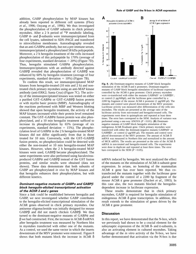
addition, GABP phosphorylation by MAP kinases hasalready been reported in different cell systems (Floryet al., 1996; Ouyanget al., 1996). We thus investigatedthe phosphorylation of GABP subunits in chick primarymyotubes. After a 2 h period of32P metabolic labeling,GABP α- and β-subunits were immunoprecipitated fromthe cell lysates, submitted to SDS–PAGE and transferredto nitrocellulose membranes. Autoradiography revealedthat an anti-GABPα antibody, but not a pre-immune serum,immunoprecipitated a phosphorylated 58 kDa polypeptide.Moreover, a 2 h heregulin treatment of the cells increasedphosphorylation of this polypeptide by 170% (average offour experiments, standard deviation5 20%) (Figure 7D).Thus, heregulin stimulated GABPα phosphorylation.Immunoprecipitations with an antibody directed againstGABPβ revealed that phosphorylation of GABPβ wasenhanced by 60% by heregulin treatment (average of fourexperiments, standard deviation5 10%) (Figure 7B).
To confirm this result, we immunoprecipitated MAPkinases from heregulin-treated (10 min and 2 h) and non-treated chick primary myotubes using an anti-MAP kinaseantibody (anti-ERK2; Santa Cruz) (Figure 7C). The activ-ity of the immunoprecipitated kinases was tested on eithera purified GST–GABPα or GST–GABPβ fusion proteinor with myelin basic protein (MBP). Autoradiography ofthe reactions performed with MBP and Western blottingshowed that upon heregulin treatment, the activity of theMAP kinases increased while their protein level remainedconstant. The GST–GABPα fusion protein was also phos-phorylated, and a 10 min heregulin treatment sufficed toincrease its phosphorylation by 65% (average of sixexperiments, standard deviation5 15%). The phospho-rylation level of GABPα in the 2 h heregulin-treated MAPkinases did not differ significantly from that in thosetreated for 10 min. Conversely, with the GST–GABPβfusion protein, no phosphorylation could be detected witheither the non-treated or 10 min heregulin-treated MAPkinases. However, when the 2 h heregulin-treated MAPkinases were used, GABPβ became phosphorylated. Thesame experiments were also performed using baculovirus-produced GABPα and GABPβ instead of the GST fusionproteins, and similar results were obtained (data notshown). These data demonstrate that both subunits ofGABP are phosphorylatedin vivo by MAP kinases andthat heregulin enhances their phosphorylation, but withdifferent kinetics.
Dominant-negative mutants of GABPα and GABPβblock heregulin-elicited transcriptional activationof the AChR δ and ε genesSince a link could be established between heregulin andGABP, we next investigated whether GABP contributesto the heregulin-elicited transcriptional stimulation of theAChR genes observed in chick primary myotubes. Ourantisense oligonucleotide was initially designed for mouseGABPβ and did not match chicken GABPβ. We thusturned to the dominant-negative mutants of GABPα andβ we had constructed. First, the increase in AChRδ mRNAafter heregulin treatment was measured by Northern blotin myotubes transfected with either one of the mutants.As a control, we used the same vector in which the insertsdownstream of the MSV promoter were removed. Figure 8shows that both mutants block the increase in AChRδ
3085
Fig. 8. (A) Dominant-negative mutants of GABP block heregulinstimulation of the AChRδ andε promoters. Dominant-negativemutants of GABP block heregulin stimulation of luciferase expressioncontrolled by the AChRε gene promoter. Chick primary myotubeswere co-transfected with either the mutants GABPαD– or GABPβD–,or control (1µg/600µl), and the luciferase gene controlled by a2200 bp fragment of the mouse AChRε promoter (1µg/600µl). Themutants and control were placed downstream of the MSV promoter.Luciferase activity was measured after differentiation of the cells intomyotubes. The results are presented as the average ratio of theluciferase activity from non-treated and heregulin-treated cells. Theexperiments were done in quintuplicate and repeated at least threetimes. The error bars correspond to the SEM. Analysis of variance wasperformed using a one-way ANOVA (P ,0.01). (B) Dominant-negative mutants of GABP block heregulin stimulation of theendogenous AChRδ-subunit gene. Chick primary myotubes weretransfected with either the dominant-negative mutants GABPαD– orGABPβD–, or control (2µg/600µl). The mutants and control wereplaced downstream of the MSV promoter. The amount of AChRδmRNA was measured in Northern blot and normalized to the amountof MCK mRNA. The results are presented as the ratio of the AChRδmRNA in non-treated and heregulin-treated cells. The experimentswere done in duplicate and repeated at least three times. The errorbars correspond to the SEM (P ,0.05).
mRNA induced by heregulin. We next analyzed the effectof the mutants on the stimulation of AChRε-subunit geneexpression. In avians, no homolog of the mammalianAChR ε gene has ever been reported. We thus co-transfected the mutants together with the luciferase geneplaced under the control of a 2200 bp fragment of themouse AChRε gene promoter (Duclertet al., 1993). Inthis case also, the two mutants blocked the heregulin-dependent increase in luciferase expression.
These results demonstrate that in chick primarymyotubes, GABP is required for heregulin stimulation ofendogenous AChRδ gene transcription. In addition, thisresult extends to the stimulation of genes driven by theAChR ε gene promoter.
Discussion
In this report, we have demonstrated that the N-box, whichwe previously had shown to be a crucial element for thesubsynaptic expression of the AChRδ- andε-subunits, isalso an activating element in cultured myotubes. Takingadvantage of thein vitro activity of the N-box, we havefurther demonstrated that activation via the N-box is due

L.Schaeffer et al.
to the binding of the transcription factor GABP. Inunraveling the mechanisms by which GABP promotes thecompartmentalized expression of the AChRin vivo, wereveal a preferential expression of GABPα mRNA at theendplate, and the stimulation of GABPα expression bythe neurotrophic factor heregulin.
After this manuscript was submitted, Sapruet al.(1998)reported studies with the AChRε promoter from rat,which is highly homologous to its mouse counterpart.They showed that a 15 bp region, which contains the N-box, contributes to the heregulin-stimulated expression ofa reporter gene regulated by the rat AChRε promoter.They also showed that this effect is mediated by anunidentified Ets and involves the MAP kinase pathway.Here, we show that both subunits of GABP are phosphoryl-ated by MAP kinases and that their phosphorylationincreases upon heregulin treatment. Finally, usingdominant-negative mutants of GABPα and GABPβ, wedemonstrate that GABP is required for heregulin-stimulated transcription of the AChRδ- and ε-subunitgenes.
GABP binds to the N-box and activatestranscription in cultured myotubesThe 50-fold increase in luciferase expression observedupon grafting the three repeats of the N-box to a minimalpromoter clearly demonstrates that the N-box behaves asan activator in cultured myotubes. This property is notspecific to C2C12 mouse myotubes as the N-box is alsofunctional in chick primary myotubes.
The low stringency PCR approach we have used toidentify the various Ets genes expressed in muscle yieldeda majority of sequences corresponding to Ets-2; othersindicated the presence of GABPα, but no new Ets motifwas ever amplified. Although not exhaustive, this approachhas already been used successfully to identify new mem-bers of the Ets family (Brown and McKnight, 1992;Giovane et al., 1994; Lopezet al., 1994). Our resultssuggest that Ets-2 and GABPα are the best representedmembers of the Ets family in adult mouse muscle.Supershift experiments show that Ets-2 does not seem tobe present in the N-box-binding activity. In addition, themobility of GABP in gel retardation experiments differsgreatly from that of the other Ets proteins (including Ets-2) which exhibit a much higher mobility (Joussetet al.,1997, and references therein). This is most likely due tothe unique multimeric nature of GABP among the membersof the Ets family. Two other Ets have a migration patternthat might correspond to GABP: Elf1 and ERF. However,Elf-1 and GABP preferentially bind to different sites(Brown and Mc Knight, 1992; Wanget al., 1992), andFigure 4A shows that competition by an oligonucleotidecontaining a strong binding site for Elf-1 is not efficient.In addition, Elf-1 and ERF have molecular weights (68and 75 kDa, respectively) which are too different fromGABPα to fit the results from the affinity purification orthe UV cross-linking. Moreover, ERF is a repressor(Sgouraset al., 1995), and we observe activation inmyotubes. The combination of the site mutagenesis data,the migration profile of GABP, the composition of theaffinity-purified fraction and the UV cross-linking experi-ment supports the conclusion that the N-box-bindingactivity we observe does not correspond to any known
3086
Ets other than GABP. Finally, the complete displacementof the N-box-binding complex by the anti-GABPα andβ antibodies confirms that, at least in gel retardationexperiments, GABP is the only muscle-derived proteinwhich binds significantly to the N-box.
The inhibition of the N-box-dependent activation bythe anti-GABPβ antisense oligonucleotide and the effectof the dominant-negative mutants demonstrate the implica-tion of GABP in this process. However, it is still possiblethat other factors also contribute to the transactivationthrough the N-box in cultured myotubes. However, nonecould be detected using the gel retardation technique.Moreover, the antisense oligonucleotide decreased theexpression of the N3M2 construct to levels comparablewith those obtained with M2 or Nmut3M2, thus suggestingthat GABP alone is responsible for the N-box-mediatedtranscriptional activation.
Possible mechanisms for GABP-mediatedcompartmentalized transcription of AChR genes atthe neuromuscular junctionIn previous studies, we have established the pivotal roleof the N-box in restricting the transcription of the AChRδ- and ε-subunit genes at the neuromuscular junction inadult muscle (Koikeet al., 1995; Duclertet al., 1996).We now demonstrate that GABP binding to the N-boxenhances transcription in myotubes and that GABP isinvolved in the heregulin-dependent transcriptional activa-tion of the AChRδ andε genes.
Comparison of N-box mutation effects on the synapticexpression of a reporter gene in adult mouse muscle andon the binding of recombinant GABP shows that the GABPDNA-binding specificity strictly parallels the sequencerequirements necessary to obtain an N-box-dependentsynaptic expression patternin vivo. Indeed, there is arobust correlation between the extent to which a mutationalters GABP binding to the probe and the relevant synapticexpression of the reporter gene. Moreover, GABP isinvolved in the transcriptional response of AChR genesto heregulin, which is concentrated at the neuromuscularjunction (Joet al., 1995). In addition, GABPα mRNAshows preferential accumulation in the synaptic regionin vivo. These observations support the conclusion that,in adult muscle, GABP is the factor which binds to the N-box and stimulates subsynaptic transcriptional activation.
The in situ hybridization experiments have furtherrevealed that, although GABPα expression is higher in thesubsynaptic nuclei, GABPβ mRNA is evenly distributedthroughout the muscle fiber. This observation can becorrelated with the fact that, in cultured myotubes, heregu-lin treatment specifically increases the GABPα proteinlevels. Since,in vivo, heregulin is concentrated at theneuromuscular junction (Joet al., 1995), a plausiblehypothesis is that it accounts for the synaptic increase inGABPα expression. If the amount of GABPα was limitingfor GABP activity, the compartmentalization of theGABPα subunit would suffice to enhance the GABPtranscriptional activity in the subsynaptic nuclei.
Several alternative or complementary mechanisms mayalso contribute to the regulation of GABP activity. Forinstance, GABPα may bind different regulatory subunitsaccording to its localization in the muscle fiber. SinceGABPα dimerizes with proteins containing ankyrin

Role of GABP and the N-box in AChR expression
repeats, one has to consider the possibility that, in theextrasynaptic areas, GABPα binds to a subunit that seques-ters it into the cytoplasm, as does the ankyrin repeat-containing protein IκB with the transactivator NF-κB(Blank et al., 1992). This possibility will be tested usingsuitable antibodies for immunofluorescence analysis. It isalso plausible that different forms of GABPβ associatewith GABPα: an active form at the endplate and aninactive form in the extrajunctional areas. Indeed, theGABPβ1 gene is alternatively spliced to produce twoforms of the protein (de la Brousseet al., 1994). Thesmaller form has a shortened C-terminal end and seemsto be transcriptionally inactive (Sawaet al., 1996). Theprobe we used for thein situ experiments does notdiscriminate between the two spliced forms, but thepossibility of distinct distributions of the various splicedforms will be tested with suitable probes and antibodies.
Alternatively, the composition of GABP might remainunchanged throughout the fiber, with nerve-derived signalsmodulating its transcriptional activity. Heregulin, secretedby the nerve terminal or even by the muscle (Moscosoet al., 1995), accumulates in the basal lamina of theneuromuscular junction where it stimulates AChR expres-sion (Joet al., 1995; Sandrocket al., 1997). Our resultsdemonstrate that, in addition to increasing GABPα proteinlevels, heregulin stimulates transcription of AChRδ andε genes via GABP and influences the phosphorylation ofboth GABP subunits via the MAP kinase signaling path-way previously shown to be required for the stimulationof AChR expression (Tanseyet al., 1996; Altiok et al.,1997). As heregulin is compartmentalized, the phospho-rylation state of GABP most probably differs in thesubsynaptic and extrasynaptic nuclei. Regulation of thetranscriptional activity of GABP by heregulin-inducedphosphorylation would be a means of obtaining synapticspecificity. This regulation could be achieved by affectingGABP stability, DNA-binding activity, heterodimerization,or its ability to regulate transcription by affecting itsinteractions with the basal transcriptional machinery orwith transcriptional regulators. Interestingly, we haveshown that GABPα andβ display different phosphoryla-tion kinetics. This suggests that although MAP kinasesare implicated in both cases, the phosphorylation ofGABPα andβ may involve additional partners.
A possible negative regulatory role for GABP inextrasynaptic nucleiIn a previous study, we have demonstrated that, on onehand, in a truncated version of the AChRδ-subunit genepromoter, the N-box behaves as a subsynaptic activator,as in the AChRε-subunit gene promoter. On the otherhand, in a larger version of theδ promoter, the N-boxgains the ability to function as an extrasynaptic silencer.This additional role of the N-box suggested the presenceof an upstream element in theδ promoter, locatedbetween –839 and –60 and absent from theε promoter,that cooperates with the N-box to inhibit transcription inthe extrajunctional nuclei (Koikeet al., 1995; Duclertet al., 1996). One possibility is that GABP confers sucha dual function on the N-box. Indeed, in the case of theribosomal protein genes, GABP serves both as an activatorand as a repressor, depending on the promoter context in
3087
which its binding site is located (Genuario and Perry,1996).
In both the AChRδ- andε-subunit gene promoters, theN-box is located in the vicinity of an E-box and aCCCACCCC box (Baldwin and Burden, 1988; Duclertet al., 1993). The conservation of the immediate contextof the N-box might be relevant and give additionalcues concerning the modes of action of GABP in thesepromoters. Indeed, the Ets transcription factors are knownto function most of the time in cooperation with othertranscription factors (Cre´pieux et al., 1994). Moreover, adomain conserved among various Ets, including GABPα,has been proposed to form a basic helix–loop–helix(bHLH)-like motif (Seth and Papas, 1990) and thus topromote interactions with members of the bHLH family,among which are the E-box-binding myogenic factors.Synergism between Ets and bHLH proteins has beendescribed in immunoglobulin gene expression (Riveraet al., 1993). Future studies will reveal if such interactionsare relevant for the regulation of AChR expression.
Since the implication of the myogenic factors in AChRtranscription regulation was shown (Pietteet al., 1990),GABP has been the first transcription factor to be foundthat might play a pivotal role in AChR transcriptioncompartmentalization to the subsynaptic nuclei. Moreover,GABP is regulated by the presynaptic trophic factorheregulin. These results will undoubtedly open newavenues of investigation towards the understanding ofneural control of transcription. In addition, the variousspliced forms of heregulin and their ErbB tyrosine kinasereceptors are expressed in a wide variety of tissues inwhich they are actively involved in cell communication(for a review see Burden and Yarden, 1997). As GABPis most probably expressed in the various ErbB-containingtissues (LaMarcoet al., 1991; de la Brousseet al., 1994),it might participate in the many aspects of the heregulin/ErbB signaling network.
Materials and methods
Cell cultures and transient transfection assaysChick primary myotube cultures were obtained and cultured as previouslydescribed (Tanseyet al., 1996). They were plated on 60 mm plates andtransfected the next day for 3 h with 2µg of DNA and 20 µl oflipofectamine (Gibco-BRL) in 2 ml of optiMEM (Gibco-BRL) accordingto the manufacturer’s instructions. For the experiments of heregulin-stimulated transcription, the cells were treated with 5 nM recombinantheregulin 24 h before harvest. C2C12 cells (Genuario and Perry, 1996)(23105) were plated on 35 mm dishes in Dulbecco’s modified Eagle’smedium (DMEM; Gibco-BRL) containing 20% fetal calf serum (FCS;Gibco-BRL) and antibiotics. The myoblasts were transfected the nextday for 3 h with 1µg of DNA (or 0.5 mM double-stranded antisenseoligonucleotides) and 12µl of lipofectamine in 600µl of optiMEM,according to the manufacturer’s recommendations. The cells were nextdifferentiated into myotubes for 2 days by replacing the FCS by 10%horse serum. For the experiments with the GABP dominant-negativemutants, no insert was placed between the MSV promoter and the SV40poly(A) for the control.
Northern blotRNA was purified according to Chomsky and Sacchi (1987), and 20µgof total RNA was loaded in each lane. Probes were radiolabeled with[32P]UTP (800 Ci/mmol, Amersham), using the Promega Riboprobe kit.The AChRδ and MCK probes were obtained from the pGEM3Z vector(Promega), in which 1.6 kb and 520 bpPstI fragments of the chickenAChR δ and MCK genes, respectively, were cloned (Nefet al., 1984;Klarsfeld and Changeux, 1985)

L.Schaeffer et al.
Plasmid constructs and oligonucleotidesConstructs were made in KS-Bluescript vector (stratagene) in whichthe M2 minimal promoter (fragment –45 to11 from the chicken AChRα gene promoter) and the luciferase gene (Bessereauet al., 1993) wereintroduced as previously described (Duclertet al., 1991). The threerepeats of the wild-type (N3) and mutated N-box (Nmut3) were intro-duced upstream of M2 by cloning double-stranded synthetic oligo-nucleotides in theXbaI and SalI sites: N3, (sense) 59-GGCCGCGT-TTCCGGCCTCGTTTCCGGCCTCGTTTCCGGCCTCT-39, (antisense)59-CTAGAGAGGCCGGAAACGAGGCCGGAAACGAGCCGGAAA-CGC-39; Nmut3, (sense) 59-GGCCGCG TTTCCAACCTCGTTTCC-AACCTCGTTTCCAACCTCT-39, (antisense) 59-CTAGAGAGGTTG-GAAACGAGGTTGGAAACGAGGTTGGAAACGC-39. All the oligo-nucleotides were from Genset (France). In the antisense oligonucleotides,the nucleotides were phosphorothioates.
Protein extractsExtracts from tissues were obtained as previously described (Koikeet al., 1995). Extracts from cultured cells were obtained using thetechnique described by Dignam (Changeuxet al., 1990).
Mobility shift and supershift assaysMobility shift assays were performed using 2 ng of32P-labeled probein 20 µl at room temperature in 20 mM HEPES, pH 8.0, 50 mM KCl,0.2 mM EDTA, 1 mM dithiothreitol (DTT), 4 mM spermidine, 0.1%NP-40, 15% glycerol, 1µg of polydI–dC. After a 30 min incubation,the reactions were electrophoresed on 6% polyacrylamide gels in 0.53TBE. The gel was then dried and autoradiographed on a phosphorimagerscreen (Molecular Dynamics). For the supershift experiments, 0.5µl ofpolyclonal antiserum was added after the 30 min incubation at roomtemperature and the reaction was incubated for a further 20 min on iceprior to loading on gel.
The probes and competitor double-stranded oligonucleotide sequences(upper strand) were as follows: the N-box oligonucleotide sequence isderived from the mouse AChRδ-subunit gene promoter, 59-GG-CCGCGTTTCCGGCCTCT-39; N1–N6 mutants were as previouslydescribed (Koikeet al., 1995); GABP consensus, 59-TTGGAAAACGG-AAGTGACG-39; Ets-2, 59-TTGGTGGAGGAAGT-39; Elf-1, 59-TTGG-TTTTTCCTCCTT-39.
UV cross-linkingThe binding to the probe and the electrophoresis of the resulting complexwere performed as described for the mobility shift assay except thatthe three central T residues of the probe were substituted by threebromodeoxyuridine residues. After electrophoresis, the gel was UVirradiated at 320 nm (128 mW/cm2 for 15 min) and autoradiographed.The gel slice containing the retarded complex subsequently was excisedand the latter was eluted from the gel in 50 mM Tris pH 7.5, 150 mMNaCl, 0.1 mM EDTA, 5 mM DTT, 0.1% SDS and migrated through adenaturing SDS–polyacrylamide gel. Once dried, the gel was autoradio-graphed on a phosphorimager screen (Molecular Dynamics).
DNA affinity purification of GABPBiotinylated double-stranded N3 oligonucleotide (200 pmol) was coupledto 1 mg of streptavidin magnetic beads (Dynal) according to themanufacturer’s instructions. The beads were then incubated with 200µgof nuclear extract of C2C12 myoblasts for 30 min at 4°C in 400µl of gelretardation assay buffer supplemented with 0.5 mM phenylmethylsulfonylfluoride (PMSF) and 5 mg/ml BSA (fraction V, Sigma). The beadssubsequently were washed three times with 400µl of the same bufferexcept that the KCl concentration was increased to 100 mM and theBSA was removed for the last two washes. The proteins retained on thebeads were then eluted in 50µl by increasing the KCl concentration ofthe buffer to 400 mM.
Low stringency PCR for the Ets gene familyThe degenerate primers were designed in the most conserved regions ofthe Ets domain (Macleodet al., 1992) and their sequences were as follows:59-YTITGGSAITTYYTIYTISA-3 9 and 59-AIYTTITCRTARTTCAT-39.
In situ hybridization studies and riboprobesDiaphragm muscles were dissected from OF1 mice and hybridized withriboprobes as previously described (Pietteet al., 1993). The riboprobesfor the mouse AChRδ-subunit, GABPα and GABPβ1 were obtainedusing cDNA fragments spaning from nucleotides14 to 1420, 11 to1553 and1485 to 1990, respectively. The presence of acetylcholin-
3088
esterase was detected using the method described by Koelle andFiedenwald (1949).
In vivo DNA injectionThe detailed protocol is described in Duclertet al. (1993). Briefly, 30µlof a 20% sucrose solution, containing 3 mg/ml or 1 mg/ml of plasmid,respectively, carrying the AChR promotersε or δ, was injected into bothtibialis anterior muscles of 3-week-old mice. One week after injection,the muscles were dissected and the fibers expressingβ-galactosidasewere stained for acetylcholinesterase to localize the synaptic zone (Koelleand Fiedenwald, 1949). Microscopic observation indicated whether ornot the β-galactosidase-expressing nuclei were co-localized with themotor endplates. Synaptic events corresponded to those for which co-localization was observed, and the percentage of synaptic events is givenby the ratio of the number of synaptic events to the total number ofevents.
Generation of a polyclonal GABPα antibodyA GABPα–GST fusion protein was generated by inserting the mouseGABPα cDNA into the NcoI–EcoRI sites of the pGEX3X vector(Pharmacia). The fusion protein was produced inEscherichia coli(strainXL-1 blue, Stratagene) and purified on a glutathione–Sepharose column(Pharmacia) according to the manufacturer’s instructions. The purifiedprotein was used to immunize rabbits following a standard protocol. InWestern blot, a major band at 58 kDa was detected in C2C12 nuclearextracts. A band of the same size was also detected in extracts fromcells infected with GABPα-expressing baculovirus, whereas no bandwas detected when the cells were infected with GABPβ-expressingbaculovirus. For the immunoprecipitations, the anti-GABPα antibodywas affinity purified on blots as previously described (Beall andMitchell, 1986).
Metabolic labeling and immunoprecipitationsChick primary myotube cultures on 60 mm plates were incubated for2 h in phosphate-free MEM (ICN), the medium was then changed and0.8 mCi/ml of 32P (Amersham) were added. After 2 h, the cells wereharvested in the immunoprecipitation buffer: 50 mM HEPES pH 8.0,150 mM NaCl, 1% Triton X-100, 1.5 mM MgCl2, 1 mM EGTA, 10%glycerol, 1 mM sodium orthovanadate, 50 mMβ-glycerophosphate,10 mM NaF, 1 mM PMSF, 10µg/ml aprotinin for GABPα immunopre-cipitations, and 50 mM Tris pH 8.0, 150 mM NaCl, 0.1% SDS, 1% NP-40, 0.5% sodium deoxycholate, 2 mM EDTA, 10% glycerol, 1 mMsodium orthovanadate, 50 mMβ-glycerophosphate, 10 mM NaF, 1 mMPMSF, 10µg/ml aprotinin for GABPβ immunoprecipitation. The lysatesupernatant was incubated with either 100 ng of purified GABPαantibody or 1µl of anti-GABPβ serum, for 90 min at 4°C. Then 20 mlof protein A–agarose beads (Santa Cruz) were added and the incubationwas continued for a further 60 min. The beads were then washed fivetimes in the GABPα immunoprecipitation buffer and resuspended indenaturing polyacrylamide gel loading buffer (Laemmli, 1970). Half ofthe reactions were loaded on SDS gels and transferred to nitrocellulosemembranes for autoradiography on a phosphoimager screen (MolecularDynamics) and Western blotting using chemiluminescent detection (ECL,Amersham). To avoid the introduction of errors due to variations inGABPα or β levels from lane to lane, the level of phosphorylation ofGABPα or GABPβ measured in each lane was normalized to the amountof the corresponding protein estimated by scanning the result of theWestern blot.
The MAP kinase immunoprecipitations were performed as for GABPα,except that 1µg of anti-ERK2 goat antibody (Santa Cruz) and proteinG–agarose beads (Santa Cruz) were used and the two final washes wereperformed in the MAP kinase assay buffer.
MAP kinase assaysOne-third of the immunoprecipitated MAP kinases was used in eachassay. The reactions was performed for 30 min at 30°C in 50µl of25 mM HEPES pH 8.0, 10 mM MgCl2, 1 mM DTT, 10% glycerol,50 µM ATP and 10µCi of [γ-32P]ATP as previously described (Skolniket al., 1993). MBP (0.5 mg) (Sigma) or 100 ng of purified GST–GABPαor GST–GABPβ fusion proteins were used as substrates. The reactionswere stopped by addition of denaturing gel loading buffer (Laemmli,1970); one half was loaded on denaturing polyacrylamide gels andtransferred to nitrocellulose membranes for autoradiography on a phos-phorimager screen (Molecular Dynamics) and Western blotting.

Role of GABP and the N-box in AChR expression
Acknowledgements
We are grateful to Jacques Ghysdael (Orsay, France) for his gift ofmouse GABPα and GABPβ cDNAs and baculovirus-produced proteins.We thank Steven L.McKnight (Tularik Inc., San Francisco CA) whoprovided us with anti-GABPβ1 and anti-GABPβ2 antibodies. Heregulinwas kindly supplied by Mark Sliwkowski (Genentech Inc., San Francisco,CA). We are also grateful to Henriette Nuret and Martine Soudant forpreparation of primary cultures. We also thank Jean-Louis Bessereau,Alain Bessis, Elisabeth Brown, Aymeric Duclert, Alban de Kerchoveand Satoshi Koike for fruitful discussions and critical reading of themanuscript. This work was supported by the Association Franc¸aisecontre les Myopathies, the Centre National de la Recherche Scientifique,the Institut National de la Sante´ et de la Recherche Me´dicale, the Colle`gede France, the Ministe`re de l’education nationale, de la recherche et dela technologie, the EEC (Biotech. and Biomed.) and the Direction desRecherches et Etudes Techniques.
References
Altiok,N., Altiok,S. and Changeux,J.P. (1997) Heregulin-stimulatedacetylcholine receptor gene expression in muscle—requirement forMAP kinase and evidence for a parallel inhibitory pathway independentof electrical activity.EMBO J., 16, 717–725.
Baldwin,T.J. and Burden,S.J. (1988) Isolation and characterization ofthe mouse acetylcholine receptor delta subunit gene: identification ofa 148-bpcis-acting region that confers myotube-specific expression.J. Cell Biol., 107, 2271–2279.
Beall,J.A. and Mitchell,G.F. (1986) Identification of a particular antigenfrom a parasite cDNA library using antibodies affinity purified fromselected portions of Western blots.J. Immunol. Methods, 86, 217–223.
Bessereau,J.L., Mendelzon,D., Le Poupon,C., Fiszman,M., Changeux,J.P.and Piette,J. (1993) Muscle specific expression of the acetylcholinereceptor alpha subunit gene requires both positive and negativeinteraction between myogenic factors Sp1 and GBF factors.EMBOJ., 12, 443–449.
Bessereau,J.L., Stratford-Perricaudet,L.D., Piette,J., Le Poupon,C. andChangeux,J.P. (1994)In vivoandin vitro analysis of electrical activity-dependent expression of muscle acetylcholine receptor genes usingadenovirus.Proc. Natl Acad. Sci. USA, 91, 1304–1308.
Blank,V., Kourilsky,P. and Israel,A. (1992) NF-kappa B and relatedproteins: Rel/dorsal homologies meet ankyrin-like repeats.TrendsBiochem. Sci., 17, 135–140.
Brenner,H.R., Witzemann,V. and Sakmann,B. (1990) Imprinting ofacetylcholine receptor messenger RNA accumulation in mammalianneuromuscular synapses.Nature, 344, 544–547.
Brown,T.A. and McKnight,S.L. (1992) Specificities of protein–proteinand protein–DNA interaction of GABP alpha and two newly definedets-related proteins.Genes Dev., 6, 2502–2512.
Burden,S. and Yarden,Y. (1997) Neuregulins and their receptors: aversatile signaling module in organogenesis and oncogenesis.Neuron,18, 847–855.
Changeux,J.P.et al. (1990) Compartmentalization of acetylcholinereceptor genes expression during development of the neuromuscularjunction.Cold Spring Harbor Symp. Quant. Biol., 55, 381–396.
Chomczynski,P. and Sacchi,N. (1987) Single step method for RNAisolation by acid guanidium thiocyanate–phenol–chloroformextraction.Anal. Biochem., 162, 156–159.
Crepieux,P., Coll,J. and Stehelin,D. (1994) The Ets family of proteins:weak modulators of gene expression in quest for transcriptionalpartners.Crit. Rev. Oncogen., 5, 615–638.
de la Brousse,F, Birkenmeier,E.H., King,D.S., Rowe,L.B. andMcKnight,S.L. (1994) Molecular and genetic characterization ofGABP beta.Genes Dev., 8, 1853–1865.
Dennis,C.L., Tinsley,J.M., Deconinck,A.E. and Davies,K.E. (1996)Molecular and functional analysis of the utrophin promoter.NucleicAcids Res., 24, 1646–1652.
Duclert,A. and Changeux,J.P. (1995) Acetylcholine receptor genesexpression at the developing neuromuscular junction.Physiol. Rev.,75, 339–367.
Duclert,A., Piette,J. and Changeux,J.P. (1991) Influence of innervationon myogenic factors and acetylcholine receptorα-subunit mRNAs.NeuroReport, 2, 25–28.
Duclert,A., Savatier,N. and Changeux,J.P. (1993) An 83 nucleotidepromoter of the acetylcholine receptor epsilon-subunit gene conferspreferential synaptic expression in mouse muscle.Proc. Natl Acad.Sci. USA, 90, 3043–3047.
3089
Duclert,A., Savatier,N., Schaeffer,L. and Changeux,J.P. (1996)Identification of an element crucial for the sub-synaptic expression ofthe acetylcholine receptor epsilon-subunit gene.J. Biol Chem., 271,17433–17438.
Falls,D.L., Rosen,K.M., Corfas,G., Lane,W.S. and Fischbach,G.D. (1993)ARIA, a protein that stimulates acetylcholine receptor synthesis, is amember of the Neu ligand family.Cell, 72, 801–815.
Flory,E., Hoffmeyer,A., Smola,U., Rapp,U.R. and Bruder,J.T. (1996)Raf-1 kinase targets GA-binding protein in transcriptional regulationof the human immunodeficiency virus type 1 promoter.J. Virol., 70,2260–2268.
Fontaine,B. and Changeux,J.P. (1989) Localization of nicotinicacetylcholine receptor alpha-subunit transcripts during myogenesisand motor endplate development in the chick.J. Cell Biol., 108,1025–1037.
Fontaine,B., Klarsfeld,A., Ho¨kfelt,T. and Changeux,J.P. (1986) Calcitoningene-related peptide, a peptide present in spinal cord motoneurons,increases the number of acetylcholine receptors in primary culturesof chick embryo myotubes.Neurosci. Lett., 71, 59–65.
Fontaine,B., Sassoon,D., Buckingham,M. and Changeux,J.P. (1988)Detection of the nicotinic acetylcholine receptor alpha-subunit mRNAby in situ hybridization at neuromuscular junctions of 15-day-oldchick striated muscles.EMBO J., 7, 603–609.
Galzi,J.L. and Changeux,J.P. (1994) Neurotransmitter-gated ion channelsas unconventional allosteric proteins.Curr. Opin. Struct. Biol., 4,554–565.
Genuario,R.R. and Perry,R.P. (1996) The GA-binding protein can serve asboth an activator and repressor of ribosomal protein gene transcription.J. Biol. Chem., 271, 4388–4395.
Giovane,A., Pintzas,A., Maira,S.M., Sobieszczuk,P. and Wasylyk,B.(1994) Net, a new ets transcription factor that is activated by Ras.Genes Dev., 8, 1502–1513.
Goldman,D. and Staple,J. (1989) Spatial and temporal expression ofacetylcholine receptor RNAs in innervated and denervated rat soleusmuscle.Neuron, 3, 219–228.
Goldman,D., Brenner,H.R. and Heinemann,S. (1988) Acetylcholinereceptor alpha-, beta-, gamma-, and delta-subunit mRNA levels areregulated by muscle activity.Neuron, 1, 329–335.
Hall,Z.W. and Sanes,J.R. (1993) Synaptic structure and development:the neuromuscular junction.Cell/Neuron, 72, 99–121.
Jia,H.T., Tsay,H.J. and Schmidt,J. (1992) Analysis of binding andactivating functions of the chick muscle acetylcholine receptor gamma-subunit upstream sequence.Cell. Mol. Neurobiol., 12, 241–258.
Jo,S.A., Zhu,X., Marchionni,M.A. and Burden,S.J. (1995) Neuregulinsare concentrated at nerve–muscle synapses and activate ACh-receptorgene expression.Nature, 373, 158–161.
Jousset,C.et al. (1997) A domain of tel conserved in a subset of etsproteins defines a specific oligomerization interface essential to themitogenic properties of the tel-pdgfr-beta oncoprotein.EMBO J., 16,69–82.
Karlin,A. and Akabas,M.H. (1995) Toward a structural basis for thefunction of nicotinic acetylcholine receptors and their cousins.Neuron,15, 1231–1244.
Klarsfeld,A. and Changeux,J.P. (1985) Activity regulates the levels ofacetylcholine receptorα-subunit mRNA in cultured chicken myotubes.Proc. Natl Acad. Sci. USA, 82, 4558–4562.
Klarsfeld,A., Bessereau,J.L., Salmon,A.M., Triller,A., Babinet,C. andChangeux,J.P. (1991) An acetylcholine receptor alpha-subunitpromoter conferring preferential synaptic expression in muscle oftransgenic mice.EMBO J., 10, 625–632.
Koelle,G.B. and Fiedenwald,J.S. (1949) A histological method forlocalizing cholinesterase activity.Proc. Soc. Biol. Med., 70, 617–622.
Koike,S., Schaeffer,L. and Changeux,J.P. (1995) Identification of a DNAelement determining synaptic expression of the mouse acetylcholinereceptor delta-subunit gene.Proc. Natl Acad. Sci. USA, 92, 10624–10628.
Kues,W.A., Sakmann,B. and Witzemann,V. (1995) Differentialexpression patterns of five acetylcholine receptor subunit genes in ratmuscle during development.Eur. J. Neurosci., 7, 1376–1385.
Laemmli,U.K. (1970) Cleavage of structural proteins during the assemblyof the head of bacteriophage T4.Nature, 227, 680–685.
LaMarco,K., Thompson,C.C., Byers,B.P., Walton,E.M. andMcKnight,S.L. (1991) Identification of Ets- and notch-related subunitsin GA binding protein.Science, 253, 789–792.
Laufer,R. and Changeux,J.P. (1989) Activity dependent regulation ofgene expression in muscle and neuronal cells.Mol. Neurobiol., 3, 1–54.

L.Schaeffer et al.
Lopez,M., Oettgen,P., Akbarali,Y., Dendorfer,U. and Libermann,T.A.(1994) ERP, a new member of the ets transcription factor/oncoproteinfamily: cloning, characterization, and differential expression duringB-lymphocyte development.Mol. Cell. Biol., 14, 3292–3309.
Macleod,K., Leprince,D. and Stehelin,D. (1992) The ets gene family.Trends Biochem. Sci., 17, 251–256.
Mendelzon,D., Changeux,J.P. and Nghieˆm,H.O. (1994) Phosphorylationof myogenin in chick myotubes: regulation by electrical activity andby protein kinase C. Implications for acetylcholine receptor geneexpression.Biochemistry, 33, 2568–2575.
Merlie,J.P. and Kornhauser,J.M. (1989) Neural regulation of geneexpression by an acetylcholine receptor promoter in muscle oftransgenic mice.Neuron, 2, 1295–1300.
Merlie,J. and Sanes,J.R. (1985) Concentration of acetylcholine receptormRNA in synaptic regions of adult muscle fibers.Nature, 317, 66–68.
Moscoso,L.M., Chu,G.C., Gautam,M., Noakes,P.G., Merlie,J.P. andSanes,J.R. (1995) Synapse-associated expression of an acetylcholinereceptor-inducing protein, ARIA/heregulin, and its putative receptors,ErbB2 and ErbB3, in developing mammalian muscle.Dev. Biol., 172,158–169.
Nef,P., Mauron,A., Stalder,R., Alloid,C. and Ballivet,M. (1984) Structurelinkage, and sequence of the two genes encoding the delta and gammasubunits of the nicotinic acetylcholine receptor.Proc. Natl Acad. Sci.USA, 81, 7975–7979.
New,H.V. and Mudge,A.W. (1986) Calcitonin gene-related peptideregulates muscle acetylcholine receptor synthesis.Nature, 323, 809–811.
Ouyang,L., Jacob,K.K. and Stanley,F.M. (1996) GABP mediates insulin-increased prolactin gene transcription.J. Biol. Chem., 271, 10425–10428.
Piette,J., Bessereau,J.L., Huchet,M. and Changeux,J.P. (1990) Twoadjacent MyoD1-binding sites regulate the expression of theacetylcholine receptorα-subunit gene.Nature, 345, 353–355.
Piette,J., Huchet,M., Houzelstein,D. and Changeux,J.P. (1993)Compartmentalized expression of theα and γ-subunits of theacetylcholine receptor in recently fused myotubes.Dev. Biol., 157,205–213.
Prody,C.A. and Merlie,J.P. (1992) The 59-flanking region of the mousemuscle nicotinic acetylcholine receptor beta-subunit gene promotesexpression in cultured muscle cells and is activated by MRF4,myogenin and MyoD.Nucleic Acids Res., 20, 2367–2372.
Rivera,R.R., Stuiver,M.H., Steenbergen,R. and Murre,C. (1993) Etsproteins: new factors that regulate immunoglobulin heavy-chain geneexpression.Mol. Cell. Biol., 13, 7163–7169.
Sandrock,A.J., Dryer,S.E., Rosen,K.M., Gozani,S.N., Kramer,R.,Theill,L.E. and Fischbach,G.D. (1997) Maintenance of acetylcholinereceptor number by neuregulins at the neuromuscular junctionin vivo.Science, 276, 599–603.
Sanes,J.R., Johnson,Y.R., Kotzbauer,P.T., Mudd,J., Hanley,T.,Martinou,J.C. and Merlie,J.P. (1991) Selective expression of anacetylcholine receptor–LacZ transgene in synaptic nuclei of adultmuscle fibers.Development, 113, 1181–1191.
Sapru,M.K., Florance,S.K., Kirk,C. and Glodman,D. (1998) Identificationof a neuregulin and protein-tyrosine phosphatase response element inthe nicotinic acetylcholine receptorε subunit gene: regulatory role ofan Ets transcription factor.Proc. Natl Acad. Sci. USA, 95, 1289–1294.
Sawa,C., Goto,M., Suzuki,F., Watanabe,H., Sawada,J.I. and Handa,H.(1996) Functional domains of transcription factor hgabp-beta-1/e4tf1-53 required for nuclear localization and transcription activation.Nucleic Acids Res., 24, 4954–4961.
Seth,A. and Papas,T.S. (1990) The c-ets-1 proto-oncogene has oncogenicactivity and is positively autoregulated.Oncogene, 5, 1761–1767.
Sgouras,D.N., Athanasiou,M.A., Beal,G.J.,Jr, Fisher,R.J., Blair,D.G. andMavrothalassitis,G.J. (1995) ERF: an Ets domain protein with strongtranscriptional repressor activity, can suppress ets-associatedtumorigenesis and is regulated by phosphorylation during cell cycleand mitogenic stimulation. EMBO J., 14, 4781–4793.
Simon,A.M. and Burden,S.J. (1993) An E box mediates activationand repression of the acetylcholine receptorδ-subunit gene duringmyogenesis.Mol. Cell. Biol., 13, 5133–5140.
Simon,A.M., Hoppe,P. and Burden,S.J. (1992) Spatial restriction ofAChR gene expression to subsynaptic nuclei.Development, 114,545–553.
Skolnik,E.Y., Batzer,A., Li,N., Lee,C.H., Lowenstein,E.,Mohammadi,M., Margolis,B. and Schlessinger,J. (1993) The functionof GRB2 in linking the insulin receptor to Ras signaling pathways.Science, 260, 1953–1955.
3090
Tang,J., Jo,S.A. and Burden,S.J. (1994) Separate pathways for synapse-specific and electrical activity-dependent gene expression in skeletalmuscle.Development, 120, 1799–1804.
Tansey,M.G., Chu,G.C. and Merlie,J.P. (1996) Aria/hg regulates Achrepsilon subunit gene expression at the neuromuscular synapse viaactivation of phosphatidylinositol 3-kinase and ras/mapk pathway.J. Cell Biol., 134, 465–476.
Thompson,C.C., Brown,T.A. and McKnight,S.L. (1991) Convergence ofEts- and notch-related structural motifs in a heteromeric DNA bindingcomplex.Science, 253, 762–768.
Tsay,H.J. and Schmidt,J. (1989) Skeletal muscle denervation activatesacetylcholine receptor genes.J. Cell Biol., 108, 1523–1526.
van Beveren,C.P., Rands,E., Chattopadhyay,S.K., Lowy,D.R. andVerma,I.M. (1982) Long terminal repeats of murine retroviral dans:sequence analysis, host-proviral junctions, and integration sites.J. Virol., 41, 542–556.
Wang,C.Y., Petryniak,B., Ho,I.C., Thompson,C.B. and Leiden,J.M.(1992) Evolutionarily conserved Ets family members display distinctDNA binding specificities.J. Exp. Med., 175, 1391–1399.
Watanabe,H., Wada,T. and Handa,H. (1990) Transcription factor E4TF1contains two subunits with different functions.EMBO J., 9, 841–847.
Received October 10, 1997; revised and accepted April 1, 1998