Polyaniline–Carboxymethyl Cellulose Nanocomposite for Cholesterol Detection

Cholesterol catabolism as a therapeutic target in Mycobacteriumtuberculosis
Hugues Ouellet, Jonathan B. Johnston, and Paul R. Ortiz de Montellano*
Department of Pharmaceutical Chemistry, University of California, San Francisco, CA94158-2517
AbstractMycobacterium tuberculosis (Mtb) is an intracellular pathogen that infects 10 million worldwideand kills 2 million people every year. The uptake and utilization of nutrients by Mtb within thehost cell is still poorly understood, although lipids play an important role in Mtb persistence. Therecent identification of a large regulon of cholesterol catabolic genes suggests that Mtb can usehost sterol for infection and persistence. In this review, we report on recent progress in elucidationof the Mtb cholesterol catabolic reactions and their potential utility as targets for tuberculosistherapeutic agents.
Tuberculosis: an old disease but still a continuing threatTuberculosis, which has been a scourge of mankind for thousands of years, continues to be amajor cause of death in developing countries and in environments where drug resistance candevelop rapidly. The threat of tuberculosis has become greater over the past decades withthe expansion from single drug resistance to multiple drug resistant strains and, mostrecently, the emergence of extreme drug resistance that threatens to overwhelm all availabledrugs. The importance of tuberculosis in developing countries, the potential of its renewal asa major threat in the developed world, and the determination of the genome ofMycobacterium tuberculosis (Mtb) [1] have intensified the search for new avenues in thedesign of therapeutic agents against Mtb.
Many of the available drugs for tuberculosis target its lipid biosynthetic pathways. Mtb hashighly unusual cell wall lipids (Figure 1) and a relatively large fraction of its genes aredevoted to the synthesis of these lipids. Although Mtb, similar to most bacteria, does notmake sterols, it has been shown that cholesterol is both required for infection ofmacrophages and that the mycobacteria can employ cholesterol as a sole source of carbon.In this review, we summarize the available information on the involvement of cholesterol inthe virulence and growth of Mtb, as well as recent work suggesting that the utilization ofcholesterol could provide new targets for the development of novel therapeutic agents.
© 2011 Elsevier Ltd. All rights reserved.*Address correspondence to: Prof. Paul R. Ortiz de Montellano, Genentech Hall, N572D, 600 16th Street, San Francisco, CA,94158-2517. Tel.: 415-476-2903, [email protected]'s Disclaimer: This is a PDF file of an unedited manuscript that has been accepted for publication. As a service to ourcustomers we are providing this early version of the manuscript. The manuscript will undergo copyediting, typesetting, and review ofthe resulting proof before it is published in its final citable form. Please note that during the production process errors may bediscovered which could affect the content, and all legal disclaimers that apply to the journal pertain.
NIH Public AccessAuthor ManuscriptTrends Microbiol. Author manuscript; available in PMC 2012 November 1.
Published in final edited form as:Trends Microbiol. 2011 November ; 19(11): 530–539. doi:10.1016/j.tim.2011.07.009.
NIH
-PA Author Manuscript
NIH
-PA Author Manuscript
NIH
-PA Author Manuscript

Mtb does not synthesize cholesterolCholesterol is a major structural component of animal cell membranes, where it is requiredto maintain proper membrane permeability and fluidity. In addition, cholesterol is animportant anabolic precursor for the biosynthesis of bile acids, vitamin D and steroidhormones. Although cholesterol is mainly synthesized in animals, small quantities are alsoproduced in plants and fungi. Sterol biosynthesis in bacteria has been controversial, with theonly unambiguously equivalent enzymes found in the proteobacterium, Methylococcuscapsulatus, and in the planctomycete, Gemmata obscuriglobus [2, 3]. In contrast, Mtbapparently lacks the squalene monooxygenase and oxidosqualene cyclase that are absolutelyessential for sterol biosynthesis. Interestingly, the Mtb genome encodes the gene forCYP51B1, a cytochrome P450 enzyme that catalyzes the 14α-demethylation of lanosterol togive the 8,14-diene, a key step in cholesterol biosynthesis [4]. Although bacterial CYP51enzymes are well conserved across actinomycetes species, it is relevant that CYP51 fromStreptomyces coelicolor A3(2), unlike its eukaryotic counterparts, is not essential for cellviability under in vitro growth conditions [5]. The physiological function of CYP51B1enzymes in Mtb and other bacteria remains obscure [5].
Cholesterol is important for Mtb infection and persistenceMtb can infect, grow and survive in the harsh environment of the macrophage and other hostcells using mechanisms that are not yet well understood [6, 7]. Host cholesterol levels arethought to be involved in the development of Mtb infection [8], with high levels ofcholesterol in the diet significantly enhancing the bacterial burden in the lung [9] andimpairing immunity to Mtb [10]. Specifically, cholesterol is required for the phagocytosis ofmycobacteria into macrophages [11, 12]. In fact, mycobacteria enter phagocytes throughcholesterol-rich membrane microdomains [13]. Additionally, cholesterol is required to holdthe host protein coronin 1 (TACO) on Mtb-infected phagosomes, leading to the inhibition ofphagosome-lysozyme fusion [14]. This experimental evidence suggests an important role forcholesterol during Mtb infection and persistence.
Mtb imports and utilizes cholesterolThe acquisition and utilization of nutrients during Mtb infection is poorly understood,although it has been proposed that host lipids play an important role in Mtb survival [15].Several lines of evidence suggest that pathogenic mycobacteria primarily use fatty acidsrather than carbohydrates as carbon substrates during infection [16]. Respiration of Mtb inmouse lungs is strongly stimulated by fatty acids but is unresponsive to carbohydrates [16,17]. An ABC-like transport system, mce4, was recently identified in Mtb that is involved incholesterol import into Mtb [18]. Deletion of the mce4 operon in Mtb resulted in a growthdefect when cholesterol was used as the primary source of carbon [18]. Mce4 loci are alsofound in many other actinomycetes species, including Rhodococcus jostii [19, 20],Mycobacterium smegmatis [21] and Mycobacterium bovis BCG [19], and these can alsoutilize cholesterol for growth. Using 14C-labeled cholesterol derivatives, it has beenelegantly demonstrated that cholesterol is degraded by Mtb, with the carbon atoms from thesterol framework and the aliphatic side-chain going to energy production and lipid synthesis,respectively [18]. Using a lipidomic approach, Yang et al. showed that cholesterolmetabolism in Mtb cells increases the average mass of the lipid virulence factor phthioceroldimycocerosate (PDIM), presumably due to a higher metabolic flux of propionate derivedfrom cholesterol catabolism [22]. Subsequently, heptadeuterated [25, 26, 26, 26, 27, 27, 27-D7]-cholesterol was used to establish the role of the cytochrome P450 encoded by the Mtbcyp125 gene in the degradation of cholesterol and incorporation of the cholesterol side-chaininto the virulence factor PDIM [23] (Figure 1). Additionally, the block of cholesterol import
Ouellet et al. Page 2
Trends Microbiol. Author manuscript; available in PMC 2012 November 1.
NIH
-PA Author Manuscript
NIH
-PA Author Manuscript
NIH
-PA Author Manuscript

in Δmce4 cells markedly attenuated infection in both activated macrophages and the mousemodel of infection, confirming that cholesterol metabolism is important for the chronicphase of infection [18]. Screening of a transposon mutant library allowed the identificationof another locus, igr, which is essential for growth in unactivated macrophages [24]. The igroperon consists of six genes, including a cytochrome P450 (cyp125), two probable acyl-CoAdehydrogenases (fadE28 and fadE29), two conserved hypothetical proteins (Rv3541-2c) anda putative lipid carrier protein (ltp2) (Figure 2). In a follow-up study, inactivation of thewhole igr operon was shown to result in a marked defect in growth on cholesterol alone orin combination with glycerol, suggesting some form of cell intoxication by cholesterol orcholesterol-derived metabolites [25]. Moreover, Δigr cells exhibited a growth defect in theearly phase of the mouse model of infection, an effect that is suppressed by mutating thesterol uptake Mce4 system. Together, these results indicate that cholesterol is importantthroughout infection and is crucial for Mtb persistence, but additionally show that ineffectivebreakdown of this sterol in the early phase of disease gives rise to a different form ofattenuation.
Mtb cholesterol degradation pathwayA growing body of evidence suggests that Mtb uses host cholesterol during infection as asource of carbon and energy. In their breakthrough study, Van der Geize et al. identified acomplete suite of genes required for steroid degradation in R. jostii [19]. Interestingly, theirbioinformatic analysis found that the 51 genes specifically expressed during growth oncholesterol, including those predicted to break down the sterol A and B rings, are likewisefound within an 82-gene cluster in the Mtb and M. bovis BCG genomes. One of the Mtbgenes thus identified, kstR (Rv3574), encodes a TetR-like transcriptional repressor.Inactivation of its homolog in the related saprophyte M. smegmatis derepressed theexpression of 83 genes, as assayed by quantitative reverse transcriptase (RT)-PCR andmicroarrays, with 74 orthologs predicted to be similarly regulated in Mtb (Figure 2) [26].The role of KstR as a cholesterol-responsive regulator in Mtb was later demonstrated byperforming transcriptional profiling of cholesterol gene inactivation and in a kstR mutant ofMtb, corroborating the results obtained from the other actinomycetes [27]. More recently, asecond transcriptional repressor, kstR2 (Rv3557c), was identified and shown to control theexpression of a subset of 15 genes (Rv3548 – Rv3565) within the kstR regulon (Figure 2)[28]. Moreover, the observation that a number of genes in the kstR/kstR2 and cholesterolregulons are induced in macrophages [29] or are essential for infection [30, 31] stronglysuggests that cholesterol catabolism is central to Mtb survival in vivo. In the followingsections we summarize the cholesterol catabolic pathway in Mtb, which can be divided intotwo major phases: (i) initial degradation of the aliphatic side-chain, and (ii) subsequentdegradation of the A–D rings (Figure 3). It remains unclear whether there is an obligatoryorder of the degradation reactions in Mtb. The evidence obtained from the literature onrhodococcal sterol catabolism suggests that intermediates of ring and side-chain degradationcan be exchanged between the two pathway branches [32]. However, the situation seems todiffer in Mtb, as blockage of the side-chain degradation resulted in the accumulation ofcholest-4-en-3-one as a major metabolite [23], suggesting that the ring-degrading enzymes(e.g. KsaAB and HsaA-C) optimally act after the side-chain is removed.
Side-chain degradationLess is known about the degradation of the aliphatic side-chain of cholesterol than about thedegradation of the sterol framework. The side-chain, as found also for the biosynthesis ofbile acids in mammals, is generally accepted to be shortened by β-oxidation reactions(Figure 3). This is consistent with the identification of genes encoding putative β-oxidationenzymes in the cholesterol regulons of Mtb and Rhodococcus rhodochrous DSM43269.These genes include Mtb fadA5. Indeed, the thiolase enzyme encoded by fadA5 catalyzes the
Ouellet et al. Page 3
Trends Microbiol. Author manuscript; available in PMC 2012 November 1.
NIH
-PA Author Manuscript
NIH
-PA Author Manuscript
NIH
-PA Author Manuscript
thiolysis of acetoacetyl-CoA in vitro and is required for growth on cholesterol [27]. Thisthiolase activity, which is consistent with removal of the side-chain by β-oxidation to yieldandrosterone metabolites [e.g. 4-androstenedione (AD) and 1,4-androstenedione (ADD)][27], is required for virulence, especially during the late stage of mouse infection. Veryrecently, the roles of several genes [fadD17, fadD19, fadE26, fadE27, and ro04690(DSM43269)] putatively involved in cholesterol side-chain degradation were investigated inthe actinomycete R. rhodococcus DSM43269 [33]. Among those genes, fadD19 encodes asteroid-coenzyme A (CoA) ligase with an essential in vivo role in degradation of the sidechains of C-24 branched-chain sterols, but interestingly not cholesterol. The high similarity(67%) between the Rhodococcus FadD19 (DSM43269) and Mtb FadD19 enzymes suggeststhat FadD19 also has an in vivo role in sterol metabolism in Mtb [33].
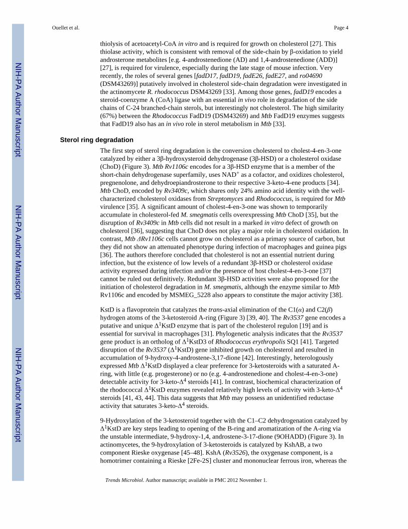
Sterol ring degradationThe first step of sterol ring degradation is the conversion cholesterol to cholest-4-en-3-onecatalyzed by either a 3β-hydroxysteroid dehydrogenase (3β-HSD) or a cholesterol oxidase(ChoD) (Figure 3). Mtb Rv1106c encodes for a 3β-HSD enzyme that is a member of theshort-chain dehydrogenase superfamily, uses NAD+ as a cofactor, and oxidizes cholesterol,pregnenolone, and dehydroepiandrosterone to their respective 3-keto-4-ene products [34].Mtb ChoD, encoded by Rv3409c, which shares only 24% amino acid identity with the well-characterized cholesterol oxidases from Streptomyces and Rhodococcus, is required for Mtbvirulence [35]. A significant amount of cholest-4-en-3-one was shown to temporarilyaccumulate in cholesterol-fed M. smegmatis cells overexpressing Mtb ChoD [35], but thedisruption of Rv3409c in Mtb cells did not result in a marked in vitro defect of growth oncholesterol [36], suggesting that ChoD does not play a major role in cholesterol oxidation. Incontrast, Mtb ΔRv1106c cells cannot grow on cholesterol as a primary source of carbon, butthey did not show an attenuated phenotype during infection of macrophages and guinea pigs[36]. The authors therefore concluded that cholesterol is not an essential nutrient duringinfection, but the existence of low levels of a redundant 3β-HSD or cholesterol oxidaseactivity expressed during infection and/or the presence of host cholest-4-en-3-one [37]cannot be ruled out definitively. Redundant 3β-HSD activities were also proposed for theinitiation of cholesterol degradation in M. smegmatis, although the enzyme similar to MtbRv1106c and encoded by MSMEG_5228 also appears to constitute the major activity [38].
KstD is a flavoprotein that catalyzes the trans-axial elimination of the C1(α) and C2(β)hydrogen atoms of the 3-ketosteroid A-ring (Figure 3) [39, 40]. The Rv3537 gene encodes aputative and unique Δ1KstD enzyme that is part of the cholesterol regulon [19] and isessential for survival in macrophages [31]. Phylogenetic analysis indicates that the Rv3537gene product is an ortholog of Δ1KstD3 of Rhodococcus erythropolis SQ1 [41]. Targeteddisruption of the Rv3537 (Δ1KstD) gene inhibited growth on cholesterol and resulted inaccumulation of 9-hydroxy-4-androstene-3,17-dione [42]. Interestingly, heterologouslyexpressed Mtb Δ1KstD displayed a clear preference for 3-ketosteroids with a saturated A-ring, with little (e.g. progesterone) or no (e.g. 4-androstenedione and cholest-4-en-3-one)detectable activity for 3-keto-Δ4 steroids [41]. In contrast, biochemical characterization ofthe rhodococcal Δ1KstD enzymes revealed relatively high levels of activity with 3-keto-Δ4
steroids [41, 43, 44]. This data suggests that Mtb may possess an unidentified reductaseactivity that saturates 3-keto-Δ4 steroids.
9-Hydroxylation of the 3-ketosteroid together with the C1–C2 dehydrogenation catalyzed byΔ1KstD are key steps leading to opening of the B-ring and aromatization of the A-ring viathe unstable intermediate, 9-hydroxy-1,4, androstene-3-17-dione (9OHADD) (Figure 3). Inactinomycetes, the 9-hydroxylation of 3-ketosteroids is catalyzed by KshAB, a twocomponent Rieske oxygenase [45–48]. KshA (Rv3526), the oxygenase component, is ahomotrimer containing a Rieske [2Fe-2S] cluster and mononuclear ferrous iron, whereas the
Ouellet et al. Page 4
Trends Microbiol. Author manuscript; available in PMC 2012 November 1.
NIH
-PA Author Manuscript
NIH
-PA Author Manuscript
NIH
-PA Author Manuscript

reductase component KshB (Rv3571) is a monomeric protein containing a plant-type[2Fe-2S] cluster and flavin adenine dinucleotide (FAD) [45]. Consistent with a role incholesterol utilization, a transposon mutant of kshA exhibited a strongly attenuatedphenotype in the infection of interferon-γ-activated macrophages [31]. Moreover, Hu et al.reported that targeted disruption of either kshA or kshB resulted in mycobacterial cell deathin the mouse model of infection [46]. Interestingly, inactivation of kshB, but not kshA, altersthe biosynthesis of penta-acylated trehalose (PAT), a multimethyl-branched lipid class,suggesting that the reductase of KshB is involved in several processes.
In R. jostii RHA1 and Mtb, the hsaACDB genes are part a single operon within thecholesterol regulon [19]. The hsaA and hsaB genes encode an oxygenase and a reductase,respectively [49]. The two proteins act together as a flavin-dependent monooxygenase thathydroxylates 3-hydroxy-9,10-seconandrost-1,3,5(10)-triene-9,17-dione (3-HSA) to thecatechol 3,4-dihydroxy-9,10-seconandrost-1,3,5(10)-triene-9,17-dione (3,4-DHSA) (Figure3). Analysis of the crystal structure of ligand-free HsaA at 2.5 Å resolution revealed that thisenzyme displays the same fold, flavin-binding site, and catalytic residues as p-hydroxyphenyl acetate hydroxylase. Additionally, deletion of hsaA in R. jostii RHA1 causeda growth defect on cholesterol, while the screening of a transposon mutant library identifiedhsaA as one of the Mtb genes required for growth in activated macrophages [31].
HsaC, the next enzyme in the pathway, is an iron-dependent extradiol dioxygenase thatefficiently oxygenates and cleaves the catecholic cholesterol metabolite 3,4-DHSA to 4,5–9,10-diseco-3-hydroxy-5,9,17-trioxoandrosta-1(10),2-diene-4-oic acid (4,9-DSHA) (Figure3). The structures of HsaC:DHSA complexes at 2.1 Å revealed two catechol-binding modeswith the position of the bicyclo-alkanone moiety of DHSA being very similar in the twobinding models, suggesting that this interaction is a determinant of the initial substrate-binding step. The inactivation of Mtb hsaC resulted in cell death when the cells were grownin the presence of cholesterol, presumably due to cell poisoning by the accumulated catecholmetabolites. The infection of immunocompromised mice and wild-type guinea pigs withΔhsaC cells also revealed a significant attenuation phenotype. From these observations, theauthors concluded that cholesterol metabolism is most important during the chronic stage ofinfection, but also contributes to pathogen dissemination throughout the infection.
The catabolic gene, hsaD, was identified as one of the Mtb genes required for survival inmacrophages [31]. HsaD is a member of the α/β hydrolase family involved in the aerobicdegradation of aromatic compounds in microbes. This enzyme, for which a crystal structureat 2.35 Å is available [50, 51], catalyzes hydrolytic carbon-carbon bond cleavage of 4,9-DSHA to yield 9,17-dioxo-1,2,3,4,10,19-hexanorandrostan-5-oic acid (DOHNAA) and 2-hydroxy-hexa-2,4-dienoic acid (HHD) (Figure 3) [51]. HHD is finally metabolized totricarboxylic acid cycle intermediates and propionyl-CoA, whereas it is unknown if Mtbfurther degrades DOHNAA, which corresponds to the C and D-ring fragment. While muchin known about the steps involved in sterol ring degradation, substantial gaps exist in ourunderstanding of this pathway. Nevertheless, the structural data obtained for HsaA, HsaCand HsaD provides insights into the binding of steroid substrates that should facilitateinhibitor design.
Role of Mtb P450 in cholesterol catabolismThe saturated side-chain of cholesterol must be chemically functionalized at the ω-positionbefore it can enter into the Mtb β-oxidation pathway (Figure 3). A minimum of fourchemical steps are necessary to prepare the side-chain for β-oxidation. The first three steps,sequential oxidations of the cholesterol side-chain to the terminal alcohol, aldehyde, andacid, are catalyzed by cytochrome P450 enzymes. Three Mtb P450 enzymes, CYP125
Ouellet et al. Page 5
Trends Microbiol. Author manuscript; available in PMC 2012 November 1.
NIH
-PA Author Manuscript
NIH
-PA Author Manuscript
NIH
-PA Author Manuscript

(Rv3545c), CYP142 (Rv3518c) and CYP124 (Rv2266), are capable of oxidizing the side-chains of cholesterol and cholest-4-en-3-one. The final ATP-dependent step is catalyzed bya sterol-CoA ligase [52].
Cytochrome P450 enzymes catalyzing steroid side chain oxidationsThe first oxidation introduces a hydroxy group onto the steroid side-chain, the secondoxidizes the alcohol to the aldehyde, and the third converts the aldehyde to a carboxylicacid. In principle, the initial oxidation could occur at a primary, secondary or tertiaryposition, as all three are found in the cholesterol side-chain, although the ease ofhydroxylation depends on the relative C-H bond dissociation energies and thus decreases inthe order 3° > 2° ≫ 1°. Nevertheless, only oxidation of the primary C-H bond to give the ω-oxidation product is observed [53]. These P450 enzymes, which have evolved to selectivelyoxidize the relatively unreactive terminal methyl group, enforce a strict regiospecificity bypreventing the more reactive carbon of the side-chain from being properly positionedrelative to the heme iron atom [53]. However, as outlined below, there are distinctdifferences in the gene induction, protein expression, stereochemical preferences andcatalytic efficiencies of the three enzymes able to oxidize cholesterol and cholest-4-en-3-one.
Which P450 enzymes are involved?Comparison of the genome sequences of Mtb CDC1551 and H37Rv reveals key differencesin the cytochrome P450 loci. In CDC1551, a 639-bp deletion encompassing the promoterregion and the sequence encoding the first 18 amino acids of the protein is found upstreamof the cyp142 (Rv3518c) gene, indicating that this strain does not produce CYP142. Thecyp124 genes of H37Rv and CDC1551 only differ by two amino acids, G25D and Y75N,respectively. However, no notable differences in catalytic activity are observed between theCYP124 variants. In contrast, the cyp125 genes are identical in both strains. In vitro,CYP125, CYP142 and CYP124 catalyze the sequential oxidation of the hydrocarbon side-chain of cholesterol to the carboxylic acid. The 2nd order rate constants (kcat/KM) forterminal hydroxylation of the cholesterol and cholest-4-en-3-one side-chains establish thatCYP125 is a slightly more efficient catalyst than CYP142 [54], and both are much moreefficient than CYP124 [54]. The characterization of CYP125, reported independently byfour groups, includes x-ray crystal structures [23, 55], biochemical studies [23, 32, 55, 56],and in vivo genetic analyses that probe the loss and gain of this P450 function in differentvirulent clinical strains and R. jostii RHA1 [23, 32, 56]. Two other Mtb P450 enzymes,CYP141 and CYP130, were found not to catalyze the oxidation of these sterols [54].
Oxidation can occur at either of the two terminal methyl groups, producing either the (25S)-and/or (25R)-26 hydroxy sterols. The stereochemical preferences of these enzymes differ:CYP125 preferentially forms the (25S)-26-hydroxy product, whereas CYP142 and CYP124produce the (25R)-26-hydroxy configuration [54]. It is unclear whether there are anyphysiological consequences of these different stereochemical preferences.
Several lines of evidence indicate that CYP125 and CYP142 are induced when Mtb is grownon cholesterol as the sole carbon source [23, 27, 54]. Immunoblots confirmed that theCYP125 and CYP142 proteins are expressed and induced in cells grown on cholesterol,whereas CYP124 was not detectably expressed under these conditions [54]. Interestingly,cyp142 is able to fully complement the loss of cyp125 in CDC1551 Mtb cells when achromosomal copy is introduced into the Δcyp125 knockout cells. CYP125 is the primaryP450 responsible for the initiation of cholesterol side-chain degradation in Mtb. CYP142 cansubstitute for CYP125 in H37Rv but not CDC1551 cells, as the cyp142 gene in the latter isinactivated by a mutation. Similar to CYP125 and CYP142, CYP124 can catalyze sequential
Ouellet et al. Page 6
Trends Microbiol. Author manuscript; available in PMC 2012 November 1.
NIH
-PA Author Manuscript
NIH
-PA Author Manuscript
NIH
-PA Author Manuscript

oxidation of the cholesterol and cholest-4-en-3-one side-chains, but CYP124 expression isnot induced by cholesterol [54]. In agreement with the in vitro kinetic data, CYP124 canonly partially reverse the abnormal growth phenotype of Δcyp125 cells grown oncholesterol. Concomitant with this partial growth restoration, cholest-4-en-3-one was foundto accumulate in the Δcyp125 CYP124 overexpressing cells [54]. The presence of multiplecytochrome P450 isoforms capable of oxidizing the sterol side-chain and differences in theirexpression highlight some of the potential challenges of drug-targeting critical pathways ofMtb and the need for understanding the biochemical and genetic basis for the differences inclinically relevant strains.
Toxicity of cholesterol metabolitesInhibition of the cholesterol catabolic pathways not only can cause carbon starvation, butblockage at certain steps has also been shown to result in cell death or bacteriostasis. Forinstance, the Mtb ΔhsaC mutant not only failed to grow on cholesterol, but the cellsdeveloped a pink color, indicating an accumulation of catechols and their conversion tocolored o-benzoquinones and condensation products [19, 57]. Additionally, a loss of cellviability was observed when ΔhsaC cells were grown in the presence of cholesterol,suggesting that the catechol derivatives are toxic to Mtb cells [57]. A growth inhibitoryeffect of cholesterol metabolism that depends on cholesterol import was observed when Δigrcells were incubated with both cholesterol and glycerol [25]. The same inhibitory effect wasobserved when cyp125, which is part of the igr operon, was deleted in similarly culturedMtb CDC1551 cells. Importantly, cholest-4-en-3-one, which was shown to accumulate inhigh amounts in cyp125 mutant cells [23, 56], was toxic to wild-type Mtb CDC1551,Erdman, and H37Rv strains grown in media containing a second carbon source [23].
A highly speculative mechanism for the toxicity associated with the accumulation ofcholest-4-en-3-one is an alteration of the cell membrane integrity. In eukaryotic plasmamembrane models, it was shown that cholesterol can participate in strong and close packingwith saturated lipids, a critical feature required for the formation of lipid domains, with thelatter implicated in numerous biological processes, including signal transduction events, andtoxin entry into cells [58]. It was shown that cholest-4-en-3-one, for which the 3-OH groupis replaced by a keto group, inhibits lipid domain formation, thus altering the plasmamembranes properties [58, 59]. When cultivated in the presence of cholesterol and a secondcarbon source, Mtb is able to accumulate cholesterol in the free-lipid zone of its cell wall,resulting in a decreased permeability for the primary antitubercular drug, rifampin [42].From these observations, it can be speculated that high levels of cholest-4-en-3-one mightrender the cell wall more permeable to drugs and xenobiotics. Whatever the mechanism, theidentification of cholest-4-en-3-one as a toxic agent in Mtb opens up the intriguingpossibility that elucidation of its site(s) of action could reveal new targets for antituberculardrug design.
Cholesterol catabolism inhibitorsIn view of the importance of cholesterol catabolism in Mtb infection and persistence,targeting this pathway could represent a fruitful avenue for the development of newtherapeutic agents. Some of the important questions that require attention are outlined inBox 1. The antifungal action of azole drugs has been shown to result from inhibition ofsterol biosynthesis at the level of CYP51. Azole drugs, most notably econazole, exhibitantimycobacterial activities against both latent Mtb and multidrug-resistant strains [60, 61],a finding that implicates one or more cytochrome P450 enzymes as targets (Table 1). In thisregard, all the Mtb cytochrome P450 enzymes characterized to date, including CYP125 andCYP142, bind azole drugs with low- to submicromolar affinities (Table 1) [62–67]. The
Ouellet et al. Page 7
Trends Microbiol. Author manuscript; available in PMC 2012 November 1.
NIH
-PA Author Manuscript
NIH
-PA Author Manuscript
NIH
-PA Author Manuscript
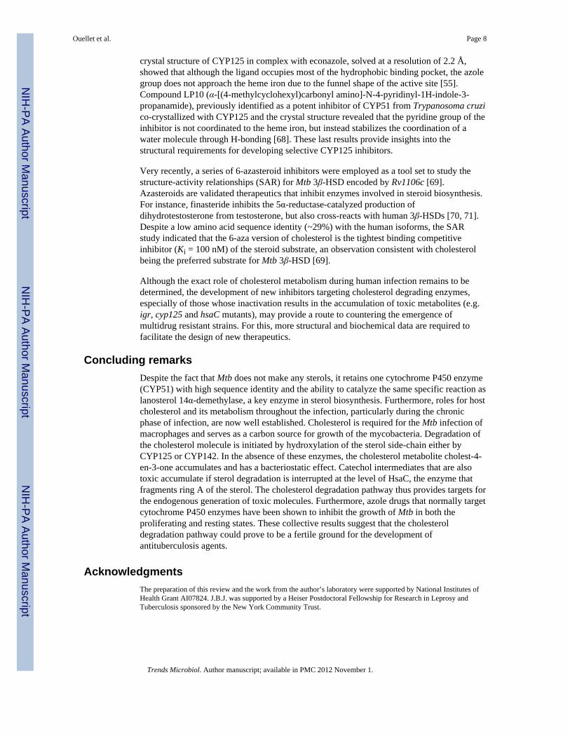
crystal structure of CYP125 in complex with econazole, solved at a resolution of 2.2 Å,showed that although the ligand occupies most of the hydrophobic binding pocket, the azolegroup does not approach the heme iron due to the funnel shape of the active site [55].Compound LP10 (α-[(4-methylcyclohexyl)carbonyl amino]-N-4-pyridinyl-1H-indole-3-propanamide), previously identified as a potent inhibitor of CYP51 from Trypanosoma cruzico-crystallized with CYP125 and the crystal structure revealed that the pyridine group of theinhibitor is not coordinated to the heme iron, but instead stabilizes the coordination of awater molecule through H-bonding [68]. These last results provide insights into thestructural requirements for developing selective CYP125 inhibitors.
Very recently, a series of 6-azasteroid inhibitors were employed as a tool set to study thestructure-activity relationships (SAR) for Mtb 3β-HSD encoded by Rv1106c [69].Azasteroids are validated therapeutics that inhibit enzymes involved in steroid biosynthesis.For instance, finasteride inhibits the 5α-reductase-catalyzed production ofdihydrotestosterone from testosterone, but also cross-reacts with human 3β-HSDs [70, 71].Despite a low amino acid sequence identity (~29%) with the human isoforms, the SARstudy indicated that the 6-aza version of cholesterol is the tightest binding competitiveinhibitor (Ki = 100 nM) of the steroid substrate, an observation consistent with cholesterolbeing the preferred substrate for Mtb 3β-HSD [69].
Although the exact role of cholesterol metabolism during human infection remains to bedetermined, the development of new inhibitors targeting cholesterol degrading enzymes,especially of those whose inactivation results in the accumulation of toxic metabolites (e.g.igr, cyp125 and hsaC mutants), may provide a route to countering the emergence ofmultidrug resistant strains. For this, more structural and biochemical data are required tofacilitate the design of new therapeutics.
Concluding remarksDespite the fact that Mtb does not make any sterols, it retains one cytochrome P450 enzyme(CYP51) with high sequence identity and the ability to catalyze the same specific reaction aslanosterol 14α-demethylase, a key enzyme in sterol biosynthesis. Furthermore, roles for hostcholesterol and its metabolism throughout the infection, particularly during the chronicphase of infection, are now well established. Cholesterol is required for the Mtb infection ofmacrophages and serves as a carbon source for growth of the mycobacteria. Degradation ofthe cholesterol molecule is initiated by hydroxylation of the sterol side-chain either byCYP125 or CYP142. In the absence of these enzymes, the cholesterol metabolite cholest-4-en-3-one accumulates and has a bacteriostatic effect. Catechol intermediates that are alsotoxic accumulate if sterol degradation is interrupted at the level of HsaC, the enzyme thatfragments ring A of the sterol. The cholesterol degradation pathway thus provides targets forthe endogenous generation of toxic molecules. Furthermore, azole drugs that normally targetcytochrome P450 enzymes have been shown to inhibit the growth of Mtb in both theproliferating and resting states. These collective results suggest that the cholesteroldegradation pathway could prove to be a fertile ground for the development ofantituberculosis agents.
AcknowledgmentsThe preparation of this review and the work from the author’s laboratory were supported by National Institutes ofHealth Grant AI07824. J.B.J. was supported by a Heiser Postdoctoral Fellowship for Research in Leprosy andTuberculosis sponsored by the New York Community Trust.
Ouellet et al. Page 8
Trends Microbiol. Author manuscript; available in PMC 2012 November 1.
NIH
-PA Author Manuscript
NIH
-PA Author Manuscript
NIH
-PA Author Manuscript

References1. Cole ST, et al. Deciphering the biology of Mycobacterium tuberculosis from the complete genome
sequence. Nature. 1998; 393:537–544. [PubMed: 9634230]2. Lamb DC, et al. Lanosterol biosynthesis in the prokaryote Methylococcus capsulatus: insight into
the evolution of sterol biosynthesis. Mol Biol Evol. 2007; 24:1714–1721. [PubMed: 17567593]3. Pearson A, et al. Phylogenetic and biochemical evidence for sterol synthesis in the bacterium
Gemmata obscuriglobus. Proc Natl Acad Sci U S A. 2003; 100:15352–15357. [PubMed: 14660793]4. Bellamine A, et al. Characterization and catalytic properties of the sterol 14alpha-demethylase from
Mycobacterium tuberculosis. Proc Natl Acad Sci U S A. 1999; 96:8937–8942. [PubMed:10430874]
5. Lamb DC, et al. Sterol 14alpha-demethylase activity in Streptomyces coelicolor A3(2) is associatedwith an unusual member of the CYP51 gene family. Biochem J. 2002; 364:555–562. [PubMed:12023899]
6. Clark-Curtiss JE, Haydel SE. Molecular genetics of Mycobacterium tuberculosis pathogenesis.Annu Rev Microbiol. 2003; 57:517–549. [PubMed: 14527290]
7. Koul A, et al. Interplay between mycobacteria and host signalling pathways. Nat Rev Microbiol.2004; 2:189–202. [PubMed: 15083155]
8. Kim MJ, et al. Caseation of human tuberculosis granulomas correlates with elevated host lipidmetabolism. EMBO Mol Med. 2010; 2:258–274. [PubMed: 20597103]
9. Schafer G, et al. The role of scavenger receptor B1 in infection with Mycobacterium tuberculosis ina murine model. PLoS One. 2009; 4:e8448. [PubMed: 20041149]
10. Martens GW, et al. Hypercholesterolemia impairs immunity to tuberculosis. Infect Immun. 2008;76:3464–3472. [PubMed: 18505807]
11. Gatfield J, Pieters J. Essential role for cholesterol in entry of mycobacteria into macrophages.Science. 2000; 288:1647–1650. [PubMed: 10834844]
12. Peyron P, et al. Nonopsonic phagocytosis of Mycobacterium kansasii by human neutrophilsdepends on cholesterol and is mediated by CR3 associated with glycosylphosphatidylinositol-anchored proteins. J Immunol. 2000; 165:5186–5191. [PubMed: 11046051]
13. Munoz S, et al. Mycobacterium tuberculosis entry into mast cells through cholesterol-richmembrane microdomains. Scand J Immunol. 2009; 70:256–263. [PubMed: 19703015]
14. Nguyen L, Pieters J. The trojan horse: survival tactics of pathogenic mycobacteria in macrophages.Trends Cell Biol. 2005; 15:269–276. [PubMed: 15866031]
15. Savvi S, et al. Functional characterization of a vitamin B12-dependent methylmalonyl pathway inMycobacterium tuberculosis: implications for propionate metabolism during growth on fatty acids.J B acteriol. 2008; 190:3886–3895.
16. Munoz-Elias EJ, McKinney JD. Mycobacterium tuberculosis isocitrate lyases 1 and 2 are jointlyrequired for in vivo growth and virulence. Nat Med. 2005; 11:638–644. [PubMed: 15895072]
17. Bloch H, Segal W. Biochemical differentiation of Mycobacterium tuberculosis grown in vivo andin vitro. J Bacteriol. 1956; 72:132–141. [PubMed: 13366889]
18. Pandey AK, Sassetti CM. Mycobacterial persistence requires the utilization of host cholesterol.Proc Natl Acad Sci U S A. 2008; 105:4376–4380. [PubMed: 18334639]
19. Van der Geize R, et al. A gene cluster encoding cholesterol catabolism in a soil actinomyceteprovides insight into Mycobacterium tuberculosis survival in macrophages. Proc Natl Acad Sci US A. 2007; 104:1947–1952. [PubMed: 17264217]
20. Mohn WW, et al. The actinobacterial mce4 locus encodes a steroid transporter. J Biol Chem. 2008;283:35368–35374. [PubMed: 18955493]
21. Av-Gay Y, Sobouti R. Cholesterol is accumulated by mycobacteria but its degradation is limited tonon-pathogenic fast-growing mycobacteria. Can J Microbiol. 2000; 46:826–831. [PubMed:11006843]
22. Yang X, et al. Cholesterol metabolism increases the metabolic pool of propionate inMycobacterium tuberculosis. Biochemistry. 2009; 48:3819–3821. [PubMed: 19364125]
Ouellet et al. Page 9
Trends Microbiol. Author manuscript; available in PMC 2012 November 1.
NIH
-PA Author Manuscript
NIH
-PA Author Manuscript
NIH
-PA Author Manuscript

23. Ouellet H, et al. Mycobacterium tuberculosis CYP125A1, a steroid C27 monooxygenase thatdetoxifies intracellularly generated cholest-4-en-3-one. Mol Microbiol. 2010; 77:730–742.[PubMed: 20545858]
24. Chang JC, et al. Identification of mycobacterial genes that alter growth and pathology inmacrophages and in mice. J Infect Dis. 2007; 196:788–795. [PubMed: 17674323]
25. Chang JC, et al. igr genes and Mycobacterium tuberculosis cholesterol metabolism. J Bacteriol.2009; 191:5232–5239. [PubMed: 19542286]
26. Kendall SL, et al. A highly conserved transcriptional repressor controls a large regulon involved inlipid degradation in Mycobacterium smegmatis and Mycobacterium tuberculosis. Mol Microbiol.2007; 65:684–699. [PubMed: 17635188]
27. Nesbitt NM, et al. A thiolase of Mycobacterium tuberculosis is required for virulence andproduction of androstenedione and androstadienedione from cholesterol. Infect Immun. 2010;78:275–282. [PubMed: 19822655]
28. Kendall SL, et al. Cholesterol utilization in mycobacteria is controlled by two TetR-typetranscriptional regulators: kstR and kstR2. Microbiology. 2010; 156:1362–1371. [PubMed:20167624]
29. Schnappinger D, et al. Transcriptional adaptation of Mycobacterium tuberculosis withinmacrophages: Insights into the phagosomal environment. J Exp Med. 2003; 198:693–704.[PubMed: 12953091]
30. Sassetti CM, Rubin EJ. Genetic requirements for mycobacterial survival during infection. ProcNatl Acad Sci U S A. 2003; 100:12989–12994. [PubMed: 14569030]
31. Rengarajan J, et al. Genome-wide requirements for Mycobacterium tuberculosis adaptation andsurvival in macrophages. Proc Natl Acad Sci U S A. 2005; 102:8327–8332. [PubMed: 15928073]
32. Rosloniec KZ, et al. Cytochrome P450 125 (CYP125) catalyses C26-hydroxylation to initiatesterol side-chain degradation in Rhodococcus jostii RHA1. Mol Microbiol. 2009; 74:1031–1043.[PubMed: 19843222]
33. Wilbrink MH, et al. FadD19 of Rhodococcus rhodochrous DSM43269, a steroid-coenzyme Aligase essential for egradation of C-24 branched sterol side chains. Appl Environ Microbiol.77:4455–4464. [PubMed: 21602385]
34. Yang X, et al. Rv1106c from Mycobacterium tuberculosis is a 3beta-hydroxysteroiddehydrogenase. Biochemistry. 2007; 46:9058–9067. [PubMed: 17630785]
35. Brzostek A, et al. Cholesterol oxidase is required for virulence of Mycobacterium tuberculosis.FEMS Microbiol Lett. 2007; 275:106–112. [PubMed: 17651430]
36. Yang X, et al. Cholesterol is not an essential source of nutrition for Mycobacterium tuberculosisduring infection. J Bacteriol. 2011; 193:1473–1476. [PubMed: 21257778]
37. Tabas I, et al. Acyl coenzyme A:cholesterol acyl transferase in macrophages utilizes a cellular poolof cholesterol oxidase-accessible cholesterol as substrate. J Biol Chem. 1988; 263:1266–1272.[PubMed: 3422077]
38. Uhia I, et al. Initial step in the catabolism of cholesterol by Mycobacterium smegmatis mc(2)155.Environ Microbiol. 2011; 4:943–959. [PubMed: 21208358]
39. Itagaki E, et al. Spectral properties of 3-ketosteroid-delta 1-dehydrogenase from Nocardiacorallina. Biochim Biophys Acta. 1990; 1040:281–286. [PubMed: 2400777]
40. Itagaki E, et al. Purification and characterization of 3-ketosteroid-delta 1-dehydrogenase fromNocardia corallina. Biochim Biophys Acta. 1990; 1038:60–67. [PubMed: 2317517]
41. Knol J, et al. 3-Keto-5alpha-steroid Delta(1)-dehydrogenase from Rhodococcus erythropolis SQ1and its orthologue in Mycobacterium tuberculosis H37Rv are highly specific enzymes thatfunction in cholesterol catabolism. Biochem J. 2008; 410:339–346. [PubMed: 18031290]
42. Brzostek A, et al. Mycobacterium tuberculosis is able to accumulate and utilize cholesterol. JBacteriol. 2009; 191:6584–6591. [PubMed: 19717592]
43. van der Geize R, et al. Molecular and functional characterization of the kstD2 gene ofRhodococcus erythropolis SQ1 encoding a second 3-ketosteroid Delta(1)-dehydrogenaseisoenzyme. Microbiology. 2002; 148:3285–3292. [PubMed: 12368462]
Ouellet et al. Page 10
Trends Microbiol. Author manuscript; available in PMC 2012 November 1.
NIH
-PA Author Manuscript
NIH
-PA Author Manuscript
NIH
-PA Author Manuscript

44. van der Geize R, et al. Unmarked gene deletion mutagenesis of kstD, encoding 3-ketosteroidDelta1-dehydrogenase, in Rhodococcus erythropolis SQ1 using sacB as counter-selectable marker.FEMS Microbiol Lett. 2001; 205:197–202. [PubMed: 11750802]
45. Capyk JK, et al. Characterization of 3-ketosteroid 9-alpha-hydroxylase, a Rieske oxygenase in thecholesterol degradation pathway of Mycobacterium tuberculosis. J Biol Chem. 2009; 284:9937–9946. [PubMed: 19234303]
46. Hu Y, et al. 3-Ketosteroid 9alpha-hydroxylase is an essential factor in the pathogenesis ofMycobacterium tuberculosis. Mol Microbiol. 2010; 75:107–121. [PubMed: 19906176]
47. van der Geize R, et al. Characterization of a second Rhodococcus erythropolis SQ1 3-ketosteroid9alpha-hydroxylase activity comprising a terminal oxygenase homologue, KshA2, active withoxygenase-reductase component KshB. Appl Environ Microbiol. 2008; 74:7197–7203. [PubMed:18836008]
48. Andor A, et al. Generation of useful insertionally blocked sterol degradation pathway mutants offast-growing mycobacteria and cloning, characterization, and expression of the terminal oxygenaseof the 3-ketosteroid 9alpha-hydroxylase in Mycobacterium smegmatis mc(2)155. Appl EnvironMicrobiol. 2006; 72:6554–6559. [PubMed: 17021205]
49. Dresen C, et al. A flavin-dependent monooxygenase from Mycobacterium tuberculosis involved incholesterol catabolism. J Biol Chem. 2010; 285:22264–22275. [PubMed: 20448045]
50. Lack N, et al. Structure of HsaD, a steroid-degrading hydrolase, from Mycobacterium tuberculosis.Acta Crystallogr Sect F Struct Biol Cryst Commun. 2008; 64:2–7.
51. Lack NA, et al. Characterization of a carbon-carbon hydrolase from Mycobacterium tuberculosisinvolved in cholesterol metabolism. J Biol Chem. 2010; 285:434–443. [PubMed: 19875455]
52. Kunau WH, et al. Beta-oxidation of fatty acids in mitochondria, peroxisomes, and bacteria: acentury of continued progress. Prog Lipid Res. 1995; 34:267–342. [PubMed: 8685242]
53. Johnston JB, et al. Structural control of cytochrome P450-catalyzed omega-hydroxylation. ArchBiochem Biophys. 2011; 507:86–94. [PubMed: 20727847]
54. Johnston JB, et al. Functional redundancy of steroid C26-monooxygenase activity inMycobacterium tuberculosis revealed by biochemical and genetic analyses. J Biol Chem. 2010;285:36352–36360. [PubMed: 20843794]
55. McLean KJ, et al. The structure of Mycobacterium tuberculosis CYP125: molecular basis forcholesterol binding in a P450 needed for host infection. J Biol Chem. 2009; 284:35524–35533.[PubMed: 19846552]
56. Capyk JK, et al. Mycobacterial cytochrome p450 125 (cyp125) catalyzes the terminalhydroxylation of c27 steroids. J Biol Chem. 2009; 284:35534–35542. [PubMed: 19846551]
57. Yam KC, et al. Studies of a ring-cleaving dioxygenase illuminate the role of cholesterolmetabolism in the pathogenesis of Mycobacterium tuberculosis. PLoS Pathog. 2009; 5:e1000344.[PubMed: 19300498]
58. Xu X, London E. The effect of sterol structure on membrane lipid domains reveals how cholesterolcan induce lipid domain formation. Biochemistry. 2000; 39:843–849. [PubMed: 10653627]
59. Beattie ME, et al. Sterol structure determines miscibility versus melting transitions in lipidvesicles. Biophys J. 2005; 89:1760–1768. [PubMed: 15951379]
60. Ahmad Z, et al. The potential of azole antifungals against latent/persistent tuberculosis. FEMSMicrobiol Lett. 2006; 258:200–203. [PubMed: 16640573]
61. Ahmad Z, et al. Antimycobacterial activity of econazole against multidrug-resistant strains ofMycobacterium tuberculosis. Int J Antimicrob Agents. 2006; 28:543–544. [PubMed: 17101262]
62. Driscoll MD, et al. Expression and characterization of Mycobacterium tuberculosis CYP144:common themes and lessons learned in the Mycobacterium tuberculosis P450 enzyme family.Biochim Biophys Acta. 2011; 1814:76–87. [PubMed: 20621636]
63. Driscoll MD, et al. Structural and biochemical characterization of Mycobacterium tuberculosisCYP142: evidence for multiple cholesterol 27-hydroxylase activities in a human pathogen. J BiolChem. 2010; 285:38270–38282. [PubMed: 20889498]
64. Johnston JB, et al. Biochemical and structural characterization of CYP124: a methyl-branched lipidomega-hydroxylase from Mycobacterium tuberculosis. Proc Natl Acad Sci U S A. 2009;106:20687–20692. [PubMed: 19933331]
Ouellet et al. Page 11
Trends Microbiol. Author manuscript; available in PMC 2012 November 1.
NIH
-PA Author Manuscript
NIH
-PA Author Manuscript
NIH
-PA Author Manuscript

65. McLean KJ, et al. Expression, purification and spectroscopic characterization of the cytochromeP450 CYP121 from Mycobacterium tuberculosis. J Inorg Biochem. 2002; 91:527–541. [PubMed:12237220]
66. McLean KJ, et al. Azole antifungals are potent inhibitors of cytochrome P450 mono-oxygenasesand bacterial growth in mycobacteria and streptomycetes. Microbiology. 2002; 148:2937–2949.[PubMed: 12368427]
67. Ouellet H, et al. Mycobacterium tuberculosis CYP130: crystal structure, biophysicalcharacterization, and interactions with antifungal azole drugs. J Biol Chem. 2008; 283:5069–5080.[PubMed: 18089574]
68. Ouellet H, et al. Reverse type I inhibitor of Mycobacterium tuberculosis CYP125A1. Bioorg MedChem Lett. 2011; 21:332–337. [PubMed: 21109436]
69. Thomas ST, et al. Inhibition of the Mycobacterium tuberculosis 3beta-hydroxysteroiddehydrogenase by azasteroids. Bioorg Med Chem Lett. 2011; 21:2216–2219. [PubMed:21439822]
70. Frye SV, et al. 6-Azasteroids: potent dual inhibitors of human type 1 and 2 steroid 5 alpha-reductase. J Med Chem. 1993; 36:4313–4315. [PubMed: 8277514]
71. Frye SV, et al. 6-Azasteroids: structure-activity relationships for inhibition of type 1 and 2 human5 alpha-reductase and human adrenal 3 beta-hydroxy-delta 5-steroid dehydrogenase/3-keto-delta5-steroid isomerase. J Med Chem. 1994; 37:2352–2360. [PubMed: 8057283]
72. McLean KJ, et al. Characterization of active site structure in CYP121. A cytochrome P450essential for viability of Mycobacterium tuberculosis H37Rv. J Biol Chem. 2008; 283:33406–33416. [PubMed: 18818197]
Ouellet et al. Page 12
Trends Microbiol. Author manuscript; available in PMC 2012 November 1.
NIH
-PA Author Manuscript
NIH
-PA Author Manuscript
NIH
-PA Author Manuscript
Box 1
Outstanding questions
• Are the C- and D- rings and the side-chain of cholesterol completely degraded,and if so, how?
• Besides leading to an increased mass of the methyl-branched lipid virulencefactor, PDIM, does the utilization of host cholesterol result in the biosynthesis ofunidentified but important lipids in vivo?
• Does Mtb possess functionally redundant and as yet unidentified cholesteroloxidase or dehydrogenase activities? The apparent dispensability of the 3β-HSDencoded by Rv1106c for infection and the lack of detectable cholesterol oxidaseactivity catalyzed by ChoD (Rv3409c) in vitro suggest that cholesterol is not anessential nutrient during infection. Could cholest-4-en-3-one be also producedby the host at the site of infection?
• Is cholesterol simply metabolized as a source of carbon and energy for growthor do cholesterol-derived metabolites secreted by Mtb cells have anyphysiological effects through host-pathogen interactions?
• What is the target of cholest-4-en-3-one that results in a bacteriostatic effect?The elucidation of this inhibition effect could potentially lead to theidentification of essential activities that could also be useful for the developmentof new therapeutics.
• Is it possible to design agents that specifically target Mtb enzymes and not thehost isoforms? Although antifungal azole drugs are known to target fungalcytochrome P450 enzymes most of them cannot be administered orally as itcould result in serious side effects due to inhibition of the human cytochromeP450 complement.
Ouellet et al. Page 13
Trends Microbiol. Author manuscript; available in PMC 2012 November 1.
NIH
-PA Author Manuscript
NIH
-PA Author Manuscript
NIH
-PA Author Manuscript

Figure 1. Schematic representation of the cell envelope of Mtb with lipidomic analysis of thesurface lipids and PDIMThe cell envelope of Mtb cells, from the inside to the outside, consists of a plasmamembrane; a cell wall composed of three covalently linked macromolecules (peptidoglycan,arabinogalactan and mycolic acids) and non-covalently linked lipids and proteins; and acapsule consisting of polysaccharides, proteins and lipids. For the mass spectrometricanalysis, total lipids were extracted from cells grown in the presence of glycerol, cholesterolor heptadeuterated cholesterol. As shown, cholesterol metabolism in Mtb cells increases theaverage mass of the methyl-branched lipid virulence factor PDIM, presumably due to ahigher metabolic flux of propionate derived from cholesterol side-chain catabolism [22].Abbreviations: PIM, phosphatidylinositol mannosides; PDIM, phthiocerol dimycoserate;PAT, polyacyltrehaloses; DAT, diacyltrehaloses; SL-1, sulfolipid-1s; TDM, trehalose-6,6′-dimycolates; TMM, trehalose-6-monomycolates.
Ouellet et al. Page 14
Trends Microbiol. Author manuscript; available in PMC 2012 November 1.
NIH
-PA Author Manuscript
NIH
-PA Author Manuscript
NIH
-PA Author Manuscript

Figure 2. The cholesterol catabolic genes of MtbThe transcriptional repressor KstR controls the expression of the majority of the genesclustered in two chromosomal segments (Rv3494c -Rv3547 and Rv3566c - Rv3574). Manyother KstR-regulated genes are scattered throughout the Mtb genome, including Rv1106c.KstR2 controls the expression of a second cholesterol regulon in M. smegmatis [28], withthe corresponding orthologs in Mtb genome (Rv3548c - Rv3565). Genes in the map arecolor-coded according to their assigned or proposed catalytic activity: red, uptake; side-chain degradation, magenta; degradation of rings A and B, green; β-oxidation, blue;transcriptional regulator, orange; unassigned function, black; not regulated by KstR orKstR2, grey. The genes encoding for enzymes involved specifically in the degradation ofrings C and D are not unambiguously assigned yet. The catalytic functions of HsaEFG(Rv3534c-Rv3536c) in Mtb have not been investigated yet. Genes predicted [30, 31] orproven [18, 24, 27, 46, 57] to be essential for survival in macrophage cells and in the mousemodel of infection are indicated by * and #, respectively. Abbreviations: ABC-transporter,ATP-binding cassette transporter; PE, protein containing Pro-Glu motifs; PE-PGRS, proteinconsisting of the PE domain followed by a C-terminal extension with multiple tandemrepetitions of Gly-Gly-Ala or Gly-Gly-Asn; CHP, conserved hypothetical protein; HP,hypothetical protein; PPE, protein containing Pro-Pro-Glu motifs.
Ouellet et al. Page 15
Trends Microbiol. Author manuscript; available in PMC 2012 November 1.
NIH
-PA Author Manuscript
NIH
-PA Author Manuscript
NIH
-PA Author Manuscript

Figure 3. Proposed degradation pathways and flux of metabolites derived from catabolism of thecholesterol aliphatic side-chain and ring nucleusAll the enzymes for which a catalytic activity has been identified are labeled in red, whereasthe putative β-oxidation enzymes are labeled in magenta. The carbon atoms derived from thering nucleus are converted to CO2 via the tricarboxylic acid cycle (TCA), whereas thepropionyl-CoA produced from the degradation of the side-chain is assimilated intomycobacterial lipids, including the virulence factor PDIM. The enzymes and the cholesterolmetabolites are described in the text. The methylmalonyl pathway enzymes include apropionyl-CoA carboxylase (PCC) and a vitamin B12-dependent methylmalonyl-CoAmutase (MCM). The fate of DOHNAA is still unknown.
Ouellet et al. Page 16
Trends Microbiol. Author manuscript; available in PMC 2012 November 1.
NIH
-PA Author Manuscript
NIH
-PA Author Manuscript
NIH
-PA Author Manuscript

NIH
-PA Author Manuscript
NIH
-PA Author Manuscript
NIH
-PA Author Manuscript
Ouellet et al. Page 17
Tabl
e 1
App
aren
t dis
soci
atio
n co
nsta
nt (K
D) f
or se
lect
azo
le d
rugs
aga
inst
Mtb
ster
ol-o
xidi
zing
cyt
ochr
ome
P450
enz
ymes
and
min
imum
inhi
bito
r con
cent
ratio
n(M
IC) v
alue
s
Inhi
bito
rK D
(μM
)a
MIC
b (μ
g/m
l)C
YP5
1C
YP1
24C
YP1
25C
YP1
42
Eco
nazo
le
0.77
±0.
042.
1 ±0
.111
.7 ±
0.7
4.6
±0.2
0.25 16b
Mic
onaz
ole
0.59
±0.
031.
9 ±0
.24.
6 ±0
.4n.
a.16
.6b
Clo
trim
azol
e
< 1.
02.
5 ±0
.15.
3 ±0
.63.
8 ±0
.90.
38 33b
Ket
ocon
azol
e
19 ±
1.9
n.a
27.1
± 0
.921
± 4
30b
a The
KD
val
ues w
ere
obta
ined
from
: [4,
56,
63,
64,
66,
67]
.
b The
MIC
val
ues i
n bo
ld w
ere
obta
ined
usi
ng B
AC
TEC
7H
12B
, whi
ch is
an
enric
hed
7H9
med
ium
con
tain
ing
radi
olab
eled
pal
miti
c ac
id a
s a so
urce
of c
arbo
n [7
2], a
s opp
osed
to th
e us
e of
You
man
’sm
inim
al m
ediu
m [6
0].
Trends Microbiol. Author manuscript; available in PMC 2012 November 1.
Copyright © 2022 FDOKUMEN