Polyaniline–Carboxymethyl Cellulose Nanocomposite for Cholesterol Detection
10
RESEARCH ARTICLE Copyright © 2010 American Scientific Publishers All rights reserved Printed in the United States of America Journal of Nanoscience and Nanotechnology Vol. 10, 1–10, 2010 Polyaniline–Carboxymethyl Cellulose Nanocomposite for Cholesterol Detection Abdul Barik 1 , Pratima R. Solanki 1 , Ajeet Kaushik 1 , Azahar Ali 1 , M. K. Pandey 1 , C. G. Kim 2 , and B. D. Malhotra 1 2 ∗ 1 Department of Science and Technology Centre on Bimolecular Electronics, National Physical Laboratory, Dr. K. S. Krishnan Marg, New Delhi 110012, India, 2 Centre for Nanobioengineering and Spintronics, Chungnam National University, Daejeon, 305-764 Korea Cholesterol oxidase (ChOx) has been covalently immobilized onto polyaniline–carboxymethyl cel- lulose (PANI–CMC) nanocomposite film deposited onto indium-tin-oxide (ITO) coated glass plate using glutaraldehyde as a cross-linker. Fourier transform infrared (FTIR) spectroscopic and electro- chemical studies have been used to characterize the PANI–CMC/ITO nanocomposite electrode and ChOx/PANI–CMC/ITO bioelectrode. Scanning electron microscopy (SEM) studies reveal the forma- tion of PANI–CMC nanocomposite fibers of size ∼150 nm in diameter. The ChOx/PANI–CMC/ITO bioelectrode exhibits linearity as 0.5–22 mM, detection limit as 1.31 mM, sensitivity as 0.14 mA/mM cm 2 , response time as 10 s and shelf-life of about 10 weeks when bioelectrode is stored at 4 C. The low value of Michaelis-Menten constant (K m obtained as 2.71 mM reveals high affinity of immobilized ChOx for PANI–CMC/ITO nanocomposite electrode. Keywords: Polyaniline, Carboxymethyl Cellulose, Cholesterol, Cholesterol Oxidase, Biosensor, Electrochemical Polymerization. 1. INTRODUCTION Polyaniline (PANI) has recently emerged as an interest- ing conducting polymer due its optical, electrical and molecular properties 1–3 and its application in actuators, artificial muscles, solar cells, electronic shielding, elec- trochemical displays, 4 corrosion protection, 5 rechargeable batteries 6 and fabrication of bio/gas sensors. This has been ascribed to its excellent conductivity, signal ampli- fication, high thermal and chemical stability as well as cost-effectiveness etc. 7–9 Moreover, easy synthesis of PANI in desired shape and size at nanoscale has led to its application in biosensing. 10 This is because nanostructured PANI (NanoPANI) exhibits large surface-to-volume ratio, high surface reaction activity, high catalytic efficiency, and strong adsorption ability that are helpful for immobiliza- tion of desired biomolecules for biosensor development. 11 However, PANI based conducting polymers have been found to have low physical and mechanical strength lead- ing to limited commercial applications. Numerous meth- ods have been developed to overcome these shortcomings. Many nanocomposite/hybrid materials involving PANI and ∗ Authors to whom correspondence should be addressed. other biopolymer systems, pluronic polymers, polystyrene and natural rubber have been utilized for composite for- mation with improved mechanical and other properties. Recently, biological and electronic properties of PANI have been found to be improved by modifying it by dif- ferent dopants. Such composites have found applications in magnetic materials, corrosion protection and as sensors. Besides this, these materials have been used for fabricating eco-friendly highly efficient electronic devices. 12–13 The composite of PANI with biopolymers such as chitosan, carboxy methyl cellulose (CMC), acacia gum, etc. 14–16 are a new class of advanced materials having unique physicochemical properties for development of a desired biosensor. Nanocomposite of PANI and CMC has recently been used to obtain improved properties 17–18 such as electrical conductivity, chemical stability, bio- compatibility of PANI resulting in enhanced biosensing performance. CMC exhibits excellent properties like film forming ability, resistance to oil grease and solvents, phys- iological inertness, anionic character, binding properties making it suitable for desired biosensor application. More- over, CMC shows nontoxic and biocompatible properties that are desirable for enzyme immobilization. And PANI and CMC have been found to have excellent properties J. Nanosci. Nanotechnol. 2010, Vol. 10, No. xx 1533-4880/2010/10/001/010 doi:10.1166/jnn.2010.2511 1
-
Upload
independent -
Category
Documents
-
view
0 -
download
0
Transcript of Polyaniline–Carboxymethyl Cellulose Nanocomposite for Cholesterol Detection

RESEARCH
ARTIC
LE
Copyright © 2010 American Scientific PublishersAll rights reservedPrinted in the United States of America
Journal ofNanoscience and Nanotechnology
Vol. 10, 1–10, 2010
Polyaniline–Carboxymethyl CelluloseNanocomposite for Cholesterol Detection
Abdul Barik1, Pratima R. Solanki1, Ajeet Kaushik1, Azahar Ali1, M. K. Pandey1,C. G. Kim2, and B. D. Malhotra1�2�∗
1Department of Science and Technology Centre on Bimolecular Electronics, National Physical Laboratory,Dr. K. S. Krishnan Marg, New Delhi 110012, India,
2Centre for Nanobioengineering and Spintronics, Chungnam National University, Daejeon, 305-764 Korea
Cholesterol oxidase (ChOx) has been covalently immobilized onto polyaniline–carboxymethyl cel-lulose (PANI–CMC) nanocomposite film deposited onto indium-tin-oxide (ITO) coated glass plateusing glutaraldehyde as a cross-linker. Fourier transform infrared (FTIR) spectroscopic and electro-chemical studies have been used to characterize the PANI–CMC/ITO nanocomposite electrode andChOx/PANI–CMC/ITO bioelectrode. Scanning electron microscopy (SEM) studies reveal the forma-tion of PANI–CMC nanocomposite fibers of size ∼150 nm in diameter. The ChOx/PANI–CMC/ITObioelectrode exhibits linearity as 0.5–22 mM, detection limit as 1.31 mM, sensitivity as 0.14 mA/mMcm2, response time as 10 s and shelf-life of about 10 weeks when bioelectrode is stored at 4 �C.The low value of Michaelis-Menten constant (Km� obtained as 2.71 mM reveals high affinity ofimmobilized ChOx for PANI–CMC/ITO nanocomposite electrode.
Keywords: Polyaniline, Carboxymethyl Cellulose, Cholesterol, Cholesterol Oxidase, Biosensor,Electrochemical Polymerization.
1. INTRODUCTION
Polyaniline (PANI) has recently emerged as an interest-ing conducting polymer due its optical, electrical andmolecular properties1–3 and its application in actuators,artificial muscles, solar cells, electronic shielding, elec-trochemical displays,4 corrosion protection,5 rechargeablebatteries6 and fabrication of bio/gas sensors. This hasbeen ascribed to its excellent conductivity, signal ampli-fication, high thermal and chemical stability as well ascost-effectiveness etc.7–9 Moreover, easy synthesis of PANIin desired shape and size at nanoscale has led to itsapplication in biosensing.10 This is because nanostructuredPANI (NanoPANI) exhibits large surface-to-volume ratio,high surface reaction activity, high catalytic efficiency, andstrong adsorption ability that are helpful for immobiliza-tion of desired biomolecules for biosensor development.11
However, PANI based conducting polymers have beenfound to have low physical and mechanical strength lead-ing to limited commercial applications. Numerous meth-ods have been developed to overcome these shortcomings.Many nanocomposite/hybrid materials involving PANI and
∗Authors to whom correspondence should be addressed.
other biopolymer systems, pluronic polymers, polystyreneand natural rubber have been utilized for composite for-mation with improved mechanical and other properties.Recently, biological and electronic properties of PANIhave been found to be improved by modifying it by dif-ferent dopants. Such composites have found applicationsin magnetic materials, corrosion protection and as sensors.Besides this, these materials have been used for fabricatingeco-friendly highly efficient electronic devices.12–13
The composite of PANI with biopolymers such aschitosan, carboxy methyl cellulose (CMC), acacia gum,etc.14–16 are a new class of advanced materials havingunique physicochemical properties for development of adesired biosensor. Nanocomposite of PANI and CMChas recently been used to obtain improved properties17–18
such as electrical conductivity, chemical stability, bio-compatibility of PANI resulting in enhanced biosensingperformance. CMC exhibits excellent properties like filmforming ability, resistance to oil grease and solvents, phys-iological inertness, anionic character, binding propertiesmaking it suitable for desired biosensor application. More-over, CMC shows nontoxic and biocompatible propertiesthat are desirable for enzyme immobilization. And PANIand CMC have been found to have excellent properties
J. Nanosci. Nanotechnol. 2010, Vol. 10, No. xx 1533-4880/2010/10/001/010 doi:10.1166/jnn.2010.2511 1

RESEARCH
ARTIC
LE
Polyaniline–Carboxymethyl Cellulose Nanocomposite for Cholesterol Detection Barik et al.
as conducting and biocompatible material, respectively forbiosensing application. However, limited conductivity ofCMC and biocompatibility of PANI has led to intensiveresearch for the past about ten years.There have been several attempts to prepare compos-
ite of PANI and CMC for obtaining desired properties.17
Lukasiewicz et al., have synthesized blends of PANI withCMC.18 PANI–CMC composite has been electropolymer-ized onto stainless steel (AISI-304) from aqueous phos-phoric acid solution using cyclic voltammetry.19 PANIcomposite film formed by chemical oxidative polymeriza-tion of aniline with CMC film has been investigated andits percolation threshold has been calculated.20 Banerjeeet al. have polymerized aniline and CMC using stericacid as a stabilizer yielding stable dispersion.21 PANI-cellulose acetate blends have been synthesized and theeffect of transport of the anionic sodium dodecyl sul-fate has been investigated.22 Methyl cellulose has beenused as a stabilizer during polymerization of aniline.23
Two types of nanocomposites have been synthesized usingcellulose nanofibers and conducting polymers (polyani-line and p-phenylene ethylnylene) doped with camphor-sulfonic acid.24 However, no attempt has as yet been tomade to utilize composite of PANI and CMC for biosensorapplication.Among the various metabolites, cholesterol is known
to be the main constituent of nerve cells, brain cells25
and is a precursor for other biological materials.26 Andvarious clinical disorders such as heart diseases, coro-nary artery diseases, cerebral thrombosis etc. are causedby high cholesterol level in blood. It is thus importantto develop a reliable and sensitive biosensor that can beused for rapid determination of cholesterol. Among thevarious matrices, it has been reported that nanostructuredPANI(NanoPANI) has the unique ability to promote fastelectron transfer between electrode and active sites of thedesired enzyme [cholesterol oxidase (ChOx)] for analyte(cholesterol) detection.27–32 PANI has been utilized fordevelopment of cholesterol biosensor. Dhand et al. haveelectrophoretically fabricated nanocomposite film of PANIand multi-walled carbon nanotubes (MWCNTs) for cova-lent immobilization of ChOx. This bioelectrode exhibitslinearity as 1.29 to 12.93 mM cholesterol with high sensi-tivity of 6800 nA mM−1�33 And a composite of biopolymerwith a nanomaterial has been used for the immobiliza-tion of ChOx for cholesterol estimation. Nanocompositefilm of ZnO nanoparticles and chitosan (CS) has beenused to immobilize ChOx for cholesterol detection and theresults obtained reveal linearity as 5 to 300 mgdl−1 andMichaelis–Menten constant (Km� value as 8.63 mgdl−1�34
A chitosan(CS)-tin oxide nanobiocomposite film has beendeposited onto an ITO to immobilize ChOx for choles-terol detection. The value of the Km has been obtainedas 3.8 mM with sensitivity as 34.7 mA/mg dL−1 cm2
and detection limit of 5 mg/dL.35 Sol–gel derived CS-silica/MWCNTs based nanocomposite has been developed
to prepare cholesterol biosensor that reveals sensitivity andKm value as 1.55 �A mM−1 and 0.41 mM, respectively.36
A platinum (Pt) decorated CNT–Pt electrode has been pre-pared by chemical reduction method and has been modi-fied by ChOx to detect cholesterol. This biosensor showsthe linearity from 4�0× 10−6 mol/L to 1�0× 10−4 mol/Lwith detection limit as 1�4×10⁻⁶ mol/L.37 There is thus aconsiderable scope to develop a composite of PANI withbiopolymers for cholesterol detection.In this manuscript, we report results of studies relating
to the immobilization of ChOx onto electrochemically pre-pared PANI–CMC nanocomposite onto ITO coated glassfor cholesterol detection.
2. METHODS AND APPROACHES
2.1. Reagents
Cholesterol oxidase (ChOx), cholesterol, Na2HPO4 andNaH2PO4 has been procured from Sigma Aldrich (USA).Cholesterol and cholesterol oxidase (3 U/ml) have beenpurchased from Sigma–Aldrich (Germany). Aniline andsodium carboxymethylcellulose (CMC) is of analyticalgrade. Aniline is distilled prior to electropolymerization.Other chemicals are of analytical grade and are used with-out further purification. ITO coated glass substrates havebeen obtained from Balzers, UK. The deionized waterobtained from Millipore water purification system (Milli Q10 TS) is used for the preparation of solutions and buffers.For the preparation of cholesterol stock solution (25 mM),Triton X-100 is used as surfactant and is kept at 4 �C. Thisstock solution is further diluted to make different concen-tration of cholesterol.
2.2. Polymerization ofPolyaniline–Carboxymethylcellulose
180 �l of aniline (1 M) monomer and 5 �l of CMC(1% in deionized water) are dissolved in 10 ml of1 M hydrochloric acid and sonicated for about 10 min-utes. Prior to polymerization of aniline and CMC film,indium-tin-oxide (ITO) coated glass plate is cleaned witha solution comprising of water, hydrogen peroxide andammonium hydroxide (5:2:2) and is then rinsed thoroughlywith deionized water. Electrochemically polymerization ofaniline and CMC onto ITO (0.25 cm2 and resistance as35 �/cm2� electrode has been carried out at 150 �A(chronopotentiometric) for about 10 min at a scan rate of50 mV/s using a three-electrode (ITO as working, plat-inum foil as counter and Ag/AgCl as a reference elec-trode) cell. These PANI–CMC/ITO nanocomposite filmsare rinsed with distilled water to remove any oligomersand dried at room temperature for 24 hours.During electrochemical synthesis of PANI, a large num-
ber of positive charges are generated on nitrogen atoms in
2 J. Nanosci. Nanotechnol. 10, 1–10, 2010
RESEARCH
ARTIC
LE
Barik et al. Polyaniline–Carboxymethyl Cellulose Nanocomposite for Cholesterol Detection
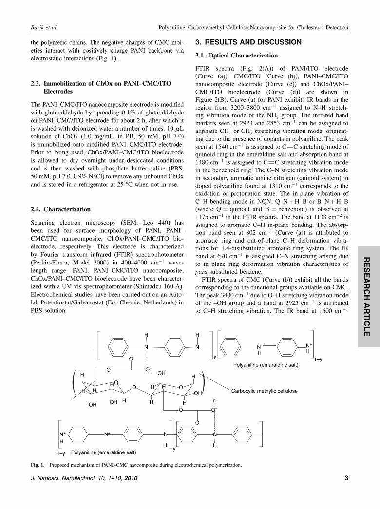
the polymeric chains. The negative charges of CMC moi-eties interact with positively charge PANI backbone viaelectrostatic interactions (Fig. 1).
2.3. Immobilization of ChOx on PANI–CMC/ITOElectrodes
The PANI–CMC/ITO nanocomposite electrode is modifiedwith glutaraldehyde by spreading 0.1% of glutaraldehydeon PANI–CMC/ITO electrode for about 2 h, after which itis washed with deionized water a number of times. 10 �Lsolution of ChOx (1.0 mg/mL, in PB, 50 mM, pH 7.0)is immobilized onto modified PANI–CMC/ITO electrode.Prior to being used, ChOx/PANI–CMC/ITO bioelectrodeis allowed to dry overnight under desiccated conditionsand is then washed with phosphate buffer saline (PBS,50 mM, pH 7.0, 0.9% NaCl) to remove any unbound ChOxand is stored in a refrigerator at 25 �C when not in use.
2.4. Characterization
Scanning electron microscopy (SEM, Leo 440) hasbeen used for surface morphology of PANI, PANI–CMC/ITO nanocomposite, ChOx/PANI–CMC/ITO bio-electrode, respectively. This electrode is characterizedby Fourier transform infrared (FTIR) spectrophotometer(Perkin-Elmer, Model 2000) in 400–4000 cm−1 wave-length range. PANI, PANI–CMC/ITO nanocomposite,ChOx/PANI–CMC/ITO bioelectrode have been character-ized with a UV–vis spectrophotometer (Shimadzu 160 A).Electrochemical studies have been carried out on an Auto-lab Potentiostat/Galvanostat (Eco Chemie, Netherlands) inPBS solution.
O
H
HHH
H
O
O O–
OH OH
O
H
HH
H
H
O
O
O–
OH
OH
O
N N N+
H H
y1–y
y1–y
n
Polyaniline (emaraldine salt)
Polyaniline (emaraldine salt)
Carboxylic methylic cellulose
HN+
H
NNN+
HH
N+
H
Fig. 1. Proposed mechanism of PANI–CMC naocomposite during electrochemical polymerization.
3. RESULTS AND DISCUSSION
3.1. Optical Characterization
FTIR spectra (Fig. 2(A)) of PANI/ITO electrode(Curve (a)), CMC/ITO (Curve (b)), PANI–CMC/ITOnanocomposite electrode (Curve (c)) and ChOx/PANI–CMC/ITO bioelectrode (Curve (d)) are shown inFigure 2(B). Curve (a) for PANI exhibits IR bands in theregion from 3200–3800 cm−1 assigned to N–H stretch-ing vibration mode of the NH2 group. The infrared bandmarkers seen at 2923 and 2853 cm−1 can be assigned toaliphatic CH2 or CH3 stretching vibration mode, originat-ing due to the presence of dopants in polyaniline. The peakseen at 1540 cm−1 is assigned to C C stretching mode ofquinoid ring in the emeraldine salt and absorption band at1480 cm−1 is assigned to C C stretching vibration modein the benzenoid ring. The C–N stretching vibration modein secondary aromatic amine nitrogen (quinoid system) indoped polyaniline found at 1310 cm−1 corresponds to theoxidation or protonation state. The in-plane vibration ofC–H bending mode in NQN, Q–N+H–B or B–N+H–B(where Q = quinoid and B = benzenoid) is observed at1175 cm−1 in the FTIR spectra. The band at 1133 cm−2 isassigned to aromatic C–H in-plane bending. The absorp-tion band seen at 802 cm−1 (Curve (a)) is attributed toaromatic ring and out-of-plane C–H deformation vibra-tions for 1,4-disubstituted aromatic ring system. The IRband at 670 cm−1 is assigned C–N stretching arising dueto in plane ring deformation vibration characteristics ofpara substituted benzene.FTIR spectra of CMC (Curve (b)) exhibit all the bands
corresponding to the functional groups available on CMC.The peak 3400 cm−1 due to O–H stretching vibration modeof the –OH group and a band at 2925 cm−1 is attributedto C–H stretching vibration. The IR band at 1600 cm−1
J. Nanosci. Nanotechnol. 10, 1–10, 2010 3

RESEARCH
ARTIC
LE
Polyaniline–Carboxymethyl Cellulose Nanocomposite for Cholesterol Detection Barik et al.
4000 3500 3000 2500 2000 1500 1000 50076
78
80
82
84
86
88
90
92
a = PANI/ITO electrode
b = CMC/ITO electrode
c = PANI-CMC/ITO electrode
d = ChOx/PANI-CMC/ITO bioelectrode
(a)
(b)
(d)
(c)
T (
%)
Wavenumber (cm–1)
(A)
(B)
(a) (b) (c)
Fig. 2. (A) FTIR spectra of (a) NanoPANI/ITO electrode, (b)CMC/ITO electrode, (c) PANI–CMC/ITO nanocomposite electrode and (c)ChOx/NanoPANI–CMC/ITO nanocomposites; (B) Scanning electron micrographs of (a) NanoPANI/ITO (b) PANI–CMC/ITO naocomposite electrode(c) ChOx/PANI–CMC/ITO bioelectrodes.
is assigned to COOH group due to ring stretching ofglucose. The bands seen around 1420 and 1315 cm−1 areassigned to C–H scissoring and –OH bending vibrationmodes in CH2 and COH groups, respectively. The band at1035 cm−1 is assigned to C–O stretching vibration modesdue to primary alcoholic –CH2OH stretching mode. Theweak band at around 700 cm−1 is due to ring stretch-ing and ring deformation of �-D-(1–4) and �-D-(1–6)linkages.38–39
It has been found that the band found at 3400 cm−1,1600 and 1420 cm−1 in FTIR spectra of CMC (curve (b))and band at 3410 cm−1 in PANI (curve (a)) disap-pears in the IR spectra of PANI–CMC nanocomposite(curve (c)) revealing that NH2 group of PANI interactswith OH/COO− group of CMC via electrostatic interac-tions (Fig. 1). Moreover, the band seen at 1480, 1310,1133 and 802 cm−1 shifts to higher frequency regionin the IR spectra of PANI–CMC nanocomposite dueto interaction between PANI and CMC revealing the
formation nanocomposite. However, in the FTIR spec-tra of PANI–CMC nanocomposite the peak intensity of670 cm−1 band at becomes weaker than that of the PANIspectra and presence of a sharp band at 830 cm−1 indi-cates that the amount of linear chain structure correspond-ing to the 1,4-para-disubstitution mode in PANI–CMC isdominant.It can be seen that ChOx/PANI/CMC/ITO bioelectrode
(Curve (c)) exhibits all bands of PANI–CMC nanocom-posite including two new bands at 3250 cm−1 assigned tothe NH2 group in protein and at 1580 cm−1 are attributedto amide I bond in enzyme revealing immobilization ofChOx onto the PANI–CMC/ITO nanocomposite electrode.The surface morphologies of PANI/ITO electrode
(image (a)), PANI–CMC/ITO nanocomposite elec-trode (image (b)), and ChOx/PANI–CMC/ITO bioelec-trode (image (c)) have been investigated using scanningelectron microscopy (SEM, Fig. 2(B)). Morphology ofPANI appears as a three-dimensional, inter-connected
4 J. Nanosci. Nanotechnol. 10, 1–10, 2010

RESEARCH
ARTIC
LE
Barik et al. Polyaniline–Carboxymethyl Cellulose Nanocomposite for Cholesterol Detection
network of smooth nanofiber with average diameter of120–160 nm and length of several microns. However,morphology of PANI changes into nanofibers (averagediameter of 150–200 nm) with globular appearance onaddition of CMC in PANI backbone.s This suggests thatCMC binds on the nanofibers of PANI via electrostaticinteractions. It can be seen that rough nanofibrous mor-phology of PANI–CMC nanocomposite further changesinto planner morphology after the immobilization ofChOx revealing immobilization of ChOx onto PANI–CMC nanocomposite. Moreover, globular nanofibrous
–0.8 –0.6 –0.4 –0.2 0.0 0.2 0.4 0.6 0.8–2.5 × 10⁻⁴
–2.0 × 10⁻⁴
–1.5 × 10⁻⁴
–1.0 × 10⁻⁴
–5.0 × 10⁻⁵
0.0
5.0 × 10⁻⁵
1.0 × 10⁻⁴
1.5 × 10⁻⁴
2.0 × 10⁻⁴
2.5 × 10⁻⁴
0.000 0.002 0.004 0.006 0.008 0.010 0.0120.0
5.0×10⁻⁵
1.0×10⁻⁴
1.5×10⁻⁴
2.0×10⁻⁴
2.5×10⁻⁴
Cur
rent
(A
)
Cellulose concentration (mM)
(c)(b)
(a) (d)
(e)
(f)
Cur
rent
(A
)
Potential (V)
(A)
0.0 5.0k 10.0k 15.0k 20.0k 25.0k 30.0k 35.0k 40.0k
2.0k
4.0k
6.0k
8.0k
10.0k
12.0k
14.0k
16.0k
(c)
(b)
(a)
–Z′′/
ohm
Z′/ohm
(B)
–0.8 –0.6 –0.4 –0.2 0.0 0.2 0.4 0.6 0.8
–2.0 × 10⁻⁴
–1.0 × 10⁻⁴
0.0
1.0 × 10⁻⁴
2.0 × 10⁻⁴
3.0 × 10⁻⁴
4.0 × 10⁻⁴
(c)
(b)
(a)
Cur
rent
(A
)
Potential (V)
(C)
0.0
Fig. 3. (A) Cyclic voltammograms of PANI–CMC naocomposite electrode at different concentration of cellulose (a) 0.5, (b) 1.25, (c) 2.5, (d) 5,(e) 7.5, (f) 11.25 �M in phosphate buffer (pH 7.0, 50 mM, 0.9% NaCl) at scan rate 30 mV/s (inset: plot of current vs. cellulose concentration);(B) Impedance spectra of (a) NanoPANI/ITO, (b) PANI–CMC/ITO naocomposite electrode and (c) PANI–CMC-ChOx/ITO bioelectrodes in phosphatebuffer (50 mM, pH 7.0, 0.9% NaCl); (C) Cyclic voltammograms of (a) NanoPANI/ITO electrode, (b) PANI–CMC/ITO naocomposite electrode and(c) ChOx/PANI–CMC/ITO bioelectrodes at scan rate 30 mV/sec in phosphate buffer [PH 7.0, 50 mM, 0.9% NaCl].
morphology of PANI–CMC provides added advantage foradsorption of ChOx.
3.2. Electrochemical Studies
The concentration of CMC of PANI–CMC nanocom-posite has been optimized using cyclic voltametric(CV) technique in phosphate buffer (50 mM, pH 7.0,0.9% NaCl) at scan rate 30 mv/s. Figure 3(A) showsCV of PANI with different concentration of CMCvarying from 0.5–11.25 �M revealing that magnitude of
J. Nanosci. Nanotechnol. 10, 1–10, 2010 5

RESEARCH
ARTIC
LE
Polyaniline–Carboxymethyl Cellulose Nanocomposite for Cholesterol Detection Barik et al.
–0.8 –0.6 –0.4 –0.2 0.0 0.2 0.4 0.6 0.8–2.0 × 10⁻⁴
–1.0 × 10⁻⁴
0.0
1.0 × 10⁻⁴
2.0 × 10⁻⁴
3.0 × 10⁻⁴
4.0 × 10⁻⁴
5.0 5.5 6.0 6.5 7.0 7.5 8.0 8.5 9.0
5.0×10⁻²
1.0×10⁻¹
1.5×10⁻¹
2.0×10⁻¹
2.5×10⁻¹
3.0×10⁻¹
3.5×10⁻¹
Cur
rent
(m
A)
PH
(c)
(b)
(a)
(d)Cur
rent
(A
)
Potentail (V)
Fig. 4. Cyclic voltammograms of the ChOx/PANI–CMC/ITO bioelectrode at different pH as (a) pH 6.0, (b) pH 6.5, (c) pH 7.0 and (d) pH 8.0 atscan rate 30mV/sec in phosphate buffer (50 mM) (inset: variation of obtained current vs. different pH).
current response is maximum for 2.5 �M PANI–CMCnanocomposite (Curve (c)). It is observed that PANI–CMC nanocomposite with 1.25 �M CMC (curve (b))exhibits oxidation and reduction peaks at 0.25 V and−0.24 V, respectively indicating a reversible system. ThusPANI–CMC nanocomposite consisting of 1.25 �M CMCconcentration has been selected for fabrication of biosen-sor electrode. It appears that as amount of CMC increasesfrom 0.5 to 2.5 �M, the emeraldine form of PANIincreases resulting in increased conductivity. However,when amount of CMC further increases above 2.5 �M,the emeraldine form of PANI is oxidized to pernigranilineform resulting in decrease electrical conductivity.Electrochemical impedance spectroscopy (EIS) tech-
nique has been used to measure resistance of the modifiedelectrode as a function of frequency due to variationin the interfacial properties. Figure 3(B) shows Faradicimpedance spectra in the frequency range 0.01–105 Hz,represented by the Nyquist plots of the PANI/ITOelectrode, PANI–CMC/ITO nanocomposite electrode andChOx/PANI–CMC/ITO bioelectrode (Curve (a), (b) and(c) respectively) obtained in phosphate buffer [50 mM, pH7.0, 0.9% NaCl]. The semicircle diameter of EIS spectragives value of charge transfer resistance (RCT� revealingelectron-transfer kinetics at the electrode interface. It isobserved that RCT value of PANI–CMC/ITO nanocompos-ite electrode (10.3 k�� is lower than that of the PANI/ITO
(41.5 k�� indicating that CMC increases the electroac-tive surface area of PANI and facilitates electron transportbetween the medium and electrode. Interestingly, after theimmobilization of ChOx onto PANI–CMC/ITO nanocom-posite electrode, RCT value decreases to 6.5 k�. Theseresults suggest that electron transfer in the ChOx/PANI–CMC/ITO electrode is easier between the solution andelectrode leading to improved diffusion of moleculestowards the electrode surface.Cyclic voltammetric (CV) studies of PANI/ITO elec-
trode, PANI–CMC/ITO nanocomposite electrode andChOx/PANI–CMC/ITO bioelectrode (Curves (a), (b)and (c) in Fig. 3(C)) have been conducted in phos-phate buffer (50 mM, pH 7.0, 0.9% NaCl) at scan rateof 30 mV/s. It is observed that PANI/ITO electrode(Curve (a)) shows well-defined oxidation peak at 0.28 Vand reduction peak at −0.24 V, indicating a reversiblesystem. The magnitude of current response for PANI–CMC/ITO nanocomposite electrode (Curve (b), 0.22 mA)is higher than that of PANI/ITO electrode (curve (a),0.34 mA). This suggests that negatively charged CMCmolecules diffuse into PANI matrix resulting in change inthe oxidation state (increased emeraldine salt form) thatmay result in enhanced electrical conductivity of PANI. Itappears that presence of CMC results in increased elec-troactivity of the PANI resulting in enhanced electrontransport between medium and the electrode. Further, mag-nitude of the response current increases (0.86 mA) after
6 J. Nanosci. Nanotechnol. 10, 1–10, 2010

RESEARCH
ARTIC
LE
Barik et al. Polyaniline–Carboxymethyl Cellulose Nanocomposite for Cholesterol Detection
(A)
–1.2 –0.9 –0.6 –0.3 0.0 0.3 0.6 0.9 1.2
–1.0 × 10⁻⁴
–5.0 × 10⁻⁵
0.0
5.0 × 10⁻⁴
1.0 × 10⁻⁴
1.5 × 10⁻⁴
2.0 × 10⁻⁴
2.5 × 10⁻⁴
3.0 × 10⁻⁴
3.5 × 10⁻⁴
4.0 × 10⁻⁴
4.5 × 10⁻⁴
0 5 10 15 20 25
5.0×10⁻⁵
1.0×10⁻⁴
1.5×10⁻⁴
2.0×10⁻⁴
2.5×10⁻⁴
3.0×10⁻⁴
3.5×10⁻⁴
4.0×10⁻⁴
4.5×10⁻⁴
Cha
nge
in c
urre
nt (
A)
Cholesterol concentartion (mM)
a = 0.55 mM
b = 5.55 mM
c = 11.1 mM
d = 13.86 mM
e = 16.65 mM
f = 19.45 mM
g = 22.25 mM
g
a
Cur
rent
(A
)
Potential (V)
–1.0 –0.8 –0.6 –0.4 –0.2 0.0 0.2 0.4 0.6 0.8 1.0–1.0 × 10⁻⁴
–5.0 × 10⁻⁵
0.0
5.0 × 10⁻⁵
1.0 × 10⁻⁴
1.5 × 10⁻⁴
2.0 × 10⁻⁴
2.5 × 10⁻⁴(B)
CH = cholesterol; U = Urea; UA = Uric Acid
Glu = Glucose; AA = Ascorbic Acid
a = CH
b = CH+AA
c = CH+Glu
d = CH+U
e = CH+UA
a b c d e0.0
2.0×10⁻⁵
4.0×10⁻⁵
6.0×10⁻⁵
8.0×10⁻⁵
1.0×10⁻⁴
1.2×10⁻⁴
1.4×10⁻⁴
1.6×10–41.8×10–4
2.0×10⁻⁴
2.2×10⁻⁴
2.4×10⁻⁴
Cur
rent
(A
)
Interferents (mM)
e
dc
ab
Cur
rent
(A
)
Potential (A)
Fig. 5. (A) Cyclic voltammograms of the ChOx/PANI–CMC/ITO bioelectrode as a function of cholesterol concentration (a) 0.55, (b)5.55, (c) 11.1,(d) 13.86, (e) 16.65, (f) 19.45, (g) 22.25 mM at scan rate 30mV/sec in phosphate buffer [pH 7, 50 mM, 0.9% NaCl] (inset: Change in current obtainedas a function of cholesterol concentration); (B) Interferent studies of the ChOx/PANI–CMC/ITO bioelectrode.
J. Nanosci. Nanotechnol. 10, 1–10, 2010 7

RESEARCH
ARTIC
LE
Polyaniline–Carboxymethyl Cellulose Nanocomposite for Cholesterol Detection Barik et al.
Table I. Characteristics of ChOx/PANI–CMC/ITO bioelectrode along with those reported for various nanocomposites utilized for cholesterol estima-tion in literature.
Detection Response ShelfElectrode Linearity Km limit Sensitivity time(s) life (Weeks) Ref.
NanoPANI-CNT 1.29–12.93 mM — — 6.8 �A/mM — 12 33ChOx/NanoZnO-CHIT 5–300 mg/dl 8.63 mgdl−1 5 mgdl−1 1�41×10−4Abs mgdl−1 15 s 12 34ChOx/Chitosan-tin oxide 0.26–10.4 mM 3.8 mM 5 mg/dL. 34.7 mA/mg dL−1 cm2 5 s, 8 35ChOx/CS/MWCNT 4�0×10−6–7�0×10−4 mM 0.24 mM <20 7 36ChOx/NanoPt/CNT 4.0×10⁻⁶–1.0×10⁻⁴ mol l−1 — — — <20 6 37ChOx/Polyaniline-Cellulose 0.5–22 mM 2.7 mM 1.31 mM 0.140 mA/mM 10 10 Present work
the immobilization of ChOx onto PANI–CMC/ITO elec-trode (Curve (c)) revealing that PANI–CMC/ITO electrodeplays an important role in accelerated electron transferbetween ChOx and electrode. Moreover, PANI–CMC/ITOnanocomposite electrode provides a three-dimensionalstructure and some of the restricted orientation that favorsdirect and faster electron communication between enzymeand the electrode surface.3The surface concentrations of redox species onto
PANI/ITO electrode, PANI–CMC/ITO nanocompositeelectrode and ChOx/PANI–CMC/ITO bioelectrode havebeen calculated using Brown-Anson model.40
Ip = N 2F 2CAV /4RT (1)
where N is the number of electrons transferred (2), F isFaraday constant (96485.3 C/mol), C is the surface con-centration of the electrode (mol/cm−2�, A is the surfacearea of the electrode (0.25 cm−2�, V is the scan rate(30 mV/s), R is the gas constant (8.314 J/mol K), T is theabsolute temperature (293 K) and Ip is the peak anodiccurrent of the electrode. The values of surface concentra-tion C of anions in PANI/ITO electrode, PANI–CMC/ITOnanocomposite electrode and ChOx/PANI–CMC/ITO bio-electrodes have been obtained as 41�5×10⁻⁶ mmol/cm−2,3�8×10−6 mmol/cm−2 and 5�8×10⁻⁶ mmol/cm−2, respec-tively. It has been found that the surface concentrationof the PANI–CMC/ITO nanocomposite is higher thanthat of PANI/ITO electrode suggesting that CMC load-ing increases electroactive surface area of the PANI–CMC nanocomposite. The higher surface concentration ofChOx/PANI–CMC/ITO bioelectrode implies high loadingof ChOx on the PANI–CMC nanocomposite matrix.
3.3. Activity of ChOx/PANI–CMC/ITONanocomposite Bioelectrode
Figure 4 shows effect of pH (6 to 8) on the electrochemi-cal behavior of ChOx/PANI–CMC/ITO bioelectrode usingCV technique. The magnitude of current response is max-imum at pH 7. This value of pH is optimum for catalyticactivity of ChOx for the hydrolysis of cholesterol. Thissuggests that ChOx/PANI–CMC/ITO bioelectrode showsmaximum activity at pH 7 at which ChOx retains its natu-ral structure and is responsible for low detection limit andhigh sensitivity for cholesterol detection.
3.4. Electrochemical Response Studies
Electrochemical response studies of ChOx/PANI–CMC/ITO bioelectrode have been carried out using cyclicvoltammetry as a function of cholesterol concentration(0.5–22 mM) in phosphate buffer (50 mM, pH 7.0, 0.9%NaCl) at scan rate of 30 mV/s. It has been found thatmagnitude of current response increases on addition ofcholesterol concentration (Fig. 5(A)). The response time ofthe ChOx/PANI–CMC/ITO bioelectrode found to be about10 s is attributed to faster electron communication featureof PANI–CMC nanocomposite. ChOx/PANI–CMC/ITObioelectrode can be used to estimate cholesterol from 0.5to 22 mM (inset, Fig. 5(A)).During biochemical reaction at the ChOx/PANI–
CMC/ITO bioelectrode surface, cholesterol converts intocholestenone and ChOx gets reduced. During reoxidationof ChOx, electrons are accepted by PANI–CMC nanocom-posite and transferred to electrode. It may be noted thatthere is no peak relating to the generation of H2O2 (0.5 V)revealing that PANI–CMC/ITO bioelecrode behaves as abetter electron acceptor than that of molecular oxygen.During enzymatic reaction of ChOx, the PANI–CMC/ITOcomposite film accepts electrons from enzyme causingincreased oxidation current.The value of apparent Michaelis-Menten constant, Km
determined by using Lineweaver-Burke plot (Supplemen-tary figure) has been found to be 2.71 mM. The observedlower Km value indicates high affinity for cholesterolattributed to the immobilization of ChOx onto PANI–CMC/ITO nanocomposite matrix resulting in enhancedbiochemical reaction.41�42 The detection limit (0.011 mM)of the bioelectrode has been calculated using the 3�b/mcriteria, where m is slope of the calibration graph and �bis standard deviation of the blank signal43 with regressioncoefficient of 0.999. The results of electrochemical exper-iments repeated at least 20 times have been found to bereproducible.This bioelectrode achieves 95% of steady state cur-
rent in less than 10 s indicating fast electron exchangebetween ChOx and the PANI–CMC nanocomposites. Theshelf-life of ChOx/PANI–CMC/ITO bioelectrode has beenmonitored by measuring electrochemical current responsewith respect to time, with regular interval of 1 week. It isobserved that this bioelectrode retains about 85% of ChOx
8 J. Nanosci. Nanotechnol. 10, 1–10, 2010

RESEARCH
ARTIC
LE
Barik et al. Polyaniline–Carboxymethyl Cellulose Nanocomposite for Cholesterol Detection
activity even after about 8 weeks when stored in refrig-erated conditions (4 �C) after which the current responsedecreases to 70% in about 10 weeks.The selectivity of the bioelectrode ChOx/PANI–
CMC/ITO composite bioelecrode has been determined bymeasuring CV response in presence of uric acid (0.2 mM),ascorbic acid (2 mM), urea (2 mM) and glucose (5 mM)with cholesterol (6 mM) in phosphate buffer (50 mM, pH7.0, 0.9% NaCl) at scan rate of 30 mV/s (Fig. 5(B)). It canbe seen that value of the electrochemical response currentremains nearly same.Table I summarizes characteristics of the ChOx/PANI–
CMC/ITO bioelectrode along with those reported in theliterature based on various nanocomposites utilized forcholesterol estimation.
4. CONCLUSIONS
It has been shown that nanocomposite film of PANIand CMC deposited onto ITO coated glass substrate canbe used for the immobilization of ChOx via glutralde-hyde as a cross-linker. PANI–CMC nanocomposite pro-vides a favorable environment for ChOx immobilizationwherein ChOx rearranged with better configuration resultsin enhanced electron transfer between medium and elec-trode. This nanocomposite bioelectrode shows high sensi-tivity, low response time, good reproducibility, and longterm stability. The presence of interferents such as glu-cose, uric acid, and ascorbic acid and urea in solution doesnot affect the observed electrochemical response of choles-terol biosensor. This interesting PANI–CMC nanocompos-ite should be used for estimation of other analytes such asurea, lipoproteins and triglycerides etc.
Acknowledgments: We thank Director, National Phys-ical Laboratory, India for the facilities. A. Barik is thankfulto Dr. J. C. Dutta, Department of Electronics and Commu-nications Enginering, Tezpur University, Tezpur. PratimaR. Solanki and Ajeet Kaushik thank CSIR, India for awardof Senior Research Associateships and Senior ResearchFellowship, respectively. We acknowledge financial sup-port received from Department of Science and Technology,India[DST/TSG/ME/2008/18 and GAP-070932], India–Japan project (DST/INT/JAP/P-21/07) and Department ofBiotechnology, Government of India (DBT/GAP 070832)and the Ministry of Education, Science and Technology(R32-20026), Korea under the World Class University(WCU) Programme.
References and Notes
1. S. I. Yoo, B. H. Sohn, W. C. Zin, and J. C. Jung, Langmuir 20, 10734(2004).
2. V. Cataldo, A. Vaze, and J. F. Ruslinga, Electroanalysis 20, 115(2008).
3. E. I. Iwuoha and M. R. Smyth, Polymer based amperometric biosen-sors, Electroactive Polymer Electrochemistry, edited by M. E. G.Lyons, Plenum Press, New York (1996), pp. 297–325.
4. K. Brazdziuviene, I. Jureviciute, and A. Malinauskas, Electrochim.Acta. 53, 785 ((2007).
5. Y. Y. Wang and X. L. Jing, Polymer J. 36, 374 (2004).6. K. Ghanbari, M. F. Mousavi, M. Shamsipur, and H. Karami, J. Power
Sources. 170, 513 (2007).7. B. D. Malhotra, S. Ghosh, and R. Chander, J. Appl. Polym. Sci.
40, 1049 (1990).8. Y. Kajiya, R. Tsuda, and H. Yoneysms, Electroanal. Chem. 301, 155
(1991).9. J. Huang, Pure Appl. Chem. 78, 15 (2006).10. B. D. Malhotra, A. Chaubey, and S. P. Singh, Anal. Chim. Acta
578, 59 (2006).11. J. Huang, S. Virji, B. H. Weiller, and R. B. Kaner, Chemistry
10, 1314 (2004).12. P. Aranda, M. Darder, and R. Fernández-Saavedra,
M. Lopez-Blanco, and E. Ruiz-Hitzky, Thin Solid Films 495, 104(2006).
13. Y. Yu, S. Zhihuai, S. Chen, C. Bian, W. Chen, and G. Xue, Langmuir22, 3899 (2006).
14. Y. A. Ismail, S. R. Shin, K. M. Shin, S. G. Yoon, K. Shon, S. I.Kim, and S. J. Kim, Sens. Actuators B 129, 834 (2008).
15. T. Thanpitcha, A. Sirivat, A. M. Jamieson, and R. Rujiravanit, Car-bohy. Polym. 64, 560 (2006).
16. J. I. Moran, V. A. Alvarez, V. P. Cyras, and A. Vazquez, Cellulose15, 149 (2008).
17. S. Ghosh, B. Vishalakshi, and V. Kalpagam, Synth. Met. 46, 349(1992).
18. M. Lukasiewicz, A. Ptaszek, L. Koziel, B. Achremowicz, andM. Grzesik, Poly. Bull. 58, 281 (2007).
19. S. R. Moraes and A. J. Motheo, Mol. Cryst. Liq. Cryst. 448, 261(2006).
20. P. Banerjee, Eur. Polym. J. 34, 1557 (1998).21. P. Banerjee, Eur. Polym. J. 34, 841 (1998).22. A. J. M. Valenate, H. D. Burrows, A. Y. Polishchuk, C. P.
Domingues, O. M. F. Borges, M. E. S. Eusebio, T. M. R. Maria,V. M. M. Lobo, and A. P. Monkman, Polymer 46, 5918 (2005).
23. D. Chattopadhyay and B. M. Mandal, Langmuir 12, 1585 (1996).24. O. V. D. Berg, M. Schroeter, J. R. Capadona, and C. Weder, J. Mater.
Chem. 17, 2746, (2007).25. N. B. Myant, The Biological of Cholesterol and Related Steroids,
William Heinemann, London (1981).26. D. S. Fredrickson, R. I. Levyin, and J. B. Wyngarden, The
Metabolic Basis of Inherited Diseases, edited by D. D. Fredrickson,McGraw-Hill, New York (1972), p. 545.
27. Z. Matharu, G. Sumana, S. K. Arya, S. P. Singh, V. Gupta, and B. D.Malhotra, Langmuir 23, 13188 (2007).
28. S. Singh, P. R. Solanki, M. K. Pandey, and B. D. Malhotra, Anal.Chim. Acta. 568, 126 (2006).
29. J. P. Li and T. Z. Peng, Chinese J. Chem. 20, 1038 (2002).30. Y. Xian, Y. Hu, F. Liu, Y. Xian, H. Wang, and L. Jin, Biosens.
Bioelectron. 21, 1996 (2006).31. A. Morrin, F. Wilbeer, O. Ngamna, S. E. Moulton, A. J. Killard,
G. G. Wallace, and M. R. Smyth. Electrochem. Commun. 7, 317(2005).
32. C. Dhand, S. P. Singh, S. K. Arya, M. Datta, and B. D. Malhotra,Anal. Chim. Acta 602, 244 (2007).
33. C. Dhand, S. K. Arya, S. P. Singh, B. P. Singh, M. Datta, and B. D.Malhotra, Carbon 46, 1727 (2008).
34. R. Khan, A. Kaushik, P. R. Solanki, A. A. Ansari, M. K. Pandey,and B. D. Malhotra Anal. Chim. Acta 616, 207 (2008).
35. A. A. Ansari, A. Kaushik, P. R. Solanki, and B. D. Malhotra. Elec-troanalysis 21, 965 (2009).
J. Nanosci. Nanotechnol. 10, 1–10, 2010 9

RESEARCH
ARTIC
LE
Polyaniline–Carboxymethyl Cellulose Nanocomposite for Cholesterol Detection Barik et al.
36. X. Tan, M. Li, P. Cai, L. Luo, and X. Zou, Anal. Biochem. 337, 111(2005).
37. S. Qiaocui, P. Tuzhi, Z. Yunu, and C. F. Yang, Electroanalysis17, 857 (2005).
38. V. Pushpamalar, S. J. Langford, M. Ahmad, and Y. Y. Lim, Carbohy.Polym. 64, 312 (2006).
39. J. Wang and P. Somasundaran, J. Colloid Inter. Sci. 291, 75(2005).
40. A. J. Bard and L. R. Faulkner, Electrochemical Methods: Fundamen-tals and Applications, 2nd edn., Wiley, New York (2000).
41. G. K. Kouassi, J. Irudayaraj, and G. McCarty, J. Nanobiotech. 3, 1(2005).
42. L. Pollegioni, G. Wels, M. S. Pilone, and S. Ghisla Eur. J. Biochem.264, 140 (1999).
43. Rajesh, V. Bisht, W. Takashima, and K. Kaneto, Biomaterials26, 3683 (2005).
Received: 21 May 2009. Accepted: 3 November 2009.
10 J. Nanosci. Nanotechnol. 10, 1–10, 2010