Synthesis of ZnO and ZnO/PVA nanocomposite using ...
13
Open Access. © 2020 K. Demssie Dejen et al., published by De Gruyter. This work is licensed under the Creative Commons Attribution 4.0 License Rev. Adv. Mater. Sci. 2020; 59:464–476 Research Article Kindnew Demssie Dejen, Enyew Amare Zereffa*, H C Ananda Murthy, and Andualem Merga Synthesis of ZnO and ZnO/PVA nanocomposite using aqueous Moringa Oleifeira leaf extract template: antibacterial and electrochemical activities https://doi.org/10.1515/rams-2020-0021 Received Jan 04, 2020; accepted Apr 23, 2020 Abstract: The application of flexible polymer nanocom- posites for food packaging to inactivate microorganisms associated with foods is the demand of the present-day food industry to assure quality throughout the packaging operation. The utilization of polyvinyl alcohol (PVA) as- sisted zinc oxide nanocomposite for food stuff packaging has been very attractive in the recent past. Nanostructured ZnO was synthesized at optimized pH (10.5) from different ratios of zinc acetate and Moringa oleifeira leaf extract (1:7, 1:3, 1:1 and 3:1). ZnO coated polyvinyl alcohol (ZnO/PVA) nanocomposites were prepared from 5, 9, 13 and 16% by wt of ZnO and PVA using solution casting method. The thermal stability of ZnO synthesized with 1:1 ratio at pH 10.5 was investigated with TGA/DTA. The analytical tech- niques such as X-ray diffraction (XRD), ultra-violate visible analysis (UV-Vis), Fourier-transform infrared spectroscopy (FTIR), and scanning electron microscope (SEM) were used for the characterization of the synthesized ZnO and ZnO/PVA nanocomposites (NCs). The antibacterial activity of the synthesized ZnO and ZnO/PVA NCs were evaluated against gram negative E. coli and gram positive S. aureus bacteria. The electrochemical stability of ZnO/PVA NCs was also investigated by cyclic voltammetric (CV) method. *Corresponding Author: Enyew Amare Zereffa: Department of Applied Chemistry, School of Applied Natural Science, Adama Science and Technology University, Ethiopia; Email: [email protected] Orcid ID: 0000-0001-5334-9626 Kindnew Demssie Dejen: Department of Chemistry, School of Natural & Computational Science, Wolkite University, Ethiopia H C Ananda Murthy: Department of Applied Chemistry, School of Applied Natural Science, Adama Science and Technology University, Ethiopia Andualem Merga: Department of Material Science & Engineering, School of Engineering, Adama Science and Technology University, Ethiopia The thermogram of ZnO indicated that the oxide was found to be stable even beyond 500 ∘ C. The SEM analysis revealed rod shaped morphology for synthesized ZnO from 1:1 ra- tio at pH 10.5. But the nanocomposite prepared with 5% of ZnO of (1:1) at the same pH exhibited uniformly dispersed rod-shaped particle on the surface as well as in matrix of polyvinyl alcohol film. According to XRD result, ZnO syn- thesized with more percentage of plant extract resulted in the small size crystallites while that with low percentage of plant extract resulted in the larger crystallite size. The an- tibacterial inhibition efficiency of ZnO/PVA NCs was better and found to increase with increase in the amount of ZnO. Keywords: Nanocomposite, Green synthesis, Moringa oleifeira, antibacterial activity, morphology 1 Introduction The spoilage and deterioration of food are mainly caused by food borne pathogens and other microorganisms, such as Escherichia coli, Listeria monocytogenes, etc., which mainly grows on food surfaces [1, 2]. Active packaging tech- nologies are being developed as a result of increased con- sumer demand for processed foods. Active packaging pro- vides interaction between food and packaging materials to maintain a microenvironment and extends the shelf life of food products [3, 4]. Active packaging has been mostly used to absorb oxygen, carbon dioxide, moisture, ethy- lene and release some substances such as antimicrobial agent and antioxidants [5]. Some of the most promising active packaging materials are antimicrobial films, which can be prepared by incorporating synthetic or natural an- timicrobial agents into films [6]. Polymeric food packag- ing films are the most commonly used films with excellent performance [7]. Polymeric food package can protect food from microbial attack and has properties such as flexibility, strength, stiffness and a barrier to oxygen and moisture.
-
Upload
khangminh22 -
Category
Documents
-
view
3 -
download
0
Transcript of Synthesis of ZnO and ZnO/PVA nanocomposite using ...
Open Access. © 2020 K. Demssie Dejen et al., published by De Gruyter. This work is licensed under the Creative CommonsAttribution 4.0 License
Rev. Adv. Mater. Sci. 2020; 59:464–476
Research Article
Kindnew Demssie Dejen, Enyew Amare Zereffa*, H C Ananda Murthy, and Andualem Merga
Synthesis of ZnO and ZnO/PVA nanocompositeusing aqueous Moringa Oleifeira leaf extracttemplate: antibacterial and electrochemicalactivitieshttps://doi.org/10.1515/rams-2020-0021Received Jan 04, 2020; accepted Apr 23, 2020
Abstract: The application of flexible polymer nanocom-posites for food packaging to inactivate microorganismsassociated with foods is the demand of the present-dayfood industry to assure quality throughout the packagingoperation. The utilization of polyvinyl alcohol (PVA) as-sisted zinc oxide nanocomposite for food stuff packaginghas been very attractive in the recent past. NanostructuredZnO was synthesized at optimized pH (10.5) from differentratios of zinc acetate andMoringa oleifeira leaf extract (1:7,1:3, 1:1 and 3:1). ZnO coated polyvinyl alcohol (ZnO/PVA)nanocomposites were prepared from 5, 9, 13 and 16% bywt of ZnO and PVA using solution casting method. Thethermal stability of ZnO synthesized with 1:1 ratio at pH10.5 was investigated with TGA/DTA. The analytical tech-niques such asX-ray diffraction (XRD), ultra-violate visibleanalysis (UV-Vis), Fourier-transform infrared spectroscopy(FTIR), and scanning electron microscope (SEM) wereused for the characterization of the synthesized ZnO andZnO/PVAnanocomposites (NCs). The antibacterial activityof the synthesized ZnO and ZnO/PVA NCs were evaluatedagainst gram negative E. coli and gram positive S. aureusbacteria. The electrochemical stability of ZnO/PVA NCswas also investigated by cyclic voltammetric (CV) method.
*Corresponding Author: Enyew Amare Zereffa: Department ofApplied Chemistry, School of Applied Natural Science, AdamaScience and Technology University, Ethiopia;Email: [email protected] ID: 0000-0001-5334-9626Kindnew Demssie Dejen: Department of Chemistry, School ofNatural & Computational Science, Wolkite University, EthiopiaH C Ananda Murthy: Department of Applied Chemistry, School ofApplied Natural Science, Adama Science and Technology University,EthiopiaAndualemMerga: Department of Material Science & Engineering,School of Engineering, Adama Science and Technology University,Ethiopia
The thermogramof ZnO indicated that the oxidewas foundtobe stable evenbeyond 500∘C. TheSEManalysis revealedrod shaped morphology for synthesized ZnO from 1:1 ra-tio at pH 10.5. But the nanocomposite prepared with 5% ofZnO of (1:1) at the same pH exhibited uniformly dispersedrod-shaped particle on the surface as well as in matrix ofpolyvinyl alcohol film. According to XRD result, ZnO syn-thesized with more percentage of plant extract resulted inthe small size crystalliteswhile thatwith lowpercentage ofplant extract resulted in the larger crystallite size. The an-tibacterial inhibition efficiency of ZnO/PVANCswas betterand found to increase with increase in the amount of ZnO.
Keywords: Nanocomposite, Green synthesis, Moringaoleifeira, antibacterial activity, morphology
1 IntroductionThe spoilage and deterioration of food are mainly causedby food borne pathogens and other microorganisms, suchas Escherichia coli, Listeria monocytogenes, etc., whichmainly grows on food surfaces [1, 2]. Active packaging tech-nologies are being developed as a result of increased con-sumer demand for processed foods. Active packaging pro-vides interaction between food and packaging materialsto maintain a microenvironment and extends the shelf lifeof food products [3, 4]. Active packaging has been mostlyused to absorb oxygen, carbon dioxide, moisture, ethy-lene and release some substances such as antimicrobialagent and antioxidants [5]. Some of the most promisingactive packaging materials are antimicrobial films, whichcan be prepared by incorporating synthetic or natural an-timicrobial agents into films [6]. Polymeric food packag-ing films are the most commonly used films with excellentperformance [7]. Polymeric food package can protect foodfrommicrobial attack andhasproperties suchasflexibility,strength, stiffness and a barrier to oxygen and moisture.

Antibacterial and electrochemical activities | 465
Theantimicrobial agent ismixedwith thepolymer andthe active compound is distributed in the polymer matrix,homogeneously when polymer and agent are chemicallycompatible, or heterogeneously, forming amultiphasema-trix in which sites enriched with the agent can be distin-guished [8]. Recently, nanoparticles have been used to en-hance mechanical performance of food packaging, suchas flexibility, stability against humidity and temperature,reducing gaseous permeation, and ultraviolet light resis-tance [9]. In addition, they have antimicrobial activity asgrowth inhibitors, killing agents and antimicrobial carri-ers [10].
Metal oxide nanoparticles (MONPs) are more stableand have potent antimicrobial effect against microorgan-isms. MONPs such as titanium dioxide (TiO2), zinc oxide(ZnO), copper oxides (CuO/Cu2O), silica (SiO2), aluminumoxide (Al2O3), magnesium oxide (MgO), and iron oxides(Fe3O4 and Fe2O3) have been commonly used for variousapplications. Among them, TiO2, ZnO and MgO have re-ceived special attention in the food packaging industrydue to their strong antimicrobial effects. ZnO NP-loadedstarch/polythene films tested for their ability to controlpathogenic E. coli [11]. It was found that the film couldprevent the bacterial contamination in food stuff by inhi-bition of E. coli. This possible action could be achievedby the production of reactive oxygen species, such as hy-droxyl and superoxide radicals. ZnO-deposited polypropy-lene packaging film was prepared by using dielectric bar-rier discharge plasma treatment and exhibited better an-timicrobial effect against pathogens S. aureus and E. coli[12].
Polyvinyl alcohol (PVA), a degradable polymer, easilydissolved in water, and a combination of ZnO nanoparti-cles and PVA results in nanocompositewith improved elec-trical, mechanical and optical properties. This non poison-ing biodegradable nanocomposite can be used as a moreeffective and environmentally friendly material for food-stuff packaging [13]. In the presentwork, we report the syn-thesis of ZnO and polyvinyl alcohol (ZnO/PVA) nanocom-posites (NCs) usingMoringa-oleifeira leaf extract and eval-uation of their electrochemical and antimicrobial activi-ties.
2 Materials and methods
2.1 Chemicals and Reagents
Polyvinyl alcohol (PVA) (Sigma Aldrich), with an av-erage molecular weight of 17,000 GRG and 97% hy-
drolyzed was used without further purification. Zinc ac-etate (C4H6O4Zn·2H2O), NaOH, Mueller-Hinton agar andDMSO were purchased from Sigma Aldrich. Graphite pow-der and silicon oil were purchased from the local mar-ket. Reactant solutions were prepared by using doubly dis-tilled water. Analytical grade chemicals reagents and sol-vents were used without further purification.
2.2 Collection of Moringa Oleifeira Leaf
Healthy and Fresh Moringa oleifeira leaves were collectedfrom DinshoWereda, Bale Zone, Oromia Regional State,Ethiopia, after conducting the field survey. Dinsho (alsocalled Gurie) is a village in south-central Ethiopia, locatedin the heart of the Bale Mountains with a latitude and lon-gitude of 705’N3945’E and an elevation of 3207 meters.
2.3 Methods
2.3.1 Preparation ofMoringa-oleifeira leaf extracts(Broth solution)
The collected Moringa-oleifeira leaves were washed thor-oughly with distilled water to remove dust and other for-eign particles and dried under shadow at room temper-ature. The dried leaves were ground to get fine powder.The extraction was carried out by taking 50 g of this pow-der in 300 mL deionized water (DI-H2O). The obtainedcolloidal suspension was boiled at 50∘C for about 1 hourwith constant stirring using magnetic stirrer. The solu-tion was allowed to cool at room temperature and filteredthrough Whatman number 1 filter paper. The filtered solu-tion was stored in the refrigerator at 4∘C for the synthesisof ZnO/PVA NCs. Broth solutions prepared from moringaoleifeira aqueous leaves extract are as shown in Figure 1.
Figure 1:Moringa oleifeira leaves broth solution prepared for thiswork.

466 | K. Demssie Dejen et al.
2.3.2 Biosynthesis of ZnO NPs using Moringa Oleifeiraleaf extract
To synthesize ZnO/PVA NCs, four different volume ratiosof 0.122 M C4H6O4Zn·2H2O and broth solution of Moringaoleifeira leaf extract were taken in the 250 mL volumetricflask as per the following procedure; 12.5 mL of 0.122 MC4H6O4Zn·2H2O with 87.5 mL leaf extract (1:7), 25 mL of0.122 M C4H6O4Zn·2H2Owith 75 mL of leaf extract (1:3), 50mL of 0.122 M C4H6O4Zn·2H2O with 50 mL of leaf extract(1:1) and 75 mL of 0.122 M C4H6O4Zn·2H2O with 25 mL ofleaf extract (3:1) were mixed in different 250 mL conicalflasks.
In all of these cases, the solutions were stirred using amagnetic stirrer for about two and a half hours, by control-ling pHwith dropwise addition of 1M NaOH solution to pH= 9, 10.5 and 12 for all the mentioned ratios. The solutionwas centrifuged at 12000 rpm for 15 min three times fol-
lowed by repeated washing with ethanol and distilled wa-ter to remove the impurities. The obtained precipitate wascollected using a ceramic crucible dish and allowed to dryin an oven at 100∘C for about one hour. Finally, the driedprecipitatewas ground into a fine powder. The thermal sta-bility of the synthesized sample was checked using a si-multaneous DTA/TGA instrument. Based on the thermalanalysis result, the obtained ZnO powder was subjected toan additional heat treatment in a furnace at 500∘C for 2 h.The overall schematic of the procedure for the synthesis ofZnO nanoparticles is shown in Figure 2.
2.3.3 Preparation of ZnO/PVA nanocomposite films
The ZnO/PVA NC films were prepared by solution castingtechnique. ZnO synthesizedwith (1:1) ratio at pH= 10.5wasadded in different weight percent ratios by (wt.%; 0 (free
Figure 2: Schematic diagram of green synthesis of ZnO nanoparticles.
Figure 3: Schematic of synthesis of ZnO/PVA NC.

Antibacterial and electrochemical activities | 467
Table 1: Ratio of precursor to plant extract used to prepare ZnO NPs
No Volume of 0.122 MC4H6O4Zn·2H2O
Volume of leafextract
Precursor toextract ratio
pH Color of ZnO crystallitesize
a 12.5 mL 87.5 mL 1:7 10.5 Brownish color 7.57 nmb 25 mL 75 mL 1:3 10.5 Light Yellow color 9.89 nmc 50 mL 50 mL 1:1 10.5 Yellow color 10.13 nmd 75 mL 25 mL 3:1 10.5 Bright yellow color 15.27nm
of ZnONP), 5, 9, 13 and 16) to PVA according to the formulagiven below (1):
x(wt%) = 100 × wf(wf + wp) (1)
where wf and wp represent the weights of ZnO and PVApolymer, respectively.
The NC films were prepared by dispersing differentamounts of ZnO nanoparticles in 2.0 g of PVA dissolved in50 mL distilled water at 80∘C under vigorous stirring for 1h to prevent any agglomeration. Lastly, the aqueous solu-tion of ZnO/PVA nanocomposite was cast into Petri dishesand allowed to dry in a dust free chamber at room tem-perature for 5 days to obtain the films. The obtained purePVA and ZnO/PVA NC films were characterized. The over-all schematic of the experiment for making ZnO/PVA NCfilm is shown in Figure 3.
2.4 Characterization
The XRD diffraction patterns of the pure PVA film, ZnOpowder and the composite films (PVA/ZnO) were recordedat room temperature using an X-ray powder diffractome-ter (XRD-700, shimadzu co. South Korea) equipped withCu Kα as radiation source (λ = 1.54 Å) in the 2θ (Braggangles) range (10∘ ≤ 2θ ≤ 80∘) to report the informa-tion about their structure. The Fourier transform infraredspectra of the nano powder and the composites were an-alyzed in the range 400–4000 cm−1 using the instrument(Perkin Elmer Spectrum 65 Spectrum BX FTIR-Model 6100,Japan) in the absorbance mode at a resolution of 4.0 cm−1.Thermal stability of the ZnOwas conducted with TGA/DTA(DTG60H shimadzu co. South Korea). The absorbance spec-tra (A) and the transmittance spectra (T) of the films wererecorded at 200–1100 nm wavelength using a dual beamUV–Visible spectrophotometer (Perkin Elmer, D-7770 Ue-berlingen, and Germany) and the morphological and com-positional analysis of all the sample were done using SEM-EDX machine (JEOL-JSM-8040, 81F, Tokyo, Japan). The CVmeasurements were carried out using an electrochemicalanalyzer CHI608E Potentiostat in a three electrode system,
consisting of carbon paste electrode, platinum wire andAg/AgCl as working, counter and reference electrodes, re-spectively and the electrolyte being a solution of 1.0 MKOH.
3 Result and DiscussionThe colors of the biogenic ZnO nanoparticles obtainedwith different ratios of zinc acetate and Moringa oleifeiraleaf extract are presented in Table 1. The different colorsobtained for various ZnO NPs are believed to be due to thevariation in the particle sizes. In this regard the ratios ofleaf extract played crucial role in capping the ZnO NPs.
3.1 Thermal analysis of synthesized Zincoxide sample
Figure 4 depicts the TGA-DTA curves for the thermal trans-formation of ZnO NPs. TGA showed a weight loss in twosteps at 100 and 500∘C, while DTA showed one exother-mic peak and one endothermic peak at 72.3∘C and 394∘Crespectively. The endothermic peak at 72.3∘C was due to
Figure 4: Thermal Analysis result of as synthesized ZnONPs.
468 | K. Demssie Dejen et al.
removal of water and the peak at 394∘C was due to de-composition of zinc hydroxide to formZnOand (or)organicbinders from leaf extracts. No considerable loss is ob-served after 500∘C, indicating that the oxide is thermallystable above 500∘C.
3.2 XRD Analysis
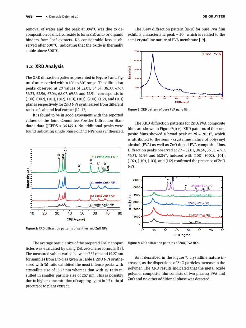
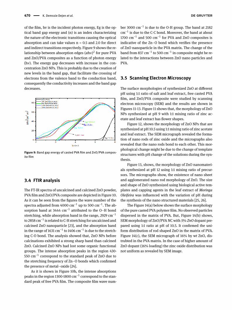
The XRD diffraction patterns presented in Figure 5 and Fig-ure 6 are recorded within 10∘ to 80∘ range. The diffractionpeaks observed at 2θ values of 32.01, 34.54, 36.33, 47.67,56.73, 62.96, 67.04, 68.07, 69.34 and 72.91∘ corresponds to(100), (002), (101), (102), (110), (103), (200), (112), and (201)planes respectively for ZnONPs synthesized from differentratios of salt and leaf extract [14–17].
It is found to be in good agreement with the reportedvalues of the Joint Committee Powder Diffraction Stan-dards data (JCPDS # 36-1451). No additional peaks werefound indicating single phase of ZnONPswas synthesized.
Figure 5: XRD diffraction patterns of synthesized ZnO NPs.
The average particle size of the prepared ZnOnanopar-ticles was evaluated by using Debye-Scherer formula [18].Themeasured values varied between 7.57 nm and 15.27 nmfor samples from a to d as given in Table 1. ZnONPs synthe-sised with 3:1 ratio exhibited the most intense peaks withcrystallite size of 15.27 nm whereas that with 1:7 ratio re-sulted in smaller particle size of 7.57 nm. This is possiblydue to higher concentration of capping agent in 1:7 ratio ofprecursor to plant extract.
The X-ray diffraction pattern (XRD) for pure PVA filmexhibits characteristic peak ~ 20∘ which is related to thesemi-crystalline nature of PVA membrane [19].
Figure 6: XRD pattern of pure PVA nano film.
The XRD diffraction patterns for ZnO/PVA compositefilms are shown in Figure 7(b-e). XRD patterns of the com-posite films showed a broad peak at 2θ = 20.11∘, whichis attributed to the semi - crystalline nature of polyvinylalcohol (PVA) as well as ZnO doped PVA composite films.Diffraction peaks observed at 2θ = 32.01, 34.54, 36.33, 47.67,56.73, 62.96 and 67.04∘, indexed with (100), (002), (101),(102), (110), (103), and (112) confirmed the presence of ZnONPs.
Figure 7: XRD diffraction patterns of ZnO/PVA NCs.
As it described in the Figure 7, crystalline nature in-creases, as the dispersions of ZnO particles increase in thepolymer. The XRD results indicated that the metal oxidepolymer composite film consists of two phases; PVA andZnO and no other additional phase was detected.

Antibacterial and electrochemical activities | 469
Table 2: XRD data of synthesized ZnO NPs with different ratios of precursors.
No 1:7 ratio 1:3 1:1 3:12-Theta(deg)
FWHM(deg)
2-Theta(deg)
FWHM(deg)
2-Theta(deg)
FWHM(deg)
2-Theta(deg)
FWHM(deg)
1 36.0487 1.05000 36.2443 0.77500 36.2316 0.79430 36.2839 0.541802 31.6889 1.00870 31.7752 0.79950 31.7637 0.75160 31.8129 0.535803 34.3985 1.04000 34.5404 0.79570 34.4814 0.77050 34.4747 0.465504 20.2670 0.59000 47.6161 1.04000 37.3359 0.47000 37.2759 0.386605 23.6370 1.12670 56.5385 0.87400 47.5769 1.10500 47.5933 0.756506 26.2454 1.19330 61.9437 0.68000 56.5705 0.94340 56.6308 0.608607 28.1159 0.42000 62.9774 1.05200 57.6252 0.58000 62.8946 0.737508 29.2843 0.76000 66.3223 0.82000 61.8237 0.76000 63.6232 0.410009 37.6756 0.00000 68.2468 1.61000 62.9092 1.15830 66.4223 0.79000
Table 3: XRD data for PVA and ZnO/PVA NCs
PVA 5%ZnO/PVA 9%ZnO/PVA 13%ZnO/PVA 16%ZnO/PVA2θ
(deg)FWHM(deg)
2θ(deg)
FWHM(deg)
2θ(deg)
FWHM(deg)
2θ(deg)
FWHM(deg)
2θ(deg) FWHM(deg)
43.9615 0.1967 20.009 2.53330 19.186 2.5433 20.35 2.4700 36.16 0.8849064.3065 0.2218 21.738 0.00000 17.668 0.0000 36.88 0.7527 31.69 0.9137077.4059 0.2655 36.461 0.78380 21.299 0.0000 22.76 0.0000 34.39 0.8406077.7591 0.1200 23.094 0.00000 22.336 0.0000 23.77 2.0134 47.52 1.0933037.7388 0.1984 23.693 2.32000 23.354 1.6000 32.40 0.7350 56.52 0.9628021.4388 0.5866 25.729 1.04000 24.212 0.9200 35.08 0.5967 61.78 0.5200064.6028 0.0960 31.967 0.82800 25.250 0.6000 41.34 0.5400 62.85 1.03070
3.3 UV-Vis analysis of PVA and ZnO/ PVAcomposite films
Figure 8 shows the UV-visible absorption spectra of PVAand ZnO/ PVA composite films. The absorption spectrumof pure PVAhas one broad absorbance band at 290 nmandone weak band or shoulder at 330 nm. The weak band orshoulder is assigned to the electronic transitions n → π*
[20]. As it is shown in Figure 8, free PVA has poor ab-sorptive in the visible region (longer wavelength) and alsohas small absorbance in the UV region as compared withits composite with ZnO NPs. Absorbance of the compos-ite films increases with increasing ZnO concentration from5% to 16%. The emission peak of ZnO/PVA composite filmhas been shifted to the blue region (shorter wavelength).The absorbance spectra of the ZnO/PVANC films consist ofthe fundamental peak of the PVA and further absorbanceband at 375 nm, corresponding to ZnO NP; which is 5 nmblue shift in comparison with the ZnO bulk material (380nm) at room temperature [21].
Figure 8: UV-Vis spectra of PVA and ZnO/PVA NCs.
3.3.1 Optical band gap
The optical transitions in composite films can be easily un-derstood by determining the optical band gap by Tauc’splot [22]. The frequency-dependent absorption coefficientis given by (αhv)n = hv−Egwhere α is the absorption coeffi-cient, which calculated using the Beer-Lambert’s relation,α = 2.303; A and t are the absorbance and the thickness
470 | K. Demssie Dejen et al.
of the film, hv is the incident photon energy, Eg is the op-tical band gap energy and (n) is an index characterizingthe nature of the electronic transitions causing the opticalabsorption and can take values n = 0.5 and 2.0 for directand indirect transitions respectively. Figure 9 shows the re-lationship between absorption edges (αhν)2 for pure PVAand ZnO/PVA composites as a function of photon energy(hν). The energy gap decreases with increase in the con-centration ZnO NPs. This is probably due to the creation ofnew levels in the band gap, that facilitate the crossing ofelectrons from the valence band to the conduction band,consequently the conductivity increases and the band gapdecreases.
Figure 9: Band gap energy of casted PVA film and ZnO/PVA compos-ite film
3.4 FTIR analysis
The FT-IR spectra of uncalcined and calcined ZnO powder,PVAfilmand ZnO/PVA composite are depicted in Figure 10.As it can be seen from the figures the wave number of thespectra adjusted from 4000 cm−1 up to 500 cm−1. The ab-sorption band at 3444 cm−1 attributed to the O–H bondstretching, while absorption band in the range, 2929 cm−1
to 2858 cm−1 is related toC-H stretching for uncalcined andcalcined ZnO nanoparticle [23], and the absorption bandin the range of 1631 cm−1 to 1406 cm−1 is due to the stretch-ing C-O bond. The analysis showed that, ZnO NPs beforecalcinations exhibited a strong sharp band than calcinedZnO. Calcined ZnO NPs had lost some organic functionalgroups. The intense absorption peaks in the region 430-550 cm−1 correspond to the standard peak of ZnO due tothe stretching frequency of Zn–O bonds which confirmedthe presence of metal–oxide [24].
As it is shown in Figure 10b, the intense absorptionspeaks in the region 1300-1800 cm−1 correspond to the stan-dard peak of free PVA film. The composite film wave num-
ber 3000 cm−1 is due to the O-H group. The band at 2182cm−1 is due to the C-C bond. Moreover, the band at about1700 cm−1 and 500 cm−1 for PVA and ZnO composites isindicative of the Zn–O bond which verifies the presenceof ZnO nanoparticle in the PVA matrix. The change of theband from 837 cm−1 to 500 cm−1 in composite might be re-lated to the interactions between ZnO nano particles andPVA.
3.5 Scanning Electron Microscopy
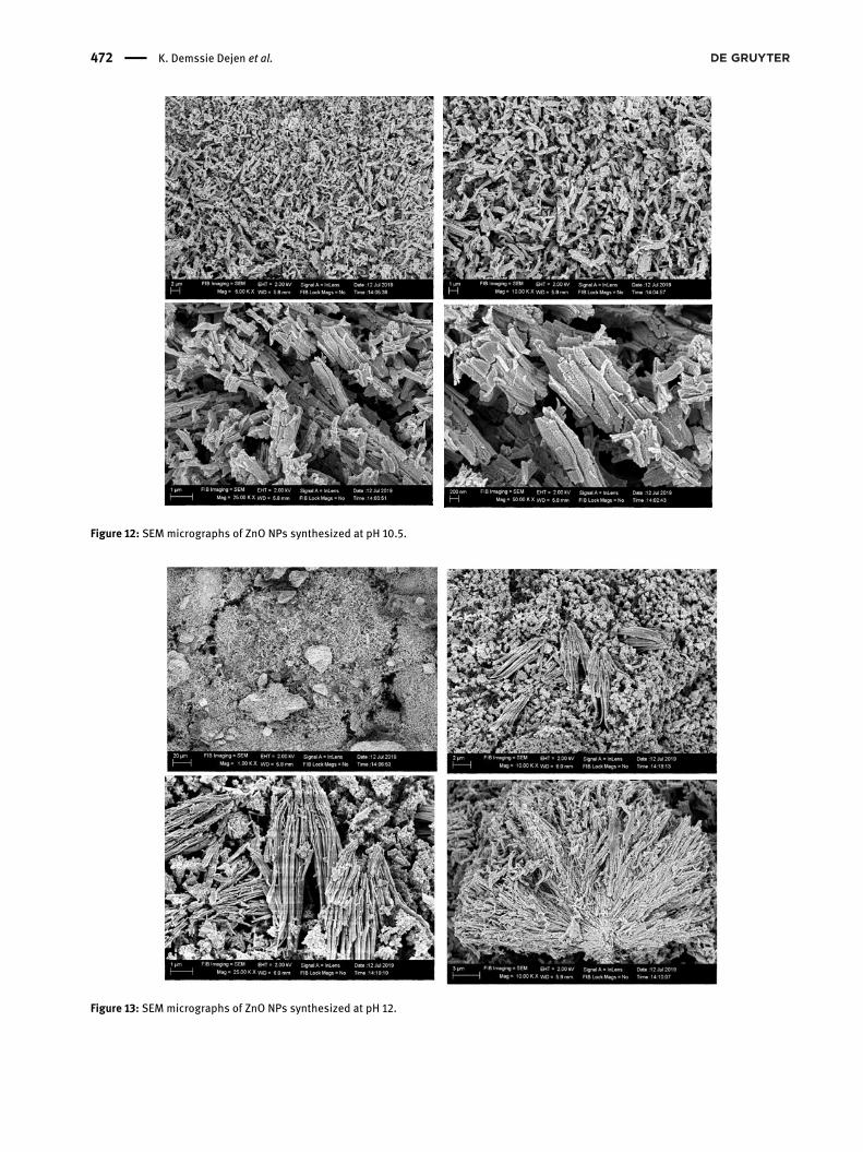
The surface morphologies of synthesized ZnO at differentpH using 1:1 ratio of salt and leaf extract, free casted PVAfilm, and ZnO/PVA composite were studied by scanningelectron microscopy (SEM) and the results are shown inFigures 11-13. Figure 11 shows that, themorphology of ZnONPs synthesized at pH 9 with 1:1 mixing ratio of zinc ac-etate and leaf extract has flower shapes.
Figure 12, shows the morphology of ZnO NPs that aresynthesized at pH 10.5 using 1:1mixing ratio of zinc acetateand leaf extract. The SEM micrograph revealed the forma-tion of nano rods of zinc oxide and the micrographs alsorevealed that the nano rods bond to each other. This mor-phological change might be due to the change of templatestructures with pH change of the solutions during the syn-thesis.
Figure 13, shows, the morphology of ZnO nanomateri-als synthesized at pH 12 using 1:1 mixing ratio of precur-sors. The micrographs show, the existence of nano sheetand agglomerated nano rod morphology of ZnO. The sizeand shape of ZnO synthesized using biological active tem-plates and capping agents in the leaf extract of MoringaOleifeira was influenced with the variation of pH duringthe synthesis of the nano structured materials [25, 26].
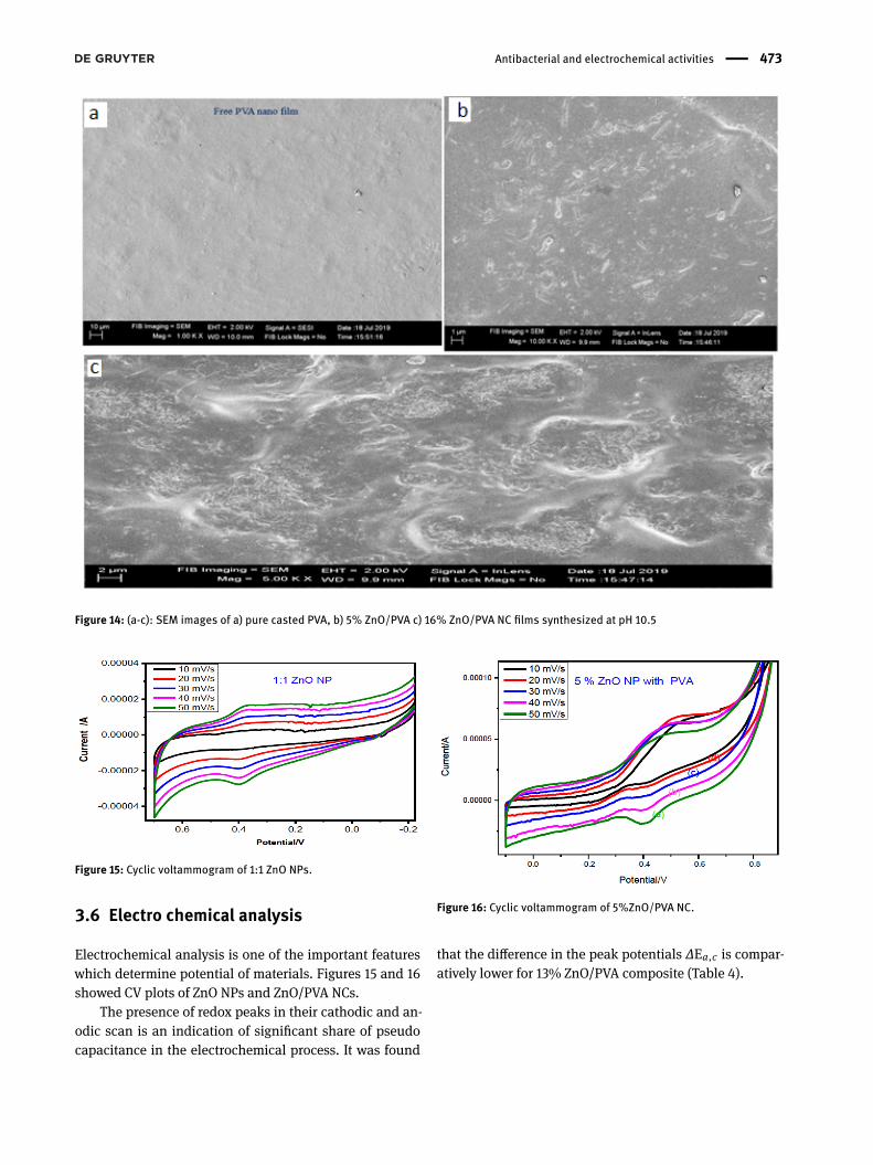
The Figure 14(a) below shows the surface morphologyof the pure casted PVA polymer film. No observed particlesdispersed in the matrix of PVA. But, Figure 14(b) shows,SEMmorphology of ZnO/PVA NC with 5% ZnO dopant pre-pared using 1:1 ratio at pH of 10.5. It confirmed the uni-form distribution of rod shaped ZnO in the matrix of PVA.Figure 14(c), the SEM micrograph of 16% by wt ZnO, dis-trubted in the PVA matrix. In the case of higher amount ofZnO dopant (16% loading) the zinc oxide distribution wasnot uniform as revealed by SEM image.

Antibacterial and electrochemical activities | 471
(a) (b)
Figure 10: a) FTIR spectra of uncalcined and calcined ZnO NP, b) FTIR spectra of free PVA and 5% ZnO/PVA composite films.
Figure 11: SEM micrographs of ZnO NPs synthesized at pH 9.
472 | K. Demssie Dejen et al.
Figure 12: SEM micrographs of ZnO NPs synthesized at pH 10.5.
Figure 13: SEM micrographs of ZnO NPs synthesized at pH 12.
Antibacterial and electrochemical activities | 473
Figure 14: (a-c): SEM images of a) pure casted PVA, b) 5% ZnO/PVA c) 16% ZnO/PVA NC films synthesized at pH 10.5
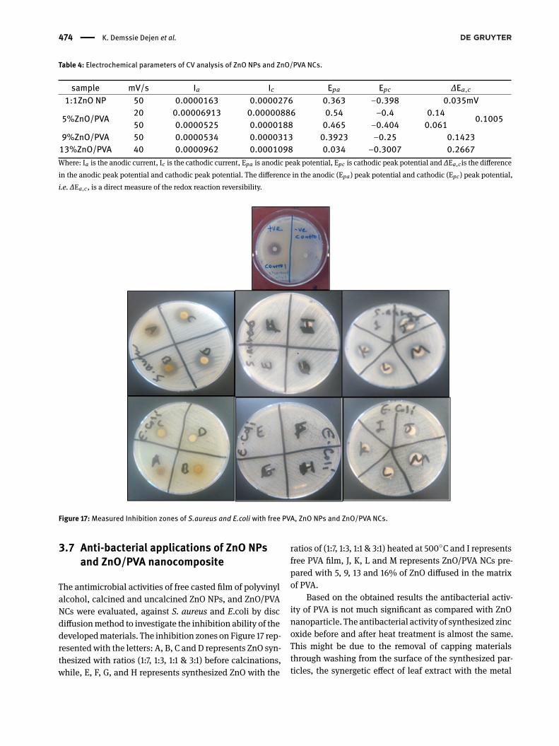
Figure 15: Cyclic voltammogram of 1:1 ZnO NPs.
3.6 Electro chemical analysis
Electrochemical analysis is one of the important featureswhich determine potential of materials. Figures 15 and 16showed CV plots of ZnO NPs and ZnO/PVA NCs.
The presence of redox peaks in their cathodic and an-odic scan is an indication of significant share of pseudocapacitance in the electrochemical process. It was found
Figure 16: Cyclic voltammogram of 5%ZnO/PVA NC.
that the difference in the peak potentials ∆Ea,c is compar-atively lower for 13% ZnO/PVA composite (Table 4).
474 | K. Demssie Dejen et al.
Table 4: Electrochemical parameters of CV analysis of ZnO NPs and ZnO/PVA NCs.
sample mV/s Ia Ic Epa Epc ∆Ea,c1:1ZnO NP 50 0.0000163 0.0000276 0.363 −0.398 0.035mV
5%ZnO/PVA 20 0.00006913 0.00000886 0.54 −0.4 0.14 0.100550 0.0000525 0.0000188 0.465 −0.404 0.0619%ZnO/PVA 50 0.0000534 0.0000313 0.3923 −0.25 0.142313%ZnO/PVA 40 0.0000962 0.0001098 0.034 −0.3007 0.2667Where: Ia is the anodic current, Ic is the cathodic current, Epa is anodic peak potential, Epc is cathodic peak potential and ∆Ea,cis the differencein the anodic peak potential and cathodic peak potential. The difference in the anodic (Epa) peak potential and cathodic (Epc) peak potential,i.e. ∆Ea,c, is a direct measure of the redox reaction reversibility.
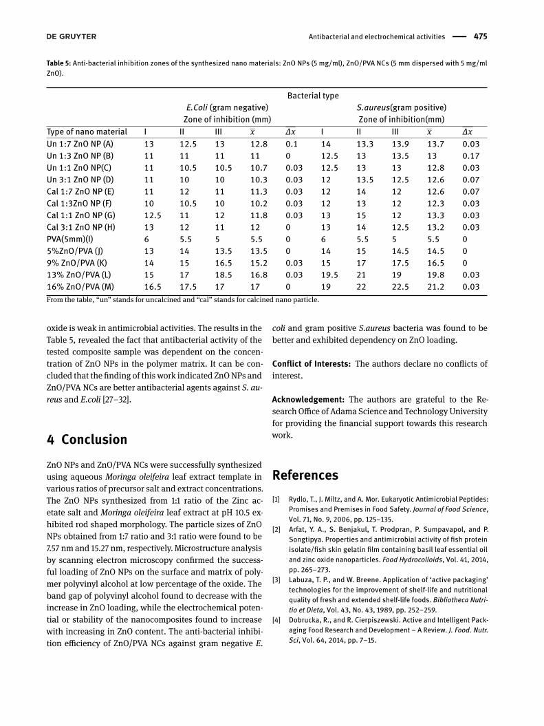
Figure 17:Measured Inhibition zones of S.aureus and E.coli with free PVA, ZnO NPs and ZnO/PVA NCs.
3.7 Anti-bacterial applications of ZnO NPsand ZnO/PVA nanocomposite
The antimicrobial activities of free casted film of polyvinylalcohol, calcined and uncalcined ZnO NPs, and ZnO/PVANCs were evaluated, against S. aureus and E.coli by discdiffusionmethod to investigate the inhibition ability of thedevelopedmaterials. The inhibition zones on Figure 17 rep-resentedwith the letters: A, B, C and D represents ZnO syn-thesized with ratios (1:7, 1:3, 1:1 & 3:1) before calcinations,while, E, F, G, and H represents synthesized ZnO with the
ratios of (1:7, 1:3, 1:1 & 3:1) heated at 500∘C and I representsfree PVA film, J, K, L and M represents ZnO/PVA NCs pre-pared with 5, 9, 13 and 16% of ZnO diffused in the matrixof PVA.
Based on the obtained results the antibacterial activ-ity of PVA is not much significant as compared with ZnOnanoparticle. The antibacterial activity of synthesized zincoxide before and after heat treatment is almost the same.This might be due to the removal of capping materialsthrough washing from the surface of the synthesized par-ticles, the synergetic effect of leaf extract with the metal
Antibacterial and electrochemical activities | 475
Table 5: Anti-bacterial inhibition zones of the synthesized nano materials: ZnO NPs (5 mg/ml), ZnO/PVA NCs (5 mm dispersed with 5 mg/mlZnO).
Bacterial typeE.Coli (gram negative) S.aureus(gram positive)Zone of inhibition (mm) Zone of inhibition(mm)
Type of nano material I II III x ∆x I II III x ∆xUn 1:7 ZnO NP (A) 13 12.5 13 12.8 0.1 14 13.3 13.9 13.7 0.03Un 1:3 ZnO NP (B) 11 11 11 11 0 12.5 13 13.5 13 0.17Un 1:1 ZnO NP(C) 11 10.5 10.5 10.7 0.03 12.5 13 13 12.8 0.03Un 3:1 ZnO NP (D) 11 10 10 10.3 0.03 12 13.5 12.5 12.6 0.07Cal 1:7 ZnO NP (E) 11 12 11 11.3 0.03 12 14 12 12.6 0.07Cal 1:3ZnO NP (F) 10 10.5 10 10.2 0.03 12 13 12 12.3 0.03Cal 1:1 ZnO NP (G) 12.5 11 12 11.8 0.03 13 15 12 13.3 0.03Cal 3:1 ZnO NP (H) 13 12 11 12 0 13 14 12.5 13.2 0.03PVA(5mm)(I) 6 5.5 5 5.5 0 6 5.5 5 5.5 05%ZnO/PVA (J) 13 14 13.5 13.5 0 14 15 14.5 14.5 09% ZnO/PVA (K) 14 15 16.5 15.2 0.03 15 17 17.5 16.5 013% ZnO/PVA (L) 15 17 18.5 16.8 0.03 19.5 21 19 19.8 0.0316% ZnO/PVA (M) 16.5 17.5 17 17 0 19 22 22.5 21.2 0.03From the table, “un” stands for uncalcined and “cal” stands for calcined nano particle.
oxide is weak in antimicrobial activities. The results in theTable 5, revealed the fact that antibacterial activity of thetested composite sample was dependent on the concen-tration of ZnO NPs in the polymer matrix. It can be con-cluded that the finding of this work indicated ZnONPs andZnO/PVA NCs are better antibacterial agents against S. au-reus and E.coli [27–32].
4 ConclusionZnO NPs and ZnO/PVA NCs were successfully synthesizedusing aqueous Moringa oleifeira leaf extract template invarious ratios of precursor salt and extract concentrations.The ZnO NPs synthesized from 1:1 ratio of the Zinc ac-etate salt and Moringa oleifeira leaf extract at pH 10.5 ex-hibited rod shaped morphology. The particle sizes of ZnONPs obtained from 1:7 ratio and 3:1 ratio were found to be7.57 nm and 15.27 nm, respectively. Microstructure analysisby scanning electron microscopy confirmed the success-ful loading of ZnO NPs on the surface and matrix of poly-mer polyvinyl alcohol at low percentage of the oxide. Theband gap of polyvinyl alcohol found to decrease with theincrease in ZnO loading, while the electrochemical poten-tial or stability of the nanocomposites found to increasewith increasing in ZnO content. The anti-bacterial inhibi-tion efficiency of ZnO/PVA NCs against gram negative E.
coli and gram positive S.aureus bacteria was found to bebetter and exhibited dependency on ZnO loading.
Conflict of Interests: The authors declare no conflicts ofinterest.
Acknowledgement: The authors are grateful to the Re-search Office of Adama Science and Technology Universityfor providing the financial support towards this researchwork.
References[1] Rydlo, T., J. Miltz, and A. Mor. Eukaryotic Antimicrobial Peptides:
Promises and Premises in Food Safety. Journal of Food Science,Vol. 71, No. 9, 2006, pp. 125–135.
[2] Arfat, Y. A., S. Benjakul, T. Prodpran, P. Sumpavapol, and P.Songtipya. Properties and antimicrobial activity of fish proteinisolate/fish skin gelatin film containing basil leaf essential oiland zinc oxide nanoparticles. Food Hydrocolloids, Vol. 41, 2014,pp. 265–273.
[3] Labuza, T. P., and W. Breene. Application of ‘active packaging’technologies for the improvement of shelf-life and nutritionalquality of fresh and extended shelf-life foods. Bibliotheca Nutri-tio et Dieta, Vol. 43, No. 43, 1989, pp. 252–259.
[4] Dobrucka, R., and R. Cierpiszewski. Active and Intelligent Pack-aging Food Research and Development – A Review. J. Food. Nutr.Sci, Vol. 64, 2014, pp. 7–15.
476 | K. Demssie Dejen et al.
[5] Quintavalla, S., and L. Vicini. Antimicrobial food packaging inmeat industry.Meat Science, Vol. 62, No. 3, 2002, pp. 373–380.
[6] Joerger, R. D. Antimicrobial films for food applications: a quanti-tative analysis of their effectiveness. Technol. Sci, Vol. 20, 2007,pp. 231–273.
[7] Silvestre, C., D. Duraccio, and S. Cimmino. Food packaging basedon polymer nanomaterials. Progress in Polymer Science, Vol. 36,No. 12, 2011, pp. 1766–1782.
[8] Huang, Y., and H. Chen. Effect of organic acids, hydrogen perox-ide and mild heat on inactivation of Escherichia coli O157:H7 onbaby spinach. Food Control, Vol. 22, No. 8, 2011, pp. 1178–1183.
[9] Wei, H., Y. YanJun, L. NingTao & W. LiBing. Application and safetyassessment for nano-composite materials in food packaging.Journal of Materials Science, Vol. 12, 2011, pp. 1216–1225.
[10] De Azeredo, H.M.C. Antimicrobial nanostructures in food pack-aging. Trends Food Sci Technol Vol.30, 2013, pp. 56–69
[11] Tankhiwale, R., and S. K. Bajpai. Preparation, characteriza-tion and antibacterial applications of ZnO-nanoparticles coatedpolyethylene films for food packaging. Biointerfaces, Vol. 90,2012, pp. 16–20.
[12] Paisoonsin, S., O. Pornsunthorntawee, and R. Rujiravanit. Prepa-ration and characterization of ZnO-deposited DBD plasma-treated PP packaging film with antibacterial activities. AppliedSurface Science, Vol. 273, 2013, pp. 824–835.
[13] Elham,G. A., H. F.Mohammad, S. Nasser, K.Mehdi, andM. Parisa.Preparation and Characterization of PVA /ZnO Nanocomposite.Journal of Food Processing and Preservation, Vol. 39, 2015, pp.1442–1451.
[14] Haddad, P. S., and A. B. Seabra. Biomedical Applications of Mag-netic Nanoparticles Iron Oxides: Structure, Properties and Appli-cations. Nova Science Publishers, Inc, Vol. 1, 2012, pp. 165–188.
[15] Tiwari, J. N., R. N. Tiwari, and K. S. Kim. Zero-dimensional, one-dimensional, two-dimensional and three-dimensional nanostruc-tured materials for advanced electrochemical energy devices.Progress in Materials Science, Vol. 57, No. 4, 2012, pp. 724–803.
[16] Weiming, W., Shijing, L., Lijuan, S., Zhengxin, D., Huarong, Z., etal. Preparation, characterization and enhanced visible light pho-tocatalytic activities of polyaniline/Bi3NbO7 nanocompositesJournal of Alloys and Compounds, Vol. 520, 2012, pp. 213–219.
[17] Bussiere, P. O., S. Therias, J. L. Gardette, M. Murariu, P. Dubois,and M. Baba. Effect of ZnO nanofillers treated with triethoxycaprylylsilane on the isothermal and non-isothermal crystalliza-tion of poly(lactic acid). Phys. Chem, Vol. 14, 2012, pp. 12301–12308.
[18] Scherrer, P. Bestimmung der Grosse und der Inneren Strukturvon Kolloidteilchen Mittels Rontgenstrahlen, Nachrichten vonder Gesellschaft der Wissenschaften, Gottingen.Mathematisch-Physikalische Klasse, Vol. 2, 1918, pp. 98–100.
[19] Abdullah, O. G., and S. A. Saleem. Effect of copper sulfidenanoparticles on the optical and electrical behavior of Poly (vinylalcohol) films. Journal of Electronic Materials, Vol. 45, No. 11,2016, pp. 5910–5920.
[20] Fatimaha, Is., P. RizqiYulia, N. Annisa. Plant extract mediated ofZnO nanoparticles by using ethanol extract of mimosa pudicaleaves and coffee powder. Elsevier Ltd, Vol. 148, 2016, pp. 43–55.
[21] Shujahadeen, B. A., T. A. Rebar, A. R. Mariwan. Gh.A. Omed, andM. A. Hameed. Effect of high salt concentration (HSC) on struc-tural, morphological, and electrical characteristics of chitosanbased solid polymer electrolyte. Polymers, Vol. 9, 2017, p. 1–19.
[22] Hemalatha, K. S., K. Rukmani, N. Suriyamurthy, and B. M. Nagab-hushana. Synthesis, characterization and optical propertiesof hybrid PVA–ZnO nanocomposite: A composition dependentstudy.Materials Research Bulletin, Vol. 51, 2014, pp. 438–446.
[23] Li, L., J. Dai, and E. Yamada. Effect of glycerin on structure transi-tion of PVA/SF blends. Journal of Applied Polymer Science, Vol.86, 2002, pp. 2342–2347.
[24] Sreetama, D., and N. G. Bichitra. Characterization of ZnOnanoparticles grown in presence of Folic acid template. Journalof Nanobiotechnology, Vol. 29, 2012, pp. 10–29.
[25] Berkovich, L., G. Earon, I. Ron. A. Rimmon, A. Vexler, andS. Levari.Moringa Oleifera aqueous leaf extract down-regulates nuclearfactor-kappaB and increases cytotoxic effect of chemotherapyin pancreatic cancer cells. BMC Complementary and AlternativeMedicine, Vol. 13, 2013, pp. 212–219.
[26] Matinise, N., X. G. Fuku, K. Kaviyarasu, N. Mayedwa, and M.Maaza. ZnO nanoparticles via Moringa oleifera green synthesis:Physical properties & mechanism of formation. Applied SurfaceScience, Vol.406, 2017, p. 339-347.
[27] Taran, M., M. Rad, and M. Alavi. Biosynthesis of TiO2 and ZnOnanoparticles by Halomonas elongata IBRC-M 10214 in differentconditions of medium. BioImpacts, Vol. 8, No. 2, 2018, pp. 81–89.
[28] Ali, K., Al. M. Mohammed, N.K. Sabah, and AL-Thomir. Prepara-tion and characterization of antimicrobial PVA/ZnO nanocompos-ite for biomaterial applications. Journal of University of Babylon,Engineering Sciences, Vol. 26, 2018, pp. 286–294.
[29] Akhavan, A., F. Khoylou E. Ataeivarjovi. Preparation and charac-terization of gamma irradiated Starch/PVA/ZnO nanocompositefilms. Radiat. Phys. Chem. Vol.138, pp. 49–53.
[30] Deepak, K. K.,.J. Suraj, K. K. Pawan, N.Vijayan, and B. Shaibal.Synthesis, Characterization, and Studies of PVA/Co-Doped ZnONanocomposite Films. International Journal of Green Nanotech-nology, Vol. 4, 2012, pp. 408–416.
[31] Muhammad, Q. K., K.Davood, N. Nazish, S. Amir, H.Tanveer,K.Zeeshan, et al., Preparation and characterizations of multifunc-tional PVA/ZnO nanofibers composite membranes for surgicalgown application. Journal of materials research and technology,Vol. 8, 2019, pp. 1328-1334.
[32] Jian, L., Z. Qun, X. Minjing, W. Changzhu, and L. Ping. Antimicro-bial eflcacy and cell adhesion inhibition of in situ synthesizedZnO nanoparticles/polyvinyl alcohol nanofibrous membranes.Advances in Condensed Matter Physics, Vol., 2016, pp. 1- 9.