Preparation and Characterization of PVA/ZnO Nanocomposite
10
PREPARATION AND CHARACTERIZATION OF PVA/ZnO NANOCOMPOSITE ELHAM GHAROY AHANGAR 1,2 , MOHAMMAD HOSSEIN ABBASPOUR-FARD 1,4 , NASSER SHAHTAHMASSEBI 2,3 , MEHDI KHOJASTEHPOUR 1 and PARISA MADDAHI 2,3 1 Biosystems Engineering Department, Faculty of Agriculture, 2 Nanoresearch Centre, 3 Physics Department, Ferdowsi University of Mashhad, Mashhad 9177948974, Iran 4 Corresponding author. TEL: +98-915-516-1510; FAX: +98-511-879-6843; EMAIL: [email protected] Received for Publication April 16, 2014 Accepted for Publication July 31, 2014 doi:10.1111/jfpp.12363 ABSTRACT ZnO nanoparticles were synthesized by the sol-gel method. The nanoparticles were added to polyvinyl alcohol (PVA) biopolymer to compare the microstruc- tural, mechanical, antibacterial and physical properties of bionanocomposite films reinforced with various loading contents (1, 3 and 5 wt %) and thicknesses (70, 100 and 130 μm). Results showed that with increasing ZnO content to 3 wt %, the tensile strength andYoung’s modulus increased by 64 and 72%, respectively. The least amount of water vapor permeability was also observed in the sample con- taining 5 wt % ZnO nanoparticles. Moreover, the antibacterial properties of the films improved with ZnO addition and increased by increasing nanoparticle content up to 5 wt %. However, the transparency of the PVA-based films decreased by ZnO addition. PRACTICAL APPLICATIONS A suitable food packaging can increase the shelf life of a food product in addition to sustain its initial quality. The majority of materials used for food packaging are made of undegradable fossil fuel products which have led to the serious environ- mental problems. On the other hand, the biodegradable films that have been developed suffer from several limitations such as frangibility due to low mechani- cal strength and weak gas exchange inhibition. Because of very high “surface to volume ratio” of nanostructured materials, their properties are drastically different from common conventional materials. Zinc oxide (ZnO) nanostructured materi- als have exhibited fascinating properties which have led to wide variety of applica- tions including biomaterial applications. In this regard, the production of the hydrogen peroxide at their surface results in outstanding antibacterial activity. Furthermore, because of ZnO biocompatibility, it is a promising candidate for substitution of silver-based nanoparticles in food packaging applications. Polyvi- nyl alcohol (PVA), a degradable polymer, is easily dissolved in water, and combina- tion of ZnO nanoparticles and PVA results in improved electrical, mechanical and optical properties. This no poisoning biodegradable nanocomposite should be used as a more effective and environmentally friendly material for food stuff packaging. INTRODUCTION In order to protect the foods against microbes and oxida- tion, the application of appropriate packaging techniques is essential. A suitable packaging should increase the durabil- ity of a food product, in addition to sustain its initial quality. Nowadays, the majority of materials used for food packaging are made of undegradable fossil fuel products which have led to serious environmental problems (Tharanathan 2003). In recent decades, the biodegradable films have been developed and studied by numerous research groups but their applications in food packing Journal of Food Processing and Preservation ISSN 1745-4549 Journal of Food Processing and Preservation 39 (2015) 1442–1451 © 2014 Wiley Periodicals, Inc. 1442
-
Upload
khangminh22 -
Category
Documents
-
view
0 -
download
0
Transcript of Preparation and Characterization of PVA/ZnO Nanocomposite

PREPARATION AND CHARACTERIZATION OFPVA/ZnO NANOCOMPOSITEELHAM GHAROY AHANGAR1,2, MOHAMMAD HOSSEIN ABBASPOUR-FARD1,4,NASSER SHAHTAHMASSEBI2,3, MEHDI KHOJASTEHPOUR1 and PARISA MADDAHI2,3
1Biosystems Engineering Department, Faculty of Agriculture, 2Nanoresearch Centre, 3Physics Department, Ferdowsi University of Mashhad,
Mashhad 9177948974, Iran
4Corresponding author.TEL: +98-915-516-1510;FAX: +98-511-879-6843;EMAIL: [email protected]
Received for Publication April 16, 2014Accepted for Publication July 31, 2014
doi:10.1111/jfpp.12363
ABSTRACT
ZnO nanoparticles were synthesized by the sol-gel method. The nanoparticleswere added to polyvinyl alcohol (PVA) biopolymer to compare the microstruc-tural, mechanical, antibacterial and physical properties of bionanocomposite filmsreinforced with various loading contents (1, 3 and 5 wt %) and thicknesses (70,100 and 130 μm). Results showed that with increasing ZnO content to 3 wt %, thetensile strength and Young’s modulus increased by 64 and 72%, respectively. Theleast amount of water vapor permeability was also observed in the sample con-taining 5 wt % ZnO nanoparticles. Moreover, the antibacterial properties of thefilms improved with ZnO addition and increased by increasing nanoparticlecontent up to 5 wt %. However, the transparency of the PVA-based filmsdecreased by ZnO addition.
PRACTICAL APPLICATIONS
A suitable food packaging can increase the shelf life of a food product in additionto sustain its initial quality. The majority of materials used for food packaging aremade of undegradable fossil fuel products which have led to the serious environ-mental problems. On the other hand, the biodegradable films that have beendeveloped suffer from several limitations such as frangibility due to low mechani-cal strength and weak gas exchange inhibition. Because of very high “surface tovolume ratio” of nanostructured materials, their properties are drastically differentfrom common conventional materials. Zinc oxide (ZnO) nanostructured materi-als have exhibited fascinating properties which have led to wide variety of applica-tions including biomaterial applications. In this regard, the production of thehydrogen peroxide at their surface results in outstanding antibacterial activity.Furthermore, because of ZnO biocompatibility, it is a promising candidate forsubstitution of silver-based nanoparticles in food packaging applications. Polyvi-nyl alcohol (PVA), a degradable polymer, is easily dissolved in water, and combina-tion of ZnO nanoparticles and PVA results in improved electrical, mechanical andoptical properties. This no poisoning biodegradable nanocomposite should beused as a more effective and environmentally friendly material for food stuffpackaging.
INTRODUCTION
In order to protect the foods against microbes and oxida-tion, the application of appropriate packaging techniques isessential. A suitable packaging should increase the durabil-ity of a food product, in addition to sustain its initial
quality. Nowadays, the majority of materials used for foodpackaging are made of undegradable fossil fuel productswhich have led to serious environmental problems(Tharanathan 2003). In recent decades, the biodegradablefilms have been developed and studied by numerousresearch groups but their applications in food packing
Journal of Food Processing and Preservation ISSN 1745-4549
Journal of Food Processing and Preservation 39 (2015) 1442–1451 © 2014 Wiley Periodicals, Inc.1442

industry suffer from several limitations such as frangibilitydue to low mechanical strength and weak gas exchange inhi-bition (Vaidya and Bhattacharya 1994).
The development of nanostructured materials for appli-cation in nanoscale devices has considerably been expand-ing. Because of the “surface to volume ratio” enhancementand quantum confinement, the properties of nanomaterialsare drastically different from their bulk counterparts(Chaudhry et al. 2008). Moreover, the use of polymernanocomposites (polymer matrices reinforced bynanomaterials) in food packaging has raised considerableattention due to the unique properties of nanomaterials(Arora and Padua 2010). The presence of nanoscaled addi-tives in polymer matrix has been proved to enhance thethermal and mechanical properties and reduce the humidityand gas permeability (Sorrentino et al. 2007; Chandrakalaet al., 2012).
In this regard, zinc oxide (ZnO) nanostructured materialshave gained much of interest as it shows fascinating electri-cal and optical properties which have led to wide variety ofapplications from electronics and solar cells to biomedicalapplication (Nair et al. 2011). Moreover, the production ofthe hydrogen peroxide at its surface results in outstandingantibacterial activity (Sawai et al. 1998). Furthermore,because of ZnO biocompatibility, it becomes a promisingcandidate for substitution of silver-based nanoparticles infood packaging applications (Dastjerdi and Montazer 2010).
Different methods can be applied to synthesis ZnO,including: sol-gel (Lee et al. 2009), hydrothermal (Suchanek2009) and laser evaporation technique (Shall et al. 1995).Among them, the sol-gel process is a simple and cost-effective method in which the high-purity product can beobtained; this has made it a popular method for the synthe-sis of nanomaterials (Livage et al. 1988).
Polyvinyl alcohol (PVA), a degradable polymer, is easilydissolved in water, and combination of ZnO nanoparticlesand PVA results in improved electrical, mechanical andoptical properties (Roy et al. 2013). In a study, the effect ofZnO nanoparticles on the physical, mechanical and antibac-terial properties of pediocin nanofilm was examined, whichindicated that the addition of ZnO nanoparticles canenhance the properties of the film (Espitia et al. 2013). In asimilar research, it was revealed that the insertion of ZnOnanorods resulted in improvements of physical, chemicaland antibacterial properties of sago starch films (Nafchiet al. 2012). In another study, the mechanical, thermal andantibacterial properties of waterborne polyurethane withaddition of flower-like ZnO were recovered (Xue and Wei2009).
Although several reports on ZnO-based nanocompositebiopolymers have appeared in the literature, it seems that afew of them focused on PVA, which is a degradable polymersuitable for food packaging. Thus, in the present study,
ZnO nanoparticles were synthesized via sol-gel method.Furthermore, the microstructure, mechanical, antibacterial,physical and optical properties of PVA nanocompositeswere compared as a function of nanoreinforcements infiller loading contents and under the same preparationconditions.
MATERIALS AND METHODS
Materials
Zinc nitrate (Zn (NO3)·6 H2O), ethanol and sodium chlo-ride (NaCl) were purchased from Scharlau, Barcelona,Spain. Glycerol, PVA (V2000), yeast, agar and tryptone werethe procurement of Merck Company, Darmstadt, Germany.
Sample Preparation
Synthesis of Nanomaterials. The so-called sol-gelmethod was employed for synthesis of ZnO nanoparticles(Kompany et al. 2012). The sol was prepared by solving Zn(NO3)·6 H2O (as Zn source) in ethanol and distilled water(1:1) as solvent. Subsequently, the ethylene glycol (aspolymerization agent) and acetic acid (as complexing agent)(1:1) were added. The homogenous mixture was main-tained under reflex at 100–110C for 5 h. After the vaporiza-tion of solvent, the gel was produced. The result of lightbrown powder was calcinated at 500C for 1 h to remove allorganic compounds and other impurities to obtain purifiedZnO nanopowder.
Nanocomposite Film Fabrication. The solution castingmethod was used for the fabrication of nanocompositefilms. PVA was dissolved in distilled water (3 wt %) at70–80C. To enhance the film flexibility, glycerol (1 wt %)was added to the solution. Then ZnO nanoparticles wereadded with different concentrations as 1, 3 and 5 wt %. Themixture was maintained under stirring for 24 h. Ball mill(MM400, Hann., Retsch Lab Equipment, Münden,Germany) was used with 10 Hz for 10 min to homogenizethe solution. Then for the effect thickness, the solution witha certain amount was poured in Petri dishes and dried inair. Nanocomposites were prepared and stored as previouslydescribed.
Characterization
X-Ray Diffraction Characterization. The crystal struc-tures of the ZnO nanoparticles and nanocomposite filmwere studied by X-ray diffraction (XRD) (D8 AdvanceBruker, Bruker AXS Beijing Office, Beijing, China) tech-nique. The patterns were taken using X-ray diffractometer(Cu Kα line λ = 0.15406 nm). The intensity was determined
E. GHAROY AHANGAR ET AL. PHYSICAL PROPERTIES OF PVA/ZnO
Journal of Food Processing and Preservation 39 (2015) 1442–1451 © 2014 Wiley Periodicals, Inc. 1443

in the range of 10° < 2θ < 70° with 0.04° step size. TheScherrer equation was used to determine the average crys-tallite size as (Azam et al. 2010)
DK= λcos
(1)
where D, K, λ, β and θ are the average crystallite (nm), con-stant factor, X-ray wavelength, full width at half height andscattering angle, respectively.
Chemical Compound Study. The Fourier transmissioninfrared (FTIR) spectrophotometer (FTIR AVATAR) in thewavelength range of 400–4,000 cm−1 was used to study thechemical bonding of samples.
Morphological Analysis. The morphology of ZnOnanoparticles and film surface was examined by transmittedelectron microscopy (TEM) (Leo 912AB, Zeiss SMT,Oberkochen, Germany) and scanning electron microscopy(SEM) (Leo 1450VP, Zeiss, Wetzlar, Germany), respectively.In addition, a particle size analyzer (Vasco3, CordouanTechnologies, Pessac, France) was employed for the deter-mination of particle size distribution.
Film Thickness Measurement. A manual digitalmicrometer (0.01 mm, Qingdao Tide Machine Tool SupplyCo., Ltd., Shandong, China) was employed to measurethe thickness of the films. For thickness measurements, thesamples of the films have all equal size of 7.5 × 2.0 cm2. Theaverage thickness of the samples was calculated from fivemeasurements (replications) in different parts of the sampleand subsequently used for further calculation of mechanicalproperties.
Mechanical Properties. The mechanical properties ofthe samples including tensile strength (TS), elongation(E%) and Young’s modulus (YM) at the break were evalu-ated according to ASTM Standard Method D882-02(ASTM, 2002) by employing an Instron material testingmachine (H5 KS, Manchester, U.K.). The distance betweenthe two jaws of the apparatus was chosen as 5 cm and thestrain rate was 50 mm/min (Abdollahi et al. 2013). All testswere performed with five replications.
Water Vapor Permeability. The water vapor permeabil-ity (WVP) of the samples was tested according to the ASTME96-92 as explained by Alboofetileh et al. (2013). The filmwas sealed on the top of a glass permeation cell containingdistilled water (100% relative humidity (RH); 2.37 × 10³ Pavapor pressure at 20C), placed in a desiccator, which wasmaintained at 20C and 1.5% RH (28.044 Pa water vaporpressure) with silica gel, and air was stirred in the desicca-tors. In short, the weight loss due to the vapor transforma-tion through the films was measured for 8 h with 1 h time
step. Then the slope of the weight loss versus time wasdetermined by linear regression. The following equationwas used to calculate the WVP:
WVP WVTR L P= × (2)
where WVTR, L and P are the water vapor transmission rate(g m2/s) of the film, the average film thickness (m) and thewater vapor pressure difference (Pa) on both sides of thefilm, respectively. WVP measurements were replicated threetimes.
Antibacterial Assays. The bactericidal activity of thefilms was evaluated by a typical agar diffusion test(Tankhiwale and Bajpai 2012). The film’s antibacterial effectwas assessed by the inhibition zone against Escherichia coli.The culture containing lysogeny broth (LB) with sodiumchloride for sterilization was autoclaved at 121C for 20 minto reach this object. Then bacteria (108 cfu/mL) were cul-tured with lawn method and incubated at 37C to initiate thebacteria growth. Nanofilms added to the culture containingthe bacteria were incubated at 37C for 48 h to achieve bacte-ria disintegration interval. The zone area surrounding thenanofilm was determined by use of ARCGIS 9.3 (ESRI, Red-lands, CA).
Color and Opacity of the Films. The color of the filmswas determined by the method mentioned in the otherstudies (Sedaghat and Zahedi 2012). In order to evaluate thequality of film’s color, their images with JPEG format weretaken using a camera (Nikon Coolpix, Nikon Corporation,Iran Branch, Tehran, Iran). Then the area of interest with500 × 500 pixels of the images was isolated and imported toMATLAB software (The MathWorks, Inc., Natick, MA). TheRGB color parameters of the images were then converted toL*(lightness), a*(red/green) and b*(yellow/blue). The colordifference (ΔE) was calculated with respect to standard plateparameters (L = 94.63, a = −0.88, b0 = 0.65) (Alboofetilehet al. 2013) by using the following equation:
ΔE L L a a b b= −( ) + −( ) + −( )* * *2 2 2 (3)
The light absorption of the films (in the wavelength of600 nm) was tested in order to evaluate their transparency.The transparency can be quantified via opacity which is cal-culated from Eq. (4) (Abdollahi et al. 2012):
Opacity AbS= 600 X (4)
where AbS600 is the value of absorbance at 600 nm and X isthe film thickness (mm). Samples were cut rectangular andput in the spectrometer cell and the apparatus was cali-brated. The light adsorption by film samples was measuredat 600 nm wavelength.
PHYSICAL PROPERTIES OF PVA/ZnO E. GHAROY AHANGAR ET AL.
Journal of Food Processing and Preservation 39 (2015) 1442–1451 © 2014 Wiley Periodicals, Inc.1444

Statistical Analysis. To compare the different factors anddetermine the significant differences of variables, theDuncan test (P < 0.05) was used. All the data were reportedin mean ± SD.
RESULTS AND DISCUSSION
Nanoparticle and NanocompositeCharacterization Results
Figure 1a shows the XRD patterns of ZnO nanoparticles,which confirms the formation of hexagonal wurtzite struc-ture of ZnO. According to Scherrer equation (Eq. 1), thenanoparticle size was around 36 nm obtained from the peakof (101). The results are in good accordance with the resultsobtained from the work of Azam et al. (2010).
Figure 1b depicts the XRD patterns of PVA/ZnOnanocomposite films with different ZnO concentrations. Awide peak in 2θ = 19.71° in all the samples is attributed to
the PVA crystalline structure (Fernandes et al. 2011). Thesmall peaks in the patterns of PVA/ZnO nanocompositesverify the incorporation of ZnO nanoparticles in such a waythat the intensity increases with the increase of ZnO content(Shivakumaraiah et al. 2012).
FTIR Analysis
The FTIR spectrum of ZnO nanoparticles is depicted inFig. 2a. As can be seen, absorption peak in the wave numberof 1,633 cm−1 could be attributed to the O–H bond ofabsorbed water on the surface of the sample, while anotherabsorption peak at 1,450 cm−1 is related to C–H deforma-tion mode. The peak at 470 cm−1 is also due to the Zn–Obond oscillation (Gayan et al. 2008; Kumar and Sahare2012).
Figure 2b depicts the infrared spectrum for PVA film andnanofilms containing ZnO. The existing peaks for PVA and
FIG. 1. XRD PATTERN OF THE ZnO POWDER (A) AND THE PVA FILMS(B) WITH ZnO CONCENTRATIONS OF 0, 1, 3 AND 5 WT %
FIG. 2. FTIR SPECTRUM OF THE ZnO POWDER (A) AND THE PVAFILMS (B) WITH ZnO CONCENTRATIONS OF 0, 1, 3 AND 5 WT %
E. GHAROY AHANGAR ET AL. PHYSICAL PROPERTIES OF PVA/ZnO
Journal of Food Processing and Preservation 39 (2015) 1442–1451 © 2014 Wiley Periodicals, Inc. 1445
the nanocomposite films at wave number of 3,300 cm−1 aredue to O–H groups. The peaks between 2,852 and2,918 cm−1 are the symmetric and asymmetric C–H2 bond’sosculation, respectively, while the peaks at 1,413 cm−1 is dueto C–C bond. Moreover, the peak at about 558 cm−1 for PVAand ZnO nanocomposites is indicative of Zn–O bond whichverifies the presence of ZnO nanoparticles in the PVAmatrix. The switching of peak from 470 cm−1 in ZnO to558 cm−1 in nanocomposite might be related to the interac-tions between ZnO particles and PVA. It is seen that byincrease of ZnO nanoparticle concentration, these peaksincreased as reported by Roy et al. (2013). A band at1,080 cm−1 corresponds to C–O–C stretching of acetylgroup present on PVA backbone (Xue and Wei 2009).
Morphology Analysis
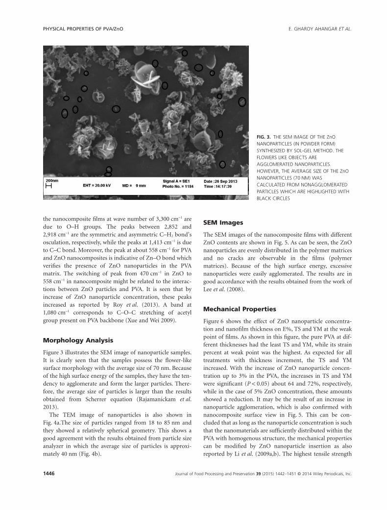
Figure 3 illustrates the SEM image of nanoparticle samples.It is clearly seen that the samples possess the flower-likesurface morphology with the average size of 70 nm. Becauseof the high surface energy of the samples, they have the ten-dency to agglomerate and form the larger particles. There-fore, the average size of particles is larger than the resultsobtained from Scherrer equation (Rajamanickam et al.2013).
The TEM image of nanoparticles is also shown inFig. 4a.The size of particles ranged from 18 to 85 nm andthey showed a relatively spherical geometry. This shows agood agreement with the results obtained from particle sizeanalyzer in which the average size of particles is approxi-mately 40 nm (Fig. 4b).
SEM Images
The SEM images of the nanocomposite films with differentZnO contents are shown in Fig. 5. As can be seen, the ZnOnanoparticles are evenly distributed in the polymer matricesand no cracks are observable in the films (polymermatrices). Because of the high surface energy, excessivenanoparticles were easily agglomerated. The results are ingood accordance with the results obtained from the work ofLee et al. (2008).
Mechanical Properties
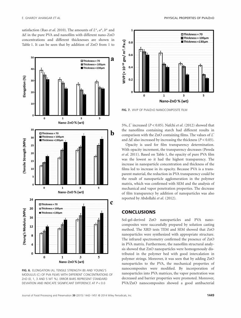
Figure 6 shows the effect of ZnO nanoparticle concentra-tion and nanofilm thickness on E%, TS and YM at the weakpoint of films. As shown in this figure, the pure PVA at dif-ferent thicknesses had the least TS and YM, while its strainpercent at weak point was the highest. As expected for alltreatments with thickness increment, the TS and YMincreased. With the increase of ZnO nanoparticle concen-tration up to 3% in the PVA, the increases in TS and YMwere significant (P < 0.05) about 64 and 72%, respectively,while in the case of 5% ZnO concentration, these amountsshowed a reduction. It may be the result of an increase innanoparticle agglomeration, which is also confirmed withnanocomposite surface view in Fig. 5. This can be con-cluded that as long as the nanoparticle concentration is suchthat the nanomaterials are sufficiently distributed within thePVA with homogenous structure, the mechanical propertiescan be modified by ZnO nanoparticle insertion as alsoreported by Li et al. (2009a,b). The highest tensile strength
FIG. 3. THE SEM IMAGE OF THE ZnONANOPARTICLES (IN POWDER FORM)SYNTHESIZED BY SOL-GEL METHOD. THEFLOWERS LIKE OBJECTS AREAGGLOMERATED NANOPARTICLES.HOWEVER, THE AVERAGE SIZE OF THE ZnONANOPARTICLES (70 NM) WASCALCULATED FROM NONAGGLOMERATEDPARTICLES WHICH ARE HIGHLIGHTED WITHBLACK CIRCLES
PHYSICAL PROPERTIES OF PVA/ZnO E. GHAROY AHANGAR ET AL.
Journal of Food Processing and Preservation 39 (2015) 1442–1451 © 2014 Wiley Periodicals, Inc.1446

was achieved at around 130 μm of film thickness and 3%nanoparticle concentration. The decrease of E% with theincrease of ZnO concentration may be due to the roughnature of filler (Pereda et al. 2011). In other words, the com-bination of ZnO limits the movement of PVA bed in termsof the strong interactions between the fillers and polymermatrix (Cao et al. 2008).
Water Vapor Penetration Properties (WVP)
WVP is one of the most important parameters for biode-gradable films. Vapor penetration to the pure PVA andnanocomposites with different concentrations of nano-ZnOand with different thicknesses is shown in Fig. 7. By adding
the nanoparticles and increasing the film thickness, theWVP of the film decreased (P < 0.05). Most of the reduc-tion in gas penetration was observed in the case of thicknessof 130 μm and the ZnO concentration of 5%. In such treat-ment, the WVP of the film decreased by 39%. The vaporpenetration reduction is due to incorporation ofnanoparticles because it would result in a more complicatedpath for oxygen and other gases to penetrate (Yu et al.2009). Nafchi et al. (2012) reported similar results for vaporpermeation after incorporation of nanoparticles.
Moreover, the results of this study confirmed the previ-ous results obtained by Yu et al. (2009) and Nafchi et al.(2012) which expressed that by increasing the film’s thick-ness, the vapor penetration shows a reduction.
Antibacterial Properties of Nanocomposites
The antibacterial properties of nanocomposites are shownas inhibition zone against E. coli in Table 1. By introductionof ZnO nanoparticles to PVA, and with increase in thenanofilm thickness, the bacteria natural growth was sloweddown sufficiently and the bacteria growth inhibition inten-sified (P < 0.05). Bacteria growth inhibition in the pure PVAat all concentrations was zero. Hence, it can be said that thepresence of ZnO nanoparticles in the PVA matrix enhancesthe bacteria growth inhibition. Nafchi et al. (2012) observedthat the growth of Staphylococcus aureus was affected sig-nificantly by ZnO-coated films compared to control films.The antibacterial activity of WPU/f-ZnO composite filmsagainst E. coli and S. aureus was also tested. The resultsrevealed that the antibacterial activity enhanced with theincrease of f-ZnO content, and the best antibacterial activitywas obtained (Xue and Wei 2009). Espitia et al. (2013) indi-cated that ZnO nanoparticles incorporated in pediocinfilms exhibited excellent antibacterial activity againstS. aureus and Listeria monocytogenes. Mechanisms of theantibacterial behavior of ZnO have been detailed by Zhanget al. (2010). They have categorized this behavior as chemi-cal and/or physical interaction between ZnO particles andthe cell envelope of microorganism. The Zn2+ penetratedinto the microorganisms’ cell walls and reacted with inter-nal materials. The antibacterial phenomena may also be dueto hydrogen peroxide production from the surface (Zhanget al. 2010). They also reported that the nanosize of ZnO ismore effective than the microsize due to easy penetrationthrough cell walls of microorganisms. Sawai (2003) alsoreported the same results and demonstrated that amongZnO, CaO and MgO, the former was the most effective forS. aureus compared to E. coli.
Color and Transparency
The color and transparency of films are important in thefood packaging appearance and customer acceptance and
FIG. 4. TEM IMAGE (A) AND HISTOGRAM OF ZnO NANOPARTICLESIZE DISTRIBUTION (B) SYNTHESIZED BY SOL-GEL METHOD
E. GHAROY AHANGAR ET AL. PHYSICAL PROPERTIES OF PVA/ZnO
Journal of Food Processing and Preservation 39 (2015) 1442–1451 © 2014 Wiley Periodicals, Inc. 1447

FIG
.5.
SEM
IMA
GES
OF
(A)
PVA
WIT
HN
OZn
OC
ON
TEN
T,(B
)PV
A/1
%Zn
O,
(C)
PVA
/3%
ZnO
AN
D(D
)PV
A/5
%Zn
O
PHYSICAL PROPERTIES OF PVA/ZnO E. GHAROY AHANGAR ET AL.
Journal of Food Processing and Preservation 39 (2015) 1442–1451 © 2014 Wiley Periodicals, Inc.1448
satisfaction (Rao et al. 2010). The amounts of L*, a*, b* andΔE in the pure PVA and nanofilm with different nano-ZnOconcentrations and different thicknesses are shown inTable 1. It can be seen that by addition of ZnO from 1 to
5%, L* increased (P < 0.05). Nafchi et al. (2012) showed thatthe nanofilms containing starch had different results incomparison with the ZnO containing films. The values of L*
and ΔE also increased by increasing the thickness (P < 0.05).Opacity is used for film transparency determination.
With opacity increment, the transparency decreases (Peredaet al. 2011). Based on Table 1, the opacity of pure PVA filmwas the lowest so it had the highest transparency. Theincrease in nanoparticle concentration and thickness of thefilms led to increase in its opacity. Because PVA is a trans-parent material, the reduction in PVA transparency could bethe result of nanoparticle agglomeration in the polymermatrix, which was confirmed with SEM and the analysis ofmechanical and vapor penetration properties. The decreaseof film transparency by addition of nanoparticles was alsoreported by Abdollahi et al. (2012).
CONCLUSIONS
Sol-gel-derived ZnO nanoparticles and PVA nano-composites were successfully prepared by solution castingmethod. The XRD tests TEM and SEM showed that ZnOnanoparticles were synthesized with appropriate structure.The infrared spectrometry confirmed the presence of ZnOin PVA matrix. Furthermore, the nanofilm structural analy-sis showed that ZnO nanoparticles were homogenously dis-tributed in the polymer bed with good intercalation inpolymer strings. Moreover, it was seen that by adding ZnOnanoparticles to the PVA, the mechanical properties ofnanocomposites were modified. By incorporation ofnanoparticles into PVA matrices, the vapor penetration wasdecreased and barrier properties were promoted. Moreover,PVA/ZnO nanocomposites showed a good antibacterial
FIG. 6. ELONGATION (A), TENSILE STRENGTH (B) AND YOUNG’SMODULUS (C) OF PVA FILMS WITH DIFFERENT CONCENTRATIONS OFZnO (0, 1, 3 AND 5 WT %). ERROR BARS REPRESENT STANDARDDEVIATION AND INDICATE SIGNIFICANT DIFFERENCE AT P < 0.0
FIG. 7. WVP OF PVA/ZnO NANOCOMPOSITE FILM
E. GHAROY AHANGAR ET AL. PHYSICAL PROPERTIES OF PVA/ZnO
Journal of Food Processing and Preservation 39 (2015) 1442–1451 © 2014 Wiley Periodicals, Inc. 1449

property, thus these films can be used as antibacterial foodpackaging materials.
REFERENCES
ABDOLLAHI, M., REZAEI, M. and FARZI, G. 2012. A novelactive bionanocomposite film incorporating rosemaryessential oil and nanoclay into chitosan. J. Food Eng. 111,343–350.
ABDOLLAHI, M., ALBOOFETILEH, M., BEHROOZ, R.,REZAEI, M. and MIRAKI, R. 2013. Reducing water sensitivityof alginate bio-nanocomposite film using cellulosenanoparticles. Int. J. Biol. Macromol. 54, 166–173.
ALBOOFETILEH, M., REZAEI, M., HOSSEINI, H. andABDOLLAHI, M. 2013. Effect of montmorillonite clay andbiopolymer concentration on the physical and mechanicalproperties of alginate nanocomposite film. J. Food Eng. 117,26–33.
ARORA, A. and PADUA, G.W. 2010. Review: Nanocompositesin food packaging. J. Food Sci. 75, 43–49.
ASTM 2002. http://www.astm.org/DATABASE.CART/HISTORICAL/D882-02.htm (accessed December 21, 2002).
AZAM, A., AHMED, F., ARSHI, N., CHAMAN, M. and NAQVI,A.H. 2010. Formation and characterization of ZnOnanopowder synthesized by sol-gel method. J. Alloys Compd.496, 399–402.
CAO, X., CHEN, Y., CHANG, P.R., STUMBORG, M. andHUNEAULT, M.A. 2008. Green composites reinforced withhemp nanocrystals in plasticized starch. J. Appl. Polym. Sci.109, 3804–3810.
CHANDRAKALA, H.N., RAMARAJ, B., MADHU, G.M.,SHIVAKUMARAIAH and SIDDARAMAIAH. 2012. Theinfluence of zinc oxide cerium oxide nanoparticles on thestructural characteristics and electrical properties of polyvinylalcohol films. J. Mater. Sci. 42, 8076–8084.
CHAUDHRY, Q., SCOTTER, M., BLACKBURN, J., ROSS, B.,BOXALL, A. and CASTLE, L. 2008. Applications and
implications of nanotechnologies for the food sector.Food Addit. Contam. Part A Chem. Anal. Control Expo.Risk Assess. 25, 241–258.
DASTJERDI, R. and MONTAZER, M. 2010. A review on theapplication of inorganic nano-structured materials in themodification of textiles: Focus on anti-microbial properties.Colloids Surf. B. Biointerfaces 79, 5–18.
ESPITIA, P.J.P., SOARES, N.F.F., TEOFILO, R.F., COIMBRA,J.S.R., VITOR, D.M., BATISTA, R.A., FERREIRA, S.O.,ANDRADE, N.J. and MEDEIRO, E.A.A. 2013.Physical-mechanical and antimicrobial properties ofnanocomposite films with pediocin and ZnO nanoparticles.Carbohydr. Polym. 94, 199–208.
FERNANDES, D.M., WINKLER HECHENLEITNER, A.A.,LIMA, S.M., ANDRADE, H.L.C., CAIRES, A.R.L. andGÓMEZ PINEDA, E.A. 2011. Preparation, characterization,and photoluminescence study of PVA/ZnO nanocampositefilms. Mater. Chem. Phys. 128, 371–376.
GAYAN, R.N., DAS, S.N., DALUI, S., BHAR, R. and PAL, A.K.2008. Zinc magnesium oxide nanofibres on glass substrateby solution growth technique. J. Cryst. Growth 310,4073–4080.
KOMPANY, A., MADAHI, P., SHAHTAHMASSBI, N. andMASHREGHI, M. 2012. Synthesis, characterization andantibacterial property of ZnO: Mg nanoparticles.AIP Conference Proceedings, USA.
KUMAR, S. and SAHARE, P.D. 2012. Observation of band gapand surface defects of ZnO nanoparticles synthesized viahydrothermal route at different reaction temperature.Opt. Commun. 285, 5210–5216.
LEE, J., BHATTACHARYYA, D., EASTEAL, A.J. and MESTON,J.B. 2008. Properties of nano- ZnO/poly (vinyl alcohol)/ poly(ethylene oxide) composite thin film. Curr. Appl. Phys. 8,42–47.
LEE, J., EASTEAL, A.J., PAL, U. and BHATTACHARYYA, D.2009. Evolution of ZnO nanostructures in sol-gel synthesis.Curr. Appl. Phys. 9, 792–796.
TABLE 1. COLOR, OPACITY AND ANTIBACTERIAL ACTIVITY OF DIFFERENT TREATMENTS OF PVA/ZnO NANOCOMPOSITES
(ZnO)% Thickness (μm) L* a* b* ΔE Opacity
Antibacterial(×103 mm2)
0 70 ± 2 45.39 ± 2.23a −4.78 ± 0.85d 21.89 ± 1.18b 50.88 ± 1.22g 1.15 ± .08a 0 ± 0a
0 100 ± 3 55.77 ± 1.26b −4.79 ± 0.84d 24.74 ± 2.37a 46.39 ± 2.37f 1.46 ± 0.24b 0 ± 0a
0 130 ± 2 60.57 ± 1.74b −5.14 ± 0.37cd 25.78 ± 2.13a 44.79 ± 0.16f 1.81 ± 0.06c 0 ± 0a
1 70 ± 2 57.46 ± 1.74b −6.54 ± 0.86a 18.50 ± 2.20cd 42.71 ± 1.78f 2.81 ± 0.14d 1.71 ± 0.29b
1 100 ± 2 66.13 ± 1.48d −6.26 ± 1.12ab 21.24 ± 2.09bc 36.30 ± 2.01e 2.99 ± 0.06d 1.76 ± 0.47a
1 130 ± 1 70.74 ± 0.74e −5.04 ± 0.22cd 16.64 ± 1.54d 30.01 ± 1.39d 3.68 ± 0.13e 2.52 ± 0.12a
3 70 ± 5 71.75 ± 1.38e −6.26 ± 0.42abc 17.06 ± 0.65d 29.45 ± 1.55d 5.29 ± 0.07f 7.74 ± 0.59c
3 100 ± 3 77.69 ± 0.32f −4.70 ± 0.49d 20.75 ± 0.91bc 27.56 ± 0.46cd 5.73 ± 0.03g 8.54 ± 0.16c
3 130 ± 1 85.30 ± 1.64g −5.04 ± 0.76cd 19.17 ± 1.09bcd 26.67 ± 1.48bc 6.44 ± 0.09h 8.95 ± 0.21c
5 70 ± 2 79.10 ± 0.36f −4.89 ± 0.11d 20.67 ± 1.15bc 23.36 ± 0.99abc 6.45 ± 0.07h 10.41 ± 0.03d
5 100 ± 3 79.74 ± 1.47f −6.14 ± 0.14abc 16.71 ± 0.94d 22.32 ± 0.30ab 6.54 ± 0.07h 11.33 ± 0.42de
5 130 ± 4 85.78 ± 1.39g −5.65 ± 0.20abcd 16.99 ± 0.57d 20.32 ± 1.03a 6.82 ± 0.10i 12.43 ± 0.42e
PHYSICAL PROPERTIES OF PVA/ZnO E. GHAROY AHANGAR ET AL.
Journal of Food Processing and Preservation 39 (2015) 1442–1451 © 2014 Wiley Periodicals, Inc.1450

LI, J.H., HONG, R.Y., LI, M.Y., LI, H.Z., ZHENG, Y. and DING,J. 2009a. Effects of ZnO nanoparticles on the mechanical andantibacterial properties of polyurethane coatings. J. Pro.Org. Coat 64, 504–509.
LI, Q., ZHOU, J. and ZHANG, L. 2009b. Structure andproperties of the nanocomposite films of chitosan reinforcedwith cellulose whiskers. J. Polym. Sci. 47, 1069–1077.
LIVAGE, J., HENRY, M. and SACHEZ, C. 1988. Sol-gel-chemistry transition metal oxide. Prog. Solid State Chem. 18,258–341.
NAFCHI, A.M., ALIAS, A.K., MAHMUD, S. and ROBAL, M.2012. Antimicrobial, rheological, and physicochemicalproperties of sago starch films filled with nanorod-rich zincoxide. J. Food Eng. 113, 511–519.
NAIR, M.G., NIRMALA, M., REKHA, K. and ANUKALIANI, A.2011. Structural, optical, photo catalytic and antibacterialactivity of ZnO and Co doped ZnO nanoparticles.Mater. Lett. 65, 1797–1800.
PEREDA, M., AMICA, G., RACZ, I. and MARCOVICN, N.E.2011. Structure and properties of nanocomposite films basedon sodium caseinate and nanocellulose fibers. J. Food Eng.103, 76–83.
RAJAMANICKAM, N., RAJASHABALA, S. andRAMACHANDRAN, K. 2013. Theoretical and experimentalinvestigation on enhance thermal behavior in chunk-shapednano ZnO. Mol. Phys. 112, 142–150.
RAO, M.S., KANATT, S.R., CHAWLA, S.P. and SHARMA, A.2010. Chitosan and guar gum composite films preparation:physical, mechanical and antibacterial properties.Carbohydr. Polym. 82, 1243–1247.
ROY, A.S., GUPTA, S., SINDHU, S., PARVEEN, A. andRAMAMURTHY, P.C. 2013. Dielectric properties of novelPVA/ZnO hybrid nanocomposite films. Compos. Part B Eng.47, 314–319.
SAWAI, J. 2003. Quantitative evaluation of antibacterial activitiesof metallic oxide powders (ZnO, MgO and CaO) byconductimetric assay. J. Microbiol. Methods 54,177–182.
SAWAI, J., SHOJI, S., IGRASHI, H., HASHIMOTO, A.,KOKUGAN, T., SHIMIZ, M. and KOJIMA, H. 1998.Hydrogen peroxide as an antibacterial factor in zinc oxidepowder slurry. J. Ferment. Bioeng. 86, 521–522.
SEDAGHAT, N. and ZAHEDI, Y. 2012. Application of ediblecoating and acidic washing for extending the storage life ofmushrooms (Agaricus bisporus). Food Sci. Technol. Int. 18,523–530.
SHALL, M.S.E., GRAIVER, D. and PERNISZ, U. 1995. Synthesisand characterization of nanoscale zinc oxide particles: l. laservaporization/condensation technique. Nanostruct. Mater. 6,297–300.
SHIVAKUMARAIAH, H.N.C., SIDDARAMAIAH, H.N.C.,RAMARAJ, B. and MADHU, G.M. 2012. The influence ofzinc oxide – cerium oxide nanoparticles on the structuralcharacteristics and electrical properties of polyvinyl alcoholfilms. J. Mater. Sci. 47, 8076–8084.
SORRENTINO, A., GORRASI, G. and VITTORIA, V. 2007.Potential perspective of bio-nanocomposites for foodpackaging applications. Trends Food Sci. Technol. 18, 84–95.
SUCHANEK, W.L. 2009. Systematic study of hydrothermalcrystallization of zinc oxide (ZnO) nano – sized powder withsuperior UV attenuation. J. Cryst. Growth 312, 100–108.
TANKHIWALE, R. and BAJPAI, S.K. 2012. Preparation,characterization and antibacterial applications of ZnO-nanoparticles coated polyethylene films for food packaging.Colloids Surf. B. Biointerfaces 90, 16–20.
THARANATHAN, R.N. 2003. Biodegradable films andcomposite coatings: Past, present and future. Trends FoodSci. Technol. 14, 71–78.
VAIDYA, U.R. and BHATTACHARYA, M. 1994. Properties ofblends of starch and synthetic polymers containing anhydridegroups. J. Appl. Polym. Sci. 52, 617–628.
XUE, Y.M. and WEI, D.Z. 2009. Effects of flower- like ZnOnanowhiskers on the mechanical, thermal and antibacterialproperties of waterborne polyurethane. Polym. Degrad. Stab.94, 1103–1109.
YU, D., CAI, R. and LIU, Z. 2009. Preparation andcharacterization of glycerol plasticized- pea starch/ZnO-carboxymethylecellulose sodium nanocomposites.Bioresour. Technol. 100, 2832–2841.
ZHANG, L., JIANG, Y., DING, Y., DASKALAKIS, N., JEUKEN,L., POVEY, M., NEILL, A.Ó. and YORK, D. 2010. Mechanisticinvestigation info antibacterial behavior of suspensions ofZnO nanoparticles against E. coli. J. Nanopart. Res. 12,1625–1636.
E. GHAROY AHANGAR ET AL. PHYSICAL PROPERTIES OF PVA/ZnO
Journal of Food Processing and Preservation 39 (2015) 1442–1451 © 2014 Wiley Periodicals, Inc. 1451