
Synthesis of ZnO and Au tethered ZnO pyramid-like microflower for photocatalytic degradation of...
8
This article appeared in a journal published by Elsevier. The attached copy is furnished to the author for internal non-commercial research and education use, including for instruction at the authors institution and sharing with colleagues. Other uses, including reproduction and distribution, or selling or licensing copies, or posting to personal, institutional or third party websites are prohibited. In most cases authors are permitted to post their version of the article (e.g. in Word or Tex form) to their personal website or institutional repository. Authors requiring further information regarding Elsevier’s archiving and manuscript policies are encouraged to visit: http://www.elsevier.com/copyright
Transcript of Synthesis of ZnO and Au tethered ZnO pyramid-like microflower for photocatalytic degradation of...

This article appeared in a journal published by Elsevier. The attachedcopy is furnished to the author for internal non-commercial researchand education use, including for instruction at the authors institution
and sharing with colleagues.
Other uses, including reproduction and distribution, or selling orlicensing copies, or posting to personal, institutional or third party
websites are prohibited.
In most cases authors are permitted to post their version of thearticle (e.g. in Word or Tex form) to their personal website orinstitutional repository. Authors requiring further information
regarding Elsevier’s archiving and manuscript policies areencouraged to visit:
http://www.elsevier.com/copyright

Author's personal copy
Materials Science and Engineering B 177 (2012) 190– 196
Contents lists available at SciVerse ScienceDirect
Materials Science and Engineering B
j o ur nal homep age: www.elsev ier .com/ locate /mseb
Synthesis of ZnO and Au tethered ZnO pyramid-like microflower forphotocatalytic degradation of orange II
Pei-Kuan Chena, Gang-Juan Leea, Sambandam Anandana,b,∗, Jerry J. Wua,∗∗
a Department of Environmental Engineering and Science, Feng Chia University, Taichung 407, Taiwanb Nanomaterials & Solar Energy Conversion Laboratory, Department of Chemistry, National Institute of Technology, Trichy 620 015, India
a r t i c l e i n f o
Article history:Received 17 July 2011Received in revised form 13 October 2011Accepted 4 December 2011Available online 16 December 2011
Keywords:ZnOAu–ZnOMicroflowerHydrothermal processOrange IIPhotodegradation
a b s t r a c t
Pyramid-like microflower structured zinc oxide (ZnO) nanoparticles were successfully prepared underhydrothermal processes. Then Au nanoparticles were tethered on ZnO pyramid-like microflower(Au–ZnO) by the reduction of HAuCl4 in the presence of �-d-glucose as stabilizer. The materials werewell characterized by different analytical techniques and they could be potentially utilized for the pho-tocatalytic degradation of orange II in the visible region. SEM images reveal that the ZnO microflowersare well formed at 100 ◦C and on increasing the temperature ZnO nuclei typically grow in the directionof c-axis alone due to typical increase in the dipole moment along that direction. TEM image shows thatgold nanoparticles are tethered on the ZnO surface. A possible mechanism for the formation of ZnO andAu–ZnO microflowers has been also discussed.
© 2011 Elsevier B.V. All rights reserved.
1. Introduction
ZnO, an important II–VI semiconductor, has attracted con-siderable worldwide interest because it possesses competitivephotocatalytic characteristics and active sites upon comparing toTiO2. Additionally, wurtzite ZnO structure possesses high ionic-ity at its polar surfaces that provide crystal growth at differentdirections and morphologies. However, a wide band gap (>3.0 eV)semiconductor oxides like ZnO and TiO2 have been figured out thatonly a small UV fraction of solar light (3–5%) can be utilized [1]and limited in the recombination of the generated photo-holes andphoto-electrons.
In order to extend the absorption range from UV to visiblelight and to control the electron/hole recombination rate, addi-tional agents were added over the surface which may serve astrapping sites for photoexcited electrons from the valence band,and in addition it lowers the band gap so that electrons can beeffectively excited by visible light. Numerous investigations areavailable regarding the addition of metal ions, including Mn [2],
∗ Corresponding author at: Department of Chemistry, National Institute of Tech-nology, Trichy 620015, India. Tel.: +91 431 2503639; fax: +91 431 2500133.∗∗ Corresponding author at: Department of Environmental Engineering and Sci-
ence, Feng Chia University, Taichung 407, Taiwan. Tel.: +886 4 24517250x5206;fax: +886 4 24517686.
E-mail addresses: [email protected] (S. Anandan), [email protected] (J.J. Wu).
Al [3], Cu [4,5], Co [6], and Cd [7], or noble metals, including Au[8–11], Pt [12–14], Pd [15], and Ag [16], onto the surface of semi-conductors in order to reduce the band gap energies and to improvethe separation efficiency of photo-induced electron–hole pairs toeffectively harvest visible light.
Over the past decades there has been an increased emphasison the topic of “green” chemistry and chemical process. That isthe utilization of nontoxic, environmentally benign, easily avail-able, and relatively inexpensive chemicals and solvents, which aresome of the key issues meriting important consideration in a greensynthesis. Therefore, catalysts were prepared in recent years on alarge scale at low cost by simple solution-based methods, such aschemical precipitation [17], sol–gel synthesis [18,3,19], solvother-mal [20], and hydrothermal reaction [21–24]. The hydrothermalprocess has several advantages over other growth process. Exam-ples are the use of low reaction temperature, simple equipment,catalyst-free growth, low cost, and large area uniform produc-tion. In this way, a variety of inorganic materials were synthesizedby self-assembly. The advantage of hydrothermal synthesis is theuse of water as the main solvent which is more environmentallyfriendly. Further in the concept of green technology, nanopar-ticles preparation was promoted by Raveendran et al. [25]. Insuch process, silver nanoparticles were prepared by soluble starchand �-d-glucose which played as a stabilizing agent and in addi-tion acted as a nontoxic reducing agent. According to Chairamet al. [26], the hydroxyl groups of �-d-glucose stabilize Ag ionsby electrostatic binding and the presence of terminal aldehyde
0921-5107/$ – see front matter © 2011 Elsevier B.V. All rights reserved.doi:10.1016/j.mseb.2011.12.001
Author's personal copy
P.-K. Chen et al. / Materials Science and Engineering B 177 (2012) 190– 196 191
group in glucose reduces Ag+ ions into metallic Ag. Similarly goldnanoparticles were also synthesized by following the same way[27]. In the present work, we followed such green approachtowards the synthesis of ZnO via hydrothermal processes and thensupported Au on ZnO heterostructures using �-d-glucose as a stabi-lizer. In addition, we performed photocatalytic degradation of dyepollutant (orange II) in order to show whether or not Au tetheredZnO can promote the dye degradation in the visible light regioncompared with as-synthesized pure ZnO catalyst.
2. Experimental
2.1. Materials and methods
All chemicals were of the highest purity available and were usedas received without further purification. Zinc chloride (ZnCl2), gold(III) chloride (HAuCl4), orange (II) dye and tetrabutylammoniumbromide (TBAB) were purchased from Sigma–Aldrich Inc.
2.2. Synthesis of ZnO pyramid-like microflower
0.1 M ZnCl2 and ammonia (0.1 M) were dissolved in 100 mL ofdeionized water. 2.5% of tetrabutylammonium bromide (TBAB) wasadded to the mixture and then transferred into a Teflon autoclavefor crystallization by heating up to 100 ◦C for 8 h. After cool-ing to room temperature, the powders were rinsed several timeswith distilled water (until the filtrate does not show any precipi-tate with AgNO3) and ethanol (thrice) to remove impurities, thendried at 105 ◦C. The dry powder was heated to 550 ◦C for 6 h ina temperature-programmed muffle furnace, which results in theproduction of ZnO microflower.
2.3. Synthesis of Au–ZnO pyramid-like microflower
Gold colloid was prepared by adding 1.8 g �-d-glucose, 0.05 MNaBH4 solution (2 mL) to 100 mL of (1.18 × 10⁻⁴ M) HAuCl4·3H2Osolution followed by irradiated with high-intensity ultrasonic horn(Ti-horn, 20 kHz, 100 W/cm2) in ambient air for 30 min. The colorof the solution changed from pale yellow to ruby red, indicatingthe formation of nanosized gold particles. To this gold colloid, 1 gof ZnO powder prepared via hydrothermal method was added andstirred vigorously for 3 h. The color of the powder changed to violetas well as the solution became transparent, indicating completeadsorption of Au nanoparticles on the surface of ZnO. Finally, theprecipitate was washed several times with water (until the filtratedoes not show any precipitate with AgNO3) and ethanol (thrice)and then vacuumed dried at 80 ◦C.
2.4. Analytical methods
Surface morphology, particle size, and the various contours ofthe photocatalyst powders were analyzed by XRD (measured usingRigaku diffractometer, Cu-K� radiation, Japan), High ResolutionScanning electron microscopy (HRSEM; recorded by JEOL 6700Fmodel) and Transmission electron microscopy (TEM; recordedby JEOL JEM2010 model), respectively. Diffuse reflectance UV–visspectra of the samples were recorded by Shimadzu UV-VIS 2550spectrophotometer. XPS of the Copper oxidation state was mea-sured using Physical Electronics PHI 5600 XPS spectrophotometerwith monochromatic Al K� (1486.6 eV) excitation source.
2.5. Measurement of photocatalytic activity
A 500 mL capacity borosilicate glass photoreactor was used in allthe experiments. The reactor walls were covered with aluminiumfoil to avoid releasing of irradiation. The degradation of orange II
was carried out at atmospheric conditions (25 ◦C) and at neutralpH. The amount of catalyst (1.5 g L−1) and orange II dye (5 × 10⁻³ M;400 mL) used for all the experiments were the same. Prior to exper-iment, the mixtures were stirred in the dark during first 30 min todisperse the catalysts and reach the adsorption equilibrium. Whenthe visible light irradiation was required, a 110 W xenon lamp(Model OSRAM BLUE; � ≥ 400 nm; intensity of incident radiationis 10,200 ± 10 lx measured using INS DX-200 LUX meter, Norway)was placed inside a quartz tube that was well placed at the middleof the reactor. 5 mL of the solution was withdrawn at given irradia-tion time intervals to determine the degradation percentage of dyewith time. The apparent disappearance of the substrate, orange II,was determined by UV–vis spectrophotometer (Shimadzu Instru-ments, Japan) at the wavelength of 485 nm. Prior to the analysis, thecatalyst was removed from samples by centrifugation at 2000 rpmand filtration using a 0.45 �m polyvinylidene fluoride (PVDF) filterin the case of experiments performed using catalysts.
3. Results and discussion
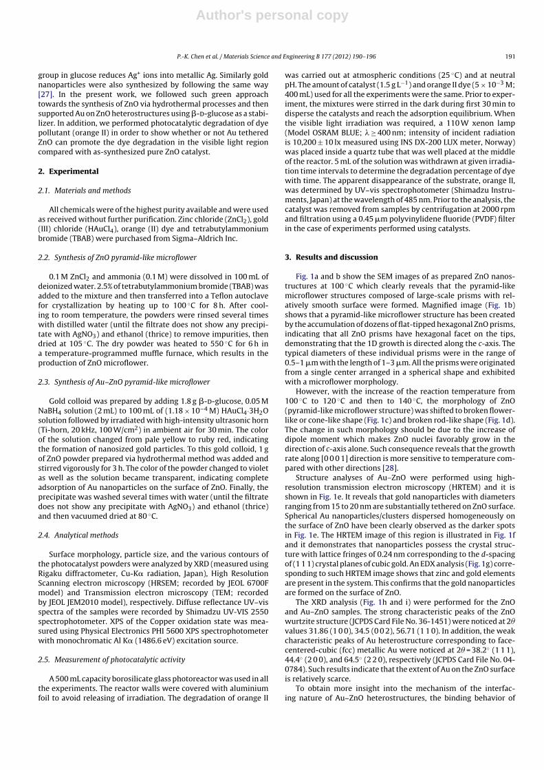
Fig. 1a and b show the SEM images of as prepared ZnO nanos-tructures at 100 ◦C which clearly reveals that the pyramid-likemicroflower structures composed of large-scale prisms with rel-atively smooth surface were formed. Magnified image (Fig. 1b)shows that a pyramid-like microflower structure has been createdby the accumulation of dozens of flat-tipped hexagonal ZnO prisms,indicating that all ZnO prisms have hexagonal facet on the tips,demonstrating that the 1D growth is directed along the c-axis. Thetypical diameters of these individual prisms were in the range of0.5–1 �m with the length of 1–3 �m. All the prisms were originatedfrom a single center arranged in a spherical shape and exhibitedwith a microflower morphology.
However, with the increase of the reaction temperature from100 ◦C to 120 ◦C and then to 140 ◦C, the morphology of ZnO(pyramid-like microflower structure) was shifted to broken flower-like or cone-like shape (Fig. 1c) and broken rod-like shape (Fig. 1d).The change in such morphology should be due to the increase ofdipole moment which makes ZnO nuclei favorably grow in thedirection of c-axis alone. Such consequence reveals that the growthrate along [0 0 0 1] direction is more sensitive to temperature com-pared with other directions [28].
Structure analyses of Au–ZnO were performed using high-resolution transmission electron microscopy (HRTEM) and it isshown in Fig. 1e. It reveals that gold nanoparticles with diametersranging from 15 to 20 nm are substantially tethered on ZnO surface.Spherical Au nanoparticles/clusters dispersed homogeneously onthe surface of ZnO have been clearly observed as the darker spotsin Fig. 1e. The HRTEM image of this region is illustrated in Fig. 1fand it demonstrates that nanoparticles possess the crystal struc-ture with lattice fringes of 0.24 nm corresponding to the d-spacingof (1 1 1) crystal planes of cubic gold. An EDX analysis (Fig. 1g) corre-sponding to such HRTEM image shows that zinc and gold elementsare present in the system. This confirms that the gold nanoparticlesare formed on the surface of ZnO.
The XRD analysis (Fig. 1h and i) were performed for the ZnOand Au–ZnO samples. The strong characteristic peaks of the ZnOwurtzite structure (JCPDS Card File No. 36-1451) were noticed at 2�values 31.86 (1 0 0), 34.5 (0 0 2), 56.71 (1 1 0). In addition, the weakcharacteristic peaks of Au heterostructure corresponding to face-centered-cubic (fcc) metallic Au were noticed at 2� = 38.2◦ (1 1 1),44.4◦ (2 0 0), and 64.5◦ (2 2 0), respectively (JCPDS Card File No. 04-0784). Such results indicate that the extent of Au on the ZnO surfaceis relatively scarce.
To obtain more insight into the mechanism of the interfac-ing nature of Au–ZnO heterostructures, the binding behavior of

Author's personal copy
192 P.-K. Chen et al. / Materials Science and Engineering B 177 (2012) 190– 196
Fig. 1. Field emission SEM images of ZnO prepared by hydrothermal method at 100 ◦C (a and b), 120 ◦C (c) and 140 ◦C (d). High-resolution TEM images of Au–ZnO (e and f).EDX pattern of Au–ZnO (g). XRD spectra of ZnO (h) and Au–ZnO (i).
Au on the surface of ZnO was investigated by XPS narrow scansspectra as shown in Fig. 2. The doublet peak (Fig. 2a) representsthe corresponding Zn 2p3/2 (2p1/2) featuring at 1021.1 (1044.2) eV,with the observed spin-orbit splitting of 23.1 eV, close to those(�E = 22.97 eV) of Zn2+, which are attributed to Zn2+ of ZnO crys-tal lattice and very close to the standard bulk ZnO binding energy
value [29]. The binding energy of Zn (2p3/2) for the Au/ZnOheterostructures shifts to the higher binding energy compared withthe corresponding value of pure ZnO due to the interaction betweenZnO and Au (Fig. 2a) [13,30,31]. The positive shift can also be esti-mated that there are interaction between Au and ZnO resultingfrom electron-transfer from the ZnO to the Au. From the O (1s)

Author's personal copy
P.-K. Chen et al. / Materials Science and Engineering B 177 (2012) 190– 196 193
Fig. 2. XPS spectra of ZnO and Au–ZnO [(a) Zn 2p line; (b) O 1s line; and (c) Au].
spectra (Fig. 2b), the lower binding energy component centered at530.0 eV is attributed to lattice oxygen in the ZnO wurtzite struc-ture that oxygen atoms bound to Zn in ZnO. The high binding energycomponents located at 531.4 ± 0.20 eV is attributed to the presenceof loosely bound oxygen on the surface of ZnO belonging to Zn OH,while that at 533 eV is due to the absorption of H2O molecule ontothe surface [30]. From the atom percentage analysis of XPS analysis,the O/Zn atom ratio of Au/ZnO significantly increases from 1.04 to5.69, comparing with ZnO. The difference between the O/Zn ratioscould be related to the additional oxygen that reflects the pres-ence of the hydrocarbon contaminants or carbon oxidized specieson the ZnO surface upon gold addition. There are two new O (1s)peak centered at 531.5 eV, corresponding to C O groups [32] andthe O (1s) peak centered at 533 eV, corresponding to C OH groups[33] as resulted from glucose used as capping agent around goldparticles (Fig. 2b).
The peak binding energies (BE) of Au (4f7/2) at 83.9 eV are signifi-cantly different from Au+ (4f7/2) (84.6 eV) and Au3+ (4f7/2) (87.0 eV),but similar to bulk metallic Au (84.0 eV) (Fig. 2c). The XPS resultsuggests that the Au species are present in the metallic state. Inthe presence of gold nanoparticles, electrons are enriched owingto the alignment of Fermi levels of the metal and semiconductor.It is inferred that the Au–ZnO interaction may involve a net chargetransfer [8].
The UV–vis absorption spectra of ZnO and Au/ZnO heterostruc-tures are shown in Fig. 3. Pure ZnO did not show any characteristicabsorption peak between 400 and 800 nm. However, the Au/ZnOheterostructure displays an obvious absorption band with a max-imum absorption wavelength up to 550 nm due to the localizedsurface plasma resonance of Au nanoparticles. The optical bandgaps were calculated from the transmittance spectra by plotting(˛h�)1/2 vs. h� and extrapolating the straight line portion of this
plot to the energy axis, where ̨ is the absorption coefficient andh� is the photon energy. The band gaps are 3.06 and 2.98 eV forZnO and Au/ZnO, respectively. It is noted that the decrease in theband gaps has been figured out with the deposition of gold on ZnOsurface.
The formation mechanism of ZnO nanostructures takes the con-sideration of the ionic species formed from water molecules dueto hydrothermal processes. The reaction steps taking place due tohydrothermal processes can be summarized as follows:
H2O → H+ + OH− (1)
3.43.23.02.82.62.42.22.00.0
0.5
1.0
1.5
2.0
2.5
3.0
3.5
4.0
4.5
5.0
200 30 0 40 0 50 0 60 0 70 0 800-0.2
0.0
0.2
0.4
0.6
0.8
1.0
Wavelen gh (nm )((ααhυυ
)1/2 (e
V)
Abso
rban
ce (a
.u.)
hυυ (eV)
ZnO
Au/Zn O
Au/ZnO
ZnO
Fig. 3. Band gap plot of ZnO and Au–ZnO. Inset shows UV–vis absorption spectraZnO and Au–ZnO.

Author's personal copy
194 P.-K. Chen et al. / Materials Science and Engineering B 177 (2012) 190– 196
Fig. 4. Schematic illustration for the formation of pyramid-like ZnO and Au–ZnO microflowers.
NH3 + H2O → NH4+ + OH− (2)
ZnCl2 → Zn2+ + 2Cl− (3)
Zn2+ + 4OH− → [Zn(OH)4]2− (4)
2[Zn(OH)4]2− → 2ZnO + 4H2O + O2 (5)
Eqs. (1)–(3) show the formation of primary ions which have beencreated by the dissociation of water, ammonia, and zinc chlorideunder hydrothermal processes. Eq. (4) shows the formation of[Zn(OH)4]2− which further decomposes to give ZnO nanoparticles.The stabilizing agent, TBAB, may act like a gluing agent to bind ZnOnuclei and show the appearance of microflower as represented inFig. 1.
In order to further explain the growth mechanism of ZnO,here we propose a scheme (Fig. 4). During hydrothermal process,hexagonal ZnO nano nuclei are formed initially. Upon increasingtemperature up to 100 ◦C, many tiny zinc nuclei are formed andmay grow radially to form one-dimensional hexagonal nanorodsin all directions and thus maintain flower-like morphology. This isbecause zinc terminated surface (0 0 0 1) is more chemically reac-tive than the oxygen terminated surface (0 0 0 1), and thus theenriched zinc atoms on the (0 0 0 1) surface lead to the rapid growthand the morphology is pyramid-like microflower. Whereas uponthe increase of temperature above 100 ◦C, concentration of zincnuclei increases rapidly due to higher vapor pressure of water in theautoclave and faster growth along the c-axis direction leads to thehexagonal nanodisks and then into nanorods with increasing con-centration of ZnO. Consequently, a fast growth of crystallographicface tends to form the prism nanorods with a tapered hexago-nal end. This is because the growth rate of the planes follows thesequence (0 0 0 1) > (1 0 1̄ 0) > (1 0 1̄ 1). Therefore, rods are taperedat the ends (as seen in Fig. 4) when temperature is higher above100 ◦C.
There is no appreciable degradation of orange II by using ZnO,Au/ZnO, and solar irradiation, respectively. It implies that thedirect adsorption and photolysis of orange II are negligible bythe as-synthesized ZnO and visible light. Thus, the photocatalyticdegradation of orange II dye combining with visible light and
catalysts as-synthesized was conducted. The concentration oforange II as a function of irradiation time was monitored by UV–visabsorption spectroscopy. A decrease in the absorption of the dyewas observed with an increase in the irradiation time, indicatingthe photocatalytic degradation of the dye was undergone in thepresence of Au/ZnO. In all degradation experiments, the degrada-tion followed pseudo first-order kinetics (plots of −ln[C/Co] vs. timeshowed linear relationship). Pseudo first-order rate constants wereevaluated from the slopes of the −ln[C/Co] vs. time plots (Fig. 5). Theobserved rate constant for the photocatalytic degradation of orangeII (5 × 10⁻³ M) in the presence of Au–ZnO microflowers (1.5 g L−1) is1.08 × 10−3 min−1, which is significantly higher than that observedfor ZnO microflowers (5.24 × 10⁻⁴ min−1). The enhancement of rateconstants observed for ZnO tethered with small amount of Au sug-gests that Au nanoparticles act as a separation center [14] to reduceelectron–hole recombination. In addition, Au–ZnO creates a localelectric field and the optical vibration of surface plasmon and makesa reasonable enhancement in the electric field. Thus, the increase
10864200.0
0.2
0.4
0.6
0.8ZnO (in dark)Au/ZnO (in dark)visible light+ZnOvisible light+Au/ZnO
ln (C
a/C
)
Time (hour)
Fig. 5. Reaction kinetics of photocatalytic degradation by ZnO and Au–ZnO undervisible light irradiation.

Author's personal copy
P.-K. Chen et al. / Materials Science and Engineering B 177 (2012) 190– 196 195
Fig. 6. Photocatalytic mechanisms for orange II dye degradation by Au/ZnO heterostructure under visible light irradiation.
in photocatalytic activity of Au–ZnO is due to the change in thesurface properties such as oxygen vacancies and crystal defects.The enhanced activity of photocatalyst can be explained by retard-ing the electron–hole recombination process. The principal methodof slowing down the electron–hole recombination is through theloading of electron accepting species on the photocatalyst surface.Accordingly, the loading of Au on the ZnO surface can expeditethe transport of photoexcited electrons to the outer system. Noblemetal, such as Au, has been shown to increase the photonic effi-ciency and inhibit electron–hole pair recombination [15]. Based onthe available literature [34], we also came to a conclusion that thebelow mentioned possible reactions are suggested to be responsi-ble for the degradation of the dye using Au–ZnO.
Au–ZnOh�−→ Au–ZnO (e−
CB) + Au–ZnO (h+VB) (6)
Au–ZnO (h+VB) + dye → products (7)
In addition to the above reactions, free gold particles depositedon the surface of ZnO may also act as electron trapping agentsleading to the prevention of the recombination of photo-generatedelectron–hole pairs.
Au–ZnO (e−CB) + Au(Surf) → Au–ZnO + Au(Surf) (e−) (8)
In addition, photocatalytic activity of ZnO can be enhanced due tothe noble metal gold used to capture the electrons and acted as aseparation center. Fig. 6 shows the photocatalytic routes of the syn-thesized Au/ZnO heterostructure. When Au/ZnO heterostructuresare dispersed in the solution with the existence of organic pollu-tant, the surface electrons on Au nanoparticles should eventuallytransfer to the dye in the dark [35]. These catalysts are irradiatedby visible light (>400 nm) with photon energy lower than the bandgap of ZnO and another photocatalytic mechanism would occur.The dye molecules adsorbed on the photocatalyst can harvest vis-ible light to produce the excited state OII∗ads. Since the oxidationpotential of the excited state is more negative than the potential ofthe conduction band of ZnO particles, an electron is injected intothe conduction band of ZnO from the excited state OII∗ads [36] asEqs. (9)–(11):
OIIads + h�(> 400 nm) → OII∗ads (9)
OII∗ads + ZnO → OII+ads + ZnO (e−) (10)
Au + e− → Au(e−) (11)
The injected electron can be scavenged by ZnO crystals. Since theenergy level of the bottom of the CB is higher than the new Fermienergy level of the Au/ZnO heterostructure, the photoexcited elec-trons would transfer from ZnO nanocrystals to Au nanoparticlesdriven by the potential energy. The electrons on Au or the con-duction band of ZnO can react with absorbed oxygen to producea superoxide anion radical (•O2
−). These radicals, which serve asa strong oxidant, could react with the surrounding organic dyemolecules and lead to the degradation by-products or mineraliza-tion into CO2, H2O, and mineral acids.
4. Conclusions
In conclusion, we report the synthesis of ZnO and Au–ZnOpyramid-like microflowers for photocatalytic degradation oforange II dye. Nanogold particles were successfully anchored onthe surface of ZnO using aqueous �-d-glucose solution as stabi-lizing agent which avoids the agglomeration of gold nanoparticleswhile reduction of Au3+ ions. As-prepared Au–ZnO microflowersshows higher photocatalytic efficiency while compared with ZnOmicroflowers. It can be attributed to Au nanoparticles on the ZnOsurface serving as electron sinks to promote the photocatalytic abil-ity.
Acknowledgements
The authors wish to thank for the financial support by theNational Science Council (NSC) in Taiwan under the contract num-ber of 98-2221-E-35-12-MY3 and New-Star Project by Feng ChiaUniversity.
References
[1] X. Li, K. Ly, K. Deng, J. Tang, R. Su, J. Sun, L. Chen, Mater. Sci. Eng. B 158 (2009)40–47.
[2] T. Meron, G. Markovich, J. Phys. Chem. B 109 (2005) 20232–20236.[3] Y.S. Kim, W.P. Tai, Appl. Surf. Sci. 253 (2006) 4911–4916.[4] U. Pal, J. Garcia-Serrano, G. Casarrubias-Segura, N. Koshizaki, T. Sasaki, S. Ter-
ahuchi, Sol. Energy Mater. Sol. Cells 81 (2004) 339–348.[5] X.B. Wang, C. Song, K.W. Geng, F. Zeng, F. Pan, Appl. Surf. Sci. 253 (2007)
6905–6909.[6] L.B. Duan, G.H. Rao, J. Yu, Y.C. Wang, Solid State Commun. 145 (2008) 525–528.[7] F. Wang, B. Liu, Z. Zhang, S. Yuan, Physica E 41 (2009) 879–882.[8] W.M. Liu, Y.X. Chen, G.T. Kou, T. Xu, D.C. Sun, Wear 254 (2003) 994–1000.[9] X. Wang, X. Kong, Y. Yu, H. Zhang, J. Phys. Chem. C 111 (2007) 3836–3841.
[10] G. Shan, S. Wang, X. Fei, Y. Liu, G. Yang, J. Phys. Chem. B 113 (2009) 1468–1472.[11] L. Chen, L. Luo, Z. Chen, M. Zhang, J.A. Zapien, C.S. Lee, S.T. Lee, J. Phys. Chem. C
114 (2010) 93–100.

Author's personal copy
196 P.-K. Chen et al. / Materials Science and Engineering B 177 (2012) 190– 196
[12] A. Bauer, K. Lee, C. Song, Y. Xie, J. Zhang, R. Hui, J. Power Sources 195 (2010)3105–3110.
[13] Y. Vasil’kov, A.V. Naumkin, I.O. Volkov, V.L. Podshibikhin, G.V. Lisichkin, A.R.Khokhlov, Surf. Interface Anal. 42 (2010) 559–563.
[14] P. Pawinrat, O. Mekasuwandumrong, J. Panpranot, Catal. Commun. 10 (2009)1380–1385.
[15] S. Sakthivel, M.V. Shankar, M. Palanichamy, B. Arabindoo, D.W. Bahnemann, V.Murugesan, Water Res. 38 (2004) 3001–3008.
[16] N.N. Binitha, Z. Yaakob, M.R. Reshmi, S. Sugunan, V.K. Ambili, A.A. Zetty, Catal.Today 147 (2009) 76–80.
[17] D.L. Chiang, Synthesis and Properties of Nano-Manganese Oxide by ChemicalMethods, Master Thesis, National Cheng Kung University, 2003.
[18] Y. Shin, J. Liu, L.Q. Wang, J.H. Chang, W.D. Samuels, L.R. Pederson, G.J. Exarhos,Res. Soc. Symp. Proc. (2002).
[19] L.H. Kao, T.C. Hsu, H.Y. Lu, Colloid Interface Sci. 316 (2007) 160–167.[20] J. Liu, D. Xue, J. Cryst. Growth 311 (2008) 500–503.[21] J. Yu, L. Zhang, B. Cheng, Y. Su, J. Phys. Chem. C 111 (2007) 10582–10589.[22] L. Zhang, W. Lu, L. Yan, Y. Feng, X. Bao, J. Ni, X. Shang, Y. Lv, Micropor. Mesopor.
Mater. 119 (2009) 208–216.[23] W. Cai, J. Yu, S. Mann, Micropor. Mesopor. Mater. 122 (2009) 42–47.
[24] X.Y. Ye, Y.M. Zhou, Y.Q. Sun, J. Chen, Z.Q. Wang, J. Nanopart. Res. 11 (2009)1159–1166.
[25] P. Raveendran, J. Fu, S.L. Wallen, J. Am. Chem. Soc. 125 (2003) 13940–13941.[26] S. Chairam, E. Somsook, J. Magn. Magn. Mater. 320 (2008) 2039–2043.[27] J. Liu, M. Anand, C.B. Roberts, Langmuir 22 (2006) 3964–3971.[28] J. Zhao, Z.G. Jin, X.X. Liu, Z.F. Liu, J. Eur. Ceram. Soc. 26 (2006) 3745–3752.[29] Y.D. Wang, S. Zhang, X.H. Wu, Eur. J. Inorg. Chem. 2005 (2006) 727–731.[30] N. Tabet, M. Faiz, A.A. Oteibi, J. Electron. Spectrosc. Relat. Phenom. 163 (2008)
15–18.[31] A. Zwijnenburg, A. Goossens, W.G. Sloof, M.W.J. Crajé, A.M. vander Kraan,
L.J. de Jongh, M. Makkee, J.A. Moulijn, J. Phys. Chem. B 106 (2002)9853–9862.
[32] K.X. Yao, H.C. Zeng, J. Phys. Chem. C 111 (2007) 13301–13308.[33] D. Yang, A. Velamakanni, G. Bozoklu, S. Park, M. Stoller, R.D. Piner, S. Stankovich,
I. Jung, D.A. Field, C.A. Ventrice Jr., R.S. Ruoff, Carbon 47 (2009) 145–152.[34] V. Subramanian, E. Wolf, P.V. Kamat, J. Am. Chem. Soc. 126 (2004) 4943–4950.[35] Y. Zheng, L. Zheng, Y. Zhan, X. Lin, Q. Zheng, K. Wei, Inorg. Chem. 46 (2007)
6980–6986.[36] N. Helaili, Y. Bessekhouad, A. Bouguelia, M. Trari, Sol. Energy 84 (2010)
1187–1192.