Cobalt Ferrite Nanoparticles Fabricated via Co-precipitation in ...
Upload
independentCategory
view
2download
0
ARTICLE IN PRESS
Journal of Magnetism and Magnetic Materials 321 (2009) 3061–3066
Contents lists available at ScienceDirect
Journal of Magnetism and Magnetic Materials
0304-88
doi:10.1
� Corr
E-m
journal homepage: www.elsevier.com/locate/jmmm
Synthesis and characterization of carboxymethyl dextran-coated Mn/Znferrite for biomedical applications
Magda Latorre-Esteves, Angel Cortes, Madeline Torres-Lugo, Carlos Rinaldi �
Department of Chemical Engineering, University of Puerto Rico at Mayaguez, PO Box 9046, Mayaguez, PR 00680, United States
a r t i c l e i n f o
Article history:
Received 27 October 2008
Received in revised form
20 April 2009Available online 22 May 2009
Keywords:
Manganese zinc ferrite
Carboxymethyl dextran
Nanoparticle
Particle stability
Biocompatibility
53/$ - see front matter & 2009 Elsevier B.V. A
016/j.jmmm.2009.05.023
esponding author.
ail address: [email protected] (C. Rinaldi).
a b s t r a c t
Previous studies have shown that magnetic nanoparticles possess great potential for various in vivo
applications such as magnetic resonance imaging contrast enhancement, tissue repair, cancer treatment
agents, and controlled drug delivery. Many of these applications require that magnetic nanoparticles be
colloidally stable in biological media. The goal of this study was to obtain a magnetic fluid produced by
the colloidal suspension of manganese/zinc ferrite (MZF) nanoparticles that could be stably dispersed in
aqueous solution throughout the range of physiological pH and ionic strength. These super-
paramagnetic nanoparticles were stabilized through steric repulsion by coating with biologically
compatible carboxymethyl dextran (CMDx). Samples of the resultant magnetic fluid were analyzed
using Inductively Coupled Plasma Optical Emission Spectrometry (ICP-OES), X-ray diffraction (XRD),
zeta potential measurements, dynamic light scattering, transmission electron microscopy (TEM), and
SQUID magnetometry. Results show that we obtained superparamagnetic metal-oxide crystals with
composition of Mn0.24Zn0.76Fe2O4. Cell viability measurements show the material is non-toxic to MCF-7
and CaCo-2 cell lines at concentrations of up to 7.5 mg/mL of particle fraction for contact time of
up to 48 h.
& 2009 Elsevier B.V. All rights reserved.
1. Introduction
Magnetic nanoparticles are being actively used and investi-gated for in vivo applications, such as magnetic resonance imagingcontrast enhancement [1], tissue repair [2], immunoassays [3],detoxification of biological fluids [4], hyperthermia [5], drugdelivery [6], and cell separation techniques [7]. Appropriatesurface chemistry is as critical in developing these nanoparticlesas the magnetic material itself [8]. There are many reports in theliterature that describe particular surface coatings, such as starch,chitosan [9], alginate [10], dextran [8], and polyethylene glycol[11]. The coating of these particles is achieved by either chemicalor physical interactions. Important factors to be taken intoconsideration when choosing a surface coating for biomedicalapplications are low toxicity, length of blood-circulation time, andstability of the particles under physiological conditions [12].
Nanoscale iron oxides, such as magnetite are ferrites thatexhibit superparamagnetic characteristics. Because superpara-magnetic particles exhibit no remanence in the absence of anexternal magnetic field [13], they are easily magnetized anddemagnetized, a property which is fundamental to biomedical
ll rights reserved.
applications such as magnetic contrast resonance imaging (MRI)and magnetic fluid hyperthermia. Data from previous reportssuggest that other metal ions (Mn, Gd, Zn) can be introduced intothe tetrahedral site of iron oxides [14,15]. Introduction of suitableatoms into the iron-oxide core offers an opportunity to producenanoparticles with varying magnetic properties. Tailoring thesuperparamagnetic properties of these nanoparticles by dopingwith transition metals such as manganese and zinc could beadvantageous, as it would provide control over properties that areimportant for biological applications, such as magnetization-dependent changes in temperature, relaxivity, and hysteresis [16].In addition, doping could allow for additional control in theamount of material used for therapeutic applications, whichwould allow for greater control of toxicity vs. successfully carryingout an application.
We have synthesized Mn0.24Zn0.76Fe2O4 nanoparticles by a saltco-precipitation method, obtaining primary particles with amedian diameter of 15 nm. After being coated with the biologi-cally compatible sugar carboxymethyl dextran (CMDx), thehydrodynamic size of these particles range from 30 to 50 nm.This study aims to present the synthesis of stable Mn and Znsubstituted ferrites, successful coating of these particles withCMDx by physical absorption, characterization of these particles,suspension of these particles in biologically compatible media,and the possible cytotoxic effects these particles might have inMCF-7 and CaCo-2 cell lines.

ARTICLE IN PRESS
M. Latorre-Esteves et al. / Journal of Magnetism and Magnetic Materials 321 (2009) 3061–30663062
2. Materials and methods
2.1. Materials
Manganese (II) chloride tetrahydrate (MnCl2 �4H2O) 99%, Zinc(II) chloride tetrahydrate (ZnCl2 �4H2O) 99%, iron (III) chloridehexahydrate (FeCl3 �6H2O) 97%, ammonium hydroxide (NH4OH)29% v/v, carboxymethyl-dextran sodium salt 10–20 kDa with1.1–1.5 mmol COOH per gram, and manganese, iron, and zincstandards were purchased from Sigma Aldrich. All materials wereused as received.
2.2. Synthesis of Mn/Zn ferrite core
Manganese/zinc ferrite (MZF) nanoparticles were synthesizedthrough the co-precipitation method [17]. An aqueous solution offerric chloride hexahydrate (0.36 M) was added to an aqueoussolution of manganese chloride tetrahydrate (0.09 M) and zincchloride tetrahydrate (.09 M) in the presence of a nitrogen streamto avoid oxidation of the ferrous ions. This mixture was heated to70 1C, and then 48% ammonium hydroxide was quickly added.Synthesis was carried out at 80 1C for 1 h under continuousstirring at pH 8.0. After cooling to room temperature, this solutionwas centrifuged to precipitate the magnetite nanoparticles.Approximately 5.0 g of MZF were produced.
2.3. Surface modification of MZF with carboxymethyl dextran
The attachment of carboxymethyl-dextran chains to theferrite core was achieved through physical interactions betweenthe positive surface charges on the magnetic core and the negativecharges due to the COO– groups in the dextran chains.The magnetic nanoparticles were peptized with 0.5 M HNO3
to positively charge the core’s surface. Then, 20 g of CMDxwere dissolved in deionized water and the pH adjusted to 10.0.The dissolved CMDx was added to the peptized nanoparticles andshaken at room temperature for 48 h. Excess CMDx was removedby washing the solution with an equal volume of ethanol. Thefunctionalized nanoparticles were dried in a vacuum oven at 60 1Covernight.
2.4. Characterization of functionalized nanoparticles
Hydrodynamic diameter and zeta potential were measuredusing a Brookhaven Instruments BI-90 Plus Particle Size and ZetaPotential Analyzer. The amount of CMDx molecules grafted ontothe MZF nanoparticle surface was determined using a TAInstruments TA-2950 Thermo Gravimetric Analyzer. The weightloss percentage was recorded in the temperature rangeof 25–800 1C with a heating rate of 10 1C/min using air at60 ml/min. A Zeiss 922 TEM was used to determine the size ofthe synthesized nanoparticles. The ImageJ program (distributedby NIH) was used to measure the diameters of the nanoparticles.A Quantum Design MPMS XL-7 SQUID magnetometer was used tomeasure the equilibrium magnetization and AC susceptibility ofthe nanoparticles. Atomic composition of MZF was measured witha Perkin-Elmer Inductively Coupled Plasma Optical EmissionSpectroscopy (ICP-OES) (Optima 2000DV, Optical Emission Spec-trometer, Perkin-Elmer Instruments, Waltham, MA). Manganese,iron, and zinc standards (Sigma, St. Louis, MO) were sequentiallydiluted in 2% HNO3 to obtain a calibration curve. MZF sample wasdiluted 3-fold in 2% HNO3. Elemental composition was derivedfrom the relative amount of iron, manganese, and zinc measuredin the sample. X-ray diffraction (XRD) was done with a SiemensB500 DACO-MP Diffractometer.
2.5. Sterilization of CMDx-coated MZF for use in biological
applications
Dry CMDx-MZF particles were sterilized at 121 1C for 30 min ina vapor autoclave. The sterilized CMDx powder was resuspendedin Dulbecco’s Modified Eagle Medium (DMEM) and heat-inacti-vated fetal bovine serum (FBS) at a concentration of 30 mg/ml.Dynamic light scattering (DLS) was performed on particles inDMEM+FBS before and after sterilization. We found the particlesreadily dispersed and the hydrodynamic diameter of the particlesdid not change, suggesting the carboxymethyl dextran coatingremains stable after being autoclaved.
2.6. CaCo-2 and MCF-7 cell culture
CaCo-2 cells and MCF-7 cells were purchased from AmericanType Culture Collection. Cells were cultivated on 75 cm2 cellculture flasks (canted neck) (Costar, Corning, NY) using Dulbecco’sModified Eagle Medium containing 10% fetal bovine serum(Invitrogen, Carlsbad, CA), 1% non-essential amino acids (Invitro-gen, Carlsbad, CA), 100 units/mL of penicillin (Sigma, St. Louis,MO) and 100mg/mL streptomycin (Sigma, St. Louis, MO). Cellswere maintained in a controlled atmosphere at 37 1C, 95% relativehumidity, and 5% CO2. The culture medium was changed everyother day for approximately 6–7 days until cells reachedapproximately 80–90% confluency. Cells were detached from theculturing flask by trypsinization, resuspended in culture medium,and counted. The known concentration of cells from the flask wasdiluted to seed experimental wells.
2.7. Studies of CaCo-2 and MCF-7 cell viability in contact with
CMDx-functionalized MZF nanoparticles
Cells were counted and seeded onto 96 well plates (0.71 cm2/well) (Costars, Corning Inc., Corning, NY) at a density of 1.4�104
cells/cm2 for CaCo-2 and 7�104 cells/cm2 for MCF-7 cells. Theplates were cultured in Dulbecco’s Modified Eagle’s Media forapproximately 6–7 days until a homogeneous cell monolayer wasformed. Magnetic nanoparticle solutions were prepared bydissolving the coated nanoparticles in deionized water at aconcentration of 30 mg/mL. The pH was adjusted to 7.0 by adding0.5 M NaOH.
A 1:2 dilution of nanoparticle solution in DMEM+FBS wasperformed, for total concentrations of 3.75 and 7.5 mg/mL ofmagnetic particles in the media. For cell viability experiments, pureDMEM+FBS was used as a negative control and 1.5% bleach solutionwas utilized as a positive control. The controls and the nanoparticlesolutions were distributed in 96 well assay plates. The cells wereincubated with the nanoparticle solution for 48 h. After the end ofthe contact period, nanoparticle solution was removed and the96 well assay plate was rinsed twice with HBSS. The cells wereincubated at 37 1C and 5% of CO2 with CellTiter-Blues (Promega,Madison, WI) solution for 2 h. Cell viability was determined by thecells’ ability to metabolize and reduce resazurin into the highlyfluorescent resorufin (excitation 560 nm, emission 590 nm). Cellviability was analyzed with a spectrofluorometer (SpectraMaxGemini EM, Molecular Devices, Sunnyvale, CA).
3. Results
3.1. Elemental composition and magnetic properties of MZF
Elemental composition of MZF particles was determined byICP-OES analysis by diluting 1 mg/mL of the magnetic sample in

ARTICLE IN PRESS
Fig. 2. TEM image of CMDx-coated Mn/Zn ferrite nanoparticles.
Fig. 3. X-ray diffraction pattern confirming Mn/Zn ferrite retain the inverse spinel
crystal structure of an iron oxide.
M. Latorre-Esteves et al. / Journal of Magnetism and Magnetic Materials 321 (2009) 3061–3066 3063
2% HNO3. Since 1 mol of Fe+2 was substituted with 0.5 mol of Mn+2
and 0.5 mol of Zn+2, we expected to obtain a metallic crystal witha composition of Mn0.5 Zn0.5Fe2O4. However, the results show thatwe obtained a composition of Mn0.24 Zn0.76 Fe2O4. The magneticproperties of powdered CMDx-coated MZF nanoparticles weredetermined using a Quantum Design MPMS XL-7 SQUID magnet-ometer. The superparamagnetic nature of these particles can bededuced from the magnetization saturation curve shown in Fig. 1.
3.2. Hydrodynamic diameter of magnetic particles and visualization
by transmission electron microscopy (TEM)
The mean particle hydrodynamic diameter of the uncoatedparticles was measured using dynamic light scattering. To thisend, 0.02 g of uncoated particles were resuspended in 30 mL ofdeionized water. The results show that we obtained a medianparticle number average diameter of around 15 nm. The volumeaverage distribution showed evidence of 150–200 nm aggregates,which accounted for less than 5% of the cumulative distribution.The diameter of the magnetic cores was determined by TEM.A dilute solution of CMDx-functionalized nanoparticles wasprepared. Afterwards, a copper grid with a carbon support film(3 nm thick) was immersed in the nanoparticle solution, andplaced on filter paper. The copper grid was dried in a vacuum ovenfor 30 min and stored in a desiccator until analysis. Fig. 2 is arepresentative image, which shows that the particles arepolydisperse, with large agglomerates. Using the ImageJprogram, we determined the mean diameter of particle clustersto be 17 nm, which is in agreement with the hydrodynamicdiameter obtained from DLS of the uncoated nanoparticles.
3.3. X-ray diffraction analysis of MZF nanoparticles
Powdered CMDx-coated MZF nanoparticles were subjected toXRD analysis to confirm that the Mn/Zn iron oxide retains theferrite crystal structure. The diffraction pattern obtained, shownin Fig. 3, matches well with that of a magnetite reference sample,confirming the particles have an inverse spinel crystal structure,characteristic of a ferrite.
3.4. Stability of CMDx-coated manganese/zinc ferrite
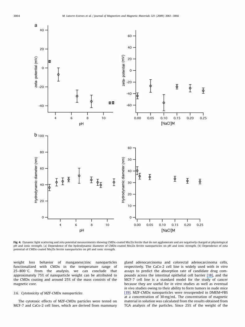
The effect of ionic strength and pH on stability of MZF-CMDxnanoparticles was determined by measuring zeta potential vs.concentration of NaCl and by measuring zeta potential vs. pH. Forthese measurements, we prepared a solution of 0.02 g nanopar-
Fig. 1. Magnetization curve showing the Mn/Zn ferrite nanoparticles are super-
paramagnetic in nature.
ticles in 30 ml deionized water (pH 4.3). Fig. 4a shows that thezeta potential measurements of these particles in a pH range from3 to 11 display an increase in negative potential as conditionsbecome more basic. The zeta potential of CMDx-coatednanoparticles ranges from around �30 to �50 mV for the NaClconcentrations tested (0–0.25 M.). This suggests that underphysiological conditions (0.15 M NaCl, pH 7.0) these particleswould be negatively charged and stabilized through steric andelectrostatic interactions. Fig. 4a demonstrates the CMDx-coatedMZF nanoparticles had a mean hydrodynamic diameter between30 and 50 nm and that no particle agglomeration was observed inthe ranges of pH and ionic strength tested, indicating the stabilityof the MZF-CMDx nanoparticles across a wide range of pH andionic strength.
3.5. Thermo gravimetric analysis (TGA)
We used TGA to determine the relative mass of CMDx attachedonto the manganese/zinc nanoparticles. Fig. 5 illustrates the
ARTICLE IN PRESS
Fig. 4. Dynamic light scattering and zeta potential measurements showing CMDx-coated Mn/Zn ferrite that do not agglomerate and are negatively charged at physiological
pH and ionic strength. (a) Dependence of the hydrodynamic diameter of CMDx-coated Mn/Zn ferrite nanoparticles on pH and ionic strength. (b) Dependence of zeta
potential of CMDx-coated Mn/Zn ferrite nanoparticles on pH and ionic strength.
M. Latorre-Esteves et al. / Journal of Magnetism and Magnetic Materials 321 (2009) 3061–30663064
weight loss behavior of manganese/zinc nanoparticlesfunctionalized with CMDx in the temperature range of25–800 1C. From the analysis, we can conclude thatapproximately 75% of nanoparticle weight can be attributed tothe CMDx coating and around 25% of the mass consists of themagnetic core.
3.6. Cytotoxicity of MZF-CMDx nanoparticles
The cytotoxic effects of MZF-CMDx particles were tested onMCF-7 and CaCo-2 cell lines, which are derived from mammary
gland adenocarcinoma and colorectal adenocarcinoma cells,respectively. The CaCo-2 cell line is widely used with in vitro
assays to predict the absorption rate of candidate drug com-pounds across the intestinal epithelial cell barrier [18], and theMCF-7 cell line is a standard model for the study of cancerbecause they are useful for in vitro studies as well as eventualin vivo studies owing to their ability to form tumors in nude mice[19]. MZF-CMDx nanoparticles were resuspended in DMEM+FBSat a concentration of 30 mg/mL. The concentration of magneticmaterial in solution was calculated from the results obtained fromTGA analysis of the particles. Since 25% of the weight of the

ARTICLE IN PRESS
Fig. 5. Thermo gravimetric analysis showing CMDx-MZF contains 25% by weight
of core particles.
Fig. 6. Relative viabilities of experimental and control wells showing that CMDx-
Mn/Zn ferrite is non-toxic to (A) CaCo-2 and (B) MCF-7 at core particle
concentrations of up to 7.5 mg/mL.
M. Latorre-Esteves et al. / Journal of Magnetism and Magnetic Materials 321 (2009) 3061–3066 3065
material corresponds to the magnetic core, a solution composedof 30 mg/mL MZF-CMDx contains 7.5 mg/mL of magnetic core.The relative viabilities of the experimental and control wells,shown in Fig. 6, indicate that the solution was not toxic after beingin contact with both types of cells for 48 h.
4. Conclusions
We have described the preparation of a Mn/Zn-doped iron-oxide crystal with a mean diameter of 15–17 nm according to DLSand TEM, an elemental composition of Mn0.24Zn0.76Fe2O3, theinverse spinel crystal structure of a ferrite, and which is super-paramagnetic in nature. These particles were coated by physicalabsorption with carboxymethyl dextran and shown to be stablydispersed in biological media. The coated nanoparticles had ahydrodynamic diameter that ranged between 30 and 50 nm acrossa wide range of pH and ionic strength. At physiological pH (7.0)and at physiological ionic strength (0.15 M NaCl) the nanoparticlespossessed a surface charge of �30 mV, suggesting they arestabilized by steric and electrostatic repulsion. The CMDx-coatednanoparticles contain around 75% total weight polysaccharide and25% total magnetic particle fraction. We tested these particles forcytotoxic effects at concentrations of up to 7.5 mg/mL in CaCo-2and MCF-7 cells for 48 h contact time and found no adverse effectsto the cell’s viability. We propose that because of thesenanoparticles’ stability, their non-toxic nature, and superpara-magnetic behavior, they should be further investigated as tools forbiomedical applications.
Acknowledgements
This work was supported by a NIRT award from the NationalScience Foundation (CBET-0609117) and the Puerto Rico Institutefor Functional Nanomaterials, supported by the NSF-EPSCoRprogram (OIA-0701525).
References
[1] H. Eun, S. Hyo, K. Byung, K. Byung-Kee, Synthesis of ferrofluid with magneticnanoparticles by sonochemical method for MRI contrast agent, Journal ofMagnetism and Magnetic Materials 289 (2005) 328–330.
[2] K. Shimizu, A. Ito, J.K. Lee, T. Yoshida, K. Miwa, H. Ishiguro, Y. Numaguchi,T. Murohara, I. Kodama, H. Honda, Construction of multi-layered cardiomyo-cyte sheets using magnetite nanoparticles and magnetic force, Biotechnologyand Bioengineering 96 (2007) 803–809.
[3] X. Li, L. Wang, C. Zhou, T. Guan, J. Li, Y. Zhang, Preliminary studies ofapplication of CdTe nanocrystals and dextran-Fe3O4 magnetic nanoparticlesin sandwich immunoassay, Clinica Chimica Acta 378 (2007) 168–174.
[4] M.S. Fallon, M. Varshney, D.M. Dennis, A. Chauhan, A physiologically basedpharmacokinetic model of drug detoxification by nanoparticles, Journal ofPharmacokinetics and Pharmacodynamics 31 (2004) 381–400.
[5] R. Hergt, R. Hiergeist, I. Hilger, W. Kaiser, Y. Lapatnikov, S. Margel, U. Richter,Maghemite nanoparticles with very high AC-losses for application inRF-magnetic hyperthermia, Journal of Magnetism and Magnetic Materials26 (2004) 3995–4021.
[6] G.M. Lanza, X. Yu, P.M. Winter, D.R. Abendschein, K.K. Karukstis, M.J. Scott,L.K. Chinen, R.W. Fuhrhop, D.E. Scherrer, S.A. Wickline, Targeted antiproli-ferative drug delivery to vascular smooth muscle cells with a magneticresonance imaging nanoparticle contrast agent: implications for rationaltherapy of restenosis, Circulation 106 (2002) 2842–2847.
[7] D. Wang, J. He, N. Rosenzweig, Z. Rosenzweig, Superparamagnetic Fe2O3
beads–CdSe/ZnS quantum dots core-shell nanocomposite particles for cellseparation, Nano Letters 4 (2004) 409–413.
[8] A. Gupta, M. Gupta, Synthesis and surface engineering of iron oxidenanoparticles for biomedical applications, Biomaterials 26 (2005) 3995–4021.
[9] D.H. Kim, K.N. Kim, K.M. Kim, Y.K. Lee, Targeting to carcinoma cells withchitosan- and starch-coated magnetic nanoparticles for magnetic hyperther-mia, Journal of Biomedical Materials Research A (2008) (February 6, [epubahead of print]).
[10] S. Teotia, M.N. Gupta, Magnetite–alginate beads for purification of somestarch degrading enzymes, Molecular Biotechnology 20 (2002) 231–237.
[11] N. Kohler, G. Fryxell, M. Zhang, A bifunctional poly(ethylene glycol) silaneinmobilized on metallic oxide-based nanoparticles for conjugation with celltargeting agents, Journal of American Chemical Society 126 (2004)7206–7211.
[12] S. Mornet, S. Vasseur, F. Grasset, E. Duguet, Magnetic nanoparticle design formedical diagnosis and therapy, Journal of Materials Chemistry 14 (2004)2161–2175.
[13] N. Feltin, M.P. Pileni, New technique for synthesizing iron ferrite magneticnanosized particles, Langmuir 13 (1997) 3927–3933.

ARTICLE IN PRESS
M. Latorre-Esteves et al. / Journal of Magnetism and Magnetic Materials 321 (2009) 3061–30663066
[14] J. Lai, K.V.P.M. Shafi, K. Loos, A. Ulman, Y. Lee, Thomas Vogt, Claude Estourne,Doping g-Fe2O3 nanoparticles with Mn(III) suppresses the transition to ther-Fe2O3 structure, Journal of the American Chemical Society 125 (2003)11470–11471.
[15] P. Drake, H.J. Cho, P.S. Shih, C.H. Kao, K.F. Lee, C.G. Kuo, X.Z. Lin, Y.J. Lin,Gd-doped iron-oxide nanoparticles for tumour therapy via magnetic fieldhyperthermia, Materials Chemistry 17 (2007) 4914–4918.
[16] E.V. Groman, J.C. Bouchard, C.P. Reinhardt, D.E. Vaccaro, Ultrasmall mixedferrite colloids as multidimensional magnetic resonance imaging, cell
labeling and cell sorting agents, Bioconjugate Chemistry 18 (2007)1763–1771.
[17] R. Massart, Preparation of aqueous magnetic liquids in alkaline and acidicmedia, IEEE Transactions on Magnetics 17 (1981) 1247–1248.
[18] P. Shah, V. Jogani, T. Bagchi, A. Misra, Role of CaCo-2 cell monolayers inprediction of intestinal drug absorption, Biotechnology Progress 22 (1) (2006)186–198.
[19] S.M. Shafie, L.A. Liotta, Formation of metastasis by human breast carcinomacells (MCF-7) in nude mice, Cancer Letters 11 (2) (1980) 81–87.
Copyright © 2022 FDOKUMEN