Revised Polyaniline Review for September 4
78
1 66--:-11--2-.6 2-116-:- Ehsan Nazarzadeh Zare 1* , Pooyan Makvandi 2,3* , Behnaz Ashtari 3 , Filippo Rossi 4 , Ahmad Motahari 5 , Giuseppe Perale 6,7,8 1 School of Chemistry, Damghan University, Damghan 36719-41167, Iran 2 Institute for Polymers, Composites and Biomaterials (IPCB), National Research Council (CNR), Naples 80125, Italy 3 Department of Medical Nanotechnology, Faculty of Advanced Technology in Medicine, Iran University of Medical Sciences, Tehran 14496 -14535, Iran 4 Department of Chemistry, Materials and Chemical Engineering, Politecnico di Milano Technical University, Milano 32, 20133, Italy 5 Young Researchers and Elite Club, Jahrom Branch, Islamic Azad University, Jahrom 74147-85318, Iran 6 Biomaterials Laboratory, Institute for Mechanical Engineering and Materials Technology, University of Applied Sciences and Arts of Southern Switzerland, Manno 2, 6928, Switzerland 7 Department of Surgical Sciences, Faculty of Medical Sciences, Orthopaedic Clinic-IRCCS A.O.U. San Martino, Genova 10, 16132, Italy 8 Ludwig Boltzmann Institute for Experimental and Clinical Traumatology, Donaueschingenstrasse 13, 1200 Vienna, Austria
-
Upload
khangminh22 -
Category
Documents
-
view
1 -
download
0
Transcript of Revised Polyaniline Review for September 4

1
6 6- - :- 1 1 - - 2 - . 6
2- 1 1 6-: -
Ehsan Nazarzadeh Zare1*, Pooyan Makvandi2,3*, Behnaz Ashtari3, Filippo Rossi4, Ahmad Motahari5,
Giuseppe Perale6,7,8
1School of Chemistry, Damghan University, Damghan 36719-41167, Iran
2Institute for Polymers, Composites and Biomaterials (IPCB), National Research Council (CNR), Naples
80125, Italy
3Department of Medical Nanotechnology, Faculty of Advanced Technology in Medicine, Iran University
of Medical Sciences, Tehran 14496 -14535, Iran
4Department of Chemistry, Materials and Chemical Engineering, Politecnico di Milano Technical
University, Milano 32, 20133, Italy
5Young Researchers and Elite Club, Jahrom Branch, Islamic Azad University, Jahrom 74147-85318, Iran
6Biomaterials Laboratory, Institute for Mechanical Engineering and Materials Technology, University of
Applied Sciences and Arts of Southern Switzerland, Manno 2, 6928, Switzerland
7Department of Surgical Sciences, Faculty of Medical Sciences, Orthopaedic Clinic-IRCCS A.O.U. San
Martino, Genova 10, 16132, Italy
8Ludwig Boltzmann Institute for Experimental and Clinical Traumatology, Donaueschingenstrasse 13,
1200 Vienna, Austria
2
Abstract
Inherently conducting polymers (ICPs) are a specific category of synthetic polymers with
distinctive electro-optic properties, which involve conjugated chains with alternating single and
double bonds. Polyaniline (PANI), as one of the most well-known ICPs, has outstanding potential
applications in biomedicine because of its high electrical conductivity and biocompatibility caused
by its hydrophilic nature, low-toxicity, good environmental stability and nanostructured
morphology. Some of the limitations in the use of PANI, such as its low processability and
degradability, can be overcome by the SG CSC KPO PH K T blends and nanocomposites K
CSKP T DKP PM NGST COF OCOPNC GSKCMT SGT GE K GM . This review describes the state-of-
the-art of biological activities and applications of conductive PANI-based nanocomposites in the
biomedical fields, such as antimicrobial therapy, drug delivery, biosensors, nerve regeneration and
tissue engineering. The latest progresses in the biomedical applications of PANI-based
nanocomposites are reviewed to provide a background for future research.
Keywords: Conductive polyaniline nanocomposites, biological activities, biosensor, drug
delivery, tissue engineering, antimicrobial activity, antioxidant activity.

3
1. Introduction
PT PM NGST CSG KOT MC PST TKOEG G CSG NCFG D EP CMGO DPOFT K P HSGG
NP GCDMG GMGE SPOT PS KPOT1 TPNG PM NGST ECO OG GS GMGTT HPSN C EPOF E K G C
OFGS CO GMGE SKECM T SGTT GTG PM NGST C G FKHHGSGO GOFGOEKGT P HPSN C
EPOF E K G C OFGS CSKP T EPOFK KPOT APNG SP GS KGT T E CT SGTKT K K CSE
SGTKT COEG FKGMGE SKE EPOT CO COF FKTTK C KPO HCE PS CSG TGF P G SGTT G
GMGE SKECM SP GS KGT PH PM NGST
9O GSGO M EPOF E KOI PM NGST 93 T CSG C T GEKHKE EC GIPS PH T O G KE PM NGST
with distinctive electro-optic properties. These polymers C G conjugated chains with alternating
single and double bonds 2. G π GMGE SPOT KO G 93 T KE CSG KI M FGMPECMK GF COF
TKN M PMCSK CDMG T P CO KN GSC K G SPMG KO G electro-optic SP GS KGT PH 93 T
6 S GSNPSG G TKPE GNKECM SP GS KGT PH 93 T T E CT T S E SCM GMGE SKECM COF
P KECM SP GS KGT CSG CHHGE GF D G OC SG PH G KO GSGO R CTK POG FKNGOTKPOCM
COF G MG GM PH DP KO SC COF KO GS E CKO FGMPECMK C KPO PH π GMGE SPOT )
G HKST EPOF E KOI PM NGS KPFKOG FP GF PM CEG MGOG CT FKTEP GSGF D 2MCO
8GGIGS 2MCO CE4KCSNKF COF 8KFGLK A KSCLC CK KO /-- G GSG C CSFGF G
PDGM SK G KO E GNKT S HPS KT FKTEP GS KO ( ( PM CEG MGOG 2T PM SSPMG
PM KP GOG PM H SCO 6 COF PM COKMKOG 2 9 CSG G CN MGT PH
4
93 T GTG 93 T CSG G GOTK GM TGF KO G DKPNGFKECM HKGMFT KOEM FKOI KTT G
GOIKOGGSKOI COF DKPTGOTPST DGEC TG PH GKS TNCS SGT POTG P G GMGE SKECM HKGMFT
2NPOI 93 T PM COKMKOG CT SGEGK GF EPOTKFGSCDMG C GO KPO KO CSKP T KOF T SKCM
COF DKPNGFKECM HKGMFT F G P GKS HCEKMG SG CSC KPO MP EPT KI GMGE SKECM
EPOF E K K DKPEPN C KDKMK MP P KEK COF GO KSPONGO CM T CDKMK ) 8P G GS
2 9 CT TPNG FKTCF CO CIGT T E CT MP TPM DKMK PS KOTPM DKMK KO G NPT
EPNNPO TPM GO T KOH TKDKMK COF GCL SPEGTTCDKMK 2MTP K T GMGE SKECM EPOF E K K
FGESGCTGT P GS C MPOI E EMG KNG , 2 O NDGS PH C SPCE GT T E CT SG FP KOI K
G H OE KPOCMK GF PSICOKE CEKFT EP PM NGSK C KPO K 2 9 FGSK C K GT PS P GS
PM NGST COF G SG CSC KPO PH G blends and nanocomposites K CSKP T NC GSKCMT
C G DGGO FG GMP GF P FKNKOKT G CHPSGNGO KPOGF FKTCF CO CIGT -
PM COKMKOG OCOPEPN PTK GT CSG POG PH G NPT SPNKTKOI 93 T OCOPEPN PTK GT
T P KOI CO GMGE SKECM EPOF E K K by combining the PANI matrix K G EPOF E KOI PS
KOT MC KOI OCOPHKMMGST GTG OCOPEPN PTK GT K KN SP GF SP GS KGT CSG
G GOTK GM TGF KO G DKPNGFKECM HKGMFT T E CT KTT G SGIGOGSC KPO COF
CO KNKESPDKCM GSC 2MTP G C G DGGO C MKGF KO G KOF T SKCM TGE PST G I KO
GMGE SPOKET COF C GS SKHKEC KPO .
The current review describes a state-of-the-art update on the biological activities (i.e.,
biocompatibility, cytotoxicity, antioxidant and antimicrobial activities) and biomedical
5
applications (i.e., antimicrobial therapy, drug delivery, bone regeneration, nerve regeneration,
wound healing, and biosensor) of conductive PANI-based nanocomposites to provide a
background for future research.
2. Synthesis
Polyaniline, or more specifically “aniline black”, is one of the oldest ICPs which was discovered
in the mid-nineteenth century 9. The molecular structure of PANI may possess either benzenoid or
quinonoid units or both types at different proportions 3,10.
The PANIs are synthesized using both chemical and electrochemical oxidative polymerization
in an acidic medium (Fig. 1). The most widely used initiators for the chemical polymerization of
aniline are ammonium persulfate (APS) COF P CTTK N GST MHC G : A ) Usually, the
electrochemical method is used for the small scale synthesis, whereas the chemical method allows
large-scale preparation of the polymer and/or the corresponding nanocomposites 11. The
electrochemical methods include the electrode coating and co-deposition approaches. In the
electrode coating method, reference, working, and counter electrodes are used in a one-
compartment cell containing the electrolyte and the monomer solution 12. In the co-deposition
method, an insulating polymer host is dissolved in an electrolyte solution comprising the monomer
of the conductive polymer 12.
6
Chemical (A) and electrochemical (B) polymerization mechanisms of polyaniline. Chemical polymerization of polyaniline is carried out in acidic medium by using a common initiator such as ammonium persulfate and potassium persulfate, while the electro-polymerization of polyaniline is carried out in the electrolyte solution of aniline and acid through applying a potential difference between the working and counter electrode.
Polyanilines containing various nanostructures with different properties have been reported by
many research groups. Different nanoarchitectures show additional advantages because of their
high surface-to-volume ratio, which leads to the improvement of the properties of their
nanocomposites 13. Therefore, the optimization of the synthesis conditions of PANI with specific
morphologies and sizes for high-performance applications is very important.
Several procedures such as solution, self-assembling, heterophase interfacial, and
electrochemical polymerizations have been used for the design and synthesis of PANI
nanostructures, such as nanospheres, nanogranules, nanorods, nanoflowers, nanofibers, and
nanotubes 7,13–19. In addition, many parameters and processes including the initiator or oxidant,

7
pH, temperature, solvent, chemical additives (oligoaniline and π bonding compounds), chemical
oxidation process (interfacial reaction), template (hard or soft), electrochemistry, radiochemistry
and sonochemistry for the design of unique PANI nanostructures should be taken into
consideration 14. T P T G TECOOKOI GMGE SPO NKESPISC T PH CSKP T 2 9
OCOPT S E SGT T O GTK GF KO CSKP T EPOFK KPOT
2T NGO KPOGF DGHPSG C O NDGS PH SGTGCSE GT C G DGGO FG P GF P G SG CSC KPO
PH 2 9 OCOPEPN PTK GT P P GSEPNG TPNG GCLOGTTGT PH G SKT KOG 2 9 GSG
CSG SGG NCKO SPEGF SGT HPS G HCDSKEC KPO PH 93 T OCOPEPN PTK GT KOEM FKOI0
APM GO ECT KOI0 9 KT POG PH G GS HKST COF TKN MGT SPEGTTKOI NG PFT HPS
G HCDSKEC KPO PH G PM NGS EPN PTK GT 9O KT GE OKR G C PM NGS KT
FKTTPM GF KO C SP GS TPM GO COF C FGTKSGF OCOPHKMMGS KT GO CFFGF P G
TPM KPO OFGS T KSSKOI ( 9O HCE G NCKO CF CO CIG PH G TPM GO ECT KOI
NG PF KT K T TKN MKEK PH NCO HCE SKOI K P G OGGF PH T GEKHKE
C CSC T GSG CSG O NGSP T HCE PST C ECO CHHGE G TPM GO ECT KOI
NG PF KOEM FKOI G PM NGS NPMGE MCS GKI PM NGS T S E SG
T PKE KPNG SKE CNP O EPN PTK KPO HKMMGS TPM GO G COF SPEGTTKOI
EPOFK KPOT GN GSC SG SC G PH FS KOI CIK C KPO SC G COF HSGR GOE PH T KSSKOI
(

8
( 9O TK PM NGSK C KPO KO G SGTGOEG PH OCOPHKMMGST0 9O KT NG PF C OCOPHKMMGS
T SHCEG KT NPFKHKGF COF GO G NPOPNGS COF KOK KC PS CSG CFFGF P HPSN C
PM NGS OCOPEPN PTK G APNG PM NGS SP GS KGT CSG KN SP GF KO KT NG PF
DGEC TG PH G T SPOI KO GSCE KPOT DG GGO G PM NGS NC SK COF G
OCOPHKMMGS ((
) 9O TK OCOP CS KEMG HPSNC KPO KO G SGTGOEG PH PM NGST0 9O KT C SPCE CO
C SP SKC G TPM GO KT TGF P FKTTPM G PM COKMKOG COF GO C OCOPHKMMGS
SGE STPS KT CFFGF HPMMP GF D GSNCM PS GMGE SPE GNKECM SGC NGO P HPSN
G PM NGS OCOPEPN PTK G G HPSNGF OCOPT S E SGT KO KT NG PF CSG
OKHPSN KO CSKP T NPS PMPIKGT K KO G PM NGS NC SK ()

9
Fig. 2 Scanning electron microscopy images of different shapes of polyaniline nanostructures; (A) polyaniline nanospheres synthesized in the acidic sodium carboxymethyl cellulose (CMC) solution. Reprinted with permission from ref 15. Copyright 2015 Royal Chemical Society. (B) polyaniline nanoflowers synthesized by the interfacial polymerization in toluene solvent. Reprinted with permission from ref 13. Copyright 2018 Elsevier. (C) polyaniline nanogranules synthesized by the sonochemistry method in the acidic aqueous solution. (D) polyaniline nanofiber synthesized by the oxidation polymerization of aniline monomer at 0 ºC in the acidic aqueous solution followed by the electrospinning in 50 ml of CHCl3. Reprinted with permission from ref 24. Copyright 2017 Hindawi (E) polyaniline nanotubes synthesized by the micelle soft-template procedure in the oxalic acid solution as a dopant. Reprinted with permission from ref 18. Copyright 2015 Royal Chemical Society. (F) polyaniline nanorods synthesized by the ultrasonication. Reprinted with permission from ref 16. Copyright 2014 Royal Chemical Society.
3. Structure and properties
3.1.Structure
Polyaniline structure consists of reduced (x) and oxidized (1-x) blocks (0 ≤ x ≤ 1) 10 (Fig. 3A).
According to the redox state of the polymer structure, PANI can be observed in one of three
oxidation forms, i.e., leucoemeraldine (LE, yellow, for x= 1, Fig. 3B), pernigraniline (PG, purple,
for x=0, Fig. 3C) and emeraldine (EM, dark green and blue, for x=0.5, Fig. 3D and E). The
emeraldine form of PANI can be found in the emeraldine-salt (EM-S, dark green) and emeraldine-
base (EM-B, blue) forms depending on the acidic and basic conditions, respectively 25. The
oxidation forms of PANI have different colors, conductivities and stabilities. The EM-S is the
conducting form of PANI. The electrical conductivity of PANI is related to several conditions,
such as the redox state, protonation degree, temperature and dopant type 3,10.
The electrical conductivity of PANI enhances with the doping of the EM-B (insulator form,
σ≤10-10 S/cm) and formation of the EM-S (conductive form, σ≥1 S/cm) 2. Inorganic acids, such as
HCl, H2SO4, HClO4 and H3PO4, and organic acids, such as camphorsulfonic acid, para-toluene
sulfonic acid and dodecyl benzenesulfonic acid are used for the doping process of PANI, while the

10
ammonium hydroxide base is responsible for the undoping process of PANI 10,26,27. In addition, it
is well known that the electrical properties of PANI-based polymers depend on the microscopic
(i.e., level of doping, conjugation, length of polymer chain) and macroscopic (i.e., materials
compactness and molecular orientation) properties 28.
Fig. 3 Structure of polyaniline (A), leucoemeraldine (B), pernigraniline (C), emeraldine base
(D) and emeraldine salt (E).
3.2.Physiochemical and mechanical properties
Polyaniline is chemically stable and demonstrates high chemical and structural resistance in
acidic and alkaline solutions without undergoing any chemical reaction or degradation 10.
Depending on the redox states, PANI has different solubility in the common organic solvents. In
general, PANI in the doped form (PANI-EM-S) is insoluble in the common organic solvents
11
including dimethyl sulfoxide (DMSO), N-methyl-2-pyrrolidone (NMP), dimethylformamide
(DMF) and tetrahydrofuran (THF), whereas in the undoped form, PANI-EM-B is soluble in the
aforementioned organic solvents 10,28.
Polyaniline pellet shows a good mechanical strength due to good compactness of the PANI
powders. Moreover, a significant improvement in the physical and mechanical properties, such as
Young’s modulus and heat resistance, has been seen in the PANI nanocomposites 29. Polyaniline
in blends and nanocomposite forms prepared using polyurethane, natural rubber, chitosan, carbon
nanotubes, and montmorillonite operates as a conducting component in a proper matrix to provide
the needed mechanical properties. Indeed, the increased toughness and decreased elongation at
break can be achieved in blends and/or nanocomposites by enhancing the amount of polyaniline
ratio.
The rheological properties of PANI and its composites are very important for their
processing in industrial and medicinal applications. For instance, the rheological parameters of the
injectable PANI nanocomposites should be evaluated before being injected to animal and human
organs. Indeed, the rheological properties provide information about viscosity, modulus, and
gelation temperature of the injectable PANI-based nanocomposites. The size, shape, and
distribution of nanomaterials have considerable effects on the rheological parameters of the PANI-
based composites
As earlier mentioned, one of the main problems of PANI is its insolubility in common
organic solvents. This problem has been solved successfully by developing various approaches for
the synthesis of soluble PANI, such as the synthesis of PANI by the micro-emulsion
polymerization or in the presence of oleic acid 30–32. On the other hand, combination of PANI with
various polymers/nanomaterials to form blends or composites can be a facile method for the
12
improvement of its mechanical properties 30,33. For example, Bilal et al. studied the rheological
properties of polyaniline-poly (ethylene oxide) (PANI-PEO) and their composites with KNO3 and
NaNO3. They reported that pyridine is the best solvent for the rheological measurements. Their
results showed that the nitrate salts of Na and K added to the PEO-based composite tend to
decrease the hydrodynamic volume of the polymer molecule, which results in the decreasing the
intrinsic viscosity of the composites. In addition, when the inorganic salts such as KNO3 and
NaNO3 were added, the composites showed somewhat higher values of viscosity. The temperature
change has a profound effect on the viscosity as well. For instance, at low temperatures, e.g. 10
°C, the pure PANI-PEO has a lower viscosity than the PANI-PEO-KNO3 composite, and the
viscosities of both composites drop by increasing the temperature to 20 °C 30.
Apart from organic solutions, the rheological properties of the aqueous PANI blends, e.g.
polyaniline-poly(vinyl pyrrolidone) (PVP) dispersion have also been investigated. The PANI-PVP
dispersion is stable enough showing no precipitates gelation. It was shown that the rheological
behavior of this system are not governed by any of the polymeric materials. In fact, the hydrogen
bonding between the PVP and PANI is the key for determination of the viscoelastic properties of
this suspended blend 31.
Polyaniline has also been used in the thermoresponsive injectable hydrogels. In-situ injectable
hydrogels can be easily administrated and match any shape of damaged tissue. These hydrogels
can reduce the suffering of patients as a minimally invasive method, and therefore, their easy
handling is an optimal choice for the clinicians. Thermosensitive hydrogels containing conductive
PANI has drawn much attention due to their conductivity, antioxidant and antimicrobial activity.
For instance, quaternary ammonium chitosan-g-PANI shows antibacterial activity and
conductivity along with biodegradability due to the presence of chitosan. These in situ forming
13
gels undergo a sol-gel transition upon the injection into the human body. In fact, in the ambient
condition or low temperature, such hydrogels act as liquid (elastic modulus < viscous modulus)
and can be easily injected. In contrast, at the body temperature, their rheological properties change
and they become gels (elastic modulus > viscous modulus). This alteration of temperature also
enhances the viscosity properties of the thermoresponsive hydrogels 34–36. Apart from temperature-
sensitive materials, polyaniline and its composite particles, due to the conductivity features, can
be used in electrorheological (ER) fluids. The ER fluid is a sort of smart electro-responsive system
showing transition characteristics from a liquid-like to a solid-like state in the presence of an
external electric field. For instance, PANI has been used as a coating for SiO2 nanoparticles to
impart electrorheological properties 37,38. The summary of the physicochemical properties of PANI
are listed in Table 1.
Table 1: Physicochemical properties of polyaniline.
- -6 6-
;G EPGNGSCMFKOG GMMP GSOKISCOKMKOG S MG COFGNGSCMFKOG FCSL ISGGO COF DM G)25
Electrical conductivity ;G EPGNGSCMFKOG KOT MC PS GSOKISCOKMKOG KOT MC PS COFGNGSCMFKOG TCM ( A%EN ( /
Common dopants 9OPSICOKE CEKFT 83M 8(A 83M COF 8) COF PSICOKECEKFT ECN PST MHPOKE CEKF CSC PM GOG T MHPOKE CEKF COFFPFGE M DGO GOGT MHPOKE CEKF (,
Solubility 5NGSCMFKOG TCM KOTPM DMG KO G EPNNPO PSICOKE TPM GO TGNGSCMFKOG DCTG TPM DMG KO 4 A 4 6 COF 86 (.
Mechanical property Young’s modulus 1.91Gpa; strength at breakpoint 89.5 Mpa; Elongation 5.88 % 29
Stability 8KI E GNKECM COF T S E SCM SGTKT COEG KO G CEKFKE COFCMLCMKOG TPM KPOT
Crystallinity Depending on the synthesis conditions and dopants can be semi-crystalline or amorphous22,39
Morphology Depending on the synthesis conditions can be OCOPT GSGOCOPHMP GS ) OCOPISCO MG- OCOPHKDGS( OCOP DG . COFOCOPSPF ,

14
Thermal Glass transition temperature (Tg) for uncross-linked PANI 70 °C and cross-linked PANI 250 °C; thermal degradation in air atmosphere near 500-600 °C10
3.3.Degradability
Non-biodegradability of some scaffolds still poses a limitation in biomedicine. Therefore,
degradable PANI, as an electrically conductive polymer, is preferred for the biomedical
applications. In recent years, the carbohydrate biopolymers (i.e., chitosan, gelatin, heparin, and
collagen) and biodegradable aliphatic polyesters (i.e., polylactide, polycaprolactone, polyglycolide
and their copolymers) have been employed for the fabrication of PANI blends/composites to
prepare degradable scaffolds for tissue engineering 40,41. As discussed earlier, one of the methods
to overcome the drawbacks such as the non-degradability of PANI is the preparation of blends and
composites based on PANI and degradable naturally occurring polymers including carbohydrates
such as dextrin (Fig. 4A), starch, and gelatin 22,39,40. Consequently, the final composites/blends can
be degraded by microorganisms in environmental conditions. For instance, Zare et al. evaluated
the soil biodegradability of PANI/dextrin nanocomposites at various weight ratios and reported
that the maximum degradation ~74.5% after two months was seen for the nanocomposite with the
weight ratio 1/3 of PANI/dextrin. Moreover, the biodegradability of the PANI/dextrin
nanocomposites improved considerably with the content of dextrin natural polymer 22.
Recently, a research conducted by Xia et al. revealed an excellent in vitro and in vivo
biodegradability and biocompatibility of a PANI-porous silicon hybrid nanocomposite (PANI-PSi
NPs). They found that the presence of the biocompatible and biodegradable PSi NPs resulted in
higher biocompatibility and biodegradability of the nanocomposites as compared with the pristine
PANI42. The biodegradable blends of PANI with poly(ethylene glycol) (PEG) and
polycaprolactone (PCL) for the tissue regeneration applications have been also reported 43,44. The
15
applications of the degradable PANI blends and composites in tissue engineering will be discussed
in Section 6.3.
4. Biocompatibility and cytotoxicity
Polyaniline and its nanocomposites are quickly developing as promising materials for the
biomedical applications. Therefore, the health risks related to PANI and PANI nanocomposites
are of great importance. G DKPEPN C KDKMK KT G CDKMK PH G NC GSKCMT P EPG KT K
MK KOI KOIT COF KTT GT K P FCNCIKOI GN O G P GS COF he cytotoxicity
depends on the chemical composition, size and shape of the nanomaterials in the nanocomposites
46. For instance, the cytotoxicity of the globular polymers is different from that of the polymer
nanoparticles. Thus, the structures of PANI and its nanocomposites can affect the biocompatibility
and cytotoxicity.
It is well-known that the deprotonation–reprotonation sequences undertaken on PANI result in
the decrease of cytotoxicity. However, PANI can hardly be toxic since it is fully insoluble and
stable in an aqueous solution. Hence, the cytotoxicity reported on the biological entities is related
to the low-molecular-weight compounds. Two types of these low-molecular-weight compounds
are available; (I) reaction of by-products with oligomers of aniline 47,48 and (II) the acids that form
the PANI salts. As a result, the modification of PANI with regard to the aforementioned materials
is of significance 49.
According to the ISO 10993 standards, PANI shows biocompatibility properties in terms of
dermal irritation and sensitization. It was reported that both PANI-EM-S and PANI-EM-B have
outstanding biocompatibility properties in the duration of dermal irritation. Moreover, the
cytotoxicity of the PANI-EM-S is lower than that of the PANI-EM-B due to the reprotonation-
deprotonation cycle and the presence of the low-molecular-weight impurities 45. The cell
16
biocompatibility of the PANI film prepared through electroless surface polymerization with PC12
cells was also reported. The results showed that the PANI film enhances the cell proliferation,
revealing promising potentials of this compound as a surface coating to cultivate neuronal cells
which can be used in tissue regeneration 50.
In a study, Humpolicek et al. used different cell lines, such as mouse embryonic fibroblast
(NIH/3T3) cell lines and embryonic stem cells to evaluate the biocompatibility of PANI. In
contrast to their previous findings, it was found that the correlation of the cytotoxicity with the
impurity contents is not always strictly linear. They reported that the cytotoxicity of PANI-salts
and PANI-based compounds are similar (Fig. 4B) 46.
The biocompatibility of the macroporous PANI cryogel prepared in the frozen poly(vinyl
alcohol) solution was investigated by the examination of its cytotoxicity on the mouse embryonic
fibroblasts as well as by the examination of the embryo-toxicity based on the production of beating
foci inside spontaneous differentiating embryonic stem cells. It was reported that the PANI cryogel
with the low contents of low-molecular-weight impurities has a good biocompatibility 51.
The cytotoxicity of the colloidal PANI on the human keratinocyte (HaCaT) and mouse
embryonic fibroblast (NIH/3T3) cell lines through 3-(4,5-dimethylthiazol-2-yl)-2,5-
diphenyltetrazolium bromide (MTT) assay was investigated and the results showed that the
cytotoxicity of the colloidal PANI is low 52. In another research, the cytotoxicity of PANI salt in
the globular and nanotubular morphologies decreased after reprecipitation from NMP compared
to the primary polymer. Also, no cytotoxicity on the NIH/3T3 cells was seen at 5 and 10% of the
extract concentration in the case of globular and nanotubular polymers, respectively 49.
The cytotoxicity evaluation of the PANI nanofibers on the rat celiac macrophages (at the
concentrations ≤1 mg/ml) showed that the PANI nanofibers did not have a considerable impact on
17
the level of cellular ROS and the loss of mitochondrial membrane potential (MMP) of the
macrophages; while a higher amount of PANI nanofibers (at the concentrations ≥10 mg/ml) caused
cell death, alterations of ROS level and MMP. It was found that the cytotoxicity of PANI
nanofibers was originated by the production of the oxidative stress and change of the intracellular
MMP 53.
In other researches, the teratogenic and eco-toxicity impacts of PANI nanoparticles and
nanofibers in Rhinella arenarum larvae and embryos were reported (Fig. 4C). The results revealed
that there is a low-risk potential after exposing R. arenarum to both PANI nanofibers and
nanoparticles 54,55.
The difference in the biocompatibility of the PANI composites is because of the various factors
such as, chemical composition, size and shape of the nanomaterials in the nanocomposites, dopants
and preparation methods 56. G DKPEPN C KDKMK PH G 2 9 DKPEPN PTK GT T E CT
T CSE % 2 9 KOESGCTGT K G KOESGCTG PH G OC SCMM PEE SSKOI PM NGS EPO GO -
9 CT SP PTGF C G T CSE % 2 9 DKPEPN PTK G DG TGF KO G KTT G GOIKOGGSKOI
Surprisingly, in another research, the polyaniline-coated cotton fabric showed high cytotoxicity on
the NIH/3T3 cell 58, which is because of the existence of the low-molecular-weight toxic impurities
in the PANI. These entrapped impurities in the PANI cotton structure are released during the
extraction of the coated cotton prior to the cytotoxicity tests 58. The aquatic toxicology of PANI
and CuO/PANI nanocomposites through acute (ISO 6341) and chronic toxicity (ISO 10706) tests
with microcrustaceans Daphnia magna and Vibrio fischeri, a marine bacterium, were evaluated by
Rossetto et al. According to their report, the PANI had no acute toxicity to D. magna (EC50,48 h,
99.21 mg/L), while the CuO/PANI nanocomposite had the EC50 value of 0.48 mg/L 56.

18
Regarding carbon-based nanocomposites of PANI, the in vitro cellular toxicity of the
nanodiamonds-PANI (NDs/PANI) composite on the human embryonic kidney (HEK) cells
showed that at low concentrations (0.1 to 1 μg/ml) the NDs/PANI composite can be applied for
the biomedical applications without a negative effect on the cells life activities 59. It was reported
that its application in bioscience depends on the determination of the proper concentration under
the in-vivo condition. The cellular biocompatibility of the poly(N-isopropylacrylamide)–carbon
nanotube (CNT)–PANI nanocomposite for tissue engineering applications using the mouse L929
fibroblast cells showed very good cells growth and viability along with the cells detachment
function 60. This nanocomposite was first fabricated via the combination of coupling and
electrospinning and then applied for the woven microfabric scaffolds construction.

19
Fig. 4 (A) The soil degradability of polyaniline/dextrin nanocomposite tablets with the diameter of 14 mm for I [polyaniline:dextrin (1:3)] and II [polyaniline:dextrin (2:1)] buried in soil (pH:7.5) in the relative humidity of 60–70 % at 28–30 ºC for 60 days. Reprinted with permission from ref 22. Copyright 2014 Springer. (B) The formation of the erythroid clusters (the red clusters marked with an arrow) within the embryoid body. (I) positive reference and (II) absence of the red erythroid clusters after cultivation in the presence of 25% extracts of PANI-salt Reprinted with permission from ref 46. Copyright 2018 Elsevier. Photographic recording of malformations detected in the embryos treated with 400 mg/L polyaniline nanofibers, (I) the control test without any treatment, (II) embryo with incurvated body axis and (III) embryo with underdeveloped gills. Reprinted with permission from ref 55. Copyright 2012 Elsevier.
5. Biological activity

20
5.1.Antimicrobial activity
The infection caused by microbes affects the human life severely. Hence, a number of studies
have been devoted to prepare new antimicrobial agents to fight pathogens. The advent of new
compounds containing antimicrobial properties continues unabated. Polymers with antibacterial
and antifungal activities are widely used in the biomedical applications since the pathogens
become resistant to the existing drugs. Conducting polymers, such as PANIs, are appealing in
biomedicine because of their high cellular response 61. In this regard, the PANI-based compounds
have been synthesized to control the microbial contaminations 62. The antimicrobial activity of
PANI towards the Gram-negative and Gram-positive bacteria have been reported 63,64. However,
a number of various nanomaterials, polymers and other compounds have been added to PANI to
enhance the antimicrobial activity, conductivity, and photocatalytic activity 65,66. Hence, the
PANI-based nanocomposites consist of different nano-architectures such as rods, spherical
particles, tubes and sheets have been exploited for the biomedical applications. For instance, the
PANI/zinc-aluminum layered double hydroxide nanocomposite prepared by the free radical
emulsion polymerization has been reported to show antibacterial activity 67. Other architectures,
such as PANI decorated Au nanorods, have also shown high antibacterial properties toward
Escherichia coli and Staphylococcus aureus 68. Nanofibers of PANI/silver NPs showed
antibacterial properties against E. coli and B. subtilis strains, while neat PANI did not show any
antibacterial activity69. The application of PANI/silver nanocomposites is not limited to
antimicrobial purposes. For instance, the use of Ag functionalized PANI-based biosensor has been
reported for the determination of anticancer drugs 70.
The PANI nanocomposites containing microbicidal nanomaterials, such as zinc oxide and Ag
nanocompounds, have shown synergistic antimicrobial effects 64. Silver NPs and carbon nanotubes

21
incorporated PANI showed higher antibacterial activity than PANI-carbon nanotubes and PANI-
Ag nanocomposites because of the synergistic effect of the fillers 71. In another study, the
antibacterial effect of the ZrO2 NPs-PANI nanocomposite against E. coli and S. aureus was
determined to be higher than that of the pure PANI 72.
The polymers containing quaternary ammonium compounds have high antibacterial and
antifungal activity 73,74. Therefore, copolymers of PANI with the biopolymers containing
quaternary ammonium salts, such as chitosan, have been employed to improve the antibacterial
activity with enhancing the biocompatibility of PANI 75. With this in mind, quaternized chitosan-
graft-PANI injectable hydrogels have been used as the biocompatible scaffolds for tissue
regeneration (Fig. 5). The in situ forming biodegradable conductive hydrogels have in vitro and in
vivo antibacterial properties and can improve the proliferation of the C2C12 myoblasts in
comparison with the quaternized chitosan hydrogel 35. The microcapsules of poly(lactic-co-
glycolic acid) have been applied as a carrier for the delivery of ginseng/PANI for the implant
restoration. The presence of PANI enhanced the antibacterial efficacy up to 88% 76. Apart from
releasing antimicrobial agents, non-leaching antibacterial and antifungal compounds, which have
a chemical linkage to the polymer matrix is another option to form an antimicrobial surface. For
instance, poly(3-aminobenzoic acid) and PANI have been applied to form an antibacterial surface
77.
The antimicrobial mechanism of PANI includes the production of H2O2 that causes the
oxidative stress characterized by the perturbation of iron homeostasis (Fenton reaction). In fact,
free iron can propagate H2O2 stress by participating in Fenton reaction which accelerates the
formation of hydroxyl radicals leading to the microorganism destruction and, subsequently, cell
death. Polyaniline is more active against Gram-negative bacteria, such as E. coli, in aerobic

22
conditions compared with the anaerobic environments. Higher antibacterial property in aerobic
conditions supports the role of reactive oxygen species 78.
Fig. 5. (A) Schematic presentation for the antibacterial activity of injectable conducting
hydrogels. (B) Live/dead staining of adipose-derived mesenchymal stem cells at successive culture periods for the quaternized chitosan-graft-PANI crosslinked by oxidized dextran using the Alamar Blue and Live/Dead Viability/Cytotoxicity assay. Scale bar: 200 µm. Reprinted with permission from ref 35. Copyright 2015 Elsevier.
5.2.Antioxidant activity
6SGG SCFKECM KO GSNGFKC GT MGCF P G KTT G FCNCIG COF FKTGCTGT SPISGTTKPO
T E CT KOHMCNNC KPO GCS FKTGCTG ECOEGS COF SGNC SG CIKOI -/ 2O KP KFCO
NC GSKCMT CSG EPN P OFT C SG GO G P KFC KPO PH P GS NC GSKCMT G MC CO

23
KN PS CO SPMG KO HPPFT COF KTT GT CT C GCM SP GE K G HCE PS COF FGESGCTG G SKTL
PH G E SPOKE FKTGCTGT -/ G CO KP KFCO CE K K PH 93 T CT KN PS CO
EPOTGR GOEGT HPS GKS C MKEC KPOT KO G DKPNGFKEKOG 9 KT CS KE MCSM TGH M KO G
KTT GT T HHGSKOI HSPN G P KFC K G T SGTT GSG G EC CDKMK P MP GS G EGTTK G
T CIGT PH G SGCE K G SCFKECM T GEKGT A KT C SP SKC G . G 93 T T E CT
COF 2 9 C G T P O IPPF CO KP KFCO CE K K KGT KO G SGTGOEG PH FK GO M (
KES M FSC M 4 8 HSGG SCFKECM TEC GOIGS . . The swift heavy ion irradiation of PANI
and the nature of the dopant acids have shown a significant role in the antioxidant activity of the
PANI nanostructures 81. The HCl-doped PANI nanofibers induced by the swift heavy ion
irradiation showed the best antioxidant activity compared to other undoped PANI structures 82. It
was reported that the observed antioxidant activity of the PANI nanofibers is related to the decrease
of the size of PANI nanofibers after the swift heavy ion irradiation, which points the access of
more reaction sites for DPPH scavenging 82. On the other hand, the antioxidant activity of the
materials depends on their capability to donate hydrogen to reduce DPPH, and therefore, the
chemical structure of the materials are important for their antioxidant activity 22.
There are a few studies on the antioxidant activity of PANI nanocomposites to be developed in
the biomedical applications. The antioxidant activity of PANI/starch biocomposites was improved
upon increasing the PANI ratio. This can be related to the fact that the PANI, because of its redox
active nature, is effective as a DPPH free radical scavenger 57. The antioxidant activity of other
polysaccharides/PANI composites, such as PANI/dextrin nanocomposite, prepared by in-situ
polymerization of aniline and dextrin biopolymer, increased up to 72 % with the aniline content.

24
This antioxidant activity of PANI can have substantial effect on the tissues and organs suffering
from the oxidative stress 22.
GICSFKOI PM COKMKOG EPO CKOKOI NG CM OCOPNC GSKCMT G NC KN N CO KP KFCO
CE K K PH G 2 9% PM CO POG SKC PMG%6G) OCOPEPN PTK G HCDSKEC GF KC CO KO
TK GN MTKPO PM NGSK C KPO CT G CM C GF P DG 2 9 . C CO KO GS CM PH (
NKO KT CNP O PH CO KP KFCO CE K K KT DGEC TG PH G SGTGOEG PH C KI GS O NDGS
PH GMGE SPOT COF FSPIGO C PNT -/
G GHHGE PH G T PS KOI GMGE SPM GT such as CSC PM GOG T MHPOKE CEKF A2)
COF :3M K FKHHGSGO NPMCS SC KPT PO G CO KP KFCO CE K K PH 2 9%SGF EGF
ISC GOG P KFG S 7 CT CMTP DGGO KO GT KIC GF 2EEPSFKOIM G CO KP KFCO
CE K K PH G 2 9%S 7 OCOPEPN PTK G KO G A2 T PS KOI GMGE SPM G
KOESGCTGF K G A2 NPMCS SC KP 9 CT CMTP HP OF C G SP POC KPO PH G
2 9%S 7 OCOPEPN PTK G D A2 FPOC GT FSPIGO P SGF EG 4 8 CT CO
CO KP KFCO .)
As mentioned earlier, the antioxidant activity of ICPs, which comes from their capability to
decrease the levels of RRS, is possibly helpful for the suffered tissues from the oxidative stress.
Thus, the antioxidant activity of polyaniline may be important, especially for the diseases that
cause excessive levels of RRS. Furthermore, the PANI composites, such as the PANI/starch, may
have the capability to decrease the oxidant produced via the chemotherapeutic drugs which can
assist in neutralizing or at least decreasing the side effects of the chemotherapeutic cancer therapy.
According to the literatures on this subject, a schematic illustration for the proposed mechanism

25
of antioxidant activity of PANI in the presence of the DPPH radical scavenger KT T P O KO
Fig. 6. The proposed mechanism for the antioxidant activity of polyaniline in the presence of DPPH radical scavenger.
6. Biomedical applications
2NPOI 93 T G 2 9 CT P T COFKOI P GO KCM C MKEC KPOT KO FKHHGSGO HKGMFT
T E CT T GS-EC CEK PS ICT TGOTPS C GS SGC NGO CO K EPSSPTKPO EPC KOI FS I
FGMK GS DKPTGOTPS COF KTT G GOIKOGGSKOI . �.,
9O G HPMMP KOI TGE KPOT G C MKEC KPOT PH 2 9 OCOPEPN PTK GT KO G
DKPNGFKECM COF EMKOKECM HKGMFT CSG CFFSGTTGF 9 T P MF DG OP GF C CEEPSFKOI P P S
LOP MGFIG DCTGF PO 3MKOKECM SKCMT IP KO GSOC KPOCM FC CDCTG 2 9 DCTGF NGFKECM
FG KEGT CT OP G SGCE GF G EMKOKECM T CIG COF EPOTGR GO M CT OP ICKOGF G
EGS KHKEC KPOT P GO GS G NCSLG 2 9 KT KOFGGF G SGNGM SPNKTKOI COF KO GSGT KOI
D G C G OP G SGCE GF C T HHKEKGO T CIG PH NC SK P CMMP C TCHG SCOTMC KPO

26
HSPN DGOE P DGFTKFG . 9O CFFK KPO G SGI MC PS HSCNG PSL CT OP G GMM FGHKOGF
G DPSFGST HPS G C MKEC KPOT EPO CKOKOI OCOPEPN POGO T
6.1.Antimicrobial therapy
Antimicrobial conducting PANI has been applied in biomedicine including electrotherapy,
antimicrobial clothing, and electromagnetic devices for monitoring health 87. Infections in the
treatment of diseases are still challenging; for instance, during or post scaffold transplantation
which reduces the efficacy of bone healing. PANI nanocomposites have been developed in
combination with various antimicrobial agents including silver NPs, TiO2, releasable drugs and
biomolecules, and the non-leachable compounds, such as quaternary ammonium salts, to prepare
a number of antimicrobial devices 42,62,69,88.
PANI nanofibers combined with mupirocin, a topical microbicidal compound, have been
prepared via a self-assembly approach for their potential applications as a wound healing dress 89.
It was shown by the agar diffusion method that the antibacterial activity of the PANI-mupirocin
was higher than that of the neat PANI due to the release of mupirocin 90. 6KDGS KI M PSP T
TECHHPMFT based on poly-ε-caprolactone-PANI were fabricated by electrospinning approach for
their potential applications in electrically stimulated cell growths and cytoprotection of cells
against oxy-radials. The nanostructured bioactive scaffolds revealed both antibacterial and
antioxidant activities (free radical-scavenging capability) with no cytotoxicity against L929 cells
on the scaffolds 91.
Colloidal aqueous dispersions of PANI showed low bactericidal effects (3500 g mL-1) against
B. cereus and E. coli. Although the PANI dispersion has low cytotoxicity, the toxicity effect
depends on the cell line and PANI dose; for example, the human keratinocyte cells were less

27
sensitive than the mouse embryonic fibroblast cells. In addition, the neutrophil oxidative burst
assay revealed that 150 g mL-1 is the critical concentration of the PANI colloid dispersions for the
biologically safe applications 52.
Thermosensitive gels possessing electrical conductivity and self-healing capability are of
great interest as cell carriers for tissue engineering 92,93. In a study conducted by Dong et al., an
injectable and biodegradable hydrogel with antibacterial activity was synthesized by mixing
dibenzaldehyde-terminated poly(ethylene glycol) (PEG-DA) and chitosan-graft-aniline tetramer
(CS-g-AT) to be used for the repair of the damaged cardiac tissue. The electroactive and
antibacterial hydrogels showed good viability and proliferation with rapid self-healing capability
because of the cross-linking network made through the Schiff-base reaction of aniline (Fig. 7) 92.
The cell growth and proliferation was also reported for hard tissue regeneration using TiO2
nanotubes-PANI nanocomposites 94.
The PANI-polyurethane foam (PANI/PUF) was employed as an antibacterial dress for the
wound healing applications. The PANI/PUF film was fabricated by using in-situ radical
polymerization of aniline monomer in the presence of usnic acid (UA, as a dopant) and PUF. The
UA improved the bactericidal property of PANI toward E.coli and S.aureus strains95. Other
bandage dressings, such as antimicrobial membrane composed of aniline tetramer/siloxane
terminated polyurethane (AT/STPU), have also been suggested. The AT/STPU membranes were
fabricated via the sol–gel method using the STPU prepolymer and AT. The presence of
polysiloxane-linked PU chains improved the dimensional stability even at the high hydrated
condition. It was also reported that the AT led to the improvement of the cells viability and
antimicrobial activity for both bacteria and fungal strains96.

28
Fig. 7 ((A) Schematic procedure for the synthesis of thermosensitive chitosan-graft-aniline tetramer (CS-g-AT) hydrogel. (B) CS-g-AT hydrogels after subcutaneous injection. (C) CS-g-AT hydrogels wrapped in the rat’s skin (row I) and the hydrogels peeled from the rat’s skin (row II). The hydrogels with different aniline tetramer (AT) contents, including CS-AT8, CS-AT10 and CS-AT20, were prepared. (D) Degradation properties of the hydrogels in vivo. Row I shows the hydrogel under the skin and row II displays the hydrogel that peeled from the skin. CS: chitosan, AT: aniline tetramer. Reprinted from ref 92. Copyright 2016 American Chemical Society.
PANI and its composites not only target the human body for the antimicrobial therapy but
also are suggested to be used in places (e.g. hospitals) and devices (e.g. medical devices) that are
prone to the microbial growth. For instance, Robertson et al. evaluated the antimicrobial activities
of PANI and poly(3-aminobenzoic acid) (P3ABA) as the effective agents to fabricate bacteria-

29
resistant surfaces. The antimicrobial activities of PANI and P3ABA against E.coli and S.aureus
were seen in both absorbent and non-absorbent surfaces. It was proposed that these surfaces could
be applied as the wall coating in hospitals 77. PANI coated modified polypropylene (MPP) has
been prepared by using dip-coating technique as an anti bioaerosol filter. The PANI NPs were
synthesized by micro-emulation polymerization and then coated onto the polypropylene filter The
antibioaerosol property of the PANI/MPP was investigated against S. aureus, E. coli, and B.
subtilis bioaerosols. It was shown that the water absorption property, stability and antibacterial
efficiency of the PANI/MPP were meaningfully improved as compared to the unmodified PP filter
97.
Metal nanomaterials embedded PANI nanocomposites have also been applied to improve the
antibacterial and antifungal activity of the nanocomposites. For instance, the conductive
polyaniline containing silver showed higher antibacterial activity than the neat PANI 88,98. Though
the mechanism of antimicrobial activity of nanomaterials against different microorganisms varies
for�the types of metals, ions and, species, the dissolution of nanomaterials into ions is often the
first step and a�common reason for the toxicity of metallic nanostructures (Fig. 8). For instance,
the metal ions can produce hydroperoxide radicals, whereas zinc oxide can form hydroxyl radicals
99.

30
Fig. 8 Illustration of potential interactions and modes of toxicity when engineered nanoparticles targeted at different parts of a generic bacterium: capsule, cell wall, cell membrane, and cytoplasmic contents. Numerous nanoparticle shapes may reduce the bactericidal toxicity by one or some of these mechanisms. These mechanisms include the cell membrane disruption, disruption of electron transport chains, ROS production, and damage of proton efflux pumps.
6.2.Drug delivery
It is widely accepted that the kinetics by which a drug is released presents a high impact on its
efficacy 100. Traditional approaches for delivering a drug or biomolecule through oral or injection
lead to the accumulation of drugs/biomolecules(concentration peak) in the human body.101
Accordingly, to reach the therapeutic levels, the initial concentration of the biomolecules must be
more than the threshold level which causes the biomolecule accumulation; however, this
concentration peak is gradually reduced to an ineffective level over time. Thus, the most important
objective behind the sustained and controlled drug delivery systems is to offer an optimal drug
31
delivery adjusting drug level to avoid under- and overdosing and preserve the released amount
within a certain period. This approach leads to the reduction of the number of drugs administration
per day 102. These strategies often called smart drug delivery systems or devices (SDDS), are based
on multidisciplinary approaches that combine pharmaceutics, materials science, and molecular
biology together with the engineering skills 103. The main aim is to release a certain amount of a
drug, loaded within carriers, to a specific target site for a prolonged period of time with a sustained
kinetics. The drug delivery device can be either inert (and so the device works only as a drug
carrier) or an active part of the therapy. Following the second strategy, due to the intrinsic
conductivity of PANI, PANI derivatives can be used as the drug carriers with the electric-driven
release 104. A common issue of those devices is their very low mechanical strength 105. A good
strategy to improve the mechanical properties is represented by using supramolecular nanofibers
self-assembled from the sorbitol derivatives 106. PANI is also a good candidate to be a photothermal
converting material for the theranostic applications. Therefore, it can be used for diagnosis and
simultaneously delivering a drug. In this regard, the biocompatible graphene and Au NP core PANI
shell nanocomposites have successfully been fabricated. These nanocomposites showed high
biocompatibility, good stability, strong near-infrared (NIR) absorbance, and suitable drug loading
efficiency. This light-sensitive system has NIR/pH-responsive drug-releasing capability, which
promotes the practical applications in the chemo-photothermal therapy 107.
Chemo-phototherapy is the incorporation of a therapeutic agent within a material that is
responsive to NIR irradiation inducing anticancer activity. Of organic NIR-responsive materials,
PANI emerged as an extremely promising material 42,108–111. This strategy has attracted great
attention in recent years in view of its enhanced drug accumulation and controlled release, while
the side effects were relatively decreased 112. For instance, Nguyen et al. 108 embedded a
32
chemotherapeutic agent, methotrexate and PANI (a photosensitizer material) within hybrid
polymer NPs that can target cancer cells after conjugation with lanreotide, a synthetic analog of
somatostatin. The synthesis and structure of these multifunctional hybrid polymer NPs together
with the mechanism of their anticancer activity are schematized in Fig. 9. In addition, the efficacy
of the composite systems to kill the cancer cells could be improved using the PANI derivatives,
such as the PANI impregnated with the magnetic (Fe3O4) nanoparticles 113.
The use of PANI nanocomposites in cancer therapy is also reported by Gao et al. 114 with the
development of folate-based particles functionalized with PANI. The decorated nanoparticles were
able to efficiently target the cancer cells and selectively accumulate within them to achieve the
NIR-triggered localized release of their content (cisplatin). It was also shown that the gold
nanorods could be coated with the PANI in order to reduce the cytotoxicity and instability. After
the incubation of the nanorods with HeLa cells and exposure to a NIR laser, the threshold energy
to kill the cancer cells was found to be significantly lower compared with the previous studies 115.
Apart from the metallic nanostructures, the PANI chains with various lengths have been
attached onto the surface of the spherical particles of hollow mesoporous silica using in situ
chemical oxidative polymerization. The encapsulated anti-cancer drug (doxorubicin) exhibited an
acidic pH-responsive release behavior; while indomethacin showed an alkaline release. The effect
of the length of PANI chain-gate in the drug encapsulation capacity was also studied. Indeed, the
long PANI chains decreased the loading capacity due to the blocked mesoporous channel 116. In
the field of photothermal chemotherapy, PANI was also exploited to cover inorganic 117 and
organic 118 NPs as a functionalizing compound to improve their performance. Table 2 represents
the use of polyaniline and its nanocomposites in drug delivery and bioimaging applications.

33
Fig. 9. Schematic illustration on the preparation of multifunctional hybrid polymer
nanoparticles along with their anticancer activity mechanism. Reprinted with permission from ref 108. Copyright 2018 Elsevier.

34
Table 2. Polyaniline nanocomposites used in drug delivery, photothermal therapy, and bioimaging applications.
Nanocomposites Nanomaterial size (nm)
Structures Drug Applications Reference
Polyaniline/porous silicon 10-20 Porous silica particles Doxorubicin hydrochloride
Cancer therapy 42
Polyaniline 90 Nanorods Cisplatin Photothermal cancer therapy
112
Polyaniline tubes 800-1500 Rectangular shaped hollow tubes
Acid Red 8 as model drug
Drug delivery 119
Polyaniline/silica nanoparticles 200 Hyaluronic acid decorated silica fluorescent NPs
- Near-infrared light responsive (bio-imaging)
120
Poly(ethylene glycol) with poly(ϵ‐caprolactone) block copolymer/polyaniline NPs
83 Core‐Crosslinked Polyaniline NPs
Cisplatin Breast cancer cells 118
Polyaniline/MoS2 Quantum Dot
5 Quantum Dot
- Bioimaging (tomography)
121
Surface modified PANI NPs by F127 48 Functionalized NPs - Near-infrared (NIR) photothermal therapy
122
Graphene and Au NP core PANI shell nanocomposites
10 Core-shell NPs
Doxorubicin Photothermal cancer therapy
107
Lanreotide-methotrexate/Polyaniline 180 Lanreotide-functionalized NPs
Methotrexate Photothermal cancer therapy
108
Polyaniline with zeolite imidazole frameworks
200 Functionalized NPs 5-fluorouracil Photothermal cancer therapy
109
Lipid-polyaniline 100 Lipid-functionalized NPs Rapamycin Photothermal cancer therapy
110

35
CaCO3/polyaniline 1200 L-cysteine-functionalized microparticles
- Photothermal cancer therapy
111
Polyaniline/Pluronic F127 50 nm Nanoparticles - Photothermal cancer therapy
122
Fe3O4/polyaniline 10 nm Core-shell NPs
- Photothermal cancer therapy
113
Polyaniline/polyglutamic acid 80 nm Cysteine-functionalized NPs
- Photothermal cancer therapy
123
Folate/poly(ethylene glycol)-distearoylphosphatidylcholine
100 nm Polyaniline-loaded NPs Cisplatin Photothermal cancer therapy
114
Au nanorods/polyaniline 40 nm Core – shell nanorods - Photothermal cancer therapy
115
Polyaniline/porous silicon 110 nm Porous silica particles Doxorubicin Photothermal cancer therapy
116
lanthanide-based upconversion NPs /Polyaniline
120 nm Core – shell NPs - Bioimaging and photothermal cancer therapy
117
36
6.3.Tissue engineering
6.3.1. Wound healing
The possibility to transplant tissue is limited by several issues, such as immune rejection and
donor shortage 124. Novel research studies are looking for the combination of cells with the active
molecules (drugs or biomolecules) to improve the regenerative therapeutic effects 125. In this
framework, in the last decades, a lot of studies were devoted to the development of proper 3D
scaffolds that can work as a temporary substrate, helping to the growth of cells in an organized
fashion, before transplanting them within patients 126. This necessity comes from the fact that the
cells injected from in vitro cultured cells can easily escape, leaving the zone of injection with an
uncertain fate through the circulatory torrent. In this regard, not only scientific literature agrees
with addressing this issue, but also regulatory supranational directives (e.g. EU668/2009 and
47/2007/EC) are now pointing towards the necessity to use cells in combination with suitable
support structures. Consequently, great attention has been devoted to the polymers that can be
applied for the production of the three-dimensional scaffolds and developing the injectable devices
for tissue regeneration. In this field, PANI can improve the scaffold physical properties 62,127. This
improvement in the elasticity and mechanical performances allows the scaffolds to better mimic
the native tissue properties 127,128. For instance, the composites based on PANI could reach a high
conductivity range and low tensile strain with the consequent high fibroblast and myoblasts
adhesion 129.
Electrical stimulation of the fibroblasts loaded within the PANI composite scaffolds presented
low cell death and improved metabolic rate. Sharma et al. 130 developed a composite system based
on PANI-carbon nanotubes, the nanofibers of poly(N-isopropyl acrylamide-co-methacrylic acid)
(PNIPAm-co-MAA), and PC/PNIPAm-co-MAA by electrospinning. The seen excellent growth of
cells on the surface of the composite can be due to the higher conductivity and mechanical strength

37
of polyaniline and carbon nanotubes. PANI-based NPs were also used to improve the
performances of the graphene papers 131. The combination of flexibility, biocompatibility, and
electrochemical properties, coming from nanostructured PANI and graphene, makes them to be
good candidates for hybrid devices for the biomedical sectors, such as flexible biosensors,
batteries, and bioelectrodes along with the ability to culture the electrically excitable cells. Indeed,
the conductivity given by PANI in hydrogels is also used in cellulose nanofibers 132, resulting in
better degradability and biocompatibility. The amelioration of the composite systems in the
applications of tissue engineering was shown in the regeneration of cardiac cells132.
PANI has also been mixed with gelatin, and the co-electrospun nanofibers were utilized to
support the cell growth 133. The experimental results revealed that all the PANI/gelatin nanofibers
support the H9c2 cell attachment and proliferation and can control the tissue culture-treated plastic
and smooth glass substrates. The use of PANI nanocomposites in cardiac tissue engineering is very
promising 134. For instance, PANI associated with caprolactone producing patches was able to
better guarantee the high viability of human mesenchymal stem cells than the neat
polycaprolactone (PCL)134.
Hsiao et al. 135 developed a mesh (Fig. 10) including aligned composite nanofibers of PANI
and PLGA, as an electrically conductive platform for coordinating the beatings of the cultured
cardiomyocytes synchronously. The PANI/lactide composite was also applied to stimulate the
neuromuscular junction 136 and nerve regeneration 137 because of its ability to provide electrical
signals. Due to this ability, PANI was used in combination with PCL to prepare the electrically
active fibers that can maintain stable electrical features in the simulated cell culture conditions for
up to one week with the improved NGF-induced neurite outgrowth of PC12 cells 138.
38
Fig. 10. Schematic diagrams on the preparation procedure of polyaniline/poly(lactic-co-glycolic acid) (PANI/PLGA) nanofibrous mesh, cell sowing, electrical excitation, and synchronous cell thrashings mechanisms. Reprinted with permission from ref 135. Copyright 2013 Elsevier.
PANI nanotubes are one of the most promising nanostructures for cardiac tissue regenerations
because of their electroactive characteristics. Their biocompatibility and low hydrophilic
characteristics can be enhanced by their functionalization with polyglycerol dendrimers 139.
6.3.2. Bone regeneration
Evidences of clinical requests concerning the bone regeneration date back to ancient Egypt and
are very important in the present age. A more rigorous scientific method has been followed since
1889 when “modern” researchers began to focus their efforts on what can be defined as the early
bone tissue regeneration 140. Although nowadays several hundred millions of surgical intervention
per year are carried out, autograft bone still remains the current clinical gold standard for the

39
treatment of critical-sized and non-union bone defects. Being advantageous for the
immunocompatibility, autografts nevertheless carry a wide spectrum of risks, such as general
anesthesia, complex surgical maneuvers, secondary infections, secondary fractures, pain, site
morbidity, etc.; since they lead to high percentage of failure (more the 10%) followed by important
costs increases 141–143. Furthermore, it is generally accepted that not all the defects can be
addressed, particularly the bigger ones, as far as few healthy sites can be harvested without loss of
function 143. The requirement of proper bone replacements for the remodeling of native bone
tissues is hence evident and sees a wide spectrum of proposed solutions related to the academia,
clinics and industry 144. In this framework, surgeons can choose from substitutes that can be
divided into three main categories apart from autografts:
1) allografts, i.e. bone segments taken from cadavers and duly sterilized
2) xenografts, i.e. bone segments taken from animal bones (bovine, equine, porcine, etc.) duly
acellularized and sterilized
3) synthetic scaffolds
Allografts derived from cadavers bone are an acceptable option. However, there are some
concerns, such as diseases transmission, toxicity associated with the requirement of sterilization,
immunologic rejection risks, and very high sample variability that progressively leads to other
available alternatives; the challenge still remains an open scientific and clinical topic but current
focus remains on autografting rather than xenografting and synthetic scaffolds 141,142.
There are many different bone grafts available on the market. Bone is the second most
transplanted tissue after blood. Today, no products based on PANI are currently certified for
routine clinical use. Nevertheless, the use of PANI in new generation of synthetic bone scaffolds
is progressively taking its way, particularly for the tissue engineering applications, i.e. where the
40
use of scaffolds and stem cells is meant to provide support to restore tissue anatomy and
functionality. Indeed, new highlights point towards the use of moderate content of PANI in the
conductive nanofibrous scaffolds made from bioresorbable aliphatic polyesters, such as poly(lactic
acid) (PLA), which significantly promotes the osteogenic differentiation of bone marrow-derived
stem cells 145. Similarly, osteoinductivity of polyethersulfone-based electrospun scaffolds
increased by using PANI 146, showing the same mechanism of action found on PLA-PANI
scaffolds.
Another class of emerging resorbable biomaterials is poly-3-hydroxybutyrate (PHB). This
material, doped with conductive PANI, has been tested for the preparation of the scaffolds that
increase the proliferation of human mesenchymal stem cells 147. The use of PANI-enhanced
structure is, indeed, gaining interest in drug delivery purposes. For instance, very recently, a thin
coating of PANI on lignin was developed to carry aminoglycoside gentamicin sulfate (GS) or
magnetite nanoparticles loaded with GS which further deposited by the matrix-assisted pulsed
laser evaporation (MAPLE) technique on titanium-based biomedical surfaces. The final purpose
is to induce the multi-functional characteristics to the implantable device, e.g. the site-specific
controlled delivery of the therapeutically active substance under a magnetic and/or electric field.148
6.3.3. Nerve regeneration
0 EPC C MD AM G CP GM F CC C GA C M FC PCN GP PCEC CP GM
NNPM AFC MD FC CSP G SC CA S C G P GEF G D SC AC FC N GC S G
MD GDC% FCPC PC CTCP AM TC GM MTC CTC MNC FCP NGC M PCN GP FC
EC CPTC % FC CTC MN C MD FC GA A DDM UFGAF PC GMAM N G C

41
GM CEP C AM SA GTC G S M MEGA G CP G DCA GM PC G G
CAC P M SNNMP FC CSPG C MS EPMU F ( /%
M G G C G NN GC DMP FC NPCN P GM MD FC GM A GTC A DDM M C S C G
FC CSP PCN GP MUG E M G FGEF C CA PGA AM SA GTG EMM C TGPM C G G
GMAM N G G G G AM A UG F FC N P GAS P AC G C containing A P G A
M P NFCMAFPM MA M AC 1() (). ( -% FC C A DDM A
C CA PGA G S C FC AC PCES C M C N P GAS P AC S P A GTG GC
FCPCDMPC DDCA FC PCEC CP GM NPMAC MD FC G SC F PC NM M FC C CA PGA
G NS C %
0 NM G G C AC S M C AM SA GTC AM NM G C F PMEC UG F FGCP PAFGA
GAPM M PSA SPC U NPCN PC FPMSEF FC G CPD AG NM CPG GM CAF G SC
A DDM CPG MD CSPM DMP FC AG GA CPTC PCEC CP GM G P M
( /% 2SC M FC FGCP PAFGA GAPM/ M PSA SPC MD 097 G C CA PGA
AM SA GTG FC AM NM G C promoted AM G CP FC FC GM ESG C FC
CV C GM MD CSPM FMUG E G FGEF NM C G DMP FC GM C GA NN GA GM %
M G G C NM C F C CE AM G AP C 097 3520 F PG F PMEC UG F
NMPMS PAFG CA SPC UCPC D PGA C FPMSEF G G S NPCAGNG GM MD 097 G FC
M S GM MD 3520 DM MUC FC APM G IG E TG FC GPP G GM % FC 097
PC S C G FC G NPMTC C MD FC GM MEGA PC NM C MD FC FS C C AF

42
C AC 8 1 1()% 7 U M FMU F FC 097 3520 F PG
CPG GEF C S G G C M C GE G MT GTC CTGAC UFGAF PC A N C MD MPC
CDDGAGC PC NM G E M CV CP C CA PGA DGC DMP FC CPTC PCEC CP GM (,'%
7 M FCP S 097 G I DG PMG MAM NM G C U D PGA C FPMSEF
C CA PM NG G E PM G E MD FC C CA PM NS FCC (,(% FC G TG PM G TGTM
GMAM N G G G MD 097 UC FC NM G GTC G S C PC NM C MP EP D PCHCA GM
MD 097 G I DG PMG MAM NM G C MTCP NCPGM MD M C C P PCTC C FC DC MD
A DDM C M 097 MAM NM G C% 7 U M FMU F FC MS G E
C CA PMNF GM MEGA N P C CP AFGCTC D CP M C C P MD G N GM MD CPTC
AM SG C M 097% 1C S P PCAPSG C C G FGAI C P CNM G GM MTCP
PCEC CP G E VM G NGPC MPC G TGTM G TC GE GM FC AM SA GTC NM CP DMP
developing FC C CA PGA AM SA GTC CPTC AM SG %
FC NPMEPC MD NMPMS A DDM G AMPNMP G E FC 097 EP NFC C
MN P GA C G M AFG M EC G PGV DMP FC CPTC PCEC CP GM NN GA GM F
M CC PCNMP C % 0AAMP G E FC C CA PGA CAF GA NPMNCP GC MD FC 097
A DDM G APC C UFG C G NMPM G U CP PC C GM A N AG CAPC C %
4SP FCP MPC FC A M MVGAG C FMUC FC PEC S CP MD AFC AFU
AC M FC A DDM AM G G E )%, U % 097 EP NFC C 9 (,)%

43
Fig. 11. (A) TEM image of the cross-section of the PANI/cellulose conductive blend. (B) Schematic illustration of the polymerization reaction of polyaniline in the presence of cellulose and phytic acid as the cross-linking agent and dopant. (C) The hierarchical micro-nano-structured polyaniline implanted in the cellulose matrix and the RSC96 cell adhesion. (D) Intraoperative image of the PANI/cellulose composite hydrogels, immediately after cutting (I), after sewing (II), and after 3 months (III). PANI/cellulose obviously speeded the injured nerve regeneration after 3 months. Reprinted from ref 149. Copyright 2016 American Chemical Society.
6.3.4. Cardiac tissue engineering
Cardiac tissue engineering (CTE) is a developing method designed to repair and regenerate
a damaged cardiac tissue by applying cellular transplantation and biomaterial 3D scaffolds153. An
ideal scaffold for the CTE should be electrically conductive and biocompatible with a similar
elasticity to the native myocardium153. The electroactive materials including conductive polymers,
such as PANI, carbon-based nanoarchitectures, such as CNTs and graphene, and metallic
nanostructures, such as Au NPs, have been developed and utilized in CTE153.
44
However, chronic inflammation in the implanted tissues was reported for pure PANI.
Accordingly, researchers have combined PANI with different safe polymers, e.g. from natural to
synthetic ones such as gelatin, PLGA, PCL and, polyurethane, to improve its
biocompatibility134,135,154. Some researchers have shown cellular interactions with PANI and its
blends in CTE. For instance, nanofibrous blends of PANI and gelatin were fabricated by means of
co-electrospinning, supported attachment, migration, and proliferation of cardiac myoblasts. In
this regard, Hsiao et al. prepared the PANI-PLGA aligned fibers to fabricate a 3D environment for
the synchronous beating of cardiomyocytes135. The cardiomyocytes formed isolated cell clusters
and beat synchronously. The HCl-doped PANI improved the electrical conductivity and cell
adhesion along with attracting positively charged proteins of the cell membrane. In order to
achieve electrically conductive compounds, a number of studies have been conducted to introduce
PANI into various materials. For instance, cardiac patches based on camphorsulfonic acid-doped
conductive PANI were fabricated by solvent casting133. The patches showed high biocompatibility
and led to attachment, elongation, and proliferation of C2C12 myoblasts. Even after 4 days, the
scaffolds conductivity was similar to the native myocardium.
Apart from the aforementioned composites and blends of PANI, the PANI containing
metal/metal oxides nanostructures and carbon-based nanomaterials can be investigated for future
studies. These nanofillers may be sued lonely or in combination with each other. Despite the
widespread lab studies of the PANI and its blends, their use in clinical practice is still hampered
by safety aspects. In order to get a complete safety profile, it is necessary to conduct clinical trials
for the final products before being commercialized.
6.4.Biosensors
45
Biosensors are analytical devices that connect the biological sensing, such as monosaccharides,
cells and nucleic acids, to a detector or transducer. Biomolecules response and then the
detector/transducer convert the biochemical reaction as a biological response into a computable
signal 155. Biosensors possess three main components: a bio-recognition element, an
immobilization surface such as NPs and conducting polymers, and a detector/transducer unit 156.
Sensors and biosensors have found many clinical applications, such as glucose for diabetic
patients, and environmental applications such as monitoring organophosphorus pesticides 157.
The advent and advances in the conducting polymers heralded a novel generation of biosensors.
Among all the electrically conducting polymers, PANI is of great interest because of its unique
properties, such as easy and reversible doping/dedoping ability, adaptable electrical conductivity,
and good stability 158. Moreover, PANI displays two redox couples which ease the charge transfer
between an enzyme and a polymer. Therefore, there is no need to add additional diffusional
mediators to the biosensing system for the electron transfer because the PANI performs as a self-
contained electron transfer mediator. As a result, the high long-term stability for the biosensor can
be achieved because the localization of the mediator to the surface of the sensor avoids the
mediator leaching into the media (Fig. 12A) 156. Due to great electrochemical properties and optical
detection along with in vivo biocompatibility, the PANI based nanocomposites can be used to
detect the negligible amount of biomolecules with high sensitivities and fast responses.
Synthesis of different nanoarchitectures of PANI including spherical particles 159, rods 160, wires
161, tubes 162, and fibers 163 leads to performance improvement in sensing. For instance, the PANI
nanofibers have higher sensitivity with a faster response as compared to their traditional bulk
counterparts, since they contain a larger surface area and have a shorter penetration depths for the
target (bio)molecule 86.
46
6.4.1. Enzyme and cholesterol biosensors
Polyaniline-based nanocomposites have been extensively used for enzymes detections, such as
estimating the blood glucose level, which is important in homecare diagnostics 8. For instance,
conducting silica-PANI nanobeads showed a great sensitivity (38.53 µA.mM-1cm2) with an
extensive linear range (from 1 to 16 mM) and a 96.4% glucose response current after 45 days 164.
The TiO2 NPs/PANI nanocomposites were also employed to achieve a good response with the
detection limit of 18 μM and shelf life of 30 days 165. The NiO NPs/PANI nanowire/graphene
oxide nanosheet composites have also been utilized for the glucose detection (376.22 μA mM−1
cm−2) with a linearity range (2-5.560 mM) in the presence of some interfering compounds, such
as dopamine, uric acid and ascorbic acid 166.
A 3D nanostructured hydrogels based on platinum NPs-PANI were also fabricated for the
determination of glucose (Fig. 12B) 167. Other types of nanostructures, such as gold NPs 168, carbon
nanotubes 169, and copper NPs 170 have been exploited for the detection of the glucose level. Apart
from enzyme biosensors, PANI has been applied for the detection of other biomolecules. For
instance, the electrodes based on chitosan-grafted polyaniline porous structured cryogel were
utilized for the determination of sialic acid as presented in Fig. 12C 171.
The development of lipids determinations, such as cholesterol biosensors, is clinically
important because of hypertension, arteriosclerosis, and cardiovascular diseases. The detection of
the free cholesterol is based on an oxidation reaction catalyzed by the cholesterol oxidase, a water-
soluble enzyme 172. Highly sensitive biosensors based on PANI, such as carbon nanotubes/PANI
and PANI fibers, have been utilized for the detection of cholesterol 173,174. Other biomolecules,
such as triglyceride, have also been detected by using PANI-based biosensors 156.
Ascorbic acid (Vitamin C) is an important analyte in the food and beverages industries and
medical applications. For instance, it is a vital antioxidant in the brain. Also it is involved in some

47
diseases such as diabetes mellitus 175,176. Bartlett et al. used microelectrodes coated with PANI-
poly(styrene sulfonate) copolymer to catalyze the ascorbate oxidation. They found that the current
for the ascorbate oxidation is independent of the thickness of the coated copolymer indicating that
the reaction carries out at the outer surface of the copolymer film 177. However, in many studies,
different conducting polymers have shown greater promises (e.g. polypyrrole family for the DNA
sensors and poly(3,4-ethylenedioxythiophene) for the detection of small molecular oxidizable
analytes such as dopamine, uric acid, and ascorbic acid)178,179. Nevertheless, PEDOT showed
several restrictions for in-vivo applications owing to its low biocompatibility and unfunctionality
required to be improved180.
48
Fig. 12. (A) Schematic image of an amperometric biosensor based on polyaniline network
showing electron transfer between the biochemical medium and electrode surface. Reprinted with permission from ref 156. Copyright Elsevier. (B) Schematic 3D image of the Pt nanoparticles/polyaniline (PtNP/PANI) hydrogel, in which the glucose oxidase (GOx) enzyme and Pt NPs immobilized onto the PANI hydrogel matrix (I) and a 2D scheme of the PtNP/PANI-based glucose biosensor reaction mechanism (II). Reprinted from ref 167. Copyright American Chemical Society. (C) Preparation of the modified electrode based on the chitosan grafted polyaniline

49
(CPANI) cryogel for the immobilization of pyruvate oxidase (PYO) and N-acetylneuraminic acid aldolase (NAL) enzymes. Reprinted with permission from ref 171. Copyright Elsevier.
6.4.2. Hydrogen peroxide and phenolic compounds biosensors
The determination of hydrogen peroxide is important in the food industry. PANI, due to its
remarkable catalytic activity and selectivity for its substrates, is used to attain the peroxidase-
modified electrodes for the preparation of the electrochemical biosensors 8,181. For example, the
glutaraldehyde functionalized-PANI, nanoCu-PANI-Ni foam, and silver–PANI nanotube
nanocomposite with good sensitivities have been used for the H2O2 detection 182,183.
Phenolic compounds, as toxic materials, are ubiquitous in nature and found in food,
environmental and biological samples and, therefore, the detection of such compounds is vital to
preserve the quality of the products 8. Polyphenol oxidase, which contains copper atom(s) in its
active center, have been employed for the phenol determination through the catalysis of phenol
oxidation 184. In regards to the types and mechanism of action, the polyphenol oxidases are of three
types including tyrosinase, catechol oxidase and laccase 184. In a study, polyphenol oxidase enzyme
was immobilized on the PANI-activated carbon composite for the determination of phenol 185.
6.4.3. Geno- and Immuno-sensors
Genetic material detection has found many applications in medical and forensic science. DNA
determination has been used for the disease diagnosis, gene analysis, and fast detection of the
biological weapons 156,186. For instance, the PANI nanotubes-indium tin oxide electrode has been
used as the electrochemical genosensor for the ultrasensitive detection of chronic myelogenous
leukemia (detection level ~ 1 × 10⁻¹⁶ M) 187. In another study, the flower-like carbon nanotubes-
PANI hybrid has been used for the amperometric detection of DNA with remarkable sensitivity
and a wide linear detection in the range from 1 fM to 10 nM 186.

50
The immunosensors based on PANI are used for the interaction between antigen and antibody,
i.e., the lock and key model. The development of nanotechnology has resulted in the precise
immobilization of antibodies along with various types and concentration of antigen 8,188. For
instance, the potassium ferricyanide-doped PANI NPs have been used for the preparation of label-
free immunosensor to detect the carcinoembryonic antigen with a wide linear range (1.0 pg mL−1
to 500.0 ng mL−1) and a low detection limit of 0.1 pg mL−1 189. In a work conducted by Li et al.
190, a film based on graphene nanosheets-PANI (GNS-PANI) was prepared for the synthesis of
thionine/GNS-PANI/Au NPs. This immunosensor was applied for the detection of kanamycin with
the detection limit of 8.6 × 10⁻⁹ M. Other immunosensors based on graphene/PANI/Au
nanocomposites were also used to detect tuberculosis 191. Table 3 represents a summary of PANI
and its nanocomposite based biosensors.
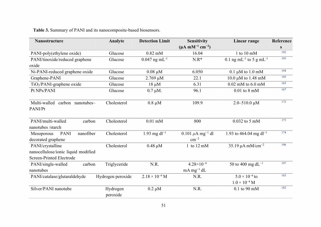
51
Table 3. Summary of PANI and its nanocomposite-based biosensors.
Nanostructure Analyte Detection Limit Sensitivity (μA mM−1 cm−2)
Linear range References
PANI-poly(ethylene oxide) Glucose 0.82 mM 16.04 1 to 10 mM 192 PANI/tinoxide/reduced graphene
oxide Glucose 0.047 ng mL-1 N.R* 0.1 ng mL-1 to 5 g mL-1 193
Ni-PANI-reduced graphene oxide Glucose 0.08 μM 6.050 0.1 μM to 1.0 mM 194 Graphene-PANI Glucose 2.769 μM 22.1 10.0 μM to 1.48 mM 195 TiO2/PANI-graphene oxide Glucose 18 μM 6.31 0.02 mM to 6.0 mM 165 Pt NPs/PANI
Glucose 0.7 μM. 96.1 0.01 to 8 mM 167
Multi-walled carbon nanotubes–PANI/Pt
Cholesterol 0.8 µM 109.9 2.0–510.0 μM 172
PANI/multi-walled carbon nanotubes /starch
Cholesterol 0.01 mM 800 0.032 to 5 mM 173
Mesoporous PANI nanofiber decorated graphene
Cholesterol 1.93 mg dl−1 0.101 μA mg−1 dl cm−2
1.93 to 464.04 mg dl−1 174
PANI/crystalline nanocellulose/ionic liquid modified Screen-Printed Electrode
Cholesterol 0.48 μM 1 to 12 mM 35.19 μA mM/cm−2 196
PANI/single‐walled carbon nanotubes
Triglyceride N.R. 4.28×10⁻⁴ mA mg−1 dL
50 to 400 mg dL−1 197
PANI/catalase/glutaraldehyde
Hydrogen peroxide 2.18 × 10⁻⁶ M N.R. 5.0 × 10⁻⁶ to 1.0 × 10⁻⁴ M
183
Silver/PANI nanotube Hydrogen peroxide
0.2 μM N.R. 0.1 to 90 mM 182

52
PANI nanotubes Hydrogen peroxide
3.56 × 10⁻¹⁰ mg/mL
N.R. 1.0 × 10⁻⁹ to 0.10 mg/mL
198
Gold NPs doped poly(8-anilino-1-naphthalene sulphonic acid)
Tyramine (a phenolic compound)
0.71 µM N.R. 10 to 120 µM 199
Carbon nanotubes-PANI nanohybrid
DNA 0.33 fM N.R. 1 fM to 10 nM 186
Gold NPs/PANI DNA 0.01 fM N.R. 0.001 to 1000 pM 200 Gold electrode modified with Au
and PANI NPs DNA 2.5×10⁻¹⁰ M N.R 10-9to 1×10⁻⁶ M 201
PANI nanotubes
Nucleic acid 10−16 M 1 × 10⁻¹⁶ M 10−6 to 10−16 M 187
Potassium ferricyanide-doped PANI NPs
Carcinoem-bryonic antigen
0.1 pg mL−1 N.R. 1.0 pg mL−1 to 500.0 ng mL−1
189
Graphene/PANI-Modified Screen-Printed Au electrode
Tuberculosis 15 ng/mL N.R. 20 to 100 ng/mL 191
PANI/Au NPs Acrylamide 5.0×10⁻¹¹M N.R. 5.0×10⁻¹⁰ M to 2.0×10⁻⁷ M
202
Polyamidoamine grafted multiwalled carbon nanotube
Urea 0.4 mM 6.6 nA/mM 1 to 20 mM 203
PANI-cladding modified fiber Urea 100 nM N.R 100 nM to 100 mM 204 PANI-encapsulated carbon/Cu
composite nanofibers Polyphenol 0.24 μM 41.65 μA mM−1 500 nM to 110 μM 205
Poly(carboxybetaine) functionalized PANI nanowires
Carcinoembryonic antigen
3.05 fg mL−1 N.R. 1.0 × 10⁻¹⁴ to 1.0 × 10⁻¹⁰ g mL−1
206

53
Peptide nanotube-Au nanoparticles-PANI immobilized pencil graphite electrode
Prostate specific antigen
0.68 ng/mL N.R. 1 to 100 ng/mL 207
Glassy carbon electrode/PANI-ZnO NPs
Dopamine 0.153 × 10⁻⁷ M 0.089 µA/µM
0.2 to 2.4 μM 208
Glassy carbon electrode/PANI-Fe3O4 NPs
Dopamine 0.176 × 10⁻⁷ M 0.058 µA/µM 0.2 to 2.4 μM 208
Glassy carbon electrode/PANI-NiO NPs
Dopamine 0.166 × 10⁻⁷ M 0.078 µA/µM 0.2 to 2.4 μM 208
Molybdenum disulfide nanosheets-PANI/Au NPs
Dopamine 1 × 10⁻⁶ M 0.0274 µA/µM 1 to 500 μM 209
Graphene nanosheets/Pt/PANI Dopamine 3.33 × 10⁻⁶ M 1.53 and 0.35 µA/µM 2.0 to 10 and 40 to 400
210
Nitrogen-doped functionalized graphene/Ag NPs/PANI
Ascorbic acid (vitamin C)
8 µM N.R 10 to 11.460 µM 211
Indium tin oxide coated glass substrate/manganese phthalocyanine/PANI
Fenitrothion (a pesticide)
0.049 µmol dm-3 4.67 Acm-2M-1 0.12 to 15.00 µmol dm-3 212
*N.R.: Not reported

54
7. Conclusion and perspectives
This review presents a comprehensive overview of polyaniline and its nanocomposites,
underlining advantages and properties, from structure, synthesis, physicochemical properties,
to their potential in a wide spectrum of biomedical applications. Conductive polyaniline-based
nanocomposites surely represent a new and promising, but not yet completely explored, area
of biomaterial science where promising results and unresolved technology challenges both call
for opportunities of deeper studies and developed researches. Polyanilines are, indeed, gaining
a continuously increasing interest from the scientific community. However, it should be
mentioned from an industrial perspective that some of these challenges have also important
drawbacks on regulatory and quality assurance issues: this widely explains the relatively low
amount of clinical data related to the use of PANI.
There are some major limitations such as cytotoxicity, processability, significant difference
of in-vivo and in-vitro studies, and physicochemical properties on the applications of PANI and
its nanocomposites into clinical practice. However, the low physical properties and
biocompatibility of PANI is the main challenge in the tissue engineering applications,
especially those that are aimed to be employed inside of the human body, e.g. for the bone
regeneration. This issue has been moderately solved with the fabrication of PANI composites
containing nanostructures, including Ag and TiO2, and/or , ,
, which improves their mechanical properties and
integration into the biological tissues. Besides, non-biodegradability of the pure PANI
scaffolds still restricts their applications in the tissue regeneration. Thus, a great consideration
has been focused on the development of the degradable PANI blends and composites with bio-
based polymers.

55
Author information
Corresponding Authors
Email: [email protected] & [email protected] (E.N. Zare);
[email protected] (P. Makvandi)
ORCID
Ehsan Nazarzadeh Zare: 0000-0002-0446-4385
Pooyan Makvandi: 0000-0003-2456-0961
Notes
The authors declare no competing financial interest.
Biographies
Ehsan Nazarzadeh Zare received his PhD in polymer chemistry from University of
Mazandaran, Iran, in 2015. He was a postdoctoral research fellow at the University of
Mazandaran from May 2015 to May 2016. Currently, he is an assistant professor in polymer
chemistry at Damghan University, Damghan, Iran. His current research interests include
biomaterials, conductive polymers and nanocomposites, biological activities of
nanocomposites, synthesis and characterization of nanomaterials.
Pooyan Makvandi received his PhD in polymer chemistry from University of Mazandaran,
Iran, in 2017. Prior to completing PhD, he was a visiting scholar at University of Salento, Italy,
for 6 months. Currently, he is working at Institute for Polymers, Composites, and Biomaterials

56
(IPCB), National Research Council (CNR) of Italy. His works focus on biomaterials for tissue
regeneration, antimicrobial compounds, dental materials, nanostructures and nanocomposites.
Behnaz Ashtari received her PhD in nanobiotechnology from the University of Tarbiat
Modaress, Tehran-Iran in 2012. She is currently working as an assistant professor at Iran
University of Medical Sciences since December 2014. Her expertise is mainly focused on the
design, synthesis, and evaluation of nanomaterials and microfluidic systems for various
medical applications.
Filippo Rossi is Assistant Professor in Applied Physical Chemistry at Politecnico di
Milano, Milano, Italy. He has worked in the field of innovative polymer materials for
nanomedicine, drug delivery and tissue engineering with experimental and model studies.
Ahmad Motahari received his PhD in physical chemistry from the University of
Mazandaran, Iran. He has worked on the structural studies of nanoporous materials and
nanocomposites using small angle X-ray scattering (SAXS) as a visiting research scholar at the
University of Cincinnati, USA. Also, he has worked as a postdoctoral researcher at Sharif
University of Technology, Iran. His current research interests are polymer nanocomposites,
nanoporous materials, and computational chemistry.
Giuseppe Perale is full professor in biomaterials at University of Applied Sciences and
Arts of Southern Switzerland, Switzerland. He has worked in the field of materials and
technologies for biomedical applications, particularly in the field of tissue engineering and
controlled drug delivery. His research and experimental activities are devoted to theoretical
investigations and development of feasible solutions for in vivo human applications, aiming
both at achieving new knowledge and developing industrially-scaled processes for applicable
medical devices.
57
Abbreviation used
APS, Ammonium persulfate; CNTs, Carbon nano-tubes; CTE, Cardiac tissue engineering;
CS-g-AT, Chitosan-graft-aniline tetramer; DMF, Dimethyl formamide; DMSO, Dimethyl
sulfoxide; DPPH, 1,1-diphenyl-2-picrylhydrazyl; EC50, Half maximal effective concentration;
EM, Emeraldine; EM-B, Emeraldine-base; EM-S, Emeraldine-salt; ER, Electrorheological;
GNS, Graphene nanosheets; GS, Gentamicin sulfate; HaCaT, Human keratinocyte; HEK,
Human embryonic kidney, HMSC, Human mesenchymal stem cells; ICPs, Inherently
conducting polymers; KPS, Potassium persulfate; LE, Leucoemeraldine; MAA, Methacrylic
acid; MAPLE, Matrix-assisted pulsed laser evaporation; MMP, Mitochondrial membrane
potential; MPP, Modified polypropylene; MTT, 3-(4,5-dimethylthiazol-2-yl)-2,5-
diphenyltetrazolium bromide; NIH/3T3, Mouse embryonic fibroblast; NIR, Near-infrared;
NMP, N-methyl-2-pyrrolidone; ND, Nanodiamond; NPs, Nanoparticles; PANI, Polyaniline;
P3ABA, Poly(3-aminobenzoic acid); Pas, Polyacetylene; PC12, Pheochromocytoma cells;
PCL, Polycaprolactone; PEGDA, Polyethyleneglycol diacrylate; PEG-DA, Dibenzaldehyde-
terminated poly(ethylene glycol); PEO, Poly (ethylene oxide); PHB, Poly-3-hydroxybutyrate;
PFu, Polyfuran; PG, Pernigraniline; PLA, Poly(lactic acid); PLGA, Poly(lactic-co-glycolic
acid); PNIPAm, Poly(N-isopropylacrylamide); PPy, Polypyrrole; PSiNPs, Porous silicon
nanoparticles; PTh, Polythiophene; pTSA, Para-toluene sulfonic acid; PUF, Polyurethane
foam; PXT, Polyxanthonetriazole; rGO, Reduced graphene oxide; ROS, Reactive oxygen
species; RRS, , ; SDDS, Smart
drug delivery systems; STPU, Siloxane terminated polyurethane; THF, Tetrahydrofuran; UA,
Usnic acid;

58
References
(1) Su, W. Principles of Polymer Design and Synthesis, 1st ed.; Carpenter, B., Ceroni, P.,
Kirchner, B., Koskinen, A., Landfester, K., Leszczynski, J., Luh, T., Mahlke, C., N. C.
Polfer, Gainesville FL, U., R. Salzer, Dresden, G., Eds.; Springer Heidelberg: New
York, 2013.
(2) Huang, J.; B. Kaner, R. Conjugated Polymers: Theory, Synthesis, Properties, and
Characterization, 3rd ed.; Skotheim, T. A., Reynolds, J., Eds.; CRC press: Boca Raton,
2006.
(3) Kiristi, M.; Uygun, A. Polyaniline. In Handbook of Engineering and Specialty
Thermoplastics; Thomas, S., Visakh, P. M., Eds.; Wiley Online Library: Salem,
Massachusetts., 2011; Vol. 4, pp 183–210.
(4) Zare, E. N.; Lakouraj, M. M.; Moghadam, P. N.; Azimi, R. Novel
Polyfuran/Functionalized Multiwalled Carbon Nanotubes Composites With Improved
Conductivity: Chemical Synthesis, Characterization, and Antioxidant Activity. Polym.
Compos. 2013, 34, 732–739.
(5) Kaur, G.; Adhikari, R.; Cass, P.; Bown, M.; Gunatillake, P. Electrically Conductive
Polymers and Composites for Biomedical Applications. RSC Adv. 2015, 5, 37553–
37567.
(6) Norouzian, R.-S.; Lakouraj, M. M.; Zare, E. N. Novel Conductive PANI/Hydrophilic
Thiacalix[4]Arene Nanocomposites: Synthesis, Characterization and Investigation of
Properties. Chinese J. Polym. Sci. 2014, 32 (2), 218–219.
(7) Moghadam, P. N.; Zareh, E. N. Synthesis of Conductive Nanocomposites Based on
Polyaniline/Poly(Styrene- Alt-Maleic Anhydride)/Polystyrene. E-Polymers 2010, 054,
1–10.
(8) Oves, M.; Shahadat, M.; Ansari, S. A.; Aslam, M. Polyaniline Nanocomposite Materials
for Biosensor Designing. In Electrically Conductive Polymer and Polymer Composites:
From Synthesis to Biomedical Applications; Khan, A., Jawaid, M., Khan, A. A. P., Asiri,
A. M., Eds.; Wiley-VCH Verlag: Weinheim, 2018; pp 113–135.
(9) Nezakati, T.; Seifalian, A.; Tan, A.; Seifalian, A. M. Conductive Polymers :
Opportunities and Challenges in Biomedical Applications. Chem. Rev. 2018, 118, 6766–
6843.
(10) Bhandari, S. Polyaniline: Structure and Properties Relationship. In Polyaniline Blends,
Composites, and Nanocomposites; P.M. Visakh, Pina, C. Della, Falletta, E., Eds.;
Elsevier: Amsterdam, Netherlands, 2018; pp 23–60.

59
(11) Itoi, H.; Hayashi, S.; Matsufusa, H.; Ohzawa, Y. Electrochemical Synthesis of
Polyaniline in the Micropores of Activated Carbon for High-Performance
Electrochemical Capacitors. Chem. Commun. 2017, 53 (22), 3201–3204.
(12) Menon, R.; Mukherjee, A. K. Polyaniline Fractal Nanocomposites. Encycl. Nanosci.
Nanotechnol. 2004, 8, 715–729.
(13) Freitas, T. V; Sousa, E. A.; Jr, G. C. F.; Arlindo, E. P. S. Different Morphologies of
Polyaniline Nanostructures Synthesized by Interfacial Polymerization. Mater. Lett.
2018, 224, 42–45.
(14) Tian, Z.; Yu, H.; Wang, L.; Saleem, M.; Ren, F.; Ren, P. Recent Progress in the
Preparation of Polyaniline Nanostructures and Their Applications in Anticorrosive
Coatings. RSC Adv. 2014, 4, 28195–28208.
(15) Peng, H.; Ma, G.; Sun, K.; Mu, J.; Lei, Z. A Novel Fabrication of Nitrogen-Containing
Carbon Nanospheres with High Rate Capability as Electrode Materials for
Supercapacitors. RSC Adv. 2015, 5, 12034–12042.
(16) Dong, Y.; Zhou, Y.; Ding, Y.; Wang, C. Sensitive Detection of Pb(II) at Gold
Nanoparticle/ Polyaniline/Graphene Modified Electrode Using Differential Pulse
Anodic Stripping Voltammetry. Anal. Methods 2014, 6, 9367–9374.
(17) Zujovic, Z. D.; Bowmaker, G. A.; Tran, H. D.; Kaner, R. B. Solid-State NMR of
Polyaniline Nanofibers. Synth. Met. 2009, 159, 710–714.
(18) Sim, B.; Choi, H. J. Facile Synthesis of Polyaniline Nanotubes and Their Enhanced
Stimuli-Response under Electric Field. RSC Adv. 2015, 5, 11905–11912.
(19) Yin, C.; Gao, L.; Zhou, F.; Duan, G. Facile Synthesis of Polyaniline Nanotubes Using
Self-Assembly Method Based on the Hydrogen Bonding : Mechanism and Application
in Gas Sensing. Polymers (Basel). 2017, 9 (544), 1–14.
(20) Moghadam, P. N.; Zare, E. N.; Amiri, H.; Lakouraj, M. M. Preparation of Conductive
Nanocomposites Based on Poly (Aniline- Co- Butyl 3-Aminobenzoate) and Poly
(Aniline-Co-Ethyl 3-Aminobenzoate) by Solution Blending Method. Compos.
Interfaces 2012, 19 (8), 457–488.
(21) I. Kong, K. Y. Tshai, M. E. H. Manufacturing of Natural Fibre-Reinforced Polymer
Composites by Solvent Casting Method. In Manufacturing of natural fibre reinforced
polymer composites; Salit, M. S., Jawaid, M., Yusoff, N. Bin, Hoque, M. E., Eds.;
Springer: Switzerland, 2015; pp 331–349.
(22) Zare, E. N.; Lakouraj, M. M. Biodegradable Polyaniline / Dextrin Conductive
Nanocomposites : Synthesis , Characterization , and Study of Antioxidant Activity and

60
Sorption of Heavy Metal Ions. Iran. Polym. J. 2014, 23, 257–266.
(23) Zhan, C.; Yu, G.; Lu, Y.; Wang, L.; Wujcik, E.; Wei, S. Conductive Polymer
Nanocomposites: A Critical Review of Modern Advanced Devices. J. Mater. Chem. C
2017, 5 (7), 1569–1585.
(24) Pierini, F.; Lanzi, M.; Nakielski, P.; Kowalewski, T. A. Electrospun Polyaniline-Based
Composite Nanofibers: Tuning the Electrical Conductivity by Tailoring the Structure of
Thiol-Protected Metal Nanoparticles. J. Nanomater. 2017, 2017, 1–10.
(25) Kaur, M.; Malik, B.; Garg, T.; Rath, G.; Goyal, A. K. Development and Characterization
of Guar Gum Nanoparticles for Oral Immunization against Tuberculosis. Drug Deliv.
2015, 22 (3), 328–334.
(26) Khalid, M.; Tumelero, M. A.; Brandt, I. S.; Zoldan, V. C.; Acuña, J. J. S.; Pasa, A. A.
Electrical Conductivity Studies of Polyaniline Nanotubes Doped with Different Sulfonic
Acids. Indian J. Mater. Sci. 2013, 2013, 1–7.
(27) Kakde, K. P. Synthesis and Characterization of Polyaniline Doped with HCl , H2SO4
and PVA as Secondary Dopant for Toxic Gas ( Ammonia ) Sensor. Indian J. Sci.
Technol. 2017, 10, 1–4.
(28) Zare, E. N.; Abdollahi, T.; Motahari, A. Effect of Functionalization of Iron Oxide
Nanoparticles on the Physical Properties of Poly (Aniline-Co-Pyrrole) Based
Nanocomposites: Experimental and Theoretical Studies. Arab. J. Chem. 2018.
https://doi.org/10.1016/j.arabjc.2018.04.016.
(29) Gholami, H.; Shakeri, A.; Saadattalab, V. Investigation of Physical and Mechanical
Properties of Polyaniline/MMT Nanocomposites. Curr. Chem. Lett. 2017, 6, 151–158.
(30) Bilal, S.; Arif, M.; Khan, M. S.; Shah, A.-H. A. Characterization of Sodium and
Potassium Nitrate Contaminated Polyaniline-Poly (Ethylene Oxide) Composites
Synthesized via Facile Solution Casting Technique. Materials (Basel). 2019, 12 (13),
2168–2182.
(31) Gangopadhyay, R. Exploring Rheological Properties of Aqueous Polyaniline‐PVP
Dispersion. J. Polym. Sci. Part B Polym. Phys. 2008, 46 (22), 2443–2455.
(32) Wang, H.; Wen, H.; Hu, B.; Fei, G.; Shen, Y.; Sun, L.; Yang, D. Facile Approach to
Fabricate Waterborne Polyaniline Nanocomposites with Environmental Benignity and
High Physical Properties. Sci. Rep. 2017, 7, 43694(1-12).
(33) Sirqueira, A. da S.; Teodoro Júnior, D.; Coutinho, M. da S.; Neto, S.; Silva, A. dos A.;
Soares, B. G. Rheological Behavior of Acrylic Paint Blends Based on Polyaniline.
Polímeros 2016, 26 (3), 215–220.

61
(34) Makvandi, P.; Ali, G. W.; Della Sala, F.; Abdel-Fattah, W. I.; Borzacchiello, A.
Biosynthesis and Characterization of Antibacterial Thermosensitive Hydrogels Based
on Corn Silk Extract, Hyaluronic Acid and Nanosilver for Potential Wound Healing.
Carbohydr. Polym. 2019, 223, 115023-115035.
(35) Zhao, X.; Li, P.; Guo, B.; Ma, P. X. Antibacterial and Conductive Injectable Hydrogels
Based on Quaternized Chitosan-Graft-Polyaniline / Oxidized Dextran for Tissue
Engineering. Acta Biomater. 2015, 26, 236–248.
(36) Zhao, X.; Wu, H.; Guo, B.; Dong, R.; Qiu, Y.; Ma, P. X. Antibacterial Anti-Oxidant
Electroactive Injectable Hydrogel as Self-Healing Wound Dressing with Hemostasis
and Adhesiveness for Cutaneous Wound Healing. Biomaterials 2017, 122, 34–47.
(37) Fang, F. F.; Lee, B. M.; Choi, H. J. Electrorheologically Intelligent Polyaniline and Its
Composites. Macromol. Res. 2010, 18 (2), 99–112.
(38) Kaur, A.; Kaur, A.; Saini, D. A Review on Synthesis of Silica Nanocomposites With
Conducting Polymers: Polyaniline. Res. Cell Int. J. Eng. Sci 2016, 6913, 40–53.
(39) Zareh, E. N.; Moghadam, P. N.; Azariyan, E.; Sharifian, I. Conductive and
Biodegradable Polyaniline/Starch Blends and Their Composites With Polystyrene. Iran.
Polym. J. 2011, 20 (4), 319–328.
(40) Guo, B.; Glavas, L.; Albertsson, A. Biodegradable and Electrically Conducting
Polymers for Biomedical Applications. Prog. Polym. Sci. 2013, 38, 1263–1286.
(41) Guo, B.; Ma, P. X. Synthetic Biodegradable Functional Polymers for Tissue
Engineering: A Brief Review. Sci. China Chem. 2014, 57 (4), 490–500.
(42) Xia, B.; Wang, B.; Shi, J.; Zhang, Y.; Zhang, Q.; Chen, Z.; Li, J. Photothermal and
Biodegradable Polyaniline / Porous Silicon Hybrid Nanocomposites as Drug Carriers
for Combined Chemo-Photothermal Therapy of Cancer. Acta Biomater. 2017, 51, 197–
208.
(43) Luo, Y.-L.; Nan, Y.-F.; Xu, F.; Chen, Y.-S.; Zhao, P. Degradation Behavior and
Biocompatibility of PEG/PANI-Derived Polyurethane Co-Polymers. J. Biomater. Sci.
Polym. Ed. 2010, 21 (8–9), 1143–1172.
(44) Sarvari, R.; Akbari-Alanjaraghi, M.; Massoumi, B.; Beygi-Khosrowshahi, Y.;
Agbolaghi, S. Conductive and Biodegradable Scaffolds Based on a Five-Arm and
Functionalized Star-like Polyaniline–polycaprolactone Copolymer with Ad-Glucose
Core. New J. Chem. 2017, 41 (14), 6371–6384.
(45) Humpolicek, P.; Kasparkova, V.; Saha, P.; Stejskal, J. Biocompatibility of Polyaniline.
Synth. Met. 2012, 162 (7–8), 722–727.

62
(46) Humpolíček, P.; Kašpárková, V.; Pacherník, J.; Stejskal, J.; Bober, P.; Capáková, Z.;
Radaszkiewicz, K. A.; Junkar, I.; Lehocký, M. The Biocompatibility of Polyaniline and
Polypyrrole: A Comparative Study of Their Cytotoxicity, Embryotoxicity and Impurity
Profile. Mater. Sci. Eng. C 2018, 91 (May), 303–310.
(47) Zhang, X.; Qi, H.; Wang, S.; Feng, L.; Ji, Y.; Tao, L.; Li, S.; Wei, Y. Cellular Responses
of Aniline Oligomers: A Preliminary Study. Toxicol. Res. (Camb). 2012, 1 (3), 201–205.
(48) Bairi, V. G.; Bourdo, S. E.; Moore, J. A.; Schnackenberg, L. K.; Berry, B. C.; Biris, A.
S.; Viswanathan, T. Separation and Spectroscopic/Molecular Weight Analysis of Crude
and Purified Polyaniline (S). J. Polym. Res. 2013, 20 (7), 193–201.
(49) Stejskal, J.; Hajná, M.; Humpolíˇ, P.; Zhigunov, A.; Trchová, M. Purification of a
Conducting Polymer, Polyaniline, for Biomedical Applications. Synth. Met. 2014, 195,
286–293.
(50) Liu, S.; Wang, J.; Zhang, D.; Zhang, P.; Ou, J.; Liu, B.; Yang, S. Investigation on Cell
Biocompatible Behaviors of Polyaniline Film Fabricated via Electroless Surface
Polymerization. Appl. Surf. Sci. 2010, 256 (11), 3426–3430.
(51) Humpolíček, P.; Radaszkiewicz, K. A.; Capáková, Z.; Pacherník, J.; Bober, P.;
Kašpárková, V.; Rejmontová, P.; Lehocký, M.; Ponížil, P.; Stejskal, J. Polyaniline
Cryogels: Biocompatibility of Novel Conducting Macroporous Material. Sci. Rep. 2018,
8 (1), 1–12.
(52) Kucekova, Z.; Humpolicek, P.; Kasparkova, V.; Perecko, T. Colloidal Polyaniline
Dispersions: Antibacterial Activity, Cytotoxicity and Neutrophil Oxidative Burst.
Colloids surfaces B Biointerfaces 2014, 116, 411–417.
(53) Li, Y.; Chen, B.; Li, X.; Zhang, W. K.; Tang, H. Cytotoxicity of Polyaniline
Nanomaterial on Rat Celiac Macrophages In Vitro. PLoS One 2014, 9 (9), 1–6.
(54) Ibarra, L. E.; Tarres, L.; Bongiovanni, S.; Barbero, C. A.; Kogan, M. J.; Rivarola, V. A.;
Bertuzzi, M. L.; Yslas, E. I. Assessment of Polyaniline Nanoparticles Toxicity and
Teratogenicity in Aquatic Environment Using Rhinella Arenarum Model. Ecotoxicol.
Environ. Saf. 2015, 114, 84–92.
(55) Yslas, E. I.; Ibarra, L. E.; Peralta, D. O.; Barbero, C. A.; Rivarola, V. A.; Bertuzzi, M.
L. Polyaniline Nanofibers: Acute Toxicity and Teratogenic Effect on Rhinella Arenarum
Embryos. Chemosphere 2012, 87 (11), 1374–1380.
(56) Rossetto, A. L. de O. F.; Vicentini, D. S.; Costa, C. H.; Melegari, S. P.; Matias, W. G.
Synthesis, Characterization and Toxicological Evaluation of a Core-Shell Copper
Oxide/Polyaniline Nanocomposite. Chemosphere 2014, 108, 107–114.

63
(57) Saikia, J. P.; Banerjee, S.; Konwar, B. K.; Kumar, A. Biocompatible Novel
Starch/Polyaniline Composites: Characterization, Anti-Cytotoxicity and Antioxidant
Activity. Colloids Surfaces B Biointerfaces 2010, 81 (1), 158–164.
(58) Maráková, N.; Humpolíček, P.; Kašpárková, V.; Capáková, Z.; Martinková, L.; Bober,
P.; Trchová, M.; Stejskal, J. Antimicrobial Activity and Cytotoxicity of Cotton Fabric
Coated with Conducting Polymers, Polyaniline or Polypyrrole, and with Deposited
Silver Nanoparticles. Appl. Surf. Sci. 2017, 396, 169–176.
(59) Villalba, P.; Ram, M. K.; Gomez, H.; Bhethanabotla, V.; Helms, M. N.; Kumar, A.;
Kumar, A. Cellular and in Vitro Toxicity of Nanodiamond-Polyaniline Composites in
Mammalian and Bacterial Cell. Mater. Sci. Eng. C 2012, 32 (3), 594–598.
(60) Tiwari, A.; Sharma, Y.; Hattori, S.; Terada, D.; Sharma, A. K.; Turner, A. P. F.;
Kobayashi, H. Influence of Poly(n-Isopropylacrylamide)-CNT-Polyaniline Three-
Dimensional Electrospun Microfabric Scaffolds on Cell Growth and Viability.
Biopolymers 2013, 99 (5), 334–341.
(61) Nam, H.; An, T.; Lim, G. Cell Behaviour on a Polyaniline Nanoprotrusion Structure
Surface. Nanoscale Res. Lett. 2014, 9 (1), 566–572.
(62) Shahadat, M.; Khan, M. Z.; Rupani, P. F.; Embrandiri, A.; Sultana, S.; Ahammad, S. Z.;
Ali, S. W.; Sreekrishnan, T. R. A Critical Review on the Prospect of Polyaniline-Grafted
Biodegradable Nanocomposite. Adv. Colloid Interface Sci. 2017, 249, 2–16.
(63) Gizdavic-nikolaidis, M. R.; Bennett, J. R.; Swift, S.; Easteal, A. J.; Ambrose, M. Broad
Spectrum Antimicrobial Activity of Functionalized Polyanilines. Acta Biomater. 2011,
7 (12), 4204–4209.
(64) Hou, Y.; Feng, J.; Wang, Y.; Li, L. Enhanced Antibacterial Activity of Ag-Doped ZnO
/ Polyaniline Nanocomposites. J. Mater. Sci. Mater. Electron. 2016, 27 (7), 6615–6622.
(65) Qi, K.; Cheng, B.; Yu, J.; Ho, W. Review on the Improvement of the Photocatalytic and
Antibacterial Activities of ZnO. J. Alloys Compd. 2017, 727, 792–820.
(66) Tanushree Sen, Satyendra Mishra, N. G. S. Synthesis and Sensing Applications of
Polyaniline Nanocomposites : A Review. RSC Adv. 2016, 6, 42196–42222.
(67) Youssef, A. M.; Moustafa, H. A.; Barhoum, A.; Hakim, A. E. A. A.; Dufresne, A.
Evaluation of the Morphological, Electrical and Antibacterial Properties of Polyaniline
Nanocomposite Based on Zn/Al‐Layered Double Hydroxides. ChemistrySelect 2017, 2
(27), 8553–8566.
(68) Liang, X.; Govindaraju, S.; Yun, K. Dual Applicability of Polyaniline Coated Gold
Nanorods: A Study of Antibacterial and Redox Activity. BioChip J. 2018, 12 (2), 137–

64
145.
(69) Zhao, S.; Huang, L.; Tong, T.; Zhang, W.; Wang, Z.; Wang, J.; Wang, S. Antifouling
and Antibacterial Behavior of Polyethersulfone Membrane Incorporating Polyaniline@
Silver Nanocomposites. Environ. Sci. Water Res. Technol. 2017, 3 (4), 710–719.
(70) Zahed, F. M.; Hatamluyi, B.; Lorestani, F.; Es’haghi, Z. Silver Nanoparticles Decorated
Polyaniline Nanocomposite Based Electrochemical Sensor for the Determination of
Anticancer Drug 5-Fluorouracil. J. Pharm. Biomed. Anal. 2018, 161, 12–19.
(71) Eren, O.; Ucar, N.; Onen, A.; Kizildag, N.; Karacan, I. Synergistic Effect of Polyaniline,
Nanosilver, and Carbon Nanotube Mixtures on the Structure and Properties of
Polyacrylonitrile Composite Nanofiber. J. Compos. Mater. 2016, 50 (15), 2073–2086.
(72) Masim, F. C. P.; Tsai, C.-H.; Lin, Y.-F.; Fu, M.-L.; Liu, M.; Kang, F.; Wang, Y.-F.
Synergistic Effect of PANI–ZrO2 Composite as Antibacterial, Anti-Corrosion, and
Phosphate Adsorbent Material: Synthesis, Characterization and Applications. Environ.
Technol. 2019, 40 (2), 226–238.
(73) Monjezi, J.; Jamaledin, R.; Ghaemy, M.; Makvandi, P. Antimicrobial Modified-
Tragacanth Gum / Acrylic Acid Hydrogels for the Controlled Release of Quercetin. J.
Appl. Chem. Res. 2019, 131 (1), 57–71.
(74) Makvandi, P.; Corcione, C. E.; Paladini, F.; Gallo, A. L.; Montagna, F.; Maffezzoli, A.
Antimicrobial Modified Hydroxyapatite Composite Dental Bite by Stereolithography.
Polym Adv Technol 2018, 29, 364–371.
(75) Yan, L.; Wang, R.; Wang, H.; Sheng, K.; Liu, C.; Qu, H.; Ma, A.; Zheng, L. Formulation
and Characterization of Chitosan Hydrochloride and Carboxymethyl Chitosan
Encapsulated Quercetin Nanoparticles for Controlled Applications in Foods System and
Simulated Gastrointestinal Condition. Food Hydrocoll. 2018, 84, 450–457.
(76) Lukman, S. K.; Saidin, S. Antibacterial Effect of Ginseng/Polyaniline Encapsulated in
Poly (Lactic-Co-Glycolic Acid) Microcapsules Coating on Stainless Steel 316L. In
MATEC Web of Conferences; EDP Sciences, 2019; Vol. 253, p 3001.
(77) Robertson, J.; Gizdavic-Nikolaidis, M.; Swift, S. Investigation of Polyaniline and a
Functionalised Derivative as Antimicrobial Additives to Create Contamination Resistant
Surfaces. Materials (Basel). 2018, 11 (3), 436–461.
(78) Robertson, J.; Gizdavic-Nikolaidis, M.; Nieuwoudt, M. K.; Swift, S. The Antimicrobial
Action of Polyaniline Involves Production of Oxidative Stress While Functionalisation
of Polyaniline Introduces Additional Mechanisms. PeerJ 2018, 6, e5135–e5171.
(79) Hasantabar, V.; Lakouraj, M. M.; Zare, E. N.; Mohseni, M. Synthesis, Characterization,

65
and Biological Properties of Novel Bioactive Poly(Xanthoneamide-Triazole-
Ethersulfone) and Its Multifunctional Nanocomposite with Polyaniline. Adv. Polym.
Technol. 2017, 36 (3), 309–319.
(80) Gizdavic-nikolaidis, M.; Travas-sejdic, J.; Bowmaker, G. A.; Cooney, R. P.; Thompson,
C.; Kilmartin, P. A. The Antioxidant Activity of Conducting Polymers in Biomedical
Applications. Curr. Appl. Phys. 2004, 4, 347–350.
(81) Prasad, J.; Banerjee, S.; Kumar, B.; Kumar, A. Biocompatible Novel Starch / Polyaniline
Composites : Characterization , Anti-Cytotoxicity and Antioxidant Activity. Colloids
Surfaces B Biointerfaces 2010, 81 (1), 158–164.
(82) Kumar, A.; Banerjee, S.; Saikia, J. P.; Konwar, B. K. Swift Heavy Ion Irradiation
Induced Enhancement in the Antioxidant Activity and Biocompatibility of Polyaniline
Nanofibers. Nanotechnology 2010, 21, 175102–175110.
(83) Parsa, A.; Salout, S. A. Investigation of the Antioxidant Activity of Electrosynthesized
Polyaniline / Reduced Graphene Oxide Nanocomposite in a Binary Electrolyte System
on ABTS and DPPH Free Radicals. J. Electroanal. Chem. 2016, 760, 113–118.
(84) Zare, E. N.; Motahari, A.; Sillanpää, M. Nanoadsorbents Based on Conducting Polymer
Nanocomposites with Main Focus on Polyaniline and Its Derivatives for Removal of
Heavy Metal Ions/Dyes: A Review. Environmental Research. 2018, pp 173–195.
(85) Bertuoli, P. T.; Baldissera, A. F.; Zattera, A. J.; Ferreira, C. A. Progress in Organic
Coatings Polyaniline Coated Core-Shell Polyacrylates : Control of Fi Lm Formation and
Coating Application for Corrosion Protection. Prog. Org. Coatings 2019, 128, 40–51.
(86) Yassin, M. A.; Shrestha, S.; Park, C. H.; Kim, C. S. Exfoliated Nanosheets of Co3O4
Webbed with Polyaniline Nanofibers: A Novel Composite Electrode Material for
Enzymeless Glucose Sensing Application. J. Ind. Eng. Chem. 2019, 73, 106–117.
(87) Srisuk, P.; Berti, F. V; da Silva, L. P.; Marques, A. P.; Reis, R. L.; Correlo, V. M.
Electroactive Gellan Gum/Polyaniline Spongy-like Hydrogels. ACS Biomater. Sci. Eng.
2018, 4 (5), 1779–1787.
(88) Ismayil, K. M. M.; Varghese, A.; Antony, R. Silver-Doped Polyaniline–polyvinyl
Chloride Nanocomposite Films for Photocatalytic and Antibacterial Applications. J.
Elastomers Plast. 2019. https://doi.org/10.1177/0095244318819238.
(89) Baker, C. O.; Huang, X.; Nelson, W.; Kaner, R. B. Polyaniline Nanofibers: Broadening
Applications for Conducting Polymers. Chem. Soc. Rev. 2017, 46 (5), 1510–1525.
(90) Jotiram, K. P.; Prasad, R. G. S. V; Jakka, V. S.; Aparna, R. S. L.; Phani, A. R.
Antibacterial Activity of Nanostructured Polyaniline Combined With Mupirocin. Nano
66
Biomed Eng 2012, 4 (3), 144–149.
(91) Wu, J. C.-C.; Ray, S.; Gizdavic-Nikolaidis, M.; Uy, B.; Swift, S.; Jin, J.; Cooney, R. P.
Nanostructured Bioactive Material Based on Polycaprolactone and Polyaniline Fiber-
Scaffolds. Synth. Met. 2014, 198, 41–50.
(92) Dong, R.; Zhao, X.; Guo, B.; Ma, P. X. Self-Healing Conductive Injectable Hydrogels
with Antibacterial Activity as Cell Delivery Carrier for Cardiac Cell Therapy. ACS Appl.
Mater. Interfaces 2016, 8 (27), 17138–17150.
(93) Deng, Z.; Guo, Y.; Zhao, X.; Ma, P. X.; Guo, B. Multifunctional Stimuli-Responsive
Hydrogels with Self-Healing, High Conductivity, and Rapid Recovery through Host–
guest Interactions. Chem. Mater. 2018, 30 (5), 1729–1742.
(94) Bhattarai, D. P.; Shrestha, S.; Shrestha, B. K.; Park, C. H.; Kim, C. S. A Controlled
Surface Geometry of Polyaniline Doped Titania Nanotubes Biointerface for
Accelerating MC3T3-E1 Cells Growth in Bone Tissue Engineering. Chem. Eng. J. 2018,
350, 57–68.
(95) dos Santos, M. R.; Alcaraz-Espinoza, J. J.; da Costa, M. M.; de Oliveira, H. P. Usnic
Acid-Loaded Polyaniline/Polyurethane Foam Wound Dressing: Preparation and
Bactericidal Activity. Mater. Sci. Eng. C 2018, 89, 33–40.
(96) Gharibi, R.; Yeganeh, H.; Gholami, H.; Hassan, Z. M. Aniline Tetramer Embedded
Polyurethane/Siloxane Membranes and Their Corresponding Nanosilver Composites as
Intelligent Wound Dressing Materials. RSC Adv. 2014, 4 (107), 62046–62060.
(97) Natarajan, T. S.; Tsai, C. H.; Huang, H. L.; Ho, K. S.; Lin, I.; Wang, Y. F. Fabrication
of Polyaniline Coated Plasma Modified Polypropylene Filter for Antibioaerosol
Application. Aerosol Air Qual. Res. 2016, 16 (8), 1911–1921.
(98) Zengin, H.; Aksin, G.; Zengin, G.; Kahraman, M.; Kilic, I. H. Preparation and
Characterization of Conductive Polyaniline/Silver Nanocomposite Films and Their
Antimicrobial Studies. Polym. Eng. Sci. 2019, 59 (S1), E182–E194.
(99) Suresh, A. K.; Pelletier, D. A.; Doktycz, M. J. Relating Nanomaterial Properties and
Microbial Toxicity. Nanoscale 2013, 5 (2), 463–474.
(100) Liu, D.; Yang, F.; Xiong, F.; Gu, N. The Smart Drug Delivery System and Its Clinical
Potential. Theranostics 2016, 6 (9), 1306.
(101) Ulbrich, K.; Hola, K.; Subr, V.; Bakandritsos, A.; Tucek, J.; Zboril, R. Targeted Drug
Delivery with Polymers and Magnetic Nanoparticles: Covalent and Noncovalent
Approaches, Release Control, and Clinical Studies. Chem. Rev. 2016, 116 (9), 5338–
5431.

67
(102) Tibbitt, M. W.; Dahlman, J. E.; Langer, R. Emerging Frontiers in Drug Delivery. J. Am.
Chem. Soc. 2016, 138 (3), 704–717.
(103) Sugianto, T. D.; Chan, H.-K. Inhaled Antibiotics in the Treatment of Non-Cystic
Fibrosis Bronchiectasis: Clinical and Drug Delivery Perspectives. Expert Opin. Drug
Deliv. 2016, 13 (1), 7–22.
(104) Dai, T.; Jiang, X.; Hua, S.; Wang, X.; Lu, Y. Facile Fabrication of Conducting Polymer
Hydrogels via Supramolecular Self-Assembly. Chem. Commun. 2008, No. 36, 4279–
4281.
(105) Dai, T.; Jia, Y. Supramolecular Hydrogels of Polyaniline-Poly (Styrene Sulfonate)
Prepared in Concentrated Solutions. Polymer (Guildf). 2011, 52 (12), 2550–2558.
(106) Li, W.; Zeng, X.; Wang, H.; Wang, Q.; Yang, Y. Polyaniline-Poly (Styrene Sulfonate)
Conducting Hydrogels Reinforced by Supramolecular Nanofibers and Used as Drug
Carriers with Electric-Driven Release. Eur. Polym. J. 2015, 66, 513–519.
(107) Chen, H.; Liu, Z.; Li, S.; Su, C.; Qiu, X.; Zhong, H.; Guo, Z. Fabrication of Graphene
and AuNP Core Polyaniline Shell Nanocomposites as Multifunctional Theranostic
Platforms for SERS Real-Time Monitoring and Chemo-Photothermal Therapy.
Theranostics 2016, 6 (8), 1096.
(108) Nguyen, H. T.; Dai Phung, C.; Thapa, R. K.; Pham, T. T.; Tran, T. H.; Jeong, J.-H.; Ku,
S. K.; Choi, H.-G.; Yong, C. S.; Kim, J. O. Multifunctional Nanoparticles as
Somatostatin Receptor-Targeting Delivery System of Polyaniline and Methotrexate for
Combined Chemo–photothermal Therapy. Acta Biomater. 2018, 68, 154–167.
(109) Silva, J. S. F.; Silva, J. Y. R.; de Sá, G. F.; Araújo, S. S.; Filho, M. A. G.; Ronconi, C.
M.; Santos, T. C.; Júnior, S. A. Multifunctional System Polyaniline-Decorated ZIF-8
Nanoparticles as a New Chemo-Photothermal Platform for Cancer Therapy. ACS omega
2018, 3 (9), 12147–12157.
(110) Wang, J.; Guo, F.; Yu, M.; Liu, L.; Tan, F.; Yan, R.; Li, N. Rapamycin/DiR Loaded
Lipid-Polyaniline Nanoparticles for Dual-Modal Imaging Guided Enhanced
Photothermal and Antiangiogenic Combination Therapy. J. Control. Release 2016, 237,
23–34.
(111) Neira-Carrillo, A.; Yslas, E.; Marini, Y. A.; Vásquez-Quitral, P.; Sánchez, M.; Riveros,
A.; Yáñez, D.; Cavallo, P.; Kogan, M. J.; Acevedo, D. Hybrid Biomaterials Based on
Calcium Carbonate and Polyaniline Nanoparticles for Application in Photothermal
Therapy. Colloids Surfaces B Biointerfaces 2016, 145, 634–642.
(112) You, C.; Wu, H.; Wang, M.; Wang, S.; Shi, T.; Luo, Y.; Sun, B.; Zhang, X.; Zhu, J. A

68
Strategy for Photothermal Conversion of Polymeric Nanoparticles by Polyaniline for
Smart Control of Targeted Drug Delivery. Nanotechnology 2017, 28 (16), 165102.
(113) Rana, S.; Jadhav, N. V; Barick, K. C.; Pandey, B. N.; Hassan, P. A. Polyaniline Shell
Cross-Linked Fe3O4 Magnetic Nanoparticles for Heat Activated Killing of Cancer
Cells. Dalt. Trans. 2014, 43 (32), 12263–12271.
(114) Gao, Z.; You, C.; Wu, H.; Wang, M.; Zhang, X.; Sun, B. FA and CRGD Dual Modified
Lipid-Polymer Nanoparticles Encapsulating Polyaniline and Cisplatin for Highly
Effective Chemo-Photothermal Combination Therapy. J. Biomater. Sci. Polym. Ed.
2018, 29 (4), 397–411.
(115) Hou, H.; Chen, L.; He, H.; Chen, L.; Zhao, Z.; Jin, Y. Fine-Tuning the LSPR Response
of Gold Nanorod–polyaniline Core–shell Nanoparticles with High Photothermal
Efficiency for Cancer Cell Ablation. J. Mater. Chem. B 2015, 3 (26), 5189–5196.
(116) Zhu, X.; Zhao, J.; Wang, C. Acid and Base Dual-Controlled Cargo Molecule Release
from Polyaniline Gated-Hollow Mesoporous Silica Nanoparticles. Polym. Chem. 2016,
7 (42), 6467–6474.
(117) Xing, Y.; Li, L.; Ai, X.; Fu, L. Polyaniline-Coated Upconversion Nanoparticles with
Upconverting Luminescent and Photothermal Conversion Properties for Photothermal
Cancer Therapy. Int. J. Nanomedicine 2016, 11, 4327.
(118) You, C.; Wu, H.; Wang, M.; Zhang, Y.; Wang, J.; Luo, Y.; Zhai, L.; Sun, B.; Zhang, X.;
Zhu, J. Near‐Infrared Light and PH Dual‐Responsive Targeted Drug Carrier Based on
Core‐Crosslinked Polyaniline Nanoparticles for Intracellular Delivery of Cisplatin.
Chem. Eur. J. 2017, 23 (22), 5352–5360.
(119) Xia, H.; Tao, X. In Situ Crystals as Templates to Fabricate Rectangular Shaped Hollow
Polyaniline Tubes and Their Application in Drug Release. J. Mater. Chem. 2011, 21 (8),
2463–2465.
(120) Mazrad, Z. A. I.; Choi, C. A.; Kim, S. H.; Lee, G.; Lee, S.; In, I.; Lee, K.-D.; Park, S.
Y. Target-Specific Induced Hyaluronic Acid Decorated Silica Fluorescent
Nanoparticles@polyaniline for Bio-Imaging Guided near-Infrared Photothermal
Therapy. J. Mater. Chem. B 2017, 5 (34), 7099–7108.
(121) Wang, J.; Tan, X.; Pang, X.; Liu, L.; Tan, F.; Li, N. MoS2 Quantum Dot@ Polyaniline
Inorganic–organic Nanohybrids for in Vivo Dual-Modal Imaging Guided Synergistic
Photothermal/Radiation Therapy. ACS Appl. Mater. Interfaces 2016, 8 (37), 24331–
24338.
(122) Zhou, J.; Lu, Z.; Zhu, X.; Wang, X.; Liao, Y.; Ma, Z.; Li, F. NIR Photothermal Therapy

69
Using Polyaniline Nanoparticles. Biomaterials 2013, 34 (37), 9584–9592.
(123) Zhou, Y.; Hu, Y.; Sun, W.; Zhou, B.; Zhu, J.; Peng, C.; Shen, M.; Shi, X. Polyaniline-
Loaded γ-Polyglutamic Acid Nanogels as a Platform for Photoacoustic Imaging-Guided
Tumor Photothermal Therapy. Nanoscale 2017, 9 (34), 12746–12754.
(124) Xu, Y.; Shi, Y.; Ding, S. A Chemical Approach to Stem-Cell Biology and Regenerative
Medicine. Nature 2008, 453 (7193), 338.
(125) Wan, A. C. A.; Ying, J. Y. Nanomaterials for in Situ Cell Delivery and Tissue
Regeneration. Adv. Drug Deliv. Rev. 2010, 62 (7–8), 731–740.
(126) Langer, R. Perspectives and Challenges in Tissue Engineering and Regenerative
Medicine. Adv. Mater. 2009, 21 (32‐33), 3235–3236.
(127) Guo, B.; Ma, P. X. Conducting Polymers for Tissue Engineering. Biomacromolecules
2018, 19 (6), 1764–1782.
(128) Qazi, T. H.; Rai, R.; Boccaccini, A. R. Tissue Engineering of Electrically Responsive
Tissues Using Polyaniline Based Polymers: A Review. Biomaterials 2014, 35 (33),
9068–9086.
(129) Li, W.; Laurencin, C. T.; Caterson, E. J.; Tuan, R. S.; Ko, F. K. Electrospun Nanofibrous
Structure: A Novel Scaffold for Tissue Engineering. J. Biomed. Mater. Res. An Off. J.
Soc. Biomater. Japanese Soc. Biomater. Aust. Soc. Biomater. Korean Soc. Biomater.
2002, 60 (4), 613–621.
(130) Sharma, Y.; Tiwari, A.; Hattori, S.; Terada, D.; Sharma, A. K.; Ramalingam, M.;
Kobayashi, H. Fabrication of Conducting Electrospun Nanofibers Scaffold for Three-
Dimensional Cells Culture. Int. J. Biol. Macromol. 2012, 51 (4), 627–631.
(131) Yan, X.; Chen, J.; Yang, J.; Xue, Q.; Miele, P. Fabrication of Free-Standing,
Electrochemically Active, and Biocompatible Graphene Oxide− Polyaniline and
Graphene− Polyaniline Hybrid Papers. ACS Appl. Mater. Interfaces 2010, 2 (9), 2521–
2529.
(132) Shi, Z.; Zang, S.; Jiang, F.; Huang, L.; Lu, D.; Ma, Y.; Yang, G. In Situ Nano-Assembly
of Bacterial Cellulose–polyaniline Composites. Rsc Adv. 2012, 2 (3), 1040–1046.
(133) Li, M.; Guo, Y.; Wei, Y.; MacDiarmid, A. G.; Lelkes, P. I. Electrospinning Polyaniline-
Contained Gelatin Nanofibers for Tissue Engineering Applications. Biomaterials 2006,
27 (13), 2705–2715.
(134) Borriello, A.; Guarino, V.; Schiavo, L.; Alvarez-Perez, M. A.; Ambrosio, L. Optimizing
PANi Doped Electroactive Substrates as Patches for the Regeneration of Cardiac
Muscle. J. Mater. Sci. Mater. Med. 2011, 22 (4), 1053–1062.

70
(135) Hsiao, C.-W.; Bai, M.-Y.; Chang, Y.; Chung, M.-F.; Lee, T.-Y.; Wu, C.-T.; Maiti, B.;
Liao, Z.-X.; Li, R.-K.; Sung, H.-W. Electrical Coupling of Isolated Cardiomyocyte
Clusters Grown on Aligned Conductive Nanofibrous Meshes for Their Synchronized
Beating. Biomaterials 2013, 34 (4), 1063–1072.
(136) McKeon, K. D.; Lewis, A.; Freeman, J. W. Electrospun Poly (D, L‐lactide) and
Polyaniline Scaffold Characterization. J. Appl. Polym. Sci. 2010, 115 (3), 1566–1572.
(137) Ghasemi-mobarakeh, L.; Prabhakaran, M. P.; Morshed, M. Application of Conductive
Polymers , Scaffolds and Electrical Stimulation for Nerve Tissue Engineering. J. Tissue
Eng. Regen. Med. 2011, 5, e17–e35.
(138) Bhang, S. H.; Jeong, S. I.; Lee, T.; Jun, I.; Lee, Y. Bin; Kim, B.; Shin, H. Electroactive
Electrospun Polyaniline/Poly [(L‐lactide)‐co‐(Ε‐caprolactone)] Fibers for Control of
Neural Cell Function. Macromol. Biosci. 2012, 12 (3), 402–411.
(139) Moura, R. M.; de Queiroz, A. A. A. Dendronized Polyaniline Nanotubes for Cardiac
Tissue Engineering. Artif. Organs 2011, 35 (5), 471–477.
(140) Senn, N. On the Healing of Aseptic Bone Cavities by Implantation of Antiseptic
Decalcified Bone. Am. J. Med. Sci. 1889, 98 (3), 219.
(141) Winkler, T.; Sass, F. A.; Duda, G. N.; Schmidt-Bleek, K. A Review of Biomaterials in
Bone Defect Healing, Remaining Shortcomings and Future Opportunities for Bone
Tissue Engineering: The Unsolved Challenge. Bone Joint Res. 2018, 7 (3), 232–243.
(142) Fernandez de Grado, G.; Keller, L.; Idoux-Gillet, Y.; Wagner, Q.; Musset, A.-M.;
Benkirane-Jessel, N.; Bornert, F.; Offner, D. Bone Substitutes: A Review of Their
Characteristics, Clinical Use, and Perspectives for Large Bone Defects Management. J.
Tissue Eng. 2018, 9, 1–18.
(143) Campana, V.; Milano, G.; Pagano, E.; Barba, M.; Cicione, C.; Salonna, G.; Lattanzi,
W.; Logroscino, G. Bone Substitutes in Orthopaedic Surgery: From Basic Science to
Clinical Practice. J. Mater. Sci. Mater. Med. 2014, 25 (10), 2445–2461.
(144) Haugen, H. J.; Lyngstadaas, S. P.; Rossi, F.; Perale, G. Bone Grafts: Which Is the Ideal
Biomaterial? J. Clin. Periodontol. 2019, 46, 92–102.
(145) Chen, J.; Yu, M.; Guo, B.; Ma, P. X.; Yin, Z. Conductive Nanofibrous Composite
Scaffolds Based on In-Situ Formed Polyaniline Nanoparticle and Polylactide for Bone
Regeneration. J. Colloid Interface Sci. 2018, 514, 517–527.
(146) Pournaqi, F.; Ghiaee, A.; Vakilian, S.; Ardeshirylajimi, A. Improved Proliferation and
Osteogenic Differentiation of Mesenchymal Stem Cells on Polyaniline Composited by
Polyethersulfone Nanofibers. Biologicals 2017, 45, 78–84.

71
(147) Timin, A. S.; Muslimov, A. R.; Zyuzin, M. V; Peltek, O. O.; Karpov, T. E.; Sergeev, I.
S.; Dotsenko, A. I.; Goncharenko, A. A.; Yolshin, N. D.; Sinelnik, A. Multifunctional
Scaffolds with Improved Antimicrobial Properties and Osteogenicity Based on
Piezoelectric Electrospun Fibers Decorated with Bioactive Composite Microcapsules.
ACS Appl. Mater. Interfaces 2018, 10 (41), 34849–34868.
(148) Visan, A. I.; Popescu-Pelin, G.; Gherasim, O.; Grumezescu, V.; Socol, M.; Zgura, I.;
Florica, C.; Popescu, R. C.; Savu, D.; Holban, A. M. Laser Processed Antimicrobial
Nanocomposite Based on Polyaniline Grafted Lignin Loaded with Gentamicin-
Functionalized Magnetite. Polymers (Basel). 2019, 11 (2), 283.
(149) Xu, D.; Fan, L.; Gao, L.; Xiong, Y.; Wang, Y. Micro-Nano-Structured Polyaniline
Assembled in Cellulose Matrix Via Interfacial Polymerization for Applications in Nerve
Regeneration Micro-Nano-Structured Polyaniline Assembled in Cellulose Matrix Via
Interfacial Polymerization for Applications in Nerve. ACS Appl. Mater. Interfaces 2016,
8 (27), 17090–17097.
(150) Guarino, V.; Alvarez-Perez, M. A.; Borriello, A.; Napolitano, T.; Ambrosio, L.
Conductive PANi/PEGDA Macroporous Hydrogels For Nerve Regeneration. Adv.
Healthc. Mater. 2013, 2 (1), 218–227.
(151) Suradip Das, Manav Sharma, Dhiren Saharia, Kushal Konwar Sarma, Elizabeth M.
Muir, U. B. Electrospun Silk-Polyaniline Conduits for Functional Nerve Regeneration
in Rat Sciatic Nerve Injury Model. Biomed. Mater. 2017, 12 (4), 045025–045035.
(152) Baniasadi, H.; A, A. R. S.; Mashayekhan, S. Fabrication and Characterization of
Conductive Chitosan / Gelatin-Based Scaffolds for Nerve Tissue Engineering. Int. J.
Biol. Macromol. 2015, 74, 360–366.
(153) Ashtari, K.; Nazari, H.; Ko, H.; Tebon, P.; Akhshik, M.; Akbari, M.; Alhosseini, S. N.;
Mozafari, M.; Mehravi, B.; Soleimani, M. Electrically Conductive Nanomaterials for
Cardiac Tissue Engineering. Adv. Drug Deliv. Rev. 2019.
https://doi.org/https://doi.org/10.1016/j.addr.2019.06.001.
(154) Baheiraei, N.; Yeganeh, H.; Ai, J.; Gharibi, R.; Azami, M.; Faghihi, F. Synthesis,
Characterization and Antioxidant Activity of a Novel Electroactive and Biodegradable
Polyurethane for Cardiac Tissue Engineering Application. Mater. Sci. Eng. C 2014, 44,
24–37.
(155) Sharma, A.; Kumar, B.; Singh, S. K.; Gulati, M.; Vaidya, Y.; Rathee, H.; Ghai, D.;
Malik, A. H.; Yadav, A. K.; Maharshi, P. In-Vitro and In-Vivo Pharmacokinetic
Evaluation of Guar Gum-Eudragit S100 Based Colon-Targeted Spheroids of

72
Sulfasalazine Co-Administered with Probiotics. Curr. Drug Deliv. 2018, 15 (3), 367–
387.
(156) Dhand, C.; Das, M.; Datta, M.; Malhotra, B. D. Recent Advances in Polyaniline Based
Biosensors. Biosens. Bioelectron. 2011, 26 (6), 2811–2821.
(157) Rassas, I.; Braiek, M.; Bonhomme, A.; Bessueille, F.; Raffin, G.; Majdoub, H.;
Jaffrezic-Renault, N. Highly Sensitive Voltammetric Glucose Biosensor Based on
Glucose Oxidase Encapsulated in a Chitosan/Kappa-Carrageenan/Gold Nanoparticle
Bionanocomposite. Sensors 2019, 19 (1), 154–165.
(158) Lai, J.; Yi, Y.; Zhu, P.; Shen, J.; Wu, K.; Zhang, L.; Liu, J. Polyaniline-Based Glucose
Biosensor : A Review. J. Electroanal. Chem. 2016, 782, 138–153.
(159) Ren, Y.; Zhu, J.; Wang, L.; Liu, H.; Liu, Y.; Wu, W.; Wang, F. Synthesis of Polyaniline
Nanoparticles Deposited on Two-Dimensional Titanium Carbide for High-Performance
Supercapacitors. Mater. Lett. 2018, 214, 84–87.
(160) DiTullio, B. T.; Wright, C. J.; Hayes, P.; Molino, P. J.; Hanks, T. W. Surface
Modification of Polyaniline Nanorods with Thiol-Terminated Poly (Ethylene Oxide).
Colloid Polym. Sci. 2018, 296 (4), 637–645.
(161) Zeng, R.; Luo, Z.; Zhang, L.; Tang, D. Platinum Nanozyme-Catalyzed Gas Generation
for Pressure-Based Bioassay Using Polyaniline Nanowires-Functionalized Graphene
Oxide Framework. Anal. Chem. 2018, 90 (20), 12299–12306.
(162) Kapralova, V. M.; Sapurina, I. Y. Variation in the Conductivity of Polyaniline
Nanotubes During Their Formation. Semiconductors 2018, 52 (6), 816–819.
(163) Zhou, K.; He, Y.; Xu, Q.; Zhang, Q.; Zhou, A.; Lu, Z.; Yang, L.-K.; Jiang, Y.; Ge, D.;
Liu, X. Y. A Hydrogel of Ultrathin Pure Polyaniline Nanofibers: Oxidant-Templating
Preparation and Supercapacitor Application. ACS Nano 2018, 12 (6), 5888–5894.
(164) Al-Sagur, H.; Komathi, S.; Karakaş, H.; Atilla, D.; Gürek, A. G.; Basova, T.; Farmilo,
N.; Hassan, A. K. A Glucose Biosensor Based on Novel Lutetium Bis-Phthalocyanine
Incorporated Silica-Polyaniline Conducting Nanobeads. Biosens. Bioelectron. 2018,
102, 637–645.
(165) Tang, W.; Li, L.; Zeng, X. A Glucose Biosensor Based on the Synergistic Action of
Nanometer-Sized TiO2 and Polyaniline. Talanta 2015, 131, 417–423.
(166) Zhuang, X.; Tian, C.; Luan, F.; Wu, X.; Chen, L. One-Step Electrochemical Fabrication
of a Nickel Oxide Nanoparticle/Polyaniline Nanowire/Graphene Oxide Hybrid on a
Glassy Carbon Electrode for Use as a Non-Enzymatic Glucose Biosensor. RSC Adv.
2016, 6 (95), 92541–92546.
73
(167) Zhai, D.; Liu, B.; Shi, Y.; Pan, L.; Wang, Y.; Li, W.; Zhang, R.; Yu, G. Highly Sensitive
Glucose Sensor Based on Pt Nanoparticle/Polyaniline Hydrogel Heterostructures. ACS
Nano 2013, 7 (4), 3540–3546.
(168) Ahammad, A. J. S.; Al Mamun, A.; Akter, T.; Mamun, M. A.; Faraezi, S.; Monira, F. Z.
Enzyme-Free Impedimetric Glucose Sensor Based on Gold Nanoparticles/Polyaniline
Composite Film. J. Solid State Electrochem. 2016, 20 (7), 1933–1939.
(169) Zeng, X.; Zhang, Y.; Du, X.; Li, Y.; Tang, W. A Highly Sensitive Glucose Sensor Based
on a Gold Nanoparticles/Polyaniline/Multi-Walled Carbon Nanotubes Composite
Modified Glassy Carbon Electrode. New J. Chem. 2018, 42 (14), 11944–11953.
(170) Shukla, S. K.; Demir, M. M.; Govender, P. P.; Tiwari, A.; Shukla, S. K. Optical Fibre
Based Non-Enzymatic Glucose Sensing over Cu2+-Doped Polyaniline Hybrid Matrix.
Sensors Actuators B Chem. 2017, 242, 522–528.
(171) Fatoni, A.; Numnuam, A.; Kanatharana, P. Conductive Porous Structured Chitosan-
Grafted Polyaniline Cryogel for Use as a Sialic Acid Biosensor. Electrochim. Acta 2014,
130, 296–304.
(172) Xu, Z.; Cheng, X.; Tan, J.; Gan, X. Fabrication of Multiwalled Carbon Nanotube–
polyaniline/Platinum Nanocomposite Films toward Improved Performance for a
Cholesterol Amperometric Biosensor. Biotechnol. Appl. Biochem. 2016, 63 (6), 757–
764.
(173) Gautam, V.; Singh, K. P.; Yadav, V. L. Polyaniline/MWCNTs/Starch Modified Carbon
Paste Electrode for Non-Enzymatic Detection of Cholesterol: Application to Real
Sample (Cow Milk). Anal. Bioanal. Chem. 2018, 410 (8), 2173–2181.
(174) Lakshmi, G.; Sharma, A.; Solanki, P. R.; Avasthi, D. K. Mesoporous Polyaniline
Nanofiber Decorated Graphene Micro-Flowers for Enzyme-Less Cholesterol
Biosensors. Nanotechnology 2016, 27 (34), 345101–345111.
(175) Sanchis, C.; Ghanem, M. A.; Salavagione, H. J.; Morallon, E.; Bartlett, P. N. The
Oxidation of Ascorbate at Copolymeric Sulfonated Poly (Aniline) Coated on Glassy
Carbon Electrodes. Bioelectrochemistry 2011, 80 (2), 105–113.
(176) Harrison, F. E.; May, J. M. Vitamin C Function in the Brain: Vital Role of the Ascorbate
Transporter SVCT2. Free Radic. Biol. Med. 2009, 46 (6), 719–730.
(177) Bonastre, A. M.; Sosna, M.; Bartlett, P. N. An Analysis of the Kinetics of Oxidation of
Ascorbate at Poly (Aniline)-Poly (Styrene Sulfonate) Modified Microelectrodes. Phys.
Chem. Chem. Phys. 2011, 13 (12), 5365–5372.
(178) Wang, M.; Cui, M.; Liu, W.; Liu, X. Highly Dispersed Conductive Polypyrrole

74
Hydrogels as Sensitive Sensor for Simultaneous Determination of Ascorbic Acid,
Dopamine and Uric Acid. J. Electroanal. Chem. 2019, 832, 174–181.
(179) Tukimin, N.; Abdullah, J.; Sulaiman, Y. Electrodeposition of Poly (3, 4-
Ethylenedioxythiophene)/Reduced Graphene Oxide/Manganese Dioxide for
Simultaneous Detection of Uric Acid, Dopamine and Ascorbic Acid. J. Electroanal.
Chem. 2018, 820, 74–81.
(180) Zamani, F. G.; Moulahoum, H.; Ak, M.; Demirkol, D. O.; Timur, S. Current Trends in
the Development of Conducting Polymers-Based Biosensors. TrAC Trends Anal. Chem.
2019, 118, 264–276.
(181) Rotariu, L.; Lagarde, F.; Jaffrezic-Renault, N.; Bala, C. Electrochemical Biosensors for
Fast Detection of Food Contaminants–trends and Perspective. TrAC Trends Anal. Chem.
2016, 79, 80–87.
(182) Lorestani, F.; Shahnavaz, Z.; Nia, P. M.; Alias, Y.; Manan, N. S. A. One-Step
Preparation of Silver–polyaniline Nanotube Composite for Non-Enzymatic Hydrogen
Peroxide Detection. Appl. Surf. Sci. 2015, 347, 816–823.
(183) Akyilmaz, E.; Oyman, G.; Cınar, E.; Odabas, G. A New Polyaniline–catalase–
glutaraldehyde-Modified Biosensor for Hydrogen Peroxide Detection. Prep. Biochem.
Biotechnol. 2017, 47 (1), 86–93.
(184) Gul, I.; Ahmad, M. S.; Naqvi, S. M. S.; Hussain, A.; Wali, R.; Farooqi, A. A.; Ahmed,
I. Polyphenol Oxidase (PPO) Based Biosensors for Detection of Phenolic Compounds:
A Review. J Appl Biol Biotechnol 2017, 5, 72–85.
(185) Arslan, H.; Şenarslan, D.; Çevrimli, B. S.; Zengin, H.; Uzun, D.; Arslan, F. Preparation
of Carbon Paste Electrode Containing Polyaniline-Activated Carbon Composite for
Amperometric Detection of Phenol. Bulg. Chem. Commun. 2018, 50 (1), 16–20.
(186) Chen, Y.; Guo, S.; Zhao, M.; Zhang, P.; Xin, Z.; Tao, J.; Bai, L. Amperometric DNA
Biosensor for Mycobacterium Tuberculosis Detection Using Flower-like Carbon
Nanotubes-Polyaniline Nanohybrid and Enzyme-Assisted Signal Amplification
Strategy. Biosens. Bioelectron. 2018, 119, 215–220.
(187) Soni, A.; Pandey, C. M.; Solanki, S.; Kotnala, R. K.; Sumana, G. Electrochemical
Genosensor Based on Template Assisted Synthesized Polyaniline Nanotubes for
Chronic Myelogenous Leukemia Detection. Talanta 2018, 187, 379–389.
(188) Mistry, K. K.; Layek, K.; Mahapatra, A.; RoyChaudhuri, C.; Saha, H. A Review on
Amperometric-Type Immunosensors Based on Screen-Printed Electrodes. Analyst
2014, 139 (10), 2289–2311.

75
(189) He, S.; Wang, Q.; Yu, Y.; Shi, Q.; Zhang, L.; Chen, Z. One-Step Synthesis of Potassium
Ferricyanide-Doped Polyaniline Nanoparticles for Label-Free Immunosensor. Biosens.
Bioelectron. 2015, 68, 462–467.
(190) Li, F.; Guo, Y.; Sun, X.; Wang, X. Aptasensor Based on Thionine, Graphene–
polyaniline Composite Film, and Gold Nanoparticles for Kanamycin Detection. Eur.
Food Res. Technol. 2014, 239 (2), 227–236.
(191) Mohd Azmi, U.; Yusof, N.; Kusnin, N.; Abdullah, J.; Suraiya, S.; Ong, P.; Ahmad
Raston, N.; Abd Rahman, S.; Mohamad Fathil, M. Sandwich Electrochemical
Immunosensor for Early Detection of Tuberculosis Based on Graphene/Polyaniline-
Modified Screen-Printed Gold Electrode. Sensors 2018, 18 (11), 3926–3940.
(192) Hansen, B.; Hocevar, M. A.; Ferreira, C. A. A Facile and Simple Polyaniline-Poly
(Ethylene Oxide) Based Glucose Biosensor. Synth. Met. 2016, 222, 224–231.
(193) Wu, S.; Su, F.; Dong, X.; Ma, C.; Pang, L.; Peng, D. Development of Glucose
Biosensors Based on Plasma Polymerization-Assisted Nanocomposites of Polyaniline ,
Tin Oxide , and Three-Dimensional Reduced Graphene Oxide. Appl. Surf. Sci. 2017,
401, 262–270.
(194) Zhang, B.; He, Y.; Liu, B.; Tang, D. Nickel-Functionalized Reduced Graphene Oxide
with Polyaniline for Non-Enzymatic Glucose Sensing. Microchim. Acta 2015, 182 (3–
4), 625–631.
(195) Feng, X.; Cheng, H.; Pan, Y.; Zheng, H. Development of Glucose Biosensors Based on
Nanostructured Graphene-Conducting Polyaniline Composite. Biosens. Bioelectron.
2015, 70, 411–417.
(196) Abdi, M. M.; Razalli, R. L.; Tahir, P. M.; Chaibakhsh, N.; Hassani, M.; Mir, M.
Optimized Fabrication of Newly Cholesterol Biosensor Based on Nanocellulose. Int. J.
Biol. Macromol. 2019, 126, 1213–1222.
(197) Dhand, C.; Solanki, P. R.; Datta, M.; Malhotra, B. D. Polyaniline/Single‐walled Carbon
Nanotubes Composite Based Triglyceride Biosensor. Electroanalysis 2010, 22 (22),
2683–2693.
(198) Wang, Q.; Xue, R.; Guo, H.; Wei, Y.; Yang, W. A Facile Horseradish Peroxidase
Electrochemical Biosensor with Surface Molecular Imprinting Based on Polyaniline
Nanotubes. J. Electroanal. Chem. 2018, 817, 184–194.
(199) da Silva, W.; Ghica, M. E.; Ajayi, R. F.; Iwuoha, E. I.; Brett, C. M. A. Tyrosinase Based
Amperometric Biosensor for Determination of Tyramine in Fermented Food and
Beverages with Gold Nanoparticle Doped Poly (8-Anilino-1-Naphthalene Sulphonic
76
Acid) Modified Electrode. Food Chem. 2019, 282, 18–26.
(200) Shoaie, N.; Forouzandeh, M.; Omidfar, K. Highly Sensitive Electrochemical Biosensor
Based on Polyaniline and Gold Nanoparticles for DNA Detection. IEEE Sens. J. 2018,
18 (5), 1835–1843.
(201) Norouzi, P.; Eshraghi, M. A.; Ebrahimi, M. DNA Biosensor for Determination of 5-
Fluorouracil Based on Gold Electrode Modified with Au and Polyaniline Nanoparticles
and FFT Square Wave Voltammetry. J. Appl. Chem. Res. 2019, 13 (1), 24–35.
(202) Norouzi, P.; Larijani, B.; Bidhendi, M. E.; Eshraghi, M.; Ebrahimi, M. A Sensitive
Biosensor for Acrylamide Detection Based on Polyaniline and Au Nanoparticles Using
FFT Admittance Voltammetry. Anal. Bioanal. Electrochem. 2018, 10 (1), 18–32.
(203) Dervisevic, M.; Dervisevic, E.; Şenel, M. Design of Amperometric Urea Biosensor
Based on Self-Assembled Monolayer of Cystamine/PAMAM-Grafted
MWCNT/Urease. Sensors Actuators B Chem. 2018, 254, 93–101.
(204) Botewad, S. N.; Pahurkar, V. G.; Muley, G. G. Fabrication and Evaluation of Evanescent
Wave Absorption Based Polyaniline-Cladding Modified Fiber Optic Urea Biosensor.
Opt. Fiber Technol. 2018, 40, 8–12.
(205) Fu, Y.; An, Q.; Ni, R.; Zhang, Y.; Li, Y.; Ke, H. Preparation of Polyaniline-Encapsulated
Carbon/Copper Composite Nanofibers for Detection of Polyphenol Pollutant. Colloids
Surfaces A Physicochem. Eng. Asp. 2018, 559, 289–296.
(206) Wang, J.; Hui, N. Zwitterionic Poly (Carboxybetaine) Functionalized Conducting
Polymer Polyaniline Nanowires for the Electrochemical Detection of Carcinoembryonic
Antigen in Undiluted Blood Serum. Bioelectrochemistry 2019, 125, 90–96.
(207) Vural, T.; Yaman, Y. T.; Ozturk, S.; Abaci, S.; Denkbas, E. B. Electrochemical
Immunoassay for Detection of Prostate Specific Antigen Based on Peptide Nanotube-
Gold Nanoparticle-Polyaniline Immobilized Pencil Graphite Electrode. J. Colloid
Interface Sci. 2018, 510, 318–326.
(208) Fayemi, O. E.; Adekunle, A. S.; Swamy, B. E. K.; Ebenso, E. E. Electrochemical Sensor
for the Detection of Dopamine in Real Samples Using Polyaniline / NiO , ZnO , and
Fe3O4 Nanocomposites on Glassy Carbon Electrode. J. Electroanal. Chem. 2018, 818,
236–249.
(209) Huang, K.-J.; Zhang, J.-Z.; Liu, Y.-J.; Wang, L.-L. Novel Electrochemical Sensing
Platform Based on Molybdenum Disulfide Nanosheets-Polyaniline Composites and Au
Nanoparticles. Sensors Actuators B Chem. 2014, 194, 303–310.
(210) Bagherzadeh, M.; Mozaffari, S. A.; Momeni, M. Fabrication and Electrochemical
77
Characterization of Dopamine-Sensing Electrode Based on Modified Graphene
Nanosheets. Anal. Methods 2015, 7 (21), 9317–9323.
(211) Salahandish, R.; Ghaffarinejad, A.; Naghib, S. M.; Niyazi, A.; Majidzadeh-A, K.;
Janmaleki, M.; Sanati-Nezhad, A. Sandwich-Structured Nanoparticles-Grafted
Functionalized Graphene Based 3D Nanocomposites for High-Performance Biosensors
to Detect Ascorbic Acid Biomolecule. Sci. Rep. 2019, 9 (1), 1226–1237.
(212) Akyüz, D.; Koca, A. An Electrochemical Sensor for the Detection of Pesticides Based
on the Hybrid of Manganese Phthalocyanine and Polyaniline. Sensors Actuators B
Chem. 2019, 283, 848–856.
78
Table of Contents