Hyperlipidemia-induced cholesterol crystal ... - Nature
14
ARTICLE Hyperlipidemia-induced cholesterol crystal production by endothelial cells promotes atherogenesis Yvonne Baumer 1,2 , Sara McCurdy 1 , Tina M. Weatherby 3 , Nehal N. Mehta 2 , Stefan Halbherr 4 , Pascal Halbherr 4 , Noboru Yamazaki 4 & William A. Boisvert 1,5 Endothelial cells (EC) play a key role in atherosclerosis. Although EC are in constant contact with low density lipoproteins (LDL), how EC process LDL and whether this influences atherogenesis, is unclear. Here we show that EC take up and metabolize LDL, and when overburdened with intracellular cholesterol, generate cholesterol crystals (CC). The CC are deposited on the basolateral side, and compromise endothelial function. When hyperlipi- demic mice are given a high fat diet, CC appear in aortic sinus within 1 week. Treatment with cAMP-enhancing agents, forskolin/rolipram (F/R), mitigates effects of CC on endothelial function by not only improving barrier function, but also inhibiting CC formation both in vitro and in vivo. A proof of principle study using F/R incorporated into liposomes, designed to target inflamed endothelium, shows reduced atherosclerosis and CC formation in ApoE -/- mice. Our findings highlight an important mechanism by which EC contribute to ather- ogenesis under hyperlipidemic conditions. DOI: 10.1038/s41467-017-01186-z OPEN 1 Center for Cardiovascular Research, John A. Burns School of Medicine, University of Hawaii, 651 Ilalo Street, BSB311, Honolulu, HI 96813, USA. 2 Section of Inflammation and Cardiometabolic Diseases, National Heart, Lung and Blood Institute, National Institutes of Health, Bldg 10-CRC, 9000 Rockville Pike, Bethesda, MD 20814, USA. 3 Pacific Biosciences Research Center, Biological Electron Microscope Facility, University of Hawaii, 2538 The Mall, Snyder Hall, Honolulu, HI 96822, USA. 4 InnoMedica Holding AG, Baarerstrasse 34, Zug 6300, Switzerland. 5 Institute of Fundamental Medicine and Biology, Kazan Federal University, 18 Kremlevskaya Str., Kazan 420008, Russia. Yvonne Baumer and Sara McCurdy contributed equally to this work. Correspondence and requests for materials should be addressed to W.A.B. (email: [email protected]) NATURE COMMUNICATIONS | 8: 1129 | DOI: 10.1038/s41467-017-01186-z | www.nature.com/naturecommunications 1 1234567890
-
Upload
khangminh22 -
Category
Documents
-
view
1 -
download
0
Transcript of Hyperlipidemia-induced cholesterol crystal ... - Nature

ARTICLE
Hyperlipidemia-induced cholesterol crystalproduction by endothelial cells promotesatherogenesisYvonne Baumer1,2, Sara McCurdy1, Tina M. Weatherby3, Nehal N. Mehta2, Stefan Halbherr4, Pascal Halbherr4,
Noboru Yamazaki4 & William A. Boisvert1,5
Endothelial cells (EC) play a key role in atherosclerosis. Although EC are in constant contact
with low density lipoproteins (LDL), how EC process LDL and whether this influences
atherogenesis, is unclear. Here we show that EC take up and metabolize LDL, and when
overburdened with intracellular cholesterol, generate cholesterol crystals (CC). The CC are
deposited on the basolateral side, and compromise endothelial function. When hyperlipi-
demic mice are given a high fat diet, CC appear in aortic sinus within 1 week. Treatment with
cAMP-enhancing agents, forskolin/rolipram (F/R), mitigates effects of CC on endothelial
function by not only improving barrier function, but also inhibiting CC formation both in vitro
and in vivo. A proof of principle study using F/R incorporated into liposomes, designed to
target inflamed endothelium, shows reduced atherosclerosis and CC formation in ApoE−/−
mice. Our findings highlight an important mechanism by which EC contribute to ather-
ogenesis under hyperlipidemic conditions.
DOI: 10.1038/s41467-017-01186-z OPEN
1 Center for Cardiovascular Research, John A. Burns School of Medicine, University of Hawaii, 651 Ilalo Street, BSB311, Honolulu, HI 96813, USA. 2 Section ofInflammation and Cardiometabolic Diseases, National Heart, Lung and Blood Institute, National Institutes of Health, Bldg 10-CRC, 9000 Rockville Pike,Bethesda, MD 20814, USA. 3 Pacific Biosciences Research Center, Biological Electron Microscope Facility, University of Hawaii, 2538 The Mall, Snyder Hall,Honolulu, HI 96822, USA. 4 InnoMedica Holding AG, Baarerstrasse 34, Zug 6300, Switzerland. 5 Institute of Fundamental Medicine and Biology, KazanFederal University, 18 Kremlevskaya Str., Kazan 420008, Russia. Yvonne Baumer and Sara McCurdy contributed equally to this work. Correspondence andrequests for materials should be addressed to W.A.B. (email: [email protected])
NATURE COMMUNICATIONS |8: 1129 |DOI: 10.1038/s41467-017-01186-z |www.nature.com/naturecommunications 1
1234
5678
90

Atherosclerosis is a disease characterized by thickening ofthe intimal area of the vasculature over many decades.Although the etiology of the disease is complex, it is
generally agreed that changes to the endothelium precede earlyatherosclerosis, characterized by accumulation of lipoproteinparticles within the subendothelial space, resulting in perturba-tion of the protective endothelial monolayer1. Under pathologicalconditions, the endothelial monolayer becomes inflamed andleaky, resulting in uncontrolled trans- and para-cellular transportof cholesterol and water2, 3, enhanced monocyte infiltration, andincreased endothelial apoptosis with reduced regenerative capa-city, leading to atherosclerotic plaque formation over time4.
It is well known that EC take up LDL through either receptor-dependent pathways involving caveolae or clathrin-coated pit, orindependent of any receptors2, 5. However, exactly how the ECprocess normal LDL, with which they are in constant contact, hasnot been clearly elucidated, particularly in the in vivo setting. It isalso not clear if and how EC respond to normolipidemic versushyperlipidemic conditions. In the context of atherosclerosis it isgenerally believed that LDL taken up by EC is transcytosedthrough the cell and deposited in the intima6. During this process,some of the lipoprotein particles become oxidatively modified3,causing activation of the overlying EC and subsequent expressionof selectins and adhesion molecules that attract monocytes to thearea3. However, the proportion of lipoproteins that transmigrateto the basolateral side of EC is very small6, and how much of it ismetabolized by EC themselves remains enigmatic. Emergingstudies indicate that uptake of LDL by EC under hypercholes-terolemic conditions is facilitated by activin-like kinase 17, andthat transcytosis of LDL across EC is mediated by scavengerreceptor B18.
Recent studies have demonstrated that cholesterol crystals(CC) may be an important driving force in the pathogenesis ofatherosclerosis. Biologically derived CC have been observed usingpolarized light microscopy (PLM) in advanced atherosclerotic
plaques as early as 19599. Crystals within plaque have beenreported10, 11, but it was only in 1976 that it was unequivocallydemonstrated using X-ray diffraction that crystals were derivedfrom cholesterol monohydrate12. A recent study using hyper-spectral CARS imaging confirmed the presence of crystallizedcholesterol in mouse atherosclerotic plaque13.
Studies concerning the biological effects of CC within theplaque have focused mainly on the contribution of macrophagefoam cells as it is widely believed that CC originate fromcholesterol-laden cells in the necrotic core14. It has also beenshown that under certain circumstances, CC can be formed byactive intracellular processing of cholesterol in macrophages15, 16.Interest in CC and atherosclerosis was renewed recently as it wasshown that CC treatment of macrophages leads to activation ofNLRP3-dependent inflammasome activation, driving plaqueprogression17–19. Various factors including temperature, pH,cholesterol hydration and saturation can play a role in CCformation20, although the exact mechanism that leads to CCformation has never been uncovered. In particular, the needle-shaped crystals are believed to be especially damaging as they canpuncture through the endothelium to weaken plaques, and evencause thrombosis in advanced, vulnerable plaques21, 22. Althoughit is clear that these crystals have pro-inflammatory effects whenencountered by macrophages, whether CC are made by EC,particularly during early stages of the disease, has not beenexplored.
We demonstrate herein that EC are indeed active in processingLDL particles and that CC are formed and deposited sub-endothelially after only a short period of hyperlipidemia. Wefurther show that the formation of CC and its subsequentpathogenic effects could be inhibited by pharmacologicallyincreasing endothelial cAMP. In addition, administration ofliposomes targeting inflamed endothelial cells to deliver cAMP-enhancing agents, decreases atherosclerotic plaque formation andCC content in vivo. On the basis of these findings, we postulate
Confocala b c
d
TEM
PD
LDLD
LD
LD
ECM
ECM
**
**
CC
CC
CC
CC
CC
PD
PD
PD
PD
PD
L
L L
L
Polarized light
Con
trol
Con
trol
Day
3
Day
1
1.0
0.8
0.6
0.4
0.2
0.0Ctl 1Length of LDL treatment (days)
% C
C-p
ositi
ve a
rea HAoEC in vitro CC production
3 5
Day
5D
ay 3
Day
5
Fig. 1 HAoEC produce cholesterol crystals under hyperlipidemic conditions in vitro. Human aortic endothelial cells (HAoEC) were treated with high levelsof LDL (250 μg/ml) or DiI-LDL (100 μg/ml) for 3 or 5 days after reaching confluence. The cells were then subjected to confocal microscopy (scale bar=50 μm) a, TEM b, or polarized light (PL) microscopy c. Quantification of crystalline-positive area after 0-5 days of LDL treatment is shown in d. PD: petridish, L: lumen, CC: cholesterol crystal, LD: lipid droplet, ECM: extracellular matrix, * indicates clathrin-coated pits, red coloring in B indicates area of twooverlapping ECs. (scale bar TEM= 500 nm, scale bar PLM= 50μm) (Representative images of n= 5, error bar given SEM)
ARTICLE NATURE COMMUNICATIONS | DOI: 10.1038/s41467-017-01186-z
2 NATURE COMMUNICATIONS |8: 1129 |DOI: 10.1038/s41467-017-01186-z |www.nature.com/naturecommunications

that this subendothelial presence of CC represents a novel andimportant feature in the pathogenesis of atherosclerosis.
ResultsECs generate cholesterol crystals upon LDL treatment. Todetermine if EC are able to take up and metabolize LDL duringhypercholesterolemic state, human aortic endothelial cells(HAoEC) were cultured in vitro and treated with DiI-labeled LDL(Fig. 1a), or unlabeled native LDL (Fig. 1b, c) for 0, 1, 3 or 5 days.Confocal images of HAoEC treated with DiI-LDL using a com-bination of bright field, DAPI and DiI- imaging revealed modestuptake of LDL at day 3. By day 5, the cells showed signs of robustLDL uptake with prominent lipid droplets visible within the cell.Of particular interest, we observed the presence of DiI-positiveneedle-shaped particles reminiscent of crystalline structures seenpreviously in atherosclerotic plaques13. This prompted us tofurther explore the identity of these particles. To determine if theparticles were of crystalline nature, LDL-treated HAoEC wereviewed using PLM. This technique confirmed irrefutably that theparticles were crystals, as indicated by the bright white/bluereflective images seen under PLM, with some appearing to beneedle-shaped (Fig. 1c), while untreated control cells did notshow the presence of crystals.
Transmission electron microscopy (TEM) performed on 60 nmcross-sections of HAoEC treated with LDL for 3 or 5 daysrevealed large intracellular lipid droplets (LD), some appearing tobe fused (Fig. 1b). We observed ‘clefts’ characteristic of the spacethat crystalline particles leave after tissue processing with organicsolvents, which we have labeled cholesterol crystals (CC). LDLtreatment for 3–5 days led to increased EC overlaps (colored),with atypical vacuous spaces between adjacent cells. In addition,these overlaps were filled with extracellular matrix (ECM)components and clefts, indicating the presence of CC bothintracellularly and on the basolateral side of the EC monolayer. Incontrast, HAoEC left untreated remained in a tightly connected,confluent monolayer displaying almost no LD or clefts. Thesein vitro results demonstrate that (1) HAoEC are capable of robustLDL uptake, and (2) that ECs produce CC under hyperlipidemicconditions, implicating a possible role of EC-mediated CCformation in the earliest stages of atherogenesis.
To test whether other cell types of human origin are capable ofCC formation, we treated monocyte-derived macrophages(HMDM), skin fibroblasts (HSF), umbilical vein endothelial cells(HUVEC) and smooth muscle cells, in addition to HAoEC, withlow and high doses of LDL, OxLDL and AcLDL (SupplementaryFig. 1a–d). Among all of these cells and conditions, only LDL-treated HAoEC showed an appreciable amount of CC formation
EC
EC
E
CE
EC
EC
SMC
CE
E
E
EC
E
E
SMC
EC
CC
C
EC
ECEC
**
**
** *
Polarized lighta
b
c
d
e
2.5
2.0
1.5
1.0
0.5
0.00 1
Weeks of HFD
500 nm
CC
-load
(% o
f aor
tic r
oot a
rea)
2
SEM
TEM TEM
SEM
1 W
k H
FD
Con
trol
- 0
Wks
HF
D
0 wk HFD 1 wk HFD 2 wk HFD
Fig. 2 Subendothelial cholesterol crystals are found after 1-week of HFD in vivo. a Aortic sinus sections from Ldlr−/− mice on HFD were subjected to PLM toquantify the CC load. (representative images of n= 4, error bars represent SEM, scale bar= 500 μm) b–e The first CC deposition beneath the endothelialmonolayer is observed within 1 week of HFD and increases over time. (representative images of n= 5) b The thoracic aorta and aortic arch of Ldlr−/− micewere subjected to SEM or TEM respectively. SEM images of control mouse aorta surfaces display a confluent, smooth endothelium overlaying the vesselsurface followed by alternating layers of elastin (E), as well as smooth muscles cells (SMC) and collagen (C). c TEM images of a representative aorta cross-section from a control Ldlr−/− mouse displays a continuous endothelial monolayer in close contact with the underlying elastin layer. d Following 1 week ofHFD treatment, subendothelial protrusions from the aortic surface are visible by SEM. Openings in these aortic surfaces (boxes) and the enlarged view in thelower panels display the deposition of subendothelial, solid crystalline particles of various shapes and sizes (*) embedded in ECM. e TEM analysis of 1-weekHFD aortas display subendothelial deposition of particles, which appear as “clefts” (*), covered by a continuous EC layer (scale bar TEM= 500 nm)
NATURE COMMUNICATIONS | DOI: 10.1038/s41467-017-01186-z ARTICLE
NATURE COMMUNICATIONS |8: 1129 |DOI: 10.1038/s41467-017-01186-z |www.nature.com/naturecommunications 3

LDLa b
d e
f
c
Dil-LDL
150 LDLCl-976DEUP *
***
***200
150
100
50
0
100
50
0Per
cent
bod
ipy-
posi
tive
cells
rela
tive
to c
ontr
ol
Per
cent
filip
in-p
ositi
ve c
ells
rela
tive
to c
ontr
ol
LDL
LDL
Control
Control
Control Control
Day
1D
ay 3
Day
5
Day
1D
ay 3
Day
5
+Cl-976
+Cl-976
+Cl-976
+DEUP
+DEUP
+DEUP Control +Cl-976 +DEUP
Fig. 3 Cholesterol crystal production affected by altering FC-CE abundance. a–c HAoEC were treated for 3 days with 250 μg/ml LDL alone, or in parallelwith CI-976 to inhibit the cholesterol ester transferase ACAT, or the cholesterol ester hydrolase DEUP to increase free cholesterol (FC) or cholesteryl ester(CE) content respectively. a Staining of neutral lipids with Oil Red O after 3 days of LDL treatment reveals differences in cellular CE content (scalebar= 100 μm). b, c EC were also analyzed by flow cytometry after being stained with Bodipy to detect CE b, or Filipin to detect FC (c) (n= 3 in duplicates,error bar indicates SEM, * indicates significance to LDL treatment at p< 0.05, *** p< 0.001 in unpaired t-test). d Polarized light microscopy analysis showsan increase in large, plate-shaped crystals after CI treatment, while DEUP treatment results in increased abundance of needle-shaped crystals as comparedwith LDL-treated control HAoEC. (scale bar= 50 μm) e Treatment of HAoEC with DiI-LDL for 1, 3 or 5 days reveals similar uptake of LDL between groups,although more needle-shaped CC (white arrows) are seen in DEUP-treated cells, while large DiI-negative spaces (yellow arrows) that we believe may becrystalline particles were observed in CI-976-treated cells. (scale bar= 50 μm) f SEM images of HAoEC in vitro after 5 days of LDL treatment revealproduction of cholesterol crystals, with CI-976 treatment leading to mainly plate-shaped and DEUP treatment leading to mainly needle-shaped CC.(representative images of n= 4)
ARTICLE NATURE COMMUNICATIONS | DOI: 10.1038/s41467-017-01186-z
4 NATURE COMMUNICATIONS |8: 1129 |DOI: 10.1038/s41467-017-01186-z |www.nature.com/naturecommunications

(Supplementary Fig. 1d). Inset panels for each cell type showF-actin staining with Phalloidin (green) revealing healthy cellmorphology.
The presence of CC in the neointima of human atheroscleroticplaque was examined in aorta and femoral artery tissue samplesfrom the same patient post-mortem. The early plaque seen in theaorta sections contained large amounts of CC as seen by PLM,whereas femoral artery sections with no visible atherosclerosisshowed no CC (Supplementary Fig. 2a). Analysis of these tissuesamples via TEM revealed numerous clefts in aorta samples
whereas very few were seen in the femoral artery sections(Supplementary Fig. 2b).
CC formation occurs in early atherosclerosis. Although it is wellestablished that CC are present in advanced atherosclerotic pla-ques23, our results indicate that under hypercholesterolemicconditions, HAoEC produce CC in vitro just days after encoun-tering and processing native LDL, leading us to question whetherCC formation might occur in early atherosclerosis development.
Gelatina
b
d
c
f g
h
e
Gelatin Gelatin control
50 μm
Gelatin control
Gelatin Gelatin
Gelatin wounding
Gelatin + CC woundingV
E-c
adhe
rinC
laud
in 5
400
150
100
50
00 2 h 4 h 8 h 12 h
Time16 h 20 h 24 h
2000
200
400 250
200
150
100
50
0
300
200
100
150
100
50
0RhoA Rac1 Cdc42 Ras
GTPase activity
Tran
smig
ratio
n of
TH
P-1
cells
(%
of g
elat
in c
oatin
g)
Tran
smig
ratio
n of
jurk
atce
lls (
% o
f gel
atin
coa
ting)
0
150
100
50
06 h
Res
ista
nce
(% o
f bas
elin
e)
12 h 18 h 24 h
**
*
*
*
* * * * *
1500
1000
500
00 h 12 h 24 h
300
200
100
0Per
mea
bilit
y (%
of c
tl)
Res
ista
nce
(ohm
)
Res
ista
nce
(% o
f ctl)
Gelatin + CC
Gelatin + CC
Gelatin
Gelatin + CC
Gelatin + CC
Gelatin + CC Gelatin + CC
Gelatin
Gelatin + CC
Gelatin + CC
Fig. 4 HAoEC become dysfunctional when cultured on cholesterol crystals in vitro. HAoEC were cultured on gelatin or gelatin+CC and kept in culture for5 days until confluent. a HAoECs cultured on gelatin resulted in formation of a confluent endothelial monolayer within 5 days. VE-cadherin and claudin-5distribution (both shown in red) along the cell borders as well as moderate actin stress fiber formation (green) are shown by immunofluorescence staining.HAoECs grown to confluence on gelatin+CC resulted in numerous gaps between cells (arrows) visualized by VE-cadherin and claudin-5 staining, indicatingincomplete junction formation. No changes in actin distribution were observed. (representative images of n= 3, scale bar= 50 μm) b A transwellpermeability assay was used to determine endothelial barrier function. HAoEC grown on gelatin + CC vs. gelatin alone show 2.5-fold increased permeability(n= 3 in triplicate). Using ECIS, endothelial barrier integrity was determined c, d. An exemplary TER curve for cells inoculated on gelatin or gelatin + CCcoated electrodes c, and data from 5 experiments are summarized d. HAoEC grown on CC show nearly 40% reduced TER compared with cells grown ongelatin. e Using the ECIS wounding assay, the regenerative potential of HAoEC was analyzed and TER over 24 h post-wounding is shown as a percentagerelative to gelatin+CC. HAoEC cultured on gelatin display full recovery within 24 h, while cells grown on gelatin+CC display delayed regeneration andincomplete recovery of barrier integrity. f, g Transwell filter assays were used to determine trans-endothelial migration of THP-1 and Jurkat cells towardsthe chemoattractants MCP-1 and SDF-1 respectively. Increased trans-endothelial passage of both cell types was observed when HAoEC were cultured ongelatin+CC transwell filters (n= 5 in duplicates). h GLISA assays show alterations in activity of Rho-GTPases after cultivation on gelatin+CC vs. gelatinalone. RhoA activity increased while Rac1 activity decreased. Cdc42 and Ras activity were not affected. (n= 5) (Error bars represent SEM, *p< 0.05)
NATURE COMMUNICATIONS | DOI: 10.1038/s41467-017-01186-z ARTICLE
NATURE COMMUNICATIONS |8: 1129 |DOI: 10.1038/s41467-017-01186-z |www.nature.com/naturecommunications 5
To estimate the timeline of CC appearance in the developingplaque, we analyzed aortic root sections of Ldlr−/− mice fed ahigh cholesterol/high fat diet (HFD) for 0–2 weeks using PLM(Fig. 2a). Interestingly, the first signs of CC in the plaque occurredwithin only 1 week of HFD, before signs of macrophage infil-tration or neointima formation. Control Ldlr−/− mice on normalchow did not contain any CC, whereas CC content was increasedwith prolonged length of HFD treatment (graph in Fig. 2a). Ourdata are in agreement with the findings of Duewell et al., whodescribe the presence of CC in ApoE−/− mouse plaque after2 weeks of HFD19.
To further confirm the presence of early subendothelial CCformation in vivo, we analyzed the aortic arch of control(no HFD) and 1-week HFD-fed Ldlr−/− mice using scanning
electron microscopy (SEM) and TEM (Fig. 2b–e). The surfaces ofcontrol aortas were smooth with a continuous layer of tightlyconnected EC (Fig. 2b). Openings in the surface of the vessel(Fig. 2b), a result of the preparation process, demonstrate theanatomy of a normal blood vessel, with a thin endothelialmonolayer in tight contact with elastin fibers (E) followed byalternating layers of smooth muscle cells (SMC)/collagen (C) andelastin layers. After only 1 week of HFD, however,dramatic changes were observed (Fig. 2d), with the endotheliallayer displaying prominent protrusions from underlyingcholesterol deposits. Openings in the luminal surface (Fig. 2d)allow us to observe the subendothelial side of the plaque, wheresolid, smooth particles are present just below the endothelialmonolayer.
UntreatedG
elat
in
Gelatin
Gelatin
Rho
A a
ctiv
ity(%
of c
trl g
elat
in c
oatin
g)
Rac
1 ac
tivity
(% o
f ctr
l gel
atin
coa
ting)
Gel
atin
+ C
CR
esis
tanc
e (o
hm)
Res
ista
nce
(% o
f gel
atin
coa
ting)
Gelatin + CC
Gelatin + CC Gelatin Gelatin + CC
Gelatin
LDL
LDL
LDL+F/R
LDL
% B
odip
y+ c
ells
rela
tive
to c
tl
% C
C-lo
ad p
er c
ell
rela
tive
to c
tl
+F/R
LDL +F/R
LDL+F/R
Gelatin + CC Gelatin Gelatin + CC
Untreated
F/R treated
Untreated
F/R treatedUntreated
F/R treated
Untreated
F/R treated
Gelatin + F/R
Gelatin controlGelatin + F/R
F/R addition
Gelatin + CC + F/R
Gelatin + CCGelatin + CC + F/R
#
4000
400 300
200
100
0
1.0
0.5
0.0
100
50
0
150 250
200
150
100
50
0
100
50
0
300
200
100
0Tran
smig
rate
d T
HP
-1 c
ells
(% o
f unt
reat
ed g
elat
ine
coat
ing)
Tran
smig
rate
d ju
rkat
cel
ls(%
of u
ntre
ated
gel
atin
e co
atin
g)
3000
2000
1000
200
150
100
50
0
Baseline(0-30 min)
1 h treatment(90 min)
00 30
* *
*
*
*
#
*
#
*
*
*#
*
#
*
*
*
*
60 90 120 150
Time (min)
F/R treateda d
b
c
e
f g
h
i
Fig. 5 F/R rescues CC-induced endothelial dysfunction in vitro. a–e HAoEC were cultured on gelatin or gelatin + CC and either left untreated, or weretreated with 10 μM Forskolin/5 μM Rolipram (F/R). (representative images of n= 5) a Staining of HAoECs for VE-cadherin (red) and actin cytoskeleton(green) is shown by immunofluorescence. Stronger VE-cadherin staining, as well as decreased stress fiber formation and increased cortical actin isobserved in F/R-treated cells. Gap formation (arrows) induced by CC was completely eliminated after F/R application. (scale bar= 50 μm) b Arepresentative barrier integrity curve for HAoEC cultured on gelatin or gelatin+CC with F/R treatment is shown and quantified in c as percentage of gelatincontrol at each time point. The application of F/R increases resistance for HAoEC grown on gelatin as well as CC surfaces (n= 5). d, e HAoEC grown ongelatin and treated with F/R showed increased Rac1 activity and no changes in RhoA. ECs grown on gelatin+CC and treated with F/R show inhibition of thepreviously observed CC-induced RhoA activation and reversal of the CC-induced Rac1 inactivation above control levels (n= 5). f, g Transwell filtermigration assays revealed increased transendothelial migration of THP-1, as well as Jurkat cells when HAoEC are cultured on CC. F/R treatment reversesthis effect. (n= 5). h, i HAoEC were treated with LDL or LDL+F/R for 3 days. (n= 4) h Cells were stained with Oil Red O and imaged, or stained with Bodipyand analyzed by flow cytometry to measure cholesteryl ester (CE) content. (scale bar= 100 μm) i Cells were analyzed by PL microscopy to visualize andmeasure CC content. (Error bars represent SEM, *p< 0.05, c–i: *p< 0.05 to gelatin ctl; #p< 0.05 to gelatin+CC ctl)
ARTICLE NATURE COMMUNICATIONS | DOI: 10.1038/s41467-017-01186-z
6 NATURE COMMUNICATIONS |8: 1129 |DOI: 10.1038/s41467-017-01186-z |www.nature.com/naturecommunications
TEM sections of control aortas (Fig. 2c) revealed a tightconnection between EC and the underlying elastin layer with nosubendothelial deposition of particles. In contrast, after 1-week ofHFD, large subendothelial deposits can be seen (*) (Fig. 2e),which also correlate with the particles seen beneath the EC layerin SEM images from the same aortas (Fig. 2d). Although the exactmechanism is not yet clear, we believe that the CC nucleatewithin EC, are secreted to the subendothelial space, and grow insize as more lipid and crystals are deposited.
Altering cholesterol metabolism affects CC formation. Next, wesought to determine whether the storage form of cholesterol,cholesteryl ester, or free cholesterol (FC) contributed more to CCformation in EC. To study this, we utilized two well-knowninhibitors; ACAT inhibitor CI-976 (raises FC) and CE hydrolaseinhibitor DEUP (raises CE). By treating HAoEC with theseinhibitors, we verified through Oil Red O, Bodipy and filipinstaining, that CI-976 indeed raises FC levels and DEUP raises CElevels (Fig. 3a–c). Treatment of cells with DiI-LDL and CI-976dramatically increased the formation of CC by EC, whereastreatment with DEUP showed a modest increase of CC with
propensity of the crystals to be more needle-shaped as shown byPLM (Fig. 3d). DiI-LDL uptake was similar among the 3 groups(Fig. 3e), indicating that differences in CC formation were notdue to differential LDL uptake. DiI-LDL treated HAoEC, espe-cially those co-treated with DEUP, showed the presence of thin,DiI-positive, needle-shaped structures (Fig. 3e, white arrows) thatcorrelate with the needle-shaped crystals observed by PLM. Cellstreated with DiI-LDL and CI-976 show large DiI-negative spaces(Fig. 3e, yellow arrows), that we believe may represent plate-shaped crystals that were formed due to the increase in FC pro-cessed from the DiI-LDL. Clear SEM examples of both plate- andneedle-shaped crystals, induced by treatment with CI-976 orDEUP, respectively, are shown in Fig. 3f. Control cells withoutinhibitor treatment tended to show both shapes of crystals.
Effects of cholesterol crystals on endothelial function. We nextexplored the effects of subendothelial CC on endothelial functionin vitro. For this we devised a culture system in which the ECmonolayer was cultured on top of synthetic CC embedded ingelatin. This was experimentally necessary because by the timeCC were formed by EC, the cells were no longer healthy enough
E ECEC
E CC SMC
C
EC
E
CC
SMCC
CCCSMC
E
CEC
SMC
1wk HFD controla
b
c d
1wk
HF
D C
tl
2wk
HF
D C
tl
1wk
HF
D +
F/R
2wk
HF
D +
1w
k F
/R
2wk HFD control
SEM SEMTEM TEM2 μm
4 μm2 μm
2 μm
2wk HFD + 1wk F/R
1wk HFD + F/R
1.5
1.0
0.5
0.0
1.5
1.0
0.5
0.0
Ctl
Ctl
F/R
**
*
*
*
*
1wk F/R
1wk HFD
CC
-load
(% o
f aor
tic r
oot)
CC
-load
(% o
f aor
tic r
oot)
2wk HFD
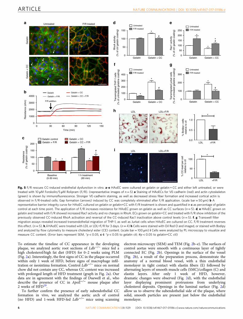
Fig. 6 F/R rescues subendothelial CC production in vivo. a, c Ldlr−/− mice were fed a HFD for 1 week with simultaneous IP injections with F/R or PBS(control) every other day. b, d Ldlr−/− mice were kept on HFD for 1 week before initiation of IP injections with F/R or PBS (control) every other day for anadditional week on HFD. a, b Aortic root sections were subjected to PL to visualize CC. (scale bar= 500 μm) c, d The aortic arch was used for analysisby SEM (luminal surface) and TEM. Yellow arrows show subendothelial protrusions from the aortic surface. (Error bars represent SEM, *p< 0.05,representative images of n= 8, IP: intraperitoneal injection, C: collagen, CC: cholesterol crystal, SMC: smooth muscle cell, E: elastin layer, EC: endothelial cell)
NATURE COMMUNICATIONS | DOI: 10.1038/s41467-017-01186-z ARTICLE
NATURE COMMUNICATIONS |8: 1129 |DOI: 10.1038/s41467-017-01186-z |www.nature.com/naturecommunications 7

100
100.3 60
2.0
1.5
1.0
0.5
0.0
0.10
0.08
0.06
0.04
0.02
0.00
40
20
0
0.2
0.1
0.0
8
6
4
2
0
80
60
40
20
0TNFα
TNFαT
NF
αHFD
–
Untargeted
Whole aorta
Untargeted
Unt
arge
ted
Control
Control
PBS PBS F/R
PBS
PBS
PBS
PBS
F/R
F/R
PBS PBS F/R
0.09
Pla
que
area
(%
)
CC
-pos
itive
are
a (m
m2 )
Aor
tic r
oot
(Pla
que
area
in m
m2 )
MO
MA
-2 s
tain
ing
(% p
laqu
e ar
ea)
SM
22 a
lpha
sta
inin
g(%
pla
que
area
)
Liposomes
Control Liposomes
Control
Oil
red-
OM
OM
A-2
SM
22 a
lpha
Liposomes
Control Liposomes
PBS PBS F/R
Control Liposomes
PBS
PBS
PBS
PBS
F/R
F/R
PBS PBS F/R
Control
Control
Liposomes
Control Liposomes
Liposome
Con
trol
Per
cent
Dil
posi
tive
endo
thel
ial c
ells
***
***
*
**
* *
*
*
**
Targeted
Targeteda
c e f
g
h
i
d
b
Targ
eted+ – +
Fig. 7 Targeted delivery of F/R-liposomes to inflamed endothelium reduces atherosclerosis development in vivo. a HAoEC treated with untargeted ortargeted DiI-filled liposomes were analyzed by fluorescence microscopy and flow cytometry. (representative images of n= 4, scale bar= 50 μm) b After IPinjection with PBS or 10 ng/ml TNFα 24 h before killing or 1 week HFD, ApoE−/− mice were treated with DiI-filled untargeted or sLex-targeted liposomes viatail vein injection. Aortas were collected and analyzed by fluorescence microscopy for areas of DiI-liposome uptake. (Scale bar= 100 μm, representativeimages of n= 3) c–i ApoE−/− mice were fed a HFD for 6 weeks while receiving weekly IP injections of PBS, or tail vein injection of targeted liposomes filledwith either PBS or 10 μM Forskolin/5 μM Rolipram (F/R). (representative images of n= 8) c Aortas were stained with Oil Red O and plaque areas werequantified with ImageJ software. d Aortic root sections were stained using Oil Red O to visualize plaque development, MOMA-2 to visualize macrophagecontent, SM22α to visualize SMC content, and PL to visualize the CC formation h. These values were quantified in e, f, g, and i, respectively. (Error barsrepresent SEM, *p< 0.05, IP: intraperitoneal, scale bar Aorta= 1 cm, Oil Red-O= 500 μm, scale bar MOMA-2/SMA/PL= 100 μm, IP intraperitonealinjection)
ARTICLE NATURE COMMUNICATIONS | DOI: 10.1038/s41467-017-01186-z
8 NATURE COMMUNICATIONS |8: 1129 |DOI: 10.1038/s41467-017-01186-z |www.nature.com/naturecommunications

to be used for further experiments. Synthetic CC were assessed bySEM and found to have various sizes and shapes (SupplementaryFig. 3a). Using energy-dispersive X-ray spectroscopy (EDS)analysis of both synthetic and naturally-occurring CC samples,we verified the organic carbon-based chemical composition ofsynthetic CC to be remarkably similar to CC found in vivo after1 week of HFD (Supplementary Fig. 3b, c). CC were visible viaPLM only in cells with embedded CC (Supplementary Fig. 3d, e).In addition, EC cultured on synthetic CC showed lipid dropletaccumulation, indicating active processing of the gelatin-embedded CC (Supplementary Fig. 3f).
Endothelial barrier integrity was significantly compromisedwhen EC were cultured on CC (Fig. 4) as evidenced by disruptedVE-cadherin and claudin 5 staining (red), and actin stress fiberformation (green) indicating impaired endothelial junctionformation (Fig. 4a). A transwell permeability assay as well aselectric cell-substrate impedance sensing (ECIS) was used toquantitatively measure endothelial integrity. Subendothelial CCresulted in a 2.5-fold increase in permeability and a 30% decreasein resistance compared with control cells (Fig. 4b–d). Endothelialregeneration, evaluated using the ECIS wounding system, wasalso compromised (Fig. 4e). To test if this compromise in barrierintegrity would affect the transendothelial migration of mono-cytes and T-cells, HAoEC were cultured with or without CC in atranswell filter setup. When subendothelial CC were present, weobserved approximately 3-fold and 2-fold increases in migrationof THP-1 and Jurkat cells, respectively (Fig. 4f, g). These dataindicate that early subendothelial CC deposition leads to impairedendothelial integrity and may facilitate the migration andsubsequent accumulation of leukocytes, which are known topromote atherosclerosis.
As previous studies have shown that Rho-GTPases are majorregulators of endothelial barrier function24, we questionedwhether early CC deposition may have an effect on endothelialfunction via alteration of Rho-GTPase activity. While Ras andCdc42 were not altered as a result of subendothelial CCdeposition, RhoA activity was enhanced and Rac1 activity wassuppressed (both known to be associated with endothelialdysfunction) (Fig. 4h), indicating that the observed effects couldat least partially be driven by alteration of RhoA/Rac1 activity.Despite RhoA’s involvement, inhibiting its well-known effector,Rho-kinase, did not affect EC function (Supplementary Fig. 4).
F/R mitigates CC-mediated endothelial dysfunction. As com-promised endothelial barrier integrity caused by subendothelialCC could have a significant impact on atherogenesis, we exploredmeans by which CC-mediated endothelial barrier destabilizationcould be minimized. Our group and others have shown pre-viously that treating EC with forskolin, an adenylate cyclaseactivator, and rolipram, a phosphodiesterase IV inhibitor, canstrengthen endothelial barrier25, 26. Culturing HAoEC with CCresulted in formation of gaps between cells in the endotheliallayer, as visualized by staining of VE-cadherin (red) and actincytoskeleton (green), which was abrogated by treatment with F/R(Fig. 5a). ECIS was used to determine if barrier integrity com-promised by CC could also be rescued by F/R. Differences inresistance appeared within minutes in both control andCC-coated cells following F/R treatment (Fig. 5b, c). F/Radministration also led to lower RhoA and higher Rac1 activity(Fig. 5d, e), another indication of improved endothelial integrity.Moreover, treatment with F/R abrogated the CC-induced increasein transendothelial migration of monocytes and T-cells (Fig. 5f,g). When HAoEC were incubated with LDL±F/R, the cells treatedwith F/R showed significantly less lipid loading (Fig. 5h), as wellas decreased CC presence (Fig. 5i).
F/R decreases subendothelial CC deposition in vivo. To assesswhether these protective effects of F/R treatment on HAoECcould be recapitulated in vivo, 4 groups of Ldlr−/− mice wereplaced on HFD. Two of the groups were fed HFD for 1 week, onegroup receiving treatment with F/R, and the control groupreceiving PBS via IP (intraperitoneal) injections. The other 2groups were given the HFD for 1 week, followed by another weekof HFD with PBS or F/R IP injection, to test whether early plaquedevelopment could be abrogated by F/R treatment once the micehad already been hyperlipidemic for a week. In both cases, themice treated with F/R showed significantly reduced CC load inthe aortic sinus plaque shown by PLM (Fig. 6a, b). SEM and TEMphotos also revealed smoother endothelial surface and less CCdeposition in the subendothelial space (Fig. 6c, d).
Targeted F/R delivery to inflamed endothelium in vivo. Despiteits effectiveness in reducing CC formation, F/R injection into miceresulted in acute fatigue of the animals, most likely due to adramatic lowering of blood pressure. These side effects and thenarrow therapeutic window rendered the use of F/R as anexperimental anti-atherosclerotic agent unfeasible. After exploringseveral options we decided to use a targeted drug delivery systemwith liposomes from our collaborators at InnoMedica to specifi-cally target inflamed endothelium. This enabled us to conduct along-term animal study to further investigate the beneficial effectsof F/R on CC production and more advanced atherosclerosisdevelopment. To assess the best target for liposome delivery, weincubated HAoEC with LDL for 6, 12, or 24 hours, and thenmeasured whether the three most common endothelial adhesionmolecules (ICAM-1, VCAM-1, E-Selectin), were upregulated byLDL. Only E-selectin surface expression was consistently upre-gulated (Supplementary Fig. 5a), even though all three were foundto be upregulated at the transcript level (Supplementary Fig. 5b).Furthermore, we verified that HFD increases surface expression ofE-selectin in vivo using flow cytometry (Supplementary Fig. 5c).Based on these results we chose to conjugate the liposomes withthe well-known ligand of E-selectin, Sialyl Lewis X (sLex) sugarchains, to facilitate their binding to E-selectin (SupplementaryFig. 6a). E-selectin is a well-established endothelial inflammatorymarker which is robustly expressed on atherosclerosis-proneendothelium27. TEM analysis was used to calculate the size of theliposomes, which ranged from 76 to 150 nm with a median size of121 nm (Supplementary Fig. 6b).
To characterize the binding of liposomes in vitro, as well as toempirically determine the concentrations to use for furtherexperiments, HAoEC were treated with various dilutions of DiI-liposomes in the absence or presence of TNFα for 24 h. Cells weresubsequently analyzed using fluorescence microscopy and flowcytometry (Supplementary Fig. 6c, d). Significant uptake ofliposomes was observed using dilutions of 1:50 to 1:25, whichbecame more prominent with the addition of TNFα, a knowninducer of E-selectin on the endothelial surface. The optimumdosage was calculated to be 1:25 dilution of liposomes in vitro and200 μl of liposome suspension for in vivo experiments.
To test the specificity of liposome binding to HAoEC, bothuntargeted and targeted DiI-liposomes were tested in vitro.Without TNFα treatment, 60% of the HAoEC were DiI-positive,which increased to 80% with TNFα treatment, compared with25% and 30%, respectively, using untargeted liposomes (Fig. 7a).After TNFα treatment, HAoEC uptake of targeted liposomesloaded with F/R resulted in a 4-fold increase in intracellularcAMP levels, a known effect of F/R28 (Supplementary Fig. 6e),providing evidence of successful drug delivery to HAoEC.
To investigate the uptake of liposomes in vivo, ApoE−/− micewere fed a HFD for 1 week or IP injected with TNFα 24 h before
NATURE COMMUNICATIONS | DOI: 10.1038/s41467-017-01186-z ARTICLE
NATURE COMMUNICATIONS |8: 1129 |DOI: 10.1038/s41467-017-01186-z |www.nature.com/naturecommunications 9
killing to induce inflammation, while control mice received PBSinjections. The mice were also treated with 200 μl of targeted oruntargeted DiI-liposomes via tail vein injection 24 h beforekilling. After killing, the inner surfaces of the aortic arches weresubjected to laser scanning confocal microscopy to visualize DiI-positive cells (Fig. 7b). Only trace amounts of untargeted DiI-liposomes were bound to the aortic surface after TNFα or HFDtreatment, while no DiI was visible in control aortas. In contrast,the sLex-conjugated DiI-liposomes were successfully delivered tothe endothelial cells of control, TNFα- and HFD-treated aortas, inincreasing order. The presence of DiI was especially prominent atareas of disturbed flow in the aortic arch, the most susceptiblearea to plaque development. These positive results indicated thatthis innovative drug delivery system could be used to administerF/R specifically to inflamed endothelial cells during a long-termatherosclerosis study.
Effect of F/R-liposomes on atherosclerosis in ApoE−/− mice. Totest the effectiveness of our targeted F/R-containing liposometreatment in reducing atherosclerosis burden, we performed aproof of principle study in ApoE−/− mice with three treatmentgroups: (a) PBS, (b) PBS-filled targeted liposomes, or (c) F/R-filled targeted liposomes via tail vein injection once per weekwhile being fed a HFD for 6 weeks. Upon completion of thestudy, analysis of atherosclerotic plaque area in the whole aortarevealed that treatment with PBS-liposomes did not affectatherosclerosis development (Fig. 7c), whereas animals treatedwith F/R-liposomes displayed a significant reduction in athero-sclerosis burden compared with control mice (Fig. 7c) Analysis ofaortic root sections (Fig. 7d) confirmed that atherosclerotic pla-que development was significantly decreased only in mice treatedwith F/R-liposomes (Fig. 7e). Immunofluorescent staining ofaortic root sections for the presence of macrophages and SMC(Fig. 7d) revealed a decrease in macrophage content and anincrease in SMC content (Fig. 7f, g) in F/R-liposome-treated micecompared with controls, whereas no differences were observed innecrotic core area (Supplementary Fig. 7a). Importantly, analysisof CC in the plaques of the mice revealed that F/R-liposometreatment led to a significant reduction in CC content of theplaque compared with both control-PBS- and PBS-liposome-treated mice (Fig. 7h). There were no significant differences inphysiological measurements among the study groups, includingbody weight, cholesterol and triglyceride levels, as well as circu-lating leukocyte populations (Supplementary Fig. 7b–d).
DiscussionAlthough EC are in constant contact with circulating lipopro-teins, whether these cells take up and metabolize the lipoproteinparticles has not been well characterized. Our study provides newevidence that EC not only take up and metabolize lipoproteins,but when they are burdened with excess intracellular cholesterolthat they generate CC. To our knowledge, we are the first toreport the synthesis of CC by EC treated only with LDL, and toshow the presence of CC in subendothelial space of Ldlr−/− aortasafter only 1 week of HFD when macrophages are largely absent.The appearance of CC in plaque is commonly associated withmacrophages in advanced atherosclerosis. We believe that this is asource of CC in the plaque that is quite distinct from the initialproduction of CC by EC in the initial stage of atherosclerosisdevelopment, as we have shown in the current study. CC havebeen reported to be present in atherosclerotic lesions of ApoE−/−
mice fed the HFD for 2 weeks, however their appearance occursin parallel with infiltrated inflammatory cells, which continueswith increasing length of HFD19. As dendritic cells (DC) havealso been seen in atherosclerotic plaques29–31, and these cells are
known to take up lipids soon after induction of hypercholester-olemia32, it is possible that the DCs contributed to the formationof CC in our mice. However, we found very few DC in our1–2 week HDF-fed aortas of both ApoE−/− and Ldlr−/− mice(Supplementary Fig. 8). Furthermore, the majority of the CC thatwe observed in the aorta samples of early atherosclerosis were notassociated with myeloid cells (macrophages and/or DC), indi-cating that the CC formation in early plaques is independent ofmyeloid cell presence.
The importance of CC in atherosclerosis was established nearly40 years ago when a comprehensive study examining the lipidcomposition of human plaques found that CC are present inadvanced stages of atherosclerosis12, 21. Prevailing current view ofCC in the context of atherosclerosis is that crystals form as aresult of lipid saturation originating from subendothelial mac-rophage foam cells. Studies using macrophages have reported CCproduction upon OxLDL treatment15 or cholesteryl ester lipiddroplets16, but not with native LDL treatment. We chose to useLDL in our studies because in normal physiological conditionsEC do not encounter modified LDL. Using Ldlr−/− animals forour in vivo studies did not interfere with endothelial LDL uptakesince most of the LDL taken up by EC occurs through receptor-independent endocytosis6. Moreover, CC formation is not a resultof cells dying since treating HUVEC and HSF with LDL, OxLDLor AcLDL resulted in high rate of cell death, but little to no CCformation.
Although there are certain physical parameters that are knownto affect CC formation and/or growth such as temperature,hydration and pH20, any information pertaining to biologicalmechanism(s) that contribute to CC formation are very scant,especially for EC. Based on our observations, we can speculatethat when EC encounter high levels of LDL, lipoproteins aretaken up and processed intracellularly. The excess cholesterol isesterified with the help of ACAT and stored in lipid droplets(LD). When cells are overloaded with LD, we believe that theequilibrium shifts and EC try to break down LD by activating thelysosomal pathway involving the lysosomal acid lipase whichconverts the CE to FC. In other cell types like the macrophage,the FC can be excreted via the cholesterol efflux pathway utilizingthe cholesterol transporters ABCA1 and ABCG1, which we haveshown previously to be highly upregulated in lipid-loaded mac-rophages33. In EC, however, we have found that ABCA1 andABCG1 are not upregulated even after the cells have taken upLDL (Supplementary Fig. 8e), indicating that cholesterol efflux inlipid-laden EC is not a robust mechanism. Thus, lipids accu-mulate in the cell and become crystallized, although the exactmechanisms involved in the cholesterol crystallization processneeds to be explored.
It is also possible, according to our findings, that liquid crystals,also known as anisotropic droplets or spherulites, are produced inthe EC as a result of LDL uptake and processing. These prematurecrystals may be secreted to the basolateral side of the endothe-lium, subsequently coated with ECM and grow into crystals byfusing with other crystals and possibly free lipid droplets.
As an alternative view to explain cellular lipid degradation,more recent studies have found that intracellular lipid metabo-lism is mediated by lipophagy involving autophagosomes andlysosomes and the fusion of these two organelles to degradecellular lipid stores34. If lipid degradation via lipophagy occurs inEC, we can assume that the autophagosomes fuse with lysosomes,leading to lysosomal FC generation. In support of this, it wasreported recently that cell-based CC formation occurs mostly inacidic lysosomes15. However, although LDL degradation mayoccur through the autophagosome-lysosome-lipophagy pathway,the mechanism to excrete the processed cholesterol, whether inlipid droplet, liquid crystal or crystal form, to the basolateral side
ARTICLE NATURE COMMUNICATIONS | DOI: 10.1038/s41467-017-01186-z
10 NATURE COMMUNICATIONS |8: 1129 |DOI: 10.1038/s41467-017-01186-z |www.nature.com/naturecommunications
is unknown. One possible mode of intracellular transport isthrough multi-vesicular bodies (MVB) that are found commonlywithin EC. While CC may form in acidic lysosomes, a recentstudy suggested that most of the cholesterol in the endocyticpathway is harbored in MVB35. We postulate that the initial CCformation in EC occurs in TSG101-positive MVB. TSG101 isnecessary for MVB formation and vesicular trafficking as part ofthe endosomal sorting complex required for transport (ESCRT)machinery, and a key marker of MVB36. According to a recentreport, the storage and metabolism of lipids in EC, especially freecholesterol, occurs in vesicular structures which are surroundedby membranes rich in free cholesterol as well as caveolin-137.Thus, while more studies are clearly needed to fully elucidate themechanisms involved in CC formation and transport, itappears that they require the orchestration of multiple cellularprocesses.
Interestingly, it was shown in human atherosclerotic samplesthat changes in serum lipid composition (via statin treatment)can alter the appearance and shape of CC38. Needle-shapedcrystals are most commonly reported in atherosclerotic plaques22,although CC in more advanced atherosclerotic plaques can beplate-shaped12, 13, 39 or random aggregates forming rod-likestructures as well40. It is noteworthy that electron microscopyimages of CC published by these groups closely match the plateand needle-shaped CC that we observed in our experiments. Webelieve that the initial crystals found in the subendothelial spacein fact are produced by EC and attract monocytes to transmigrateinto this space. Once these monocytes differentiate into macro-phages they take up CC as well as other forms of free lipids andperpetuate the formation and progression of atherosclerotic pla-que. Interestingly, it was reported recently that a C-type lectinreceptor called Mincle can bind to CC and trigger apro-inflammatory immune response in human macrophages aswell as dendritic cells41. This was the first demonstration of adirect innate immune receptor for CC, but it remains to be seenwhether CC-Mincle interaction is an active phenomenon inatherosclerosis.
Although the mechanism behind F/R’s ability to reduce CCformation under hypercholesterolemic conditions, and therebyattenuate the atherosclerotic burden, remains to be investigated,forskolin and/or rolipram used as therapeutic agents have provento be beneficial in a number of conditions including spinalmuscular dystrophy42, septic shock43, rheumatoid arthritis44,memory performance45 and pulmonary capillary ischemia-reperfusion injury46. Importantly, recent research suggests thatadministration of F/R results in stimulation of lipolysis andattenuation of weight gain in rats fed a HFD28. The sLex-lipo-somes used in our studies were prepared exclusively for this studyand were meant to demonstrate the delivery of F/R to inflamedendothelium as a proof-of-principle only. It is worth noting,however, that the utility of this liposome system has beendemonstrated by other investigators in a variety of diseasemodels. For example, the sLex-conjugated liposomes loaded withfluorescent material designed to target tumors have been utilizedfor in vivo imaging of tumors47. Furthermore, sLex-liposomeshave been used successfully to deliver dexamethasone to inflamedeyes in a murine model of experimental autoimmune uveor-etinitis48. Most recently, sLex-liposomes containing doxorubicinalong with several controls were injected into rats that haveundergone angioplasty to prevent stenosis. The rats that receivedthe drug incorporated into the sLex-liposome exhibited sig-nificantly larger lumen area compared with controls49. Thesestudies along with our own data on the use of sLex-liposomeindicate that E-selectin expressed on inflamed endothelium is auseful target to deliver drugs and other agents to the affectedendothelium, including atherosclerosis-prone areas.
In summary, we show in vitro that aortic endothelial cellsproduce CC under conditions of hyperlipidemia, and that CCaccumulate subendothelially in vivo after only 1 week of HFD, atime point that precedes macrophage infiltration. This phenom-enon can be prevented by F/R administration as demonstrated bya study in which short-term use of F/R in vivo successfullyinhibited endothelial CC production after 1 week of HFD. Fur-thermore, we report an effective method of specific drug deliveryto inflamed endothelium using F/R-incorporated liposomesconjugated with sLex. This technique is particularly useful whenthe systemic application of a drug results in adverse side effects, asis the case with F/R. Using this specialized delivery system, wehave verified the specific targeting of inflamed endothelial cellsand clearly demonstrated that this application reduces thedevelopment of atherosclerosis in vivo.
MethodsMaterials. Human LDL, DiI-LDL, oxLDL and AcLDL were purchased from AlfaAesar (Formerly Biomedical Technologies, USA). Forskolin (adenylyl cyclaseactivator) and rolipram (PDE IV-inhibitor) were used as a cAMP- increasing drugcombination and were purchased from Sigma-Aldrich (MO, USA).
Human tissue samples. Carotid and Femoral artery samples from an autopsy of aCVD patient were generously provided by the Queens Medical Center (HI, USA).The artery samples were collected after obtaining consent from a family memberand reviewed by the Research and Institutional Review Committee of the QueensMedical Center. The tissues were frozen immediately and kept at −80 °C in thehospital before being transported on dry ice to the University of Hawaii. A portionof each tissue sample was either embedded and frozen in OCT (Sakura Finetek,USA), cut into 10uM sections and analyzed by polarized light microscopy, orprepared as described above for TEM analysis.
Cell culture. Primary human aortic endothelial cells (HAoEC, Cat#: C-12272),primary human umbilical vein endothelial cells (HUVEC, Cat#: C-12253), primaryhuman skin fibroblasts (HSF, Cat#: C-12352) as well as primary human aorticsmooth muscle cells (VSMC, Cat#: C-12532) (all PromoCell, Germany) werecultured according to manufacturer’s recommendation using the correspondingmedia as well as the split kit (all, PromoCell, Germany). Cells were used up topassage 8. Human monocyte-derived macrophages (HMDM) were isolated fromwhole blood from consenting healthy volunteers by Histopaque (Sigma-Aldrich)density gradient centrifugation. Informed consent was obtained from all donorsand IRB approval was received under protocol CHS# 19345 prior to experimentsinvolving HMDM. Cells from the buffy coat were plated with hematopoieticX-VIVO 15 medium (purchased from Lonza, Switzerland) supplemented with 20%heat inactivated human serum for 1 hour before non-adherent cells were washedaway. Adherent cells were maintained for 5 days on glass slide chambers beforebeing used in experiments.
In vitro HAoEC treatment. HAoEC were cultured for 3 days after splitting. At day4, cells were treated with 50 μg/ml LDL (low dose), 250 μg/ml LDL (high dose), 50μg/ml AcLDL or OxLDL or 100 μg/ml DiI-LDL. After 3, or 5 days of treatment,cells and their according controls were washed with PBS, fixed and prepared forsubsequent PL and confocal microscopy. For PL as well as confocal microscopy,cells were fixed for 10 min at room temperature using 4% PFA. DiI-LDL-treatedsamples were counterstained with DAPI, mounted with DAKO fluorescencemounting media and analyzed using confocal microscopy (Olympus FV-1000). ForSEM and TEM analysis, cells were fixed using 4% glutaraldehyde and 0.1 M cal-cium chloride in 0.1 M sodium cacodylate buffer (pH 7.2) for 24 h.
HAoEC, HUVEC, HSF and HMDM were treated with 50 μg/ml LDL (lowdose), 250 μg/ml LDL (high dose), 50 μg/ml AcLDL or OxLDL or 100 μg/mlDiI-LDL. After 5 days of treatment, cells were fixed using 4% PFA and subjected toPL microscopy. A second set of treated cells was fixed and permeabilized using0.1% TritonX-100 in PBS for 5 min at RT. The actin cytoskeleton as well as thenuclei of these cells was labeled using Phalloidin-Alexa488 and DAPI, respectivelyand mounted using DAKO fluorescence mounting media (DAKO). Fluorescentlylabeled cells were analyzed and photographed using an Axiovert (Zeiss, Germany)microscope.
Preparation of cell culture dishes with CC and gelatin. HAoEC used forexperiments were cultured on gelatin or gelatin+CC coated surfaces. Cell culturedish surfaces were covered completely using 0.5 mg/ml gelatin in PBS or 0.5 mg/mlgelatin in PBS plus 1 mg/ml CC and incubated at 37 °C for 1 h. Afterwards surfaceswere gently rinsed three times using PBS. Coating of surfaces was prepared freshfor each experiment.
NATURE COMMUNICATIONS | DOI: 10.1038/s41467-017-01186-z ARTICLE
NATURE COMMUNICATIONS |8: 1129 |DOI: 10.1038/s41467-017-01186-z |www.nature.com/naturecommunications 11
Endothelial barrier integrity measurement using ECIS. An electric cell substrateimpedance-sensing set-up (ECISZΘ, Applied BioPhysics Inc, Troy, NY, USA) wasused to measure and analyze the transendothelial electrical resistance (TER) as ameasure of HAoEC barrier integrity. Electrode arrays were coated as describedabove and equilibrated for 1 h in the incubator. HAoEC were seeded into theseequilibrated ECIS arrays and the measurement started immediately. By measuringthe capacity and the impedance/TER we were able to detect the process of celladherence and endothelial barrier formation over time (24 h). Afterwards, cellswere treated with forskolin/rolipram (5 μM/10 μM) and changes in endothelialbarrier function observed by ongoing measurement of TER at 4000 Hz. In case ofwounding/regeneration experiments a current of 30,000 Hz for 30 s per well wasapplied. Recovery of the wounded area was observed by ongoing measurement.Obtained data were analyzed using ECIS software and Microsoft Excel.
Trans-endothelial migration in a transwell filter setup. Transmigrationexperiments in vitro were performed using human monocyte THP-1 (ATCCnumber TIB-202) and T-cell Jurkat (ATCC number TIB-152) cell lines, which werepurchased directly from ATCC (which included certifications of analysis but werenot further tested for mycoplasma). HAoEC were cultured on gelatin or gelatin +CC coated 5 µm pore size transwell filters for 5 days. At day 5, 2.5 × 10⁵ THP-1 orJurkat cells per cm2 along with fresh media were added to the HAoECs and wereallowed to transmigrate through the endothelium and the coated filters into thelower compartment supplemented with 100 ng/ml SDF-1 or 20 ng/ml MCP-1 for24 h. F/R treatment consisted of 5 μM/10 μM forskolin/rolipram for 1 h beforestarting the transmigration experiment. Transmigrated live cells were labeled withCalcein and counted using a BD FACS Calibur.
Detection of RhoGTPase activity and intracellular cAMP level. HAoEC werecultured on gelatin or gelatin + CC coated 6-well plates. At day 5 cells were F/Rtreated, harvested and analyzed for RhoA, Rac1, Cdc42 and Ras activity as well asfor their cAMP levels according to manufactures protocol. GLISAs were purchasedfrom Cytoskleton Inc and cAMP ELISA was purchased from EnzoLifeSciences.
Flow cytometry analysis. HAoEC were cultured for 5 div on gelatin or gelatin+CC. At day 5 HAoEC were trypsinized and stained for ICAM-1 (a-ICAM-1-FITC), VCAM-1 (a-CD106-APC) and E-selectin (a-CD62E-PE) for 20 min on icein the dark. The expression of these intercellular adhesion molecules was analyzedby flow cytometry using the FACS Calibur.
Secondly, Ldlr−/− mice were put on HFD for 0–3 weeks, the aorta isolated andthoroughly cleaned, digested using LiberaseTM (Roche), as described previously50
and E-selectin expression was quantified using flow cytometry. Data were analyzedusing FlowJo software according to the flow chart in Supplementary Fig. 9b. Toverify the functionality of the antibody used against mouse E-selectin, we utilizedthe MS1 mouse endothelial cell line (ATCC® CRL-2279™) stimulated with variousconcentrations of TNFα and observed a concentration dependent increase of E-selectin positively stained cells (Supplementary Fig. 9a). Digested aortas were alsoanalyzed for the presence of CD11b and CD11c positive cells. Further informationon the antibodies used can be found in Supplementary Table 1.
Analysis of lipids in HAoEC and aortic root sections. For visualization ofesterified lipids in the incubated cells or in the aortic root sections, Oil Red Ostaining was used as described previously51. For quantification purposes, treatedHAoEC were stained using Bodipy-Alexa488 (Invitrogen, USA) and analyzed usingflow cytometry (FACS Calibur, BD). To measure free cholesterol content, HAoECwere stained using filipin (Sigma) and analyzed by flow cytometry (Beckman-Coulter Altra).
Transmission electron microscopy (TEM). For TEM, specimens were fixed with4% glutaraldehyde and 1M calcium chloride in 0.1 M sodium cacodylate buffer,pH 7.2 for 24 h and washed in 0.1 M cacodylate for 3 × 10 min, followed bypost-fixation with 1% OsO4 in 0.1 M cacodylate buffer for 1 h. Cells and tissue weredehydrated in a graded ethanol series (30%, 50%, 70%, 85%, 95%, 100%), sub-stituted with propylene oxide, and embedded in LX112 epoxy resin. Ultrathin (60-80 nm) sections were obtained with a diamond knife on an RMC Powertomeultramicrotome, and double stained with uranyl acetate and lead citrate. Grids wereviewed and photographed on a Hitachi HT7700 transmission electron microscopeoperating at 100 kV with an AMT XR41 4 megapixel camera (Advanced Micro-scopy Techniques, Corp.).
HAoEC subjected to TEM were grown and fixed in tissue culture treated 35mmdishes, fixed and prepared as described above, with the exemption that nopropylene oxide was used.
Scanning electron microscopy. For SEM, specimens were fixed and processedexactly as with TEM through ethanol dehydration, at which point the samples werecritical point dried, mounted on aluminum stubs, sputter coated with gold/palla-dium, and viewed with a Hitachi S-4800 field emission scanning electron micro-scope at 5 kV. For energy-dispersive X-ray spectroscopy (EDS), spectra were
acquired operating the SEM at 20 kV field emission and using an Oxford INCAPentaFET-x3 Si (li) EDS detector.
HAoEC that were subjected to SEM were grown and treated on 12mm glasscoverslips and prepared as described above. Mouse aortas were cracked open usingcactus spines after mounting to the aluminum stubs to allow SEM analysis of a‘cross section’ of the vessel wall.
Energy-dispersive X-ray spectroscopy (SEM/EDS). For energy-dispersive X-rayspectroscopy (EDS) samples in the SEM were used, spectra were acquired operatingthe SEM at 20 kV field emission and using an Oxford INCA PentaFET-x3 Si (li)EDS detector.
Atherosclerosis model and analysis. All animal protocols were approved by theUniversity of Hawaii Institutional Animal Care and Use Committee. For initialanalysis of aortic changes after hyperlipidemia, male Ldlr−/− mice on C57BL/6background 8-10 weeks of age were put on a high fat diet (HFD) containing 15.8%(wt/wt) fat and 1.25% cholesterol (94059; Harlan Teklad) for 1 or 2 weeks. In asecond set of animal experiments, 4 groups of 8-week old male Ldlr−/− mice werekept on HFD, half for 1 week and half for 2 weeks. The first 2 groups on HFD for1 week were intraperitoneally (IP) injected every 48 h with: (a) PBS or (b) acombination of 2 mg/kg forskolin and 0.5 mg/kg rolipram. The second 2 groupswere put on HFD for 1 week, then during the second week of HFD were intra-peritoneally (IP) injected every 48 h with: (a) PBS or (b) a combination of 2 mg/kgforskolin and 0.5 mg/kg rolipram. In a third set of experiments, 6 week old maleApoE−/− mice were IP injected with (a) PBS + untargeted liposomes, (b) 1 μg/kgTNFα + untargeted liposomes, (c) untargeted liposomes and fed a HFD, (d) tar-geted liposomes, (e) 1 μg/kg TNFα + targeted liposomes and (f) untargeted lipo-somes and fed a HFD every 48 h for 1 week. In a fourth set of experiments 6 weekold male ApoE−/− mice were IP injected with (a) PBS and (b) 2 mg/kg forskolinand 0.5 mg/kg rolipram every 48 h while tail-vein injected with (c) 200 μl PBS-targeted liposomes or (d) 200 μl 5 μM forskolin/10 μM rolipram targeted liposomesonce a week for 6 weeks while on HFD.
Upon killing, the mouse hearts and aortas were collected and processed forfurther analysis. Two different portions of the aortic arch were used for SEM andTEM analyses. These samples were fixed immediately as described above. Theaortic sinus was cut into 10μm sections and analyzed using polarized lightmicroscopy, Oil Red O staining and immunohistochemistry.
Immunofluorescence of aortic root sections and HAoEC. The immuno-fluorescent visualization of macrophages and smooth muscle cells in the aortic rootsections were done with a Rat-α-MOMA-2 (AbCAM) as well as Rabbit-α-SM22α(ProteinTech) primary antibodies and Goat-α-Rat Cy2- and Goat-α-RabbitCy3-labeled secondary antibodies, respectively, as described previously51.
Immunofluorescent visualization of VE-cadherin and claudin5 in HAoEC wasperformed after fixation of HAoEC using 4% PFA in PBS (pH 7.4) for 10 min atRT, permeabilization using 0.1% TritonX-100in PBS for 5 min at RT and blockingusing 2% BSA/10% normal goat serum in PBS for 30 min at RT. Primary antibodieswere added for overnight incubation at 4 °C. Cy3-labeled secondary antibodieswere used, F-actin and nuclei were stained with Phalloidin-Alexa488 and DAPI,respectively. Samples were mounted in DAKO mounting media (DAKO) andvisualized using a Zeiss Axiovert microscope. Further information on antibodiesused can be found in Supplementary Table 1.
sLex-liposome treatment of HAoEC. sLex-liposomes were created by Dr. NoboruYamazaki (details under US patent application #20090169610) and designed tobind E-selectin on the surface of target cells. We obtained both targeted anduntargeted liposomes, with or without sLex respectively, from Dr. Yamazaki (nowat Innomedica). Liposomes containing DiI were used to characterize effectivebinding to target cells using fluorescence, while liposomes containing F/R wereused for the atherosclerosis studies described. HAoEC were grown to confluence on12-well plates or 8-chambered well glass slides and treated with DiI-filled untar-geted or targeted liposomes, with or without TNFα (10ng/ml) stimulation. Forconfirmation of F/R delivery via targeted liposomes, HAoEC were grown in6-well plates and treated as indicated and lysates were harvested as described above.cAMP concentration was measured using a cAMP assay (Enzo Life Sciences, USA)according to the manufacturer’s protocol as described previously25, 52.
Preparation of sLex-labeled liposomes. The liposomes were prepared using theimproved cholate dialysis method53. Dipalmitoylphosphatidylcholine (16.8 mg),cholesterol (10.1 mg), diacetylphosphate (1.8 mg), ganglioside (14.6 mg), dipalmi-toylphosphatidylethanolamine (2.3 mg), and sodium cholate (46.9 mg) were mixedand dissolved in 3 ml of chloroform/methanol (1:1, v/v) solution. The solvent wasevaporated using a rotating evaporator at 30 °C and the lipid film was obtainedafter drying under vacuum. This lipid film was dissolved in 3 ml of tris(hydro-xymethyl) methylaminopropanesulfonic acid buffer (TAPS, pH 8.4), and themicelle suspension was then obtained after sonicating. The labeling method forbinding Cy5.5 to human serum albumin (HSA) is as follows. Twenty mg of HSAand 2mg of Cy5.5-NHS ester (GE Healthcare) were dissolved in 3 ml of TAPS (pH8.4) and stirred at 37 °C for 3 h. To remove residual Cy5.5-NHS ester, the solution
ARTICLE NATURE COMMUNICATIONS | DOI: 10.1038/s41467-017-01186-z
12 NATURE COMMUNICATIONS |8: 1129 |DOI: 10.1038/s41467-017-01186-z |www.nature.com/naturecommunications

was ultrafiltrated with TAPS (pH 8.4) using an 8010 ultrafiltration cell (Amicon)fitted with an PM10 membrane (Amicon). Three milliliter of HSA withCy5.5 solution was mixed with the micelle suspension above, and this was ultra-filtered with TAPS (pH 8.4) by PM10 membrane to remove residual HSA withCy5.5, then 10 ml of liposome solution was obtained. In addition, the solution wasultrafiltered with sodium hydrogen carbonate buffer (CBS, pH 8.5) by XM300membrane (Amicon) to exchange buffer. Ten milligram of bis(sulfosuccinimidyl)suberate (BS3, Pierce), a crosslinking agent, was added to 10 ml of liposomesolution, and stirred at 20–25 °C for 2 h, then further stirred overnight at 4 °C. BS3was combined to the liposome surface. Then, 40 mg of tris(hydroxymethyl) ami-nomethane (Tris) was added, and stirred at 20–25 °C for 2 h, then further stirredovernight at 4 °C to bind Tris to BS3. This was ultrafiltered with TAPS(pH 8.4) by XM300 membrane to remove residual Tris. To bind HSA to theliposome surface, the coupling method was used53. To oxidize the liposome sur-face, 10.8 mg of sodium periodate was added to 10 ml of liposome solution andstirred at 4 °C overnight. To remove residual sodium periodate, it was ultrafilteredwith phosphate saline buffer (PBS, pH 8.0) by XM300 membrane. Twenty milli-gram of HSA was added to it and stirred at 20–25 °C for 2 h. Then 3.13 mg ofsodium cyanoborohydrate was added, and stirred at 20–25 °C for 2 h and furtherovernight at 4 °C. To remove residual sodium cyanoborohydrate, the solution wasultrafiltered with CBS (pH 8.5) by XM300 membrane. Sugar chains were combinedon the liposome surface through 3,3-dithiobis(sulfosuccinimidylpropionate)(DTSSP, Pierce). DTSSP was used as a cross-linking reagent. Ten milligram ofDTSSP was added to 10 ml of liposome solution and stirred at 20–25 °C for 2 h,and further overnight at 4 °C. To remove residual DTSSP, the solution wasultrafiltrated with CBS (pH 8.5) by XM300 membrane. The amination of thereducing group terminal of sugar chain was done by the glycosyl aminationreaction. Two milligram of SLX (Calbiochem) was dissolved in 0.5 ml of distilledwater. A total of 0.25 g of NH4HCO3 was added and stirred at 37 °C for 3 days.Aminated SLX was added to reach a final concentration of 100 μg/ml and stirred at25 °C for 2 h. Tris was then added to reach a final concentration of 132 mg/ml, andstirred overnight at 4 °C for repeated hydrophilization of the liposome surface. Toremove residual SLX and Tris, the solution was ultrafiltered with n-(2-hydro-xyethyl)piperazine-n′-(2-ethanesulfonic acid) (Hepes, pH 7.2) by XM300 mem-brane. The preparation of liposome without sugar chains was similar to the SLX-Lipo-Cy5.5 case except for the process for binding sugar chains.
Statistics. For in vitro experiments, at least three biological replicates were ana-lysed, with 2 or 3 technical replicates run for each assay as indicated. For the in vivomouse studies comparing 2 groups with 8 mice per group and a resulting degree offreedom of 14, a student t-distribution of 2.3 is used as the limitation for statisticalsignificance with alpha= 0.05 (two-tailed test). With a power of 80% and standarddeviation within groups < 20% from the mean, which is typical for the plaque andother analyses, a minimum of 8 mice per group was set for detecting significantdifferences of 25% between groups. Mice were randomly assigned to treatmentgroups with littermates divided evenly between groups. Blinding was not performedfor experiments. Comparison of means between groups with normal sample dis-tribution was performed with Prism software (GraphPad) using the Student’sunpaired, two-tailed t-test. In the case of 3 or more groups, one way ANOVA wasperformed. Any outlier values were determined by statistical test using Prism andexcluded from data sets. Data are presented as mean±the standard error of themean (SEM). Statistical significance was accepted at the level of p< 0.05.
Data availability. The data that support the findings of this study are availablefrom the corresponding author upon request.
Received: 21 July 2016 Accepted: 24 August 2017
References1. Cahill, P. A. & Redmond, E. M. Vascular endothelium-Gatekeeper of vessel
health. Atherosclerosis 248, 97–109 (2016).2. Simionescu, M. Implications of early structural-functional changes in the
endothelium for vascular disease. Arterioscler. Thromb. Vasc. Biol. 27, 266–274(2007).
3. Tabas, I., Williams, K. J. & Boren, J. Subendothelial lipoprotein retention as theinitiating process in atherosclerosis: update and therapeutic implications.Circulation 116, 1832–1844 (2007).
4. Gimbrone, M. A. Jr. & Garcia-Cardena, G. Endothelial cell dysfunction and thepathobiology of atherosclerosis. Circ. Res. 118, 620–636 (2016).
5. Frank, P. G., Pavlides, S. & Lisanti, M. P. Caveolae and transcytosis inendothelial cells: role in atherosclerosis. Cell Tissue Res. 335, 41–47 (2009).
6. Vasile, E., Simionescu, M. & Simionescu, N. Visualization of the binding,endocytosis, and transcytosis of low-density lipoprotein in the arterialendothelium in situ. J. Cell. Biol. 96, 1677–1689 (1983).
7. Kraehling, J. R. et al. Genome-wide RNAi screen reveals ALK1 mediates LDLuptake and transcytosis in endothelial cells. Nat. Commun. 7, 13516 (2016).
8. Armstrong, S. M. et al. A novel assay uncovers an unexpected role for SR-BI inLDL transcytosis. Cardiovasc. Res. 108, 268–277 (2015).
9. Stewart, G. T. Liquid crystals of lipid in normal and atheromatous tissue.Nature 183, 873–875 (1959).
10. Hata, Y., Hower, J. & Insull, W. Jr. Cholesteryl ester-rich inclusions fromhuman aortic fatty streak and fibrous plaque lesions of atherosclerosis. I.Crystalline properties, size and internal structure. Am. J. Pathol. 75, 423–456(1974).
11. Small, D. M. & Shipley, G. G. Physical-chemical basis of lipid deposition inatherosclerosis. Science 185, 222–229 (1974).
12. Katz, S. S., Shipley, G. G. & Small, D. M. Physical chemistry of the lipids ofhuman atherosclerotic lesions. Demonstration of a lesion intermediate betweenfatty streaks and advanced plaques. J. Clin. Invest. 58, 200–211 (1976).
13. Lim, R. S. et al. Identification of cholesterol crystals in plaques of atheroscleroticmice using hyperspectral CARS imaging. J. Lipid. Res. 52, 2177–2186 (2011).
14. Janoudi, A., Shamoun, F. E., Kalavakunta, J. K. & Abela, G. S. Cholesterolcrystal induced arterial inflammation and destabilization of atheroscleroticplaque. Eur. Heart. J. 37, 1959–1967 (2016).
15. Sheedy, F. J. et al. CD36 coordinates NLRP3 inflammasome activation byfacilitating intracellular nucleation of soluble ligands into particulate ligands insterile inflammation. Nat. Immunol. 14, 812–820 (2013).
16. Tangirala, R. K. et al. Formation of cholesterol monohydrate crystals inmacrophage-derived foam cells. J. Lipid. Res. 35, 93–104 (1994).
17. Freigang, S. et al. Nrf2 is essential for cholesterol crystal-inducedinflammasome activation and exacerbation of atherosclerosis. Eur. J. Immunol.41, 2040–2051 (2011).
18. Rajamaki, K. et al. Cholesterol crystals activate the NLRP3 inflammasome inhuman macrophages: a novel link between cholesterol metabolism andinflammation. PLoS ONE 5, e11765 (2010).
19. Duewell, P. et al. NLRP3 inflammasomes are required for atherogenesis andactivated by cholesterol crystals. Nature 464, 1357–1361 (2010).
20. Vedre, A. et al. Physical factors that trigger cholesterol crystallization leading toplaque rupture. Atherosclerosis 203, 89–96 (2008).
21. Abela, G. S. Cholesterol crystals piercing the arterial plaque and intima triggerlocal and systemic inflammation. J. Clin. Lipidol. 4, 156–164 (2010).
22. Abela, G. S. et al. Effect of cholesterol crystals on plaques and intima in arteriesof patients with acute coronary and cerebrovascular syndromes. Am. J. Cardiol.103, 959–968 (2009).
23. Lupu, F., Danaricu, I. & Simionescu, N. Development of intracellular lipiddeposits in the lipid-laden cells of atherosclerotic lesions. A cytochemical andultrastructural study. Atherosclerosis 67, 127–142 (1987).
24. Spindler, V., Schlegel, N. & Waschke, J. Role of GTPases in control ofmicrovascular permeability. Cardiovasc. Res. 87, 243–253 (2010).
25. Baumer, Y., Spindler, V., Werthmann, R. C., Bunemann, M. & Waschke, J. Roleof Rac 1 and cAMP in endothelial barrier stabilization and thrombin-inducedbarrier breakdown. J. Cell. Physiol. 220, 716–726 (2009).
26. Sands, W. A., Woolson, H. D., Milne, G. R., Rutherford, C. & Palmer, T. M.Exchange protein activated by cyclic AMP (Epac)-mediated induction ofsuppressor of cytokine signaling 3 (SOCS-3) in vascular endothelial cells. Mol.Cell. Biol. 26, 6333–6346 (2006).
27. Davies, M. J. et al. The expression of the adhesion molecules ICAM-1, VCAM-1, PECAM, and E-selectin in human atherosclerosis. J. Pathol. 171, 223–229(1993).
28. Doseyici, S., Mehmetoglu, I., Toker, A., Yerlikaya, F. H. & Erbay, E. The effectsof forskolin and rolipram on cAMP, cGMP and free fatty acid levels in dietinduced obesity. Biotechnol. Histochem. 89, 388–392 (2014).
29. Choi, J. H. et al. Flt3 signaling-dependent dendritic cells protect againstatherosclerosis. Immunity 35, 819–831 (2011).
30. Choi, J. H. et al. Identification of antigen-presenting dendritic cells in mouseaorta and cardiac valves. J. Exp. Med. 206, 497–505 (2009).
31. Jongstra-Bilen, J. et al. Low-grade chronic inflammation in regions of thenormal mouse arterial intima predisposed to atherosclerosis. J. Exp. Med. 203,2073–2083 (2006).
32. Paulson, K. E. et al. Resident intimal dendritic cells accumulate lipid andcontribute to the initiation of atherosclerosis. Circ. Res. 106, 383–390 (2010).
33. Meiler, S., Baumer, Y., Toulmin, E., Seng, K. & Boisvert, W. A. MicroRNA 302ais a novel modulator of cholesterol homeostasis and atherosclerosis.Arterioscler. Thromb. Vasc. Biol. 35, 323–331 (2015).
34. Singh, R. et al. Autophagy regulates lipid metabolism. Nature 458, 1131–1135(2009).
35. Mobius, W. et al. Recycling compartments and the internal vesicles ofmultivesicular bodies harbor most of the cholesterol found in the endocyticpathway. Traffic 4, 222–231 (2003).
36. Razi, M. & Futter, C. E. Distinct roles for Tsg101 and Hrs in multivesicularbody formation and inward vesiculation. Mol. Biol. Cell 17, 3469–3483(2006).
NATURE COMMUNICATIONS | DOI: 10.1038/s41467-017-01186-z ARTICLE
NATURE COMMUNICATIONS |8: 1129 |DOI: 10.1038/s41467-017-01186-z |www.nature.com/naturecommunications 13

37. Amiya, E. et al. Angiotensin II impairs endothelial nitric-oxide synthasebioavailability under free cholesterol-enriched conditions via intracellular freecholesterol-rich membrane microdomains. J. Biol. Chem. 288, 14497–14509 (2013).
38. Abela, G. S. et al. Effect of statins on cholesterol crystallization andatherosclerotic plaque stabilization. Am. J. Cardiol. 107, 1710–1717 (2011).
39. Solomonov, I., Weygand, M. J., Kjaer, K., Rapaport, H. & Leiserowitz, L.Trapping crystal nucleation of cholesterol monohydrate: relevance topathological crystallization. Biophys. J. 88, 1809–1817 (2005).
40. Busch, N., Lammert, F., Marschall, H. U. & Matern, S. A new subgroup oflectin-bound biliary proteins binds to cholesterol crystals, modifies crystalmorphology, and inhibits cholesterol crystallization. J. Clin. Invest. 96,3009–3015 (1995).
41. Kiyotake, R. et al. Human mincle binds to cholesterol crystals and triggersinnate immune responses. J. Biol. Chem. 290, 25322–25332 (2015).
42. Mack, S. G., Cook, D. J., Dhurjati, P. & Butchbach, M. E. Systems biologyinvestigation of cAMP modulation to increase SMN levels for the treatment ofspinal muscular atrophy. PLoS ONE 9, e115473 (2014).
43. Link, A., Selejan, S., Maack, C., Lenz, M. & Bohm, M. Phosphodiesterase 4inhibition but not beta-adrenergic stimulation suppresses tumor necrosisfactor-alpha release in peripheral blood mononuclear cells in septic shock. Crit.Care 12, R159 (2008).
44. Foey, A. D. et al. Impact of VIP and cAMP on the regulation of TNF-alpha andIL-10 production: implications for rheumatoid arthritis. Arthritis Res. Ther. 5,R317–328 (2003).
45. Sato, T. et al. Inhibitory effects of group II mGluR-related drugs on memoryperformance in mice. Physiol. Behav. 80, 747–758 (2004).
46. Barnard, J. W. et al. Reversal of pulmonary capillary ischemia-reperfusioninjury by rolipram, a cAMP phosphodiesterase inhibitor. J. Appl. Physiol. 77,774–781 (1994).
47. Hirai, M. et al. Accumulation of liposome with Sialyl Lewis X to inflammationand tumor region: application to in vivo bio-imaging. Biochem. Biophys. Res.Commun. 353, 553–558 (2007).
48. Hashida, N. et al. High-efficacy site-directed drug delivery system using sialyl-Lewis X conjugated liposome. Exp. Eye. Res. 86, 138–149 (2008).
49. Tsuruta, W. et al. Application of liposomes incorporating doxorubicin withsialyl Lewis X to prevent stenosis after rat carotid artery injury. Biomaterials 30,118–125 (2009).
50. Baumer, Y. et al. CD98 regulates vascular smooth muscle cell proliferation inatherosclerosis. Atherosclerosis 256, 105–114 (2017).
51. Meiler, S. et al. Selenoprotein K is required for palmitoylation of CD36 inmacrophages: implications in foam cell formation and atherogenesis. J. Leukoc.Biol. 93, 771–780 (2013).
52. Baumer, Y., Drenckhahn, D. & Waschke, J. cAMP induced Rac 1-mediatedcytoskeletal reorganization in microvascular endothelium. Histochem. Cell Biol.129, 765–778 (2008).
53. Yamazaki, N., Kodama, M. & Gabius, H. J. Neoglycoprotein-liposome andlectin-liposome conjugates as tools for carbohydrate recognition research.Methods Enzymol. 242, 56–65 (1994).
AcknowledgementsWe thank M. Montgomery and W. Regan for excellent technical support. We are gratefulto Dr. J. Hammer and Dr. E. Hellebrand for the use of the polarized light microscope.Core facilities were supported by NIH grants P20GM103516, P20RR016453,G12RR003061, and G12MD007601. The Hitachi HT7700 TEM was acquired with NSFgrant DBI-1040548. This project was funded by NIH grants R01HL075677 andR01HL081863, and by Innovative Research Grant 16IRG27540027 from the AmericanHeart Association to W.A.B. This work was performed within the Russian GovernmentProgram of Competitive Growth of Kazan Federal University.
Author contributionsY.B. and S.M. designed the study, performed experiments, analyzed data, and wrote themanuscript. T.M.W. performed and interpreted SEM/TEM data. N.N.M. provided datapertaining to adhesion molecule analysis. S.H., P.H. and N.Y. developed and provided uswith the liposomes. W.A.B. designed the study, analyzed data, provided funding andwrote the manuscript.
Additional informationSupplementary Information accompanies this paper at doi:10.1038/s41467-017-01186-z.
Competing interests: The authors declare no competing financial interests.
Reprints and permission information is available online at http://npg.nature.com/reprintsandpermissions/
Publisher's note: Springer Nature remains neutral with regard to jurisdictional claims inpublished maps and institutional affiliations.
Open Access This article is licensed under a Creative CommonsAttribution 4.0 International License, which permits use, sharing,
adaptation, distribution and reproduction in any medium or format, as long as you giveappropriate credit to the original author(s) and the source, provide a link to the CreativeCommons license, and indicate if changes were made. The images or other third partymaterial in this article are included in the article’s Creative Commons license, unlessindicated otherwise in a credit line to the material. If material is not included in thearticle’s Creative Commons license and your intended use is not permitted by statutoryregulation or exceeds the permitted use, you will need to obtain permission directly fromthe copyright holder. To view a copy of this license, visit http://creativecommons.org/licenses/by/4.0/.
© The Author(s) 2017
ARTICLE NATURE COMMUNICATIONS | DOI: 10.1038/s41467-017-01186-z
14 NATURE COMMUNICATIONS |8: 1129 |DOI: 10.1038/s41467-017-01186-z |www.nature.com/naturecommunications