Endogenous inequality and fluctuations in a two-country model
Upload
independentCategory
view
1download
0
2007;67:6238-6246. Cancer Res Rosalyn M. Adam, Nishit K. Mukhopadhyay, Jayoung Kim, et al. Cholesterol Sensitivity of Endogenous and Myristoylated Akt
Updated version
http://cancerres.aacrjournals.org/content/67/13/6238
Access the most recent version of this article at:
Material
Supplementary
http://cancerres.aacrjournals.org/content/suppl/2007/07/18/67.13.6238.DC1.html
Access the most recent supplemental material at:
Cited Articles
http://cancerres.aacrjournals.org/content/67/13/6238.full.html#ref-list-1
This article cites by 43 articles, 21 of which you can access for free at:
Citing articles
http://cancerres.aacrjournals.org/content/67/13/6238.full.html#related-urls
This article has been cited by 19 HighWire-hosted articles. Access the articles at:
E-mail alerts related to this article or journal.Sign up to receive free email-alerts
Subscriptions
Reprints and
To order reprints of this article or to subscribe to the journal, contact the AACR Publications
Permissions
To request permission to re-use all or part of this article, contact the AACR Publications
Research. on February 25, 2014. © 2007 American Association for Cancercancerres.aacrjournals.org Downloaded from
Research. on February 25, 2014. © 2007 American Association for Cancercancerres.aacrjournals.org Downloaded from
Cholesterol Sensitivity of Endogenous and Myristoylated Akt
Rosalyn M. Adam,1,3Nishit K. Mukhopadhyay,
1,3Jayoung Kim,
1,3Dolores Di Vizio,
1,3
Bekir Cinar,1,3Kelly Boucher,
2Keith R. Solomon,
1,2,3and Michael R. Freeman
1,3,4
1The Urological Diseases Research Center and 2Department of Orthopedic Surgery, Children’s Hospital Boston; Departments of3Surgery and 4Biological Chemistry and Molecular Pharmacology, Harvard Medical School, Boston, Massachusetts
Abstract
The serine-threonine kinase, Akt, has been linked to choles-terol-sensitive signaling mechanisms, suggesting a possiblemeans whereby cholesterol might affect tumor cell growthand survival. However, it has not been shown whether Aktitself, as distinct from upstream components of the pathway(e.g., membrane phosphoinositides), can be directly responsi-ble for cholesterol-mediated effects. Consistent with thispossibility, we identified an Akt1 subpopulation in cholester-ol-rich lipid raft fractions prepared from LNCaP humanprostate cancer cells. Phosphorylation of this Akt subspecieswas ablated with methyl-B-cyclodextrin, a cholesterol-bindingcompound, under conditions where nonlipid raft-residentAkt was unaffected. A myristoylated Akt1 (MyrAkt1) fusionprotein expressed in LNCaP cells was found to be highlyenriched in lipid rafts, indicating that oncogenic Akt is over-represented in cholesterol-rich membranes compared withwild-type Akt. Notably, lipid raft-resident MyrAkt1 exhibiteda markedly distinct substrate preference compared withMyrAkt1 immunoprecipitated from cytosol and nonraft mem-brane fractions, suggesting a redirection of signal transductionwhen the protein is present in cholesterol-rich membranes.Expression of MyrAkt1 in LNCaP cells overcame their char-acteristic dependence on constitutive signaling through thephosphoinositide 3¶-kinase pathway. This protective effectwas substantially diminished with cyclodextrin treatment.Phosphorylation of Akt substrates in lipid raft fractions, butnot in cytosol/nonraft membrane fractions, was ablated withcyclodextrin. In addition, in control (LacZ transfected) cells,lipid raft fractions were relatively enriched in phosphorylatedAkt substrates. Collectively, these data show that a subpopu-lation of Akt is cholesterol sensitive and that the oncogeniceffects conferred by myristoylation arise, in part, from thetendency of the membrane-targeted form of the protein toreside in cholesterol-rich membrane microdomains. [CancerRes 2007;67(13):6238–46]
Introduction
Cholesterol is a critical component of biological membranes. Inaddition to regulating membrane fluidity, cholesterol is an
important constituent of a class of detergent-resistant micro-domains, generally referred to as ‘‘lipid rafts’’ (1). The invaginated,vesicular structures known as ‘‘caveolae’’ are a specialized form oflipid raft that contain caveolin proteins. Noncaveolar, ‘‘flat’’ lipidrafts are also believed to exist, based on experimental andtheoretical evidence (2). The structural and biophysical propertiesof lipid rafts result in the retention and exclusion of certain classesof proteins, such that these microdomains can be viewed as‘‘privileged’’ sites that promote interaction between discrete subsetsof signaling intermediates, thereby serving as platforms for signaltransduction (reviewed in ref. 3). In cancer cells, lipid rafts/detergent-resistant microdomains may provide an importantsubcellular microenvironment in which signals are processed thatare central to tumor cell growth, resistance to apoptotic signals,and other aggressive characteristics.
Although elevation in circulating cholesterol levels has long beenassociated with cardiovascular disease, there is now increasingevidence to suggest a link between cholesterol accumulation andthe risk of certain malignancies. Several recent epidemiologicstudies have described a reduction in incidence of certain cancersin patients taking 3-hydroxy-3-methyl-glutaryl CoA (HMG-CoA)reductase inhibitors (‘‘statins’’) for cardiovascular indications(reviewed in ref. 4). Statins inhibit the rate-limiting step incholesterol biosynthesis (conversion of HMG-CoA to mevalonate)and thereby reduce synthesis of cholesterol and its isoprenoidprecursors, geranylgeranyl pyrophosphate and farnesyl pyrophos-phate. The effect of statin therapy on incidence of solid tumors mayvary with organ site. Current evidence supports the hypothesis thatprostate cancer may be particularly sensitive to this intervention(5, 6). This may be a reflection of aspects of cholesterol metabolismcharacteristic of prostate cells and tissues, including highendogenous levels of cholesterol seen in the normal prostate, theabnormal accumulation of cholesterol in prostate tumors, and thesensitivity of prostate cancer cells to cholesterol depletion (7–9).
The Akt/protein kinase B family (Akt1, Akt2, and Akt3) of serine-threonine kinases processes signals in tumor cells that mediatetumor cell proliferation, survival, and migratory behavior (10–12).Akt has also been linked to pathways sensitive to changes inmembrane cholesterol. Data from our group and others haveshown that constitutive and epidermal growth factor–stimulatedAkt activation and cell survival are regulated by cholesterol-sensitive signaling mechanisms in prostate cancer cells (9, 13).Elevation of circulating cholesterol levels in mice promoted growth,kinase activation, and survival signaling in human prostate tumorxenografts (14). In cell culture studies, simvastatin preferentiallyinhibited phosphorylation at a key regulatory site, Ser473 on Akt1present in lipid rafts, whereas Akt1 at other locations in the cellwas relatively resistant to the effects of the drug (14). Thesefindings suggest that prostate cancer and possibly other tumorcells contain discrete Akt populations that process distinct signalsdepending on subcellular location. These results also implicate
Note: Supplementary data for this article are available at Cancer Research Online(http://cancerres.aacrjournals.org/).
Requests for reprints: Michael R. Freeman, The Urological Diseases ResearchCenter, Enders Research Laboratories, Room 1161, Children’s Hospital Boston, 300Longwood Avenue, Boston, MA 02115. Phone: 617-919-2644; Fax: 617-730-0238; E-mail:[email protected] and Rosalyn M. Adam, The UrologicalDiseases Research Center, Enders Research Laboratories, Room 1077, Children’sHospital Boston, 300 Longwood Avenue, Boston, MA 02115. Phone: 617-919-2019;Fax: 617-730-0248; E-mail: [email protected].
I2007 American Association for Cancer Research.doi:10.1158/0008-5472.CAN-07-0288
Cancer Res 2007; 67: (13). July 1, 2007 6238 www.aacrjournals.org
Research Article
Research. on February 25, 2014. © 2007 American Association for Cancercancerres.aacrjournals.org Downloaded from

lipid raft microdomains as sites where the signaling effects ofcholesterol may influence the regulatory dynamics of Akt, a criticalnode in cancer cell signaling.
Although evidence has been obtained implicating membranecholesterol as a direct regulator of signal transduction of relevanceto cancer, it has not been shown that Akt itself, as opposed toupstream effectors, such as growth factor receptors or membranephospholipids, is a direct target for such cholesterol-mediatedeffects. For example, because statin drugs, in addition to theircholesterol-lowering ability, affect post-translational isoprenylationand activation of proteins, such as Rho, Ras, and Rac, (4), it ispossible that inhibitory effects of simvastatin on raft-resident Akt,observed previously (14), are not primarily the result of cholesterolsynthesis inhibition. In this study, we provide evidence that Aktitself is cholesterol sensitive as a result of the localization of an Aktsubpopulation within lipid raft microdomains. Our results alsoindicate that the raft microenvironment processes distinct Akt-dependent signals.
Materials and Methods
Antibodies and reagents. The antibodies used in this study include
anti-Akt polyclonal antibody (pAb; this antibody recognizes endogenous
Akt1, Akt2, and Akt3), anti-Akt1 monoclonal antibody (mAb; clone 5G3),anti-Akt1 mAb 2H10, immobilized Akt IG1 mAb, anti–phospho-Akt
(Thr308) pAb, anti–phospho-Akt (Ser473) pAb, anti–phospho-glycogen
synthase kinase-3a/h (p-GSK3a/h) pAb, and anti–phospho-Akt substrate
rabbit mAb (all from Cell Signaling Technology); anti-Akt1 pAb (UpstateBiotechnology, Inc.); anti-Akt1 pAb, anti-h-tubulin mAb (clone D10), and
anti-Gia2 subunit pAb (Santa Cruz Biotechnology); and anti-HA tag (clone
HA.11; Covance, Inc.). GSK3 fusion protein was from Cell SignalingTechnology. Protein A-Sepharose and protein G-Sepharose were obtained
from Amersham Biosciences. Opti-MEM reduced serum medium was from
Invitrogen. Fugene 6 was from Roche Applied Science. All other chemicals
were obtained from Sigma Chemical Co. Akt expression vectors encodingwild-type (WT) Akt1 tagged with the T7 epitope or myristoylated Akt1
(MyrAkt1) were generous gifts from Dr. William Sellers (Dana-Farber
Cancer Institute and Harvard Medical School, Boston, MA) and Dr. Thomas
Franke (Columbia University, New York, NY).Cell culture and transfections. LNCaP human prostate cancer cells
were cultured in RPMI 1640/10% fetal bovine serum (FBS). Human
embryonic kidney (HEK) 293 cells were cultured in DMEM/10% FBS. Allmedia were supplemented with penicillin/streptomycin and L-glutamine,
and cells were maintained in a humidified atmosphere of 5% CO2 at 37jC.
Cells in 150-mm dishes at f80% confluence were transfected using Fugene
6 according to the manufacturer’s instructions. In selected experiments,LNCaP cells were transduced with viral supernatants of 293FT cells
transfected with pLenti6-MyrAkt1 or pLenti6-LacZ, and stable populations
were isolated following selection with 2 Ag/mL blasticidin.
Preparation of membrane fractions. Lipid raft membrane fractionswere isolated using two methods. In the first method, lipid rafts were
isolated from LNCaP cells using sucrose gradient ultracentrifugation as
described (15). In the second method, a procedure involving successivedetergent extraction of cell membranes was used essentially as described
(13, 14, 16, 17). In some experiments, the cytosolic fraction was isolated
before membrane fractionation. Briefly, cell pellets were resuspended in
50 mmol/L HEPES (pH 7.4), 10 mmol/L NaCl, 1 mmol/L MgCl2, 1 mmol/LEDTA, 1 mmol/L phenylmethylsulfonyl fluoride (PMSF), and 1 mmol/L
Na3VO4 and subjected to mechanical disruption with 12 strokes of a
Dounce homogenizer (1,800 rpm). Homogenized samples were centrifuged
at 14,000 � g for 20 min at 4jC and the supernatant was removed as thecytosolic fraction. Membrane pellets were washed with buffer A and lysed as
described above to extract Triton-soluble and raft membrane fractions.
The protein content of fractions was determined using the Micro-
bicinchoninic acid (BCA) assay (Pierce Chemical Co.).
Cholesterol assay. Cholesterol determinations were done on 300 ALfractions from either membrane preparations or sucrose gradients prepared
as described (15). Lipids were solubilized in chloroform, extracted twice
through H2O, dried, and analyzed with the Infinity cholesterol determina-
tion assay kit (Sigma Chemical).Immunoprecipitations and Akt kinase assay. Equal amounts of
protein from cytosol and nonraft membrane (C+M fraction) or lipid raft
fractions were precleared with protein A-Sepharose or protein G-Sepharose
beads for 1 h at 4jC. In selected experiments, membrane fractions were
subjected to buffer exchange using BioSpin6 gel filtration columns (Bio-
Rad). Antibodies were added to precleared lysates and incubated overnight
at 4jC, before addition of 40 AL protein A or G beads (50% v/v slurry) for
a further 2 h at 4jC. Immunoprecipitates were washed four times with
lysis buffer [20 mmol/L Tris-Cl (pH 7.5), 150 mmol/L NaCl, 1 mmol/L
EDTA, 1 mmol/L EGTA, 1% Triton X-100, 2.5 mmol/L NaPPi, 1 mmol/L
h-glycerophosphate, 1 mmol/L Na3VO4, 1 Ag/mL leupeptin, 1 mmol/L PMSF]
and resuspended in 2� SDS loading buffer. To assay Akt kinase activity,
a nonradioactive assay kit was used (Cell Signaling Technology). Briefly,
immunoprecipitates were washed twice with lysis buffer and twice with
kinase assay buffer [25 mmol/L Tris-Cl (pH 7.5), 5 mmol/L h-glycerophosphate,
2 mmol/L DTT, 0.1 mmol/L Na3VO4, 10 mmol/L MgCl2] to equilibrate the
beads before assay. Beads were resuspended in kinase reaction mix,
comprising 40 AL kinase assay buffer, 1 Ag GSK3 fusion protein substrate,
and 200 Amol/L ATP, and incubated for 30 min at 30jC. Reactions were
terminated by the addition of 20 AL 3� SDS loading buffer supplemented
with 150 mmol/L DTT.For determination of Akt kinase activity against myelin basic protein
(MBP) or the histone subunit H2B, Akt immune complexes were incubated
with 1 Ag substrate and 5 ACi [g-32P]dATP (3,000 Ci/mmol). Samples were
resolved by gel electrophoresis, and gels were fixed with acetic acid anddestained before exposure of dried gels to X-ray film to visualize signal. To
quantitate incorporation of 32P, gel slices were excised and radioactivity
was determined by scintillation counting. Kinase activity was also assessedusing Crosstide as substrate. Briefly, immune complexes were resuspended
in kinase buffer [25 mmol/L HEPES (pH 7.4), 10 mmol/L MgCl2, 1 mmol/L
DTT] containing 5 ACi [g-32P]dATP and 1 Ag Crosstide (GRPRTSSFAEG) in
a volume of 30 AL and incubated at 30jC for 20 min. To terminate thereactions, 25 AL aliquots were spotted onto p81 phosphocellulose paper and
the incorporated radioactivity was determined essentially as described (18).
Preparation of whole-cell lysates and immunoblot analysis. Cells
were washed twice in ice-cold PBS and lysed in a minimum volume of 1�cell lysis buffer (Cell Signaling Technology) supplemented with 60 mmol/L
octylglucoside and 1 mmol/L PMSF. Protein content was determined using
the Micro-BCA protein assay reagent. Cell extracts (10 Ag/lane) andimmunoprecipitates were resolved by 12% SDS-PAGE and electrotransferred
to nitrocellulose membranes. Following transfer, membranes were stained
with Ponceau S to confirm equal protein loading, where appropriate. Mem-
branes were blocked with PBS/0.1% Tween 20/5% IgG-free bovine serumalbumin (BSA) and incubated with antibodies overnight at 4jC. Following
incubation with species-specific horseradish peroxidase–conjugated sec-
ondary antibodies, signals were detected using SuperSignal chemilumines-
cent reagent (Pierce Chemical) and exposure of blots to X-ray film. Inselected experiments, densitometric analysis of bands was done using the
public domain NIH Image (version 1.63) program. Relative Akt kinase
activity was determined by dividing the signal for p-GSK3a/h by the signalfor total Akt within the same fraction. A value of 100% was assigned to the
normalized kinase activity (p-GSK3 signal divided by Akt signal) in the
cytosolic/Triton-soluble membrane (C+M) fraction following treatment
(e.g., pervanadate) and all other values were expressed relative to this.Immunofluorescence microscopy. To image Akt, LNCaP cells were
seeded in chamber slides or coverslips and treated as indicated in legends.
Cells were washed once with ice-cold PBS and incubated on ice with
tetramethylrhodamine-conjugated cholera toxin B (CTxB) subunit (ListBiological Laboratories) or Alexa 594–conjugated CTxB diluted in medium.
Cells were fixed in ice-cold methanol or 4% paraformaldehyde and
nonspecific binding sites blocked in PBS/0.1% Tween 20/3% BSA ( for
endogenous Akt) or PBS/0.1% BSA (ectopically expressed Akt1) for 1 h at
Cholesterol Sensitivity of Akt
www.aacrjournals.org 6239 Cancer Res 2007; 67: (13). July 1, 2007
Research. on February 25, 2014. © 2007 American Association for Cancercancerres.aacrjournals.org Downloaded from

room temperature. Cells were incubated with Ser473-phosphorylated Akt(S473-P Akt) primary antibody diluted 1:100 in PBS/0.1% Tween 20/3% BSA
overnight at 4jC or PBS/0.1% BSA for 1 h at room temperature followed by
FITC-conjugated secondary antibody (1:200) for 1 h at room temperature.
Slides were mounted in Vectashield medium containing 4¶,6-diamidino-2-phenylindole (DAPI; Vector Laboratories, Inc.) and analyzed using an
LSM 510 META NLO laser scanning confocal microscope or an Axioplan 2
microscope (Carl Zeiss MicroImaging, Inc.).
Results
Targeting membrane cholesterol with cholesterol-binding com-pounds, or by inhibiting cholesterol synthesis endogenously, wasshown to attenuate phosphorylation of Akt1 at Ser473 in androgen-sensitive LNCaP prostate cancer cells (13, 14). The followingexperiments were conducted to determine whether cholesterolregulates Akt1 directly. We initially assessed the extent to whichAkt1 is present in cholesterol-rich (lipid raft) membrane fractionscompared with nonraft subcellular compartments. We used twocomplementary methods to fractionate cells into lipid raft-enriched and nonraft components. First, LNCaP cells homogenizedby mechanical disruption in Triton X-100–containing buffer weresubjected to sucrose density gradient ultracentrifugation. In thesecond method, cells were fractionated by successive detergentextraction as described previously (13, 14, 16, 17) to isolate Triton-soluble cytosolic and nonraft membrane components (C+M) andTriton-insoluble, octylglucoside-soluble raft fractions. Equalamounts of gradient, C+M, or raft fractions were resolved bySDS-PAGE and blotted with antibodies to Akt. The fidelity offractionation was confirmed by blotting of fractions with anti-bodies to h-tubulin or Gia2 as markers of nonraft membranes/cytosol and lipid rafts, respectively.
As shown in Fig. 1, the patterns of Akt distribution in cellsanalyzed by density gradient (Fig. 1A) or by differential solubility innonionic detergent (Fig. 1B) were similar. The majority of Akt waspresent in higher density fractions in LNCaP cells (Fig. 1A) thatcorrespond to the cytosol + nonraft membrane (C+M) fraction(Fig. 1B), with only a small proportion of total Akt present in rafts(Fig. 1A, fraction 4 ; Fig. 1B). Consistent with the PTEN-null status ofLNCaP cells, Akt in both C+M and raft fractions was phosphor-ylated on Thr308 and Ser473, with the extent of phosphorylationcommensurate with the level of Akt. To confirm the cholesterolsensitivity of lipid raft-resident Akt, LNCaP cells were treated withthe cholesterol-binding agent, methyl-h-cyclodextrin, and fraction-ated into C+M and raft components. Akt was immunoprecipitatedfrom each fraction and blotted with antibodies to total and S473-PAkt. Cyclodextrin treatment did not appreciably alter the amountor extent of phosphorylation of Akt isolated from the C+M fraction.In contrast, cyclodextrin ablated phosphorylation of raft-residentAkt (Fig. 1C). We also assessed the subcellular localization of Akt byimmunofluorescence imaging of LNCaP cells treated with perva-nadate (0.5 mmol/L), the most potent known Akt activator (19, 20).Under basal conditions, S473-P Akt was located predominantly inthe cytoplasm (Fig. 1D , i–iv). However, following pervanadatetreatment of LNCaP cells, there was a marked translocation ofP-Akt to the plasma membrane (Fig. 1D , v–viii). Notably, a smallamount of this Akt cohort was found to colocalize with the lipidraft marker ganglioside GM1, as visualized by CTxB staining,following pervanadate treatment (Fig. 1D , ix), consistent with theexistence of a subpopulation of raft-resident Akt.
To determine whether raft-resident Akt is active as a kinase, wecompared Akt kinase activity between C+M and raft fractions of
LNCaP cells. Cells were serum depleted for 48 h before harvestingbecause this has been reported previously to increase the activity ofboth phosphoinositide 3¶-kinase (PI3K) and Akt in this cell line (21).Equivalent amounts of protein from C+M or raft fractions weresubjected to immunoprecipitation with an anti-Akt antibody thatenriches for Akt phosphorylated at Ser473. The kinase activity of Aktimmune complexes was measured by determining the extent ofphosphorylation of a GSK3 fusion protein substrate. Consistentwith the PTEN-null status of LNCaP cells, robust Akt kinase activitywas detected in the C+M fraction following serum depletion(Fig. 2A). In contrast, the kinase activity of Akt isolated from raftswas attenuated. When normalized to total Akt levels, kinaseactivity of raft-resident Akt was f8% of that detected in the C+Mfraction. To circumvent the concern that low levels of Akt kinaseactivity simply resulted from low levels of Akt protein in rafts, weused two approaches to increase the amount of raft-resident Akt.First, we treated LNCaP cells with pervanadate to promotemovement of endogenous Akt into rafts (Fig. 2B, left). Second, wetransiently overexpressed WT Akt1 in LNCaP cells beforepervanadate treatment (Fig. 2B, right). In either case, Akt wasimmunoprecipitated from C+M or raft fractions and assayed forkinase activity toward GSK3 (Fig. 2B). Pervanadate treatmentinduced a marked increase in kinase activity of Akt immunecomplexes isolated from the C+M fraction of cells expressingendogenous or overexpressed Akt. Despite eliciting a substantialenrichment of Akt in the raft fraction, however, the pervanadate-stimulated activity of raft-resident Akt complexes toward GSK3 wasmarkedly attenuated, displaying only up to f20% of the activitypresent in the C+M fraction. The relatively low level of kinaseactivity in Akt immune complexes isolated from rafts was not dueto enzyme inactivation during fractionation because we were ableto recover active endogenous Akt from the rafts of PC3 prostatecancer and MC3T3 osteoblast-like cells treated with pervanadateand isolated under identical conditions (Supplementary Fig. S1).These findings suggest that the low Akt activity toward GSK3observed in LNCaP cells is a reflection of the raft environmentspecifically in this cell type.
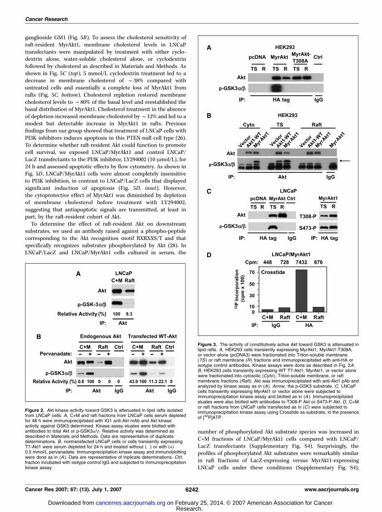
To further investigate the apparent attenuation in activity of Aktimmune complexes isolated from lipid rafts, we measured theactivity of both WT Akt (Akt1-WT) and constitutively active,MyrAkt1 expressed transiently in HEK293 and LNCaP cells. Unlikeendogenous or overexpressed WT Akt, MyrAkt1 partitioned almostequally between the nonraft and raft membrane compartments.MyrAkt1 immune complexes isolated from the C+M fraction ofHEK293 cells elicited robust phosphorylation of the GSK3 substrate(Fig. 3A and B), consistent with the reported activity of this Aktfusion protein (22). Mutation of Thr308 in the catalytic loop toalanine ablated GSK3 phosphorylation in agreement with theabsolute requirement for phosphorylation at T308 for Akt kinaseactivity (Fig. 3A ; ref. 23). In contrast, no GSK3 phosphorylation wasobserved following incubation of the substrate with Akt1-WT orMyrAkt1 immune complexes isolated from rafts (Fig. 3A and B),despite the high level of Akt present in these fractions. A similarresult was obtained with LNCaP cells (Fig. 3C). Despite strong Aktkinase activity in the C+M fraction of LNCaP cells, no GSK3phosphorylation was observed in MyrAkt1 complexes precipitatedfrom rafts and subjected to kinase assay (Fig. 3C, left). The lack ofkinase activity of raft-localized MyrAkt1 was not due to lack ofphosphorylation at Thr308 or Ser473 because these sites were heavilyphosphorylated in Akt immune complexes isolated from rafts(Fig. 3C, right). We confirmed our observations in LNCaP cells in an
Cancer Research
Cancer Res 2007; 67: (13). July 1, 2007 6240 www.aacrjournals.org
Research. on February 25, 2014. © 2007 American Association for Cancercancerres.aacrjournals.org Downloaded from

independent assay using Crosstide as substrate. As shown inFig. 3D , the activity of MyrAkt1 immune complexes isolated fromrafts toward Crosstide was dramatically reduced compared withthat precipitated from the C+M fraction.
To determine whether the activity of Akt isolated from rafts wasalso attenuated when assayed against other substrates, wemeasured the activity of MyrAkt1 immune complexes towardhistone H2B or MBP, both of which have been used previously asAkt substrates (24–27). In contrast to the activity observed againstGSK3, MyrAkt1 immune complexes isolated from rafts elicitedrobust incorporation of 32P into H2B and displayed more than10 times the activity observed with MyrAkt1 precipitated from theC+M fraction (Fig. 4A). Similar results were obtained with MyrAkt1transiently expressed in HEK293 cells (Fig. 4B), with raft-residentAkt complexes approximately four times more active against H2B
than was Akt isolated from the C+M fraction. The enhancedactivity of raft-resident MyrAkt1 relative to MyrAkt1 in the C+Mfraction was also evident when MBP was used as the substrate(Supplementary Fig. S2). Collectively, these findings suggest thatraft-resident Akt is functionally distinct from Akt present at othersubcellular locations.
To further understand how signals transmitted from raft-resident Akt differed from signaling downstream of Akt in otherlocations within the cell, we generated populations of LNCaP cellsstably expressing MyrAkt1 (Supplementary Fig. S3). Localization ofMyrAkt1 within rafts was confirmed by sucrose density gradientanalysis (Fig. 5A), which showed enrichment of the ectopicallyexpressed protein in the light buoyant density fractions. Inaddition, immunofluorescence imaging showed membrane locali-zation of MyrAkt1 as well as colocalization with the raft-restricted
Figure 1. A population of endogenousAkt1 resides in a cholesterol-richmembrane fraction. A, LNCaP cells weresubjected to sucrose density centrifugationas described. One milliliter fractions wereanalyzed for cholesterol content or blottedwith antibodies to total Akt, Gia2, c-Crk,and h-tubulin. The protein content ofindividual fractions was visualized byPonceau S staining of membranesafter transfer (bottom ). Star, thecholesterol-enriched lipid raft microdomain(fraction 4). AU, absorbance units.B, LNCaP cells growing in serum werefractionated into cytosolic/Triton-solublemembrane (C+M ) and lipid raft (Raft )fractions as described. Ten microgramsC+M and raft fractions were blottedwith antibodies to total Akt,Thr308-phosphorylated Akt (T308-P ),S473-P Akt (S473-P ), h-tubulin, andGia2. C, Akt was immunoprecipitated fromLNCaP cells exposed to cyclodextrin(CD ) and fractionated into C+M and raftfractions using IG1 anti-Akt mAb.Immunoprecipitated (IP ) eluates wereblotted with antibodies to total or S473-PAkt. D, serum-starved LNCaP cellstreated without (left, i–iv ) or with(right, v–viii ) 0.5 mmol/L pervanadate for15 min were incubated with 10 Ag/mLtetramethylrhodamine-conjugated CTxBsubunit on ice for 30 min, before incubationwith anti-S473-P Akt antibody (1:100)and FITC-conjugated secondary antibody(1:200). Arrowhead, a region ofcolocalization of S473-P Akt with CTxB asshown in (ix ). The image in (ix ) hasbeen enlarged to show red, green, andyellow pixels.
Cholesterol Sensitivity of Akt
www.aacrjournals.org 6241 Cancer Res 2007; 67: (13). July 1, 2007
Research. on February 25, 2014. © 2007 American Association for Cancercancerres.aacrjournals.org Downloaded from
ganglioside GM1 (Fig. 5B). To assess the cholesterol sensitivity ofraft-resident MyrAkt1, membrane cholesterol levels in LNCaPtransfectants were manipulated by treatment with either cyclo-dextrin alone, water-soluble cholesterol alone, or cyclodextrinfollowed by cholesterol as described in Materials and Methods. Asshown in Fig. 5C (top), 5 mmol/L cyclodextrin treatment led to adecrease in membrane cholesterol of f38% compared withuntreated cells and essentially a complete loss of MyrAkt1 fromrafts (Fig. 5C, bottom). Cholesterol repletion restored membranecholesterol levels to f80% of the basal level and reestablished thebasal distribution of MyrAkt1. Cholesterol treatment in the absenceof depletion increased membrane cholesterol by f12% and led to amodest but detectable increase in MyrAkt1 in rafts. Previousfindings from our group showed that treatment of LNCaP cells withPI3K inhibitors induces apoptosis in this PTEN-null cell type (26).To determine whether raft-resident Akt could function to promotecell survival, we exposed LNCaP/MyrAkt1 and control LNCaP/LacZ transfectants to the PI3K inhibitor, LY294002 (10 Amol/L), for24 h and assessed apoptotic effects by flow cytometry. As shown inFig. 5D , LNCaP/MyrAkt1 cells were almost completely insensitiveto PI3K inhibition, in contrast to LNCaP/LacZ cells that displayedsignificant induction of apoptosis (Fig. 5D, inset). However,the cytoprotective effect of MyrAkt1 was diminished by depletionof membrane cholesterol before treatment with LY294002,suggesting that antiapoptotic signals are transmitted, at least inpart, by the raft-resident cohort of Akt.
To determine the effect of raft-resident Akt on downstreamsubstrates, we used an antibody raised against a phospho-peptidecorresponding to the Akt recognition motif RXRXXS/T and thatspecifically recognizes substrates phosphorylated by Akt (28). InLNCaP/LacZ and LNCaP/MyrAkt1 cells cultured in serum, the
number of phosphorylated Akt substrate species was increased inC+M fractions of LNCaP/MyrAkt1 cells compared with LNCaP/LacZ transfectants (Supplementary Fig. S4). Surprisingly, theprofiles of phosphorylated Akt substrates were remarkably similarin raft fractions of LacZ-expressing versus MyrAkt1-expressingLNCaP cells under these conditions (Supplementary Fig. S4),
Figure 2. Akt kinase activity toward GSK3 is attenuated in lipid rafts isolatedfrom LNCaP cells. A, C+M and raft fractions from LNCaP cells serum depletedfor 48 h were immunoprecipitated with IG1 anti-Akt mAb and Akt kinaseactivity against GSK3 determined. Kinase assay eluates were blotted withantibodies to total Akt or p-GSK3a/h. Relative activity was determined asdescribed in Materials and Methods. Data are representative of duplicatedeterminations. B, nontransfected LNCaP cells or cells transiently expressingT7-Akt1 were serum depleted for 24 h and treated without (�) or with (+)0.5 mmol/L pervanadate. Immunoprecipitation kinase assay and immunoblottingwere done as in (A). Data are representative of triplicate determinations. Ctrl,fraction incubated with isotype control IgG and subjected to immunoprecipitationkinase assay.
Figure 3. The activity of constitutively active Akt toward GSK3 is attenuated inlipid rafts. A, HEK293 cells transiently expressing MyrAkt1, MyrAkt1-T308A,or vector alone (pcDNA3) were fractionated into Triton-soluble membrane(TS ) or raft membrane (R ) fractions and immunoprecipitated with anti-HA orisotype control antibodies. Kinase assays were done as described in Fig. 2A .B, HEK293 cells transiently expressing WT T7-Akt1, MyrAkt1, or vector alonewere fractionated into cytosolic (Cyto ), Triton-soluble membrane, or raftmembrane fractions (Raft). Akt was immunoprecipitated with anti-Akt1 pAb andanalyzed by kinase assay as in (A). Arrow, the p-GSK3 substrate. C, LNCaPcells transiently expressing MyrAkt1 or vector alone were subjected toimmunoprecipitation kinase assay and blotted as in (A ). Immunoprecipitatedeluates were also blotted with antibodies to T308-P Akt or S473-P Akt. D, C+Mor raft fractions from LNCaP cells transfected as in (C ) were subjected toimmunoprecipitation kinase assay using Crosstide as substrate, in the presenceof [32P]ATP.
Cancer Research
Cancer Res 2007; 67: (13). July 1, 2007 6242 www.aacrjournals.org
Research. on February 25, 2014. © 2007 American Association for Cancercancerres.aacrjournals.org Downloaded from

despite the enrichment for activated Akt in this compartment inLNCaP/MyrAkt1 cells. To determine the effect of cholesterolmanipulation on signaling from Akt to its substrates, whole-celllysates, C+M, or raft fractions were prepared from LNCaP/LacZand LNCaP/MyrAkt1 cells subjected to cholesterol depletion and/or repletion and blotted with the Akt substrate antibody. In controlLNCaP/LacZ cells, cyclodextrin treatment elicited a modestreduction in phosphorylation of certain Akt substrates in rafts(Fig. 6A , compare lanes 2 and 4). In marked contrast, cyclodextrintreatment led to ablation of Akt substrate phosphorylation in raftsof LNCaP/MyrAkt1 transfectants (Fig. 6A , compare lanes 6 and 8).Interestingly, signals in the C+M fraction of LNCaP/MyrAkt1transfectants, which were markedly enhanced relative to LNCaP/LacZ cells (Fig. 6A , compare lanes 1 and 5), were also diminishedwith cyclodextrin treatment in LNCaP/MyrAkt1 (Fig. 6A , comparelanes 5 and 7), suggesting cross-talk between the raft and nonraftcohorts of Akt. Repletion of membrane cholesterol led to thereappearance of Akt substrate phosphorylation predominantly in
the raft fraction of LNCaP/MyrAkt1 transfectants (data not shown),confirming the cholesterol sensitivity of Akt-dependent signaling inthese cells.
We also assessed the effect of cyclodextrin treatment on knownAkt effectors, including p70S6K, FKHR, and GSK3. As shown inFig. 6B , MyrAkt1-stimulated phosphorylation of p70S6K, FKHR,and GSK3 was diminished by cyclodextrin treatment, consistentwith cholesterol-sensitive signaling to these targets. Interestingly,the raft fraction was highly enriched for phosphorylated Aktsubstrates in the control cells compared with the C+M fraction(Fig. 6A), suggesting that in the PTEN-null background, Aktsignaling within cholesterol-rich membranes is extensive.
Discussion
In this study, we show that Akt1 is regulated directly by acholesterol-sensitive mechanism. The evidence for this conclusionis the following: (a) a population of endogenous Akt resides in acholesterol-rich membrane fraction; (b) phosphorylation of lipidraft-resident Akt1 can be attenuated by membrane cholesteroldepletion; (c) MyrAkt1, which is an oncogene, is overrepresentedin lipid raft fractions compared with WT Akt1; (d) localization ofMyrAkt1 within rafts is modulated reversibly by manipulation ofmembrane cholesterol; (e) cholesterol depletion attenuates thecytoprotective effect of MyrAkt1 in LNCaP cells exposed to PI3Kinhibition; ( f ) signaling from raft-resident Akt to its effectors isinhibited by cholesterol depletion and restored by cholesterolrepletion; and (g ) multiple targets of Akt phosphorylation residein cholesterol-rich membranes. Together, these findings indicatethat a subpopulation of Akt1 is regulated in a cholesterol-sensitive manner. They further imply that the alterations incholesterol synthesis and homeostasis observed in malignancyplay a role in the regulation of signal transduction through theAkt pathway.
The initial characterization of Akt revealed that, unlike theproto-oncogene c-akt , the oncogenic form (v-akt) possessed amyristoylation sequence arising from its fusion to the viral proteinGag (29, 30). Myristoylation of v-Akt was found to promote itsmembrane association, which in turn enhanced the tumorigenicityof v-Akt–expressing cells in xenograft experiments (31). Morerecently, a synthetic MyrAkt1 fusion protein was shown to causeprostatic intraepithelial neoplasia when expressed in the mouseprostate (32). The transforming properties of MyrAkt are believedto result from constitutive membrane targeting and constitutivephosphorylation at Thr308 and Ser473 (25). However, in view of ourcurrent findings showing that myristoylation results in overrepre-sentation of the kinase in cholesterol-rich membranes, localizationof Akt within lipid raft microdomains, as opposed to simply plasmamembrane localization, is likely to be a key determinant ofoncogenicity.
Consistent with this idea, we found that MyrAkt1 isolated fromrafts in several cell types displayed attenuated kinase activitytoward GSK3 and Crosstide despite (a) robust phosphorylation ofthe regulatory sites Thr308 and Ser473 and (b) measurable activitytoward these substrates in MyrAkt1 immune complexes isolatedfrom nonraft fractions. Interestingly, this conclusion is consistentwith an unexplained finding in a previous report that amyristoylated phospho-mimetic Akt1 mutant (Akt1-T308D/S473D), which is functionally equivalent to the MyrAkt1 constructused in our analyses, was kinase inactive toward Crosstide,despite robust activation of WT Akt1 under identical conditions
Figure 4. Constitutively active Akt1 isolated from lipid rafts displays alteredsubstrate specificity. LNCaP (A) or HEK293 (B ) cells transiently expressingMyr-HA-Akt1 were fractionated into C+M or raft fractions. Akt wasimmunoprecipitated from each fraction with HA antibody and immune complexeswere subjected to kinase assay using histone H2B as substrate. Bottom,inputs and immunoprecipitated eluates were blotted (WB ) with antibodies to totalAkt; top, incorporation of 32P into histone H2B under each condition. Dataare representative of two independent trials. Columns, mean of duplicatedeterminations; bars, SD.
Cholesterol Sensitivity of Akt
www.aacrjournals.org 6243 Cancer Res 2007; 67: (13). July 1, 2007
Research. on February 25, 2014. © 2007 American Association for Cancercancerres.aacrjournals.org Downloaded from

(33). Based on our own data, we interpret this result to mean thatmyristoylation targets proteins to lipid rafts (34–37), where thekinase complex is functionally redirected toward alternativesubstrate targets. In contrast to our observations with GSK3/Crosstide, the activity of raft-resident MyrAkt1 immune com-plexes toward H2B was 5 to 15 times greater compared withimmune complexes precipitated from C+M fractions. Thesefindings suggest for the first time that the raft environment canmodulate the activity of raft-resident kinases toward targetproteins. The difference in substrate preference cannot beexplained by differential phosphorylation of Akt regulatory sites,Thr308 and Ser473, but might result from a change in conformationof the enzyme in rafts. Alternatively, this difference may arisefrom differential association of Akt with interacting proteins
enriched in distinct subcellular compartments. In support of thissecond possibility, we recently identified a novel Akt bindingpartner that associates preferentially with Akt in cholesterol-richmembranes.5
These findings emphasize the concept that kinase activity perse is not the sole determinant of Akt oncogenicity. For example,the constitutively active Akt mutant, Akt1-T308D/S473D, displaysminimal transforming activity in chick embryo fibroblasts,whereas the kinase-inactive mutant MyrAkt1-T308A forms tumorsin chickens, albeit with an extended latency period (25). In
5 B. Cinar et al., submitted for publication.
Figure 5. Oncogenic Akt1 is enriched inlipid raft fractions and confers resistance toapoptosis induced by PI3K inhibition.A, LNCaP cells stably expressing MyrAkt1(LNCaP/MyrAkt1) were subjectedto sucrose density centrifugation. Onemilliliter fractions were blotted withantibodies to total Akt and HA. Fractions 5to 8, raft fraction. B, lipid rafts in LNCaP/MyrAkt1 cells were stained with 0.5 Ag/mLAlexa 594-CTxB for 10 min before stainingwith anti-S473-P Akt (1:100) andFITC-conjugated secondary antibody(1:100). Nuclei were counterstainedwith DAPI before imaging. Originalmagnification, �63. C, bottom, LNCaPtransfectants were treated with either5 mmol/L cyclodextrin for 1 h, 45 Ag/mLwater-soluble cholesterol (Chol ) alone for1 h, or cyclodextrin for 1 h followed bycholesterol for 1 h. Cells incubated inserum-free medium served as controls.Ten micrograms of C+M or raft fractionswere blotted with antibodies to total HA,Akt1, h-tubulin, or Gia2. Top, membranecholesterol levels following the indicatedtreatments. Columns, mean of duplicatedeterminations; bars, SD. D, inset,LNCaP/MyrAkt1 or control cells expressingLacZ (LNCaP/LacZ) were treated withoutor with 10 Amol/L LY294002 (LY) for24 h and the extent of apoptosis wasdetermined by flow cytometry. LNCaP/MyrAkt1 cells were treated without (Ctrl ) orwith 5 mmol/L cyclodextrin (1 h), 10 Amol/LLY294002 (24 h), or both agents(cyclodextrin for 1 h followed by LY294002for 24 h) and harvested for flow cytometry.Data are presented as apoptotic cells(sub-G1 peak) expressed as a percentageof the total cell population and arerepresentative of two independent trials.
Cancer Research
Cancer Res 2007; 67: (13). July 1, 2007 6244 www.aacrjournals.org
Research. on February 25, 2014. © 2007 American Association for Cancercancerres.aacrjournals.org Downloaded from

contrast, Akt3 possesses high kinase activity but poor trans-forming ability (38) consistent with the possibility that Akt exertssignaling activities independently of its kinase function. In supportof this hypothesis, two recent studies show that Akt modulates theactivity of the transforming growth factor-h effector Smad3 in amanner that is independent of Akt kinase activity (39, 40). Inthose studies, kinase-active and kinase-inactive forms of Akt werefound to interact directly with Smad3 and thereby inhibit itsactivity.
Another implication of our findings is that signals processed byraft-resident Akt are distinct from those transduced by Akt locatedelsewhere in the cell. Consistent with this possibility, localization ofMyrAkt1 to rafts seemed to sensitize Akt signaling to cholesteroldepletion. Whereas signaling to Akt targets in rafts of LNCaP/LacZcells did not change appreciably in response to cyclodextrintreatment, phosphorylation of Akt substrates in rafts of MyrAkt1-expressing cells was essentially ablated. Under these conditions, thecytoprotective effect of MyrAkt1 in LNCaP cells exposed to PI3Kinhibition was also diminished, suggesting that raft localizationalters the nature of Akt signaling to its effectors. Consistent withthese effects, changes in membrane lipid composition, eitherthrough the generation of sphingosine-1-phosphate or by exoge-nous application of omega-3 fatty acids, have been shown toredirect signaling in other cell types (41, 42). These observationsmay be consistent with recent findings showing increasedsensitivity to apoptosis induced by cholesterol depletion in cellsharboring higher amounts of cholesterol-rich membranes (9). Ourfindings may also partially explain the reported reduction in
incidence of tumors/aggressive tumors that accumulate choles-terol, such as prostate cancer, in patients on long-term statintherapy (5, 6).
Several recent studies have reported a link between elevatedcholesterol levels and cancer. Li et al. (9) showed that breast andprostate tumor cells contained higher levels of membranecholesterol and rafts/caveolae compared with their normalcounterparts. In addition, elevated synthesis of the cholesterolprecursor, mevalonate, resulting from higher expression and/oractivity of HMG-CoA reductase, has been reported in severaltumor types. Direct administration of mevalonate was found topromote the growth of orthotopic mammary tumor xenografts innude mice (43). Interestingly, Akt was shown recently to inducesterol-regulatory element binding protein–dependent transcrip-tion of multiple genes involved in fatty acid and cholesterolmetabolism, including HMG-CoA reductase, HMG-CoA synthase,and fatty acid synthase (44). In that study, a variant of MyrAkt1fused to the hormone-binding domain of the estrogen receptor(MyrAkt-ER) was used to induce gene expression changes inretinal pigment epithelial cells. Together with our current findingsshowing enrichment of MyrAkt1 in rafts, the existence of afunctional link between raft-resident Akt, regulation of cholesterolmetabolism, and membrane lipid composition is stronglysuggested. Our results suggest the existence of a potentialfeedback mechanism, whereby signals transmitted by raft-residentAkt can lead to enhanced cholesterol synthesis, expansion of thecholesterol-rich compartment, and increased localization of Aktto rafts.
Figure 6. Phosphorylation of Aktsubstrates is sensitive to cholesteroldepletion. A, 10 Ag C+M and raft fractionsfrom LNCaP/LacZ and LNCaP/MyrAkt1transfectants treated without (�) or with (+)5 mmol/L cyclodextrin were blotted withantibodies to HA, Akt1, h-tubulin, Gia2, andphosphorylated Akt substrates. B, 10 Agwhole-cell lysates prepared from LNCaP/MyrAkt1 transfectants treated as in(A) were blotted with antibodies tophosphorylated forms of p70S6K, FKHR,and GSK3. The S6K-P antibody is knownto cross-react with phosphorylatedp85S6K; the FKHR-P antibodycross-reacts with phosphorylated FoxO4;and the GSK3-P antibody recognizes bothGSK3a and GSK3h phosphorylated atSer21 and Ser9, respectively. C, the modelsummarizes the findings of the study. Aktpresent in lipid rafts displays a distinctsubstrate preference to Akt in the nonraftcompartment, consistent with a redirectionof Akt-dependent signaling when theprotein resides in cholesterol-richmembranes. Following cholesteroldepletion, signaling from raft-resident Aktis essentially ablated, whereas signalingfrom Akt in the nonraft compartment islargely unaffected.
Cholesterol Sensitivity of Akt
www.aacrjournals.org 6245 Cancer Res 2007; 67: (13). July 1, 2007
Research. on February 25, 2014. © 2007 American Association for Cancercancerres.aacrjournals.org Downloaded from
In summary, we have obtained evidence that cholesterol isa direct regulator of Akt-dependent signaling in caveolin-negativecells and that localization of Akt to lipid raft microdomainsalters the substrate preference of the Akt kinase complex.These findings suggest a direct mechanistic link betweencholesterol and cell survival signaling in tumor cells and maybe functionally relevant to the reported chemopreventive benefitof long-term use of cholesterol-lowering drugs in certaincancers.
Acknowledgments
Received 1/26/2007; revised 3/29/2007; accepted 4/19/2007.Grant support: NIH grants R37 DK47556, P50 DK65298, R01 CA112303 (M.R.
Freeman), R21 DK066412 (R.M. Adam), and R01 CA101046 (K.R. Solomon);Department of Defense grant DAMD17-03-2-0033 (M.R. Freeman); New York Academyof Medicine and the Robert Leet and Clara Guthrie Patterson Trust (R.M. Adam); andAmerican-Italian Cancer Foundation Fellowship (D. Di Vizio).
The costs of publication of this article were defrayed in part by the payment of pagecharges. This article must therefore be hereby marked advertisement in accordancewith 18 U.S.C. Section 1734 solely to indicate this fact.
We thank Dr. Gaoyuan Meng for contributions at an early stage of the study.
References1. Edidin M. The state of lipid rafts: from model mem-
branes to cells. Annu Rev Biophys Biomol Struct 2003;32:257–83.
2. Hancock JF. Lipid rafts: contentious only fromsimplistic standpoints. Nat Rev Mol Cell Biol 2006;7:456–62.
3. Simons K, Toomre D. Lipid rafts and signal transduc-tion. Nat Rev Mol Cell Biol 2000;1:31–9.
4. Demierre MF, Higgins PD, Gruber SB, Hawk E,Lippman SM. Statins and cancer prevention. Nat RevCancer 2005;5:930–42.
5. Graaf MR, Beiderbeck AB, Egberts AC, Richel DJ,Guchelaar HJ. The risk of cancer in users of statins.J Clin Oncol 2004;22:2388–94.
6. Platz EA, Leitzmann MF, Visvanathan K, et al. Statindrugs and risk of advanced prostate cancer. J Natl CancerInst 2006;98:1819–25.
7. Swyer GI. The cholesterol content of normal andenlarged prostates. Cancer Res 1942;2:372–5.
8. Schaffner CP. Prostatic cholesterol metabolism: regu-lation and alteration. In: The prostatic cell: structureand function. New York (NY): Alan R. Liss, Inc.; 1981.p. 10279–324.
9. Li YC, Park MJ, Ye SK, Kim CW, Kim YN. Elevatedlevels of cholesterol-rich lipid rafts in cancer cells arecorrelated with apoptosis sensitivity induced by choles-terol-depleting agents. Am J Pathol 2006;168:1107–18;quiz 404–5.
10. Plas DR, Thompson CB. Akt-dependent transforma-tion: there is more to growth than just surviving.Oncogene 2005;24:7435–42.
11. Stambolic V, Woodgett JR. Functional distinctions ofprotein kinase B/Akt isoforms defined by their influenceon cell migration. Trends Cell Biol 2006;16:461–6.
12. Toker A, Yoeli-Lerner M. Akt signaling and cancer:surviving but not moving on. Cancer Res 2006;66:3963–6.
13. Zhuang L, Lin J, Lu ML, Solomon KR, Freeman MR.Cholesterol-rich lipid rafts mediate akt-regulated survi-val in prostate cancer cells. Cancer Res 2002;62:2227–31.
14. Zhuang L, Kim J, Adam RM, Solomon KR, FreemanMR. Cholesterol targeting alters lipid raft compositionand cell survival in prostate cancer cells and xenografts.J Clin Invest 2005;115:959–68.
15. Kim J, Adam RM, Solomon KR, Freeman MR.Involvement of cholesterol-rich lipid rafts in interleu-kin-6-induced neuroendocrine differentiation of LNCaPprostate cancer cells. Endocrinology 2003;145:613–19.
16. Solomon KR, Mallory MA, Finberg RW. Determinationof the non-ionic detergent insolubility and phospho-protein associations of glycosylphosphatidylinositol-anchored proteins expressed on T cells. Biochem J1998;334:325–33.
17. MacLellan DL, Steen H, Adam RM, et al. Aquantitative proteomic analysis of growth factor-
induced compositional changes in lipid rafts of humansmooth muscle cells. Proteomics 2005;5:4733–42.
18. Meier R, Alessi DR, Cron P, Andjelkovic M,Hemmings BA. Mitogenic activation, phosphorylation,and nuclear translocation of protein kinase Bh. J BiolChem 1997;272:30491–7.
19. Andjelkovic M, Jakubowicz T, Cron P, Ming XF,Han JW, Hemmings BA. Activation and phosphorylationof a pleckstrin homology domain containing proteinkinase (RAC-PK/PKB) promoted by serum and proteinphosphatase inhibitors. Proc Natl Acad Sci U S A 1996;93:5699–704.
20. Conus NM, Hannan KM, Cristiano BE, HemmingsBA, Pearson RB. Direct identification of tyrosine 474 as aregulatory phosphorylation site for the Akt proteinkinase. J Biol Chem 2002;277:38021–8.
21. Murillo H, Huang H, Schmidt LJ, Smith DI, Tindall DJ.Role of PI3K signaling in survival and progression ofLNCaP prostate cancer cells to the androgen refractorystate. Endocrinology 2001;142:4795–805.
22. Andjelkovic M, Alessi DR, Meier R, et al. Role oftranslocation in the activation and function of proteinkinase B. J Biol Chem 1997;272:31515–24.
23. Alessi DR, Andjelkovic M, Caudwell B, et al.Mechanism of activation of protein kinase B by insulinand IGF-1. EMBO J 1996;15:6541–51.
24. Alessi DR, Caudwell FB, Andjelkovic M, HemmingsBA, Cohen P. Molecular basis for the substrate specificityof protein kinase B; comparison with MAPKAP kinase-1and p70 S6 kinase. FEBS Lett 1996;399:333–8.
25. Aoki M, Batista O, Bellacosa A, Tsichlis P, Vogt PK.The akt kinase: molecular determinants of oncogenicity.Proc Natl Acad Sci U S A 1998;95:14950–5.
26. Lin J, Adam RM, Santiestevan E, Freeman MR. Thephosphatidylinositol 3¶-kinase pathway is a dominantgrowth factor-activated cell survival pathway in LNCaPhuman prostate carcinoma cells. Cancer Res 1999;59:2891–7.
27. Peak M, Rochford JJ, Borthwick AC, Yeaman SJ, AgiusL. Signalling pathways involved in the stimulation ofglycogen synthesis by insulin in rat hepatocytes.Diabetologia 1998;41:16–25.
28. Zhang H, Zha X, Tan Y, et al. Phosphoprotein analysisusing antibodies broadly reactive against phosphorylatedmotifs. J Biol Chem 2002;277:39379–87.
29. Bellacosa A, Testa JR, Staal SP, Tsichlis PN. A retroviraloncogene, akt, encoding a serine-threonine kinasecontaining an SH2-like region. Science 1991;254:274–7.
30. Bellacosa A, Franke TF, Gonzalez-Portal ME, et al.Structure, expression, and chromosomal mapping ofc-akt: relationship to v-akt and its implications. Onco-gene 1993;8:745–54.
31. Ahmed NN, Franke TF, Bellacosa A, et al. Theproteins encoded by c-akt and v-akt differ in post-translational modification, subcellular localization, andoncogenic potential. Oncogene 1993;8:1957–63.
32. Majumder PK, Yeh JJ, George DJ, et al. Prostateintraepithelial neoplasia induced by prostate restrictedAkt activation: the MPAKT model. Proc Natl Acad SciU S A 2003;100:7841–6.
33. Dufner A, Andjelkovic M, Burgering BM, HemmingsBA, Thomas G. Protein kinase B localization andactivation differentially affect S6 kinase 1 activity andeukaryotic translation initiation factor 4E-bindingprotein 1 phosphorylation. Mol Cell Biol 1999;19:4525–34.
34. Moffett S, Brown DA, Linder ME. Lipid-dependenttargeting of G proteins into rafts. J Biol Chem 2000;275:2191–8.
35. Su MW, Yu CL, Burakoff SJ, Jin YJ. Targeting Srchomology 2 domain-containing tyrosine phosphatase(SHP-1) into lipid rafts inhibits CD3-induced T cellactivation. J Immunol 2001;166:3975–82.
36. Rodgers W. Making membranes green: constructionand characterization of GFP-fusion proteins targeted todiscrete plasma membrane domains. Biotechniques2002;32:1044–6, 8, 50–1.
37. Fragoso R, Ren D, Zhang X, Su MW, Burakoff SJ, JinYJ. Lipid raft distribution of CD4 depends on itspalmitoylation and association with Lck, and evidencefor CD4-induced lipid raft aggregation as an additionalmechanism to enhance CD3 signaling. J Immunol 2003;170:913–21.
38. Mende I, Malstrom S, Tsichlis PN, Vogt PK, Aoki M.Oncogenic transformation induced by membrane-targeted Akt2 and Akt3. Oncogene 2001;20:4419–23.
39. Remy I, Montmarquette A, Michnick SW. PKB/Aktmodulates TGF-h signalling through a direct interactionwith Smad3. Nat Cell Biol 2004;6:358–65.
40. Conery AR, Cao Y, Thompson EA, Townsend CM, Jr.,Ko TC, Luo K. Akt interacts directly with Smad3 toregulate the sensitivity to TGF-h induced apoptosis. NatCell Biol 2004;6:366–72.
41. Osawa Y, Uchinami H, Bielawski J, Schwabe RF,Hannun YA, Brenner DA. Roles for C16-ceramide andsphingosine 1-phosphate in regulating hepatocyteapoptosis in response to tumor necrosis factor-a. J BiolChem 2005;280:27879–87.
42. Massaro M, Habib A, Lubrano L, et al. The omega-3fatty acid docosahexaenoate attenuates endothelialcyclooxygenase-2 induction through both NADP(H)oxidase and PKCe inhibition. Proc Natl Acad Sci U S A2006;103:15184–9.
43. Duncan RE, El-Sohemy A, Archer MC. Mevalonatepromotes the growth of tumors derived from humancancer cells in vivo and stimulates proliferation in vitrowith enhanced cyclin-dependent kinase-2 activity. J BiolChem 2004;279:33079–84.
44. Porstmann T, Griffiths B, Chung YL, et al. PKB/Aktinduces transcription of enzymes involved in cholesteroland fatty acid biosynthesis via activation of SREBP.Oncogene 2005;24:6465–81.
Cancer Research
Cancer Res 2007; 67: (13). July 1, 2007 6246 www.aacrjournals.org
Research. on February 25, 2014. © 2007 American Association for Cancercancerres.aacrjournals.org Downloaded from
Copyright © 2022 FDOKUMEN