Characterization of endogenous calcium responses in neuronal cell lines
13
Characterization of endogenous calcium responses in neuronal cell lines Irina Vetter, Richard J. Lewis * Institute for Molecular Bioscience, The University of Queensland, St Lucia, Queensland 4072, Australia 1. Introduction In recent years, an increasing number of receptors and ion channels expressed on central or peripheral neurons have been identified as putative therapeutic targets for the treatment of neuronal pathophysiologies such as chronic pain, stroke, epilepsy and schizophrenia. These targets include transient receptor potential channels (e.g. TRPV1), voltage-gated calcium channels (e.g. Ca v 2.2 and 3.2), ionotropic glutamate (e.g. NMDA and AMPA receptors) and nicotinic acetylcholine receptors (e.g. a7 and a4 nAChR), as well as numerous G-protein coupled receptors (GPCR) [1–7]. GPCR targets of particular interest include the protease-activated receptors, speci- fically PAR1, PAR2 and PAR4, which are expressed in dorsal root ganglion (DRG) and central neurons and contribute to a variety of painful conditions, including visceral and arthritic pain and have also been speculated to contribute to epilepsy and blood brain barrier integrity [8–13]. Similarly, interest in bradykinin B1 and B2 receptors has been renewed with the observation that intra-articular administration of a bradykinin B2 antagonist appears promising in the treatment of painful osteoarthritis [14], while bradykinin B1 antagonists may be particularly useful in chronic inflammatory pain [15]. Also emerging as potential therapeutic targets are the neurotensin receptors, with both neurotensin 1 and neurotensin 2 receptors contributing to the antinociceptive effects of neurotensin [16] and the neurotensin analogue contulakin G from Conus geographus [17]. In addition, targeting neurotensin receptors has also been suggested as a promising novel approach to treat schizophrenia and epilepsy [18,19]. Additional GPCRs with ther- apeutic potential in neuronal pathophysiologies such as pain, epilepsy, stroke and schizophrenia include muscarinic acetylcholine receptors [20,21], in particular M2 and M4 receptors which mediate analgesia [22,23]; D2 dopamine receptors [24,25], histamine H1–H3 receptors [26,27], several metabotropic glutamate receptors [28,29], Biochemical Pharmacology 79 (2010) 908–920 ARTICLE INFO Article history: Received 18 September 2009 Accepted 21 October 2009 Keywords: Endogenous Neuronal cell line Characterization Calcium Receptor Ion channel ABSTRACT An increasing number of putative therapeutic targets have been identified in recent years for the treatment of neuronal pathophysiologies including pain, epilepsy, stroke and schizophrenia. Many of these targets signal through calcium (Ca 2+ ), either by directly facilitating Ca 2+ influx through an ion channel, or through activation of G proteins that couple to intracellular Ca 2+ stores or voltage-gated Ca 2+ channels. Immortalized neuronal cell lines are widely used models to study neuropharmacology. However, systematic pharmacological characterization of the receptors and ion channels expressed in these cell lines is lacking. In this study, we systematically assessed endogenous Ca 2+ signaling in response to addition of agonists at potential therapeutic targets in a range of cell lines of neuronal origin (ND7/23, SH-SY5Y, 50B11, F11 and Neuro2A cells) as well as HEK293 cells, a cell line commonly used for over-expression of receptors and ion channels. This study revealed a remarkable diversity of endogenous Ca 2+ responses in these cell lines, with one or more cell lines responding to addition of trypsin, bradykinin, ATP, nicotine, acetylcholine, histamine and neurotensin. Subtype specificity of these responses was inferred from agonist potency and the effect of receptor subtype specific antagonist. Surprisingly, HEK293 and SH-SY5Y cells responded to the largest number of agonists with potential roles in neuronal signaling. These findings have implications for the heterologous expression of neuronal receptors and ion channels in these cell lines, and highlight the potential of neuron-derived cell lines for the study of a range of endogenously expressed receptors and ion channels that signal through Ca 2+ . Crown Copyright ß 2009 Published by Elsevier Inc. All rights reserved. Abbreviations: Ca 2+ , calcium ion; GPCR, G-protein coupled receptor; ATP, adenosine triphosphate; TRPV1, transient receptor potential vanilloid 1; PAR, protease- activated receptor; GABA, g-aminobutyric acid; DMEM, Dulbeccos’ Modified Eagle Medium; FBS, foetal bovine serum; PBS, phosphate buffered saline; PSS, physiological salt solution; EDTA, ethylenediaminetetraacetic acid; HAT, hypox- anthine, aminopterin and thymidine; db-cAMP, N6,2 0 -O-dibutyryladenosine 3 0 :5 0 cyclic monophosphate; NGF, nerve growth factor; BAPTA, 1,2-bis(o-aminophenox- y)ethane-N,N,N 0 ,N 0 -tetraacetic acid; AFU, arbitrary fluorescence unit; SEM, standard error of the mean; NMDA, N-methyl-D-aspartic acid; pFHHSiD, p- fluorohexahydro-sila-difenidol hydrochloride; PLC, phopholipase C; Fluo-4-AM, Fluo-4 acetomethoxyester; HEK293, human embryonic kidney cells; RPMI, Roswell Park Memorial Institute; PDL, Poly-D-lysine. * Corresponding author. Tel.: +61 7 3346 2984; fax: +61 7 3346 2101. E-mail addresses: [email protected] (I. Vetter), [email protected] (R.J. Lewis). Contents lists available at ScienceDirect Biochemical Pharmacology journal homepage: www.elsevier.com/locate/biochempharm 0006-2952/$ – see front matter . Crown Copyright ß 2009 Published by Elsevier Inc. All rights reserved. doi:10.1016/j.bcp.2009.10.020
Transcript of Characterization of endogenous calcium responses in neuronal cell lines

Characterization of endogenous calcium responses in neuronal cell lines
Irina Vetter, Richard J. Lewis *
Institute for Molecular Bioscience, The University of Queensland, St Lucia, Queensland 4072, Australia
Biochemical Pharmacology 79 (2010) 908–920
A R T I C L E I N F O
Article history:
Received 18 September 2009
Accepted 21 October 2009
Keywords:
Endogenous
Neuronal cell line
Characterization
Calcium
Receptor
Ion channel
A B S T R A C T
An increasing number of putative therapeutic targets have been identified in recent years for the
treatment of neuronal pathophysiologies including pain, epilepsy, stroke and schizophrenia. Many of
these targets signal through calcium (Ca2+), either by directly facilitating Ca2+ influx through an ion
channel, or through activation of G proteins that couple to intracellular Ca2+ stores or voltage-gated Ca2+
channels. Immortalized neuronal cell lines are widely used models to study neuropharmacology.
However, systematic pharmacological characterization of the receptors and ion channels expressed in
these cell lines is lacking. In this study, we systematically assessed endogenous Ca2+ signaling in
response to addition of agonists at potential therapeutic targets in a range of cell lines of neuronal origin
(ND7/23, SH-SY5Y, 50B11, F11 and Neuro2A cells) as well as HEK293 cells, a cell line commonly used for
over-expression of receptors and ion channels. This study revealed a remarkable diversity of endogenous
Ca2+ responses in these cell lines, with one or more cell lines responding to addition of trypsin,
bradykinin, ATP, nicotine, acetylcholine, histamine and neurotensin. Subtype specificity of these
responses was inferred from agonist potency and the effect of receptor subtype specific antagonist.
Surprisingly, HEK293 and SH-SY5Y cells responded to the largest number of agonists with potential roles
in neuronal signaling. These findings have implications for the heterologous expression of neuronal
receptors and ion channels in these cell lines, and highlight the potential of neuron-derived cell lines for
the study of a range of endogenously expressed receptors and ion channels that signal through Ca2+.
Crown Copyright � 2009 Published by Elsevier Inc. All rights reserved.
Contents lists available at ScienceDirect
Biochemical Pharmacology
journa l homepage: www.e lsev ier .com/ locate /b iochempharm
1. Introduction
In recent years, an increasing number of receptors and ionchannels expressed on central or peripheral neurons have beenidentified as putative therapeutic targets for the treatment ofneuronal pathophysiologies such as chronic pain, stroke, epilepsyandschizophrenia.Thesetargets includetransientreceptorpotentialchannels (e.g. TRPV1), voltage-gated calcium channels (e.g. Cav2.2and 3.2), ionotropic glutamate (e.g. NMDA and AMPA receptors) and
Abbreviations: Ca2+, calcium ion; GPCR, G-protein coupled receptor; ATP, adenosine
triphosphate; TRPV1, transient receptor potential vanilloid 1; PAR, protease-
activated receptor; GABA, g-aminobutyric acid; DMEM, Dulbeccos’ Modified Eagle
Medium; FBS, foetal bovine serum; PBS, phosphate buffered saline; PSS,
physiological salt solution; EDTA, ethylenediaminetetraacetic acid; HAT, hypox-
anthine, aminopterin and thymidine; db-cAMP, N6,20-O-dibutyryladenosine 30:50
cyclic monophosphate; NGF, nerve growth factor; BAPTA, 1,2-bis(o-aminophenox-
y)ethane-N,N,N0 ,N0-tetraacetic acid; AFU, arbitrary fluorescence unit; SEM,
standard error of the mean; NMDA, N-methyl-D-aspartic acid; pFHHSiD, p-
fluorohexahydro-sila-difenidol hydrochloride; PLC, phopholipase C; Fluo-4-AM,
Fluo-4 acetomethoxyester; HEK293, human embryonic kidney cells; RPMI, Roswell
Park Memorial Institute; PDL, Poly-D-lysine.
* Corresponding author. Tel.: +61 7 3346 2984; fax: +61 7 3346 2101.
E-mail addresses: [email protected] (I. Vetter),
[email protected] (R.J. Lewis).
0006-2952/$ – see front matter . Crown Copyright � 2009 Published by Elsevier Inc. A
doi:10.1016/j.bcp.2009.10.020
nicotinic acetylcholine receptors (e.g. a7 and a4 nAChR), as well asnumerous G-protein coupled receptors (GPCR)[1–7]. GPCRtargetsofparticular interest include the protease-activated receptors, speci-fically PAR1, PAR2 and PAR4, which are expressed in dorsal rootganglion (DRG) and central neurons and contribute to a variety ofpainful conditions, including visceral and arthritic pain and have alsobeen speculated to contribute to epilepsy and blood brain barrierintegrity [8–13]. Similarly, interest inbradykinin B1and B2receptorshas been renewed with the observation that intra-articularadministration of a bradykinin B2 antagonist appears promisingin the treatment of painful osteoarthritis [14], while bradykinin B1antagonists may be particularly useful in chronic inflammatory pain[15]. Also emerging as potential therapeutic targets are theneurotensin receptors, with both neurotensin 1 and neurotensin 2receptors contributing to the antinociceptive effects of neurotensin[16] and the neurotensin analogue contulakin G from Conus
geographus [17]. In addition, targeting neurotensin receptors hasalso been suggested as a promising novel approach to treatschizophrenia and epilepsy [18,19]. Additional GPCRs with ther-apeutic potential in neuronal pathophysiologies such as pain,epilepsy, stroke and schizophrenia include muscarinic acetylcholinereceptors [20,21], in particular M2 and M4 receptors which mediateanalgesia [22,23]; D2 dopamine receptors [24,25], histamine H1–H3receptors [26,27], several metabotropic glutamate receptors [28,29],
ll rights reserved.

I. Vetter, R.J. Lewis / Biochemical Pharmacology 79 (2010) 908–920 909
GABAB receptors [30] and several serotonin 5-HT [31–34] as well aspurinergic receptor subtypes [35–39].
Importantly, many of these putative therapeutic targets signalthrough an increase in intracellular calcium (Ca2+), either bydirectly facilitating Ca2+ influx through an ion channel, or throughactivation of G proteins that couple to intracellular Ca2+ stores orvoltage-gated Ca2+ channels. This makes assessment of Ca2+ fluxesin response to agonists and antagonists at these targets a broadlyapplicable approach that can be optimized for detailed functionalstudies as well as the high throughput identification of novelmodulators of these targets.
Primary cultures of adult and embryonic neurons are widelyused to investigate neuronal pharmacology and signaling mechan-isms. However, the culture of primary neurons is not only time-intensive but can also yield cell populations that have undergonevariable extents of differentiation associated with their removaland culture. In addition, in the case of primary DRG neurons,primary culture yields heterogenous cell populations, which canmake identification of neuronal cells that express receptors or ionchannels of interest difficult. To gain more reproducible responses,several immortalized dorsal root ganglion and neuroblastoma-derived cell lines have been established that allow the study of thephysiology and pharmacology of endogenously expressed recep-tors and ion channels in vitro. While some neuronal cell lines havebeen used extensively to assess neuronal physiology such asneurotransmitter release, the systematic characterization of Ca2+
responses to agonists of endogenously expressed receptors and ionchannels is lacking.
In the present study, we have systematically characterizedendogenous Ca2+ responses to several agonists at GPCRs and ionchannels in three dorsal root ganglion-derived neuronal cell lines,F11 [40], ND7/23 [41] and 50B11 [42] cells, as well as thecommonly used human neuroblastoma cell lines SH-SY5Y [43] andNeuro2A [44], and HEK293 (human embryonic kidney) cells [45]which are commonly used to express neuronal receptors and ionchannels. Surprisingly, Ca2+ responses of these cell lines werediverse, with HEK293 and SH-SY5Y cells responding to the largestnumber of agonists with potential roles in neuronal signaling. Ourresults highlight the potential of DRG- and neuroblastoma-derivedcell lines for the study of endogenously expressed neuronalreceptors and ion channels through Ca2+ signaling and provide thefirst systematic evaluation of endogenous Ca2+ signaling incommonly used cell lines of neuronal origin.
2. Materials and methods
2.1. Compounds
The following compounds and materials were obtained fromthe sources indicated: L-glutamine, horse serum, Dulbecco’sModified Eagle Medium (DMEM), Ham’s F12, Neurobasal, RPMI(Roswell Park Memorial Institute), 0.25% trypsin/EDTA, B27, foetalbovine serum (FBS), Fluo-4-AM (Fluo-4 acetomethoxyester),phosphate buffered saline (PBS), Invitrogen (Mulgrave, Victoria,Australia); HAT media supplement Hybri-MaxTM, trypsin, brady-kinin, adenosine triphosphate (ATP), nicotine, acetylcholine, cho-line bitartrate, noradrenaline, dopamine, histamine, glutamate, N-methyl-D-aspartic acid (NMDA), g-aminobutyric acid (GABA),serotonin, adenosine, substance P, neurotensin, neurokinin A,neurokinin B, capsaicin, Triton-X 100, MnCl2, HOE-140, p-fluorohexahydro-sila-difenidol hydrochloride, pyrilamine, piren-zepine (pFHHSiD), BAPTA (1,2-bis(o-aminophenoxy)ethane-N,N,N0,N0-tetraacetic acid), ranitidine, Sigma–Aldrich (Castle Hill,New South Wales, Australia); morphine, David Bull LaboratoriesAustralia (Rowville, Victoria, Australia); nerve growth factor (NGF),GroPep Australia (Thebarton, South Australia, Australia). Capsaicin
was prepared as a stock solution in ethanol, with the maximumfinal ethanol concentration not exceeding 0.001%. All otherreagents were prepared as stock solutions in PBS. SLIGRL-NH2
and TFLLR-NH2 were a kind gift from Prof. David Fairlie, TheInstitute for Molecular Bioscience, University of Queensland.
2.2. Cell lines and culture
All cell lines were routinely maintained in antibiotic-freemedia, with regular testing confirming mycoplasma-free status ofcell lines.
HEK293 cells (American Tissue Culture Collection, Manassas,VA, USA) were maintained at 37 8C in a 5% humidified CO2
incubator in DMEM containing 10% heat-inactivated FBS, 2 mM L-glutamine, pyridoxine and 110 mg/ml sodium pyruvate. Cells weresplit every 4–6 days or when approximately 90% confluent in aratio of 1:5 to 1:10 using 0.25% trypsin/EDTA and subcultured intoT-75 cm2 or T-175 cm2 tissue culture flasks.
SH-SY5Y human neuroblastoma cells, a kind gift from VictorDiaz (Max Planck Institute for Experimental Medicine, Goettingen,Germany), were maintained at 37 8C/5% CO2 in RPMI containing15% FBS and 2 mM L-glutamine. Cells were routinely passaged at a1:5 dilution every 3–5 days using 0.25% trypsin/EDTA (Gibco).
F11 neuroblastoma � DRG neuron hybrid cells were kindlyprovided by Daniel Kapitzke (School of Pharmacy, The Universityof Queensland, Australia) and maintained in Ham’s F12 mediasupplemented with 10% FBS, 100 mM hypoxanthine, 0.4 mMaminopterin and 16 mM thymidine (HAT media supplementHybri-MaxTM, Sigma–Aldrich, Castle Hill, Australia). F11 cellswere passaged at a 1:5 dilution following mechanical dissociationevery 2–3 days or when approximately 80% confluent.
ND7/23 neuroblastoma � DRG neuron hybrid cells wereobtained from Sigma–Aldrich (Castle Hill, New South Wales,Australia) and grown to approximately 90–95% confluency inDMEM containing 10% heat-inactivated FBS, 2 mM L-glutamine,pyridoxine and 110 mg/ml sodium pyruvate. Confluent cultureswere dissociated using 0.25% trypsin/EDTA and split at 1:5 dilutionevery 3–5 days.
The immortalized DRG sensory neuron cell line 50B11 was akind gift from A/Prof Ahmet Hoke (Departments of Neurology andNeuroscience, Johns Hopkins University, Baltimore, USA). 50B11cells were maintained in Neurobasal medium (Gibco) supplemen-ted with 10% FBS, 1� B27 (Gibco), 0.5 mM L-glutamine and 0.2%glucose [42]. Cells were grown to approximately 90–95%confluency and dissociated using 0.25% trypsin/EDTA for passagingat 1:3 dilution every 3–5 days. Under these conditions, 50B11 cellsremained sensitive to differentiation by forskolin for up to 20passages.
Neuro2A cells (American Tissue Culture Collection, Manassas,VA, USA) were maintained in DMEM containing 10% heat-inactivated FBS, 2 mM L-glutamine, pyridoxine and 110 mg/mlsodium pyruvate. Cells were split every 4–6 days or whenapproximately 90% confluent at a 1:2 to 1:4 dilution using0.25% trypsin/EDTA and subcultured into T-75 cm2 or T-175 cm2
tissue culture flasks.Experiments on each cell line were conducted over a period of
several months and spanned on average a minimum of 10–20passages per cell line. Responses were not significantly affected bypassage number, with the exception of histamine responses in SH-SY5Y cells, which were consistent in size during the first 5 passagesbefore becoming variable in subsequent passages.
2.3. Measurement of intracellular Ca2+ responses
To assess increases in intracellular Ca2+ in response to additionof agonists, cells were seeded on either uncoated (SH-SY5Y and

Ta
ble
1C
ha
ract
eri
zati
on
of
en
do
ge
no
us
Ca
2+
resp
on
ses
ince
llli
ne
so
fn
eu
ron
al
ori
gin
a.
ND
7/2
3F1
15
0B
11
SH
-SY
5Y
Ne
uro
2A
HE
K
pE
C5
0%
F Ma
xp
EC
50
%F M
ax
pE
C5
0%
F Ma
xp
EC
50
%F M
ax
pE
C5
0%
F Ma
xp
EC
50
%F M
ax
Ace
tylc
ho
lin
e–
––
6.7�
0.2
29
.6–
46
.4–
7.1�
0.0
25
7.9
–6
5.0
AT
P–
4.9�
0.1
26
.0–
41
.74
.9�
0.1
1.5
–6
.8–
5.2�
0.5
1.8
–3
.56
.0�
0.1
40
.6–
55
.0
Bra
dy
kin
in8
.3�
0.1
12
.6–
16
.21
1.4�
0.1
47
.2–
69
.3–
7.4�
0.3
4.1
–5
.16
.6�
0.0
75
.6–
6.0
6.9�
0.5
6.3
–1
1.4
8.7�
0.1
Ch
oli
ne
––
–5
.1�
0.2
45
.6–
71
.0–
–
His
tam
ine
––
–5
.8�
0.0
91
2.3
-23
.7–
4.9�
0.1
2.7
–3
.2
Ne
uro
ten
sin
––
––
–7
.2�
0.1
3.2
–3
.6
Nic
oti
ne
––
–5
.5�
0.0
41
3.4
–2
9.0
––
Try
psi
n–
7.4�
0.2
22
.6–
38
.2–
7.0�
0.1
27
.6–
47
.87
.8�
0.0
23
.1–
3.3
8.3�
0.0
36
7.5
–7
7.1
aC
a2
+re
spo
nse
sto
ad
dit
ion
of
ag
on
ists
we
rea
sse
sse
db
ym
on
ito
rin
gFl
uo
-4fl
uo
resc
en
cein
HE
K2
93
,ND
7/2
3,S
H-S
Y5
Y,5
0B
11
,F1
1a
nd
Ne
uro
2A
cell
su
sin
ga
FLIP
RT
ET
RA
flu
ore
sce
nce
pla
tere
ad
er.
Re
spo
nd
ers
an
dn
on
-re
spo
nd
ers
(�)
we
red
ete
rmin
ed
by
init
iala
sse
ssm
en
to
fa
pp
rox
ima
tely
lite
ratu
reE
C8
0a
go
nis
tco
nce
ntr
ati
on
s.N
ore
spo
nse
sw
ere
ob
serv
ed
inth
ese
cell
lin
es
toa
dd
itio
no
fa
de
no
sin
e,c
ap
saic
in,d
op
am
ine
,GA
BA
,glu
tam
ate
,mo
rph
ine
,ne
uro
kin
inA
an
dB
,NM
SA
,no
rad
ren
ali
ne
,se
roto
nin
an
dsu
bst
an
ceP
.EC
50s,
ma
xim
um
resp
on
se(%
F Ma
x)
an
dH
ills
lop
ew
ere
de
term
ine
dfo
ra
cety
lch
oli
ne
,AT
P,b
rad
yk
inin
,ch
oli
ne
,his
tam
ine
,ne
uro
ten
sin
,nic
oti
ne
an
dtr
yp
sin
inre
spo
nd
ing
cell
lin
es.
Hil
lslo
pe
sw
ere
1.0
for
all
ag
on
ists
ex
cep
ta
cety
lch
oli
ne
inH
EK
29
3ce
lls
(2.2�
0.1
),A
TP
inF1
1(2
.1�
0.3
)an
dH
EK
29
3ce
lls
(2.2�
0.9
),ch
oli
ne
inSH
-SY
5Y
(2.7�
0.3
)an
dtr
yp
sin
inF1
1ce
lls
(3.3�
0.8
).D
ata
are
pre
sen
ted
asra
ng
e(%
F Ma
x)
or
mea
n�
SEM
(pE
C5
0)
of
3in
dep
end
ent
exp
erim
ents
.
I. Vetter, R.J. Lewis / Biochemical Pharmacology 79 (2010) 908–920910
50B11 cells) or Poly-D-lysine (PDL)-coated (HEK293, ND7/23, F11and Neuro2A cells) black-walled imaging plates (Corning, Lowell,MA, USA) 48 h prior to the experiment. Seeding densities were50 000 cells/well (HEK293, ND7/23, F11 and Neuro2A cells),100 000 cells/well (50B11 cells) or 120 000 cells/well (SH-SY5Ycells), resulting in 90–95% confluent monolayers on the day of Ca2+
imaging.To induce differentiation, media was changed 24 h after plating
to DMEM supplemented with 0.5% FBS, 1 mM db-cAMP (N6,20-O-dibutyryladenosine 30:50 cyclic monophosphate) and 2 nM NGF(ND7/23, F11 and Neuro2A cells) or complete Neurobasal mediacontaining 75 mM forskolin (50B11 cells). Microscopic examina-tion of cells differentiated in this manner confirmed extensiveneurite outgrowth and neuronal morphology compared toundifferentiated cultures.
Cells were loaded for 30 min at 37 8C with 5 mM Fluo-4-AM inphysiological salt solution (PSS; composition NaCl 140 mM,glucose 11.5 mM, KCl 5.9 mM, MgCl2 1.4 mM, NaH2PO4 1.2 mM,NaHCO3 5 mM, CaCl2 1.8 mM, HEPES 10 mM) containing inaddition 0.3% BSA. To allow for completion of de-esterification,cells were incubated for 10–15 min with PSS. These conditionsresulted in good loading for all cell lines assessed, evidenced bygood signal-to-noise ratios. After 2 washes with PSS, or Ca2+-freePSS containing 0.5 mM BAPTA to assess ATP responses in theabsence of extracellular Ca2+, cells were transferred to theFLIPRTETRA (Molecular Devices, Sunnyvale, CA) fluorescent platereader and Ca2+ responses measured using a cooled CCD camerawith excitation at 470–495 nM and emission at 515–575 nM.Camera gain and intensity were adjusted for each plate to yield aminimum of 1000 arbitrary fluorescence units (AFU) baselinefluorescence. Prior to addition of agonists, 10 baseline fluorescencereadings were taken, followed by fluorescent readings everysecond for 300 s.
To normalize fluorescence signals between cell lines, maximum(FMax) and minimum (FMin) fluorescence values were determinedfor each well. Specifically, to determine maximal fluorescence(FMax values) for each well, 0.3% Triton X-100 was added at the endof each experiment and resultant fluorescence increases mon-itored for a further 60 s. To determine FMin values, 100 mM MnCl2
was subsequently added and fluorescence monitored for anadditional 60 s. Responses were then normalized as described inSection 2.4. FMax values obtained after addition of 0.3% Triton-Xwere 6099 � 239 for 50B11 cells, 4242 � 220 for F11 cells,2424 � 118 for Neuro2A cells, 4438 � 544 for ND7/23 cells,6559 � 343 for SH-SY5Y cells and 5729 � 117 for HEK293 cells(n = 4–6 plates).
To assess putative endogenous Ca2+ responses to agonists, aninitial screen was carried out using a single agonist concentrationapproximating literature reported EC80 values. Agonists that failedto change the Fluo-4 fluorescence were confirmed a minimum ofthree times, and full concentration–response curves were deter-mined for responders. For each concentration–response curve, Ca2+
responses were assessed in at least triplicate and confirmed in atleast 3 independent experiments.
2.4. Data analysis
Unless otherwise stated, all data are expressed as mean� standard error of the mean (SEM) either from a single representa-tive experiment or averaged data from at least three separateexperiments. Unless otherwise stated, statistical significance wasdetermined using an unpaired, two-tailed Student’s t-test, withstatistical significance defined as p < 0.05.
To enable comparison of the magnitude of responses betweencell lines, raw fluorescence values were converted to % FMax valuesusing (F � FMin)/(FMax � FMin) � 100 where F is the fluorescence

I. Vetter, R.J. Lewis / Biochemical Pharmacology 79 (2010) 908–920 911
after agonist addition and Fmin is the fluorescence after addition ofMnCl2, which was generally comparable to baseline fluorescence.To establish concentration–response curves, the maximumincrease in % FMax after addition of agonist was plotted usingGraphPad Prism Version 4.01 and a 4-parameter Hill equationeither with Hill coefficient = 1, a variable slope, or a two-site fit wasfitted to the data. For statistical comparison of best-fit values forthe different concentration–response curves, the comparisonfunction of GraphPad Prism was used to compute the F-test value,with statistical significance defined as p < 0.05.
3. Results
Endogenous Ca2+ responses to 19 agonists of ligand-gated ionchannels and G-protein coupled receptors were assessed inHEK293, SH-SY5Y, ND7/23, 50B11, F11 and Neuro2A (Table 1).
Fig. 1. Trypsin elicits Ca2+ transients in HEK293, SH-SY5Y, F11 and Neuro2A cells. (A) Ad
HEK293, SH-SY5Y, F11 and Neuro2A but not ND7/23 and 50B11 cells. Trypsin-mediated
and of relatively small magnitude in Neuro2A cells. (B) Addition of trypsin to HEK2
concentration leading to rapid and sustained elevations in Ca2+ (red trace), while repeat
trypsin concentrations caused small transient with delayed onset (blue trace). (C) Repres
(blue trace), F11 (green trace) and Neuro2A (orange trace) cells. (D) Addition of the PAR2
HEK293, but not SH-SY5Y, F11 or Neuro2A cells. (E) The PAR1-specific agonist elicited re
addition of SLIGRL-NH2 (purple; 1 mM) and TFLLR-NH2 (blue; 1 mM) with concentration-
single Ca2+ transient, SLIGRL-NH2, similar to trypsin, caused concentration-dependent Ca2
are representative of 3 independent experiments. (For interpretation of the references to
Initially, responses to approximately literature EC80 concentra-tions of agonists were assessed to identify responders and non-responders, followed by generation of full concentration–responsecurves for agonists eliciting detectable Ca2+ responses. Specifically,Ca2+ responses to addition of trypsin (1 mM), bradykinin (100 mM),nicotine (100 mM), acetylcholine (10 mM), noradrenaline(100 mM), dopamine (1 mM), histamine (1 mM), glutamate(1 mM), NMDA (1 mM), GABA (1 mM), serotonin (1 mM), adeno-sine (100 mM), ATP (1 mM), substance P (100 mM), neurotensin(1 mM), neurokinin A and B (1 mM), capsaicin (300 nM) andmorphine (1 mM) were assessed by monitoring Fluo-4 fluores-cence using the FLIPRTETRA fluorescence plate reader. One or morecell lines responded with either transient or sustained Ca2+
increases to addition of trypsin, bradykinin, ATP, nicotine,acetylcholine, histamine and neurotensin (Figs. 1–8). No increasesin intracellular Ca2+ were observed in response to addition of
dition of trypsin caused concentration-dependent increases in intracellular Ca2+ in
Ca2+ responses were largest in HEK293 cells, intermediate in SH-SY5Y and F11 cells
93 cells caused concentration-dependent Ca2+ oscillations, with high (100 nM)
ed peaks were observed upon addition of 10 nM trypsin (purple trace). Low (1 nM)
entative Ca2+ transients to addition of 1 mM trypsin in HEK293 (red trace), SH-SY5Y
-selective agonist SLIGRL-NH2 caused a measurable increase in intracellular Ca2+ in
sponses in HEK293, SH-SY5Y, F11 and Neuro2A cells. (F) HEK293 cells responded to
dependent transient increases in intracellular Ca2+. While TFLLR-NH2 lead to only a+ oscillations in HEK293 cells. Data are presented as mean � SEM of n = 3–4 wells and
color in this figure legend, the reader is referred to the web version of the article.)

Fig. 2. Bradykinin elicits Ca2+ transients in HEK293, SH-SY5Y, ND7/23, F11 and
Neuro2A cells. (A) Addition of bradykinin caused concentration-dependent
increases in intracellular Ca2+ in HEK293, SH-SY5Y, ND7/23, F11 and Neuro2A
cells but not 50B11 cells. F11 cells (green) responded to addition of bradykinin with
large Ca2+ transients that fit preferentially to a two-site fit (p < 0.05). Responses in
ND7/23 cells (purple) were intermediate, with relatively small responses in
Neuro2A (orange), HEK293 (red) and SH-SY5Y (blue) cells. (B) Representative Ca2+
transients to addition of 1 mM bradykinin in F11 (green), ND7/23 (purple), Neuro2A
(orange), HEK293 (red) and SH-SY5Y (blue) cells. (C) The bradykinin B2 antagonist
HOE-140 inhibited bradykinin (1 mM) induced Ca2+ transients in a concentration-
dependent manner in F11 (green), ND7/23 (purple), SH-SY5Y (blue), HEK293 (red)
and Neuro2A (orange) cells. Data are presented as mean � SEM of n = 3–4 wells and
are representative of 3 independent experiments. (For interpretation of the references
to color in this figure legend, the reader is referred to the web version of the article.)
I. Vetter, R.J. Lewis / Biochemical Pharmacology 79 (2010) 908–920912
noradrenaline, dopamine, glutamate, NMDA, GABA, serotonin,adenosine, substance P, neurokinin A and B, capsaicin andmorphine in any of the cell lines investigated.
3.1. Trypsin
HEK293, SH-SY5Y, F11 and Neuro2A cells responded to the PARagonist trypsin with increases in intracellular Ca2+, while 50B11and ND7/23 cells did not respond (Fig. 1A). The observed EC50s fortrypsin-induced Ca2+ responses in SH-SY5Y, F11 and Neuro2Acells were 8.9 � 10�8, 4.1 � 10�8 and 1.7 � 10�8 M, respectively,while trypsin was�10-fold more potent (EC50 of 5.1 � 10�9 M) inHEK293 cells. Trypsin responses in SH-SY5Y, F11 and particularlyNeuro2A cells were significantly smaller compared to responsesin HEK293 cells (Fig. 1A). In HEK293 cells, addition of intermediateconcentrations of trypsin evoked Ca2+ oscillations, consistentwith IP3-mediated release of Ca2+ from intracellular storesthrough PAR2 (Fig. 1B). In contrast, addition of trypsin to SH-SY5Y, F11 and Neuro2A cells caused a single transient Ca2+
increase, consistent with activation of GPCR without Ca2+
oscillations (Fig. 1C). In SH-SY5Y, F11 and Neuro2A cells, trypsindid not cause Ca2+ oscillations at concentrations from 1 nM to1 mM. To confirm the PAR subtype(s) involved in mediatingresponses to trypsin, increases in intracellular Ca2+ to the PAR2-specific peptide SLIGRL-NH2 and the PAR1-specific peptide TFLLR-NH2 were assessed. Only HEK293 cells responded to addition ofSLIGRL-NH2 with an EC50 of 1.1 � 10�6 M, while F11, Neuro2A andSH-SY5Y cells did not respond to SLIGRL-NH2 (Fig. 1D). In contrast,all cell lines responded to addition of the PAR1 agonist TFLLR-NH2
with transient increases in intracellular Ca2+ (Fig. 1E). Theobserved EC50s for TFLLR-NH2-induced Ca2+ responses inHEK293, SH-SY5Y, F11 and Neuro2A cells were 5.4 � 10�6,5.0 � 10�6, 3.2 � 10�6 and 5.2 � 10�6 M, respectively. Similar totrypsin responses, SLIGRL-NH2 caused Ca2+ oscillations in HEK293cells, while TFLLR-NH2 caused a single transient Ca2+ increase(Fig. 1F).
3.2. Bradykinin
All cell lines except 50B11 immortalized DRG neuronsresponded to bradykinin with a transient increase in intracellularCa2+ (Fig. 2A and B). HEK293, SH-SY5Y and Neuro2A cells exhibitedsmall increases in intracellular Ca2+ in response to bradykinin andhad comparatively high EC50 values of 1.4 � 10�7, 3.8 � 10�8 and2.3 � 10�7 M, respectively. ND7/23 cells responded with highermagnitude increases in intracellular Ca2+, with a low nanomolarEC50 of 7.5 � 10�9 M. F11 cells displayed very large Ca2+ transientsin response to addition of bradykinin, with responses occurringover a wide range of concentrations. The preferred fit (p < 0.05)was a two-site model with EC50s of 1.8 � 10�9 and 3.7 � 10�12 Mand an approximate response fraction of 0.5, suggesting thepresence of distinct subtypes or two conformational or signalingstates of the bradykinin B2 receptors.
The specific bradykinin B2 receptor antagonist HOE-140inhibited bradykinin (1 mM) responses with an IC50 of 4.1 �10�8 M in HEK293, 7.0 � 10�8 M in SH-SY5Y cells, 5.8 � 10�8 M inND7/23 and 5.8 � 10�8 M in Neuro2A cells. Surprisingly, brady-kinin-induced responses in F11 cells were less sensitive than theother cell lines to inhibition by HOE-140, with an IC50 of3.0 � 10�7 M and, despite a two-site fit for bradykinin, were fullyinhibited by HOE-140 (Fig. 2C).
3.3. Acetylcholine
Addition of acetylcholine induced Ca2+ responses in SH-SY5Yand HEK293 cells with EC50s of 1.8 � 10�7 and 7.8 � 10�8 M,

Fig. 3. Acetylcholine elicits Ca2+ transients in HEK293 and SH-SY5Y cells by activation of endogenous M3 muscarinic receptors. (A) Addition of acetylcholine evoked
concentration-dependent Ca2+ transients in HEK293 (red) and SH-SY5Y (blue) cells with approximately equipotent EC50, albeit the magnitude of response was greater in
HEK293 compared to SH-SY5Y cells. (B) Representative Ca2+ responses caused by addition of 1 mM acetylcholine in HEK293 (red) and SH-SY5Y (blue) cells. (C) The M3
muscarinic antagonist pFHHSiD concentration-dependently inhibited acetylcholine (1 mM)-elicited responses in HEK293 (red) and SH-SY5Y (blue) cells. (D) Consistent with
predominant involvement of M3 muscarinic receptors, pirenzepine inhibited acetylcholine-evoked responses in HEK293 and SH-SY5Y cells with IC50s of 1.8 � 10�7 and
2.6 � 10�7 M, respectively. Data are presented as mean � SEM of n = 3–4 replicates and are representative of 3 independent experiments. (For interpretation of the references to
color in this figure legend, the reader is referred to the web version of the article.)
I. Vetter, R.J. Lewis / Biochemical Pharmacology 79 (2010) 908–920 913
respectively (Fig. 3A and B). While the EC50s for acetylcholine-induced Ca2+ responses were similar in both cell lines, themagnitude of the response was significantly (p < 0.05) larger inHEK293 (maximum response 60.5 � 2.2% FMax) compared to SH-SY5Y cells (maximum response 35.9 � 1.5% FMax). Surprisingly, theM3 receptor-specific antagonist p-fluorohexahydro-sila-difenidolhydrochloride (pFHHSiD) completely inhibited acetylcholine-induced responses in both HEK293 and SH-SY5Y cells with similarIC50s of 2.2 � 10�7 and 8.2 � 10�8 M, respectively (Fig. 3C), suggest-ing a predominant role for M3 muscarinic receptors in acetylcholine-induced responses in these cells. While pFHHSiD is generallyconsidered a M3 muscarinic receptor-specific antagonist, it distin-guishes only poorly between M3 and M1 subtypes. Thus, inhibition ofacetylcholine-induced responses by the M1-specific antagonistpirenzepine was also assessed. Pirenzepine is typically more potentat M1 (pIC50 7.8–8.5) than M3 (pIC50 6.7–7.1) muscarinic receptors[46–49].
Consistent with predominant involvement of endogenousM3 receptors in HEK293 and SH-SY5Y cells, pirenzepineinhibited the acetylcholine-evoked Ca2+ responses with IC50s of1.8 � 10�7 and 2.6 � 10�7 M (pIC50 6.3 and 6.5), respectively[48,50].
3.4. Nicotine
Of the cell lines tested, only SH-SY5Y neuroblastoma cellsresponded to the addition of nicotine with an increase inintracellular Ca2+ (Fig. 4A and B). Specifically, addition of nicotineinduced a transient Ca2+ increase with an EC50 of 3.1 � 10�6 M.This response was fully inhibited by tubocurarine, partiallyinhibited by the a3b4 nicotinic antagonists AuIB (10 mM) whilemethyllycaconitine (100 nM) did not significantly inhibit nicotine-
induced Ca2+ responses (Fig. 4C). Addition of the a7 nicotinic AChRagonist choline up to 30 mM did not induce a Ca2+ response in SH-SY5Y cells (Fig. 5A). However, in the presence of the allosteric a7modulator PNU120596 (10 mM), choline elicited large Ca2+
transients (Fig. 5A and B). We confirmed these Ca2+ responseswere elicited by a7 receptors, as the a7-selective antagonistmethyllycaconitine completely abolished choline-inducedresponses with an IC50 of 2.8 � 10�9 M (Fig. 5C).
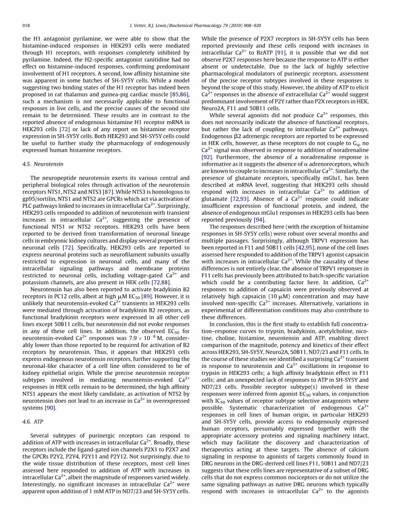
3.5. Histamine
Only HEK293 and SH-SY5Y cells responded with increases inintracellular Ca2+ following addition of histamine (Fig. 6A and B).EC50s for histamine-induced Ca2+ transients differed significantly(p < 0.05) between cell lines, with responses in SH-SY5Y cellsdisplaying a higher affinity (EC50 1.4 � 10�6 M) compared toHEK293 cells (EC50 1.4 � 10�5 M). A second component, matchingthe affinity seen for HEK293 cells, was apparent in some batches ofSH-SY5Y cells but did not consistently reach statistical significanceover a single-site fit in Prism. The EC50 for histamine-induced Ca2+
transients in HEK293 cells was 1.4 � 10�5 M and preferably fit asingle-site model. Histamine-induced Ca2+ transients were gen-erally small, with responses in SH-SY5Y cells significantly larger(maximum response 15.8 � 2.6% FMax) compared to HEK293 cells(maximum response 3.0 � 0.2% FMax, p < 0.05). The H1-specificantagonist pyrilamine completely inhibited histamine-inducedincreases in Ca2+ in HEK293 with an IC50 of 7.0 � 10�9 M (Fig. 6C).In SH-SY5Y cells, pyrilamine inhibited histamine-induced responseswith an IC50 of 1.6 � 10�9 M (Fig. 6C). In contrast, the H2-specificantagonist ranitidine up to a concentration 10 mM did not affecthistamine-induced responses in either SH-SY5Y or HEK293 cells(Fig. 6D).

Fig. 4. Nicotine elicits Ca2+ responses in SH-SY5Y cells. (A) Concentration–response
curve of nicotine-mediated increases in intracellular Ca2+ in SH-SY5Y cells. (B)
Nicotine-elicited Ca2+ responses in SH-SY5Y cells are transient in nature. Shown is a
representative response of SH-SY5Y cells to addition of 30 mM nicotine. (C) Ca2+
responses in SH-SY5Y cells in response to addition of nicotine are not mediated by
the a7 nAChR, as methyllycaconitine (blue) did not inhibit nicotine responses. The
a3b4-selective conotoxin AuIB (red) partially inhibited nicotine responses, while
tubocurarine (purple) completely and concentration-dependently abolished Ca2+
transients elicited by addition of nicotine. Data are presented as mean � SEM of
n = 3–4 wells and are representative of 3 independent experiments. (For interpretation
of the references to color in this figure legend, the reader is referred to the web version
of the article.)
Fig. 5. The a7 nAChR agonist choline elicits Ca2+ responses in SH-SY5Y cells in the
presence of the a7 nAChR allosteric modulator PNU120596. (A) Addition of the a7
nAChR agonist choline did not cause increases in intracellular Ca2+ in SH-SY5Y cells
(open circles), however, in the presence of 10 mM PNU120596, an a7-selective
allosteric modulator, large concentration-dependent responses in response to
choline were observed (blue). (B) Responses to addition of choline were relatively
sustained in nature. Shown is a representative Ca2+ response to addition of 30 mM
choline. (C) Choline-mediated responses were completely and concentration-
dependently inhibited by the a7-selective nAChR antagonist methyllycaconitine.
Data are presented as mean � SEM of n = 3–4 wells and are representative of 3
independent experiments. (For interpretation of the references to color in this figure
legend, the reader is referred to the web version of the article.)
I. Vetter, R.J. Lewis / Biochemical Pharmacology 79 (2010) 908–920914
3.6. Neurotensin
Surprisingly, neurotensin only elicited Ca2+ transients inHEK293 cells, with an EC50 of 6.5 � 10�8 M (Fig. 7A and B). Ca2+
responses to addition of neurotensin were small with a maximumresponse of 2.4 � 0.9% FMax.
3.7. ATP
HEK293, F11, 50B11 and Neuro2A cells responses to addition ofATP with a transient increase in intracellular Ca2+ (Fig. 8A and B). Incontrast, ND7/23 and SH-SY5Y cells did not respond to addition of

Fig. 6. Histamine causes Ca2+ transients in HEK293 and SH-SY5Y cells. (A) Addition of histamine caused concentration-dependent Ca2+ transients in HEK293 (red) and SH-
SY5Y cells (blue). The magnitude of histamine-elicited responses was significantly larger in SH-SY5Y compared to HEK293 cells. (B) Representative Ca2+ transients in response
to addition of 100 mM histamine. SH-SY5Y cells responded with rapid increases in intracellular Ca2+ that did not return to baseline up to approximately 200 s (blue). HEK293
cells responded with slower increases in Ca2+ that returned to baseline within approximately 60 s (red). (C) Histamine-induced Ca2+ transients in HEK293 and SH-SY5Y cells
are completely and concentration-dependently inhibited by the H1 antagonist pyrilamine, albeit histamine responses in SH-SY5Y cells were significantly more sensitive to
inhibition by pyrilamine than in HEK293 cells. (D) The H2-specific antagonist ranitidine did not inhibit Ca2+ responses to histamine in HEK293 (red) and SH-SY5Y cells (blue)
at concentrations up to 10 mM. Data are presented as mean � SEM of n = 3–4 wells and are representative of 2–3 independent experiments. (For interpretation of the references to
color in this figure legend, the reader is referred to the web version of the article.)
I. Vetter, R.J. Lewis / Biochemical Pharmacology 79 (2010) 908–920 915
ATP up to 1 mM (data not shown). HEK293 cells exhibitedcomparatively large responses to ATP and were the most sensitiveto ATP-induced Ca2+ influx, with an EC50 of 9.2 � 10�7 M. Incontrast, ATP induced Ca2+ responses of intermediate magnitude inF11 cells, with an EC50 of 1.3 � 10�5 M, while 50B11 and Neuro2Acells responded with very small increases in intracellular Ca2+,with EC50s of 1.2 � 10�5 and 7.0 � 10�6 M, respectively. Due to thelack of highly selective agonists or antagonists, as well as the largenumber of ATP-sensitive P2X and P2Y receptors, the exact receptorsubtypes contributing to these responses remain to be determined.However, as evidenced by the ability of ATP to elicit Ca2+ responsesin the absence of extracellular Ca2+, it appears that G-proteincoupled P2Y receptors account for the majority of ATP-inducedCa2+ influx in HEK293, F11, 50B11 and Neuro2A cells (Fig. 8C–F).
4. Discussion
The study of endogenously expressed receptors and ionchannels can provide physiologically relevant information oftenlacking in over-expression systems which fail to adequatelyaccount for the role of auxiliary protein effects on receptorfunction and regulation. Immortalized neuronal cell lines provide aconvenient model to study neuronal pharmacology, circumventingprimary culture of neurons, and allowing access to endogenousreceptors. However, systematic data on receptors and ion channelsexpressed in these cell lines is lacking.
Here, we describe the first systematic evaluation of endogenousCa2+ responses to several agonists at potential therapeutic targetsin F11, ND7/23, 50B11 as well as Neuro2A, SH-SY5Y and HEK293cells. These cell lines represent commonly used DRG-derived cells,as well as neuroblastoma cells and a non-neuronal cell linecommonly used for expression of neuronal receptors and ion
channels. Endogenous Ca2+ responses were obtained upon additionof bradykinin, trypsin, ATP, nicotine, acetylcholine, histamine andneurotensin in one or more of these cell lines. While preciseinvolvement of specific receptor subtypes remains to be verified atprotein and mRNA level, insight into possible receptor subtypesinvolved in these responses was provided by establishment of fullconcentration–response curves as well as pharmacological char-acterization using receptor subtype specific agonists and antago-nists. Such information is particularly valuable when identifyingcell lines which are suitable to study putative neuronal targets, asover-expression of these targets in non-neuronal cell lines mayalter the properties of these receptors and ion channels. Forexample, TRPV1 overexpressed in non-neuronal cell lines has amarkedly different pharmacological profile compared to nativeTRPV1, most likely due to the absence of the neuronal accessoryprotein Fas-associated factor 1 [51]. In addition, the reverse is truewhen expressing receptors in cell lines that already constitutivelyexpress the target of interest. One such example is the bradykininB2 receptor, which, despite being endogenously expressed inHEK293 cells, has been overexpressed in these cells to studybradykinin signaling [52,53]. Not surprisingly, this can lead toresults that are conflicting or difficult to interpret, especially whennon-human receptor is expressed. This systematic characteriza-tion of receptors endogenously expressed in commonly used celllines may facilitate the study of receptors and ion channelsassociated with neuronal signaling.
4.1. Trypsin
Trypsin activates protease-activated receptors or PARs, whichare GPCR that couple to increases in intracellular Ca2+ throughactivation of PLC (phospholipase C). Specifically, trypsin activates

Fig. 7. Neurotensin causes Ca2+ transients in HEK293 cells. (A) Addition of neurotensin
caused small but concentration-dependent increases in intracellular Ca2+ in HEK293,
but not any of the neuronal cell lines tested. (B) Representative Ca2+ transient caused
by addition of 100 nM neurotensin in HEK293 cells. Data are presented as
mean � SEM of n = 6–8 wells and are representative of 3 independent experiments.
I. Vetter, R.J. Lewis / Biochemical Pharmacology 79 (2010) 908–920916
PAR2 with an EC50 of approximately 1–10 nM [54–56], but alsoPAR1 and PAR4 isoforms albeit with higher EC50s [57,58].Surprisingly, all cell lines except 50B11 and ND7/23 respondedto addition of trypsin with increases in intracellular Ca2+, thoughthe higher EC50 values suggest the involvement of PAR isoformsother than PAR2 in these neuronal cell lines. In light of expressionof PAR1 and PAR4 on both central and peripheral neurons,involvement of one or more of these isoforms in the trypsin-mediated responses in F11, SH-SY5Y and Neuro2A cells seemslikely. In contrast, PAR2 expression has previously been reported inHEK293 cells [59] and indeed the EC50 of trypsin-mediated Ca2+
mobilization was consistent with activation of PAR2. Surprisingly,trypsin at an EC50 concentration (10 nM) induced Ca2+ oscillationsin HEK293 cells, similar to thrombin-evoked responses in platelets[60]. As this is the first report to describe trypsin-induced Ca2+
oscillations, the physiological mechanism involved remains to bedetermined, though it is likely that release of Ca2+ fromintracellular stores contributes to this phenomenon. It has recentlybeen proposed that the release of Ca2+, and thus Ca2+ oscillations, iscontrolled by the regulators of G protein signaling (RGS) proteins,in particular RGS4, which in turn is regulated by PIP3 and Ca2+-calmodulin [61,62]. It remains to be determined if such amechanism could also contribute to the trypsin-evoked oscilla-tions observed in here in HEK293 cells.
4.2. Bradykinin
Bradykinin B1 and B2 receptors belong to a GPCR family thatsignals through Gq to increases in intracellular Ca2+. The principal
agonist of these receptors is bradykinin, which activates recombi-nant human B1 with an EC50 of 1 mM and B2 with an EC50 of 2 nM[63]. However, while the bradykinin B2 receptor is constitutivelyexpressed in many tissues, including the central and peripheralnervous system, bradykinin B1 receptor expression is rapidlyinduced from low basal levels following tissue injury andinflammation [15,64]. Accordingly, bradykinin B2 receptors mayplay a greater role in acute pain, whereas bradykinin B1 receptorsappear an interesting therapeutic target in chronic pain [15,64].Interestingly, F11 cells responded to addition of nM and sub-nanomolar concentrations of bradykinin with Ca2+ transients thatwere best fitted with a two-site model. Heterogeneity of B2receptors has been suggested previously, with high and lowaffinity bradykinin B2 receptor sites observed in several tissues[65–68]. A related phenomenon has been described in NG108neuroblastoma cells [67], where guanyl-50-yl-imidodiphosphateconverted the high affinity bradykinin B2 site into a low affinitysite. However, such a mechanism is not necessarily applicable tofunctional responses in live cells and the different affinities ofbradykinin observed in F11 cells may represent different B2receptor subtypes, binding to non-bradykinin receptor sites, ormay perhaps be the result of activation of different effectorpathways or cell sub-populations. While SH-SY5Y and Neuro2Acells responded only to high concentrations of bradykinin andND7/23 cells responded to nM concentrations of bradykinin, whichcould suggest a B1 response, inhibition of bradykinin responses bythe B2 antagonist HOE-140 supports predominant involvement ofB2 receptors in all three cell lines.
4.3. Acetylcholine and nicotine
Acetylcholine activates muscarinic and nicotinic AChR, of whichthere are several subtypes. HEK293 and SH-SY5Y cells respondedto addition of acetylcholine, while only SH-SY5Y cells responded tonicotine, suggesting that acetylcholine-induced Ca2+ responses inHEK293 cells are mediated by muscarinic rather than nicotinicreceptors. Indeed, HEK293 cells express M3 muscarinic AChR [50],though the presence of M1 muscarinic receptors has also beensuggested [69,70]. Acetylcholine responses in HEK293 cellsdisplayed a marked second phase, indicative of M3 rather thanM1 acetylcholine receptor Ca2+ mobilization characteristics [71].Supporting the predominant involvement of M3 receptors in theacetylcholine-evoked response in HEK293 cells, the M3-specificantagonist p-fluorohexahydro-sila-difenidol hydrochloride com-pletely abolished acetylcholine-induced Ca2+ transients.
Although the presence of a7 and a5 nicotinic AChR subunits inHEK293 cells has been suggested at the mRNA level [72], neithernicotine nor the a7-selective agonist choline were able to evokeCa2+ responses in HEK293 cells. Indeed, this is consistent with thereported absence of a7 AChR protein endogenously expressed inHEK293 cells [73]. Conversely, SH-SY5Y cells have been reported toexpress a3, a5, a7, b2 and b4 nicotinic AChR subunits [74,75] anddid indeed respond to addition of nicotine with transient increasesin intracellular Ca2+. These responses were not mediated througha7 nicotinic AChR, as the specific a7 antagonist methyllycaconi-tine did not inhibit nicotine-evoked responses. However, SH-SY5Yneuroblastoma cells do express functional a7 nAChR, as responsesto the specific a7 agonists choline in the presence of the a7allosteric modulator PNU120596 were completely abolished bymethyllycaconitine. Consistent with a model describing>1 agonistbinding sites in the homopentameric a7 nAChR, the Hill slope forcholine-induced responses was approximately 2.7 � 0.3, suggest-ing that>1 molecule of choline is required for activation of a7 nAChR,while block of only one binding site by methyllycaconitine issufficient to completely abolish these responses. Nicotine-evokedresponses were inhibited by tubocurarine with an IC50 of

Fig. 8. ATP induces Ca2+ responses in HEK293, F11, 50B11 and Neuro2A cells. (A) Addition of ATP concentration-dependently increased intracellular Ca2+ in HEK293 (red), F11
(green), 50B11 (turquoise) and Neuro2A (orange) cells, but not in ND7/23 or SH-SY5Y cells. The magnitude of ATP-induced responses was largest in HEK293 cells,
intermediate in F11 and comparatively small in 50B11 and Neuro2A cells. (B) Representative Ca2+ responses to addition of 100 mM ATP in HEK293 (red), F11 (green), 50B11
(turquoise) and Neuro2A (orange) cells. ATP-mediated responses were transient in nature, with a biphasic response evident in HEK293 cells. (C–F) ATP-mediated increases in
intracellular Ca2+ in the presence (PSS, filled symbols) and absence (extracellular Ca2+-free, open symbols) of extracellular Ca2+. Ca2+ transients in response to ATP were not
significantly inhibited by the absence of extracellular Ca2+ in HEK293 (C), 50B11 (D), F11 (E) and Neuro2A (F) cells, suggesting contribution of P2Y receptors to the ATP
response. Data are presented as mean � SEM of n = 3–4 wells and are representative of 3 independent experiments. (For interpretation of the references to color in this figure
legend, the reader is referred to the web version of the article.)
I. Vetter, R.J. Lewis / Biochemical Pharmacology 79 (2010) 908–920 917
1.7 � 10�6 M, suggesting involvement of nicotinic AChR containingb2 subunits [76]. While previous studies were unable to demonstratecontribution of a3b4 nAChR to the nicotine-evoked Ca2+ responses inSH-SY5Y cells [75], we found that the a3b4-specific conotoxin AuIB[77] inhibited approximately 50% of the nicotine-evoked response,confirming involvement of a3b4 containing nAChRs. Surprisingly,the acetylcholine-evoked response was also completely abolished bythe M3 antagonist p-fluorohexahydro-sila-difenidol hydrochloride,suggesting that activation of nicotinic receptor subtypes did notcontribute to the acetylcholine response in SH-SY5Y cells.
4.4. Histamine
All four subtypes of histamine receptors, H1–H4, belong to theGPCR family of receptors, with H1 linked to intracellular Ca2+
mobilization through Gq coupled IP3 production and subsequent
store release. Albeit histamine H2, H3 and H4 receptors aregenerally considered to couple to other G protein effectorsincluding adenylate cyclase and voltage-gated Ca2+ channels,several reports have demonstrated increases in intracellular Ca2+
through activation of H4, H2 and H3 receptors in mast cells,keratinocytes and neuroblastoma cells, respectively [78–80].However, where histamine H1 and H2 subtypes demonstraterelatively low affinity to histamine (EC50s of 24 and �10 mM,respectively) [81,82], H3 and H4 receptors are high affinityhistamine receptors with EC50 values in the nanomolar range[83,84].
In light of the relatively wide tissue distribution of histamine H1and H2 receptors, together with literature EC50 values similar tothose obtained in the present study, it seems likely that thehistamine-mediated increase in Ca2+ observed in HEK293 and SH-SY5Y cells are mediated through H1 or H2 receptors. Indeed, using
I. Vetter, R.J. Lewis / Biochemical Pharmacology 79 (2010) 908–920918
the H1 antagonist pyrilamine, we were able to show that thehistamine-induced responses in HEK293 cells were mediatedthrough H1 receptors, with responses completely inhibited bypyrilamine. Indeed, the H2-specific antagonist ranitidine had noeffect on histamine-induced responses, confirming predominantinvolvement of H1 receptors. A second, low affinity histamine sitewas apparent in some batches of SH-SY5Y cells. While a modelsuggesting two binding states of the H1 receptor has indeed beenproposed in rat thalamus and guinea-pig cardiac muscle [85,86],such a mechanism is not necessarily applicable to functionalresponses in live cells, and the precise causes of the second siteremain to be determined. These results are in contrast to thereported absence of endogenous histamine H1 receptor mRNA inHEK293 cells [72] or lack of any report on histamine receptorexpression in SH-SY5Y cells. Both HEK293 and SH-SY5Y cells couldbe useful to further study the pharmacology of endogenouslyexpressed human histamine receptors.
4.5. Neurotensin
The neuropeptide neurotensin exerts its various central andperipheral biological roles through activation of the neurotensinreceptors NTS1, NTS2 and NTS3 [87]. While NTS3 is homologous togp95/sortilin, NTS1 and NTS2 are GPCRs which act via activation ofPLC pathways linked to increases in intracellular Ca2+. Surprisingly,HEK293 cells responded to addition of neurotensin with transientincreases in intracellular Ca2+, suggesting the presence offunctional NTS1 or NTS2 receptors. HEK293 cells have beenreported to be derived from transformation of neuronal lineagecells in embryonic kidney cultures and display several properties ofneuronal cells [72]. Specifically, HEK293 cells are reported toexpress neuronal proteins such as neurofilament subunits usuallyrestricted to expression in neuronal cells, and many of theintracellular signaling pathways and membrane proteinsrestricted to neuronal cells, including voltage-gated Ca2+ andpotassium channels, are also present in HEK cells [72,88].
Neurotensin has also been reported to activate bradykinin B2receptors in PC12 cells, albeit at high mM EC50 [89]. However, it isunlikely that neurotensin-evoked Ca2+ transients in HEK293 cellswere mediated through activation of bradykinin B2 receptors, asfunctional bradykinin receptors were expressed in all other celllines except 50B11 cells, but neurotensin did not evoke responsesin any of these cell lines. In addition, the observed EC50 forneurotensin-evoked Ca2+ responses was 7.9 � 10�8 M, consider-ably lower than those reported to be required for activation of B2receptors by neurotensin. Thus, it appears that HEK293 cellsexpress endogenous neurotensin receptors, further supporting theneuronal-like character of a cell line often considered to be ofkidney epithelial origin. While the precise neurotensin receptorsubtypes involved in mediating neurotensin-evoked Ca2+
responses in HEK cells remain to be determined, the high affinityNTS1 appears the most likely candidate, as activation of NTS2 byneurotensin does not lead to an increase in Ca2+ in overexpressedsystems [90].
4.6. ATP
Several subtypes of purinergic receptors can respond toaddition of ATP with increases in intracellular Ca2+. Broadly, thesereceptors include the ligand-gated ion channels P2X1 to P2X7 andthe GPCRs P2Y2, P2Y4, P2Y11 and P2Y12. Not surprisingly, due tothe wide tissue distribution of these receptors, most cell linesassessed here responded to addition of ATP with increases inintracellular Ca2+, albeit the magnitude of responses varied widely.Interestingly, no significant increases in intracellular Ca2+ wereapparent upon addition of 1 mM ATP in ND7/23 and SH-SY5Y cells.
While the presence of P2X7 receptors in SH-SY5Y cells has beenreported previously and these cells respond with increases inintracellular Ca2+ to BzATP [91], it is possible that we did notobserve P2X7 responses here because the response to ATP is eitherabsent or undetectable. Due to the lack of highly selectivepharmacological modulators of purinergic receptors, assessmentof the precise receptor subtypes involved in these responses isbeyond the scope of this study. However, the ability of ATP to elicitCa2+ responses in the absence of extracellular Ca2+ would suggestpredominant involvement of P2Y rather than P2X receptors in HEK,Neuro2A, F11 and 50B11 cells.
While several agonists did not produce Ca2+ responses, thisdoes not necessarily indicate the absence of functional receptors,but rather the lack of coupling to intracellular Ca2+ pathways.Endogenous b2 adrenergic receptors are reported to be expressedin HEK cells, however, as these receptors do not couple to Gq, noCa2+ signal was observed in response to addition of noradrenaline[92]. Furthermore, the absence of a noradrenaline response isinformative as it suggests the absence of a adrenoreceptors, whichare known to couple to increases in intracellular Ca2+. Similarly, thepresence of glutamate receptors, specifically mGlu1, has beendescribed at mRNA level, suggesting that HEK293 cells shouldrespond with increases in intracellular Ca2+ to addition ofglutamate [72,93]. Absence of a Ca2+ response could indicateinsufficient expression of functional protein, and indeed, theabsence of endogenous mGlu1 responses in HEK293 cells has beenreported previously [94].
The responses described here (with the exception of histamineresponses in SH-SY5Y cells) were robust over several months andmultiple passages. Surprisingly, although TRPV1 expression hasbeen reported in F11 and 50B11 cells [42,95], none of the cell linesassessed here responded to addition of the TRPV1 agonist capsaicinwith increases in intracellular Ca2+. While the causality of thesedifferences is not entirely clear, the absence of TRPV1 responses inF11 cells has previously been attributed to batch-specific variationwhich could be a contributing factor here. In addition, Ca2+
responses to addition of capsaicin were previously observed atrelatively high capsaicin (10 mM) concentration and may haveinvolved non-specific Ca2+ increases. Alternatively, variations inexperimental or differentiation conditions may also contribute tothese differences.
In conclusion, this is the first study to establish full concentra-tion–response curves to trypsin, bradykinin, acetylcholine, nico-tine, choline, histamine, neurotensin and ATP, enabling directcomparison of the magnitude, potency and kinetics of their effectacross HEK293, SH-SY5Y, Neuro2A, 50B11, ND7/23 and F11 cells. Inthe course of these studies we identified a surprising Ca2+ transientin response to neurotensin and Ca2+ oscillations in response totrypsin in HEK293 cells; a high affinity bradykinin effect in F11cells; and an unexpected lack of responses to ATP in SH-SY5Y andND7/23 cells. Possible receptor subtype(s) involved in theseresponses were inferred from agonist EC50 values, in conjunctionwith IC50 values of receptor subtype selective antagonists wherepossible. Systematic characterization of endogenous Ca2+
responses in cell lines of human origin, in particular HEK293and SH-SY5Y cells, provide access to endogenously expressedhuman receptors, presumably expressed together with theappropriate accessory proteins and signaling machinery intact,which may facilitate the discovery and characterization oftherapeutics acting at these targets. The absence of calciumsignaling in response to agonists of targets commonly found inDRG neurons in the DRG-derived cell lines F11, 50B11 and ND7/23suggests that these cells lines are representative of a subset of DRGcells that do not express common nociceptors or do not utilize thesame signaling pathways as native DRG neurons which typicallyrespond with increases in intracellular Ca2+ to the agonists

I. Vetter, R.J. Lewis / Biochemical Pharmacology 79 (2010) 908–920 919
investigated. Additional immortalized DRG cell lines selected forexpression of markers such as CGRP (calcitonin-gene relatedpeptide) or IB4 (isolectin B4) that respond to other agonists ofantinociceptive targets not covered by the cell lines assessed here(such as adenosine, capsaicin, dopamine, GABA, glutamate,morphine, neurokinin A and B, NMSA, noradrenaline, serotoninand substance P) may expand the range of endogenously expressednociceptive targets accessible with this approach.
Conflict of interest
The authors declare no competing interest.
Acknowledgements
This work was supported by a NHMRC Australian BasedBiomedical Post doctorate Fellowship (IV) and an NHMRC ProgramGrant (RJL). RJL is an NHMRC Research Fellow.
Contributions: IV carried out experimental work and drafted themanuscript, RJL participated in the study design and coordinationand helped to draft the manuscript. All authors read and approvedthe final manuscript.
References
[1] Raggenbass M, Bertrand D. Nicotinic receptors in circuit excitability andepilepsy. J Neurobiol 2002;53:580–9.
[2] Khosravani H, Zamponi GW. Voltage-gated calcium channels and idiopathicgeneralized epilepsies. Physiol Rev 2006;86:941–66.
[3] Dray A. Neuropathic pain: emerging treatments. Br J Anaesth 2008;101:48–58.[4] Jensen AA, Frolund B, Liljefors T, Krogsgaard-Larsen P. Neuronal nicotinic
acetylcholine receptors: structural revelations, target identifications, andtherapeutic inspirations. J Med Chem 2005;48:4705–45.
[5] Triggle DJ. Drug targets in the voltage-gated calcium channel family: whysome are and some are not. Assay Drug Dev Technol 2003;1:719–33.
[6] Lu Y, Wang X. Genes associated with idiopathic epilepsies: a current overview.Neurol Res 2009;31:135–43.
[7] Bowie D. Ionotropic glutamate receptors & CNS disorders. CNS Neurol DisordDrug Targets 2008;7:129–43.
[8] Saito T, Bunnett NW. Protease-activated receptors: regulation of neuronalfunction. Neuromolecular Med 2005;7:79–99.
[9] Vergnolle N, Wallace JL, Bunnett NW, Hollenberg MD. Protease-activatedreceptors in inflammation, neuronal signaling and pain. Trends PharmacolSci 2001;22:146–52.
[10] Asfaha S, Cenac N, Houle S, Altier C, Papez MD, Nguyen C, et al. Protease-activated receptor-4: a novel mechanism of inflammatory pain modulation. BrJ Pharmacol 2007;150:176–85.
[11] Oliver KR, Hill RG. Feeling below PAR: proteinase-activated receptors and theperception of neuroinflammatory pain. Pharmacogenomics J 2002;2:10–1.
[12] Bueno L. Protease activated receptor 2: a new target for IBS treatment. Eur RevMed Pharmacol Sci 2008;12(Suppl. 1):95–102.
[13] Lohman RJ, O’Brien TJ, Cocks TM. Protease-activated receptor-2 regulatestrypsin expression in the brain and protects against seizures and epileptogen-esis. Neurobiol Dis 2008;30:84–93.
[14] Meini S, Maggi CA. Knee osteoarthritis: a role for bradykinin? Inflamm Res2008;57:351–61.
[15] Chen JJ, Johnson EJ. Targeting the bradykinin B1 receptor to reduce pain. ExpertOpin Ther Targets 2007;11:21–35.
[16] Kitabgi P. Targeting neurotensin receptors with agonists and antagonists fortherapeutic purposes. Curr Opin Drug Discov Devel 2002;5:764–76.
[17] Allen JW, Hofer K, McCumber D, Wagstaff JD, Layer RT, McCabe RT, et al. Anassessment of the antinociceptive efficacy of intrathecal and epidural con-tulakin-G in rats and dogs. Anesth Analg 2007;104:1505–13. table of contents.
[18] Lee HK, Zhang L, Smith MD, White HS, Bulaj G. Glycosylated neurotensinanalogues exhibit sub-picomolar anticonvulsant potency in a pharmacoresis-tant model of epilepsy. ChemMedChem 2009;4:400–5.
[19] Kinkead B, Nemeroff CB. Novel treatments of schizophrenia: targeting theneurotensin system. CNS Neurol Disord Drug Targets 2006;5:205–18.
[20] Wein AJ. Muscarinic acetylcholine receptor knockout mice: novel phenotypesand clinical implications. J Urol 2005;173:2199.
[21] Wess J. Muscarinic acetylcholine receptor knockout mice: novel phenotypesand clinical implications. Annu Rev Pharmacol Toxicol 2004;44:423–50.
[22] Tata AM. Muscarinic acetylcholine receptors: new potential therapeutic tar-gets in antinociception and in cancer therapy. Recent Pat CNS Drug Discov2008;3:94–103.
[23] Wess J, Duttaroy A, Gomeza J, Zhang W, Yamada M, Felder CC, et al. Muscarinicreceptor subtypes mediating central and peripheral antinociception studiedwith muscarinic receptor knockout mice: a review. Life Sci 2003;72:2047–54.
[24] Bozzi Y, Borrelli E. Dopamine in neurotoxicity and neuroprotection: what doD2 receptors have to do with it? Trends Neurosci 2006;29:167–74.
[25] Wood PB. Role of central dopamine in pain and analgesia. Expert Rev Neu-rother 2008;8:781–97.
[26] Raffa RB. Antihistamines as analgesics. J Clin Pharm Ther 2001;26:81–5.[27] Gemkow MJ, Davenport AJ, Harich S, Ellenbroek BA, Cesura A, Hallett D. The
histamine H3 receptor as a therapeutic drug target for CNS disorders. DrugDiscov Today 2009;14:509–15.
[28] Tang FR, Bradford HF, Ling EA. Metabotropic glutamate receptors in the controlof neuronal activity and as targets for development of anti-epileptogenicdrugs. Curr Med Chem 2009;16:2189–204.
[29] Neugebauer V. Metabotropic glutamate receptors—important modulators ofnociception and pain behavior. Pain 2002;98:1–8.
[30] Enna SJ, McCarson KE. The role of GABA in the mediation and perception ofpain. Adv Pharmacol 2006;54:1–27.
[31] Bagdy G, Kecskemeti V, Riba P, Jakus R. Serotonin and epilepsy. J Neurochem2007;100:857–73.
[32] Sommer C. Serotonin in pain and analgesia: actions in the periphery. MolNeurobiol 2004;30:117–25.
[33] Okamoto K, Imbe H, Morikawa Y, Itoh M, Sekimoto M, Nemoto K, et al. 5-HT2Areceptor subtype in the peripheral branch of sensory fibers is involved in thepotentiation of inflammatory pain in rats. Pain 2002;99:133–43.
[34] Wu S, Zhu M, Wang W, Wang Y, Li Y, Yew DT. Changes of the expression of 5-HT receptor subtype mRNAs in rat dorsal root ganglion by complete Freund’sadjuvant-induced inflammation. Neurosci Lett 2001;307:183–6.
[35] Burnstock G. Purinergic signalling and disorders of the central nervous system.Nat Rev Drug Discov 2008;7:575–90.
[36] Yoon MH, Bae HB, Choi JI, Kim SJ, Chung ST, Kim CM. Roles of adenosinereceptor subtypes in the antinociceptive effect of intrathecal adenosine in a ratformalin test. Pharmacology 2006;78:21–6.
[37] Liu XJ, Salter MW. Purines and pain mechanisms: recent developments. CurrOpin Investig Drugs 2005;6:65–75.
[38] Donnelly-Roberts D, McGaraughty S, Shieh CC, Honore P, Jarvis MF. Painfulpurinergic receptors. J Pharmacol Exp Ther 2008;324:409–15.
[39] Reeve AJ, Dickenson AH. The roles of spinal adenosine receptors in the controlof acute and more persistent nociceptive responses of dorsal horn neurones inthe anaesthetized rat. Br J Pharmacol 1995;116:2221–8.
[40] Platika D, Baizer L, Fishman MC. Sensory neurons ‘‘immortalized’’ by fusionwith neuroblastoma cells. Trans Assoc Am Physicians 1985;98:301–4.
[41] Wood JN, Bevan SJ, Coote PR, Dunn PM, Harmar A, Hogan P, et al. Novel celllines display properties of nociceptive sensory neurons. Proc Biol Sci1990;241:187–94.
[42] Chen W, Mi R, Haughey N, Oz M, Hoke A. Immortalization and characterizationof a nociceptive dorsal root ganglion sensory neuronal line. J Peripher NervSyst 2007;12:121–30.
[43] Biedler JL, Helson L, Spengler BA. Morphology and growth, tumorigenicity, andcytogenetics of human neuroblastoma cells in continuous culture. Cancer Res1973;33:2643–52.
[44] Olmsted JB, Carlson K, Klebe R, Ruddle F, Rosenbaum J. Isolation of microtubuleprotein from cultured mouse neuroblastoma cells. Proc Natl Acad Sci USA1970;65:129–36.
[45] Graham FL, Smiley J, Russell WC, Nairn R. Characteristics of a human cell linetransformed by DNA from human adenovirus type 5. J Gen Virol 1977;36:59–74.
[46] Mutschler E, Moser U, Wess J, Lambrecht G. Muscarinic receptor subtypes—pharmacological, molecular biological and therapeutical aspects. Pharm ActaHelv 1995;69:243–58.
[47] Zlotos DP, Bender W, Holzgrabe U. Muscarinic receptor agonists and antago-nists. Expert Opin Ther Patents 1999;9:1029–53.
[48] Weill C, Galzi JL, Chasserot-Golaz S, Goeldner M, Ilien B. Functional character-ization and potential applications for enhanced green fluorescent protein- andepitope-fused human M1 muscarinic receptors. J Neurochem 1999;73:791–801.
[49] Alexander SP, Mathie A, Peters JA. Guide to receptors and channels (GRAC),2nd edition. Br J Pharmacol 2006;147(Suppl. 3):S1–68.
[50] Dowling MR, Willets JM, Budd DC, Charlton SJ, Nahorski SR, Challiss RA.A single point mutation (N514Y) in the human M3 muscarinic acetylcho-line receptor reveals differences in the properties of antagonists: evidencefor differential inverse agonism. J Pharmacol Exp Ther 2006;317:1134–42.
[51] Kim S, Kang C, Shin CY, Hwang SW, Yang YD, Shim WS, et al. TRPV1 recapi-tulates native capsaicin receptor in sensory neurons in association with Fas-associated factor 1. J Neurosci 2006;26:2403–12.
[52] Pizard A, Blaukat A, Muller-Esterl W, Alhenc-Gelas F, Rajerison RM. Bradyki-nin-induced internalization of the human B2 receptor requires phosphoryla-tion of three serine and two threonine residues at its carboxyl tail. J Biol Chem1999;274:12738–47.
[53] Lamb ME, De Weerd WF, Leeb-Lundberg LM. Agonist-promoted trafficking ofhuman bradykinin receptors: arrestin- and dynamin-independent sequestra-tion of the B2 receptor and bradykinin in HEK293 cells. Biochem J2001;355:741–50.
[54] Luo W, Wang Y, Reiser G. Two types of protease-activated receptors (PAR-1and PAR-2) mediate calcium signaling in rat retinal ganglion cells RGC-5. BrainRes 2005;1047:159–67.
[55] Shpacovitch VM, Brzoska T, Buddenkotte J, Stroh C, Sommerhoff CP, Ansel JC,et al. Agonists of proteinase-activated receptor 2 induce cytokine release and

I. Vetter, R.J. Lewis / Biochemical Pharmacology 79 (2010) 908–920920
activation of nuclear transcription factor kappaB in human dermal micro-vascular endothelial cells. J Invest Dermatol 2002;118:380–5.
[56] Steinhoff M, Vergnolle N, Young SH, Tognetto M, Amadesi S, Ennes HS, et al.Agonists of proteinase-activated receptor 2 induce inflammation by a neuro-genic mechanism. Nat Med 2000;6:151–8.
[57] Xu WF, Andersen H, Whitmore TE, Presnell SR, Yee DP, Ching A, et al. Cloningand characterization of human protease-activated receptor 4. Proc Natl AcadSci USA 1998;95:6642–6.
[58] Blackhart BD, Emilsson K, Nguyen D, Teng W, Martelli AJ, Nystedt S, et al.Ligand cross-reactivity within the protease-activated receptor family. J BiolChem 1996;271:16466–71.
[59] Grishina Z, Ostrowska E, Halangk W, Sahin-Toth M, Reiser G. Activity ofrecombinant trypsin isoforms on human proteinase-activated receptors(PAR): mesotrypsin cannot activate epithelial PAR-1, -2, but weakly activatesbrain PAR-1. Br J Pharmacol 2005;146:990–9.
[60] Ozaki Y, Yatomi Y, Wakasugi S, Shirasawa Y, Saito H, Kume S. Thrombin-induced calcium oscillation in human platelets and MEG-01, a megakaryo-blastic leukemia cell line. Biochem Biophys Res Commun 1992;183:864–71.
[61] Shin DM, Luo X, Wilkie TM, Miller LJ, Peck AB, Humphreys-Beher MG, et al.Polarized expression of G protein-coupled receptors and an all-or-none dis-charge of Ca2+ pools at initiation sites of [Ca2+]i waves in polarized exocrinecells. J Biol Chem 2001;276:44146–5.
[62] Luo X, Popov S, Bera AK, Wilkie TM, Muallem S. RGS proteins providebiochemical control of agonist-evoked [Ca2+]i oscillations. Mol Cell 2001;7:651–60.
[63] Simpson PB, Woollacott AJ, Hill RG, Seabrook GR. Functional characterizationof bradykinin analogues on recombinant human bradykinin B(1) and B(2)receptors. Eur J Pharmacol 2000;392:1–9.
[64] Marceau F, Regoli D. Bradykinin receptor ligands: therapeutic perspectives.Nat Rev Drug Discov 2004;3:845–52.
[65] Reissmann S, Pagelow I, Liebmann C, Steinmetzger H, Jankova T, Arold H.Investigations on the mechanism of bradykinin action on smooth muscles. In:Papasova MA, editor. Physiology and Pharmacology of Smooth Muscle. Sofia:Bulgarian Academy of Science; 1977. pp. 208–17.
[66] Odya CE, Goodfriend TL, Pena C. Bradykinin receptor-like binding studied withiodinated analogues. Biochem Pharmacol 1980;29:175–85.
[67] Hall JM. Bradykinin receptors: pharmacological properties and biologicalroles. Pharmacol Ther 1992;56:131–90.
[68] Osugi T, Imaizumi T, Mizushima A, Uchida S, Yoshida H. Phorbol ester inhibitsbradykinin-stimulated inositol trisphosphate formation and calcium mobili-zation in neuroblastoma � glioma hybrid NG108-15 cells. J Pharmacol ExpTher 1987;240:617–22.
[69] Conklin BR, Chabre O, Wong YH, Federman AD, Bourne HR. Recombinant Gqalpha. Mutational activation and coupling to receptors and phospholipase C. JBiol Chem 1992;267:31–4.
[70] Mundell SJ, Benovic JL. Selective regulation of endogenous G protein-coupledreceptors by arrestins in HEK293 cells. J Biol Chem 2000;275:12900–8.
[71] Collison DJ, Coleman RA, James RS, Carey J, Duncan G. Characterization ofmuscarinic receptors in human lens cells by pharmacologic and moleculartechniques. Invest Ophthalmol Vis Sci 2000;41:2633–41.
[72] Shaw G, Morse S, Ararat M, Graham FL. Preferential transformation of humanneuronal cells by human adenoviruses and the origin of HEK 293 cells. FASEB J2002;16:869–71.
[73] Williams ME, Burton B, Urrutia A, Shcherbatko A, Chavez-Noriega LE, Cohen CJ,et al. Ric-3 promotes functional expression of the nicotinic acetylcholinereceptor alpha7 subunit in mammalian cells. J Biol Chem 2005;280:1257–63.
[74] Wang F, Gerzanich V, Wells GB, Anand R, Peng X, Keyser K, et al. Assembly ofhuman neuronal nicotinic receptor alpha5 subunits with alpha3, beta2, andbeta4 subunits. J Biol Chem 1996;271:17656–65.
[75] Dajas-Bailador FA, Mogg AJ, Wonnacott S. Intracellular Ca2+ signals evoked bystimulation of nicotinic acetylcholine receptors in SH-SY5Y cells: contribution
of voltage-operated Ca2+ channels and Ca2+ stores. J Neurochem 2002;81:606–14.
[76] Cachelin AB, Rust G. Unusual pharmacology of (+)-tubocurarine with ratneuronal nicotinic acetylcholine receptors containing beta 4 subunits. MolPharmacol 1994;46:1168–74.
[77] Luo S, Kulak JM, Cartier GE, Jacobsen RB, Yoshikami D, Olivera BM, et al. alpha-conotoxin AuIB selectively blocks alpha3 beta4 nicotinic acetylcholine recep-tors and nicotine-evoked norepinephrine release. J Neurosci 1998;18:8571–9.
[78] Hofstra CL, Desai PJ, Thurmond RL, Fung-Leung WP. Histamine H4 receptormediates chemotaxis and calcium mobilization of mast cells. J Pharmacol ExpTher 2003;305:1212–21.
[79] Bongers G, Sallmen T, Passani M, Mariottini C, Wendelin D, Lozada Aea. Newsignaling pathways for the histamine H3 receptor. In: Proceedings of theXXXVth Annual Meeting of European Histamine Research Society; 2006.
[80] Koizumi H, Ohkawara A. H2 histamine receptor-mediated increase in intra-cellular Ca2+ in cultured human keratinocytes. J Dermatol Sci 1999;21:127–32.
[81] Mitsuhashi M, Payan DG. Characterization of functional histamine H1receptors on a cultured smooth muscle cell line. J Cell Physiol 1988;134:367–75.
[82] Mitsuhashi M, Mitsuhashi T, Payan DG. Multiple signaling pathways ofhistamine H2 receptors. Identification of an H2 receptor-dependent Ca2+
mobilization pathway in human HL-60 promyelocytic leukemia cells. J BiolChem 1989;264:18356–62.
[83] Lovenberg TW, Pyati J, Chang H, Wilson SJ, Erlander MG. Cloning of rathistamine H(3) receptor reveals distinct species pharmacological profiles. JPharmacol Exp Ther 2000;293:771–8.
[84] Liu C, Wilson SJ, Kuei C, Lovenberg TW. Comparison of human, mouse, rat, andguinea pig histamine H4 receptors reveals substantial pharmacological spe-cies variation. J Pharmacol Exp Ther 2001;299:121–30.
[85] Soria-Jasso LE, Bahena-Trujillo R, Arias-Montano JA. Histamine H1 receptorsand inositol phosphate formation in rat thalamus. Neurosci Lett1997;225:117–20.
[86] Krzan M, Erjavec F, Carman-Krzan M. Characterization of coexistent histamineH1- and H2-receptor binding sites in the purified guinea pig myocardialmembranes from ventricles. Inflamm Res 1993;38:C289–91.
[87] Vincent JP, Mazella J, Kitabgi P. Neurotensin and neurotensin receptors. TrendsPharmacol Sci 1999;20:302–9.
[88] Thomas P, Smart TG. HEK293 cell line: a vehicle for the expression ofrecombinant proteins. J Pharmacol Toxicol Methods 2005;51:187–200.
[89] Park TJ, Kim KT. Activation of B2 bradykinin receptors by neurotensin. CellSignal 2003;15:519–27.
[90] Richard F, Barroso S, Martinez J, Labbe-Jullie C, Kitabgi P. Agonism, inverseagonism, and neutral antagonism at the constitutively active human neuro-tensin receptor 2. Mol Pharmacol 2001;60:1392–8.
[91] Larsson KP, Hansen AJ, Dissing S. The human SH-SY5Y neuroblastoma cell-lineexpresses a functional P2X7 purinoceptor that modulates voltage-dependentCa2+ channel function. J Neurochem 2002;83:285–98.
[92] Daaka Y, Luttrell LM, Lefkowitz RJ. Switching of the coupling of the beta2-adrenergic receptor to different G proteins by protein kinase A. Nature1997;390:88–91.
[93] Yarwood SJ, Woodgett JR. Extracellular matrix composition determines thetranscriptional response to epidermal growth factor receptor activation. ProcNatl Acad Sci USA 2001;98:4472–7.
[94] Jong YJ, Schwetye KE, O’Malley KL. Nuclear localization of functional metabo-tropic glutamate receptor mGlu1 in HEK293 cells and cortical neurons: role innuclear calcium mobilization and development. J Neurochem 2007;101:458–69.
[95] Kusano K, Gainer H. Modulation of voltage-activated Ca currents by pain-inducing agents in a dorsal root ganglion neuronal line, F-11. J Neurosci Res1993;34:158–69.