Development of an endogenous Androgen Receptor-mediated
139
Development of an endogenous A ndrogen R eceptor-mediated LU ciferase eX pression assay (AR-LUX) for interactive androgenic action: Application to screening for environmental androgenic activity and veterinary growth promoters Barry Blankvoort
-
Upload
khangminh22 -
Category
Documents
-
view
0 -
download
0
Transcript of Development of an endogenous Androgen Receptor-mediated

Development of an endogenous Androgen Receptor-mediated LUciferase eXpression assay (AR-LUX) for interactive androgenic
action: Application to screening for environmental androgenic activity and veterinary growth promoters
Barry Blankvoort
Promotor: Prof. Dr. J.H. Koeman Hoogleraar in de Toxicologie
Leerstoelgroep Toxicologie, Wageningen Universiteit Co-promotoren: Dr. Ir. J.M.M.J.G. Aarts
Universitair Docent, Leerstoelgroep Toxicologie Wageningen Universiteit Dr. R.J.T. Rodenburg Senior Scientist, Afdeling Bioanalyse, TNO Pharma, Zeist
Promotiecommissie: Prof. F.A. Claessens
Katholieke Universiteit Leuven (België) Dr. A.O. Brinkmann Erasmus Universiteit Rotterdam Prof. Dr. J. Fink-Gremmels Universiteit Utrecht Prof. Dr. M. Müller Wageningen Universiteit
Development of an endogenous Androgen Receptor-mediated LUciferase eXpression assay (AR-LUX) for interactive androgenic
action: Application to screening for environmental androgenic activity and veterinary growth promoters
Barry Marinus Gregorius Blankvoort
Proefschrift ter verkrijging van de graad van doctor
op gezag van de rector magnificus van Wageningen Universiteit
Prof. Dr. Ir. L. Speelman In het openbaar te verdedigen op dinsdag 16 december 2003
des middags te half twee in de Aula

Title Development of an endogenous Androgen Receptor-mediated LUciferase
eXpression assay (AR-LUX) for interactive androgenic action: Application to screening for environmental androgenic activity and veterinary growth promoters
Author Barry Marinus Gregorius Blankvoort Thesis Wageningen University, Wageningen, the Netherlands (2003) with abstract-with references-with summary in Dutch ISBN 90-5808-977-0

Abstract The research described in this thesis was aimed at developing an in vitro cell-based
reporter gene system applicable to the detection of the illegal use of androgenic growth promoters in cattle, and the presence of potential endocrine disrupters present in surface waters and interfering with androgenic action. The system is based on a luciferase reporter gene placed under transcriptional control of an authenticated androgen-responsive element (ARE) and an endogenously expressed androgen receptor. This system allows for the integration of the effects of certain modulators of androgenic signal transduction. A second important goal of the research was to gain insight into the mechanisms underlying enhanced growth promotion by mixtures of androgenic and estrogenic compounds. The use of such mixtures, which results in activation and subsequent interaction of multiple steroid receptors, is occasionally observed in illegal hormonal treatments of cattle.
When applied to the screening of calf urine samples for anabolic androgens, the developed AR-LUX assay was able to identify androgen-treated animals with similar results as obtained by standard GC-MS analysis. However, both techniques should be regarded as complementary rather than interchangeable screening tools. Liquid samples confiscated at cattle farms outside the Netherlands were found to generate a very strong response in the AR-LUX assay despite the fact that GC-MS analysis did not detect the presence of any anabolic compounds. Possibly, the samples contained a mixture of conventional androgenic compounds, each at undetectably low amounts and/or (novel) unknown compounds not tested for by GC-MS. These results emphasize the additional value of the developed AR-LUX assay.
Also, the AR-LUX assay was used to determine the androgenic activity of a number of aquatic environmental samples. A number of these samples were found to contain androgenic activity at varying concentrations. Interestingly, in 2 samples containing androgens, enhancing interactive mixture effects were observed, which were probably due to interactions by estrogenic compounds and estrogen receptor activation.
Our research furthermore indicates that certain established progestagens are able to activate ARE-mediated luciferase expression via progesterone receptors; we hypothesise preferentially through the progesterone receptor-α isoform. This indicates that androgen reporter assays based on the activation of the androgen receptor alone rather than on activation of a response element might produce results quite different from those observed in assay systems featuring multiple steroid receptors. This further emphasizes the notion that the AR-LUX assay is not merely detecting activation of the androgen receptor by androgens, but also allows for the detection of other androgenic substances that regulate gene expression via alternative pathways leading to activation of an established androgen response element.


Contents
Chapter 1 Introduction 9
Chapter 2 Construction and application of a reporter cell line for
androgenic activity and preliminary research into the revealed complex receptor-mediated effects of anabolics on gene expression: Principles and procedures
21
Chapter 3
Development of an androgen reporter gene assay (AR-LUX) utilising a human cell line with an endogenously regulated androgen receptor
41
Chapter 4 Androgen activity in surface water samples detected using the AR-LUX assay: Indications for mixture effects
59
Chapter 5 Detection of hormonal anabolic compounds in calf urine and unverified growth-promoting preparations: Application of the AR-LUX bioassay for screening and determination of androgenic activity
77
Chapter 6 Complex multi-receptor interactions are involved in androgen response element-driven gene expression as revealed by the AR-LUX assay for androgenic activity
95
Chapter 7 Summary & Concluding remarks
115
Samenvatting en Slotbeschouwingen
123
Curriculum Vitae
132
List of Publications
133
Abbreviations 134 Dankwoord 136


11
Introduction
Chapter 1
10
Research described in this thesis was aimed at developing an in vitro cell based system capable of detecting the illegal use of androgenic growth promoters and the presence of potential endocrine disrupters in various environmental matrices. The system was intended to be capable of integrating the effects of modulating compounds as well. Furthermore gaining insights into the mechanisms underlying growth promoting effects regulated via multiple steroid receptor interactions and their activation was an important goal. These interactions have been reported in the context of illegal anabolic growth promoters in cattle and in the context of in vivo interactions in humans. Furthermore (anti)androgens have been implied in playing a role in environmental pollution leading to endocrine disruption (Galbraith and Topps, 1981; Sonnenschein and Soto, 1998; Simon, 2001). Steroid hormones
The development and maintenance of male and female morphological and functional characteristics in human and numerous other vertebrate species depends largely on the action of steroid hormones. Hormones are compounds produced in specialised tissues which are subsequently transported via the blood stream to their effector sites. The steroid hormones represent a subgroup mediating their action via a large group of related proteins, the superfamily of nuclear receptors (NRs) that function as ligand-activated transcription factors. Members of this group include -amongst others- the thyroid, vitamin D, retinoic acid and peroxisome proliferator-activated receptor. Furthermore, a number of orphan receptors have been identified of which ligands and functions are largely unknown.
Androgens are steroids based on a nineteen carbon atoms containing sterane structure and are primarily released by the testis and adrenal cortex. Hormonally active androgens promote reproductive and anabolic (myotropic) functions. They induce these effects as a consequence of their interaction with the androgen receptor (AR) (Roy et al., 1999).
The main male hormones are considered to be testosterone (T) and its derivative 5α-di-hydro-testosterone (DHT) (Michal, 1998; Rang and Dale, 2000).
As described by Michal (1998), estrogens (C18-steroids) control the development of the reproduction system and reproductive functions in female vertebrates. For instance, estrogens act on the ovaries, promoting the development of small groups of follicles in the end producing an ovum. The main estrogen is 17β-estradiol. In female mammals progesterone (a C21-steroid) plays an essential role as the only active gestagen. It is produced mostly in the corpus luteum of the ovaries during the second half of the menstrual cycle and in the placenta during pregnancy. Its main functions are preparation of the uterus for implantation of the fertilised ovum, preservation of the mucous coat of the uterus during pregnancy, prevention of further ovulations and formation of lactating alveoli in the breasts (synergistically with estrogens) (Michal, 1998). These steroids are synthesised through a shared pathway in which cholesterol (containing 27 carbon atoms) provides the basis for the different steroid structures. Carbon atoms are removed via numerous metabolic steps, subgroups are added and ring structures changed. Intermediates in this pathway include progesterone (C21), testosterone (C19) and estradiol (C18). Obviously, this pathway provides one way of interaction between the different

Chapter 1
11
Cholesterol
P450ssc
Pregnenolone
Progesterone17-OH-Pregnenolone
Dehydroepiandrosterone 17-OH-Progesterone
Androstenediol Androstenedione
Estrone
Estradiol
Testosterone
Di-hydro-testosterone
aromatase17-ketoreductase
17,20-desmolase
P450c1717,20-desmolase
P450c17
2
3
1
10
56
7
98
12
1113
14
15
16
1720
2221
23 24
25
24
24
27
26
29
4
28
19
18
A B
C D
1
2
2
3
1
10
56
7
98
12
1113
14
15
16
17
4
30
CH3
OHCH3
O
A B
C D
A B
C D
H
2
3
1
10
6
7
98
12
1113
14
15
16
17
4
30
CH3
OHCH3
O
3β-OH-∆4,6-isomerase
3β-OH-∆4,6-isomerase
3β-OH-∆4,6-isomerase
3β-OH-∆4,6-isomerase
3β-OH-∆4,6-isomerase
17-ketoreductase
5α-reductase
17-ketoreductase
Figure 1 Part of the steroid synthesis pathway showing interrelationship in steroid synthesis (reproduced with modifications from http://www.indstate.edu/thcme/mwking/steroid-hormones.html). Upper right structure represents the way carbon atoms are numbered in steroids (JCBN, 1989).
steroid receptors (Fig. 1). Steroid hormone receptors
Steroid hormone receptors are structurally very alike. They all contain an N-terminally located transactivation domain (NTD) of variable length (~25-600 amino acids) which is followed by a 66-68 amino acid long DNA binding domain (DBD). The DBD harbours two zinc fingers that facilitate the stable insertion of the receptor into the major groove of a DNA duplex. The NTD and DBD are linked to the C-terminally located ligand binding domain (LBD) by the hinge region (~50-70 amino acids) (Roy et al., 1999). This region has been implicated in AR specificity (Schoenmakers et al., 1999) (Fig. 2). Upon binding of androgens to the LBD subsequent conformational changes of the AR facilitate transactivation by the receptor. Furthermore these changes allow receptor dimerisation and cooperative interaction between the C-terminal domain and the N-terminal domain (Doesburg et al., 1997). In addition, binding of agonists or antagonists regulates the interactions with numerous proteins including co-activators and repressors. Between the steroid receptors the N-terminal domain is the most variable while the other domains are highly conserved (Keller et al., 1996). Full length AR is a protein of approximately 110 kDa with size variations due to polymorphisms in the length of polyglutamine (~11-31 residues) and polyglycine (~24 residues) stretches

Chapter 1
12
Figure 2 a 3D model of full size AR liganded with R1881 (reproduced from J.H. Wu, http://ww2.mcgill.ca/androgendb/) b Modular structure of nuclear receptors. The main functions of each domain are shown. The numbers depict the number of amino acids in each domain (reproduced from U. Karvonen (Karvonen, 2003)). c Model of conformation of the activated AR showing multiple interactions within the receptor protein. Abbreviations: NTD, N-terminal domain; DBD, DNA-binding domain, LBD, ligand-binding domain; T, testosterone, AF2, activation function 2 of LBD, Zn, zinc finger (reproduced from U. Karvonen (Karvonen, 2003)) d Proposed model explaining specificity of the AR-DBD by either binding to an inverted repeat (activation by GR, PR, MR and AR) or to a direct repeat (AR specific). The arrows indicate the orientations of the core hexamers in both repeats proposedly determining the orientation of receptors binding to the elements, thereby enforcing androgen specificity. Figure reproduced from (Schoenmakers et al., 2000) with minor modifications.
present within the N-terminal transactivation domain. Also the degree of phosphorylation influences molecular mass of the AR. Alternatively, an N-terminally truncated AR-α isoform has been described. This isoform is a result of alternative translation initiation by the transcription machinery and has been found in numerous human tissues (Wilson and McPhaul, 1996). The full length androgen receptor is designated as AR-β if both androgen receptor subtypes are mentioned in the literature.
The receptor contains several transactivation functions (AFs).Within the AR-LBD, relative weak activity is displayed by the AF-2 compared to the same domain in other steroid receptors. Following ligand binding, the AF-2 can be activated. This activation is strongly enhanced in a promoter dependent way by co-activators. The N-terminal domain harbors AF-1 and AF-5. For transactivation activity in the full length receptor AF-1 is essential. In contrast, AF-5 determines the constitutive activity of the C-terminal truncated androgen
a b
c d

Chapter 1
13
receptor and therefore operates ligand independent. Activation by AF-1 and AF-2 is however ligand dependent. This suggests an inhibitory action of the unliganded LBD on AF-1 mediated activation in the full length receptor whereas AF-5 activity might be subject to inhibition exerted by the LBD in the presence or absence of ligand. Structurally diverse anti-androgens induce different conformational changes of the LBD resulting in no or partial stimulation of transactivation (Brinkmann et al., 1999).
One of the most intriguing aspects of steroid hormone regulation is the specificity of responses that is achieved in vivo. Specific DNA binding sites commonly known as hormone responsive elements (HREs) are generally made up of 15 base pair regions of the target gene consisting of two imperfect inverted repeats or �half sites� separated by three base pair spacers. The so called class I receptors AR, progesterone (PR), glucocorticoid (GR) and mineralocorticoid (MR) receptor use a common consensus half site to which their DBD preferentially binds: TGTTCT. The estrogen receptor (ER) prefers an AGGTCA half site. Class I steroid receptors function as dimeric transcription factors, each of the half sites is bound by one receptor monomer in a head-to-head configuration. However, recently an alternative mechanism has been proposed in which AR uses a direct repeat to confer androgen-specific gene activation via a head-to-tail configuration (Fig 2d). An example of a gene regulated by the AR utilising this mechanism is the rat probasin gene via its PB-ARE2 element (Claessens et al., 2001).
Steroid receptor pathway interactions
In recent years numerous mechanisms intervening in AR-regulated pathways have been described, many of which have been summarised in a review by Roy et al. (Roy et al., 1999). Levels of regulation include direct interactions of the AR with other nuclear receptors, an example being hetero-dimerisation between the GR and AR (Chen et al., 1997). Different interactions with various response elements based on sequence and a direct or inverted repeat structure (Hsiao et al., 2000; Schoenmakers et al., 2000; Holterhus et al., 2002), phosphorylation of the androgen receptor altering -amongst other effects- the Kd value for methyltrienolone (R1881), a potent androgen (Blok et al., 1996; Brinkmann et al., 1999). Furthermore, ligand mediated differences in androgen receptor activity have been described, resulting in different profiles of AR activation via different response elements by different ligands such as for instance testosterone and stanozolol (Kemppainen et al., 1999; Holterhus et al., 2002). Changes in expression of the receptor protein occur (Hall et al., 1992), different metabolic pathways can be employed in different tissues resulting in changes of steroids present and subsequently alternate activation of AR or recruitment of other receptors and accessory proteins (Sundaram et al., 1995; Roy et al., 1999). The influence of a wide array of co-activators and repressors is also significant (McKenna et al., 1999) as are their expression patterns (Magklara et al., 2002) and ratios (Liu et al., 2002). Numerous of these co-factors and repressors also interact with multiple steroid receptors; adding yet again a level of regulation (Onate et al., 1998; Heinlein et al., 1999; Ma et al., 1999) (Fig 3).
The aforementioned classical view of the effects of steroid hormones, primarily effectuated in either male or female, has also been subject of revision in recent years. For

Chapter 1
14
Figure 3 Schematic of androgen receptor interacting proteins. Proteins are grouped by the AR domain with which they interact. Proteins may interact with more than one AR domain (reproduced with minor modifications from http://ww2.mcgill.ca/androgendb/ARIPmap.gif).
instance, from animal experiments it has become apparent that androgens inhibit estrogen-induced sexual receptivity in female rats via the AR (Blasberg et al., 1998) and that progesterone receptors mediate male sexual behavior (Phelps et al., 1998). Androgens have also been implicated in the epidemiology of breast cancer (Lillie et al., 2003). Furthermore, multiple roles of estrogens in males have been identified, such as development of the testis and cessation of linear growth in boys (Sharpe, 1998). The use of steroids as anabolic compounds
Two endogenous hormones, testosterone and its 5α-reduced derivative DHT (Fig 1), primarily mediate the induction of the male phenotype and are consequently involved in mediating anabolic effects, such as increases in bone mass and muscle bulk (EU, 1999). DHT has the highest affinity for the AR. In the bloodstream the level of testosterone (T) is 10-times higher than the level of DHT. However, intracellular levels of DHT can be substantially higher than T due to rapid metabolisation of T into DHT by 5α-reductase. It is known that T exhibits stronger anabolic properties while DHT is primarily responsible for the classic androgenic processes in cells (Hsiao et al., 2000). Furthermore, in skeletal muscle the activity of 5α-reductase is low and activities of 3α- and 3β-hydroxysteroid dehydrogenase are higher than in most tissues, resulting in high T levels in muscle and low DHT levels. This knowledge is used to optimize results obtained by illegal hormone treatment of athletes or cattle. For instance, 19-nortestosterone, also known as nandrolone, is converted into di-hydro-nor-testosterone in tissues with high reductase activity, e.g. the testis or prostate. This metabolite has a lower affinity for the AR than its parent compound, thus minimising its undesired androgenic effects, such as increased aggression and effects on spermatogenesis. In contrast

Chapter 1
15
to the conversion of T to DHT, this pathway therefore results in less androgenic effects. However, in muscle, levels of reductase are low, enabling nortestosterone to exert its anabolic effect for a longer period of time. Other suggestions about the anabolic effects of nandrolone include its metabolisation into ER and PR agonists, also thought to enhance its anabolic properties via cross talk of receptors. Other modifications of steroids to improve their anabolic and decrease their androgenic properties include the introduction of an additional double bond in ring A, oxidation of the 17β-hydroxylgroup to a 17-oxo function and hydrogen substitution by numerous side groups (Cl, F, HO, CH3, CHO and others). To prevent metabolism, and to increase the effectiveness of the compound, the C17-atom or C7 atom can also be methylated (Puymbroeck, 2000). A disadvantage of these latter modifications is an increased liver toxicity. As a result of all these modifications, a vast number of different anabolics can be produced, thereby rendering detection of illegal use of these compounds on a single compound basis particularly prone to false negatives.
Combining androgens and estrogens and/or progestagens in growth-promoting anabolic cocktails in cattle effectuates enhancement of feeding efficiency and/or body fat to muscle repartitioning. This increases the cost effectiveness of cattle breeding. Initial reports concerning the improvement of growth and carcass quality following implantation of lambs with testosterone date back to 1949. Research into implants containing estrogens started as early as the 1940s, when it was demonstrated that subcutaneous implantation of diethylstilbestrol stimulated the growth of heifers. Progestagens were found to have some potency on their own but are most effective in combination treatments with estrogens. Injections combining testosterone-propionate and estradiol-benzoate were already found to be effective in 1953 (Galbraith and Topps, 1981). Ever since these initial studies, additional knowledge concerning optimisation of hormone cocktails has been gathered, especially in the United States where application of these compounds to livestock is allowed.
However, little knowledge is available with regard to the exact mechanism responsible for the enhanced growth of animals upon treatment with cocktails of steroids. Since these cocktails do cause profound effects on growth, it can not be excluded that by consuming animal products still containing residues of these hormonal agents, humans are at risk due to possibly elevated levels of the predominant sex hormone of the opposite sex. This could lead to adverse hormonal interactive effects such as perceptible muscle/fat repartitioning, or other unexpected responses. A particular group at risk is formed by children. Childhood is characterised by extremely low concentrations of steroids in serum. Therefore, increasing exogenous hormone levels might induce unwanted effects. For instance, an increase of steroid hormone levels is a signal for the somatotrope axis to initiate the pubertal growth spurt. It is thought that exogenous sex hormones with either estrogenic activity or androgens aromatisable to estrogenic compounds may participate in this regulatory loop thereby advancing the onset of the pubertal growth spurt (EU, 1999). The issue of illegal use of anabolic growth promoters
Since the first of January 1989, according to directive 88/146/EEC, replaced later by directive 96/22/EC, the European Union (EU) prohibits the administering to a farm animal, by
Chapter 1
16
any means, of substances for growth promotion purposes. This policy by the European Union was contested by the United States and Canada before the world trade organisation (WTO) in 1996 due to the import restrictions imposed on meat products derived from treated animals (EU, 1999). The EU claims consumer health considerations form the basis for banning the hormones while the USA claims these effects are unproven or minimal at best. This dispute has not been solved ever since.
Despite the ban by the EU, the use of anabolic steroids and repartitioning agents in cattle is still occasionally observed (Courtheyn et al., 2002; Nielen et al., 2003) although to a lesser extent than in the early nineties when a large number of abuses was reported (Vanoosthuyze et al., 1994) while in recent years the number of positively identified samples has decreased (Courtheyn et al., 2002). One could argue that this development indicates increased consumer safety; however, it has become apparent that black market cocktails nowadays frequently contain mixtures of unknown compounds for which obviously no routine chemical analyses are available or applied in the continuing screening effort by authorities. An example concerning an unknown beta-agonist was recently published. The presence of an active compound was detected with an enzyme immuno-assay and receptor-assay after which further research applying chromatography and spectrometry methods revealed the structure of the unknown compound (Nielen et al., 2003). This shows that although the abuse of illegal growth promoters appears to decrease, continuous innovation of screening methods to enable detection of new compounds is essential. Next to applying novel compounds, cocktails containing multiple steroids activating the same receptor pathways are an effective tactic to escape detection. Since a mixture elicits a biological response equal to a large quantity of a single compound the increases in live stock yield are still present. However, detection is hampered due to low levels of individual congeners that might be below the limit of detection for methods aimed at identifying single compounds. Therefore, development of bioassays measuring the integrated effect of a mixture of compounds designed to activate a single or multiple steroid receptors might provide valuable additional tools in the continuing screening effort for illegal anabolic growth promoters. Environmental aspects of hormonally active compounds
In recent years a number of cases of endocrine-disruptive effects elicited by environmental pollutants have been described. As reviewed by Miyamoto et al. (1998), sex hormone-related effects range from super feminisation in alligators (lake Apopka, Florida, USA) and feminisation in male rainbow trout (rivers in England), to masculinisation of females of the common mosquitofish Gambusia affinis (Florida, USA) and imposex in rock shell and several buccinidae species (coastal seas of Japan, Singapore and Indonesia). Compounds held responsible for these effects include chlorinated pesticides such as DDT and its metabolite DDE, dieldrin and dicofol, and also hormones released from waste water treatment plants (WTPs) such as ethynyl-estradiol, estrone and estradiol. They also include the organotin compounds tributyltin-hydride (TBT-H) and its oxide TBTO, polychlorinated biphenyls (PCBs), and presumably, other still unidentified compounds (Miyamoto and Klein, 1998; Legler et al., 2003). As environmental contaminants with hormone-mimicking
Chapter 1
17
properties in most instances appear in mixtures of generally very low concentrations, it is often difficult to estimate the risk based on chemical analysis of a limited amount of known endocrine-disruptive compounds (EDCs). In addition, it is becoming increasingly apparent that interactions between different endocrine systems occur (Jaussi et al., 1992; Zhou et al., 2000; Simon, 2001). Therefore biological detection systems are needed since they represent a closer reflection of actual in vivo responses that might occur upon exposure to complex mixtures.
Goals of this thesis
We embarked upon developing an in vitro cell based system capable of detecting the illegal use of androgenic growth promoters and the presence of potential endocrine disrupters in various environmental matrices. The system was intended to be capable of integrating the effects of enhancing compounds as well. For this purpose, a mammalian breast cancer cell line was stably transfected with a reporter plasmid containing a luciferase gene under transcriptional control of an established androgen response element. This T47D-sutherland cell line features endogenous expression of the androgen receptor, progesterone, estrogen and other steroid receptors (Hall et al., 1992; Liberato et al., 1993; Buras et al., 1994). Therefore, the endogenous Androgen Receptor-mediated LUciferase eXpression (AR-LUX) assay constitutes a reporter gene assay based on the endogenous expression and regulation of a full set of steroid receptor genes. In addition to compounds that directly act on the ARE, the AR-LUX measures effects of compounds that indirectly induce ARE-mediated gene-expression through alternative cellular pathways, thus enabling detection of enhancing effects of complex mixtures. To gain further insights into the mechanism of growth enhancement and risks posed by mixtures of steroid hormone (ant)agonists, the interactive effects of steroid receptors and a number of other cellular pathways was also investigated. Furthermore, we attempted to acquire data on the mechanism and genes involved in the enhancement of steroid effects via crosstalk of receptors utilising DNA micro arrays applied to an in vitro muscle model based on fused myoblasts treated with steroids. References Blasberg, M. E., S. Robinson, L. P. Henderson and A. S. Clark (1998). "Inhibition of
estrogen-induced sexual receptivity by androgens: role of the androgen receptor." Horm Behav 34(3): 283-293.
Blok, L. J., P. E. de Ruiter and A. O. Brinkmann (1996). "Androgen receptor phosphorylation." Endocr.Res. 22: 197-219.
Brinkmann, A. O., L. J. Blok, P. E. de Ruiter, P. Doesburg, K. Steketee, C. A. Berrevoets and J. Trapman (1999). "Mechanisms of androgen receptor activation and function." J Steroid Biochem Mol Biol 69(1-6): 307-313.
Buras, R. R., L. M. Schumaker, F. Davoodi, R. V. Brenner, M. Shabahang, R. J. Nauta and S. R. Evans (1994). "Vitamin D receptors in breast cancer cells." Breast Cancer Res Treat 31(2-3): 191-202.
Chapter 1
18
Chen, S., J. Wang, G. Yu, W. Liu and D. Pearce (1997). "Androgen and glucocorticoid receptor heterodimer formation. A possible mechanism for mutual inhibition of transcriptional activity." J Biol Chem 272(22): 14087-14092.
Claessens, F., G. Verrijdt, E. Schoenmakers, A. Haelens, B. Peeters, G. Verhoeven and W. Rombauts (2001). "Selective DNA binding by the androgen receptor as a mechanism for hormone-specific gene regulation." J Steroid Biochem Mol Biol 76(1-5): 23-30.
Courtheyn, D., B. Le Bizec, G. Brambillac, H. F. De Brabander, E. Cobbaert, M. Van de Wiele, J. Vercammen and K. De Wasch (2002). "Recent developments in the use and abuse of growth promoters." Analytica Chimica Acta 473(Issues 1-2): 71-82.
Doesburg, P., C. W. Kuil, C. A. Berrevoets, K. Steketee, P. W. Faber, E. Mulder, A. O. Brinkmann and J. Trapman (1997). "Functional in vivo interaction between the amino-terminal, transactivation domain and the ligand binding domain of the androgen receptor." Biochemistry. 36: 1052-1064.
EU, European Union (1999). Scientific Committee on Veterinary measures relating to Public Health: "Assessment of potential risks to human health from hormone residues in bovine meat and meat products." 1-145.
Galbraith, H. and J. H. Topps (1981). "Effect of hormones on the growth and body composition of animals." Nutrition abstracts and Reviews - series B 51(8): 521-539.
Hall, R. E., W. D. Tilley, M. J. McPhaul and R. L. Sutherland (1992). "Regulation of androgen receptor gene expression by steroids and retinoic acid in human breast-cancer cells." Int.J.Cancer 52: 778-784.
Heinlein, C. A., H. J. Ting, S. Yeh and C. Chang (1999). "Identification of ARA70 as a ligand-enhanced coactivator for the peroxisome proliferator-activated receptor gamma." J.Biol.Chem. 274: 16147-16152.
Holterhus, P. M., S. Piefke and O. Hiort (2002). "Anabolic steroids, testosterone-precursors and virilizing androgens induce distinct activation profiles of androgen responsive promoter constructs." J Steroid Biochem Mol Biol 82(4-5): 269-275.
Hsiao, P. W., T. H. Thin, D. L. Lin and C. Chang (2000). "Differential regulation of testosterone vs. 5alpha-dihydrotestosterone by selective androgen response elements." Mol Cell Biochem 206(1-2): 169-175.
Jaussi, R., G. Watson and K. Paigen (1992). "Modulation of androgen responsive gene expression by estrogen." Mol.Cell. Endocrinol(86): 187-192.
JCBN (1989). "IUPAC-IUB Joint Commission on Biochemical Nomenclature (JCBN). The nomenclature of steroids. Recommendations 1989." Eur J Biochem 186(3): 429-458.
Karvonen, U. (2003). The role of ligand in the interaction of androgen receptor with DNA and coactivators. Institute of Biomedicine/Physiology. Helsinki, University of Helsinki: 75.
Keller, E. T., W. B. Ershler and C. Chang (1996). "The androgen receptor: A mediator of diverse responses." Front.Biosci. 1: 59-71.
Kemppainen, J. A., E. Langley, C. I. Wong, K. Bobseine, W. R. Kelce and E. M. Wilson (1999). "Distinguishing androgen receptor agonists and antagonists: distinct

Chapter 1
19
mechanisms of activation by medroxyprogesterone acetate and dihydrotestosterone." Mol.Endocrinol. 13: 440-454.
Legler, J., P. Leonards, A. Spenkelink and A. J. Murk (2003). "In vitro biomonitoring in polar extracts of solid phase matrices reveals the presence of unknown compounds with estrogenic activity." Ecotoxicology 12(1-4): 239-249.
Liberato, M. H., S. Sonohara and M. M. Brentani (1993). "Effects of androgens on proliferation and progesterone receptor levels in T47D human breast cancer cells." Tumour.Biol. 14(1): 38-45.
Lillie, E. O., L. Bernstein and G. Ursin (2003). "The role of androgens and polymorphisms in the androgen receptor in the epidemiology of breast cancer." Breast Cancer Res 5(3): 164-173.
Liu, Z., D. Auboeuf, J. Wong, J. D. Chen, S. Y. Tsai, M. J. Tsai and B. W. O'Malley (2002). "Coactivator/corepressor ratios modulate PR-mediated transcription by the selective receptor modulator RU486." Proc Natl Acad Sci U S A 99(12): 7940-7944.
Ma, H., H. Hong, S. M. Huang, R. A. Irvine, P. Webb, P. J. Kushner, G. A. Coetzee and M. R. Stallcup (1999). "Multiple signal input and output domains of the 160-kilodalton nuclear receptor coactivator proteins." Mol Cell Biol 19(9): 6164-6173.
Magklara, A., T. J. Brown and E. P. Diamandis (2002). "Characterization of androgen receptor and nuclear receptor co-regulator expression in human breast cancer cell lines exhibiting differential regulation of kallikreins 2 and 3." Int J Cancer 100(5): 507-514.
McKenna, N. J., J. Xu, Z. Nawaz, S. Y. Tsai, M. J. Tsai and B. W. O'Malley (1999). "Nuclear receptor coactivators: multiple enzymes, multiple complexes, multiple functions." J Steroid Biochem Mol Biol 69(1-6): 3-12.
Michal, G. (1998). Biochemical Pathways: An Atlas of Biochemistry and Molecular Biology, Wiley, John & Sons, Incorporated.
Miyamoto, J. and W. Klein (1998). "Natural and anthropogenic environmental oestrogens: the scientific basis for risk assessment." Pure Appl. Chem. 70(9): 1829-1845.
Nielen, M. W., C. T. Elliott, S. A. Boyd, D. Courtheyn, M. L. Essers, H. H. Hooijerink, E. O. van Bennekom and R. E. Fuchs (2003). "Identification of an unknown beta-agonist in feed by liquid chromatography/bioassay/quadrupole time-of-flight tandem mass spectrometry with accurate mass measurement." Rapid Commun Mass Spectrom 17(14): 1633-1641.
Onate, S. A., V. Boonyaratanakornkit, T. E. Spencer, S. Y. Tsai, M. J. Tsai, D. P. Edwards and B. W. O'Malley (1998). "The steroid receptor coactivator-1 contains multiple receptor interacting and activation domains that cooperatively enhance the activation function 1 (AF1) and AF2 domains of steroid receptors." J Biol Chem 273(20): 12101-12108.
Phelps, S. M., J. P. Lydon, W. O'Malley B and D. Crews (1998). "Regulation of male sexual behavior by progesterone receptor, sexual experience, and androgen." Horm Behav 34(3): 294-302.
Puymbroeck, M. V. (2000). Identification of selective metabolites to reveal the abuse of some synthetic anabolic steroids in cattle, Limburgs Universitair Centrum: 199.
Chapter 1
20
Rang and Dale (2000). The Endocrine system. Pharmacology 2nd edition: 469-556. Roy, A. K., Y. Lavrovsky, C. S. Song, S. Chen, M. H. Jung, N. K. Velu, B. Y. Bi and B.
Chatterjee (1999). "Regulation of androgen action." Vitam.Horm. 55: 309-352. Schoenmakers, E., P. Alen, G. Verrijdt, B. Peeters, G. Verhoeven, W. Rombauts and F.
Claessens (1999). "Differential DNA binding by the androgen and glucocorticoid receptors involves the second Zn-finger and a C-terminal extension of the DNA-binding domains." Biochem J 341(Pt 3): 515-521.
Schoenmakers, E., G. Verrijdt, B. Peeters, G. Verhoeven, W. Rombauts and F. Claessens (2000). "Differences in DNA binding characteristics of the androgen and glucocorticoid receptors can determine hormone-specific responses." J Biol Chem 275(16): 12290-12297.
Sharpe, R. M. (1998). "The Roles of Oestrogen in the Male." Trends in Endocrinology and Metabolism 9(9): 371-377.
Simon, J. A. (2001). "Safety of estrogen/androgen regimens." J Reprod Med 46(3 Suppl): 281-290.
Sonnenschein, C. and A. M. Soto (1998). "An updated review of environmental estrogen and androgen mimics and antagonists." J Steroid Biochem Mol Biol 65(1-6): 143-150.
Sundaram, K., N. Kumar, C. Monder and C. W. Bardin (1995). "Different patterns of metabolism determine the relative anabolic activity of 19-norandrogens." J Steroid Biochem Mol Biol 53(1-6): 253-257.
Vanoosthuyze, K., E. Daeseleire, A. Van Overbeke, C. Van Peteghem and A. Ermens (1994). "Survey of the hormones used in cattle fattening based on the analysis of Belgian injection sites." Analyst 119: 2655-2658.
Wilson, C. M. and M. J. McPhaul (1996). "A and B forms of the androgen receptor are expressed in a variety of human tissues." Mol Cell Endocrinol 120(1): 51-57.
Zhou, J., S. Ng, O. Adesanya-Famuiya, K. Anderson and C. A. Bondy (2000). "Testosterone inhibits estrogen-induced mammary epithelial proliferation and suppresses estrogen receptor expression." Faseb J 14(12): 1725-1730.

22
Construction and application of a reporter cell line for androgenic activity and preliminary research into the revealed complex receptor-mediated
effects of anabolics on gene expression: Principles and procedures

Chapter 2
22
Introduction Recombinant DNA techniques have been frequently applied to study the mechanisms
by which compounds exert their effects in cells or even in entire organisms (Gardner et al., 1991; Phelps et al., 1998). Besides studying biological and toxicological effects, numerous screening methods have been developed to detect the presence as well as to quantify the biological effects of bio-active compounds. In particular, reporter gene systems have been widely applied to study the effects of chemicals on signal transduction and gene expression. Deoxyribonucleic acid (DNA), the genetic information-carrying material that comprises the genes consists of two complementary molecules of single-stranded DNA. These are held together by hydrogen bonds between complementary nucleotides on each strand forming base pairs. In eukaryotic organisms virtually all DNA is packed in chromosomes present in the nucleus. However, prokaryotic organisms do not have a nucleus. Instead, they carry their genetic material unseparated form the main cell compartment, and frequently contain small circular DNA molecules, called plasmids. These often carry genes that encode resistance to antibiotics or drugs and play a role in industrially important micro organisms (Gardner et al., 1991). Isolation, manipulation, and multiplication of these plasmids have become routine laboratory techniques. With the appropriate sequences present for transcription initiation and termination, genes encoded on plasmids will also be transcribed upon introduction of plasmids into mammalian cells. This feature of plasmids has lead to the widespread use of recombinant plasmids in the study of mechanisms of gene regulation and has enabled the development of screening methods aimed at identifying compounds that interfere with the endogenous regulation of genes.
During the course of our research, a cell line was developed that contains a luciferase reporter gene under transcriptional control of an androgen-responsive element. The luciferase gene, which is derived from the firefly (Photinus pyralis), is one of the most widely applied reporter genes in reporter plasmids, because the encoded luciferase enzyme can be quantified with a very high sensitivity. Luciferase is a 61 kDa protein that catalyses the mono-oxygenation of luciferin, a process emitting photons (Fig. 1); the amount of photons can be measured using photomultiplier tube-based equipment, such as a luminometer or a scintillation counter, and is an indirect measure of the level of gene expression.
S
N N
S
COOH
OH+ ATP + O2
Luciferase
Mg2+
S
N N
S
O
OH
OxyluciferinLuciferin
+ AMPPPiCO2
+ hv (500 nm)
Figure 1 Reaction catalysed by luciferase (reproduced with modifications from http://www.probes.com/handbook/ images/g000258.gif)
RNA interference is another molecular biological technique that is increasingly
applied in various fields of research. In recent years, it has emerged as a powerful technique to study the role that genes or proteins play in cellular pathways. RNA interference (RNAi), or post-transcriptional gene silencing, is a technique derived from the conserved biological

Chapter 2
23
response of eukaryotic cells to short double-stranded RNA sequences. In vivo functions of RNA interference are hypothesised to include removal of transposon sequences and resistance to viruses, as well as the regulation of mRNA levels. RNAi has been advertised as a means to manipulate gene expression experimentally and to probe the function of a selected gene in the context of a complete genome (Hannon, 2002). RNA interference results in post transcriptionally �knocking down� the expression of a gene of interest. This is initiated by double-stranded RNA (dsRNA) that is homologous in sequence to the target gene (Elbashir et al., 2001). As described by Hannon (2002), in vivo dsRNA complementary to a target sequence is cleaved by DICER, a member of the RNaseIII ribonuclease family. DICER processes dsRNA into small interfering RNAs (siRNA) that initiate RNA interference. The actual RNA interference is effectuated by incorporation of siRNAs into a multicomponent nuclease: RISC (RNA-Induced Silencing Complex) (Hannon, 2002). RISC is subsequently activated by ATP, a process during which the double stranded siRNA is unwound into single stranded siRNA, which is subsequently used as a guide to substrate selection (Fig. 2a). Recently, a system for stable expression of siRNAs in mammalian cells was described (Brummelkamp et al., 2002). This system uses a mammalian expression vector that directs intracellular synthesis of siRNA-like transcripts that are subsequently processed by DICER into siRNA (Fig. 2b). The expression plasmid also contains a gene conferring resistance to puromycin, a commonly applied compound that is toxic to mammalian cells, thus enabling the selection of stably transfected clones. DNA-micro arrays (also called DNA-chips, because they are printed using the same technology as has been used to produce computer microchips) are glass slides onto which thousands of DNA fragments are spotted. Hybridisation of mRNA or DNA-derived samples to DNA-chips is generally used to monitor the expression of mRNAs or the occurrence of polymorphisms in genomic DNA (Gerhold et al., 1999). This technology also offers the possibility to study changes in gene expression of thousands of genes when cells are dosed with compounds of interest, e.g. steroid hormones. Thus an overview of gene expression modulation by a compound is obtained which may subsequently produce valuable leads for further research. This chapter describes the application of the before-mentioned techniques in the course of our research with an emphasis on how experiments were performed. Construction of recombinant cell lines: principles To develop a reporter gene assay sensitive to androgen receptor-mediated effects, two androgen response elements were selected and inserted into a plasmid vector containing a luciferase gene. For our research the plasmid ptataluc+ was used as backbone plasmid since it contains an enhanced luciferase gene preceded by a minimal tata-box that, by itself, does not efficiently promote initiation of gene transcription (Altschmied and Duschl, 1997). The tata-box is required to facilitate the binding of essential basal transcription factors. Inclusion of a

Chapter 2
24
Figure 2 Principle of RNA interference. a schematic representation of the mechanism of RNA interference (Hannon, 2002) and b the utilised plasmid system and androgen receptor siRNA sequence used in our research.
(BglII) Target sequence: sense Hairpin Target sequence: antisense
(HindIII)
∆
b
a Dicer
RISC
RISC*m7G
Target mRNAsubstrate
AAAAAAAA
activation by ATP
dsRNA
b
a

Chapter 2
25
transcription initiation element (for example those derived from the promoter of the thymidine kinase gene) may result in higher transcription levels. However, since crosstalk by steroid receptors on transcription initiation sites has been reported (Ibrahim et al., 2000), we decided not to use this type of transcriptional regulatory sequences. The ptataluc+ plasmid contains a polylinker in front of the minimal tata-box, thus enabling insertion of desired promoter or enhancer sequences. Due to their reported selectivity, the direct repeat 1 (DR1) (Zhou et al., 1997) and probasin response element 2 (PB-ARE2) (Claessens et al., 1996) were selected for insertion in ptataluc+. Both elements feature (overlapping) direct repeats of the two core sequences that comprise the element. This feature is most likely responsible for their reported androgen receptor specificity, in contrast to the inverted core sequence repeats that are more commonly found in non-specific steroid hormone-responsive elements. Two copies of each element were placed in tandem, flanked by cleavage sites of restriction enzymes enabling their insertion into the polylinker of ptataluc+ between the HindIII and SalI sites. Subsequent control of successful integration was performed by utilising the additional KpnI site that is present in the recombinant plasmid after integration of the insert (table 1, Fig 3a, b). Upon isolation of the recombinant plasmids, androgen receptor-mediated luciferase transcription was tested in transient transfection assays using CV1 cells (African green monkey kidney cells) and T47D/Sutherland (T47D-Su) cells (human breast carcinoma cells). CV1 cells are generally considered not to express endogenous steroid receptors, although recently the presence of a progesterone receptor was described (Hofman et al., 2002), which disqualifies this cell line in retrospect as an �empty shell� ideal for studying the exclusive interactions of the androgen receptor with the constructed reporter plasmids. Transient transfections resulted in a 3- to 4-fold induction for pDR1tataluc+ in CV1 and up to a 12-fold induction of pPBARE2tataluc+ (data not shown). Based on these results, stable transfections were performed with pPBARE2tataluc+ and the selection plasmid pSV2-neo in T47D/Su and with pPBARE2tataluc+, pSV2-neo and pSVAR0 in CV1 cells. Several stable T47D/Su clones were tested and clone D3 was identified as displaying best performance as to the maximal induction factor achieved and the absolute response level. Although several CV1 neomycin- resistant clones were isolated, none of them was found to express androgen-mediated luciferase expression, suggesting that stable integration into the genome of CV1 cells of three plasmids at once is quite unlikely. Subsequent characterisation of T47D/D3 established that assay performance is optimal with 18.000 cells/well and 36 h of incubation. However, assays were performed with 24 hours incubation since 36 h would be impractical (Fig. 4a-c). Table 1 Androgen response element insert sequences. HindIII KpnI Insert sequence SalI XhoI 5�-agctt ggtacc TCTTGAAGGAACGGAACGGAACAGACTGACG gtcgac c-�3 DR1 5�-agctt ggtacc AGCTTAATAGGTTCTTGGAGTACTTTACGTCGA-
AGCTTAATAGGTTCTTGGAGTACTTTACGTCGA gtcgac c-�3 PB-ARE2

Chapter 2
26
ptataluc+4643 bp
pPBARE2tataluc+ 4709 bp pDR1tataluc+
4664 bp
1 2 3 4 5 6 7 8 9 10 11 12 13 14 15 16 17 18 19 20
2561 bp
2148 bp
Figure 3 a Plasmid maps of ptataluc+, pPBARE2tataluc+ and pDR1tataluc+. b Restriction analyses of pPBARE2tataluc+ clones by digestion with KpnI; lane 1 = λ ladder, 2 = ptataluc+, 3 = ptataluc+ x KpnI, 4-20 are recombinant plasmids x KpnI. Recombinant plasmids will yield characteristic fragments of 2561 and 2148 bp due to the presence of an additional KpnI site.
a
b
Chapter 2
27
0.01 0.1 1
R1881 (nM)
0
1
2
3
4
5
6
7
indu
ctio
n
t=2 t=4 t=8 t=16t=24 t=32 t=48
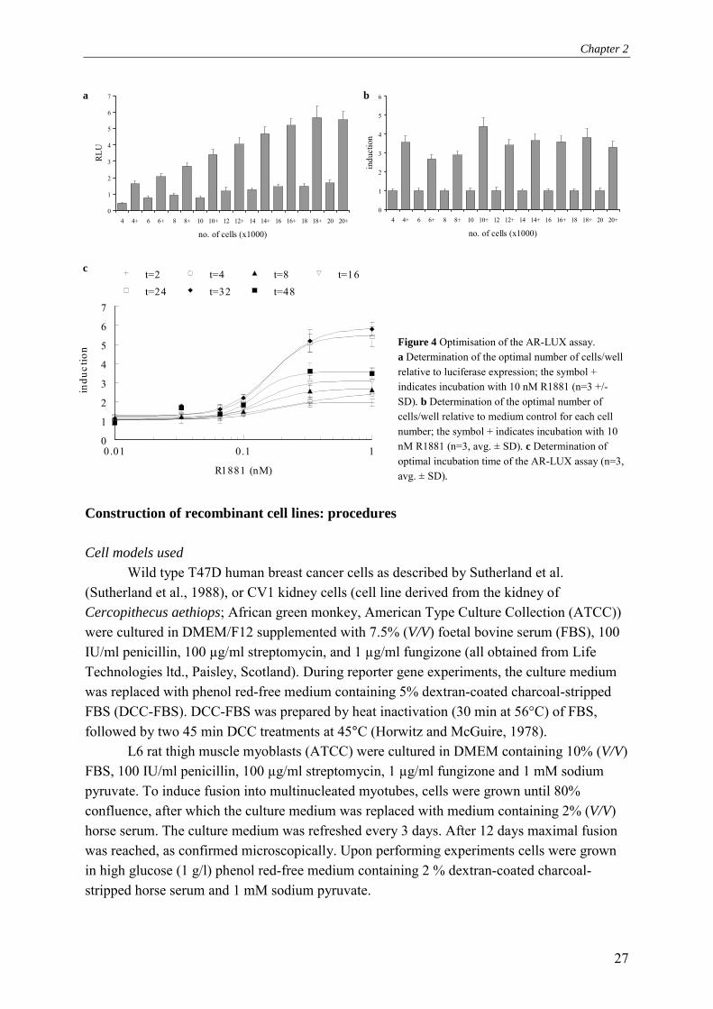
Figure 4 Optimisation of the AR-LUX assay. a Determination of the optimal number of cells/well relative to luciferase expression; the symbol + indicates incubation with 10 nM R1881 (n=3 +/- SD). b Determination of the optimal number of cells/well relative to medium control for each cell number; the symbol + indicates incubation with 10 nM R1881 (n=3, avg. ± SD). c Determination of optimal incubation time of the AR-LUX assay (n=3, avg. ± SD).
Construction of recombinant cell lines: procedures Cell models used
Wild type T47D human breast cancer cells as described by Sutherland et al. (Sutherland et al., 1988), or CV1 kidney cells (cell line derived from the kidney of Cercopithecus aethiops; African green monkey, American Type Culture Collection (ATCC)) were cultured in DMEM/F12 supplemented with 7.5% (V/V) foetal bovine serum (FBS), 100 IU/ml penicillin, 100 µg/ml streptomycin, and 1 µg/ml fungizone (all obtained from Life Technologies ltd., Paisley, Scotland). During reporter gene experiments, the culture medium was replaced with phenol red-free medium containing 5% dextran-coated charcoal-stripped FBS (DCC-FBS). DCC-FBS was prepared by heat inactivation (30 min at 56°C) of FBS, followed by two 45 min DCC treatments at 45°C (Horwitz and McGuire, 1978).
L6 rat thigh muscle myoblasts (ATCC) were cultured in DMEM containing 10% (V/V) FBS, 100 IU/ml penicillin, 100 µg/ml streptomycin, 1 µg/ml fungizone and 1 mM sodium pyruvate. To induce fusion into multinucleated myotubes, cells were grown until 80% confluence, after which the culture medium was replaced with medium containing 2% (V/V) horse serum. The culture medium was refreshed every 3 days. After 12 days maximal fusion was reached, as confirmed microscopically. Upon performing experiments cells were grown in high glucose (1 g/l) phenol red-free medium containing 2 % dextran-coated charcoal-stripped horse serum and 1 mM sodium pyruvate.
0
1
2
3
4
5
6
7
4 4+ 6 6+ 8 8+ 10 10+ 12 12+ 14 14+ 16 16+ 18 18+ 20 20+
no. of cells (x1000)
RLU
0
1
2
3
4
5
6
4 4+ 6 6+ 8 8+ 10 10+ 12 12+ 14 14+ 16 16+ 18 18+ 20 20+
no. of cells (x1000)
indu
ctio
n
c
a b

Chapter 2
28
Ligation & restriction analysis Ligase and restriction enzymes and their buffers were obtained from Gibco/Invitrogen
(California, USA). Ligase reactions were performed exactly according to the manufacturer�s protocol. Following ligation of double stranded oligos into ptataluc+ (Altschmied and Duschl, 1997), successful integration was verified by restriction analysis. 1 µg of plasmid was digested with ten units of restriction enzyme in the appropriate buffer as prescribed by the manufacturer. Following 1 h incubation at 37°C, the digested plasmids were loaded on a 1% agarose gel followed by electrophoresis after which the restriction pattern could be analysed. Plasmid isolation
Competent E. coli DH5α (Gibco/Invitrogen, California, USA) bacteria were transformed with plasmids according to the supplier's protocol. Briefly, 100 µl of bacteria were thawed and kept on ice. 1 to 10 ng of (ligated) plasmid was added to the bacteria followed by 30 min incubation on ice. Following a heat shock at 42°C for 45 sec, cells were placed on ice for 2 min after which 0.9 ml Luria Bertani (LB) medium (10g tryptone/l, 5 g/l yeast extract, 10 g/l NaCl) was added and cells were incubated at 37°C for 1 h. Subsequently the cells were plated on agar/LB plates containing 100 µg/ml ampicillin and allowed to grow overnight at 37 °C in an incubator. The following day colonies were picked and transferred to 250 ml LB-medium (50 µg/ml ampicillin) and incubated overnight at 37°C in a shaking water bath. Plasmids were subsequently isolated with the Qiafilter Plasmid Maxikit (Qiagen/Westburg, Leusden, the Netherlands) according to the manufacturer�s protocol. Stable transfection
Transfections were performed in 24-well plates (Corning Life Sciences, Schiphol-Rijk the Netherlands) by the standard calcium phosphate co-precipitation technique (Sambrook et al., 1989) or by using lipofectamin 2000TM according to the manufacturer's protocol (Invitrogen, California, USA). Transient transfections were performed in DMEM/F12 without phenol red, supplemented with 5% (V/V) DCC-FBS. Cells were cotransfected with pRLSV40 to enable correction for differences in transfection efficiency. Stable transfections with pPBARE2tataluc+ were carried out in normal culture medium by cotransfection with pSV2-neo (Southern and Berg, 1982) in a molar ratio of 4:1. Subsequent selection for stable transfectants was carried out with 1 mg/ml geneticin (Life Technologies Ltd., Paisley, Scotland). Selection with 50 µg/ml puromycin was applied for siRNA clones transfected with recombinant pSUPER.retro. AR-LUX assay procedure
For AR-LUX assays, cells were seeded in white 96-well plates with clear flat bottoms (Corning Life Sciences, Schiphol-Rijk, the Netherlands) at a density of 18,000 cells/well. After 24 h medium was changed to 5% (V/V) DCC-stripped FBS DMEM/F12 without phenol red. Cells were exposed in triplicate with the chemicals of interest dissolved in ethanol or DMSO, with a maximum solvent concentration of 0.2% (V/V). Following 24 h incubation, cells were harvested and luciferase expression was subsequently measured using a
Chapter 2
29
luminometer (Labsystems Luminoscan RS) or a Wallac 1450 microbeta liquid scintillation counter. When using the luminometer, cells were washed once with 0.5x PBS (Life Technologies Ltd.) followed by the addition of 30 ml lysis buffer (2 mM dithiothreitol, 2 mM 1,2-diaminocyclohexane-N,N,N',N'-tetraacetic acid, 10 mM Tris, pH 7.8). Cells were incubated on ice for 15 min and subsequently frozen at -80°C for at least 1 h. After thawing, shaking, and equilibrating to room temperature on a microtiter plate shaker (200 rpm), the plates were mounted in the luminometer and 100 µl flashmix (20 mM Tricine, 1.07 mM (MgCO3)4Mg(OH)2, 2.67 mM MgSO4, 0.1 mM EDTA, 2.0 mM DTT, 470 mM luciferine, 5.0 mM ATP) was added and subsequently luciferase activity was determined and expressed as relative light units (RLUs). Directly after measurement of each well, 100 µl of 0.2 M NaOH was added to quench the remaining signal in the well, thus preventing cross-talk between neighbouring wells. When using the Wallac 1450, medium was removed from the cells and 20 µl of fresh medium was added with 20 µl of Steady-Glo (Promega Corporation, Madison, USA). Following 10 min incubation at room temperature in the instrument, luciferase activity was counted for 30 s and expressed as luminescence counts. Induction factors were subsequently calculated relative to solvent controls unless indicated otherwise. Re-evaluation of the receptor specificity conferred by the Probasin Androgen Response Element 2 by applying RNA interference: principles The probasin androgen-response element 2 (PB-ARE2) (located at -140 to -117 in the upstream regulatory region of the probasin gene; GenBank accession number AY370611) has been extensively investigated. It has been hypothesised to be androgen-specific due to the fact that it features a direct repeat instead of an inverted repeat also found in steroid receptor regulated genes (Rennie et al., 1993; Claessens et al., 1996; Schoenmakers et al., 1999; Claessens et al., 2001). Indeed, based on our data presented in chapter 3, exclusively the androgen receptor mediates gene expression via PB-ARE2. However, further investigations of the responses of the AR-LUX cells revealed induction of luciferase expression by the specific progestagen promegestone (R5020) at picomolar levels. Although R5020 does bind to the androgen receptor, 3.1% binding compared to R1881 for baboon AR, (Lin et al., 1981) and is able to slightly activate human AR at 1 µM (Poujol et al., 2000), this is not sufficient to explain its activity in AR-LUX cells. Furthermore, triamcinolone acetonide (TA) induced luciferase expression at micromolar concentrations (chapter 6, Fig 5). TA is a compound often used in androgen receptor binding assays since it blocks binding of radioligands to the progesterone receptor but not to the androgen receptor (Zava et al., 1979). Therefore, the observed activity of TA is another indication of PR involvement in PB-ARE2 mediated gene expression in the AR-LUX cell line. The question of receptor-specific activation of the PB-ARE2 element cannot be resolved by using ligands, since almost all steroids display ligand binding site-mediated crosstalk above certain threshold concentrations. For instance, activation of the progesterone receptor by testosterone and 5α-di-hydro-testosterone (DHT) has been reported at µmolar concentrations (Markiewicz and Gurpide, 1997). Therefore, we decided to decrease the level of androgen or progesterone receptors expressed in the AR-LUX cell line by applying RNA interference.

Chapter 2
30
Two siRNA vectors targeting the progesterone receptor and one vector targeting the androgen receptor were designed and transfected into AR-LUX cells after which clonal selection was applied. Puromycin-resistant clones were isolated from cells transfected with siRNA vectors targeting PR. However, the response of these clones to several steroids was not different from AR-LUX wild type cells. Furthermore, no difference in PR-mRNA expression was found with quantitative PCR (QPCR, data not shown). This suggests that either the designed sequences were ineffective or that T47D cells are unable to survive with the resulting diminished progesterone receptor expression, and thus clones having only integrated the puromycin resistance gene were isolated. By contrast, a clone (called ARdown-LUX) featuring AR mRNA down-regulation could be isolated successfully, as described in chapter 6. Its response to various steroids was clearly different from that of the wild-type AR-LUX cells. Since AR-expression is not totally blocked by siRNA -it is knocked down to approximately 6% at the mRNA level- it cannot be excluded that the remaining androgen receptors are involved in the observed effects. However, the ARdown-LUX cells were no longer inducible by DHT and therefore the role of the AR in mediating gene transcription in the siRNA-clone is probably minimal. Therefore, it seems likely that receptors other than the AR are able to mediate gene expression activation via the PB-ARE2 element. We speculate that this may be one of the progesterone receptor isoforms. Since PR-β is not able to induce full luciferase expression via PB-ARE2 (Schoenmakers et al., 1999), we hypothesise that the progesterone receptor-α also mediates gene expression via PB-ARE2 in our assay. As a consequence, the AR-LUX assay does not only allow screening for compounds that activate the androgen receptor but also compounds that induce responses via the established prostatic probasin androgen response element through alternative pathways. In the context of risk evaluation, this is preferable to systems exclusively monitoring androgen receptor activation since in both males and females effects of steroid hormones previously identified as sex-specific have been reported. For instance, inhibition of estrogen-induced sexual receptivity in female rats by androgens has been reported, as has regulation of male sexual behavior by the progesterone receptor in male rats (Blasberg et al., 1998; Phelps et al., 1998). Effects of estrogens in males have also been reported (Sharpe, 1998). Re-evaluation of the receptor specificity conferred by the Probasin Androgen Response Element 2 by applying RNA interference: procedures siRNA design
The pSUPER.retro vector (Oligoengine, Seattle, USA) was digested according to the manufacturer�s protocol. Subsequently the 64 bp double stranded siRNA oligos were inserted into the BglII and HindIII sites of the vector. Following ligation, recombinant vectors were stably transfected into AR-LUX cells. The siRNA oligo sequences are given in Table 2. RNA isolation and purification RNA was isolated using Trizol (Invitrogen, California, USA) according to the manufacturer�s protocol. Following Trizol isolation, RNA was further purified with RNeasy
Chapter 2
31
mini columns (Qiagen/Westburg, Leusden, the Netherlands) according to the manufacturer�s protocol. Optionally, a DNase step (Rnase-free DNase Set, Qiagen/Westburg, Leusden, the Netherlands) was incorporated and performed according to the manufacturer�s protocol to remove residual DNA from RNA samples. Table 2 Sequences of siRNA oligos. Bold underlined sequences represent the actual sequences responsible for RNA interference.
hPR1111siRNAPRETROX1 5�-GATCCCCTCACGCCTTATTGGCAGCCTTCAAGAGAGGCTGCCAATAAGGCGTGATTTTTGGAAA-3� hPR1111siRNAPRETROX1-AS 5�-AGCTTTTCCAAAAATCACGCCTTATTGGCAGCCTCTCTTGAAGGCTGCCAATAAGGCGTGAGGG-3� hPR2757siRNAPRETROX1 5�-GATCCCCAAGGAGTTGTGTCGAGCTCTTCAAGAGAGAGCTCGACACAACTCCTTTTTTTGGAAA-3� hPR2757siRNAPRETROX1-AS 5�-AGCTTTTCCAAAAAAAGGAGTTGTGTCGAGCTCTCTCTTGAAGAGCTCGACACAACTCCTTGGG-3� hAR1061siRNAPRETROX1 5�-GATCCCCCGCCAAGGAGTTGTGTAAGTTCAAGAGACTTACACAACTCCTTGGCGTTTTTGGAAA-3� hAR1061siRNAPRETROX1-AS 5�-AGCTTTTCCAAAAACGCCAAGGAGTTGTGTAAGTCTCTTGAACTTACACAACTCCTTGGCGGGG-3�
Quantitative RT-PCR RNA was reverse transcribed with AMV reverse transcriptase (Promega, Madison, USA). Briefly, 1 µl oligo dT (0.5 mg/ml) was added to 1 µg RNA and adjusted to a volume of 10 µl with H2O. Subsequently the sample was incubated at 70°C for 5 min followed by 5 min on ice. Subsequently, 5 µl AMV buffer (5x), 2.5 µl dNTPmix (10 mM each), 1 µl RNase inhibitor (40U/µl), 1.5 µl AMV RT (10 units/ml) was added to the RNA and adjusted to a total volume of 25 µl with H2O. Reactions were subsequently incubated at 42°C for 60 min after which 475 µl H2O was added. For quantitative PCR, 5 µl of this mixture was added as the starting concentration of template for PCR. QPCR was performed with the quantitect SYBR green PCR kit (Qiagen/Westburg, Leusden, the Netherlands) with minor modifications to the manufacturer�s protocol. To a total volume of 20 µl, 1 µl of each primer (20 µM), 10 µl Qiagen SYBR green mix and 5 µl cDNA template were added. Subsequently, QPCR was performed and product formation quantitated in a BioRad Icycler. PCR for the androgen receptor mRNA was performed as follows: cycle 1 (1x): 95°C, 10 min, Cycle 2 (42x): 95°C, 15 sec; 54°C, 30 sec; 72°C, 20 sec; Cycle 3: 72°C, 5 min; Cycle 4: 95°C, 1 min; Cycle 5: 95°C, 10 sec followed by collection of the melt curve data points. Expression of mRNAs was normalised by dividing calculated target mRNA expression by β-actin expression. Primer sequences are given in Table 3. Identification by DNA-micro array analysis of candidate genes possibly involved in synergistic growth enhancement by androgens and estrogens: principles Despite the before-mentioned intricacies of steroid hormone interactions, an attempt was made to identify genes specifically affected by treatment with a mixture of an androgen and an estrogen. Testosterone is the major systemic androgen while DHT is primarily formed in target tissues. Testosterone rather than DHT has been identified as being primarily

Chapter 2
32
Table 3 Sequences of PCR primers used in QPCR. target sequence accession no.
hPRsirnafw1111pcr 5' tgcctatcctgcctctcaat 3' NM000926
hPRsirnarev1111pcr 5' ggggaagtcgcctacagc 3' NM000926
hPRsirnafw2757pcr 5' agctcatcaaggcaattggt 3' NM000926
hPRsirnarev2757pcr 5' agtgcccgggactggata 3' NM000926
hARsirnafw1061pcr 5' caacgccaaggagttgtgta 3' NM000044
hARsirnarev1061pcr 5' cgctgtcgtctagcagagaa 3' NM000044
myosin heavy chain 3 5' tgagtagcgacaccgagatg 3' NM012604
myosin heavy chain 3 5' caccagggtcctgttgtctt 3' NM012604
ribosomal protein L39 5' cctggcaaagaaacaaaagc 3' NM012875
ribosomal protein L39 5' aaatccatctggtcggactg 3' NM012875
fibromodulin 5' agaaatggccgcagagtcta 3' X82152
fibromodulin 5' aaggagtaggagcccagagc 3' X82152
cyclin L 5' agcctccaaaccatcatcac 3' AF030091
cyclin L 5' tggcacttctgctgtttctg 3' AF030091
apoptosis inhibitor 3 5' gacaaatgtcccatgtgctg 3' NM022231
apoptosis inhibitor 3 5' ctaatggactgcgatgctga 3' NM022231
disc homolog 5' tctcccacacacattccaga 3' NM019621
disc homolog 5' cccaaaaaccacctttgaga 3' NM019621
rat beta actine 5' ttcaacaccccagccatgt 3' NM031144
rat beta actine 5' gtggtacgaccagaggcataca 3' NM031144
responsible for anabolic effects in muscle (Sundaram et al., 1995). Due to lack of 5α-reductase in muscle cells, testosterone is not reduced into DHT. Therefore, testosterone was used as a model anabolic androgen and it was combined with the estrogen estradiol. Low concentrations of both compounds (100 nM) were used in our in vitro muscle model to prevent possible crosstalk via other receptors as a consequence of ligand binding domain (LBD)-mediated cross-talk at high concentrations.
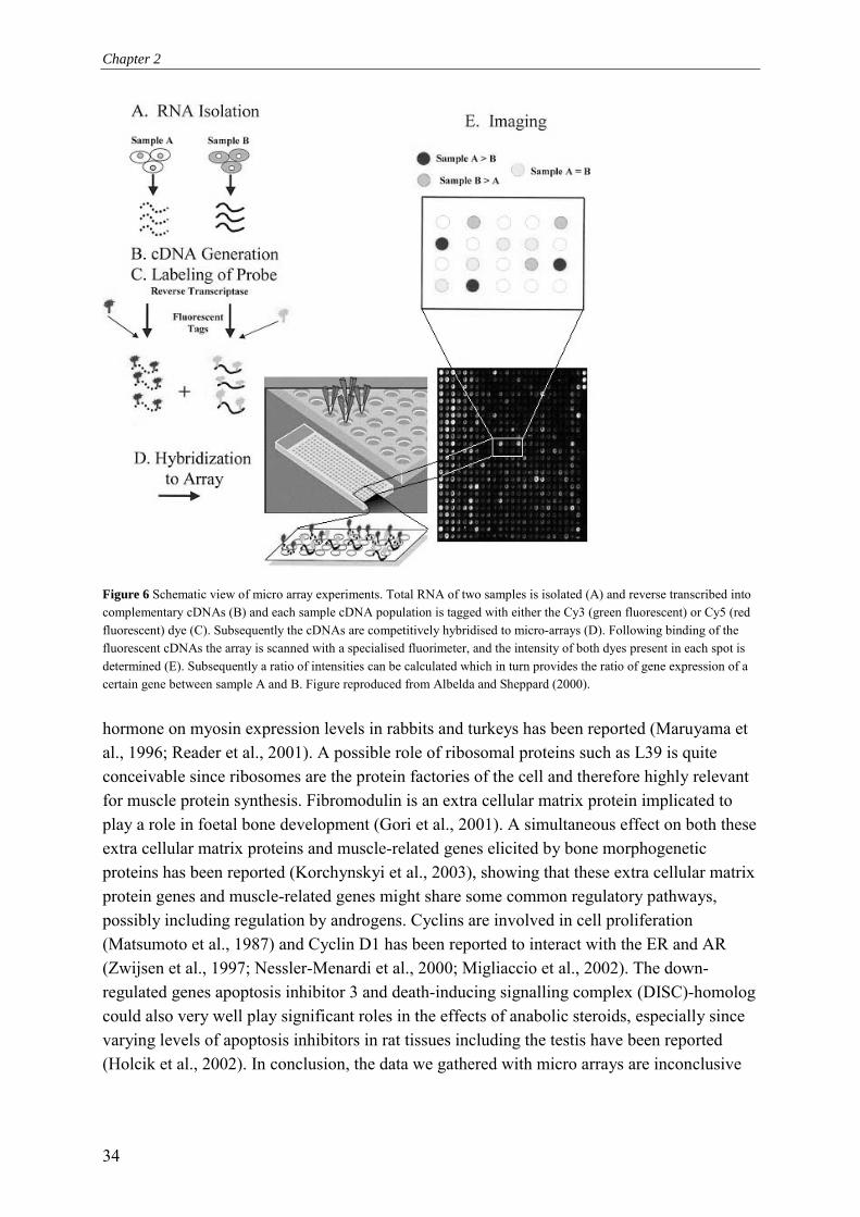
L6 rat myoblast cells were cultured under conditions inducing fusion into myotubes. This myogenic line was isolated originally by Yaffe from primary cultures of rat thigh muscle maintained for the first two passages in the presence of methyl-cholanthrene (Yaffe, 1968). L6 cells fuse in culture to form multinucleated myotubes and striated fibers. The myotubes were subsequently treated with 100 nM testosterone, 100 nM estradiol or a mixture of testosterone and estradiol (both 100 nM) (Fig. 5). Hormones were applied for 8 h to simulate short term effects and 48 h to simulate long term effects. Following treatment, total RNA was isolated for micro array analysis to obtain an overview of induced changes in the gene expression pattern. RNA samples were sent to the Genomics and Micro array Laboratory (GML) at the University of Cincinnati. They were hybridised to operon rat oligo micro arrays (for more information also see http://microarray.uc.edu/). The RNA provided the template to generate fluorescently labelled (cytidine-3 and cytidine-5) target cDNAs. These cDNAs labelled with either Cy-3 or Cy-5 from untreated cells and treated cells were subsequently hybridised to micro arrays carrying oligonucleotide
Chapter 2
33
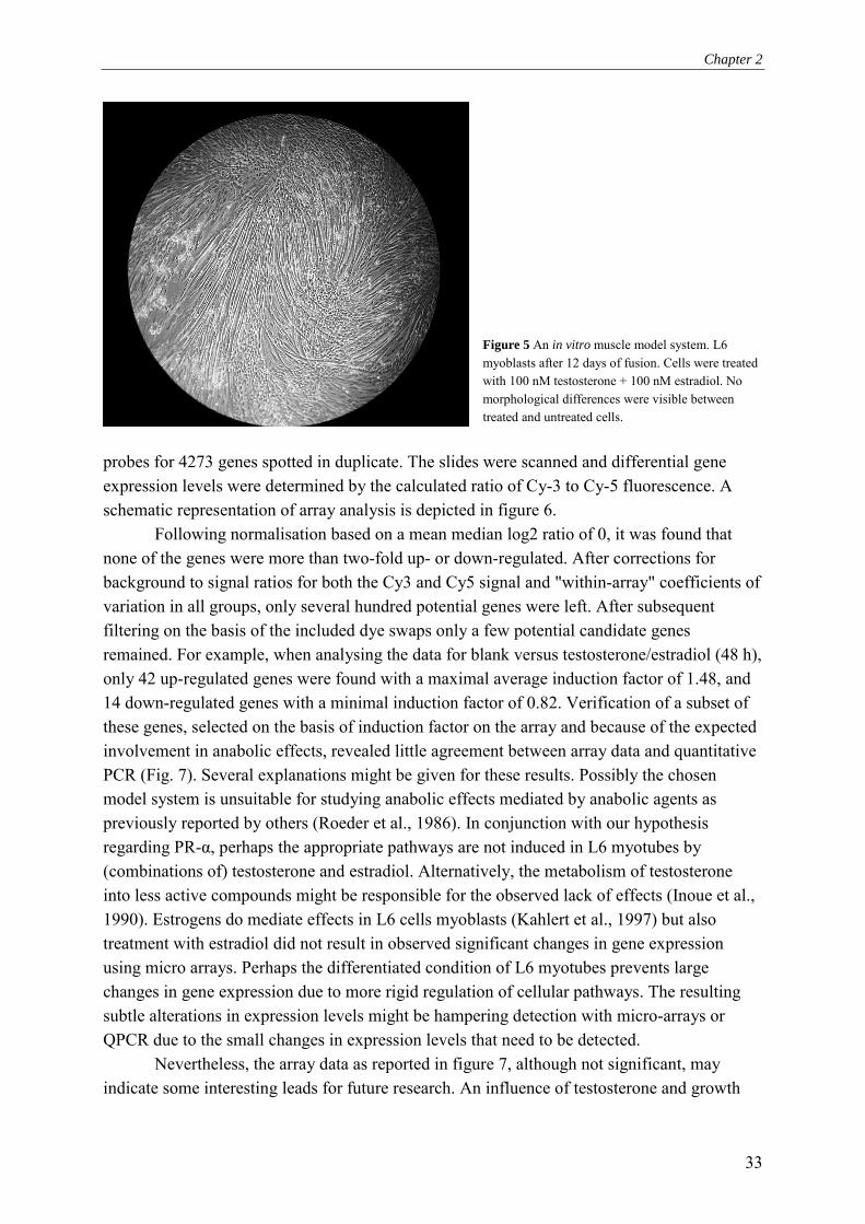
Figure 5 An in vitro muscle model system. L6 myoblasts after 12 days of fusion. Cells were treated with 100 nM testosterone + 100 nM estradiol. No morphological differences were visible between treated and untreated cells.
probes for 4273 genes spotted in duplicate. The slides were scanned and differential gene expression levels were determined by the calculated ratio of Cy-3 to Cy-5 fluorescence. A schematic representation of array analysis is depicted in figure 6.
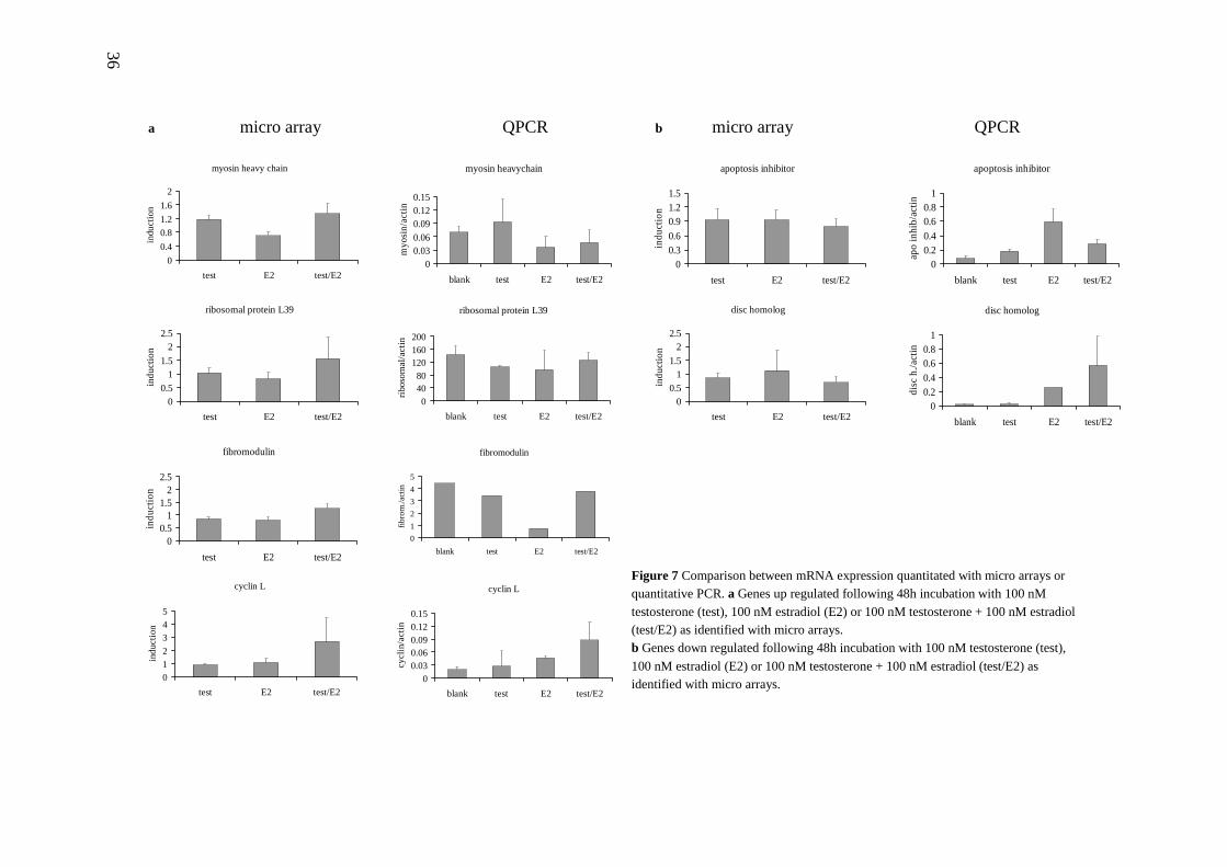
Following normalisation based on a mean median log2 ratio of 0, it was found that none of the genes were more than two-fold up- or down-regulated. After corrections for background to signal ratios for both the Cy3 and Cy5 signal and "within-array" coefficients of variation in all groups, only several hundred potential genes were left. After subsequent filtering on the basis of the included dye swaps only a few potential candidate genes remained. For example, when analysing the data for blank versus testosterone/estradiol (48 h), only 42 up-regulated genes were found with a maximal average induction factor of 1.48, and 14 down-regulated genes with a minimal induction factor of 0.82. Verification of a subset of these genes, selected on the basis of induction factor on the array and because of the expected involvement in anabolic effects, revealed little agreement between array data and quantitative PCR (Fig. 7). Several explanations might be given for these results. Possibly the chosen model system is unsuitable for studying anabolic effects mediated by anabolic agents as previously reported by others (Roeder et al., 1986). In conjunction with our hypothesis regarding PR-α, perhaps the appropriate pathways are not induced in L6 myotubes by (combinations of) testosterone and estradiol. Alternatively, the metabolism of testosterone into less active compounds might be responsible for the observed lack of effects (Inoue et al., 1990). Estrogens do mediate effects in L6 cells myoblasts (Kahlert et al., 1997) but also treatment with estradiol did not result in observed significant changes in gene expression using micro arrays. Perhaps the differentiated condition of L6 myotubes prevents large changes in gene expression due to more rigid regulation of cellular pathways. The resulting subtle alterations in expression levels might be hampering detection with micro-arrays or QPCR due to the small changes in expression levels that need to be detected.
Nevertheless, the array data as reported in figure 7, although not significant, may indicate some interesting leads for future research. An influence of testosterone and growth
Chapter 2
34
Figure 6 Schematic view of micro array experiments. Total RNA of two samples is isolated (A) and reverse transcribed into complementary cDNAs (B) and each sample cDNA population is tagged with either the Cy3 (green fluorescent) or Cy5 (red fluorescent) dye (C). Subsequently the cDNAs are competitively hybridised to micro-arrays (D). Following binding of the fluorescent cDNAs the array is scanned with a specialised fluorimeter, and the intensity of both dyes present in each spot is determined (E). Subsequently a ratio of intensities can be calculated which in turn provides the ratio of gene expression of a certain gene between sample A and B. Figure reproduced from Albelda and Sheppard (2000).
hormone on myosin expression levels in rabbits and turkeys has been reported (Maruyama et al., 1996; Reader et al., 2001). A possible role of ribosomal proteins such as L39 is quite conceivable since ribosomes are the protein factories of the cell and therefore highly relevant for muscle protein synthesis. Fibromodulin is an extra cellular matrix protein implicated to play a role in foetal bone development (Gori et al., 2001). A simultaneous effect on both these extra cellular matrix proteins and muscle-related genes elicited by bone morphogenetic proteins has been reported (Korchynskyi et al., 2003), showing that these extra cellular matrix protein genes and muscle-related genes might share some common regulatory pathways, possibly including regulation by androgens. Cyclins are involved in cell proliferation (Matsumoto et al., 1987) and Cyclin D1 has been reported to interact with the ER and AR (Zwijsen et al., 1997; Nessler-Menardi et al., 2000; Migliaccio et al., 2002). The down-regulated genes apoptosis inhibitor 3 and death-inducing signalling complex (DISC)-homolog could also very well play significant roles in the effects of anabolic steroids, especially since varying levels of apoptosis inhibitors in rat tissues including the testis have been reported (Holcik et al., 2002). In conclusion, the data we gathered with micro arrays are inconclusive

Chapter 2
35
but do warrant further research on a genomic scale, since, as we also observed, regulation of widely divergent proteins is probably involved in the effects of anabolic steroids. Identification by DNA-micro array analysis of candidate genes possibly involved in synergistic growth enhancement by androgens and estrogens: procedures Micro array analysis
Micro array analyses were performed at the Genomics and Micro array Laboratory (GML) at the University of Cincinnati according to their standard protocols as described below. For each incubation one RNA sample was analysed in triplicate including one dye swap.
Oligonucleotide micro array preparation
The Operon 70-mer oligonucleotides were purchased from QIAGEN Operon, Inc, (Alameda, USA). Each oligonucleotide was suspended in 3 x SSC (1M NaCl, 0.1 M Na-citrate) and printed on aminosilane-coated UltraGAPS slides (Corning Life science, USA). In total 4273 oligonucleotides were deposited on the slides in duplicate. The slides were UV-cross linked in a Stratalinker (Stratagene, La Jolla, USA).
cDNA synthesis and labelling
For labelling of target cDNA with Cy-3 or Cy-5 dyes an indirect amino-allyl labelling method was used. The reverse transcription (RT) reaction was performed on 20 µg of total RNA with superscript II RT (Gibco/Invitrogen, California, USA) and a dNTP solution (4:1 ratio aminoallyl-dUTP (Sigma-Aldrich Corporation, St. Louis, MO, USA) to dTTP). RNA was degraded by hydrolysis in 0.1 M NaOH (10 min 70°C) and the solution was subsequently neutralised with 0.1 M HCl. After ethanol precipitation the cDNA was coupled to either the Cy-3 or the Cy-5 fluorophore. The reaction mixture was quenched with 4 M hydroxylamine (5 h at room temperature in the dark). Labeled cDNA was purified using the Qia-Quick PCR purification kit (Qiagen, Alameda, USA). Hybridisation was carried out using Slide-Hyb Glass Array Hybridisation buffer (Ambion, Houston, USA) and a hybridisation station (Genomic solutions, Huntingdon Cambridgeshire, UK). The accompanying Ambion protocol was followed, with the exception of the pre-hybridisation and blocking steps that were not performed. The slides were dried by centrifugation. Scanning and data analysis
The dried arrays were scanned using the GenePix 4000B scanner and GenePixpro 4.0 Array analysis software (Axon Instruments, Foster City, USA). The GenePixpro 4.0 array analysis software processed the acquired images into result files. The result files were further processed in Excel (Microsoft Corporation). For each spot, the local background intensity was subtracted from the signal intensity. Spots with a signal-to-noise ratio smaller than 3 for both Cy-3 and Cy-5, a coefficient of variation between the duplicate spots on an array larger than
36
cyclin L
012345
test E2 test/E2
indu
ctio
n
cyclin L
00.030.060.090.120.15
blank test E2 test/E2
cycl
in/a
ctin
fibromodulin
00.5
11.5
22.5
test E2 test/E2
indu
ctio
n
fibromodulin
012345
blank test E2 test/E2
fibro
m./a
ctin
myosin heavy chain
00.40.81.21.6
2
test E2 test/E2
indu
ctio
n
ribosomal protein L39
00.5
11.5
22.5
test E2 test/E2
indu
ctio
n
myosin heavychain
00.030.060.090.120.15
blank test E2 test/E2
myo
sin/
actin
ribosomal protein L39
04080
120160200
blank test E2 test/E2rib
osom
al/a
ctin
apoptosis inhibitor
00.30.60.91.21.5
test E2 test/E2
indu
ctio
n
disc homolog
00.5
11.5
22.5
test E2 test/E2
indu
ctio
n
apoptosis inhibitor
00.20.40.60.8
1
blank test E2 test/E2
apo
inhi
b/ac
tin
disc homolog
00.20.40.60.8
1
blank test E2 test/E2
disc
h./a
ctin
a micro array QPCR b micro array QPCR
Figure 7 Comparison between mRNA expression quantitated with micro arrays or quantitative PCR. a Genes up regulated following 48h incubation with 100 nM testosterone (test), 100 nM estradiol (E2) or 100 nM testosterone + 100 nM estradiol (test/E2) as identified with micro arrays. b Genes down regulated following 48h incubation with 100 nM testosterone (test), 100 nM estradiol (E2) or 100 nM testosterone + 100 nM estradiol (test/E2) as identified with micro arrays.

Chapter 2
37
50%, or an average ratio of medians larger than 50 were excluded from further analysis. Mean median log2 ratios were corrected towards an average of 0 on each array by applying the normalisation parameters for the two dye wavelengths provided by GenePixpro 4.0. Genes that failed to show similar results in the dye swaps were removed from further analysis. Quantitative RT-PCR
QPCR was performed as described earlier under �Re-evaluation of the receptor specificity conferred by the Probasin Androgen Response Element 2 by applying RNA interference: procedures�. Primer sequences are given in Table 3. References Albelda, S. M. and D. Sheppard (2000). "Functional genomics and expression profiling: be
there or be square." Am J Respir Cell Mol Biol 23(3): 265-269. Altschmied, J. and J. Duschl (1997). "Set of optimized luciferase reporter gene plasmids
compatible with widely used CAT vectors." Biotechniques. 23: 436-438. Blasberg, M. E., S. Robinson, L. P. Henderson and A. S. Clark (1998). "Inhibition of
estrogen-induced sexual receptivity by androgens: role of the androgen receptor." Horm Behav 34(3): 283-293.
Brummelkamp, T. R., R. Bernards and R. Agami (2002). "A system for stable expression of short interfering RNAs in mammalian cells." Science 296(5567): 550-553.
Claessens, F., P. Alen, A. Devos, B. Peeters, G. Verhoeven and W. Rombauts (1996). "The androgen-specific probasin response element 2 interacts differentially with androgen and glucocorticoid receptors." J.Biol.Chem. 271(32): 19013-19016.
Claessens, F., G. Verrijdt, E. Schoenmakers, A. Haelens, B. Peeters, G. Verhoeven and W. Rombauts (2001). "Selective DNA binding by the androgen receptor as a mechanism for hormone-specific gene regulation." J Steroid Biochem Mol Biol 76(1-5): 23-30.
Elbashir, S. M., J. Harborth, W. Lendeckel, A. Yalcin, K. Weber and T. Tuschl (2001). "Duplexes of 21-nucleotide RNAs mediate RNA interference in cultured mammalian cells." Nature 411(6836): 494-498.
Gardner, E. J., S. M. J. and D. P. Snustad (1991). Principles of Genetics, John Wiley & sons, inc.
Gerhold, D., T. Rushmore and C. T. Caskey (1999). "DNA chips: promising toys have become powerful tools." Trends.Biochem.Sci. 24: 168-173.
Gori, F., E. Schipani and M. B. Demay (2001). "Fibromodulin is expressed by both chondrocytes and osteoblasts during fetal bone development." J Cell Biochem 82(1): 46-57.
Hannon, G. J. (2002). "RNA interference." Nature 418(6894): 244-251. Hofman, K., J. V. Swinnen, G. Verhoeven and W. Heyns (2002). "Coactivation of an
endogenous progesterone receptor by TIF2 in COS-7 cells." Biochem Biophys Res Commun 295(2): 469-474.

Chapter 2
38
Holcik, M., C. A. Lefebvre, K. Hicks and R. G. Korneluk (2002). "Cloning and characterization of the rat homologues of the Inhibitor of Apoptosis protein 1, 2, and 3 genes." BMC Genomics 3(1): 5.
Horwitz, K. B. and W. L. McGuire (1978). "Estrogen control of progesterone receptor in human breast cancer: Correlation with nuclear processing of estrogen receptor." J. Biol. Chem. 253: 2222-2228.
Ibrahim, N. M., A. C. Marinovic, S. R. Price, L. G. Young and O. Frohlich (2000). "Pitfall of an internal control plasmid: response of Renilla luciferase (pRL-TK) plasmid to dihydrotestosterone and dexamethasone." Biotechniques 29(4): 782-784.
Inoue, S., I. Morimoto, S. Yamashita, M. Izumi and S. Nagataki (1990). "Androgen metabolism in rat L6 myoblast cells; high formation of 5 alpha-androstane-3 alpha,17 beta-diol from testosterone." J Steroid Biochem 35(1): 77-81.
Kahlert, S., C. Grohe, R. H. Karas, K. Lobbert, L. Neyses and H. Vetter (1997). "Effects of estrogen on skeletal myoblast growth." Biochem Biophys Res Commun 232(2): 373-378.
Korchynskyi, O., K. J. Dechering, A. M. Sijbers, W. Olijve and P. ten Dijke (2003). "Gene array analysis of bone morphogenetic protein type I receptor-induced osteoblast differentiation." J Bone Miner Res 18(7): 1177-1185.
Lin, A. L., H. C. McGill, Jr. and S. A. Shain (1981). "Hormone receptors of the baboon cardiovascular system. Biochemical characterization of myocardial cytoplasmic androgen receptors." Circ Res 49(4): 1010-1016.
Markiewicz, L. and E. Gurpide (1997). "Estrogenic and progestagenic activities of physiologic and synthetic androgens, as measured by in vitro bioassays." Methods.Find.Exp.Clin.Pharmacol. 19: 215-222.
Maruyama, K., N. Kanemaki and J. A. Proudman (1996). "Effects of growth hormone injection to embryos on growth and myosin heavy chain isoforms in growing turkeys (Meleagris gallopavo)." Comp Biochem Physiol A Physiol 113(4): 315-321.
Matsumoto, K., T. Moriuchi, T. Koji and P. K. Nakane (1987). "Molecular cloning of cDNA coding for rat proliferating cell nuclear antigen (PCNA)/cyclin." EMBO J. 6(3): 637-642.
Migliaccio, A., G. Castoria, M. Di Domenico, A. de Falco, A. Bilancio, M. Lombardi, D. Bottero, L. Varricchio, M. Nanayakkara, A. Rotondi and F. Auricchio (2002). "Sex steroid hormones act as growth factors." J Steroid Biochem Mol Biol 83(1-5): 31-35.
Nessler-Menardi, C., I. Jotova, Z. Culig, I. E. Eder, T. Putz, G. Bartsch and H. Klocker (2000). "Expression of androgen receptor coregulatory proteins in prostate cancer and stromal-cell culture models." Prostate 45(2): 124-131.
Phelps, S. M., J. P. Lydon, W. O'Malley B and D. Crews (1998). "Regulation of male sexual behavior by progesterone receptor, sexual experience, and androgen." Horm Behav 34(3): 294-302.
Poujol, N., J.-M. Wurtz, B. Tahiri, S. Lumbroso, J.-C. Nicolas, D. Moras and C. Sultan (2000). "Specific recognition of androgens by their nuclear receptor: a structure-function study." J. Biol. Chem.: 24022-24031.
Chapter 2
39
Reader, M., G. Schwartz and A. W. English (2001). "Brief exposure to testosterone is sufficient to induce sex differences in the rabbit masseter muscle." Cells Tissues Organs 169(3): 210-217.
Rennie, P. S., N. Bruchovsky, K. J. Leco, P. C. Sheppard, S. A. McQueen, H. Cheng, R. Snoek, A. Hamel, M. E. Bock, B. S. MacDonald and et al. (1993). "Characterization of two cis-acting DNA elements involved in the androgen regulation of the probasin gene." Mol Endocrinol 7(1): 23-36.
Roeder, R. A., S. D. Thorpe, F. M. Byers, G. T. Schelling and J. M. Gunn (1986). "Influence of anabolic agents on protein synthesis and degradation in muscle cells grown in culture." Growth 50(4): 485-495.
Sambrook, J., E. F. Fritsch and T. Maniatis (1989). molecular cloning: a laboratory manual. New York, Cold Spring Harbor Laboratory Press.
Schoenmakers, E., P. Alen, G. Verrijdt, B. Peeters, G. Verhoeven, W. Rombauts and F. Claessens (1999). "Differential DNA binding by the androgen and glucocorticoid receptors involves the second Zn-finger and a C-terminal extension of the DNA-binding domains." Biochem J 341: 515-521.
Sharpe, R. M. (1998). "The Roles of Oestrogen in the Male." Trends in Endocrinology and Metabolism 9(9): 371-377.
Southern, P. J. and P. Berg (1982). "Transformation of mammalian cells to antibiotic resistance with a bacterial gene under control of the SV40 early region promoter." J Mol Appl Genet 1(4): 327-341.
Sundaram, K., N. Kumar, C. Monder and C. W. Bardin (1995). "Different patterns of metabolism determine the relative anabolic activity of 19-norandrogens." J Steroid Biochem Mol Biol 53(1-6): 253-257.
Sutherland, R. L., R. E. Hall, G. Y. Pang, E. A. Musgrove and C. L. Clarke (1988). "Effect of medroxyprogesterone acetate on proliferation and cell cycle kinetics of human mammary carcinoma cells." Cancer.Res. 48: 5084-5091.
Yaffe, D. (1968). "Retention of differentiation potentialities during prolonged cultivation of myogenic cells." Proc Natl Acad Sci U S A 61(2): 477-483.
Zava, D. T., B. Landrum, K. B. Horwitz and W. L. McGuire (1979). "Androgen receptor assay with [3H]methyltrienolone (R1881) in the presence of progesterone receptors." Endocrinology 104(4): 1007-1012.
Zhou, Z., J. L. Corden and T. R. Brown (1997). "Identification and characterization of a novel androgen response element composed of a direct repeat." J.Biol.Chem. 272(13): 8227-8235.
Zwijsen, R. M., E. Wientjens, R. Klompmaker, J. van der Sman, R. Bernards and R. J. Michalides (1997). "CDK-independent activation of estrogen receptor by cyclin D1." Cell 88(3): 405-415.

40

33
Development of an androgen reporter gene assay (AR-LUX) utilising a human cell line with an endogenously regulated androgen receptor
Analytical Biochemistry 298(1): 93-102, 2001
B.M.G. Blankvoort1, 2, E.M. de Groene3, A.P. van Meeteren-Kreikamp1, R.F. Witkamp1, R.J.T. Rodenburg1, J.M.M.J.G. Aarts2
1 Department of Bioanalysis, TNO Pharma, the Netherlands, 2 Department of Agrotechnology and Food Sciences, Sub-
department of Toxicology, Wageningen University, Wageningen, the Netherlands, 3 Unilever Health Institute, Vlaardingen, the Netherlands
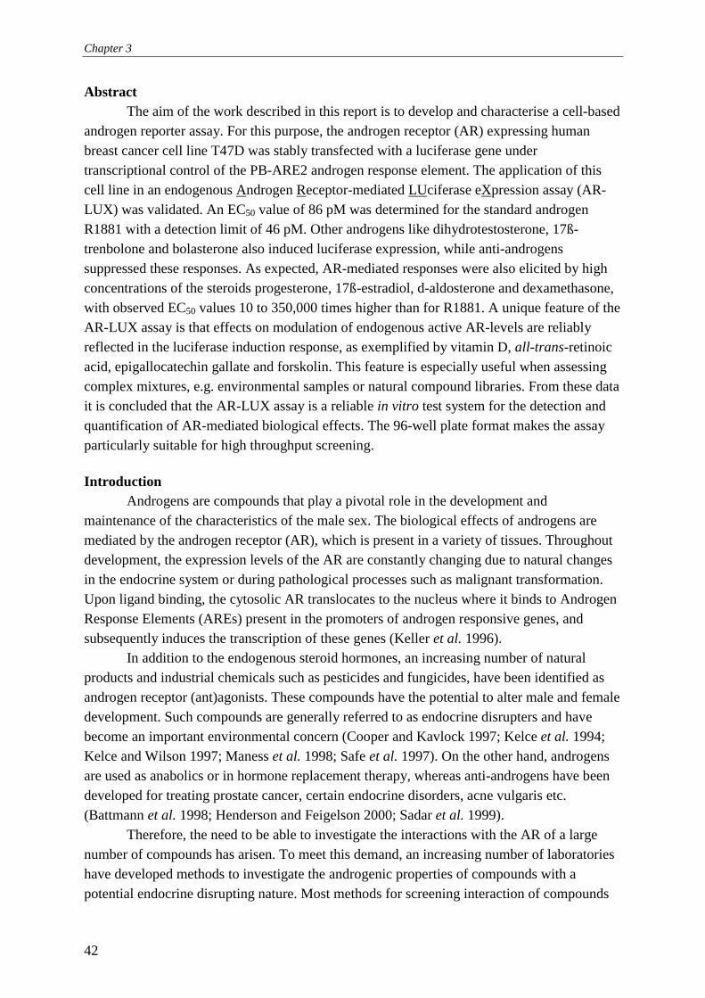
Chapter 3
42
Abstract The aim of the work described in this report is to develop and characterise a cell-based
androgen reporter assay. For this purpose, the androgen receptor (AR) expressing human breast cancer cell line T47D was stably transfected with a luciferase gene under transcriptional control of the PB-ARE2 androgen response element. The application of this cell line in an endogenous Androgen Receptor-mediated LUciferase eXpression assay (AR-LUX) was validated. An EC50 value of 86 pM was determined for the standard androgen R1881 with a detection limit of 46 pM. Other androgens like dihydrotestosterone, 17ß-trenbolone and bolasterone also induced luciferase expression, while anti-androgens suppressed these responses. As expected, AR-mediated responses were also elicited by high concentrations of the steroids progesterone, 17ß-estradiol, d-aldosterone and dexamethasone, with observed EC50 values 10 to 350,000 times higher than for R1881. A unique feature of the AR-LUX assay is that effects on modulation of endogenous active AR-levels are reliably reflected in the luciferase induction response, as exemplified by vitamin D, all-trans-retinoic acid, epigallocatechin gallate and forskolin. This feature is especially useful when assessing complex mixtures, e.g. environmental samples or natural compound libraries. From these data it is concluded that the AR-LUX assay is a reliable in vitro test system for the detection and quantification of AR-mediated biological effects. The 96-well plate format makes the assay particularly suitable for high throughput screening. Introduction
Androgens are compounds that play a pivotal role in the development and maintenance of the characteristics of the male sex. The biological effects of androgens are mediated by the androgen receptor (AR), which is present in a variety of tissues. Throughout development, the expression levels of the AR are constantly changing due to natural changes in the endocrine system or during pathological processes such as malignant transformation. Upon ligand binding, the cytosolic AR translocates to the nucleus where it binds to Androgen Response Elements (AREs) present in the promoters of androgen responsive genes, and subsequently induces the transcription of these genes (Keller et al. 1996).
In addition to the endogenous steroid hormones, an increasing number of natural products and industrial chemicals such as pesticides and fungicides, have been identified as androgen receptor (ant)agonists. These compounds have the potential to alter male and female development. Such compounds are generally referred to as endocrine disrupters and have become an important environmental concern (Cooper and Kavlock 1997; Kelce et al. 1994; Kelce and Wilson 1997; Maness et al. 1998; Safe et al. 1997). On the other hand, androgens are used as anabolics or in hormone replacement therapy, whereas anti-androgens have been developed for treating prostate cancer, certain endocrine disorders, acne vulgaris etc. (Battmann et al. 1998; Henderson and Feigelson 2000; Sadar et al. 1999).
Therefore, the need to be able to investigate the interactions with the AR of a large number of compounds has arisen. To meet this demand, an increasing number of laboratories have developed methods to investigate the androgenic properties of compounds with a potential endocrine disrupting nature. Most methods for screening interaction of compounds
Chapter 3
43
with the AR are currently based on in vivo tests using rats (Gladue and Clemens 1980; Ostby et al. 1999), or in vitro tests such as receptor binding, or transient reporter gene assays using mammalian cell lines or yeast cells (Gaido et al. 1997; Vinggaard et al. 1999). An alternative approach and convenient modification of the latter is to use cell lines that are stably transfected with androgen responsive reporter genes. Until now, three such reporter gene assay systems have been described. All three systems employ cell lines which lacked an endogenous androgen receptor and were stably cotransfected with either a human or rat AR expression plasmid in combination with a reporter plasmid containing either a chloramphenicol acetyl transferase (CAT) or a luciferase reporter gene under transcriptional control of an MMTV promoter (Fuhrmann et al. 1992; Schrader and Cooke 2000; Terouanne et al. 2000). A disadvantage of such systems is that the expression of the AR is controlled by a constitutively active promoter, instead of its natural promoter. Therefore, regulation of AR expression, which is an important aspect of the responsiveness of target cells to androgens, is not taken into account.
In this paper, we describe an androgen reporter system that utilises an endogenously expressed AR combined with PB-ARE2 a highly specific natural androgen-responsive transcriptional enhancer from the rat probasin gene regulatory region. We constructed a reporter plasmid containing a luciferase gene under transcriptional control of the PB-ARE2 and stably transfected it into T47D human breast carcinoma cells. The Androgen Receptor-mediated LUciferase eXpression (AR-LUX) system was evaluated for its responsiveness to a number of androgens, anti-androgens, non-androgenic steroids, and to compounds modulating the AR itself. Methods Chemicals
R1881 and 3H-R1881 were purchased from NEN life science Products (Hoofddorp, the Netherlands). Dihydrotestosterone, bolasterone, flutamide, cyproterone acetate, spironolactone, dexamethasone, d-aldosterone, 17ß-estradiol, progesterone, epigallocatechin gallate, forskolin, 1α,25-dihydroxycholecalciferol (vitamin D), and all-trans-retinoic acid were obtained from Sigma-Aldrich (Zwijndrecht, the Netherlands). Methylboldenone and oxandralone were purchased from Steraloids Inc. (Newport R.I., USA). Vinclozolin and 4,4’-DDE were purchased from Riedel-de Haën (the Netherlands). 17ß-Trenbolone was a gift from RIVM (Bilthoven, the Netherlands) and hydroxy-flutamide was kindly provided by Schering-Plough (Belgium). Plasmids and reporter gene constructs
The enhanced luciferase plasmid ptataluc+ was a gift from Dr. J. Altschmiedt (University of Würzburg, Würzburg, Germany). pSVAR0, a human androgen receptor expression plasmid (Brinkmann et al. 1989), was a gift from Dr. A.O. Brinkmann (Erasmus University, Rotterdam, the Netherlands). pSV2-neo was purchased from Clontech (Clontech
Chapter 3
44
Laboratories, Inc., Palo Alto, USA) and pRLSV40 was obtained from Promega (Promega Corporation, Madison, USA). The reporter gene plasmid pPBARE2tataluc+ was constructed by using ptataluc+ (Altschmied and Duschl 1997) as a backbone. A ds-oligo was designed containing two copies of the probasin androgen response element 2 (PB-ARE2) of the rat probasin promoter (Cleutjens et al. 1996), and HindIII and XhoI compatible 5' and 3' ends: 5'-AGCTTGGTACCAGCTTAA TAGGTTCTTGGAGTACTTTACGTCGAAGCTTAATAGGTTCTTGGAGTACTTTAC GTCGAGTCGACC-’3. This element was inserted in the polylinker of ptataluc+ at the HindIII and XhoI restriction sites upstream of the minimal promoter containing a tata-box. Cells & transfection
Parental T47D/Sutherland human breast cancer cells were cultured in DMEM/F12 supplemented with 7.5% foetal bovine serum (FBS), 100 IU/ml penicillin, 100 µg/ml streptomycin and 1 µg/ml fungizone (all obtained from Life Technologies ltd., Paisley, Scotland). When performing experiments involving steroids, phenol red-free medium containing 5% or 2.5% dextran coated charcoal-stripped serum was used (DCC-FBS). DCC-FBS was prepared by heat inactivation (30 min at 56°C) of FBS, followed by two 45 min DCC treatments at 45°C (Horwitz and McGuire 1978).
Transfections were performed in 24 well plates (Corning Incorporated, Corning, USA) by the standard calcium phosphate co-precipitation technique (Sambrook et al. 1989). Transient transfections were performed in DMEM/F12 without phenol red supplemented with 5% DCC-FBS. Cells were cotransfected with pRLSV40 to correct for differences in transfection efficiency. Stable transfections with pPBARE2tataluc+ were carried out in normal culture medium by cotransfection with pSV2-neo in a molar ratio of 4:1. Selection with 1 mg/ml Geneticin (Life Technologies ltd., Paisley, Scotland) was applied and 72 geneticin-resistant clones were isolated and subcultured. Inducibility of luciferase expression was tested by exposure to 10 nM R1881. A clone combining a high fold induction with high levels of Relative Light Units (RLU) was used for further experiments. AR-LUX assay procedure
For AR-LUX assays cells were seeded in white 96 well plates with clear flat bottoms (Corning Incorporated, Cambridge, USA) at a density of 18,000 cells/well. After 24 hours medium was changed to 2.5% DCC-stripped FBS DMEM/F12 without phenol red. Cells were exposed in triplicate with the chemicals of interest, dissolved in ethanol or DMSO, with a maximum solvent concentration of 0.2%. Following a 24 hours incubation cells were harvested and luciferase expression was subsequently measured using a luminometer (Labsystems Luminoscan RS) or a Wallac 1450 microbeta liquid scintillation counter which only recently became available in our lab. When using the luminometer, cells were washed once with 0.5 x PBS (Life Technologies ltd., Paisley, Scotland) followed by the addition of 30 µl lysis buffer (2 mM dithiothreitol, 2 mM 1,2,-diaminocyclohexane-N,N,N’,N’-tetra acetic acid, 10 mM Tris, pH 7.8). Cells were incubated on ice for 15 minutes and subsequently frozen at –80°C for at least one hour. After thawing, shaking and equilibrating to room

Chapter 3
45
temperature, the plates were mounted in the luminometer and 100 µl flashmix (20 mM Tricine, 1.07 mM (MgCO3)4Mg(OH)2, 2.67 mM MgSO4, 0.1 mM EDTA, 2.0 mM DTT, 470 µM luciferine, 5.0 mM ATP) was added and subsequently luciferase activity was determined and expressed as Relative Light Units (RLUs). Before measuring the next well, 100 µl of 0.2 M NaOH was added to quench the remaining signal in the well, thus preventing cross-talk between neighbouring wells. When using the Wallac 1450, medium was removed from the cells and 50 µl of fresh medium was added with 50 µl of Steady-Glo (Promega Corporation, Madison, USA). Following 10 min incubation at room temperature in the instrument, luciferase activity was counted for 30 seconds and expressed as luminescence counts. Receptor binding assays Receptor binding was carried out according to (Wong et al. 1995) with minor modifications. Cells were seeded in a 48 well plate (Costar Incorporated, Corning, USA) at a density of 50.000 cells/well in DMEM/F12 without phenol red, supplemented with 5% DCC-FBS and allowed to attach for 24 hours. Subsequently exposure to the test compounds was carried out in DMEM/F12 without phenol red and without serum for 2 hours at 37°C. Cells were subsequently washed with PBS and lysed in 100 µl lysis buffer (2% SDS, 10% glycerol, 10 mM Tris, pH 6.8). Subsequently, the lysate was transferred to a 5 ml scintillation vial, and 4 ml of scintillation fluid was added (Safe fluor-S, lumac lsc. B.V., Groningen, the Netherlands). The number of counts was measured in a Wallac 1410 liquid scintillation counter. Non-specific binding was determined by co-incubating cells with 3H-R1881 and a 100-fold excess of unlabelled R1881. Data and statistical analysis
AR-LUX data were fitted using Sigmaplot 2000 for Windows utilising a 4-parameter Hill plot (f=y0+a*x^b/(c^b+x^b)). Cell-based AR receptor binding data were fitted according to a one site binding hyperbola (Y=Bmax*X/(Kd+X)) or according to a one site competition model (Y=Bottom + (Top-Bottom)/(1+10^(X-LogEC50)) using Graphpad Prism 4. Statistical analysis was carried out using a paired two sample t-Test for means using Microsoft Excell 97. Results Construction of an androgen-responsive reporter cell line
An androgen responsive reporter plasmid, pPBARE2tataluc+, was constructed and tested in transient transfection assays using T47D cells, which express endogenous AR. Up to three-fold induction was observed (data not shown).
Subsequently, a stably transfected T47D androgen responsive reporter cell line was constructed by stable cotransfection of T47D cells with pBARE2tataluc+ and pSV2-neo. This cell line features endogenous AR-controlled luciferase expression and is the basis for the Androgen Receptor mediated-Luciferase Expression assay (AR-LUX).

Chapter 3
46
R1881 (nM)0.001 0.01 0.1 1
fold
indu
ctio
n
0
1
2
3
4
5
6
Response of AR-LUX to androgens When cells were dosed with an increasing concentration R1881 for 24 hours,
luciferase was induced in a dose-dependent manner and induction reached a maximum of five-fold induction at 0.33 nM R1881. This type of response is consistent with a response mediated by a receptor. A 4 parameter Hill plot curve fit (Andersen and Barton 1999) was performed on the acquired luciferase data, and an EC50 value of 86 pM was calculated, with a dynamic range between 10 pM and 1000 pM (Fig. 1). The limit of detection was 46 pM (blank + 3*SEM) and the observed luciferase induction was reproducible from passages 6 to at least 40, confirming the stable genomic integration of the reporter gene. To further characterise the nature of luciferase induction, we subsequently tested other androgens known to have a weaker affinity for the androgen receptor: DHT, the principal human androgen, and the synthetic androgens 17ß-trenbolone and bolasterone, which are used as anabolics. All of these androgens induced luciferase in a dose dependent manner (Fig. 2) and their EC50 values for induction of luciferase activity were higher than for R1881 as expected based on AR-binding and activation data reported in literature (Matias et al. 2000; Willemart and Bouffault 1983). Specificity of AR-LUX assay
To confirm that the luciferase induction in the assay is AR mediated, we coincubated AR-LUX cells with 0.1 nM R1881 and an increasing concentration of known AR antagonists. The anti-androgenic compounds flutamide, OH-flutamide, cyproterone acetate and spironolactone were used. In addition, the anti-androgenic environmental contaminants 4,4’- DDE and vinclozolin were tested. The response to R1881 was completely suppressed by flutamide, OH-flutamide, 4,4’-DDE and vinclozolin to levels below the response found in cells treated with the solvents EtOH or DMSO alone. Cyproterone acetate behaved as a partial agonist/antagonist and spironolactone did not antagonise the response to R1881 and behaved
Figure 1 Dose-response curve for luciferase induction relative to solvent control in the AR-LUX assay as measured on a luminoscan RS using flashmix. Cells were dosed with R1881 for 24 hr. EC50: 86.40 pM (n=3, avg. +/- SD).

Chapter 3
47
concentration (nM)0.001 0.01 0.1 1 10 100 1000 10000
% m
axim
al in
duct
ion
0
20
40
60
80
100
120
140 R1881 17β-trenbolone bolasterone DHT
Figure 2 Luciferase induction in AR-LUX cells by various known androgens relative to the calculated maximum of R1881 (n=3, avg. +/- SD). Cells were dosed with the compounds for 24 hr. EC50 R1881: 86.40 pM; EC50 17β-trenbolone: 2.18 nM; EC50 bolasterone: 18.88 nM; EC50 DHT: 115 nM.
as an agonist (Fig. 3). Since some members of the steroid receptor family exhibit considerable cross-talk via
promiscuous binding of other steroid hormones to their ligand binding sites (Markiewicz and Gurpide 1997; Muhn et al. 1995; O'Connor et al. 2000; Vinggaard et al. 1999), the AR-LUX response to the principal ligands for other steroid hormone receptors was also investigated. All steroid compounds tested induced a response, although the EC50 differed widely for the respective steroids. Furthermore, 17ß-estradiol (E2) and dexamethasone were not able to induce the maximal response of the system (Fig. 4a). Moreover, upon coincubation of the steroids with anti-androgens, the response to these “non androgen” steroids could be suppressed to control levels or less, which is consistent with an AR-mediated mechanism of luciferase induction (Fig. 4b). Further characterisation of this response to progesterone receptor (PR), mineralocorticoid receptor (MR), estrogen receptor (ER) and glucocorticoid receptor (GR) agonists was carried out using cell-based AR binding assays. AR-LUX cells were coincubated with 1 nM 3H-R1881 and an increasing concentration of various unlabelled steroid receptor agonists. All tested compounds competed for binding to the AR with 3H-R1881 (Fig. 5a). Furthermore, their binding affinities matched their ability to induce luciferase in the AR-LUX luciferase assay with the exception of E2 that showed a higher affinity than expected based on its luciferase inducing properties. Next, AR-LUX cells were coincubated with respectively R1881, progesterone, d-aldosterone, E2 and dexamethasone at their calculated or estimated EC50 values together with an increasing concentration of the anti-androgen hydroxy-flutamide. Subsequently luciferase activity was measured and the respective IC50 values for hydroxy-flutamide were calculated. As expected, the IC50 values found for hydroxy-flutamide in this competition experiment were similar, ranging from 3.9 to 25 µM, indicating that competition with the different steroids took place at the androgen receptor ligand binding site (fig. 5b). These results strongly support the notion that the response of the ARLUX to “non- androgenic” steroids is mediated by the androgen receptor
Chapter 3
48
Figure 3 Inhibition of the induction of luciferase activity by 0.1 nM R1881 upon cotreatment during 24 hr. with increasing concentrations of various known androgen receptor antagonists. Fold induction was calculated relative to blanks (n=3, avg. +/- SD).
which is in agreement with previously published observations (Gaido et al. 1997; Terouanne et al. 2000). Influence of AR regulation on response
An advantage of using T47D cells is that in addition to the AR, this cell line also expresses other members of the nuclear receptor family, including PR, ER, VDR and RAR/RXR albeit at different levels (Buras et al. 1994; Kasper et al. 1999; Schneider et al. 2000). Steroid receptor pathways interact at numerous points and regulation of the androgen receptor by other (steroid) receptors has been reported (Hackenberg et al. 1992). Therefore, the effects of regulation of AR expression levels on luciferase induction in AR-LUX cells were investigated. All-trans-retinoic acid (atRA), which downregulates AR levels in T47D cells (Hall et al. 1992), was tested. In addition, epigallocatechin gallate (EGCG, a polyphenol present in green tea) and vitamin D (vit. D), both reported to be capable of down-regulating or up-regulating AR-levels (Ahonen et al. 2000; Ren et al. 2000) were analysed. Furthermore, we investigated forskolin, a compound known to upregulate the in vitro response to androgens. AR-LUX cells were incubated with atRA, EGCG, vit. D or forskolin with or without R1881 for 24 hours. atRA, EGCG and vitamin D were found to inhibit the luciferase induction as compared to that elicited by R1881 alone, whereas coincubation with forskolin
OH-Flutamide
00.5
11.5
22.5
0 0.5 1 5 10 50
dose (µM)
indu
ctio
n
Vinclozolin
00.5
1
1.52
2.5
0 0.5 1 5 10 50
dose (µM)
indu
ctio
nFlutamide
00.5
1
1.52
2.5
0 0.5 1 5 10 50
dose (µM)in
duct
ion
4,4'-DDE
00.5
1
1.52
2.5
0 0.5 1 5 10 50
dose (µM)
indu
ctio
n
CPA
012
345
0 0.5 1 5 10 50
dose (µM)
indu
ctio
n
Spironolactone
0123456
0 0.5 1 5 10 50
dose (µM)
indu
ctio
n
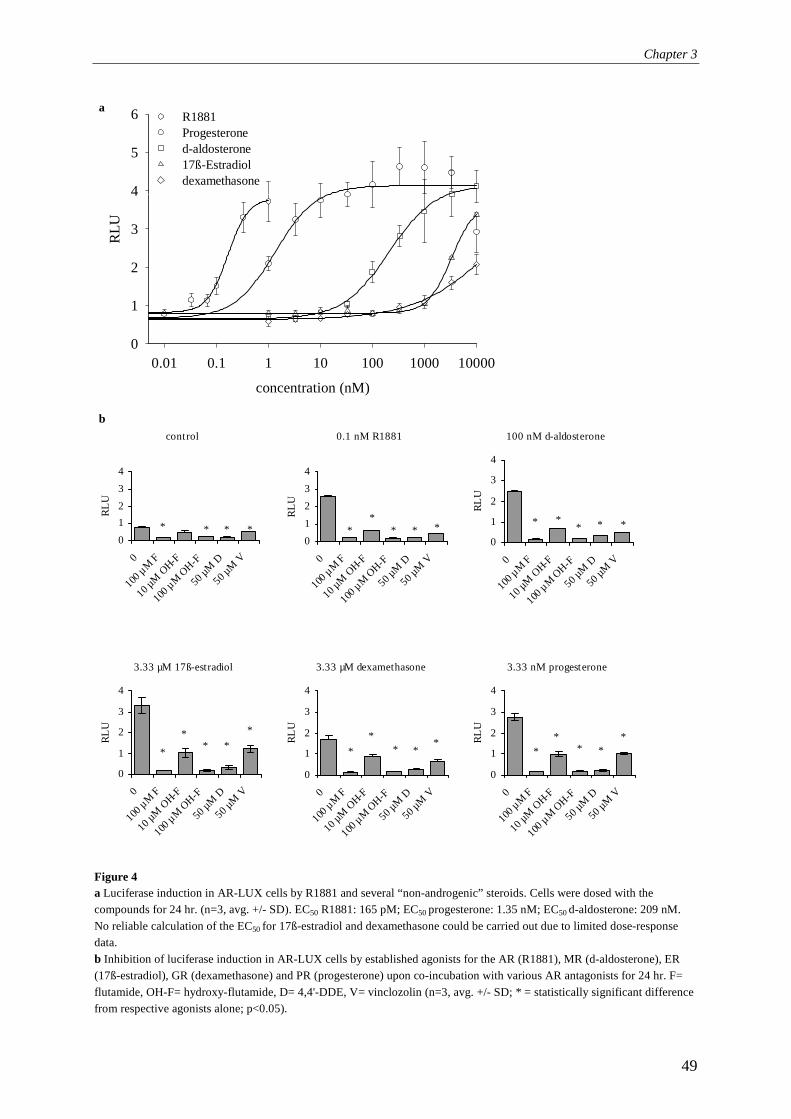
Chapter 3
49
Figure 4 a Luciferase induction in AR-LUX cells by R1881 and several “non-androgenic” steroids. Cells were dosed with the compounds for 24 hr. (n=3, avg. +/- SD). EC50 R1881: 165 pM; EC50 progesterone: 1.35 nM; EC50 d-aldosterone: 209 nM. No reliable calculation of the EC50 for 17ß-estradiol and dexamethasone could be carried out due to limited dose-response data. b Inhibition of luciferase induction in AR-LUX cells by established agonists for the AR (R1881), MR (d-aldosterone), ER (17ß-estradiol), GR (dexamethasone) and PR (progesterone) upon co-incubation with various AR antagonists for 24 hr. F= flutamide, OH-F= hydroxy-flutamide, D= 4,4'-DDE, V= vinclozolin (n=3, avg. +/- SD; * = statistically significant difference from respective agonists alone; p<0.05).
concentration (nM)
0.01 0.1 1 10 100 1000 10000
RLU
0
1
2
3
4
5
6 R1881 Progesterone d-aldosterone 17ß-Estradiol dexamethasone
a
b 0.1 nM R1881
01
234
0
100 µM F
10 µM O
H-F
100 µM O
H-F
50 µM D
50 µM V
RLU
**
* * *
control
01234
0
100 µM F
10 µM O
H-F
100 µM O
H-F
50 µM
D
50 µM V
RLU
* * * *
100 nM d-aldosterone
0
1
2
3
4
0
100 µM F
10 µM O
H-F
100 µM O
H-F
50 µM D
50 µM
V
RLU
* ** * *
3.33 µM 17ß-estradiol
0
1
2
3
4
0
100 µM F
10 µM O
H-F
100 µM O
H-F
50 µM D
50 µM V
RLU
*
** *
*
3.33 µM dexamethasone
0
1
2
3
4
0
100 µM F
10 µM O
H-F
100 µM O
H-F
50 µM D
50 µM V
RLU
**
* **
3.33 nM progesterone
0
1
2
3
4
0
100 µM F
10 µM
OH-F
100 µM O
H-F
50 µM D
50 µM V
RLU
**
* **

Chapter 3
50
increased luciferase expression (Fig. 6a). Binding to the AR was not responsible for the observed effects since none of the compounds was able to efficiently compete with 3H-R1881 for binding to the AR (data not shown). Subsequently, we quantified changes in the number of available androgen binding sites under the influence of atRA, vit. D, EGCG and forskolin using androgen receptor binding assays performed with 3H-R1881. Cells received the same dose as in the AR-LUX assay and R1881 itself, atRA, vitamin D and EGCG did indeed downregulate AR-expression in AR-LUX cells although with different potencies. Forskolin did not affect the amount of AR present in the cells, and therefore presumingly does not exert its effect through regulation of AR levels (Fig. 6b). Discussion
The AR-LUX assay described in this paper represents an effective tool for screening large numbers of compounds for their (anti)androgenic properties. The luciferase induction response produced in the AR-LUX assay was found to be strictly androgen receptor-mediated. This confirmed our expectations with regard to the rat probasin ARE-2 (PB-ARE2) response element controlling luciferase expression in the AR-LUX cell line. PB-ARE2 has previously been reported to exclusively confer androgen receptor controlled gene transcription (Claessens et al. 1996; Kasper et al. 1999; Snoek et al. 1998).
We have shown that the responsiveness of the assay to a number of known androgens is in the picomolar and nanomolar range, which implies sufficient sensitivity for most practical applications. For DHT and R1881, a synthetic androgen, the limit of detection was determined at 24.8 nM and 46 pM, respectively.
The androgen specificity of the AR-LUX assay was further demonstrated by the ability of a number of anti-androgens to suppress the luciferase induction in the AR-LUX assay. Known anti-androgens, such as flutamide, hydroxy-flutamide, vinclozolin, and 4,4'-DDE antagonised luciferase induction by R1881 and by all other inducers tested. The reported AR antagonist spironolactone did not antagonise the response to androgens at the tested antagonist/agonist concentration ratios, but instead acted as weak agonist at high concentrations levels. These observations are consistent with literature reports showing its AR agonistic, but also antagonistic properties (Terouanne et al. 2000). CPA turned out to be a mixed agonist/antagonist in the AR- LUX assay whereas it behaved as a pure AR agonist in transient reporter gene expression assays in CV1 cells (Kemppainen et al. 1999). It is conceivable that differences in expression pattern and levels of co-activators of the AR (Jenster et al. 1997; Miyamoto et al. 1998) play a pivotal role in the different actions of various anti-androgenic compounds in different cell lines. For example, in DU145 cells, co-factor ARA70 has been established as the most optimal mediator of the agonistic action of anti-androgens such as hydroxy-flutamide, bicalutamide (casodex), cyproterone acetate, and RU58841, and other compounds such as genistein and RU486 (Yeh et al. 1999), whereas other co-factors, such as ARA55, were found to be less effective. Another possible explanation could be interference through other signal transduction pathways or squelching of common transcription factors. Spironolactone and CPA, for example, in addition to being androgen

Chapter 3
51
Figure 5 a T47D cell-based competitive AR-binding of various steroids. AR-LUX cells were coincubated for 2 hours with 1 nM 3H-R1881 and an increasing concentration of various unlabelled steroid receptor agonists and the established AR-antagonist flutamide. Data were expressed as the percentage of binding found with 1 nM 3H-R1881 alone. All steroids tested were found to displace R1881 according to a one-site competition model (n=3, avg. +/- SD). b Inhibition of luciferase induction in AR-LUX cells dosed with established agonists for the AR (R1881), MR (d-aldosterone), ER (17ß-estradiol), GR (dexamethasone) and PR (progesterone) at concentrations corresponding to their calculated or estimated EC50 values. AR-LUX cells were coincubated with the respective steroids and an increasing concentration of the established anti-androgen hydroxy-flutamide. Subsequently, luciferase activity was measured. Using these data, the IC50 values for hydroxy-flutamide in competition with the various steroids were calculated according to a one site competition model: OH-F vs. R1881: 3.9 µM; OH-F vs. progesterone: 7.1 µM; OH-F vs. d-aldosterone: 7.6 µM; OH-F vs. 17ß-estradiol: 16.1 µM; OH-F vs. dexamethasone: 25 µM.
receptor agonists have also been identified as progesterone, mineralocorticoid, and pregnenolone X receptor (ant)agonists (Markiewicz and Gurpide 1997; Muhn et al. 1995; Schuetz et al. 1998).
As expected, not only established androgens elicited a response in the AR-LUX assay, but other steroids (progesterone >> d-aldosterone > 17ß-estradiol > dexamethasone) induced an androgen receptor-mediated effect as well. This was concluded from coincubation experiments with anti-androgens and from cell-based receptor binding assays. Similar observations have been done by others using their reporter gene systems, although magnitude of the response and inducer concentrations needed were quite different. For instance, Gaido et al. (1997) reported an equal response to progesterone and 17ß-estradiol and no response to hydrocortisone in their androgen sensitive yeast-based steroid hormone receptor gene transcription assay. Vinggaard et al. (1999) reported no response to dexamethasone and found a higher response to 17ß-estradiol as compared to progesterone in their transient mammalian reporter gene system. Perhaps, differences in (levels of) androgen receptor co-activators present in the respective cell types might account for these differences in androgenic potency. Other explanations, however, cannot be ruled out such as differences in kinetics of uptake and metabolism of the various inducing compounds, and, possibly, differences in biological effects produced beyond the AR signal transduction pathway. Taken together, it turns out that steroid receptor reporter gene systems in general experience ligand binding site-mediated cross-talk by high concentrations of steroids from other classes. However, this has to be considered a true reflection of a certain degree of natural non-specificity among steroid hormone receptors. After all, androgens bind to and induce effects through the estrogen and progesterone receptors as well (Le Bail et al. 1998; Markiewicz and Gurpide 1997; Matias et al. 2000).
0 -8 -7 -6 -5 -4
0
1
R1881 (0.33 nM)
progesterone (3 nM)
d-aldosterone (400 nM)17β-estradiol (7 µM)dexamethasone (60 µM)
log [OH-F] (M)
rela
tive
indu
ctio
na b
-11 -9 -7 -5
0
25
50
75
100
progesterone17β-estradiol
R1881
d-aldosterone
flutamidedexamethasone
log [competitor] (M)
% b
indi
ng

Chapter 3
52
The T47D is one of the few cell lines available that in addition to the AR, also expresses other members of the nuclear receptor family, including PR, ER, VDR and RAR/RXR albeit at different levels. Therefore, AR-mediated responses in this cell line are likely to present a closer reflection of the actual in vivo situation than can be achieved with cell lines lacking one or more of these steroid receptors. We indeed found that regulation at the level of androgen receptor expression and through other modulating pathways, are reliably reflected by the expression of the luciferase reporter gene in AR-LUX cells. It has been previously reported that the amount of AR protein present is the limiting factor determining the maximal attainable level of reporter gene expression (Hall et al. 1992). Our receptor
0.0 2.5 5.0 7.5 10.0
0
50
100 blank10 µM forskolin10 nM vitamin D50 µM EGCG0.33 nM R18811 µM atRA
3H-R1881 (nM)
% b
indi
ng
Figure 6 a Influence of modulation of endogenous active AR-levels in AR-LUX cells on the response to R1881. Cells were coincubated for 24 hr. with all-trans-retinoic acid (atRA), vitamin D, epigallocatechin gallate (EGCG) and forskolin +/- 0.33 nM R1881 after which luciferase activity was determined on a Wallac 1450 microbeta (n=3, avg. +/- SD; * = statistically significant difference from 0.33 nM R1881; p<0.05). b 3H-R1881 binding curves obtained after 24 hr. pre-treatment of AR-LUX cells with EtOH, 10 µM forskolin, 10 nM vitamin D, 50 µM EGCG, 0.33 nM R1881 or 1 µM all-trans-retinoic acid. The number of available AR binding sites diminish considerably indicating the capability of AR-LUX cells for regulation of AR-levels (n=3, avg. ± SD).
b
0
0.5
1
1.5
0 0.5 1
atRA (µM)
indu
ctio
n
R1881EtOH* *
0
0.5
1
1.5
0 10 20 50
EGCG (µM)
indu
ctio
n
R1881EtOH
* * *
0
0.5
1
1.5
0 1 10 100Vit. D (nM)
indu
ctio
n R1881EtOH* *
*
012345
0 0.1 1 10
forskolin (µM)
indu
ctio
n
R1881EtOH
*
*
a

Chapter 3
53
binding studies and AR expression studies in human cell lines by other investigators show that the apparent “anti-androgenic” effect of all-trans-retinoic acid, epigallocatechin gallate, and vitamin D on luciferase expression is in fact caused by downregulation of AR expression and not by binding to the AR. Downregulation of the AR by atRA in T47D cells (Hall et al. 1992), and by epigallocatechin gallate (EGCG), a naturally occurring polyphenol, in LNCaP cells (Ren et al. 2000) has been reported previously. Vitamin D has been reported to upregulate AR levels in human Ovcar-3 and LNCaP cells (Ahonen et al. 2000; Zhao et al. 1999). However, vitamin D cotreatment in the AR-LUX assay did not result in a higher maximal luciferase induction level, but instead in a lower one. This discrepancy with other cell lines is apparently due to (species) differences in the regulation of androgen receptor expression levels. Besides regulation of the numbers of AR receptors, regulation of androgenic responses occurs also via the interaction with co-activators, through (de)phosphorylation (Blok et al. 1996; Fujimoto et al. 1999; Heinlein et al. 1999) and/or acetylation (Jenster et al. 1997) of the receptor. Some of these mechanisms, such as dephosphorylation of the AR, might play a role in the luciferase expression enhancing effect of forskolin, which is a protein kinase A activator. The exact explanation of the effect of forskolin is still unclear, but a possible mechanism could be through recruitment of CREB-binding protein (CBP), a transcription factor which derived its name from its association with the cAMP responsive element binding protein (CREB). CBP is a coactivator for AR-dependent transcriptional activation in LNCaP and CV1 cells (Aarnisalo et al. 1998; Fronsdal et al. 1998) and exposure to forskolin is known to induce the phosphorylation of CREB by protein kinase A, and its subsequent association with CBP (Wolfl et al. 1999). Therefore, it is conceivable that forskolin influences the availability of CBP in AR-LUX cells as well. Others (Blok et al. 1998) have reported that the phosphorylation state of the AR itself influences its transactivating ability, and have found that forskolin-induced dephosphorylation of the AR impairs ligand binding in LNCaP cells.
In summary, we have developed an assay for the quantification of AR-mediated biological effects which is highly specific and does not only assess AR activation but reliably reflects the influence of related cellular pathways on AR expression and activity as well. Moreover, the 96 well plate format makes the AR-LUX assay easily amenable to automation and high-throughput screening. Possible applications for the AR-LUX assay include screening single compounds or complex mixtures, such as extracts from environmental samples or compound libraries in drug discovery, for their androgenic, anti-androgenic or AR-regulating properties. References Aarnisalo, P., Palvimo, J. J., and Janne, O. A. (1998). "CREB-binding protein in androgen
receptor-mediated signaling." Proc Natl Acad Sci USA 95, 2122-2127. Ahonen, M. H., Zhuang, Y. H., Aine, R., Ylikomi, T., and Tuohimaa, P. (2000). "Androgen
receptor and vitamin D receptor in human ovarian cancer: growth stimulation and inhibition by ligands." Int J Cancer 86, 40-46.

Chapter 3
54
Altschmied, J., and Duschl, J. (1997). "Set of optimized luciferase reporter gene plasmids compatible with widely used CAT vectors." Biotechniques. 23, 436-438. Andersen, M. E., and Barton, H. A. (1999). "Biological regulation of receptor-hormone complex concentrations in relation to dose-response assessments for endocrine-active compounds." Toxicol Sci 48, 38-50.
Battmann, T., Branche, C., Bouchoux, F., Cerede, E., Philibert, D., Goubet, F., Teutsch, G., and Gaillard-Kelly, M. (1998). "Pharmacological profile of RU 58642, a potent systemic antiandrogen for the treatment of androgen-dependent disorders." J Steroid Biochem Mol Biol 64, 103-111.
Blok, L. J., de Ruiter, P. E., and Brinkmann, A. O. (1996). "Androgen receptor phosphorylation." Endocr.Res. 22, 197-219.
Blok, L. J., de Ruiter, P. E., and Brinkmann, A. O. (1998). "Forskolin-induced dephosphorylation of the androgen receptor impairs ligand binding." Biochemistry 37, 3850-3857.
Brinkmann, A. O., Faber, P. W., van Rooij, H. C., Kuiper, G. G., Ris, C., Klaassen, P., van der Korput, J. A., Voorhorst, M. M., van Laar, J. H., and Mulder, E. (1989). "The human androgen receptor: domain structure, genomic organization and regulation of expression." J.Steroid.Biochem. 34, 307-310.
Buras, R. R., Schumaker, L. M., Davoodi, F., Brenner, R. V., Shabahang, M., Nauta, R. J., and Evans, S. R. (1994). "Vitamin D receptors in breast cancer cells." Breast Cancer Res Treat 31, 191-202.
Claessens, F., Alen, P., Devos, A., Peeters, B., Verhoeven, G., and Rombauts, W. (1996). "The androgen-specific probasin response element 2 interacts differentially with androgen and glucocorticoid receptors." J.Biol.Chem. 271, 19013-19016.
Cleutjens, K. B., van Eekelen, C. C., van der Korput, H. A., Brinkman, A. O., and Trapman, J. (1996). "Two androgen response regions cooperate in steroid hormone regulated activity of the prostate-specific antigen promoter." J.Biol.Chem. 271, 6379-6388.
Cooper, R. L., and Kavlock, R. J. (1997). "Endocrine disruptors and reproductive development: a weight-of- evidence overview." J.Endocrinol. 152, 159-166.
Fronsdal, K., Engedal, N., Slagsvold, T., and Saatcioglu, F. (1998). "CREB binding protein is a coactivator for the androgen receptor and mediates cross-talk with AP-1" [published erratum appears in J Biol Chem 1999 Aug 27;274(35):25188]. J Biol Chem 273, 31853-31859.
Fuhrmann, U., Bengtson, C., Repenthin, G., and Schillinger, E. (1992). "Stable transfection of androgen receptor and MMTV-CAT into mammalian cells: inhibition of cat expression by anti-androgens." J.Steroid Biochem.Mol.Biol. 42, 787-793.
Fuhrmann, U., Krattenmacher, R., Slater, E. P., and Fritzemeier, K. H. (1996). "The novel progestin drospirenone and its natural counterpart progesterone: biochemical profile and antiandrogenic potential." Contraception. 54, 243-251.
Fujimoto, N., Yeh, S., Kang, H. Y., Inui, S., Chang, H. C., Mizokami, A., and Chang, C. (1999). "Cloning and characterization of androgen receptor coactivator, ARA55, in human prostate." J Biol Chem 274, 8316-8321.
Chapter 3
55
Gaido, K. W., Leonard, L. S., Lovell, S., Gould, J. C., Babai, D., Portier, C. J., and McDonnell, D. P. (1997). "Evaluation of chemicals with endocrine modulating activity in a yeast-based steroid hormone receptor gene transcription assay." Toxicol.Appl.Pharmacol. 143, 205-212.
Gladue, B. A., and Clemens, L. G. (1980). "Flutamide inhibits testosterone-induced masculine sexual behavior in male and female rats." Endocrinology 106, 1917-1922.
Hackenberg, R., Hawighorst, T., Filmer, A., Slater, E. P., Bock, K., Beato, M., and Schulz, K. D. (1992). "Regulation of androgen receptor mRNA and protein level by steroid hormones in human mammary cancer cells." J.Steroid.Biochem.Mol.Biol. 43, 599-607.
Hall, R. E., Tilley, W. D., McPhaul, M. J., and Sutherland, R. L. (1992). "Regulation of androgen receptor gene expression by steroids and retinoic acid in human breast-cancer cells." Int.J.Cancer 52, 778-784.
Heinlein, C. A., Ting, H. J., Yeh, S., and Chang, C. (1999). "Identification of ARA70 as a ligand-enhanced coactivator for the peroxisome proliferator-activated receptor gamma." J.Biol.Chem. 274, 16147-16152.
Henderson, B. E., and Feigelson, H. S. (2000). "Hormonal carcinogenesis". Carcinogenesis 2000. 21, 427-433.
Horwitz, K. B., and McGuire, W. L. (1978). "Estrogen control of progesterone receptor in human breast cancer: Correlation with nuclear processing of estrogen receptor." J. Biol. Chem. 253, 2222-2228.
Jenster, G., Spencer, T. E., Burcin, M. M., Tsai, S. Y., Tsai, M. J., and O'Malley, B. W. (1997). "Steroid receptor induction of gene transcription: a two-step model." Proc Natl Acad Sci USA 94, 7879-7884.
Kasper, S., Rennie, P. S., Bruchovsky, N., Lin, L., Cheng, H., Snoek, R., Dahlman-Wright, K., Gustafsson, J. A., Shiu, R. P., Sheppard, P. C., and Matusik, R. J. (1999). "Selective activation of the probasin androgen-responsive region by steroid hormones." J.Mol.Endocrinol. 22, 313-325.
Kelce, W. R., Monosson, E., Gamcsik, M. P., Laws, S. C., and Gray, L. E., Jr. (1994). "Environmental hormone disruptors: evidence that vinclozolin developmental toxicity is mediated by antiandrogenic metabolites." Toxicol.Appl.Pharmacol. 126, 276-285.
Kelce, W. R., and Wilson, E. M. (1997). "Environmental antiandrogens: developmental effects, molecular mechanisms, and clinical implications." J.Mol.Med. 75, 198-207.
Keller, E. T., Ershler, W. B., and Chang, C. (1996). "The androgen receptor: A mediator of diverse responses." Front.Biosci. 1, 59-71.
Kemppainen, J. A., Langley, E., Wong, C. I., Bobseine, K., Kelce, W. R., and Wilson, E. M. (1999). "Distinguishing androgen receptor agonists and antagonists: distinct mechanisms of activation by medroxyprogesterone acetate and dihydrotestosterone." Mol.Endocrinol. 13, 440-454.
Le Bail, J. C., Marre-Fournier, F., Nicolas, J. C., and Habrioux, G. (1998). "C19 steroids estrogenic activity in human breast cancer cell lines: importance of dehydroepiandrosterone sulfate at physiological plasma concentration." Steroids 63, 678-683.
Chapter 3
56
Maness, S. C., McDonnell, D. P., and Gaido, K. W. (1998). "Inhibition of androgen receptor-dependent transcriptional activity by DDT isomers and methoxychlor in HepG2 human hepatoma cells." Toxicol.Appl.Pharmacol. 151, 135-142.
Markiewicz, L., and Gurpide, E. (1997). "Estrogenic and progestagenic activities of physiologic and synthetic androgens, as measured by in vitro bioassays." Methods.Find.Exp.Clin.Pharmacol. 19, 215-222.
Matias, P. M., Donner, P., Coelho, R., Thomaz, M., Peixoto, C., Macedo, S., Otto, N., Joschko, S., Scholz, P., Wegg, A., Basler, S., Schafer, M., Ruff, M., Egner, U., and Carrondo, M. A. (2000). "Structural evidence for ligand specificity in the binding domain of the human Androgen receptor: implications for pathogenic gene mutations." J. Biol. Chem. 275, 26164-26171.
Miyamoto, H., Yeh, S., Wilding, G., and Chang, C. (1998). "Promotion of agonist activity of antiandrogens by the androgen receptor coactivator, ARA70, in human prostate cancer DU145 cells." Proc.Natl.Acad.Sci.U.S.A. 95, 7379-7384.
Muhn, P., Fuhrmann, U., Fritzemeier, K. H., Krattenmacher, R., and Schillinger, E. (1995). "Drospirenone: a novel progestogen with antimineralocorticoid and antiandrogenic activity." Ann.N.Y.Acad.Sci. 761, 311-335.
O'Connor, J. C., Davis, L. G., Frame, S. R., and Cook, J. C. (2000). "Evaluation of a Tier I screening battery for detecting endocrine-active compounds (EACs) using the positive controls testosterone, coumestrol, progesterone, and RU486." Toxicol Sci 54, 338-354.
Ostby, J., Kelce, W. R., Lambright, C., Wolf, C. J., Mann, P., and Gray, L. E. J. (1999). "The fungicide procymidone alters sexual differentiation in the male rat by acting as an androgen-receptor antagonist in vivo and in vitro." Toxicol.Ind.Health. 15, 80-93.
Ren, F., Zhang, S., Mitchell, S. H., Butler, R., and Young, C. Y. (2000). "Tea polyphenols down-regulate the expression of the androgen receptor in LNCaP prostate cancer cells." Oncogene 19, 1924-1932.
Sadar, M. D., Hussain, M., and Bruchovsky, N. (1999). "Prostate cancer: molecular biology of early progression to androgen independence." Endocr. Relat. Cancer 6, 487-502.
Safe, S., Connor, K., Ramamoorthy, K., Gaido, K., and Maness, S. (1997). "Human exposure to endocrine-active chemicals: hazard assessment problems." Regul.Toxicol.Pharmacol. 26, 52-58.
Sambrook, J., Fritsch, E. F., and Maniatis, T. (1989). molecular cloning: a laboratory manual. Cold Spring Harbor Laboratory Press, New York.
Schneider, S. M., Offterdinger, M., Huber, H., and Grunt, T. W. (2000). "Activation of Retinoic Acid Receptor alpha Is Sufficient for Full Induction of Retinoid Responses in SK-BR-3 and T47D Human Breast Cancer Cells." Cancer Res 60, 5479-5487.
Schrader, T. J., and Cooke, G. M. (2000). "Examination of selected food additives and organochlorine food contaminants for androgenic activity in vitro." Toxicol Sci 53, 278-288.
Schuetz, E. G., Brimer, C., and Schuetz, J. D. (1998). "Environmental xenobiotics and the antihormones cyproterone acetate and spironolactone use the nuclear hormone
Chapter 3
57
pregnenolone X receptor to activate the CYP3A23 hormone response element." Mol Pharmacol 54, 1113-1137.
Snoek, R., Bruchovsky, N., Kasper, S., Matusik, R. J., Gleave, M., Sato, N., Mawji, N. R., and Rennie, P. S. (1998). Differential transactivation by the androgen receptor in prostate cancer cells. Prostate. 36, 256-263.
Terouanne, B., Tahiri, B., Georget, V., Belon, C., Poujol, N., Avances, C., Orio, F., Balaguer, P., and Sultan, C. (2000). A stable prostatic bioluminescent cell line to investigate androgen and antiandrogen effects. Mol.Cell Endocrinol. 160, 39-49.
Vinggaard, A. M., Joergensen, E. C., and Larsen, J. C. (1999). Rapid and sensitive reporter gene assays for detection of antiandrogenic and estrogenic effects of environmental chemicals. Toxicol.Appl.Pharmacol. 155, 150-160.
Willemart, J. P., and Bouffault, J. C. (1983). A RAL compound as an anabolic in cattle. Vet Res Commun 7, 35-44.
Wolfl, S., Martinez, C., and Majzoub, J. A. (1999). Inducible binding of cyclic adenosine 3',5'-monophosphate (cAMP)-responsive element binding protein (CREB) to a cAMP-responsive promoter in vivo. Mol Endocrinol 13, 659-669.
Wong, C., Kelce, W. R., Sar, M., and Wilson, E. M. (1995). Androgen receptor antagonist versus agonist activities of the fungicide vinclozolin relative to hydroxy-flutamide. J Biol Chem 270, 19998-20003.
Yeh, S., Chang, H. C., Miyamoto, H., Takatera, H., Rahman, M., Kang, H. Y., Thin, T. H., Lin, H. K., and Chang, C. (1999). Differential induction of the androgen receptor transcriptional activity by selective androgen receptor coactivators. Keio.J.Med. 48, 87-92.
Zhao, X. Y., Ly, L. H., Peehl, D. M., and Feldman, D. (1999). Induction of androgen receptor by 1alpha,25-dihydroxyvitamin D3 and 9-cis retinoic acid in LNCaP human prostate cancer cells. Endocrinology 140, 1205-1212.

58

44
Androgen activity in surface water samples detected using the AR-LUX assay: Indications for mixture effects
Submitted to Environmental Toxicology and Pharmacology
B.M.G. Blankvoort1, 2, R.J.T. Rodenburg1, 4, A.J. Murk2, J.H. Koeman2, R. Schilt3 and J.M.M.J.G. Aarts2
1 Department of Bioanalysis, TNO Nutrition and Food Research, Zeist, the Netherlands; 2 Department of Agrotechnology and
Food Sciences, Sub-department of Toxicology, Wageningen University, Wageningen, the Netherlands; 3 Department of residue analysis, TNO Nutrition and Food Research, Zeist, the Netherlands; 4 UMC St Radboud, Laboratory of Paediatrics
and Neurology, Nijmegen, the Netherlands

Chapter 4
60
Abstract This paper describes the screening of 22 extracts from 18 different aquatic
environmental samples for androgenic activity, including indirect and interactive effects on androgen receptor (AR)-mediated signal transduction, using the AR-LUX bioassay. Four samples, originating from an industrial wastewater treatment plant (WTP) or the river Meuse, were shown to contain substantial androgenic activity. Moreover, the samples originating from the industrial WTP showed an enhancement of the maximal androgenic response relative to that elicited by the standard androgen methyltrienolone (R1881) in the AR-LUX assay. This indicates the involvement of cellular mechanisms other than receptor-ligand interaction influencing AR-regulated pathways. This also demonstrates the additional value of cell based assays featuring a more complete array of fully functional interacting pathways. Chemical analysis using GC-MS confirmed the presence of a number of androgens and also estrogens in these WTP samples. Subsequently, we showed that estrone and tributyltin-hydride (TBT-H) enhance the response to androgens. This indicates that the presence of numerous compounds in addition to androgens in environmental mixtures might very well result in a more profound perturbation of the normal physiology of exposed organisms than estimated based on the androgen levels alone. Therefore, risk assessment of environmental samples should include an evaluation of the presence and the interactive effects of (ant)agonists of carefully selected relevant cellular receptors in order to provide a realistic estimate of the integrated ecotoxicological risk of the compounds present. Introduction
In recent years numerous examples have been described of endocrine-disruptive effects elicited by environmental pollutants. As reviewed by Miyamoto et al. (1998), sex hormone-related effects range from super feminisation in alligators (lake Apopka, Florida, USA) and feminisation in male rainbow trout (rivers in England), to masculinisation of females of the common mosquito fish Gambusia affinis (Florida, USA) and imposex in rock shell and several buccinidae species (coastal seas of Japan, Singapore and Indonesia). A broad range of compounds are held responsible for these effects, or are under suspicion to cause endocrine disruption (Groshart and Okkerman, 2000) including chlorinated pesticides such as DDT and its metabolite DDE, dieldrin and dicofol, and also hormones released from waste water treatment plants (WTPs) such as ethynyl-estradiol, estrone and estradiol. They further include the organotin compounds tributyltin-hydride (TBT-H) and its oxide TBTO, polychlorinated biphenyls (PCBs), and presumably other, yet unidentified compounds (Miyamoto and Klein, 1998; Legler et al., 2002). As environmental contaminants with hormone-mimicking properties usually appear in mixtures consisting of compounds present in very low concentrations, it is often difficult to estimate the risk based on chemical analysis of a limited number of known endocrine-disruptive compounds (EDCs). In addition, it is becoming increasingly apparent that interactions between different endocrine systems occur (Jaussi et al., 1992; Zhou et al., 2000; Simon, 2001). Therefore, biological detection systems are needed since they represent a closer reflection of the responses that might occur in vivo upon exposure to complex mixtures.
Chapter 4
61
An example of an in vitro biological detection system is the estrogen receptor-mediated, chemical-activated luciferase expression (ER-CALUX) reporter gene assay. It is an assay aimed at determining the total amount of estrogenic activity present in various matrices (Legler et al., 1999). This assay -amongst others- was employed in a Dutch national survey (acronym LOES) in which the presence and effects of estrogenic compounds in the environment were investigated and confirmed on various locations in the Netherlands, including domestic and industrial WTPs, surface water and animal dung (Vethaak et al., 2002). In recent years, similar studies using these kind of biological detection systems have generated a considerable amount of knowledge regarding the occurrence of estrogens in the environment (Behnisch et al., 2001; Fawell et al., 2001; Fenet et al., 2003; Huggett et al., 2003). By contrast, very little is known about the presence of (anti)androgens in the environment, although androgens also play a major role in endocrine regulation and a few examples of environmental contaminants with (anti)androgenic action are known such as vinclozolin and 4,4'-DDE which interact with the androgen receptor (Kelce et al., 1995; Kelce et al., 1997; Wolf et al., 2000). Therefore the possible presence of (anti)androgens in the environment, which might lead to perturbations of endocrine regulation, is not unlikely and is worth investigating.
For this purpose, we selected 22 extracts originating from the LOES project representing a broad range of estrogenic potencies. We used the recently developed Androgen Receptor-mediated LUciferase eXpression (AR-LUX) assay (Blankvoort et al., 2001) to screen these samples for their androgenic potency. The AR-LUX assay is based on a genetically engineered T47D human breast carcinoma cell line carrying a stably integrated luciferase reporter gene. This gene is under transcriptional control of the endogenously expressed androgen receptor through an upstream rat probasin androgen response element 2 (PB-ARE2). This response element is an authenticated androgen response element (ARE), controlling expression of a prostate gene representing a typical male function (Rennie et al., 1993; Claessens et al., 1996). The T47D cell line expresses the androgen receptor, as well as various other functional members of the nuclear receptor family, such as the estrogen (ER), progesterone (PR), retinoic acid (RAR) and retinoid X (RXR) receptor (Hackenberg et al., 1992; Kasper et al., 1999; Schneider et al., 2000). Therefore, activation of reporter gene transcription through the PB-ARE2 by endogenous signal transduction pathways presents one of the most indicative in vitro biomarkers for androgenic action currently available. As a consequence, this in vitro assay can not only be used to determine direct androgen receptor-activating potency of single compounds and mixtures, but will also reflect any indirect effects on the androgen receptor-mediated response through related signal transduction routes and biochemical pathways expressed in this cell line. This feature of the AR-LUX has previously been validated for forskolin and some other compounds showing indirect effects on androgen receptor-mediated signal transduction (Blankvoort et al., 2001).
An example of a compound known to interfere with the androgenic pathway is tributyltin-hydride (TBT-H). It causes masculinisation in juvenile female whelks (Mensink et al., 1996; Mensink et al., 2001; Tillmann et al., 2001). This lipophilic compound has a log Kow between 3.19 and 3.84, which is in the range of steroids such as testosterone (log Kow =

Chapter 4
62
3.3) or 17ß-estradiol (log Kow = 4). In extracts of environmental matrices such as effluents of WTPs, it will therefore be present in the same fraction as steroids. For that reason we were interested in the possible interactive mixture effects of TBT-H as well as estrogens when coincubated in our assay with the standard (synthetic) androgen methyltrienolone (R1881). Methods Chemicals
Methyltrienolone (R1881, 100%) was purchased from NEN Life Science Products (Hoofddorp, the Netherlands). Methyltestosterone, nortestosterone (100%), estrone (99%), tri-butyl-tin-hydride (TBT-H, 97%) and DMSO (100%) were obtained from Sigma-Aldrich (Zwijndrecht, the Netherlands).
AR-LUX assay procedure
T47D/Sutherland human breast cancer cells stably transfected with pPBARE2tataluc+
were seeded in white 96 well plates with clear flat bottoms (Corning Incorporated, Cambridge, USA) at a density of 18,000 cells/well in DMEM/F12 medium (Life Technologies ltd., Paisley, Scotland) containing 5% (v/v) foetal bovine serum (FBS). The serum had previously been treated with dextran-coated charcoal (DCC) as described by Horwitz and McGuire (1978) to remove any traces of steroid hormones present in the serum. Cells were cultured at 5% (v/v) CO2 and 100% relative humidity. After 24 hours, the medium was replaced and the chemicals of interest, dissolved in ethanol or DMSO, were tested in triplicate with a maximum solvent concentration of 0.2%. Following a 24 hours incubation, cells were harvested and luciferase expression was subsequently measured using a luminometer (Labsystems Luminoscan RS) or a Wallac 1450 microbeta liquid scintillation counter which recently became available in our lab. When using the luminometer, cells were washed once with 100 µl 0.5 x PBS (Life Technologies ltd., Paisley, Scotland) followed by the addition of 30 µl low salt buffer (2 mM dithiothreitol, 2 mM 1,2,-diaminocyclohexane-N,N,N�,N�-tetra acetic acid, 10 mM Tris pH 7.8). Cells were lysed by incubation on ice for 15 minutes and subsequent freezing at �80°C for at least one hour. After thawing, shaking and equilibrating to room temperature, the plates were mounted in the luminometer and upon injection of 100 µl flash mix (20 mM Tricine, 1.07 mM (MgCO3)4Mg(OH)2, 2.67 mM MgSO4, 0.1 mM EDTA, 2.0 mM dithiothreitol, 470 µM luciferine, 5.0 mM ATP) per well, luciferase activity was immediately determined and expressed as Relative Light Units (RLUs). Before measuring the next well, 100 µl of 0.2 M NaOH was injected to quench the remaining signal in the well, thus preventing signal contamination between neighbouring wells. When using the Wallac 1450, medium was removed from the cells and subsequently, 20 µl of fresh medium and 20 µl of Steady-Glo reagent (Promega Corporation, Madison, USA) was added. Following 10 min incubation at room temperature in the instrument, luciferase activity was counted for 30 seconds and expressed as luminescence counts. Induction factors were subsequently calculated relative to solvent controls. When appropriate, the AR-LUX response of the test samples was also expressed as a percentage of the maximal

Chapter 4
63
response produced by the standard androgen R1881. Alternatively, using a dose-response curve for luciferase induction by R1881 in AR-LUX cells, the androgenic content of the samples was calculated as R1881 equivalents (REQs), defined as the amount of the standard androgen R1881 inducing an equivalent response in the AR-LUX as observed with the tested sample.
Sample treatment and locations
Two categories of samples were extracted: (1) water phase of river and waste water and (2) solid phase of river and wastewater. All water samples were collected in 1999 and stored at 4°C until further processing within the framework of the Dutch LOES project (Vethaak et al., 2002). Treatment: Water samples
Water samples were filtered over a 0.45 µm glass fibre filter followed by solid phase extraction using C18-columns (C18-SPE). Some of the extracts were filtered over anhydrous Na2SO4 (eluted with diethyl ether, DEE) to remove small volumes of residual water and particles. If they contained too much residual water to be removed with anhydrous Na2SO4 they were extracted three times with DEE. The combined DEE fractions were gently evaporated under N2 (g) at 30°C and DMSO was added just before dryness. Dilutions in DMSO were used for the in vitro assays. Some of the extracts made were extracted as a total sample, i.e. without filtration, applying three liquid-liquid extractions with DEE. Solid phase samples
Suspended particles of wastewater or river water were collected by filtering (1 to 4 litres) over a Whatman GF/C glass fibre filter. The filters were dried in a desiccator for at least 3 hours. Sediment and suspended matter samples were mixed with anhydrous Na2SO4 in a mortar and dried in a desiccator. These dried samples or filters were Soxhlet-extracted with either hexane/acetone (1:1, v/v) for 6 hours (spring samples) or DEE/acetone (3:1, v/v) (summer and fall samples). The Soxhlet extract was concentrated on a rotary evaporator and sulphur compounds were removed by means of the TBA (tetrabutylammonium) method (De Voogt et al., 1990; Verbrugge et al., 1991), by mixing the extract with a TBASO3 solution. Consequently, the resulting extracts still contained considerable amounts of water and therefore, the water phase was three times extracted with DEE and the DEE phases were combined.
To separate the very lipophilic and more polar compounds the DEE extract was gently evaporated to dryness, 2 ml of hexane was added and filtered over anhydrous Na2S04 to catch un-dissolved particles. This was repeated twice. To rinse the Na2S04 filter it was again eluted with hexane and all eluents were combined in a second test tube and evaporated and dissolved in DMSO (hexane fraction). The remaining pellet in the first tube was re-dissolved in acetone and also filtered over the same Na2S04 filter. Finally the Na2S04 filter
Chapter 4
64
was eluted with acetone. All acetone fractions were combined, gently evaporated and dissolved in DMSO (acetone fraction). The acetone fractions and some hexane fractions were tested in the ER-CALUX (Legler et al., 2003; Legler et al., submitted) and used in this study. Samples locations and abbreviations:
The sample locations are indicated by the same abbreviations as in the LOES study (Vethaak et al., 2002): CHH: effluent of a chemical company; entire wastewater stream after a number of treatment steps. CHM: effluent of a biological WTP from a chemical company. EHV: untreated wastewater from the city of Eindhoven. VTL: manure from a cattle farm. POL: manure from a cattle farm. HHW: domestic wastewater from a residential area in the city of Steenwijk. ANP: untreated, effluent from a sewage treatment plant (STP) named �Sint Annaparochie�; mainly domestic wastewater. WST: untreated wastewater and effluent STP �Amsterdam Westpoort�. ZKH: untreated wastewater from a hospital. EYS: river Meuse at the city of Eysden; entry point of the river into the Netherlands. DOM: river Dommel at STP from Eindhoven. DON: river Dommel downstream of STP from Eindhoven. IJM: North Sea Canal at city of IJmuiden. Samples were either collected in the spring (Sp), summer (Su) or autumn (Au). HPLC separation/GC-MS analysis A number of samples have been separated by HPLC in two fractions (called the androgenic and estrogenic fraction, respectively), which were subsequently analysed with GC-MS to confirm the presence of certain androgenic and estrogenic chemicals in the respective fractions. In short: After addition of a mixture of deuterated internal standards, samples were enzymatically deconjugated with Helix Pomatia juice (glucuronidase and arylsulphatase) at 37°C for 16 hours. The hydrolysed samples were loaded onto 300 mg C18 solid phase extraction columns. After washing with 1 ml water, 1 ml acetone/water (20/80) and 1 ml methanol/water (30/70), the analytes were extracted with 3 ml methanol. After evaporation of the solvent, the residue was dissolved in heptane/iso-propanol (250:30) and injected into a normal phase HPLC (LiChrosorb 5 Diol) for fractionation into an androgenic and an estrogenic fraction. After evaporation of the solvents, the extracts were either derivatised with heptafluorobutyric anhydride/acetone (1:4) during 60 min at 60°C for GC-MS, or dissolved in DMSO if the fractions were collected for the purpose of AR-LUX analysis (in which case no deuterated standards were added).
GC-MS analysis of the derivatives was performed on an Agilent 6890 GC (DB-5 column or DB-17 column, splitless injection) in combination with a Thermo Finnigan
Chapter 4
65
MAT95 sector mass spectrometer operating at a resolution of 5000 (10% valley) using selected ion monitoring with at least 4 ions per analyte.
Quantification was based on comparison with the signal generated by the corresponding deuterated internal standards that had been added in the first step of the analysis. Identification was based on EU criteria 2002/657/EC, using at least 3 ion ratios with a maximal variation of 10%. Data and statistical analysis
AR-LUX data were fitted to a 4-parameter Hill plot (f=y0+a*x^b/(c^b+x^b)) using Slide Write Plus for Windows. Subsequently R1881 EQuivalents (REQs) in samples could be calculated by entering the induction factors of samples as y-values in the Hill formula and calculating the corresponding concentration of R1881 that would be required to elicit the same response in the AR-LUX assay. Statistical analysis was carried out using a paired two-sample t-test for means using Microsoft Excel 97. Results In a preliminary test, water was spiked with 1 to 20 nM of the pharmaceutically and veterinarilly applied androgenic compound 17β-trenbolone and subjected to AR-LUX analysis. The results confirmed the essential suitability of the AR-LUX assay for assessing androgenic activity in water at detection limits similar to those of GC-MS analysis (Daeseleire et al., 1992; Casademont et al., 1996) (data not shown).
Subsequently, a selection from a set of aquatic environmental samples was tested in the AR-LUX assay. Two types of sample were available, taken either from the water phase or the solid phase of the collected material, and extracts of both sample types were prepared as described in Materials and Methods. Twenty-two extracts, together representing eighteen different aquatic samples, were selected to be analysed in the AR-LUX assay from the set of extracts prepared for the Dutch national survey for the presence of estrogenic compounds (LOES). Of these extracts selected on the basis of their estrogenic potency, 14 did not yield any androgenic response at all. Nine extracts showed detectable androgenic activity (Table 1), including one extract (the acetone-extracted solid phase from WTP influent sample AI.sp.ZKH) which displayed only detectable androgenic activity in the isolated androgen-containing fraction. We also tested the samples for the presence of anti-androgenic activity by coincubations with R1881, but did not detect this in any of the samples (data not shown). The two effluent samples (CHH) originating from an industrial WTP induced the strongest androgenic response in the AR-LUX assay (Fig.1a, b). Also summer and autumn samples originating from the river Meuse at Eysden (EYS) both displayed substantial and dose-dependent androgenic potency (Fig. 1 c, d). Although the activity was comparable up to 5 µl/ml, concentrations of 11 µl and more were cytotoxic in the au sample. Interestingly, both CHH samples were not only found to have a considerable androgenic activity, but were shown to activate luciferase expression to a higher extent than the maximum luciferase

Chapter 4
66
Table 1 Estrogen EQuivalents (expressed as 17β-estradiol Equivalents, EEQs), androgen equivalents (expressed as R1881 EQuivalents, REQs) in unfractionated samples and REQs present in the isolated HPLC-AR-fraction of LOES samples. Abbreviations: First part of code AI: WTP influent, AE: WTP effluent, AER: WTP sludge residue, OZ: surface water total suspended solids, OW: surface water, OS: surface water sediment. Second part of code refers to moment of sampling sp: spring, su: summer, au: autumn Third part of code, refers to location (see materials & methods) LOES code Total
EEQ Total REQ
HPLC-AR fraction REQ
(pmol/l or /kg) (nmol/l or /kg) (nmol/l or /kg) industrial wastewater: AI.sp.CHH 560 629 116 AI.au.CHH 253 29.0 114 AE.sp.CHM 9.51 n.d., tox not fractionated AE.au.CHM 0.90 n.d., tox n.d. AER.sp.EHV # 460 n.d. not fractionated
animal dung: AI.sp.VTL# 18429 0.14 (*) 0.00089 (*) AI.au.POL# 1351 n.d., tox n.d. AI.sp.VTL ~ 7685 5.23 (*) 2.67 (*) AI.au.POL ~ 48000. 17 (*) not fractionated
domestic waste water: AI.au.HHW 195 n.d., tox not fractionated AI.au.ANP n.d. n.d. n.d. AI.au.WST 27.4 0.70 (*) not fractionated AI.au.ANP # 11769 n.d. n.d. AI.au.HHW # 344 n.d. not fractionated AI.sp.ZKH # 381 n.d. 0.095 (*)
surface water: OW.su.EYS 0.40 0.02 n.d. OW.au.EYS 0.07 0.02, tox not fractionated OZ.sp.IJM. # 33.1 n.d. not fractionated OZ.sp.TER # 113 n.d. not fractionated OZ.sp.DOM # 151 n.d. not fractionated OS.au.DON ~ 959 n.d. not fractionated OZ.au.EYS ~ 783 n.d., tox not fractionated n.d. = no activity detectable (*) = slightly above detection, estimated REQs based on less reliable linear fit tox = toxic effects at higher dosages # = polar (acetone) fraction of the solid phase extract ~ = apolar (hexane) fraction of the solid phase of extract
induction attained with the standard androgen R1881 or any other classical androgen (Fig.1a, b). The other 5 positively testing samples induced a low but well detectable AR-LUX response.
To obtain insights into the origin of the androgenic response induced by the environmental samples, HPLC/GC-MS analysis was performed on the two samples

Chapter 4
67
Figure 1 Luciferase induction in the AR-LUX assay by samples taken from a factory wastewater effluent (CHH) or from the river Meuse (EYS) at different time points. X-axis represents the amount (µl or ml) of original unextracted sample of which the extract was dissolved in one ml of assay medium. Y-axis represents the percentage induction in AR-LUX cells (n=3, avg. ± SD) relative to the maximal response elicited by the standard androgen R1881 (100% level, indicated by a dashed line).
(AI.sp.CHH and AI.au.CHH) showing the highest androgenic activity in the AR-LUX assay. Previously estrogenic activity was also observed in these two samples (Table 1). The results demonstrated that these two effluent water samples (CHH) contained minute amounts of methyltestosterone and nortestosterone respectively, albeit at quantities insufficient to explain the observed androgenic response (Table 2, Fig. 3a). However, in addition to these androgens, these samples also contained appreciable amounts of the estrogenic substances ethynyl-estradiol, 17β-estradiol and its metabolites estrone and estriol, as well as the progestagen norethynodrel (Table 2).
Since cross-talk between different steroid classes has been described, (Jaussi et al., 1992; Kumar and Tindall, 1998) we decided to separate the bulk of the androgens from the estrogens by HPLC and test the two fractions separately. A subset of 10 extracts displaying varying but distinct levels of androgenic and/or estrogenic activity either in the complete extract or in an isolated subfraction (Table 1) was selected for this separation method. When using this fractionation procedure, only estrone remains in the same fraction as most known androgens (data not shown). Since estrone itself does not display androgenic activity (Fig. 3a), any activity present in the androgen fraction must be due to either known androgens or unknown androgenic compounds with similar chemical characteristics as the common
CHHspeffluent water spring
0
50
100
150
200
0.015 0.08 0.4 2 10 25
ul water equivalents/ml medium
% o
f max
R18
81CHHau
effluent water autumn
0
50
100
150
200
0.4 1.9 9.6 48.2 241.2
ul water equivalents/ml medium
% o
f max
R18
81
EYSsuriver Meuse summer
010
2030
4050
3 10 30
ml water equivalents/ml medium
% o
f max
R18
81
EYSauriver Meuse autumn
01020304050
0.05 0.23 1.16 2.89 5.78 11.56
ml water equivalents/ml medium
% o
f max
R18
81
b a
c d

Chapter 4
68
Table 2 Estrogen EQuivalents (expressed as 17β-estradiol Equivalents, EEQs), androgen equivalents (expressed as R1881 EQuivalents, REQs) and chemically determined levels of steroids present in the AI.sp.CHH and AI.au.CHH LOES samples, as measured by ER-CALUX, and AR-LUX assay, and GC/MS analysis respectively.
* Legler et al., submitted. # AR= androgen receptor, ER = estrogen receptor, PR = progesterone receptor. androgens. Indeed, following fractionation, in samples CHHsp and CHHau, the highest responses were found in the androgen fraction whereas the estrogenic fraction of CHHau displayed minor activity at high concentrations. Again, the induced luciferase expression reached higher levels than that attained with R1881 (Fig. 2). Furthermore, the calculated amount of R1881 EQuivalents (REQs) per ml water measured in the androgen fraction of sample CHHau was found to be significantly higher than the level of REQs observed in the unfractionated sample (Table 1). A similar increase in AR-LUX response was observed with the acetone-extracted solid phase from WTP influent sample AI.sp.ZKH: the complete sample showed no detectable androgenic activity, whereas in the isolated androgen fraction distinct activity was found (Table 1). With sample EYSsu, the opposite phenomenon was observed. Although the un-fractionated EYSsu sample generated a significant response in the AR-LUX (Fig. 1c, d), neither fraction of sample EYSsu showed any significant androgenic activity (Table 1; data for estrogen-fraction not shown).
The observed levels of the established androgens that we measured in the environmental samples using GC-MS analysis could far from explain the extent of the androgenic activity present in these samples as measured by AR-LUX analysis. Since the only additional compound identified in these samples, besides the androgens, was estrone, we decided to further investigate the possible enhancing effect of estrone on luciferase expression in AR-LUX cells. To this end AR-LUX cells were incubated with either methyltestosterone or nortestosterone, both alone and in combination with 10 nM estrone. Indeed an enhancing effect of estrone was found resulting in a shift of the entire dose response curve for nortestosterone and methyltestosterone towards lower EC50 values and higher observed maximal responses, whereas estrone by itself showed essentially no androgenic activity in the
code source Bioassay-determined levels Chemically measured levels
Androgen equivalents (nmol REQ/l)
Estrogen equivalents (pmol EEQ/l)*
GC-MS
corresponding predominant receptor#
AI.sp.CHH
industrial WTP
629
560
4.40 pM methyltestosterone
AR
42.3 pM 17ß-estradiol ER 5.04 pM 17α-ethynyl-
estradiol ER
49.3 pM estrone ER 10.08 pM norethynodrel PR
AI.au.CHH industrial WTP 29.0 253 1.53 pM ß-nortestosterone AR 144 pM 17ß-estradiol ER 11.1 pM 17α-ethynyl-
estradiol ER
252 pM estrone ER 43.0 pM estriol ER 0.63 pM norethynodrel PR
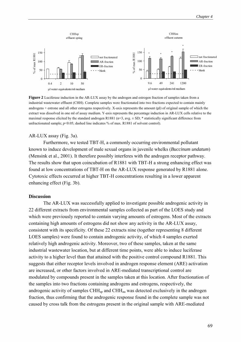
Chapter 4
69
Figure 2 Luciferase induction in the AR-LUX assay by the androgen and estrogen fraction of samples taken from a industrial wastewater effluent (CHH). Complete samples were fractionated into two fractions expected to contain mainly androgens + estrone and all other estrogens respectively. X-axis represents the amount (µl) of original sample of which the extract was dissolved in one ml of assay medium. Y-axis represents the percentage induction in AR-LUX cells relative to the maximal response elicited by the standard androgen R1881 (n=3, avg. ± SD; * statistically significant difference from unfractionated sample; p<0.05; dashed line indicates % of max. R1881 of solvent control).
AR-LUX assay (Fig. 3a).
Furthermore, we tested TBT-H, a commonly occurring environmental pollutant known to induce development of male sexual organs in juvenile whelks (Buccinum undatum) (Mensink et al., 2001). It therefore possibly interferes with the androgen receptor pathway. The results show that upon coincubation of R1881 with TBT-H a strong enhancing effect was found at low concentrations of TBT-H on the AR-LUX response generated by R1881 alone. Cytotoxic effects occurred at higher TBT-H concentrations resulting in a lower apparent enhancing effect (Fig. 3b).
Discussion
The AR-LUX was successfully applied to investigate possible androgenic activity in 22 different extracts from environmental samples collected as part of the LOES study and which were previously reported to contain varying amounts of estrogens. Most of the extracts containing high amounts of estrogens did not show any activity in the AR-LUX assay, consistent with its specificity. Of these 22 extracts nine (together representing 8 different LOES samples) were found to contain androgenic activity, of which 4 samples exerted relatively high androgenic activity. Moreover, two of these samples, taken at the same industrial wastewater location, but at different time points, were able to induce luciferase activity to a higher level than that attained with the positive control compound R1881. This suggests that either receptor levels involved in androgen response element (ARE) activation are increased, or other factors involved in ARE-mediated transcriptional control are modulated by compounds present in the samples taken at this location. After fractionation of the samples into two fractions containing androgens and estrogens, respectively, the androgenic activity of samples CHHsp and CHHau was detected exclusively in the androgen fraction, thus confirming that the androgenic response found in the complete sample was not caused by cross talk from the estrogens present in the original sample with ARE-mediated
CHHspeffluent spring
0
50
100
150
0.4 2 10 50
µl water equivalents/ml medium
% o
f max
R18
81
not fractionatedAR-fractionER-fractionblank
CHHaueffluent autumn
0
50
100
150
9.6 49 241 1200
µl water equivalents/ml medium
% o
f max
R18
81
not fractionatedAR-fractionER-fractionblank
*
***

Chapter 4
70
nM
0 1 10 100 1000 10000
indu
ctio
n
0
20
40
60
80 nortestosterone nortestosterone + estrone methyltestosterone methyltestosterone + estrone estron
0
20
40
60
80
0 10 100 1000
TBT-H (nM)
indu
ctio
n
TBT-H only
1 nM R1881 + TBT-H
*
*
Figure 3 a Luciferase induction in AR-LUX cells treated with a concentration range of nortestosterone or methyltestosterone, both alone and in combination with 10 nM estrone. Calculated EC50 were respectively: nortestosterone = 10.0 nM, nortestosterone + 10 nM estrone = 2.9 nM, methyltestosterone = 214 nM, methyltestosterone + 10 nM estrone = 31.8 nM. b Luciferase induction in AR-LUX cells treated with a concentration range of TBT-H alone or in combination with 1 nM R1881 (n=3, avg. ± SD; *statistically significant difference from 1 nM R1881 alone; p < 0.05).
signal transduction, a phenomenon that has been previously described (Terouanne et al., 2000; Blankvoort et al., 2001). Furthermore, the amount of measured R1881 equivalents increased in samples CHHau and ZKHau upon fractionation, and became undetectably low in sample EYSsu. This suggests that by studying subfractions of complex mixtures in isolation over- and underestimations of overall androgenic potency can be made due to the possible disappearance of compounds with a synergistic or antagonistic interactive effect on ARE-mediated signal transduction. This observation might very well represent a more general phenomenon that may not be restricted to the androgen receptor, but could also be relevant to many other receptors and signal transduction pathways. Thus, environmental mixtures may
b
a

Chapter 4
71
give rise to complex interactive effects, resulting in a different outcome of intracellular processes than predicted on the basis of chemical analysis of single compounds.
In contrast to the observations made for sample CHHau, the activity present in the intact sample EYSsu was absent in the androgen or estrogen fraction, thus suggesting the presence of androgenic compounds with extraction and HPLC characteristics different from common steroids. Alternatively, a strong synergistic effect of the estrogen fraction upon undetectable traces of activity in the androgen fraction may have been responsible for the androgenic activity in the unfractionated sample. Parks et al. (2001) have reported androgenic activity in kraft mill effluent-contaminated river water. Although they did find androstenedione, the amounts were not sufficient to explain the level of androgenic activity they observed. They also concluded that unknown androgenic compounds might be present. Possible candidates include unknown pesticide metabolites and quinolone derivatives, of which both antagonistic and agonistic activity have been reported, and which can be persistent in sewage water treatment plants (Sonnenschein and Soto, 1998; Halling-Sorensen et al., 2000). Also plant sterols have been implicated in causing reproductive effects and masculinisation of female fish (Lehtinen et al., 1999). In solid sample extracts also unknown, non-estrogenic compounds have been detected (Legler et al., 2003).
However, besides unidentified androgenic compounds, interactive effects by other components may have to be considered as a possible alternative explanation of the discrepancies found, as is illustrated by the synergistic effects from estrogens and TBT-H described in this paper. It will require a considerable research effort to isolate and identify these unknown androgens, not to mention the compounds which themselves show no androgen receptor-mediated activity, but rather act by modulating the AR-mediated response.
Our results clearly show that estrone shifted the entire dose-response curves towards a higher sensitivity for methyltestosterone and nortestosterone. This indicates that the presence of both estrogens and androgens in environmental samples could result in a more profound perturbation of the normal physiology of exposed organisms than when exposed to androgens alone. These types of effects might be further compounded by the presence of commonly occurring environmental pollutants other than estrogens, such as TBT-H, which have an additional disruptive effect on ARE-mediated signal transduction. In fact, when chemically assessing environmental samples, or when using reporter cell lines containing -unlike the AR-LUX cell line assay- constitutively overexpressed androgen receptors under the control of strong viral promoters, an under- or overestimation of the androgenic potency might easily occur. We are currently investigating these interactive effects in more detail. This also implicates that using androgen equivalency factors, analogous to the toxic equivalency factors (TEFs) used to calculate aryl hydrocarbon (Ah) receptor activating potencies of complex Ah receptor agonist mixtures (Safe, 1994), would not be the best way to evaluate the androgenic activity of complex environmental samples. Although total androgen equivalent calculations serve a useful purpose as an indication for androgenicity, one always has to keep in mind that unaccounted interactions by endocrine pathways linked to the receptor of interest might play a crucial role in the final response. Therefore, risk assessment of environmental samples should

Chapter 4
72
include evaluation of the possibility, that modulators of the respective pathway are present, in order to provide a realistic estimate of the integrated toxicological risk of the entire sample. References Behnisch, P. A., K. Fujii, K. Shiozaki, I. Kawakami and S. Sakai (2001). "Estrogenic and
dioxin-like potency in each step of a controlled landfill leachate treatment plant in Japan." Chemosphere 43(4-7): 977-984.
Blankvoort, B. M., E. M. de Groene, A. P. van Meeteren-Kreikamp, R. F. Witkamp, R. J. Rodenburg and J. M. Aarts (2001). "Development of an androgen reporter gene assay (AR-LUX) utilizing a human cell line with an endogenously regulated androgen receptor." Anal Biochem 298(1): 93-102.
Casademont, G., B. Perez and J. A. Garcia Regueiro (1996). "Simultaneous determination, in calf urine, of twelve anabolic agents as heptafluorobutyryl derivatives by capillary gas chromatography-mass spectrometry." J Chromatogr B Biomed Appl 686(2): 189-198.
Claessens, F., P. Alen, A. Devos, B. Peeters, G. Verhoeven and W. Rombauts (1996). "The androgen-specific probasin response element 2 interacts differentially with androgen and glucocorticoid receptors." J.Biol.Chem. 271(32): 19013-19016.
Daeseleire, E. A., A. De Guesquiere and C. H. Van Peteghem (1992). "Multiresidue analysis of anabolic agents in muscle tissues and urines of cattle by GC-MS." J Chromatogr Sci 30(10): 409-414.
De Voogt, P., D. E. Wells, L. Reutergardh and U. A. T. Brinkman (1990). "Biological activity, determination and occorunce of planar, mono-and di-ortho PCBs." Int. J. Environ. Anal. Chem.(16): 584-595.
Fawell, J. K., D. Sheahan, H. A. James, M. Hurst and S. Scott (2001). "Oestrogens and oestrogenic activity in raw and treated water in Severn Trent Water." Water Res 35(5): 1240-1244.
Fenet, H., E. Gomez, A. Pillon, D. Rosain, J. C. Nicolas, C. Casellas and P. Balaguer (2003). "Estrogenic activity in water and sediments of a French river: contribution of alkylphenols." Arch Environ Contam Toxicol 44(1): 1-6.
Groshart, C. and P. C. Okkerman (2000). Towards the establishment of a priority list of substances for further evaluation of their role in endocrine disruption. Delft, European Commission DG ENV: 29.
Hackenberg, R., T. Hawighorst, A. Filmer, E. P. Slater, K. Bock, M. Beato and K. D. Schulz (1992). "Regulation of androgen receptor mRNA and protein level by steroid hormones in human mammary cancer cells." J.Steroid.Biochem.Mol.Biol. 43: 599-607.
Halling-Sorensen, B., H. C. Lutzhoft, H. R. Andersen and F. Ingerslev (2000). "Environmental risk assessment of antibiotics: comparison of mecillinam, trimethoprim and ciprofloxacin." J Antimicrob Chemother 46(Suppl 1): 53-58; discussion 63-65.
Chapter 4
73
Horwitz, K. B. and W. L. McGuire (1978). "Estrogen control of progesterone receptor in human breast cancer: Correlation with nuclear processing of estrogen receptor." J. Biol. Chem. 253: 2222-2228.
Huggett, D. B., C. M. Foran, B. W. Brooks, J. Weston, B. Peterson, K. E. Marsh, T. W. La Point and D. Schlenk (2003). "Comparison of in vitro and in vivo bioassays for estrogenicity in effluent from North American municipal wastewater facilities." Toxicol Sci 72(1): 77-83.
Jaussi, R., G. Watson and K. Paigen (1992). "Modulation of androgen responsive gene expression by estrogen." Mol.Cell. Endocrinol(86): 187-192.
Kasper, S., P. S. Rennie, N. Bruchovsky, L. Lin, H. Cheng, R. Snoek, K. Dahlman-Wright, J. A. Gustafsson, R. P. Shiu, P. C. Sheppard and R. J. Matusik (1999). "Selective activation of the probasin androgen-responsive region by steroid hormones." J.Mol.Endocrinol. 22: 313-325.
Kelce, W. R., C. R. Christy R. Stone, S. C. Susan C. Laws, L. Earl Gray, J. A. Kemppainen and E. M. Wilson (1995). "Persistent DDT metabolite p,p'DDE is a potent androgen receptor antagonist." Nature 375: 581-585.
Kelce, W. R., C. R. Lambright, L. E. Gray, Jr. and K. P. Roberts (1997). "Vinclozolin and p,p'-DDE alter androgen-dependent gene expression: in vivo confirmation of an androgen receptor-mediated mechanism." Toxicol Appl Pharmacol 142(1): 192-200.
Kumar, M. V. and D. J. Tindall (1998). "Transcriptional regulation of the steroid receptor genes." Prog.Nucleic.Acid.Res.Mol.Biol. 59: 289-306.
Legler, J., A. S. Bulder, P. De Voogt, J. De Boer, M. Schrap and A. J. Murk (submitted). Bioanalysis of estrogenic and dioxin-like activity in field samples. SETAC-book: chapter xx Submitted.
Legler, J., M. Dennekamp, A. D. Vethaak, A. Brouwer, J. H. Koeman, B. van der Burg and A. J. Murk (2002). "Detection of estrogenic activity in sediment-associated compounds using in vitro reporter gene assays." Sci Total Environ 293(1-3): 69-83.
Legler, J., P. Leonards, A. Spenkelink and A. J. Murk (2003). "In vitro biomonitoring in polar extracts of solid phase matrices reveals the presence of unknown compounds with estrogenic activity." Ecotoxicology 12(1-4): 239-249.
Legler, J., C. E. van den Brink, A. Brouwer, A. J. Murk, P. T. van der Saag, A. D. Vethaak and B. van der Burg (1999). "Development of a stably transfected estrogen receptor-mediated luciferase reporter gene assay in the human T47D breast cancer cell line." Toxicol.Sci. 48: 55-66.
Lehtinen, K. J., K. Mattsson, J. Tana, C. Engstrom, O. Lerche and J. Hemming (1999). "Effects of wood-related sterols on the reproduction, egg survival, and offspring of brown trout (Salmo trutta lacustris L.)." Ecotoxicol Environ Saf 42(1): 40-49.
Mensink, B. P., J. M. Everaarts, H. Kralt, C. C. Ten Hallers-Tjabbes and J. P. Boon (1996). "TBT exposure in early life stages induces the development of male sexual characteristics in the common whelk, Buccinum undatum." Mar. Envir. Research 42: 151-154.

Chapter 4
74
Mensink, B. P., H. Kralt, A. D. Vethaak, C. C. Ten Hallers-Tjabbes, J. H. Jan H. Koeman, B. van Hattum and J. P. Boon (2001). "Imposex induction in laboratory reared juvenile Buccinum undatum by tributyltin (TBT)." Environ. Toxicol. Pharmacol. 11(1): 49-65.
Miyamoto, J. and W. Klein (1998). "Natural and anthropogenic environmental oestrogens: the scientific basis for risk assessment." Pure Appl. Chem. 70(9): 1829-1845.
Parks, L. G., C. S. Lambright, E. F. Orlando, L. J. Guillette, Jr., G. T. Ankley and L. E. Gray, Jr. (2001). "Masculinization of female mosquitofish in Kraft mill effluent-contaminated Fenholloway River water is associated with androgen receptor agonist activity." Toxicol Sci 62(2): 257-267.
Rennie, P. S., N. Bruchovsky, K. J. Leco, P. C. Sheppard, S. A. McQueen, H. Cheng, R. Snoek, A. Hamel, M. E. Bock, B. S. MacDonald and et al. (1993). "Characterization of two cis-acting DNA elements involved in the androgen regulation of the probasin gene." Mol Endocrinol 7(1): 23-36.
Safe, S. H. (1994). "Polychlorinated biphenyls (PCBs): environmental impact, biochemical and toxic responses, and implications for risk assessment." Crit Rev Toxicol 24(2): 87-149.
Schneider, S. M., M. Offterdinger, H. Huber and T. W. Grunt (2000). "Activation of Retinoic Acid Receptor alpha Is Sufficient for Full Induction of Retinoid Responses in SK-BR-3 and T47D Human Breast Cancer Cells." Cancer Res 60(19): 5479-5487.
Simon, J. A. (2001). "Safety of estrogen/androgen regimens." J Reprod Med 46(3 Suppl): 281-290.
Sonnenschein, C. and A. M. Soto (1998). "An updated review of environmental estrogen and androgen mimics and antagonists." J Steroid Biochem Mol Biol 65(1-6): 143-150.
Terouanne, B., B. Tahiri, V. Georget, C. Belon, N. Poujol, C. Avances, F. Orio, P. Balaguer and C. Sultan (2000). "A stable prostatic bioluminescent cell line to investigate androgen and antiandrogen effects." Mol.Cell Endocrinol. 160(Feb): 39-49.
Tillmann, M., U. Schulte-Oehlmann, M. Duft, B. Markert and J. Oehlmann (2001). "Effects of endocrine disruptors on prosobranch snails (Mollusca: Gastropoda) in the laboratory. Part III: Cyproterone acetate and vinclozolin as antiandrogens." Ecotoxicology 10(6): 373-388.
Verbrugge, D. A., R. A. Othoudt, K. R. Grzyb, R. A. Hoke, J. B. Drake, J. P. Giesy and D. Anderson (1991). "Concentrations of inorganic and organic contaminants in sedimments of six harbors on the North American Great Lakes." Chemosphere 22: 809-820.
Vethaak, A. D., G. B. J. Rijs, S. M. Schrap, H. Ruiter, J. Gerritsen and J. Lahr (2002). Estrogens and xeno-estrogens in the aquatic environment of the Netherlands, RIZA/RIKZ: 1-293.
Wolf, C. J., G. A. LeBlanc, J. S. Ostby and L. E. Gray, Jr. (2000). "Characterization of the period of sensitivity of fetal male sexual development to vinclozolin." Toxicol Sci 55(1): 152-161.

Chapter 4
75
Zhou, J., S. Ng, O. Adesanya-Famuiya, K. Anderson and C. A. Bondy (2000). "Testosterone inhibits estrogen-induced mammary epithelial proliferation and suppresses estrogen receptor expression." Faseb J 14(12): 1725-1730.

76

55
Detection of hormonal anabolic compounds in calf urine and unverified growth-promoting preparations: Application of the AR-LUX bioassay for
screening and determination of androgenic activity
Analyst 128(11): 1373-1381, 2003
B.M.G. Blankvoort1, 2, J.M.M.J.G. Aarts2, R. Schilt3, P. Geerdink2, B. Spenkelink2, R.J.T. Rodenburg1, 4
1 Department of Bioanalysis, TNO Pharma, the Netherlands, 2 Department of Agrotechnology and Food Sciences, Sub-
department of Toxicology, Wageningen University, Wageningen, the Netherlands, 3 Department of Residue Analysis, TNO Nutrition and Food Research, the Netherlands; 4 UMC St Radboud, Laboratory of Paediatrics and Neurology, Nijmegen, the
Netherlands

Chapter 5
78
Abstract Despite a ban by the European Union, the use of anabolic steroids and repartitioning
agents in cattle is still occasionally observed. Due to continuing improvements in analytical techniques, very low detection limits for individual compounds have been achieved. In response to these developments, cocktails composed of several steroids have been applied, thus hampering detection due to lower levels of the individual compounds. Bioassays capable of measuring the integrated effect of cocktails might therefore provide valuable additional tools in controlling the use of illegal anabolics. We investigated the feasibility of using the AR-LUX assay to detect the presence in cattle urine of growth promoters that exert their effects via androgen response elements (AREs). The AR-LUX assay is based on a human cell line featuring a luciferase reporter gene under transcriptional control of an authenticated ARE. Several column purification and liquid/liquid extraction methods were investigated to optimise the efficiency of anabolic compounds extraction and minimise cytotoxic effects of the urine matrix. The AR-LUX assay was found to be applicable to the detection of anabolic steroids excreted in urine samples with a discriminatory power similar to that of GC-MS analysis. Finally, some liquid products probably destined for growth-promoting purposes confiscated outside the Netherlands were analysed. Although common chemical-analytical methods did not detect any anabolic steroids in these samples, the presence of compounds activating ARE-mediated gene expression was clearly established. Introduction
Since the first of January 1989, according to directive 88/146/EEC, replaced later by directive 96/22/EC, the European Union (EU) prohibits the administering to a farm animal, by any means whatsoever, of substances for growth promotion purposes (EU, 1999). Nevertheless, the use of anabolic steroids and repartitioning agents in cattle is still occasionally observed (Courtheyn et al., 2002; Nielen et al., 2003). The main reason for illegal use is the increased meat production and therefore higher earnings achieved after use of these compounds. These (cocktails of) anabolic steroids and β-agonists effectuate enhancement of feeding efficiency and/or body fat to muscle repartitioning. In the United States, the use of six hormones is allowed. These are the estrogens 17β-estradiol and zeranol, the androgens testosterone and trenbolone, and the progestagens progesterone and melengestrol-acetate (EU, 1999). After the European Union prohibited the use of anabolic compounds as growth promoters, a wide range of these agents have been found in plasma, urine and tissue samples (Maghuin-Rogister, 1990; Debruyckere et al., 1993). In general, their detection is hampered in samples that contain complex mixtures of different compounds, each having a similar biological effect on animal development through common regulatory pathways. By using mixtures with low levels of individual compounds, the detection by chemical analysis is becoming increasingly difficult. This further stresses the need for a detection method that measures the integral anabolic content of samples. Therefore, the development of detection methods that measure the presence of these compounds by using their target receptors without knowing the exact identities might provide valuable additional tools in the screening effort. In this study, we focused on steroids that mediate gene

Chapter 5
79
expression via the probasin androgen response element 2 (PB-ARE2). We have previously constructed a reporter plasmid containing a luciferase gene under transcriptional control of the PB-ARE2 element and stably transfected it into T47D human breast carcinoma cells. The resulting endogenous Androgen Receptor-mediated LUciferase eXpression (AR-LUX) system has been evaluated for its responsiveness to a number of androgens, anti-androgens and their combinations, and to other modulators of androgenic responses, as described earlier (Blankvoort et al., 2001). This reporter cell line features endogenous expression of the androgen receptor, progesterone, estrogen and other steroid receptors (Hall et al., 1992; Liberato et al., 1993; Buras et al., 1994) and constitutes a reporter gene assay based on the endogenous expression and regulation of a number of relevant steroid receptor genes. In addition to compounds that directly act on the ARE, the AR-LUX measures effects of compounds that indirectly modulate ARE-mediated gene-expression through alternative cellular pathways. Here we tested its suitability for the detection of steroid growth promoters in urine and in anabolic cocktails of unknown composition. Experimental procedures Chemicals
R1881 was purchased from NEN Life Science Products (Hoofddorp, the Netherlands). Methyltestosterone, β-nortestosterone, estrone and DMSO were obtained from Sigma-Aldrich (Zwijndrecht, the Netherlands) and α-trenbolone as well as deuterated steroids from RIVM (Bilthoven, the Netherlands). AR-LUX assay procedure T47D/Sutherland human breast cancer cells stably transfected with pPBARE2tataluc+
were seeded in white 96 well plates with clear flat bottoms (Corning Inc., Cambridge, USA) at a density of 18,000 cells/well in 100 µl DMEM/F12 medium (Life Technologies ltd., Paisley, Scotland) containing 5% (v/v) foetal bovine serum (FBS). The serum had previously been treated with dextran-coated charcoal (DCC) as described previously (Horwitz and McGuire, 1978) to remove any traces of steroid hormones present in the serum. Cells were cultured at 5% (v/v) CO2 and 100% relative humidity. After 24 hours, the medium was replaced with 100 µl medium containing the chemicals or extracts of interest, dissolved in ethanol or DMSO. Incubations were performed in triplicate with a maximum solvent concentration of 0.2% (v/v). Following 24 hours of incubation, medium was removed from the cells by aspiration and subsequently, 20 µl of fresh medium and 20 µl of Steady-Glo reagent (Promega Corporation, Madison, USA) was added. Following 10 min incubation at room temperature in the instrument, luciferase activity was determined by counting the amount of photons produced following conversion of luciferine into oxyluciferin by the luciferase released upon cell lysis. Emitted photons were counted for 30 seconds using a Wallac 1450 microbeta liquid scintillation counter and expressed as luminescence counts. Induction factors were subsequently calculated relative to solvent controls unless indicated otherwise.
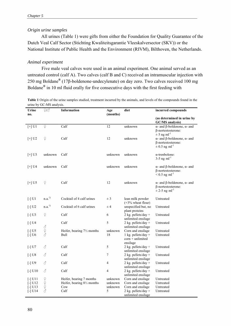
Chapter 5
80
Origin urine samples All urines (Table 1) were gifts from either the Foundation for Quality Guarantee of the
Dutch Veal Calf Sector (Stichting Kwaliteitsgarantie Vleeskalversector (SKV)) or the National Institute of Public Health and the Environment (RIVM), Bilthoven, the Netherlands. Animal experiment
Five male veal calves were used in an animal experiment. One animal served as an untreated control (calf A). Two calves (calf B and C) received an intramuscular injection with 250 mg Boldane® (17β-boldenone-undecylenate) on day zero. Two calves received 100 mg Boldane® in 10 ml fluid orally for five consecutive days with the first feeding with Table 1 Origin of the urine samples studied, treatment incurred by the animals, and levels of the compounds found in the urine by GC-MS analysis. Urine no.
♀/♂ Information Age (months)
diet incurred compounds (as determined in urine by GC/MS analysis)
[+] U1 ♀ Calf 12 unknown α- and β-boldenone, α- and β-nortestosterone: ± 5 ng ml-1
[+] U2 ♀ Calf 12 unknown α- and β-boldenone, α- and β-nortestosterone: ± 0.5 ng ml-1
[+] U3 unknown Calf unknown unknown α-trenbolone:
3-5 ng ml-1
[+] U4 unknown Calf unknown unknown α- and β-boldenone, α- and
β-nortestosterone: < 0.5 ng ml-1
[+] U5 ♀ Calf 12 unknown α- and β-boldenone, α- and
β-nortestosterone: ± 2-5 ng ml-1
[-] U1 n.a.1) Cocktail of 4 calf urines ± 3 lean milk powder
(+3% wheat flour) Untreated
[-] U2 n.a.1) Cocktail of 6 calf urines ± 4 unspecified but, no plant proteins
Untreated
[-] U3 ♀ Calf 6 2 kg. pellets/day + unlimited ensilage
Untreated
[-] U4 ♂
Calf 5 2 kg. pellets/day + unlimited ensilage
Untreated
[-] U5 ♀ Heifer, bearing 7½ months unknown Corn and ensilage Untreated [-] U6 ♂ Bull 18 1 kg. pellets/day +
corn + unlimited ensilage
Untreated
[-] U7 ♂ Calf 5 2 kg. pellets/day + unlimited ensilage
Untreated
[-] U8 ♂ Calf 7 2 kg. pellets/day + unlimited ensilage
Untreated
[-] U9 ♂ Calf 4 2 kg. pellets/day + unlimited ensilage
Untreated
[-] U10 ♂ Calf 4 2 kg. pellets/day + unlimited ensilage
Untreated
[-] U11 ♀ Heifer, bearing 7 months unknown Corn and ensilage Untreated [-] U12 ♀ Heifer, bearing 8½ months unknown Corn and ensilage Untreated [-] U13 ♀ Cow unknown Corn and ensilage Untreated [-] U14 ♂ Calf 5 2 kg. pellets/day +
unlimited ensilage Untreated

Chapter 5
81
administration starting on day zero (calf D and E). Urine and serum was collected on days t=-1, 0, 1, 2, 4, 6, 8 and 13. The sample taken at t=0 was just after treatment for the orally dosed group and prior to treatment for the injected group. Only diethyl ether extracted urine samples were available and these were analysed with GC-MS and in the AR-LUX assay. HPLC separation-GC-MS analysis A number of urine samples have been subjected to a standard GC-MS confirmatory procedure. Briefly, following the addition of a mixture of deuterated internal standards, urine samples were enzymatically deconjugated with Helix Pomatia juice (containing glucuronidase and arylsulphatase) at 37°C for 16 hours. The hydrolysed samples were loaded onto 300 mg C18 solid phase extraction columns. After evaporation of the solvent, the residue was dissolved in 30 µl isopropanol after which 250 µl heptane was added. This extract was subsequently injected into a normal phase HPLC (LiChrosorb 5 Diol, 100 × 4 mm). Heptane and heptane/isopropanol (3:2 v/v) were used as solvents A and B, respectively. A gradient was programmed from 10% A (0 min) to 87% A (15 min) to 50% A (20 min) to 10% A (25 min). A single fraction was collected from 1 to 7 minutes. After evaporation of the solvents of the fraction, the extract was derivatised with heptafluorobutyric anhydride/acetone (20/80; v/v) during 60 min at 60°C. GC-MS analysis of the derivatives was performed on an Agilent 6890 GC (DB-5 column or DB-17 column, splitless injection) in combination with a Thermo Finnigan MAT95 sector mass spectrometer operating at a resolution of 5000 (10% valley) using selected ion monitoring with at least 4 ions per analyte. Quantification was based on comparison with the signal generated by the corresponding deuterated internal standards that had been added in the first step of the analysis. Identification was based on EU criteria 2002/657/EC, using at least 3 ion ratios with a maximal variation of 10%. Deconjugation of urinary steroids prior to extraction and AR-LUX analysis
To 670 µl urine 330 µl 0.2 M sodium acetate buffer (pH 4.6) was added and pH was adjusted to 4.5-5.0. Subsequently 3.3 µl glucuronidase/arylsulfatase mixture (Helix pomatia juice, Bio Sepra, Brunschwig Chemie, Amsterdam, the Netherlands) was added followed by overnight incubation at 37°C or 1 hour at 55°C. In follow-up extractions this mixture of urine and acetate buffer is referred to as urine; however in calculations of µl urine equivalents ml-1 medium the actual amount of added urine (in this case 670 µl) was used. Solid Phase Extraction (SPE-C18)
Bakerbond SPE-C18 columns (J.T. Baker, Phillipsburg, NJ, USA) were conditioned by adding 2 × 1 ml methanol (MeOH) followed by 2 × 1 ml H2O. Subsequently deconjugated urine was added and passively allowed to flow through the column. The column was washed by adding 1 ml H2O followed by 1 ml H2O/acetone (99.5%, p.a., Merck, Darmstadt, Germany) (80/20; v/v) and 1 ml H2O/MeOH (HPLC grade, Rathburn, Walkerburn, Scotland) (70/30; v/v). The flow-through was discarded after which 2 × 1 ml MeOH was added and the eluate was collected in a clean tube after which the MeOH was evaporated under a stream of

Chapter 5
82
N2 at 45°C. Finally, the residue was dissolved in 20 µl DMSO or in solvents appropriate for the follow-up extraction. Amino column extraction (NH2-SPE)
Isolute NH2-SPE columns containing 1 gram of sorbent and a 6 ml column reservoir were purchased from IST international (Mid Glamorgan, UK). Columns were conditioned with 5 ml MeOH/water (80/20; v/v). After deconjugation, 4 ml MeOH was added to 1 ml urine/acetate buffer mixture, thus creating a MeOH/water mixture (80/20; v/v) which was applied to the preconditioned column. The flow-through containing the unbound steroid hormones was collected and another 2 ml of MeOH/H2O (80/20; v/v) was applied and the flow through also collected in the same tube. Subsequently, the solvents were evaporated at 45°C under a stream of N2. Finally the residue was dissolved in 20 µl DMSO or in solvents appropriate for follow-up extraction. Heptane/butanol extraction
After deconjugation pH was adjusted to a value between 9.0 and 10.0 with 2M sodium carbonate solution (pH 10). To 1 ml of urine, 1.4 ml n-heptane (99.4% , HPLC grade, Labscan, Dublin Ireland)/butanol (99.4%, J.T.Baker, Mallinckrodt Baker B.V., Deventer, the Netherlands) (80/20; v/v) was added followed by vortexing for 1 min and shaking for 15 min. Subsequently, samples were centrifuged at 750 × g and the top organic layer was collected in a clean tube. The extraction was repeated and the organic layer was again collected and added to the first collected layer. The total volume was evaporated under a stream of N2 at 45°C. The residue was dissolved in 20 µl DMSO ml-1 extracted urine mixture or dissolved in solvents appropriate for follow-up extraction. Ethyl acetate extraction
The procedure was adapted from Legler et al. (2002). After deconjugation, 50 µl 1 M HCl was added per ml urine followed by briefly vortexing and addition of 1 ml ethyl acetate (EtAc, analytical reagent (a.r.), Biosolve ltd., Valkenswaard, the Netherlands) and vortexing. Subsequently, 250 µl isopropanol (99.5%, J.T. Baker) was added to precipitate proteins and samples were centrifuged at 1100 × g after which the organic phase was collected. The extraction was repeated twice by adding 1 ml EtAc and transferring the organic layer to a clean tube. The collected organic phase was subsequently evaporated under a stream of N2 at 45°C and dissolved in 20 µl DMSO per ml of extracted urine mixture or dissolved in solvents appropriate for follow-up extraction. Diethyl ether extraction
After deconjugation, 6 ml diethyl ether (DEE, 99.5%, a.r., Labscan) was added to 1 ml urine mixture followed by vortexing for one min. Subsequently, the tube was transferred to an acetone/dry ice bath in order to freeze the water phase, after which the liquid organic phase was decanted into a clean tube. This was repeated once, after which the DEE was evaporated under a stream of N2 at 45°C. The remaining dry extracted matter was dissolved in 5 µl

Chapter 5
83
DMSO per ml of extracted urine mixture and vortexed, after which 500 µl assay medium was added. In order to obtain a homogeneous solution, the sample was mixed by ultra-sonification for 5 min. Hexane extraction
After deconjugation, 6 ml n-hexane (99.5%) was added to 1 ml urine mixture followed by vortexing for one min. Subsequently, the tube was transferred to an acetone/dry ice bath to freeze the water phase. The liquid organic phase was decanted into a clean tube containing 4 ml 0.2 M sodium acetate (NaAc) buffer (pH 4.5) to wash the n-hexane phase. This second tube was also vortexed for one min and also transferred to an acetone/dry ice bath to freeze the water phase. The hexane phase was subsequently transferred into the following tube containing 4 ml of 0.2 M NaAc buffer (pH 4.5) and in this way, the hexane containing the steroids was washed four times with the acetate buffer in order to remove any compounds that may interfere with the AR-LUX bioassay. Finally, the hexane was evaporated under a stream of N2 at 45°C. The remaining extract was dissolved in 5 µl DMSO per ml of extracted urine mixture and vortexed, followed by the addition of 500 µl assay medium and ultra-sonification for 5 min. MTT cytotoxicity assay
The assay is based on the cleavage of the yellow tetrazolium salt MTT (3-[4,5-dimethylthiazol-2-yl]-2,5-diphenyl tetrazolium bromide) to purple insoluble formazan crystals by metabolically active cells. The formazan crystals formed are solubilised and the resulting purple coloured solution is quantified by measuring the absorbance at 540 nm using a spectrophotometer. Since dead cells will not convert MTT into its purple metabolite, this assay can be used as a means to quantify the cytotoxicity of extracts added to the cells. Cells were incubated in culture medium containing the compounds or extracts of interest for 24 h in 96-wells plates. Subsequently, 20 µl MTT solution (1 mg ml-1) was added to the culture medium. Plates were gently shaken and incubated at 37°C for 3 hours. After centrifugation at 300 × g at room temperature, the supernatant was removed and 100 µl ethanol was added to the wells in order to dissolve the formazan crystals formed during the incubation. After crystals were dissolved by gently shaking the well-plates, the absorbance was measured at A540 with a Wallac Victor2 1420 multilabel counter. Data and statistical analysis
Significance of mean differences was tested using an unpaired two-sample t-test for means using Graphpad Prism version 3.

Chapter 5
84
Results and discussion Untreated calf urine contains compounds with synergistic action
Urine is known to contain many different compounds that can interfere with bioassays (Chou and Hee, 1993; Parsons et al., 2000). Therefore, initial experiments were designed to identify an optimal extraction procedure for steroids in calf urine samples, in order to limit the adverse effects of unwanted urine constituents without reducing the androgenic potential of the samples, as measured by the AR-LUX bioassay. Since urine from untreated animals may harbour intrinsic androgenic activity, e.g. due to endogenous hormones (Velle, 1976), an additional goal of these experiments was to gain insight into the background signal induced by urine extracts from untreated calves. Furthermore, the effects of urine extracts on the response elicited by a known androgen receptor agonist, methyltrienolone (R1881), were analysed.
Two cocktails of urine samples collected from either 4 untreated calves ([-] U1) or 6 untreated calves ([-] U2) were used for an initial comparison of different steroid extraction methods. Both urine cocktails were split in two portions, one of which was spiked with R1881. The two portions were subsequently extracted using different (combinations of) extraction methods. Following these extractions, the urine extracts were added to the AR-LUX cells, generating a range of urine equivalents per volume of medium and concentration of spiked R1881 (assuming 100% recovery). The response in the AR-LUX assay was found to increase with increasing amounts of extracted urine (expressed as urine equivalent concentrations) (Fig. 1a to 1d). This indicates the presence of detectable amounts of endogenous steroids in urine from untreated calves. Addition of R1881 induced a clear increase in the measured response, showing that in spite of the background signal, exogenous androgenic activity (less than 110 pM R1881 equivalents) present in urine extracts is detectable using the AR-LUX. Interestingly, all R1881-spiked urines ultimately attain a higher maximal induction factor and absolute luciferase activity level at high R1881 concentrations than elicited by R1881 alone. This suggests a synergistic effect of compounds present in the urine on cellular pathways resulting in increased ARE-mediated gene expression. The induction found for R1881-spiked urine relative to its corresponding unspiked urine is essentially the same as that of assay medium spiked with R1881 relative to a solvent blank in assay medium (data not shown). This indicates that the presence of urine extract apparently produces a proportional change in both background and induced reporter gene transcription levels. Although it remains speculation, this may be achieved through an effect on the levels of receptors or transcriptional co-factors involved. Comparison of urine extraction methods
SPE-C18 extraction is commonly applied to the isolation of steroids from urine samples. Addition of up to 50 µl urine equivalents of extract per ml resulted in a dose-dependent response of the AR-LUX. However, we observed a decrease in reporter gene expression when exceeding 50 µl urine equivalents ml-1 medium (Fig. 1a). This suggests that high urine extract concentrations are cytotoxic, thereby inhibiting the luciferase gene

85
Figure 1 Comparison of the effects of different extraction methods on the response of the AR-LUX assay to R1881-spiked calf urine samples. a Luciferase induction in AR-LUX cells by control urine [-] U1 extracted with different SPE-C18 column based methods. The symbol [+] indicates control U1 spiked with R1881; + 56°C indicates an additional incubation of urine at 56°C prior to extraction (n=3, avg. ± SD). b Luciferase induction in AR-LUX cells by control urine [-] U1 extracted with combined heptane/butanol liquid/liquid (L/L) extraction + NH2-column + SPE-C18
column-based methods. The symbol [+] indicates control U1 spiked with R1881; + 56°C indicates an additional incubation of urine at 56°C prior to extraction (n=3, avg. ± SD). c Luciferase induction in AR-LUX cells by control urine [-] U2 extracted with heptane/butanol (L/L) extraction +/- SPE-C18 column based clean-up. The symbol [+] indicates control U2 spiked with R1881 (n=3, avg. ± SD). d Luciferase induction in AR-LUX cells by control urine [-] U2 extracted with heptane/butanol (L/L) extraction-based methods +/- NH2-column +/- SPE-C18 column based clean-up. The symbol [+] indicates control U2 spiked with R1881 (n=3, avg. ± SD). The dashed line in panel a-d represents maximal induction attained with R1881 in assay medium.
0
30
60
90
120
6.25 (0.11) 12.5 (0.22) 25 (0.44) 50 (0.88) 100 (1.75) 200 (3.50)
µl urine equivalents ml-1 (nM R1881)
indu
ctio
n
SPE-C18
SPE-C18 [+]
SPE-C18 + 56°C
SPE-C18 + 56°C [+]
max. R1881
0
30
60
90
120
6.25 (0.11) 12.5 (0.22) 25 (0.44) 50 (0.88) 100 (1.75) 200 (3.50)
µl urine equivalents ml-1 (nM R1881)
indu
ctio
n
L/L + NH2 + SPE-C18
L/L + NH2 + SPE-C18 [+]
L/L + NH2 + SPE-C18 + 56°C
L/L + NH2 + SPE-C18 + 56°C[+]max. R1881
0
30
60
90
120
6.25 (0.11) 12.5 (0.22) 25 (0.44) 50 (0.88) 100 (1.75) 200 (3.50)
µl urine equivalents ml-1 (nM R1881)
indu
ctio
n
L/L
L/L [+]
L/L + SPE-C18
L/L + SPE-C18 [+]
max R1881
0
30
60
90
120
6.25 (0.11) 12.5 (0.22) 25 (0.44) 50 (0.88) 100 (1.75) 200 (3.50)
µl urine equivalents ml-1 (nM R1881)
indu
ctio
n
NH2
NH2 [+]
L/L+ NH2
L/L + NH2 [+]
L/L + NH2 + SPE-C18
L/L + NH2 + SPE-C18 [+]
max R1881
a
c d
b

Chapter 5
86
expression in a non-specific manner. Combining SPE-C18 with a preceding heptane/butanol liquid/liquid (L/L) extraction and an amino column extraction improved the assay performance (Fig. 1b), although also in this case the response of the cells decreased after addition of high concentrations of urine extracts (above 100 µl urine equivalents ml-1
medium). Heat pre-treatment of urine samples at 56°C for 30 min did not increase the responses observed (Fig. 1a, b). Heptane/butanol L/L extraction (either alone or in combination with other extraction clean-up methods) yielded the most favourable induction factors, allowing addition of up to 200 µl urine equivalents per ml of culture medium without interfering with the assay (Fig. 1 c,d). Column extraction alone (Fig 1a) yielded higher responses at low urine concentrations (<50 µl volume equivalents of urine ml-1 of medium). However, since levels of anabolic steroids found in real-life urine samples often range from only 0.1 to 2 ppb (Scippo et al., 1994; Walshe et al., 1998), we decided to focus on liquid/liquid extraction methods that allow addition of large equivalent volumes of urine in order to obtain the necessary low detection limits. Although the results shown in Fig. 1d suggest that the single amino column extraction procedure yield the most optimal results, follow-up experiments with positive urines U1-5, extracted using the same amino column purification, resulted in oil-like residues that could not be evaporated. Moreover, the oil-like residues were found to be severely cytotoxic upon addition to the AR-LUX cells (data not shown). From these observations we concluded that the single NH2-column extraction method is not reproducibly compatible with the AR-LUX bioassay.
The results given in Fig. 1 seemed to indicate that in some cases, high concentrations of urine samples interfered with the AR-LUX bioassay, possibly by inducing a cytotoxic effect. To further investigate this, an MTT cytotoxicity assay was performed to test whether exposure to urine extracts leads to cell death of the AR-LUX cells. For this purpose, blank urine sample [-] U6 was extracted utilising either an amino column extraction (NH2) or a combination of heptane/butanol L/L extraction followed by an amino column extraction. The results show that the amino column-only purified extract displays minor cytotoxicity at a concentration of 200 µl urine equivalents ml-1 and severe cytotoxicity at 400 µl ml-1 (Fig. 2a).
Figure 2 Example of cytotoxic and inhibitory effects induced by a urine extract in the AR-LUX asssay. a Cell viability as determined with an MTT assay in AR-LUX cells upon exposure to urine [-] U6 extracted with an NH2-column or with a combined heptane/butanol + NH2-column extraction (n=3, avg. ± SD). b Luciferase response in AR-LUX cells dosed upon exposure to [-] U6 extracted with an NH2-column or a combined heptane/butanol + NH2-column extraction (n=3, avg. ± SD).
MTT assay
0
0.1
0.2
0.3
0.4
0.5
50 100 200 400
µl urine equivalents ml-1
A54
0
[-] U6 NH2
[-] U6 [heptane/butanol]/NH2
AR-LUX assay
0
4
8
12
16
50 100 200 400
µl urine equivalents ml-1
indu
ctio
n
[-] U6 NH2
[-] U6 [heptane/butanol]/NH2
a b
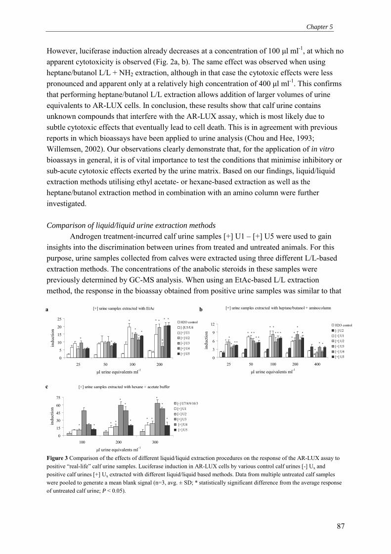
Chapter 5
87
However, luciferase induction already decreases at a concentration of 100 µl ml-1, at which no apparent cytotoxicity is observed (Fig. 2a, b). The same effect was observed when using heptane/butanol L/L + NH2 extraction, although in that case the cytotoxic effects were less pronounced and apparent only at a relatively high concentration of 400 µl ml-1. This confirms that performing heptane/butanol L/L extraction allows addition of larger volumes of urine equivalents to AR-LUX cells. In conclusion, these results show that calf urine contains unknown compounds that interfere with the AR-LUX assay, which is most likely due to subtle cytotoxic effects that eventually lead to cell death. This is in agreement with previous reports in which bioassays have been applied to urine analysis (Chou and Hee, 1993; Willemsen, 2002). Our observations clearly demonstrate that, for the application of in vitro bioassays in general, it is of vital importance to test the conditions that minimise inhibitory or sub-acute cytotoxic effects exerted by the urine matrix. Based on our findings, liquid/liquid extraction methods utilising ethyl acetate- or hexane-based extraction as well as the heptane/butanol extraction method in combination with an amino column were further investigated. Comparison of liquid/liquid urine extraction methods
Androgen treatment-incurred calf urine samples [+] U1 � [+] U5 were used to gain insights into the discrimination between urines from treated and untreated animals. For this purpose, urine samples collected from calves were extracted using three different L/L-based extraction methods. The concentrations of the anabolic steroids in these samples were previously determined by GC-MS analysis. When using an EtAc-based L/L extraction method, the response in the bioassay obtained from positive urine samples was similar to that
Figure 3 Comparison of the effects of different liquid/liquid extraction procedures on the response of the AR-LUX assay to positive �real-life� calf urine samples. Luciferase induction in AR-LUX cells by various control calf urines [-] Ux and positive calf urines [+] Ux extracted with different liquid/liquid based methods. Data from multiple untreated calf samples were pooled to generate a mean blank signal (n=3, avg. ± SD; * statistically significant difference from the average response of untreated calf urine; P < 0.05).
[+] urine samples extracted with heptane/butanol + aminocolumn
0
3
6
9
12
25 50 100 200 400
µl urine equivalents ml-1
indu
ctio
n
H2O control[-] U2[+] U1[+] U2[+] U3[+] U4[+] U5
**
*
**
***
**
*****
***
*
**
*
*
[+] urine samples extracted with hexane + acetate buffer
0
15
30
45
60
75
100 200 300
µl urine equivalents ml-1
indu
ctio
n
[-] U7/8/9/10/3[+] U1 [+] U2 [+] U3 [+] U4 [+] U5
* *
*
*
*
***
**
**
[+] urine samples extracted with EtAc
0
5
10
15
20
25
25 50 100 200
µl urine equivalents ml-1
indu
ctio
n
H2O control[-]U3/U4[+] U1[+] U2[+] U3[+] U4[+] U5
*
**
*
**
* *
*
* *
a b
c
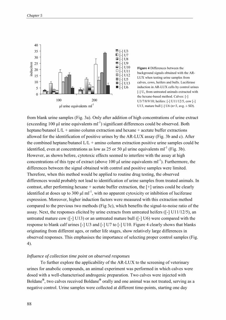
Chapter 5
88
05
10152025303540
100 200µl urine equivalents ml-1
indu
cito
n
[-] U3[-] U7[-] U8[-] U9[-] U10[-] U11[-] U12[-] U5[-] U13[-] U6
Figure 4 Differences between the background signals obtained with the AR-LUX when testing urine samples from calves, cows, heifers and bulls. Luciferase induction in AR-LUX cells by control urines [-] Ux from untreated animals extracted with the hexane-based method. Calves: [-] U3/7/8/9/10; heifers: [-] U11/12/5, cow [-] U13, mature bull [-] U6 (n=3, avg. ± SD).
from blank urine samples (Fig. 3a). Only after addition of high concentrations of urine extract (exceeding 100 µl urine equivalents ml-1) significant differences could be observed. Both heptane/butanol L/L + amino column extraction and hexane + acetate buffer extractions allowed for the identification of positive urines by the AR-LUX assay (Fig. 3b and c). After the combined heptane/butanol L/L + amino column extraction positive urine samples could be identified, even at concentrations as low as 25 or 50 µl urine equivalents ml-1 (Fig. 3b). However, as shown before, cytotoxic effects seemed to interfere with the assay at high concentrations of this type of extract (above 100 µl urine equivalents ml-1). Furthermore, the differences between the signal obtained with control and positive samples were limited. Therefore, when this method would be applied to routine drug testing, the observed differences would probably not lead to identification of urine samples from treated animals. In contrast, after performing hexane + acetate buffer extraction, the [+] urines could be clearly identified at doses up to 300 µl ml-1, with no apparent cytoxicity or inhibition of luciferase expression. Moreover, higher induction factors were measured with this extraction method compared to the previous two methods (Fig 3c), which benefits the signal-to-noise ratio of the assay. Next, the responses elicited by urine extracts from untreated heifers ([-] U11/12/5), an untreated mature cow ([-] U13) or an untreated mature bull ([-] U6) were compared with the response to blank calf urines [-] U3 and [-] U7 to [-] U10. Figure 4 clearly shows that blanks originating from different ages, or rather life stages, show relatively large differences in observed responses. This emphasises the importance of selecting proper control samples (Fig. 4). Influence of collection time point on observed responses
To further explore the applicability of the AR-LUX to the screening of veterinary urines for anabolic compounds, an animal experiment was performed in which calves were dosed with a well-characterised androgenic preparation. Two calves were injected with Boldane®, two calves received Boldane® orally and one animal was not treated, serving as a negative control. Urine samples were collected at different time-points, starting one day
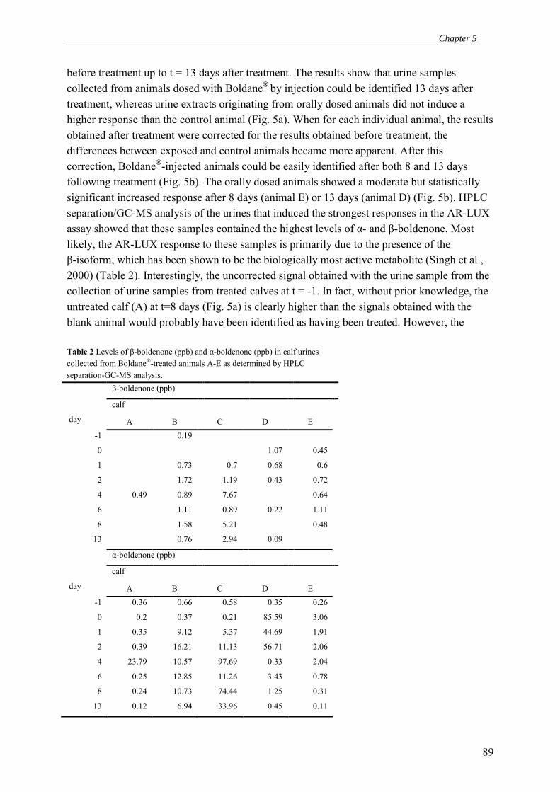
Chapter 5
89
before treatment up to t = 13 days after treatment. The results show that urine samples collected from animals dosed with Boldane® by injection could be identified 13 days after treatment, whereas urine extracts originating from orally dosed animals did not induce a higher response than the control animal (Fig. 5a). When for each individual animal, the results obtained after treatment were corrected for the results obtained before treatment, the differences between exposed and control animals became more apparent. After this correction, Boldane®-injected animals could be easily identified after both 8 and 13 days following treatment (Fig. 5b). The orally dosed animals showed a moderate but statistically significant increased response after 8 days (animal E) or 13 days (animal D) (Fig. 5b). HPLC separation/GC-MS analysis of the urines that induced the strongest responses in the AR-LUX assay showed that these samples contained the highest levels of α- and β-boldenone. Most likely, the AR-LUX response to these samples is primarily due to the presence of the β-isoform, which has been shown to be the biologically most active metabolite (Singh et al., 2000) (Table 2). Interestingly, the uncorrected signal obtained with the urine sample from the collection of urine samples from treated calves at t = -1. In fact, without prior knowledge, the untreated calf (A) at t=8 days (Fig. 5a) is clearly higher than the signals obtained with the blank animal would probably have been identified as having been treated. However, the Table 2 Levels of β-boldenone (ppb) and α-boldenone (ppb) in calf urines collected from Boldane®-treated animals A-E as determined by HPLC separation-GC-MS analysis. β-boldenone (ppb)
calf
day A B C D E -1 0.19
0 1.07 0.45
1 0.73 0.7 0.68 0.6
2 1.72 1.19 0.43 0.72
4 0.49 0.89 7.67 0.64
6 1.11 0.89 0.22 1.11
8 1.58 5.21 0.48
13 0.76 2.94 0.09
α-boldenone (ppb)
calf
day A B C D E -1 0.36 0.66 0.58 0.35 0.26
0 0.2 0.37 0.21 85.59 3.06
1 0.35 9.12 5.37 44.69 1.91
2 0.39 16.21 11.13 56.71 2.06
4 23.79 10.57 97.69 0.33 2.04
6 0.25 12.85 11.26 3.43 0.78
8 0.24 10.73 74.44 1.25 0.31
13 0.12 6.94 33.96 0.45 0.11

Chapter 5
90
Figure 5 Time-course analysis of urine samples from calves dosed with Boldane® with the AR-LUX assay. a Luciferase induction relative to medium control in AR-LUX cells dosed with extracts from urines collected from calves treated with boldane. Only diethyl ether-extracted urine samples were available. Calf A is the untreated control, calf B and C received an intramuscular injection with 250 mg Boldane® (17β-boldenone-undecylenate) on day zero, calves D and E received 100 mg Boldane® in 10 ml fluid orally for five consecutive days (n=3, avg. ± SD; * statistically significant difference from the untreated calf at the same time point; p < 0.05). b Luciferase induction relative to individual controls collected at t=-1 in AR-LUX cells dosed with extracts from urines collected from calves treated with boldane. Only diethyl ether extracted urine samples were available. Calf A is the untreated control, calf B and C received an intramuscular injection with 250 mg Boldane® (17β-boldenone-undecylenate) on day zero, calves D and E received 100 mg Boldane® in 10 ml fluid orally for five consecutive days with the first administration on day zero (n=3, avg. ± SD; * statistically significant difference from the untreated calf at the same time point; p < 0.05).
0
2
4
6
8
10
-1 0 1 2 4 6 8 13
collection timepoint
indu
ctio
n
ABCDE
* *
*
*
0
2
4
6
8
10
-1 0 1 2 4 6 8 13
collection timepoint
indu
ctio
n vs
. t=-
1
ABCDE*
*
*
*
*
*
*
*
b
a
Chapter 5
91
HPLC separation/GC-MS analysis results indicate that this sample does not contain significant amounts of α- and β-boldenone at this time point, thus identifying this sample as a false positive with respect to Boldane® treatment. The reason as to why the untreated animal at one stage does produce androgen-containing urine is at present unknown. It might have been caused by unknown stressors experienced by this animal resulting in a perturbation of homeostasis. Orally treated animals were not convincingly identified by either AR-LUX or HPLC separation/GC-MS suggesting this route of administration leads to pharmacokinetics clearly different from that of injected Boldane®. Concluding remarks on urine sample analysis
We conclude that when applying a bioassay to detect steroid growth promoters in urine, the use of an extraction method that provides an optimal balance between removal of interfering compounds in urine but maximising the amount of isolated analyte is of vital importance. Furthermore, the choice of appropriate blanks is of the utmost importance. The best results were obtained when the response to urine extracts from androgen-treated animals were corrected for the response to urine from the same animal before treatment. However, in a normal screening effort of urine samples collected from farms for the purpose of testing for growth promoters, individual blank samples for each animal to be tested are obviously not available. Nevertheless, our results show that the AR-LUX allows differentiation between urines from treated and untreated animals even when the individual blank samples are not available. Our results indicate that the responses of the AR-LUX to urine extracts are most likely a result of the combination of endogenous and exogenous hormones. Therefore, an exact detection limit of the assay is difficult to calculate. Based on the HPLC separation/GC- MS results given in table 2, we estimate the detection limit for β-boldenone in urine at approximately 1 ppb. However, changes in endogenous hormone levels might occur as a result of feedback loops activated by exogenous compounds (Scippo et al., 1994). Therefore, samples containing high concentrations of exogenous growth promoters - as identified for instance by HPLC separation/GC-MS analysis - do not necessarily generate the highest luciferase inductions in the AR-LUX assay. The response of the AR-LUX may in some cases also have been affected by unknown interfering inhibitory urine constituents whose effects can be minimised by using appropriate extraction methods. The results obtained with the hexane extraction method indicate that the interfering compounds are probably more hydrophilic than steroids since this was the only extraction including a buffer extraction step of the organic phase, resulting in the least interference with the response in the AR-LUX cells compared to the other extraction methods tested. Efforts to separate the interfering compounds from steroids by HPLC resulted in a co-elution of the steroids and the cytotoxic compounds (data not shown). Further research will be needed to identify these compounds and subsequently to find methods to eliminate them from urine extracts.

Chapter 5
92
0
30
60
90
120
150
180
[0.05%] [0.1%] [0.2%] [0.5%] [1%] [2%] [5%] [10%]
[%] cocktail in medium
indu
ctio
n sample 1sample 2sample 3
Additional applications of the AR-LUX assay in controlling anabolic steroids. To illustrate possible additional applications of the AR-LUX in monitoring veterinary
drug abuse, the response of three growth-promoting liquids of unknown formulas and origin were measured in the AR-LUX assay. The liquids were confiscated by authorities outside the Netherlands in a case of suspicion of illegal use of growth promoters. Previous analyses by GC-MS, LC-MS, HPLC and immuno assays failed to detect any known anabolic compound. In the AR-LUX bioassay, these samples elicited a response close to maximal induction at concentrations ranging from 0.05% to 10% (Fig. 6). Addition of higher concentrations resulted in cytotoxicity. This result clearly shows that the confiscated samples contain a compound or a mixture of compounds that exert a strong biological response on ARE-driven gene expression. Possible candidates include plant-derived compounds (Beck et al., 2003) or possibly new generations of structurally altered steroids (Nielen et al., 2003). Our results illustrate the additional value bioassays can have in the screening effort for unknown growth- promoting mixtures. Detection by the AR-LUX assay of (cocktails of) compounds with
Figure 6 Testing of growth-promoting liquids of unknown formulas for androgenic activity by the AR-LUX. Luciferase induction relative to medium control in AR-LUX cells dosed with three growth-promoting liquids of unknown formulas and origin, confiscated at foreign farms under suspicion of illegal use of growth promoters (n=3, avg. ± SD).
anabolic properties that have not been commonly applied until now will enable tracing of the fractions harbouring the anabolic potency. In this way, the AR-LUX may facilitate subsequent targeted extraction, purification and characterisation of these as yet unknown anabolic steroids. Acknowledgements
The authors wish to thank the Foundation for Quality Guarantee of the Dutch Veal Calf Sector (SKV) and the National Institute of Public Health and the Environment (RIVM) for providing bovine urines. We also like to thank the colleagues of the department of Residue Analysis at TNO Nutrition and Food Research for providing their expertise in extraction methods and performing GC-MS analysis. Finally, we like to thank Tinus Wintermans of Wageningen University for his contribution to this paper. References Beck, V., E. Unterrieder, L. Krenn, W. Kubelka and A. Jungbauer (2003). "Comparison of
hormonal activity (estrogen, androgen and progestin) of standardized plant extracts for
Chapter 5
93
large scale use in hormone replacement therapy." J Steroid Biochem Mol Biol 84(2-3): 259-268.
Blankvoort, B. M., E. M. de Groene, A. P. van Meeteren-Kreikamp, R. F. Witkamp, R. J. Rodenburg and J. M. Aarts (2001). "Development of an androgen reporter gene assay (AR-LUX) utilizing a human cell line with an endogenously regulated androgen receptor." Anal Biochem 298(1): 93-102.
Buras, R. R., L. M. Schumaker, F. Davoodi, R. V. Brenner, M. Shabahang, R. J. Nauta and S. R. Evans (1994). "Vitamin D receptors in breast cancer cells." Breast Cancer Res Treat 31(2-3): 191-202.
Chou, C. C. and S. S. Hee (1993). "Separation of pH, dilution, ionic strength and chemical matrix effects for biological monitoring of urines with the Microtox test using nicotine, cotinine and reference urines." J Biolumin Chemilumin 8(1): 39-48.
Courtheyn, D., B. Le Bizec, G. Brambillac, H. F. De Brabander, E. Cobbaert, M. Van de Wiele, J. Vercammen and K. De Wasch (2002). "Recent developments in the use and abuse of growth promoters." Analytica Chimica Acta 473(1-2): 71-82.
Debruyckere, G., C. H. van Pethegem and R. de Sagher (1993). "Influence of the consumption of meat contaminated with anabolic steroids on doping tests." Analytica Chimica Acta 275(1-2): 49-56.
EU, European Union, Scientific Committee on Veterinary measures relating to Public Health (1999). "Assessment of potential risks to human health from hormone residues in bovine meat and meat products." 1-145.
Hall, R. E., W. D. Tilley, M. J. McPhaul and R. L. Sutherland (1992). "Regulation of androgen receptor gene expression by steroids and retinoic acid in human breast-cancer cells." Int.J.Cancer 52: 778-784.
Horwitz, K. B. and W. L. McGuire (1978). "Estrogen control of progesterone receptor in human breast cancer: Correlation with nuclear processing of estrogen receptor." J. Biol. Chem. 253: 2222-2228.
Legler, J., A. Jonas, J. Lahr, A. D. Vethaak, A. Brouwer and A. J. Murk (2002). "Biological measurement of estrogenic activity in urine and bile conjugates with the in vitro ER-CALUX reporter gene assay." Environ Toxicol Chem 21(3): 473-479.
Liberato, M. H., S. Sonohara and M. M. Brentani (1993). "Effects of androgens on proliferation and progesterone receptor levels in T47D human breast cancer cells." Tumour.Biol. 14(1): 38-45.
Maghuin-Rogister, G. (1990). The residue problem. Animal Biotechnology and the Quality of Meat Production: OECD Workshop., Melle (Belgium), Elsevier Science Publishers.
Nielen, M. W., C. T. Elliott, S. A. Boyd, D. Courtheyn, M. L. Essers, H. H. Hooijerink, E. O. van Bennekom and R. E. Fuchs (2003). "Identification of an unknown beta-agonist in feed by liquid chromatography/bioassay/quadrupole time-of-flight tandem mass spectrometry with accurate mass measurement." Rapid Commun Mass Spectrom 17(14): 1633-1641.

Chapter 5
94
Parsons, C. L., S. L. Bautista, P. C. Stein and P. Zupkas (2000). "Cyto-injury factors in urine: a possible mechanism for the development of interstitial cystitis." J Urol 164(4): 1381-1384.
Scippo, M. L., G. Degand, A. Duyckaerts, G. Maghuin-Rogister and P. Delahaut (1994). "Control of the illegal administration of natural steroid hormones in the plasma of bulls and heifers." Analyst 119(12): 2639-2644.
Singh, S. M., S. Gauthier and F. Labrie (2000). "Androgen receptor antagonists (antiandrogens): structure- activity relationships." Curr.Med.Chem.2000.Feb;7(2):211-47. 7: 211-247.
Velle, W. (1976). "Endogenous anabolic agents in farm animals." Environ Qual Saf Suppl(5): 159-170.
Walshe, M., M. O'Keeffe and B. Le Bizec (1998). "Studies on the determination of chlorotestosterone and its metabolites in bovine urine." Analyst 123(12): 2687-2691.
Willemsen, P., Scippo, M.L., Maghuin-Rogister, G., Martial, Marc Muller, M. (2002). "Use of specific bioluminescent cell lines for the detection of steroid hormone (ant)agonists in meat producing animals." Analytica Chimica Acta 473(1-2): 119-126.

66
Complex multi-receptor interactions are involved in androgen response element-driven gene expression as revealed by the AR-LUX assay for
androgenic activity
In preparation
B.M.G. Blankvoort1, 2, J.M.M.J.G. Aarts2, D.T. Timmer1, J.H. Koeman2, R.J.T. Rodenburg1, 3
1Department of Bioanalysis, TNO Nutrition and Food Research, the Netherlands, 2Department of Agrotechnology and Food Sciences, Sub-department of Toxicology, Wageningen University, Wageningen, the Netherlands, 3UMC St Radboud,
Laboratory of Paediatrics and Neurology, Nijmegen, the Netherlands

Chapter 6
96
Abstract Anabolic steroid hormones are frequently applied in cattle breeding in a number of countries but not in the European Union where application of growth promoting agents to livestock has been banned. Compounds used include androgens, estrogens and progestagens. Often, cocktails of these compounds are used. Despite their in vivo effectiveness, little is known about the mechanism of improved growth enhancement induced by cocktails of androgens and estrogens. To investigate the nature of these interactive effects of the two hormones, we applied the AR-LUX reporter gene cell line. The AR-LUX has been developed to analyse androgen-responsive element-mediated modulation of gene transcription. An important feature of the cell line is that it expresses multiple endogenously regulated steroid hormone receptors. The results show that enhancement of androgenic effects on luciferase expression by the estrogens estradiol and estrone could be detected. This effect could be repressed by the anti-estrogens tamoxifen and ICI. Experiments with an androgen receptor (AR) knock down clone confirmed the role of AR in transcription. Moreover, the results indicate that a progesterone receptor most likely plays a role in androgen response element-mediated gene expression. We speculate that through this novel mechanism, the PR is possibly also involved in the in vivo observed growth enhancement. The results presented emphasise the added value that reporter gene assays incorporating multiple pathways in the observed responses can have in research aimed at elucidating the pathways that ultimately result in anabolic growth enhancement by cocktails of anabolic compounds. Introduction
The development and maintenance of male and female characteristics in human and numerous other vertebrate species depends largely on the action of steroid hormones. Hormones are compounds produced in specialised tissues which are subsequently transported via the blood stream to its effector sites. The steroid hormones represent a subgroup that mediates their action via intracellular receptors. These receptors belong to a large group of related proteins, the super family of nuclear receptors (NRs) that function as ligand-activated transcription factors. Members of this group include -amongst others- the thyroid, vitamin D, retinoic acid and peroxisome proliferator-activated receptor. Furthermore, a number of orphan receptors have been identified of which the ligands and functions are largely unknown.
Steroid hormone receptors are structurally very similar. They all contain an N-terminally located transactivation domain (NTD) of variable length (~25-600 amino acids), which is followed by a 66-68 amino acid long DNA binding domain (DBD). The DBD harbours two zinc fingers that facilitate the stable insertion of the receptors into the major groove of a DNA duplex. The NTD and DBD are linked to the C-terminally located ligand binding domain (LBD) by the hinge region (~50-70 amino acids)(Roy et al., 1999). The LBD activates the androgen receptor upon binding of the appropriate ligands. Binding induces conformational changes in the protein allowing receptor dimerisation and cooperative interaction between the C-terminal domain and the N-terminal domain (Doesburg et al., 1997; Roy et al., 1999), ultimately leading to transcriptional transactivation by the receptor. In
Chapter 6
97
addition, interactions with various proteins, including co-activators and repressors, are initiated or terminated upon binding of agonists or antagonists.
One of the most intriguing aspects of steroid hormone regulation is the specificity of the in vivo responses. Specific DNA binding sites commonly known as hormone responsive elements (HREs) are generally made up of 15 base pair regions of the target gene. HREs consist of two imperfect inverted repeats or �half-sites� separated by three base pair spacers. The DBDs of the so-called class I receptors androgen receptor (AR), progesterone (PR), glucocorticoid (GR) and mineralocorticoid (MR) receptor preferentially bind to the consensus half-site TGTTCT. The estrogen receptor (ER) is selective for the AGGTCA half-site. Since class I steroid receptors function as dimeric transcription factors, each of the half-sites is bound by one receptor monomer in a head-to-head configuration. Recently however an alternative mechanism was proposed for the AR in which it uses a direct repeat to confer androgen-specific gene activation via a head-to-tail configuration. An example of a gene regulated by the AR utilising this mechanism is the rat probasin gene via its PB-ARE2 element (Claessens et al., 2001).
Combining androgens and estrogens and/or progestagens in growth-promoting anabolic cocktails in cattle effectuates an enhanced feeding efficiency and/or body fat to muscle repartitioning. This leads to an increased cost effectiveness of cattle breeding. Injections in which testosterone-propionate and estradiol-benzoate are combined were already found to be effective in cattle as early as 1953, as reviewed by Galbraith and Topps (1981). Furthermore, similar cocktails are possibly also used by athletes in order to improve their muscle development and performance (DesJardins, 2002; Laure et al., 2003; Prendergast et al., 2003). Research continues on the development of more efficient mixtures (mostly of androgens and estrogens) for the purpose of growth enhancement (Kreikemeier and Unruh, 1997; Wilson et al., 1999).
The mechanism underlying this improved growth characteristics as a consequence of combined hormone treatment are however still largely unknown. The research described in this paper was aimed at providing insight into the mechanism of growth enhancement by steroid hormone mixtures. As an alternative to animal experiments, we employed a reporter gene assay for androgen-responsive element-mediated modulation of gene transcription. It is based on an authentic androgen-responsive element from the rat probasin gene (PB-ARE2), and utilises a cell line expressing multiple endogenously regulated steroid hormone receptors (Blankvoort et al., 2001). From our results, a complex web of interactions emerges involving multiple receptors and possibly their accessory proteins as well. Methods Chemicals
R1881 and 3H-R1881 were purchased from NEN Life Science Products (Hoofddorp, the Netherlands). Methyltestosterone, nortestosterone, estrone, tamoxifen and DMSO were obtained from Sigma-Aldrich (Zwijndrecht, the Netherlands). ICI 182.780 was a gift from Zeneca.

Chapter 6
98
AR-LUX assay procedure T47D/Sutherland human breast cancer cells stably transfected with pPBARE2tataluc+
were seeded in white 96 well plates with clear flat bottoms (Corning Incorporated, Cambridge, USA) at a density of 18,000 cells/well in DMEM/F12 medium (Life Technologies ltd., Paisley, Scotland) containing 5% (v/v) foetal bovine serum (FBS). The serum had previously been treated with dextran-coated charcoal (DCC) as described by Horwitz and McGuire (1978) to remove any traces of steroid hormones present in the serum. Cells were cultured at 5% (v/v) CO2 and 100% relative humidity. After 24 hours, the medium was replaced and the chemicals of interest, dissolved in ethanol or DMSO, were tested in triplicate with a maximum solvent concentration of 0.2%. Following 24 hours of incubation, cells were harvested and luciferase expression was subsequently measured using a luminometer (Labsystems Luminoscan RS) or a Wallac 1450 microbeta liquid scintillation counter. When using the luminometer, cells were washed once with 100 µl 0.5 x PBS (Life Technologies ltd., Paisley, Scotland) followed by the addition of 30 µl low salt buffer (2 mM dithiothreitol, 2 mM 1,2,-diaminocyclohexane-N,N,N�,N�-tetra acetic acid, 10 mM Tris, pH 7.8). Cells were lysed by incubation on ice for 15 minutes and subsequent freezing at �80°C for at least one hour. After thawing, shaking and equilibrating to room temperature, the plates were mounted in the luminometer and upon injection of 100 µl flash mix (20 mM Tricine, 1.07 mM (MgCO3)4Mg(OH)2, 2.67 mM MgSO4, 0.1 mM EDTA, 2.0 mM DTT, 470 µM luciferine, 5.0 mM ATP) per well, luciferase activity was immediately determined and expressed as Relative Light Units (RLUs). Before measuring the next well, 100 µl of 0.2 M NaOH was added to quench the remaining signal in the well, thus preventing cross-talk between neighbouring wells. When using the Wallac 1450, medium was removed from the cells and subsequently, 20 µl of fresh medium and 20 µl of Steady-Glo reagent (Promega Corporation, Madison, USA) was added. Following 10 min incubation at room temperature in the instrument, luciferase activity was counted for 30 seconds and expressed as luminescence counts. RNA interference
The pSUPER.retro vector (oligoengine, Seattle, USA) was digested according to the manufacturer�s protocol. Subsequently, 64 bp double stranded oligos were designed as described by Brummelkamp et al. (Brummelkamp et al., 2002) and inserted into the BglII and HindIII sites of the vector. Following ligation the recombinant vector was stably transfected into AR-LUX cells. Sequences of siRNA oligos: 5�-GATCCCCCGCCAAGGAGTTGTGTAAGTTCAAGAGACTTACACAACTCCTTG GCGTTTTTGGAAA-3� and 5�-AGCTTTTCCAAAAACGCCAAGGAGTTGTGTAAGTC TCTTGAACTTACACAACTCCTTGGCGGGG-3�; bold nucleotides indicate the actual siRNA sequences. Transfections
Stable siRNA clones were generated by transfecting T47D AR-LUX cells in 75 cm2 flasks by transfection with lipofectamin 2000TM according to the manufacturer�s protocol

Chapter 6
99
(Invitrogen, California, USA). Selection with 50 µg/ml puromycin was applied to select clones carrying the recombinant pSUPER.retro vector. Quantitative RT-PCR RNA was reverse transcribed with AMV reverse transcriptase (Promega, Madison USA). Briefly, 1 µl oligo dT (0.5 mg/ml) was added to 1 µg RNA and adjusted to a volume of 10 µl with H2O. Subsequently the sample was incubated at 70°C for 5 min followed by 5 min on ice. Subsequently, 5 µl AMV buffer (5x), 2.5 µl dNTPmix (10 mM each), 1 µl Rnase inhibitor, 1.5 µl AMV RT (10 units/ml) was added to the RNA and adjusted to a total volume of 25 µl with H2O. Reactions were subsequently incubated at 42°C for 60 min after which 475 µl H2O was added. For quantitative PCR (QPCR) 5 µl of this mixture was added as the starting concentration of template for PCR. QPCR was performed with the quantitect SYBR green PCR kit (Qiagen) with minor modifications to the manufacturer�s protocol. To a total volume of 20 µl, 1 µl of each primer (20 µM), 10 µl Qiagen SYBR green mix and 5 µl cDNA template were added. Subsequently, QPCR was performed and product formation quantitated in a Biorad Icycler. PCR for the androgen receptor mRNA was performed as follows: cycle 1 (1x): 95°C, 10 min, Cycle 2 (42x): 95°C, 15 sec, 54°C, 30 sec, 72°C, 20 sec, Cycle 3: 72°C, 5 min, Cycle 4: 95°C, 1 min, Cycle 5: 95°C, 10 sec followed by collection of the melt curve data points. Expression of mRNAs was normalised by dividing calculated target mRNA expression by β-actin expression. AR primer sequences: 5�-caacgccaaggagttgtgta-3� (upstream) and 5�-cgctgtcgtctagcagagaa-3� (downstream). Receptor binding assays
Receptor binding was carried out according to Wong et al. (Wong et al., 1995), with minor modifications. Cells were seeded in a 48-well plate (Costar Incorporated, Corning, NY) at a density of 50,000 cells/well in DMEM/F12 without phenol red, supplemented with 5% DCC-FBS, and allowed to attach for 24 h. Subsequently exposure to the test compounds was carried out in DMEM/F12 without phenol red and without serum for 2 h at 37°C. Cells were subsequently washed with PBS and lysed in 100 ml low salt buffer (2% SDS, 10% glycerol, 10 mM Tris, pH 6.8). Subsequently, the lysate was transferred to a 5-ml scintillation vial, and 4 ml of scintillation fluid was added (Safe fluor-S, Lumac Lsc. B. V., Groningen, the Netherlands). The number of counts was measured in a Wallac 1410 liquid scintillation counter. Non-specific binding was determined by coincubating cells with 3H-R1881 and a 100-fold excess of unlabeled R1881. Data and Statistical Analysis
AR-LUX data were fitted using a demo version of Sigmaplot 2000 for Windows utilising a 4-parameter Hill plot f = y0+a*x^b/(c^b+x^b). Cell-based AR receptor-binding data were fitted according to as one-site binding hyperbola (Y=Bmax * X/(Kd + X)) or according to a one-site competition model (Y = bottom + (top-bottom)/(1+10 �(X-log EC50))

Chapter 6
100
using Graphpad Prism 4. Statistical analysis was carried out using an unpaired two-sample t-test for means using Graphpad Prism 4. Results Enhancing effects of estrogens on the luciferase induction response in AR-LUX cells
Methyltrienolone (R1881) and 5α-di-hydro-testosterone (DHT) are potent inducers of PB-ARE2-mediated luciferase expression in the AR-LUX assay. Coincubation of these compounds with estradiol (E2) enhances their effect on luciferase expression levels. The response to R1881 is especially enhanced at high concentrations, and an increased maximal luciferase expression is achieved (Fig. 1a). Enhancement of the response to DHT is stronger at lower concentrations, and the maximal luciferase induction level attained by DHT is not surpassed upon addition of E2 (Fig 1b). Enhancement by both estrogens estradiol and estrone is dose-dependent with a decrease in enhancement (E2) or even in the total response (estrone) at higher concentrations of estrogens (Fig. 1c, d), similar to the observed effects of E2 reported by Kumar et al. (Kumar et al., 1994). Based on these results further experiments were carried out with 100 pM estradiol and 10 nM estrone, respectively. The influence of anti-estrogens on estrogen-induced response enhancement in AR-LUX cells Next we sought to establish that the observed enhancement by estrogens was indeed
Figure 1 Enhancement of the response to androgens in the AR-LUX assay following coincubation with estrogens. a Enhancement of the response to R1881 by coincubation with 100 pM estradiol (E2) (n=3, avg. ± SD, * statistically significant difference from incubation with R1881 alone; P < 0.05). b Enhancement of the response to DHT by coincubation with 100 pM estradiol (n=3, avg. ± SD, * statistically significant difference from incubation with DHT alone; P < 0.05). c Enhancement of the response to 0.25 nM R1881 by estradiol is dose-dependent (n=3, avg. ± SD). d Enhancement of the response to 1 nM R1881 by estrone is dose-dependent (n=3, avg. ± SD).
05
101520253035
0 0.067 0.1 0.2 0.33 1
nM R1881
indu
ctio
n
R1881+ 100 pM E2
*
**
*
*
a
02468
1012
0 10 100 330 1000
nM DHT
indu
ctio
n
DHT+ 100 pM E2
**
b
0123456
0 30 100 1000 3000 10000
Estradiol (pM)
indu
ctio
n
E2 only+ 0.25 nM R1881
c
0
5
10
15
20
25
0 1 10 100 1000
Estrone (nM)
indu
ctio
n
oestrone+ 1 nM R1881
d

Chapter 6
101
mediated by the estrogen receptor. Therefore, R1881 was coincubated with and without estrogens in the presence or absence of the anti-estrogens tamoxifen or ICI 182,780 (ICI). Tamoxifen and ICI both repress enhancement by E2 (Fig 2a, b). Only ICI is able to repress enhancement elicited by estrone, although at higher concentrations than required to counteract the effect of E2 (Fig. 2c, d). Remarkably, both ICI and tamoxifen gave rise to a slight repression of the luciferase expression induced by R1881 alone (Fig 2e), implicating that these compounds, well-known as anti-estrogens, influence the response to androgens as well.
Since the effects of estrone were inconsistent with our observations with estradiol, we decided to further investigate the influence of anti-estrogens on estrone-induced enhancement of PB-ARE2-controlled transcriptional activation using two typically anabolic steroids: 19-nortestosterone (norT) and 17α-methyltestosterone (meT). Upon coincubation of norT with estrone not only the absolute response of the cells is enhanced, but the calculated apparent EC50 value for norT is 9.5 nM in the presence of estrone instead of 17.6 nM for the pure compound. ICI antagonises the effect of estrone but not completely, and tamoxifen appears unable to block the influence of estrone. Interestingly, when coincubating norT with tamoxifen or ICI alone, the apparent EC50 value of norT was also decreased (Fig. 3a). For meT the EC50 value is also decreased after coincubation with estrone. Again, the mixed ER-(ant)agonist tamoxifen is unable to block this enhancing effect of estrone, while the pure anti-estrogen ICI does diminish the effects of estrone on the PB-ARE2-mediated luciferase induction response. Upon coincubation of meT with the anti-estrogens alone the EC50 was found to increase with tamoxifen and to remain unaltered with ICI (Fig. 3b). The observed different interactions between structurally diverse androgens, estrogens and anti-estrogens suggest that additional mechanisms exist besides straightforward (competition for) binding to LBDs of the respective established receptors of these compounds. Multiple steroid receptors are expressed by AR-LUX cells and both norT (and its metabolites) and ICI have been known to exert additional effects via the progesterone receptor (Markiewicz and Gurpide, 1997; Nawaz et al., 1999). Therefore we decided to further investigate the possible role of the progesterone receptor in the effects of ICI and the possible role of other receptor-mediated pathways than the AR signal transduction route as determinants of the response generated in the AR-LUX assay. ICI alters the number of and competes for R1881 binding sites
First, possible additional mechanism besides competition for the ligand binding site of the estrogen receptor involved in the inconsistent inhibitory behaviour of ICI were investigated. To this end the influence of E2 and ICI on the number of available R1881 binding sites was studied. AR-LUX cells were incubated for 24 hours with combinations of R1881, E2 and ICI. In line with previous results (Fig. 2), E2 enhanced the luciferase induction by R1881, while ICI antagonised this enhancement effectively, to even lower responses than to R1881 alone (Fig. 4a). Since E2 and ICI possibly exert their enhancing respectively inhibitory effects (partly) via alteration of the number of available binding sites for R1881, a 3H-R1881 binding assay was subsequently performed following the before-mentioned incubations (Fig. 4b). E2 alone appeared to have no effect on the number of 3H-R1881
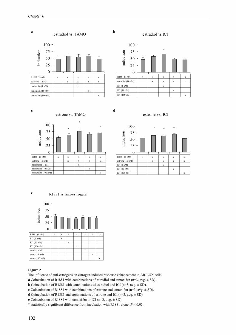
Chapter 6
102
Figure 2 The influence of anti-estrogens on estrogen-induced response enhancement in AR-LUX cells. a Coincubation of R1881 with combinations of estradiol and tamoxifen (n=3, avg. ± SD). b Coincubation of R1881 with combinations of estradiol and ICI (n=3, avg. ± SD). c Coincubation of R1881 with combinations of estrone and tamoxifen (n=3, avg. ± SD). d Coincubation of R1881 and combinations of estrone and ICI (n=3, avg. ± SD). e Coincubation of R1881 with tamoxifen or ICI (n=3, avg. ± SD). * statistically significant difference from incubation with R1881 alone; P < 0.05.
estradiol vs. TAMO
0
25
50
75
100in
duct
ion
xtamoxifen (100 nM)
xtamoxifen (10 nM)
xtamoxifen (1 nM)
xxxxestradiol (1 nM)
xxxxxR1881 (1 nM)
estradiol vs ICI
0
25
50
75
100
indu
ctio
n
xICI (100 nM)
xICI (10 nM)
xICI (1 nM)
xxxxestradiol (10 nM)
xxxxxR1881 (1 nM)
*
R1881 vs. anti-estrogens
0
25
50
75
100
indu
ctio
n
xtamo (100 nM)xtamo (10 nM)
xtamo (1 nM)xICI (100 nM)
xICI (10 nM)
xICI (1 nM)xxxxxxxR1881 (1 nM)
oestrone vs. ICI
0
25
50
75
100in
duct
ion
* * *
xICI (100 nM)xICI (10 nM)
xICI (1 nM)
xxxxestrone (10 nM)xxxxxR1881 (1 nM)
estrone vs. ICI estrone vs. TAMO
0
25
50
75
100
indu
ctio
n *
**
xtamoxifen (100 nM)
xtamoxifen (10 nM)xtamoxifen (1 nM)
xxxxestrone (10 nM)
xxxxxR1881 (1 nM)
c d
e
a b

Chapter 6
103
Figure 3 The influence of anti-estrogens on estrone-induced enhancement of PB-ARE2-controlled transcriptional activation. a Coincubation of nortestosterone with (combinations) of estrone (10 nM) and tamoxifen (100 nM) or ICI (100 nM) (n=3, avg. ± SD). b Coincubation of methyltestosterone with (combinations) of estrone (10 nM) and tamoxifen (100 nM) or ICI (100 nM) (n=3, avg. ± SD).
binding sites in AR-LUX cells whereas R1881 turned out to be highly potent in decreasing its own binding sites. Coincubation of E2 and R1881 partly reversed the effect of R1881 and the addition of ICI again reversed the effect of E2, as expected. Incubation of cells with only ICI also significantly reduced the number of available R1881 binding sites, suggesting that ICI exhibits additional properties besides competing for the ER-LBD with estrogens. Not only does ICI influence the number of binding sites for R1881, but it competes for these sites as well, although no complete blocking of R1881 binding is observed (Fig 4c).
Since R1881 is not only an AR ligand but is also binding to the PR, the observed competition for R1881-binding sites may also include competition for R1881-binding sites on the PR. In contrast to ICI, the established PR and AR antagonist RU486 is able to completely block R1881 binding to specific sites (Fig. 4c), which would be consistent with the explanation that only RU486 is an efficient competitor for R1881-binding sites on both the PR and the AR, whereas ICI is less efficient. Coincubation in the AR-LUX assay with ICI at a concentration that does not significantly compete for R1881-binding sites results in a shift of the R1881 dose response curve towards a slightly higher EC50 value, thus suggesting that mechanisms other than competition for R1881 binding sites play a role in this effect (Fig 4d).
Knock-down of AR reveals a role for other steroid hormone receptors in transcription activation through PB-ARE2
Thus far, we have shown that ICI modulates estrogen enhancement of PB-ARE2-driven gene expression as well as R1881-binding sites. This suggests additional mechanisms besides straightforward activation of AR inducing PB-ARE2-mediated gene expression. This issue cannot be resolved by using steroid receptor (ant)agonists due to promiscuous binding to
nortestosterone (nM)10-1 100 101 102 103 104
indu
ctio
n
0
20
40
60
80
norT norT/esnorT/tamonorT/es/tamonorT/es/ICInorT/ICI
EC50 stdev.norT 17.6 6.0norT/es 9.4 1.8norT/tamo 12.2 2.8norT/es/tamo 8.4 1.3norT/es/ICI 14.2 1.2norT/ICI 12.9 2.0
methyltestosterone (nM)10-1 100 101 102 103 104
indu
ctio
n
0
15
30
45
60
MeTMeT/es MeT/tamo MeT/es/tamo MeT/es/ICI MeT/ICI
EC50 stdev.meT 131.6 43.7meT/es 115.4 23.6meT/tamo 154.0 48.1meT/es/tamo 111.1 35.0meT/es/ICI 152.6 14.3meT/ICI 129.0 23.7
a b

Chapter 6
104
Figure 4 Additional mechanisms involved in the inhibitory behavior of ICI on estrogen-induced response enhancement in AR-LUX cells. a Luciferase induction in AR-LUX cells dosed with R1881 (1 nM) in combinations with estradiol and ICI (n=3, avg. ± SD). b 3H-R1881 binding assay following coinbutations with combinations of R1881, estradiol and ICI (n=3, avg. ± SD) as shown in panel a. c Cell-based competitive AR binding. AR-LUX cells were coincubated with 1 nM 3H-R1881 and an increasing concentration of ICI or RU486 (n=3, avg. ± SD). d Coincubation of R1881 with ICI (100 nM) in the AR-LUX assay; EC50 R1881= 0.13 nM ± 0.05, EC50 R1881 + ICI =0.22 nM ± 0.026 (n=3, avg. ± SD).
0
5
10
15
20
blank 1 nM E2 100 nM ICI 1 nm R 1 nM R + 1nM E2
1 nM R + 1nM E2 + 100
nM ICI
indu
ctio
n
1 nM R+ 1 nM E2
1 nM R+ 1 nM E2+ 100 nM ICI
b
a
0.0 2.5 5.0 7.5 10.00
5000
10000
1 nM R1881
1 nM E2
1 nM R + 1 nM E2
100 nM ICI
1 nM R + 1 E2 + 100 nM ICI
blank
3H-R1881 (nM)
coun
ts
0.0 2.5 5.0 7.5 10.00
1000
2000
1 nM R
1 nM R+ 1 nME2
1 nM R+ 1 nM E2+ 100 nM ICI
3H-R1881 (nM)
coun
ts
-5 -4 -3 -2 -1 0 1 20
2500
5000
7500
10000 ICIRU486
log10 [uM]
coun
ts
R1881 (nM)0.01 0.1 1 10
indu
ctio
n
0
5
10
R1881 [+] 100 nM ICI 182.780
c d

Chapter 6
105
a b
0
0.0005
0.001
0.0015
AR-LUX ARdown-LUX
AR
/act
in
0
10
20
30
40
blank
1 nM R18
81
1 nM R18
81 + 10
uM O
H-F
1 nM R18
81 + 5
uM CPA
300 pM
R5020
30 nM no
rT
300 nM
17B-bold
300 nM
DHT
10 uM TA
10 nM 17
B-trenb
AR-LUXARdown-LUX
various receptors of virtually all steroid receptor ligands. Therefore we decided to knock down AR expression to more extensively investigate the role of the androgen receptor in the observed effects on gene expression. A stable androgen receptor knock-down clone of the original AR-LUX cell line was constructed utilising small interfering RNA (siRNA). This ARdown-LUX variant only expresses approximately 6% of the amount of AR-mRNA compared to the level in wildtype T47D-AR-LUX cells, as determined by quantitative RT-PCR (Fig. 5a). Upon dosing these ARdown-LUX cells with various androgen and progesterone (ant)agonists, substantial differences in responses compared to the wild-type AR-LUX cells were observed. Most significant was the observation that the ARdown-LUX cells could not be induced by DHT, an established androgen, and 17β-boldenone. R1881, a strong AR and PR agonist, produced a substantial response in both cell lines, which was partly antagonised by hydroxy-flutamide. These observations suggest that the R1881 induces a response in the ARdown-LUX via a combination of residual AR as well as the PR. The possible role of the PR in the ARdown-LUX was further investigated by incubations with cyproterone-acetate, both an AR antagonist and a PR-agonist. The results show that CPA did not antagonise the response of the ARdown-LUX cells to R1881, whereas it did so in wild type AR-LUX cells. This indicates that R1881-induced PB-ARE2-mediated gene expression in the ARdown cell line is primarily mediated by the PR. Promegestone (R5020), an established specific progesterone receptor agonist is also active in both variants. The substantial response generated in the ARdown-LUX cells again suggests involvement of the progesterone receptor in this cell line. The compounds norT and 17β-trenbolone, generally considered as being specific androgen receptor agonists, also showed activity in both cell lines suggesting they might exert additional effects besides activating the AR. The response to these compounds was indeed lower, yet still clearly present in the ARdown-LUX cells.
In conclusion, these results confirm the involvement of the AR in PB-ARE2-mediated luciferase expression and suggest that PR (isoforms) might also exert an influence on transcription activation through this probasin element.
Figure 5 Characterisation of role of the androgen receptor in the observed effects on gene expression in AR-LUX cells. a Quantitative RT-PCR on androgen receptor mRNA in AR-LUX cells and ARdown-LUX cells, y-axis represents AR expression normalised for β-actin expression (n=2, avg. ± SD). b Response induced in AR-LUX and ARdown-LUX cells dosed with various androgen and progesterone (ant)agonists (17β-bold = 17β-boldenone, 17β-trenb = 17β-trenbolone, n=3, avg. ± SD).
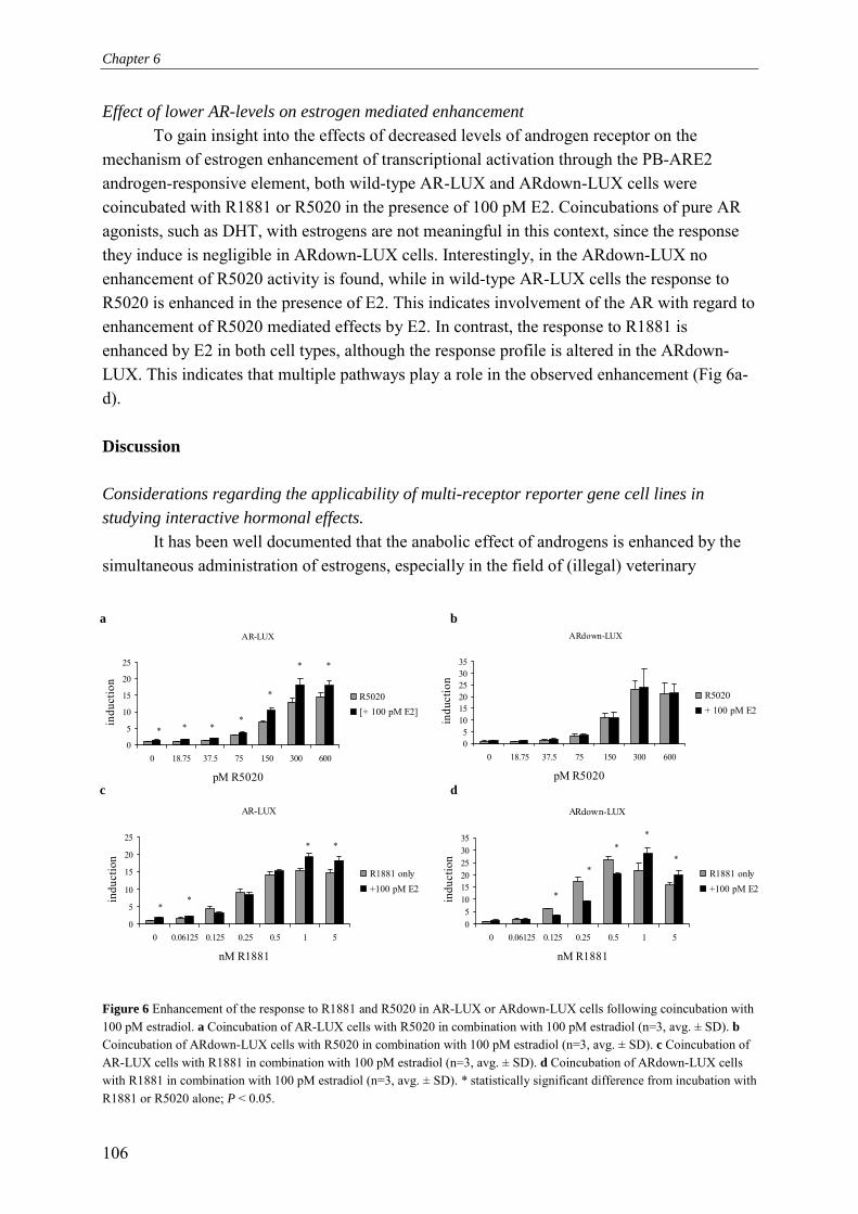
Chapter 6
106
Effect of lower AR-levels on estrogen mediated enhancement To gain insight into the effects of decreased levels of androgen receptor on the mechanism of estrogen enhancement of transcriptional activation through the PB-ARE2 androgen-responsive element, both wild-type AR-LUX and ARdown-LUX cells were coincubated with R1881 or R5020 in the presence of 100 pM E2. Coincubations of pure AR agonists, such as DHT, with estrogens are not meaningful in this context, since the response they induce is negligible in ARdown-LUX cells. Interestingly, in the ARdown-LUX no enhancement of R5020 activity is found, while in wild-type AR-LUX cells the response to R5020 is enhanced in the presence of E2. This indicates involvement of the AR with regard to enhancement of R5020 mediated effects by E2. In contrast, the response to R1881 is enhanced by E2 in both cell types, although the response profile is altered in the ARdown-LUX. This indicates that multiple pathways play a role in the observed enhancement (Fig 6a-d). Discussion Considerations regarding the applicability of multi-receptor reporter gene cell lines in studying interactive hormonal effects.
It has been well documented that the anabolic effect of androgens is enhanced by the simultaneous administration of estrogens, especially in the field of (illegal) veterinary
Figure 6 Enhancement of the response to R1881 and R5020 in AR-LUX or ARdown-LUX cells following coincubation with 100 pM estradiol. a Coincubation of AR-LUX cells with R5020 in combination with 100 pM estradiol (n=3, avg. ± SD). b Coincubation of ARdown-LUX cells with R5020 in combination with 100 pM estradiol (n=3, avg. ± SD). c Coincubation of AR-LUX cells with R1881 in combination with 100 pM estradiol (n=3, avg. ± SD). d Coincubation of ARdown-LUX cells with R1881 in combination with 100 pM estradiol (n=3, avg. ± SD). * statistically significant difference from incubation with R1881 or R5020 alone; P < 0.05.
ARdown-LUX
05
101520253035
0 18.75 37.5 75 150 300 600
pM R5020
indu
ctio
n
R5020+ 100 pM E2
AR-LUX
0
5
10
15
20
25
0 18.75 37.5 75 150 300 600
pM R5020
indu
ctio
n
R5020[+ 100 pM E2]
*
***
**
*
AR-LUX
0
5
10
15
20
25
0 0.06125 0.125 0.25 0.5 1 5
nM R1881
indu
ctio
n
R1881 only+100 pM E2
*
*
*
*
ARdown-LUX
05
101520253035
0 0.06125 0.125 0.25 0.5 1 5
nM R1881
indu
ctio
n
R1881 only+100 pM E2
*
**
*
*
a b
c d

Chapter 6
107
applications of growth promoters to achieve repartitioning of body mass distribution towards increased muscle formation (Galbraith and Topps, 1981; Lone, 1997). In this study we initially investigated the capability of the AR-LUX, a reporter gene cell line for androgenic activity, to reflect the estrogen enhancement observed in vivo. The AR-LUX is based on PB-ARE2, an authentic mammalian androgen-responsive element, to achieve luciferase reporter gene expression reflecting androgen-responsive element-mediated gene expression modulation. Furthermore, the AR-LUX utilises a cell line showing endogenous expression of various steroid hormone receptors and associated co-factors, in addition to a functional androgen receptor signal transduction pathway. This enables the assay to reflect relevant (multi-receptor-mediated) interactive effects complementary to AR-mediated transcriptional activation.
The classical approach in the design of reporter gene assays for steroid hormone receptor-mediated gene expression is to construct recombinant cellular systems that indicate activation of the transactivation function of a single receptor, thereby allowing dissection of the effects of hormonal compounds into the contributions of singular receptors. However, multiple receptors can contribute to the activation of transcription through a certain responsive element, as is clearly the case for the PB-ARE2 element. The ultimate biological effect in such settings is determined by the combination of the presence of the receptors and the set of genes under control of the particular type of response element, instead of by activation of the receptor alone. Therefore, as far as PB-ARE2, and perhaps also other, similar androgen-responsive elements are concerned, we adhere to the point of view that a complicated web of interactions between steroid receptors (and possibly their co-factors as well) mediate androgenic and androgen-induced anabolic effects, and that these interactions are highly relevant. The AR-LUX assay appears to reliably reflect at least part of these relevant interactions. In addition to the detection of interactive effects, the AR-LUX allowed elucidation of some mechanistic aspects of the interaction, such as the ER-mediated nature of the estrogen enhancement. Our view with respect to the reliability of the AR-LUX to study interactive mechanisms governing ARE-mediated gene expression is confirmed by the modulating effects of (combinations of) estrogens and anti-estrogens on the AR-LUX response. These were in agreement with the synergistic effects between androgens and estrogens observed in the practice of cattle breeding (Galbraith and Topps, 1981; Kreikemeier and Unruh, 1997), and were found to be consistent with the literature reports on these interactions (see below). Enhancement of androgen-induced responses by estrogens is reflected by the AR-LUX
In the AR-LUX assay, the response to various androgenic/anabolic compounds is enhanced by the estrogens estradiol and estrone. Interestingly, we observed that the enhancement of the effects of R1881 by both estrone and estradiol decreased at high concentrations of estrogens. This is in agreement with Kumar et al. who observed that higher E2 concentrations result in a decrease of the enhancement of the effects of the pure androgen receptor agonist DHT (Kumar et al., 1994). A possible explanation for this phenomenon might be that at high concentrations, estrogens will exert antagonistic activity by binding to or

Chapter 6
108
competing for binding sites other than those on the ER (Tindall et al., 1981; Murthy et al., 1984; Blankvoort et al., 2001), and this may involve co-factor-dependent pathways as well (Yeh et al., 1998).
Differences in enhancement levels were observed for methyltestosterone (meT) and nortestosterone (norT) following coincubation with estrone. This is in agreement with the varying degrees of estrogen enhancement of the growth promoting effects of different androgens observed in cattle treated with various steroid hormone cocktails (Galbraith and Topps, 1981; Kreikemeier and Unruh, 1997; Meyer, 2001).
Our results show that the estrogen estrone exerts a more pronounced effect on norT-induced luciferase expression than on that induced by meT. In contrast to meT, norT can be extensively metabolised into progestagenic and estrogenic compounds, subsequently leading to the activation of additional biochemical pathways besides that mediated by the AR. Therefore, we speculate that the more pronounced effects of estrone in combination with norT are due to estrone not only influencing the pathway mediated by the AR but also additional pathways induced by norT metabolites (Traish et al., 1986; Sundaram et al., 1995; Markiewicz and Gurpide, 1997; Mor et al., 2001).
The role of the estrogen receptor and its (ant)agonists in enhancement of androgen response element-mediated transcription
The results obtained with the ER antagonists ICI and tamoxifen further contributed to the notion of the complexity of the functional interactions between steroid receptors in androgen-induced gene expression. Coincubations with tamoxifen and ICI confirmed the predicted role of the ER in the observed enhancement of PB-ARE2-mediated gene expression. Remarkably, both anti-estrogens in the absence of estrogens seemed to enhance the effects of norT, while this was not observed for meT. It can be envisioned that these responses of anti-estrogens combined with norT may be the result of functional interactions between the biochemical pathways in which the AR, ER and PR are involved, of which the latter two are induced by metabolites of norT as hypothesised above. The results obtained with the AR-LUX provide indications that these interactions affect androgen-response element-driven gene expression. Our observations emphasise the complexity of multiple steroid receptor pathways, of which the molecular mechanisms remain to be established.
A second intriguing finding is that tamoxifen clearly gave rise to a further induction of estrone-enhanced androgen mediated gene expression, whereas it seemed to antagonise the enhancing effects of estradiol. The cause of these apparently contradictory results may be that estrogen receptors bound to either E2 or estrone recruit different co-factors (Margeat et al., 2003). We hypothesise that the two different co-factor-ER-estrogen complexes respond differentially to tamoxifen, favouring an increase of the ER-agonistic versus antagonistic properties of tamoxifen in the case of estrone (Gee et al., 1999; Takimoto et al., 1999; Margeat et al., 2003).
Although ICI has been reported to be an ER-antagonist, our results indicate that the effects of ICI in the AR-LUX are only partly explained by a direct binding of ICI to the ER. Enhancement of progesterone receptor-mediated effects by low concentrations of ICI

Chapter 6
109
followed by antagonistic effects upon using higher concentrations has been reported before (Nawaz et al., 1999), similar to our observations concerning the inhibition of estrone enhancement in the AR-LUX. At 1 nM, ICI is probably not able to completely displace E2 from the ER, since an IC50 of 2.4 nM was reported in the presence of 1 nM E2 for binding to the ER (Blair et al., 2000). Higher ICI concentrations might however have resulted in total displacement of E2 from the ER, thereby relieving its enhancing effects on androgens. Furthermore, ICI is probably not only an antagonist competing for ER binding sites. It has been reported to induce gene expression in both in vitro and in vivo models (Nawaz et al., 1999; Robertson et al., 2001; Hyder and Stancel, 2002; Ni et al., 2002). Indeed, our 3H-R1881 binding assay following preincubation with ICI showed that this anti-estrogen down-regulates the number of available binding sites for R1881. Additionally, we showed that ICI binds to R1881-binding sites, but not at a concentration of 100 nM. This suggests, that the shift observed in the dose-response curve for R1881 following coincubation with 100 nM ICI is due to a mechanism affecting luciferase expression in the AR-LUX other than direct competition for R1881 binding sites. In conclusion, the effects of ICI on the AR-LUX most likely include a direct competition with estrogens and other compounds such as R1881. Additionally, yet to be defined mechanisms may also play a role.
Complex interactions are involved in enhancement of androgen response element mediated gene expression
In our ARdown-LUX DHT and β-boldenone were shown to be specific androgen receptor ligands while R1881, nortestosterone and 17β-trenbolone might be mixed ligands for AR and PR as suggested by their ability to activate luciferase expression in ARdown-LUX cells.
The observed complexity of the enhancement of androgenic effects by anti-estrogens in the AR-LUX is further complicated by our observations made with compounds believed to bind exclusively to the PR. The specific progestagen promegestone (R5020) induces luciferase expression in the AR-LUX at picomolar levels whereas triamcinolone, a compound that does not bind to the AR but instead displays affinity for PR and GR (Murthy et al., 1984), activated luciferase expression at µM concentrations.
Chalbos et al. suggested that low concentrations of R5020 induce specific proteins in T47D cells via the AR, although they could not exclude that the observed effects were mediated via the PR (Chalbos et al., 1987). Furthermore, the binding of R5020 to baboon AR is only 3.1% compared to binding of R1881 (Lin et al., 1981) and R5020 is able to slightly activate an MMTV-LTR-luc reporter plasmid cotransfected with human wild-type AR in CV1 cells (at 1 µM R5020) (Poujol et al., 2000). In contrast, the low concentrations of R5020 needed to elicit a profound luciferase induction in both AR-LUX and ARdown-LUX cells strongly suggest a significant progesterone receptor-mediated effect in these two cell lines. Schoenmakers et al. (Schoenmakers et al., 1999) showed that transient cotransfection of COS cells (that are devoid of androgen receptors) with PR-β and a reporter plasmid containing luciferase under transcriptional control of a PB-ARE2 element did not attain the maximal luciferase induction as obtained by cotransfection with the AR. Two isoforms of PR exist

Chapter 6
110
however, and T47D-cells express both the PR-α and PR-β isoform (Richer et al., 2002). Therefore our observations lead us towards hypothesising that the PR-α is in fact the isoform which is significantly, or perhaps even preferentially contributing to PR-mediated transcriptional activation through the PB-ARE2 element. Since cocktails containing progestagens and estrogens have been shown to be effective modulators of animal growth, this would explain part of the increased effectiveness of treating animals with these cocktails. It will be challenging to test whether PR-α truly has a prominent role in mediating anabolic effects.
In conclusion, the interactive enhancing effects of combinations of cocktails compared to the effects of single hormones, commonly observed in in vivo situations such as cattle breeding are also reflected in the response of the AR-LUX reporter gene assay applied to study their PB-ARE2-mediated effects. To our knowledge this is one of the few reports describing such effects. This emphasises the additional value, besides being a detection and quantitation tool, that carefully designed reporter gene assays for androgenic activity can provide, when based on an authentic androgen-responsive element and expressing multiple endogenously regulated receptors. Our results illustrate that the AR-LUX and similar reporter gene systems present useful tools not only for screening purposes but also in research aimed at elucidating the underlying pathways that mediate androgen-responsive element-controlled biological effects, such as anabolic growth enhancement by cocktails of hormonal compounds. References Blair, R. M., H. Fang, W. S. Branham, B. S. Hass, S. L. Dial, C. L. Moland, W. Tong, L. Shi,
R. Perkins and D. M. Sheehan (2000). "The estrogen receptor relative binding affinities of 188 natural and xenochemicals: structural diversity of ligands." Toxicol Sci 54(1): 138-153.
Blankvoort, B. M., E. M. de Groene, A. P. van Meeteren-Kreikamp, R. F. Witkamp, R. J. Rodenburg and J. M. Aarts (2001). "Development of an androgen reporter gene assay (AR-LUX) utilizing a human cell line with an endogenously regulated androgen receptor." Anal Biochem 298(1): 93-102.
Brummelkamp, T. R., R. Bernards and R. Agami (2002). "A system for stable expression of short interfering RNAs in mammalian cells." Science 296(5567): 550-553.
Chalbos, D., D. Haagensen, T. Parish and H. Rochefort (1987). "Identification and androgen regulation of two proteins released by T47D human breast cancer cells." Cancer Res 47(11): 2787-2792.
Claessens, F., G. Verrijdt, E. Schoenmakers, A. Haelens, B. Peeters, G. Verhoeven and W. Rombauts (2001). "Selective DNA binding by the androgen receptor as a mechanism for hormone-specific gene regulation." J Steroid Biochem Mol Biol 76(1-5): 23-30.
DesJardins, M. (2002). "Supplement use in the adolescent athlete." Curr Sports Med Rep 1(6): 369-373.
Doesburg, P., C. W. Kuil, C. A. Berrevoets, K. Steketee, P. W. Faber, E. Mulder, A. O. Brinkmann and J. Trapman (1997). "Functional in vivo interaction between the amino-

Chapter 6
111
terminal, transactivation domain and the ligand binding domain of the androgen receptor." Biochemistry. 36: 1052-1064.
Galbraith, H. and J. H. Topps (1981). "Effect of hormones on the growth and body composition of animals." Nutrition abstracts and Reviews - series B 51(8): 521-539.
Gee, A. C., K. E. Carlson, P. G. Martini, B. S. Katzenellenbogen and J. A. Katzenellenbogen (1999). "Coactivator peptides have a differential stabilizing effect on the binding of estrogens and antiestrogens with the estrogen receptor." Mol Endocrinol 13(11): 1912-1923.
Horwitz, K. B. and W. L. McGuire (1978). "Estrogen control of progesterone receptor in human breast cancer: Correlation with nuclear processing of estrogen receptor." J. Biol. Chem. 253: 2222-2228.
Hyder, S. M. and G. M. Stancel (2002). "Inhibition of progesterone-induced VEGF production in human breast cancer cells by the pure antiestrogen ICI 182,780." Cancer Lett 181(1): 47-53.
Kreikemeier, K. and J. Unruh (1997). "Feedlot Performance and Carcass Characteristics of Heifers implanted with component E-H and component T-H or Finaplex-H" Cattle feeders' day '97, Kansas, USA, Kansas State University.
Kumar, M. V., M. E. Leo and D. J. Tindall (1994). "Modulation of androgen receptor transcriptional activity by the estrogen receptor." J.Androl. 15: 534-542.
Laure, P., C. Binsinger and T. Lecerf (2003). "General practitioners and doping in sport: attitudes and experience." Br J Sports Med 37(4): 335-338; discussion 338.
Lin, A. L., H. C. McGill, Jr. and S. A. Shain (1981). "Hormone receptors of the baboon cardiovascular system. Biochemical characterization of myocardial cytoplasmic androgen receptors." Circ Res 49(4): 1010-1016.
Lone, K. P. (1997). "Natural sex steroids and their xenobiotic analogs in animal production: growth, carcass quality, pharmacokinetics, metabolism, mode of action, residues, methods, and epidemiology." Crit.Rev.Food Sci.Nutr. 37(2): 93-209.
Margeat, E., A. Bourdoncle, R. Margueron, N. Poujol, V. Cavailles and C. Royer (2003). "Ligands differentially modulate the protein interactions of the human estrogen receptors alpha and beta." J Mol Biol 326(1): 77-92.
Markiewicz, L. and E. Gurpide (1997). "Estrogenic and progestagenic activities of physiologic and synthetic androgens, as measured by in vitro bioassays." Methods.Find.Exp.Clin.Pharmacol. 19: 215-222.
Meyer, H. H. (2001). "Biochemistry and physiology of anabolic hormones used for improvement of meat production." Apmis 109(1): 1-8.
Mor, G., M. Eliza, J. Song, B. Wiita, S. Chen and F. Naftolin (2001). "17alpha-methyl testosterone is a competitive inhibitor of aromatase activity in Jar choriocarcinoma cells and macrophage-like THP-1 cells in culture." J Steroid Biochem Mol Biol 79(1-5): 239-246.
Murthy, L. R., C. H. Chang, D. R. Rowley, P. T. Scardino and D. J. Tindall (1984). "Physicochemical characterization of the androgen receptor from hyperplastic human prostate." Prostate 5(6): 567-579.
Chapter 6
112
Nawaz, Z., G. M. Stancel and S. M. Hyder (1999). "The pure antiestrogen ICI 182,780 inhibits progestin-induced transcription." Cancer Res 59(2): 372-376.
Ni, X., R. C. Nicholson, B. R. King, E.-C. Chan, M. A. Read and R. Smith (2002). "Estrogen Represses whereas the Estrogen-Antagonist ICI 182780 Stimulates Placental CRH Gene Expression." J Clin Endocrinol Metab 87(8): 3774-3778.
Poujol, N., J.-M. Wurtz, B. Tahiri, S. Lumbroso, J.-C. Nicolas, D. Moras and C. Sultan (2000). "Specific recognition of androgens by their nuclear receptor: a structure-function study." J. Biol. Chem.: 24022-24031.
Prendergast, H. M., T. Bannen, T. B. Erickson and K. R. Honore (2003). "The toxic torch of the modern Olympic Games." Vet Hum Toxicol 45(2): 97-102.
Richer, J. K., B. M. Jacobsen, N. G. Manning, M. G. Abel, D. M. Wolf and K. B. Horwitz (2002). "Differential Gene Regulation by the Two Progesterone Receptor Isoforms in Human Breast Cancer Cells." J. Biol. Chem. 277(7): 5209-5218.
Robertson, J. A., Y. Zhang and N. H. Ing (2001). "ICI 182,780 acts as a partial agonist and antagonist of estradiol effects in specific cells of the sheep uterus." J Steroid Biochem Mol Biol 77(4-5): 281-287.
Roy, A. K., Y. Lavrovsky, C. S. Song, S. Chen, M. H. Jung, N. K. Velu, B. Y. Bi and B. Chatterjee (1999). "Regulation of androgen action." Vitam.Horm. 55: 309-352.
Schoenmakers, E., P. Alen, G. Verrijdt, B. Peeters, G. Verhoeven, W. Rombauts and F. Claessens (1999). "Differential DNA binding by the androgen and glucocorticoid receptors involves the second Zn-finger and a C-terminal extension of the DNA-binding domains." Biochem J 341: 515-521.
Sundaram, K., N. Kumar, C. Monder and C. W. Bardin (1995). "Different patterns of metabolism determine the relative anabolic activity of 19-norandrogens." J Steroid Biochem Mol Biol 53(1-6): 253-257.
Takimoto, G. S., J. D. Graham, T. A. Jackson, L. Tung, R. L. Powell, L. D. Horwitz and K. B. Horwitz (1999). "Tamoxifen resistant breast cancer: coregulators determine the direction of transcription by antagonist-occupied steroid receptors." Journal of Steroid Biochemistry and Molecular Biology 69: 45-50.
Tindall, D. J., F. S. French and S. N. Nayfeh (1981). "Estradiol-17 beta inhibition of androgen uptake, metabolism and binding in epididymis of adult male rats in vivo: a comparison with cyproterone acetate." Steroids 37(3): 257-268.
Traish, A. M., D. F. Williams and H. H. Wotiz (1986). "Binding of (3H)7 alpha,17 alpha-dimethyl-19-nortestosterone (mibolerone) to progesterone receptors: comparison with binding of (3H)R5020 and (3H)ORG2058." Steroids 47(2-3): 157-173.
Wilson, L. L., J. L. Smith, D. L. Swanson and E. W. Mills (1999). "Implant sequence effects in intact male Holstein veal calves: carcass characteristics." J Anim Sci 77(12): 3133-3139.
Wong, C., W. R. Kelce, M. Sar and E. M. Wilson (1995). "Androgen receptor antagonist versus agonist activities of the fungicide vinclozolin relative to hydroxyflutamide." J Biol Chem 270(34): 19998-20003.
Chapter 6
113
Yeh, S., H. Miyamoto, H. Shima and C. Chang (1998). "From estrogen to androgen receptor: a new pathway for sex hormones in prostate." Proc.Natl.Acad.Sci.U.S.A. 95: 5527-5532.

114

77
Summary & Concluding remarks
Chapter 7
116
Interactive biological effects of different steroid receptors and their accessory pathways in the context of normal endocrine regulation, anabolic cocktails and mixtures of compounds present in the environment have been frequently observed (Galbraith and Topps, 1981; Nazareth and Weigel, 1996; Sharpe, 1998; Sonnenschein and Soto, 1998; Simon, 2001). These interactions can result in effects quite different from those occurring as a consequence of exposure to single compounds. Effects of steroids can be studied using bioassays. Most currently available in vitro bioassays focus on elucidating the effects mediated via a single receptor. Therefore, development of bioassays incorporating the effects of multiple cellular pathways will provide valuable additional tools in research aimed at interactive effects.
The research described in this thesis was aimed at developing an in vitro cell based reporter gene assay capable of detecting the presence of compounds activating gene expression via an androgen response element. This type of androgen reporter system that is sensitive to the activation of multiple pathways has not been described yet.
Following construction of this bioassay, the presence of potential endocrine disrupters in environmental samples collected from surface waters and sediments was investigated. Furthermore, possible application of the bioassay for detecting the illegal use of androgenic growth promoters in cattle was investigated by screening urine of treated animals. A second important goal of the research described in this thesis was to gain insights into the mechanisms underlying the interactive effects of growth promoting cocktails containing androgenic anabolics. Summary of results obtained with the endogenous Androgen Receptor-mediated LUciferase eXpression assay (AR-LUX).
Chapter 2 describes the methods applied to and considerations involved in the creation of an androgen-responsive reporter gene assay. In addition, methods and results regarding RNA interference applied to gain further insight into the role of the androgen and progesterone receptor in the AR-LUX assay are presented. Application of micro arrays for the elucidation of interactive effects between testosterone and estradiol in a L6 rat myoblast derived myofiber model is also described in this chapter.
The validation of the endogenous Androgen Receptor-mediated LUciferase eXpression assay (AR-LUX) based on the probasin androgen response element 2 (PB-ARE2) is described in chapter 3. The inducibility of luciferase expression by androgens was confirmed by the results obtained with methyltrienolone (R1881) and 5α-di-hydro-testosterone (DHT). Coincubations of androgens with established pharmaceutical and environmentally occurring anti-androgens showed that the luciferase expression in the AR-LUX was mediated by the androgen receptor. Luciferase expression was also observed after dosing the AR-LUX cells with steroids that are known as agonists of other members of the steroid receptor family, including progesterone (PR), 17β-estradiol (ER), dexamethasone (GR) and d-aldosterone (MR). However, this activation could be counteracted by coincubation with anti-androgens; suggesting promiscuous binding and activation of the AR by established ligands of other steroid receptors. As intended, the AR-LUX assay incorporates

Chapter 7
117
the influence of various pathways on PB-ARE2 mediated gene expression as exemplified by the influence exerted by all-trans-retinoic acid (atRA), vitamin D, epigallocatechin gallate (EGCG), and forskolin, compounds that do not directly interact with the androgen receptor.
Subsequently, the androgenic activity of a number of aquatic environmental samples was determined. The majority of these samples contain estrogens whereas 9 out of 22 extracts contained varying concentrations of androgens. However, in 2 samples containing androgens, interactive mixture effects were observed, which were probably due to interactions as a consequence of estrogen receptor activation (chapter 4). This was confirmed by collecting dose response curves obtained by coincubations of androgens and estrogens. From this experiment, it was concluded that the response of the AR-LUX to the androgens was enhanced by the estrogens, emphasising the potential importance of mixture effects in environmental samples. Also an interactive effect on PB-ARE2 mediated luciferase expression was observed by tributyltin-hydride, a frequently found environmental pollutant of which the endocrine disrupting mechanism is still largely unknown (Tillmann et al., 2001; Verslycke et al., 2003).
The possible contribution of the AR-LUX assay system towards the continuing screening effort for illegal anabolic growth promoters in cattle is described in chapter 5. The feasibility of positive identification of hormonally treated animals by screening collected urines was established. Furthermore, the results obtained with the AR-LUX assay were similar to those of GC-MS analysis. Both techniques should be regarded as complementary rather than interchangeable due to the fundamental differences between determining the biological activity of (a mixture of) compounds and measuring the concentrations of certain compounds. To emphasise the additional value of the AR-LUX in the screening for the use of illegal growth promoters, AR-LUX cells were dosed with confiscated liquid samples suspected to have been used as growth promoters in cattle. In spite of the fact that GC-MS analysis had previously been unable to detect the presence of any anabolic compounds, the samples were found to be potent activators of luciferase expression. This suggests the presence of either a mixture of related compounds or new unknown compounds in the confiscated samples.
Finally, chapter 6 describes investigations into the interactive effects of estrogens and progestagens. The AR-LUX cell line is capable of providing insights into multi-receptor interactions ultimately resulting in activation of an androgen response element (PB-ARE2). Responses to R1881 and DHT were enhanced by the estrogens estradiol and estrone. Enhancement could be reversed with the anti-estrogens tamoxifen and ICI, although differences were observed between the two ER-antagonists. These differences were probably a consequence of structural differences of the diverse applied steroids resulting in alternate receptor conformations and co-factor recruitment. The interactive effects observed were in agreement with interactive effects (both in vivo and in vitro) reported in the literature. In addition, it was observed that ER-antagonists exert interactive effects without the presence of estrogens. The observed effects differ upon coincubation with methyltestosterone and nortestosterone (norT), with strongest effects being found upon coincubation of anti-estrogens with norT. This is yet another manifestation of the complexity of the interactive effects
Chapter 7
118
observed. Furthermore, a possible direct role of the progesterone receptor-α in PB-ARE2-mediated gene expression was hypothesised based on results observed with an androgen receptor knock down variant of the AR-LUX cell line. DHT was unable to activate luciferase expression in the ARdown-LUX cells, confirming the role of the androgen receptor in PB-ARE2 mediated gene expression. However, promegestone, an established PR agonist, was found to activate luciferase expression in the AR knock down clone. This strongly suggests a role for the progesterone receptor or one of its isoforms in PB-ARE2 mediated gene expression. These results emphasise the additional value reporter gene assays featuring multiple receptors might provide in research aimed at elucidating interactive effects or identifying compounds displaying effects via multiple proteins. However, these results also underline the inherent differences between assays aimed at one receptor mediated pathway versus assays aimed at incorporating the influence of multiple pathways.
Environmental aspects of hormonally active compounds
Results obtained in chapter 4 do not indicate a wide spread presence of androgen response element (ARE) activating compounds. Antagonists blocking ARE mediated effects were not observed. However, a number of samples did contain varying amounts of compounds activating luciferase expression via the PB-ARE2. Furthermore, the occurrence of interactive effects is an important find emphasising that risk evaluations of these mixtures will come with potential pitfalls if no research is dedicated towards establishing the occurrence of interactive effects. In this respect the transgenic zebra fish assay for detecting estrogens as described by Legler et al. provides a valuable tool incorporating the interactive effects in a complete organism (Legler et al., 2002) and the development of these types of assays next to in vitro assays such as the AR-LUX should be encouraged, obviously encompassing the issue of animal welfare.
The issue of illegal use of anabolic growth promoters The AR-LUX assay can be applied in the continuing screening effort for illegal anabolic growth promoters in cattle. The urine samples analysed with the AR-LUX assay were collected in controlled animal experiments. Therefore only the possibility of applying the AR-LUX for screening of anabolic androgenic compounds was investigated. However the observed activity of confiscated liquids is worrying and warrants further research into the elucidation of the compounds present. As yet, the exact composition of the liquids is still unknown. A similar case was reported for unknown β-agonists whose structure was eventually resolved (Nielen et al., 2003). These findings emphasise the important additional value reporter gene assays offer. Therefore, in any screening effort aimed at compounds activating biological pathways bioassays should be applied next to chemical analysis whenever possible. The confiscation of these liquids also indicates that the issue of illegal anabolic growth promoters will need continuing attention of the scientific community and regulating authorities.
Chapter 7
119
Implications of the interactive effects observed with the AR-LUX reporter gene assay The AR-LUX assay was designed to incorporate the effects of multiple cellular
pathways on androgen mediated luciferase expression. This model provides a closer reflection of the in vivo situation than cell based systems based on a single receptor, such as previously described (Vinggaard et al., 1999; Terouanne et al., 2000). The latter type of assay only provides insight into activation of the androgen receptor as such. In the context of screening for toxic endocrine disrupting compounds that have affinity for the AR, or for example drug discovery in the pharmaceutical industry, this provides an excellent means of investigation. The importance of interactive effects is however neglected by this type of reporter gene assays. This might render them less suitable for risk evaluation of compound mixtures, such as anabolic cocktails that are designed to boost their effect through utilising the interactive effects of different receptors. Furthermore, coincidental but potentially relevant mixture effects in environmental samples will not be detected by these types of assays. In any event, it is obvious that the choice of which reporter gene assay to use or develop depends on the research questions to be answered. The main goal of the research presented here was to develop an assay capable of detecting (interactive) mixtures that elicit their integrated effect via an androgen response element, and to gain insight into the underlying mechanisms. The AR-LUX assay meets these requirements whereas a single receptor assay would not have met these requirements. Therefore, �interactive� multi receptor/pathway reporter assays or single receptor/pathway reporter assays should be considered supplementary rather than just alternatives. An alternative toxicological approach with regard to mixture effects. The probasin element 2 has been extensively characterised as androgen specific. The probasin gene is expressed in an androgen receptor (AR)-dependent manner and exclusively in the prostate gland, a typical male organ (Claessens et al., 1996; Kasper and Matusik, 2000; Schoenmakers et al., 2000; Claessens et al., 2001). Moreover, the PB-ARE2 enhancer element was found to be selectively activated by the AR in studies comparing activation by the AR and by the glucocorticoid receptor (GR) (Claessens et al., 1996; Kasper et al., 1999; Claessens et al., 2001). Therefore, activation of gene transcription through the PB-ARE2 enhancer represents one of the most authentic biomarkers for androgenic gene transcription modulation currently available. Nonetheless, our research strongly suggests that R5020, an established PR-agonist is able to activate PB-ARE2 mediated luciferase expression. This effect would probably not have been observed in an empty shell cell line -containing either no or only a single endogenous steroid receptor- expressing only the androgen receptor. This suggests that androgen reporter assays based on the activation of a receptor rather than on activation of a response element might produce results quite different from those observed in the AR-LUX.
In toxicology, the toxic equivalency factor (TEF) concept (Safe, 1994) is often applied. Briefly, the response of an assay system to a standard compound is determined, and subsequently the response to another compound is expressed as the concentration of that standard compound that would be required to elicit the same response. In this manner, the
Chapter 7
120
virtual total concentration of the standard compound is calculated to facilitate risk assessment. However, as exemplified by the probable involvement of the PR in PB-ARE2 mediated gene expression, androgen equivalency factors calculated in different assays could be quite different. Such effects have been observed in a comparison between in vitro and in vivo ER reporter assays (Legler et al., 2002). Therefore, the concept of a single hormone receptor being activated by a compound, in turn activating gene expression via a response element should perhaps be slightly redefined. From a toxicological point of view, possibly defining hormone action in cascades of receptors and their co-factors and accessory pathways resulting in activation of a response element might be more relevant. In this view, activation of a response element known in vivo to lead to certain (e.g. anabolic) biological effects should be the central focus point rather than activation of a receptor. Such a view would circumvent the inherent difficulties encountered when rigid separation is applied between different receptors yet it would incorporate relevant toxicological aspects of mixtures of compounds. In the literature numerous reports can be found regarding effects mediated by specific compounds that do not fit into the classical view of its cognate receptor as was exemplified in chapter 1 with respect to actions male and female hormones and in chapter 6 with regard to the anti-estrogens ICI and tamoxifen. Perhaps therefore the TEF concept can also be applied in the proposed cascade model centred on response elements, thereby facilitating improved risk assessments. Perspectives regarding reporter gene assays; incorporation of inducible RNA interference As discussed above, reporter gene assays that incorporate multiple pathways provide both advantages and disadvantages, depending on the application of the assay. The complexity of interactions in AR-LUX-like reporter assays can however be altered to a certain level as we showed with the ARdown-LUX clone in chapter 6. Ideally however knocking down a gene of interest would be inducible. This would allow flexible adaptation of the characteristics of a reporter assay depending on the question at hand. Furthermore, constructing a single cell line containing multiple inducible siRNAs would circumvent possible alterations in expression levels of accessory proteins that can occur as a consequence of genetic alterations upon sub cloning of individual knock down cell lines. Recently, inducible RNA interference has become available (Miyagishi and Taira, 2002) by using promoters derived from bacteria that confer inducibility to antibiotics, such as tetracycline. This enables the construction of cell lines that can be used to study the effects of knocking down one or multiple genes on responses to cocktails of compounds. For instance, an AR-LUX cell line could be created providing the option of knocking down the estrogen receptor, progesterone receptor, androgen receptor and possibly important co-factors as well. Such an inducible �empty shell� cell line could provide valuable insights into the mechanisms governing interactive effects between pathways and would combine the advantages of both single and multi-receptor based reporter gene assays.

Chapter 7
121
Overall conclusions A bioassay capable of detecting androgen response element-mediated luciferase expression was successfully developed. The assay can be applied in a toxicological setting although one should be aware of the intricacies involved. The assay will detect the presence of androgenic compounds but also of certain progestagenic compounds. However, when considering the response element as the most relevant part of the assay the actual active receptor is less important, especially from a biological point of view. Although an in vitro cell based bioassay is still quite remote from an intact organism, the AR-LUX does represent an in vitro assay capable of generating relevant knowledge with respect to toxicological applications and to the actual in vivo situation. References Claessens, F., P. Alen, A. Devos, B. Peeters, G. Verhoeven and W. Rombauts (1996). "The
androgen-specific probasin response element 2 interacts differentially with androgen and glucocorticoid receptors." J.Biol.Chem. 271(32): 19013-19016.
Claessens, F., G. Verrijdt, E. Schoenmakers, A. Haelens, B. Peeters, G. Verhoeven and W. Rombauts (2001). "Selective DNA binding by the androgen receptor as a mechanism for hormone-specific gene regulation." J Steroid Biochem Mol Biol 76(1-5): 23-30.
Galbraith, H. and J. H. Topps (1981). "Effect of hormones on the growth and body composition of animals." Nutrition abstracts and Reviews - series B 51(8): 521-539.
Kasper, S. and R. J. Matusik (2000). "Rat probasin: structure and function of an outlier lipocalin." Biochim Biophys Acta 1482(1-2): 249-258.
Kasper, S., P. S. Rennie, N. Bruchovsky, L. Lin, H. Cheng, R. Snoek, K. Dahlman-Wright, J. A. Gustafsson, R. P. Shiu, P. C. Sheppard and R. J. Matusik (1999). "Selective activation of the probasin androgen-responsive region by steroid hormones." J.Mol.Endocrinol. 22: 313-325.
Legler, J., L. M. Zeinstra, F. Schuitemaker, P. H. Lanser, J. Bogerd, A. Brouwer, A. D. Vethaak, P. De Voogt, A. J. Murk and B. Van der Burg (2002). "Comparison of in vivo and in vitro reporter gene assays for short-term screening of estrogenic activity." Environ Sci Technol 36(20): 4410-4415.
Miyagishi, M. and K. Taira (2002). "Development and application of siRNA expression vector." Nucleic Acids Res Suppl(2): 113-114.
Nazareth, L. V. and N. L. Weigel (1996). "Activation of the human androgen receptor through a protein kinase A signaling pathway." J Biol Chem 271(33): 19900-19907.
Nielen, M. W., C. T. Elliott, S. A. Boyd, D. Courtheyn, M. L. Essers, H. H. Hooijerink, E. O. van Bennekom and R. E. Fuchs (2003). "Identification of an unknown beta-agonist in feed by liquid chromatography/bioassay/quadrupole time-of-flight tandem mass spectrometry with accurate mass measurement." Rapid Commun Mass Spectrom 17(14): 1633-1641.
Safe, S. H. (1994). "Polychlorinated biphenyls (PCBs): environmental impact, biochemical and toxic responses, and implications for risk assessment." Crit Rev Toxicol 24(2): 87-149.
Chapter 7
122
Schoenmakers, E., G. Verrijdt, B. Peeters, G. Verhoeven, W. Rombauts and F. Claessens (2000). "Differences in DNA binding characteristics of the androgen and glucocorticoid receptors can determine hormone-specific responses." J Biol Chem 275(16): 12290-12297.
Sharpe, R. M. (1998). "The Roles of Oestrogen in the Male." Trends in Endocrinology and Metabolism 9(9): 371-377.
Simon, J. A. (2001). "Safety of estrogen/androgen regimens." J Reprod Med 46(3 Suppl): 281-290.
Sonnenschein, C. and A. M. Soto (1998). "An updated review of environmental estrogen and androgen mimics and antagonists." J Steroid Biochem Mol Biol 65(1-6): 143-150.
Terouanne, B., B. Tahiri, V. Georget, C. Belon, N. Poujol, C. Avances, F. Orio, P. Balaguer and C. Sultan (2000). "A stable prostatic bioluminescent cell line to investigate androgen and antiandrogen effects." Mol.Cell Endocrinol. 160(Feb): 39-49.
Tillmann, M., U. Schulte-Oehlmann, M. Duft, B. Markert and J. Oehlmann (2001). "Effects of endocrine disruptors on prosobranch snails (Mollusca: Gastropoda) in the laboratory. Part III: Cyproterone acetate and vinclozolin as antiandrogens." Ecotoxicology 10(6): 373-388.
Verslycke, T., S. Poelmans, K. De Wasch, J. Vercauteren, C. Devos, L. Moens, P. Sandra, H. F. De Brabander and C. R. Janssen (2003). "Testosterone metabolism in the estuarine mysid Neomysis integer (Crustacea; Mysidacea) following tributyltin exposure." Environ Toxicol Chem 22(9): 2030-2036.
Vinggaard, A. M., E. C. Joergensen and J. C. Larsen (1999). "Rapid and sensitive reporter gene assays for detection of antiandrogenic and estrogenic effects of environmental chemicals." Toxicol.Appl.Pharmacol. 155: 150-160.

Samenvatting en Slotbeschouwingen
Samenvatting en slotbeschouwingen
124
Interactieve biologische effecten van verschillende steroïdreceptoren en hun aanverwante regulerende processen zijn herhaaldelijk waargenomen in de context van normale endocriene regulatie, de toepassing van anabole cocktails en mengsels van verschillende stoffen die aanwezig zijn in het milieu (Galbraith and Topps, 1981; Nazareth and Weigel, 1996; Sharpe, 1998; Sonnenschein and Soto, 1998; Simon, 2001). Deze interacties kunnen resulteren in beduidend andere effecten dan die welke optreden ten gevolge van blootstelling aan één enkele stof. De effecten van steroïden kunnen onder andere worden bestudeerd met behulp van bio-assays. De meeste op dit moment beschikbare in vitro bio-assays zijn gericht op het ophelderen van effecten die verlopen via één enkele receptor. De ontwikkeling van bio-assays die de effecten van meerdere receptoren integreren zal daarom een waardevolle bijdrage kunnen leveren aan onderzoek dat gericht is op interactieve effecten.
Het onderzoek beschreven in dit proefschrift was gericht op het ontwikkelen van een in vitro reportergen-assay die, gebruik makend van een cellijn, in staat is de aanwezigheid waar te nemen van stoffen die genexpressie kunnen activeren via een androgen-responselement. Een dergelijk androgen-reportersysteem waarmee de effecten van activatie van meerdere cellulaire routes wordt geïntegreerd in de uiteindelijke respons is tot op heden nog niet beschreven.
Na constructie van deze bio-assay werd de aanwezigheid onderzocht van potentieel hormoonbalans verstorende stoffen (zogenaamde endocrine disrupters) in milieumonsters verzameld uit oppervlaktewater en sediment. Daarnaast werd de toepasbaarheid van de bio-assay voor detectie van het gebruik van illegale androgene groeibevorderaars in vee onderzocht door urines van behandelde dieren te analyseren. Een tweede belangrijk doel van het onderzoek was het verkrijgen van inzichten in de onderliggende mechanismen van interactieve effecten die optreden in cocktails van meerdere stoffen die, onder andere, androgene anabolen bevatten.
Samenvatting van de resultaten verkregen met de endogene Androgen Receptor gemedieerde LUciferase eXpressie assay (AR-LUX)
In Hoofdstuk 2 worden de toegepaste methoden en overwegingen beschreven die een rol speelden in het opzetten van een androgen-responsieve reportergen-assay. Daarnaast worden RNA-interferentiemethoden en resultaten beschreven die werden toegepast om meer inzicht te krijgen in de rol van de androgen- en progesteron receptor in de AR-LUX. De toepassing van micro-arrays in het ophelderen van interactieve effecten tussen testosteron en estradiol wordt eveneens beschreven in dit hoofdstuk. Hierbij werd gebruik gemaakt van een celmodel voor myofibers, afgeleid van L6 rat myoblastcellen.
De validatie van de AR-LUX assay, gebaseerd op via het probasine androgen-responselement 2 (PB-ARE2) en de endogene Androgen Receptor (AR) gemedieerde LUciferase eXpressie wordt beschreven in hoofdstuk 3. De induceerbaarheid van luciferase-expressie door androgenen werd bevestigd door de resultaten verkregen met de androgenen methyltriënolon (R1881) en 5α-di-hydro-testosteron (DHT). Co-incubaties van androgenen met farmaceutische en in het milieu voorkomende anti-androgenen bevestigde dat de luciferase-expressie in de AR-LUX wordt gemedieerd door de androgen receptor.

Samenvatting en slotbeschouwingen
125
Luciferase-expressie werd ook waargenomen na blootstelling aan steroïden die bekend staan als agonisten van andere leden van de steroïd receptorfamilie, waaronder progesteron, 17β-oestradiol, dexamethason en d-aldosteron, de respectievelijke liganden van de progesteron receptor (PR), estrogen receptor (ER), glucocorticoïd receptor (GR) en mineralocorticoïd receptor (MR). De waargenomen expressie kon echter worden geblokkeerd door co-incubatie met anti-androgenen hetgeen binding en activatie van de AR suggereert door stoffen die oorspronkelijk geïdentificeerd zijn als liganden van andere steroïd receptoren. De AR-LUX assay integreert, zoals bedoeld, de invloed van verschillende cellulaire mechanismen op genexpressie gemedieerd via PB-ARE2 zoals weergegeven door de invloed uitgeoefend door all-trans-retinylzuur (all-trans-retinoic acid, atRA), vitamine D, epigallocatechine-gallaat (EGCG), en forskolin; stoffen die geen rechtstreekse interactie aangaan met de AR.
Vervolgens werd de androgene activiteit van een aantal extracten van aquatische milieumonsters bepaald. De meerderheid van deze extracten bevat estrogenen terwijl 9 van de 22 extracten variërende hoeveelheden androgenen bevatten. In 2 androgenen bevattende monsters werden interactieve effecten waargenomen, waarschijnlijk veroorzaakt door activatie van de ER (Hoofdstuk 4). Dit werd bevestigd door het bepalen van dosis-responscurven verkregen na co-incubaties met androgenen en estrogenen in de AR-LUX assay. De conclusie die uit dit experiment getrokken kan worden is dat de respons van de AR-LUX op de androgenen werd versterkt door de aanwezige estrogenen, hetgeen het potentiële belang van mengseleffecten in milieumonsters benadrukt. Een interactief effect op luciferase expressie gemedieerd via PB-ARE2 werd ook waargenomen na co-incubaties met tributyltin-hydride, een veel voorkomende milieuverontreiniging waarvan het mechanisme van endocriene verstoring tot op heden nog grotendeels onbekend is (Tillmann et al., 2001; Verslycke et al., 2003).
De mogelijke bijdrage van het AR-LUX assay-systeem aan de permanente controle op het gebruik van illegale groeibevorderaars in vee is beschreven in Hoofdstuk 5. Dat het haalbaar is om behandelde dieren succesvol te identificeren aan de hand van hun urines werd bevestigd. De resultaten verkregen met de AR-LUX waren vergelijkbaar met die van GC-MS analyse. Beide technieken moeten echter eerder worden beschouwd als complementair dan als uitwisselbaar, doordat er fundamentele verschillen zijn tussen het bepalen van de biologische activiteit van (mengsels van) stoffen (AR-LUX) en het bepalen van hun concentratie (GC-MS). De additionele waarde die de AR-LUX zou kunnen bieden in de controle op illegale groeibevorderaars werd benadrukt door cellen bloot te stellen aan in beslag genomen vloeistoffen waarvan het vermoeden bestaat dat ze werden toegepast als groeibevorderaars bij vee. Hoewel eerdere GC-MS analyses geen anabole stoffen hadden aangetoond, bleek dat de vloeistoffen een hoge potentie bezitten om luciferase expressie in de AR-LUX te activeren. Dit duidt op de mogelijke aanwezigheid van een mengsel van gerelateerde stoffen beneden de detectielimiet van GC-MS analyses. Tevens zou het kunnen wijzen op de aanwezigheid van onbekende nieuwe groeibevorderaars die nog niet in standaard GC-MS analyses worden bepaald.

Samenvatting en slotbeschouwingen
126
Hoofdstuk 6 ten slotte beschrijft onderzoek naar de interactieve effecten van estrogenen en progestagenen. De AR-LUX cellijn levert inzicht in multi-receptor interacties die uitmonden in activatie van een androgen-responselement (PB-ARE2). De respons op R1881 en DHT werd versterkt door de estrogenen estradiol en estron. Dit effect kon opgeheven worden door co-incubatie met de anti-estrogenen tamoxifen en ICI, hoewel verschillen werden waargenomen tussen deze twee ER antagonisten. Deze verschillen zijn waarschijnlijk een gevolg van structuurverschillen tussen de verschillende onderzochte steroïden, resulterend in alternatieve receptor conformaties en co-factorrekrutering. De waargenomen interactieve effecten komen overeen met soortgelijke effecten (zowel in vivo als in vitro) gerapporteerd in de literatuur. Interactieve effecten van ER-antagonisten in afwezigheid van estrogenen werden eveneens waargenomen. Deze effecten verschillen na co-incubatie met methyltestosteron of nortestosteron (norT), waarbij de sterkste effecten werden waargenomen bij co-incubaties van anti-estrogenen en norT. Hieruit blijkt wederom de complexiteit van de interactieve effecten die worden waargenomen.
Een directe rol van de progesteron receptor-α in genexpressie, gemedieerd via PB-ARE2, werd verondersteld naar aanleiding van resultaten die werden waargenomen met een AR-knock-down variant, die aanzienlijk minder AR bevat dan de oorspronkelijke AR-LUX cellijn. DHT was niet in staat luciferase expressie te induceren in deze ARdown-LUX cellen, hetgeen de rol van de AR in via PB-ARE2 gemedieerde gen expressie bevestigt. Promegeston, een bekende PR agonist, activeerde luciferase-expressie in de AR-knock-down kloon. Dit is een sterke aanwijzing voor een rol van de progesteron receptor of één van haar isovormen in genexpressie gemedieerde via PB-ARE2 . Deze resultaten benadrukken de additionele waarde van het gebruik in reportergen-assays van cellen die meerdere receptoren tot expressie brengen. Deze toegevoegde waarde komt in het bijzonder tot uiting in onderzoek, gericht op het ophelderen van interactieve effecten, of het identificeren van stoffen die effecten teweeg brengen via meerdere cellulaire routes. De gepresenteerde resultaten onderstrepen echter ook de inherente verschillen tussen assays gericht op de activatie van één receptor versus assays gericht op het integreren van de invloed van meerdere cellulaire routes op de uiteindelijke respons. Milieu aspecten van stoffen met hormonale activiteit
De resultaten beschreven in hoofdstuk 4 wijzen niet op een alom aanwezigheid van androgen respons element (ARE) activerende stoffen. De aanwezigheid van antagonisten die door de ARE gemedieerde effecten blokkeren werd niet aangetoond. Een aantal monsters bevatte echter wel degelijk stoffen die luciferase expressie activeren via het PB-ARE2 element. Verder is het waargenomen interactieve effect een belangrijke vondst die benadrukt dat risico-evaluatie van dit soort mengsels een aantal valkuilen in zich draagt, als er geen onderzoek naar het optreden van interactieve effecten wordt uitgevoerd. In dat opzicht is bijvoorbeeld de transgene zebravis-assay, waarmee de aanwezigheid van estrogenen kan worden vastgesteld, een waardevolle methode waarmee interactieve effecten in een compleet organisme kunnen worden geïntegreerd (Legler et al., 2002). De ontwikkeling van dergelijke
Samenvatting en slotbeschouwingen
127
assays, naast in vitro assays zoals de AR-LUX, moet derhalve worden aangemoedigd waarbij uiteraard het welzijn van dieren de nodige aandacht verdient. Aangaande het gebruik van illegale anabole groeibevorderaars De AR-LUX assay kan worden ingezet bij de permanente controle op het gebruik van illegale groeibevorderaars bij vee. De geanalyseerde urinemonsters werden verkregen door het uitvoeren van dierproeven. Derhalve werd alleen de mogelijke toepassing van de AR-LUX met betrekking tot controle op het gebruik van anabole androgene stoffen onderzocht. De waargenomen activiteit in de in beslag genomen vloeistoffen is echter reden tot zorg en vraagt om verder onderzoek om op te helderen wat de actieve ingrediënten zijn. Tot op heden is de exacte samenstelling van deze vloeistoffen onbekend. Een soortgelijk geval is beschreven voor een onbekende β-agonist waarvan uiteindelijk de structuur werd opgehelderd (Nielen et al., 2003). Dit soort waarnemingen benadrukken de belangrijke additionele waarde die reportergen-assays kunnen bieden. Indien mogelijk zouden bio-assays daarom in elke controle op het gebruik van stoffen die biologische routes activeren toegepast moeten worden, naast chemische analysemethoden. De inbeslagname van de beschreven vloeistoffen is tevens een aanwijzing dat voortdurende aandacht vanuit de wetenschap en de regulerende autoriteiten vereist is aangaande illegale anabole groeibevorderaars.
Implicaties van waargenomen interactieve effecten in de AR-LUX reportergen-assay
De AR-LUX assay werd ontworpen met het doel de effecten van meerdere cellulaire routes op androgen gemedieerde genexpressie te omvatten. Dit model biedt een beter model van de in vivo situatie dan eerder beschreven systemen, gebaseerd op cellijnen die één receptor tot expressie brengen (Vinggaard et al., 1999; Terouanne et al., 2000). Dit laatste type assays biedt enkel inzicht in de directe activatie van de androgen receptor. Dit zijn assays die uitstekend functioneren in de context van "drug discovery" of in onderzoek naar toxische stoffen die het endocriene systeem verstoren via hun affiniteit voor de AR. Het belang van interactieve effecten wordt hierbij echter grotendeels verwaarloosd. Dit maakt ze wellicht minder geschikt voor toepassingen in de risico-evaluatie van mengsels van stoffen zoals bijvoorbeeld anabole cocktails. Deze kunnen namelijk bewust samengesteld zijn om juist de interactieve effecten tussen verschillende receptoren te benutten zodat hun effectiviteit toeneemt. Toevallige maar toch relevante mengseleffecten in milieumonsters zullen eveneens niet worden waargenomen door assays gericht op exclusieve effecten via één receptor. Hoe dan ook, het ligt voor de hand dat de keuze welke reportergen-assay te gebruiken of te ontwikkelen bepaald wordt door de onderzoeksvragen die beantwoord moeten worden.
Het hoofddoel van het hier gepresenteerde onderzoek was de ontwikkeling van een assay waarmee de geïntegreerde activiteit van (interactieve) mengsels van stoffen, gemedieerd via een androgen-responselement, bepaald kan worden terwijl tevens inzichten verkregen worden in de onderliggende mechanismen verantwoordelijk voor deze interacties. De AR-LUX voldoet aan deze eisen, terwijl een assay gebaseerd op enkel de rechtstreekse activatie van één receptor niet toereikend zou zijn geweest. Derhalve moeten de twee genoemde types

Samenvatting en slotbeschouwingen
128
reportergen-assays niet slechts worden gezien als alternatieven maar eerder als elkaar aanvullend. Een alternatieve toxicologische benadering met betrekking tot mengseleffecten
Het probasine androgen responselement 2 is uitvoerig gekarakteriseerd als androgen specifiek. Het probasine-gen komt exclusief tot expressie in de prostaat, een typisch mannelijk orgaan, op een androgen receptor afhankelijke wijze (Claessens et al., 1996; Kasper and Matusik, 2000; Schoenmakers et al., 2000; Claessens et al., 2001). De PB-ARE2 enhancer wordt bovendien selectief geactiveerd door de AR in studies waarin activatie door AR en GR met elkaar werden vergeleken (Claessens et al., 1996; Kasper et al., 1999; Claessens et al., 2001). Daarom biedt de activatie van genexpressie via de PB-ARE2 enhancer een van de meest authentieke biomarkers voor androgene modulering van genexpressie die op dit moment beschikbaar is.
Uit ons onderzoek komen echter sterke aanwijzingen naar voren dat promegeston (R5020), een bekende PR agonist, in staat is via PB-ARE2 luciferase expressie te activeren. Dit effect zou hoogstwaarschijnlijk niet zijn waargenomen in een �empty shell�-cellijn (die geen of slechts één receptor bevat) die alleen de AR bevat. Dit suggereert dat de AR-LUX, die gebaseerd is op activatie van een androgen respons element en reporter assays gebaseerd op activatie van de androgen receptor nogal verschillende resultaten zouden kunnen opleveren.
Het toxic equivalency factor (TEF) concept (Safe, 1994) wordt vaak toegepast in de toxicologie. Het komt erop neer dat de respons van een assay-systeem op een standaard stof wordt bepaald. Vervolgens wordt aan de hand van de respons van een andere (onbekende) stof in hetzelfde systeem de concentratie van de standaard stof berekend die nodig zou zijn geweest om een zelfde respons op te wekken. Op deze manier kan de totale virtuele concentratie (of het aantal equivalenten) van een standaard stof worden berekend om risico-evaluaties te vergemakkelijken. Zoals echter wordt aangetoond door het voorbeeld van de invloed van de PR op genexpressie gemedieerd via PB-ARE2, zullen androgen-equivalenten berekend met behulp van verschillende assays tamelijk kunnen verschillen. Dit is ook waargenomen na vergelijking van in vitro en in vivo ER reporter assays (Legler et al., 2002). Daarom zal het concept van een enkele hormoonreceptor, geactiveerd door één stof, die op zijn beurt weer genexpressie activeert, wellicht enigszins geherdefinieerd moeten worden. Vanuit een toxicologisch standpunt is het misschien relevanter om hormonale activiteit te definiëren als cascades van geactiveerde receptoren en hun co-factoren en aanverwante regulerende processen die uiteindelijk leiden tot activatie van een bepaald repons element. Vanuit dit gezichtspunt zou de nadruk eerder moeten liggen op de activatie van een respons element waarvan bekend is tot welke (bijvoorbeeld anabole) biologische effecten dit in vivo leidt dan op de activatie van een receptor. Dit zou de inherente problemen die optreden bij een strikte scheiding tussen activatie van verschillende receptoren voorkomen terwijl het wel de relevante toxicologische mengseleffecten zal omvatten. In de literatuur zijn meerdere publicaties over dit onderwerp verschenen, zoals weergegeven in hoofdstuk 1, met betrekking tot de activiteit van klassiek als mannelijk en vrouwelijk beschouwde hormonen. De effecten

Samenvatting en slotbeschouwingen
129
van de anti-estrogenen ICI en tamoxifen gevonden in ons onderzoek zijn evenzeer voorbeelden van relevante mengseleffecten die vragen om een alternatieve benadering. Misschien kan het TEF concept ook worden toegepast in het voorgestelde cascade model gecentreerd rondom responselementen hetgeen kan leiden tot verbeterde risico-evaluaties.
Perspectieven aangaande reportergen assays; incorporatie van induceerbare RNA interferentie Zoals eerder besproken bieden reportergen-assays die meerdere cellulaire routes integreren zowel voor- als nadelen. De complexiteit van de interacties in reportergen-assays zoals de AR-LUX kan echter tot op zekere hoogte aangepast worden. Dit werd beschreven in Hoofdstuk 6 met betrekking tot de ARdown-LUX kloon. In het ideale geval echter is het grotendeels uitschakelen van een gen te reguleren. Dit zou het flexibel aanpassen van de karakteristieken van een reporter-assay mogelijk maken afhankelijk van de vraag die beantwoord moet worden. Daarnaast zou constructie van één cellijn die meerdere induceerbare siRNAs bevat problemen met betrekking tot verschillende expressieniveaus van relevante eiwitten voorkomen zoals die kunnen optreden ten gevolge van genetische verschillen die kunnen ontstaan tussen individuele knock-down cellijnen tijdens het opkweken vanuit één uitgangscellijn. Induceerbaar siRNA is onlangs beschikbaar gekomen (Miyagishi and Taira, 2002) en maakt gebruik van bacteriële promotoren die induceerbaar zijn met antibiotica zoals tetracycline. Hierdoor wordt de constructie van cellijnen mogelijk waarmee, na blootstelling aan een mengsel van stoffen, de effecten van het verminderen van de expressie van een of meerdere eiwitten op de respons kunnen worden onderzocht. Een AR-LUX cellijn waarin de expressie van de ER, PR en/of AR kan worden gewijzigd zou bijvoorbeeld geconstrueerd kunnen worden. Een dergelijke induceerbare �empty shell� cellijn zou waardevolle inzichten kunnen bieden in de mechanismen van de interacties tussen meerdere cellulaire routes en zou de voordelen van enkelvoudige receptor en multi-receptor reportergen assays combineren. Algemene conclusies Een bio-assay waarmee androgen-responselement gemedieerde luciferase expressie kan worden bepaald werd succesvol ontwikkeld. De assay kan worden toegepast in een toxicologische setting waarbij men zich echter bewust moet zijn van de complexe processen die een rol spelen. De assay meet de aanwezigheid van androgene stoffen maar ook van bepaalde progestagenen. Wanneer echter het respons element wordt beschouwd als het meest relevante aspect van de assay dan is de eigenlijke receptor die geactiveerd wordt van minder belang, in het bijzonder vanuit biologisch oogpunt. Hoewel een in vitro bio-assay gebaseerd op een cellijn nog steeds ver afstaat van een intact organisme is de AR-LUX wel degelijk een systeem dat in staat is relevante kennis te genereren met betrekking tot toxicologische toepassingen en de feitelijke in vivo situatie.
Samenvatting en slotbeschouwingen
130
Literatuur Claessens, F., P. Alen, A. Devos, B. Peeters, G. Verhoeven and W. Rombauts (1996). "The
androgen-specific probasin response element 2 interacts differentially with androgen and glucocorticoid receptors." J.Biol.Chem. 271(32): 19013-19016.
Claessens, F., G. Verrijdt, E. Schoenmakers, A. Haelens, B. Peeters, G. Verhoeven and W. Rombauts (2001). "Selective DNA binding by the androgen receptor as a mechanism for hormone-specific gene regulation." J Steroid Biochem Mol Biol 76(1-5): 23-30.
Galbraith, H. and J. H. Topps (1981). "Effect of hormones on the growth and body composition of animals." Nutrition abstracts and Reviews - series B 51(8): 521-539.
Kasper, S. and R. J. Matusik (2000). "Rat probasin: structure and function of an outlier lipocalin." Biochim Biophys Acta 1482(1-2): 249-258.
Kasper, S., P. S. Rennie, N. Bruchovsky, L. Lin, H. Cheng, R. Snoek, K. Dahlman-Wright, J. A. Gustafsson, R. P. Shiu, P. C. Sheppard and R. J. Matusik (1999). "Selective activation of the probasin androgen-responsive region by steroid hormones." J.Mol.Endocrinol. 22: 313-325.
Legler, J., L. M. Zeinstra, F. Schuitemaker, P. H. Lanser, J. Bogerd, A. Brouwer, A. D. Vethaak, P. De Voogt, A. J. Murk and B. Van der Burg (2002). "Comparison of in vivo and in vitro reporter gene assays for short-term screening of estrogenic activity." Environ Sci Technol 36(20): 4410-4415.
Miyagishi, M. and K. Taira (2002). "Development and application of siRNA expression vector." Nucleic Acids Res Suppl(2): 113-114.
Nazareth, L. V. and N. L. Weigel (1996). "Activation of the human androgen receptor through a protein kinase A signaling pathway." J Biol Chem 271(33): 19900-19907.
Nielen, M. W., C. T. Elliott, S. A. Boyd, D. Courtheyn, M. L. Essers, H. H. Hooijerink, E. O. van Bennekom and R. E. Fuchs (2003). "Identification of an unknown beta-agonist in feed by liquid chromatography/bioassay/quadrupole time-of-flight tandem mass spectrometry with accurate mass measurement." Rapid Commun Mass Spectrom 17(14): 1633-1641.
Safe, S. H. (1994). "Polychlorinated biphenyls (PCBs): environmental impact, biochemical and toxic responses, and implications for risk assessment." Crit Rev Toxicol 24(2): 87-149.
Schoenmakers, E., G. Verrijdt, B. Peeters, G. Verhoeven, W. Rombauts and F. Claessens (2000). "Differences in DNA binding characteristics of the androgen and glucocorticoid receptors can determine hormone-specific responses." J Biol Chem 275(16): 12290-12297.
Sharpe, R. M. (1998). "The Roles of Oestrogen in the Male." Trends in Endocrinology and Metabolism 9(9): 371-377.
Simon, J. A. (2001). "Safety of estrogen/androgen regimens." J Reprod Med 46(3 Suppl): 281-290.
Sonnenschein, C. and A. M. Soto (1998). "An updated review of environmental estrogen and androgen mimics and antagonists." J Steroid Biochem Mol Biol 65(1-6): 143-150.

Samenvatting en slotbeschouwingen
131
Terouanne, B., B. Tahiri, V. Georget, C. Belon, N. Poujol, C. Avances, F. Orio, P. Balaguer and C. Sultan (2000). "A stable prostatic bioluminescent cell line to investigate androgen and antiandrogen effects." Mol.Cell Endocrinol. 160(Feb): 39-49.
Tillmann, M., U. Schulte-Oehlmann, M. Duft, B. Markert and J. Oehlmann (2001). "Effects of endocrine disruptors on prosobranch snails (Mollusca: Gastropoda) in the laboratory. Part III: Cyproterone acetate and vinclozolin as antiandrogens." Ecotoxicology 10(6): 373-388.
Verslycke, T., S. Poelmans, K. De Wasch, J. Vercauteren, C. Devos, L. Moens, P. Sandra, H. F. De Brabander and C. R. Janssen (2003). "Testosterone metabolism in the estuarine mysid Neomysis integer (Crustacea; Mysidacea) following tributyltin exposure." Environ Toxicol Chem 22(9): 2030-2036.
Vinggaard, A. M., E. C. Joergensen and J. C. Larsen (1999). "Rapid and sensitive reporter gene assays for detection of antiandrogenic and estrogenic effects of environmental chemicals." Toxicol.Appl.Pharmacol. 155: 150-160.

Curriculum Vitae
132
Curriculum Vitae Barry Marinus Gregorius Blankvoort was born in Zwolle, the Netherlands on March 4, 1973. In 1990 he graduated with a higher general secondary education diploma and in 1992 with a pre-university education diploma both from the Thomas à Kempis scholengemeenschap, Zwolle. In 1992 he commenced studying Environmental Science with a specialisation in Environment, Labor and Health at Wageningen University and graduated in 1997. During this study periods of practical training were performed at the Department of Toxicology of Wageningen University (supervisor Dr. Ir. J.M.M.J.G Aarts), the Department of Molecular Toxicology of TNO Nutrition (supervisor ing. M.J. Steenwinkel) and at the Department of Toxicology of Texas A&M University (supervisor Prof. S.H. Safe). Following graduation he worked at the Department of Molecular Toxicology of TNO Nutrition for 5 months as a researcher. Subsequently he started working on his PhD project which was a collaboration between the Department of Toxicology, WU (Dr. Ir. J.M.M.J.G. Aarts) and the Department of Bioanalysis, TNO Nutrition and Food Research (Dr. E.M. de Groene, Dr. R.J.T. Rodenburg). In 1999 his position was extended to include performing contract research studies at TNO. During the PhD project several courses were attended including a number of the Postdoctoral Education in Toxicology (PET) program. The research described in this thesis was conducted between 1998 and 2003.
List of Publications
133
List of Publications J.M.M.J.G. Aarts, P.H. Cenijn, B.M.G. Blankvoort, A.J. Murk, A. Brouwer, T.F.H. Bovee, W.A. Traag, L.A.P. Hoogenboom, S. Patandin, N. Weisglas-Kuperus, P.J.J. Sauer, M.S. Denison (1996). “Application of the chemical-activated luciferase expression (CALUX) bioassay for quantification of dioxin-like compounds in small samples of human milk and blood plasma”. Organohalogen compounds, 27: 285-290. Gaido K, Dohme L, Wang F, Chen I, Blankvoort B, Ramamoorthy K, Safe S (1998). “Comparative estrogenic activity of wine extracts and organochlorine pesticide residues in food.” Environ Health Perspect, 106 Suppl 6:1347-1351. Blankvoort, B.M., E.M. de Groene, A.P. van Meeteren-Kreikamp, R.F. Witkamp, R.J. Rodenburg, J.M. Aarts (2001). “Development of an androgen reporter gene assay (AR-LUX) utilizing a human cell line with an endogenously regulated androgen receptor.” Anal Biochem, 298(1): 93-102. B.M.G. Blankvoort, J.M.M.J.G. Aarts, R. Schilt, P. Geerdink, B. Spenkelink, R.J.T. Rodenburg (2003). “Detection of hormonal anabolic compounds in calf urine and unverified growth-promoting preparations: Application of the AR-LUX bioassay for screening and determination of androgenic activity.” Analyst, 128(11): 1373-1381. B.M.G. Blankvoort, R.J.T. Rodenburg, A.J. Murk, J.H. Koeman, R. Schilt, J.M.M.J.G. Aarts. “Androgen activity in surface water samples detected using the AR-LUX assay: Indications for mixture effects.” Submitted to Environmental Toxicology and Pharmacology. B.M.G. Blankvoort, J.M.M.J.G. Aarts, D.T. Timmer, J.H. Koeman, R.J.T. Rodenburg. “Complex multi-receptor interactions are involved in androgen response element-driven gene expression as revealed by the AR-LUX assay for androgenic activity.” In preparation.

Abbreviations
134
Abbreviations a.r. analytical reagent AF transactivation function Ah receptor aryl hydrocarbon receptor AR androgen receptor ARE androgen response Element AR-LUX endogenous Androgen Receptor-mediated LUciferase
eXpression assay ATCC American Type Culture Collection atRA all-trans-retinoic acid au autumn C18-SPE C18-solid phase extraction CAT chloramphenicol acetyl transferase CBP CREB-binding protein CREB cAMP responsive element binding protein DBD DNA binding domain DCC-FBS dextran-coated charcoal-stripped FBS DEE diethyl ether DHT 5α-di-hydro-testosterone DISC death-inducing signaling complex DNA deoxyribonucleic acid DR1 direct repeat 1 dsRNA double-stranded RNA E2 17β-estradiol EDC endocrine-disruptive compound EGCG epigallocatechin gallate ER estrogen receptor ER-CALUX estrogen receptor-mediated, chemical-activated luciferase-
expression reporter gene assay Etac ethyl acetate EU European Union FBS foetal bovine serum GR glucocorticoid receptor HRE hormone responsive element ICI ICI 182,780 L/L liquid/liquid LB Luria Bertani LBD ligand binding domain MeOH methanol meT 17α-methyltestosterone MR mineralocorticoid receptor

Abbreviations
135
MTT 3-[4,5-dimethylthiazol-2-yl]-2,5-diphenyl tetrazolium bromide NaAc sodium acetate norT 19-nortestosterone NR nuclear receptor NTD N-terminally located transactivation domain PB-ARE2 probasin androgen response element 2 PCB polychlorinated biphenyl PR progesterone receptor QPCR quantitative PCR R1881 methyltrienolone R5020 promegestone REQ R1881 EQuivalent RISC RNA-induced silencing complex RIVM National Institute of Public Health and the Environment RLU relative light unit RNAi RNA interference RT reverse transcription RT-PCR reverse transcriptase-polymerase chain reaction siRNA small interfering RNA SKV Foundation for Quality Guarantee of the Dutch Veal Calf Sector
(Stichting Kwaliteitsgarantie Vleeskalversector (SKV)) sp spring STP sewage treatment plant su summer T testosterone TA triamcinolone acetonide TBA tetrabutylammonium TBT-H tributyltin hydride TEF toxic equivalency factor WTO world trade organization WTP waste water treatment plant
Dankwoord
136
Dankwoord “Het is avond en de zon gaat onder. Een klein vuurvliegje wordt geboren. Hij vouwt
zijn vleugeltjes uit en vliegt de donkere lucht in. Het is een eenzaam vuurvliegje. Hij flitst zijn lichtje aan en uit” (uit “Het eenzame vuurvliegje, Eric Carle, 1997).
Ziedaar, in een notedop het bestaan van een AIO. Toen ik begon was het ietwat donker op het gebied van de bioassays gericht op androgenen. Dat zou ik wel eens even gaan verlichten. Het is een geval van licht aan en licht uit geworden, succesjes afgewisseld met mindere dagen. Het was toch gecompliceerder dan ik begin 1998 dacht. Beetje zwaar op de hand inderdaad maar het is niet alleen meegevallen. Tegenslagen horen er zonder meer bij maar daar staat tegenover dat de kick van een geslaagde assay en een goed resultaat zeer de moeite waard is.
Als AIO ben je soms “eenzaam”, niemand weet of begrijpt precies wat je doet en je werkt toe naar een succesvolle afronding van een leerzame periode in je leven. Uiteraard ben je nooit echt alleen en daarom wil ik van de gelegenheid gebruik maken om een aantal mensen te bedanken.
Laat ik beginnen met de Vakgroep, nee, Leerstoelgroep Toxicologie van de Landbouw Universiteit, nee, Wageningen University. Ik ben daar begonnen in 1996 met een afstudeervak en van het een kwam het ander. Ik heb het er altijd erg naar mijn zin gehad vooral ook vanwege een ontspannen makkelijke sfeer. Eigenlijk kan je er bij iedereen langslopen voor hulp en er word dan altijd wel tijd gemaakt ook in tijden van toenemende bezuinigingen. De eerste jaren heb ik vooral veel te maken gehad met een aantal collega’s die nu in het filiaal in Amsterdam (:>)) zitten, Peter Cenijn (goede collega en net als ik prettig gepreoccupeerd met computers maar Peter rules), Arjen Jonas altijd in voor een bakkie en vraagbaak aangaande luciferase metingen, Timo Hamers, begenadigd voetballer en in voor koffie op elk moment van de dag, Harrie Besselink, eveneens begenadigd voetballer en altijd gezellig, Julliette Legler, estrogene CALUX expert en altijd te vinden voor een cellijntje, plasmide of chemicalie. Verder natuurlijk andere oud collega’s die sfeer maakten zoals Marlou van Iersel, Gerlienke Schuur, Eric Vis en anderen die ik nu vergeet. Was altijd gezellig. Na een aantal jaren ben ik verhuisd naar Utrecht en ben daardoor meer bij TNO gaan werken (fietsen is toch lekkerder dan autorijden, echt). Daarnaast bleken er opeens maar weinig mensen over uit de begin periode in Wageningen. Ik heb me daar toch altijd nog steeds welkom gevoeld hoewel ik daar beduidend minder te vinden was. Goede zaak, bedankt alle AIOs en andere collega’s. Merijn (let op, de TOX-AIO) deeltijd roomie, video recensent en mede film organisator heeft nog geprobeerd een mannenkamer op te zetten op tox met Marcel en ik maar ik was er helaas echt te weinig, was wel een puik idee, hou vol jongens. Verder wil ik Tinka Murk en Bert Spenkelink bedanken voor hun hulp tijdens mijn werkzaamheden.
Het tweede gedeelte van mijn AIO-periode heb ik dus voornamelijk bij de afdeling analytische en moleculaire farmacologie nee biomoleculaire screening nee…uhm ik weet de nieuwe naam even niet meer, bij TNO doorgebracht. Eerst op een kamer met mooi uitzicht samen met Oscar Izeboud, altijd in voor een goede grap. Stuurde ik hem nog een zeer doorzichtige mail zogenaamd vanuit de mailbox van toenmalig minister president Wim Kok,

Dankwoord
137
stuurt Oscar er een namens P&O naar een collega die niet door had dat haar salarisvermindering wegens slecht functioneren een grap was. Goud. Ook van Oscar een aantal kneepjes van receptor binding opgepikt. Waarvoor dank. Verder natuurlijk alle collega’s van de afdeling bedankt voor de hulp en gezelligheid in de loop der jaren; Angelique weet altijd alles te vinden en anders is Linda een goede tweede. Kitty, Maarten, Hans, Fransziska, Gina, Robert, Lars en Henk en anderen die ik onverhoeds mocht vergeten, ik vond het altijd gezellig in de pauzes en natuurlijk daarbuiten, bedankt voor alle hulp, ik heb altijd prettig bij TNO gewerkt; ook al toen ik nog veel in Wageningen zat. Daarnaast wil ook even een aantal collega’s van divisie toxicologie, nee, ABMS (afdeling biomolecular sciences) van TNO bedanken. Marie-José Steenwinkel, Johan Burgsteden, Cyrille Krul en anderen die altijd wel bereid waren een flesje medium of een cellijn te leveren en een praatje als ik weer ‘ns wat kwam bietsen. Ook worden uiteraard de collega’s van Residu Analyse bedankt voor hun bijdrage in de vorm van know-how en materiaal, met name Martin van Baak, Robert Schilt en Bill Floor.
Tevens een woord van dank aan Cor Berrevoets van de Erasmus Universiteit Rotterdam die een aantal bevrijdende western blots voor me heeft gedraaid, bedankt! Also I would like to thank Karim Sultan for the good times we had while working while relaxing, Jever rules! Wilfred, naast de “gewone” gezelligheid hebben we ook regelmatig goede gesprekken gevoerd over het AIO-bestaan na een balleke skwas bij Skuutreg, daar heb ik veel aan gehad.
Kom ik bij de studenten. Daphne Timmer, Annemieke de Ruijter, Tinus Wintermans, Sven de Blaes en Peter Geerdink. Bedankt allemaal voor jullie inzet, ik heb er veel van geleerd en aan gehad en hoop dat jullie het ook zo ervaren hebben.
Bram Brouwer, Els de Groene en Renger Witkamp wil ik bedanken voor hun bijdrage aan mijn AIO project, vooral in de eerste jaren voordat zij doorstroomden naar andere functies en op grotere afstand kwamen te staan.
Jan Koeman ben ik dank verschuldigt voor zijn interessante subtiele manier van deadline handhaving en zijn vermogen om snel het geheel te overzien en vervolgens met waardevolle suggesties te komen.
Richard Rodenburg wil ik uiteraard bedanken als co-promoter voor zijn prettige begeleiding bij TNO en voor zijn continue kritische blik op mijn schrijfsels, af en toe gelardeerd met een leuk stukje humoristische ironie via de opmerkingen optie van Word. Jac, aan een bijna onafgebroken goed verlopen samenwerking vanaf 1996 komt nu dan toch een eind. Ik heb altijd prettig met je samengewerkt als collega en persoon hoewel de schier eindeloze roodheid van je correcties soms bijna te uitdagend werd. We doen nog een biertje op het einde van onze collegiale status.
Naast het werk zijn er ook altijd familieleden, vrienden en kennissen geweest die me al dan niet bewust gesteund hebben; jullie natuurlijk ook bij deze bedankt.
Hester en Merijn worden als belangrijksten uiteraard als laatste genoemd. Na een AIOdip wist Hester me er altijd wel weer uit te kletsen, niet onbelangrijk voor het afronden van een AIO baan. Werken op zaterdag en zondag is niet altijd even grappig maar nooit een klacht gehad. Ik kom dus graag thuis en je weet ’t wel hè…. Merijn, de laatste 16 maanden

Dankwoord
138
heb je een bijzonder talent laten zien om mij/ons te verstrooien na lange dagen werk. Ik heb er alle vertrouwen in dat Hester, jij en ik kunnen blijven zwelgen in ons geluk.
Nou, dan zit het erop, tijd om ons licht te laten schijnen op het fenomeen feest en het starten van een “echte” carriere!

The studies described in this thesis were partly performed at TNO Nutrition and Food Research, Zeist, the Netherlands. Publication of this thesis was financially supported by TNO Nutrition and Food Research.
The research described in this thesis was part of the research program of the Graduate School VLAG (Food Technology, Agrobiotechnology, Nutrition and Health Sciences) of Wageningen University. Cover design: Ron van Hengel (Drawing depicts the web of interactions involved in the control of luciferase gene expression in the developed AR-LUX assay. The large spider (androgen receptor) controls the firefly, while other smaller spiders (accessory proteins involved in AR/ARE-mediated gene expression) also exert their influence.
Printed by Ponsen & Looijen b.v., Wageningen, the Netherlands