Androgen Receptor and Histone Lysine Demethylases in Ovine Placenta
19
RESEARCH ARTICLE Androgen Receptor and Histone Lysine Demethylases in Ovine Placenta Ellane R. Cleys 1 , Jennifer L. Halleran 1 , Vanessa A. Enriquez 1 , Juliano C. da Silveira 1 , Rachel C. West 1 , Quinton A. Winger 1 , Russell V. Anthony 1 , Jason E. Bruemmer 1,2 , Colin M. Clay 1 , Gerrit J. Bouma 1 * 1 Department of Biomedical Sciences, Animal Reproduction and Biotechnology Laboratory, Colorado State University, Fort Collins, Colorado, United States of America, 2 Department of Animal Sciences, Colorado State University, Fort Collins, Colorado, United States of America * [email protected] Abstract Sex steroid hormones regulate developmental programming in many tissues, including pro- gramming gene expression during prenatal development. While estradiol is known to regu- late placentation, little is known about the role of testosterone and androgen signaling in placental development despite the fact that testosterone rises in maternal circulation during pregnancy and in placenta-induced pregnancy disorders. We investigated the role of testos- terone in placental gene expression, and focused on androgen receptor (AR). Prenatal androgenization decreased global DNA methylation in gestational day 90 placentomes, and increased placental expression of AR as well as genes involved in epigenetic regula- tion, angiogenesis, and growth. As AR complexes with histone lysine demethylases (KDMs) to regulate AR target genes in human cancers, we also investigated if the same mechanism is present in the ovine placenta. AR co-immunoprecipitated with KDM1A and KDM4D in sheep placentomes, and AR-KDM1A complexes were recruited to a half-site for androgen response element (ARE) in the promoter region of VEGFA. Androgenized ewes also had increased cotyledonary VEGFA. Finally, in human first trimester placental samples KDM1A and KDM4D immunolocalized to the syncytiotrophoblast, with nuclear KDM1A and KDM4D immunostaining also present in the villous stroma. In conclusion, placental andro- gen signaling, possibly through AR-KDM complex recruitment to AREs, regulates placental VEGFA expression. AR and KDMs are also present in first trimester human placenta. An- drogens appear to be an important regulator of trophoblast differentiation and placental de- velopment, and aberrant androgen signaling may contribute to the development of placental disorders. Introduction Proper placentation is required for normal fetal development and nutrient transport to the fetus. Placentation begins as trophoblast cells from the implanting embryo differentiate to form cytotrophoblasts and syncytiotrophoblast [1]. Trophoblast differentiation and invasion PLOS ONE | DOI:10.1371/journal.pone.0117472 February 12, 2015 1 / 19 OPEN ACCESS Citation: Cleys ER, Halleran JL, Enriquez VA, da Silveira JC, West RC, Winger QA, et al. (2015) Androgen Receptor and Histone Lysine Demethylases in Ovine Placenta. PLoS ONE 10(2): e0117472. doi:10.1371/journal.pone.0117472 Academic Editor: Antimo Migliaccio, II Università di Napoli, ITALY Received: June 6, 2014 Accepted: December 24, 2014 Published: February 12, 2015 Copyright: © 2015 Cleys et al. This is an open access article distributed under the terms of the Creative Commons Attribution License, which permits unrestricted use, distribution, and reproduction in any medium, provided the original author and source are credited. Data Availability Statement: All relevant data are within the paper and its Supporting Information files. Funding: This project was supported by Agriculture and Food Research Initiative competitive grant 2010- 38420-20397 from the United States Department of Agriculture National Institute of Food and Agriculture, and by the Colorado State University College of Veterinary Medicine and Biomedical Sciences College Research Council. The funders had no role in study design, data collection and analysis, decision to publish, or preparation of the manuscript.
-
Upload
independent -
Category
Documents
-
view
1 -
download
0
Transcript of Androgen Receptor and Histone Lysine Demethylases in Ovine Placenta

RESEARCH ARTICLE
Androgen Receptor and Histone LysineDemethylases in Ovine PlacentaEllane R. Cleys1, Jennifer L. Halleran1, Vanessa A. Enriquez1, Juliano C. da Silveira1,Rachel C. West1, Quinton A. Winger1, Russell V. Anthony1, Jason E. Bruemmer1,2,Colin M. Clay1, Gerrit J. Bouma1*
1 Department of Biomedical Sciences, Animal Reproduction and Biotechnology Laboratory, Colorado StateUniversity, Fort Collins, Colorado, United States of America, 2 Department of Animal Sciences, ColoradoState University, Fort Collins, Colorado, United States of America
AbstractSex steroid hormones regulate developmental programming in many tissues, including pro-
gramming gene expression during prenatal development. While estradiol is known to regu-
late placentation, little is known about the role of testosterone and androgen signaling in
placental development despite the fact that testosterone rises in maternal circulation during
pregnancy and in placenta-induced pregnancy disorders. We investigated the role of testos-
terone in placental gene expression, and focused on androgen receptor (AR). Prenatal
androgenization decreased global DNA methylation in gestational day 90 placentomes,
and increased placental expression of AR as well as genes involved in epigenetic regula-
tion, angiogenesis, and growth. As AR complexes with histone lysine demethylases
(KDMs) to regulate AR target genes in human cancers, we also investigated if the same
mechanism is present in the ovine placenta. AR co-immunoprecipitated with KDM1A and
KDM4D in sheep placentomes, and AR-KDM1A complexes were recruited to a half-site for
androgen response element (ARE) in the promoter region of VEGFA. Androgenized ewes
also had increased cotyledonary VEGFA. Finally, in human first trimester placental samples
KDM1A and KDM4D immunolocalized to the syncytiotrophoblast, with nuclear KDM1A and
KDM4D immunostaining also present in the villous stroma. In conclusion, placental andro-
gen signaling, possibly through AR-KDM complex recruitment to AREs, regulates placental
VEGFA expression. AR and KDMs are also present in first trimester human placenta. An-
drogens appear to be an important regulator of trophoblast differentiation and placental de-
velopment, and aberrant androgen signaling may contribute to the development of
placental disorders.
IntroductionProper placentation is required for normal fetal development and nutrient transport to thefetus. Placentation begins as trophoblast cells from the implanting embryo differentiate toform cytotrophoblasts and syncytiotrophoblast [1]. Trophoblast differentiation and invasion
PLOSONE | DOI:10.1371/journal.pone.0117472 February 12, 2015 1 / 19
OPEN ACCESS
Citation: Cleys ER, Halleran JL, Enriquez VA, daSilveira JC, West RC, Winger QA, et al. (2015)Androgen Receptor and Histone LysineDemethylases in Ovine Placenta. PLoS ONE 10(2):e0117472. doi:10.1371/journal.pone.0117472
Academic Editor: Antimo Migliaccio, II Università diNapoli, ITALY
Received: June 6, 2014
Accepted: December 24, 2014
Published: February 12, 2015
Copyright: © 2015 Cleys et al. This is an openaccess article distributed under the terms of theCreative Commons Attribution License, which permitsunrestricted use, distribution, and reproduction in anymedium, provided the original author and source arecredited.
Data Availability Statement: All relevant data arewithin the paper and its Supporting Information files.
Funding: This project was supported by Agricultureand Food Research Initiative competitive grant 2010-38420-20397 from the United States Department ofAgriculture National Institute of Food and Agriculture,and by the Colorado State University College ofVeterinary Medicine and Biomedical SciencesCollege Research Council. The funders had no role instudy design, data collection and analysis, decision topublish, or preparation of the manuscript.

during the first trimester is essential for establishing placental vascularization for prenatal sup-port throughout gestation [1–3]. While the placenta is widely recognized as an endocrineorgan, sex steroids act on placental cells to regulate placental development and function. Forexample, estrogen signaling has been shown to regulate trophoblast differentiation and inva-sion in a hypoxic environment of first trimester primate placenta [4–7]. Estrogen receptoralpha (ESR1) localizes primarily in human cytotrophoblast and differentiating cytotrophoblastcells [8–9], and estradiol produced by the placenta regulates trophoblast invasion by increasingmatrix metalloproteinase (MMP) activity and promoting angiogenesis in baboon [7,10]. Al-though estrogen contributes to placentation via trophoblast differentiation and uterine spiralartery remodeling [4,7], surprisingly little is known about the role of placental androgens con-sidering maternal androgen levels rise during normal pregnancy, and abnormal androgen lev-els observed in women with polycystic ovarian syndrome and preeclampsia are associated withcompromised pregnancies [11–14].
During human pregnancy, maternal peripheral plasma concentrations of androstenedioneand testosterone increase approximately two-fold and four-fold, respectively [11,12]. A similarincrease in serum testosterone is seen in pregnant cows [15]. Plasma levels of androgens returnto non-pregnant levels a few days postpartum, suggesting the placenta as a source of androgenproduction [11]. Importantly, androgen receptor (AR) is present in trophoblast cells inhuman, cow, goat, and pig placentas [16–19] and has been characterized recently in differenti-ating bovine invasive trophoblast giant cells [16]. In the human placenta, AR is localized tosyncytiotrophoblast and vascular endothelial cells, although it also has been reported in cyto-trophoblasts and invasive extra villous trophoblasts [17,19,20]. Interestingly, linkage analysisand AR polymorphisms are associated with preterm birth and spontaneous abortions, respec-tively [21,22].
Ligand bound AR functions as a dimer by binding androgen response elements (AREs:GGA/TACAnnnTGTTCT) in promoter regions of target genes for transcription initiation[23]. Androgen signaling in non-placental tissues has been shown to regulate cell proliferation,invasion, and angiogenesis [24–26]. Recently, AR has been shown to complex with lysinedemethylase 1 family and Jumonji-C domain containing histone demethylases (KDMs) to reg-ulate AR target genes [27–29]. KDMs function to regulate gene expression by demethylatingmono-, di-, and tri-methylated lysines on histone N-terminal tails [30], increasing DNA accessto sex hormone receptors and coactivators for transcription initiation [31]. Additionally,KDMs bind to and are co-activators of AR and ESR1 transcription [27,31–33], and increase ex-pression of sex hormone target genes [27–29,34,35]. While KDMs appear to play a prominentrole in regulating target gene expression, they have not been identified in placental tissue.
The overall goal of this study was to identify a role for androgen signaling in placental cellsby focusing on the AR, and we hypothesize that AR binds to KDM’s in trophoblast cells. Tothis end, we used three approaches: (1) we examined changes in placentome morphology andgene expression following a well-established model of prenatal androgenization in ewes thatwere treated with testosterone propionate from gestational day GD30 to GD90 [36]. Previousstudies clearly demonstrate this model for polycystic ovarian syndrome in women leads to ab-normal fetal programming and intrauterine growth restriction [36], abnormal placental devel-opment [37], and offspring with reduced fertility, and hyperinsulinemia [38,39]. Furthermore,although there are many possible gene targets, we focused our attention on genes known tobe targets of AR in cancer cells and play a role in placental processes such as proliferation, mi-gration and angiogenesis. (2) We determined the localization of AR and KDM’s, as well astheir interaction, in sheep placentomes. (3) Finally, because AR localization in placental tissuehas been described, we assessed the localization of KDM’s in human first trimesterplacental tissue.
Androgen Receptor and Histone Lysine Demethylases in Ovine Placenta
PLOS ONE | DOI:10.1371/journal.pone.0117472 February 12, 2015 2 / 19
Competing Interests: Corresponding author GerritJ. Bouma is a PLOS ONE Editorial Board member.This does not alter the authors’ adherence to PLOSONE Editorial policies and criteria.
Materials and Methods
Prenatal Androgenization and Placentome CollectionAll experiments were approved by the Colorado State University Institutional Animal Careand Use Committee. Fourteen crossbred ewes were bred and randomly assigned to one of twogroups, control or testosterone propionate (TP) treated. Control ewes received intramuscularinjections of 2 mL vehicle (cottonseed oil) biweekly from gestational day GD30 to GD90(n = 7). TP treated ewes received biweekly intramuscular injections of 100 mg of testosteronepropionate resuspended in 2 mL of cottonseed oil as previously described (n = 7) [36]. OnGD90, ewes were anesthetized, uteri were surgically removed, fetal lambs (S1 Table) wereweighed, and the placentas were collected. Individual placentomes from each placenta wereisolated and classified as either type A, B, C, or D based on gross morphology [40,41]. For pla-centome types A, B, and C, the cotyledon was separated from the caruncle and processed indi-vidually; for type D placentomes this was not possible. The 5 placentomes closest to theumbilicus were used for analysis.
Of the five placentomes closest to the umbilicus, two were randomly selected for fixation inice cold 4% paraformaldehyde (PFA) overnight at 4°C. The following day, PFA was removedand the placentomes were stored in 70% ethanol at 4°C until embedded in paraffin blocks. Re-maining cotyledon and caruncle tissue were snap frozen in liquid nitrogen and stored at -80°Cuntil processed. Prior to RNA, DNA, and protein isolation, samples were pulverized in liquidnitrogen. Pulverized tissue was stored at -80°C until placed directly in appropriate lysis bufferfor each isolation protocol. To avoid any possible prejudices in data due to fetal sex, placen-tomes from both male and female fetuses from each treatment group were used for analysis.For all experiments, unless otherwise noted, there were 4 type A placentomes from 4 controls,3 type A placentomes from 3 testosterone treated ewes.
First Trimester Human Placenta SamplesHuman first trimester placental samples were donated from three elective terminations fromanonymous, non-smoking, non-drug using patients, with written consent in accordance withthe Colorado State University Institutional Review Board policy. Samples were stored in sterilePBS upon collection and were transferred to ice cold 4% PFA upon receipt. Samples werestored overnight at 4°C in PFA, then transferred to 70% ethanol at 4°C until embedded in par-affin blocks. For immunohistolocalization, three 11.5 weeks of gestation samples were exam-ined in triplicate. Tissue sections of 5μm thickness were taken from the center ofparaffin blocks.
DNA Isolation and ELISA of Global DNA MethylationGenomic DNA was isolated using approximately 10 mg of pulverized ovine cotyledon and car-uncle tissue using Wizard Genomic DNA Isolation Kit (Promega, #A1125) per manufacturer’sprotocol. Concentration and purity of DNA was determined using a NanoDrop 1000 Spectro-photometer (NanoDrop Technologies, Wilmington, DE). DNA was stored at -80°C until usedfor analysis. MethylFlash Methylated DNA Quantification Kit (Epigentek, P1034) was used fordetection of 5-methylcytosine as per the manufacturer’s protocol. 100 ng of DNA from eachsample was used in duplicate. Standards were used in duplicate from 0.5 ng/μL to 10 ng/μL toquantify 5-methylcytosine in samples. Colormetric readings at 450 nm were recorded on amodel 680 microplate reader (BioRad). Average absorbance values for the negative controlwere subtracted from each sample to obtain normalized values. A linear equation was calculat-ed from standard curve dilutions. Normalized values were divided by the slope of the linear
Androgen Receptor and Histone Lysine Demethylases in Ovine Placenta
PLOS ONE | DOI:10.1371/journal.pone.0117472 February 12, 2015 3 / 19

equation multiplied by two to obtain the amount of 5-mehtylcytosine present in each sample(ng of 5-mC). Statistical analysis was performed using ANOVA followed by Tukey pairwisecomparison (Minitab 16).
Protein Isolation andWestern BlotApproximately 2 mg of pulverized ovine placentome tissues were added directly to 2mL of icecold lysis buffer containing 0.48M Tris pH7.5, 10mM EGTA pH8.6, 10mM EDTA pH8.0, and0.1% (w/v) PMSF and protease inhibitor. Samples were sonicated on ice for 5 minutes and cen-trifuged at 10,000 rpm for 10 minutes at 4°C. Supernatant was collected and frozen prior toprotein concentration analysis using a Bradford standard curve (BioRad). Supernatant was di-luted in 6x SDS-DTT loading dye with 0.375M Tris pH6.8, 4M glycerol, 0.21M SDS, 0.6MDTT, and 0.06% (w/v) bromophenol blue. β-mercaptoethanol (1.75 μL) and water was addedto reach a final concentration of 4.29 μg/μL of protein in 35 μL (150 μg total protein). Sampleswere boiled for 5 minutes after addition of β-mercaptoethanol, then electrophoresed at 95 voltsin 10% Tris-HCL polyacrylamide gels (BioRad). For ESR1 blots, 10% Tris polyacrylamide gelswere made using 10.2% acrylamide/bis solution (BioRad), 3.5mM SDS, 4.5mM TEMED(BioRad), and 43.3mM APS. Protein was electrophoresed in an ice cold running buffer con-taining 50mM Tris, 384mM glycine, and 7mM SDS. Protein was transferred onto 0.2 μm nitro-cellulose membranes (Protran) for 1 hour at 200 milliamps at 4°C in transfer buffer containing2mM Tris, 150 mM glycine, 5M methanol, and 3.5mM SDS. After protein transfer, blots wereblocked for non-specific binding with 2% milk-TBST for 1 hour at room temperature. Afterblocking, blots were washed with TBST and left overnight at 4°C with primary antibody dilutedin 2% milk-TBST. Antibodies used for Western blot analysis are listed in Table 1 with their re-spective dilutions. After incubation with primary antibody, blots were washed in TBST and in-cubated for 1 hour at room temperature with secondary antibodies, goat-anti-rabbit-HRP(Abcam ab6721, 1:1000) or goat-anti-mouse-HRP (Abcam ab6789, 1:1000). Blots were subse-quently washed in TBST and ECL Prime Western Blotting Detection System (Amersham Bio-sciences) was applied to detect immunoreactivity. Chemiluminescent bands were detectedusing the Storm Scanner 860 (Amersham Biosciences). AR and KDM1A immunoreactivebands were confirmed by preabsorption of antibody with available blocking peptides for onehour at room temperature prior to overnight incubation.
Table 1. List of antibodies and their dilutions used for Western blot or IHC protocols.
Protein Antibody for Western Blot and Dilution Utilized Blocking Peptide Ratio to Antibody Antibody for IHC and Dilution Utilized
AR Santa Cruz sc816 Santa Cruz sc816-P
1:500 3:1
KDM1A Abcam ab17721 Abcam ab17763 Abcam ab17721
1:500 2:1 1:100
KDM4D Abcam ab93694 Abcam ab93694
1:300 1:2000
VEGFA Santa Cruz sc152
1:500
DNMT1 Abcam ab92453
1:1000
β-actin Santa Cruz sc47778
1:1000
doi:10.1371/journal.pone.0117472.t001
Androgen Receptor and Histone Lysine Demethylases in Ovine Placenta
PLOS ONE | DOI:10.1371/journal.pone.0117472 February 12, 2015 4 / 19
Immunoreactive bands were quantified using ImageJ software (NIH). Pixelation for theprotein of interest was divided by that of β-actin for each sample to normalize for protein load-ing. The average amount of normalized protein was then compared between control and TPtreated ewe cotyledon and caruncle tissue using ANOVA followed by Tukey pairwise compari-son (Minitab 16).
ImmunohistolocalizationTo determine cellular localization of AR, KDM1A, and KDM4D proteins in the placenta, typeA placentomes from control ewes (n = 4) and available first trimester human placenta (n = 3)were used for immunohistochemistry. Briefly, 5μm paraffin sections were cut from the centerof the tissue blocks, placed on slides, deparaffinized with Citrosolve (Fisherbrand), and rehy-drated with washes of decreasing percentages of ethanol. Slides were boiled for 15 minutes in10mM sodium citrate pH6 for antigen retrieval using 5 minute intervals with 2 minute breaks.Non-specific peroxidase activity was inhibited by a 30 minute incubation in 3% hydrogen per-oxide solution in PBS. Slides were incubated for 1 hour in 2% Superblock Blocking Buffer(Thermo Scientific, Waltham, MA) in PBS prior to incubation with KDM1A and KDM4D an-tibodies, and in 2% goat serum in PBS preceding all other antibodies. After blocking, slideswere washed in PBS. Antibody dilutions are listed in Table 1. AR antibody was diluted in 2%goat serum in PBS. KDM1A and KDM4D antibodies were diluted in 2% Superblock BlockingBuffer in PBS. Primary antibodies were incubated overnight at 4°C. Control slides were incu-bated with 2% Superblock or 2% goat serum in PBS with exclusion of primary antibody. ARblocking peptide (Santa Cruz sc816-P) was used at a 3:1 ratio with antibody to confirm specificimmunolocalization. After incubation with primary antibody, slides were washed in PBS andincubated with secondary antibody for 30 minutes at room temperature. A goat-anti-rabbitpolyclonal secondary antibody tagged with HRP (Abcam ab6721, 1:1000) was used for AR,KDM1A, and KDM4D detection. Slides were washed in PBS and stained using avidin-biotinstaining with diaminobenzidine (DAB) peroxidase substrate kit (Vector Labs, Burlingame,CA) to detect HRP immunoreactivity. Slides were washed again in PBS and dehydrated priorto mounting.
Placentome RNA Isolation and Real Time PCRPrior to real time PCR amplification, primer specificity was confirmed using DNA sequencingof PCR amplicons. Briefly, PCR analysis was performed using a placentome cDNA pool andGoTaq (Promega) per the manufacturer’s recommendations. PCR product was electropho-resed on a 1% agarose gel with a 100bp ladder (New England BioLabs) to determine the pres-ence of expected amplicon sizes. The amplicon band was excised from the agarose gel andDNA was isolated using QIAquick Gel Extraction Kit (Qiagen). The resulting DNA was se-quenced at the Colorado State University Proteomics and Genomics Laboratory. The se-quences were blasted using NCBI blast to confirm specificity. Primer efficiency was assessedusing serial dilutions of a cDNA pool from placentome samples. Standard curves from serialdilutions were analyzed on the Roche LightCycler 480 (Roche, Basel, Switzerland), and primerefficiencies used for real time PCR assay were calculated and are listed in S2 Table.
Total RNA was isolated from pulverized samples using the RNeasy Mini kit (Qiagen) andwas treated with RNase-Free DNase (Qiagen) to eliminate any genomic DNA contamination.RNA quality and purity was determined with a NanoDrop 1000 Spectrophotometer (Nano-Drop Technologies) and RNA aliquots were stored at -80°C. Total RNA was processed for re-verse transcription using qScript (Quanta Biosciences). 1μg of total RNA was added to eachreverse transcription reaction with 4μL reverse transcriptase QScript Supermix (Quanta
Androgen Receptor and Histone Lysine Demethylases in Ovine Placenta
PLOS ONE | DOI:10.1371/journal.pone.0117472 February 12, 2015 5 / 19

Biosciences) and nuclease free water up to 20μL total reaction volume. Reverse transcriptionof total RNA was performed according to manufacturer’s specifications with 5 minutes 25°C,30 minutes 42°C, 5 minutes 85°C, and holding at 4°C for use the same day. Resulting cDNAwas used as a template for real time PCR quantification. cDNA was diluted 1:4 in nuclease freewater prior to loading into real time PCR reactions. For real time PCR analysis, 2.5 μl of nano-pure water was added to every 5μl of LightCycler 480 SYBR Green I Master (Roche, Basel Swit-zerland). Primer sets were added to reach a final concentration of 0.5μM in each reaction and1μl of diluted cDNA was added for a final cDNA concentration of 1.25 ng/μL in a total volumeof 10μL. Samples were loaded into 384 well LightCycler 480 plates (Roche) and analyzed usinga LightCycler 480 PCR system (Roche Applied Science) in duplicate. Real time PCR cycle con-ditions were 95°C for 5 minutes, proceeded by 45 cycles of denaturing at 95°C for 30 seconds,annealing at 60°C for 15 seconds, and extension at 72°C for 10 seconds. Following real timePCR amplification, melt peaks were generated with an incubation at 95°C for 30 seconds toconfirm a single amplicon was present. Cp values were normalized using the geometric meanof RN18s and GAPDH. Statistical analysis between TP treated and control samples ANOVAfollowed by Tukey pairwise comparison (Minitab 16). Results reported for real time PCR datause 2-ΔCp values for statistical analysis and graphs, and fold changes between 2-ΔCp values in ta-bles for comparison [42].
Coimmunoprecipitation of AR with KDM1A and KDM4D fromPlacentomesProtein was isolated from pulverized snap frozen cotyledon and caruncle tissue from controland TP treated ewes in RIPA buffer containing 10mM Tris-HCl pH 7.5, 140mM NaCl, 1mMEDTA, 0.5mM EGTA, 1% Triton X-100, 0.1% SDS, 0.1% proteinase inhibitor, and 0.1%100mM PMSF. Protein concentration was determined by a BCA assay (Pierce). 250 μg of coty-ledon and 250 μg caruncle protein were diluted in RIPA buffer to reach a final concentration of1 μg/μL in 500 μL RIPA buffer. Antibodies were combined with 500 μL of diluted placentomeprotein and incubated for an hour at 4°C on a rotational mixer. 2 μg of AR antibody (SantaCruz, sc816), 2 μg of KDM1A antibody (Abcam, ab17721), 2 μg KDM4D antibody (Abcam,ab93694), or 1 μg secondary antibody as a negative control (Abcam, anti-rabbit ab6721) wereadded to each sample. Preabsorption of AR and KDM1A antibodies with their blocking pep-tides (4 μg) occurred at room temperature 1 hour prior to incubation with placentome protein.Following antibody incubation, 20 μL of A/G PLUS agarose beads (Santa Cruz, sc2003) wereadded for an hour at 4°C on a rotational mixer. Samples were briefly spun at 2500 rpm for 5minutes at 4°C. Agarose beads were centrifuged and washed two times with RIPA buffer fol-lowed by three washes with PBS. Protein was resuspended in 6.67 μL 6x Western blot loadingdye (described above) diluted with 31.33 μL RIPA buffer, then stored at -80°C overnight. Thefollowing day, samples were thawed and 2 μL of β-mercaptoethanol was added to reach a finalvolume of 40 μL. Samples were immediately incubated at 95°C for 12 minutes and centrifugedat 10,000 x g for 1 minute at room temperature. 20 μL of immunoprecipitated protein wasloaded per lane on 10% Tris-HCl gels (BioRad) and electrophoresed. Western blot protocoland antibody dilutions followed those described above and in Table 1.
Chromatin Immunoprecipitation with AR and KDM1A in PlacentomesChIP was performed using ChIP-It Express High Throughput (Actif Motif) following the man-ufacturer’s protocol. Approximately 2 mg of pulverized cotyledon and caruncle tissue fromcontrol and TP treated ewes was added to 500 μL of ice cold PBS. Cotyledon and caruncle tis-sue was mixed for each ewe and mechanically homogenized by pestle on ice. 14 μL of 37%
Androgen Receptor and Histone Lysine Demethylases in Ovine Placenta
PLOS ONE | DOI:10.1371/journal.pone.0117472 February 12, 2015 6 / 19

formaldehyde (VWR International) was added, samples were vortexed, then incubated on icefor 8 minutes. 57 μL of 125 mM glycine was added to quench crosslinking. Samples were vor-texed and incubated on ice for 5 minutes. Samples were centrifuged for 2 minutes at 10,000 x gat 4°C. Supernatant was aspirated off and samples were resuspended in 500 μL ice cold PBS.Samples were centrifuged again, supernatant was removed, and samples were resuspended in500 μL PBS. 130 μL of lysis buffer containing 50mM Tris HCl pH8.0, 10mM EDTA, 1% SDS,and 1:100 proteinase inhibitor was added. Samples were homogenized by pestle, vortexed, andincubated on ice for 30 minutes. Samples were homogenized again then sonicated using a Bior-upter for 30 minutes with cycles of 5 seconds on, 5 seconds off. 400 μL of RIPA buffer wasadded, then samples were centrifuged for 10 minutes at 12,000 x g at 4°C. Supernatant wasplaced in a new tube and remaining sample was resuspended again in 400 μL RIPA buffer andcentrifuged. Supernatant was combined and DNA concentration was determined by Nanodrop1000 Spectrophotometer (NanoDrop Technologies). 6.3 μg of DNA was placed in a new tubefor immunoprecipitation.
25 μL of magnetic G-protein beads (Active Motif) were added with 10 μL of ChIP buffer 1(Actif Motif) and 1 μL of proteinase inhibitor. 2 μg of AR antibody (Santa Cruz, sc816), 2 μg ofKDM1A antibody (Abcam, ab17721), or 2 μg anti-rabbit secondary antibody (Abcam ab6721,negative control) were added to each sample. RIPA buffer was added to bring the total immu-noprecipitation reaction volume up to 200 μL. Samples were incubated overnight at 4°C on arotational mixer, and briefly spun at 2500 rpm for 1 minute at 4°C. Magnetic G-protein beadswere pulled down and washed two times with 200 uL of ChIP buffer 1, then washed 3 timeswith 100 μL of ChIP buffer 2 (Actif Motif). Magnetic beads were then resuspended in elutionbuffer AM2 and incubated at room temperature for 15 minutes on a rotational rotor. Sampleswere centrifuged at 2500 rpm for 1 minute at room temperature. 50 μL of reverse crosslinkbuffer was added and samples were vortexed. Magnetic beads were pulled down and superna-tant was removed and placed in a new tube. 10 μL of input cross-linked DNA was added to atube containing 88 μL of ChIP buffer 2 and 2 μL of 5 M NaCl as a PCR positive control (ChIPinput DNA). All samples were incubated overnight at 64°C. Samples were cooled to room tem-perature and 2 μL of proteinase K was added. Samples were incubated for 1 hour at 37°C, then2 μL of Proteinase Stop Solution was added.
1 μL of ChIP sample was added per each PCR reaction in technical triplicates. SonicatedDNA samples and ChIP input DNA were used as positive controls for PCR. PCR and ampliconsequencing was performed as described above. Androgen response element (ARE) half-siteswere identified in a-5000bp region of the 5’ flanking sequence of bovine and human VEGFA asthe promoter region of ovine VEGFA has not been published. Primers were designed to amplifyan ARE region of genomic DNA situated between two ARE half-site in bovine VEGFA, 181 nu-cleotides away from the upstream ARE and 44 nucleotides away from the downstream ARE,and was located 3,118 nucleotides away from the coding region. Primers also were designed tospan a region 1,774 nucleotides from an ARE half-sites (non-ARE) in the-5,000 5’ flanking se-quence in the human VEGFA, located 4,786 nucleotides away the coding region (primers listedin Table 2).
Results
Placentome Morphology and Fetal GrowthThe total number and weight of placentomes collected from control and TP treated ewes didnot differ at GD 90 (Fig. 1A and 1B). Placentas from TP treated ewes had a reduced number oftype A placentomes (P = 0.001) and an increased number of type C (P = 0.002) and type D (P= 0.06) placentomes compared to control ewes (Fig. 1A). There was no difference between
Androgen Receptor and Histone Lysine Demethylases in Ovine Placenta
PLOS ONE | DOI:10.1371/journal.pone.0117472 February 12, 2015 7 / 19

weight of male (n = 4) and female (n = 7) fetuses in control pregnancies or between male(n = 4) and female (n = 4) fetuses from TP treated ewes. However, female fetuses from TPtreated ewes weighed less (P = 0.022) compared to female fetuses from control ewes (Fig. 1C).
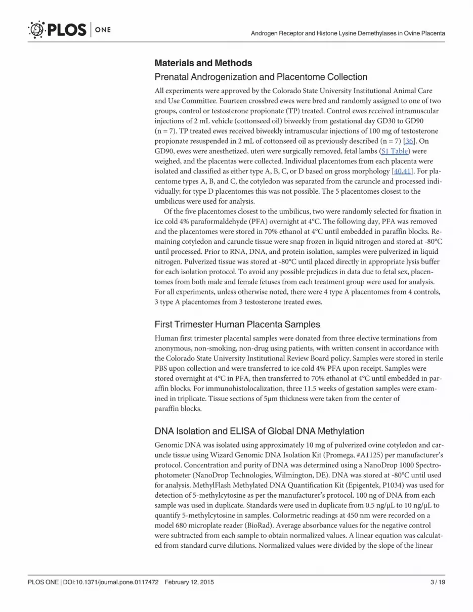
Changes in Epigenetic Factors and Steroid Hormone ReceptorsIn addition to altered gross morphology of the placentomes, TP treatment led to global changesin DNA methylation (S1 Fig.) indicating prenatal androgenization is associated with epigeneticchanges. Transcript levels for several genes involved in epigenetics (KDM’s, DNA methyltrans-ferases (DNMTs), and imprinted long non-codingH19) were assessed in GD90 placentomesfrom control and TP treated ewes (Figs. 2, 3, and S2 Fig.). While no difference was observed inKDM3A or KDM4AmRNA levels (S1 Fig.), KDM4C was lower in type A cotyledons from TPtreated ewes compared to controls (P<0.001, Fig. 2).H19mRNA levels were greater in
Fig 1. Placentomemorphology and fetal weight. A) Testosterone propionate treatment (TP) decreasedthe number of type A placentomes, and increased type C and type D placentomes collected at gestationalday 90. B) TP treatment did not affect placental weight whileC) female fetuses from TP ewes hadsignificantly reduced body weight at gestational day 90 compared to female fetuses from controls. * IndicatesP�0.06
doi:10.1371/journal.pone.0117472.g001
Table 2. List of primer sequences used for PCR of ChIP samples spanning an ARE and non-ARE region in the-5000bp 5’UTR of VEGFA.
Gene ARE/non-ARE Region Primer Sequence Amplicon Size
VEGFA ARE F-50 CTCTGTCTGGGCTGCTCTCT 179
R-50 TCCTCCCATGGACGTAACTC
VEGFA Non-ARE F-50 GCCTGTAATCCCAGCACTCT 181
R-50 GAGCAATTCTCCTGCCTCAG
doi:10.1371/journal.pone.0117472.t002
Androgen Receptor and Histone Lysine Demethylases in Ovine Placenta
PLOS ONE | DOI:10.1371/journal.pone.0117472 February 12, 2015 8 / 19
cotyledon tissue from type A placentomes from TP treated ewes compared to controls(P = 0.005, Fig. 2). DNMT1 levels were greater in type A cotyledon tissue from TP treated ewescompared to controls (P<0.001, Fig. 2). More DNMT1 protein also was observed in TP treatedewe type A cotyledon and placentome tissue (P<0.001, Fig. 2). No difference was observed inDNMT3a or DNMT3b with TP treatment (S2 Fig.). TP treatment did not alter placentome AR,KDM1A, and KDM4DmRNA levels (Fig. 3).
Further characterization of AR, KDM1A and KDM4D in GD90 ovine placentomes wascompleted to determine their response to TP treatment (Fig. 3). AR protein was greater in car-uncle tissue from type A placentomes in TP treated ewes compared to controls (P = 0.014)(Fig. 3). Preabsorption with the complementary AR peptide resulted in loss of immunoreactiveband (S3 Fig.). No significant difference was observed in KDM1A protein. Although TP treat-ment did not alter KDM4DmRNA levels, KDM4D protein levels were greater in type A placen-tome tissue from TP treated ewes when compared to controls (P<0.001), but was not differentin type D placentomes from TP treated ewes (Fig. 3).
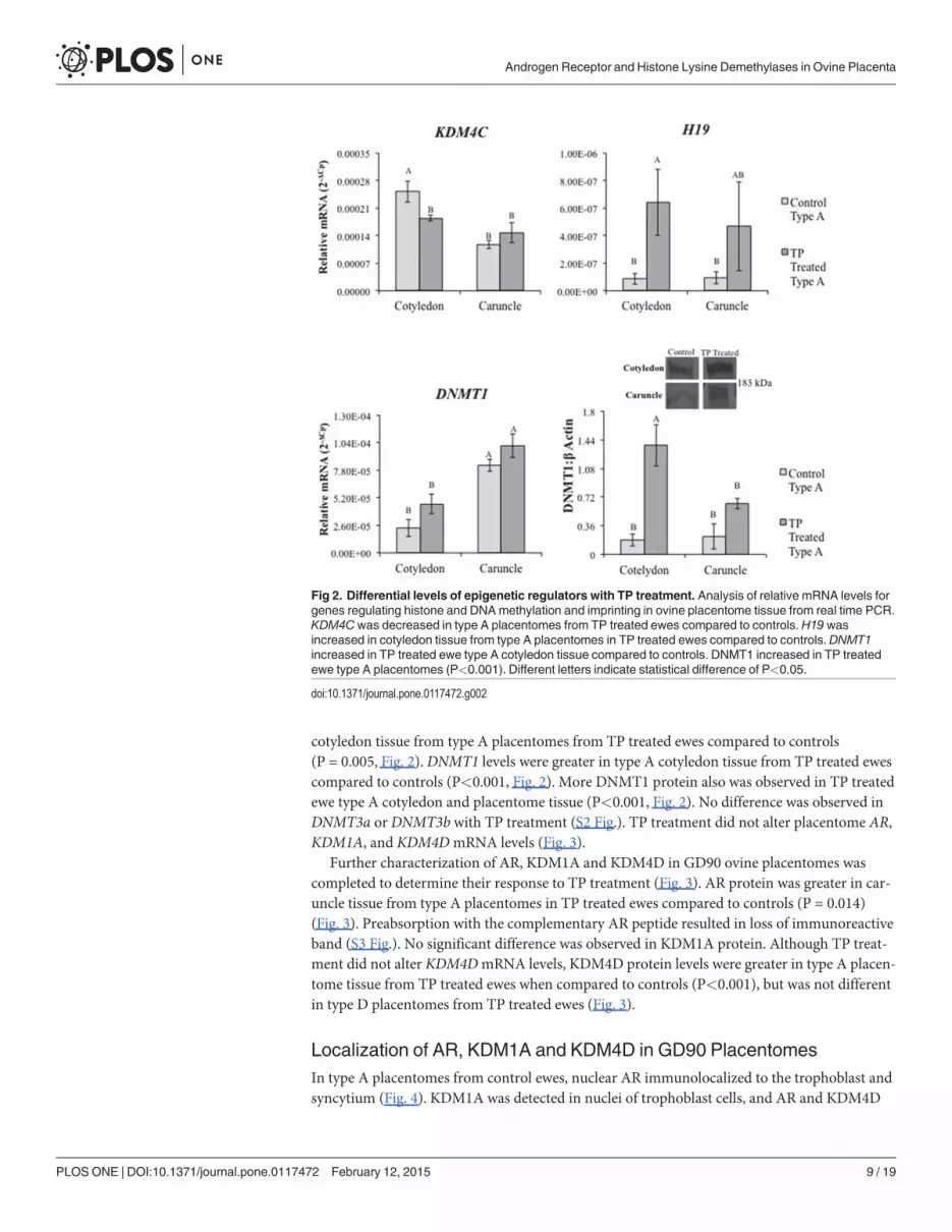
Localization of AR, KDM1A and KDM4D in GD90 PlacentomesIn type A placentomes from control ewes, nuclear AR immunolocalized to the trophoblast andsyncytium (Fig. 4). KDM1A was detected in nuclei of trophoblast cells, and AR and KDM4D
Fig 2. Differential levels of epigenetic regulators with TP treatment. Analysis of relative mRNA levels forgenes regulating histone and DNAmethylation and imprinting in ovine placentome tissue from real time PCR.KDM4Cwas decreased in type A placentomes from TP treated ewes compared to controls.H19 wasincreased in cotyledon tissue from type A placentomes in TP treated ewes compared to controls. DNMT1increased in TP treated ewe type A cotyledon tissue compared to controls. DNMT1 increased in TP treatedewe type A placentomes (P<0.001). Different letters indicate statistical difference of P<0.05.
doi:10.1371/journal.pone.0117472.g002
Androgen Receptor and Histone Lysine Demethylases in Ovine Placenta
PLOS ONE | DOI:10.1371/journal.pone.0117472 February 12, 2015 9 / 19

both immunolocalized to the syncytium. Predominate immunostaining for KDM1A was ob-served in trophoblast nuclei, while KDM4D localized to the apical surface of the syncytium(Fig. 4).
mRNA Levels of Androgen Responsive Genes in PlacentomesReal time PCR of selected androgen responsive genes (Androgen Responsive Gene Database;http://argdb.fudan.edu.cn/) known to regulate placentation was undertaken to determine if TPtreatment altered relative mRNA levels (Fig. 5 and S4 Fig.). Insulin-like growth factor 2 (IGF2)was lower in cotyledon tissue from type A placentomes from TP treated ewes compared to con-trols (P<0.001, Fig. 5). IGF binding protein 2 (IGFBP2) was lower in caruncle tissue from TPtreated ewes compared to type A placentomes from control ewes (P<0.001). Aromatase
Fig 3. Real time PCR and representative Western blot depicting placentomemRNA and protein levelsof AR, KDM1A and KDM4D in control and TP treated ewes. TP treatment did not alter mRNA for AR,KDM1A, or KDM4D. AR was increased in type A caruncle tissue from TP treated ewes compared to controls.No difference was found in KDM1A, though KDM4D increased in type A placentomes from TP treated ewescompared to controls. Placentome values represent average of cotyledon and caruncle quantified mRNAlevels in type A cotyledons. Different letters indicate statistical difference of P<0.05.
doi:10.1371/journal.pone.0117472.g003
Androgen Receptor and Histone Lysine Demethylases in Ovine Placenta
PLOS ONE | DOI:10.1371/journal.pone.0117472 February 12, 2015 10 / 19
(CYP19A1)mRNA levels were greater in type A placentomes from TP treated ewes comparedto type A placentomes from controls (P<0.001, Fig. 5). Vascular endothelial growth factor(VEGFA) was greater in cotyledon compared to caruncle tissue in type A placentomes fromcontrol as well as TP treated ewes (P<0.001, Fig. 5), and was not different between control andTP treated tissues. However, VEGFA protein monomer was higher in cotyledon tissue in typeA placentomes from TP treated ewes compared to controls (P = 0.005, Fig. 5).
AR Complexes with Histone Demethylases and binds to VEGFACoimmunoprecipitation and chromatin immunoprecipitation were used to determine if ARinteracts with KDM1A and/or KDM4D in ovine placenta to regulate androgen responsiveVEGFA. Co-immunoprecipitation and Western blot revealed AR binds to KDM1A andKDM4D in GD90 placentomes (Fig. 6A, S5 Fig.), and preabsorption with AR or KDM1Ablocking peptide decreased or inhibited immunoreactive bands, respectively (S6 Fig.). Further-more, both AR and KDM1A protein bind the same DNA region in the 5’ flanking sequencingof VEGFA containing an ARE half-site (Fig. 6B) according to PCR amplification. PCR withprimers designed for a non-ARE region in the 5’ flanking sequencing of VEGFA did not ampli-fy in AR or KDM1A chromatin immunoprecipitation samples, but was present in genomicDNA (data not shown).
Immunolocalization of KDM1A, and KDM4D in First Trimester HumanPlacentaIn 11.5 weeks of gestation tissue samples, KDM1A, and KDM4D immunolocalized to nuclei insyncytiotrophoblast (Fig. 7). Additional immunostaining was observed in cells in the villousstroma at 11.5 weeks of gestation.
DiscussionPrevious studies revealed that the placenta contains AR, and normal pregnancy is associatedwith an increase in serum testosterone levels [11,12]. Moreover, abnormal testosterone levelsare associated with a number of placental-associated disorders, including preeclampsia and in-trauterine growth restriction, whereas conditions such as polycystic ovarian syndrome lead topregnancy complications [13,14]. Prenatal androgenization models previously have been
Fig 4. Immunolocalization of AR, KDM1A, and KDM4D in type A placentomes from control ewes at GD90. Inserts represent control IHC straining.Immunolocalization of AR was present in the trophoblasts of the villous epithelium with primarily nuclear staining (arrow). KDM1A immunolocalized to thenucleus of the trophoblasts in the villous epithelium (arrow), while KDM4D immunostaining was prominent in the apical surface of maternal uterine epithelium(arrow). Images were taken at 20X maginification.
doi:10.1371/journal.pone.0117472.g004
Androgen Receptor and Histone Lysine Demethylases in Ovine Placenta
PLOS ONE | DOI:10.1371/journal.pone.0117472 February 12, 2015 11 / 19

employed to study abnormal developmental programming, and the prenatal androgenized ewehas demonstrated to be an excellent model, especially for PCOS in women [36,38,39]. For ex-ample, adult ewes that were exposed to increased androgens during the first half of gestation inthis model exhibit hyperandrogenism, anovulation, metabolic dysfunction, and infertility.However, no study to date has described the presence of AR in sheep placentomes. In ourstudy, prenatal androgenization from GD30 to 90 led to reduced fetal weight of femalelambs. Androgen induced intrauterine growth restriction has been described in sheep [36,38]and prenatal androgenized rats [43] along with reduced placental weights [44]. These datasuggest androgen as a regulator of fetal growth and placental function. We also report that pre-natal androgenization alters placentome morphology with the appearance of greater numbersof type D placentomes. While the increase in type C and D placentomes has been associated
Fig 5. Real time PCR results of AR target-genes known to regulate trophoblast differentiation andproliferation. IGF2 increased in type D placentomes from TP treated ewes compared to type A placentomesfrom control or TP treated ewes. IGFBP1 and IGFBP2 both decreased in caruncle tissue from type Aplacentomes from TP treated ewes compared to controls. IGFBP2 also decreased in type A and Dplacentomes from TP treated ewes compared to control type A placentomes. CYP19 increased in type Dplacentomes form TP treated ewes compared to type A placentomes from controls. TP treated ewes hadincreased VEGFA in type D placentomes compared to type A placentomes from control ewe. TP treated eweshad increased VEGFAmonomer in type A placentome cotyledon tissue. Different letters indicate statisticaldifference of P<0.05.
doi:10.1371/journal.pone.0117472.g005
Androgen Receptor and Histone Lysine Demethylases in Ovine Placenta
PLOS ONE | DOI:10.1371/journal.pone.0117472 February 12, 2015 12 / 19

previously with prenatal androgenization [45], increased presence of type C and D placen-tomes in pregnancies complicated by maternal nutrient restriction corresponds to increasedinterdigitated fetal villi, heightened cotyledon proliferation and increased vascular density[45–48].
Fig 6. Interaction of KDMs with AR. A) Immunoprecipitation of AR, KDM1A, and KDM4D from placentomeprotein isolated from control and TP treated ewes followed byWestern blot detection of co-immunoprecipitated protein. Anti-rabbit IP represents pull down by secondary antibody used for Western blotimmunolabeling. Antibody preabsorption with blocking peptide was used as a negative control.B) PCR of anARE promoter region of VEGFA from ChIP samples. IP, immunoprecipitate; ChIP, chromatinimmunoprecipitation; ARE, primers spanning androgen response element in promoter region
doi:10.1371/journal.pone.0117472.g006
Fig 7. Immunolocalization of KDM1A and KDM4D in first trimester human placenta samples. KDM1Aimmunostaining also localized to the nuclei in the syncytium and cells in the villous stroma at 11.5 weeks ofgestation. KDM4D immunolocalization was similar to KDM1A, with nuclear immunostaining present in thesyncytium and in the villous stroma at 11.5 weeks of gestation. White scale bar = 20μm for 20X and 40X.Insert is a representative image from control slides.
doi:10.1371/journal.pone.0117472.g007
Androgen Receptor and Histone Lysine Demethylases in Ovine Placenta
PLOS ONE | DOI:10.1371/journal.pone.0117472 February 12, 2015 13 / 19

In addition to altered ovine placentome morphology and decreased global DNAmethyla-tion in placentomes from TP treated ewes, we speculated abnormal epigenetic regulation oftrophoblast gene expression was induced. Proper regulation of DNAmethylation in tropho-blasts is necessary for normal placenta developmental programming. For example, reductionof DNA methyltransferases (DNMT1, DNMT3a and DNMT3b) in choriocarcinoma (BeWo)cells prevents cell migration [49], and reduced placental DNMT1 expression in in vitro pro-duced ovine embryos is associated with early embryonic loss [50]. Similarly, loss of DNAmeth-ylation imprinting in long non-coding H19 and neighboring IGF2 in the placenta is associatedwith Beckwith-Wiedemann Syndrome and increased prenatal and placental growth [51,52]. Inthe prenatal androgenization ewe model, we report up-regulatedH19 and IGF2 in placentomesfrom TP treated ewes, suggesting that excess prenatal androgen exposure may lead to dysregu-lated imprinting in the placenta via decreased global DNAmethylation, contributing to abnor-mal fetal growth observed with prenatal androgenization [36,37,43,44].
Regulation of gene expression involves histone modifications with site-specific adaptationsof amino acids in histone N-terminal tails, including methylation of lysine residues [53,54].KDMs demethylate lysines in histones to typically increase DNA accessibility and promotetranscription in the presence of transcription activators, such as ligand-bound nuclear recep-tors. Previous studies in cancer cells demonstrate that KDMs interact with and bind to sex hor-mone receptors to regulate androgen and estrogen signaling [32,35]. In addition, knock-out ofKDM1A in mice results in early embryonic lethality [55,56]. Considering the many similaritiesbetween cancer and placental cells, we reasoned that KDM’s also are present and involved inmediating androgen signaling in placental cells. Not only are AR and histone demethylasesKDM1A and KDM4D present in ovine placenta and first trimester human placenta, but ARco-localizes and complexes with KDM1A and KDM4D. This agrees with previous studies thatreported AR heterocomplexes with KDMs to regulate target genes [27–35], though this is thefirst study to our knowledge to show this interaction also occurs in placental tissue.
With the decreased DNAmethylation observed in placental tissue from TP treated ewes,the shift in placentome morphology may be a direct result of altered gene expression, more spe-cifically from increased growth and angiogenic factors induced by androgen signaling in pla-cental tissues. In GD90 placentomes from TP treated ewes, we observed increased mRNAlevels of AR target genes insulin-like growth factor 2 (IGF2) and vascular endothelial growth fac-tor (VEGF), and increased protein levels of AR and VEGFA. During prenatal development,IGFs and VEGFA regulate placentation and fetal growth. IGFs are expressed throughout theplacenta of various species, including maternal decidualized cells, cytotrophoblasts, and chori-onic mesoderm [57,58] and function as a key regulator of prenatal growth [59]. DecreasedIGFBP1 and IGFBP2 in caruncle tissue from TP treated ewes may be an additional mechanismto heighten local IGF signaling in androgenized placenta [59], explaining the rise in placen-tome overgrowth observed with the increase in type C and D placentomes [46–48].
VEGFA is a critical regulator of placental development and function and stimulates fetaland placental growth through increased angiogenesis [60,61]. While androgen responsiveVEGFA stimulates placental and prenatal angiogenesis, it also stimulates differentiation ofextravillous trophoblasts that remodel maternal spiral arteries in the human decidua for en-hanced blood flow to the placenta [60]. Insufficient extravillous trophoblast differentiation andinvasion has been associated with placental insufficiency and the development of pregnancy in-duced pathologies, including fetal growth restriction and preeclampsia [62]. Multiple studieshave also shown that maternal plasma testosterone is increased in cases of severe preeclampsia[63,64], with placentas from preeclamptic pregnancies expressing increased syncytiotropho-blast and stromal cell expression of AR [19]. Mutations in AR are correlated with increased riskfor the development of preeclampsia [65]. As we demonstrated AR and KDM1A binding to an
Androgen Receptor and Histone Lysine Demethylases in Ovine Placenta
PLOS ONE | DOI:10.1371/journal.pone.0117472 February 12, 2015 14 / 19

ARE in VEGFA’s promoter region in ovine placental tissue, placental androgens may have animportant function in regulating trophoblast function in sheep, possibly through VEGFA sig-naling. Although KDM4D also complexed with AR, the KDM4D antibody was not suitable forChIP and as such we were not able to demonstrate if KDM4D possibly is involved with AR reg-ulation of VEGFA in placentomes.
This proposed mechanism for placental androgen signaling is further supported by evidencethat hypoxia induces AR activity and expression of androgen-regulated genes, includingVEGFA [24–26], while androgen withdrawal leads to hypoxia [66]. Although nothing isknown about the role of KDMs in placental cells, KDMs are regulated by hypoxia to promotevascularization, invasion, migration, and cell proliferation in cancer tissue [27,34,35,67]. Thisis of particular interest as hypoxia-regulated trophoblast differentiation and invasion in thefirst trimester is required to establish proper placentation [1,62,68].
Prenatal androgenization and increased ligand could promote AR-KDM1A complex re-cruitment to AR-target genes in the placenta to stimulate gene transcription by promoting re-duced methylation signatures near target genes [28,53]. Therefore, ligand-dependentrecruitment of AR-KDM1A to androgen target genes could regulate growth, invasion, and an-giogenesis and, as such, placental development and function. However, despite the observedup-regulation in mRNA levels for AR-target genes regulating growth, invasion, and angiogene-sis, GD90 prenatal androgenized ewe lambs had reduced fetal weight, suggesting that increasedandrogen signaling, either directly or indirectly, dysregulated fetal developmental program-ming through other factors, such as placental nutrient transport, that were not investigated.
In conclusion, the present study revealed that prenatal androgenization alters ovine placen-tome development and is associated with decreased global DNA methylation and altered genetranscription of angiogenic and growth factor pathways. Importantly, we demonstrated for thefirst time that AR complexes to KDM’s in ovine placenta, and that AR and KDM1A bind to thesame promoter region of AR-target gene VEGFA in trophoblast cells. These results indicate an-drogen signaling functions jointly with KDMs to play a regulatory role in placental develop-ment. Finally, we were able to detect KDM1A and KDM4D in syncytiotrophoblast and invillous stromal cells in first trimester human placenta, indicating AR-KDM signaling also has apossible role in human trophoblast differentiation. Current studies are underway to determinethe role of histone demethylases in human placentation and to determine androgen’s effects ontrophoblast differentiation.
Supporting InformationS1 Fig. Global methylation in ovine GD90 placentomes. Global DNAmethylation decreasedin type A placentomes from TP treated ewes when compared to type A placentomes from con-trols. � Indicates P<0.05.(TIF)
S2 Fig. Real time PCR results for other selected epigenetic regulators. Difference in letter in-dicates significant difference of P<0.05.(JPG)
S3 Fig. Representative Western blot blocking peptide controls. Loss of immunoreactiveband for AR when antibody is preabsorbed with AR blocking peptide at a 1:3 ratio. LNCaP,human prostate adenocarcinoma cells.(JPG)
S4 Fig. Real time PCR results for other AR target-genes known to regulate trophoblast dif-ferentiation and proliferation. Placentome values represent average of cotyledon and caruncle
Androgen Receptor and Histone Lysine Demethylases in Ovine Placenta
PLOS ONE | DOI:10.1371/journal.pone.0117472 February 12, 2015 15 / 19

quantified mRNA levels in type A cotyledons. Difference in letter indicates significant differ-ence of P<0.05.(JPG)
S5 Fig. Interaction of KDM1A with AR.Western blot detection of KDM1A (105 kDa) fromplacentome protein following immunoprecipitation (IP) using AR and KDM1A antibodies.Anti-rabbit IP represents pull down by secondary antibody as a negative control.(TIF)
S6 Fig. Reduced AR immunoreactive band in immunoprecipitation of AR when antibodyis preabsorbed with blocking peptide (1:1 dilution). Loss of KDM1A immunoreactive bandin immunoprecipitation when antibody is preabsorbed with blocking peptide (1:1 dilution). IP= immunoprecipitation.(JPG)
S1 Table. List of ewes, number of fetuses, fetal sex and placentome number per pregnancy.(DOC)
S2 Table. Primer sequences used for real time PCR analysis of sheep placentomes.(DOC)
AcknowledgmentsWe would like to thank Dr. Stuart Tobet and Dr. Santiago Di Pietro for their generous supportand assistance with experiment design. Additional thanks to Dr. Alfredo Antoniazzi, Dr. JesusArreguin-Arevalo, Lisa Nulton, Kristin Klohonatz, Ashley Cameron, and Analisa Schilling fortheir assistance during ovine surgeries. Thanks to Richard Brandes and Greg Harding of theColorado State University’s Animal Reproduction Biotechnology Laboratory who providedanimal care.
Author ContributionsConceived and designed the experiments: ERC GJB. Performed the experiments: ERC JLHVAE JCS RCW. Analyzed the data: ERC GJB. Contributed reagents/materials/analysis tools:QAW RVA JEB CMC. Wrote the paper: ERC GJB.
REFERENCES1. Kaufmann P, Black S, Huppertz B (2003) Endovascular Trophoblast Invasion: Implications for the path-
ogenesis of intrauterine growth retardation and preeclampsia. Biol Reprod 69: 1–7. PMID: 12620937
2. Brosens IA (1988) The uteroplacental vessels at term: the distribution and extent of physiologicalchanges. Trophoblast Res 3: 61–68.
3. Blackenship TN, Enders AC, King BF (1993) Trophoblastic invasion and the development of uteropla-cental arteries in the macaque: immunohistochemical localization of cytokeratins, desmin, type IV colla-gen, laminin, and fibronectin. Cell Tissue Res 272: 227–236. PMID: 7685655
4. Albrecht ED, Pepe GJ (1999) Central integrative role of oestrogen in modulating the communication be-tween the placenta and fetus that results in primate fetal-placental development. Placenta 20: 129–139.PMID: 10195732
5. Rama S, Petrusz P, Rao AJ (2004) Hormonal regulation of human trophoblast differentiation: a possiblerole for 17β-estradiol and GnRH. Mol Cellular Endocrinol 218: 79–94. PMID: 15130513
6. Albrecht ED, Robb VA, Pepe GJ (2004) Regulation of placental vascular endothelial growth/permeabili-ty factor expression and angiogenesis by estrogen during early baboon pregnancy. J Clin EndocrinolMetab 89: 5803–5809. PMID: 15531545
7. Albrecht ED, Bonagura TW, Burleight DW, Enders AC, Aberdeen GW, et al. (2006) Suppression ofextravillous trophoblast invasion of uterine spiral arteries by estrogen during early baboon pregnancy.Placenta 27: 483–490. PMID: 15990167
Androgen Receptor and Histone Lysine Demethylases in Ovine Placenta
PLOS ONE | DOI:10.1371/journal.pone.0117472 February 12, 2015 16 / 19
8. Bukovsky A, Caudle MR, Cekanova M, Fernando RI, Wimalasena J, et al. (2003) Placental expressionof estrogen receptor beta and its hormone binding variant- comparison with estrogen receptor alphaand a role for estrogen receptors in asymmetric division and differentiation of estrogen-dependentcells. Reprod Biol Endocrinol 1: 36–57. PMID: 12740031
9. Bukovsky A, Cekanova M, Caudle MR, Wimalasena J, Foster JS, et al. (2003) Expression and localiza-tion of estrogen receptor-alpha protein in normal and abnormal term placentae and stimulation of tro-phoblast differentiation by estradiol. Reprod Biol Endocrinol 1: 13–31. PMID: 12646062
10. Niklaus AL, Aberdeen GW, Babischkin JS, Pepe GJ, Albrecht ED (2003) Effect of estrogen on vascularendothelial growth/permeability factor expression by glandular epithelial and stromal cells in the ba-boon endometrium. Biol Reprod 68: 1997–2004. PMID: 12606344
11. Mizuno M, Lobotsky J, Lloyd CW, Kobayashi T, Murasawa Y (1968) Plasma androstenedione and tes-tosterone during pregnancy and in the newborn. J Clin Endocrinol Metab 28: 1133–1142. PMID:5676177
12. Castracane VD, Stewart DR, Gimpel T, Overstreet JW, Lasley BL (1998) Maternal serum androgens inhuman pregnancy: early increases within the cycle of conception. Hum Reprod 13: 460–464. PMID:9557857
13. Carlsen SM, Romundstad P, Jacobsen G (2005) Early second-trimester maternal hyperandrogenemiaand subsequent preeclampsia: a prospective study. Acta Obstet Gynecol Scand 84: 117–121. PMID:15683369
14. Iavazzo C, Vitoratos N (2010) Polycystic ovarian syndrome and pregnancy outcome. Arch GynecolObstet 282: 235–239. doi: 10.1007/s00404-010-1495-0 PMID: 20458488
15. Gaiani R, Chiesa F, Mattili M, Nannetti G, Galeati F (1984) Androstenedione and testosterone concen-trations in plasma and milk of the cow throughout pregnancy. J Reprod Fertil 70: 55–59. PMID:6694152
16. Khatri P, Hoffmann B, Schuler G (2013) Androgen receptor is widely expressed in bovine placentomesand up-regulated during differentiation of bovine trophoblast giant cells. Placenta 34: 416–423. doi: 10.1016/j.placenta.2013.01.018 PMID: 23481223
17. Iwamura M, Abrahamsson PA, Benning CM, Cockett AT, di Sant’Agnese PA (1994) Androgen receptorimmunostaining and its tissue distribution in formalin-fixed, paraffin-embedded sections after micro-wave treatment. J Histochem Cytochem 42: 783–788. PMID: 8189040
18. Wieciech I, Durlej-Grzesiak M, Stomczyńska M (2013) Influence of the antiandrogen flutamide on theandrogen receptor gene expression in the placenta and umbilical cord during pregnancy in the pig.Acta Histochem 115: 290–295. doi: 10.1016/j.acthis.2012.08.003 PMID: 22951468
19. Hsu T-Y, Lan K-C, Tsai C-C, Ou C-Y, Cheng B-H, et al. (2009) Expression of androgen receptor inhuman placentas from normal and preeclamptic pregnancies. Taiwanese J Obstet Gynecol 48: 262–267. doi: 10.1016/S1028-4559(09)60301-6 PMID: 19797017
20. Horie K, Takakura K, Imai K, Liao S, Mori T (1992) Immunohistochemical localization of androgen re-ceptor in the human endometrium, decidua, placenta, and pathological conditions of the endometrium.Hum Reprod 7: 1461–1466. PMID: 1291578
21. Karvela M, Stefanakis N, Papadopoulou S, Tsitilou SG, Tsilivakos V, et al. (2008) Evidence for associa-tion of the G1733A polymorphism of the androgen receptor gene with recurrent spontaneous abortions.Fertil Steril 90: doi: 10.1016/j.fertnstert.2008.04.071 PMID: 18692840
22. Karjalainen MK, Huusko JM, Ulvila J, Sotkasiira J, Luukkonen A, et al. (2012) A potential novel sponta-neous preterm birth gene, AR, identified by linkage and association analysis of X chromosomal mark-ers. PLoS One 7: e51378. doi: 10.1371/journal.pone.0051378 PMID: 23227263
23. Roche PJ, Hoare SA, Parker MG (1992) A consensus DNA-binding site for the androgen receptor. MolEndocrinol 6: 2229–2235. PMID: 1491700
24. Comstock CE, Burd CJ, Jessen WJ, Knudsen KE (2008) Gene profiling analysis of androgen recep-tor mediated function. In: Handweger S, Aronow B, editors. Genomics in endocrinology: DNA micro-array analysis in endocrine health and disease. Contemp Endocrinol. Totowa, NJ, HumanaPress. pp. 83–113.
25. Shabisgh A, Tanji N, D’Agati V, Burchardt M, Rubin M, et al. (1999) Early effects of castration on thevascular system of the rat ventral prostate gland. Endocrinology 140: 1920–1926. PMID: 10098532
26. Mabjeesh NJ, Willard MT, Fredrickson CE, Zhong H, Simons JW (2003) Androgens stimulate hypoxia-inducible factor 1 activation via autocrine loop of tyrosine kinase receptor/phosphatidylinositol 3’-ki-nase/protein kinase B in prostate cancer cells. Clin Cancer Res 9: 2416–2425. PMID: 12855613
27. Shin S, Janknecht R (2007) Activation of androgen receptor by histone demethylases JMJD2A andJMJD2D. Biochem Biophys Res Commun 359: 742–746. PMID: 17555712
Androgen Receptor and Histone Lysine Demethylases in Ovine Placenta
PLOS ONE | DOI:10.1371/journal.pone.0117472 February 12, 2015 17 / 19

28. Wissmann M, Yin N, Müller JM, Greschik H, Fodor BD, et al. (2007) Cooperative demethylation byJMJD2C and LSD1 promotes androgen receptor-dependent gene expression. Nature Cell Biol 9:347–353. PMID: 17277772
29. Yamane K, Toumazou C, Tsukada Y-I, Erdjument-Bromage H, Tempst P, et al. (2006) JHDM2A, aJmjC-contain H3K9 demethylase, facilitates transcription activation by androgen receptor. Cell 125:483–495. PMID: 16603237
30. Verrie L, Vandromme M, Trouche D (2011) Histone demethylases in chromatin cross-talks. Biol Cell103: 381–401. doi: 10.1042/BC20110028 PMID: 21736555
31. Perillo B, Ombra MN, Bertoni A, Cuozzo C, Sacchetti S, et al. (2008) DNA Oxidation as triggered byH3K9me2 demethylation drives estrogen-induced gene expression. Science 319: 202–206. doi: 10.1126/science.1147674 PMID: 18187655
32. Kawazu M, Saso K, Tong KI, McQuire T, Goto K, et al. (2011) Histone demethylase JMJD3B functionsas a co-factor of estrogen receptor in breast cancer proliferation and mammary gland development.PLoS ONE 6:e17830. doi: 10.1371/journal.pone.0017830 PMID: 21445275
33. Garcia-Bassets I, Kwon Y-S, Telese F, Prefontaine GG, Hutt KR, et al. (2007) Histone methylation-dependent mechanisms impose ligand dependency for gene activation by nuclear receptors. Cell 128:505–518. PMID: 17289570
34. Metzger E, Wissmann M, Yin N, Muller JM, Schneider R, et al. (2005) LSD1 demethylates repressivehistone marks to promote androgen-receptor-dependent transcription. Nature 437: 436–439. PMID:16079795
35. Coffey K, Rogerson L, Ryan-Munden C, Alkharaif D, Stockley J, et al. (2013) The lysine demethylase,KDM4B, is a key molecule in androgen receptor signaling and turnover. Nucleic Acids Res 41:4433–4446. doi: 10.1093/nar/gkt106 PMID: 23435229
36. ManikkamM, Crespi EJ, Doop DD, Herkimer C, Lee JS, et al. (2004) Fetal programming: prenatal tes-tosterone excess leads to fetal growth retardation and postnatal catch-up growth in sheep. Endocrinolo-gy 145: 790–798. PMID: 14576190
37. Beckett EM, Veiga-Lopez A, DeHaan AM, Padmanabhan V (2011) Developmental programming: roleof androgen and insulin in advancing placentome differentiation in testosterone treated sheep. Abstractof the 44th Annual Meeting of the Society for the Study of Reproduction, Portland, Oregon. Biol Reprod85(Suppl): Abstract 301.
38. Recabarren SE, Padmanabhan V, Codner E, Lobos A, Durán C, et al. (2005) Postnatal developmentalconsequences of altered insulin sensitivity in female sheep treated prenatally with testosterone. Am JPhysiol Endocrinol Metab 289: E801–E806. PMID: 16215166
39. Veiga-Lopez A, YeW, Phillips DJ, Herkimer C, Knight PG, et al. (2008) Developmental programming:deficits in reproductive hormone dynamics and ovulatory outcomes in prenatal, testosterone-treatedsheep. Biol Reprod 78: 636–647. PMID: 18094354
40. van der Linden PS, Sciascia Q, Sales F, McCoard SA (2013) Placental nutrient transport is affected bypregnancy rank in sheep. J Animal Science 91: 644–653. doi: 10.2527/jas.2012-5629 PMID: 23097400
41. Vatnick I, Scholknecht R, Darrigrand R, Bell AW (1991) Growth and metabolism of the placenta afterunilateral fetectomy in twin pregnant ewes. J Dev Physiol 15: 351–356. PMID: 1753075
42. Schmittgen TD, Livak KJ (2008) Analyzing real-time PCR data by the comparative CT method. NatureProtocols 3: 1101–1108. PMID: 18546601
43. Sathishkumar K, Elkins R, Chinnathambi V, Gao H, Hankins GDV, et al. (2011) Prenatal testosterone-induced fetal growth restriction is associated with down-regulation of rat placental amino acid transport.Reprod Biol Endocrinol 9: 110–122. doi: 10.1186/1477-7827-9-110 PMID: 21812961
44. Sun M, Maliqueo M, Benrick A, Hohansson J, Shao R, et al. (2012) Maternal androgen excess reducesplacental and fetal weights, increases placental steroidogenesis, and leads to long-term health effectsin their female offspring. Am J Physiol Endocrinol Metab 303: E1373–E1385. doi: 10.1152/ajpendo.00421.2012 PMID: 23047983
45. Vonnahme KA, Hess BW, Nijland MJ, Nathanielsz PW, Ford SP (2006) Placentomal differentiationmay compensate for maternal nutrient restriction in ewes adapted to harsh range conditions. J AnimSci 84: 3451–3459. PMID: 17093240
46. Osgerby JC, Wathes DC, Howard D, Gadd TS (2004) The effect of maternal undernutrition on the pla-cental growth trajectory and the uterine insulin-like growth factor axis in the pregnant ewe. J Endocrinol182: 89–103. PMID: 15225134
47. Ford SP, Vonnahme KA, Drumhiller MC, Reynolds LP, Nijland MJ, et al. (2004) Arteriolar density andcapillary volume increase as placentomes advance from type A through type D developmental stages.Biol Reprod 71(Suppl): Abstract 509.
Androgen Receptor and Histone Lysine Demethylases in Ovine Placenta
PLOS ONE | DOI:10.1371/journal.pone.0117472 February 12, 2015 18 / 19
48. Ford SP, Nijland MJ, Miller MM, Hess BW, Nathanielsz PW (2006) Maternal undernutrition advancedplacentomal type, in association with increase placentomal size and cotyledonary (cot) blood flow. JSoc Gynecol Investig 13(Suppl): Abstract 212.
49. Rahnama F, Shafiei F, Cluckman PD, Mitchell MD, Lobie PE (2006) Epigenetic regulation of human tro-phoblastic cell migration and invasion. Endocrinology 147: 5275–5283. PMID: 16887905
50. Ptak GE, D’Agostino A, Toschi P, Fidanza A, Zacchini F, et al. (2013) Post-implantation mortality of invitro produced embryos is associated with DNAmethyltransferase I dysfunction in sheep placenta.Hum Reprod 28: 298–305. doi: 10.1093/humrep/des397 PMID: 23169866
51. Angiolini E, Fowden A, Coan P, Sandovici I, Smith P, et al. (2006) Regulation of placental efficiency fornutrient transport by imprinted genes. Placenta 27(Suppl A):Vol. 20. PMID: 16618444
52. Weksberg R, Shuman C, Beckwith JB (2010) Beckwith-Wiedemann Syndrome. J HumGenet 18: 8–14.
53. Johnson LM, Cao X, Jacobsen SE (2002) Interplay between two epigenetic marks: DNAmethylationand histone H3 lysine 9 methylation. Curr Biol 12: 1360–1367. PMID: 12194816
54. Cedar H, Bergman Y (2009) Linking DNAmethylation and histone modification: patterns and para-digms. Nature Genet 10:295–304.
55. Wang J, Scully K, Zhu X, Cai L, Zhang J, et al. (2007) Opposing LSD1 complexes function in develop-mental gene activation and repression programs. Nature 466: 882–887.
56. Wang J, Hevi S, Kurash JK, Lei H, Gay F, et al. (2009) The lysine demethylase LSD1 (KDM1) is re-quired for maintenance of global DNAmethylation. Nature Genet 41: 125–129. doi: 10.1038/ng.268PMID: 19098913
57. Hill DJ, Clemmons DR, Riley SC, Bassett N, Challis JRG (1993) Immunohistochemical localization ofinsulin-like growth factors (IGFs) and IGF binding proteins-1, -2, and-3 in human placenta and fetalmembranes. Placenta 14: 1–12. PMID: 7681208
58. Han VKM, Carter AM (2000) Spatial and temporal patterns of expression of messenger mRNA for insu-lin-like growth factors and their binding proteins in the placenta of man and laboratory animals. Placenta21: 289–305. PMID: 10833363
59. Randawa R, Cohen P (2005) The role of the insulin-like growth factor system in prenatal growth. MolGenet Metab 86: 84–90. PMID: 16165387
60. Zhou Y, Bellingard V, Feng KT, McMaster M, Fisher SJ (2003) Human cytotrophoblasts promote endo-thelial survival and vascular remodeling through secretion of Ang 2, PIGF, and VEGF-C. Dev Biol 263:114–125. PMID: 14568550
61. Zhou Y, McMaster M, Woo K, Janatpour M, Perry J, et al. (2002) Vascular endothelial growth factor li-gands and receptors that regulate human cytotrophoblast survival are dysregulated in severe pre-eclampsia and hemolysis, elevated liver enzymes, and low platelets syndrome. Am J Pathol 160:1405–1423. PMID: 11943725
62. Bdolah Y, Sukhatme VP, Karumanchi SA (2004) Angiogenic Imbalance in the Pathophysiology of Pre-eclampsia: Newer Insights. Semin Nephrol 24: 548–556. PMID: 15529289
63. Salamalekis E, Bakas P, Vitoratos N, Eleptheriadis M, Creatsas G (2006) Androgen levels in the thirdtrimester of pregnancy in patients with preeclampsia. Eur J Obstet Gynecol Reprod Biol 126: 16–19.PMID: 16139944
64. Acromite MT, Mantzoros CS, Leach RE, Hurwitz J, Dorey LG (1999) Androgens in preeclampsia. Am JObstet Gynecol 180: 60–63. PMID: 9914579
65. Lim JH, Kim SY, Lee SW, Park SY, Han JY, et al. (2011) Association between genetic polymorphisms inandrogen receptor gene and risk of preeclampsia in Korean women. J Assist Reprod Genet 28: 85–90.doi: 10.1007/s10815-010-9485-5 PMID: 20922474
66. Milosevic M, Chung P, Parker C, Bristow R, Toi A, et al. (2007) Androgen withdrawal in patients re-duces prostate cancer hypoxia: implications for disease progression and radiation response. CancerRes 67: 6022–6025. PMID: 17616657
67. Yang J, Jubb AM, Pike L, Buffa FM, Turley H, et al. (2010) The histone demethylase JMJD2B is regulat-ed by estrogen receptor α and hypoxia, and is a key mediator of estrogen induced growth. Cancer Res70(16): 6456–6466. doi: 10.1158/0008-5472.CAN-10-0413 PMID: 20682797
68. Zhou Y, Chiu K, Brescia RJ, Combs CA, Katz MA, et al. (1993) Increased depth of trophoblast invasionafter chronic constriction of the lower aorta in rhesus monkeys. Am J Obstet Gynecol 169: 224–229.PMID: 8333462
Androgen Receptor and Histone Lysine Demethylases in Ovine Placenta
PLOS ONE | DOI:10.1371/journal.pone.0117472 February 12, 2015 19 / 19