Oxygen regulation of macrophage migration inhibitory factor in human placenta
32
OXYGEN REGULATION OF MACROPHAGE MIGRATION INHIBITORY FACTOR (MIF) IN HUMAN PLACENTA Francesca Ietta 1,2 , Yuanhong Wu 1 , Roberta Romagnoli 2 , Nima Soleymanlou 1,3 , Barbara Orsini 4 , Stacy Zamudio 5 , Luana Paulesu 2 , Isabella Caniggia 1, 3 1 Department of Obstetrics and Gynecology, Mount Sinai Hospital, Samuel Lunenfeld Research Institute, University of Toronto, Toronto, Ontario, Canada, M5G 1X5; 2 Department of Physiology, Division of Immunoendocrinology and Reproductive Physiology, University of Siena, 53100 Siena, Italy; 3 Department of Physiology; University of Toronto, Toronto, Ontario, Canada; 4 Department of Patophysiology, Gastroenterology Unit, University of Florence, 50139 Florence, Italy; 5 New Jersey Medical School, Newark, New Jersey, USA Correspondence : Luana Paulesu, Ph.D., Department of Physiology, Division of Immunoendocrinology and Reproductive Physiology, University of Siena, 53100 Siena, Italy Tel: +39 0577 234224, Fax: +39 0577 234219. E-mail: [email protected] Running Head : Oxygen and MIF in human placenta. Page 1 of 32 Articles in PresS. Am J Physiol Endocrinol Metab (August 29, 2006). doi:10.1152/ajpendo.00086.2006 Copyright © 2006 by the American Physiological Society.
Transcript of Oxygen regulation of macrophage migration inhibitory factor in human placenta

OXYGEN REGULATION OF MACROPHAGE MIGRATION INHIBITORY FACTOR
(MIF) IN HUMAN PLACENTA
Francesca Ietta1,2, Yuanhong Wu1, Roberta Romagnoli2, Nima Soleymanlou1,3, Barbara Orsini4,
Stacy Zamudio5, Luana Paulesu2, Isabella Caniggia1, 3
1Department of Obstetrics and Gynecology, Mount Sinai Hospital, Samuel Lunenfeld Research
Institute, University of Toronto, Toronto, Ontario, Canada, M5G 1X5; 2Department of Physiology,
Division of Immunoendocrinology and Reproductive Physiology, University of Siena, 53100 Siena,
Italy; 3Department of Physiology; University of Toronto, Toronto, Ontario, Canada; 4Department of
Patophysiology, Gastroenterology Unit, University of Florence, 50139 Florence, Italy; 5New Jersey
Medical School, Newark, New Jersey, USA
Correspondence: Luana Paulesu, Ph.D., Department of Physiology, Division of
Immunoendocrinology and Reproductive Physiology, University of Siena, 53100 Siena, Italy Tel:
+39 0577 234224, Fax: +39 0577 234219. E-mail: [email protected]
Running Head: Oxygen and MIF in human placenta.
Page 1 of 32Articles in PresS. Am J Physiol Endocrinol Metab (August 29, 2006). doi:10.1152/ajpendo.00086.2006
Copyright © 2006 by the American Physiological Society.

ABSTRACT
Macrophage Migration Inhibitory Factor (MIF) is an important proinflammatory cytokine involved
in regulation of macrophage function. In addition, MIF may also play a role in murine and human
reproduction. While both first trimester trophoblast and decidua express MIF, the regulation and
functional significance of this cytokine during human placental development remains unclear. We
assessed MIF expression throughout normal human placental development, as well as in in vitro
(chorionic villous explants) and in vivo (high altitude placentae) models of human placental
hypoxia. Dimethyloxalylglycine (DMOG), which stabilizes Hypoxia Inducible Factor-1 (HIF-
1) under normoxic conditions, was also used to mimic the effects of hypoxia on MIF expression.
Quantitative RT-PCR and Western blot analysis showed high MIF protein and mRNA expression at
7-10 weeks, and lower levels at 11-12 weeks until term. Exposure of villous explants to 3% O2
resulted in increased MIF expression and secretion relative to standard conditions (20% O2).
DMOG-treatment under 20% O2 increased MIF expression. In situ hybridization and
immunohistochemistry showed elevated MIF expression in low oxygen-induced extravillous
trophoblast cells. Finally, a significant increase in MIF transcript was observed in placental tissues
from high altitude pregnancies. Hence three experimental models of placental hypoxia (early
gestation, DMOG-treatment, and high altitude) converge in stimulating increased MIF, supporting
the conclusion that placental-derived MIF is an oxygen-responsive cytokine highly expressed in
physiological in vivo and in in vitro low oxygen conditions.
Keywords: hypoxia, human trophoblast, proinflammatory cytokines.
Page 2 of 32

INTRODUCTION
Important aspects of placental development are regulated by changes in O2 tension. During
the early stages of pregnancy (prior to 10-12 weeks of gestation), the trophoblast is in a low-oxygen
environment due to the absence of maternal blood flow in the intervillous space (36). With
advancing gestation, the maternal circulation becomes fully established and Po2 increases (22). Both
in vivo and in vitro studies support that the low oxygen environment is required for embryo
development and the balance of trophoblast proliferation/differentiation essential for early placental
development. Likewise, the late first and second trimester increase in Po2 is critical to complete the
process of trophoblast differentiation/invasion (3, 18).
An increasing body of evidence indicates that oxygen tension modifies the expression and
activity of a variety of regulatory genes that govern trophoblast differentiation. We have recently
shown that oxygen is involved in the modulation of focal adhesion kinase, a key signaling molecule
regulating trophoblast proliferation and invasion (28). In addition we have reported that the early
events of trophoblast differentiation are mediated by O2-sensitive transcriptional activity of the HIF-
1 system and the subsequent expression of TGF-β3 (16). Moreover, in vitro studies demonstrated
that low oxygen tension, comparable to that of early placentation, can stimulate the expression of
several proinflammatory cytokines, such as Tumor Necrosis Factor-α (TNF-α) and Interleukin-1
(IL-1) (9).
MIF is a proinflammatory cytokine first described as a T-cell derived factor capable of
inhibiting random migration of macrophages (10, 17). Although the activity of MIF was originally
described in T-lymphocyte-conditioned media, MIF protein and transcript expression has been
widely documented in numerous cell types (14, 1). We focused on MIF in this study because
emerging evidence points to MIF as important to early pregnancy development. MIF is expressed
during zygote and blastocyst formation in mice (40). MIF has been detected at the implantation site
in maternal decidua, in chorionic villous trophoblast cells and in extravillous trophoblast cells
Page 3 of 32

within the anchoring villi of humans (5, 6). However the biological significance of MIF during
placental development and in particular its regulation remains to be elucidated.
Because we were struck by the apparent prominence of MIF in early gestational events, we
tested the hypothesis that O2 tension contributes to the regulation of MIF expression and secretion.
Herein we demonstrate that MIF expression is spatially and temporally regulated during placental
development. Furthermore we show that low oxygen tension, both environmentally or
pharmacologically-induced, increases MIF expression and secretion in an in vitro villous explants
system. Finally we demonstrate that MIF expression is elevated in placentae from high altitude
pregnancy, an in vivo physiological model of chronic hypoxia.
Page 4 of 32

MATERIALS AND METHODS
Placental collection and processing
All tissue samples were obtained after informed consent in accordance with participating
institutions’ Ethics Guidelines. Tissue collection strictly adhered to the guidelines outlined in the
Declaration of Helsinki. Placental tissues from first-trimester (5-10 weeks of gestation, n = 19) and
second trimester (11-13 weeks of gestation, n = 6; and 14-20 weeks of gestation, n = 7) normal
pregnancies, terminated for psychological reasons, were obtained in Toronto, Ontario, Canada by
dilatation and curettage. Gestational age was determined by the date of the last menstrual period and
ultrasound measurement of crown-rump-length (CRL). High altitude placentae were collected in
Leadville, Colorado, USA (HA, 3100 meters above sea level, masl). Moderate altitude placentae
were collected in Denver, Colorado, USA (MA, 1600 masl ). Sea level placentae were collected
from term deliveries at Mount Sinai Hospital in Toronto, Ontario, Canada (SL, ~40 masl). All third
trimester specimens were obtained immediately after delivery from normal looking cotyledons
randomly collected. Areas with calcified, necrotic or visually ischemic tissue were omitted from
sampling. Subjects suffering from diabetes, essential hypertension and pregnancies affected by
preeclampsia and intra uterine growth restriction (IUGR) were excluded. All groups did not show
clinical or pathological signs of preeclampsia, infections or other maternal or placental diseases.
Birth weight, gestational age and laboratory values or clinical observations relevant to the health of
the mother were abstracted from the clinical records. Term control placental tissues (n=10) were
obtained from women with normal pregnancies undergoing elective cesarean section or vaginal
delivery at sea level. Samples from high (n=12) and moderate altitude (n=12) were collected from
normal pregnancies delivered vaginally or by elective caesarean delivery. The clinical
characteristics of the patients are shown in Table 1. All samples were snap-frozen immediately after
collection and stored at -80°C for MIF mRNA and protein analysis or fixed in formalin and
embedded in paraffin for immunohistochemistry.
Page 5 of 32

Villous explant cultures
First trimester placental tissue (5-10 weeks of gestation, 14 separate sets) was obtained from
consenting patients undergoing elective termination of pregnancy. Placentae were immediately
rinsed in sterile cold phosphate-buffered saline (PBS) and processed within 2 hours of collection.
Endometrial tissue and fetal membranes were dissected out. Terminal villi were then placed on
Millicel-CM culture dish inserts (Millipore, Bedford, MA) previously coated with 150 µl of
undiluted Matrigel (Collaborative Biomedical Products, Bedford, MA) and transferred to 24-well
culture plates. The explants were cultured in DMEM/F12 (GIBCO-BRL, Grand Island, NY)
supplemented with 100 µg/ml streptomycin, 100 U/ml penicillin and 2mM glutamine. Explants
were maintained in standard condition (5% CO2 in 95% air, 20% O2 ~150 mmHg) or in an
atmosphere of 3% (92% N2 and 5% CO2, ~21 mmHg ) or 8% (87% N2 and 5% CO2, ~57 mmHg)
O2 for 48 hours at 37°C. The morphological integrity and viability of villous explants and their
extra villous trophoblast (EVT) outgrowth and migration were monitored daily for the duration of
the various experiments as previously reported (16, 32). In additional experiments we treated
explant cultures with dimethyloxalylglycine (DMOG; 1mM; Frontier Scientific, Logan, Utah) for
24 hours under standard oxygenation (20% O2). DMOG is an inhibitor of prolyl-hydroxylases
activity mimicking hypoxia via stabilization of HIF-1α (20). At the end of incubation, supernatants
were collected and frozen until assayed for MIF concentration. Explants were either fixed in 4%
paraformaldehyde and embedded in paraffin for immunohistochemistry (n=3 separate sets of
placental explants, each experimental condition was carried out in triplicate) and in situ
hybridization (n=3 separate sets of placental explants) or snap-frozen and processed for protein
(n=5 separate sets of placental explants, each experimental condition was carried out in triplicate)
and mRNA analysis (n=3 separate sets of placental explants, each experiment condition was carried
out in triplicate).
RNA analysis
Page 6 of 32

Total RNA extracted from placental tissues and villous explants was treated with DNaseI to
remove genomic DNA contamination. One µg of total RNA was reverse transcribed using random
hexamer and MultiScribe enzyme (Applied Biosystems, Foster City, CA). Real time quantitative
PCR (qRT-PCR) reactions were run in an ABI Prism 7700 sequence detection system (Applied
Biosystems) using TaqMan chemistry. Five µL cDNA in a final volume of 50 µL were amplified
using the 20X Assays-on-DemandTM gene expression assay mix (Applied Biosystems). TaqMan
probes and specific primers for MIF and ribosomal 18S, selected as control housekeeping gene,
were purchased from Applied Biosystems.
The relative expression was calculated as 2-(Ctgene of interest-Ct18S). Fold change was calculated
according to Livak et al (27).
In situ hybridization
Sections of first trimester villous placental explants cultured at 3% and 20% O2 were used
for MIF mRNA in situ hybridization (ISH). MIF cDNA was generated by oligo-(dT)-primed
reverse transcription of T helper cell clones with subsequent amplification using specific
oligodeoxyribonucleotide primers (5). After sequencing, an aliquot of the 255 bp PCR product was
used to generate (Lig’n Scribe kit, Ambion, TX) the sense and antisense RNA probes tailed with
SP6 RNA polymerase promoter without the need for subcloning. Transcription and labelling of
RNA probes were performed with 35S-uridine-5’-(thio)-triphosphate.
Prior to to ISH, 5 µm sections were dewaxed in xylene, taken through a series of graded
ethanol, then subjected to enzymatic digestion with pronase (125 µg/ml). Prehybridization,
hybridization, removal of nonspecific bound probe by digestion and further washing procedures
were performed (33). Autoradiography was carried out and the slides were developed after exposure
for 2 weeks. The specific signal was acquired by a CCD video camera connected to the microscope.
The threshold of specific detection was automatically calibrated on control sections hybridized with
the sense probe.
Page 7 of 32

Western blot analysis
Placental tissues and villous explants were homogenized in ice-cold RIPA buffer (Tris-HCl
50 mM, NaCl 150 mM, Triton X100 1% vol/vol, Na deoxycholate 1% wt/vol, SDS 0.1% wt/vol,
pH 7.5) supplemented with proteinase inhibitor cocktail (Roche Diagnostic, Mannheim, Germany).
After centrifugation at 15000Xg for 15 minutes at 4°C, the supernatant was assayed for total protein
concentration by the Bradford method (12) and MIF was detected by Western blot analysis. Thirty
µg of total proteins were separated on 15% polyacrylamide gel in the presence of SDS. Proteins
were then transfered to PVDF membranes. Primary anti-human MIF monoclonal antibody (R&D
Systems, Abingdon, UK) was used at dilution of 1:500. The membrane was then exposed to
horseradish peroxidase conjugated rabbit anti-mouse secondary antibody. The bands were detected
using an enhanced chemiluminescence kit (Perkin-Elmer Life Sciences, Boston, MA) according to
the manufacturer’s instructions. Equal loading of the proteins was confirmed by staining the blots
with a 10% (v/v) Ponceau S solution (31) ( Sigma Chemical Co., St. Louis, MO).
Immunohistochemistry
Immunohistochemical analysis was performed using an avidin-biotinylated horseradish
peroxidase macromolecular complex (ABC) kit according to the manufacturer’s instructions
(Vector Laboratories, Burlington, Ontario, Canada). Every 10th section was stained with
hematoxylin and eosin to verify the quality of the tissue, and whether the structures of interest were
present within single sections. Selected sections (5 µm) were dewaxed in xylene and rehydrated in a
descending alcohol series. Antigen retrieval was carried out by incubating the sections in sodium
citrate buffer (10 mM, pH 6) in a microwave oven. Endogenous peroxidase activity was blocked
with 3% hydrogen peroxide (vol/vol) in PBS. Slides were then incubated with 5% normal goat
serum for 1 hour at room temperature to prevent nonspecific binding. Mouse anti human MIF
monoclonal antibody (R&D Systems) was used at 1:200 dilution. After overnight incubation at 4°C
with primary antibody, slides were washed several times in PBS and incubated with biotinylated
Page 8 of 32

goat anti-mouse secondary antibody (DAKO, Copenhagen, Denmark) diluted 1:300 for 1 hour at
room temperature. After washing , slides were then incubated for 1 hour at room temperature with
avidin-biotin complex and then developed with 0.075% 3,3-diaminobenzidine (vol/vol) in PBS
containing 0.002% hydrogen peroxide (vol/vol). Sections were then counterstained with
hematoxylin and dehydrated. In each experiment, a negative control was used by replacing the
primary antibody with normal murine immunoglobulins.
MIF ELISA
MIF release in supernatant of villous explants cultures maintained at 3% and 20% O2 was
measured by a colorimetric sandwich ELISA. Briefly, 96-well plates were coated overnight at room
temperature with anti-human MIF monoclonal antibody (R&D Systems) (2 µg/ml). The plates were
then washed with washing solution (10 mM PBS, pH 7.4, 0.05% (vol/vol) Tween 20), blocked by
adding 300 µl of blocking solution (10 mM PBS, pH 7.4, 1% (wt/vol) bovine serum albumin
(BSA), 5% (wt/vol) sucrose) and incubated at room temperature for 1.5 hours. The samples, diluted
in Tris buffered saline-BSA (2.0 mM Tris-HCl, 150 mM NaCl, pH 7.3, 0.1% (wt/vol) BSA, 0.05%
(vol/vol) Tween 20), were added in duplicate (100 µl/well) and incubated for 2 hours at room
temperature. The plates were then washed three times and 100 µl of biotinylated goat anti-human
MIF antibody (R&D Systems) (200 ng/ml) was added to each well. Plates were incubated for 2
hours at room temperature and then washed again. Streptavidin horseradish peroxidase (Zymed, San
Francisco, CA) was subsequently added to each well, after which the plates were incubated for 20
minutes at room temperature. The plates were then washed and 3,3’,5,5’-tetramethylbenzidine
(Zymed) was added to each well; the reaction was stopped after 20 minutes by adding H2SO4.
Absorbance was measured at 450 nm using an ELISA SR 400 microplate reader (Sclavo, Siena,
Italy). The MIF concentration was extrapolated from a standard curve ranging from 25-2500 pg/ml
using human recombinant MIF (R&D Systems) as standard. The sensitivity limit was 18 pg/ml.
Intra- and inter-assay coefficients of variation were 3.86 (0.95)% and 9.14 (0.47)%, respectively.
Page 9 of 32

Data analysis
All data are presented as mean ± SEM of at least 4-6 separate experiments carried out in
triplicate. Statistical significance was determined by One Way ANOVA followed by Student-
Newman-Keuls post-hoc testing. Significance was defined as p<0.05. qRT-PCR statistical analysis
was performed using the relative expression software tool (REST®), a Pair-Wise Fixed
Reallocation Randomization Test (34).
Page 10 of 32

RESULTS
Expression of MIF during placental development
We first investigated MIF mRNA expression in human placentae throughout gestation by
qRT-PCR. Using specific primers and TaqMan probes, we found that MIF transcripts were present
at all the examined stages of gestation. However, the pattern of MIF mRNA expression was unique:
it was significantly higher during early pregnancy (7-10 weeks of gestation), then declined in the
late first trimester (11-12 weeks of gestation) to levels that remained constant during the second
trimester and at term (Figure 1A).
A similar expression pattern was obtained at protein level using Western blot analysis. A
specific 12 kDa band corresponding to the predicted molecular weight of MIF was expressed
throughout gestation. Its intensity was higher in the early first trimester, decreased by week 10 and
was similar at all time points tested thereafter (Figure 1B, upper panel). Protein densitometric
analysis revealed that MIF was significantly increased between 7-10 weeks of gestation when
compared to other gestational stages examined: late first trimester, second trimester and term (7-10
weeks vs 11-12 weeks fold increase = 1.85 ± 0.12, P<0.01; 7-10 weeks vs 14-20 weeks fold
increase = 1.40± 0.15, P<0.01; 7-10 weeks vs 39-40 weeks fold increase = 1.68±0.03, P<0.01 p
values) (Figure 1B, lower panel). No differences in MIF mRNA and protein expression were found
between tissues obtained from vaginal delivery and caesarean section (data not shown).
Immunostaining with anti-MIF antibody showed strong positive immunoreactivity in first
trimester villous cytotrophoblast and extravillous trophoblast cells (EVT) within the invading
columns (Figure 1C). The syncytiotrophoblast exhibited no MIF staining. Low/positive
immunoreactivity was observed in the stroma. By week 13, MIF staining was generally reduced. In
stem chorionic cytotrophoblast cells, MIF staining declined and only a few cells in the distal part of
the extravillous trophoblast were positive for MIF. No positive staining was observed in control
sections, where non-immune immunoglobulins were used in place of the MIF antibody in all tissues
sections examined (Figure 1C, right panel).
Page 11 of 32

Effect of oxygen on MIF expression in villous explants
Based on the first set of experiments on the developmental expression of MIF, we
hypothesized that changes in oxygen tension might regulate MIF mRNA and protein expression. To
test this hypothesis, we cultured chorionic villous explants (5-10 weeks of gestation, 14 separate
sets; each experimental condition was repeated 3 times using three different explants from the same
placenta) at 3%,(physiological < 10 weeks) (36), 8% (physiological >10 weeks) (22) and 20% Po2
(standard conditions) for 48 hours. Quantitative analysis by qRT-PCR showed that MIF mRNA
expression was increased at 8% relative to 20% Po2 , and further increased by exposure to 3% Po2
(Figure 2A). MIF concentration in villous explant conditioned media (measured by ELISA) showed
that the amount of soluble MIF was significantly higher in cultures kept at 3% relative to those kept
at 20% Po2 (fold increase = 1.75) (Figure 2B).
We next tested the possibility for involvement of the HIF-1α transcription factor in the
modulation of MIF expression. Villous cultures were maintained at 3% of oxygen overnight in
order to induce HIF-1α expression and subsequently some of them were transferred to 20% oxygen
in the presence or absence of DMOG a general competitive inhibitor of the oxygen-sensing
enzymes HIF-α prolyl hydroxylases (HIF-PHs), which target HIF-1α for degradation (20, 21). MIF
transcript was significantly increased in DMOG-treated cultures in 20% O2 when compared to
control cultures, and the increase in MIF transcript was similar to that observed in untreated cultures
maintained at 3% oxygen (Figure 2C). MIF protein expression as assessed by Western blot analyses
in explants from 5 versus 10 weeks of pregnancy showed that MIF induction under low oxygen
tension and DMOG-treatment was more prominent at 5 weeks (Figure 2D, left panel), consistent
with the greater expression of HIF-1α in placenta tissue early on in gestation (16). Densitometric
analysis showed a 1.4 fold increase in MIF protein levels in the 3% oxygen-treated explants
(P<0.05) and a 1.88 fold increase in the DMOG-treated (P<0.05) cultures (Figure 2D, right panel).
Page 12 of 32

Although villous explant sections for in situ and immunohistochemistry (n=3 separate sets
of placental explants) analyses were prepared from placenta at 5-7 weeks of gestation, when MIF is
present, exposure of explants to 3% oxygen induced strong MIF mRNA (Figure 3A) and protein
(Figure 3B) expression within extravillous trophoblast cells (EVT) forming outgrowths and in
cytotrophoblast cells. Interestingly, immunoistochemical analysis revealed intense nuclear staining
in EVT and cytotrophoblast cells of explants kept at 3% and in cytotrophoblast cells in those
maintained at 20% O2. Low/absent immunoreactivity for MIF was observed mainly in the
syncytiotrophoblast of the 3% O2 treated cultures (Figure 3B). No immunoreactivity was observed
in control sections in which primary MIF antibody was omitted (Figure 3B, right panel).
MIF expression in high-altitude placental tissues
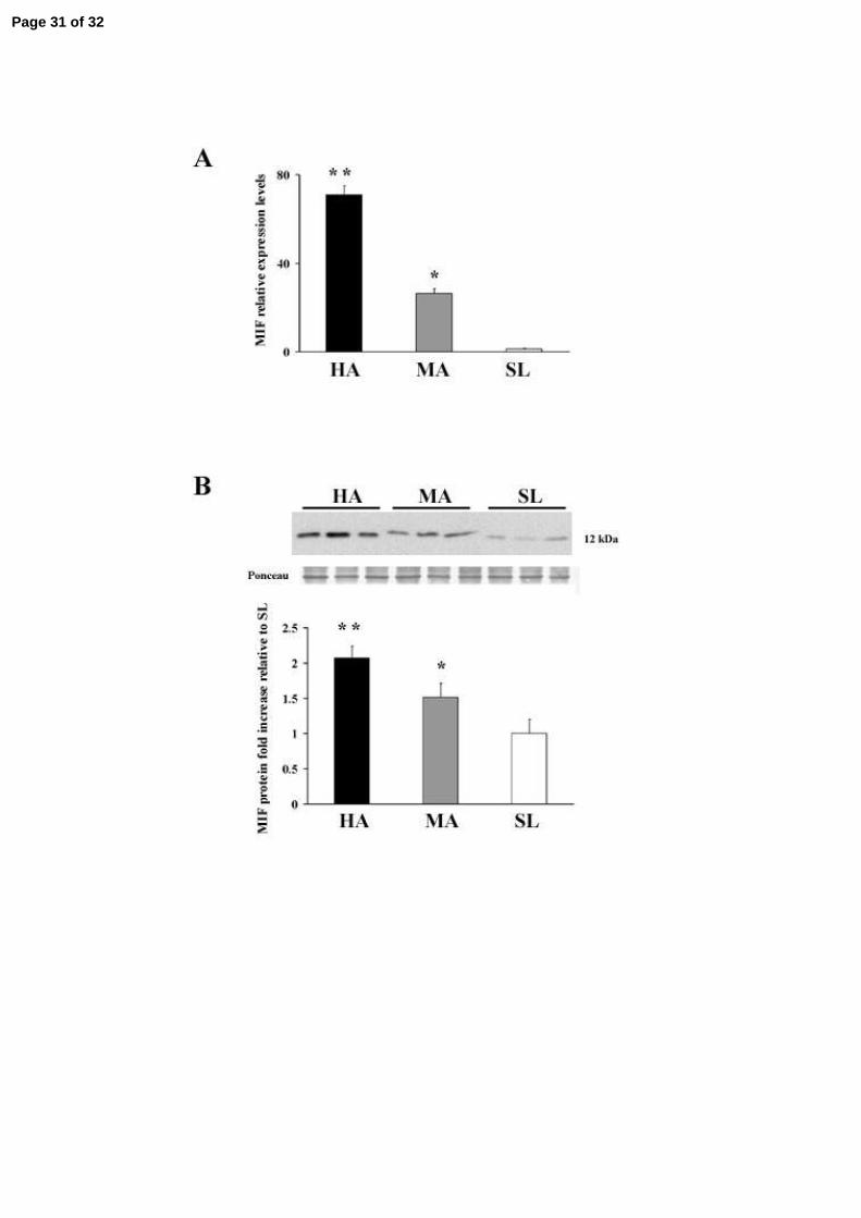
To further investigate the effect of low oxygen tension, we performed a quantitative analysis
of MIF transcript and protein in normal term placentae from women living at high (3100 masl)
moderate (1600 masl) and sea level altitude. MIF mRNA expression increased by 70-fold
(p=0.004), and by 26-fold (p=0.001) in high and moderate-altitude vs sea level placentae,
respectively (Figure 4A). MIF Protein expression was parallel: densitometric analyses revealed a 2-
fold increase in MIF at 3100 masl vs. sea level (p<0.05) and a 1.5 fold increase at 1600 masl vs. SL
(p <0.05 Figure 4B). No difference in MIF mRNA and protein expression levels were noted
between vaginal delivery and caesarean section (data not shown).
Page 13 of 32

DISCUSSION
The present findings demonstrate for the first time that MIF expression in human placenta is
upregulated by low oxygen tension, both in in vivo and in vitro placenta hypoxia. In this study, we
found that MIF mRNA and protein in placental tissues peak at 7-10 weeks of gestation when
oxygen is low, while the levels decrease at 11-12 weeks and thereafter when the blood flow is fully
established and oxygen tension increases. In vitro experiments confirmed that reduced Po2 is a
powerful inducer of MIF expression from early first trimester trophoblast cells. Explants maintained
under low (3%) oxygen tension exhibited significantly higher levels of MIF transcript and protein
as well as a higher MIF secretion into conditioned medium with respect to standard (20%) oxygen
conditions. Finally we showed that under conditions of chronically reduced oxygen tension in vivo,
at the physiological equivalent of 18% inspired O2 at 1600 m and 15% at 3100 m, MIF expression
in placental tissues paralleled that of our in vitro experiments at 8 and 3% O2 respectively. This
remarkable gradation of MIF response in relation to relatively subtle changes in oxygen suggests
exquisite sensitivity of MIF regulation to oxygen tension. The present findings are consistent with
our recent data showing a pattern of global gene expression that is similar in placentae from first
trimester and high altitude pregnancies as well as first trimester villous explants maintained at 3%
oxygen (39). Our results suggest MIF is one of the genes accounting for similarity between in vivo
and in vitro models of placental hypoxia.
MIF expression, normally constitutive at low levels, can be induced by increased glucose
levels in Β-cells of the pancreatic islet and adipocytes (46, 37), mitogens in T-cells (7),
corticotrophin-releasing factor (CRF) in the anterior pituitary cells and lipopolysaccharide (LPS) in
monocytes-macrophages (45, 15). More recently, it was shown that human chorionic gonadotrophin
(hCG) increases MIF secretion in granulosa cell cultures (44). Consistent with our experiments
testing the influence of HIF-1alpha on MIF expression, new studies on tumor cell lines and cardiac
myocytes indicate that MIF is one of the hypoxia-induced genes characterizing tumor phenotypes
and that upregulation of MIF mRNA and protein occurs under hypoxic conditions (41, 25, 8).
Page 14 of 32

The presence of MIF has been widely demonstrated in tissues and fluids during pregnancy.
MIF transcript and protein are expressed in first trimester trophoblast and evidence of a MIF-like
protein was shown in term placenta (5, 49). MIF is also expressed by term extraembryonic
membranes and high concentrations of MIF were detected in amniotic fluid and maternal serum
(19). Although MIF is ubiquitous, increasing or decreasing concentrations of this cytokine have
been correlated with various physiological and pathological events during pregnancy. A recent
report by Yamada et al. (47) showed decreased MIF plasma levels during early gestation in women
with recurrent miscarriages. We previously reported that MIF levels in amniotic fluid are higher at
term that in midgestation and higher in laboring than in non laboring women and this is possibly
due to the local secretion by fetal membranes (19). More recently we found that maternal serum
MIF is significantly higher in patients affected by severe preeclampsia (42) a clinical condition
known to be associated with reduced utero-placental perfusion and placental hypoxia (39),
suggesting that there is increased placental MIF production and secretion into the maternal
circulation in response to placental ischemia.
Whether oxygen has a direct effect on MIF induction remains to be elucidated. We have
previously reported that in the early phases of placental development, low oxygen tension is
associated with increased expression and transcriptional activity of HIF-1α (16), a basic helix-loop-
helix pre arnt sim (PAS) domain transcription factor. HIF-1α mediates the transcriptional response
to oxygen by binding to hypoxia response elements (HRE) in the promoter region of TGFβ3, the
molecule that mediates the biological effects of low-oxygen environment on the early events of
trophoblast differentiation (38). Interestingly, we found that following treatment of explants with
DMOG an inhibitor of HIF-1α degradation (21), high MIF expression levels were preserved also in
20% of oxygen. Moreover, using in silico analysis we have recently identified three putative
binding sites for HIF-1α in the promoter region of the human MIF gene (data not shown). Such data
suggest that the HIF system is responsible for the graded MIF response to changes in oxygen
tension.
Page 15 of 32

The specific role of MIF during placentation remains unclear. MIF is an important regulator of cell
proliferation and differentiation (48, 26). Normal placental development is dependent upon
proliferation of the cytotrophoblast and further differentiation of the various trophoblast cell
populations: the syncytiotrophoblast which forms the epithelial covering of the villous tree and is
the main endocrine component of the placenta, the villous cytotrophoblast which represents the
trophoblast stem cell population that proliferates throughout pregnancy and fuses to generate the
syncytiotrophoblast layer, and the extravillous trophoblast cells which anchor the villi to the
maternal uterus. It is well established that during early placental development low oxygen
environment supports the early events of trophoblast differentiation including proliferation of the
undifferentiated villous cytotrophoblast (18, 16) and of the extravillous trophoblast cells forming
the proximal column of the anchoring villi (16). Higher MIF expression in the early stages of
placentation and its localization to the villous cytotrophoblast suggests that MIF might contribute to
sustain the trophoblast proliferative phenotype, typical of low oxygen environment. Consistent with
our previous reports (5), we showed that immunostaining for MIF in the proliferative villous
cytotrophoblast and extravillous trophoblast was drastically reduced by the 13th week of gestation.
First trimester explants also showed MIF protein and transcript expression in the proliferative
cytotrophoblast and in the extravillous trophoblast with prominent staining in the cellular outgrowth
of cultures maintained in 3% O2. Intranuclear MIF immunoreactivity observed in trophoblast cells,
particularly in 3% O2 explants, seems to be in agreement with reports on other cell types possessing
a high proliferation index, e.g. pituitary adenoma and lung adenocarcinoma cells (35, 23). The
proliferative response to MIF stimulation in fibroblasts is associated with phosphorylation and
activation of p44/p42 ERK (extracellular signal-regulated kinase 1/2) (30). In human placenta, the
expression of ERK1 and ERK2 is localized in the proliferative cytotrophoblast and, interestingly,
expression of active forms of ERK1/ERK2 is much higher in first trimester placental tissues
compared to second trimester (24). The primary action of MIF is on macrophages, where it inhibits
their migration and stimulates their scavenger activity at the site of inflammation (10). Macrophages
Page 16 of 32

constitute the majority of immune cell types populating the endometrium and decidua, and their
number increases during early pregnancy (29). Decidual macrophages help to remove cellular
debris, thereby facilitating the invasion of trophoblast into the maternal tissues (2). We showed that
in vitro explants release high levels of MIF under reduced oxygenation. Hence MIF could be a
paracrine mediator in early gestation, determining macrophage accumulation and activation.
Alternatively, MIF could also suppress activity of decidual NK cells, an important population in the
maternal decidua, particularly at the implantation site (43). Indeed, MIF inhibits NK cell-mediated
cytolysis of both neoplastic and normal target cells (4). Although the functions of decidual NK cells
must be clarified, inhibition of their cytolytic activity could be a critical mechanism of
immunotolerance at the feto-maternal interface.
Although these studies support a central role of MIF in human pregnancy, MIF -/- mice
failed to show reduced fertility (11). However from the relevant report, litter sizes, pup weight and
other indicators of functional pregnancy outcome were not examined (11). Additionally, MIF has
complex interactions with other cytokines, i.e. TNF-α, IFN-γ and IL-1β (13). Hence, it can be
hypothesized that other cytokines compensate for the deficiency of MIF.
In view of the multifunctional aspects of MIF, our results in the human placenta, showing a
higher expression in the earlier phases of pregnancy and MIF upregulation by low oxygen
conditions, suggest a major role of MIF in the control of trophoblast growth and in the modulation
of maternal immune tolerance. Of clinical importance, reduced placental perfusion leading to
placental hypoxia/ischemia might induce the synthesis of placental MIF. This might account for the
high MIF serum levels in patients affected by severe preeclampsia (42).
Page 17 of 32

ACKNOWLEDGMENT
We thank Dr. Ljiljiana Petkovic for placental collection. We also would like to thank Dr. Kent L.
Thornburg for carefully reading the manuscript. This work was supported by the Canadian Institutes
of Health Research (CIHR) Grant (MT1406) to Isabella Caniggia, by research grants from the
University of Siena to Luana Paulesu and NIH HD042737 grant to Stacy Zamudio. Isabella
Caniggia is the recipient of a mid-career CIHR Award administered through the Ontario Women's
Health Council.
Page 18 of 32
REFERENCES
1. Abe R, Shimizu T, Ohkawara A, Nishihira J. Enhancement of macrophage migration
inhibitory factor (MIF) expression in injured epidermis and cultured fibroblasts. Biochim
Biophys Acta. 1500: 1-9, 2000.
2. Abrahams VM, Kim YM, Straszewski SL, Romero R, Mor G. Macrophages and
apoptotic cell clearance during pregnancy. Am J Reprod Immunol 51: 275-282, 2004.
3. Aplin JD. Hypoxia and human placenta development. J Clin Invest 105: 559-560, 2000.
4. Apte RS, Sinha D, Mayhew E, Wistow GJ, Niederkorn JY. Cutting edge: role of
macrophage migration inhibitory factor in inhibiting NK cell activity and preserving
immune privilege. J Immunol 160: 5693-5696, 1998.
5. Arcuri F, Cintorino M, Vatti R, Carducci A, Liberatori S, Paulesu L. Expression of
macrophage migration inhibitory factor transcript and protein by first-trimester human
trophoblasts. Biol Reprod 60: 1299-1303, 1999.
6. Arcuri F, Ricci C, Ietta F, Cintorino M, Tripodi SA, Cetin I, Garcia E, Schatz F, Klemi
P, Santopietro R, Paulesu L. Macrophage migration inhibitory factor in the human
endometrium: expression and localization during the menstrual cycle and early pregnancy.
Biol Reprod 64: 1200-1205, 2001.
7. Bacher M, Metz CN, Calandra T, Mayer K, Chesney J, Lohoff M, Gemsa D, Donnelly
T, Bucala R. An essential regulatory role for macrophage migration inhibitory factor in T-
cell activation. Proc Natl Acad Sci U S A 93: 7849-7854, 1996.
8. Bacher M, Schrader J, Thompson N, Kuschela K, Gemsa D, Waeber G, Schlegel J. Up-
regulation of macrophage migration inhibitory factor gene and protein expression in glial
tumor cells during hypoxic and hypoglycemic stress indicates a critical role for angiogenesis
in glioblastoma multiforme. Am J Pathol 162: 11-17, 2003.
Page 19 of 32

9. Benyo DF, Miles TM, Conrad KP. Hypoxia stimulates cytokine production by villous
explants from the human placenta. J Clin Endocrinol Metab 82: 1582-1588, 1997.
10. Bloom BR, Bennett B. Mechanism of a reaction in vitro associated with delayed-type
hypersensitivity. Science 153: 80-82, 1966.
11. Bozza M, Satoskar AR, Lin G, Lu B, Humbles AA, Gerard C, David. Targeted
disruption of migration inhibitory factor gene reveals its critical role in sepsis. J Exp Med
189: 341-346, 1999.
12. Bradford MM. A rapid and sensitive method for the quantification of microgram quantities
of protein utilizing the principle of protein-dye binding. Anal Biochem 72: 248-254, 1976.
13. Calandra T, Bernhagen J, Metz CN, Spiegel LA, Bacher M, Donnelly T, Cerami A,
Bucala R. MIF as a glucocorticoid-induced modulator of cytokine production. Nature 377:
68-71, 1995
14. Calandra T, Bernhagen J, Mitchell RA, Bucala R. The macrophage is an important and
previously unrecognized source of macrophage migration inhibitory factor. J Exp Med 179:
1895-1902, 1994.
15. Calandra T, Spiegel LA, Metz CN, Bucala R. Macrophage migration inhibitory factor is a
critical mediator of the activation of immune cells by exotoxins of Gram-positive bacteria.
Proc Natl Acad Sci U S A 95: 11383-11388, 1998.
16. Caniggia I, Mostachfi H, Winter J, Gassmann M, Lye SJ, Kuliszewski M, Post M.
Hypoxia-inducible factor-1 mediates the biological effects of oxygen on human trophoblast
differentiation through TGFbeta(3). J Clin Invest 105: 577-587, 2000.
17. David JR: Delayed hypersensitivity in vitro : its mediation by cell-free substances formed
by lymphoid cell-antigen interaction. Proc Natl Acad Sci USA 56: 72-77, 1966.
18. Genbacev O, Zhou Y, Ludlow JW, Fisher SJ. Regulation of human placental
development by oxygen tension. Science 277: 1669-1672, 1997.
Page 20 of 32

19. Ietta F, Todros T, Ticconi C, Piccoli E, Zicari A, Piccione E, Paulesu L. Macrophage
migration inhibitory factor in human pregnancy and labor. Am J Reprod Immunol 48: 404-
409, 2002.
20. Ietta F, Wu Y, Winter J, Xu J, Wang J, Post M, Caniggia I. Dynamic HIF-1alpha
regulation during human placental development. Biol Reprod 75: 112-121, 2006.
21. Jaakkola P, Mole DR, Tian YM, Wilson MI, Gielbert J, Gaskell SJ, Kriegsheim Av,
Hebestreit HF, Mukherji M, Schofield CJ, Maxwell PH, Pugh CW, Ratcliffe PJ.
Targeting of HIF-a to the von Hippel-Lindau ubiquitylation complex by O2-regulated prolyl
hydroxylation. Science 292: 468-472, 2001.
22. Jauniaux E, Watson AL, Hempstock J, Bao YP, Skepper JN, Burton GJ. Onset of
maternal arterial blood flow and placental oxidative stress. A possible factor in human early
pregnancy failure. Am J Pathol 157: 2111-2122, 2000.
23. Kamimura A, Kamachi M, Nishihira J, Ogura S, Isobe H, Dosaka-Akita H, Ogata A,
Shindoh M, Ohbuchi T, Kawakami Y. Intracellular distribution of macrophage migration
inhibitory factor predicts the prognosis of patients with adenocarcinoma of the lung. Cancer
89: 334-341, 2000.
24. Kita N, Mitsushita J, Ohira S, Takagi Y, Ashida T, Kanai M, Nikaido T, Konishi I.
Expression and activation of MAP kinases, ERK1/2, in the human villous trophoblast.
Placenta 24: 164-172, 2003.
25. Koong AC, Denko NC, Hudson KM, Schindler C, Swiersz L, Koch C, Evans S,
Ibrahim H, Le QT, Terris DJ, Giaccia AJ. Candidate genes for the hypoxic tumor
phenotype. Cancer Res 60: 883-887, 2000.
26. Lanahan A, Williams JB, Sanders LK, Nathans D. Growth factor-induced delayed early
response genes. Mol Cell Biol 12: 3919-3929, 1992.
27. Livak KJ, Schmittgen TD. Analysis of relative gene expression data using real-time
quantitative PCR and the 2(-Delta Delta C(T)) Method. Methods 25: 402-408, 2001.
Page 21 of 32

28. MacPhee DJ, Mostachfi H, Han R, Lye SJ, Post M, Caniggia I. Focal adhesion kinase is
a key mediator of human trophoblast development. Lab Invest 81: 1469-1483, 2001.
29. Miller L., Hunt J. Sex steroids hormones and macrophage functions. Life Sciences 59: 1-
14, 1996.
30. Mitchell RA, Metz CN, Peng T, Bucala R. Sustained mitogen-activated protein kinase
(MAPK) and cytoplasmic phospholipase A2 activation by macrophage migration inhibitory
factor (MIF). Regulatory role in cell proliferation and glucocorticoid action. J Biol Chem
274: 18100-18106, 1999.
31. Moore NK, Viselli SM. Staining and quantification of proteins transferred to
polyvinylidene fluoride membranes. Anal Biochem 279: 241-242, 2000.
32. Nevo O, Soleymanlou N, Wu Y, Xu J, Kingdom J, Many A, Zamudio S, Caniggia I.
Increased Expression of sFlt-1 in In Vivo and In Vitro Models of Human Placental Hypoxia
is Mediated by HIF-1. Am J Physiol Regul Integr Comp Physiol. 2006 in press.
33. Orsini B, Ciancio G, Censini S, Surrenti E, Pellegrini G, Milani S, Herbst H, Amorosi
A, Surrenti C. Helicobacter pylori cag pathogenicity island is associated with enhanced
interleukin-8 expression in human gastric mucosa. Digest Liver Dis 32: 458-467, 2000.
34. Pfaffl MW, Horgan GW, Demple L. Relative expression software (REST) for group-
wise comparison and statistical anlysis of relative expression results in real-time PCR.
Nucleic Acids Res 30: e36, 2002.
35. Pyle ME, Korbonits M, Gueorguiev M, Jordan S, Kola B, Morris DG, Meinhardt A,
Powell MP, Claret FX, Zhang Q, Metz C, Bucala R, Grossman AB. Macrophage
migration inhibitory factor expression is increased in pituitary adenoma cell nuclei. J
Endocrinol 176: 103-110, 2003.
36. Rodesch F, Simon P, Donner C, Jauniaux E. Oxygen measurements in endometrial and
trophoblastic tissues during early pregnancy. Obstet Gynecol 80: 283-285, 1992.
Page 22 of 32

37. Sakaue S, Nishihira J, Hirokawa J, Yoshimura H, Honda T, Aoki K, Tagami S,
Kawakami Y. Regulation of macrophage migration inhibitory factor (MIF) expression by
glucose and insulin in adipocytes in vitro. Mol Med 5: 361-371, 1999.
38. Schaffer L, Scheid A, Spielmann P, Breymann C, Zimmermann R, Meuli M,
Gassmann M, Marti HH, Wenger RH. Oxygen-regulated expression of TGF-beta 3, a
growth factor involved in trophoblast differentiation. Placenta 24: 941-950, 2003.
39. Soleymanlou N, Jurisica I, Nevo O, Ietta F, Zhang X, Zamudio S, Post M, Caniggia I.
Molecular Evidence of Placental Hypoxia in Preeclampsia. J Clin Endocrinol Metab 90:
4299-4308, 2005.
40. Suzuki H, Kanagawa H, Nishihira J. Evidence for the presence of macrophage migration
inhibitory factor in murine reproductive organs and early embryos. Immunol Lett 51: 141-
147, 1996.
41. Takahashi M, Nishihira J, Shimpo M, Mizue Y, Ueno S, Mano H, Kobayashi E, Ikeda
U, Shimada K. Macrophage migration inhibitory factor as a redox-sensitive cytokine in
cardiac myocytes. Cardiovasc Res 52: 438-445, 2001.
42. Todros T, Bontempo S, Piccoli E, Biolcati M, Ietta F, Romagnoli R, Castellucci M,
Paulesu L. Increased levels of macrophage migration inhibitory factor (MIF) in
preeclampsia. Eur J Obstet Gynecol Reprod Biol 123: 162-166, 2005.
43. Trundley A., Moffet A. Human uterine leukocytes and pregnancy. Tissues Antigens 63: 1-
12, 2004.
44. Wada S, Kudo T, Kudo M, Sakuragi N, Hareyama H, Nishihira J, Fujimoto S.
Induction of macrophage migration inhibitory factor in human ovary by human chorionic
gonadotrophin Hum Reprod 14: 395-399, 1999.
45. Waeber G, Thompson N, Chautard T, Steinmann M, Nicod P, Pralong FP, Calandra
T, Gaillard RC Transcriptional activation of the macrophage migration-inhibitory factor
gene by the corticotropin-releasing factor is mediated by the cyclic adenosine 3',5'-
Page 23 of 32

monophosphate responsive element-binding protein CREB in pituitary cells. Mol
Endocrinol 12: 698-705, 1998.
46. Waeber G, Calandra T, Roduit R, Haefliger JA, Bonny C, Thompson N, Thorens B,
Temler E, Meinhardt A, Bacher M, Metz CN, Nicod P, Bucala R. Insulin secretion is
regulated by the glucose-dependent production of islet beta cell macrophage migration
hibitory factor. Proc Natl Acad Sci U S A 94: 4782-4787, 1997.
47. Yamada H, Kato EH, Morikawa M, Shimada S, Saito H, Watari M, Minakami H,
Nishihira J. Decreased serum levels of macrophage migration inhibition factor in
miscarriages with normal chromosome karyotype. Hum Reprod 18: 616-620, 2003.
48. Yang Y, Degranpre P, Kharfi A, Akoum A. Identification of macrophage migration
inhibitory factor as a potent endothelial cell growth-promoting agent released by ectopic
human endometrial cells. J Clin Endocrinol Metab 85: 4721-4727, 2000.
49. Zeng FY, Weiser WY, Kratzin H, Stahl B, Karas M, Gabius HJ. The major binding
protein of the interferon antagonist sarcolectin in human placenta is a macrophage migration
inhibitory factor. Arch Biochem Biophys 303: 74-80, 1993.
Page 24 of 32

FIGURE LEGENDS
Figure 1. MIF mRNA and protein expression during placental development. A. Level of MIF
mRNA expression assessed by qRT-PCR analysis in placental tissues during gestation (n = 6 for
each gestational window). Significantly higher level of MIF mRNA expression was found at 7-10
weeks of gestation. * p<0.05 versus term placental tissues. B. Upper panel: representative Western
blot of total placental lysates at different stages of gestation. A single band of approximate
molecular weight of 12kDa corresponding to MIF was obtained in each specimen. Ponceau
staining shows equal protein loading. Lower panel: densitometric analisys of MIF protein level in
placental tissues at 5-10 weeks of gestation (n=5), 11-12 weeks of gestation (n=6), 14-20 weeks of
gestation (n=6) and term control placental tissues (n=6). * p<0.05 versus term placental tissues. C.
Immunohistochemical localization of MIF in placental tissues at 6 and 13 weeks of gestation.
Immunohistochemistry was performed with avidin-biotin-peroxidase complex (ABC) method and
anti-human MIF monoclonal antibodies. Brownish staining represents immunopositivity. Strong
immunoreactivity was present in first trimester villous cytotrophoblast (arrow) and extravillous
trophoblast of the invading columns (EVT). MIF immunoreactivity was also present in the stroma
while the syncytiotrophoblast (arrowhead) was negative. By week 13, a reduced immunostaining
was observed in the cytotrophoblast cells (arrows) and in some cells of the distal part of the cellular
extravillous columns (EVT). Negative control was obtained by replacing the primary antibody with
normal murine immunoglobulins. Slides were counterstained with haematoxylin. X40 original
magnifications.
Figure 2. Oxygen regulation of MIF expression and secretion in first trimester chorionic
villous explants. A. Level of MIF mRNA expression assessed by qRT-PCR analysis in placental
Page 25 of 32

explants (n=5 sets of placental explants) maintained at 20% O2 (white bar), 8% O2 (grey bar) and
3% O2 (black bar) for 48 hours. Significantly higher levels of MIF expression were found at 3% and
8% O2 versus 20% O2. **p<0.005 and *p<0.05. B. MIF concentration, detected by ELISA, in
culture medium of placental explants at 20% (white bar) and 3% (black bar) O2 (n=5 sets of
placental explants). Results are expressed as fold increase. Significantly higher levels of MIF were
detected at 3% O2 versus 20 % O2. * p<0.05. C. qRT-PCR analysis in placental explants (n=3 sets
of placental explants) maintained at 20% O2 (white bar), 3% O2 (black bar) or kept overnight at 3%
O2 then, at 20% O2 plus DMOG (grey bar) for another 24 hours. Significantly higher level of MIF
mRNA was observed in DMOG-treated cultures when compared to 20% O2. *p<0.05. D. Left
panel: representative Western blot analysis in placental explants maintained at 3%, 20% O2 or first
in 3% then 20% O2 plus DMOG as above. A single band with an apparent molecular weight of
12kDa corresponding to MIF was obtained. Ponceau staining shows equal protein loading. Right
panel: densitometric analysis of MIF protein in explants (n= 3 sets of placental explants) maintained
at 3%, 20% O2 or first in 3% then 20% O2 plus DMOG. *p<0.05 versus 20% O2.
Figure 3. MIF localization in first trimester villous placental explants. A. In situ hybridization
for MIF mRNA in paraffin sections of placental explants maintained at 20% and 3% O2. Bright-
field photomicrograph with 35S-labelled anti-sense MIF RNA probe in 20% and 3% O2 (left panels).
Dark-field photomicrograph with 35S-labelled anti-sense MIF RNA probe in 20% and 3% O2 (right
panels). Strong reactivity (depicted as white bright dots) was observed in the villous cytotrophoblast
and extravillous trophoblast of 3% O2 culture explants. A reduced reactivity was depicted in the
20% O2 explant cultures. B. Immunohistochemical localization of MIF in villous explants cultured
under 20% and 3% O2. Positive immunoreactivity (depicted as brownish staining) was observed as
intranuclear immunostaining in the villous cytotrophoblast (arrows) and extravillous trophoblast
cells (EVT) both in 20% and 3% O2 explant cultures. Syncytiotrophoblast was negative
Page 26 of 32

(arrowheads). Negative control was obtained by replacing the primary antibody with normal murine
immunoglobulins. EVT: extravillous cytotrophoblast. 40X original magnification.
Figure 4. MIF mRNA and protein expression in high altitude placental tissues. A. Level of
MIF mRNA expression assessed by qRT-PCR analysis of placental tissues from high altitude (HA)
(3100 m asl) (n = 4 separate experiments) , moderate altitude (MA) (1600 m asl) (n = 4 ) and sea
level (SL) (n = 4). Significantly higher level of MIF expression was found in HA (* p<0.005) and
MA (** p<0.05) placental tissues compared to sea level placentae; B. Upper panel: representative
Western blot analysis of MIF performed on placenta lysates from HA, MA and SL showing a single
band of 12 kDa. Ponceau staining shows equal protein loading. Lower panel: densitometric analysis
of MIF protein level in HA (n = 8), MA (n=8), SL (n=8). Results are expressed as fold increase. *
p<0.005 versus sea level; ** p<0.05 versus sea level.
Page 27 of 32

Page 28 of 32

Page 29 of 32

Page 30 of 32
Page 31 of 32

Table 1. Clinical parameters of participants.
Term Control n = 10
Moderate Altitude n = 12
High Altitude n = 12
Mean Maternal Age (Range in years) 33 ± 4.5 29.0 ± 2.5 29.0 ± 6.
Mean Gestational Age (Range in weeks)
39.6 ± 0.9
(38-41)
39.4 ± 1.4 (37-41)
39.1 ± 1.3 (37-41)
Blood Pressure (mm Hg)
(S:Systolic; D: Diastolic)
S: 111 ± 6.0 D: 67 ± 6.3
S: 113 ± 5.0 D: 72 ± 5.0
S: 116 ± 8.0 D: 72 ± 5.0
Proteinuria
Absent
Absent Absent
Edema
Absent
Absent Absent
Fetal Weight (g)
A.G.A.:
3328 ± 421
A.G.A:
3553 ± 312
A.G.A:
3060 ± 190
Mode of Delivery CS: 2 VD: 8
CS: 3 VD: 9
CS: 2 VD: 10
Data are represented as mean ± standard deviation. A.G.A.: Appropriate for gestational age; VD:
Vaginal delivery; CS: Caesarian section delivery.
Page 32 of 32