The inhibitory cascade in marsupials
86
______________________________________________________________ 11 th International Conference on Tooth Morphogenesis and Differentiation Organized by Laurent VIRIOT (Chair), Françoise BLEICHER, and Vincent LAUDET Plenary poster sessions abstract program May 26-31, 2013 - La Londe Les Maures (France)
Transcript of The inhibitory cascade in marsupials

______________________________________________________________
11th International Conference on Tooth Morphogenesis and Differentiation
Organized by Laurent VIRIOT (Chair), Françoise BLEICHER, and Vincent LAUDET
Plenary poster sessions abstract program
May 26-31, 2013 - La Londe Les Maures (France)

TMD 2013 Tooth Development ______________________________________________________________________________
______________________________________________________________________________ Monday May 27: 13.30 → 14.30 Poster presentation PA1
Understanding the mechanisms of root formation from rootless tooth Cheol-Hyeon Bae*, Eui-Sic Cho
Cluster for Craniofacial Development and Regeneration Research, Institute of Oral Biosciences, Chonbuk National University School of Dentistry, Jeonju, South Korea
* presenting author: [email protected] BACKGROUND: The Wnt family of proteins plays an important role in morphogenesis and cellular differentiation in many tissues [1]. It is well-known that Wnt/ß-catenin signaling plays multiple roles in various stages of tooth morpho-genesis [2]. However, little is known about the involvement of Wnt/ß-catenin signaling in odon-toblast differentiation. The tooth root is important part of tooth together with surrounding periodon-tium to maintain tooth in alveolar socket [3]. While the molecular and cellular mechanisms of early tooth development and crown morphogene-sis have been extensively studied, little is known about the molecular mechanisms controlling tooth root formation.
METHODOLOGY: We generated and analyzed mice with tissue-specific inactivation of ß-catenin, an obligatory transducer of canonical Wnt signaling, in developing odontoblasts to directly investigate the role of Wnt/ß-catenin signaling in root formation.
PRINCIPAL FINDINGS: Here, we show that ß-catenin is strongly expressed in odontoblast li-neage cells, and it is required for root formation (Fig. A-B). Tissue-specific inactivation of ß-catenin in developing odontoblasts produced mo-lars lacking roots and aberrantly thin incisors (Fig. C-D). At the beginning of root formation in the mutant molars, the cervical loop epithelium extended apically to form Hertwig’s epithelial root sheath (HERS), but root odontoblast diffe-rentiation was disrupted and followed by the loss of some HERS inner layer cells. However, outer layer of HERS extended without the root (Fig. E-F), and the mutant molars finally erupted. The periodontal tissues invaded extensively into the dental pulp (Fig. G-H).
Legend: Expression of ß-catenin in the odontoblast lineage cells and tooth phenotypes in mice with tissue-specific inactivation of ß-catenin in developing odon-toblasts. (A) Localization of ß-catenin in the odonto-blast lineages during root formation in mouse molar (B) OC-Cre activities in the differentiating odonto-blasts during root formation. (C-D) Molars lacking roots and thin incisors in OC-Cre:Ctnnb1CO/CO mice (P28). (E-F) Disrupted differentiation of root odonto-blasts in OC-Cre:Ctnnb1CO/CO mice at the beginning of root formation (P8). (G-H) Erupted molars without root formation (P28).
DISCUSSION and CONCLUSIONS: In this study, we investigated the functional significance of Wnt/ß-catenin signaling in the dental mesen-chyme during tooth root formation. OC-Cre;Ctnnb1CO/CO mice exhibited remarkable tooth phenotypes characterized by erupted molars lack-ing roots and thin incisors. This abnormality was closely associated with odontoblast differentia-tion. The results of the present study further imp-ly that Wnt/ß-catenin signaling in the dental me-senchyme is required to differentiate odontoblasts for root formation.
REFERENCES: [1] Logan and Nusse. Annu Rev Cell Dev Biol (2004). [2] Liu and Millar. J Dent Res (2010). [3] Thomas HF. Int J Dev Biol (1995).
Funding: This work was supported by National Re-search Foundation of Korea (NRF) grants, funded by the Korean government (MEST) (Nos. 2009-0085733 and 2012R1A1A2000919).

TMD 2013 Tooth Development ______________________________________________________________________________
______________________________________________________________________________ Monday May 27: 13.30 → 14.30 Poster presentation PA2
The pattern of cementum formation on the developing roots in mice during stages P12–P36
Valentina Woth*, Isabel Nowak, Herbert Renz, Ralf. J. Radlanski Charité -Campus Benjamin Franklin at Freie Universität Berlin, Center for Dental and Cra-
niofacial Sciences, Dept. of Craniofacial Developmental Biology, Berlin, Germany
* presenting author: valentina.woth@charité.de BACKGROUND: Cementum formation is es-sential for the formation of the tooth-bone inter-face, because of the insertion of the periodontal fibers. Different types of cementum, primarily as acellular cementum on the cervical root and cellu-lar cementum covering the apical root have been studied by many authors [2,3,4]. However, it has not been described in detail, how the temporo-spacial formation and morphogenesis of dental cementum takes place on the root. The purpose of this study is to elucidate the pattern of dental cementum formation on the surfaces of develop-ing roots in mice.
METHODOLOGY: 13 Mice, ranging from postnatal day 12-36, were prepared as serial histo-logical sections (thickness 10 μm) and stained with H.E., Alcianblue and Trichrom. 3-D- recon-structions were made using the software Analy-SIS (Olympus, Berlin). Regions of dental cemen-tum on the developing roots of mandibular M1, M2, M3 were identified in the histological sec-tions, marked and reconstructed in 3-D together with dentin and pulp.
PRINCIPAL FINDINGS: Alcianblue turned out to be a specific staining for identifying acellular cementum. A thin layer of acellular cementum could be found on the root surface as soon as it was formed itself (stage P12). However, forma-tion of cellular cementum lagged behind the for-mation of the root, starting at stage P 20. There-fore, large areas of root surface remain uncovered from cellular cementum.
DISCUSSION and CONCLUSIONS: Our study showed that cementum formation seems not to be a uniformly layered process – instead, we revealed an insular formation pattern for specific regions of cementum. This specific pattern and location of dental cementum formation of the root
Legend: M1-M3 at P36. Cellular cementum is shown in blue-grey.
plays an important role for further development of fiber insertion leading to tooth-bone- osseo-integration. Insights like these are crucial for a possible, future implantation of extracorporal cultured tooth germs into an adult jaw.
REFEFENCES: [1] Foster Int. J. of Oral Sci. (2012); [2] Nanci et al. Periodontology (2006); [3] Radlanski Entwicklungsbiologie, Quintessenz Verlag (2011); [4] Schroeder The Periodontium, Springer Verlag (1986); [5] Shukla J. Forensic Odontostomatology (2012).
FUNDING: Supported by grants Ra 428/1-9. Deutsche Forschungsgemeinschaft. We thank Mrs. B. Danielowski, Mrs. I. Schwarz for their most valuable and skillfull assistance.

TMD 2013 Tooth Development ______________________________________________________________________________
______________________________________________________________________________ Monday May 27: 13.30 → 14.30 Poster presentation PA3
Precise chronology of differentiation of developing human primary dentition Xuefeng Hu1*, Shan Xu1, Bingmei Wang1, YiPing Chen1,2, Yanding Zhang1
1Fujian Key Laboratory of Developmental and Neuro Biology, College of Life Science, Fujian Normal University, Fuzhou, China; 2Department of Cell and Molecular Biology, Tulane University,
New Orleans, LA, USA * presenting author: [email protected]
BACKGROUND: While correlation of devel-opmental stage with embryonic age of the human primary dentition has been well documented, the available information regarding the differentiation timing of the primary teeth varies significantly. In this study, we aimed to document precise differ-entiation timing of the developing human primary dentition.
METHODOLOGY: Dental tissues were col-lected from chemically-terminated human em-bryos with well defined ages. Tissue sections were subjected to Hematoxylin/Eosin staining for standard histological analysis, and to dichromic staining for detection of mineralized tissues, and for immunohistochemical staining for expression of molecular markers.
PRINCIPAL FINDINGS: We systematically examined the expression of odontogenic differen-tiation markers along with the formation of min-eralized tissue in each developing maxillary and mandibular tooth from human embryos with well-defined embryonic age. We show that, despite that all primary teeth initiate development at the same time, odontogenic differentiation begins in the maxillary incisors at the 15th week and in the mandibular incisors at the 16th week of gestation, followed by the canine, the first primary premo-lar, and the second primary premolar at a week interval sequentially. Despite that the mandibular primary incisors erupt earlier than the maxillary incisors, this distal to proximal sequential differ-entiation of the human primary dentition coin-cides in general with the sequence of tooth erup-tion.
DISCUSSION and CONCLUSIONS: Although timing of eruption of each primary tooth differs significantly between each individual, the gesta-tion ages of tooth development at each stage dur-
ing human embryogenesis should be rather con-sistent. However, current information regarding the differentiation timing of the human primary dentition varies dramatically. Our results pre-sented here provide an accurate chronology of odontogenic differentiation in the developing human primary dentition, which could serve as a standard base for future studies of human tooth development and could also be used in textbook as a standard reference.
REFERENCES: [1] Welbury et al. Pediatric Denti-stry. 4th Ed. (2012) Oxford Press; [2] Schoenwolf et al. Larsen’s Human Embryology. 4th Ed. (2009). Chur-chill Livingston, Elsevier, Philadelphia, PA.
Funding: The 973 Project of China (2010CB944800); National Natural Science Foundation of China (81100730, 81271102); The Natural Science Founda-tion of Fujian Province (2012J01119).

TMD 2013 Tooth Development ______________________________________________________________________________
______________________________________________________________________________ Monday May 27: 13.30 → 14.30 Poster presentation PA4
The role of Fibrillin during tooth root and periodontal ligament tissue development
Kyoko Oka1*, Michiko Kira1, Eichi Tsuruga2, Hidemitsu Harada3, Naoki Fujiwara3, Yoshihiko Sawa2, Masao Ozaki1
1Pediatric Dentistry, Department of Oral Growth and Development, 2Functional structure, Department of Morphological Biology, Fukuoka Dental College, Fukuoka Japan; 3Developmental Biology and Regen-
erative Medicine, Department of Anatomy, Iwate Medical University, Yahaba, Japan * presenting author: [email protected]
BACKGROUND: Fibrillin microfibrils are unique architectural elements of the extracellular matrix (ECM) that endow connective tissues with specific physical properties, either as obligatory constituents of elastic fiber or as elastin-free as-semblies. Mutation of fibrillin genes result in the severe heritable connective tissue diseases such as Marfan syndrome. The oxytalan fibers are de-fined as fibrillin microfibril assemblies without elastin deposition and are specially located in periodontal ligament tissues (PDL) [1]. The bio-logical function of oxytalan fibers in periodontal ligaments of an erupted tooth has been reported in several articles [2]. However, the role of fibrillin during tooth development has not been fully un-derstood. Furthermore, fibrillin can bind TGF-βs through latent transforming growth factors β-binding proteins (LTBPs). The aim of this study is to examine and compare the localization and distribution of fibrillin-1 and -2 and TGF-βs dur-ing tooth development.
METHODOLOGY: Samples in vivo. Pregnant mice were prepared for collect embryos from embryonic stage 14.5 (E14.5) to post neonatal day 10 (P10). The dissected mouse head or man-dibular bones without chemical fixation and de-calcification were embedded in super cryo-embedding medium, and rapid-frozen by the hex-ane-dry ice method. The samples were cut with a cryostat into 8-µm-thick sections. Post neonatal samples were prepared by using the Film transfer method. In vitro: Murine HERS cells (HERS01a) were established by the limiting dilution-culture technique. They were cultured in DMEM/HAMF-12 medium with supplement in a humidified 5% CO2 atmosphere at 37°C [3]. Immunofluores-cence: Anti-Fibrillin-1, -2, TGF-β3, and Cytoke-ratin were used as the primary antibodies.
Legend: The expression of Fibrillin-1 and -2 at P5 in mouse.
The immunoreaction was visualized on sections with anti-IgG antibody conjugated with Alexa Fluor. Immuno-stained sections and cells were then counterstained with DAPI.
PRINCIPAL FINDINGS: In vivo analysis, Fi-brillin-1 expression started in periodontal liga-ment tissue around P10. This fiber-like expres-sion was attached with root surface (cementum) but not with alveolar bone. Interestingly, Fibril-lin-2 was expressed in dental epithelium since E14.5. This expression was observed in the cer-vical loop and Hertwig’s epithelial sheath during root development. TGF-β3 was also expressed in Hertwig’s epithelial sheath. In vitro analysis, HERS01a cells clearly expressed Fibrillin2.
DISCUSSION and CONCLUSIONS: These findings suggest that Fibrillin-2 protein not only plays a part in forming oxytalan fibers in PDL, but also potentially serves as a regulator for root formation via regulation of TGF-β signaling dur-ing tooth development. REFERENCES: [1] Tsuruga et al. J Periodontal Res. (2002); [2] Kondo et al. Acta Histchemica. (2011); [3] Akimoto et al. Biochem Biophys Res Commun. (2011).
Funding: Grant-in-Aid for Scientific Research from the Japanese Ministry of Education, Culture, Sports, Science and Technology [KAKENHI (24593116) to KO].

TMD 2013 Tooth Development ______________________________________________________________________________
______________________________________________________________________________ Monday May 27: 13.30 → 14.30 Poster presentation PA5
Spatio-temporal expression and possible function of tuftelin during mouse embryonic craniofacial development
Dekel Shilo1, Yoav Leiser1, Boaz Shay1, Asher Ornoy2, Anat Blumenfeld1, Dan Deutsch1* 1Dental Research Laboratory, Faculty of Dental Medicine, 2Laboratory of Teratology, Department of
Anatomy and Cell Biology, The Hebrew University of Jerusalem - Hadassah, Jerusalem, Israel * presenting author: [email protected]
BACKGROUND: Tuftelin is expressed in the epithelial ameloblasts at an early stage of amelogenesis. Tuftelin cDNA sequences were detected in morula and embryonic stem cells as well as in many different soft tissues, normal and cancerous. The tuftelin protein was also detected in different soft tissues such as eye, brain, kidney, lung and testis. Little was known about tuftelin expression in the developing embryo, where tuftelin was proposed to be involved in epithelial mesenchymal reciprocal interactions. In addition, tuftelin-soaked agarose beads induced recruitment and proliferation of embryonic mesenchymal cells.
METHODOLOGY: Embryos from pregnant CD-1 mice aged E10.5-E18.5 and newborns aged P3 were used in this study. All experiments were approved by the Animal Care Ethical Committee of The Hebrew University of Jerusalem. Heads were removed and mRNA and protein were ex-tracted. Real-time quantitative PCR was per-formed using TaqMan® AOD. Indirect immuno-histo- chemistry and In-situ hybridization were performed on craniofacial sections and Western blot was performed on the extracted protein.
PRINCIPAL FINDINGS: (1). The expression of tuftelin mRNA in the cranio-facial complex at E10.5 up to E16.5, was analyzed by RT-PCR, followed by cDNA sequencing and compared to endogenous control Actin. Two known tuftelin isoforms were detected along the different stages of mouse embryonic cranio-facial development; one containing all 13 exons of tuftelin and one lacking exon 2. (2). Tuftelin mRNA expression level increased during mouse embryonic cranio-facial development from E10.5 up to E16.5. Tuftelin protein was also detected. (3). Tuftelin is expressed in various tissues of the developing
mouse embryonic craniofacial complex such as brain, eye, ganglion, cartilage and bone, and is already expressed at E10.5 in the brain and eye, long before the initiation of tooth formation. Tuftelin protein expression was detected in the tooth germ (dental lamina) already at E12.5, much earlier than previously reported (E17).
DISCUSSION and CONCLUSIONS: In the developing tooth, tuftelin protein is already ex-pressed from the initiation of tooth germ forma-tion at E12.5 and is continuously expressed throughout tooth development, at least up to P3 (the latest age tested). The expression pattern of tuftelin protein and mRNA exhibits dynamic spa-tio-temporal changes in the various tissues. Tufte-lin protein expression was detected in the cranio-facial complex already at E10.5 (brain, eye), long before the initiation of tooth formation, suggest-ing possible additional role(s) for tuftelin other than those suggested in the past in epithelial-mesenchymal reciprocal interactions during tooth germ development. At younger embryonic age tuftelin protein expression was detected mainly in the cytoplasm while at later embryonic ages and post-nataly its expression concentrates in the per-inuclear/nuclear region. Tuftelin expression seems to shift to the nucleus with developmental progression. The present results, combined with our previous results regarding tuftelin involve-ment in HIF1α and NGF pathways, may suggest that tuftelin has a role in cell signalling also dur-ing tooth development.
REFERENCES: [1] Zeichner-David et al. Int J Dev Biol (1997); [2] MacDougall et al. J Dent Res (1998); [3] Deutsch et al. Connect Tissue Res (2002); [4] Shay et al. Protein Expr Purif (2009); [5] Leiser et al. J Cell Physiol (2011).
Funding: Israel Science Foundation (ISF) 75/08

TMD 2013 Tooth Development ______________________________________________________________________________
______________________________________________________________________________ Monday May 27: 13.30 → 14.30 Poster presentation PA6
Overactivation of Wnt/β-catenin signalling pathway induces Fgf4 overexpression in tooth development
Maitane Aurrekoetxea, Jon López, Igor Irastorza, Patricia García-Gallastegui, Gaskon Ibarretxe, Fernando Unda*
Department of Cell Biology and Histology, Faculty of Medicine and Dentistry, University of the Basque Country, Leioa 48940, Bizkaia. Spain
* presenting author: [email protected] BACKGROUND: Previous results about overac-tivation of the Wnt/β-catenin signalling pathway in mesenchymal cells are contradictory and de-pend on the tissue being studied. Some authors have found a direct relationship between in-creased cell proliferation or tumour formation and Wnt/β-catenin overactivation [1, 2].
METHODOLOGY: In this work we have pro-voked the overactivation of the Wnt pathway at key moments of dental development through in-hibition of glycogen synthase kinase 3 (GSK-3) by means of 6-bromoindirubin-3'-oxime (BIO).
PRINCIPLE FINDINGS: In tooth morphogene-sis and odontoblast differentiation stage, H3P immunohistochemistry demonstrated that Wnt/β-catenin overactivity promoted proliferation of dental mesenchymal cells. This increase in the proliferation reflects on an enlargement of the dental piece, but ameloblast and odontoblast cell differentiation is delayed with respect to control [3]. However, when overactivation of the route is induced in MDPC-23, a dental mesenchymal cell line, cell proliferation decreased significantly after BIO treatment. These data suggest that in-creased proliferation in mesenchymal cells could depend on the activation of the Wnt/β-catenin in dental epithelium. In this context, in molar teeth exposed to BIO and overactivating the Wnt/β-catenin pathway, Fgf4 expression increased dra-matically and extended along the inner dental epithelium. The increase of expression of Fgf4 in epithelium seems to be related with the previ-ously observed enhancement of proliferation in the dental mesenchyme.
DISCUSSION and CONCLUSIONS: This work shows that epithelial-mesenchymal interac-tions, and in particular Wnt-dependent dental epithelial FGF4 production, are essential for den-tal mesenchymal cell proliferation, tooth morpho-genesis and correct dental development.
REFERENCES: [1] Miyake et al. Pathol Int. (2006); [2] Siriwardena et al. Oral Oncol. (2009); [3] Aurrekoetxea et al. Biol Cel. (2012).
FUNDING: UFI11/44, GUI09/70, Basque Govern-ment (SAIOTEK), Jesús Gangoiti Barrera Foundation.

TMD 2013 Tooth Development ______________________________________________________________________________
______________________________________________________________________________ Monday May 27: 13.30 → 14.30 Poster presentation PA7
Msx2 is required to regulate cell differentiation in stratum intermedium Mitsushiro Nakatomi*, Hiroko Ida-Yonemochi, Hayato Ohshima
Division of Anatomy and Cell Biology of the Hard Tissue, Niigata University Graduate School of Medical and Dental Sciences, Niigata, Japan
* presenting author: [email protected] BACKGROUND: Msx2 encodes a homeodo-main type transcription factor and mutations of the MSX2 gene are known to be responsible for amelogenesis imperfecta in humans. In accor-dance with this phenotype, Msx2 null homozyg-ous mice exhibit disrupted ameloblast differentia-tion and hypoplastic enamel formation [1]. Msx2 is expressed in the enamel organ including ame-loblasts and stratum intermedium (SI) cells during amelogenesis as well as in the enamel knot during the early phase of tooth morphogenesis. Though several target genes of Msx2 related to tooth de-velopment have been reported to date such as Amelogenin, Bmp4 and Laminin 5 alpha 3 [2,3,4,5], Msx2 function for amelogenesis have not yet been fully understood and some unsolved questions remain to be elucidated. Among those is whether loss of Msx2 affects ameloblast diffe-rentiation cell-autonomously or secondarily due to abnormal SI function. To gain further insight into the specific role of Msx2 for amelogenesis, we investigated the detailed dental phenotypes of Msx2 null mice.
METHODOLOGY: Post-natal 3d, 5d, 7d, 9d, 10w, 20w and 25w Msx2+/+, Msx2+/- and Msx2-/- mice were dissected and analyzed by H&E stain-ing, section and whole-mount in situ hybridiza-tion, immunohistochemistry, micro-CT, TEM, semi-thin section, electron probe microanalysis and conventional RT-PCR.
PRINCIPAL FINDINGS: In the early process of ameloblast differentiation, some marker genes including Sox2, Shh, Patched1, Dspp, ALP, Mmp20 and Klk4 were almost normally expressed in mutants, implying that Msx2 is not essential for initial ameloblast differentiation. However, nota-bly, SI cells became multi-layered and ectopically expressed Heat-shock protein (Hsp) 25. The sig-nal intensity of Hsp25 in mutant SI cells was
identical to what is commonly seen in the skin and oral mucosa in wild-type.
Legend: Sagittal sections of the labial side of the up-per incisor of P10w mice. The incisal side is to the right. While anti-Hsp25 immunoreactivity is detectable in ameloblasts and almost negative in SI cells in WT, Hsp25 is remarkably expressed ectopically in mutant SI cells (arrows).
DISCUSSION and CONCLUSIONS: Our data suggest that Msx2 deficiency may alter the cell character of SI to what resembles keratinized tissues and raise a possibility that aberrant SI formation might affect the maintenance of diffe-rentiated state of ameloblasts.
REFERENCES: [1] Satokata et al. Nat Genet (2000); [2] Zhou et al. J Biol Chem (2000); [3] Bei et al. Dev Dyn (2004); [4] Aïoub et al. Bone (2007); [5] Molla et al. Am J Pathol (2010).
Funding: Grant-in-Aid for Scientific Research (B) (no. 22390341 to H.O.) from JSPS, Japan.

TMD 2013 Tooth Development ______________________________________________________________________________
______________________________________________________________________________ Monday May 27: 13.30 → 14.30 Poster presentation PA8
Absence of the transcription factor osterix in zebrafish disturbs dentinogenesis but does not affect tooth replacement
Ann Huysseune1*, Erika Kague2, Mieke Soenens1, P. Eckhard Witten1, Shannon Fisher2
1Evolutionary Developmental Biology, Ghent University, Belgium; 2University of Pennsylvania, Philadelphia, USA
* presenting author: [email protected] BACKGROUND: The transcription factor oste-rix (osx) is an important regulator of bone devel-opment [1]. Using an osterix-deficient zebrafish (Danio rerio) model, we tested the hypothesis that osx is required for tooth development and replacement and that osx deficiency differentially affects the teeth and the bone.
METHODOLOGY: We prepared serial semithin (1 µm) plastic sections of 5 week and 6 month old zebrafish homozygous for osx deficiency.
PRINCIPAL FINDINGS: Patterning of teeth is unaffected in osx -/- mutant zebrafish. Like in the WT, eleven tooth families are present on each side, each family consisting of two tooth germs. Within each tooth family, the larger tooth germ is considered to be the “functional tooth”, its tip occasionally protruding into a crypt of the pha-ryngeal epithelial lining. The other tooth germ, considered to be its successor, is observed to be in a ventral position and connected to the base of the crypt. Remarkably, unlike in the WT, the pulp cavity of predecessor and successor are con-nected. Moreover, several teeth display an anomalous orientation, with the tip pointing later-ally rather than medially. Unlike patterning, tooth development is severely disturbed in the absence of osterix. An enamel organ forms properly, and becomes organized into an inner and an outer dental epithelium. The inner dental epithelium differentiates into polarized ameloblasts, which develop even into ruffled-bordered ameloblasts. A cap of enameloid is laid down. Subsequently a small amount of dentine is deposited, and odon-toblast processes can be found in the thin layer of dentine. Still, odontoblasts never acquire a high cylindrical shape, and in none of the teeth a layer of dentine thicker than 6 to 8 µm could be ob-served. Thus, development appears to be arrested
in early dentinogenesis. No attachment bone is ever formed and none of the teeth is attached to the underlying ceratobranchial bone.
DISCUSSION and CONCLUSIONS: The data revealed by this mutant demonstrate that neither attachment nor eruption is required as a stimulus for initiation of the new tooth, unlike what was formerly suggested based on observations in WT zebrafish [2]. This suggests that downstream tar-gets of osterix are either not involved in repeated downgrowth of the epithelium, or are activated via an alternative pathway. Moreover, not even defective differentiation of the tooth prevents a successional tooth to be formed. These data do not necessarily challenge the assumption of local control over tooth replacement; rather they reveal a hitherto unknown mechanism for triggering replacement tooth formation. The data further-more show that teeth and bones have a distinct developmental program, in support of data pub-lished before on other mutants [3].
REFERENCES: [1] Fong. Nature Rev Molec Cell Biol (2012); [2] Huysseune. Int J Dev Biol (2006); [3] Schilling et al. Development (1996).

TMD 2013 Tooth Development ______________________________________________________________________________
______________________________________________________________________________ Monday May 27: 13.30 → 14.30 Poster presentation PA9
The role of Nogo-A in orofacial development and regeneration Pierfrancesco Pagella1,2*, Maria Alexiou1, Martin E. Schwab3, Thimios Mitsiadis1
1Institute of Oral Biology, Center of Dental Medicine, University of Zurich, Switzerland; 2Molecular Life Sciences PhD Program, University of Zurich and ETH Zurich, Switzerland; 3Brain Research Institute,
University of Zurich and ETH Zurich, Switzerland. * presenting author: [email protected]
BACKGROUND: The role of innervation in tooth and orofacial development is still poorly understood. Accumulating evidence supports an active role of innervation in the development of orofacial organs [1-2]. Nogo-A was identified as a strong regulator of neurite outgrowth and matu-ration, and it was mainly studied for its role in CNS repair and regeneration [3]. Nogo-A may play an important role in orofacial innervation and development. Moreover, Nogo-A role outside the nervous system has never been investigated.
METHODOLOGY: Description of Nogo-A expression in the orofacial complex. Nogo-A knockout characterization. Organ cultures and co-cultures (cranial ganglia/neurons and target or-gans), Nogo-A inhibition and overexpression. Nogo-A in tooth injury in vivo.
PRINCIPAL FINDINGS: a) Development of co-culture methods for trigeminal ganglia and target organs. b) Description of Nogo-A expres-sion in orofacial organs, particularly teeth, at different developmental stages. Nogo-A is ex-pressed in ameloblasts and odontoblasts postna-tally. c) Characterization of Nogo-A knockout mice – focus on the orofacial area and teeth in particular; expression of markers is altered in Nogo-A knockout teeth.
DISCUSSION and CONCLUSIONS: Nogo-A is expressed both in nerve fibers and in teeth. Analysis of Nogo-A expression and knockout mice suggests that Nogo-A may play innervation-independent roles in tooth development. Nogo-A knockout mice are currently being analysed. Moreover, Nogo-A function on tooth develop-ment is being assessed in vitro, using tooth organ cultures and co-cultures of trigeminal ganglia and different orofacial target organs.
Legend: Working hypothesis. Nogo-A may regulate tooth and other organs development through its effect on innervation or independently of innervation.
REFERENCES: [1] Oakley and Witt. J.Neurocyt. (2004); [2] Knox et al. Science (2010); [3] Schwab. Nat. Rev. Neurosc. (2010).
Funding: University of Zurich

TMD 2013 Tooth Development ______________________________________________________________________________
______________________________________________________________________________ Monday May 27: 13.30 → 14.30 Poster presentation PA10
Role of Barx1 during molar tooth development Srishti Datta*, Julie de Keyser, Adam Kara, Isabelle Miletich, Paul T. Sharpe
Craniofacial Development and Stem Cell Biology, Guy’s Hospital, Guy’s Tower Floor 27, London, United Kingdom
* presenting author: [email protected] BACKGROUND: The Barx1 gene, which en-codes a homeobox transcription factor, is ex-pressed at all stages of molar tooth development but is not expressed at any stage of incisor devel-opment. Prior to tooth development (E9-E10), Barx1 is expressed in ectomesenchyme cells of the developing mandibular and maxillary primor-dia in a small region that corresponds to the posi-tion where molar tooth buds will form. Ectopic expression of Barx1 at E10 in the region where incisors develop, results in incisor primordia de-veloping into teeth with molar crown characteris-tics [1-2]. By the bud stage of tooth development Barx1 expression localizes to the condensing mesenchyme of molar buds. Barx1 expression patterns, together with the results of the ectopic expression experiments, strongly implicate Barx1 in molar morphogenesis.
METHODOLOGY: In situ hybridization was carried out with riboprobes labeled with digox-ygenin on paraffin sections of paraformaldehyde-fixed tissue. Anti-phospho-Smad 1/5/8 antibodies (Cell Signaling Technology) and secondary anti-bodies conjugated with biotin (Vector) were used. Fluorescent signal was amplified with TSA Fluo-rescein system (PerkinElmer).
PRINCIPAL FINDINGS: In the absence of Barx1, molar teeth exhibit a 24 hour delay of development at the bud stage [3]. Molar tooth development subsequently restarts and accele-rates, catching up with the rest of the embryo. Developmental stalling is associated with a sharp drop in Bmp4 transcription and Bmp signaling in the molar dental mesenchyme. It has been pre-viously shown that a multiprotein complex in-volving Msx1 and Pax9 is critical for the bud-to-cap transition as well as maintenance of Bmp4 expression levels within the dental mesenchyme and we demonstrated that Barx1 genetically and
physically interacts with Msx1. We have also identified new downstream targets of Barx1.
Legend: Molar tooth development in a wild-type (A) and Barx1 homozygous mutant (B).
DISCUSSION and CONCLUSIONS: Barx1 positively regulates BMP signalling activity in molar tooth bud mesenchyme, which has been reported to play a role in the régulation of cusp formation [4]. Further proof that Barx1 has a specific role in cuspal morphogenesis comes from observations of expression of Barx1 in the devel-opment of premolar but not canine (unicuspid) teeth, in shrews that are mammals with interme-diate tooth crown shapes. Evidence in shrew pre-molar development indicates a level of Barx1 expression that is lower than that observed in molar development [1], which may be indicative of the mechanism of reduced cusp number in premolars.
REFERENCES: [1] Tucker et al., Science (1998); [2] Miletich, I et al., J Anat (2005); [3] Miletich et al. PNAS (2011); [4]. Plikus et al., Evol Dev (2005).
FUNDING: The Wellcome Trust and Research Coun-cils UK supported this research.

TMD 2013 Tooth Development ______________________________________________________________________________
______________________________________________________________________________ Monday May 27: 13.30 → 14.30 Poster presentation PA11
Biomineralisation in pig molars Susanna Sova1,2*, Aki Kallonen3, Keijo Hämäläinen3, Jukka Jernvall1, Pasi Heikkilä2
1Institute of Biotechnology, University of Helsinki, Helsinki, Finland; 2Department of Geoscience and Geography, University of Helsinki, Helsinki, Finland; 3Department of Physics, University of Helsinki,
Helsinki, Finland * presenting author: [email protected]
BACKGROUND: Enamel is the hardest part of mammalian body and the only one that is fully mineralized. Even if tooth development is better known than that of many other organs, the matu-ration of enamel is still not completely unders-tood. Enamel formation is biologically a slow process. For humans, the mineralization of ena-mel for the first permanent molars starts at the time of birth, and the mineralization still contin-ues after the eruption of the tooth in the age of 6. Any discontinuities during the enamel formation or later on will become permanent, as enamel does not reform. The aim of this work is to doc-ument the enamel maturation of the molars of 6 months old pigs.
METHODOLOGY: Domestic pig (Sus Scrofa domesticus) molars were scanned using custom-built MicroCT and 3D electron density models were constructed using ImageJ software. Polished thin sections of the teeth and Vickers hardness tested epoxy mounts were compared to the elec-tron density values of the MicroCT models. The crystallographic orientation of the mineralized prisms and the degree of mineralisation were studied using X-ray diffraction method.
RESULTS: The MicroCT models of the non-erupted molars show that the electron density of the enamel starts to increase the enamel-dentine junction of the cusp tips. In erupted teeth the highest electron density can be found from the surface of the tooth cusp.
DISCUSSION and CONCLUSIONS: For mod-ern domestic pigs of the study, the mineralisation of m3 started earlier than anticipated [1]. Mi-croCT proved out to be a valuable starting point for the traditional destructive methods used in the study of biomineralisation.
Legend: Compiled plane polarized thin section image of a non-erupted m2. The most mineralized part is visible in brown.
REFERENCES: [1] Tonge & MacCane; J. of Anat. (1973).
Funding: Academy of Finland and University of Hel-sinki

TMD 2013 Dental Clinics ______________________________________________________________________________
______________________________________________________________________________ Monday May 27: 13.30 → 14.30 Poster presentation PA12
Alveolar bone maintenance after teeth extractions Jean Raphäel Nefussi1,2*, Raquel Benfredj, Marie Karrelle Riviere3, Ariane Berdal1,2
1Laboratoire de Physiologie Orale Moléculaire, INSERM, UMRS 872, Equipe 5, Centre de Recherche des Cordeliers Université Paris-Diderot 15 rue de l'Ecole de Médecine 75270 PARIS cedex 06, France; 2 AP-HP Hopital Rotchschild, Service d’Odontologie, Paris, France; 3 INSERM, U872, Equipe 22, Centre de
Recherche des Cordeliers, Université Paris 5, Université Paris 6, Paris, France. * presenting author: [email protected]
BACKGROUND: Clinical observations of pa-tients presenting missing teeth years after teeth extractions showed alveolar ridges with various bone resorption without relationship of mobile prosthetic restoration(1). If it has been established that alveolar bone formation is developed during tooth formation (2), bone resorption after teeth extraction appeared to be variable producing con-flicting results (3). Therefore, this work limited to the mandible bone has been undertaken in order to evaluate if teeth extraction is always correlated to mandible bone height resorption.
METHODOLOGY: The present study was per-formed in Rothschild Hospital in Paris, France. Measures of alveolar (MC) and basal bone (MB) height at the mandible foramen and at the first molar were recorded using a Kodak 8000 pano-ramic X-ray. Distance (MP) between apical first molar when present and alveolar canal was also measured. Three groups have been constituted: group 1 with 30 patients aging from 18 to 70 years old (16 women and 14 men) presenting complete dentures; group 2 with 78 patients aging from 26 to 80 years old (44 women and 34 men) presenting unilateral edentulous sector and finally group 3 with 14 patients aging from 45 to 75 years old (8 women and 6 men) presenting com-plete edentulous mandible arch.
PRINCIPAL FINDINGS: Investigation for gold standard proportions measurements between mandible alveolar bone and basal bone height have been researched in group 1. We found re-gardless of age 70 % and 60 % of alveolar bone height respectively at the first molar level and at the mandible foramen. Alveolar bone height re-sorption was then recorded in group 2 and 3 and compared to this gold standard reference. In both groups, we noted various alveolar bone height
proportions with an alveolar bone resorption ranging from less than 5% to over 65% with three picks distribution. Strong correlations were found between MP and MC alveolar bone height (R = 0,83, p < 0,0001) (group 1) and between MP and MC alveolar bone ridge height (R = 0,70, p < 0,01) (group 2).
DISCUSSION and CONCLUSIONS: This pio-neer study shows that (1) gold standard propor-tions exist between mandible alveolar bone and basal bone height (2) alveolar bone height resorp-tion can be evaluated on one panoramic X-Ray without originally passed X-Rays (3) teeth extrac-tion are not always followed by bone resorption showing an indirect link between presence of teeth and maintenance of mandible alveolar bone (4) predicable alveolar bone resorption after teeth extraction can be determinate before surgery us-ing MP measurement.
REFERENCES: [1] Tallgren J Prosthet Dent (2003); [2] Thesleff Am J Med Genet A. (2006); [2] Bodic et al. Joint bone spine (2005).

TMD 2013 Dental Evolution ______________________________________________________________________________
______________________________________________________________________________ Monday May 27: 13.30 → 14.30 Poster presentation PA13
Masticatory and dental functions in early mammals Julia A. Schultz*
Vertebrate Palaeontology, Steinmann-Institut für Geologie, Mineralogie und Paläontologie, Rheinische Friedrich-Wilhelms-Universität Bonn, Bonn, Germany
* presenting author: [email protected] BACKGROUND: During mammalian evolution the primitive orthal jaw movement of tetrapods has been modified into different chewing move-ments. In combination with the modification of the postcanine teeth complex mastication patterns for food reduction evolved. The best known ex-ample for taking advantage of the transversal jaw movement is the tribosphenic molar [1]. Modern placentals and marsupials developed various ver-satile masticatory functions from this molar type. Before this significant molar type evolved, differ-ent efficient chewing patterns for food comminu-tion already existed in the Mesozoic Era. In this study various pre-tribosphenic molar types were investigated in order to reconstruct chewing cy-cles and to compare the ability of food reduction.
METHODOLOGY: Striation analysis and vir-tual simulation of the relative movements of the molars using the newly developed software “Oc-clusal Fingerprint Analyser” (OFA) [2] demon-strate functional differences. 3D surface models of different molar types, generated from µ-CT scans, are the basis for the analysis of masticatory movements with the OFA. Quantification of shearing planes and collision areas allow estimat-ing the efficiency of various molar types.
PRINCIPAL FINDINGS: The hypoflexid groove is of great importance for the food reduc-tion in dryolestids, while it is less involved in occlusal contacts in tribosphenids. Two directions of striations on dryolestid molars indicate a chew-ing cycle consisting of two phases: an initial piercing-cutting phase followed by a shearing phase, ending in centric occlusion [3]. In tri-bosphenids, centric occlusion is followed by a grinding phase in the talonid basin. The chewing patterns of tritylodontids and multituberculates are fundamentally different. The postcanine mor-phology in both groups is triggered by the palinal (mesial to distal) chewing movement [4]. Despite the similarity of the occlusal surface adapted to
Legend: Collision areas plotted against timesteps in a) a multituberculate, and b) a tritylodontid. In multitu-berculates multiple cusp rows are active simultane-ously, while two cusp rows are active in tritylodontids. c) Three steps of the OFA simulated chewing move-ment of multituberculate molars with coloured colli-sion areas (upper transparent, lower non-transparent).
herbivory both groups achieved different strate-gies of mastication. Food particles are cut along a series of blade-like edges in tritylodontids, while in multituberculates food is mainly sheared be-tween large shearing areas.
DISCUSSION AND CONCLUSIONS: The OFA allows the study of functional evolution of chewing abilities within pre-tribosphenic dryoles-tid mammals and other mammaliaforms. For the first time masticatory movements and the original wear pattern of fossil teeth can be integrated in a quantitative 3D-surface analysis. It shows that the ability of precise occlusion is mandatory to evolve highly efficient dentitions long before the tribosphenic molar appeared in the mammalian fossil record.
REFERENCES: [1] Crompton, Zool. J. Linn. Soc. (1971); [2] Kullmer et al., J. Hum. Evol. (2013); [3] Schultz & Martin, Paläontol. Z.. (2011); [4] Lazzari et al., Paläontol. Z. (2010).
Funding: Deutsche Forschungsgemeinschaft (DFG); DFG research unit 771 ‘Function and performance enhancement in the mammalian dentition’ (project D1; MA 1643/16-1).

TMD 2013 Dental Evolution ______________________________________________________________________________
______________________________________________________________________________ Monday May 27: 13.30 → 14.30 Poster presentation PA14
New evidence for a lamprey-like motion of euconodonts’ feeding elements Nicolas Goudemand1*, Guido Roghi2, Manuel Rigo2,3, Morgane Brosse1, Maximiliano Meier1,
Paul Tafforeau4, Séverine Urdy5
1Paleontological Institute and Museum, Zurich, Switzerland; 2IGG, Padova, Italy; 3Depart. of Geosciences, Padova, Italy; 4ESRF, Grenoble, France; 5CWI, Amsterdam, Netherlands.
* presenting author: [email protected] BACKGROUND: Conodonts were marine chordates with an eel-like shape. The general architecture of their oral skeleton is a bilaterally symmetrical array of usually 15 phosphatic elements. Exceptional fused clusters of elements of Novispathodus, an Early Triassic conodont, led Goudemand et al. [1] to revise the positional homologies of the feeding elements within the conodont’s head (Figure). Goudemand et al. [2] re-interpretated some uncommon bedding-plane natural assemblages as potentially recording rare but alternative living configurations of the elements, and proposed an animated reconstruction of the feeding apparatus at work. The reconstructed movements are best explained by the presence of a lingual cartilage about which the elements were rotated by pairs of antagonistic muscles, i.e. a pulley-like mechanism also present in extant cyclostomes (hagfishes and lampreys). One of the predictions of this feeding model is that the unpaired S0 element and the pair of anterior and obliquely pointed M elements performed a synchronized pinching movement (Figure). This unfrequent configuration is apparently recorded by some bedding-plane natural assemblages. Yet, their preservation renders them somewhat equivocal. In three-dimensional fused clusters either one of the Ms or the S0 element is usually missing.
PRINCIPAL FINDINGS: We have recently imaged some newly discovered and exceptionally preserved fused clusters of elements of the Norian Mockina (Cypridodella) using propagation phase-contrast X-ray synchrotron microtomography. The analysis of these almost complete clusters lends further support to the revised positional homologies [1] and to the proposed model [2]. In particular, the recorded positions of the M elements relative to the S elements add new
constraints that are compatible with the model and one cluster even shows direct evidence of the predicted pinching configuration.
Legend: Oblique anterior views of the reconstructed feeding apparatus of Novispathodus in opened (left) and pinching (right) configurations. CONCLUSIONS: Contrary to the previous model [3] this conodont feeding model was derived independently of any assumption about the affinity of conodonts. Hence it can be put forward to suggest that the proposed pulley-like mechanism, which is also shared by cyclostomes and possibly some ‘ostracoderms’ was probably the plesiomorphic condition of vertebrates before the apparition of jaws.
REFERENCES: [1] Goudemand et al. Palaeontology (2012); [2] Goudemand et al. PNAS (2011); [3] Purnell, Donoghue. Phil. T. Roy. Soc. B (1997).
Funding: N.G., M.B. and M.M are funded by the Swiss SNF project 135446 (to H. Bucher). We acknowledge the ESRF for provision of synchrotron radiation facilities and for granting access to beamline ID19 (proposal ec1024).

TMD 2013 Dental Evo-Devo ______________________________________________________________________________
______________________________________________________________________________ Monday May 27: 13.30 → 14.30 Poster presentation PA15
The African bichir (Polypterus senegalus): an attractive model for comparative studies on tooth replacement
Sam Vandenplas1*, Adelbert De Clercq1, Ann Huysseune1
1Evolutionary Developmental Biology, Ghent University, Ghent, Belgium * presenting author: [email protected]
BACKGROUND: Most actinopterygians replace their teeth continuously throughout life. Major differences are nevertheless observed in the spa-tial and temporal relationship between a develop-ing replacement tooth and its predecessor. For example, species differ in the presence or absence of a distinct epithelial downgrowth, termed suc-cessional dental lamina. If present, this lamina develops from the functional tooth and gives rise to the new replacement tooth [1,2]. To address the question of where and how replacement teeth form in actinopterygians, it is advisable to inves-tigate well-chosen representatives within the li-neage. The African bichir, Polypterus senegalus, occupies a basal position within the actinoptery-gians. Its well characterized dentition [3], togeth-er with its phylogenetic position, make this spe-cies an attractive model to answer the following questions: (1) when and where does the replace-ment tooth form and how is it connected with the dental organ of the predecessor, and (2) is there any evidence for the presence of epithelial stem cells, hypothesized to play a role in replacement [4]?
METHODOLOGY: We investigated stages of tooth development and replacement in juvenile bichirs of approx. 12 cm, using serial semithin (2 µm) Technovit sections. A BrdU pulse-chase experiment was conducted on 20 juvenile Polyp-terus senegalus. BrdU was injected 5 times in 72 h and chased for 1, 2, 4 and 8 weeks. Dentaries were serially sectioned. Confocal laser scanning microscopy was used to visualize labeled cells.
PRINCIPAL FINDINGS: Dentary teeth are organized in one row. Each tooth family was observed to house three members: one functional tooth showing resorption near the tooth base, its successor in late cytodifferentiation, close to at-tachment, and a secondary successor in morpho-
genesis stage. Replacement teeth in cytodifferen-tiation stage showed proliferation in their cervical loop and the postero-lingual side of the outer dental epithelium. This is the site of the initiation of a new replacement tooth. No clear succession-al lamina was observed to connect the functional tooth and its successor. No label retaining cells were observed in the dental organ after 4 and 8-weeks chase time. A weak and mostly scattered BrdU signal was nevertheless observed in the basal layer of the oral epithelium.
DISCUSSION AND CONCLUSIONS: Similar to the euteleost Atlantic salmon (Salmo salar), another species that we include in our survey, but different from many other actinopterygians, a distinct successional lamina is absent. Like in salmon, label retaining cells are absent from the dental organ of the replacement tooth. We fur-thermore compare our results in Polypterus sene-galus with data previously obtained on Salmo salar in terms of proliferation patterns, tooth growth and organization of tooth families.
REFERENCES: [1] Huysseune et al. Evol Dev (2008); [2] Smith et al. J Anat (2009); [3] Wacker et al. Ann. anat. (2001); [4] Huysseune et al. BioEssays (2004).
Acknowledgements: We thank Mieke Soenens and Dennis Vlaeminck for their superior paraffin and Technovit sections, and Prof. dr. P. Eckhard Witten, together with the other EDB lab members, for many fruitful discussions.

TMD 2013 Dental Evo-Devo ______________________________________________________________________________
______________________________________________________________________________ Monday May 27: 13.30 → 14.30 Poster presentation PA16
The inhibitory cascade in marsupials Alistair Evans1*, Karlena Proctor1, Jesse Vitacca1, Md Roysul Islam1
1School of Biological Sciences, Monash University, Melbourne, Australia * presenting author: [email protected]
BACKGROUND: The inhibitory cascade (IC) is a developmental and macroevolutionary model for the evolution of relative molar sizes in mam-mals [1]. It predicts that the relative sizes of mo-lars follow a simple formula, with the middle of three molars being 1/3 of the total row area. We present the first comprehensive test of the model in the second-largest radiation of extant mam-mals, the marsupials. Marsupials have four mo-lars rather than the general maximum of three in eutherians, in which the model was developed.
METHODOLOGY: We digitised tooth outlines for over 300 individuals in over 100 species of extant and extinct marsupials. We considered molars 1-3 as the first series of three teeth and molars 2-4 as the second series.
PRINCIPAL FINDINGS: We found substantial deviation from the predicted IC pattern in some species. In a few species, e.g. koala Phascolarc-tos cinereus, all four teeth were close to the same size and so both series fell in the centre of the morphospace. In others, both series were close to the IC line, with the first further to the top right than the second. Several species showed one se-ries on the IC line and the other series below it (e.g. sugar glider Petaurus breviceps), and in others, both series sat an equal distance below the line (such as Antechinus). The special case of the nabarlek, the pygmy rock-wallaby with continu-ally replacing teeth, showed the first two series fell to the top-right of the morphospace, and the remainder close to the centre. The South Ameri-can marsupials were more conservative in their variation around the IC. Molar ratios for fossil species examined largely followed their closest extant relatives.
DISCUSSION AND CONCLUSIONS: The inhibitory cascade line appears to form an upper bound in the morphospace in marsupials, but some deviation below the line for one or both series is common. For species such as P. breviceps only the anterior half of the first molar deviates from the IC pattern. We conclude that
the inhibitory cascade is therefore a major con-trolling factor in molar size patterning in mam-mals but not the only developmental factor.
0.0
0.5
1.0
1.5
2.0
2.5

TMD 2013 Dental Evo-Devo ______________________________________________________________________________
______________________________________________________________________________ Monday May 27: 13.30 → 14.30 Poster presentation PA17
Comparative dynamics of sequential molar row and molar cusps formation in mouse and hamster
Anne Lambert1, Manon Peltier1, Vincent Laudet1, Marie Sémon1§, Sophie Pantalacci1§* 1Molecular Zoology, IGFL, Lyon, France; § co-direction
* presenting author: [email protected] BACKGROUND: Activation-inhibition mecha-nisms have been involved in the sequential for-mation of molar row1 and molar cusps2. Kavanagh proposed the "cascade model" where the balance between activators and inhibitors control the timing of initiation of the next tooth, and ultimately the proportions of the three molars found in adult. Together with the team of R. Pe-terkova, we proposed that the vestigial buds initi-ate this cascade of sequential formation of the tooth row. Salazar-Ciudad developed an in silico model of molar morphogenesis3, where reaction-diffusion mechanisms coupled with tissue morphogenesis control the sequential formation and final size of cusps. Mouse (Mus musculus) and hamster (Mesocricetus auratus) differ in terms of molar proportions (Fig 1A) and number of main cusps (mouse displaying an additional row of lingual cusps at the upper molar). In order to gain insights into the evolution of the above-mentioned developmental mechanisms, we com-pared the dynamics of molar row and molar cusps formation in these two rodents.
METHODOLOGY: In situ hybridization (ISH) against markers of primary and secondary enamel knots (Shh, fgf4, Edar) were performed on series of carefully staged mouse and hamster embryos.
PRINCIPAL FINDINGS: Vestigial buds are found in the hamster molar row development as well, although with a different spatio-temporal dynamics. The latter may explain the difference in the timing of formation of the anterior cusps within (lower/upper) and between species. On top of this finding, we found interesting similarities and differences in the timing of cusp development in the two species. Interestingly, in both mouse and hamster, the first cusp to form is a labial one (Protoconid in the lower molar, Paracone in the upper molar), the second is more lingual (Meta-
Legend: A. molar rows of hamster and mouse; B. dissociated epithelium of the developing first molars at the 3 cusps stage (ISH against fgf4).
conid, Protoconid), but the third cusp to form is labial in the lower molar and lingual in the upper molar (Fig 1B). Overall, the timing of cusp for-mation is more similar for the lower than for the upper first molar, in agreement with the cusp number difference between mouse and hamster upper molars. Indeed, although the two additional cusps of the upper mouse molar are the last to be added, the relative timing of formation of other cusps is also different. Finally, we found that the formation of the second molar is anticipated in mouse as compared with hamster.
DISCUSSION and CONCLUSIONS: Our re-sults on timing of cusp formation may highlight an intrinsic difference in the development of up-per versus lower molars, which could date back to the tribosphenic molars of early mammals. The mouse upper molar morphology with two addi-tional cusps probably results not from a simple terminal addition but from a more global change in cusp formation dynamics. Finally, we discuss our results in frame with the "cascade" model.
REFERENCES: [1] Kavanagh et al. Nature 2007; [2] Prochazka et al. PNAS 2010; [3] Salazar-Ciudad & Jernvall, PNAS 2002.
Funding: Agence Nationale de la Recherche (France)

TMD 2013 Dental Stem Cells ______________________________________________________________________________
______________________________________________________________________________ Monday May 27: 13.30 → 14.30 Poster presentation PA18
Human epithelial cell source for tooth bioengineering Ana Angelova-Volponi1,4, Maiko Kawasaki1,2, Paul Sharpe1,3,4*
1Department of Craniofacial Development and Stem Cell Biology, Dental Institute 2Division of Bio-Prosthodontics, Niigata University Graduate School of Medical and Dental Sciences, Niigata-city, Niiga-ta, Japan 3MRC Centre for Transplantation 4NIHR GSTT/KCL Comprehensive Biomedical Research Cen-
tre, Kings College London * presenting author: [email protected]
BACKGROUND: The first demonstration of the success of adult non-dental cells being used to form a biotooth came from recombinations be-tween embryonic tooth epithelium and adult bone marrow stromal cells [1]. Subsequent studies have focussed on the use of embryonic cells and although it is clear that embryonic tooth primor-dia cells can readily form teeth following dissoci-ation into single cell populations and subsequent recombination, such cell sources have little relev-ance for the development of a clinical therapy [2]. What is required is the identification of adult sources of human epithelial and mesenchymal cells that can be obtained in sufficient numbers to make biotooth formation a viable alternative to dental implants.
METHODOLOGY: Gingival epithelium was cut into pieces and plated in 6 well dishes. The cells were grown in feeder-free and serum-free conditions using progenitor cell targeted media CNT24 (CellNTec media) at 37°C. After 2 days in culture outgrow of typical cobblestone epi-thelial cells around the explant was evident and there were no mesenchymal cells present. At 90% of confluence, the cells were trypsinised and cen-trifuged at 400g for 5 minutes. The cell pellet was then resuspended in CNT24 (CellNTec™ media) and plated 6x10³ per sm2 on Hydrocell low bind-ing plates (Nunc). After 3 days in culture, cell clusters were collected using pipettes and gently pipetting used in order to break the clusters into single cells, the cells were centrifuged at 400g for 5 minutes and the pellet was used in re-association experiment with mouse tooth germ mesenchymal cells.
PRINCIPAL FINDINGS: In heterotypic tissue recombinations, early dental epithelium is able to induce tooth formation in a non-dental mesen-
chyme, as long as such mesenchyme has stem cell-like properties. Similarly, tooth bud mesen-chyme is able to induce tooth formation in a non-dental epithelium as long as the epithelial cells are undifferentiated. The first experiments using adult bone marrow stroma containing mesen-chymal stem cells as a source showed that the embryonic inductive tooth epithelium could in-duce tooth formation in an adult non-dental me-senchymal cells population [1]. We now show here that the reciprocal inductive interaction be-tween human adult epithelial cells isolated from gingival tissue and embryonic tooth-inducing mesenchyme will also produce teeth with roots following growth in renal capsules, where all tooth epithelial cells are derived from the adult epithelial cells.
DISCUSSION and CONCLUSIONS: We de-scribe here the isolation and culture of a popula-tion of human adult epithelial cells from oral mu-cosa that when combined with mouse embryonic tooth inducing mesenchyme cells, form teeth. The epithelial cell contribution to these teeth includes ameloblast-like cells and Rests of Mallasez. Thus in addition to being able to respond in an em-bryonic-like manner to the mesenchymal induc-ing signals, the adult epithelial cells are also able to differentiate into appropriate specialised epi-thelial cell derivatives and fully contribute to tooth structure.
REFERENCES: [1] Ohazama et al. J. Dent. Res. (2004); [2] Hu et al. Arch. Oral Biol. (2005).
Funding: NIHR GSTT/KCL Comprehensive Biomed-ical Research Centre.

TMD 2013 Dental Stem Cells ______________________________________________________________________________
______________________________________________________________________________ Monday May 27: 13.30 → 14.30 Poster presentation PA19
The use of antimicrobials accelerates the dental pulp regeneration following intentionally-delayed tooth replantation in mice
Angela Quispe-Salcedo*, Hiroko Ida-Yonemochi, Hayato Ohshima Division of Anatomy and Cell Biology of the Hard Tissue, Department of Tissue Regeneration and Recon-
struction, Niigata University Graduate School of Medical and Dental Sciences, Niigata, Japan * presenting author: [email protected]
BACKGROUND: The mixture of ciprofloxacin, metronidazole, and minocycline has been re-ported to be effective against oral bacteria from carious and endodontic lesions in vitro and in vivo1. Recently, the use of these antibiotics in the regenerative endodontic therapy for necrotic im-mature teeth has been demonstrated to reduce the size of periapical lesion and to improve root de-velopment2. However, its potential use in tooth replantation procedures remains to be clarified.
METHODOLOGY: To elucidate the effect of the antibiotic mixture on the pulpal regeneration process after intentionally-delayed tooth replanta-tion3, the upper first molars of 3-weeks-old ICR mice were extracted and immersed in an experi-mental solution containing ciprofloxacin (0.1 mg), metronidazole (0.2 mg) and minocycline (0.1 mg) for 30-60 minutes, in addition to PBS alone (control). Immunohistochemistry for nestin and Ki-67 and TUNEL assay were performed to assess the progression of the dental pulpal healing from 0-14 days after operation. Furthermore, the gene expressions were analyzed by RT-PCR using Dspp, ALP, Cyclin D1, Caspase3, and the stem cell markers Oct 3/4A and B primers.
PRINCIPAL FINDINGS: In the experimental group, a considerable amount of tertiary dentin was induced in the dental pulp at Week 2. In con-trast, the regenerative process was still on-going in the dental pulp in the control at the same time point, when matrix-like structures were seen in some areas, particularly in the root pulp. The number of Ki-67- and TUNEL-positive cells in the experimental group consistently decreased from Day 7 onward compared to the control. The expression of the stem cells marker Oct3/4 B, related to cell differentiation, was first detected in the experimental group at Day 3, while in the
Legend: Nestin-positive newly-differentiated odon-toblast-like cells (Ob) are observed beneath tertiary dentin (TD) in the regenerated dental pulp (DP) at Week 2. (D): preexisting dentin. control the intensive signal of Oct 3/4 B and ALP were observed at Day 5. Furthermore, at Day 7, Dspp signal was intensely expressed in the expe-rimental group.
DISCUSSION and CONCLUSIONS: The use of antimicrobials seems to be helpful for the den-tal pulp regeneration. Thus, the results suggest that the combination of ciprofloxacin, metronida-zole, and minocycline triggers stem/progenitor cell-mediated cell differentiation that leads to the pulpal regeneration and accelerates the rate of dentinogenesis following intentionally-delayed tooth replantation.
REFERENCES: [1] Hoshino E et al. Int Endod J (1996); [2] Cvek M et al. Endod Dent Traumatol (1990); [3] Hasegawa T et al. Cell Tissue Res (2007).
Funding: This work was supported by Grant-in-Aid for Scientific Research (B) (no.22390341 to H.O.) from JSPS and Grant for Promotion of Niigata Univer-sity Research Projects (no.24H086).

TMD 2013 Dental Stem Cells ______________________________________________________________________________
______________________________________________________________________________ Monday May 27: 13.30 → 14.30 Poster presentation PA20
Dental pulp-derived mesenchymal cells in supernumerary teeth Momoko Sato, Yuko Akiyama, Taku Toriumi, Keitaro Isokawa, Masaki Honda*
1Department of Anatomy, Nihon University School of Dentistry, Tokyo, Japan * presenting author: [email protected]
BACKGROUND: A population of post-natal stem cells in the human dental pulp of both per-manent and exfoliated deciduous teeth has been proposed as a promising source for dental rege-nerative medicine. These cells were characterized by self-renewal, colony forming capacity, and multipotent differentiation potential in vitro. Re-cently, a new cell source for MSCs was identified in dental pulp from supernumerary tooth. Since supernumerary teeth produce occlusal and dental problems, they may be easily accessible and non-invasive source of MSCs. The objective of this study was the comparative analysis of the growth potential, gene expression pattern, immunopheno-typic, in vitro osteo/odontogenic and adipogenic differentiation characteristics of MSCs derived from dental pulp of permanent, deciduous, and supernumerary teeth.
METHODOLOGY: Human dental pulp (hDP) were isolated from mesiodens supernumerary teeth at the Hospital of Nihon University School of Dentistry (n=10). When the cells that grew out from the explants had reached pre-confluence, they were harvested and seeded on the culture dish. All cells were used between the second and fourth passage in culture. The phenotype of the hDP-derived cells obtained was identified using standard FACS techniques to assay typical cell surface markers. The hDP-derived cells were also examined the gene expressions of embryonic stem cell markers, Oct4, Nanog, SOX2. In addi-tion, the colony-forming efficiency was calcula-ted as the number of colonies. Cell cycle analysis was performed using Click-iTTM EdU Flow Cy-tometry Assay Kits For the assessment of odon-togenic/osteogenic and adipogenic differentiation potential, cells were cultured by exposure to selective culture media.
PRINCIPAL FINDINGS: All cultures from the mesiodens supernumerary tooth-derived dental
pulp mesenchymal cell population contains the cells that display MSC properties, however, the variation between patient samples were observed. In particular, the average of CD105-positive cells showed inter-individual variability in the dental pulp-derived cells from supernumerary teeth. In addition, there were scarcely any cells with posi-tive Oil-Red-O staining in the culture from super-numerary teeth.
DISCUSSION and CONCLUSIONS: Totally, all property of dental pulp-derived mesenchymal cells from supernumerary tooth was similar to that of other mesenchymal cells from either per-manent or deciduous tooth-derived dental pulp in our culture condition.
REFERENCES: [1] Huang AH et al.J Oral Pathol Med (2008).
Funding: This work was supported in part by Grant-in-Aid for Scientific Research (B) (21390528 & 24390447 to MH), and Nihon University Research Grant for 2011 and 2012 (MH).

TMD 2013 Dental Stem Cells ______________________________________________________________________________
______________________________________________________________________________ Monday May 27: 13.30 → 14.30 Poster presentation PA21
Estradiol Promotes Proliferation and Odontogenic Differentiation of Human Dental Pulp Cells
Ji-Yeon Jung*, Su-Mi Woo, Min-Seok Kim, Jin-Hyoung Cho, Won-Jae Kim Department of Oral Physiology, Dental Science Research Institute, Medical Reserch Center for Bio-
mineralization Disorders, Chonnam National University School of Dentistry, Gwangju, Republic of Korea * presenting author: [email protected]
BACKGROUND: Human dental pulp cells (HDPCs) are isolated from dental pulp tissues, which are known as stem cells with a high capaci-ty for proliferation and multi-potency, and these cells have the ability to differentiate into odontob-last-like cells and form dentin-like structures. The differentiated and undifferentiated cells within dental pulp may contribute to the dentinal regene-ration process. Estrogens (female strogenic hor-mones such as 17β-estradiol) are candidates for extrinsic regulators that influence on proliferarion and differentiation by regulating differentiated or undifferentiated pulp cells. The aim of this study is to investigate the effects of estradiol on the proliferation and odontoblastic differentiation of HDPCs and their signal transduction pathways.
METHODOLOGY: For differentiation condi-tion, cells were cultured in differentiation induc-tive medium including 100μM/L ascorbic acid and 10 mM/L β-glycerophosphate. Cell viability, proliferation, levels of messenger RNA for diffe-rentiation-related genes, alkaline phosphate (ALP) activity, and mineralization were assessed in HDPCs treated with various concentrations of estradiol.
PRINCIPAL FINDINGS: Estradiol stimulated cell viability in a dose-dependent manner. BrdU positive cells and expression of proliferating cell nuclear antigen (PCNA) were increased in estra-diol-treated HDPCs, compared with control. However, estradiol increased the ALP activity, and enhanced formation of mineralized nodule. Moreover, estradiol upregulated odontoblastic markers including ALP, bone sialoprotein (BSP), dentin matrix protein-1 (DMP-1) and dentin sia-lophosphoprotein (DSPP). In addition, estradiol increased phosphorylation of Erk, p38 and JNK in differentiation condition.
Legend: (A) Estradiol increased cell viability in HDPCs. The cells were incubated with different con-centration of estradiol for indicated days (1, 3, 5 or 7 days). The cell viability was determined using a MTS assay. (B) Alizarin red S stain in estradiol-treated HDPCs. The cells were cultured in differentiation inductive medium supplementally with 0.1, 1 and 10 μM estradiol for 14 days and then stained using aliza-rin red S. DISCUSSION and CONCLUSIONS: Taken together, estradiol enhanced proliferation and promoted odontoblastic differentiation of HDPCs, as evidenced by up-regulation of odon-to/osteoblastic markers, enhanced ALP activity and accelerated mineralized nudules formation. Thus, estradiol is an important factor in HDPCs, with clinical implications for pulp injuries or the regeneration of human dental tissues for tissue engineering.
REFERENCES: [1] Agata et al. Differentiation (2008); [2] Gronthos et al. Cell Biology (2000); [3] Okada et al. Int J Mol Sci (2010).
Funding: Basic Science Research Program through the National Research Foundation of Korea (NRF) funded by the Ministry of Education, Science and Technology (2009-0075373 and 2010-0006901) and National Research Foundation of Korea (NRF) grants funded by the Korea government (MEST) (2011-0030761).
A
B

TMD 2013 Dental Stem Cells ______________________________________________________________________________
______________________________________________________________________________ Monday May 27: 13.30 → 14.30 Poster presentation PA22
HtrA1 may enhance the osteogenesis of human periodontal ligament cells Qi Zhang12*, Ran Li2, Xianyu Li2, Nana Han2, Mi Zhou2
1School of Stomatology, Tongji University, Shanghai, PR China; 2School & Hospital of Stomatology, Wuhan University, Wuhan, PR China
* presenting author: [email protected] BACKGROUND: HtrA1 (high-temperature requirement protein A1) is a key regulator of physiological and pathological matrix mineraliza-tion. Periodontal ligament (PDL) cells possess osteoblastic differentiation ability to regenerate alveolar bone and cementum. In our previous study, we have found that HtrA1 is expressed in human periodontal ligament cells (hPDLCs) and may regulate hPDLCs osteogenic differentiation. However, the exact role of HtrA1 in hPDLCs differentiation has not been defined yet. This study aimed to investigate the effect of HtrA1 on hPDLCs osteogenic differentiation by overex-pression and reduction of HtrA1.
METHODOLOGY: hPDLCs were obtained from extracted healthy premolar teeth and cul-tured in mineralizing medium containing dexame-thasone, ascorbic acid and β-glycerophosphate. Lentivirus-mediated overexpression and reduc-tion of HtrA1 level was used to explore effects of the protease on hPDLCs osteogenic differentia-tion in vitro. Alkaline phosphatase (ALPase) ac-tivity was measured with p-nitrophenol phosphate assay and mineralized nodules formation was detected by Alizarin Red staining. The growth of hPDLCs was characterized using CCK-8 assay.
PRINCIPAL FINDINGS: HtrA1 overexpres-sion inhibited the proliferation of hPDLCs (Fig. 1A), while the proliferation rate of hPDLCs was increased after down-regulation of HtrA1 (Fig. 1B). During 14 days of osteogenic differentiation, ALPase activity showed a time-dependent in-crease. Interestingly, the ALPase activity in HtrA1 overexpression group (HtrA1) was higher compared with that in negative control group (NC) at day 7 and day 14 (Fig. 2A), while down-regulation of HtrA1 led to lower ALPase activity at day 14 (Fig. 2B). By Alizarin Red staining, we found that HtrA1 enhanced the formation of mi
Fig. 1 (A) HtrA1 overexpression inhibited the proliferation of hPDLCs. (B) Down-regulation of HtrA1 increased the proliferation of hPDLCs. NC, negative control group ; HtrA1, HtrA1 overexpression group; rHtrA1, reduction of HtrA1 group. * p<0.05, ** p<0.01. Fig. 2 (A) HtrA1 overex-pression increased the ALPase activity of hPDLCs during hPDLCs osteogenic differentiation. (B) Down-regulation of HtrA1 reduced the ALPase activity of hPDLCs at day 14. ** p<0.01. Fig. 3 (A) By Alizarin Red staining at day 7 and 14, HtrA1 overexpression enhanced the formation of mineral-ized nodules. (B) Reduction of HtrA1 level decreased the mineralized nodules formation.
neralized nodules, however, reduction of HtrA1 level could decrease mineralized nodules forma-tion.
DISCUSSION and CONCLUSIONS: Taken together, our findings suggest that HtrA1 could enhance the osteogenesis of hPDLCs as evi-denced by significant changes in ALPase activity and mineralized nodules formation. HtrA1 played an important role in regulating hPDLCs osteogen-ic differentiation.
REFERENCES: [1] Hadfield et al. J Biol Chem (2008); [2] Jingjing et al. PLoS One (2012); [3] Tiaden et al. Stem Cells (2012).
FUNDING: National Natural Science Foundation of China (Grant No. 81070824); the Program for New Century Excellent Talents from the Ministry of Educa-tion of the People’s Republic of China (NCET-080-0412); the Fundamental Research Funds for the Cen-tral Universities, Tongji University.

TMD 2013 Dental Stem Cells ______________________________________________________________________________
______________________________________________________________________________ Monday May 27: 13.30 → 14.30 Poster presentation PA23
Enhanced efficiency of induction of human keratinocyte stem cells into enamel-secreting ameloblasts by low temperature culture
Bingmei Wang1*, Yingnan Song1, Chenlin Lin1, Xuefeng Hu1, YiPing Chen1,2, Yanding Zhang1 1Fujian Key Laboratory of Developmental and Neuro Biology, College of Life Science, Fujian Normal
University, Fuzhou, China; 2Department of Cell and Molecular Biology, Tulane University, New Orleans, LA, USA
* presenting author: [email protected] BACKGROUND: Stem cell based tissue/organ regeneration is becoming a practical approach for tissue/organ repair and replacement therapy in human beings. A major problem in tooth bioengi-neered regeneration is the lack of dental epithelial cells in the adult tooth. Keratinocyte stem cells (KSCs), located in the basal layer of the skin, are essential for epidermal self-renewal and multi-directional differentiation. Our previous studies have demonstrated that human KSCs can be in-duced into enamel-secreting ameloblasts when recombined with mouse embryonic dental mesen-chyme in the presence of FGF8. However, in such recombinants, the efficiency of tooth formation (28%) and ameloblastic differentiation (32%) was relatively lower. It is known that the temperature of human skin varies widely, but is generally below 37ºC and maintained at between 26ºC - 34ºC. We therefore hypothesized that lower tem-perature could provide a more appropriate envi-ronment for hKSC growth and differentiation and for the maintenance of the stemness of hKSCs.
METHODOLOGY: Human KSCs were isolated from paediatric foreskin. Cells were cultured in 37ºC with 5% CO2 for 96 hours for colony for-mation of KSC. KSCs were confirmed by the expression of KSC markers. Selected hKSCs were subsequently cultured under 26ºC with 5% CO2 for 12, 24, 48, and 72 hours, respectively. hKSCs after 24 hours cultured in 26ºC were re-combined with mouse E13.5 molar mesenchyme in the presence of exogenous FGF8. Recombi-nants were placed in subrenal culture for 3 weeks, and then were subjected to histological and im-munohistochemical analyses for tooth formation and differentiation.
PRINCIPAL FINDINGS: We first compared the expression level of stemness markers and differentiation markers of hKSCs under the cul-ture with 26ºC and 37ºC. We found that at 26ºC, the expression levels of keratin 15 keratin 18, but not integrin β1 and p63, all stemness markers, were increased, with the highest level after 24 hours in culture. In contrast, the expression of Keratin 10, a marker of epidermal differentiation, was decreased, but the expression of another marker loricrin was not changed. We next asked if hKSCs cultured under 26ºC could have better capability to differentiate into ameloblasts. We recombined hKSCs after 24 hours in culture at 26ºC with mouse E13.5 molar mesenchyme in the presence of exogenous FGF8. Out of 25 recombi-nants, 12 gave tooth formation (48%), with 9 of them showing ameloblastic differentiation (75%).
DISCUSSION and CONCLUSIONS: Based on these observations, we conclude that hKSCs cul-tured under lower temperature exhibit higher stemness and enhanced efficiency of tooth forma-tion and ameloblastic differentiation when re-combined with mouse dental mesenchyme in the presence of FGF8.
REFERENCES: [1] Wang et al. Dev Biol (2010).
Funding: The 973 Project of China (2010CB944800); National Natural Science Foundation of China (81100730, 81271102); The Natural Science Founda-tion of Fujian Province (2012J01119).

TMD 2013 Dental Stem Cells ______________________________________________________________________________
______________________________________________________________________________ Monday May 27: 13.30 → 14.30 Poster presentation PA24
Differential Gene Expression of Dental Pulp Stem/progenitor Cells in the Deciduous and Permanent Teeth
N. Kaukua1,3*, M. Chen1, P. Guarnieri2, J. J. Mao1, K. Fried3 1Center for Craniofacial Regeneration, Columbia University Medical Center, New York, USA, 2Columbia Center for Computational Biology and Bioinformatics, Columbia University Medical Center, New York,
USA, 3Department of Dental Medicine, Karolinska Institutet, Huddinge, Sweden * presenting author: [email protected]
BACKGROUND: Tooth pulp in both deciduous and adult teeth harbors rare cells with stem/progenitor cell properties1,2. In comparison to bone marrow stem/stromal cells that are conti-nually present in children and adults, dental pulp stem/progenitor cells in deciduous teeth are lost to shedding and do not give rise to stem/proge-nitor cells in permanent teeth. We hypothesized that genetic profiles of deciduous pulp cells differ from adult pulp cells, due to ontogeny.
METHODOLOGY: Deciduous teeth were col-lected from 3-11-year-old), whereas the perma-nent teeth were collected from adult. Cell isola-tion followed previously described methods. RNA and protein were extracted from with the Qiagen RNeasy Mini Kit (Valencia, CA, USA). Microarray analysis was performed using PI-QOR™ Stem Cell Microarray chip (Clara, CA, USA). Quantitative real-time RT-PCR procedure was performed with cDNA, and the Taqman Uni-versal PCR mastermix and gene expression as-says performed using the Applied Biosystems ViiA 7™ Real-Time PCR System. Deciduous dental pulp stem/progenitor cells were set as con-trol to calculate the differences from the perma-nent tooth dental pulp stem/progenitor cells. Pro-teins were isolated with the Qiagen RNeasy Mini Kit followed by acetone precipitation. Protein concentration was measured with Biorad RC DC Protein Assay Kit and SmartSpec Plus Spectro-photometer.
PRINCIPAL FINDINGS: We show for the first time that HMGIC (HMGA2), a neural stem cell gene characteristic of embryogenesis, was robust-ly expressed in deciduous pulp cells. Several mi-tosis genes, CDC2A and CDK4, were prominent in deciduous pulp cells, while matrix genes, espe-
cially COL1A1 and fibronectin, and several sig-naling molecules, such as VEGF, FGFr-1 and IGFr-1, were highly expressed in the adult.
DISCUSSION and CONCLUSION: Our results show that there are differentially expressed genes in the deciduous and permanent teeth. Especially genes involved in cell division, mitosis, stemness and ageing were differently expressed in favor of pulp cells from deciduous teeth. The results indi-cate a regeneration-favorable perspective of pulp cells from deciduous teeth3. It was also shown that HMGIC, which functions by allowing the stem cells to harbor their self-renewal and proliferation capacity4, 5 was immensely differentially expressed with a higher expression in cells from deciduous teeth. Our results suggest that cells from deciduous teeth may be more suitable than cells from perma-nent teeth from a tissue engineering perspective.
REFERENCES: [1] Gronthos et al. PNAS (2000); [2] Miura et al. PNAS (2003); [3] Nakamura et al. J En-dod (2009); [4] Ashar et al. BBA (2009); [5] Nishino et al. Cell (2008).
Funding: NIH grants R01DE018248 and RC2DE020767 (to J.J.M.) and Swedish Science Coun-cil grant 8654 (to K.F.)

TMD 2013 Dental Stem Cells ______________________________________________________________________________
______________________________________________________________________________ Monday May 27: 13.30 → 14.30 Poster presentation PA25
The Potential of Dental Stem Cells in Regenerative Medicine Anna Woloszyk1,2*, Nagihan Bostanci1, Sabrina Holsten Dircksen3, Gianpaolo Papaccio4,
Sandra Hofmann3, Ralph Müller3, Thimios Mitsiadis1,2
1Institute of Oral Biology, Department of Orofacial Development & Regeneration, Center of Dental Medi-cine, University of Zurich, Switzerland; 2Molecular Life Sciences, University of Zurich and ETH Zurich, Switzerland; 3Institute for Biomechanics, ETH Zurich, Switzerland; 4Second University of Naples, Italy
* presenting author: [email protected] BACKGROUND: The ability to control the be-haviour of stem cells, which is regulated by their environment, is an essential goal in the field of regenerative dentistry. Since the discovery of dental mesenchymal stem cells (DMSCs) in hu-man teeth [1] their potential as an alternative source for the use in cell-based regenerative therapies has been studied intensely. Understand-ing the migration, proliferation, and differentia-tion capabilities of various DMSC populations in both mono- and co-cultures, in artificial or natural 3D environments will support the development of new therapeutic approaches for dental tissue re-pair.
METHODOLOGY: Human dental pulp stem cells (DPSCs) and dental follicle stem cells (DFSCs) were grown in mono- and co-cultures. Their migratory and proliferative behaviours were analysed by time-lapse imaging and regulatory genes involved in dental cell migration and dif-ferentiation were measured by qRT-PCR [2]. Furthermore the ability of DPSCs to form miner-alized tissue in 3D silk scaffolds was tested by μCT [3]. These samples were cultured in well plates or in spinner flask bioreactors with or without osteogenic medium.
PRINCIPAL FINDINGS: DPSCs and DFSCs presented low and irregular migration profiles under mono-culture conditions. In co-cultures, DFSCs showed an increased migration activity and velocity and surrounded the DPSCs (Fig.). The gene expression profiles of DPSCs and DFSCs were influenced also by culture condi-tions. DPSCs grown on silk scaffolds gave rise to mineralized structures, which were increased by mechanical loading and addition of osteogenic factors by a factor of 10.5±4.7.
Legend: DPSC and DFSC in co-culture. (A) Scheme of human tooth germ. (B) Time-lapse images with DPSCs and DFSCs in co-culture (1:10). DISCUSSION and CONCLUSIONS: The pre-sent findings prove that the culture environment does influence the behaviour of DPSCs and DFSCs, which keep their genetic memory and compete with each other for territory in vitro. Furthermore, mechanical loading seems to play an important role in the mineralization process. Future studies will investigate the effect of denti-nogenic factors [4] on the behaviour of DMSCs.
REFERENCES: [1] Gronthos et al. PNAS (2000); [2] Schiraldi et al. Eur Cells Mater (2012); [3] Master Thesis of S. Holsten Dircksen (2011); [4] Li et al. Biomaterials (2011).
Funding: University of Zurich, European Science Foundation (ESF) COST Action 1005 NAMABIO, Swiss National Foundation (SNF).
24h 36h
48h 60h
A B enamel organ follicle pulp
?

TMD 2013 Dental Stem Cells ______________________________________________________________________________
______________________________________________________________________________ Monday May 27: 13.30 → 14.30 Poster presentation PA26
Expansion of STRO-1 positive cells during osteogenic differentiation of dental stem cells
Katalin Perczel-Kovách*, Krisztina Nagy, Orsolya Hegedűs, Róbert Rácz, Krisztián Benedek Csomó, Gábor Varga
Department of Oral Biology, Semmelweis University, Budapest, Hungary * presenting author: [email protected]
BACKGROUND: Periodontal ligament stem cells (PDLSC) and dental follicle stem cells (DFSC) were disco-vered in 2004 [1] and 2005 [2], respectively. There are several data about their potential for tissue regeneration [3], but the precise cell biological characterization of the different stages during their osteogenic differen-tiation is still unclear. To our knowledge, the proportion of the positive cells for the mesen-chymal stem cell marker STRO-1 during osteo-genesis has never been followed before. We aimed to compare the time course of the osteo-genic differentiation of PDLSCs and DFSCs ap-plying various cell biological methods and quanti-fying STRO-1 positivity.
METHODOLOGY: DF and PDL stem cells were isolated from human impacted wisdom teeth. During the 3-week-long osteogenic diffe-rentiation [4], vimentin and osteogenic markers (BSP, ON) was investigated by immunocytoche-mistry and STRO-1 expression was also quanti-fied weekly. Cell proliferation was assessed with WST-1 assay. Mineralization was followed by measurement of alkaline phosphatase (ALP) acti-vity and von Kossa staining.
PRINCIPAL FINDINGS: We successfully iso-lated stem cells from human dental follicle and periodontal ligament. In both cultures, about 95% of the cells express BSP, ON and vimentin, and about 10 % also the STRO-1. During the osteo-genic differentiation, expression level of BSP and ON is unchanged, vimentin is reduced. Our inter-esting finding is that the proportion of the STRO-1 positive cells increased to 40-60 % by week 3. Von Kossa staining was positive from day 14.
DISCUSSION AND CONCLUSIONS: We successfully characterized the changes accom-
panying the time course of osteogenic differentia-tion of human periodontal ligament and dental follicle stem cells by investigating proliferative activity, immunophenotype and mineralization. We quantified the STRO-1 positive cells weekly, and demonstrated for the first time that the STRO-1 positive subpopulation expands signifi-cantly during osteogenesis. This can be explained by selective proliferation and survival of STRO-1 positive cells and/or by phenotype changes in the osteogenic medium.
REFERENCES: [1] Seo et al. Lancet (2004); [2] Morsczeck et al. Matrix Biology (2005); [3] Huang et al. Journal of Dental Research (2009); [4] Kadar et al. J Physiol Pharmacol (2009).
Funding: OTKA–NKTH-CK80928; TÁMOP-4.2.1 /B-09/1/ KMR-2010-0001; TÁMOP-4.2.2/B-10/1-2010-0013.

TMD 2013 Dental Stem Cells ______________________________________________________________________________
______________________________________________________________________________ Monday May 27: 13.30 → 14.30 Poster presentation PA27
Alternative stem cell sources for tooth tissue engineering Soledad Acuna Mendoza1*, Sabine Kuchler3,4, Sabrina Martin2, Sandy Ribes1, Laetitia Keller3,4,
Catherine Chaussain1, Frank Lebrin2, Hervé Lesot3,4, Anne Poliard1
1EA 2496, Faculté de Chirurgie Dentaire, Université Paris Descartes, Montrouge, France; 2CNRS UMR 7241/INSERM U1050, Collège de France, Paris, France; 3UMR 1109, Faculté de Médecine et 4Faculté de
Chirurgie Dentaire, Université de Strasbourg, Strasbourg, France. * presenting author: [email protected]
BACKGROUND: The possibility of generating a tooth by mimicking development through reassociations between dental epithelial cells and ectomesenchymal cells derived from the cranial neural crest (CNC) has been demonstrated in the mouse [1]. In search of alternative mesenchymal cell sources to be used for a human transfer, dental pulp stem cells or embryonic pluripotent stem cells specified towards an ectomesenchymal fate could represent good alternative candidates. Our study thus focuses on determining whether ectomesenchymal cell populations capable of promoting tooth histomorphogenesis, upon reassociation with a competent dental epithelium, can be obtained from the pulpal tissue or ES cells.
METHODOLOGY: Dental pulp stem cells were isolated from PN5 mice molars expanded in culture and enriched on stromal markers (Sca-1, CD90 and CD105+, CD45-) before being reassociated with a dental epithelium obtained from ED14 molar. Embryonic Stem (ES) cells were differentiated towards the CNC phenotype under the influence of FGF2 and BMP4 or cyclopamine and characterized by quantitative RT-PCR before their reassociations with the competent dental epithelium.
PRINCIPAL FINDINGS: Dental pulp stem cells, enriched on stromal cell markers, were not able to establish a correct dialogue with the dental epithelium from ED14 to initiate tooth histomorphogenesis [2]. Differentiation of ES cells towards the NC phenotype by the 2 protocols lead to a strong activation of neural crest specificator gene expression as observed by RT-q-PCR analyses. Early CN specificator expression (Snail and Ap2-α) started around day 5 while that of the late specificator (FoxD3, Sox9 and 10) followed. Of particular interest is the activation, only in the FGF2/BMP4 protocol, of Twist, a specificator gene known to mark
ectomesenchymal CNC cells [3]. At day 10 of the FGF2/BMP4-directed differentiation, the level of expression of Snail, Ap2-α and Twist appears similar to that of ED14 molar tooth mesenchyme.
DISCUSSION AND CONCLUSIONS: Two non-exclusive hypotheses can explain the observation that a cell population enriched in DPSCs was not able to initiate tooth histomorphogenesis: (1) a loss of multipotent properties upon expansion culture or (2) a loss of potential upon developmental maturation [2]. Culture conditions allowing the maintenance of the stem state of DPSCs are now under study to determine whether this may allow the cells to establish the dialogue with a dental epithelium. The second candidates, the pluripotent cells induced towards a CNC phenotype, display a gene expression pattern that suggests a commitment towards the ectomesenchyme. Whether this will be sufficient for allowing histomorphogenesis after reassociation with the dental epithelium is currently under study. If not, a second induction, following that by FGF2 and BMP4 will be performed by epithelial cells.
REFERENCES: [1] Kuchler-Bopp, et al. Tissue engineering for Tissue and organ Regeneration (2011); [2] Keller et al. Front Physiol (2011); [3] Acuna Mendoza et al. Bull Girso (2013).
Funding: IFRO and DIM Stem Pole Biothérapies doctoral fellowship to SAM.

TMD 2013 Dental Stem Cells ______________________________________________________________________________
______________________________________________________________________________ Monday May 27: 13.30 → 14.30 Poster presentation PA28
Pulp progenitors’ recruitment is selectively guided by a C5a gradient Fanny Chmilewsky*, Charlotte Jeanneau, Patrick Laurent, Imad About
Institut des Sciences du Mouvement CNRS UMR 7287 Aix-Marseille Université, Faculté d’odontologie, Marseille, France * Presenting author: [email protected]
BACKGROUND: Complement activation pro-vides powerful signals initiating wound heal-ing/tissue regeneration after infection/injury [1]. This is mediated by biologically active fragments such as C5a which attracts cells expressing its receptor (C5aR/CD88) to the injury site. Beside inflammatory cells as the main C5aR-expressing cells, various tissue cells express this receptor [2 and 3]. It has been shown that when the den-tin/pulp tissue is damaged, progenitors are re-cruited from their perivascular niches to the in-jury site [4].We hypothesized that pulp progeni-tors may also express this receptor and evaluated the ability of C5a to induce dental pulp progeni-tor migration.
METHODOLOGY: Immunofluorescence dou-ble-staining was performed on third molar pulp sections to localize the mesenchymal stem cell marker STRO-1 and C5aR. Human pulp pro-genitors were isolated using STRO-1 magnetic cell sorting. C5aR expression was studies by RT-PCR and immunofluorescence double-staining on cell cultures. A direct fixation assay was real-ised to check C5a fixation on sorted and non-sorted cells. Finally, C5a gradient effect on cell migration was studied in μ-slide chemotaxis 3D-chambers.
PRINCIPAL FINDINGS: Both STRO-1 and C5aR were localized in the perivascular area of human dental pulps. In cell cultures, pulp pro-genitors co-express both STRO-1 and C5aR and fix the C5a fragment. Finally, we found that C5a gradient induced a significant and selective mi-gration of pulp progenitors.
DISCUSSION and CONCLUSIONS: Upon local complement activation, C5a induces pulp progenitors’ migration which is critical in initiat-ing the regenerative processes after dentin/pulp injury. To our knowledge, this is the first work to
Legend: (A) Immunofluorescence double-staining was performed on STRO-1 sorted cells. All sorted cells co-expressed both STRO-1 (green) and C5aR (red). Nuclei were counter-stained with DAPI. (B) Representative plots of progenitor migration in a linear gradient over a 48hrs-period: progenitors mi-grate towards the C5a gradient.
demonstrate the involvement of C5a biologically active fragment in the recruitment human pulp progenitor cells. This may provide a useful fu-ture therapeutic tool in targeting the progenitor cells in a dentin/pulp regeneration process.
REFERENCES: [1] Ehrnthaller et al. Molecular Medicine (2011); [2] Klos et al. Molecular Immunol-ogy (2009); [3] Schraufstatter et al.The Journal of Immunology (2009); [4] Téclès et al. Archives of oral biology (2005).
Funding: CNRS, Aix-Marseille Université and Euro-pean Endodontic Society Annual Research Grant to Imad About.
- C5a + C5a A
B

TMD 2013 Dental Development ______________________________________________________________________________
______________________________________________________________________________ Tuesday May 28: 13.30 → 14.30 Poster presentation PB1
NFI-C induced MET by regulating E-cadherin during tooth development, anti-tumorigenesis, and cellular reprogramming
Hye-Kyung Lee*, Hyun-Jung Oh, Joo-Cheol Park
Department of Oral Histology-Developmental Biology, School of Dentistry and Dental Research Institute, BK 21, Seoul National University
* presenting author: [email protected] BACKGROUND: The mesenchymal–epithelial transition (MET) is an important event in the developmental process of various organs and repression to metastasis of most cancer-related mortality. In enamel and pulp development dur-ing tooth formation, some odontoblast cells change from mesenchymal to epithelial pheno-type. Not only undergo pulp cells MET in denti-nogenesis, but metastatic cancer cells also meet with MET by anti-cancer drug, tumor suppressor, and so on. We identify nuclear factor I-C (NFI-C) as a transcription factor, which is upstream of Snail, KLF4, and E-cadherin on MET process.
METHODOLOGY: The effect of NFI-C on MET and the regulatory mechanisms were stu-died by Western blot analysis, reverse transcrip-tion-polymerase chain reaction, immunohisto-chemistry, chromatin immunoprecipitation, re-porter gene assay, wound healing assay, and so on.
PRINCIPAL FINDINGS: Western blot and RT-PCR analysis showed that although in the pres-ence of TGF-β, odontoblast and cancer cells ex-pressed diverse mesenchymal markers, as well as Snail and Slug as EMT markers, NFI-C overex-pression increased KLF4 and E-cadherin as epi-thelial markers, but decreased Snail and Slug as mesenchymal cell markers. NFI-C found a direct repressed target of Snail and increased target of KLF4 on the promoter. Manipulation of NFI-C levels by expression of a stabilized version or using small interfering RNA showed that NFI-C counteracts EMT, motility, invasiveness, and tumor growth.
DISCUSSION and CONCLUSIONS: The re-levance of these findings is strengthened by the correlation between NFI-C expression and epithe-
Legend: Model on NFI-C role in MET lial phenotypes in pulp and cancer cells, consis-tent with the interpretation that NFI-C inhibits migration and metastasis. Taken together, our data shows that NFI-C induces unique features associated with MET in the root formation process and inhibition of metastasis and serves an important factor in the molecular mechanism underlying the MET.
REFERENCES: [1] Acloque et al. The Journal of Clinical Investigation (2009); [2] Nilsson et al. Cancer research (2010); [3] Lee et al. The Journal of Biologi-cal Chemistry (2009); [4] Plasari et al. Molecular and Cellular Biology (2009).
Funding: This study was supported by a grant from the National R&D Program for Cancer Control, Minis-try for Health, Welfare, and Family Affairs, Republic of Korea (No. 1020140).

TMD 2013 Dental Development ______________________________________________________________________________
______________________________________________________________________________ Tuesday May 28: 13.30 → 14.30 Poster presentation PB2
The pattern of periodontal ligament formation on the developing roots in mice during stages P12–P36
Isabel Nowak*, Valentina Woth, Herbert Renz, Ralf J. Radlanski Charité -Campus Benjamin Franklin at Freie Universität Berlin, Center for Dental and Craniofacial Sci-
ences, Dept. of Craniofacial Developmental Biology, Berlin, Germany * presenting author: [email protected]
BACKGROUND: It is already known, that the periodontal ligament (PDL) in human deciduous teeth and accessional teeth develops simultane-ously with the root. In successional teeth the for-mation of the PDL is delayed [3]. Only when the tooth is erupted to the oral cavity the cemento-alveolar and transseptal fibers develop. The apical fibers arise when the tooth comes into occlusal function [1,2]. It is unknown yet if the PDL of mice develops simultaneously with the root or shortly afterwards. The 3D structure of PDL in the early stages of root development is poorly understood. The purpose of our study is a tem-poro-spatial reconstruction of the development of the PDL of mouse molar M1, M2 and M3.
METHODOLOGY: 13 mice, ranging from postnatal days 12–36 were prepared as serial his-tological sections (thickness 10 μm) and stained with H.E., Alcian Blue and Trichrome. 3D-reconstructions were made using the software AnalySIS (Olympus, Berlin, Germany). The PDL on the developing roots of the mandibular molars M1, M2 and M3 were identified, in the histological sections, traced and reconstructed in 3D together with dentin, pulp and surrounding bone.
PRINCIPAL FINDINGS: Our study shows the temporo-spatial development of the PDL in early stages of root development in the mouse molar. As soon as the root development starts, a thin layer of short fiber bundles is located at the sur-face of the root in all three molars. In the space between bone and tooth, the fibers are loosely packed and not in a specific orientation. The for-mation of the tightly packed fiber bundles as a continuous structure, running from root to bone surface is delayed at the M3.
Legend: M1, M2, M3 stage P20. Dentin and PDL is shown in turquoise.
DISCUSSION and CONCLUSIONS: As M3 is more completely surrounded by bone than M1 and M2, the M3 is possibly more suitable as a model for the development of the osseointegration of the human successional teeth. Its location in a more bony surrounding may be a reason for the delayed formation of a continuous structure of fiber bun-dles. Particularly, the specific sites of PDL forma-tion play an important role in gaining insights for a possible, future implantation of extracorporal cultured tooth germs into an adult jaw.
REFERENCES: [1] Berkovitz et al. Oral Anatomy, Histology and Embryology (2002); [2] Grant & Ber-nick. Formation of the periodontal ligament. (1972); [3] Schroeder. The Periodontium. The Handbook of Microscopic Anatomy (1986).
Funding: Supported by Deutsche Forschungsgemein-schaft (DFG) Ra 428/1-9. We thank Mrs. B. Danielowski, Mrs. I. Schwarz for their most valuable and skilful assistance.

TMD 2013 Dental Development ______________________________________________________________________________
______________________________________________________________________________ Tuesday May 28: 13.30 → 14.30 Poster presentation PB3
Role of FGF8 in Human Tooth Development Xuefeng Hu1*, Chensheng Lin1, YiPing Chen1,2, Yanding Zhang1
1Fujian Key Laboratory of Developmental and Neuro Biology, College of Life Science Fujian Normal University, Fuzou, China ; 2Department of Cell and Molecular Biology, Tulane University, New Orleans,
LA, USA * presenting author: [email protected]
BACKGROUND: The expression and function of FGF8 in mouse tooth development have been well documented. In mice, Fgf8 is initially ex-pressed in the presumptive dental epithelium, and the expression persists in the dental epithelium until the bud stage, being responsible for the de-termination of tooth forming sites, induction of a number of tooth developmental genes, and the initiation of molar development. After the bud stage, Fgf8 expression is not detectable in the tooth germ. Here we report that, in the developing human tooth germ, FGF8 exhibits a different expression pattern, being expressed in both the dental epithelium and mesenchyme throughout the development and differentiation stages. Ap-plication of exogenous FGF8 is able to induce the differentiation of human keratinocytes into enamel-secreting ameloblasts in a human-mouse chimeric tooth crown.
METHODOLOGY: Human tooth organs were collected from fresh human embryos of 12-week gestation with chemically induced termination of pregnancy. The epithelial and mesenchymal com-ponents were separated with treatment of Dispase II and collected for tissue recombination. The human embryonic dental mesenchyme was re-combined with the E13.5 mouse dental epithelia. Recombinants were grafted underneath the kid-ney capsule of nude mice for subrenal culture for 4-8 weeks, and afterwards were harvested for histology and immunochemical staining. Lentivi-rus-mediated overexpression or Osr2-Cre me-diated conditional transgenic overexpression was conducted to achieve overexpression of Fgf8 in mouse embryonic dental mesenchyme. RNAi approach was employed to achieve FGF8 knock-down in human embryonic dental mesenchymal cells.
PRINCIPAL FINDINGS: Tissue recombination of the mouse dental epithelium of E13.5 and hu-man dental mesenchyme of 12-week gestation (the early bell stage) also produces a mouse-human chimeric tooth crown, with significantly delayed mouse ameloblast differentiation. Lenti-virus-mediated overexpression of Fgf8 in the dental mesenchyme of mouse E13.5 molar tooth germ leads to delayed tooth development and differentiation in subrenal culture. Transgenic overexpression of Fgf8 in the mouse dental mes-enchyme also delays odontogenic differentiation. RNAi knockdown of FGF8 in human dental mes-enchymal cells accelerate odontogenic differen-tiation.
DISCUSSION and CONCLUSIONS: Our re-sults support the idea that FGF8 represents a key molecule that is not only essential for tooth initia-tion, as evidenced in mice, but also plays a criti-cal role in regulating odontogenic differentiation, and possibly in controlling tooth size by slowing down tooth differentiation and stimulating cell proliferation in human. Our results will have im-portant implication in generation of bioengi-neered tooth for future tooth replacement therapy in humans.
REFERENCES: [1] Lin et al. Dev. Dyn. (2007).
Funding: The 973 Project of China (2010CB944800); National Natural Science Foundation of China (81100730, 81271102); The Natural Science Founda-tion of Fujian Province (2012J01119); and The NIH/NIDCR (R01 DE14044).

Dental Development
______________________________________________________________________________
______________________________________________________________________________
Tuesday May 28: 13.30 → 14.30 Poster presentation PB4
Molar incisor hypomineralization: a disease of purely environmental origin?
Stefanie Feierabend*, Elmar Hellwig
Department of Operative Dentistry and Periodontology, Albert-Ludwigs-University Freiburg, Germany
* presenting author: [email protected]
BACKGROUND: Molar incisor hypominerali-
zation (MIH) as a disease entity has been paid
attention to for little more than a decade. While
the aetiology has remained associative in nature
(health-disease related events during pregnancy,
impaired general health over the first 3 years of
life) [1], a causation has not been identified, and
several factors with respect to genetics or epige-
netics have not been considered so far. The aim of
this review was to identify new research fields
like transcriptional regulation, genetics and/or
epigenetics in MIH, in order to come to full circle
in understanding this possibly multifactorial dis-
ease.
METHODOLOGY: A structured literature
search was conducted in the following databases:
PubMed (Medline), MEDPILOT.DE and Sci-
ELO. All articles investigating enamel hypomine-
ralization in humans and animals were consi-
dered. Publication dates from 1980 to present
were accepted. The abstracts of the identified
articles were selected for appropriateness and the
full text of included studies obtained. Of these,
the most suitable studies for the objective of this
review were analyzed in detail.
PRINCIPAL FINDINGS: While in humans
most of the studies focused on chemical and/or
mechanical aspects of MIH-teeth [2], in animals
there were various studies in which potential fac-
tors that might explain the flawed mineralization
during the maturation phase of the ameloblasts
were discovered [3]. Furthermore, even large
cohort studies did not pay particular attention to
MIH in twins, although numerous twin-pairs were
included [4,5].
Legend: Selection of possible flaws in the transcrip-
tional regulation within ameloblast cells.
DISCUSSION and CONCLUSIONS: With the
joined results from studies in humans and ani-
mals, prospective approaches need to consider
that (i) a genetic basis cannot be completely ruled
out, (ii) the associative factors identified so far
might have an epigenetic effect, and (iii) the tran-
scriptional regulation might explain why only few
teeth are affected.
REFERENCES: [1] Alaluusua 2010; [2] Fagrell et al.
2010; [3] Lacruz et al. 2013; [4] Lygidakis et al. 2008;
[5] Elfrink et al. 2012.

Dental Development
______________________________________________________________________________
______________________________________________________________________________
Tuesday May 28: 13.30 → 14.30 Poster presentation PB5
The importance of blood vessels during zebrafish tooth development
Jeroen Crucke*, Ann Huysseune
Evolutionary Developmental Biology, Ghent University, Belgium
* presenting author: [email protected]
BACKGROUND: Given the importance of the
circulatory system in homeostasis as well as in
oxygen and nutrient supply, we can safely assume
a role for blood vessels in tooth development and
replacement. However, data regarding the vascu-
lar supply of teeth are scarce, even for mammals,
and usually limited to a description of the intrin-
sic blood supply of the tooth via the pulp cavity
[1]. Furthermore, studies that have focused on the
role of blood vessels in the process of tooth de-
velopment and replacement are limited [2]. Re-
cently, the zebrafish (Danio rerio) has emerged as
an excellent model to study pro-cesses of angio-
genesis. Yet, the role of blood vessels in tooth
development and replacement in the zebrafish has
never been addressed. Not even the vascular sup-
ply to the tooth-bearing pharyngeal jaws has been
identified. Here, we examine the vascular supply
to zebrafish jaws and teeth, and test the role of
vascularization in the process of tooth replace-
ment.
METHODOLOGY: We have used serial se-
mithin sections to identify both the arterial supply
to the tooth-bearing pharyngeal jaws, and the
spatial and temporal relationships between blood
vessels and the developing dentition in larval and
juvenile zebrafish. In addition, we have studied
the role of blood vessels during tooth replacement
through pharmaceutical blocking of angiogenesis
using a VEGFR2 kinase inhibitor (SU5416).
PRINCIPAL FINDINGS: We have identified
the hypobranchial artery as the vessel responsible
for supplying blood to the last pair of branchial
arches, which carries the teeth in zebrafish. Sur-
prisingly, the arteries supplying the pharyngeal
jaws show an asymmetric pattern. Moreover, the
blood vessel pattern that serves each jaw is re-
markable and can best be described as a sinusoi-
dal cavity encircling the bases of both the func-
tional and replacement teeth. Capillaries branch-
ing from this sinusoidal cavity enter the pulp and
constitute the intrinsic blood supply of the at-
tached teeth. Furthermore, developing replace-
ment teeth only appear to become vascularized
during late cytodifferentiation/ attachment stage.
Our VEGFR inhibition experiments on larvae (up
to 6 dPF) have failed to show any effect on the
initiation or further development of replacement
teeth. However, the same experiments performed
on juveniles, reveal a delay in developmental
stage of the replacement tooth compared to what
can be expected based on the maturation stage of
the functional tooth. Such a delay occurred in
37% of the replacement teeth (n = 66).
DISCUSSION and CONCLUSIONS: The
elaborate vascularization of the dentigerous area
in zebrafish strongly suggests that the vascular
system is very important in the development,
maintenance, and possibly the replacement of
teeth in this model system. However, the relative-
ly late invasion of blood vessels into the pulp
cavity, along with results from our VEGFR inhi-
bition experiments seem to indicate that vascular-
ization is not required for replacement tooth initi-
ation, but rather involved in later steps of odonto-
genesis. In conclusion, our data suggest that the
vascular system serves a nutritive rather than
inductive function in tooth development and re-
placement in zebrafish.
REFERENCES: [1] Matuella et al. Journal of Endo-
dontics (2007); [2] Miller et al. Journal of Dental Re-
search (1957).
Funding: JC acknowledges a grant from the IWT-
Vlaanderen.

TMD 2013 Dental Development ______________________________________________________________________________
______________________________________________________________________________ Tuesday May 28: 13.30 → 14.30 Poster presentation PB6
Msx2 regulates late stage differentiation and activity of odontoblast: impact of SOST/Sclerostin regulation
Stephane Xavier Djole1*, Hassan Shabana1, Paul R.Cooper2, Anthony J Smith2, Ariane Berdal1, Stéphane Simon1,2
1Team “Molecular Oral Pathophysiology” INSERM UMRS872 Paris-Diderot University, France; 2School of dentistry, University of Birmingham, United Kingdom
* presenting author: [email protected] BACKGROUND: Msx2 plays an important role in tooth development. Members of this homeobox gene family show different functional impacts during both postnatal development and adulthood, whilst their expression patterns overlap during early patterning. Several cell types, including the dental epithelium, pulpal mesenchymal cells and odontoblasts appear as potential targets for the Msx2 homeoprotein. On the other hand, SOST/Sclerostin is involved in bone homeostasis by regulating the osteoblast differentiation from progenitors through the Wnt pathway [1]. Its im-pact on dental pulp has been less studied [2] and especially in the context of Msx2-related patho-physiology. The objective of this study was to characterise the function of Msx2 in odontoblasts during initial and reparative dentinogenesis and investigate putative cross talk between Msx2 and SOST/Sclerostin in odontoblast differentiation.
METHODS: The first upper molar was investi-gated in Msx2 homozygous and heterozygous Msx2-LacZ-Knock-In mice at 7, 14, 21 & 28 days, and 4 months, postnatally. Msx2 expression was detected by BetaGal immunostaining. DSP protein and Sclerostin expression were analysed by immunohistochemistry and Collagen-I-alpha1 transcripts, detected by in situ hybridization. Fi-nally, pulp capping were completed on the first upper molar of Msx2 null mutant with Mineral trioxide aggregate, and the following controlled formation of reparative dentinogenesis was ana-lysed by histology and immuno-histochemistry with anti-sclerostin antibody in comparison with the wild-type situation. Msx2, SOST and osteo-calcin gene expression was analyzed by qRT PCR in Msx2 -/-, Msx2 +/- and WT dental mesen-chyme on one hand, and in a cell line (MDPC23) after Msx2 RNAi.
PRINCIPAL FINDINGS: Immunohistochemi-cal staining indicated a differential expression of Msx2 in coronal and radicular odontoblasts. Inte-restingly MSX2 protein expression appeared to decrease with cellular maturity. The MSX2 -/- tooth phenotype was noticeable with crown dys-morphology, inclusion of cells within the coronal dentine and stricking impairment of root elonga-tion and dentin deposition. Loss of cell polariza-tion was assessed morphologically and defined in more detail by following in situ hybridization of type I collagen overexpressing cells and DSP labelling. In Msx2 -/- mice, sclerostin was conti-nously expressed in odontoblast layer, whereas it was expressed in WT coronal odontoblasts tran-siently until 7 post-natal days. RT-qPCR con-firmed the data. As expected, osteocalcin and SOST levels were increased in Msx2 mutants when compared to controls. On the treated teeth by pulp capping, the pulp space was nearly com-pletely obturated by osteodentine was noticed, showing dys-regulation of the pulp homeostasis.
DISCUSSION and CONCLUSIONS: Our ob-servations support a key role for Msx2 in odon-toblast late-stage differentiation and cellular ac-tivity with the inclusion of cells within dentine appearing as an osteodentine-like structure. We also demonstrated the existence of cross talk be-tween Msx2 and SOST leading to the hypothesis that SOST/Sclerostin may be implicated in the radicular odontoblast final differentiation through Wnt pathway regulation. Finally, the large dysre-gulation of pulp homeostasis after pulp capping led us hypothesize that Msx2 may be implicated in development but also in pulp healing process.
REFERENCES: [1] Bonewald et al. Bone (2008); [2] Simon et al. Bone (2009).

Dental Development
______________________________________________________________________________
______________________________________________________________________________
Tuesday May 28: 13.30 → 14.30 Poster presentation PB7
An analysis of the dental defect in heterozygous Fgf10 mutant mice:
a model of LADD syndrome
Jeong-Yeon Rhee, Abigail S. Tucker*
Craniofacial Development & Stem Cell Biology, King’s College London, London, UK
* presenting author: [email protected]
BACKGROUND: Lacrimo auriculo dento digital
(LADD) syndrome is a genetic disorder associat-
ed with abnormalities in salivary and lacrimal
glands, inner ear, digits and teeth. The dental
defects include microdontia, hypodontia and
enamel hypoplasia, in combination with a high
incidence of caries. The syndrome has been
shown to be caused by heterozygous mutations in
either fibroblast growth factor 10 (FGF10) or its
receptor, FGFR2b [1,2]. Fgf10 knockout mice die
at birth due to aplasia of lungs and limbs [3]. The
teeth have been shown to develop but the molars
are smaller than wildtype littermates, and the
labial cervical loop of the incisors fails to be
maintained [4]. Heterozygous mice are viable but
have been shown to have defects in salivary gland
formation [5]. We wished to assess whether the
heterozygous mice also had defects in their denti-
tion, similar to those associated with LADD syn-
drome patients.
METHODOLOGY: Fgf10 heterozygous (het)
and wildtype littermates were analysed at a num-
ber of postnatal stages before and after tooth
eruption. Heads were scanned using a GE Locus
SP microCT (computerised tomography) scanner
and analysed using GEHC microView. Our initial
analysis concentrated on development of the mo-
lars.
PRINCIPAL FINDINGS: Analysis of the jaw
showed that the morphology of the teeth in the
heterozygous mice was unaffected, with similar
cusp and root patterns. When the volume of the
individual molar teeth was analysed, however, a
significant size reduction was noted in the M1 (1st
Molar) and M2 (2nd
Molar) in heterozygous com-
pared to wildtype littermates. As LADD patients
can suffer from a high incidence of caries and
enamel hypoplasia we also examined the enamel
thickness in the molars. In all post-erupted molars
Legend: MicroCT images of the mouse dentition. 3D
image of M1 and section through three molars in adult.
the enamel was significantly reduced in the Fgf10
het mice. This could be a consequence of reduced
saliva caused by defective salivary glands, which
could accelerate the tooth wear. Therefore, the
enamel was also measured pre-eruption, concen-
trating on the M1. At this stage the heterozygous
enamel was significantly thinner than observed in
wildtypes. The reduction in enamel is therefore a
primary defect but this may be exacerbated by
saliva defects post-eruption.
DISCUSSION and CONCLUSIONS: The level
of expression of Fgf10 is important in regulating
both the size of molars and the development of
the enamel but appears not to control morphology
in the mouse. We aim to now investigate the inci-
sor phenotype. In conclusion, Fgf10 heterozygous
mice are useful models to learn about dental de-
fects associated with LADD syndrome.
REFERENCES: [1] Rohman et al. Nature Genetics
(2006); [2] Milunsky et al. Clin. Genet. (2006); [3]
Min et al. Genes & Dev (1998); [4] Harada et al.
Development (2002); [5] Entesarian et al. Nature Ge-
netics (2005).
Funding: Dental Institute, King’s College London.

Dental Development
______________________________________________________________________________
______________________________________________________________________________
Tuesday May 28: 13.30 → 14.30 Poster presentation PB8
Visualization methods in tooth replacement in Danio rerio
Bart Bruneel1*
, Vagan Mushegyan2, Amnon Sharir
2, Ophir Klein
2, Ann Huysseune
1
1Evolutionary Developmental Biology, UGent, Ghent, Belgium;
2Department of Orofacial Sciences, Uni-
versity of California San Francisco, San Francisco, USA
*presenting author: [email protected]
BACKGROUND: The mouse, the most popular
model to study tooth development, has an evolu-
tionarily derived dentition in which teeth are nev-
er replaced. Gaining a full understanding of the
mechanism of (continuous) tooth replacement
thus requires the use of another experimental
model. The zebrafish, Danio rerio, is an excellent
model to address developmental questions regard-
ing tooth replacement. Current methods to visual-
ize the fully developed dentition of juvenile and
adult zebrafish in toto, such as clearing and stain-
ing using alizarin compounds [1], conventional
X-raying or micro-CT scanning [2,3] and syn-
chrotron analysis [4], rely entirely on characters
of the mineralized tooth matrix. Yet, if we wish to
examine early stages of tooth replacement, we
need to be able to detect changes in soft tissue
architecture, such as budding or folding of the
epithelium, or formation of a successional lamina.
The methods listed above do not allow an analy-
sis of dental epithelial morphogenesis. Thus,
studying tooth replacement in the fully estab-
lished dentition of juvenile or adult zebrafish still
relies on laborious and time-consuming section-
ing work. We set out to establish a medium
throughput system to visualize the dentition in
one-month old zebrafish. This should allow, in a
later stage, a quick and efficient screening of the
effects of inhibitors and activators on tooth re-
placement in this animal model.
METHODOLOGY: We used one-month old
zebrafish to test different combinations of live
staining, post-mortem processing and imaging
using a range of microscopy techniques.
RESULTS and DISCUSSION: We established
a protocol for imaging soft tissues in the pharyn-
geal jaws of juvenile zebrafish using a combina-
tion of live staining with bodipy ceramide, vi-
bratome sectioning and confocal laser scanning
microscopy (CLSM). While this protocol is tech-
nically sound and yields consistent results, it does
not provide the cellular detail required for an in-
depth analysis of the replacement process. Further
refinements are required and ongoing research
that will be highlighted, focuses on the use of
other visualization methods such as dual pho-
ton/multiphoton confocal microscopy, either or
not combined with live staining or immunostain-
ing of the basal lamina.
REFERENCES: [1] Conolly & Yelick Journal of
Applied Ichtyology (2010); [2] Yelick & Schilling
Critical Reviews In Oral Biology & Medicine (2002);
[3] Pasco-Viel et al. PLoS ONE (2010); [4] Neues et
al. Materialwissenschaft und Werkstofftechnik (2006).
Funding: Research is funded by grant FWO12
/ASP/031 from FWO to Bart Bruneel.

Dental Development
______________________________________________________________________________
______________________________________________________________________________
Tuesday May 28: 13.30 → 14.30 Poster presentation PB9
Generation and functional characterisation of a Pax9Cre
mouse model
Timothy Bates1*
, Rene Maehr2, Kerstin Seidel
3, Max Robinson
1,
Simon Bamforth3, Ralf Kist
1,3, Heiko Peters
3
1Centre for Oral Health Research, Newcastle University, Newcastle upon Tyne, UK;
2Joslin Diabetes
Center, Boston, USA; 3Institute of Genetic Medicine, Newcastle University, Newcastle upon Tyne, UK
* presenting author: [email protected]
BACKGROUND: A number of key regulatory
genes are critically required at early stages of
embryonic development, are expressed at several
stages during organogenesis, or involve dynamic
expression switches between epithelium and mes-
enchyme. These situations often complicate func-
tional analyses by classical gene knockout strate-
gies and thus require conditional gene inactiva-
tion approaches. Although the number of tissue-
specific Cre mouse strains has increased steadily
over the last decade, not many have been de-
scribed to inactivate genes in specific regions or
at specific time-points during craniofacial devel-
opment. In this study we targeted the Pax9 gene
as a potential locus allowing to express Cre re-
combinase in localised, mesenchymal domains
during craniofacial and tooth development.
METHODOLOGY: A Pax9Cre
mouse line was
generated using standard procedures for gene
targeting in mouse embryonic stem cells. Pax9Cre
mice were mated to ROSA26R mice [1] and resul-
ting embryos were stained with X-Gal at various
developmental time-points.
PRINCIPAL FINDINGS: Successful and cor-
rect integration of the Pax9Cre
targeting vector
was confirmed by PCR and Southern blotting. X-
Gal staining of Pax9Cre
;ROSA26R mouse embryos
broadly recapitulated endogenous Pax9 expres-
sion patterns [2] between E9.5 and E12.5 (see
figure). Cre activity was found in the foregut
endoderm, sclerotomes, anterior limb bud mesen-
chyme, and in craniofacial mesenchymes, includ-
ing nasal processes and the tooth-forming region.
However, these patterns were not entirely con-
sistent and several regions not described as ex-
pressing Pax9 normally were stained in a certain
proportion of embryos.
Legend: X-Gal staining of a Pax9Cre
;ROSA26R mouse
embryo demonstrating Cre activity at sites known to
express Pax9 at this developmental stage (E11.5),
including craniofacial regions.
DISCUSSION and CONCLUSION: Pax9Cre
mice expand the spectrum of Cre mouse strains
and may be a useful tool to inactivate conditional-
ly targeted genes in specific regions of neural
crest cell-derived craniofacial mesenchyme. Un-
specific and mosaic Cre expression may result
from early activity of the Pax9 promoter, which
may be associated with the heterogenous genetic
background of the mice used. A modified target-
ing strategy involving a hormone- inducible Cre
expression cassette could overcome this problem.
REFERENCES: [1] Soriano Nat Genet. (1999); [2]
Neubüser et al. Dev Biol. (1995).
FUNDING: Medical Research Council (UK), New-
castle University (UK).

Dental Development
______________________________________________________________________________
______________________________________________________________________________
Tuesday May 28: 13.30 → 14.30 Poster presentation PB10
Bucco-lingual asymmetry in mouse molar development
Coraline Petit, Anne Lambert, Manon Peltier, Vincent Laudet,
Marie Sémon§, Sophie Pantalacci
§*
Molecular Zoology, IGFL, Lyon, France; §co-direction
* presenting author: [email protected]
BACKGROUND: In mammals, the development
of molariform teeth is known to be asymmetric
regarding the bucco-lingual axis. For example,
replacement teeth form on the lingual side, and in
the mouse lower molar, the first formed cusp
(protoconid) is buccal and the neighboring lingual
cusp (metaconid) comes later1,2
. The asymmetry
is even more obvious in the upper mouse molar in
which a third lingual cusp comes late during de-
velopment. Surprisingly, the developmental
mechanisms underlying this intrinsic asymmetry
have been poorly studied, and in mouse, we know
only a few genes (eg. bmp4, osr2...) with a bucco-
lingual bias of expression. We are particularly
interested in learning more on this asymmetry and
its link with cusp development, because it could
explain why molars can form one, two or even
three rows of cusp like the upper mouse molar.
Our strategy aims at 1) revisiting the development
of mouse lower and upper first molar by paying
attention to the growth dynamics of the buccal
and lingual sides in relation with cusp patterning
and 2) comparing the transcriptome of the buccal
and lingual part of the developing molars, both
for the lower (m1, ultimately 2 cusps) and the
upper (M1, 3 cusps) first molar.
METHODOLOGY: We perform 3D reconstruc-
tions of anti-laminin5 stained tooth germs imaged
with biphotonic microscopy. For transcriptome
analysis, m1 and M1 tooth germs at 15.0 days
(figure 1) were dissected and cut into buccal and
lingual halves, and independently submitted to
RNA sequencing (Illumina technology).
PRINCIPAL FINDINGS: 3-D reconstructions
are ongoing. By comparing developmental series
of tooth germs, we will try answering questions
such as: 1) is the asymmetry caused by an in-
creased growth rate of the lingual side or a paus-
ing of the buccal side? 2) is the growth pattern
Legend: From late cap stage (15.0 dpc) to early bell
stage (16.0 dpc); dissociated epithelia stained by ISH
against fgf4. Prd= protoconid, Pa= paracone, Med=
metaconid; Me= metacone
similar (yet stronger) for lower and upper molars
or is it radically different? New genes with a buc-
co-lingual bias are being identified in the tran-
scriptomic analysis. We currently contrast lower
and upper molar gene sets and expression levels
to find genes that correlate with the stronger
asymmetric phenotype of the upper molar. We
also use available whole tooth germ transcriptome
data covering the period of cusp formation to find
bucco-lingual biased genes, which temporal dy-
namics could explain the lower-upper molar dif-
ference. For the best candidates, the upper versus
lower spatial and temporal dynamics is also tested
by in situ hybridization (ISH).
DISCUSSION and CONCLUSIONS: We ex-
pect that this study will shed light on a neglected
aspect of molar development and provides clue to
understand molar evolution.
REFERENCES: [1] Jernvall & Thesleff Develop-
ment (2012); [2] Cho et al. Differentiation (2007).
Funding: Agence Nationale de la Recherche (France)
Genoscope (France).

Dental Development
______________________________________________________________________________
______________________________________________________________________________
Tuesday May 28: 13.30 → 14.30 Poster presentation PB11
The etiology of cleft palate formation in BMP7-deficient mice
Anastasiia Kozlova1*
, Thaleia Kouskoura1,2
, Maria Alexiou1, Susanne Blumer
2, Vasiliki
Zouvelou3, Christos Katsaros
2, Matthias Chiquet
2, Thimios A. Mitsiadis
1, Daniel Graf
1
1Section of Orofacial Development and Regeneration, Institute of Oral Biology, ZZM, Faculty of Medi-
cine, University of Zurich, Switzerland; 2Department of Orthodontics and Dentofacial Orthopedics,
School of Dental Medicine, University of Bern, Switzerland; 3Department of Histology and Embryology,
Medical School, National and Kapodistrian University of Athens, Greece
* presenting author: [email protected]
BACKGROUND: Secondary palate forms from
paired vertically oriented outgrowths of the max-
illary processes called palatal shelves. In a coor-
dinated manner, the palatal shelves expand and
elevate to a horizontal position above the tongue,
approximate and finally fuse with each other at
the midline. Deregulation in any of these devel-
opmental steps may result in cleft-palate, a com-
mon birth defect in humans. Both in humans [1]
and mice [2] BMP7 has been shown to be a key
factor for the development of the secondary pal-
ate. In this study we investigated which orofacial
structures require Bmp7 for secondary palate
formation. We find that the combined absence of
Bmp7 in several orofacial structures is necessary
to cause the cleft palate phenotype.
METHODOLOGY: Embryos from germline or
conditionally deleted Bmp7-mutant mice [3] were
analyzed at appropriate developmental time
points. Bmp7 expression was determined using
Bmp7-lacZ reporter mice [4]. For functional stud-
ies in vitro palatal shelves culture and organotyp-
ic shelf explant cultures were performed.
PRINCIPAL FINDINGS: Bmp7 expression was
found in several orofacial structures including the
edges of the palatal shelves prior and during their
fusion. Bmp7 deletion resulted in a general altera-
tion of oral cavity morphology, unpaired palatal
shelf elevation, delayed shelf approximation, and
subsequent lack of fusion. Cell proliferation and
expression of specific genes involved in pala-
togenesis were not altered in Bmp7-deficient em-
bryos. Conditional ablation of Bmp7 with Kera-
tin14-Cre or Wnt1-Cre revealed that neither epi-
thelial nor neural crest-specific loss of Bmp7
alone could recapitulate the cleft palate pheno-
type. Palatal shelves from mutant embryos were
able to fuse when cultured in vitro as isolated
shelves in proximity, but not when cultured as
whole upper jaw explants.
Legend: Palate-intrinsic differences in Bmp7
∆/∆ mu-
tants revealed by organotypic palate cultures. Ventral
view of upper jaw explants from E13.5 Bmp7wt/lacZ
control (A, C, E) or Bmp7Δ/lacZ
(B, D, F) embryos prior
to culture (A, B), and following culture for 3 days
unstained (C, D) or lacZ stained (E, F). Compared to
control palatal shelves (C, E) mutant shelf elevation
was poorer and fusion was never attained (D, F).
DISCUSSION and CONCLUSIONS: These
studies show that deformations in the oral cavity
of Bmp7-deficient embryos such as the shorter
and wider mandible were not solely responsible
for cleft palate formation. These findings indicate
a complex requirement for Bmp7 signals for the
overall coordination of. developmental and mech-
anistic aspects of palatogenesis.
REFERENCES: [1] Wyatt et al. Hum. Mut.; [2]
Zouvelou et al J Exp Zool B; [3] Zouvelou et al. Int J
Dev Biol; [4] Godin RE et al. Development.
Funding: Swiss National Science Foundation (SNSF),
Swiss Dental Association (SSO), University of Zurich.

Dental Development
______________________________________________________________________________
______________________________________________________________________________
Tuesday May 28: 13.30 → 14.30 Poster presentation PB12
Tooth cap stage: the perfect timing for detection of genes
involved in development and rare diseases
Marie Paschaki1*, Virginie Laugel-Haushalter
1, Esma Karayigit
1, Christelle Thibault-Carpentier
2,
Doulaye Dembelé2, Supawich Morkmued
1,3, Raymond Ripp
2,
Pascal Dollé1, Agnès Bloch-Zupan
1,3,4
1Developmental Biology and Stem Cells Department, IGBMC, Université de Strasbourg, Illkirch, France,
2Bioinformatics Unit, IGBMC, Illkirch, France,
3Faculty of Dentistry, University of Strasbourg,
France, 4Reference Centre for Orodental Manifestations of Rare Diseases, Pôle de Médecine et Chi-
rurgie Bucco-Dentaires, Hôpitaux Universitaires de Strasbourg, France
* presenting author: [email protected]
BACKGROUND: Oro-dental anomalies are one
of the phenotypical aspects of almost 900 rare
diseases amongst 7000 syndromes affecting by
definition less than 1 in 2000 individual within
the population (almost 25 million persons in Eu-
rope) [1]. They are often described in association
with other organs or system malformations,
which is understandable, because the same genes
and signalling pathways regulate odontogenesis
and the development of other organs. To gain
insight in genes involved in tooth development
and disease we cross-linked a comparative tran-
scriptomic analysis of developing murine teeth at
the developmental cap stage (E14.5) [2] with data
issued from the OMIM® database (Online Men-
delian Inheritance in Man, McKusick-Nathans
Institute of Genetic Medicine, Johns Hopkins
University (Baltimore, MD), {June 2012} URL:
http://omim.org/).
METHODOLOGY: Tooth samples (E14.5)
were microdissected. Total RNA was extracted
with the RNAeasy micro Kit (Qiagen). Biotinyl-
ated single strand cDNA targets were prepared,
starting from 300 ng of total RNA, using the Am-
bion WT Expression Kit and the Affymetrix
GeneChip WT Terminal Labeling Kit. Samples
were hybridized on Affymetrix GeneChip Mouse
Gene 1.0 ST arrays.
PRINCIPAL FINDINGS: 17995 genes were
found to be expressed in tooth cap stage, some of
them belonging to pathways or families known as
being important for tooth development like the
FGF, TGFβ/BMP, Wnt, Hedgehog, retinoic acid,
Notch and NF-kB pathways. Among them 2513
genes were encountered in 3372 rare diseases
when compared to genes retrieved from OMIM.
A further stringent selection using the following
keywords (tooth, teeth, dental, oral) displayed a
list of 322 genes involved in 466 dental/oral
anomalies and syndromes.
DISCUSSION and CONCLUSIONS: The his-
tomorphogenesis cap stage molecular signature
contains genes expressed in human rare disease
presenting with various dental anomalies either so
called “early” genes involved in patterning and
tooth number/shape anomalies issues or even
“late” genes encountered in odontoblast or ame-
loblast differentiations along with dentin and
enamel matrices deposition and hard tissues struc-
tures anomalies.
REFERENCES: [1] Bloch-Zupan et al. Dento/Oro/
Craniofacial Anomalies and Genetics, Elsevier (2012);
[2] Laugel-Haushalter et al. BMC Res Notes (2013).
Funding: UdS, HUS (API, 2009-2012), IFRO, institu-
tional funds from CNRS, INSERM. V.L-H. (PhD
fellowship from the Ministère Français de la Re-
cherche). M.P. (FRM). E.K. is a PhD Fellow from
Offensive Sciences, a Science Initiative in the Trina-
tional Metropolitan Region of the Upper Rhine, FED-
ER (European fund for regional development), Pro-
gram INTERREG IV Upper Rhine, Project A27 Oro-
dental manifestations of rare diseases.

TMD 2013 Dental Development ______________________________________________________________________________
______________________________________________________________________________ Tuesday May 28: 13.30 → 14.30 Poster presentation PB13
Cytoskeleton remodelling and adhesion junction regulate epithelial morpho-genesis in response to FGF4 signalling
Liwen Li, Han-Sung Jung* Division in Anatomy and Developmental Biology, College of Dentistry, Yonsei University, Seoul
* presenting author: [email protected] BACKGROUND: Epithelial sheets establish apicobasal polarity, change cell shape and under-go invagination by organizing cytoskeleton com-ponents [1]. Our understanding of how the cell arrangements responding to signaling to deform the tissue is relatively immature.
METHODOLOGY: We compared tooth mor-phogenesis in two species of rodents, gerbils and mice, which display significant differences on cuspal patterns.
PRINCIPAL FINDINGS: Fgf4 expressed cells organized into a “rope structure” which lined in bucca-lingua direction in gerbil and is restrict to secondary enamel knots in mouse. SU5402 ap-plied in gerbil tooth germ to disrupt the “rope struction” of FGF4 signaling converted the flat loph to sharp cusp in tooth organ. FGF4 promoted the Rac1 and inhibited the RhoA in dental epi-thelial cells culture analysis. Inhibition of Rac1 by NSC23766 in gerbil is able to convert the tooth shape from lophs to cusps, a similar pheno-type with SU5402 treatment. Conversely, both inhibition of RhoA by Y-27632 and inhibition of fibronectin by anti-fibronectin have the ability to convert the sharp cusps to lophs in mouse.
DISCUSSION and CONCLUSIONS: The po-tential driving morphogenesis firstly resides in the dental mesenchyme and then shifts to the dental epithelium. The expression pattern of growth factor changes the cell shape and arrangement within epithelium, and the cytoskeleton organiza-tion and adhesions junction that occur secondary to growth factor are responsible for epithelial deformation.
REFERENCES: [1] Sai et al., Curr. Biol. (2008).
Legend: (A, C) Occlusal view of three-dimensional reconstructions based on micro-CT scans of mandible. (A) Gerbil teeth were nearly flat (asterisks). (B) Mouse teeth contained buccal and lingual cusps separated by longitudinal valley. (B, D) H & E staining transverse section of tooth germ at bell stage showed the IDE aligned into a straight sheet in gerbil and were invagi-nated in intercusps regions in mouse. (E-H) Fgf4 ex-pression pattern in tooth germ at bell stage in gerbil and mouse, the species were indicated. (F and H) Transverse section tooth germ in E and G respectively. (I) Cultured gerbil tooth germ at cap stage (J) Gerbil tooth give rise to two cusps by NSC23766.(K, L) Buc-cal-lingual cusps fuse into ridge in mouse tooth after treated by Y-27632 and anti-Fibronectin.
Funding: This work was supported by the grant for the Bio & Medical Technology Development Program (2012M3A9B4028738) funded by the National Re-search Foundation of Korea (NRF) of the Ministry of Education, Science and Technology (MEST), Republic of Korea.

Dental Evolution
______________________________________________________________________________
______________________________________________________________________________
Tuesday May 28: 13.30 → 14.30 Poster presentation PB14
Evolutionary changes of the chewing cycle in primitive Artiodactyla
Leonie Schwermann*, Wighart von Koenigswald
Vertebrate Palaeontology, Steinmann-Institut für Geologie, Mineralogie und Paläontologie, Rheinische
Friedrich-Wilhelms-Universität Bonn, Germany
* presenting author: [email protected]
BACKGROUND: Most modern selenodont arti-
odactyls have a relatively uniform and derived
masticatory cycle with a one-phased power stroke
and no centric occlusion. Their teeth, equipped
with many sharp enamel edges, are ideal tools for
cutting, shearing and grinding fibrous, plant food.
Diacodexis, the most basal known artiodactyl, has
a morphologically and functionally distinctly
different dentition compared to modern seleno-
dont species. In order to understand the evolution
of the tooth pattern and the linked functional dif-
ferences in mastication among the artiodactyls,
the masticatory cycles of Diacodexis and some
other primitive taxa with bunoselenodont and
selenodont dentitions were investigated.
METHODOLOGY: The occlusal surfaces of the
teeth were investigated and analyzed, focusing on
the location and orientation of facets and stria-
tions. Polyworks 11 was used for charting facets
on the 3D models and performing measurements.
The data thus obtained allow to reconstruct the
power stroke and quantify the movement by en-
trance and inclination angles that can be visua-
lized graphically by the mastication compass [1].
The software Occlusal Fingerprint Analyser
(OFA) [2], developed by the DFG research unit
771, visualizes the masticatory movement and
approves the analysis of the power stroke and the
collisions that happen between antagonistic teeth.
PRINCIPAL FINDINGS: Diacodexis has a
two-phased power stroke with phase I and phase
II separated by centric occlusion, characterized by
changes in direction movement and inclination.
This is the primitive condition in tritubercular
tribosphenic dentitions [3]. Microbunodon (An-
thracotheriidae) and Caenomeryx (Cainothe-
riidae) have a two-phased power stroke as well,
characterized by a change in inclination. The
basal oreodont Agriochoerus and a more derived
oreodont have a power stroke that shows neither a
change in inclination nor in direction.
Legend: Upper and lower dentition of Diacodexis in
the OFA. Left: The blue line between the points marks
the hypothesized masticatory path. Right: In occlusion,
collisions are marked by colours. The mastication
compass visualizes direction and inclination of the
lower jaw during the power stroke.
DISCUSSION and CONCLUSIONS: The OFA
allows the analysis and quantification of the
chewing movements of the investigated extinct
taxa.The power stroke of Diacodexis shows the
primitive condition of the power stroke in tritu-
bercular tribosphenic molars and in artiodactyls.
The morphological and functional changes of the
dentition in primitive bunoselenodont and seleno-
dont taxa (anthracotheres, cainotheres, oreodonts)
show the development from a two-phased power
stroke to a simplified uniform movement that is
accompanied by the change of the morphology of
the teeth and leads to the one-phased power
stroke that is found in many modern selenodonts.
REFERENCES: [1] Koenigswald et al., Paläontol. Z.
(2012); [2] Kullmer et al., J. Hum. Evol. (2013); [3]
Kay & Hiiemae, Am. J. Phys. Anthropol. (1974).
Funding: This project is funded by the Deutsche For-
schungsgemeinschaft (DFG), research unit 771:
“Function and performance enhancement in the mam-
malian dentition - phylogenetic and ontogenetic impact
on the masticatory apparatus”- Project B2 (KO
627/37-2).

Dental Evolution
______________________________________________________________________________
______________________________________________________________________________
Tuesday May 28: 13.30 → 14.30 Poster presentation PB15
Functional and developmental inferences from dental morphologies:
the adaptive case of voles
Gaëlle Labonne1-2*
, Rémi Laffont2, Nicolas Navarro
1-2, Sophie Montuire
1-2
1Paléobiodiversité et Evolution, EPHE;
2BioME, UMR 6282 Biogéosciences. Dijon, France
* presenting author: [email protected]
BACKGROUND: Morphological integration
and modularity are usually studied to detect co-
variation patterns within organisms and identify
developmental modules. On vole lower molars,
this method demonstrated that i) each tooth repre-
sents a quasi-independent developmental module
and ii) the first and second molars display more
integration than the third molar [1]. Because of
particular prismatic molars in vole, upper and
lower jaws occlude more or less during mastica-
tion. We asked if diet function and development
may produce integration among and within upper
and lower molar rows.
METHODOLOGY: 85 landmarks were digit-
ized on the upper and lower molars (both sides)
of 169 individuals from France. Procrustes super-
imposition was performed separately for each
molar (SP) and on global molar row (GP) to re-
tain relative position and size of molars on the
dental row. We used the Rv coefficient – a multi-
variate generalization of the squared Pearson
coefficient - to test the integration of modules on
the among-individual variation (IV) and on the
fluctuating asymmetry (FA). This latter compo-
nent of variation is commonly interpreted as a
proxy for direct developmental interactions [2].
PRINCIPAL FINDINGS: Results on lower
molars (SP) are congruent with previous studies
[1]. Patterns of integration among upper molars
are different from the integration patterns found
on the lower row. Integration level and modulari-
ty are different when procrustes superimposition
is performed on SP or on GP. Cohesiveness be-
tween lower and upper rows is found at both
morphological (VI) and developmental levels
(FA).
DISCUSSION and CONCLUSIONS: Relative
position and size of molars on each row are major
factors of tooth integration. Despite contrasting
Legend: Occlusal surface of upper (left) and lower
(right) molars of Microtus arvalis with the landmarks.
morphologies, upper and lower rows constitute an
integrated functional module for an effective mas-
tication. Previous study on molar shape modulari-
ty (one molar = one developmental module) was
not congruent with the idea of a determinist mod-
el controlling molar development (inhibitory cas-
cade, IC) [3]. Our results recover the integrative
effect of the IC model at the level of the tooth
row. We exemplified the presence of two under-
lying processes controlling either the relative size
and position of molars (integration) or the relative
size and position of cusps within molar (modular-
ity).
REFERENCES: [1] Laffont et al. Evolution &
Development (2009); [2] Klingenberg Evolution &
Development (2009); [3] Kavanagh et al. Nature
(2007).
Funding: Région Bourgogne, EPHE, UMR 6282
Biogéosciences.

Dental Evolution
______________________________________________________________________________
______________________________________________________________________________
Tuesday May 28: 13.30 → 14.30 Poster presentation PB16
Dental adaptations to vermivorous and termitophageous diets
as examples of relaxed selection
Cyril Charles1*
, Floréal Solé1,2
, Helder Gomes Rodrigues1, Laurent Viriot
1
1Evo-Devo of Vertebrate Dentition, IGFL, ENS Lyon, Lyon, France
2Present address: Department of Paleontology, RBINS, Brussels, Belgium
* presenting author: [email protected]
BACKGROUND: The extant mammals have
evolved highly diversified diets associated with
many specialized morphologies. Two rare diets,
termitophagy and vermivory, are characterized by
unusual morphological and dental adaptations
that have evolved independently in several clades.
Termitophagy is known to be associated with
increases in tooth number, crown simplification,
enamel loss, and the appearance of intermolar
diastemata [1]. The adaptations to vermivorous
diet have been less studied. We thus aim to com-
pare dental adaptations to both diets. We also
looked for the evolutionary mechanisms leading
to such adaptations.
METHODOLOGY: We analyzed the dentition
of the termitophageous aardvark, aardwolf, sloth
bear, pangolins, anteaters, armadillos, numbats,
and echidnas compared to the dentition of the
vermivorous shrew-rats, moss-mice, falanouc,
and tenrec. Specimens came from the collection
of the National Museum of Natural History of
Paris (MNHN) and from the collections of the
University of Lyon Dental School. We then com-
pared the observed evolutionary trends with the
effects of already known genetic mutations in
laboratory mice.
PRINCIPAL FINDINGS: We observed similar
modifications in termitophageous and vermivo-
rous clades (Figure). Interestingly, the vermivo-
rous mammals lack secondarily derived tools that
compensate for the dentition's reduced function in
termitophageous mammals.
DISCUSSION and CONCLUSIONS: The par-
allel dental changes associated with specializa-
tions in vermivory and termitophagy are the result
of relaxed selection on occlusal functions of the
dentition, which allow similar cascades of chang-
es to occur independently. Comparison of the
Legend: Schematic representation of evolutionary
trends associated with the acquisition of the vermivo-
rous diet.
phenotypes of Rhynchomys, a vermivorous ro-
dent, and strains of mice whose EDA pathway
has been mutated [2] revealed several shared
dental features. Our results point to the potential
involvement of this genetic pathway in the stud-
ied rapid, parallel morphological specializations.
We also show that diets or feeding mechanisms in
other mammals that are linked to decreased reli-
ance on complex can lead to similar cascades of
change.
REFERENCES: [1] Davit-Béal et al. J. Anat (2009) ;
[2] Charles et al. PlOS One (2009).
Funding: This work has been supported by the French
National Research Agency (ANR).

Dental Evo-Devo
______________________________________________________________________________
______________________________________________________________________________
Tuesday May 28: 13.30 → 14.30 Poster presentation PB17
An Evo-Devo perspective on conodonts’ tooth-like elements
Nicolas Goudemand*
Paleontological Institute and Museum, Zurich, Switzerland
* presenting author: [email protected]
BACKGROUND: Conodonts were eel-shaped
marine chordates with an oro-pharyngeal skeleton
composed of usually 15 apatite elements. Wheth-
er these feeding elements were the first mineral-
ized skeleton of vertebrates [1] is still a matter of
debate. Yet, they seem to have grown in a way
similar to the growing scales and teeth of other
vertebrates [2]. Herer I review what is currently
known about the development of conodonts’
tooth-like elements and discuss how the recent
and future advances in the Evo-Devo of verte-
brate teeth can help conodont studies. Indeed,
only a mechanistic theory of an organ’s pheno-
typic variability will inform us whether a particu-
lar trait can evolve, how fast and in which direc-
tions it can evolve. In other words we have to
reconstruct the state and parameter space of the
evolving system in order to predict the probability
of ‘future’ state transitions. Sound empirical ob-
servations of recurrent patterns of past states and
state transitions (paleontology) are crucial to pave
the way towards a mechanistic theory of cono-
dont element evolution. In vivo and in silico in-
vestigations of the properties and interactions of
analog (or homolog) tissues in modern (and past)
organisms are essential too.
PRINCIPAL FINDINGS: Latest evidence sug-
gest that all odontodes (oral and pharyngeal teeth,
dermal denticles) of a particular organism are
serial homologs, which lends strong support for
the ‘inside and out’ evolutionary model of the
vertebrate skeleton [3]. Though conodont ele-
ments are not homologous to odontodes, each
element would comprise one or several odontodes
[2]. Hence oro-lingual (S and M) elements and
pharyngeal (P) elements in conodonts are likely
to share a common developmental module, whose
nature is currently being explored in silico, start-
ing from current teeth morphodynamic models
[4]. Moreover, some observed similarities be-
tween patterns of phenotypic variation in cono-
Legend: Posterior (left) and lateral (right) views of a
pharyngeal element of the conodont Neospathodus.
dont elements and teeth respectively may actually
reflect commonality of the underlying (generic)
processes.
CONCLUSIONS: Conodont studies will benefit
from the inputs of developmental biology. Con-
versely, studying the rich and diverse fossil rec-
ord of conodont elements and unraveling the evo-
lutionary patterns of their peculiar morphologies
is likely to provide insights into properties of the
growing teeth that can not be gained by studying
the extant vertebrates only. In particular, and
since conodont elements are usually much less
than 1 mm in length, a dialogue between cono-
dont workers and teeth evo-devo-ists seems best
suited to address issues of scaling in the morpho-
genesis of oro-pharyngeal elements.
REFERENCES:
[1] Donoghue, Sansom. Microsc. Res. Tech. (2002);
[2] Donoghue. Phil. T. Roy. Soc. B (1998);
[3] Debiais-Thibaud. BMC Evol. Biol. (2011);
[4] Salazar-Ciudad, Jernvall. Nature (2010).
Funding: Swiss SNF project 135446 (to H. Bucher).
Acknowledgement to the ESRF for provision of syn-
chrotron radiation facilities and for granting access to
beamline ID19 (proposal EC1024).

Dental Evo-Devo
______________________________________________________________________________
______________________________________________________________________________
Tuesday May 28: 13.30 → 14.30 Poster presentation PB18
Continuous tooth replacement in Atlantic salmon (Salmo salar):
in search for epithelial stem cells
Sam Vandenplas*, Maxime Willems, Ann Huysseune
Evolutionary Developmental Biology, Ghent University, Ghent, Belgium
* presenting author: [email protected]
BACKGROUND: With the exception of mam-
mals, most tooth-possessing vertebrates have the
ability to replace their teeth throughout life. It has
been suggested that adult epithelial stem cells are
required for continuous tooth replacement [1].
Our goal is to test this hypothesis in different
osteichthyan and chondrichthyan species using
label retention as a hallmark for stem cells. Even-
tually we wish to place our findings in an evolu-
tionary developmental perspective. Here, we fo-
cus on Atlantic salmon (Salmo salar), a repre-
sentative for a basal group of euteleosts [2]. The
dentition of this species has been well character-
ized [3]. Similar to other salmonids, replacement
teeth develop without the involvement of a suc-
cessional dental lamina.
METHODOLOGY: To study cell proliferation
and to detect the presence of label retaining cells
(LRCs) in the dental organ, we conducted a BrdU
pulse-chase experiment on 48 specimens of
around 10g. 3D reconstructions based on serial
histological sections were made to characterize
the different layers of the dental organ and to
visualize BrdU immunolocalisation in the devel-
oping replacement tooth.
PRINCIPAL FINDINGS: After pulsing, high
numbers of labeled cells were found in the outer
dental epithelium (ODE) of the developing re-
placement tooth but these were restricted to the
cervical loop. Labeled cells were also numerous
in the middle dental epithelium (MDE) of the
functional tooth. After prolonged chase times (4
and 8 weeks), labeled cells could no longer be
detected in any of the epithelial layers of the den-
tal organ. In contrast, in skin epithelium, cells of
the stratum basale still contained BrdU labeled
cells.
DISCUSSION and CONCLUSIONS: Data on
the instantaneous pattern of proliferation in the
dental organ provides us with new insights in
tooth growth and replacement. The absence of
LRCs in the dental organ after prolonged chase
times raises serious doubts about the presence of
stem cells in the dental organ. We discuss these
results in relation to ongoing work on other mod-
els, including basal and advanced actinopterygi-
ans as well as a chondrichthyan. Thereby we fo-
cus on (1) presence or absence of a successional
dental lamina, (2) differences in number of mem-
bers within one tooth family, and (3) size, orienta-
tion and complexity of the dental organ. Finally
we elaborate on potential stem cell niches in the
context of continuous tooth replacement and on
the unexpected absence of label-retaining cells in
the dental organ.
REFERENCES: [1] Huysseune et al. BioEssays
(2004); [2] Near et al. PNAS (2012); [3] Huysseune et
al. Journal of Anatomy (2007).
Acknowledgements: P.G. Fjelldal and T. Hansen
(IMR, Matre) for help with the salmon work, and M.
Soenens and D. Vlaeminck for excellent technical
help. Funding from FWO 3G.0040.08 and BOF (Ghent
University) BOF08/GOA/019.

Dental Evo-Devo
______________________________________________________________________________
______________________________________________________________________________
Tuesday May 28: 13.30 → 14.30 Poster presentation PB19
Comparative genomics and evolution of dental morphology in mammals
Vagan Mushegyan1*
, Cyril Charles2, Laurent Viriot
2, Ophir Klein
1
1Craniofacial and Mesenchymal Biology, UCSF, San Francisco, CA, USA;
2Evo-Devo of Vertebrate Dentition, IGFL, Lyon, France
* presenting author: [email protected]
BACKGROUND: Mammalian dental morpholo-
gy is under strong evolutionary pressure because
of its impact on mastication and diet. While the
mechanisms underlying tooth development have
been widely studied in model organisms, the role
of genes in patterning of the different elements of
the occlusal surface is not fully elucidated. Previ-
ous studies showed that Fibroblast Growth Factor
(FGF) genes are central regulators of tooth devel-
opment and influence occlusal morphology varia-
tion [1-4]. We hypothesize that the interspecfic
variation of murid dental morphology is governed
by the variation of the regulatory genetic code,
which works through orchestration of the variable
spatio-temporal expression of the FGF signalling
molecules involved in tooth development. To test
this, we used functional genomics assays.
METHODOLOGY: We described the dentition
of 12 taxa of North American rodents. We used
Evolutionary Conserved Region (ECR) Browser
and Transcription Factor Binding Site (TFBS)
Analyzer options in the program rVista 2.0 to
elucidate conserved enhancer regions in the se-
lected rodents. Identified enhancer regions were
sequenced for all 12 species and haplotype diver-
sity was cross-correlated to tooth morphology
diversity. To identify the relationship between
genetic variation in the FGF regulatory genome
and dental morphology, we are currently testing
the correlation between the variation of ECR
haplotypes and individual dental character states
using in-vivo and in-vitro minimal promoter as-
says.
PRINCIPAL FINDINGS: We characterized the
differences in dental morphology among 12 spe-
cies of rodents utilizing 55 morphological charac-
ter states. Using comparative genomic analyses,
we identified 6 highly conserved non-coding re-
gions (ECRs) in Fgf3, 8, 9, and 10 in Mus corre-
sponding to variation of tooth morphology (Fig-
ure). Further analysis of ECRs revealed the pres-
ence of transcription factor (TF)-binding sites for
Runx1, Lef-1, and Pax-9. Preliminary results
confirmed TF expression in developing teeth of
mouse embryos at E14.5.
Legend: Example of two identified ECRs (Fgf9A and
Fgf10D) haplotype variation correlation to crown-root
ratio. Both ECRs have 2 major haplotype groups cor-
relating to either Brachydont (B) or Hypsodont (H)
molar tooth phenotype.
DISCUSSION and CONCLUSIONS: We have
identified conserved ECR of several Fgfs in-
volved in tooth development. To identify the rela-
tionship between genetic variation in the Fgf reg-
ulatory genome and dental morphology, we are
currently testing the correlation between the vari-
ation of ECR haplotypes and individual dental
character states using in-vivo and in-vitro mini-
mal promoter assays.
REFERENCES: [1] Kettunen et al. Dev Dynamics
(2000); [2] Neubuser et al. Cell (1997); [3] Yokoha-
ma-Tamaki et al. Development (2006); [4] Charles et
al. PNAS (2009).
Funding: This research was sponsored in part by the
NIH F30DE022482-01A1 award.

TMD 2013 Dental Stem Cells ______________________________________________________________________________
______________________________________________________________________________ Tuesday May 28: 13.30 → 14.30 Poster presentation PB20
B C D
Characterization of human dental pulp stem cells in serum-free culture Virginie Bonnamain1, Reynald Thinard1, Solène Tanguy1, Jean-Christophe Farges4, Pascal Huet2,
Philippe Naveilhan1, Brigitte Alliot-Licht123*
1INSERM, UMR1064 ITERT, Institut de Transplantation et de Recherche en Transplantation, Nantes, France; 2CHU Nantes, France; 3Faculty of Odontology, University of Nantes, France,
4Odontoblast Physiopathology, IGFL, Lyon, France * presenting author: [email protected]
BACKGROUND: There are growing evidences that human dental pulp stem cells (DPSCs) give an opportunity to provide adult stem cells for the treatment of neuronal degenerative pathologies [1-3]. In this study, DPSCs were expanded with a culture protocol generally used for brain stem cells separated according to their differential ad-herence to plastic.
METHODOLOGY: DPSCs’ cultures were es-tablished from the dental pulp of human third molars extracted in patients aged 16–22 years (n=18) for orthodontic purposes. The pulp tissue was retrieved, minced and then digested in a solu-tion of 3 mg/ml collagenase type I and 4 mg/ml dispase for 1 h at 37°C. The culture method con-sisted of the separation of adherent (ADH) and non adherent (Non-ADH) cells after a 12-hour culture in standard medium and then culture in serum-free N2-medium supplemented with epi-dermal growth factor (EGF) and fibroblast growth factor (bFGF) [4]. The two populations of DPSCs (ADH and Non-ADH) were analyzed by FACS and Q-PCR.
PRINCIPAL FINDINGS: ADH-DPSCs exhi-bited a heterogeneous morphology when cultured in EGF-bFGF-serum-free medium (Fig.A). FACS analysis revealed the expression of the mesen-chymal cell marker CD90, but no expression of the hematopoietic cell markers CD45 and CD34. Regarding the level of transcripts associated with neural differentiation, our data showed that all of the ADH-DPSCs obtained from 9 different pa-tients expressed transcripts coding for nestin, but expression of β-III tubulin and NF-M genes de-pended on the donors. ADH-DSPCs did not ex-press genes of GFAP, an astrocyte marker, or of PLP-1, an oligodendrocyte marker. For the Non-ADH population, our data showed that DPSCs
Legend: A) ADH-DPSCs observed by phase contrast microscopy after 28 days in culture; B) Spheroids observed by phase contrast microscopy; C) odontob-last-like morphology of cells growing from spheroids and D) DSPP analyzed by Q-PCR. (Scale bars=20µm).
grown as spheroids expressed transcripts for nes-tin and β-III tubulin (Fig.B). Interestingly, this study revealed for the first time that, after adhe-rence, DPSCs migrated out of the spheroids, ex-hibited an odontoblast-like morphology (Fig.1 C) and expressed DSPP, a gene associated with odontoblast differentiation (Fig. 1 D).
DISCUSSION and CONCLUSIONS: Collec-tively, these data indicate that DPSCs can effi-ciently expand from spheroids and differentiate into odontoblast-like cells in a serum-free neuro-genic environment.
REFERENCES: [1] Sugiyama et al. Tissue Eng (2011); [2] Karbanova et al. Cells Tissues Organs (2011); [3] Gronthos. Cytotherapy (2011); [4] Morfouace et al. J Biol Chem (2012).
FUNDING: INSERM (French National Institute for Health and Medical Research).

TMD 2013 Dental Stem Cells ______________________________________________________________________________
______________________________________________________________________________ Tuesday May 28: 13.30 → 14.30 Poster presentation PB21
Role of Rho-GTPase in Dental Epithelial Stem Cell Keishi Otsu1*, Mika Sakano1, Tomoyuki Masuda1,2, Naoki Fujiwara1, Hidemitsu Harada1
1Division of Developmental Biology and Regenerative medicine, Department of Anatomy, 2Division of Maxillofacial Surgery, Department of Oral and Maxillofacial Surgery, Iwate Medical
University, Yahaba, Japan
* presenting author: [email protected] BACKGROUND: Rho GTPase is a molecular switch that regulates several steps during cytoske-letal dynamics and contributes to various cellular processes. We have previously reported that Rho-kinase (ROCK), downstream effector molecule of the RhoA, played an important role in ameloblast differentiation through maintenance of the cell polarity (1). However, potential role for Rho GTPase in dental epithelial stem cell niche have not been elucidated. In this study, we examined the role of Rho GTPase for the maintenance of stemness in dental epithelial stem cell by using mouse incisor organ culture and dental epithelial cell line.
METHODOLOGY: Paraffin embedded samples were stained with H-E, or anti-active RhoA (Ne-wEast) and ARHGEF5 (Proteintech) antibodies. The mouse incisor organ culture has been pre-viously described (2). mHAT9d cells are dental epithelial cell line derived from the apical bud of a mouse incisor. For real time RT-PCR, total RNA was extracted using RNeasy mini kit (Qia-gen) and RT was performed using a PrimeScript RT reagent kit (Takara bio). cDNA amplification was performed with Thermal Cycler Dice (Takara bio). siRNA that targeted Slug were transfected using Lipofectamine RNAiMAX (Invitrogen).
PRINCIPAL FINDINGS: The expression of active RhoA and ARHGEF5, which promote GDP/GTP exchange leading to activation of Rho protein, gradually increased in the process of dental epithelial differentiation in P2 mouse inci-sor. In apical bud, active RhoA expression was very weak and ARHGEF5 was not expressed. In control organ culture of mouse incisor, the apical bud produced new dental epithelium with upward movement after 7 days. In contrast, treatment of Rho activator I inhibited the upward movement epithelial cell aggregation was observed in the
Legend: Effect of Rho activator on apical bud in mouse incisor organ cultures for 7 days. Left; control. Right; Rho activator treated incisor cultured for 7 days. An epithelial cell aggregation was seen (arrow). and the production of new dental epithelium. Further, a small stellate reticulum (fig). Real time RT-PCR for mHAT9d cells showed expression of dental epithelial stem cell related gene (Sox2, Notch1, Jagged1 and Bmi1) and Slug were in-creased with Rho activator I, and decreased with Rho inhibitor I. Moreover, Knocking down ex-pression of slug by using siRNA decreased these stem cell related genes.
DISCUSSION and CONCLUSIONS: From this study, we conclude that inactivation of Rho GTPase is necessary for the maintenance of stemness of dental epithelial stem cells.
REFERENCES: [1] Otsu et al. J. Cell. Physiol. (2011); [2] Harada et al. J.Cell. Biol. (1999).
Funding: This work was supported, in part, by KA-KENHI (22791774 to K. O.) from the Ministry of Education, Culture, Sports, Science and Technology of Japan.

TMD 2013 Dental Stem Cells ______________________________________________________________________________
______________________________________________________________________________ Tuesday May 28: 13.30 → 14.30 Poster presentation PB22
Role of polycomb complex 1 (PRC1) activity in mouse incisor mesenchymal stem cell niche
Zhengwen An, Longlong Wang*, Paul Sharpe Department of Craniofacial Development and Stem cell Biology, Dental Institute,
Kings College London, UK. * presenting author: [email protected]
BACKGROUND: In teeth that grow conti-nuously such as rodent incisors, all mesen-chymal cell-derivatives need to be constantly replaced and thus an as yet, largely uncharacte-rised, stem cell population exists to provide a pool of mesenchymal stem (precursor) cells [1]. Using nucleoside analog (BrdU) label retention we have identified slow cycling cells (stem cells?) and very fast (transit amplifying cells) cycling mesenchymal cells in the cervical (proximal) end of mouse incisors [2]. In mam-mals Polycomb complex proteins are important in controlling stem cell state and fate. Expres-sion of polycomb repressive genes was identi-fied as corresponding to the fast cycling proge-nitor cells in the continuous growth of the mouse incisor [3].
METHODOLOGY: Combinations of gene microarray expression screens assay and in situ hybridization are used to identify the gene ex-pression in mouse incisor dental pulp. Real-time PCR is used to validate genes expression and H&E staining is used for morphology examina-tion of gene mutants.
PRINCIPAL FINDINGS: In situ hybridization was used to show that some PRC1 genes such as Ring1a/b, Pcgf1, Bcor were strongly expressed in the dental mesenchyme close to both labial and lingual cervical loops of the incisor and in the area of cells with high rates of proliferation, cor-responding to the fast cycling (transit amplifying cells) mesenchymal cells in the mouse incisor. Polycomb gene target PCR array showed that a PCGF1, Phc1 and CBX3 are the main compo-nents of Posterior Sex combs (PSC), Polyhome-otic (PH) and Polycomb (PC) orthologs subunit respectively of the Ring1a/b as core PRC1 com-plex in transit amplifying cells in the mouse inci-
sor. Microarray expression screens showed about 2000 genes including Notch signaling molecules and Hox genes were significantly up-regulated upon Ring1 deletion, indicative of the repressive role of PRC1 in the mouse incisor. Down regula-tion of genes included Thy-1 indicative of an indirect role of PRC1 in the mesenchymal stem cells. BCoR, a Bcl6 co-repessor protein and a member of the PRC1 polycomb complex was found to also be spatially coexpressed with other PRC1 proteins in transit amplfying cells. Condi-tional mutation of BcoR results in massive over-growth of mineralized tissues from the cervical end of mouse incisors, consistent with the obser-vation of abnormal root growth in oculofaciocar-diodental (OFCD) patents.
DISCUSSION and CONCLUSIONS: Conti-nuous growth of the mouse incisor needs epi-thelial and mesenchymal stem cells to provide precursor cells to form ameloblast and odontob-lasts to compensate for functional attrition. Using in situ hybridization and nucleoside analog label retention we were able to identify the expression and localization of PRC1 in the continuous growth of the mouse incisor. PRC1 complex has distinct functions vis its core component Ring1a/b association with different targets [4]. Our results suggest both a repressive role of PRC1 and the role in the regulation of cell proli-feration in transit amplifying cell in the mouse incisor mesenchymal stem cell niche during MSC differentiation process.
REFERENCES: [1] Feng et al. Proc Nat1 Acad Sci USA. (2011); [2] Kurth et al. Arthritis Rheum. (2011); [3] Lapthanasupkul et al. Dev.Biol. (2012); [4] Luis et al. Cell Stem Cell. (2012).
FUNDING: Medical Research Council, FAPESP.

TMD 2013 Dental Stem Cells ______________________________________________________________________________
______________________________________________________________________________ Tuesday May 28: 13.30 → 14.30 Poster presentation PB23
Effects of the hypoxia inducible factor-1 activation-mimicking agent CoCl2 on osteogenic differentiation of human mesenchymal stem cells
Yeon-Hee Moon, Sun-Hun Kim, Jin-Hyoung Cho, Ji-Yeon Jung, Min-Seok Kim*
Department of Oral Anatomy, Dental Science Research Institute, Medical Reserch Center for Biominera-lization Disorders, Chonnam National University School of Dentistry, Gwangju, Republic of Korea.
* presenting author: [email protected] BACKGROUND: The properties of rapid ex-pansion in vitro and multipotential of differentia-tion make MSCs one of the most important adult stem cell sources for potential therapeutic use and tissue engineering. In this study, it is hypothe-sized that differentiation of MSCs into osteoblast, one of the most essential steps in bone regenera-tion strategies, can be affected by temporary CoCl2 pre-treatment and possibly acquire im-proved osteogenic potential of MSCs when prop-er culture conditions are provided. The aims of this study was to elucidate potential role of tem-porary CoCl2 pretreatment in bone regeneration by examining the effects of CoCl2 on osteogenic differentiation of hMSC and understanding its possible molecular mechanisms in vitro culture conditions.
METHODOLOGY: Variable dosages and time of CoCl2 exposure to hMSC were determined by cell viability assays. After confirmation of CoCl2-induced HIF-1α and vascular endothelial growth factor expression in hMSC by RT-PCR, the ef-fects of temporary preconditioning using CoCl2
on hMSC osteogenic differentiation was analysed by RT-PCR on osteogenic gene expression of, alkaline phosphatase activity assay and alizarin red S staining.
PRINCIPAL FINDINGS: CoCl2dosages (up to 500 μM) and exposure times (up to 7 days) on hMSC had little effect on hMSC survival. After CoCl2 (100 μM, 24 or 48 h) exposure of hMSC followed by cell culture in osteogenic differentiat-ing media, several osteogenic markers such as Runx-2, osteocalcin and osteopontin, bone sialo-protein mRNA expression level were up-regulated. Moreover, ALP activity was also in-creased and acceleration of osteogenic capacity was verified by alizarin red S staining results.
Legend: A. CoCl2 increased expression of HIF-1α and VEGF mRNA in hTERT-hfMSCs.. B. Effects of tem-
porary exposure of CoCl2 on alkaline phosphatase activiy of hTERT-hfMSCs. Alizarin red S staining results after temporary exposure of CoCl2 on hTERT-hfMSCs. DISCUSSION and CONCLUSIONS: The present study might address that osteogenic diffe-rentiation potential of hMSC could be accelerated after temporary exposure of CoCl2. Therefore, when hMSCs will be implanted to the site with low-oxygen tension, temporary pre-treatment with CoCl2 to them might raise the probability of clinical success.
REFERENCES: [1] Isenmann et al. JBMR (2007); [2] Ma et al. Biotechnol Prog (2009) ; [3] Volkmer et al. Tssue Eng (2010).
Funding: This study was supported by National Re-search Foundation of Korea Grant funded by the Ko-rean Government (KRF-2010-002598).

TMD 2013 Dental Stem Cells ______________________________________________________________________________
______________________________________________________________________________ Tuesday May 28: 13.30 → 14.30 Poster presentation PB24
Topical application of LiCl on the pulp induces dentin regeneration Takeshi Yanagita1*, Satoru Hayano2, Kazuya Ishimoto2, Takashi Yamashiro2
1Department of orthodontics, Okayama University Hospital, Okayama, Japan; 2Department of orthodontics, Medicine, Dentistry and Pharmaceutics sciences, Graduate School of Oka-
yama University, Okayama, Japan * presenting author: [email protected]
BACKGROUND: Wnt genes are locally ex-pressed regulatory genes and are essential for initiation of tooth morphological development at the placode formation and the bud stage. Canoni-cal pathway, one of the Wnt signaling pathways, mainly regulates the development of hair, tooth, eye and limb according to the research with Dkk1 transgenic mice. Meanwhile, it was revealed that the Wnt10a up-regulates DSPP gene expression at the odontoblasts in a 14-days-old mouse, suggest-ing that Wnt signaling also regulate the late stage of tooth morphorogical development. Additional-ly, we had previously demonstrated that a treat-ment with LiCl, an activator of the canonical Wnt signaling, resulted in up-regulation of Dspp ex-pression in the odontoblast cell line. In this report, we examine whether in vivo canonical Wnt path-way activates dentin regeneration.
METHODOLOGY: Pulpotomy in the first mo-lar was performed on young Wistrar strain rats, followed by the treatment of the wound surfaces of the sectioned pulps with CaOH to cover pulp stumps with LiCl containing pulp capping agent. Non-LiCl agent was used as the negative control. After the pulp covering, cavities were filled with resin cement. The dentin regeneration was eva-luated by micro CT and histologically subsequent to 4weeks of pulpotomy and treatment.
PRINCIPAL FINDINGS: The regenerated den-tin-like tissue appeared just beneath the treated pulp surface of LiCl-applied rats in micro-CT evaluation. Histological observation identified mineralized tissues with striped pattern of the dentin tubules. In contrast, control rats did not show such regenerated tissue, but osteoid dentin matrix were observed in the pulp.
DISCUSSION and CONCLUSIONS: These data indicated that up-regulated Wnt signaling
Legend: The calcified tissues regenerated in the pulps after 4 weeks of the pulpotomy. may promote the regeneration of dentin via acti-vation of pulpal odontoblast in vivo. These finidngs demonstrated that topical application of the LiCl on the dental pulp, which activated ca-nonical wnt signaling, induced dentin regenera-tion.
REFERENCES: [1] Yamashiro et al. Differentiation (2007); [2] Hayano et al. JBC (2012).
Funding: This work was supported by The Ministry of Education, Culture, Sports, Science and Technology. KAKENHI.

TMD 2013 Dental Stems Cells ______________________________________________________________________________
______________________________________________________________________________ Tuesday May 28: 13.30 → 14.30 Poster presentation PB25
Stimulation of root formation and regeneration by natural compound Naoki Fujiwara1*, Masato Ota2, Mika Sakano1, Keishi Otsu1, Je-Tae Woo3, Hidemitsu Harada1
1Division of Developmental Biology and Regenerative Medicine, Department of Anatomy, Iwate Medical University, Iwate, Japan; 2Laboratory of Biochemistry and Food Biological Science, Department of Food
and Nutrition, Women's University, Tokyo, Japan; 3Research Institute for Biological Functions, Chubu University, Aichi, Japan
* presenting author: [email protected]
BACKGROUND: Root formation starts after the completion of crown morphogenesis and proceeds coordinately with the formation of the periodontal tissues that supports the tooth. To solve an accidental clinic problem during orthodontic treatment such as root resorption, it is necessary to develop the technique of root regeneration. We have screened natural compounds that stimulate the formation of calcified tissues and found harmine stimulated differentiation and calcification in MC3T3-E1 osteoblastic cells [1]. Here, we examined the effect of harmine against development of molar tooth roots and periodontal tissues in postnatal mouse molar tooth.
METHODOLOGY: Root developments were assessed by implantation of PN5 mouse molar tooth germs along with harmine-soaked beads into kidney capsules. After 3 weeks, teeth were removed and pictures taken with micro CT and stereo-microscope. To verify direct effects of harmine for developments of tooth roots and periodontal tissues, we examined by our organ culture system being capable of observing root formation, as described previously [2]. To assess cell proliferation, we performed by BrdU assay for HERS and periodontal tissues of PN5 molars in the organ culture system and by MTT assay for primary culture from HERS cells in PN5 molar germs and cell line (HERS01a) derived from HERS of tooth germ [3].
PRINCIPAL FINDINGS: After kidney capsule transplantation during 3 weeks, Harmine demonstrated a capacity to promote the root formation in observations by micro CT and stereomicroscope. Harmine also accelerated elongation of tooth root in histological sections after organ culture of PN8 molars, and cellular
Legend: Stereo microscopy of molars implanted 6µg harmine-soaked and/or PBS-soaked beads into dental papilla transplanted under kidney capsule during 3 weeks. Red dotted line shows border between crown and root. Roots of molar with harmine were longer in compared with one of control. cementogenesis in PN20 molars cultured for 2 weeks. BrdU labeling assay in the organ culture of PN8 molars showed that harmine increased the number of proliferating cells in HERS and periodontal ligament. Furthermore, harmine also stimulated robustly the cell-proliferation in primary cell culture of HERS and HERS01a by manner of dose-dependently.
DISCUSSION and CONCLUSIONS: Harmine was related to growth of tooth root, and accelerated cell proliferation of HERS and dental follicle cells. Our results raise the possibility that harmine regulated development of tooth root and periodontal tissues.
REFERENCES: [1] Yonezawa et al. BBRC (2011); [2] Fujiwara et al. Cell & Tissue Res (2005); [3] Akimoto et al. BBRC (2011).
Funding: This study was supported in part by grants, KA-KENHI (C) [No. 24592776 to N. F.] from MEXT.

TMD 2013 Dental Genetics ______________________________________________________________________________
______________________________________________________________________________ Tuesday May 28: 13.30 → 14.30 Poster presentation PB26
A new mutation in ROGDI gene causes Kohlschütter-Tönz Syndrome Mathilde Huckert1,2,3*, Helen Mecili4, Virginie Haushalter-Laugel5, Corinne Stoetzel3,
Vincent Laugel4, Hélène Dollfus3, Agnès Bloch-Zupan1,2,5
1Faculty of Dentistry, University of Strasbourg, France; 2Reference Center for Orodental Manifestations of Rare Diseases, Hôpitaux Universitaires de Strasbourg,France; 3Laboratoire Physiopathologie des Syn-
dromes rares héréditaires, Faculty of Medicine, Strasbourg, France; 4Centre de référence des maladies neuromusculaires d’origine génétique de l’enfant et l’adulte, HUS, France; 5Institut de Génétique et de
Biologie Moléculaire et Cellulaire, UMR 7104,INSERM U964,UDS, France * presenting author: [email protected]
BACKGROUND: Kolschütter-Tönz syndrome (KTS, OMIM #226750) is a rare autosomal reces-sive syndrome characterized by epilepsy, psy-chomotor regression and amelogenesis imperfec-ta. Different mutations in the ROGDI gene (chr16 p13.3) causing this syndrome, have been recently described in 2012 in Caucasian patients [1-3].
METHODOLOGY: We report a case of a 13 years old girl, the third child of consanguineous parents from Mali, presenting a hypomineralised-hypomature amelogenesis imperfecta, a psycho-motor delay and a pharmaco-resistant epilepsy. Considering this phenotype, very similar to KTS, a sequencing of ROGDI gene was proposed. Sali-vary samples from the patient and her parents were collected by using OG-575 and genomic DNA was extracted using prepIT-L2P (Oragene DNA Kit, Genotek). Coding exons and flanking intronic regions of ROGDI were PCR amplified. Based on the sequences retrieved from the En-sembl website [4], six sets of primers were de-signed using the primer3 software [5]. Bidirec-tional sequencing of the purified PCR products was performed by Applied Biosystems ABI 3730xl from GATC Biotech. Sequences were aligned and compared with reference sequences using the SeqScape software (v2.6). ROGDI cDNA was partially sequenced to determine the consequence of the genomic mutation and to con-firm its role in causing disease. RNA was ex-tracted from fibroblasts from the affected patient (cutaneous biopsy). RT-PCR using PCR primers specific for exons 1 to 3 were designed to amplify the anticipated altered splice site junction.
PRINCIPAL FINDINGS: Bidirectional se-quencing analysis of ROGDI identified a homo
Homozygous Affected girl
c.117+1G>T / c.117+1G>T Heterozygous
Mother c.117+1 G>T /
N Heterozygous
Father c.117+1 G>T /
N
Legend: DNA sequencing chromatogram zygous mutation c.117+1G>T in the second in-tron for the affected girl. Each parent was hetero-zygous for this mutation (G/T). Sequencing of the RT-PCR amplicons revealed the absence of exon 2 for the patient, while it was present in the con-trol. This mutation causes the destruction of exon 2 splice donor site, and exon 3 is assembled in phase. DISCUSSION and CONCLUSIONS: The find-ing of this new mutation confirms the diagnosis of KTS for this patient and could provide further information towards the understanding of ROGDI role in enamel formation, in epilepsy and in de-velopment. REFERENCES: [1] Schossig et al. Eur J Med Genet (2012); [2] Mory et al. Am J Hum Genet (2012); [3] Tucci et al. Hum Mutat (2012); [4] http://www. en-sembl.org (2012/10); [5] http://frodo.wi.mit.edu /primer3 (2012/10).
Funding: Offensive Sciences, a Science Initiative in the Trinational Metropolitan Region of the Upper Rhine, FEDER (European fund for regional develop-ment), Programme INTERREG IV Upper Rhine Pro-ject A27 "Oro-dental manifestations of rare diseases".

TMD 2013 Dental Stem Cells ______________________________________________________________________________
______________________________________________________________________________ Tuesday May 28: 13.30 → 14.30 Poster presentation PB27
Human dental pulp stem cells (DPSC) can contribute to bone-like tissue in cell and organotypic culture
Igor Irastorza, Maitane Aurrekoetxea, Jon Luzuriaga, Jon López, Verónica Uribe-Etxebarria, Gaskon Ibarretxe*, Fernando Unda
Department of Cell Biology and Histology, Faculty of Medicine and Dentistry, University of the Basque Country (UPV/EHU), Leioa 48940, Bizkaia. Spain.
*Presenting author: [email protected]
BACKGROUND: Stem cells can be found in different sources of the human body, including the pulp of adult (Gronthos et al. 2000) and deciduous teeth (Miura et al., 2003). Teeth are reported to harbor a very active stem cell population with a particularly high differ-rentiation potential, and a neural crest stem cell phenotype (Ibarretxe et al., 2012). Dental pulp stem cells (DPSC) are a very attractive option for its use in tissue engineering. The aim of our research is to explore the osteogenic potential of these cells to enhance bone regeneration.
METHODOLOGY: DPSC culture and marker expression. We isolated and cultured DPSC from third molars of healthy adult human donors. Marker expression in cultures was performed by RT-PCR and immunocytochemistry. Cells were exposed in culture to dexamethasone, ascorbate and β-glycerolphosphate. Formation of mine-ralized tissue was quantified by Alizarin Red staining. Cells marked with fluorescent DiI/DiO lipophilic tracers were transplanted to bell-stage mouse molar teeth, and mouse mandibular bone explants. Organotypic cultures were kept for 2 weeks in vitro and examined by H/E histology, fluorescence microscopy and scanning electron microscopy.
PRINCIPAL FINDINGS: Freshly-plated DPSCs in DMEM + 10%FBS formed adherent colonies, which proliferated to eventually create a uniform collagen I-containing monolayer. We examined the expression of pluripotency and osteogenic/dentinogenic markers in these conditions. DPSC were positive for bone markers collagen I, osteocalcin and osteonectin. Interestingly, the same cells also expressed a wide range of pluripotent stem cell markers, such as Oct-4, Sox-2 and Nanog. We next
differentiated DPSC to osteogenic lineages based on reported protocols (Laino et al., 2006). Cells in the presence of osteogenic differentiation factors during 5 days significantly increased the production of mineralized tissue, quantified by Alizarin Red staining up to more than 3-fold, compared to controls. Finally, we transplanted DiI/DiO-labeled DPSC, either undifferentiated or treated with osteogenic differentiation factors, to bone and tooth organotypic cultures. Cells survived well after transplantation, and contributed to the generation of new integrated tissue, in both cases. In bone explants, the generation of bone-like tissue containing transplanted cells was observed. In tooth explants, DPSC integrated within the host dental pulp and periodontal tissues.
DISCUSSION and CONCLUSIONS: Our results corroborate recent reports that DPSC express pluripotency markers in culture, and demonstrate that these cells can be readily induced to generate bone-like mineralized tissue. Owing to their high accessibility and easy manipulation, the clinical use of these cells constitutes an interesting approach to boost bone formation.
REFERENCES: [1] Gronthos et al. PNAS (2000); [2] Miura et al. PNAS (2003); [3] Ibarretxe et al. Stem Cells Int (2012); [4] Laino et al. J Craniofac Surg. (2006).
Funding: UFI11/44, GUI09/70, Basque Government (SAIOTEK), Jesús Gangoiti Barrera Foundation.

TMD 2013 Dental Stem Cells ______________________________________________________________________________
______________________________________________________________________________ Tuesday May 28: 13.30 → 14.30 Poster presentation PB28
Comparative measurements of osseointegration of implants into rat tail vertebrae with non-invasive and invasive methods
Sándor Farkasdi1*; Katalin Perczel-Kovách1; Beáta Kerémi1; Márta Fülöp Papp1; Bence Szabó2; Róbert Rácz1; Csaba Dobó-Nagy2; Gábor Varga1; József Blazsek1
1Department of Oral Biology, 2Department of Oral Diagnostic, Semmelweis University, Budapest, Hungary
* presenting author: [email protected] BACKGROUND: Osseointegration was intro-duced by Branemark PI as a rigid fixation of a dental implant within the alveolar bone. Non-invasive and invasive methods are available for measuring the strength of integration of implants [1]. An experimental model was developed in our department for evaluation of osseointegration and new-bone formation, named OSSI [2]. Until re-cent time during evaluation of osseointegration we could only measure a tangential fixation force. In the past few months we succeeded in the adap-tation of OSSI model to a non-invasive method - resonance frequency analysis (RFA) [3].
METHODOLOGY: During our experiments female Wistar rats (Crl(Wi)Br, Charles River; 250–370 g) were used (ethical permission for animal experiments No: 1799/003/2004). The surgical procedure was performed according to the OSSI model [2]. The tail was disinfected and ligatured in order to control bleeding during sur-gery. At the level of caudal C4–C5 joint vertebrae a circular incision was made on the skin. After the skin retraction vertebrae were dissected under sterile conditions. In the exposed joint surface of C4, axial cavity for screw type titanium implant placement was formed (Full-Tech Ltd, Hungary). Evaluation in previous experiments took place at 8, 12 and 16 weeks after the surgery. Animals were sacrificed at different time points. The axial removal force of the implant from the vertebra was evaluated with a force measurement system (extraction force with Tenzi, pull-out test). The extraction force is expressed in Newtons (N). New bone formation was also examined using a micro-CT instrument. Now, after a successful adaptation of non-invasive measurements, before using invasive evaluation method (pull-out test), we can measure implant stability with resonance
frequency analysis (RFA, Osstell ISQ). For that we fixed the vertebrae into plaster.
PRINCIPAL FINDINGS: The strength of os-seointegration increased gradually with time as measured by the force needed to extract the im-plant. It reached a plateau at 36 weeks after im-plantation (extraction force on week 6, 18 and 36 were 30.0±5.9N, 107.9±22.7N and 148.9±29.2N, respectively). Micro-CT investigations confirmed these functional observations showing a gradual increase of bone hard tissue structure during the investigated period. We found that there is a cor-relation between extraction force and RFA val-ues.
DISCUSSION and CONCLUSIONS: Our OS-SI model allows us to quantitatively study os-seointegration and new bone formation around implants. In addition, we successfully adapted the RFA non-invasive technique to our experimental model, which is comparable to the pull-out test. We can study correlation between the results of two different methods for evaluating osseointe-gration.
REFERENCES: [1] Chang et al. Clin Oral Implants Res (2010); [2] Blazsek et al. Pathol Oncol Res (2009); [3] Gupta et al. Indian J Dent Res (2011).
Funding: OTKA–NKTH-CK80928; TAMOP-4.2.1/B-09/1/KMR-2010-0001; TAMOP-4.2.2/B-10/1-2010-0013.

TMD 2013 Dental Development ______________________________________________________________________________
______________________________________________________________________________ Tuesday May 28: 13.30 → 14.30 Poster presentation PB29
N-cadherin is required for cytodifferentiation during zebrafish odontogenesis Barbara Verstraeten1, Jolanda van Hengel2,3, Ellen Sanders1,2,3,
Frans Van Roy2,3, Ann Huysseune1*
1Evolutionary Developmental Biology, Ghent University, Belgium; 2Molecular Cell Biology Unit, De-partment for Molecular Biomedical Research, VIB Ghent, Belgium; 3Department of Biomedical Mo-
lecular Biology, Ghent University, Ghent, Belgium * presenting author: [email protected]
BACKGROUND: N-cadherin is a well-studied classic cadherin involved in multiple develop-mental processes [1]. It is also known to have a signaling function. Using the zebrafish (Danio rerio) as a model, we tested the hypothesis that tooth morphogenesis is accompanied by dynamic changes in distribution of N-cadherin, encoded by the gene cdh2, and that absence of N-cadherin disturbs tooth development. We also examined whether E-cadherin, normally expressed through-out odontogenesis in the epithelial-derived part of the tooth [2], is affected by the absence of N-cadherin in mutant zebrafish.
METHODOLOGY: We used immunohistoche-mistry combined with thin section histology to examine the detailed cellular distribution of N-cadherin during tooth development in larval stag-es, as well as during replacement in adults. We also examined tooth development in the zebrafish parachute (pac) mutant [3,4], deficient for N-cadherin.
PRINCIPAL FINDINGS: N-cadherin is absent during initiation and morphogenesis stages of both primary (first-generation) and replacement teeth. However, N-cadherin is upregulated at the onset of differentiation of cells of the inner dental epithelium and of the dental papilla, i.e., the ame-loblasts and odontoblasts, respectively. In the inner dental epithelium, N-cadherin is co-expressed with E-cadherin. In pac mutants, the first tooth, 4V1, forms but its development is ar-rested at the early cytodifferentiation stage. N-cadherin deficiency also completely inhibits the development of the other first-generation teeth. No replacement teeth are observed, despite the fact that mutants survive up to 5dpf.
DISCUSSION and CONCLUSIONS: Both in zebrafish and in humans, N-cadherin expression is associated with differentiation of odontogenic cells. Yet, unlike in humans, where E- and N-cadherin have mutually exclusive patterns of ex-pression [5], E- and N-cadherin are co-expressed in the zebrafish inner dental epithelium, exclud-ing a possible cadherin switching. While early lethality of N-cadherin knockout mice prevents any functional study of N-cadherin in murine odontogenesis, zebrafish N-cadherin deficient mutants survive beyond the age when primary teeth normally start to form. Tooth development is arrested at the onset of cytodifferentiation, sug-gesting a vital requirement for N-cadherin in odontoblast and ameloblast differentiation. We furthermore speculate that N-cadherin deficiency in tooth 4V1 leads to failure in FGFR-mediated signaling and failure of the formation of adjacent teeth.
REFERENCES: [1] Hulpiau, Van Roy. Int J Bio-chem Cell Biol (2009); [2] Verstraeten et al. BMC Dev Biol (2010); [3] Jiang et al. Development (1996); [4] Lele et al. Development (2002); [5] Heymann et al. Am J Pathol (2002).
Funding: We acknowledge a GOA research grant (BOF08/ GOA/019) to JvH, ES, FVR and AH, grants from VIB (Flanders Institute for Biotechnology) to FVR, and from the Agency for Innovation by Science and Technology (IWT) to BV.

TMD 2013 Dental Development ______________________________________________________________________________
______________________________________________________________________________ Thursday May 30: 13.30 → 14.30 Poster presentation PC1
Comparative analysis of different genes in crown and root dentin formation Hyun-Jung Oh*, Hye-Kyung Lee, Joo-Cheol Park
Department of Oral Histology-Developmental Biology, School of Dentistry, Seoul National University * presenting author: [email protected]
BACKGROUND: Tooth organ development involves crown and root formation through se-quential interaction between dental epithelial and mesenchymal cells. There are many fundamental differences between crown dentin and root dentin, including the biochemical components, and mor-phology in crown and root odontoblasts. Also, crown dentin is covered by enamel from ameloblasts, but root dentin is covered by cemen-tum. Moreover, crown formation completes after birth in mice, but root formation begins following crown formation to 3 weeks. The dental pulp stem cells (DPSCs) are capable to differentiate into odontoblast lineages. The Wnt/beta-catenin signalling is required in tooth morphogenesis and dentin formation. Also, the nuclear factor I (NFI-C) is known to be an important transcription fac-tor for odontoblast differentiation and tooth root formation. In this study, we investigated whether NFI-C is involved in the root dentin formation and compared with the potential of crown or root dentin formation in DPSCs.
METHODOLOGY: Mesenchymal cells from human third molar were isolated and expanded in vitro. They were compared in respect to prolifera-tion (MTT assay), morphology and STRO-1 ex-pression. Expression of beta-catenin and NFI-C protein was evaluated by Immunohistochemistry analysis in postnatal day of mice and by RT-PCR and Westernblot analysis in odontoblast cells.
PRINCIPAL FINDINGS: The DPSCs isolated from dental pulp, and they were separated two parts of DPSCs, including crown area and root area. These cells were positive for the early me-senchymal stem/progenitor cell marker STRO-1. We checked various genes expression in crown pulp and root pulp by real time PCR and Western blot assay. These genes showed different expres-sion patterns in crown pulp and root pulp cells.
Legend: Expression of NFI-C and b-catenin. Among these genes, the beta-catenin and NFI-C expression were greater in each part of crown and root pulp. Moreover, the beta-catenin was ex-pressed in the differentiated odontoblast and ame-loblasts in future cusp area at postnatal day 1 (P1) and NFI-C was expressed in the differentiated odontoblasts and preodontoblasts, when molar root formation is in progress (P7–P16) by Immu-nohistochemistry. During odontoblast differentia-tion (day 0-14) in MDPC-23 cells, the beta-catenin was expressed in early time (day 0–4) and NFI-C was unregulated after 7 day.
DISCUSSION and CONCLUSIONS: There-fore, our results suggest that beta-catenin and NFI-C were involved in crown and root dentin formation during dentinogenesis.
REFERENCES: [1] Huang et al. JDR (2009); [2] Bakopoulou et al. AOB (2011); [3] Huang et al. JB-MR (2009); [4] Lee et al. JBC (2009).

TMD 2013 Dental Development ______________________________________________________________________________
______________________________________________________________________________ Thursday May 30: 13.30 → 14.30 Poster presentation PC2
Expression of SHH signalling molecules in the developing human tooth germ Yide Huang1*, Xuefeng Hu1, Guimiao Chen1, Chensheng Lin1, Zhen Huang1, YiPing Chen1,2,
Yanding Zhang1
1Fujian Key Laboratory of Developmental and Neuro Biology, College of Life Science, Fujian Normal University, Fuzhou, China; 2Department of Cell and Molecular Biology, Tulane University, New Orleans,
U.S.A. * presenting author: [email protected]
BACKGROUND: Our current knowledge on tooth development derives primarily from studies in mice. Very little is known about gene expres-sion and function during human odontogenesis. Sonic Hedgehog (SHH) signalling has been dem-onstrated to play crucial roles in the development of multiple organs in mice, including the tooth. However, if SHH signalling molecules are ex-pressed and function in the developing human tooth remain unknown.
METHODOLOGY: Microarray assay was con-ducted to reveal the expression profile of SHH signaling pathway molecules. In situ hybridiza-tion was used to validate and reveal spatial and temporal expression patterns of a number of se-lected molecules, including SHH, PTC1, SMO, GLI1, GLI2, and GLI3, in the developing human tooth germs, and compared them with that in mice.
PRINCIPAL FINDINGS: SHH signalling molecules are expressed in the developing human tooth germs at a higher level compared to the adjacent tissue. Several key molecules of SHH signalling including SHH, PTC1, SMO, GLI1-3 exhibit similar expression patterns in the develop-ing human and mouse tooth germs. GLI and PTC1 genes show slightly distinct expression in the developing human tooth germ as compared to that in mice.
DISCUSSION and CONCLUSIONS: Our re-sults demonstrate an active SHH signalling that operates in the developing human tooth and sug-gest a conserved function of SHH signalling pathway during human odontogenesis.
REFERENCES: [1] Hardcastle et al. Development (1998); [2] Lin et al. Dev Dyn (2007).
Funding: ‘973’ Project of China (2010CB944800); National Natural Science Foundation of China (81100730, 81271102); The Natural Science Founda-tion of Fujian Province (2012J01119).

TMD 2013 Dental Development ______________________________________________________________________________
______________________________________________________________________________ Thursday May 30: 13.30 → 14.30 Poster presentation PC3
The functional significance of glycogen accumulation in cell differentiation during amelogenesis
Hiroko Ida-Yonemochi1*, Hayato Ohshima1, Hidemitsu Harada2
1Division of Anatomy and Cell Biology of the Hard Tissue, Department of Tissue Regeneration and Re-construction, Niigata University Graduate School of Medical and Dental Sciences, Niigata, Japan;
2Division of Developmental Biology and Regenerative Medicine, Department of Anatomy, Iwate Medical University, Iwate, Japan
* presenting author: [email protected] BACKGROUND: Cells play important roles in the production, degradation, secretion and trans-port of substances in organogenesis. It is consi-dered that the energy for the functional activity is supported by glucose via bloodstream. Recently, we found that some cells accumulate transiently glycogen in the cytoplasm according to the ad-vance of their action. Here, we focused on amelo-genesis and provided novel findings regarding glucose-glycogen metabolism in the process of cell differentiation.
METHODOLOGY: Distribution of glycogen and its related molecules in the ameloblast li-neage cells of murine tooth germ was studied by immunohistochemistry. To understand the signi-ficance of glycogen accumulation, we examined the effect of activator/inhibitor molecules related glycogen metabolism and growth factors on ame-loblast differentiation under tooth germ organ and cell culture.
PRINCIPAL FINDINGS: Glycogen synthase changed from inactive to active types in the tran-sition between preameloblasts and secretory ame-loblasts, and subsequently glycogen accumulation occurred in concomitant with the advance of ac-tive enamel matrix secretion. Glucose-free condi-tion inhibited the growth of tooth germ, but we could observe only the differentiation from prea-meloblasts to ameloblasts under the culture of 1st molar germ of E16. The inhibition of glycogen synthesis/degradation disturbed ameloblast diffe-rentiation and enamel matrix secretion. While the activation of Akt signaling by IGF-1 promoted glycogen accumulation and ameloblast differen-tiation.
Legend: Double immunohistochemical staining with glycogen (red) and p-glycogen synthase (inactive form; green) in the maxillary incisor. am: ameloblast, e: enamel, d: dentin, ob: odontoblast. DISCUSSION and CONCLUSIONS: The tim-ing of glycogen synthesis, accumulation and de-gradation is tightly associated with the process of ameloblast differentiation. Furthermore, transito-ry glycogen accumulation is regulated by the signaling pathway of IGF and some other growth factors. In conclusion, the similar glycogen ac-cumulation in the process of osteoblast differen-tiation and salivary gland development suggests that the accumulation of a potent energy source made from glycogen is an important system for cell differentiation.
REFERENCES: [1] Ohshima et al. Cell Tissue Res (1999); [2] Ida-Yonemochi et al. Dev Biol (2012).
Funding: This work was supported in part by a Grant- in-Aid for Exploratory Research (no. 24659810 to H.I.-Y.).

TMD 2013 Dental Development ______________________________________________________________________________
______________________________________________________________________________ Thursday May 30: 13.30 → 14.30 Poster presentation PC4
Compositional and Structural Heterogeniety of Peritubular Dentin Alix Black1, Jason R. Dorvee1, Stuart R. Stock2, Lauren Gerkowicz1, Arthur Veis1*
1Department of Cell and Molecular Biology, 2Department of Molecular Pharmacology and Biochemistry, Feinberg School of Medicine, Northwestern University, Chicago, IL, USA,
* presenting author: [email protected] BACKGROUND: Peritubular dentin (PTD) is deposited in the space surrounding the dentinal processes (OP) within the dentinal tubules [1,2], separating the cellular process from the bulk in-tertubular dentin (ITD) collagen-rich phase. The ITD and PTD differ in contents of carbonated hydroxylapatite (cAP), collagen, non-collagenous proteins and proteolipids [3,4,5]. The intimate entanglement of collagen fibril matrix and tubules has made it been difficult to analyze the PTD and ITD without destructive extraction procedures. In this work we have used procedures to retain the intact structures for imaging and analysis, refe-renced to the macroscopic position of the PTD within the tooth. METHODOLOGY: Intact fresh mandibular teeth from 18 mo. old Aberdeen Angus cattle were sliced serially at 2-3 mm intervals ⊥ to the tooth long axis. Blocks 2x2 mm were cut and polished such that one surface had its exposed tubule cross sections ⊥ to the tubule long axis. Each block was indexed in position along its slice. SEM was used to compare PTD distribu-tions in deciduous and adult teeth, crystal texture, size and intratubular morphology. EDX and TOF-SIMS compared elemental co-localizations. PRINCIPAL FINDINGS: The PTD, with a rough surface texture, was easily distinguished from the surrounding ITD. The a-b plane of the PTD platy cAP crystals was equiaxid at ~25x25 nm. The PTD is not distributed as a uniform col-lar but appears preferentially located along one edge of the tubule. The PTD is highly fenestrated with pore diameters up to ~75 μm. Lateral branches from the OP penetrate some of the PTD pores. The SEM structure and EDX and TOF-SIMS elemental distributions on the same section, Fig., 1, showed agreement of the Ca distributions, reflected the porosity, the coarse structure of the PTD and its eccentric placement within in the tubule. The reduced C content confirmed the low collagen content of the PTD, while the O distribu
Legend: Correlation of SEM and EDX analyses of a single tubule containing PTD and its surrounding ITD. The left panels show the texture of the mineral phase, the right panels, are the SEM and EDX as noted. The red line is the extent of the ITD-PTD boundary. The yellow dashed line is the boundary of the empty tubule lumen and PTD.
tion, correlating with the cAP phosphate, empha-sized the PTD porosity. The average tubule di-ameter was 3.02±0.40 μm. DISCUSSION and CONCLUSIONS: Tubules originate at the DEJ are initially smaller in diame-ter than in the deeper portion of the dentin, and have little PTD. PTD accumulates early in devel-opment as the tubule diameter increases at a depth of 150 μm from DEJ. In the minimally treated teeth, the rough textured PTD is easily distin-guishable from the smoother ITD. Pores traverse the PTD but do not continue into the ITD. The pores do house lateral branches of the OP, and may thus anchor the process to some extent, pre-venting OP retraction. The porosity of the high mineral-low protein PTD allows the intrinsically brittle material to become a high energy absorber, enhancing its material properties. The porosity of the PTD allows for flux of materials between the living dentin matrix of the ITD and the odontob-lasts. The compositional and structural hetero-geneity of the PTD appears to be relevant to bio-logical functionality. REFERENCES: [1] Frank, Arch. Oral Biol. (1959); [2] Gotliv et al. J. Struct Biol (2006); [3] Gotliv & Veis. Calcif. Tiss. Int. (2007); [4] Takuma.J. Dent. Res. (1960); [5] Takuma & Edo. J Dent Res. (1966). Funding: NIH-NIDCR R01-DE001374

TMD 2013 Dental Development ______________________________________________________________________________
______________________________________________________________________________ Thursday May 30: 13.30 → 14.30 Poster presentation PC5
Caspase-7 in odontogenesis Eva Svandova1,2*, Abigail S. Tucker3, Paul T. Sharpe3, Hervé Lesot4,
Tom Vanden Berghe5, Eva Matalova1,2
1Department of Physiology, University of Veterinary and Pharmaceutical Sciences, Brno, Czech Republic, 2Laboratory of Animal Embryology, Institute of Animal Physiology and Genetics CAS, v.v.i., Brno, Czech Republic, 3Department of Craniofacial Development and Stem Cell Biology, King´s College London, UK, 4INSERM U977,University of Strasbourg, Strasbourg, France, 5Department of Molecular Biology, Ghent
University, Ghent, Belgium * presenting author: [email protected]
BACKGROUND: Apoptosis in odontogenesis requires caspase-9 and caspase-3. The role of other caspases, such as caspase-7, has not been investigated yet. Caspase-7 belongs to the proa-poptotic and proinflammatory caspases and is a member of executioner trio (caspase-3, 6, 7). In addition to their apoptotic roles, non-apoptotic functions of caspases have been reported in others system but has not bee examined in the tooth. Therefore, this research focuses on apoptotic and non-apoptotic effects of caspase-7 in prenatal and postnatal odontogenesis.
METHODOLOGY: Serial histological sections of mouse heads (prenatal) and mouse mandibles (postnatal) were used to evaluate apoptosis based on TUNEL assay, and to colocalize apoptotic cells with caspase-7 activation based on immuno-histochemistry. Moreover, comparative analysis of wild type vs. caspase-7 deficient mice was performed. Along with morphological evalua-tions, microCT scans were applied to reveal any changes in the dental hard tissues.
PRINCIPAL FINDINGS: Activated caspase-7 was associated with apoptotic cells within the enamel knot but did not appear to be essential for programmed death of these cells. Activated cas-pase-7 was also found in differentiated odonto-blasts and ameloblasts, where expression did not correlate with apoptosis. Analysis of caspase-7-deficient mice revealed a thinner layer of hard tissue in the adult teeth. Indicating that caspase-7 might be involved in functional cell differentia-tion and in the regulation of mineralization.
DISCUSSION and CONCLUSIONS: It has been shown that some caspases are required for differentiation of a variety of cell types. The me-
Legend: Immunohistochemistry of cleaved caspase-7 in odontoblasts (brown) and negative dental pulp (blue). chanism of how these apoptotic molecules can influence cell-fate decision and regulate differen-tiation, survival or proliferation remains unclear. One hypothesis includes a role in cleavage of proteins typical associated with undifferentiated cells, such as OCT4 and NANOG, or participa-tion in the activation of NF-κB. Subcellular local-ization of caspases also appears to influence their apoptotic vs. non-apoptotic effect. Analyses based on PCR Arrays may help reveal this mo-lecular network.
FUNDING: The research was funded by the Grant Agency of the Czech Republic (P502/12/1285). Inter-national cooperation was supported by the Academy of Sciences of the Czech Republic (project M200451201). The IAPG lab runs under RVO: 67985904.

TMD 2013 Dental Development ______________________________________________________________________________
______________________________________________________________________________ Thursday May 30: 13.30 → 14.30 Poster presentation PC6
The roles of RhoA and Rac1 in palatal shelf movement Qinghuang Tang, Han-Sung Jung*
Division in Anatomy and Developmental Biology, College of Dentistry, Yonsei University, Seoul * presenting author: [email protected]
BACKGROUND: During secondary palate de-velopment in mammals, the elevation of palatal shelves from a vertical to a horizontal plane in the oral-nasal cavity is a complex process that in-volves the coordination of a series of cellular arrangement. Since members of the GTPase fam-ily are involved in mechanical force, cell migra-tion and cell arrangement [1], we decided to study their putative roles in the process of palatal shelves.
METHODOLOGY: We firstly examined the expression pattern of the members of Rho GTPases family in developing palate. We then treated palate in with lentivirus overexpression vector in vitro to evaluat the effects of RhoA and Rac1 on palate elevation and the cell arrangement involved.
PRINCIPAL FINDINGS: Our results revealed that RhoA, Rac1, pMLC2, Fibronectin and β-catenin are enriched in the regional palate shelves which faced the tongue at early embryonic day 13 (E13). At E13.5, the RhoA, Rac1 and β-catenin intensive cells were found in the dorsal part of the palatal shelves base, where palatal shelves con-nect to the maxillary process. Palate failed to elevate after RhoA was inhibited.
DISCUSSION and CONCLUSIONS: The present study provided evidence that the balance of RhoA and Rac1 pathway plays a pivotal role in linking actin cytoskeleton to ECM protein, Fibro-nectin and Paxillin, which provided a mechanical palatal elevation.
Legend: pMLC2, RhoA and Rac1 localization in palate before elevation were analyzed by immunofluo-resence microscopy. (A) pMLC2 enrich in palate epi-thelium facing the tongue (yellow arrow) and weaken in the palate epithelium facing the mandible (white arrow) at E13. (B) RhoA is intensive in mesenchymal cells in dorsal part of the palate base (yellow arrow), from which palate originated from the maxillay process. RhoA exhibit faint in the mesenchyme on the tip of the palate shelf (white arrow). (C) Rac1 shows a similar expression pattern with RhoA.
REFERENCES: [1] Thomas et al. Developmental biology (2010); [2] Loebel et al. Development (2012).
Funding: This work was supported by the grant for the Bio & Medical Technology Development Program (2012M3A9B4028738) funded by the National Re-search Foundation of Korea (NRF) of the Ministry of Education, Science and Technology (MEST), Republic of Korea.

TMD 2013 Dental Development ______________________________________________________________________________
______________________________________________________________________________ Thursday May 30: 13.30 → 14.30 Poster presentation PC7
Differences in the early development of the mouse upper and lower incisors Lucie Smrckova1,2*, Katerina Lochovska1,4, Herve Lesot3, Renata Peterkova1, Maria Hovorakova1 1Department of Teratology, Institute of Experimental Medicine, Academy of Sciences of the Czech Repub-lic, Prague, Czech Republic, 2Department of Cell Biology, Faculty of Natural Science, Charles University,
Prague, Czech Republic,, 3INSERM UMR 1109 and Dental School, Université de Strasbourg, France, 4Department of Anthropology and Human Genetics, Faculty of Natural Science, Charles University, Pra-
gue, Czech Republic * presenting author: [email protected]
BACKGROUND: The mouse incisor develop-ment has been documented on histological sec-tions and 3D reconstructions in the lower [1] and upper jaws [2]. A detailed description of initial stages of the incisor development using combina-tion of histology, whole mount in situ hybridiza-tion, 3D reconstructions and fluorescent micros-copy have been recently made for the lower jaw [3]. Using Shh as a marker of odontogenesis, we compared the early stages of the upper and lower incisor development in wild type mice. The aim was to search for differences in the temporo-spatial expression of Shh as well as in the mor-phology of the oral and dental epithelia.
METHODOLOGY: Whole mount in situ hy-bridizations of the upper and lower jaws from finely staged wild-type mouse embryos were combined with computer-aided 3D-reconstructions of the dental epithelium in the incisor region and with fluorescent microscopy to visualize Shh expression.
PRINCIPAL FINDINGS: There were similari-ties in the antero-posterior pattern of the Shh sig-nalling domains between the upper and lower incisor area. However, the fate of cells of the first signalling domains (corresponding to the rudi-mentary tooth primordia) showed differences when comparing the upper and lower jaws. In the lower jaw, the descendants of cells originating in the anterior (firstly appearing) signalling center contributed to the formation of the vestibular lamina. In the upper jaw (where such a vestibular lamina is not present), the descendants of corre-sponding cells took part in adjacent structures formation.
DISCUSSION and CONCLUSIONS: A de-tailed knowledge on the development and deve-
lopmental relationships of the structures in the incisor region and on the differences between the upper and lower jaw in normal mice, are essential for the understanding of abnormal incisor devel-opment in various genetic disorders.
REFERENCES: [1] Kieffer et al. Int. J. Dev. Biol. (1999); [2] Peterkova et al. Int. J. Dev. Biol. (1993); [3] Hovorakova et al. J. Exp. Zool. (Mol. Dev. Evol.) (2011).
Funding: This work was supported by the Grant Agency of the Czech Republic (grant P305/12/1766).

TMD 2013 Dental Development ______________________________________________________________________________
______________________________________________________________________________ Thursday May 30: 13.30 → 14.30 Poster presentation PC8
Characterization of DSPP-Cerulean/DMP1-Cherry reporter mice Mina Mina1*, Sean Ghassem-Zadeh1, Barbara Rodgers1, Karen Sagomonyants1,
Yu Fu2, Xi Jiang2, Peter Maye2 Departments of Craniofacial Sciences1 and Reconstructive Sciences2 School of Dental Medicine, University of Connecticut, Farmington, CT,
* presenting author: [email protected] BACKGROUND: Terminal differentiation of odontoblasts from dental papilla involves several intermediate steps and changes in the transcrip-tional profile and expression of proteins secreted by cells in the odontoblast lineage. During this process dental papilla differentiates into pre-odontoblasts then functional odontoblasts and finally terminally differentiated odontoblasts. In mice the steps between the formation of pre-odontoblasts and highly differentiated odontob-lasts are completed within 6–10h. Transgenic mouse lines in which Green Fluorescence Protein (GFP) expression is under the control of tissue- and stage specific promoters have provided po-werful experimental tools for identification and isolation of cells at specific stages of differentia-tion along many lineages including odontoblast lineage. Our previous studies showed utilization of pOBCol3.6GFP and pOBCol2.3GFP animals for isolation and identification of odontoblasts at early and late stages of polarization respectively [1]. Our further studies on DMP1-GFP transgenic animal showed that DMP1-GFP transgene can be used for identification and isolation of functional odontoblasts [2]. These studies also showed that the 2.3-GFP, 3.6-GFP and DMP1-GFP transgenes similar to expression of the endogenous proteins, are expressed by both odontoblasts and osteob-lasts [1,2] and therefore make it difficult to dis-tinguish between the two cell types. To overcome this difficulty, we have generated a DSPP-Cerulean/DMP1-Cherry transgenic mice.
OBJECTIVE: To generate a new fluorescent protein reporter transgenic mice that allows iden-tification and isolation of odontoblasts at later stages of differentiation and to distinguish be-tween odontoblasts and osteoblasts.
MATERIALS and METHODS: DSPP-Cerulean /DMP1-Cherry transgenic mice were generated using a bacterial recombination strate-
gy with the mouse BAC clone RP24-258g7. The expression of both fluorescent reporters in the mandibular molars and incisor was examined in frozen sections at various stages of tooth devel-opment. Primary pulp cultures from these animals were used to examine the stage-specific activation of both fluorescent reporters during in vitro mine-ralization. Reporter gene expression was com-pared to the expression of endogenous Dspp and Dmp1.
RESULTS and PRINCIPLE FINDINGS: DSPP-Cerulean and DMP1-Cherry were not ex-pressed in pre-odonblasts, ameloblasts or dental pulp. The expression of both reporter genes was first detected in functional odontoblasts asso-ciated with pre-dentin and increased in newly differentiated odontoblasts. Fully differentiated odontoblasts exhibited high levels of DSPP-Cerulean and reduced levels of DMP1-Cherry expression. Unlike DSPP-Cerulean, DMP1-Cherry was also expressed at high levels in the osteoblasts and osteocytes within the alveolar bone. In primary pulp cultures DSPP-Cerulean expressing cells appeared within the mineralized regions. There was close correlation between the expression of DSPP-Cerulean and endogenous Dspp.
CONCLUSIONS: In DSPP-Cerulean/DMP1-Cherry transgenic mice DSPP-Cerulean transgene can be used for identification and isolation of secretory/functional odontoblasts and DMP1-Cherry for isolation of functional odontoblasts and osteocytes. This transgenic animal provides a new experimental model to study the molecular mechanisms that regulate the progression of pro-genitors into odontoblasts vs. osteoblasts /osteocytes. REFERENCES: [1] Balic et al. Bone. (2010); [2] Balic and Mina; Bone (2011). FUNDING: Supported by a Grant R01-DE016689.
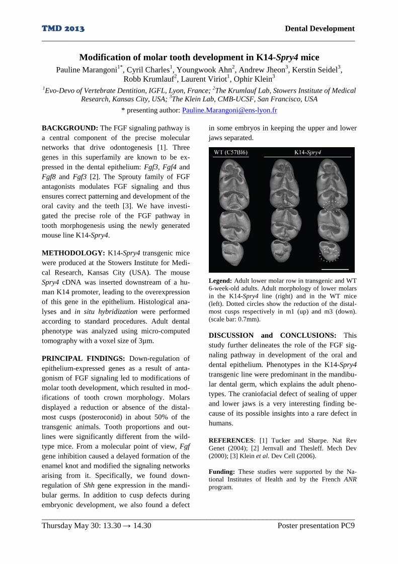
TMD 2013 Dental Development ______________________________________________________________________________
______________________________________________________________________________ Thursday May 30: 13.30 → 14.30 Poster presentation PC9
Modification of molar tooth development in K14-Spry4 mice Pauline Marangoni1*, Cyril Charles1, Youngwook Ahn2, Andrew Jheon3, Kerstin Seidel3,
Robb Krumlauf2, Laurent Viriot1, Ophir Klein3
1Evo-Devo of Vertebrate Dentition, IGFL, Lyon, France; 2The Krumlauf Lab, Stowers Institute of Medical Research, Kansas City, USA; 3The Klein Lab, CMB-UCSF, San Francisco, USA
* presenting author: [email protected] BACKGROUND: The FGF signaling pathway is a central component of the precise molecular networks that drive odontogenesis [1]. Three genes in this superfamily are known to be ex-pressed in the dental epithelium: Fgf3, Fgf4 and Fgf8 and Fgf3 [2]. The Sprouty family of FGF antagonists modulates FGF signaling and thus ensures correct patterning and development of the oral cavity and the teeth [3]. We have investi-gated the precise role of the FGF pathway in tooth morphogenesis using the newly generated mouse line K14-Spry4.
METHODOLOGY: K14-Spry4 transgenic mice were produced at the Stowers Institute for Medi-cal Research, Kansas City (USA). The mouse Spry4 cDNA was inserted downstream of a hu-man K14 promoter, leading to the overexpression of this gene in the epithelium. Histological ana-lyses and in situ hybridization were performed according to standard procedures. Adult dental phenotype was analyzed using micro-computed tomography with a voxel size of 3µm.
PRINCIPAL FINDINGS: Down-regulation of epithelium-expressed genes as a result of anta-gonism of FGF signaling led to modifications of molar tooth development, which resulted in mod-ifications of tooth crown morphology. Molars displayed a reduction or absence of the distal-most cusps (posteroconid) in about 50% of the transgenic animals. Tooth proportions and out-lines were significantly different from the wild-type mice. From a molecular point of view, Fgf gene inhibition caused a delayed formation of the enamel knot and modified the signaling networks arising from it. Specifically, we found down-regulation of Shh gene expression in the mandi-bular germs. In addition to cusp defects during embryonic development, we also found a defect
in some embryos in keeping the upper and lower jaws separated.
Legend: Adult lower molar row in transgenic and WT 6-week-old adults. Adult morphology of lower molars in the K14-Spry4 line (right) and in the WT mice (left). Dotted circles show the reduction of the distal-most cusps respectively in m1 (up) and m3 (down). (scale bar: 0.7mm).
DISCUSSION and CONCLUSIONS: This study further delineates the role of the FGF sig-naling pathway in development of the oral and dental epithelium. Phenotypes in the K14-Spry4 transgenic line were predominant in the mandibu-lar dental germ, which explains the adult pheno-types. The craniofacial defect of sealing of upper and lower jaws is a very interesting finding be-cause of its possible insights into a rare defect in humans.
REFERENCES: [1] Tucker and Sharpe. Nat Rev Genet (2004); [2] Jernvall and Thesleff. Mech Dev (2000); [3] Klein et al. Dev Cell (2006).
Funding: These studies were supported by the Na-tional Institutes of Health and by the French ANR program.

Dental Development
______________________________________________________________________________
______________________________________________________________________________
Thursday May 30: 13.30 → 14.30 Poster presentation PC10
Deletion of Bmp2 or Bmp7 from dental epithelium cause enamel defects
Zeba Malik*, Anastasiia Kozlova, Maria Alexiou, Thimios Mitsiadis, Daniel Graf
Section of Orofacial Development and Regeneration, Institute of Oral Biology, ZZM, Faculty of Medicine,
University of Zurich, Zurich, Switzerland
* presenting author: [email protected]
BACKGROUND: Tooth development is suc-
cessfully used as a genetic model to study for-
mation of organs that derive through reciprocal
epithelial-mesenchymal interactions. These inter-
actions involve reciprocal signaling loops of only
a few conserved signaling networks, including the
Bone Morphogenetic Protein (BMP) family.
BMPs are secreted proteins belonging to the
TGFß superfamily. Several BMPs have been
implicated at various stages of tooth formation [1]
though their precise roles remain elusive. Both
BMP2 and BMP7 are expressed in the dental
lamina at initiation of tooth development and
remain expressed in tooth epithelium up to the
cap stage of developing molars. Both molecules
are also found in epithelial structures of the con-
tinuously growing incisor indicating a function of
these molecules also for enamel formation. Here
we present evidence that BMP2 and BMP7 are
redundant for early stages of tooth development
but have non-redundant roles in the regulation of
enamel formation and mineralization.
METHODOLOGY: Conditional alleles for
Bmp2 [2] and Bmp7 [3] were crossed to a Kera-
tin14-Cre driver to ablate either gene in tooth
epithelium. A histological and morphometric
microCT analysis was performed at various stag-
es of tooth development and in adult teeth.
PRINCIPAL FINDINGS: Neither ablation of
Bmp2 nor Bmp7 from the tooth epithelium inter-
rupted the early stages of tooth development.
Adult mice showed a normal dentition. Morpho-
metric analysis of the lower incisor in adult mice
revealed non-redundant functions during enamel
formation. Deletion of Bmp2 resulted in a delay
in mineralization and a shorter dental pulp cham-
ber. Histological analysis revealed an altered
morphology of cells in the stratum intermedium.
Bmp2fl/ko;K-14Cre
Bmp2fl/w
t
M3
M2
M1
d c
Delayed enamel mineralization in the absence of
Bmp2. Serial sections through a microCT recording of
the lower incisor reveal a delay in mineralization when
Bmp2 is deleted from the dental epithelium.
DISCUSSION AND CONCLUSIONS: Individ-
ual loss of Bmp2 or Bmp7 did not compromise
tooth development indicating a functional redun-
dancy. This is in line with the overlapping ex-
pression pattern of these molecules during early
stages of tooth development and suggests that
BMP2/7 heterodimers might be operational. A
non-redundant function for BMP2 was revealed
during enamel formation, where enamel minerali-
zation was delayed. The histological disruption of
cells of the stratum intermedium indicates a role
for BMP2 in organizing this supporting cell layer,
which in turn controls enamel mineralization.
REFERENCES: [1] Aberg et al. Dev Dyn (1997); [2]
Graf & Economides, Birkhauser Verlag (2008); [3]
Zouvelou et al. Int J Dev Biol (2009); [4] Miyoshi et
al. J Med Invest (2008).
Funding: Swiss National Science Foundation (SNSF),
Swiss Dental Association (SSO), University of Zurich.

TMD 2013 Dental Development ______________________________________________________________________________
______________________________________________________________________________ Thursday May 30: 13.30 → 14.30 Poster presentation PC11
Quantitative X-ray microprobe analysis of fluorotic amelogenin-null enamel Donacian M. Lyaruu1*, Jing Guo1, Carolyn W. Gibson2, Yoshiro Takano3,
Pamela DenBesten4, Antonius LJJ Bronckers1
1Oral Cell Biology, ACTA, VU, Amsterdam, the Netherlands; 2Anatomy and Cell Biology, UPSDM, Phila-delphia, USA; 3Tokyo Medical and Dental University Graduate School, Tokyo, Japan,4Oral Sci-
ences, UCSF, San Francisco, USA * presenting author: [email protected]
BACKGROUND: Fluorotic enamel is character-ized by the presence of a hypermineralized sur-face and a porous hypomineralized subsurface. We hypothesized that enamel fluorosis develops by enhanced acid formation when mineralization is stimulated by F. Amelogenins can bind protons and may be involved in buffering the enamel fluid to sustain mineral growth. We tested whether formation of enamel crystals was changed in ab-sence of amelogenins and what the effect of F would be on enamel formation in amelogenn null mice.
METHODOLOGY: Four groups of mice (n=4-5 per group) were examined: groups 1 and 2; wild type controls and groups 3 and 4; amelogenin knockout (Ako) mice. Groups 2 and 4 were ex-posed to 100m ppm F in drinking water for 6 weeks. The lower jaws were freeze-dried, anhy-drously embedded in MMA and then examined by back-scattered electron detector (BSD) and quantitative X-ray micro probe analysis.
PRINCIPAL FINDINGS: The enamel layer of incisor of Ako mice was very thin. In Ako mice at late maturation stage the mineral content in enamel was lower and sulphur content (a measure for organic matrix) was higher than in wild type controls. Exposure of Ako mice to F enhanced the fluorine content 3-fold but in contrast to fluorotic wild type controls this slightly enhanced mineral content. In wild type littermates exposure to F reduced mineral content more than the ab-sence of amelogenins did in Ako mice. Back-scattered electron detector (BSD) images fur-thermore confirmed that the subsurface layers of fluorotic wild type enamel were indeed severely hypomineralized below an hypermineralized sur-face. In fluorotic Ako mice however, hypominer-alization did not occur in spite of high F levels
Legend: Effect of absence of amelogenins and expo-sure to F on composition of enamel and mineral content on the other hand increased to that found in WT mice and reached values similar to dentin and bone. In WT mice, F re-duced mineral density in maturation stage enamel more than 50% in but in AKO mice it did not change the mineral density.
DISCUSSION and CONCLUSIONS: The data show that the hypomineralisation found in fluorotic enamel depends on the presence of amelogenins in the enamel space. Fluorotic ef-fects in enamel (increased mineral -and F content) are enhanced in the absence of amelogenins sug-gesting amelogenins control the rate of crystal formation and may be involved in buffering of enamel fluid.
Funding: This study was supported by NIH grant DE13508.

TMD 2013 Dental Evolution ______________________________________________________________________________
______________________________________________________________________________ Thursday May 30: 13.30 → 14.30 Poster presentation PC12
New non-model fish for the study dentition diversity and evolution Johan Malègue1*, Philippe Janvier2, Laurent Viriot1
1Evo-Devo of Vertebrate Dentition, IGFL, Lyon, France 2Paleobiodiversity: history and dynamics, MNHN, Paris, France.
* presenting author: [email protected] BACKGROUND: Fish display an amazing di-versity in tooth implantation sites within the oro-pharyngeal cavity, along with a tremendous varie-ty of tooth shape as well as various patterns of continuous dental replacement. Otophysi is the most important group of freshwater fish, encom-passing 9,741 valid species (i.e. about 72% of the extant known freshwater species in the world). Among them, the monophyletic Characiphysi brings together Gymnotiforms (electric fish, 194 species), Siluriforms (cat fish, 3529 species) and Characiforms (characin fish, 1995 species). Cha-raciphysi appears to be an excellent candidate in order to (1) find new fish models for the study of dental diversity and evolution, (2) analyze dental patterns in the otophysian phylogenetic frame, and (3) confront tooth shape and arrangement with diet.
METHODOLOGY: These questions require the analysis of the dental phenotype at the genus level for the majority of characiphysian genera and then the study of some groups of interest, at the genus or species level. We used conventional X-ray microtomography in order to extract maximal information of the specimens without damaging them, with a voxel size ranging from 2 to 10 µm2.
PRINCIPAL FINDINGS: The family Loricarii-dae includes good candidates to study tooth shape as well as continuous dental replacement (CDR). In particular, Peckoltia viattata has a treadmill CDR located inside the jaw bone. As the number of specimens scanned increase, coherent dental patterns emerge on the phylogeny, especially on the Gymnotiforms and Siluriforms which can be divided into to 2 main groups with different den-tal patterns.
DISCUSSION and CONCLUSIONS: Characi-physi show a much more important dental diver-sity than expected, diversity in terms of tooth
Legend: 3D reconstruction of the silurid genus Pan-gasius showing oral, palatal, and pharyngeal teeth.
shape, in terms of implantation sites and in terms of patterns of continuous dental replacement. To keep screening the whole group at the genus level will enable to link dental pattern with evolutio-nary history of the different families as well as find new models for further studies in tooth evo-lution.
REFERENCES: [1] Nakatani et al. BMC Evolutio-nary Biology (2011); [2] Wei-Jen Chen et al, Evolu-tion (2013); [3] Briggs. Journal of Biogeography (2005); [4] Geerinckx et al. Journal of morphology (2007).
Funding: PhD funded by the ministry of research and higher education.

TMD 2013 Dental Evolution ______________________________________________________________________________
______________________________________________________________________________ Tuesday May 28: 13.30 → 14.30 Poster presentation PC13
Immunohistochemical observations on ganoine in the scales of Lepisosteus oculatus
Ichiro Sasagawa1*, Mikio Ishiyama2, Hiroyuki Yokosuka2, Masato Mikami3, Takashi Uchida4
1Advanced Research Center, 2Department of Histology, 3Depaertment of Microbiology, Nippon Dental University, Niigata, Japan; 4Department of Oral Biology, Graduate School of Biomedical Sciences, Hiro-
shima University, Hiroshima, Japan. * presenting author: [email protected]
BACKGROUND: Although skin denticles (scales) have been suggested as the homologous organ of teeth, the origin of teeth remains contro-versial. It is important to compare teeth with scales when we consider the origin and evolution of teeth in lower vertebrates. The ganoine is a well-mineralized layer existing on the surface of ganoid scales, and corresponds to tooth enamel [1]. In Polypterus, the preganoine (ganoine ma-trix), as well as collar enamel exhibited immuno-reactivity for anti-mammalian amelogenin anti-bodies [2, 3]. However, there have been no histo-chemical findings on the preganoine in Lepisos-teus. In this study, the preganoine in Lepisosteus oculatus was immunohistochemically examined using several anti-mammalian amelogenin anti-bodies and antisera.
METHODOLOGY: Four Lepisosteus oculatus, spotted gar, (TL. 16-23 cm), an actinopterygian, were used in this study. The tooth-bearing jaws were removed after anesthesia and decapitation, and were placed in 4% paraformaldyhyde- 0.2% glutaraldyhyde fixative for 3 h at 4°C. Selected samples were demineralized with EDTA-2Na. Specimens were dehydrated and embedded in LR-White resin. Semithin sections were cut and mounted on glass slides for light microscopy. Serial ultrathin sections were cut from the same resin block and mounted on nickel grids for transmission electron microscopy. The protein A-gold method was employed for immunohisto-chemistry. The antibody against bovine ameloge-nin, antiserum against porcine 25 kDa ameloge-nin, and region-specific antibodies or antiserum against the C-terminus, central region and N-terminus of porcine amelogenin were used in this study.
PRINCIPAL FINDINGS: The preganoine showed intense immunoreactivity to the antibody
Legend: Light micrograph showing immunoreactivity against the porcine 25kDa amelogenin antiserum (a). Panel b shows a part of the longitudinal section of a scale stained with toluidine blue during ganoine for-mation. B: bony plate, IGE: inner ganoine epithelial cells, PG: preganoine. Bar=20µm. against bovine amelogenin, the antiserum against porcine amelogenin, and central and C-terminus region-specific antibodies, but not to the N-terminus region-specific antibody.
DISCUSSION and CONCLUSIONS: The find-ings of the present study suggest that the prega-noine in Lepisosteus possesses amelogenin-like proteins containing a domain that closely resem-bles the C-terminal and central region of porcine amelogenin, and that the proteins in the prega-noine are similar to the enamel proteins of Lepi-sosteus [4]; however, the amelogenin-like pro-teins in Lepisosteus are different from mamma-lian amelogenin. These results support the ga-noine being an enamel-like tissue.
REFERENCES: [1] Sire, Anat Rec (1994); [2] Zyl-berberg et al. Anat Rec (1997); [3] Sasagawa et al. Cell Tissue Res (2012); [4] Ishiyama et al. Arch Histol Cytol (1999).
Funding: Grant-in-Aid for Scientific Research, 16591844, from the Ministry of Education, Science, Sports and Culture, Japan, and by a Research Promo-tion Grant (NDUF-12-12, 10-13, 09-18, 08-19, 07-12) from Nippon Dental University.

TMD 2013 Dental Evo-Devo ______________________________________________________________________________
______________________________________________________________________________ Thursday May 30: 13.30 → 14.30 Poster presentation PC14
Evolution of teeth and tooth-like structures in vertebrates Mélanie Debiais-Thibaud1*, Silvan Oulion1, Franck Bourrat2, Patrick Laurenti3,
Didier Casane3, Véronique Borday-Birraux3 1ISEM, Montpellier, France; 2INAF, Gif-sur-Yvette, France; 3LEGS, Gif-sur-Yvette, France
* presenting author: [email protected] BACKGROUND: Teeth and tooth-like struc-tures, together named odontodes, are repeated organs thought to share a common evolutionary origin. These structures can be found in gna-thostomes at different locations along the body: oral teeth in the jaws, teeth and denticles in the oral-pharyngeal cavity, and dermal denticles on elasmobranch skin.
METHODOLOGY: We present a detailed com-parison of the first developing teeth and dermal denticles (caudal primary scales) of the lesser spotted catshark (Scyliorhinus canicula). The precise Dlx gene expression patterns are com-pared during tooth and dermal denticle develop-ment.
PRINCIPAL FINDINGS: We show that both odontodes develop through identical stages that correspond to the common stages of oral and pharyngeal odontogenesis. We identified six Dlx paralogs in the catshark and examined their ex-pression patterns in oral teeth and dermal denti-cles in order to test the hypothesis of serial ho-mology between these odontodes. Our histologi-cal and expression data strongly suggest that teeth and dermal denticles develop from the same de-velopmental module and under the control of the same set of Dlx genes.
DISCUSSION AND CONCLUSIONS: Teeth and dermal denticles should therefore be consid-ered as serial homologs developing through the initiation of a common gene regulatory network (GRN) at several body locations. This mechanism of heterotopy supports the 'inside and out' model [1] that has been recently proposed for odontode evolution.
REFERENCES: [1] Fraser et al. Bioessays (2010)
Legend: Adult jaw in the lesser spotted catshark

TMD 2013 Dental Evo-Devo ______________________________________________________________________________
______________________________________________________________________________ Thursday May 30: 13.30 → 14.30 Poster presentation PC15
Transgenic PPII rich bos amelogenin enhances enamel matrix sub-unit compaction in a mouse model
David A. Reed1, Thomas G.H. Diekwisch1*
Oral Biology, The University of Illinois at Chicago, USA * presenting author: [email protected]
BACKGROUND: The evolution of the feeding system from basal vertebrates to mammals cor-responds with specializations for mastication as the principal mechanism of food reduction. Ru-minants represent an ideal model for studying these specializations as they average daily feeding times between 6-10 hrs. On a materials level, cyclic loading of the tooth occlusal surface for extended periods of time necessitates unique de-sign criteria to resist fracture and abrasion. To understand the changes in enamel growth facili-tating changes in the mechanical surface proper-ties, we have examined the patterning of the or-ganic enamel matrix as the primary crystal nuc-leation site. Previous studies have demonstrated that the number of polyproline tri-peptide repeats in the principal enamel matrix protein, ameloge-nin substantially increase when comparing mam-mals to non-mammals and is especially pro-nounced in ruminants [1]. We have thus asked the question whether a polyproline-rich amelogenin enamel matrix affects matrix subunit dimensions when compared to mouse and non-mammalian counterparts. Such a change in matrix organiza-tion could explain a gradual change in enamel crystal dimension and organization during verte-brate evolution.
METHODOLOGY: A transgenic mouse over-expressing the PPII rich bos (bovine) amelogenin was created. Enamel protein supramolcular as-sembly is assessed with transmission electron microscopy. Stippled enamel matrix subunits are imaged in two transgenic and control mice. Ma-trix subunits are measured from three locations, with 10-20 subunits measured from each image.
PRINCIPAL FINDINGS: The three day post-natal transgenic mouse produced significantly smaller subunits within the electron dense stip-pled material of the enamel matrix when compa-
Legend: Enamel matrix ultrastructure in three day postnatal mice. (A) is from a wild-type control and (B) is from a transgenic mouse expressing bos amelogenin. Scale bar equals 100 nm. Numbers in the blow up region reflect averages taken from multiple mice and sections. Note that the subunits in the bovine trans-genic mouse are more compact when compared to the control. red to a WT control (16.65 nm, 22.27 nm respec-tively; p<001).
DISCUSSION and CONCLUSIONS: These data, for the first time, demonstrate that a PPII rich amelogenin increases subunit compaction in the enamel protein supramolecular assembly in vivo. Subunit compaction has been hypothesized as a mechanism to densely template nanothin enamel crystals and modulate the mechanical properties of the enamel. As such, these results support the hypothesis that modifications within the polyproline rich region of amelogenin can have direct functional consequences on enamel matrix structure and underscores the importance of polyproline-repeats in the evolution of verte-brate mineralization proteins.
REFERENCES: [1] Jin et al. PLoS Biology (2009).
Funding: NIDCR grant DE018900 to TGHD.

TMD 2013 Dental Evo-Devo ______________________________________________________________________________
______________________________________________________________________________ Thursday May 30: 13.30 → 14.30 Poster presentation PC16
Genetic basis of evolutionary significant tooth shape variations: the role of Notch signaling pathway
Liza Ramenzoni1*, Maria Alexiou1, Helder Gomes Rodrigues2, Paul Tafforeau3, Laurent Viriot2, Daniel Graf1, Thimios Mitsiadis1
1Institute of Oral Biology, University of Zurich, Zurich, Switzerland; 2Evo-Devo of Vertebrate Dentition, IGFL, Lyon, France; 3Beamline ID 19, ESRF, Grenoble, France
* presenting author: [email protected] BACKGROUND: Evolution of teeth has been shown be greatly influenced by environment but the genetic basis of microevolutionary subtle changes in the tooth shape is largely unknown. Morphology of the hard tissues of the teeth is largely unaffected, making the tooth an excellent model to study the genetic basis of organs shapes. The aim of this work is to identify genetic path-ways linked to the evolution of species-specific tooth shapes by comparing gene activities during tooth shape development. METHODOLOGY: Dental morphological vari-ations of location, size and interconnection of cusps were analyzed in the wild type (WT) and K14Cre;Jagged1fl/fl mice. Images of mice skulls were obtained using Synchrotron X-ray microto-mography at the European Synchrotron Radiation Facility (France). The Student's t-test and Fisch-er's F-test were used on each dental measurement (W, L, d1-d5) for WT and K14Cre;Jagged1fl/fl mice to check mean and variance equality. Jagged2, Shh, Bmp4 and Bmp7 expression pat-terns in the enamel knot region were examined at E14.5 and E17.5 stages by in situ hybridization performed using previously described methods.
PRINCIPAL FINDINGS: According to Stu-dent's t-tests, mean sizes (L and W) of all molars are significantly lower for K14Cre;Jagged1fl/fl mice than for wild type specimens. The morpho-logical differences observed between wild type and K14Cre;Jagged1fl/fl mice are slight, and mainly concerning the location and connections of a few cusps. The most striking variations on upper molars involve the c1 cusp. On M1, the c1-c2 connection profile is generally high and V-shape in the wild type, while it is more frequently low and U-shape in K14Cre;Jagged1fl/fl. On lower molars, a slight variation was located on the me-sial part of M1. In situ hybridization showed that the expression of Shh and Bmp7 were decreased
Legend: A, B: upper molars of wild type specimens. C: Lower molars of wild type specimens. D, E: Upper molars of knockout mice. F: lower molars of knockout mice. Red arrows indicate differences found between wild type and mutant.
in the enamel knot of the K14Cre;Jagged1fl/fl mice when compared with the enamel knot of the wild type mice. DISCUSSION and CONCLUSIONS: Our re-sults revealed that K14Cre;Jagged1fl/fl mice present minor differences but important tooth shape variances such as slight size and shape modifications of the molars. The present results could be rather understood in a microevolutionary than at a macroevolutionary perspective. The Jagged1 gene influences important morphogenet-ic genes such as Shh and BMPs and therefore could be involved in minor dental modifications. Such mutations appear to be very interesting and highly promising regarding the enormous crown variation observed in human molars. Subtle mor-phological changes in the molar cusps of this model mouse may elucidate how genetic signal-ing interactions affect developing morphology in humans as a key transition to evolution. REFERENCES: [1] Jernvall et al. Science (1996); [2] Mitsiadis et al. Dev Biol (1998); [3] Charles et al. PNAS (2009) Funding: Forschungskredit, University of Zurich.

TMD 2013 Dental Stem Cells ______________________________________________________________________________
______________________________________________________________________________ Thursday May 30: 13.30 → 14.30 Poster presentation PC17
Odontoblast behaviour upon Toll-like receptor-2 activation Jean-Christophe Farges1,2, Aurélie Bellanger1, Marie-Jeanne Staquet1, Caroline Baudouin3, Phi-
lippe Msika3, Françoise Bleicher1, Brigitte Alliot-Licht4, Florence Carrouel1,2* 1Odontoblast Physiopathology Group, Institute of functional Genomics of Lyon, 2University Lyon 1, Fac-ulty of Odontology, Lyon, France; 3Lab. Expanscience, Epernon, France; 4UMR1064 ITERT, University
of Nantes, Faculty of Odontology, Nantes, France * presenting author: [email protected]
BACKGROUND: Previous studies have sug-gested that human odontoblasts sense components from Gram-positive bacteria through Toll-like receptor 2 (TLR2) and trigger innate immunity in the dental pulp by producing specific pro-inflammatory cytokines and chemokines [1-5]. However, most of the determinants of the odon-toblast response to these pathogens have probably not yet been identified. The aim of this study was to determine what are the pro-inflammatory en-zymatic pathways activated in vivo in bacteria-challenged inflamed dental pulps and in vitro in odontoblast-like cells upon TLR2 activation by specific agonists.
METHODOLOGY: Healthy dental pulps were taken from five non-erupted human third molars. Inflamed pulps were taken from five decayed erupted molars with clinical features of irreversi-ble acute pulpitis. Odontoblast-like cells were differentiated from dental pulp explants and sti-mulated with two TLR2 specific ligands: lipotei-choic acid (LTA), a cell wall component of Gram-positive bacteria, and Pam2CSK4, a syn-thetic diacylated lipopeptide. Expression of genes coding for cyclooxygenase-2 (COX2), the NADPH oxidase subunits NOX1, NOX2, NOX3, NOX4, NOX5, DUOX1 and DUOX2, and heme oxygenase-1 (HMOX1) was assessed by real-time PCR.
PRINCIPAL FINDINGS: The strong expres-sion increase of the CXCL8 (= interleukin-8) gene confirmed the inflammatory status of the pulp tissues beneath caries lesions. Gene expres-sion of COX2, the NADPH oxidase subunits NOX1, NOX2 and NOX4, and HMOX1 were significantly increased in inflamed pulps com-pared to healthy ones, whereas expression of
Legend: Real-time PCR analysis of gene expression
NOX5, DUOX1 and DUOX2 genes was not mod-ified (see the graph below). In vitro, LTA stimu-lation of human odontoblast-like cells induced a low but significant increase of COX2, NOX1, NOX2, NOX5 and DUOX1 gene expression, whereas Pam2CSK4 induced a stronger increase of the same genes and of HMOX1.
DISCUSSION and CONCLUSIONS: Together these results suggest that part of the inflammatory response that occurs in the human dental pulp in response to Gram-positive bacteria entering the dentin tissue during the caries process might imp-ly odontoblasts.
REFERENCES: [1] Farges et al. Immunobiology (2011); [2] Keller et al. Innate Immun (2011); [3] Farges et al. J Exp Zoolog B (2009); [4] Goldberg et al. Pharmacol Res (2008); [5] Durand et al. J Immunol (2006).
Funding: National Center of Scientific Research (CNRS), Rhône-Alpes region, French Ministry of Higher Education and Research, French Institute of Odontological Research (IFRO).

TMD 2013 Dental Stem Cells ______________________________________________________________________________
______________________________________________________________________________ Thursday May 30: 13.30 → 14.30 Poster presentation PC18
D[4]/Phenodent: A bioinformatics tool for the oral aspects of rare diseases Sébastien Troester1*, Carole Schneider2, Marie-Cécile Maniere1,2, Agnès Bloch-Zupan1,2,3
1Reference Centre for Orodental Manifestations of Rare Diseases, Pôle de Médecine et Chirurgie Bucco-Dentaires, Hôpitaux Universitaires de Strasbourg, France; 2Faculty of Dentistry, University of Stras-
bourg, UdS, Strasbourg, France; 3IGBMC; INSERM, U964; CNRS, UMR7104, Illkirch, France * presenting author: [email protected]
BACKGROUND: Gathering patient data in rare diseases is of the greatest importance to enable the extraction of new knowledge, to engage into clinical research and trials, and to evaluate new therapies outcome. Developmental dental anoma-lies exist in isolation or associated to other traits in syndromes. They constitute trigger signs con-tributing to rare disease diagnosis.
METHODOLOGY: D[4]/Phenodent collabora-tive interactive biomedical database (Diagnosing Dental Defects Database) has been developed since 2005 by the Reference Centre for Orodental Manifestation of Rare Diseases, University Hos-pital of Strasbourg (www.phenodent.org). It is stored on a MySQL database available from a Web-based platform offering high security levels and complying with the ethic and law regulations concerning sensitive biomedical data collection. D[4]/Phenodent assists in oro-dental phenotyping and allows standardisation of data collection inte-grating them into the medical and genetic context.
PRINCIPAL FINDINGS: Currently the data-base displays 2360 patient files, over 185 differ-ent rare diseases, and is used in different research projects such as: a) oro-dental phenotypes in syn-dromes [1]; b) identification of mutations in known genes involved in odontogenesis and dis-eases; c) phenotype/genotype correlations [2]; d) new gene identification [3].
DISCUSSION and CONCLUSIONS: D[4]/ Phenodent facilitates understanding oral biology and associated diseases, stimulates patients’ re-cruitment and provides a basis for molecular analysis and anatomo-pathological investigations. Standardisation helps also sharing of data and materials among investigators.
Legend: Patient record – Tooth Chart D[4]/Phenodent constitutes a link between partic-ipating clinical diagnosis centers and research laboratories thus representing a powerful tool for national and international networks indispensable for rare diseases research and management. It can be individually tailored. D[4]/Phenodent wel-comes further collaboration. ([email protected]).
REFERENCES: [1] Bloch-Zupan et al. Orphanet J Rare Dis (2013); [2] Reibel et al. (2009) Orphanet J Rare Dis. (2009); [3] Bloch-Zupan et al. Am J Hum Genet. (2011).
Funding: This work is funded partially via INSERM « Réseau de Recherche Clinique et Réseau de Re-cherche en Santé des populations 2003 », COST-STSM-B23-00900 and funds allocated to the Refe-rence Centre for Orodental Manifestations of Rare Diseases (Ministère des Affaires sociales et de la San-té, Ministère de l’Enseignement supérieur et de la Recherche). Offensive Sciences, a Science Initiative in the Trinational Metropolitan Region of the Upper Rhine, FEDER (European fund for regional develop-ment), Program INTERREG IV Upper Rhine, Project A27 Oro-dental manifestations of rare diseases.

TMD 2013 Dental Stem Cells ______________________________________________________________________________
______________________________________________________________________________ Thursday May 30: 13.30 → 14.30 Poster presentation PC19
TALEN induced mutations in mouse Enamelin gene Zheqiong Yang1, Frederic Michon1, Kirmo Wartiovaara1,2, Irma Thesleff1*
1Developmental Biology Program, Institute of Biotechnology, University of Helsinki, Helsinki, Finland; 2Developmental Biology, Institute of Biomedicine,, University of Helsinki, Helsinki, Finland
* presenting author: [email protected] BACKGROUND: TALENs are new tools for genome editing. Fusions of transcription activa-tor-like (TAL) effectors of plant pathogenic Xan-thomonas spp. to the FokI nuclease, TALENs bind and cleave DNA in pairs, and produce dou-ble-strand breaks between the target sequences. Binding specificity is determined by customizable arrays of polymorphic amino acid repeats in the TAL effectors. The double-strand break can be repaired through nonhomologous end-joining to drive targeted gene disruption or through the ho-mology-directed DNA repair pathway using an exogenous donor plasmid as a template. Depend-ing on the donor design, this repair reaction can be used to generate large-scale deletions, gene disruptions, DNA addition or single-nucleotide changes. In order to introduce a fluorescent marker into mouse Enamelin (Enam) gene. we designed two pairs of TALEN targeting mouse Enam gene, and constructed a donor vector bear-ing the coding sequence of Red Fluorescent Pro-tein (RFP) gene.
METHODOLOGY: We assembled two pairs of TALENs targeting the third exon of mouse Enam gene by using the Golden Gate TALEN kit. We also constructed a donor vector with coding se-quences of RFP gene and puromycin-resistance gene in between 5’ and 3’ homologue arms. TALEN expression constructs were transfected into 3T3 cells to test the cutting efficiency, and both TALEN vectors and corresponding donor plasmids bearing homologous sequences were introduced into mouse E14 embryonic cell to produce the homologous recombination.
PRINCIPAL FINDINGS: Both pairs of TALEN caused deletion and/or mutation between TALEN binding sites in Enam gene.
Legend: TALEN induced gene modification in mouse Enam gene. Underlined bases denote TALEN pairs binding sites. DISCUSSION and CONCLUSIONS: TALENs are effective and useful tools for investigator-specified targeting and genetic modification. They are now used in various species, such as zebrafish, mouse and rat in vitro and in vivo, and also for genomic editing in human iPS cells.
REFERENCES: [1] Cermak et al. Nucleic Acids Res (2011); [2] Hockemeyer et al. Nat Biotechnol (2011).
Funding: Academy of Finland, Juselius Foundation

TMD 2013 Dental Stem Cells ______________________________________________________________________________
______________________________________________________________________________ Thursday May 30: 13.30 → 14.30 Poster presentation PC20
Reproducible Bone Healing Process in Calvarial Defect Yuko Akiyama1*, Yoko saito1, Nina Tsurumachi1, Keitaro Isokawa2,
Noriyoshi Shimizu1, Masaki Honda2
1Department of Orthodontics, Nihon University School of Dentistry, Tokyo, Japan; 2Department of Anatomy, Nihon University School of Dentistry, Tokyo, Japan
* presenting author: [email protected] BACKGROUND: Our study aimed to elucidate the mechanisms of bone repair after injury. Sev-eral genes make important contributions to the regulatory pathway at specific time points. In order to study the differential expression at vari-ous stages of bone repair, a reproducible method is thus required. In this study, we developed a new device to prepare the reproducible bone de-fect size and evaluated the bone healing process.
METHODOLOGY: The skin and periosteum of 8-week-old male C57BL/6N mice (experimental group: 45, control: 3) were excised to expose the parietal bone. In the controls, they were then placed back. In the experimental group, a circular defect in the parietal bone was created using a novel device with a drill (diameter: φ1.4 mm, depth: 100 µm). Thereafter, the periosteum and cutaneous flaps were replaced. The bone healing process was histologically evaluated from imme-diately after surgery (day 0) to 14 days after sur-gery (day 14). Furthermore, gene expression pat-terns of inflammatory cytokines and hypoxia-inducible factor-1α (HIF-1α) were examined by real-time PCR.
PRINCIPAL FINDINGS: No structure in bone defect was observed on day 0 in the experimental group. Inflammatory cell infiltration and a small amount of hematoma were observed, and the periosteum was restored on day 1. The number of inflammatory cells was reduced in the bone de-fect on day 2. Fibrous tissue development began on day 4, Finally, new woven bone was observed on day 14. mRNA expression of inflammatory cytokine was highest on day 1 and decreased gradually thereafter. Furthermore, HIF-1α expres-sion on day 8 was significantly higher than that on day 1 (p < 0.05).
DISCUSSION and CONCLUSIONS: The re-producible bone healing process was observed histologically and gene expression patterns sup-ported the evidence. These results suggest that the device developed by us is a novel system to eva-luate the bone healing process. Furthermore, the mouse calvarial defect is an established and suit-able model for gene expression profiling studies regarding intramembranous bone regeneration. We are now conducting microarray gene expres-sion analysis.
REFERENCES : [1] Holstein et al. Injury (2011); [2] Holstein et al. J Orthop Res. (2011).
Funding: Grant-in-Aid for Scientific Research (KA-KENHI)

TMD 2013 Dental Stem Cells ______________________________________________________________________________
______________________________________________________________________________ Thursday May 30: 13.30 → 14.30 Poster presentation PC21
Postnatal changes of pulp cell population demonstrated by allogenic tooth germ transplantation in mice
Tetsuro Nakaki, Kotaro Saito, Hiroko Ida-Yonemochi, Eizo Nakagawa, Shin-ichi Kenmotsu, Hayato Ohshima*
Division of Anatomy and Cell Biology of the Hard Tissue, Department of Tissue Regeneration and Recon-struction, Niigata University Graduate School of Medical and Dental Sciences, Niigata, Japan
* presenting author: [email protected] BACKGROUND: Tooth transplantation would be applied to patients with congenital missing teeth, if the tooth germ transplant becomes a common procedure in dentistry. However, there are no available data on the chronological changes of the transplanted tooth germ at the cellular level. This study aimed at establishing the animal model for allogenic tooth germ trans-plantation using mice and investigating the donor-host interaction using this model combined with prenatal BrdU labeling and GFP mice.
METHODOLOGY: Three intraperitoneal injec-tions of BrdU (150 mg/kg) were given to preg-nant C57BL/6J mice (once a day at embryonic Days 15 to 17) to map slow-cycling long-term label-retaining cells (LRCs) in the mature tissues of animals born according to the prenatal BrdU labeling method for mice1. The mandibular first molar (M1) tooth germs from postnatal 1-2-day-old BrdU-labeled, GFP, or wild-type mice were transplanted into the alveolar socket of the maxil-lary first molars of 12-14-day-old non BrdU-labeled, wild-type, or GFP mice. At intervals of 3-21 days after operation, the maxillae were cut and processed for immunohistochemistry for BrdU, nestin, and Ki67, and TUNEL assay fol-lowing CT analysis and decalcification.
PRINCIPAL FINDINGS: In the current model, cell proliferation and differentiation in the tooth germ progressed in the same manner as normal tooth development. The number of dense LRCs was constant during the experimental periods, whereas numerous granular LRCs on Day 3 were significantly decreased in number on Day 5 and remained great in number until Day 14. Donor-derived resident dense LRCs were maintained in the center of pulp tissue associating with blood vessels and committed in the nestin-positive new-ly-differentiated odontoblasts.
Legend: µCT image of the transplants on Day 14. The three-dimensionally-reconstructed transplant (M1) shows the normal configuration consisting of 6 cusps and 2 roots and the completion of tooth eruption and occlusion Allogenic tooth germ transplantation using GFP and wild-type mice in this study demonstrated that the donor cells were maintained in the pulp cells including odontoblasts and the cells of blood vessels, and the host cells also immigrated into the dental pulp during the postoperative periods.
DISCUSSION and CONCLUSIONS: We suc-ceeded in establishing the animal model using mice for tooth germ transplantation into the max-illae and clarified the chronological changes of the transplanted tooth germ at the cellular level for the first time. The pulp cell population changed postnatally during odontogenesis, sug-gesting that these changes affect the differentia-tion capacity of the dental pulp.
REFERENCES: [1] Ishikawa et al. Cell Tissue Res (2012);348:95-107.
Funding:This work was supported by a Grant-in-Aid for Scientific Research (C) (no. 23593026) from JSPS.

TMD 2013 Dental Stem Cells ______________________________________________________________________________
______________________________________________________________________________ Tuesday May 28: 13.30 → 14.30 Poster presentation PC22
Expression of TRPM8 and TRPA1 channels in rat odontoblasts Masaki Sato1*, Maki Tsumura1,2, Ubaidus Sobhan1, Masakazu Tazaki2,Yoshiyuki Shibukawa1,2
1Oral Health Science Center hrc8, Tokyo Dental College, Chiba, Japan; 2Department of Physiology, Tokyo Dental College, Chiba, Japan
* Presenting author: [email protected] BACKGROUND: Odontoblasts are involved in the transduction of physiological or pathological stimuli applied to exposed dentin and the result-ing dentinogenesis [1]. Expression of various transient receptor potential (TRP) vanilloid sub-family member (TRPV) channels has been well documented [2-5] in odontoblasts; that of TRP melastatin subfamily member 8 (TRPM8) and TRP ankyrin subfamily member 1 (TRPA1) channels, however, remains controversial [4]. We investigated the expression and biophysical and pharmacological properties of TRPM8 and TRPA1 channels in acutely isolated rat odontob-lasts to clarify the precise ionic mechanism un-derlying transduction of stimuli applied to odon-toblasts.
METHODOLOGY: We measured intracellular free calcium concentrations ([Ca2+]i) by fura-2 fluorescence in dentin sialoprotein-positive acute-ly isolated rat odontoblasts [2, 3, 5].
PRINCIPAL FINDINGS: WS3 and WS12, selective agonists of TRPM8 channels, elicited a transient increase in [Ca2+]i in the presence of extracellular Ca2+. WS3-induced increase in [Ca2+]i was not observed in the absence of extra-cellular Ca2+, and was inhibited by TRPM8 chan-nel antagonist capsazepine. WS12-induced tran-sient increases in [Ca2+]i were inhibited by 5-benzyloxytriptamine (5-BOT), a specific TRPM8 channel antagonist. TRPA1 channel agonist allyl isothiocyanate (AITC) also evoked an increase in [Ca2+]I, which was inhibited by HC030031, a TRPA1 channel antagonist. A series of repeated applications of WS12 or AITC induced a signifi-cant desensitizing effect on each [Ca2+]i increase. Hypotonic (200 mOsm/L) solution-induced plas-ma membrane stretch resulted in an increase in [Ca2+]I, which was sensitive to HC030031, but not 5-BOT.
DISCUSSION AND CONCLUSIONS: The results indicate that odontoblasts express TRPM8 and TRPA1 channels, and that membrane stret-ching of odontoblasts is received by TRPA1, but not by TRPM8 channels. This suggests that TRPM8 channels in odontoblasts participate in detection of low-temperature stimuli applied to the surface of dentin, while TRPA1 channels play a role in sensing movement of dentinal fluid in-duced by membrane stretching.
REFERENCES: [1] Goldberg & Smith. Crit Rev Oral Biol Med. (2004); [2] Magloire et al., J Orofac Pain (2010); [3] Okumura et al. Arch Histol Cytol (2005); [4] Son et al., J Dent Res (2009); [5] Tsumura., Cell Calcium (2012).
Funding: Oral Health Science Center Grant hrc8 from Tokyo Dental College, a Project for Private Universi-ties: matching fund subsidy from MEXT of Japan (2011–2013), and Grant-in-Aid (No. 23592751 /23792132) for Scientific Research from the MEXT of Japan.

TMD 2013 Dental Stem Cells ______________________________________________________________________________
______________________________________________________________________________ Thursday May 30: 13.30 → 14.30 Poster presentation PC23
Role of pkd1 in a murine odontoblast cell-line Béatrice Thivichon-Prince1,2*, Anne Lambert1, Béatrice Richard1,2, Maxime Ducret1,2,
Marie-Lise Couble1, Françoise Bleicher1
1Odontoblast Physiopathology, IGFL, UMR5242, Lyon, France; 2Hospices Civils de Lyon, France * presenting author: [email protected]
BACKGROUND: Odontoblasts are organized as a layer of specialized cells responsible for dentin formation. Their situation at the interface between dentin and pulp gives to odontoblasts a pivotal role to sense both external stimuli and/or transient changes in pulp microcirculation [1]. Therefore, the control of dentin deposition may involve not only inductive molecules released from den-tine/pulp matrix but also a direct mechano-transduction process. This is supported by odon-toblast mechano-sensitive ion channels and pres-ence of a primary cilium in the vicinity of the Golgi apparatus [2]. Recently we identified two potentially mechano-sensitive receptors (TRPP1 and TRPP2) in odontoblasts. TRPP1 has been described as a mechanical sensor in bone [3] and cartilage [4], which are two skeletal tissues close to the tooth. Therefore we speculate that TRPP1 could be a good candidate to explain the mecha-no-sensory property of odontoblasts and could participate in the sensory transduction process in tooth. The aim of our work was to obtain an odontoblast model down expressing pkd1, the gene encoding for TRPP1, and to study the role of this gene in cell proliferation, migration, and den-tine matrix synthesis.
METHODOLOGY: Pkd1 down expression was obtained by shRNA technology on MO6-G3 (mu-rine odontoblast cell-line). CollagenIα1, osteo-pontin, osteonectin and osteocalcin expression was analyzed by real-time polymerase chain reac-tion. MTT tests were performed to study cell proliferation and migration was recorded as time lapse images and analyzed using ImageJ soft-ware.
PRINCIPAL FINDINGS: We found that a 60% reduction of pkd1 expression by shRNA in mu-rine MO6-G3 cells resulted in decreased cell pro-
Legend: Cell migration analysis by time-lapse micro-scopy. Downregulation of Pkd1 expression impairs odontoblast migration.
liferation, impaired cell migration and enhanced collagen Iα1 and osteonectin expression.
DISCUSSION and CONCLUSIONS: For the future, this new model would help us to under-stand the role of TRPP1 in odontoblast mechano-transduction process in response to mechanical stimuli.
REFERENCES: [1] Magloire et al, J. Exp. Zool. B (2009); [2] Thivichon-Prince et al. J. Dent. Res. (2009); [3] Xiao et al. FASEB J. (2011); [4] Hou et al. Bone (2009).
Funding: IFRO supported this work.
TMD 2013 Dental Stem Cells ______________________________________________________________________________
______________________________________________________________________________ Thursday May 30: 13.30 → 14.30 Poster presentation PC24
Dental stem cells: the key to angiogenesis? Petra Hilkens*, Yanick Fanton, Annelies Bronckaers, Wendy Martens, Tom Struys,
Constantinus Politis, Ivo Lambrichts Department of Morphology, BIOMED Hasselt University, Diepenbeek, Belgium
* presenting author: [email protected] BACKGROUND: Despite today’s technological advances, tooth loss remains an important public health issue. In particular the dental pulp appears to be very vulnerable to external insults, poten-tially leading to pulp necrosis. Since traditional endodontic treatments often fail, stem cell-based therapies are currently being investigated as a promising method for pulp restoration. As angi-ogenesis is a key process in the development and repair of the human tooth, the main focus of this study was to compare the angiogenic properties of postnatal dental stem cells (DSC). The para-crine actions of three stem cell populations de-rived from human third molars, namely dental pulp stem cells (DPSC), cells from the apical papilla of the developing root (SCAP) and dental follicle precursor cells (FSC), were analyzed and compared with a human gingival fibroblast cell-line (HGF-1).
METHODOLOGY: The angiogenic secretion profile of DSC was identified by means of an antibody array, of which the results were vali-dated by RT-PCR, ELISA and immunohistoche-mistry. In order to determine the potential impact of DSC on the behavior of human microvascular endothelial cells (HMEC-1), several in vitro models of angiogenesis were performed, such as a MTT assay for proliferation, a transwell assay to assess migration and a Matrigel assay for tube formation. Finally, a chorioallantoic membrane assay (CAM) was used to determine the angi-ogenic potential of DSC in ovo.
PRINCIPAL FINDINGS: Besides in situ ex-pression of angiogenic factors in dental tissues, differential expression of pro- and anti-angiogenic factors was observed in cultured DSC and HGF-1. DPSC expressed significantly more VEGF than the other DSC populations and fi-broblasts. Furthermore, DPSC and SCAP signifi-
cantly increased endothelial (HMEC-1) migra-tion, while there was no effect on HMEC-1 proli-feration. In addition, a Matrigel assay showed that DPSC significantly increased endothelial tube formation. Finally, the chicken chorioallantoic membrane assay indicated a positive effect of DSC, in particular DPSC and SCAP, on blood vessel formation in ovo.
DISCUSSION and CONCLUSIONS: The dif-ferential expression of angiogenic factors by DSC and fibroblasts could indicate potential comple-mentary paracrine actions. Furthermore, DSC seemed to have a predominant pro-angiogenic impact in vitro and in ovo, in particular on endo-thelial migration and tube formation. Although no superior cell population could be identified, these results suggest a stronger angiogenic profile for DPSC and SCAP, compared to FSC.
Funding: Petra Hilkens benefits from a PhD scholar-ship from the FWO.

TMD 2013 Dental Cell Physiology ______________________________________________________________________________
______________________________________________________________________________ Thursday May 30: 13.30 → 14.30 Poster presentation PC25
Odontoblast as sensory receptor cell: TRP channels, pannexin 1 and P2X3 re-ceptor coupling mediate sensory transduction in dentin
Yoshiyuki Shibukawa1,2*, Masaki Sato1, Maki Tsumura12, Hidetaka Kuroda13, Ubaidus Sobhan1, Masakazu Tazaki2
1hrc8; 2Physiology, 3Dental Anesthesiology, Tokyo Dental College, Chiba, Japan * Presenting author: [email protected]
BACKGROUND: A wide range of stimuli, in-cluding thermal, tactile, osmotic, and chemical, induce pain when applied to the surface of ex-posed dentin. The hydrodynamic theory has been posited to explain the primary mechanism under-lying the generation of such pain. The role of odontoblasts in this sensory transduction se-quence, however, remains unclear. We investi-gated transient receptor potential (TRP) channel activation following direct mechanical stimula-tion of odontoblasts to clarify their role as sensory receptors in this sequence. We then examined signal coupling between odontoblasts and neu-rons, which is mediated through release of ATP from pannexin-1 on stimulated odontoblasts, and subsequent activation of P2X3 receptors (purino-ceptors for ATP) on trigeminal ganglion (TG) neurons.
METHODOLOGY: Dentin sialoprotein-positive acutely isolated odontoblasts and DiO-labeled TG cells from neonatal rat were primary cultured for 1 hr. The cells were subjected to direct mechani-cal stimulation by means of a glass microelec-trode filled with standard extracellular solution. We measured intracellular free calcium concen-tration ([Ca2+]i) by fura-2 in stimulated odonto-blasts, as well as in odontoblasts- or TG cells located near them.
PRINCIPAL FINDINGS: Direct mechanical stimulation of single odontoblasts increased [Ca2+]i via TRPV1, TRPV2, TRPV4, and TRPA1, but not by TRPM8 channel activation. In co-culture of odontoblasts and TG cells with direct mechanical stimulation of single odontoblasts, we observed an increase in [Ca2+]i not only in stimu-lated odontoblasts, but also in both nearby odon-toblasts and TG cells. This increase in [Ca2+]i in both nearby odontoblasts and TG cells, but not in
Legend: Odontoblast neuron coupling in sensory transduction via TRP channels/pannexin-1/P2X3 re-ceptor.
stimulated odontoblasts, was inhibited by pannex-in-1 inhibitor in a concentration- and spatial-dependent manner. In the presence of P2X3 recep-tor antagonist, this increase in [Ca2+]i in nearby TG cells following mechanical stimulation of single odontoblasts was abolished, but that in stimulated and nearby odontoblasts was not.
DISCUSSION and CONCLUSIONS: The present results clearly showed that ATP released from mechanically stimulated odontoblasts via pannexin-1 by TRP channel activation transmits a signal to P2X3 receptors on TG neurons (chemical odontoblast-TG neuron coupling). The results also strongly suggest that odontoblasts are sen-sory receptor cells, and that ATP released from pannexin-1 on odontoblasts acts as a transmitter in the sensory transduction sequence.
Funding: This research was supported by MEXT of Japan.

TMD 2013 Dental Evolution ______________________________________________________________________________
______________________________________________________________________________ Thursday May 30: 13.30 → 14.30 Poster presentation PC26
Mastication and dental wear pattern of the extant marsupial Didelphis marsupialis
Achim Schwermann1*, Ottmar Kullmer2, Thomas Martin1
1Vertebrate Palaeontology, Steinmann-Institut für Geologie, Mineralogie und Paläontologie, Rheinische Friedrich-Wilhelms-Universität Bonn, Bonn, Germany; 2Palaeoanthropology and Messel Research, Senc-
kenberg Forschungsinstitut und Naturmuseum, Frankfurt am Main, Germany * presenting author: [email protected]
BACKGROUND: The extant marsupial Didel-phis marsupialis served as a comparative taxon to Mesozoic basal Theria in various studies on den-tal function (for example[1]), because of its basal tribosphenic molar dentition. The species of Di-delphis are characterized as opportunistic and omnivorous feeders[2]. Fruits, seeds, leafs, grass, insects and other arthropods, earthworms, verte-brates up to the size of chickens and rabbits are documented as potential food sources. The cur-rent project investigates functional details and variability of the dental macrowear pattern in the molar dentition of Didelphis marsupialis.
METHODOLOGY: More than 70 individual tooth rows of different aged Didelphis marsupia-lis (juvenile, adult, senile) were examined to ana-lyze the wear pattern in various ontogenetic stag-es. 3D models generated from µCT data were used for constructing the occlusal compass[3]. The new software tool “Occlusal Fingerprint Analys-er” (OFA[4]; developed by DFG research unit FOR 771) enables the visualisation and quantita-tive analysis of successive tooth-tooth contacts during the power stroke.
PRINCIPAL FINDINGS: The comparison of individual wear patterns showed a high variation in several aspects of wear: a wear gradient exists between the first and last molar, various types of abrasion, formation of facets, and preferenced chewing sides were observed. About one-third of the individuals possess additional to the normal wear pattern areas of exposed dentine at the lin-gual side of the molars. These areas occur more often in upper than in lower molars and cannot be produced through the interaction with dental an-tagonists. The comparison of the occlusal com-passes shows less steep wear facets in Didelphis marsupialis as in other Mesozoic basal Metathe-rians such as Alphadon.
Legend: Digital models of M1 and m1-m2 of Didel-phis marsupialis in occlusion, simulated with the Oc-clusal Fingerprint Analyser (OFA). Areas of tooth-tooth contacts are marked by different colours. DISCUSSION AND CONCLUSIONS: The OFA results demonstrate a characteristic combi-nation of two major functions in tribosphenic molars: (1) high-angle shear-cutting along the interdental space between upper molars and the trigonid of the lower molar and (2) low-angled shear-cutting between protocone and talonid. The high variation of individual wear patterns in Di-delphis marsupialis corresponds well to the wide range of habitats and nutriment. The development of an additional lingual area of exposed dentine is most probably related to the interaction of the tongue and its manipulation of food particles. The detailed description and comparison of the wear pattern with Mesozoic taxa allows to identify similarities and differences in occlusion and func-tion due to varying tooth morphology, formation and inclination of wear facets.
REFERENCES: [1] Crompton & Hiiemae Zool J Lin Soc (1970); [2] Cordero & Nicolas Studies on Neo-tropical Fauna and Environment (1987); [3] Kullmer et al. Am J Phys Anthrop (2009); [4] Kullmer et al. J Hum Evol (2013).
Funding: This project is funded by the Deutsche For-schungs-gemeinschaft within the DFG research unit FOR 771 (D3, MA 1643/17-1).

TMD 2013 Dental Development ______________________________________________________________________________
______________________________________________________________________________ Thursday May 30: 13.30 → 14.30 Poster presentation PC27
Edar highlights the dynamics of molar row patterning Alexa Sadier*, Anne Lambert, Manon Peltier, Vincent Laudet and Sophie Pantalacci
Molecular Zoology, IGFL, Lyon, France * presenting author: [email protected]
BACKGROUND: Mouse molar row formation is thought to rely on activation/inhibition mechan-isms, which control the sequential segmentation of molars from the dental lamina [1, 2]. The de-velopment of the molar row is initiated by the sequential formation of two vestigial buds (MS, R2) [2] followed by molar 1 (M1), 2 (M2) and 3 (M3). Defects of dental lamina segmentation are seen in many mutants, most often including the formation of a supernumerary tooth in premolar position. Among the genes behind these defects, Edar is particularly interesting: it is implicated in reaction/diffusion mechanisms in hair develop-ment, where it plays a major role for the regular spacing and the maintenance of the signaling center of the primary hair [3]. Moreover, several studies have shown the role Edar and its target genes for the formation of correct tooth shape and number [4]. Here, we revisit the role of Edar by considering the sequential formation of the tooth row.
METHODOLOGY: The dynamics of signalling center formation was followed on dissociated dental epithelia by monitoring Shh expression (in situ hybridization: ISH) and Wnt pathway activa-tion (topgal activity) and comparing it with Edar expression (ISH). Embryos were wild type, Tab-byeda-/- and DlJedar-/- mutants. Both upper and low-er jaws were studied since it is expected that the dynamics of vestigial buds will be different. The regulation of Edar expression is then tested in ex vivo tooth cultures by adding putative regulators and following the pattern modifications by ISH (in progress).
PRINCIPAL FINDINGS: As in hair, Edar is first broadly expressed in a competent field and become restricted to the signalling centers (enamel knots, EK) just as Shh get expressed and the Wnt pathway activated. This scenario is seen repeatedly with the sequential formation of tooth
Legend: Edar expression in WT (A) and DlJedar-/- (C) and topgal activity in WT (B) dissociated epithelia of the molar row at 15 dpc. germs and later when cusps formation proceeds. The only exception to this rule happens in the lower jaw (but not the upper jaw), where at the time of M1 pEK formation, a single Edar domain gathers the newly born pEK with the declining R2 pEK (fig. 1A, 1B). This regulation of Edar ex-pression seems to be essentially transcriptional, and persists in Tabbyeda-/- and DlJedar-/- mutants although it is modified through space and time in line with defective lamina segmentation (fig 1A, 1C). Putative regulators of this dynamic expres-sion are currently tested using in vitro tooth cul-ture.
DISCUSSION AND CONCLUSIONS: Our study highlights the dynamics of formation and stabilisation of signalling centers (EK) in the antero-posteriorly growing dental lamina. These results allow us to propose a new model for the patterning of M1 and the whole molar row, to-gether with a testable scenario for the develop-ment of so far unexplained mutant phenotypes and a hypothesis for molar raw evolution.
REFERENCES: [1] Kavanagh et al. Nature (2007); [2] Prochazka, Pantalacci et al, PNAS (2008); [3] Mou et al, PNAS (2006); [4] Tucker et al, Dev Biol (2004).
Funding: Fondation ARC pour la Recherche sur le Cancer; Agence Nationale de la Recherche
M1 R2

TMD 2013 Dental Stem Cells ______________________________________________________________________________
______________________________________________________________________________ Thursday May 30: 13.30 → 14.30 Poster presentation PC28
In vitro regenerative potential of rat dental pulp stem cells, neurogenic and osteogenic differentiation
Karola Kálló*, Krisztina Nagy, Bernadett Gánti, Beáta Kerémi, Katalin Perczel-Kovách, Róbert Rácz, Krisztián Benedek Csomó, Kristóf Kádár, Gábor Varga
Department of Oral Biology, Semmelweis University, Budapest, Hungary *presenting author: [email protected]
BACKGROUND: It is of great interest to identi-fy a neural progenitor pool in dental tissues, be-cause neural crest-derived mesenchymal cells play an important role in tooth formation. In our study we investigated the presence of such cells in cultures from rat incisors [3].
METHODOLOGY: The pulp tissue from rat incisors was digested with collagenase type I, and cultivated under standard conditions in alfa-MEM (10% FBS). Dexamethazone, L-ascorbic acid 2-phosphate and ß-glycerophosphate were used in the presence of 1% FBS for 2 to 3 weeks to in-duce osteogenic differentiation. Calcium deposits were stained with Alizarin Red S and according to von Kossa. For neurogenic differentiation we developed a three-step protocol: 1) pretreatment (5-azacytidine, bFGF), 2) induction (bFGF, NGF, NT-3 and consecutive stimulation of the PKC and cAMP pathways), 3) maturation - increased cAMP level, with NT-3 and other neurodifferen-tiation factors [1-3].
PRINCIPAL FINDINGS: We could distinguish three main cell types in the cultures by morpholo-gy: fibroblast-like cells, small epithelial-like cells growing in a cobblestone pattern, and large flat polymorphic cells. Under non-differentiating conditions the cultured cells showed vimentin, BSP, and even NeuN and NFM expression by immuno-fluorescent staining (see Figure). During osteogenic differentiation calcium deposits ap-peared, scattered throughout the culture dish. The neuroinduction procedure resulted in differentia-tion of rat pulp cultures towards neural morphol-ogy. Differentiated cells showed mainly a stellate form. Quantitative RT-PCR showed a decrease in vimentin and nestin expression while neurospe-cific enolase and neurofilament M levels 2-3 fold increased.
Legend: Undifferentiated, cultured rDPSCs expressed different mesenchymal, osteogenic and neuronal markers: A) mesenchymal vimentin in the intermedier filaments, B) osteogenic marker bone sialoprotein in cytoplasmic areas (BSP), C) neuronal marker Neurofi-lament M (NFM) in the cytoskeleton, D) neuronal nuclear marker protein NeuN around many cell nuclei. Nuclei were visualized by DAPI. Bars indicate 50 μm. DISCUSSION and CONCLUSIONS: The present data suggest that cultured cells from the rat incisor contain a cell population highly capa-ble of osteogenic and neurogenic differentiation. Based on these characteristics these cultured cells may serve as progenitor cell resource in tissue regeneration studies. However, our data also indi-cate that the proliferative and differentiation po-tencies of the rat originated cultures are some-what lower then those isolated from human dental pulp.
REFERENCES: [1] Király et al. Neurochem Int (2009); [2] Kadar et al. J Physiol Pharmacol (2009); [3] Varga et al. Curr Pharm Des (2013).
Funding: OTKA–NKTH-CK80928; TÁMOP-4.2.1/B-09/1/KMR-2010-0001; TÁMOP-4.2.2/B-10/1-2010-0013.