Sniffing enables communication and environmental control for the severely disabled
Upload
khangminh22Category
view
0download
0
ARTICLE
Dynamic targeting enables domain-generalinhibitory control over action and thought by theprefrontal cortexDace Apšvalka 1,6✉, Catarina S. Ferreira 2,6, Taylor W. Schmitz3, James B. Rowe 1,4,5 &
Michael C. Anderson 1,5✉
Over the last two decades, inhibitory control has featured prominently in accounts of how
humans and other organisms regulate their behaviour and thought. Previous work on how the
brain stops actions and thoughts, however, has emphasised distinct prefrontal regions sup-
porting these functions, suggesting domain-specific mechanisms. Here we show that stop-
ping actions and thoughts recruits common regions in the right dorsolateral and ventrolateral
prefrontal cortex to suppress diverse content, via dynamic targeting. Within each region,
classifiers trained to distinguish action-stopping from action-execution also identify when
people are suppressing their thoughts (and vice versa). Effective connectivity analysis reveals
that both prefrontal regions contribute to action and thought stopping by targeting the motor
cortex or the hippocampus, depending on the goal, to suppress their task-specific activity.
These findings support the existence of a domain-general system that underlies inhibitory
control and establish Dynamic Targeting as a mechanism enabling this ability.
https://doi.org/10.1038/s41467-021-27926-w OPEN
1MRC Cognition and Brain Sciences Unit, University of Cambridge, 15 Chaucer Road, Cambridge CB2 7EF, UK. 2 School of Psychology, University ofBirmingham, Birmingham B15 2TT, UK. 3Department of Physiology and Pharmacology, University of Western Ontario, London, ON N6A 5B7, Canada.4Department of Clinical Neurosciences and Cambridge University Hospitals NHS Trust, University of Cambridge, Cambridge CB2 0SZ, UK. 5 Behavioural andClinical Neuroscience Institute, University of Cambridge, Cambridge CB2 3EB, UK. 6These authors contributed equally: Dace Apšvalka, Catarina S. Ferreira.✉email: [email protected]; [email protected]
NATURE COMMUNICATIONS | (2022) 13:274 | https://doi.org/10.1038/s41467-021-27926-w |www.nature.com/naturecommunications 1
1234
5678
90():,;

Well-being during difficult times requires the ability tostop unwelcome thoughts. This vital ability may begrounded in inhibitory control mechanisms that also
stop physical actions1–5. According to this hypothesis, the rightlateral prefrontal cortex (rLPFC) supports self-control, allowingpeople to regulate their thoughts and behaviours when fears,ruminations, or impulsive actions might otherwise hold sway6–8.This proposal rests on the concept of inhibitory control, a puta-tive domain-general control mechanism that has attracted muchinterest in psychology and neuroscience over the last twodecades9–19 (for early work, see ref. 20). Despite the widespreadand enduring interest, direct evidence for the neural basis ofdomain-general inhibitory control is missing: no study has showna control region that dynamically shifts its connectivity to sup-press local processing in diverse cortical areas depending on thestopping goal—a fundamental capability of this putativemechanism. Stopping actions and memories, for example,requires that an inhibitory control region target disparate spe-cialised brain areas to suppress motoric or mnemonic processing,respectively. We term this predicted capability dynamic targeting.Here, we tested the existence of dynamic targeting by askingparticipants to stop unwanted actions or thoughts. Using func-tional magnetic resonance imaging (fMRI) and pattern classifi-cation, we identified prefrontal regions that contribute tosuccessful stopping in both domains. Critically, we then testedwhether people’s intentions to stop actions or thoughts werereflected in altered effective connectivity between the domain-general inhibition regions in the prefrontal cortex with memoryor motor-cortical areas. By tracking the dynamic targeting ofinhibitory control in the brain, we provide a window intohumans’ capacity for self-control over their thoughts andbehaviours21.
Our analysis builds on evidence that two regions of the rLPFCmay contribute to stopping both actions and thoughts: the rightventrolateral prefrontal cortex (rVLPFC) and the right dorso-lateral prefrontal cortex (rDLPFC). For example, stopping motoractions activates rVLPFC (especially in BA44/45, pars oper-cularis), rDLPFC, and anterior insula10,22–26. Disrupting rVLPFCimpairs motor inhibition, whether via lesions27, transcranialmagnetic stimulation28, intracranial simulation in humans29 ormonkeys30, establishing its causal role in stopping. RVLPFC thuscould promote top-down inhibitory control over actions, andpossibly inhibitory control more broadly3,10,31–33. Within-subjects comparisons also have identified shared activations inrDLPFC (BA 9/46) that could support a domain-generalmechanism that stops both actions and thoughts5.
If these rLPFC regions play a causal role in how domain-general inhibitory control achieves stopping, the question arisesas to how inhibition is directed at actions or thoughts. In ourdynamic targeting hypothesis, this function is achieved bydomain-general sources of inhibitory control in the LPFC thatinteract with specialised domain-specific target regions, theactivity of which may require stopping. Here we tested whetherany regions within the rLPFC had the dynamic targeting capacityneeded to support domain-general inhibitory control.
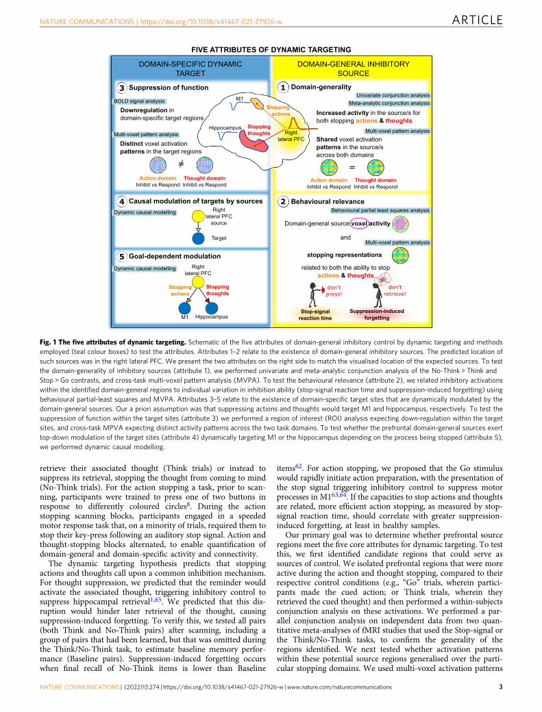
Dynamic targeting requires that a candidate inhibitory controlsystem exhibit five core attributes during stopping (see Fig. 1).First, stopping in diverse domains should engage the proposedsource of control, with activation patterns within this regiongeneralising over the specific demands of each stopping type.Consequently, activation patterns during any one form of stop-ping should contain information shared with inhibition in otherdomains. Second, the engagement of the proposed prefrontalsource should track indices of inhibitory control in diversedomains, demonstrating its behavioural relevance. Third,stopping-related activity in the prefrontal sources should co-
occur with interrupted functioning in domain-specific target sitesrepresenting thoughts or actions. Fourth, the prefrontal sourceshould exert top-down inhibitory coupling with these target sites,providing the causal basis of their targeted suppression. Finally,dynamic targeting requires that inhibitory coupling betweenprefrontal source and domain-specific target regions be selectiveto current goals. Note that domain-general inhibitory controldoes not require direct monosynaptic connections between thesource(s) and target(s) of control.
These five attributes of dynamic targeting remain unproven,despite the fundamental importance of inhibitory control.Research on response inhibition and thought suppression insteadhas focused on how the prefrontal cortex contributes to stoppingwithin each domain9,34–36. For example, research on thoughtsuppression has revealed top-down inhibitory coupling from theanterior rDLPFC to the hippocampus, and to several corticalregions representing specific mnemonic content8,37–41. Moreover,suppressing thoughts down-regulates hippocampal activity, withthe down-regulation linked to hippocampal GABA and forgettingof the suppressed content8. Top-down modulation of actions byrVLPFC suggests that premotor and primary motor cortex aretarget sites42–44. Action stopping engages local intracorticalinhibition within M1 to achieve stopping45–48, with a person’sstopping efficacy related to local GABAergic inhibition49. Rein-forcing this domain-specific focus, research has posited thatcontrol originates from different prefrontal regions in these twodomains suggesting separate control abilities: whereas the rightanterior DLPFC has received attention in work on thoughtsuppression2, the right VLPFC has been the focus in work onresponse inhibition10,11, despite both regions often arising in bothstopping tasks22. To integrate research from these separatedomains, we sought to determine which of these candidatesources of domain-general inhibitory control participate instopping both actions and thoughts and which exhibit the keyattributes of dynamic targeting.
Although dynamic inhibitory targeting has not been tested,some large-scale networks flexibly shift their coupling withdiverse brain regions that support task performance. Diverse tasksengage a frontoparietal network50–53, which exhibits greatercross-task variability in coupling with other regions than othernetworks51,54. Variable connectivity may index this network’sability to reconfigure flexibly and coordinate multiple task ele-ments in the interests of cognitive control51. A cingulo-opercularnetwork, including aspects of rDLPFC and rVLPFC, also is tied tocognitive control, including conflict and attentionalprocessing55–61, with the prefrontal components exhibiting highconnectivity variability over differing tasks54. However, previousanalyses of these networks do not address dynamic inhibitorytargeting: Dynamic targeting requires not merely that the pre-frontal cortex exhibits connectivity to multiple regions, but thatthe connectivity includes a top-down component that suppressestarget regions.
We sought to test the presence of dynamic targeting throughthe properties of prefrontal, motor and hippocampal networks(see Fig. 1 for an overview of our approach). We combined,within one fMRI session, a cognitive manipulation to suppressunwanted thoughts, the Think/No-Think paradigm6,62, withmotor action stopping in a stop-signal task63,64. This designprovided the opportunity to identify co-localised activations ofdomain-general inhibitory control in prefrontal sources andobserve their changes in effective connectivity with motor corticaland hippocampal targets. For the thought suppression task, priorto scanning, participants learned word pairs, each composed of areminder and a paired thought (Fig. 2). During thought-stoppingscanning blocks, on each trial, participants viewed one of thesereminders. For each reminder, we cued participants either to
ARTICLE NATURE COMMUNICATIONS | https://doi.org/10.1038/s41467-021-27926-w
2 NATURE COMMUNICATIONS | (2022) 13:274 | https://doi.org/10.1038/s41467-021-27926-w |www.nature.com/naturecommunications
retrieve their associated thought (Think trials) or instead tosuppress its retrieval, stopping the thought from coming to mind(No-Think trials). For the action stopping a task, prior to scan-ning, participants were trained to press one of two buttons inresponse to differently coloured circles8. During the actionstopping scanning blocks, participants engaged in a speededmotor response task that, on a minority of trials, required them tostop their key-press following an auditory stop signal. Action andthought-stopping blocks alternated, to enable quantification ofdomain-general and domain-specific activity and connectivity.
The dynamic targeting hypothesis predicts that stoppingactions and thoughts call upon a common inhibition mechanism.For thought suppression, we predicted that the reminder wouldactivate the associated thought, triggering inhibitory control tosuppress hippocampal retrieval1,65. We predicted that this dis-ruption would hinder later retrieval of the thought, causingsuppression-induced forgetting. To verify this, we tested all pairs(both Think and No-Think pairs) after scanning, including agroup of pairs that had been learned, but that was omitted duringthe Think/No-Think task, to estimate baseline memory perfor-mance (Baseline pairs). Suppression-induced forgetting occurswhen final recall of No-Think items is lower than Baseline
items62. For action stopping, we proposed that the Go stimuluswould rapidly initiate action preparation, with the presentation ofthe stop signal triggering inhibitory control to suppress motorprocesses in M163,64. If the capacities to stop actions and thoughtsare related, more efficient action stopping, as measured by stop-signal reaction time, should correlate with greater suppression-induced forgetting, at least in healthy samples.
Our primary goal was to determine whether prefrontal sourceregions meet the five core attributes for dynamic targeting. To testthis, we first identified candidate regions that could serve assources of control. We isolated prefrontal regions that were moreactive during the action and thought stopping, compared to theirrespective control conditions (e.g., “Go” trials, wherein partici-pants made the cued action; or Think trials, wherein theyretrieved the cued thought) and then performed a within-subjectsconjunction analysis on these activations. We performed a par-allel conjunction analysis on independent data from two quan-titative meta-analyses of fMRI studies that used the Stop-signal orthe Think/No-Think tasks, to confirm the generality of theregions identified. We next tested whether activation patternswithin these potential source regions generalised over the parti-cular stopping domains. We used multi-voxel activation patterns
FIVE ATTRIBUTES OF DYNAMIC TARGETING
Suppression of function3
Goal-dependent modulation5
Behavioural relevance2
Downregulation in domain-specific target regions
4 Causal modulation of targets by sources
Distinct voxel activation patterns in the target regions
Thought domainInhibit vs Respond
Action domainInhibit vs Respond
=
related to both the ability to stopactions & thoughts
and
stopping representations
don't press!
Stop-signal reaction time
don't retrieve!
Suppression-induced forgetting
Domain-general source voxel activity
Behavioural partial least squares analysis
Multi-voxel pattern analysis
Domain-generality1
Increased activity in the source/s for both stopping actions & thoughts
Shared voxel activation patterns in the source/s across both domains
Thought domainInhibit vs Respond
Action domainInhibit vs Respond
=
Multi-voxel pattern analysis
Univariate conjunction analysisMeta-analytic conjunction analysis
Dynamic causal modelling
Dynamic causal modelling
Multi-voxel pattern analysis
BOLD signal analysis
HippocampusM1
Stopping actions
Stopping thoughts
Right lateral PFC
Hippocampus
M1
Rightlateral PFC
Stopping actions
Stopping thoughts
Target
Right lateral PFC
source
DOMAIN-SPECIFIC DYNAMIC TARGET
DOMAIN-GENERAL INHIBITORY SOURCE
Fig. 1 The five attributes of dynamic targeting. Schematic of the five attributes of domain-general inhibitory control by dynamic targeting and methodsemployed (teal colour boxes) to test the attributes. Attributes 1–2 relate to the existence of domain-general inhibitory sources. The predicted location ofsuch sources was in the right lateral PFC. We present the two attributes on the right side to match the visualised location of the expected sources. To testthe domain-generality of inhibitory sources (attribute 1), we performed univariate and meta-analytic conjunction analysis of the No-Think > Think andStop > Go contrasts, and cross-task multi-voxel pattern analysis (MVPA). To test the behavioural relevance (attribute 2), we related inhibitory activationswithin the identified domain-general regions to individual variation in inhibition ability (stop-signal reaction time and suppression-induced forgetting) usingbehavioural partial-least squares and MVPA. Attributes 3–5 relate to the existence of domain-specific target sites that are dynamically modulated by thedomain-general sources. Our a priori assumption was that suppressing actions and thoughts would target M1 and hippocampus, respectively. To test thesuppression of function within the target sites (attribute 3) we performed a region of interest (ROI) analysis expecting down-regulation within the targetsites, and cross-task MPVA expecting distinct activity patterns across the two task domains. To test whether the prefrontal domain-general sources exerttop-down modulation of the target sites (attribute 4) dynamically targeting M1 or the hippocampus depending on the process being stopped (attribute 5),we performed dynamic causal modelling.
NATURE COMMUNICATIONS | https://doi.org/10.1038/s41467-021-27926-w ARTICLE
NATURE COMMUNICATIONS | (2022) 13:274 | https://doi.org/10.1038/s41467-021-27926-w |www.nature.com/naturecommunications 3

to train a classifier to discriminate stopping from going in onemodality (e.g., action stopping), to test whether it could identifystopping in the other modality (e.g., thought suppression).Finally, to examine behavioural relevance, we related inhibitoryactivations within these meta-analytic conjunction areas to indi-vidual variation in inhibition ability (e.g., suppression-inducedforgetting and stop-signal reaction time) using behavioural partialleast squares and multi-voxel pattern analysis. Any region sur-viving these constraints was considered a strong candidate for a
hub of inhibitory control. We hypothesised that these analyseswould identify the right anterior DLPFC5,6,22,37, and rightVLPFC10,24.
To verify that inhibitory control targets goal-relevant brainregions during stopping, we next confirmed that a prioritarget sites are suppressed in a goal-specific manner. Specifically,stopping retrieval should down-regulate hippocampalactivity1,4,37,39–41,65, more than does action stopping. In contrast,stopping actions should inhibit motor cortex more than does
Stop-signal task
Think/No-Think task
Procedure
Conditions: Baseline, 20 items Think, 20 items No-Think, 20 items
Cue-Target word pairs e.g. PART - BOWL
Think trials
+~2.3 s
3 s PART
BOWL
retrieve!
~2.3 s +
No-Think trials
~2.3 s + CORD
3 s SPRAY suppress!
~2.3 s +
7Think/No-Think task recall phase
Stop-signal taskstimulus-response learning
1 2 Stop-signal practice3
Think/No-Think task encoding phase
4 Think/No-Think practice
PARTBOWL
SPRAYCORD
6 Experimental phase and fMRI acquisition
8x
250 ms
Stop trials
+
~750 ms
+250 ms
~230 ms delayed signal
Go stimuli
Response button box
1000 Hz "beep" tone for 100 ms
Stop signal
+250 ms
~750 ms
+250 ms
Go trials
until response or 2500 ms
until response or 2500 ms
16 interleaved task blocks: 8 Think/No-Think, 8 Stop-signal (4 Go only)
Think/No-Think 6 trials
Stop-signal12 trialsRest Rest
30 s 4 s 30 s 4 s8x
5 Interleaved Stop-signal and Think/No-Think practice
Think/No-Think 6 trials
Stop-signal12 trialsRest Rest
30 s 4 s 30 s 4 s8x
a
b
c
respond!
withold!
Fig. 2 Schematic of the experimental paradigm and procedure. a In the Stop-signal task, the Go stimuli were red, green, blue, and yellow coloured circles.On Go trials, participants responded by pressing one of the two buttons on a button box according to learned stimulus–response associations. On Stoptrials, shortly after the Go stimulus, an auditory “beep” tone would signal participants to withhold the button press. The stop-signal delay varieddynamically in 50ms steps to achieve approximately a 50% success-to-stop rate for each participant. b In the Think/No-Think task, participants learned78 cue-target word pair associations. Sixty of the word pairs were then divided into three lists composed of 20 items each and allocated to the threeexperimental conditions: Think, No-Think, and Baseline. During Think trials, a cue word appeared in green, and participants had 3 s to retrieve and think ofthe associated target word. On No-Think trials, a cue word appeared in red and participants were asked to suppress the retrieval of the associated targetword and push it out of awareness if it popped into their mind. c The procedure consisted of 7 steps: (1) stimulus–response learning for the Stop-signaltask: (2) Stop-signal task practice; (3) encoding phase of the Think/No-Think task; (4) Think/No-Think practice; (5) practice of interleaved Stop-signal andThink/No-Think tasks; (6) the main experimental phase during fMRI acquisition where participants performed interleaved 30 s blocks of Stop-signal andThink/No-Think tasks; (7) recall phase of the Think/No-Think task.
ARTICLE NATURE COMMUNICATIONS | https://doi.org/10.1038/s41467-021-27926-w
4 NATURE COMMUNICATIONS | (2022) 13:274 | https://doi.org/10.1038/s41467-021-27926-w |www.nature.com/naturecommunications

thought stopping8. To determine whether these differences inmodulation arise from inhibitory targeting by our putativedomain-general prefrontal control regions, we used dynamiccausal modelling66. If both DLPFC and VLPFC are involved, asprior work suggests, we sought to evaluate whether one or bothregions are critical sources of inhibitory control.
Here, we show that stopping unwanted thoughts and actionsengages common regions in the rDLPFC and rVLPFC. Critically,these regions did not merely share common activation duringthese forms of stopping, but also exhibited the five core attributesneeded to infer dynamic targeting, shifting their connectivity todomain-specific target regions to suppress their regional activity.These findings confirm central predictions of a domain-generalinhibitory control mechanism and establish the joint role of bothrDLPFC and rVLPFC in achieving this function.
ResultsStopping actions and thoughts recruits right DLPFC andVLPFC. We first identified brain regions that could provide asource of inhibitory control during action and thought stop-ping (Establishing Attribute 1: domain-generality). Thewhole-brain voxel-wise conjunction analysis of the Stop > Goand the No-Think > Think contrasts revealed that both motorand thought inhibition evoked conjoint activations in the rightprefrontal cortex (PFC), specifically, the rDLPFC (middlefrontal and superior frontal gyri), rVLPFC (ventral aspects ofinferior frontal gyrus, including BA44/45, extending intoinsula), precentral gyrus, and supplementary motor area (seeTable 1 and Fig. 3). These findings suggest a role of the rightPFC in multiple stopping domains5,10,67, necessary fordynamic targeting.
The observation that rDLPFC contributes to inhibitory controlmight seem surprising, given the published emphasis on therVLPFC in motor stopping studies10,11. It could be that rDLPFC
activation arises from the need to alternate between the Stop-signal and Think/No-Think tasks, or from carryover effectsbetween tasks. We, therefore, compared the activations observedin our within-subjects conjunction analysis to a meta-analyticconjunction analysis of independent Stop-signal (N= 40) andThink/No-Think (N= 16) studies (see the “Methods” section)conducted in many different laboratories with different variationson the two procedures (see ref. 22 for an earlier version with fewerstudies). The meta-analytic conjunction results were highlysimilar to our within-subjects results, with conjoint clusters inmatched regions of DLPFC, VLPFC (BA44/45, extending intoinsula), right anterior cingulate cortex, and right basal ganglia(see Table 1 and Fig. 3). Notably, in both the within-subjects andmeta-analytic conjunctions, the domain-general activation inrDLPFC did not spread throughout the entire right middle frontalgyrus but was confined to the anterior portion of the rDLPFC,spanning BA9/46 and BA10. The convergence of these conjunc-tion analyses suggests that the involvement of the rDLPFC, andour findings of conjoint activations across the two stoppingdomains more broadly, do not arise from the specific proceduresof the stopping tasks or to carryover effects arising from ourwithin-subjects design; rather, they indicate a pattern thatconverges across laboratories and different experimentalprocedures.
The domain-general stopping activations included areas out-side of the prefrontal cortex (see Table 1 and Fig. 3). Althoughnot the focus on the current investigation, we characterised theseactivations in relation to large-scale brain networks, using apublicly available Cole-Anticevic brain-wide network partition(CAB-NP)68. We used the Connectome Workbench software69 tooverlay our activations over the CAB-NP to estimate the parceland network locations of our clusters. Domain-general clustersprimarily were in the Cingulo-Opercular (CON) and Frontopar-ietal (FPN) networks (86% of parcels fell within these two
Table 1 Within-subjects and meta-analysis domain-general inhibition-induced activations (Stop > Go and No-Think > Think).
Hemisphere Region ~BA Network MNI of the peak Volume (mm3)
x y z
Within-subjects, Stop > Go & No-Think > ThinkRight Inferior frontal gyrus (VLPFC) Insula 44, 45 CON, FPN 45 18 8 5366Right Inferior parietal lobule 40 CON, FPN, PMM 63 −42 41 3611Right Supplementary motor area 6, 8 CON, FPN, LAN 15 18 64 2498Right Middle frontal gyrus (DLPFC)
Superior frontal gyrus (DLPFC)9, 10, 46 CON 33 42 23 1654
Right Precentral gyrus 6 CON, FPN, LAN 42 3 41 945Left Inferior parietal lobule 40 CON, FPN −60 −48 41 641Meta-analysis, Stop > Go & No-Think > ThinkRight Inferior frontal gyrus (VLPFC) Insula 44, 45 CON, FPN 36 26 0 4523Right/Left Supplementary motor area 6, 8 CON, FPN, LAN 14 14 60 3071Left Inferior frontal gyrus Insula 44, 45 CON, FPN −44 18 0 2970Right Inferior parietal lobule 40 CON, FPN, PMM 58 −46 34 2633Right Anterior cingulate cortex 24, 32 CON, FPN 6 22 38 1620Right Middle frontal gyrus (DLPFC)
Superior frontal gyrus (DLPFC)9, 10, 46 CON 36 50 22 844
Right Basal ganglia 16 8 8 776Left Inferior parietal lobule 40 CON, FPN −60 −50 34 608Right Precentral gyrus 6 CON, LAN 44 2 46 270Right Superior parietal lobule 7 FPN, DAN 34 −48 46 176Within-subjects & Meta-analysis, Stop > Go & No-Think > ThinkRight Inferior frontal gyrus (VLPFC) Insula 44, 45 CON, FPN 45 18 8 2666Right Inferior parietal lobule 40 CON, FPN, PMM 63 −42 38 1620Right Supplementary motor area 6, 8 CON, FPN, LAN 15 18 64 1418Right Middle frontal gyrus (DLPFC) 9, 10, 46 CON 33 39 26 338Left Inferior parietal lobule 40 CON, FPN −60 −48 41 270Right Precentral gyrus 6 CON, LAN 42 3 41 135
NATURE COMMUNICATIONS | https://doi.org/10.1038/s41467-021-27926-w ARTICLE
NATURE COMMUNICATIONS | (2022) 13:274 | https://doi.org/10.1038/s41467-021-27926-w |www.nature.com/naturecommunications 5

networks in the within-subjects conjunction), but also includedPosterior-Multimodal and Language networks parcels (seeSupplementary Table 1 and Supplementary Fig. 1). Of the 21cortical parcels identified for the within-subjects conjunction (seeSupplementary Table 1), the majority (57%) participated in theCON, whereas 29% were involved in the FPN; the independentmeta-analysis yielded similar findings (56% vs. 30%; seeSupplementary Table 2 and Supplementary Fig. 2). Our mainright prefrontal regions both featured parcels from the CON;however, whereas rDLPFC was located solely in the CON (in boththe within-subjects and meta-analytic conjunctions), the rVLPFCregion also included parcels from the FPN.
Together, these findings confirm the role of both the rightanterior DLPFC and rVLPFC for both motor and memoryinhibition, consistent with prior evidence of a causal role of theseregions in the stopping function of inhibitory control27–30.Moreover, they show that stopping recruits a larger network ofregions, dominated by the CON, and to a lesser degree, FPN.These findings suggest that to achieve stopping, domain-generalinhibitory control may reflect a special configuration of the CONthat includes elements of the FPN and other networks. Notably,key regions of the FPN were absent from all analyses (nosuprathreshold activations), including the large middle frontalregion often taken as a hallmark of domain-general cognitivecontrol51,52.
Action and thought stopping abilities are related. We nextconfirmed that action-stopping efficiency was associated withsuccessful thought suppression. To quantify action stoppingefficiency, we computed stop-signal reaction times (SSRTs) usingthe consensus standard integration method64. We confirmed thatthe probability of responding to Stop trials (M= 0.49, SD= 0.07;ranging from 0.36 to 0.69) fell within the recommended range forreliable estimation of SSRTs64, and that the probabilities of Goomissions (M= 0.002, SD= 0.01) and choice errors on Go trials(M= 0.04, SD= 0.02) were low. We next verified that the correctGo RT (M= 600.91 ms, SD= 54.63 ms) exceeded the failed StopRT (M= 556.92 ms, SD= 56.77) in all but one participant (9 msdifference between the failed Stop RT and correct Go RT;including this participant makes little difference to any analysis,so they were not excluded). Given that the integration methodrequirements were met, the average SSRT, our measure ofinterest, was 348.34 ms (SD= 51.25 ms), with an average SSD of230 ms (SD= 35.68 ms).
We next verified that the Think/No-Think task had inducedforgetting of suppressed items. We compared final recall of No-Think items to that of Baseline items that had neither beensuppressed nor retrieved (see the “Methods” section). Consistentwith a previous analysis of these data8 and with priorfindings1,62,65,70, suppressing retrieval impaired No-Think recall(M= 72%, SD= 9%) relative to Baseline recall (M= 77%,
No-Think > Think & Stop > GoWithin-subjects Meta-analysis Within-subjects & Meta-analysis
R R
44
45IFG
Insula
46
10
40
6
96
8
3224
GyriSulci
Fig. 3 Domain-general inhibition-induced activations. Red: within-subjects (N= 24) conjunction of the Stop > Go and the No-Think > Think contraststhresholded at p < 0.05 FDR corrected for whole-brain multiple comparisons. Blue: meta-analytic conjunction of Stop > Go and the No-Think > Thinkcontrasts from independent 40 Stop-signal and 16 Think/No-Think studies. Yellow: overlap of the within-subjects and meta-analytic conjunctions. Resultsare displayed on an inflated MNI-152 surface with outlined and numbered Brodmann areas (top panel), as well as on MNI-152 volume slices (bottompanel). R: right hemisphere; L: left hemisphere. The brain images were generated using FreeSurfer software (http://surfer.nmr.mgh.harvard.edu), andPySurfer (https://pysurfer.github.io) and Nilearn (https://nilearn.github.io) Python (Python Software Foundation, DE, USA) packages. Conjunction mapsand visualisation notebook are available on the GitHub repository104 (https://github.com/dcdace/Domain-general/).
ARTICLE NATURE COMMUNICATIONS | https://doi.org/10.1038/s41467-021-27926-w
6 NATURE COMMUNICATIONS | (2022) 13:274 | https://doi.org/10.1038/s41467-021-27926-w |www.nature.com/naturecommunications
SD= 9%), yielding a suppression-induced forgetting (SIF) effect(Baseline − No-Think= 5%, SD= 9%, one-tailed t23= 2.55,p= 0.009, d= 0.521). Thus, suppressing retrieval yielded thepredicted inhibitory aftereffects on unwanted thoughts.
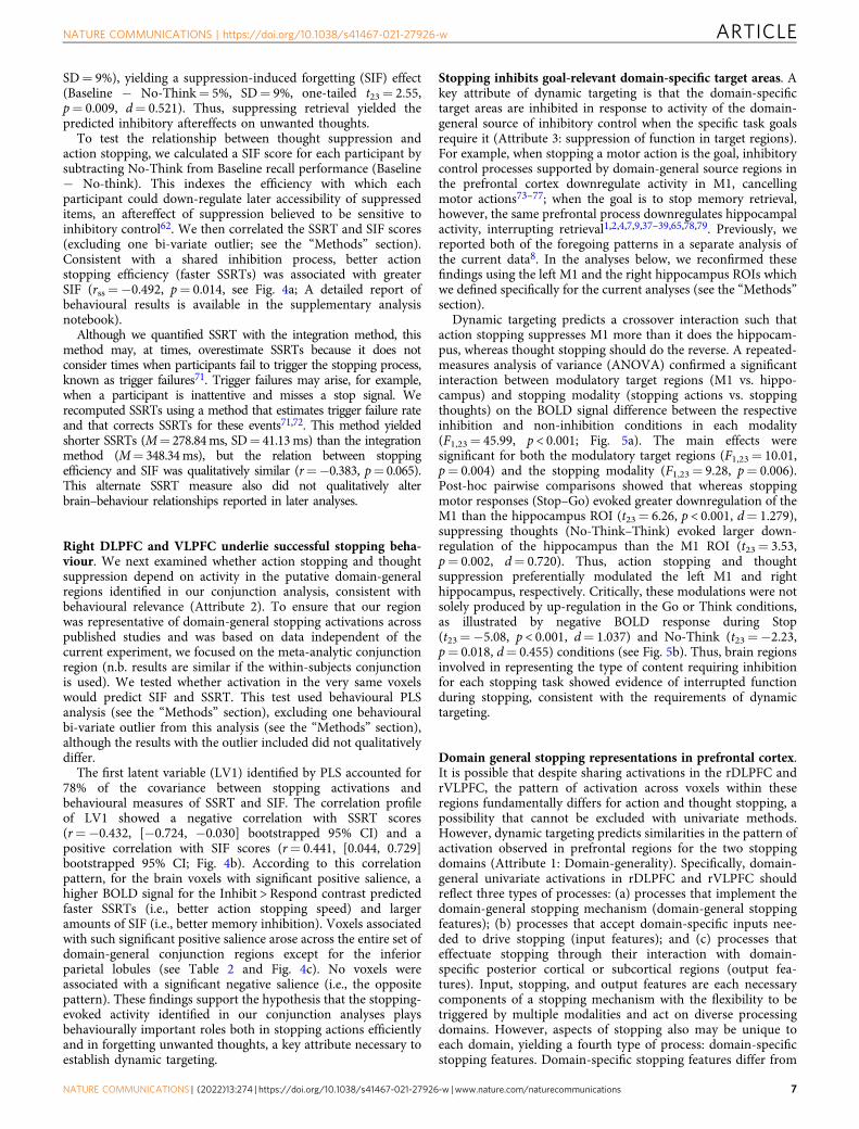
To test the relationship between thought suppression andaction stopping, we calculated a SIF score for each participant bysubtracting No-Think from Baseline recall performance (Baseline− No-think). This indexes the efficiency with which eachparticipant could down-regulate later accessibility of suppresseditems, an aftereffect of suppression believed to be sensitive toinhibitory control62. We then correlated the SSRT and SIF scores(excluding one bi-variate outlier; see the “Methods” section).Consistent with a shared inhibition process, better actionstopping efficiency (faster SSRTs) was associated with greaterSIF (rss=−0.492, p= 0.014, see Fig. 4a; A detailed report ofbehavioural results is available in the supplementary analysisnotebook).
Although we quantified SSRT with the integration method, thismethod may, at times, overestimate SSRTs because it does notconsider times when participants fail to trigger the stopping process,known as trigger failures71. Trigger failures may arise, for example,when a participant is inattentive and misses a stop signal. Werecomputed SSRTs using a method that estimates trigger failure rateand that corrects SSRTs for these events71,72. This method yieldedshorter SSRTs (M= 278.84ms, SD= 41.13ms) than the integrationmethod (M= 348.34ms), but the relation between stoppingefficiency and SIF was qualitatively similar (r=−0.383, p= 0.065).This alternate SSRT measure also did not qualitatively alterbrain–behaviour relationships reported in later analyses.
Right DLPFC and VLPFC underlie successful stopping beha-viour. We next examined whether action stopping and thoughtsuppression depend on activity in the putative domain-generalregions identified in our conjunction analysis, consistent withbehavioural relevance (Attribute 2). To ensure that our regionwas representative of domain-general stopping activations acrosspublished studies and was based on data independent of thecurrent experiment, we focused on the meta-analytic conjunctionregion (n.b. results are similar if the within-subjects conjunctionis used). We tested whether activation in the very same voxelswould predict SIF and SSRT. This test used behavioural PLSanalysis (see the “Methods” section), excluding one behaviouralbi-variate outlier from this analysis (see the “Methods” section),although the results with the outlier included did not qualitativelydiffer.
The first latent variable (LV1) identified by PLS accounted for78% of the covariance between stopping activations andbehavioural measures of SSRT and SIF. The correlation profileof LV1 showed a negative correlation with SSRT scores(r=−0.432, [−0.724, −0.030] bootstrapped 95% CI) and apositive correlation with SIF scores (r= 0.441, [0.044, 0.729]bootstrapped 95% CI; Fig. 4b). According to this correlationpattern, for the brain voxels with significant positive salience, ahigher BOLD signal for the Inhibit > Respond contrast predictedfaster SSRTs (i.e., better action stopping speed) and largeramounts of SIF (i.e., better memory inhibition). Voxels associatedwith such significant positive salience arose across the entire set ofdomain-general conjunction regions except for the inferiorparietal lobules (see Table 2 and Fig. 4c). No voxels wereassociated with a significant negative salience (i.e., the oppositepattern). These findings support the hypothesis that the stopping-evoked activity identified in our conjunction analyses playsbehaviourally important roles both in stopping actions efficientlyand in forgetting unwanted thoughts, a key attribute necessary toestablish dynamic targeting.
Stopping inhibits goal-relevant domain-specific target areas. Akey attribute of dynamic targeting is that the domain-specifictarget areas are inhibited in response to activity of the domain-general source of inhibitory control when the specific task goalsrequire it (Attribute 3: suppression of function in target regions).For example, when stopping a motor action is the goal, inhibitorycontrol processes supported by domain-general source regions inthe prefrontal cortex downregulate activity in M1, cancellingmotor actions73–77; when the goal is to stop memory retrieval,however, the same prefrontal process downregulates hippocampalactivity, interrupting retrieval1,2,4,7,9,37–39,65,78,79. Previously, wereported both of the foregoing patterns in a separate analysis ofthe current data8. In the analyses below, we reconfirmed thesefindings using the left M1 and the right hippocampus ROIs whichwe defined specifically for the current analyses (see the “Methods”section).
Dynamic targeting predicts a crossover interaction such thataction stopping suppresses M1 more than it does the hippocam-pus, whereas thought stopping should do the reverse. A repeated-measures analysis of variance (ANOVA) confirmed a significantinteraction between modulatory target regions (M1 vs. hippo-campus) and stopping modality (stopping actions vs. stoppingthoughts) on the BOLD signal difference between the respectiveinhibition and non-inhibition conditions in each modality(F1,23= 45.99, p < 0.001; Fig. 5a). The main effects weresignificant for both the modulatory target regions (F1,23= 10.01,p= 0.004) and the stopping modality (F1,23= 9.28, p= 0.006).Post-hoc pairwise comparisons showed that whereas stoppingmotor responses (Stop–Go) evoked greater downregulation of theM1 than the hippocampus ROI (t23= 6.26, p < 0.001, d= 1.279),suppressing thoughts (No-Think–Think) evoked larger down-regulation of the hippocampus than the M1 ROI (t23= 3.53,p= 0.002, d= 0.720). Thus, action stopping and thoughtsuppression preferentially modulated the left M1 and righthippocampus, respectively. Critically, these modulations were notsolely produced by up-regulation in the Go or Think conditions,as illustrated by negative BOLD response during Stop(t23=−5.08, p < 0.001, d= 1.037) and No-Think (t23=−2.23,p= 0.018, d= 0.455) conditions (see Fig. 5b). Thus, brain regionsinvolved in representing the type of content requiring inhibitionfor each stopping task showed evidence of interrupted functionduring stopping, consistent with the requirements of dynamictargeting.
Domain general stopping representations in prefrontal cortex.It is possible that despite sharing activations in the rDLPFC andrVLPFC, the pattern of activation across voxels within theseregions fundamentally differs for action and thought stopping, apossibility that cannot be excluded with univariate methods.However, dynamic targeting predicts similarities in the pattern ofactivation observed in prefrontal regions for the two stoppingdomains (Attribute 1: Domain-generality). Specifically, domain-general univariate activations in rDLPFC and rVLPFC shouldreflect three types of processes: (a) processes that implement thedomain-general stopping mechanism (domain-general stoppingfeatures); (b) processes that accept domain-specific inputs nee-ded to drive stopping (input features); and (c) processes thateffectuate stopping through their interaction with domain-specific posterior cortical or subcortical regions (output fea-tures). Input, stopping, and output features are each necessarycomponents of a stopping mechanism with the flexibility to betriggered by multiple modalities and act on diverse processingdomains. However, aspects of stopping also may be unique toeach domain, yielding a fourth type of process: domain-specificstopping features. Domain-specific stopping features differ from
NATURE COMMUNICATIONS | https://doi.org/10.1038/s41467-021-27926-w ARTICLE
NATURE COMMUNICATIONS | (2022) 13:274 | https://doi.org/10.1038/s41467-021-27926-w |www.nature.com/naturecommunications 7
domain-specific output features in that the latter govern theinteraction of the stopping mechanism with posterior-cortical orsubcortical target regions, whereas the former reflect computa-tions specific to a stopping domain that are local to the prefrontalsource region. Domain-general stopping features should yieldsimilarities between the multivariate patterns for action andthought stopping; in contrast, input, output, and domain-specificstopping features should yield differences in the multivariate
patterns for thought and action stopping, with the relative con-tributions of each being difficult to disentangle. Cross-modalitydecoding should not be possible in domain-specific targetregions, reflecting their specialised involvement in action ormemory stopping. Conversely, between-modality decoding,reflecting domain-specific features, must exist in the domain-specific target regions and to some extent in the domain-generalsource regions.
c
a b
R R
Meta-analysis No-Think > Think & Stop > Go conjunction maskVoxels expressing brain/behaviour correlations as revealed by LV1
Stop-signalreaction time
Suppressioninduced
forgetting
Brai
n-be
havi
our c
orre
latio
n * *
-0.8
-0.4
0.0
0.4
0.8
The first latent variable (LV1)
250
300
350
400
450
0 5 10 15 20Suppression induced forgetting (%)
Stop
-sig
nal r
eact
ion
time
(ms)
Spearman skipped correlation rss = -0.492, p = 0.014Outliers (n=1) not displayed
GyriSulci
Fig. 4 Domain-general behavioural and brain/behaviour relationships. a and b Source data are provided as a Source Data file. a Better action stoppingefficiency (shorter stop-signal reaction time) was associated with better inhibitory control over thoughts (percentage of items forgotten for No-Thinkrelative to Baseline conditions at the final recall phase), i.e. suppression-induced forgetting; rss=−0.492, p= 0.014, n= 24. One bivariate outlier isnot displayed on the scatterplot. Shading represents 95% CI. b and c A behavioural partial least squares (PLS) analysis was conducted to identify brainareas where individual variation in inhibition ability (SSRT and SIF) was related to increased inhibition-induced activity (main effect contrast ofinhibition from the within-subject experiment, masked by the meta-analytic conjunction). b The first latent variable (LV1) identified voxels showing asignificant pattern of brain/behaviour correlations to both SSRT and SIF (error bars indicate bootstrapped 95% CI, n= 5000, *p < 0.05). c The voxelsalience map expressing LV1. Blue: meta-analytic conjunction mask. Red: voxels showing a significant pattern of brain/behaviour correlations asrevealed by the LV1; thresholded at bootstrapped standard ratio 1.96, corresponding to p < 0.05, two-tailed. Results are displayed on an inflated MNI-152 surface (top panel), as well as on MNI-152 volume slices (bottom panel). R: right hemisphere; L: left hemisphere. The brain images were generatedusing FreeSurfer software (http://surfer.nmr.mgh.harvard.edu), and PySurfer (https://pysurfer.github.io) and Nilearn (https://nilearn.github.io)Python (Python Software Foundation, DE, USA) packages. Conjunction mask, PLS results and visualisation notebook are available on the GitHubrepository104 (https://github.com/dcdace/Domain-general/).
ARTICLE NATURE COMMUNICATIONS | https://doi.org/10.1038/s41467-021-27926-w
8 NATURE COMMUNICATIONS | (2022) 13:274 | https://doi.org/10.1038/s41467-021-27926-w |www.nature.com/naturecommunications
To identify the predicted cross-modality similarities, withineach subject, we trained a classifier to distinguish Inhibit andRespond conditions in one modality and tested the ability todistinguish Inhibit and Respond conditions in the other modality.We performed the classification analysis on the rDLPFC,rVLPFC, right hippocampus, and left M1 ROIs (see the“Methods” section). The analysis revealed that a classifier trainedon one modality could discriminate Inhibition from Respondconditions in the other modality significantly above chance (50%)for both rDLPFC (M= 58%, SD= 9%, one-tailed t23= 4.17,padj= 0.001, d= 0.852) and rVLPFC (M= 60%, SD= 11%, one-tailed t23= 4.46, padj < 0.001, d= 0.911). This cross-modalitydecoding suggests that a domain-general inhibitory controlprocess contributes to activity in these regions (see Fig. 5c;cross-modality prediction accuracy is even stronger in early taskblocks—see next section). To identify between-task differences(the domain-specific features), we trained a classifier todiscriminate Stop from No-Think trials (see the “Methods”section). The classifier could indeed discriminate action andthought stopping in both rDLPFC (M= 70%, SD= 13%, one-tailed t23= 7.37, padj < 0.001, d= 1.504) and rVLPFC (M= 82%,SD= 11%, t23= 13.89, padj < 0.001, d= 2.835). The superiordiscrimination of action and thought stopping in the rVLPFCcompared to rDLPFC was reliable; however, control analysesmatching ROI size eliminated this advantage, suggesting that itwas an artefact of the larger ROI used for rVLPFC (seeSupplementary Fig. 4). It is unclear whether this domain-specific component in the LPFC reflects evidence for the inputand output features required by the dynamic targeting hypothesisor instead domain-specific stopping processes, either of whichmay be exploited by a classifier to enhance between-modalityprediction performance. The cross-modality prediction findings,however, clearly confirm predictions of the domain-generalityattribute of dynamic targeting.
In contrast to the patterns observed in the prefrontal cortex, weobserved no evidence of cross-modality decoding in the modality-specific regions targeted by inhibitory control. This pattern arosefor both right hippocampus (M= 49%, SD= 10%, one-tailedt23=−0.37, padj= 1, d= 0.075) and also left M1 (M= 48%,SD= 10, one-tailed t23=−1.16, padj= 1, d= 0.236), in which thecross-modality classifier accuracy did not significantly differ fromchance performance (see Fig. 5c). An estimated one-sample t-testBayes factor (one-tailed; medium prior Cauchy scale 0.707; null/alternative) suggested that the data were substantially in favour ofthe null hypothesis for both the hippocampus (B01= 6.01,posterior distribution: Median= 0.109, 95% CI= [0.004, 0.385])and M1 (B01= 9.14, posterior distribution: Median= 0.071, 95%CI= [0.003, 0.294]). Nevertheless, these putative target regionsresponded very differently to the two modalities of inhibitorycontrol, as evidenced by presence of significant domain-specificinformation in each region (Attribute 3: suppression of function).
A classifier could reliably distinguish No-Think trials from Stoptrials within both the right hippocampus (M= 63%, SD= 11%,t23= 5.89, padj < 0.001, d= 1.202) and left M1 (M= 65%, SD=12%, t23= 6.56, padj < 0.001, d= 1.338; see Fig. 5c). Again, thesedifferences may reflect input features (either from perception ortop-down control), output features, or the impact of the domain-specific inhibition processes on the target regions. Notably,although comparisons of classification accuracies across ROIsshould be interpreted with caution80, the ability of the classifier todistinguish No-Think from Stop trials did not vary across ourfour ROIs (rDLPFC, rVLPFC, hippocampus, M1) when ROI sizewas controlled (see Supplementary Fig. 4). Thus, all ROIssupported comparable classification performance in domain-specific classification, making it unlikely that the null classifica-tion results in the between-domain classifier in the hippocampusand M1 simply reflect poor signal quality in those target regions.
Because we z-normalised activation within each of theseregions within each task, the ability to distinguish No-Think fromStop trials was not based on differences in overall univariatesignal, but instead on information contained in distinct patternsof activity in each task. These findings reinforce the assumptionthat the hippocampus and M1 are uniquely affected by thoughtand action stopping respectively, as expected for domain-specifictargets of inhibitory control. Taken together, these contrastingfindings from the PFC and domain-specific regions arecompatible with the view that rDLPFC and rVLPFC jointlycontribute to a domain-general stopping process that dynamicallytargets different regions, depending on the nature of the contentto be suppressed.
Action stopping representations predict adaptive forgetting.Because dynamic targeting posits that LPFC contains domain-general stopping representations, training a classifier to distin-guish stopping in one domain should predict stopping behaviourin other domains. For example, the ability of an action stoppingclassifier to distinguish when people are suppressing thoughtsraises the intriguing possibility that it also may identify partici-pants who successfully forget those thoughts (establishing furtherevidence of Attribute 2, behavioural relevance). To test thispossibility, we capitalised on an adaptive forgetting phenomenonknown as the conflict reduction benefit (for a review, see6). Theconflict-reduction benefit refers to the declining need to expendinhibitory control resources that arises when people repeatedlysuppress the same intrusive thoughts. This benefit arises becauseinhibitory control induces forgetting of inhibited items, whichthereafter cause fewer control problems. For example, overrepeated inhibition trials, activation in rDPLFC, rVLPFC, andanterior cingulate cortex decline, with larger declines in partici-pants who forget more of the memories they suppressed6,81,82. Ifan action-stopping classifier detects the inhibition process, twofindings related to conflict-reduction benefits should emerge.
Table 2 Control network regions showing a significant pattern of brain/behaviour correlations as revealed by the first latentvariable of the PLS analysis.
Brain region ~BA MNI of the peak Volume (mm3)
x y z
Right Inferior frontal gyrus (VLPFC) Insula 44, 45 45 21 0 3375Right Anterior cingulate cortex 24, 32 6 30 34 1418Left Inferior frontal gyrus Insula 44, 45 −33 21 4 1046Right/Left Supplementary motor area 6, 8 6 9 64 1013Right Basal ganglia 15 3 8 709Right Middle frontal gyrus (DLPFC) 10, 46 33 48 19 304Right Precentral gyrus 6 42 3 41 68
NATURE COMMUNICATIONS | https://doi.org/10.1038/s41467-021-27926-w ARTICLE
NATURE COMMUNICATIONS | (2022) 13:274 | https://doi.org/10.1038/s41467-021-27926-w |www.nature.com/naturecommunications 9

First, over Think/No-Think task blocks, the action-stoppingclassifier should discriminate thought suppression less well, withhigh classification in early blocks that drops as memories areinhibited. Second, this decline should be larger for peopleshowing greater SIF.
We examined how accurately an action-stopping classifierdistinguishes No-Think from Think conditions for the 8 fMRIruns (we note that there were three missing data points for the8th run and one missing data point for the 7th run due toexclusion of some functional runs; see Methods). The rDLPFC
showed a robust linear decline (F7,157= 9.61, p= 0.002) inclassification accuracy from the first (M= 77%) to the eighth(M= 40%) run (see Fig. 6a). This result is consistent with aconflict-reduction benefit and suggests that domain-generalprocesses are especially important during early attempts atthought stopping. The rVLPFC exhibited a marginal lineardecline (F1,157= 2.88, p= 0.092) in classification accuracy fromthe first (M= 67%) to the eighth (M= 29%) run (see Fig. 6b).Critically, for both rDLPFC (rss=−0.676, p < 0.001; Fig. 6c) andrVLPFC (rss=−0.570, p= 0.004; Fig. 6d), participants showing
c
a b
**
***
-0.3
-0.2
-0.1
0.0
Hippocampus M1Target region *
% s
igna
l cha
nge
diffe
renc
e
Modality *: No-Think - Think Stop - Go
*** ***
*** ***
−0.2
0.0
0.2
0 2 4 6 8 10Peristimulus time (s)%
sig
nal c
hang
e Go StopGo/Stop, M1
***
*
−0.10−0.05
0.000.050.10
0 2 4 6 8 10Peristimulus time (s)%
sig
nal c
hang
e Think No−Think
Think/No−Think, Hippocampus
chance level 50%
20
40
60
80
100
***rDLPFC
***rVLPFC
***Hippocampus
***M1
Cla
ssifi
catio
n ac
cura
cy (%
)
Inhibition representations
Train: Stop, No-ThinkTest: Stop ≠ No-Think
Train: Stop, GoTest: No-Think = Stop
Train: No-Think, ThinkTest: Stop = No-Think
average
***rDLPFC
***rVLPFC HippocampusM1
Domain-specific component Domain-general component
Target region x Modality interaction ***
Fig. 5 ROI analysis of domain-specific and domain-general modulation during thought and action suppression. a–c ***p < 0.001; **p < 0.01; *p < 0.05.Data are presented as mean values. Error bars/shading represent within-subject standard error. Sample size n= 24. Source data are provided as a SourceData file. a Target areas M1 and hippocampus were modulated in a domain-specific manner. We calculated the BOLD signal in each target ROI for eachcondition by averaging across the time points from 2 to 6 s post-stimulus onset and subtracting out the onset value to account for pretrial variability. Thenwe subtracted the values of Go from Stop and Think from No-Think and entered them into a region by modality repeated-measures ANOVA. The ANOVAconfirmed a significant interaction between modulatory target regions and stopping modality (F1,23= 45.99, p < 0.001). Stopping actions (in yellow) evokedgreater downregulation of M1 than of the hippocampus (t23= 6.26, p < 0.001, d= 1.279) but suppressing thoughts (in red) evoked greater downregulationof the hippocampus than of M1 (t23= 3.53, p= 0.002, d= 0.720). b The BOLD signal time-course in M1 (top panel) and hippocampus (bottom panel).During inhibition conditions (Stop and No-Think; in blue), the BOLD signal decreased below the baseline, whereas during respond conditions (Go andThink; in green) the BOLD signal increased above the baseline. Significance stars represent one-tailed one-sample t-test results, Bonferroni corrected formultiple comparisons. c Using MVPA, we tested whether action and thought inhibition share a common voxel activation pattern within the four ROIs. Weperformed two types of pattern classification to identify domain-general (cross-task classification; in violet) and domain-specific (between-taskclassification; in green) components within each ROI. Large circles represent group average classification accuracies, and small circles represent individualparticipant accuracies. The stars represent the significance of classification accuracy being above 50% chance level (Bonferroni corrected for the numberof ROIs).
ARTICLE NATURE COMMUNICATIONS | https://doi.org/10.1038/s41467-021-27926-w
10 NATURE COMMUNICATIONS | (2022) 13:274 | https://doi.org/10.1038/s41467-021-27926-w |www.nature.com/naturecommunications
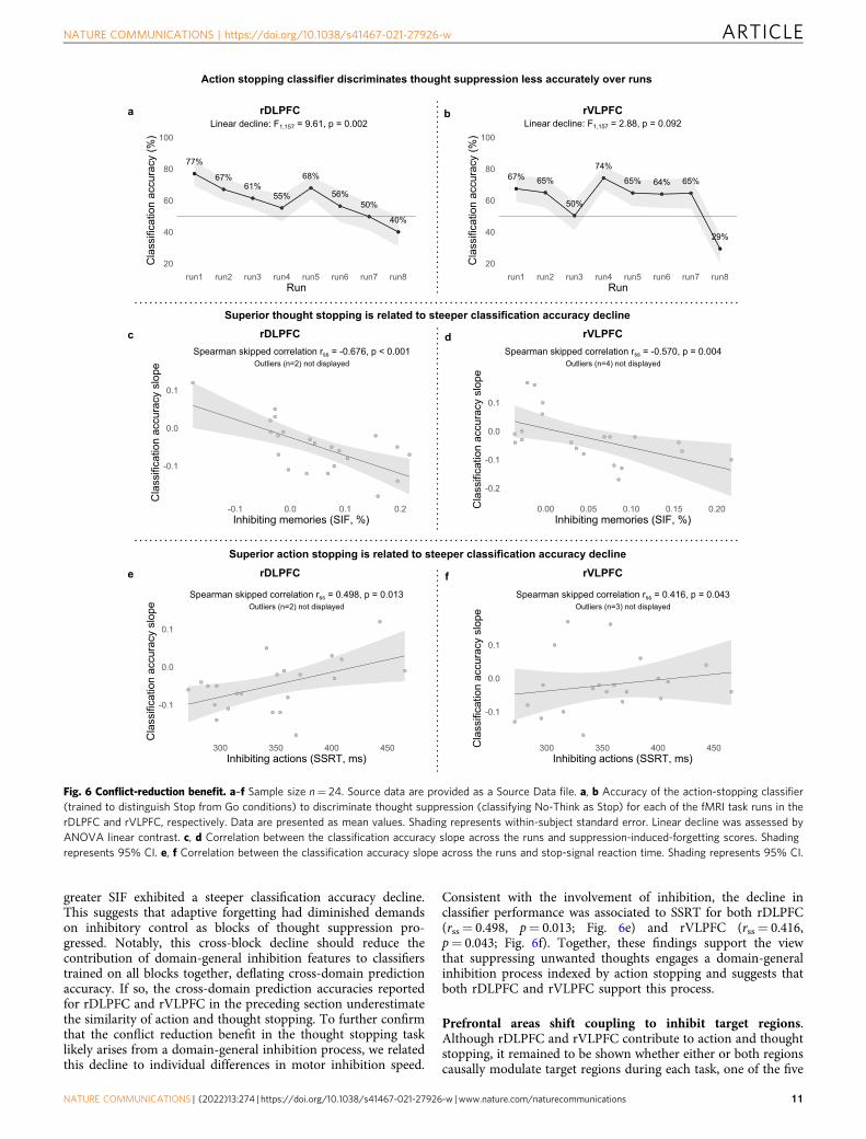
greater SIF exhibited a steeper classification accuracy decline.This suggests that adaptive forgetting had diminished demandson inhibitory control as blocks of thought suppression pro-gressed. Notably, this cross-block decline should reduce thecontribution of domain-general inhibition features to classifierstrained on all blocks together, deflating cross-domain predictionaccuracy. If so, the cross-domain prediction accuracies reportedfor rDLPFC and rVLPFC in the preceding section underestimatethe similarity of action and thought stopping. To further confirmthat the conflict reduction benefit in the thought stopping tasklikely arises from a domain-general inhibition process, we relatedthis decline to individual differences in motor inhibition speed.
Consistent with the involvement of inhibition, the decline inclassifier performance was associated to SSRT for both rDLPFC(rss= 0.498, p= 0.013; Fig. 6e) and rVLPFC (rss= 0.416,p= 0.043; Fig. 6f). Together, these findings support the viewthat suppressing unwanted thoughts engages a domain-generalinhibition process indexed by action stopping and suggests thatboth rDLPFC and rVLPFC support this process.
Prefrontal areas shift coupling to inhibit target regions.Although rDLPFC and rVLPFC contribute to action and thoughtstopping, it remained to be shown whether either or both regionscausally modulate target regions during each task, one of the five
Action stopping classifier discriminates thought suppression less accurately over runs
rDLPFCa
Superior thought stopping is related to steeper classification accuracy declinerDLPFCc
rDLPFCe
Superior action stopping is related to steeper classification accuracy decline
b rVLPFC
d rVLPFC
rVLPFCf
Linear decline: F1,157 = 2.88, p = 0.092
77%
67%61%
55%
68%
56%50%
40%
20
40
60
80
100
run1 run2 run3 run4 run5 run6 run7 run8Run
Cla
ssifi
catio
n ac
cura
cy (%
)
Linear decline: F1,157 = 9.61, p = 0.002
-0.1
0.0
0.1
-0.1 0.0 0.1 0.2Inhibiting memories (SIF, %)
Cla
ssifi
catio
n ac
cura
cy s
lope
Spearman skipped correlation rss = -0.676, p < 0.001Outliers (n=2) not displayed
-0.1
0.0
0.1
300 350 400 450Inhibiting actions (SSRT, ms)
Cla
ssifi
catio
n ac
cura
cy s
lope
Spearman skipped correlation rss = 0.498, p = 0.013Outliers (n=2) not displayed
67% 65%
50%
74%
65% 64% 65%
29%
20
40
60
80
100
run1 run2 run3 run4 run5 run6 run7 run8Run
Cla
ssifi
catio
n ac
cura
cy (%
)
Spearman skipped correlation rss = -0.570, p = 0.004Outliers (n=4) not displayed
-0.2
-0.1
0.0
0.1
0.00 0.05 0.10 0.15 0.20Inhibiting memories (SIF, %)
Cla
ssifi
catio
n ac
cura
cy s
lope
-0.1
0.0
0.1
300 350 400 450Inhibiting actions (SSRT, ms)
Cla
ssifi
catio
n ac
cura
cy s
lope
Spearman skipped correlation rss = 0.416, p = 0.043Outliers (n=3) not displayed
Fig. 6 Conflict-reduction benefit. a–f Sample size n= 24. Source data are provided as a Source Data file. a, b Accuracy of the action-stopping classifier(trained to distinguish Stop from Go conditions) to discriminate thought suppression (classifying No-Think as Stop) for each of the fMRI task runs in therDLPFC and rVLPFC, respectively. Data are presented as mean values. Shading represents within-subject standard error. Linear decline was assessed byANOVA linear contrast. c, d Correlation between the classification accuracy slope across the runs and suppression-induced-forgetting scores. Shadingrepresents 95% CI. e, f Correlation between the classification accuracy slope across the runs and stop-signal reaction time. Shading represents 95% CI.
NATURE COMMUNICATIONS | https://doi.org/10.1038/s41467-021-27926-w ARTICLE
NATURE COMMUNICATIONS | (2022) 13:274 | https://doi.org/10.1038/s41467-021-27926-w |www.nature.com/naturecommunications 11

key attributes of dynamic targeting (Attribute 4: Causal mod-ulation). On the one hand, rVLPFC alone might show dynamictargeting, exerting inhibitory modulation on the hippocampus orM1 in a task-dependent manner, as emphasised in research onmotor response inhibition10,11; rDLPFC may only be involved tomaintain the inhibition task set in working memory, possiblyexerting a modulatory influence on rVLPFC to achieve this(rVLPFC alone model). On the other hand, rDLPFC alone mightshow dynamic inhibitory targeting, consistent with the emphasison the rDLPFC as the primary source of inhibitory control inresearch on thought suppression2,6; rVLPFC may only beinvolved when attention is captured by salient stimuli83,84, suchas the stop signal or intrusions, possibly exerting a modulatoryeffect on rDLPFC to upregulate its activity (rDLPFC alonemodel). A third possibility is that rDLPFC and rVLPFC eachcontribute to top-down modulation in a content-specific manner,with only rDLPFC modulating the hippocampus during memorycontrol, but only rVLPFC modulating M1 during action stopping.By this independent pathway hypothesis, both structures arepivotal to inhibitory control functions, but only with respect totheir special domains, contrary to dynamic targeting. Finally,both rDLPFC and rVLPFC may be involved in dynamic targeting,modulating both hippocampus and M1 in a task-dependentmanner; they may interact with one another to support stopping(Parallel modulation hypothesis).
To determine the way that rDLPFC and rVLPFC interact witheach other and with the target regions (M1 and hippocampus),we analysed effective connectivity between regions using dynamiccausal modelling (DCM, see Methods). DCM accommodatesmono- and poly-synaptic mediation of the causal influence thatprefrontal regions could exert on activity in the hippocampus andin M19. DCM is ideally suited to test our hypotheses about whichprefrontal regions drive inhibitory interactions, whether thesevary by task context, and whether and how those prefrontalregions interact with one another to achieve inhibitory controlduring stopping.
Our model space included a null model with no modulatoryconnections and 72 distinct modulatory models (see Fig. 7a)differing according to whether the source-target modulation wasbidirectional, top-down, or bottom-up, whether rDLPFC,rVLPFC or both were sources of modulation, whether rDLPFCand rVLPFC interacted during inhibition tasks, and whether thesite on which top-down modulation acted was appropriate to theinhibition task or not. We first compared the null model andmodels in which the direction of source-target modulation waseither bidirectional, top-down, or bottom-up (24 models in eachof the three families). The findings from these connectivityanalyses were unambiguous. Bayesian Model Selection (BMS)overwhelmingly favoured models with bidirectional connectionsbetween the sources (rDLPFC and rVLPFC) and targets (M1 andhippocampus) with an exceedance probability (EP) of 0.9999. Incontrast, the null modulation, top-down, and bottom-up modelshad EP of 0/0.0001/0, respectively (see Fig. 7b). Exceedanceprobability refers to the extent to which a model is more likely inrelation to other models considered. The bidirectional modula-tion confirms the existence of a top-down (our focus of interest)influence that prefrontal regions exert on activity in thehippocampus and in M1, alongside bottom-up modulation.
We next compared, within the 24 bidirectional models (models1–24, see Fig. 7a), whether either rDLPFC or rVLPFC was the soledominant top-down source of inhibitory control (rDLPFC onlyvs. rVLPFC only models) to models in which both regionscomprised independent modulatory pathways (independentpathways model) or instead, contributed cooperatively toachieving top-down inhibitory control (parallel inhibitionmodel). The BMS overwhelmingly favoured models in which
both rDLPFC and rVLPFC contributed to modulating both thehippocampus and M1 with an exceedance probability (EP) of0.9999; in contrast, Independent Pathways, rDLPFC alone, andrVLPFC alone models had an EP of 0.0001/0/0, respectively (seeFig. 7c).
We next sought to distinguish subfamilies within this parallelmodel (models 9–12, and 21–24, see Fig. 7a) that varied accordingto whether and how rDLPFC and rVLPFC interacted duringinhibition: no-interaction at all between rDLPFC and rVLPFC(none); unidirectional interaction from rVLPFC to rDLPFC(unidirectional rVLPFC); unidirectional interaction fromrDLPFC to rVLPFC (unidirectional rDLPFC) and bidirectionalinteraction (rDLPFC and rVLPFC interact with each other). IfrDLPFC and rVLPFC work as a functional unit to achieveinhibitory control, one would expect clear evidence that someform of interaction occurs. Consistent with this view, BMSstrongly favoured models with bidirectional interactions betweenthe rDLPFC and rVLPFC (EP= 0.91; EP for the none,unidirectional rDLPFC, and unidirectional rVLPFC being 0.01/0.07/0.02; see Fig. 7d).
Next, we tested whether inhibitory control is dynamicallytargeted to the appropriate target structure (e.g., hippocampus orM1), depending on which process needs to be stopped (memoryretrieval or action production). According to our hypothesis, therDLPFC and rVLPFC should down-regulate hippocampal activityduring thought suppression, but should instead modulate M1,during action stopping (Attribute 5: Goal Dependence). To testthis goal-dependence, we compared the two remaining models(12 and 24, see Fig. 7a) within our winning parallel/bidirectionalsubfamily. In the “preferred targets”model, rDLPFC and rVLPFCmodulated the hippocampus during thought suppression, but M1during action stopping; in the “non-preferred targets” model,these structures modulated content-inappropriate targets (e.g.,M1 during thought suppression, but hippocampus during actionstopping). BMS strongly favoured the model with preferred(EP= 0.95) over the non-preferred (EP= 0.05) target modulation(see Fig. 7e). Indeed, the overall winning model also was stronglyfavoured by BMS even when directly assessing all 73 models, sideby side, without grouping them into model families andsubfamilies (BMS= 0.92; see Fig. 7f).
The preferential modulations of hippocampus vs. M1,depending on whether thoughts vs. actions are to be suppressed,confirm our key hypothesis that top-down modulation byrDLPFC and rVLPFC is dynamically targeted depending onparticipants’ task goals. However, a winning model with goal-dependent top-down connectivity to the hippocampus and M1might be identified for any brain region robustly activated byboth action and retrieval stopping, and not just the rDLPFC andrVLPFC. To test this possibility, we modified our DCM analysisby replacing the rDLPFC and rVLPFC nodes with two otherregions from our meta-analytic conjunction analysis as sources ofcontrol. To choose regions, we performed our domain-generalclassification analysis on all ten meta-analytic conjunction regions(see Table 1). Apart from rDLPFC and rVLPFC, only the rightand left inferior parietal lobule (IPL) exhibited significantdomain-general components (see Supplementary Fig. 5). Usingthe right and left IPL as sources of control, DCM did not reveal amodel with clear evidence for top-down modulation ofhippocampus and M1 (see Supplementary Fig. 6). Thus, to beactivated by both stopping tasks and to show cross-task decodingis not sufficient to infer goal-dependent inhibitory modulation ofconnectivity. Instead, our results suggest that rDLPFC andrVLPFC may be particularly important origins of this targetedsignal. Together, the results of the DCM analysis suggest that,when stopping a prepotent response, rDLPFC and rVLPFC,interact with each other and are both selectively coupled with M1
ARTICLE NATURE COMMUNICATIONS | https://doi.org/10.1038/s41467-021-27926-w
12 NATURE COMMUNICATIONS | (2022) 13:274 | https://doi.org/10.1038/s41467-021-27926-w |www.nature.com/naturecommunications
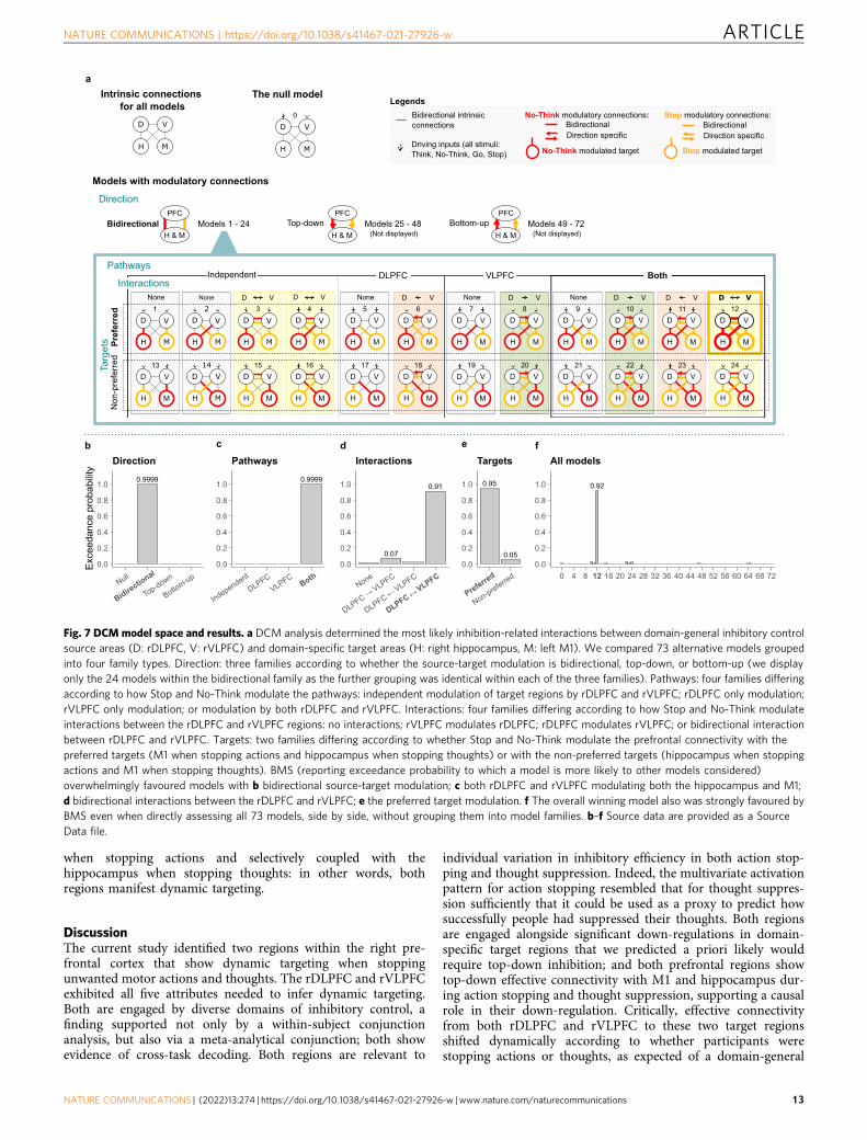
when stopping actions and selectively coupled with thehippocampus when stopping thoughts: in other words, bothregions manifest dynamic targeting.
DiscussionThe current study identified two regions within the right pre-frontal cortex that show dynamic targeting when stoppingunwanted motor actions and thoughts. The rDLPFC and rVLPFCexhibited all five attributes needed to infer dynamic targeting.Both are engaged by diverse domains of inhibitory control, afinding supported not only by a within-subject conjunctionanalysis, but also via a meta-analytical conjunction; both showevidence of cross-task decoding. Both regions are relevant to
individual variation in inhibitory efficiency in both action stop-ping and thought suppression. Indeed, the multivariate activationpattern for action stopping resembled that for thought suppres-sion sufficiently that it could be used as a proxy to predict howsuccessfully people had suppressed their thoughts. Both regionsare engaged alongside significant down-regulations in domain-specific target regions that we predicted a priori likely wouldrequire top-down inhibition; and both prefrontal regions showtop-down effective connectivity with M1 and hippocampus dur-ing action stopping and thought suppression, supporting a causalrole in their down-regulation. Critically, effective connectivityfrom both rDLPFC and rVLPFC to these two target regionsshifted dynamically according to whether participants werestopping actions or thoughts, as expected of a domain-general
a
0.07
0.91
0.0
0.2
0.4
0.6
0.8
1.0
None
DLPFC→VLPFC
DLPFC←VLPFC
DLPFC↔VLPFC
Interactionsd
0.95
0.050.0
0.2
0.4
0.6
0.8
1.0
Preferred
Non-preferred
Targetse
0.92
0.0
0.2
0.4
0.6
0.8
1.0
0 4 8 12 16 20 24 28 32 36 40 44 48 52 56 60 64 68 72
All modelsf
0.9999
0.0
0.2
0.4
0.6
0.8
1.0
Null
Bidirectio
nal
Top-down
Bottom-up
Exce
edan
ce p
roba
bilit
y
Directionb
0.9999
0.0
0.2
0.4
0.6
0.8
1.0
Independent
DLPFCVLPFC
Both
Pathwaysc
Models with modulatory connections
D V
H M
Intrinsic connections for all models
D V
H M
0
The null model
Bidirectional intrinsic connections
Driving inputs (all stimuli: Think, No-Think, Go, Stop)
Stop modulatory connections:BidirectionalDirection specific
No-Think modulatory connections: BidirectionalDirection specific
No-Think modulated target Stop modulated target
Legends
Direction
Top-downPFC
H & MModels 25 - 48Bidirectional
PFC
H & MModels 1 - 24 Bottom-up
PFC
H & MModels 49 - 72
(Not displayed) (Not displayed)
VLPFC
D V
H M
D V
H M
12
24
InteractionsIndependent
Pathways
Targ
ets Pr
efer
red
Non
-pre
ferre
d
DLPFC Both
None None
D V
H M
D V
H M
D V
H M
D V
H M
D V
H M
D V
H M
5 6 7
17 18 19D V
H M
D V
H M
8
20D V
H M
D V
H M
10
22D V
H M
D V
H M
11
23
None
D V
H M
D V
H M
9
21
D V
H M
None
D V
H M
1
13
D V
H M
None
D V
H M
2
14
H M
H M
4
16
D V
D V
H M
H M
3
15
D V
D V
D VD V D V D V D V D V D V
Fig. 7 DCM model space and results. a DCM analysis determined the most likely inhibition-related interactions between domain-general inhibitory controlsource areas (D: rDLPFC, V: rVLPFC) and domain-specific target areas (H: right hippocampus, M: left M1). We compared 73 alternative models groupedinto four family types. Direction: three families according to whether the source-target modulation is bidirectional, top-down, or bottom-up (we displayonly the 24 models within the bidirectional family as the further grouping was identical within each of the three families). Pathways: four families differingaccording to how Stop and No-Think modulate the pathways: independent modulation of target regions by rDLPFC and rVLPFC; rDLPFC only modulation;rVLPFC only modulation; or modulation by both rDLPFC and rVLPFC. Interactions: four families differing according to how Stop and No-Think modulateinteractions between the rDLPFC and rVLPFC regions: no interactions; rVLPFC modulates rDLPFC; rDLPFC modulates rVLPFC; or bidirectional interactionbetween rDLPFC and rVLPFC. Targets: two families differing according to whether Stop and No-Think modulate the prefrontal connectivity with thepreferred targets (M1 when stopping actions and hippocampus when stopping thoughts) or with the non-preferred targets (hippocampus when stoppingactions and M1 when stopping thoughts). BMS (reporting exceedance probability to which a model is more likely to other models considered)overwhelmingly favoured models with b bidirectional source-target modulation; c both rDLPFC and rVLPFC modulating both the hippocampus and M1;d bidirectional interactions between the rDLPFC and rVLPFC; e the preferred target modulation. f The overall winning model also was strongly favoured byBMS even when directly assessing all 73 models, side by side, without grouping them into model families. b–f Source data are provided as a SourceData file.
NATURE COMMUNICATIONS | https://doi.org/10.1038/s41467-021-27926-w ARTICLE
NATURE COMMUNICATIONS | (2022) 13:274 | https://doi.org/10.1038/s41467-021-27926-w |www.nature.com/naturecommunications 13

mechanism that is flexibly targeted to suppress specialised con-tent in multiple domains.
Based on these and related findings, we propose that anteriorrDLPFC and rVLPFC constitute key hubs for a domain-generalinhibitory control mechanism that can be dynamically targeted tostop processing of diverse content represented throughout thebrain. This proposal complements recent work positing a broadprefrontal inhibition mechanism that can interrupt both cogni-tion and action32,33,85. We focused here on the stopping of simplemanual actions and verbal thoughts. Given this approach, thisstudy does not address the breadth of thought content that can betargeted by this mechanism. However, when considered alongsidethe growing literature on retrieval suppression, the breadth ofcontent is considerable. For example, the anterior rDLPFC andrVLPFC regions identified in the meta-analytic conjunction havebeen observed during the suppression of a range of stimuli,including words1,37,65, visual objects40,41, neutral and aversivescenes4,38,39,79 and person-specific fears about the future7. Inaddition, during retrieval suppression, these frontal regions exerttop-down inhibitory modulation not only of thehippocampus9,65, but also of other domain-specific contentregions, including areas involved in representing visualobjects40,41, places38,39, and also emotional content in theamygdala4,39. Content-specific modulations are triggered espe-cially when these types of content intrude into awareness inresponse to a cue and need to be purged39, indicating that inhi-bition can be dynamically targeted to diverse cortical sites to meetcontrol demands. The current findings broaden the scope of thismechanism further by showing that it is not limited to stoppingretrieval processes, but also extends to stopping the preparationand execution of motor responses, consistent with a broadmechanism involved in self-control over action and thought.
The proposed role of the rDLPFC and rVLPFC as hubs ofdomain-general inhibitory control during stopping does notimply that these regions are exclusively dedicated to stopping.Indeed, it seems likely that these regions contribute to manycognitive functions. Rather, the current evidence suggests thatwhen stopping an action or thought is required, these regions arerecruited to cancel processing in target areas involved in repre-senting to-be-suppressed content, thereby achieving the desiredstopping outcome. Methodologically diverse evidence indicatesthat this contribution is causally necessary to successful inhibitorycontrol and is not a mere epiphenomenon of doing difficult tasks.First, the current effective connectivity analyses indicate a robusttop-down modulation of target regions by these putative pre-frontal sources. This finding comports well with lesion and brainstimulation work in both humans and animals, indicating thatdisrupting the function of rDLPFC and rVLPFC severely disruptsthe capacity to stop, consistent with causal necessity27–30. Second,although action and thought stopping are both difficult tasks, thenetwork dynamically reconfigured its connectivity to targetregions to suppress their function in a manner compatible withtask goals. These features have the hallmarks of a control processconfigured to implement a particular regulatory function, ratherthan a generic response to task difficulty. RDLPFC and rVLPFCare likely to work in concert with a broader network to achievethese goals, as our conjunction analyses suggest. The currentwork does not address the functional role of domain-generalregions outside of the prefrontal cortex, the contributions ofwhich should be examined in future work.
We considered the possibility that only one of these two frontalregions is central to implementing top-down inhibitory controlduring stopping, with the other providing upstream inputsessential to initiate successful control. Our effective connectivityanalysis probed alternative hypotheses about the way rDLPFCand rVLPFC interact during stopping. RDLPFC might implement
the true inhibitory signal, receiving salience detection input fromrVLPFC that up-regulates rDLPFC function, consistent with apossible role of the VLPFC in the ventral attention network83,84.Alternatively, rVLPFC may implement inhibition, with rDLPFCpreserving task set by sending driving inputs to the rVLPFC. Ourfindings indicate that both structures contributed in parallel totop-down inhibitory control and interacted bidirectionally duringboth action and thought stopping. Little evidence suggested astrong asymmetry in how rDLPFC and rVLPFC interacted, asshould arise if one region simply served a role in saliencedetection or task-set maintenance. It remains possible, however,that rDLPFC and rVLPFC serve distinct functions that are notreadily separable given the current manipulations and the level oftemporal resolution available in fMRI data. Nevertheless, thesefindings suggest that rDLPFC and rVLPFC, at a minimum, acttogether to implement top-down inhibitory control duringstopping. Although it might seem surprising that two spatiallysegregated prefrontal regions would act in concert to achieve thisfunction, it seems less unusual considering their potential role inthe cingulo-opercular network (CON). Most of the regionsidentified in our inhibition conjunction analysis participate in thisnetwork, suggesting that it may play an important role inachieving stopping. Given the strong integrated activity of thisnetwork, elements of which are distributed throughout thebrain51,54, this suggests future work should examine how rDLPFCand rVLPFC work together with other elements of this networkto achieve successful stopping.
The current proposal contrasts with models that emphasise theprimacy of either rVLPFC or rDLPFC in inhibitory control, andwhich have not addressed dynamic targeting to diverse content.Research on motor inhibition has emphasised the rVLPFC as thesource of top-down inhibitory control10,11, although withoutevidence to exclude the role of rDLPFC. Indeed, studies cited asfavouring the selective role of rVLPFC often support contribu-tions of the anterior rDLPFC structure identified here. Forexample, whereas intracranial stimulation in primates establishesthe causal necessity of the rVLPFC in motor stopping, so too doesstimulation of the dorsal bank of the principal sulcus, the putativemonkey homologue of the rDLPFC in humans30; and whereasintracranial recordings in humans show stopping-related activityin rVLPFC, they also reveal it in anterior rDLPFC and often priorto rVLPFC86. Research on thought suppression has emphasisedthe rDLPFC as the source of top-down inhibitory control1,2,9; butmost studies supporting the role of rDLPFC in thought sup-pression also reveal activations in the rVLPFC22. Indeed, as ourwithin-subjects and meta-analytic conjunctions unambiguouslyconfirm, both regions are recruited during both stopping tasks.The current study goes further than establishing conjoint acti-vation: Pattern classification and connectivity analyses show theinvolvement of both regions in the dynamics of control, withoutselectivity. These findings validate the importance of both regions,establish the domain-generality of their influence, and demon-strate the dynamic inhibitory targeting capacity necessary to infera flexible control mechanism.
Although rDLPFC and rVLPFC exhibit core properties neededto achieve domain-general stopping, there is also evidence ofdomain-specificity within these regions. A classifier trained todistinguish stopping actions from stopping thoughts performedwell. Although seemingly inconsistent with domain generality,such effects can be understood as consequences of the dynamictargeting functionality afforded by these regions. For a region toserve as a flexible hub of inhibitory control, it must be able toreceive inputs from diverse cortical sites, representing informa-tion needed to drive the stopping (e.g., the perception of thecoloured word for the No-Think task, and of the tone for thestop-signal task). Moreover, to flexibly target inhibitory control,
ARTICLE NATURE COMMUNICATIONS | https://doi.org/10.1038/s41467-021-27926-w
14 NATURE COMMUNICATIONS | (2022) 13:274 | https://doi.org/10.1038/s41467-021-27926-w |www.nature.com/naturecommunications
source regions must interact with diverse cortical and subcorticaltargets (e.g., hippocampus vs. M1), the processing which must bestopped. These input and output processes may manifest inunique multivariate patterns over common voxels within eachregion. We cannot rule out, however, the possibility that domain-specific inhibition processes are also manifested by unique pat-terns in these regions. Future work should examine the additionalcontributions of input, output, and domain-specific stoppingfeatures to the activations found in rDLPFC and rVLPFC.
The present findings highlight a potentially important differ-ence between the brain networks involved in stopping and otherforms of cognitive control that do not require the full cessation ofa motor or cognitive process. Maintaining rules in workingmemory, implementing task sets, performing multi-tasking, andmanipulating information actively are all clear cases of cognitivecontrol that can require interference resolution, but do notnecessarily entail active stopping. The above functions engage thewidely discussed frontoparietal network (FPN), often assigned acentral role in implementing cognitive control more broadly50–53.One might assume that because stopping is a form of cognitivecontrol that the FPN would be central to it as well. Nevertheless,the FPN, though involved in our tasks, appeared less prominentthan the CON, which accounted for most of the distinct corticalparcels participating in our domain-general stopping regions. Wefound little evidence for the involvement of major areas of theFPN, including much of the middle frontal gyrus bilaterally in ourmultimodal inhibition regions. As our meta-analysis and within-subjects comparisons confirm, inhibitory control during stoppingis strongly right lateralised, which also is not a feature emphasisedin research on the FPN. Our findings raise the possibility thatstopping actions and thoughts may rely on a distinct network,with different functional characteristics to the FPN. Whetherother functions thought to require inhibitory control (e.g.,selection between competing responses, as in Stroop or Flankerinterference) also preferentially engage the stopping networksidentified here is not addressed in the current work, althoughsome empirical precedents suggest that stopping and selectionmay engage partially distinct mechanisms26,37,87.
Dynamic inhibitory targeting provides a neurocognitive fra-mework that can account for both associations and dissociationsin the abilities to suppress unwanted thoughts and actions. On theone hand, deficits in both action and thought stopping shouldarise with dysfunction in the rDLPFC or rVLPFC, given thecommon reliance of these abilities on those regions. Such asso-ciations occur frequently. In the general population, peoplescoring highly on self-report measures of impulsivity or com-pulsivity also report greater difficulty with intrusive thoughts88,89.Clinically, persistent intrusive thoughts and action stopping def-icits co-occur in numerous disorders: Obsessive thoughts andcompulsive actions in obsessive-compulsive disorder90,91; intru-sive memories and impaired response inhibition in PTSD92–96;persistent worry and impulsivity in anxiety disorders97 andintrusive thoughts and compulsivity in addiction98–100. These co-morbid deficits may reflect dysfunction in the rDLPFC, therVLPFC or in other shared components of their control path-ways. On the other hand, dissociations should arise when dys-function selectively disrupts a domain-specific pathway linkingrLPFC to target sites involved in generating actions and thoughts,including dysfunction to local inhibition at the target site itself.For example, individual variation in local GABAergic inhibitionwithin the hippocampus or M1 predict inhibitory control overmemories and actions, respectively, independently of prefrontalfunction8,49. Thus, selective difficulties in action stopping orthought inhibition may arise, given focal deficits in either motorcortical or hippocampal GABA8. The separate contributions ofdomain-general and domain-specific factors to inhibitory control
implied by dynamic targeting constrains the utility of motorinhibition as a metric of inhibitory control over thought and mayexplain the surprisingly small SSRT deficits in major depressionand anxiety, relative to attention deficit hyperactivity disorder orobsessive-compulsive disorder19.
The current study did not seek to characterise the polysynapticpathways through which the rDLPFC and rVLPFC suppressactivity in either M1 or the hippocampus5,9. Rather, we focusedon the existence of a central, domain-general inhibitory controlfunction capable of flexibly shifting its top-down influence to stopactions and thoughts. By juxtaposing two well characterisedmodel systems for stopping actions and thoughts, each withdistinct neural targets of inhibition, we were able to show that thesame set of prefrontal regions is involved in stopping processingin different cortical target areas, in a rapid, flexible manner. Indoing so, we established evidence for dynamic inhibitory target-ing as a key mechanism of domain-general inhibitory controlduring stopping in the human brain. More broadly, this worksuggests that the human capacity for self-control in the face oflife’s challenges may emerge from a common wellspring of con-trol over our actions and thoughts.
MethodsWe used a dataset from a published study8. However, here the data were inde-pendently re-analysed with a different focus.
Participants. Thirty right-handed native English speakers participated. Partici-pants gave written informed consent and received money for participating. Fiveparticipants did not reach the 40% learning criterion on the Think/No-Think task,and one fell asleep during fMRI acquisition. The final sample comprised 24 par-ticipants (7 males, 17 females), 19–36 years old (M= 24.67 years, SD= 4.31).Participants had normal or corrected-to-normal vision and no reported history ofneurological, medical, or memory disorders, and they were asked not to consumepsychostimulants, drugs, or alcohol before the experiment. The Cambridge Psy-chology Research Ethics Committee approved the project.
Experimental paradigm. Participants performed adapted versions of the Stop-signal20 and Think/No-Think62 tasks. Both tasks require participants to stopunwanted processes, but in the motor and memory domains, respectively.
The Stop-signal task assesses the ability to stop unwanted actions. Participantsfirst learn stimulus–response associations and then perform speeded motorresponses to the presented (Go) stimuli. Occasionally, shortly after the Gostimulus, a stop signal occurs, and participants must withhold their response. Wemeasured the stop-signal reaction time (SSRT), an estimate of how long it takes theparticipant to stop.
The Think/No-Think task assesses the ability to stop unwanted memoryretrievals. Participants first form associations between unrelated cue-target wordpairs. Then participants receive two-thirds of the cues as reminders (one at a time)and are asked to either think (Think items) or to not-think (No-Think items) of theassociated target memory, with each Think and No-Think reminder repeatednumerous times throughout the task. Finally, participants attempt to recall allinitially learned associations. Typically, recall performance suffers for No-Thinkitems compared to Baseline items that were neither retrieved nor suppressedduring the think/no-think phase. This phenomenon, known as suppression-induced forgetting (SIF), indirectly measures the ability to stop unwanted memoryretrievals by quantifying inhibitory aftereffects of this process2,101.
Stimuli and apparatus. We presented stimuli and recorded responses with Pre-sentation software (Neurobehavioral Systems, Albany, CA, USA). For the Stop-signal task, four visually discriminable red, green, blue, and yellow coloured circlesof 2.5 cm in diameter, presented on a grey background, constituted the Go stimuli(Fig. 2a). Participants responded by pressing one of the two buttons (left or right)with a dominant (right) hand on a button box. An auditory 1000 Hz “beep” tonepresented at a comfortable volume for 100 ms signalled participants to stop theirresponses. A fixation cross appeared in 50-point black Arial Rounded font on agrey background prior to the onset of the Go stimulus.
For the Think/No-Think task, we constructed 78 weakly relatable English wordpairs (cue-target words, e.g., Part-Bowl) as stimuli and an additional 68 semanticallyrelated cue words for 68 of the target words (e.g., a cue word ‘Cornflake’ for thetarget word ‘Bowl’). We used 60 of the target words and their related and weak cuesin the critical task, with the other items used as fillers. We divided the critical itemsinto three lists composed of 20 targets and their corresponding weak cue words(the related word cues were set aside to be used as independent test cues on thefinal test; see procedure). We counterbalanced these lists across the within-subjects
NATURE COMMUNICATIONS | https://doi.org/10.1038/s41467-021-27926-w ARTICLE
NATURE COMMUNICATIONS | (2022) 13:274 | https://doi.org/10.1038/s41467-021-27926-w |www.nature.com/naturecommunications 15

experimental conditions (Think, No-Think, and Baseline) so that across allparticipants, every pair participated equally often in each condition. We used thefiller words both as practice items and also to minimise primacy and recency effectsin the study list102. Words appeared in a 32-point Arial font in capital letters on agrey background (Fig. 2b). During the initial encoding and final recall phases, wepresented all cues and targets in black. For the Think/No-Think phase, we presentedthe Think cues in green and the No-Think cues in red, each preceded by a fixationcross in 50-point black Arial Rounded font on a grey background.
Procedure. The procedure consisted of seven steps: (1) stimulus-response learningfor the Stop-signal task: (2) Stop-signal task practice; (3) encoding phase of theThink/No-Think task; (4) Think/No-Think practice; (5) practice of interleavedStop-signal and Think/No-Think tasks; (6) experimental phase during fMRIacquisition; (7) recall phase of the Think/No-Think task. We elaborate these stepsbelow (see also Fig. 2c).
Step 1—Stop-signal task stimulus-response learning. Participants first formedstimulus-response associations for the Stop-signal task. As Go stimuli, we pre-sented circles in four different colours (red, green, blue, and yellow) and partici-pants had to respond by pressing one of the two buttons depending on the circle’scolour. Thus, each response button had two colours randomly assigned to it andparticipants associated each colour to its particular response.
Participants learned the colour-button mappings in two sets of two colours,with the first colour in a set associated with one button, and the second with theother button. After practising the responses to these colours in random order 10times each, the same training was done on the second set. Subsequently,participants practised the colour-button mappings of all four colours in randomorder until they responded correctly to each colour on 10 consecutive trials. Duringthe practice, we instructed participants to respond as quickly and accurately aspossible and provided feedback for incorrect or slow (>1000 ms) responses.
Step 2—Stop-signal task practice. Once participants learned the stimulus–responseassociations, we introduced the Stop-signal task. We instructed participants to keepresponding to each coloured circle as quickly and accurately as possible butindicated that on some trials, after the circle appeared, a beep would sound andthat they should not press any button on these trials. We also told participants toavoid slowing down and waiting for the beep, requesting instead that they treatfailures to stop as normal and always keep responding quickly and accurately.Thus, on Go trials, participants responded as quickly as possible, whereas, on Stoptrials, a tone succeeded the cue onset, signalling participants to suppress theirresponse. To facilitate performance, participants received on-screen feedback forincorrect and too slow (>700 ms) responses to Go trials, and for pressing a buttonon Stop trials.
Figure 2a presents the trial timings. Go trials started with a fixation cross,presented for 250 ms, followed by a coloured circle until response or for up to2500 ms. After the response and a jittered inter-trial interval (M= 750 ms,SD= 158.7 ms), a new trial commenced. Stop trials proceeded identically exceptthat a tone sounded shortly after the circle appeared. This stop signal delay varieddynamically in 50 ms steps (starting with 250 or 300 ms) according to a staircasetracking algorithm to achieve approximately a 50% success-to-stop rate for eachparticipant. Note that the longer the stop signal delay is, the harder it is to not pressthe button. The dynamic tracking algorithm reduces participants’ ability toanticipate stop signal delay timing and provides a method for calculating the SSRT.In this practice step, participants performed 96 trials, of which 68 (71%) were Gotrials and 28 (29%) were Stop trials.
Step 3—Think/No-Think task encoding phase. Once participants had learned theStop-signal task, we introduced the Think/No-Think task. In the encoding phase,participants formed associations between 60 critical weakly related word pairs (e.g.,Part-Bowl) and between 18 filler pairs. First, participants studied each cue-targetword pair for 3.4 s with an inter-stimulus interval of 600 ms. Next, from eachstudied pair, participants saw the cue word only and recalled aloud the corre-sponding target. We presented each cue for up to 6 s or until a response was given.600 ms after cue offset, regardless of whether the participant recalled the item, thecorrect target appeared for 1 s. We repeated this procedure until participantsrecalled at least 40% of the critical pairs (all but 5 participants succeeded within themaximum of three repetitions). Finally, to assess which word-pairs participantslearned, each cue word appeared again for 3.3 s with an inter-stimulus interval of1.1 s and participants recalled aloud the corresponding target. We provided nofeedback on this test.
Step 4—Think/No-Think practice. After participants encoded the word pairs, theThink/No-Think practice phase commenced. On each trial, a cue word appearedon the screen in either green or red. We instructed participants to recall and thinkof the target words for cues presented in green (Think condition) but to suppressthe recall and avoid thinking of the target words for those cues presented in red(No-Think condition). Participants performed the direct suppression variant of theThink/No-Think task37,103 in which, after reading and comprehending the cue,they suppressed all thoughts of the associated memory without engaging in any
distracting activity or thoughts. We asked participants to “push the memory out ofmind” whenever it intruded.
Trial timings appear in Fig. 2b. A trial consisted of presenting a cue in thecentre of the screen for 3 s, followed by an inter-stimulus interval (≥0.5 s,M= 2.3 s,SD= 1.7 s) during which we displayed a fixation cross. We jittered the inter-stimulus interval (≥0.5 s,M= 2.3 s, SD= 1.7 s) to optimise the event-related design(as determined by optseq2: http://surfer.nmr.mgh.harvard.edu/optseq).
In this practice phase, we used 12 filler items, six of which were allocated to theThink condition and six to the No-Think condition. We presented each item threetimes in random order (36 trials in total). In the middle of the practice, weadministered a diagnostic questionnaire to ensure participants had understood andfollowed the instructions.
Step 5—Interleaved Stop-signal and Think/No-Think practice. Before moving intothe MRI scanner, participants performed an extended practice phase interleavingthe Stop-signal and Think/No-Think tasks. For the Think/No-Think task, we againused 12 filler items. Other than that, and the fact that the practice took placeoutside the MRI scanner, this phase was identical to a single fMRI acquisitionsession described into more detail next.
Step 6—Experimental phase and fMRI acquisition. In the main experimental phase,participants underwent 8 fMRI scanning runs in a single session. Before thescanning began, participants saw the correct button-colour mappings and all 78-word pairs briefly presented on the screen to remind them of the task and items.After the brief refresher, the fMRI acquisition started. During each fMRI run,participants performed 8 blocks of the Think/No-Think task interleaved with 8blocks of the Stop-signal task. All blocks lasted 30 s. To minimise carry-over effects,we interspersed 4 s rest periods (blank screen with a grey background) betweenblocks. Each block began with items that we did not score (the filler items) toreduce task-set switching effects between blocks. Within each block, we pseudo-randomly ordered all trials, and the trial timings for both tasks were identical tothose used in their respective practice phases (steps 2 and 4; Fig. 2a and b).
Four of the Stop-signal task blocks contained Go trials only. We did not usethese blocks in this report. Each of the other four Stop-signal blocks contained 12trials, yielding 384 trials in total (8 runs * 4 blocks per run * 12 trials per block). Onaverage, across participants, Stop trials constituted 32% (SD= 2%) of the trials. Asin the practice phase, a staircase tracking algorithm varied the delay between cueonset and stop-signal tone according to each participant’s performance, keepingthe stopping success at ~50%.
Each of the Think/No-Think blocks contained 6 trials, starting with a filler itemas a Think trial followed by 5 Think or No-Think items in a pseudo-random order.Within each fMRI run, participants saw all 20 critical Think and 20 critical No-Think items once. Thus, across the 8 runs, participants recalled or suppressed eachmemory item 8 times. The proportion of the Think trials (58%) exceeded theproportion of the No-Think trials (42%) to better resemble the higher frequency ofGo trials than Stop trials during the Stop-signal task. We accomplished this byassigning Think trials to the filler items, without changing the frequency of Thinktrials on critical experimental items. After the fourth (middle) run, to allowparticipants to rest, we acquired their anatomical scan and administered thediagnostic questionnaire to ensure that participants closely followed theinstructions of the Think/No-Think task.
Step 7—Think/No-Think recall phase. In the final step (inside the scanner butwithout any scan acquisition), we measured the aftereffects of memory retrievaland suppression via a cued-recall task on all word pairs (encoded in step 3). Thisincluded 20 Baseline items that were neither retrieved nor suppressed during theThink/No-Think phase and that this provided a baseline estimate of the memor-ability of the pairs.
To reinstate the context of the initial encoding phase, we first tested participantson 10 filler cue words, 6 of which they had not seen since the encoding phase (step3) and 4 of which they saw during the interleaved Stop-signal and Think/No-Thinkpractice phase (step 5). We warned participants that the cues in this phase could beones they had not seen for a long time and encouraged them to think back to theencoding phases to retrieve targets.
Following context reinstatement, participants performed the same-probe andindependent-probe memory tests. In the same-probe test, we probed memory with theoriginal cues (e.g., the weakly related cue word ‘Part’ for the target word ‘Bowl’). Weincluded the independent-probe test to test whether forgetting generalised to novelcues62, using the related cues we had designed for each target. For example, we cuedwith the semantic associate of the memory and its first letter (e.g., ‘Cornflake—B’ for thetarget ‘Bowl’). Across participants, we counterbalanced the order in which the testsappeared. In both tests, cues appeared for a maximum of 3.3 s or until participants gavea response, with an inter-stimulus interval of 1.1 s. We coded response as correct ifparticipants correctly recalled the target while the cue was onscreen.
Finally, we debriefed participants and administered a post-experimentalquestionnaire to capture participants’ experiences and the strategies they used inthe Think/No-Think and Stop-signal tasks.
Brain image acquisition. We collected MRI data using a 3-T Siemens Tim TrioMRI scanner (Siemens, Erlangen, Germany) fitted with a 32-channel head coil.
ARTICLE NATURE COMMUNICATIONS | https://doi.org/10.1038/s41467-021-27926-w
16 NATURE COMMUNICATIONS | (2022) 13:274 | https://doi.org/10.1038/s41467-021-27926-w |www.nature.com/naturecommunications
Participants underwent eight functional runs of the blood-oxygenation-level-dependent (BOLD) signal acquisitions. We acquired functional brain volumesusing a gradient-echo, T2*-weighted echoplanar pulse sequence (TR= 2000 ms,TE= 30 ms, flip angle= 90°, 32 axial slices, descending slice acquisition, voxelresolution= 3 mm3, 0.75 mm interslice gap). We discarded the first four volumesof each session to allow for magnetic field stabilisation. Due to technical problemsencountered during task performance, we discarded from the analysis one func-tional run from two participants each, and two functional runs from anotherparticipant. After the fourth functional run, we acquired an anatomical referencefor each participant, a high-resolution whole-brain 3D T1-weighted magnetisation-prepared rapid gradient echo (MP-RAGE) image (TR= 2250 ms, TE= 2.99 ms,flip angle= 9°, field of view= 256 × 240 × 192 mm, voxel resolution= 1 mm3).Following the acquisition of the anatomical scan, participants underwent theremaining four functional runs.
Behavioural performance analysis. For statistical analyses of the behaviouraldata, we used R (v4, 2020-04-24) in Jupyter Notebook (Anaconda, Inc., Austin,TX). The data and detailed analysis notebook are freely available at GitHubrepository104 (https://github.com/dcdace/Domain-general/). For all statisticalcomparisons, we adopted p < 0.05 as the significance threshold.
For correlation analyses, we followed recommendations by Pernet et al.105 andused one of three correlation methods depending on whether the data werenormally distributed or contained outliers. If there were no outliers and data werenormally distributed, we performed Pearson correlation and reported it as ‘r’. Ifthere were univariate outliers (but no bivariate) or data were not normallydistributed, we performed robust 20% Bend correlation and reported it as ‘rpp’. Ifthere were bivariate outliers, we performed robust Spearman skipped correlationusing the minimum covariance determinant (MCD) estimator and reported it as‘rss’. For univariate and bivariate outlier detection, we used boxplot and bagplotmethods, respectively.
For the analysis of Stop-signal task data, we followed the guidelines byVerbruggen et al.64 and calculated SSRT using the integration method with thereplacement of Go omissions. Specifically, we included all Stop trials and all Gotrials (correct and incorrect), replacing missed Go responses with the maximum GoRT. To identify the nth fastest Go RT, we multiplied the number of total Go trialsby the probability of responding to stop signal (unsuccessful stopping). Thedifference between the nth fastest Go RT and the mean SSD provided our estimateof SSRT.
In addition to SSRT, we calculated the probability of Go omissions, probabilityof choice errors on Go trials, probability of responding to Stop trials, mean SSD ofall Stop trials, mean correct Go RT, and mean failed Stop RT. We also comparedRTs of all Go trials against RTs of failed Stop trials to test the assumption of anindependent race between a go and a stop runner. Besides, we assessed the changeof Go RTs across the eight experimental blocks. Prior work suggests that theexperiment-wide integration method can result in underestimation bias of SSRT ifparticipants slow their RT gradually across experimental runs. In that case, ablocked integration method would provide a better measure of SSRT106. In ourdata, however, on average within the group, we observed a negligible decrease inRT across runs (B=−2.555, p= 0.250), suggesting that the experiment-wideintegration method was more appropriate.
For the Think/No-Think task data, we focused on the critical measure: SIF. Weused the final recall scores (from step 7) of No-Think and Baseline itemsconditionalized on correct initial training performance (at step 3), as in priorwork1. Thus, in the final recall scores, we did not include items that were notcorrectly recalled (M= 29%, SD= 17) during the criterion test of the encodingphase, as the unlearned items can be neither suppressed nor retrieved during theThink/No-Think phase (step 6). As in our previous work8, we averaged the scoresacross the same-probe and independent-probe tests and the difference between theBaseline and No-think item recall scores constituted our measure of SIF. To assessthe group effect of SIF, we tested the data for normality (W= 0.95, p= 0.264) andperformed a one-sample, one-sided t-test to determine if SIF is greater than zero.Finally, to assess whether inhibition ability generalises across motor and memorydomains, we performed a correlation between the SSRT and SIF scores.
To identify univariate and bi-variate outliers in the SSRT and SIF scores, weused box plot method, which relies on the interquartile range. Univariate outlierswere not present for any of the two measures. One bi-variate outlier was removedfrom the correlation analysis and the behavioural partial least squares analysis(described below). Nevertheless, outlier removal did not qualitatively alter theresults.
Brain imaging data analysisPre-processing. We pre-processed and analysed the brain imaging data using Sta-tistical Parametric Mapping v12 release 7487 (SPM12; Wellcome Trust Centre forNeuroimaging, London) in MATLAB vR2012a (The MathWorks, MA, USA). Toapproximate the orientation of the standard Montreal Neurological Institute (MNI)coordinate space, we reoriented all acquired MRI images to the anterior-posteriorcommissure line and set the origins to the anterior commissure. Next, we appliedour pre-processing procedure to correct for head movement between the scans(images realigned to the mean functional image) and to adjust for temporal dif-ferences between slice acquisitions (slice-time correction relative to the middle axial
slice). The procedure then co-registered each participant’s anatomical image to themean functional image and segmented it into grey matter, white matter, andcerebrospinal fluid. We then submitted the segmented images for each participantto the DARTEL procedure107 to create a group-specific anatomical template thatoptimises inter-participant alignment. The DARTEL procedure alternates betweencomputing a group template and warping an individual’s tissue probability mapsinto alignment with this template and ultimately creates an individual flow field ofeach participant. Subsequently, the procedure transformed the group template intoMNI-152 space. Finally, we applied the MNI transformation and smoothing withan 8 mm full-width-at-half-maximum (FWHM) Gaussian kernel to the functionalimages for the whole-brain voxel-wise analysis.
Univariate whole-brain analysis. To identify brain areas engaged in both inhibitingactions and inhibiting memories, we performed a whole-brain voxel-wise uni-variate analysis. We high-pass filtered the time series of each voxel in the nor-malised and smoothed images with a cut-off frequency of 1/128 Hz, to remove low-frequency trends, and modelled for temporal autocorrelation across scans with thefirst-order autoregressive (AR(1)) process. We then submitted the pre-processeddata of each participant to the first-level, subject-specific, general linear model(GLM) modelling a single design matrix for all functional runs.
We modelled the Stop-signal task and Think/No-Think task conditions asboxcar functions, convolved with a haemodynamic response function (HRF). Inthe model, we used group average response latencies for each trial type as the trialdurations for the Stop-signal task condition, but we used 3 s epochs for the Think/No-Think task condition. As in the behavioural analysis, we conditionalized theThink and No-Think conditions on initial encoding performance. The mainconditions of interest for our analysis included: correct Stop, correct Go (from themixed Stop-signal and Go trial blocks only), conditionalized No-Think andconditionalized Think. Unlearned No-Think and Think items, filler items,incorrect Stop, incorrect Go and Go trials from the Go-only blocks we modelled asseparate regressors of no interest. We also included the six realignment parametersfor each run as additional regressors of no interest, to account for head motionartefacts, and a constant regressor for each run. We obtained the first-level contrastestimates for Stop, Go, No-Think, and Think conditions, and the main effect ofInhibit [Stop, No-Think] > Respond [Go, Think].
At the second-level random-effect group analysis we entered the first-levelcontrast estimates of Stop, Go, No-Think, and Think conditions into a repeated-measures analysis of variance (ANOVA), which used pooled error and correctionfor non-sphericity, with participants as between-subject factor. We then performeda conjunction analysis of Stop > Go and No-Think > Think contrasts, using theminimum statistics analysis method implemented in SPM12, and testing theconjunction null hypothesis108,109. The results of the conjunction analysisrepresent voxels that were significant for each individual contrast thresholded atp < 0.05 false discovery rate (FDR) corrected for whole-brain multiple comparisons.
Behavioural partial least squares (PLS) analysis. We hypothesised that domain-general inhibitory control brain activity would be related to domain-generalinhibitory behaviour. To test our hypothesis, we performed behavioural PLSanalysis110,111 following a previously employed strategy39. We restricted our ana-lysis to an independent domain-general inhibitory control mask derived from ameta-analytic conjunction analysis of 40 Stop-signal and 16 Think/No-Think fMRIstudies (described below). Within this mask, we identified voxels where the BOLDsignal from the main effect of Inhibit > Respond contrast depicted the largest jointcovariance with the SSRT and SIF scores.
Specifically, Inhibit > Respond contrast values from each voxel of an MNI-normalised brain volume were aligned and stacked across participants into a brainactivation matrix X, and SSRT and SIF scores were entered into a matrix Y. In bothmatrices, rows represented participants. We then individually mean-centred the X andY matrices and normalised each row in the matrix X (representing each participant’svoxel activations) so that the row sum of squares equalled to one. Setting an equalvariance of voxel activities across subjects ensured that the observed differences betweenparticipants were not due to overall differences in activation. Hereafter, a correlation ofX and Y matrices produced a matrix R encoding the relationship between each voxelactivity and behavioural scores across participants. We then applied a singular-valuedecomposition to the correlation matrix R to identify LVs that maximise the covariancebetween voxel activation (X) and behavioural measurements (Y). Each LV contains asingular value, singular image, and correlation profile. The singular value represents theamount of the covariance explained by the LV. The singular image identifies a collectionof voxels that, as a group, are most related to the effects expressed in the LV. Thenumerical weights within the singular image are called brain saliences and represent thestrength of the relationship between the BOLD signal and the behavioural scores. Acorrelation profile represents how the behaviour correlates with the pattern of brainactivity identified in the singular saliences image. A dot product of subject’s raw imagevolume and the singular saliences image produces a brain score for each subject. Brainscores indicate how strongly individual subjects express the pattern on the LV.
To assess the statistical significance of each LV and the robustness of voxelsaliences, we used 5000 permutation tests and 5000 bootstrapped resamples,respectively. By dividing each voxel’s initial salience by the standard error of itsbootstrapped distribution, we obtained a bootstrapped standard ratio, equivalent toa z-score, to assess the significance of a given voxel. We thresholded the acquired
NATURE COMMUNICATIONS | https://doi.org/10.1038/s41467-021-27926-w ARTICLE
NATURE COMMUNICATIONS | (2022) 13:274 | https://doi.org/10.1038/s41467-021-27926-w |www.nature.com/naturecommunications 17
scores at 1.96, corresponding to p < 0.05, two-tailed. The multivariate PLS analysismethod does not require correction for multiple comparisons as it quantifies therelationship between the BOLD signal and behavioural scores in a single analyticstep110.
Dynamic causal modelling (DCM) analysis. We conducted a DCM analysis66 todetermine the most likely inhibition-related interactions between domain-generalinhibitory control areas in the right prefrontal cortex and domain-specific targetareas. For the domain-specific target areas, we selected the left primary motorcortex (M1) and right hippocampus, based on our previous findings showing thatstopping actions and stopping memories preferentially downregulates M1 andhippocampus, respectively8.
DCM enables one to investigate hypothesised interactions among pre-definedbrain regions by estimating the effective connectivity according to (1) the activity ofother regions via intrinsic connections; (2) modulatory influences on connectionsarising through experimental manipulations; and (3) experimentally defineddriving inputs to one or more of the regions66. The intrinsic, modulatory, anddriving inputs one specifies constitute the model structure assumed to represent thehypothesised neuronal network underlying the cognitive function of interest.
With DCM, a set of models can be defined that embody alternate hypothesesabout the average connectivity and conditional moderation of connectivity. Thesemodels are inverted to the data and then compared in terms of the relative modelevidence using Bayesian model selection (BMS). The differential model evidencefrom BMS indicates the probability that a given model is more likely to havegenerated the data than the other models and allows to infer both the presence anddirection of modulatory connections. This can be estimated for individual models,or families of models that share critical features.
For the DCM analysis, we defined four regions of interest (ROIs): the rightdorsolateral prefrontal cortex (rDLPFC), the right ventrolateral prefrontal cortex(rVLPFC), the right hippocampus, and the left M1. We obtained the rDLPFC andrVLPFC ROIs, centred at MNI coordinates 35, 45, 24 and 44, 21, −1, respectively,from an independent meta-analytic conjunction analysis (described below). Wedefined the M1 ROI (centred at MNI coordinates −33, −22, 46) from a groupanalysis (N= 30) of an independent M1 localiser study on different participants(Button Press > View contrast). We mapped the rDLPFC, rVLPFC, and M1 ROIsfrom the MNI space to participants’ native space. We manually traced thehippocampal ROIs in native space for each participant, using ITK-SNAP v3.8.0(www.itksnap.org112) and following established anatomical guidelines113,114.Within each subject-specific ROI, we identified all significant voxels (thresholded atp < 0.05, uncorrected for multiple comparisons) for that participant based on themain effect of interest, which included Stop, Go, No-Think, and Think conditions.This selection was performed on the first-level GLMs with concatenated functionalruns, described in the next paragraph. Only the identified significant voxels wereincluded in the final ROIs for the DCM analysis.
We performed the DCM analysis on participants’ native-space, unsmoothedbrain images, to maximise the anatomical specificity of the hand-tracedhippocampal ROI. We estimated a first-level GLM for each participant in theirnative space. The GLM model was closely similar to the first-level model definedfor the univariate whole-brain analysis (see above). But in this new model, weconcatenated all functional runs into a single run to form a single time series perparticipant. Because we concatenated the runs, we did not model conditions thatstarted <24 s before the end of each run (apart from the very last run), and we didnot use the SPM high-pass filtering and temporal autocorrelation options, but asadditional regressors of no interest we included sines and cosines of up to threecycles per run to capture low-frequency drifts, and regressors modelling each run.
From each of the four ROIs, we extracted the first eigenvariate of the BOLDsignal time-course, adjusted for effects of interest. Based on these data, weestimated and compared a null model with no modulatory connections and 72models with modulatory connections (73 models in total) to test alternativehypotheses about how suppressing actions and memories modulate connectivitybetween the four ROIs (see Fig. 7a). All 72 models with modulatory connectionswere variants of the same basic model with intrinsic bidirectional connectionsbetween all regions except no intrinsic connections between M1 and hippocampus,and with driving inputs from the Stop-signal (Stop and Go trials) and Think/No-Think (No-Think and Think trials) tasks into both rDLPFC and rVLPFC regions.Across models, we varied the modulatory influences on the intrinsic connectionsarising through Stop or No-Think trials.
We grouped the 72 models into three families differing according to whetherthe source–target modulation was bidirectional, top-down, or bottom-up. Withineach family, we defined four subfamilies that differed according to how Stop andNo-Think trials modulate the prefrontal control and inhibitory target pathways:independent modulation of target regions by rDLPFC and rVLPFC (testing theidea that two parallel inhibition pathways might exist); rDLPFC only modulation(testing the idea that only rDLPFC supports inhibition); rVLPFC only modulation(testing the idea that only rVLPFC supports inhibition); or modulation of bothrDLPFC and rVLPFC (testing the idea that both contribute to inhibition). Withinthe four subfamilies, we defined further four subfamilies according to how Stop andNo-Think trials modulate interactions between the rDLPFC and rVLPFC regions:no interactions; rVLPFC modulates rDLPFC; rDLPFC modulates rVLPFC; orbidirectional interaction between rDLPFC and rVLPFC.
Furthermore, within each subfamily, we defined two additional subfamiliesaccording to whether Stop and No-Think trials modulate the prefrontalconnectivity with the preferred targets (M1 when stopping actions andhippocampus when stopping memories) or with the non-preferred targets(hippocampus when stopping actions and M1 when stopping memories), testingthe idea that inhibitory modulation must affect a task appropriate structure tomodel the data well.
We compared the model evidence for the 73 models (the null model and 72models with modulatory connections) and the groups and subgroups of familiesacross the 24 subjects using random-effects BMS115,116. BMS reports theexceedance probability, which is a probability that a given model, or family ofmodels, is more likely than any other model or family tested, given the group data.
Multi-voxel pattern analysis. We performed multi-voxel pattern analysis (MVPA)to test whether action and memory inhibition share a common voxel activationpattern within an ROI. We used linear discriminant analysis (LDA) and within-subject classification to classify voxel activity patterns within the same four ROIsthat we used for the DCM analysis (rDLPFC, rVLPFC, right hippocampus, and leftM1). Same as for the DCM analysis, within each subject-specific ROI, we identifiedall significant voxels (thresholded at p < 0.05, uncorrected for multiple compar-isons) for that participant based on the main effect of interest, which included Stop,Go, No-Think, and Think conditions. This selection was performed on the first-level GLMs described in the next paragraph. Only the identified significant voxelswere included in the final ROIs for the MVPA analysis. Note that the selectedvoxels within the basis ROIs might not be exactly the same as used in the DCManalysis as for the DCM analysis we used the first-level model with concatenatedfunctional runs. Nevertheless, the selection was based on the same principles andusing the same basis ROIs and does not affect the interpretation of the usedregions.
We performed a run-wise classification. For designs like ours (non-blockedevent-related, within-subject classification), compared to trial-wise classification,run-wise classification is proven to be a better approach. It provides a clearer classidentity and temporal independence and improves the signal-to-noise117–121. Foreach participant on their native-space unsmoothed brain images, we estimated afirst-level GLM which was identical to the first-level model defined for theunivariate whole-brain analysis (see above). The estimated beta weights of thevoxels in each ROI were extracted and pre-whitened to construct noise normalisedactivity patterns for each event of interest (No-Think, Think, Stop, Go) within eachof the eight functional fMRI runs.
To increase the reliability of pattern classification accuracy, we used a randomsubset approach122. Specifically, for each ROI separately, we created up to 2000unique subsets of randomly drawn 90% of ROI voxels (for smaller ROIs, there were<2000 possible combinations). We then applied the LDA on each subset of voxelsand averaged the subset results to obtain the final classification accuracy for eachROI. We performed two types of pattern classification to identify domain-generaland domain-specific components within each ROI. Note that with the within-subject classification, the trained classifiers cannot be generalised to other subjects.
For the domain-general component, we performed a within-subject cross-taskclassification. We trained the LDA classifier to distinguish Inhibit from Respondconditions in one modality (e.g., No-Think from Think) and tested whether thetrained classifier could distinguish Inhibit from Respond in the other modality(e.g., Stop from Go). Both training and testing data consisted of two (conditions)by eight (runs) activation estimates for a set of voxels (e.g., 13 × 16 matrix for a setof 13 voxels). For training and testing sets separately, for each voxel, we z-scoredthe activity patterns across the 16 activation estimates setting the mean activitywithin each voxel to zero. This way, each voxel represented only the relativecontribution of Inhibit vs Respond conditions within the Think/No-Think andStop-signal tasks. For each ROI subset, we performed the LDA twice. The firstclassifier trained to discriminate Think from No-Think and returned the accuracyof distinguishing Stop from Go; the second classifier trained to discriminate Stopfrom Go and returned the accuracy of distinguishing Think from No-Think. Thefinal score was the average classification accuracy of all subsets and the twoclassification variants (up to 2000 × 2) per ROI and subject.
For the domain-specific component, we trained and tested the within-subjectLDA classifier to distinguish No-Think from Stop conditions. The input dataconsisted of two (conditions) by eight (runs) activation estimates for a set of voxels.We z-scored the activity patterns across voxels for each event of interest. Thus, themean ROI activity for each event was zero, and each voxel represented only itsrelative contribution to the given event. That way, we accounted for the univariateintensity differences between No-Think and Stop conditions. For each ROI subset,we performed leave-one-run out cross-validated LDA and averaged theclassification accuracies across the eight cross-validation folds. The final score wasthe average classification accuracy of all subsets and cross-validation folds (up to2000 × 8) per ROI and subject.
At the group level, for each ROI, we tested the data for normality (for alldomains and ROIs p > 0.05, confirming normality assumption) and performedone-tailed t-tests to assess the statistical significance of classification accuracy beingabove the 50% chance level (each classifier was distinguishing between twoconditions). All tests were Bonferroni corrected for the number of ROIs, andadjusted p-values (padj) reported.
ARTICLE NATURE COMMUNICATIONS | https://doi.org/10.1038/s41467-021-27926-w
18 NATURE COMMUNICATIONS | (2022) 13:274 | https://doi.org/10.1038/s41467-021-27926-w |www.nature.com/naturecommunications

A meta-analytic conjunction analysis of Stop-signal and Think/No-Think studies. Toacquire an independent mask of brain areas involved in domain-general inhibitorycontrol, we updated a previously published meta-analysis of Stop-signal and Think/No-Think fMRI studies22. The study selection process and included studies arereported in detail in ref. 22. From the original meta-analysis, we excluded thecurrent dataset8 and included a different within-subjects (but with each task per-formed on different days) Stop-signal and Think/No-Think study from our lab123.Consequently, our analysis included 40 Stop-signal and 16 Think/No-Think stu-dies. We focused the meta-analysis on the conjunction of Stop > Go and No-Think > Think contrasts which we conducted using Activation Likelihood Esti-mation (ALE) with GingerALE v3.0.2 (http://www.brainmap.org/ale/124–127). Weused the same settings as reported before22. Specifically, we used a less conservativemask size, a non-additive ALE method, no additional FWHM, and cluster analysispeaks at all extrema. In addition, we set the coordinate space to MNI152.
First, we conducted separate meta-analyses of Stop > Go, No-Think > Think,and their pooled data using cluster-level FWE corrected inference (p < 0.05, cluster-forming threshold uncorrected p < 0.001, threshold permutations= 1000). We thensubmitted the obtained thresholded ALE maps from the three individual meta-analyses to a meta-analytic contrast analysis128, which produced the conjunction ofthe Stop > Go & No-Think > Think contrasts. We thresholded the conjunctionresults at voxel-wise uncorrected p < 0.001, with the p-value permutations of 10,000iterations, and the minimum cluster volume of 200 mm3.
Reporting summary. Further information on research design is available in the NatureResearch Reporting Summary linked to this article.
Data availabilityWe used a dataset from a previously published study8. The behavioural and group-levelimaging data have been deposited on the GitHub repository (https://github.com/dcdace/Domain-general/) and are also available on Zenodo (https://doi.org/10.5281/zenodo.5732892)104. Raw brain imaging data may be made available via data request atMRC Cognition & Brain Sciences Unit, University of Cambridge ([email protected]). The raw imaging data cannot be made publicly available due toResearch Ethics Board restrictions for the current project. These data can only be sharedwith researchers working on similar ethically-approved projects and requires managedaccess. Source data are provided with this paper.
Code availabilityThe analysis code and notebooks have been deposited on the GitHub repository (https://github.com/dcdace/Domain-general/) and are also available on Zenodo (https://doi.org/10.5281/zenodo.5732892)104.
Received: 5 November 2020; Accepted: 21 December 2021;
References1. Anderson, M. C. et al. Neural systems underlying the suppression of unwanted
memories. Science 303, 232–235 (2004).2. Anderson, M. C. & Hanslmayr, S. Neural mechanisms of motivated forgetting.
Trends Cogn. Sci. 18, 279–292 (2014).3. Castiglione, A., Wagner, J., Anderson, M. & Aron, A. R. Preventing a thought
from coming to mind elicits increased right frontal beta just as stopping actiondoes. Cereb. Cortex 29, 2160–2172 (2019).
4. Depue, B. E., Curran, T. & Banich, M. T. Prefrontal regions orchestrate via atwo-phase process. Science 317, 215–219 (2007).
5. Depue, B. E., Orr, J. M., Smolker, H. R., Naaz, F. & Banich, M. T. Theorganization of right prefrontal networks reveals common mechanisms ofinhibitory regulation across cognitive, emotional, and motor processes. Cereb.Cortex 26, 1634–1646 (2016).
6. Anderson, M. C. & Hulbert, J. C. Active forgetting: adaptation of memory byprefrontal control. Annu. Rev. Psychol. 72, 1–36 (2021).
7. Benoit, R. G., Davies, D. J. & Anderson, M. C. Reducing future fears bysuppressing the brain mechanisms underlying episodic simulation. Proc. Natl.Acad. Sci. USA 113, E8492–E8501 (2016).
8. Schmitz, T. W., Correia, M. M., Ferreira, C. S., Prescot, A. P. & Anderson, M.C. Hippocampal GABA enables inhibitory control over unwanted thoughts.Nat. Commun. 8, 1311 (2017).
9. Anderson, M. C., Bunce, J. G. & Barbas, H. Prefrontal–hippocampal pathwaysunderlying inhibitory control over memory. Neurobiol. Learn. Mem. 134,145–161 (2016).
10. Aron, A. R., Robbins, T. W. & Poldrack, R. A. Inhibition and the right inferiorfrontal cortex. Trends Cogn. Sci. 8, 170–177 (2004).
11. Aron, A. R., Robbins, T. W. & Poldrack, R. A. Inhibition and the right inferiorfrontal cortex: one decade on. Trends Cogn. Sci. 18, 177–185 (2014).
12. Banich, M. T. & Depue, B. E. Recent advances in understanding neuralsystems that support inhibitory control. Curr. Opin. Behav. Sci. 1, 17–22(2015).
13. Bari, A. & Robbins, T. W. Inhibition and impulsivity: behavioral and neuralbasis of response control. Prog. Neurobiol. 108, 44–79 (2013).
14. Boucher, L., Palmeri, T. J., Logan, G. D. & Schall, J. D. Inhibitory control inmind and brain: an interactive race model of countermanding saccades.Psychol. Rev. 114, 376–397 (2007).
15. Diamond, A. Executive functions. Annu. Rev. Psychol. 64, 135–168 (2013).16. Ersche, K. D. et al. Abnormal brain structure implicated in stimulant drug
addiction. Science 335, 601–604 (2012).17. Eysenck, M. W., Derakshan, N., Santos, R. & Calvo, M. G. Anxiety and
cognitive performance: Attentional control theory. Emotion 7, 336–353(2007).
18. Joormann, J. & Tanovic, E. Cognitive vulnerability to depression: examiningcognitive control and emotion regulation. Curr. Opin. Psychol. 4, 86–92(2015).
19. Lipszyc, J. & Schachar, R. Inhibitory control and psychopathology: a meta-analysis of studies using the stop signal task. J. Int. Neuropsychol. Soc. 16,1064–1076 (2010).
20. Logan, G. D., Cowan, W. B. & Davis, K. A. On the ability to inhibit simple andchoice reaction time responses: a model and a method. J. Exp. Psychol. Hum.Percept. Perform. 10, 276–291 (1984).
21. Nigg, J. T. Annual Research Review: on the relations among self-regulation,self-control, executive functioning, effortful control, cognitive control,impulsivity, risk-taking, and inhibition for developmental psychopathology. J.Child Psychol. Psychiatry 58, 361–383 (2017).
22. Guo, Y., Schmitz, T. W., Mur, M., Ferreira, C. S. & Anderson, M. C. Asupramodal role of the basal ganglia in memory and motor inhibition: meta-analytic evidence. Neuropsychologia 108, 117–134 (2018).
23. Jahanshahi, M., Obeso, I., Rothwell, J. C. & Obeso, J. A. Afronto–striato–subthalamic–pallidal network for goal-directed and habitualinhibition. Nat. Rev. Neurosci. 16, 719–732 (2015).
24. Levy, B. J. & Wagner, A. D. Cognitive control and right ventrolateralprefrontal cortex: reflexive reorienting, motor inhibition, and action updating.Ann. N. Y. Acad. Sci. 1224, 40–62 (2011).
25. Rae, C. L., Hughes, L. E., Weaver, C., Anderson, M. C. & Rowe, J. B. Selectionand stopping in voluntary action: a meta-analysis and combined fMRI study.Neuroimage 86, 381–391 (2014).
26. Zhang, R., Geng, X. & Lee, T. M. C. Large-scale functional neural networkcorrelates of response inhibition: an fMRI meta-analysis. Brain Struct. Funct.222, 3973–3990 (2017).
27. Aron, A. R., Fletcher, P. C., Bullmore, E. T., Sahakian, B. J. & Robbins, T. W.Stop-signal inhibition disrupted by damage to right inferior frontal gyrus inhumans. Nat. Neurosci. 6, 115–116 (2003).
28. Chambers, C. D. et al. Executive ‘brake failure’ following deactivation ofhuman frontal lobe. J. Cogn. Neurosci. 18, 444–455 (2006).
29. Wessel, J. R., Conner, C. R., Aron, A. R. & Tandon, N. Chronometric electricalstimulation of right inferior frontal cortex increases motor braking. J.Neurosci. 33, 19611–19619 (2013).
30. Sasaki, K., Gemba, H. & Tsujimoto, T. Suppression of visually initiated handmovement by stimulation of the prefrontal cortex in the monkey. Brain Res.495, 100–107 (1989).
31. Aron, A. R. The neural basis of inhibition in cognitive control. Neuroscientist13, 214–228 (2007).
32. Wessel, J. R. et al. Surprise disrupts cognition via a fronto-basal gangliasuppressive mechanism. Nat. Commun. 7, 11195 (2016).
33. Wessel, J. R. & Aron, A. R. On the globality of motor suppression: unexpectedevents and their influence on behavior and cognition. Neuron 93, 259–280(2017).
34. Jana, S. et al. Temporal cascade of frontal, motor and muscle processesunderlying human action-stopping. Elife 9, e50371 (2020).
35. Wiecki, T. V. & Frank, M. J. A computational model of inhibitory control infrontal cortex and basal ganglia. Psychol. Rev. 120, 329–355 (2013).
36. Schall, J. D., Palmeri, T. J. & Logan, G. D. Models of inhibitory control. Philos.Trans. R. Soc. B Biol. Sci. 372, 20160193 (2017).
37. Benoit, R. G. & Anderson, M. C. Opposing mechanisms support the voluntaryforgetting of unwanted memories. Neuron 76, 450–460 (2012).
38. Benoit, R. G., Hulbert, J. C., Huddleston, E. & Anderson, M. C. Adaptivetop–down suppression of hippocampal activity and the purging of intrusivememories from consciousness. J. Cogn. Neurosci. 27, 96–111 (2015).
39. Gagnepain, P., Hulbert, J. & Anderson, M. C. Parallel regulation of memoryand emotion supports the suppression of intrusive memories. J. Neurosci. 37,6423–6441 (2017).
40. Gagnepain, P., Henson, R. N. & Anderson, M. C. Suppressing unwantedmemories reduces their unconscious influence via targeted cortical inhibition.Proc. Natl. Acad. Sci. USA 111, E1310–E1319 (2014).
NATURE COMMUNICATIONS | https://doi.org/10.1038/s41467-021-27926-w ARTICLE
NATURE COMMUNICATIONS | (2022) 13:274 | https://doi.org/10.1038/s41467-021-27926-w |www.nature.com/naturecommunications 19
41. Mary, A. et al. Resilience after trauma: the role of memory suppression.Science 367, eaay8477 (2020).
42. Aron, A. R. & Poldrack, R. A. Cortical and subcortical contributions to Stopsignal response inhibition: role of the subthalamic nucleus. J. Neurosci. 26,2424–2433 (2006).
43. Zandbelt, B. B., Bloemendaal, M., Hoogendam, J. M., Kahn, R. S. & Vink, M.Transcranial magnetic stimulation and functional mri reveal cortical andsubcortical interactions during stop-signal response inhibition. J. Cogn.Neurosci. 25, 157–174 (2013).
44. Rae, C. L., Hughes, L. E., Anderson, M. C. & Rowe, J. B. The prefrontal cortexachieves inhibitory control by facilitating subcortical motor pathwayconnectivity. J. Neurosci. 35, 786–794 (2015).
45. Coxon, J. P., Stinear, C. M. & Byblow, W. D. Intracortical inhibition duringvolitional inhibition of prepared action. J. Neurophysiol. 95, 3371–3383 (2006).
46. Sohn, Y. H., Wiltz, K. & Hallett, M. Effect of volitional inhibition on corticalinhibitory mechanisms. J. Neurophysiol. 88, 333–338 (2002).
47. van den Wildenberg, W. P. M. et al. Mechanisms and dynamics of corticalmotor inhibition in the stop-signal paradigm: a TMS study. J. Cogn. Neurosci.22, 225–239 (2010).
48. Stinear, C. M., Coxon, J. P. & Byblow, W. D. Primary motor cortex andmovement prevention: where Stop meets Go. Neurosci. Biobehav. Rev. 33,662–673 (2009).
49. He, J. L. et al. Individual differences in intracortical inhibition predict motor-inhibitory performance. Exp. Brain Res. 237, 2715–2727 (2019).
50. Cole, M. W. & Schneider, W. The cognitive control network: Integratedcortical regions with dissociable functions. Neuroimage 37, 343–360 (2007).
51. Cole, M. W. et al. Multi-task connectivity reveals flexible hubs for adaptivetask control. Nat. Neurosci. 16, 1348–1355 (2013).
52. Duncan, J. The multiple-demand (MD) system of the primate brain: mentalprograms for intelligent behaviour. Trends Cogn. Sci. 14, 172–179 (2010).
53. Fox, M. D. et al. From the cover: the human brain is intrinsically organizedinto dynamic, anticorrelated functional networks. Proc. Natl. Acad. Sci. USA102, 9673–9678 (2005).
54. Cocuzza, C. V., Ito, T., Schultz, D., Bassett, D. S. & Cole, M. W. Flexiblecoordinator and switcher hubs for adaptive task control. J. Neurosci. https://doi.org/10.1523/JNEUROSCI.2559-19.2020 (2020).
55. Dosenbach, N. U. F. et al. A core system for the implementation of task sets.Neuron 50, 799–812 (2006).
56. Petersen, S. E. & Posner, M. I. The attention system of the human brain: 20years after. Annu. Rev. Neurosci. 35, 73–89 (2012).
57. Yeo, B. T. T. et al. Functional specialization and flexibility in humanassociation cortex. Cereb. Cortex 25, 3654–3672 (2015).
58. Crittenden, B. M., Mitchell, D. J. & Duncan, J. Task encoding across themultiple demand cortex is consistent with a frontoparietal and cingulo-opercular dual networks distinction. J. Neurosci. 36, 6147–6155 (2016).
59. Botvinick, M. M. Conflict monitoring and decision making: reconciling twoperspectives on anterior cingulate function. Cogn. Affect. Behav. Neurosci. 7,356–366 (2007).
60. Cole, M. W., Yeung, N., Freiwald, W. A. & Botvinick, M. Cingulate cortex:Diverging data from humans and monkeys. Trends Neurosci. 32, 566–574(2009).
61. Seeley, W. W. et al. Dissociable intrinsic connectivity networks for salienceprocessing and executive control. J. Neurosci. 27, 2349–2356 (2007).
62. Anderson, M. C. & Green, C. Suppressing unwanted memories by executivecontrol. Nature 410, 366–369 (2001).
63. Logan, G. D. & Cowan, W. B. On the ability to inhibit thought and action: atheory of an act of control. Psychol. Rev. 91, 295–327 (1984).
64. Verbruggen, F. et al. A consensus guide to capturing the ability to inhibitactions and impulsive behaviors in the stop-signal task. Elife 8, e46323 (2019).
65. Levy, B. J. & Anderson, M. C. Purging of memories from conscious awarenesstracked in the human brain. J. Neurosci. 32, 16785–16794 (2012).
66. Friston, K. J., Harrison, L. & Penny, W. Dynamic causal modelling.Neuroimage 19, 1273–1302 (2003).
67. Garavan, H., Ross, T. J., & Stein, E. A. Right hemispheric dominance ofinhibitory control: an event-related functional MRI study. Proc. Natl. Acad.Sci. USA 96, 8301–8306 (1999).
68. Ji, J. L. et al. Mapping the human brain’s cortical-subcortical functionalnetwork organization. Neuroimage 185, 35–57 (2019).
69. Marcus, D. S. et al. Informatics and data mining tools and strategies for thehuman connectome project. Front. Neuroinform. 5, 1–12 (2011).
70. Anderson, M. C. & Huddleston, E. Towards a cognitive and neurobiological modelof motivated forgetting. In True and False Recovered Memories. NebraskaSymposium on Motivation, Vol 58 (ed. Belli, R. F.) 53–120 (Springer, 2012).
71. Matzke, D., Love, J. & Heathcote, A. A Bayesian approach for estimating theprobability of trigger failures in the stop-signal paradigm. Behav. Res. Methods49, 267–281 (2017).
72. Matzke, D. et al. Release the BEESTS: Bayesian estimation of ex-GaussianSTop-Signal reaction time distributions. Front. Psychol. 4, 1–13 (2013).
73. Badry, R. et al. Suppression of human cortico-motoneuronal excitabilityduring the Stop-signal task. Clin. Neurophysiol. 120, 1717–1723 (2009).
74. Zandbelt, B. B. & Vink, M. On the role of the striatum in response inhibition.PLoS ONE 5, e13848 (2010).
75. Sumitash, J., Ricci, H., Muralidharan, V. & Aron, A. R. Temporal cascade offrontal, motor and muscle processes underlying human action-stopping.bioRxiv 1–41. https://doi.org/10.1101/700088 (2019).
76. Chowdhury, N. S., Livesey, E. J. & Harris, J. A. Individual differences inintracortical inhibition during behavioural inhibition. Neuropsychologia 124,55–65 (2019).
77. Mattia, M. et al. Stop-event-related potentials from intracranial electrodesreveal a key role of premotor and motor cortices in stopping ongoingmovements. Front. Neuroeng. 5, 1–13 (2012).
78. Hu, X., Bergström, Z. M., Gagnepain, P. & Anderson, M. C. Suppressingunwanted memories reduces their unintended influences. Curr. Dir. Psychol.Sci. 26, 197–206 (2017).
79. Liu, Y. et al. Memory consolidation reconfigures neural pathways involved inthe suppression of emotional memories. Nat. Commun. 7, 1–12 (2016).
80. Haynes, J.-D. A primer on pattern-based approaches to fMRI: principles,pitfalls, and perspectives. Neuron 87, 257–270 (2015).
81. Kuhl, B. A., Dudukovic, N. M., Kahn, I. & Wagner, A. D. Decreased demandson cognitive control reveal the neural processing benefits of forgetting. Nat.Neurosci. 10, 908–914 (2007).
82. Wimber, M., Alink, A., Charest, I., Kriegeskorte, N. & Anderson, M. C.Retrieval induces adaptive forgetting of competing memories via corticalpattern suppression. Nat. Neurosci. 18, 582–589 (2015).
83. Corbetta, M. & Shulman, G. L. Control of goal-directed and stimulus-drivenattention in the brain. Nat. Rev. Neurosci. 3, 201–215 (2002).
84. Corbetta, M., Patel, G. & Shulman, G. L. The reorienting system of the humanbrain: from environment to theory of mind. Neuron 58, 306–324 (2008).
85. Soh, C. & Wessel, J. R. Unexpected sounds nonselectively inhibit active visualstimulus representations. Cereb. Cortex 31, 1632–1646 (2021).
86. Swann, N. C., Tandon, N., Pieters, T. A. & Aron, A. R. Intracranialelectroencephalography reveals different temporal profiles for dorsal- andventro-lateral prefrontal cortex in preparing to stop action. Cereb. Cortex 23,2479–2488 (2013).
87. Hulbert, J. C., Henson, R. N. & Anderson, M. C. Inducing amnesia throughsystemic suppression. Nat. Commun. 7, 11003 (2016).
88. Gay, P., Schmidt, R. E. & Van der Linden, M. Impulsivity and intrusivethoughts: Related manifestations of self-control difficulties? Cognit. Ther. Res.35, 293–303 (2011).
89. Gillan, C. M., Kosinski, M., Whelan, R., Phelps, E. A. & Daw, N. D.Characterizing a psychiatric symptom dimension related to deficits in goal-directed control. Elife 5, e11305 (2016).
90. Gillan, C. M., Fineberg, N. A. & Robbins, T. W. A trans-diagnostic perspectiveon obsessive-compulsive disorder. Psychol. Med. 47, 1528–1548 (2017).
91. Fineberg, N. A. et al. Mapping compulsivity in the DSM-5 obsessivecompulsive and related disorders: cognitive domains, neural circuitry, andtreatment. Int. J. Neuropsychopharmacol. 21, 42–58 (2018).
92. Falconer, E. et al. The neural networks of inhibitory control in posttraumaticstress disorder. J. Psychiatry Neurosci. 33, 413–422 (2008).
93. Sadeh, N., Spielberg, J. M. & Hayes, J. P. Impulsive responding in threat andreward contexts as a function of PTSD symptoms and trait disinhibition. J.Anxiety Disord. 53, 76–84 (2018).
94. Sadeh, N. et al. Neurobiological indicators of disinhibition in posttraumaticstress disorder. Hum. Brain Mapp. 36, 3076–3086 (2015).
95. van Rooij, S. J. H. & Jovanovic, T. Impaired inhibition as an intermediatephenotype for PTSD risk and treatment response. Prog. Neuro-Psychopharmacology. Prog. Neuro-Psychoparmacol. Biol. Psychiatry 89,435–445 (2019).
96. Wu, J. et al. The relationship between response inhibition and posttraumaticstress symptom clusters in adolescent earthquake survivors: an event-relatedpotential study. Sci. Rep. 5, 8844 (2015).
97. Berg, J. M., Latzman, R. D., Bliwise, N. G. & Lilienfeld, S. O. Parsing theheterogeneity of impulsivity: a meta-analytic review of the behavioral implicationsof the UPPS for psychopathology. Psychol. Assess. 27, 1129–1146 (2015).
98. Everitt, B. J. & Robbins, T. W. Drug addiction: updating actions to habits tocompulsions ten years on. Annu. Rev. Psychol. 67, 23–50 (2016).
99. Kavanagh, D. J., Andrade, J. & May, J. Imaginary relish and exquisite torture:the elaborated intrusion theory of desire. Psychol. Rev. 112, 446–467 (2005).
100. May, J., Kavanagh, D. J. & Andrade, J. The elaborated intrusion theory ofdesire: a 10-year retrospective and implications for addiction treatments.Addict. Behav. 44, 29–34 (2015).
101. Anderson, M. C. & Weaver, C. Inhibitory control over action and memory. InEncyclopedia of Neuroscience, Vol. 5 (ed. Squire, L.) 153–163 (Academic Press,2009).
102. Murdock, B. B. The serial position effect of free recall. J. Exp. Psychol. 64,482–488 (1962).
ARTICLE NATURE COMMUNICATIONS | https://doi.org/10.1038/s41467-021-27926-w
20 NATURE COMMUNICATIONS | (2022) 13:274 | https://doi.org/10.1038/s41467-021-27926-w |www.nature.com/naturecommunications

103. Bergström, Z. M., de Fockert, J. W. & Richardson-Klavehn, A. ERP andbehavioural evidence for direct suppression of unwanted memories.Neuroimage 48, 726–737 (2009).
104. Apšvalka, D., Ferreira, C. S., Schmitz, T. W., Rowe, J. B. & Anderson, M. C.Dynamic targeting enables domain-general inhibitory control over action andthought by the prefrontal cortex (data & code) [Data set]. Zenodo https://doi.org/10.5281/zenodo.5732892 (2021).
105. Pernet, C. R., Wilcox, R. & Rousselet, G. A. Robust correlation analyses: falsepositive and power validation using a new open source matlab toolbox. Front.Psychol. 3, 1–18 (2013).
106. Verbruggen, F., Chambers, C. D. & Logan, G. D. Fictitious inhibitorydifferences: how skewness and slowing distort the estimation of stoppinglatencies. Psychol. Sci. 24, 352–362 (2013).
107. Ashburner, J. A fast diffeomorphic image registration algorithm. Neuroimage38, 95–113 (2007).
108. Friston, K. J., Penny, W. D. & Glaser, D. E. Conjunction revisited. Neuroimage25, 661–667 (2005).
109. Nichols, T., Brett, M., Andersson, J., Wager, T. & Poline, J. B. Validconjunction inference with the minimum statistic. Neuroimage https://doi.org/10.1016/j.neuroimage.2004.12.005 (2005).
110. McIntosh, A. R. & Lobaugh, N. J. Partial least squares analysis ofneuroimaging data: applications and advances. Neuroimage 23, 250–263(2004).
111. Krishnan, A., Williams, L. J., McIntosh, A. R. & Abdi, H. Partial Least Squares(PLS) methods for neuroimaging: a tutorial and review. Neuroimage 56,455–475 (2011).
112. Yushkevich, P. A. et al. User-guided 3D active contour segmentation ofanatomical structures: significantly improved efficiency and reliability.Neuroimage 31, 1116–1128 (2006).
113. Pruessner, J. C. et al. Volumetry of hippocampus and amygdala with high-resolution MRI and three-dimensional analysis software: minimizing thediscrepancies between laboratories. Cereb Cortex 10, 433–442 (2000).
114. Duvernoy, H. M., Cattin, F. & Risold, P.-Y. The Human Hippocampus:Functional Anatomy, Vascularization and Serial Sections with MRI (Springer,2013).
115. Penny, W. D. et al. Comparing families of dynamic causal models. PLoSComput. Biol. 6, e1000709 (2010).
116. Stephan, K. E. et al. Ten simple rules for dynamic causal modeling.Neuroimage 49, 3099–3109 (2010).
117. Mourão-Miranda, J., Reynaud, E., McGlone, F., Calvert, G. & Brammer, M.The impact of temporal compression and space selection on SVM analysis ofsingle-subject and multi-subject fMRI data. Neuroimage 33, 1055–1065(2006).
118. Etzel, J. A., Gazzola, V. & Keysers, C. An introduction to anatomical ROI-based fMRI classification analysis. Brain Res. 1282, 114–125 (2009).
119. Etzel, J. A., Valchev, N. & Keysers, C. The impact of certain methodologicalchoices on multivariate analysis of fMRI data with support vector machines.Neuroimage 54, 1159–1167 (2011).
120. Allefeld, C. & Haynes, J.-D. Searchlight-based multi-voxel pattern analysis offMRI by cross-validated MANOVA. Neuroimage 89, 345–357 (2014).
121. Yang, Z. et al. Using fMRI to decode true thoughts independent of intention toconceal. Neuroimage 99, 80–92 (2014).
122. Diedrichsen, J., Wiestler, T. & Ejaz, N. A multivariate method to determinethe dimensionality of neural representation from population activity.Neuroimage 76, 225–235 (2013).
123. Guo, Y. The Role of the Basal Ganglia in Memory and Motor Inhibition(University of Cambridge, 2017).
124. Eickhoff, S. B. et al. Coordinate-based activation likelihood estimation meta-analysis of neuroimaging data: a random-effects approach based on empiricalestimates of spatial uncertainty. Hum. Brain Mapp. 30, 2907–2926 (2009).
125. Eickhoff, S. B., Bzdok, D., Laird, A. R., Kurth, F. & Fox, P. T. Activation likelihoodestimation meta-analysis revisited. Neuroimage 59, 2349–2361 (2012).
126. Eickhoff, S. B., Laird, A. R., Fox, P. M., Lancaster, J. L. & Fox, P. T.Implementation errors in the GingerALE Software: description andrecommendations. Hum. Brain Mapp. 38, 7–11 (2017).
127. Turkeltaub, P. E. et al. Minimizing within-experiment and within-groupeffects in Activation Likelihood Estimation meta-analyses. Hum. Brain Mapp.33, 1–13 (2012).
128. Eickhoff, S. B. et al. Co-activation patterns distinguish cortical modules, theirconnectivity and functional differentiation. Neuroimage 57, 938–949 (2011).
AcknowledgementsThis work was supported by the UK Medical Research Council grant MC-A060-5PR00 toM.C.A. and SUAG051/101400. J.B.R. is also supported by the Wellcome Trust (103838)and the NIHR Cambridge Biomedical Research Centre (BRC-1215-20014). The viewsexpressed are those of the authors and not necessarily those of the NIHR or theDepartment of Health and Social Care.
Author contributionsC.S.F. and M.C.A. designed and conducted the experiment. D.A. and T.W.S. analysed thedata. D.A., C.S.F., T.W.S, J.B.R. and M.C.A. contributed to the analysis approach anddata interpretation. D.A. and M.C.A. wrote the paper, and C.S.F., T.W.S. and J.B.R.provided critical feedback and approved the final version of the paper.
Competing interestsThe authors declare no competing interests.
Additional informationSupplementary information The online version contains supplementary materialavailable at https://doi.org/10.1038/s41467-021-27926-w.
Correspondence and requests for materials should be addressed to Dace Apšvalka orMichael C. Anderson.
Peer review information Nature Communications thanks Carrisa Cocuzza, MichaelCole, Jan Wessel and the other, anonymous, reviewer(s) for their contribution to the peerreview of this work. Peer reviewer reports are available.
Reprints and permission information is available at http://www.nature.com/reprints
Publisher’s note Springer Nature remains neutral with regard to jurisdictional claims inpublished maps and institutional affiliations.
Open Access This article is licensed under a Creative CommonsAttribution 4.0 International License, which permits use, sharing,
adaptation, distribution and reproduction in any medium or format, as long as you giveappropriate credit to the original author(s) and the source, provide a link to the CreativeCommons license, and indicate if changes were made. The images or other third partymaterial in this article are included in the article’s Creative Commons license, unlessindicated otherwise in a credit line to the material. If material is not included in thearticle’s Creative Commons license and your intended use is not permitted by statutoryregulation or exceeds the permitted use, you will need to obtain permission directly fromthe copyright holder. To view a copy of this license, visit http://creativecommons.org/licenses/by/4.0/.
© The Author(s) 2022
NATURE COMMUNICATIONS | https://doi.org/10.1038/s41467-021-27926-w ARTICLE
NATURE COMMUNICATIONS | (2022) 13:274 | https://doi.org/10.1038/s41467-021-27926-w |www.nature.com/naturecommunications 21
Copyright © 2022 FDOKUMEN