Expression of matrix metalloproteinases during murine chorioallantoic placenta maturation
Upload
khangminh22Category
view
0download
0
PLACENTA DEVELOPMENTIN
MAMMALS

Unlike other vertebrates, including reptiles
and birds, placental mammals develop
membranes that form a placenta during
pregnancy.
The placenta allows nutrient uptake,
waste elimination, and gas exchange
via the mother's blood supply, fights
against internal infection (placental
barrier) and produces hormones to
support pregnancy.

The placenta is a FETOMATERNAL organ
composed of two different surfaces:
- The maternal surface, facing towards
the outside. Derived from the
endometrium.
- The fetal surface, facing towards the
inside, or the fetus. Derived from the
chorionic sac.
Placental development
On the fetal surface, we can observe the umbilical cord, the link
between the placenta and the fetus. Placenta and umbilical cord form a
transport system for substances between mother and fetus.

In the mammalian organisms, the three germ layers give
rise to the four extraembryonic membranes that
surround the developing embryo
Amnion – fluid filled membranous sac that encloses the embryo. Protects embryo from shock.Yolk sac – Source of stem cells that give rise to blood and lymphoid cells. Stem cells migrate to into the developing embryo Allantois - storage of metabolic wastes during development. Contributes to the formation of the umbilical cordChorion - lies beneath the eggshell and encloses the embryo and other extraembryonic membrane. It forms most of the Placenta.
As embryo grows, the need for oxygen increases: Allantois and Chorion fuse to form a respiratory surface, the chorioallantoic membrane.

As the fetus is in full development, it requires a certain amount of
gases and nutrients to help support its growth. Because the fetus
is unable to do so on its own, the placenta provides these gases
and nutrients throughout pregnancy.
During pregnancy, the placenta has 6 main roles to maintain
good health and a good environment for the growing fetus:
•1 Respiration
•2 Nutrition
•3 Excretion
•4 Protection
•5 Endocrine
•6 Immunity
Placental functions

•1 Respiration
The fetus must obtain oxygen and excrete carbon dioxide through the
placenta. Oxygen from the mother’s blood passes into the fetal blood by
simple diffusion; similarly the fetus gives off carbon dioxide into the
maternal blood
•2 Nutrition
The fetus needs nutrients for growth and development. They are
actively transferred from the maternal to the fetal blood through the
walls of the villi
•3 Storage
Placenta metabolizes glucose, stores it in the form of glycogen and
reconverts it to glucose as required. It can also store iron, fat and
soluble vitamins.

•4 Excretion
The main substance exctreted from the fetus is carbon dioxide. Bilirubin
will also be excreted as red blood cells are replaced relatively frequently
•5 Protection
The placenta provides a limited barrier to infection. However, substances
including alcohol, some chemicals and several types of viruses, such as
human cytomegalovirus and rubella are not filtered out. These
substances can cross the placental barrier freely and may cause
congenital abnormalities
•6 Endocrine
Oestrogens are growth stimulating hormones, which are secreted
throughout pregnancy. They are produced by the placenta as the activity of
the corpus luteum declines, the fetus providing the placenta with the vital
precursor for their production

Placenta: definition and classification
The placenta is a “vascular organ in most mammals that provide
connection between the fetus to the uterus of the mother. It mediates the
metabolic exchanges of the developing individual through an intimate
association of embryonic tissues and of certain uterine tissues, serving the
functions of nutrition, respiration, and excretion.”

The placentas of all eutherian (placental) mammals provide common
structural and functional features, but there are differences among species
in gross and microscopic structure of the placenta.
Two characteristics are particularly divergent and form bases for
classification of placental types:
-The gross shape of the placenta and the distribution of
contact sites between fetal membranes and endometrium.
-The number of layers of tissue between maternal and fetal
vascular systems.
Placenta: definition and classification

Gross anatomical classification of placentas is based on the pattern
of contact between chorion and endometrium

Gross anatomical classification of placentas is based on the pattern
of contact between chorion and endometrium
DIFFUSE (horse, pigs)
Uniform distribution of chorionic villi
over contact surface
COTYLEDONARY (ruminants)
Villi restricted to defined area. The fetal
portions of this type of placenta are called
cotyledons, the maternal contact sites
(caruncles), and the cotyledon-caruncle
complex a placentome

Gross anatomical classification of placentas is based on the pattern
of contact between chorion and endometrium
ZONARY (dogs, cats)
circle of chorionic villi around the middle
of chorionic sac. The placenta takes the
form of a complete or incomplete band of
tissues surrounding the fetus
DISCOIDAL (humans, rodents)
Disc-shaped area on chorionic sac. Part of
the chorion remains smooth, while the
other part interacts with the endometrium
to form the placenta.

Image shows caruncles in an incised non-pregnant
sheep uterus (left) and cross sections through
placentomes from a midgestation sheep pregnancy
(right).
The image shows an incised uterus from
a pregnant sheep, roughly 50 days of
gestation. The numerous button-shaped
structures are placentomes. The slightly
milky- looking membrane covering and
between placentomes is the
chorioallantois. The fetus is clearly
visible inside the amnion.

Histological classification of placentas is based on the degree
of removal of the maternal layers (number of layers)
There are 3 layers of fetal extraembryonic
membranes in the chorioallantoic placenta of all
mammals, all of which are components of the
mature placenta:
1.Endothelium lining allantoic capillaries
2.Connective tissue
3.Chorionic epithelium, the outermost layer of
fetal membranes
There are also 3 layers on the maternal side, but
the number of these layers which are retained
varies greatly among species.
The three potential maternal layers in a placenta
are:
1.Endothelium lining endometrial blood vessels
2.Connective tissue of the endometrium
3.Endometrial epithelial cells

Maternal endometrial
epithelium intact
(Pig; Horse)
Removal of
endometrial epithelium
and connective tissue
(Dog; Cat)
Removal of maternal
endothelium
(Human; Rodent)
Syncytium of maternal
epithelium and chorion
(Ruminants)
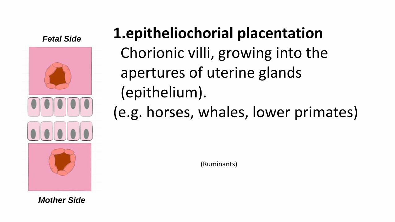
Fetal Side
Mother Side
1.epitheliochorial placentationChorionic villi, growing into the apertures of uterine glands(epithelium).
(e.g. horses, whales, lower primates)
(Ruminants)

Fetal Side
Mother Side
SynepitheliochorialplacentationSyncytium of maternal endometral epithelium and chorion

Fetal Side
Mother Side
Endotheliochorial placentationIn this type of placentation, the chorionic villi are in contact with the endothelium of maternal bloodvessels. (e.g. in most carnivoreslike cats and dogs)

Fetal Side
Mother Side
Hemochorial placentation(e.g. in higher order primates, including humans, and also in rabbits, guineapigs, mice, and rats). The haemochorialplacenta shows the intimate juxtaposition of foetal and maternal blood allowing efficient exchange.
Copyright © 2022 FDOKUMEN