Identification of hot spots of malaria transmission for targeted malaria control
Upload
independentCategory
view
0download
0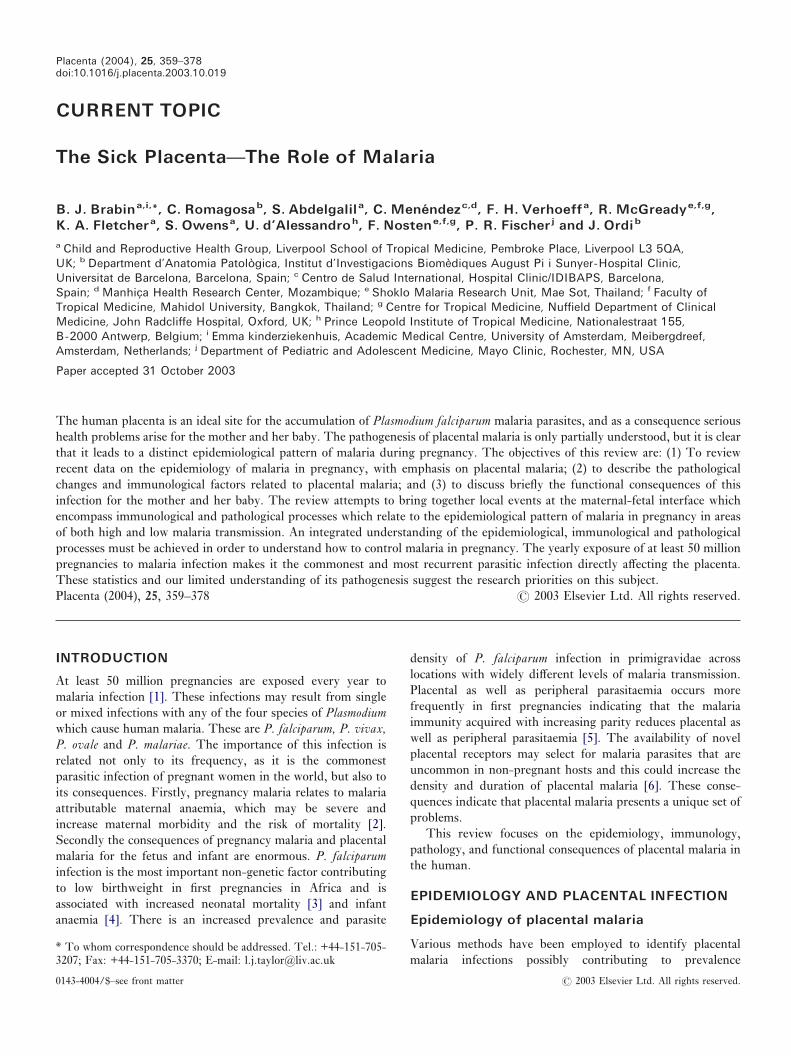
CURRENT TOPIC
The Sick Placenta—The Role of Malaria
B. J. Brabina,i,*, C. Romagosab, S. Abdelgalila, C. Menendezc,d, F. H. Verhoeffa, R. McGreadye,f,g,
K. A. Fletchera, S. Owensa, U. d’Alessandroh, F. Nostene,f,g, P. R. Fischerj and J. Ordib
a Child and Reproductive Health Group, Liverpool School of Tropical Medicine, Pembroke Place, Liverpool L3 5QA,UK; b Department d’Anatomia Patologica, Institut d’Investigacions Biomediques August Pi i Sunyer-Hospital Clinic,Universitat de Barcelona, Barcelona, Spain; c Centro de Salud International, Hospital Clinic/IDIBAPS, Barcelona,Spain; d Manhica Health Research Center, Mozambique; e Shoklo Malaria Research Unit, Mae Sot, Thailand; f Faculty ofTropical Medicine, Mahidol University, Bangkok, Thailand; g Centre for Tropical Medicine, Nuffield Department of ClinicalMedicine, John Radcliffe Hospital, Oxford, UK; h Prince Leopold Institute of Tropical Medicine, Nationalestraat 155,B-2000 Antwerp, Belgium; i Emma kinderziekenhuis, Academic Medical Centre, University of Amsterdam, Meibergdreef,Amsterdam, Netherlands; j Department of Pediatric and Adolescent Medicine, Mayo Clinic, Rochester, MN, USA
Paper accepted 31 October 2003
The human placenta is an ideal site for the accumulation of Plasmodium falciparum malaria parasites, and as a consequence serioushealth problems arise for the mother and her baby. The pathogenesis of placental malaria is only partially understood, but it is clearthat it leads to a distinct epidemiological pattern of malaria during pregnancy. The objectives of this review are: (1) To reviewrecent data on the epidemiology of malaria in pregnancy, with emphasis on placental malaria; (2) to describe the pathologicalchanges and immunological factors related to placental malaria; and (3) to discuss briefly the functional consequences of thisinfection for the mother and her baby. The review attempts to bring together local events at the maternal-fetal interface whichencompass immunological and pathological processes which relate to the epidemiological pattern of malaria in pregnancy in areasof both high and low malaria transmission. An integrated understanding of the epidemiological, immunological and pathologicalprocesses must be achieved in order to understand how to control malaria in pregnancy. The yearly exposure of at least 50 millionpregnancies to malaria infection makes it the commonest and most recurrent parasitic infection directly affecting the placenta.These statistics and our limited understanding of its pathogenesis suggest the research priorities on this subject.
� 2003 Elsevier Ltd. All rights reserved.Placenta (2004), 25, 359–378
INTRODUCTION
At least 50 million pregnancies are exposed every year tomalaria infection [1]. These infections may result from singleor mixed infections with any of the four species of Plasmodiumwhich cause human malaria. These are P. falciparum, P. vivax,P. ovale and P. malariae. The importance of this infection isrelated not only to its frequency, as it is the commonestparasitic infection of pregnant women in the world, but also toits consequences. Firstly, pregnancy malaria relates to malariaattributable maternal anaemia, which may be severe andincrease maternal morbidity and the risk of mortality [2].Secondly the consequences of pregnancy malaria and placentalmalaria for the fetus and infant are enormous. P. falciparuminfection is the most important non-genetic factor contributingto low birthweight in first pregnancies in Africa and isassociated with increased neonatal mortality [3] and infantanaemia [4]. There is an increased prevalence and parasite
density of P. falciparum infection in primigravidae acrosslocations with widely different levels of malaria transmission.Placental as well as peripheral parasitaemia occurs morefrequently in first pregnancies indicating that the malariaimmunity acquired with increasing parity reduces placental aswell as peripheral parasitaemia [5]. The availability of novelplacental receptors may select for malaria parasites that areuncommon in non-pregnant hosts and this could increase thedensity and duration of placental malaria [6]. These conse-quences indicate that placental malaria presents a unique set ofproblems.
This review focuses on the epidemiology, immunology,pathology, and functional consequences of placental malaria inthe human.
EPIDEMIOLOGY AND PLACENTAL INFECTION
Epidemiology of placental malaria
Various methods have been employed to identify placentalmalaria infections possibly contributing to prevalence
* To whom correspondence should be addressed. Tel.: +44-151-705-3207; Fax: +44-151-705-3370; E-mail: [email protected]
Placenta (2004), 25, 359–378doi:10.1016/j.placenta.2003.10.019
0143-4004/$–see front matter � 2003 Elsevier Ltd. All rights reserved.
differences observed between studies. In some studies placentalmalaria was defined only by the presence of parasites betweenthe villi while in others the definition includes the presence ofpigment. The appearance of malaria parasites early in preg-nancy and subsequently pigment has been used to characterizethe chronology of infection in the placenta [7]. Intervillousspaces, where parasites sequester later in pregnancy, form aslacunae in the trophoblast between days 10 and 21 of gestation,but it is unclear whether maternal blood enters these spacesbefore 12 weeks of gestation [8] and this may explain whyparasites do not sequester in the placenta before the fourthmonth of gestation [9].
Parasite data on placental malaria are available only atdelivery when peripheral and placental parasite prevalenceestimates show reasonable correlation [10]. However somestudies have shown high density placental parasites with scantyperipheral parasitaemia [9,11,12], whereas other studies haveshown peripheral parasitaemias in the absence of placentalinfection [13–15]. Ismail et al. [16] showed that as many as 68.8per cent of women with negative peripheral blood parasitaemiahad some evidence of malarial infection (either active or past)in placental samples. In this study, 46.3 per cent of activelyinfected placentae did not show parasites in the peripheralblood examination, whereas only 5.2 per cent of cases with noparasites in the placental histological study had a positiveperipheral blood. In contrast, Walter et al. [13] reported thatonly 9.5 per cent women had negative peripheral blood andpositive placental parasitaemias, while 61 per cent of cases werepositive in peripheral blood and negative in the placenta. Onthe Thai–Burmese border, a very low malaria transmissionarea, where all women with positive peripheral parasitaemia atweekly antenatal screening were treated, positive placentalparasitaemia only occurred in women with positive peripheralparasitaemia at the time of delivery, and then only in womenwith recent infection: median (range) of 1 (0–3) day from theend of pregnancy [15]. Variations in epidemiological charac-teristics of malarial infection between different areas as well asdifferences in methodology, particularly the use of polarizedlight, may partially explain these various findings. A regressionplot of placental against peripheral parasitaemia for 20 pub-lished studies estimated an average frequency of placentalparasitaemia of 9.2 per cent in the absence of peripheralparasitaemia [10]. Cord blood parasitaemias have not in-frequently been reported in blood smears examined by lightmicroscopy and are more closely associated with placentalparasitaemia than with maternal parasitaemia [13,15,16]. Ina holoendemic area of Kenya, microscopy indicated noPlasmodium species in cord blood, in contrast the polymerasechain reaction (PCR) method in cord blood showed aprevalence of 32 per cent for P. falciparum, 23 per cent forP. malariae, and 21 per cent for P. ovale [17]. These findings,and comparable data from Malawi, indicate that cord bloodPlasmodium infections are common in areas of high malariatransmission and may be acquired before delivery [18].
There are similarities and differences between P. falciparumand P. vivax infection of the placenta. P. falciparum can
achieve high parasite densities (greater than 90 per cent of redcells in the placenta infected) and invades erythrocytes of allages. P. vivax exclusively invades young red cells (reticulo-cytes) and consequently achieves relatively low parasite densi-ties. The mature stages of P. falciparum sequester in deeporgan vasculature and do not appear in peripheral blood,except in severe clinical infections [19]. All asexual stages ofP. vivax appear in the peripheral circulation and this specieshas only recently been reported to occur in the placentaalthough less frequently and at less density than P. falciparum[15].
Placental parasitaemia occurs more frequently in first preg-nancies. Peripheral parasite prevalence is also higher in the firsthalf of pregnancy and decreases with advancing gestational age[20]. Interestingly, placental involvement in malarial infectionsis closely related to parity and both frequency and parasitedensity decrease as parity increases in higher transmissionareas [21–23]. This effect is considered to be largely parityspecific, but there is also an age-dependent component[5,24,25]. An important inter-related factor is co-infection withHIV, which increases the prevalence of peripheral and placen-tal parasitaemia [26–28]. Although HIV-infected women dem-onstrate parity-specific immunity, they have higher parasiteprevalence than HIV-uninfected women as multigravidae [28].
As mentioned previously, placental parasites may occur inthe absence of peripheral parasites, and during seasonal periodsof low malaria transmission placental malaria infection maypersist. Table 1 shows a summary of data on the seasonalprevalence of placental parasitaemia for women of all paritiesfor areas of high (rural) and low (urban) transmission in Africa.Five of these eight studies showed no significant difference inparasite prevalence (P. falciparum) between wet and dryseasons suggesting that in urban areas of lower transmission, aswell as in areas with higher transmission, placental infectionpersists despite the expected reduced incidence of infectionduring the dry season. Hidden placental P. falciparum para-sites, undetected by microscopy, have been demonstrated byPCR in a study from a low transmission area of Senegal whichshowed different parasite genotype profiles in the peripheralcirculation and in the placenta [29]. In the vast majority ofcases, some sequestered genotypes remained hidden, undetec-ted in the peripheral circulation, indicating that analysisof peripheral parasites generates only a partial picture ofP. falciparum infection. Likewise, analysis of placental parasitesonly gives a partial picture of P. falciparum infection duringpregnancy. Similar findings are reported from Kenya [17].
Placental malaria and low birthweight
Both P. falciparum and P. vivax malaria are associated withincreased low birthweight (<2500 gms) risk [30,31]. Theincreased low birthweight prevalence in primigravidae attribu-table to malaria is substantial, ranging from below 10 per centin low endemic areas to over 50 per cent in high transmissionareas. Figure 1 shows the association of low birthweight in
360 Placenta (2004), Vol. 25
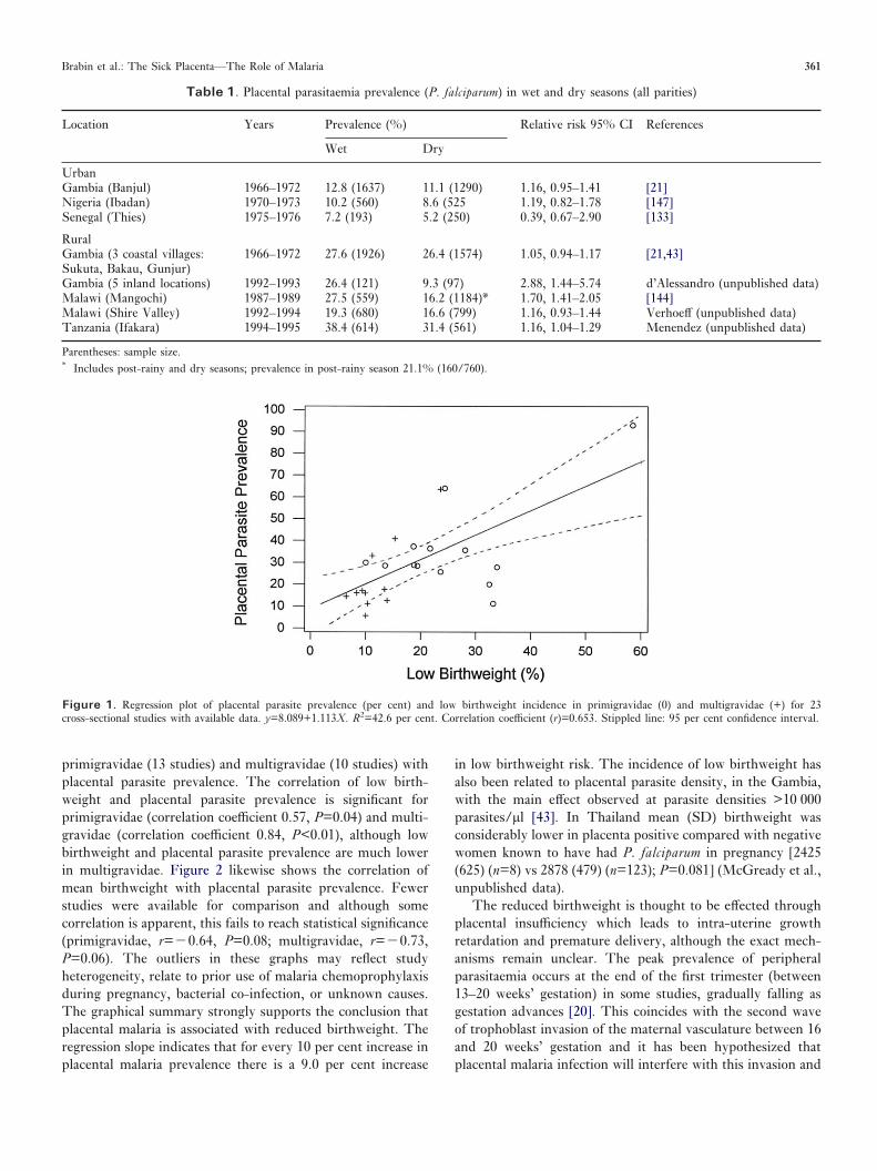
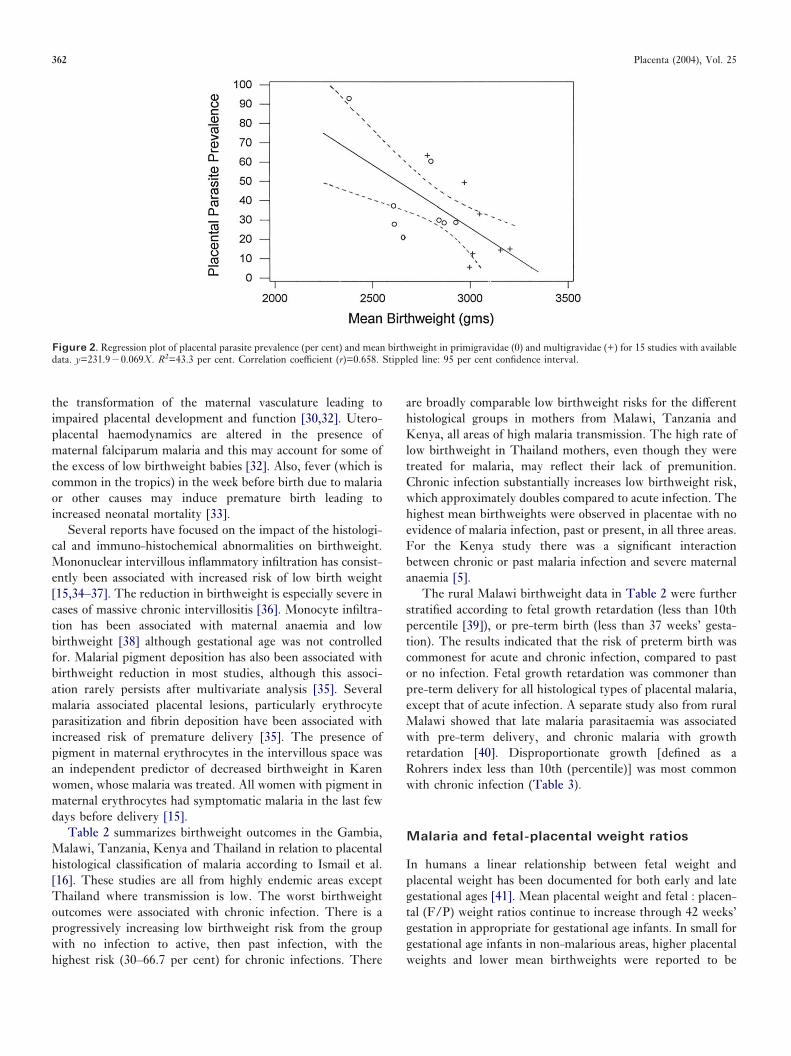
primigravidae (13 studies) and multigravidae (10 studies) withplacental parasite prevalence. The correlation of low birth-weight and placental parasite prevalence is significant forprimigravidae (correlation coefficient 0.57, P=0.04) and multi-gravidae (correlation coefficient 0.84, P<0.01), although lowbirthweight and placental parasite prevalence are much lowerin multigravidae. Figure 2 likewise shows the correlation ofmean birthweight with placental parasite prevalence. Fewerstudies were available for comparison and although somecorrelation is apparent, this fails to reach statistical significance(primigravidae, r=�0.64, P=0.08; multigravidae, r=�0.73,P=0.06). The outliers in these graphs may reflect studyheterogeneity, relate to prior use of malaria chemoprophylaxisduring pregnancy, bacterial co-infection, or unknown causes.The graphical summary strongly supports the conclusion thatplacental malaria is associated with reduced birthweight. Theregression slope indicates that for every 10 per cent increase inplacental malaria prevalence there is a 9.0 per cent increase
in low birthweight risk. The incidence of low birthweight hasalso been related to placental parasite density, in the Gambia,with the main effect observed at parasite densities >10 000parasites/µl [43]. In Thailand mean (SD) birthweight wasconsiderably lower in placenta positive compared with negativewomen known to have had P. falciparum in pregnancy [2425(625) (n=8) vs 2878 (479) (n=123); P=0.081] (McGready et al.,unpublished data).
The reduced birthweight is thought to be effected throughplacental insufficiency which leads to intra-uterine growthretardation and premature delivery, although the exact mech-anisms remain unclear. The peak prevalence of peripheralparasitaemia occurs at the end of the first trimester (between13–20 weeks’ gestation) in some studies, gradually falling asgestation advances [20]. This coincides with the second waveof trophoblast invasion of the maternal vasculature between 16and 20 weeks’ gestation and it has been hypothesized thatplacental malaria infection will interfere with this invasion and
Table 1. Placental parasitaemia prevalence (P. falciparum) in wet and dry seasons (all parities)
Location Years Prevalence (%) Relative risk 95% CI References
Wet Dry
UrbanGambia (Banjul) 1966–1972 12.8 (1637) 11.1 (1290) 1.16, 0.95–1.41 [21]Nigeria (Ibadan) 1970–1973 10.2 (560) 8.6 (525 1.19, 0.82–1.78 [147]Senegal (Thies) 1975–1976 7.2 (193) 5.2 (250) 0.39, 0.67–2.90 [133]
RuralGambia (3 coastal villages:Sukuta, Bakau, Gunjur)
1966–1972 27.6 (1926) 26.4 (1574) 1.05, 0.94–1.17 [21,43]
Gambia (5 inland locations) 1992–1993 26.4 (121) 9.3 (97) 2.88, 1.44–5.74 d’Alessandro (unpublished data)Malawi (Mangochi) 1987–1989 27.5 (559) 16.2 (1184)* 1.70, 1.41–2.05 [144]Malawi (Shire Valley) 1992–1994 19.3 (680) 16.6 (799) 1.16, 0.93–1.44 Verhoeff (unpublished data)Tanzania (Ifakara) 1994–1995 38.4 (614) 31.4 (561) 1.16, 1.04–1.29 Menendez (unpublished data)
Parentheses: sample size.* Includes post-rainy and dry seasons; prevalence in post-rainy season 21.1% (160/760).
Figure 1. Regression plot of placental parasite prevalence (per cent) and low birthweight incidence in primigravidae (0) and multigravidae (+) for 23cross-sectional studies with available data. y=8.089+1.113X. R2=42.6 per cent. Correlation coefficient (r)=0.653. Stippled line: 95 per cent confidence interval.
Brabin et al.: The Sick Placenta—The Role of Malaria 361
the transformation of the maternal vasculature leading toimpaired placental development and function [30,32]. Utero-placental haemodynamics are altered in the presence ofmaternal falciparum malaria and this may account for some ofthe excess of low birthweight babies [32]. Also, fever (which iscommon in the tropics) in the week before birth due to malariaor other causes may induce premature birth leading toincreased neonatal mortality [33].
Several reports have focused on the impact of the histologi-cal and immuno-histochemical abnormalities on birthweight.Mononuclear intervillous inflammatory infiltration has consist-ently been associated with increased risk of low birth weight[15,34–37]. The reduction in birthweight is especially severe incases of massive chronic intervillositis [36]. Monocyte infiltra-tion has been associated with maternal anaemia and lowbirthweight [38] although gestational age was not controlledfor. Malarial pigment deposition has also been associated withbirthweight reduction in most studies, although this associ-ation rarely persists after multivariate analysis [35]. Severalmalaria associated placental lesions, particularly erythrocyteparasitization and fibrin deposition have been associated withincreased risk of premature delivery [35]. The presence ofpigment in maternal erythrocytes in the intervillous space wasan independent predictor of decreased birthweight in Karenwomen, whose malaria was treated. All women with pigment inmaternal erythrocytes had symptomatic malaria in the last fewdays before delivery [15].
Table 2 summarizes birthweight outcomes in the Gambia,Malawi, Tanzania, Kenya and Thailand in relation to placentalhistological classification of malaria according to Ismail et al.[16]. These studies are all from highly endemic areas exceptThailand where transmission is low. The worst birthweightoutcomes were associated with chronic infection. There is aprogressively increasing low birthweight risk from the groupwith no infection to active, then past infection, with thehighest risk (30–66.7 per cent) for chronic infections. There
are broadly comparable low birthweight risks for the differenthistological groups in mothers from Malawi, Tanzania andKenya, all areas of high malaria transmission. The high rate oflow birthweight in Thailand mothers, even though they weretreated for malaria, may reflect their lack of premunition.Chronic infection substantially increases low birthweight risk,which approximately doubles compared to acute infection. Thehighest mean birthweights were observed in placentae with noevidence of malaria infection, past or present, in all three areas.For the Kenya study there was a significant interactionbetween chronic or past malaria infection and severe maternalanaemia [5].
The rural Malawi birthweight data in Table 2 were furtherstratified according to fetal growth retardation (less than 10thpercentile [39]), or pre-term birth (less than 37 weeks’ gesta-tion). The results indicated that the risk of preterm birth wascommonest for acute and chronic infection, compared to pastor no infection. Fetal growth retardation was commoner thanpre-term delivery for all histological types of placental malaria,except that of acute infection. A separate study also from ruralMalawi showed that late malaria parasitaemia was associatedwith pre-term delivery, and chronic malaria with growthretardation [40]. Disproportionate growth [defined as aRohrers index less than 10th (percentile)] was most commonwith chronic infection (Table 3).
Malaria and fetal-placental weight ratios
In humans a linear relationship between fetal weight andplacental weight has been documented for both early and lategestational ages [41]. Mean placental weight and fetal : placen-tal (F/P) weight ratios continue to increase through 42 weeks’gestation in appropriate for gestational age infants. In small forgestational age infants in non-malarious areas, higher placentalweights and lower mean birthweights were reported to be
Figure 2. Regression plot of placental parasite prevalence (per cent) and mean birthweight in primigravidae (0) and multigravidae (+) for 15 studies with availabledata. y=231.9�0.069X. R2=43.3 per cent. Correlation coefficient (r)=0.658. Stippled line: 95 per cent confidence interval.
362 Placenta (2004), Vol. 25
associated with increased neonatal morbidity [42]. McGregoret al. [43] reported lower birthweights in Gambian infants inthe presence of placental malaria defined by positive bloodslides (especially in first-born children), but not depressedplacental weight. F/P ratios recalculated from their data aresummarized in Table 4 and consistently show lower F/P ratios
with placental malaria. As placental weight was not affected,the effect of malaria on the infant weight was considered due toplacental insufficiency, leading to growth retardation, or anincreased incidence of pre-term deliveries, or a combination ofboth. However, data on gestational age were not presented inthis report. The data shown in Table 4 from a low trans-
Table 2. Birthweight and placental histological classification
Location No infection Acute Past Chronic All
Gambia*Low birthweight, % 22.2 (18) 25.0 (8) 31.6 (19) 40.0 (35) 32.5 (80)Mean birthweight, g 2863 (�490) 2539 (�326) 2601 (�464) 2576 (�544) 2643 (�502)
Malawi Rural†Low birthweight, % 13.0 (23) 50.0 (2) 17.1 (292) 29.9 (117) 20.5 (434)Mean birthweight, g 2930 (�342) 2660 (�339) 2894 (�439) 2756 (�530) 2857 (�462)
Malawi Urban‡Low birthweight, % 9.6 (10) 10.5 (4) 11.3 (16) – –Mean birthweight, g 3101 (455) 2923 (423) 2978 (425) – –
Tanzania§Low birthweight, % 14.9 (289) 8.9 (112) 13.7 (475) 29.6 (301) 17.6 (1177)Mean birthweight, g – – – – 2819 (�429)
Kenya¶Low birthweight, % 13.2 (423) 11.5 (61) 22.3 (215) 29.3 (116) 17.9 (815)Mean birthweight, g – – – – 2900 (�510)
Thai–Burmese border�Low birthweight, % 10.0 (1) – 10.0 (1) 66.7 (4) 21.4 (6)Mean birthweight, g 3085 (�325) (10) 2700 (�n.a) (2) 2860 (�370) (10) 1992 (�754) (6) 2742 (�600) (28)
Histological classification follows Ismail et al. [16]. Brackets: sample size; square brackets: standard deviation.* d’Alessandro—unpublished data.† Verhoeff—unpublished data, only singleton, live births included.‡ [62].§ Menendez—personal communication and [35].¶ [5].� Only includes women with parasites and pigment detected histopathologically in women with a known positive peripheral P. falciparum infection detected andtreated antenatally i.e. the classification of ‘no infection’ in the placenta at delivery does not mean the woman was not infected during pregnancy.
Table 3. Fetal growth retardation, pre-term delivery and placental histological classification in Malawian and Thai* pregnancies
Pregnancy outcome (%) No infection Acute Past Chronic All
IUGRMalawi 22.7 (22) 50.0 (2) 29.3 (290) 30.4 (115) 29.4 (429)Thailand 6.3 (16) – 11.1 (18) 33.3 (9) 13.0 (46)
Pre-termMalawi 9.1 (22) 50.0 (2) 9.7 (290) 20.9 (115) 12.8 (429)Thailand – – 18.2 (11) 4.2 (48)
Low birthweightMalawi 13.0 (23) 50.0 (2) 17.1 (292) 29.9 (117) 20.5 (434)Thailand 6.3 (16) – 11.1 (18) 45.5 (11) 16.7 (48)
DisproportionateMalawi 0.0 (22) 0.0 (2) 5.2 (292) 9.6 (115) 6.1 (429)
Brackets: sample size. IUGR: intra-uterine growth retardation less than <10th percentile [39]. Pre-term: less than 2500 g. Disproportionate: less than 10% Rohrersindex [135].* Thai data includes women known to have had only P. falciparum infection detected and treated antenatally, including all women in the ‘No infection’ group.
Brabin et al.: The Sick Placenta—The Role of Malaria 363
mission area in Thailand also shows significantly reduced F/Pratios in primigravidae, but with a much reduced effect inmultigravidae.
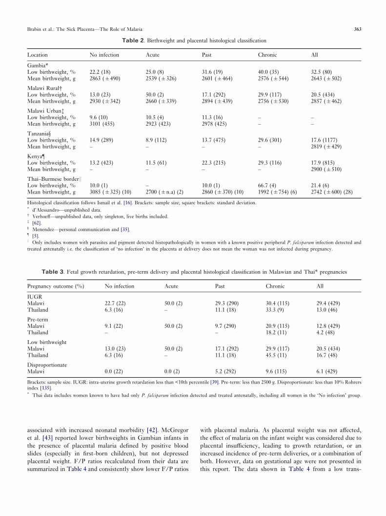
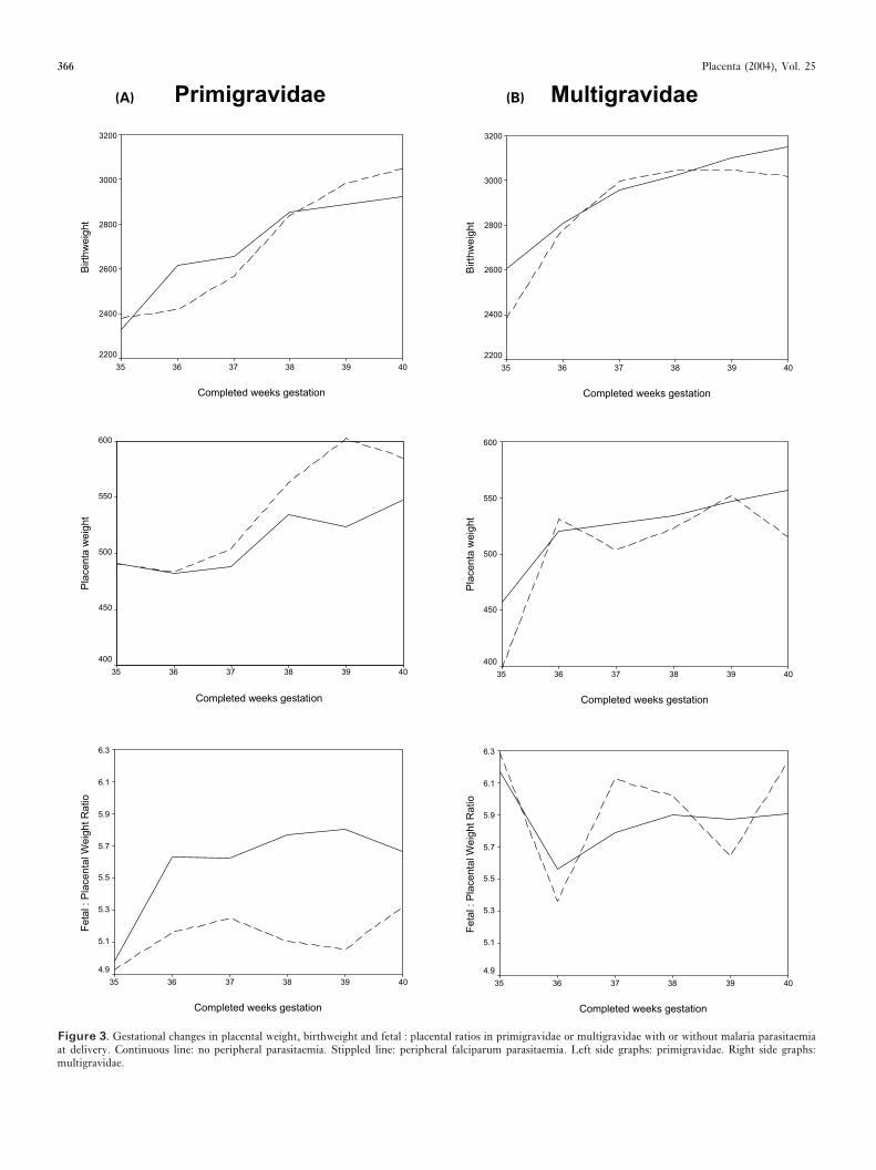
An analysis of F/P ratios by gestational age in relation toplacental malaria has been possible from data available from alarge cohort study of mothers and babies delivering in a ruralarea of southern Malawi where malaria transmission is peren-nial [44]. Gestational age was assessed using a modified BallardScale [45]. The quantitative data is summarized in Table 5. Inprimigravidae from 36 weeks’ gestation mean placental weightwas increased and mean birthweight reduced. This results inan overall reduction in the F/P ratio between 36–40 weeks’gestation. Conversely in multigravidae only small differenceswere found in mean placental weight or birthweight betweeninfected and non-infected mothers and no consistent differencein the F/P ratios was apparent. These patterns are illustratedin Figure 3 for primigravidae and multigravidae. These resultssuggest that in African primigravidae the low F/P ratio wasdue not only to a reduced birthweight, but also to increasedplacental weight. This supports the placental microscopicstructural features model proposed in the term variety of fetalgrowth retardation [46]. Severe anaemia associated with ma-laria, which is commoner in primigravidae in highly malariousareas, may contribute to this process [47].
THE PATHOLOGY OF PLACENTAL INFECTION
Placental findings associated with malarial
parasites or products
During P. falciparum infections, the placenta can harbour astriking density of parasites, macrophages and pigment.Bignami [48] and Serini [49] first described these featureswhile studying congenital malaria. Blacklock and Gordon [12]
subsequently reported the placental involvement in malarialinfection describing the presence of parasites and malarialpigment in thick smears from placental blood.
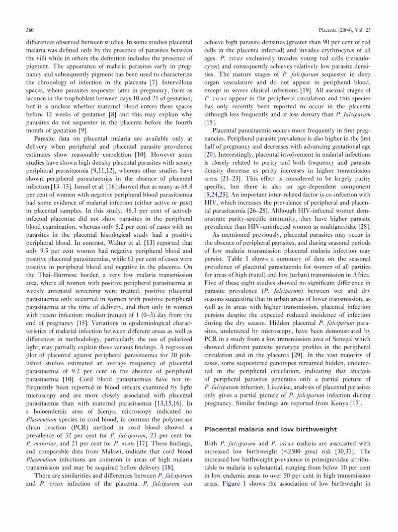
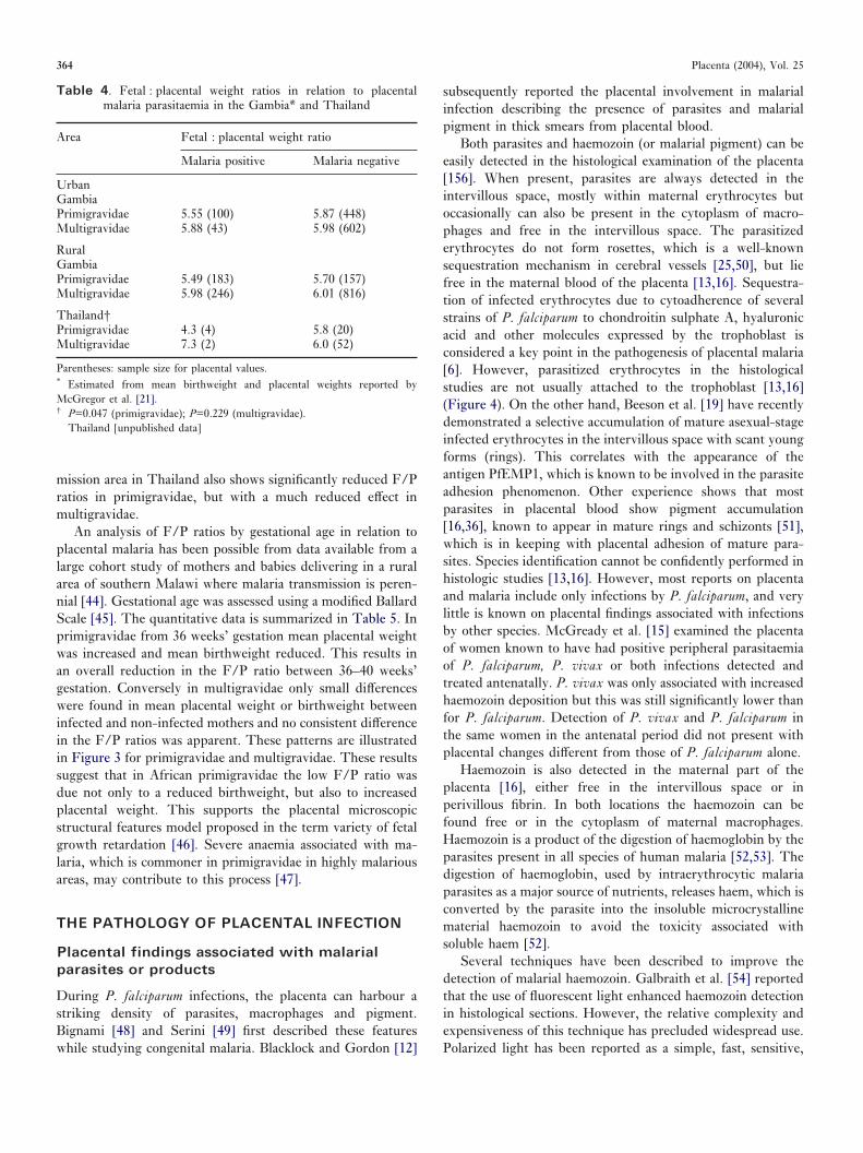
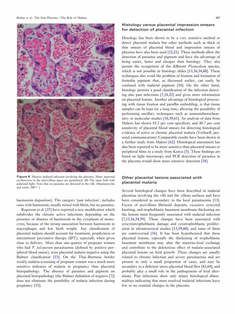
Both parasites and haemozoin (or malarial pigment) can beeasily detected in the histological examination of the placenta[156]. When present, parasites are always detected in theintervillous space, mostly within maternal erythrocytes butoccasionally can also be present in the cytoplasm of macro-phages and free in the intervillous space. The parasitizederythrocytes do not form rosettes, which is a well-knownsequestration mechanism in cerebral vessels [25,50], but liefree in the maternal blood of the placenta [13,16]. Sequestra-tion of infected erythrocytes due to cytoadherence of severalstrains of P. falciparum to chondroitin sulphate A, hyaluronicacid and other molecules expressed by the trophoblast isconsidered a key point in the pathogenesis of placental malaria[6]. However, parasitized erythrocytes in the histologicalstudies are not usually attached to the trophoblast [13,16](Figure 4). On the other hand, Beeson et al. [19] have recentlydemonstrated a selective accumulation of mature asexual-stageinfected erythrocytes in the intervillous space with scant youngforms (rings). This correlates with the appearance of theantigen PfEMP1, which is known to be involved in the parasiteadhesion phenomenon. Other experience shows that mostparasites in placental blood show pigment accumulation[16,36], known to appear in mature rings and schizonts [51],which is in keeping with placental adhesion of mature para-sites. Species identification cannot be confidently performed inhistologic studies [13,16]. However, most reports on placentaand malaria include only infections by P. falciparum, and verylittle is known on placental findings associated with infectionsby other species. McGready et al. [15] examined the placentaof women known to have had positive peripheral parasitaemiaof P. falciparum, P. vivax or both infections detected andtreated antenatally. P. vivax was only associated with increasedhaemozoin deposition but this was still significantly lower thanfor P. falciparum. Detection of P. vivax and P. falciparum inthe same women in the antenatal period did not present withplacental changes different from those of P. falciparum alone.
Haemozoin is also detected in the maternal part of theplacenta [16], either free in the intervillous space or inperivillous fibrin. In both locations the haemozoin can befound free or in the cytoplasm of maternal macrophages.Haemozoin is a product of the digestion of haemoglobin by theparasites present in all species of human malaria [52,53]. Thedigestion of haemoglobin, used by intraerythrocytic malariaparasites as a major source of nutrients, releases haem, which isconverted by the parasite into the insoluble microcrystallinematerial haemozoin to avoid the toxicity associated withsoluble haem [52].
Several techniques have been described to improve thedetection of malarial haemozoin. Galbraith et al. [54] reportedthat the use of fluorescent light enhanced haemozoin detectionin histological sections. However, the relative complexity andexpensiveness of this technique has precluded widespread use.Polarized light has been reported as a simple, fast, sensitive,
Table 4. Fetal : placental weight ratios in relation to placentalmalaria parasitaemia in the Gambia* and Thailand
Area Fetal : placental weight ratio
Malaria positive Malaria negative
UrbanGambiaPrimigravidae 5.55 (100) 5.87 (448)Multigravidae 5.88 (43) 5.98 (602)
RuralGambiaPrimigravidae 5.49 (183) 5.70 (157)Multigravidae 5.98 (246) 6.01 (816)
Thailand†Primigravidae 4.3 (4) 5.8 (20)Multigravidae 7.3 (2) 6.0 (52)
Parentheses: sample size for placental values.* Estimated from mean birthweight and placental weights reported byMcGregor et al. [21].† P=0.047 (primigravidae); P=0.229 (multigravidae).
Thailand [unpublished data]
364 Placenta (2004), Vol. 25
and specific alternative method for localizing intracellularpigmented malarial parasites both in histological slides [7,16]and in wet preparations of blood (Lawrence et al., 1994), dueto the marked birefringence of haemozoin (Figure 4). We haverecently compared the sensitivity and specificity of polarizedlight and non-polarized light to detect malarial pigment inroutinely processed placental histological slides (Romagosaet al., manuscript in preparation). The use of polarized lightsignificantly increased the sensitivity of detection of pigmentand parasites (100 per cent and 98.1 per cent respectively),compared with non-polarized light (40.5 per cent and 50.3 percent respectively). The sensitivity of non-polarized light isespecially poor in cases with scant parasites (12.7 per cent for<1 per cent vs 97.8 per cent for >5 per cent parasitizederythrocytes, P<0.001), or minimal pigment deposition (42.4per cent vs 84.5 per cent when severe, P<0.001). However,careful attention to the fixation process is essential due to thesimilar physical properties of formalin and haemozoin. The useof neutral buffered formalin reduces the formation of formalinpigment, even after long fixation periods, and should bestressed when planning histological studies of placentalmalaria. Formation of formalin pigment can be avoided byusing streck tissue fixative (STF) (manufactured by StreckLaboratories, Omaha, NE, USA).
Haemozoin can be quantified by a sensitive and specificassay [55]. In an area of high transmission, recent falciparummalaria infections (many of which were untreated) resulted insignificantly higher placental haemozoin concentrations [53].On the Thai–Burmese border, the same laboratory foundplacental haemozoin concentration was strongly positivelycorrelated with P. falciparum malaria infection in pregnancybut not with P. vivax infection, being a primigravidae, andwith malaria infection (positive peripheral parasitaemia) in thelast trimester of pregnancy [15].
Most evidence indicates that malarial products do notaccumulate in fetal structures. Although malarial parasites aredetected in a small proportion of cases in the cord blood bystandard light microscopic examination of Giemsa stainedblood smears, malarial parasites have never been described infetal erythrocytes or in fetal structures in histological studies
[13,16,56]. This suggests that malarial parasites cannot crossthe placental barrier until delivery, when multiple vesselruptures and mixing of maternal and fetal blood takes place. Afew reports have described the presence of malarial pigment invillous stroma but not in fetal vessels [15,54,57], but thisfinding has not been confirmed in most studies. McGreadyet al. observed one case with pigment in fetal monocytes infetal vessels in a primigravid woman with hyperparasitaemia(>4 per cent RBC prarasitized) who started premature labourspontaneously but delivered by Caesarean section for fetaldistress. As suggested previously the mode of delivery mayaffect the placental histopathological findings of some featuresincluding sequestration [58]. Only trophoblastic cells coveringthe fetal villi and acting as a barrier between maternal and fetalblood have consistently been shown to contain haemozoin[13,15,54,59,155].
Classification of malarial infection
Bulmer et al. [7] introduced the first classification of placentalmalarial infection. This classification was later slightlymodified by Ismail et al. [16]. The rationale for histologicalclassifications is based on the different significance of haemo-zoin and parasites and on the assumption of the progression ofthe infection that is often left untreated. Thus, the presence ofparasites indicates active infection whereas haemozoin depo-sition, either free or in macrophages, indicates chronic haemo-lysis. During the early stages of infection, only parasites can bedetected. After the initial haemolytic episodes, malarial pig-ment is detected in intervillous monocytes. Later, haemozoin iscovered by fibrin and is detected in the perivillous fibrin, eitherfree or within the cytoplasm of entrapped macrophages, or inthe decidua. Obviously, parasites can coincide with haemozoinin macrophages and in fibrin. After clearance of the infection,pigment persists only within the fibrin for a period of time, butit eventually disappears. Thus, active infections, defined by thepresence of parasitized red blood cells in the intervillous spaceof the placenta, includes two categories, acute infections (onlyparasites and minimal haemozoin deposition in the macro-phages, but not fibrin), and chronic infections (parasites and
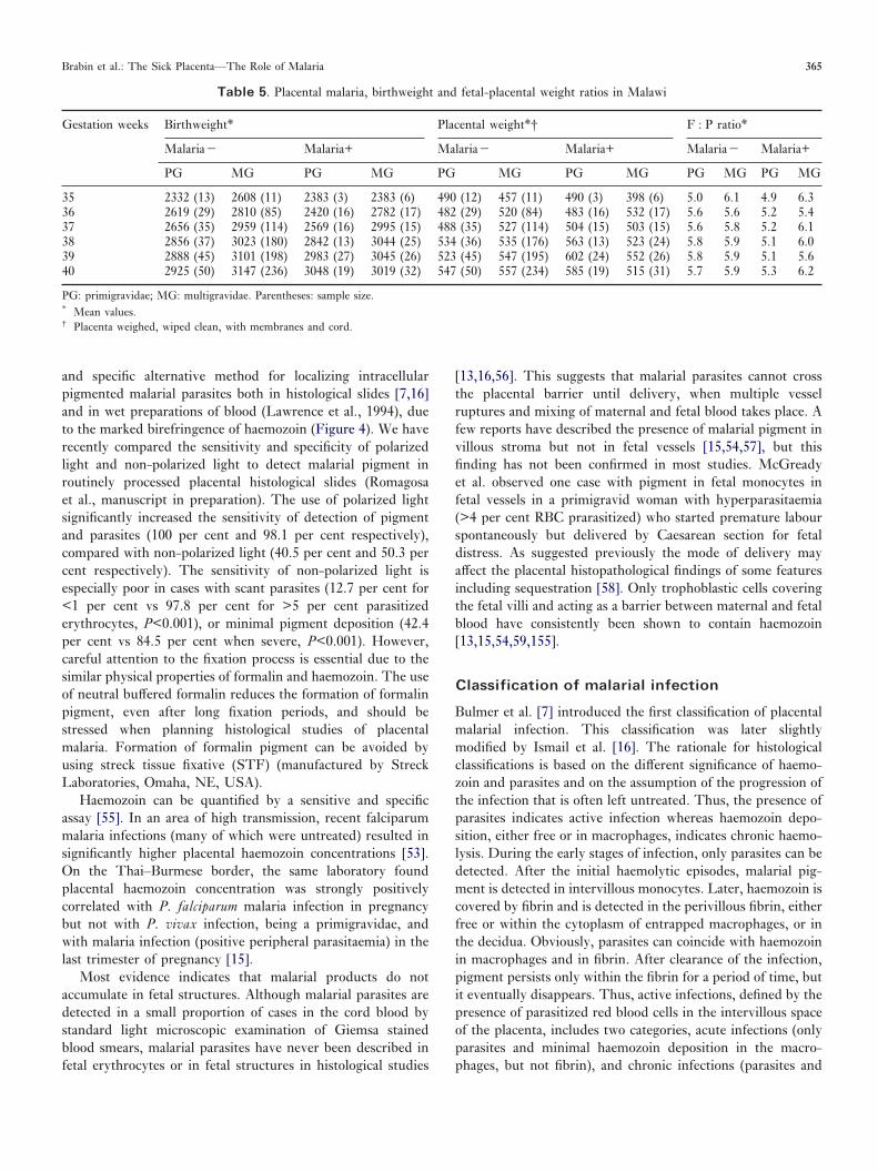
Table 5. Placental malaria, birthweight and fetal-placental weight ratios in Malawi
Gestation weeks Birthweight* Placental weight*† F : P ratio*
Malaria� Malaria+ Malaria� Malaria+ Malaria� Malaria+
PG MG PG MG PG MG PG MG PG MG PG MG
35 2332 (13) 2608 (11) 2383 (3) 2383 (6) 490 (12) 457 (11) 490 (3) 398 (6) 5.0 6.1 4.9 6.336 2619 (29) 2810 (85) 2420 (16) 2782 (17) 482 (29) 520 (84) 483 (16) 532 (17) 5.6 5.6 5.2 5.437 2656 (35) 2959 (114) 2569 (16) 2995 (15) 488 (35) 527 (114) 504 (15) 503 (15) 5.6 5.8 5.2 6.138 2856 (37) 3023 (180) 2842 (13) 3044 (25) 534 (36) 535 (176) 563 (13) 523 (24) 5.8 5.9 5.1 6.039 2888 (45) 3101 (198) 2983 (27) 3045 (26) 523 (45) 547 (195) 602 (24) 552 (26) 5.8 5.9 5.1 5.640 2925 (50) 3147 (236) 3048 (19) 3019 (32) 547 (50) 557 (234) 585 (19) 515 (31) 5.7 5.9 5.3 6.2
PG: primigravidae; MG: multigravidae. Parentheses: sample size.* Mean values.† Placenta weighed, wiped clean, with membranes and cord.
Brabin et al.: The Sick Placenta—The Role of Malaria 365
Pla
cent
a w
eigh
t(A) (B)
Figure 3. Gestational changes in placental weight, birthweight and fetal : placental ratios in primigravidae or multigravidae with or without malaria parasitaemiaat delivery. Continuous line: no peripheral parasitaemia. Stippled line: peripheral falciparum parasitaemia. Left side graphs: primigravidae. Right side graphs:multigravidae.
366 Placenta (2004), Vol. 25
haemozoin deposition). The category ‘past infection’, includescases with haemozoin, usually mixed with fibrin, but no parasites.
Rogerson et al. [37] have reported a new modification whichsubdivides the chronic active infections depending on thepresence or absence of haemozoin in the cytoplasm of mono-cytes, because of the strong association between haemozoin inmacrophages and low birth weight. Any classification ofplacental malaria should account for treatment, prophylaxis orintermittent preventive therapy (IPT), especially when givenclose to delivery. More than one-quarter of pregnant womenwho had P. falciparum parasitaemia (defined by positive per-ipheral blood smear), were placental malaria negative using theBulmer classification [15]. On the Thai–Burmese borderweekly malaria screening of pregnant women was a much moresensitive indicator of malaria in pregnancy than placentalhistopathology. The absence of parasites and pigment onplacental histopathology (the Bulmer definition of negative [7])does not eliminate the possibility of malaria infection duringpregnancy [15].
Histology versus placental impression smears
for detection of placental infection
Histology has been shown to be a very sensitive method todetect placental malaria but other methods such as thick orthin smears of placental blood and impression smears ofplacenta have also been used [12,21]. These methods allow thedetection of parasites and pigment and have the advantage ofbeing easier, faster and cheaper than histology. They alsopermit the recognition of the different Plasmodium species,which is not possible in histology slides [13,16,54,60]. Thesetechniques also avoid the problem of fixation and formation offormalin pigment that, as discussed earlier, can easily beconfused with malarial pigment [16]. On the other hand,histology permits a good classification of the infection detect-ing also past infections [7,16,22] and gives more informationon placental lesions. Another advantage of histological process-ing with tissue fixation and paraffin embedding, is that tissuesamples can be kept for a long time, allowing the possibility ofperforming ancillary techniques such as immunohistochem-istry or molecular studies [16,59,61]. An analysis of data fromMalawi has shown 85.5 per cent specificity and 40.7 per centsensitivity of placental blood smears for detecting histologicalevidence of active or chronic placental malaria (Verhoeff, per-sonal communication). Comparable results have been shown ina further study from Malawi [62]. Histological assessment hasalso been reported to be more sensitive than placental smears orperipheral films in a study from Kenya [5]. These findings arebased on light microscopy and PCR detection of parasites inthe placenta would show more sensitive detection [18].
Other placental lesions associated with
placental malaria
Several histological changes have been described in malarialplacentae involving the villi and the villous surfaces and havebeen considered as secondary to the local parasitaemia [13].Excess of perivillous fibrinoid deposits, excessive syncytialknotting, and trophoblastic basement membrane thickening arethe lesions most frequently associated with malarial infection[7,13,16,54,59]. These changes have been associated withsyncytiotrophoblastic damage and cytotrophoblastic prolifer-ation in ultrastructural studies [13,59,60], and some of themare controversial [16]. It has been hypothesized that theseplacental lesions, especially the thickening of trophoblasticbasement membrane may alter the materno-fetal exchangeand contribute to the deleterious effect of malaria-associatedplacental lesions on fetal growth. These changes are usuallyrelated to chronic infection and severe parasitaemia and arepresent in only a small proportion of cases, and may besecondary to a deficient utero-placental blood flow [63,64], andprobably play a small role in the pathogenesis of fetal alter-ations. Past infections show only minor histological abnor-malities indicating that most resolved malarial infections leavefew or no residual changes in the placenta.
Figure 4. Massive malarial infection involving the placenta. Many maternalerythrocytes in the intervillous space are parasitized. (B) The same field withpolarized light. Note that no parasites are detected in the villi. (Haematoxylinand eosin, 200�.)
Brabin et al.: The Sick Placenta—The Role of Malaria 367
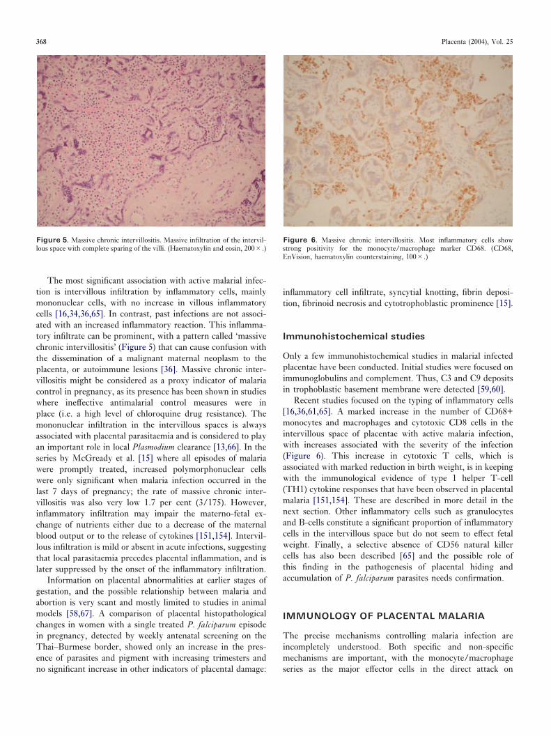
The most significant association with active malarial infec-tion is intervillous infiltration by inflammatory cells, mainlymononuclear cells, with no increase in villous inflammatorycells [16,34,36,65]. In contrast, past infections are not associ-ated with an increased inflammatory reaction. This inflamma-tory infiltrate can be prominent, with a pattern called ‘massivechronic intervillositis’ (Figure 5) that can cause confusion withthe dissemination of a malignant maternal neoplasm to theplacenta, or autoimmune lesions [36]. Massive chronic inter-villositis might be considered as a proxy indicator of malariacontrol in pregnancy, as its presence has been shown in studieswhere ineffective antimalarial control measures were inplace (i.e. a high level of chloroquine drug resistance). Themononuclear infiltration in the intervillous spaces is alwaysassociated with placental parasitaemia and is considered to playan important role in local Plasmodium clearance [13,66]. In theseries by McGready et al. [15] where all episodes of malariawere promptly treated, increased polymorphonuclear cellswere only significant when malaria infection occurred in thelast 7 days of pregnancy; the rate of massive chronic inter-villositis was also very low 1.7 per cent (3/175). However,inflammatory infiltration may impair the materno-fetal ex-change of nutrients either due to a decrease of the maternalblood output or to the release of cytokines [151,154]. Intervil-lous infiltration is mild or absent in acute infections, suggestingthat local parasitaemia precedes placental inflammation, and islater suppressed by the onset of the inflammatory infiltration.
Information on placental abnormalities at earlier stages ofgestation, and the possible relationship between malaria andabortion is very scant and mostly limited to studies in animalmodels [58,67]. A comparison of placental histopathologicalchanges in women with a single treated P. falciparum episodein pregnancy, detected by weekly antenatal screening on theThai–Burmese border, showed only an increase in the pres-ence of parasites and pigment with increasing trimesters andno significant increase in other indicators of placental damage:
inflammatory cell infiltrate, syncytial knotting, fibrin deposi-tion, fibrinoid necrosis and cytotrophoblastic prominence [15].
Immunohistochemical studies
Only a few immunohistochemical studies in malarial infectedplacentae have been conducted. Initial studies were focused onimmunoglobulins and complement. Thus, C3 and C9 depositsin trophoblastic basement membrane were detected [59,60].
Recent studies focused on the typing of inflammatory cells[16,36,61,65]. A marked increase in the number of CD68+monocytes and macrophages and cytotoxic CD8 cells in theintervillous space of placentae with active malaria infection,with increases associated with the severity of the infection(Figure 6). This increase in cytotoxic T cells, which isassociated with marked reduction in birth weight, is in keepingwith the immunological evidence of type 1 helper T-cell(TH1) cytokine responses that have been observed in placentalmalaria [151,154]. These are described in more detail in thenext section. Other inflammatory cells such as granulocytesand B-cells constitute a significant proportion of inflammatorycells in the intervillous space but do not seem to effect fetalweight. Finally, a selective absence of CD56 natural killercells has also been described [65] and the possible role ofthis finding in the pathogenesis of placental hiding andaccumulation of P. falciparum parasites needs confirmation.
IMMUNOLOGY OF PLACENTAL MALARIA
The precise mechanisms controlling malaria infection areincompletely understood. Both specific and non-specificmechanisms are important, with the monocyte/macrophageseries as the major effector cells in the direct attack on
Figure 5. Massive chronic intervillositis. Massive infiltration of the intervil-lous space with complete sparing of the villi. (Haematoxylin and eosin, 200�.)
Figure 6. Massive chronic intervillositis. Most inflammatory cells showstrong positivity for the monocyte/macrophage marker CD68. (CD68,EnVision, haematoxylin counterstaining, 100�.)
368 Placenta (2004), Vol. 25
parasitized erythrocytes. Adults and older children uncom-monly develop severe malaria in areas of high transmission.This partial and developing immunity is associated withvariant-specific agglutinating antibodies against different para-site isolates, which reflects previous exposure [68]. Thisnaturally acquired immunity reduces the frequency and den-sity of parasitaemia. During pregnancy the immune systemalters to accept the fetal allograft while maintaining hostdefences against foreign antigens. A degree of immunomodu-lation occurs, which increases susceptibility of pregnantwomen to certain infections. There are several well-describedexamples of these infections, including P. falciparum malaria.
Immune pathways in placental malaria
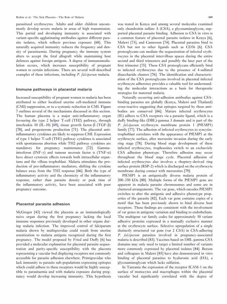
Increased susceptibility of pregnant women to malaria has beenattributed to either localized uterine cell-mediated immune(CMI) suppression, or to a systemic reduction in CMI. Figure7 outlines several of the mechanisms discussed in this section.The human placenta is a major anti-inflammatory organfavouring the type 2 helper T-cell (TH2) pathway, throughinterleukin 10 (IL-10) [69], tissue growth factor-� (TGF-�)[70], and progesterone production [71]. The placental anti-inflammatory cytokines are likely to suppress CMI. Expressionof type 1 helper T-cell (TH1) pathway cytokines is associatedwith spontaneous abortion while TH2 pathway cytokines aremandatory for pregnancy maintenance [72]. Gamma-interferon (INF-�) and tumour necrosis factor � (TNF-�)have direct cytotoxic effects towards both intracellular organ-isms and the villous trophoblast. Malaria stimulates the pro-duction of pro-inflammatory mediators, shifting the cytokinebalance away from the TH2 response [66]. Both the type ofinflammatory activity and the chronicity of the inflammatoryresponse, rather than parasite density or peak time ofthe inflammatory activity, have been associated with poorpregnancy outcome.
Placental parasite adhesion
McGregor [43] viewed the placenta as an immunologicallynaıve organ during the first pregnancy lacking the localimmune responses previously developed in other organs dur-ing malaria infection. The improved control of falciparummalaria shown by multigravidae could result from uterinesensitization to malaria antigens recognized during the firstpregnancy. The model proposed by Fried and Duffy [6] hasprovided a molecular explanation for placental parasite seques-tration and parity-specific susceptibility with the placentarepresenting a vascular bed displaying receptors not commonlyaccessible for parasite adhesion elsewhere. Primigravidae wholack immunity to parasite sub-populations expressing epitopeswhich could adhere to these receptors would be highly suscep-tible to parasitaemia and with malaria exposure during preg-nancy would develop increasing immunity. This hypothesis
was tested in Kenya and among several molecules examinedonly chondroitin sulfate A (CSA), a glycosaminoglycon, sup-ported placental parasite binding. Adhesion to CSA in vitro isa common feature of placental parasite isolates in Kenya [6],Malawi [73], and Cameroon [74]. Placental parasites bind toCSA but not to other ligands such as CD36 [6]. CSAproteoglycans can mediate the sequestration of infected eryth-rocytes in the placental intervillous spaces during the entiresecond and third trimesters and possibly the later part of thefirst trimester [75]. These CSA proteoglycans efficiently bindto infected erythrocytes due to the presence of 4-sulfateddisaccharide clusters [76]. The identification and characteriz-ation of the CSA proteoglycans involved in placental infectederythrocyte adherence provides a valuable tool for understand-ing the molecular interactions as a basis for therapeuticstrategies for maternal malaria.
Naturally occurring anti-adhesion antibodies against CSA-binding parasites are globally (Kenya, Malawi and Thailand)cross-reactive suggesting that epitopes targeted by these anti-bodies are conserved [66]. Mature infected erythrocytes(IE) adhere to CSA receptors via a parasite ligand, which is aduffy binding-like (DBL)-gamma 3 domain and is part of theP. falciparum erythrocyte membrane protein 1 (PfEMP1)family [77]. The adhesion of infected erythrocytes to syncytio-trophoblast correlates with the appearance of PfEMP1 at theerythrocyte surface, after merozoite invasion during the earlyring stage [78]. During blood stage development of theseinfected erythrocytes, trophozoites switch to an exclusvielyCSA adhesion phenotype. Therefore adhesion may occurthroughout the blood stage cycle. Placental adhesion ofinfected erythrocytes also involves a rhoptery-derived ringsurface protein (RSP-2) which is discharged on the erythrocytemembrane during contact with merozoites [79].
PfEMP1 is an antigenically diverse malaria protein of200–350 kDa [80]. Multiple forms of the PfEMP1 gene areapparent in malaria parasite chromosomes and some are inclustered arrangements. The var gene, which encodes PfEMP1switches to alter the antigenic and adhesive phenotype prop-erties of the parasite [82]. Each var gene contains copies of amotif that has been previously shown to bind diverse hostreceptors. These findings are consistent with the involvementof var genes in antigenic variation and binding to endothelium.The multigene var family codes for approximately 50 variantadhesive proteins expressed in a mutually exclusive mannerat the erythrocyte surface. Selective upregulation of a singledistinctly structured var gene (var 2 CSA) in CSA-adheringP. falciparum parasites involved in pregnancy-associatedmalaria is described [83]. Vaccines based on DBL gamma CSAdomains may only need to target a limited number of variantsmore commonly expressed by placental isolates [84]. Beesonand colleagues in Malawi [85] have also demonstrated in vitrobinding of placental parasites to hyaluronic acid (HA), aglycosaminoglycan which lacks sulfation.
In Tanzania the expression of the receptor ICAM-1 on thesurface of monocytes and macrophages within the placentalvascular bed significantly correlated with the degree of
Brabin et al.: The Sick Placenta—The Role of Malaria 369
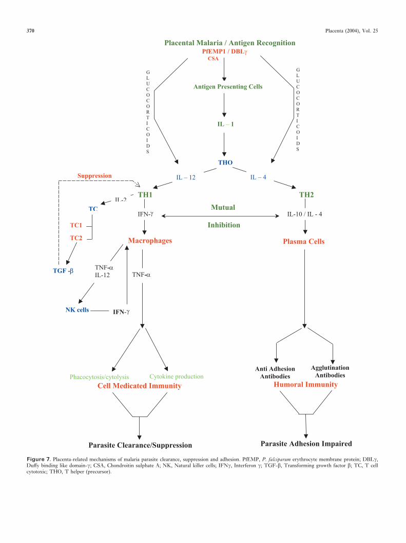
Figure 7. Placenta-related mechanisms of malaria parasite clearance, suppression and adhesion. PfEMP, P. falciparum erythrocyte membrane protein; DBL�,Duffy binding like domain-�; CSA, Chondroitin sulphate A; NK, Natural killer cells; IFN�, Interferon �; TGF-�, Transforming growth factor �; TC, T cellcytotoxic; THO, T helper (precursor).
370 Placenta (2004), Vol. 25
intervillous leukocyte infiltration [86]. It was suggested thatthis might contribute indirectly to the sequestration of infectederythrocytes within the intervillous spaces. Pro-inflammatorycytokines increase the expression of HA on microvascularendothelial cells [87], but do not alter expression of CSA in theplacenta [50]. CSA and HA are widely distributed in periph-eral vascular beds and it is unclear whether malaria parasitesuse these molecules as receptors for adhesion outside theplacenta in non-placental tissue during pregnancy [88].
Placental parasites are also unable to form rosettes, which isanother differentiating feature from parasite isolates infectingnon-pregnant hosts [89]. Little is known about the relativecontribution of different receptors in rosetting and theirprevalence in nature [90]. Parasitized RBCs form rosettes morereadily with RBCs belonging to blood group A or B, than withthose belonging to group O [91]. Blood group A has beenreported as a risk factor for severe malaria [152]. This may bedue to the fact that group A blood antigens have an importantrole as co-receptors in P. falciparum rosetting [90]. Host bloodgroup in combination with a parasitic preference for the sameblood group may provide a favourable biological environmentfor P. falciparum infection. No information has been identifiedexamining blood group types and their relation to placentalmalaria.
Sequestration in the placenta, via cytoadherence, is relatedto parasite pathogenesis and antigenic phenotype. Pathologicalevents may occur through several possible mechanisms: ob-struction of blood flow; systemic or local production ofpro-inflammatory cytokines; blockage of signal transduction[92].
Placental parasites express unique antigens (CSA and HAbinding epitopes) different from which are usually seen inother parasite isolates. A specific form of immunity is requiredto confer protection against these parasite subpopulations viaanti-adhesion antibodies and/or variant-specific agglutinatingantibodies [157].
Anti-adhesion antibodies
There is growing evidence that anti-adhesion antibodies pro-vide immune protection against placental malaria [66,93,150].Anti-adhesion antibodies against CSA-binding parasites areassociated with reduced prevalence and density of placentalmalaria. Malaria susceptibility in primigravidae has been re-lated to the lack of these antibodies [66]. In Cameroon thisantibody response towards pregnancy-associated parasites wasrelated to parity, explaining the difference in susceptibilitybetween primigravidae and multigravidae [94]. The differencein the levels of anti-adhesion antibodies rather than the degreeof parity-dependent humoral immunity has been related toprotection from placental malaria [93,95]. In Ghana antibodyrecognition of placental parasites at term correlated with donorparity, although adhesion inhibition was related to the anti-body level regardless of parity. Plasma samples from primi-gravidae at term, with high plasma levels of anti-adhesion
antibodies, were as efficient as those from multigravidae ininhibiting CSA-specific adhesion [93]. O’Neil-Dunne andcolleagues [91] reported that pregnant women with placentalmalaria in Cameroon, regardless of parity, lacked anti-adhesionantibodies during early gestation. Eighty-eight per cent of theirsample acquired antibodies during the second trimester.Multigravidae produced anti-adhesion antibodies earlier at 12weeks’ gestation, whereas primigravidae started at 20 weeks’gestation. At term most pregnant women, regardless of parity,had adequate anti-adhesion antibody and these protectiveantibodies after pregnancy can block binding to CSA ofparasites from different parts of the world. The delayedresponse in primigravidae reduces protection against placentalmalaria during the second trimester. Agglutination antibodiesmay be found more commonly in multigravidae with placentalmalaria, but have not been shown to be associated withprotection from infection. Immunization of monkeys withrecombinant duffy binding-like-gamma 3 has been shown toinduce pan-reaction and adhesion-blocking antibodies againstplacental CSA-binding P. falciparum parasites suggesting thatthe development of a vaccine to prevent placental adhesionis feasible [96]. Strategies exploring these conserved epitopesas vaccine candidates against placental malaria should beinvestigated.
Cell mediated immunity
Without anti-adhesion antibodies an extensive CMI responseoccurs leading to chronic inflammatory placental malaria.Pro-inflammatory cytokines, inducing a TH1 response, domi-nate in primigravidae in the absence of an adequate anti-inflammatory TH2 cytokine response. Disequilibria betweenthe TH1/TH2 responses results in adverse outcomes inplacental malaria in the non-immune pregnant host. Thedevelopment of acquired specific immunity over one or twosuccessive pregnancies limits both parasite sequestration andinflammatory infiltrates. This will decrease both the intensityand duration of pro-inflammatory cytokines in the placenta.
With placental malaria transcription of certain cytokines isup-regulated (IL-1, INF-�), leading to increased levels ofplacental cytokines (INF-� TNF-�, IL-2) (Figure 7). Theirlevels, as well as soluble cytokine receptors, generally exceednormal peripheral circulatory levels indicating a local inflam-matory process [66,97]. Dense accumulation of intervillousinflammatory cells takes place in active placental malaria [65].Macrophages are the predominant cells, considered to be theprincipal arm of protective immunity to placental malaria. Inmurine pregnancy placental macrophages have been shown tobe as effective as peritoneal exudate cells in phagocytosingparasite derived material in vitro [98]. Pro-inflammatory cyto-kine secretion (TNF and IL-8) was shown by immunohisto-chemistry to be localized in haemozoin-laden macrophagesactively participating in phagocytosis [99]. Macrophagespresent in infected placentae express tissue factor, the initiatorof the clotting system, and the subsequent perivillous clot
Brabin et al.: The Sick Placenta—The Role of Malaria 371
formation leads to a narrowing and plugging of the intervillousspace and disturbance of the blood supply [100]. Macrophagetissue factor expression in malarial placentae could be associ-ated with retarded placental growth and low birthweight. Thequantitative response of these inflammatory cells may not bethe only determinant factor for outcome in placental malaria.The rate of appearance of these cells is important as thereduced proliferative capacity of the placental mononuclear cellin comparison to peripheral mononuclear cells may contributeto heavy placental parasite colonization [101,102].
Ordi and colleagues [65] in Tanzania reported that placentalmalaria does not appear to be associated with CMI suppres-sion, as there was a selective absence of NK cells in placentalmalaria, which may contribute to the failure of parasiteclearance. The interaction of NK cells and other lymphocytesmay determine pregnancy outcome via the regulation of theNK cell/MQ inflammatory engine. While there is good evi-dence for the protective role of humoral immunity in placentalmalaria, it is not yet clear whether inappropriate and/orinsufficient CMI is linked to pregnancy outcome.
Cytokines and placental malaria
Placental malaria has been associated with elevated levels ofplacental TNF-� [38,66,99,102,103] and high levels of thiscytokine were related to poor pregnancy outcome [37,66,99].The concentration of placental TNF-� has been correlatedwith placental parasite densities and intervillous monocyteinfiltrates [37,38]. Peripheral blood levels of TNF-� wereweakly associated with placental malaria and/or pregnancyoutcome [37]. Elevated levels of placental INF-� during firstpregnancies may be a contributory factor to poor outcome inpregnancy. Multigravidae showed a high placental INF-�response early in the acute phase of placental malaria whileprimigravidae showed a high level even in the absence ofplacental parasitaemia. Parity differences in susceptibility toplacental malaria may be associated with differential ability tomount local cytokine responses in the placenta [102,104]. Thepregnant women whose intervillous inflammatory cells pro-duce a high level of INF-� should more effectively controlinfection than women whose INF-� response is less efficient.Primigravidae placentae induce weak primary INF-� responsesand further exposure to placental malaria amplifies the INF-�response as multigravidae more effectively clear parasites. Inprimigravidae placentae, with cord blood parasitaemia fromMalawi, neither INF-� nor IL-12 was detected using measure-ments of cytokine mRNA [99]. Rogerson and colleagues [38]have also reported, in a study from Malawi, a slight butsignificantly elevated placental plasma INF-� response withplacental parasites but concluded that elevated INF-� levelswere not associated with poor pregnancy outcomes. MaternalHIV infection may partly explain these differences betweenstudies [148].
Impaired production of INF-� in HIV sero-positive preg-nant women is associated with increased susceptibility to
placental malaria [105,148]. The triad of pregnancy, HIV sero-positivity and placental malaria seems to affect the IL-12response, leading to decreased parasite clearance [106]. Multi-gravidae with placental malaria can produce high levels ofTGF-� [66] and this ‘mature’ response, together with aspecific humoral immunity, would reduce an extensive CMIresponse. TGF-� levels have been reported to be lower inprimigravidae with, placental malaria compared to those with-out [99], which could lead to an uncontrolled TH1 response,especially in the absence of specific humoral immunity (Figure7). Although both primigravidae and multigravidae with pla-cental malaria produce high levels of IL-2, only multigravidaehave been reported to produce high levels of TGF-� [66].Parity seems to affect placental cytokine synthesis and inflam-matory pathway maturation. The IL-2 response to differentmalaria antigens gradually increased with increasing paritysuggesting the construction of a T cell memory responseduring successive pregnancies [104]. IL-10 acts as a regulatorycytokine and its production may serve to regulate INF-� andTNF-�, as its synthesis in placental malaria paralleled that ofINF-� and to lesser extent TNF-� [102]. In placenta fromholoendemic areas, in contrast to low endemic areas, thecytokine balance was shifted toward a TH1 response reducingthe level of IL-10 via the mutual inhibitory effect [66].Pregnancy may influence malaria parasite dynamics in waysthat alter immune responses, rather than influence immuneresponses in ways that alter parasite dynamics. Placentalparasite proteins or peptides could be selected in the placenta,or be specifically induced by pregnancy hormones [107]. Thetrigger for the pro-inflammatory responses in placental malariamay be parasite cytoadhesion to CSA, HA and other placentalreceptors. Conversely pro-inflammatory cytokines may inducethese placental vascular receptors, which promote parasitecytoadhesion.
FUNCTIONAL CONSEQUENCES OF
PLACENTAL MALARIA
Infant morbidity and survival
Fetal growth restriction and pre-term delivery are recognizedmajor consequences of placental malaria. These have import-ant implications as low birthweight babies have increasedmorbidity and mortality. Maternal placental infection withP. falciparum has been positively associated with malariamorbidity during the child’s first 2 years of life ([108];Menendez et al., in preparation), infant anaemia ([4];Menendez et al., in preparation); fetal anaemia [109,110,149];cord malaria parasitaemia [17], prenatal immune priming tomalaria antigens [111], and perinatal and neonatal mortality[3,28,112,113]. Placental infection with P. falciparum appearsto have a much more significant role in infant survival in Africathan has been previously assumed [114].
These risks occur even though symptomatic congenitalmalaria is an unusual event for babies born to semi-immune
372 Placenta (2004), Vol. 25
mothers who are living under holoendemic conditions [81].Accumulation of infected red cells at the interface between thematernal and fetal circulation does however result in a smallnumber of cases as symptomatic congenital malaria. Impor-tantly, placental malaria and anaemia are associated with theproduction of P. falciparum-specific immunoglobulins by thefetus leading to early immune priming and acquisition ofimmunity by infants [115,116]. This may partly explain whyneonates and infants are relatively protected from clinicalmalaria if born to semi-immune mothers [117,118]. Yet in lowtransmission areas placental malaria can lead to congenitalinfection and infant death [119]. Placental parasitaemia can beused as an alert signal to identify which infants need activescreening and treatment of peripheral parasitaemia, to preventdeath from malaria [15].
Placental transfer of maternal antibody
Placental transfer of maternal malaria antibody relates to infantimmune status and the protection of young infants from severemalaria morbidity and mortality [109,118,120]. The data onthe effect of placental malaria on transfer of maternal antibodyto a variety of antigens is of particular interest because ofeffects on infant immunity to infectious illness. This is anactive process and believed to be mediated by specific receptorsat the materno-fetal interface, or syncytiotrophoblast [121].There are a number of candidate glycoproteins but recently,attention has focused on the human analogue of a rat pupenterocyte receptor, FcRn. In the proposed model, immuno-globulin is taken across the apical surface of the syncytiotro-phoblast, and bound to FcRn within endosomes. The receptorprotects antibody from degradation within the cell, beforeorchestrating its release into the chorionic stroma [121].
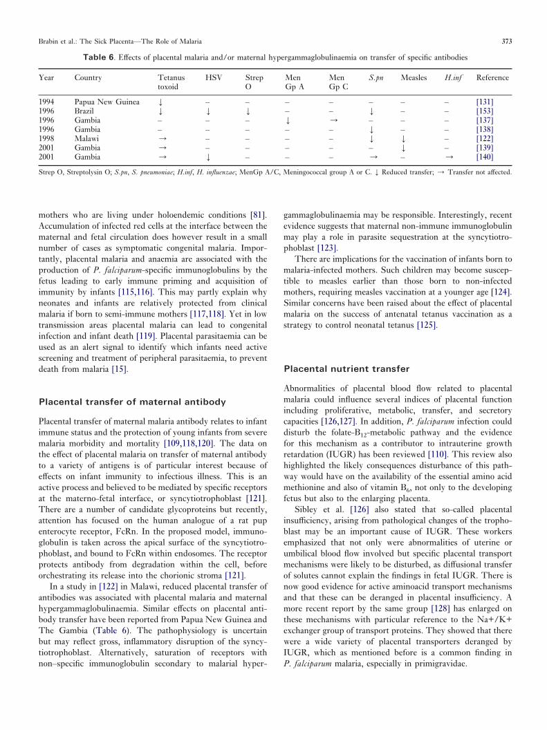
In a study in [122] in Malawi, reduced placental transfer ofantibodies was associated with placental malaria and maternalhypergammaglobulinaemia. Similar effects on placental anti-body transfer have been reported from Papua New Guinea andThe Gambia (Table 6). The pathophysiology is uncertainbut may reflect gross, inflammatory disruption of the syncy-tiotrophoblast. Alternatively, saturation of receptors withnon–specific immunoglobulin secondary to malarial hyper-
gammaglobulinaemia may be responsible. Interestingly, recentevidence suggests that maternal non-immune immunoglobulinmay play a role in parasite sequestration at the syncytiotro-phoblast [123].
There are implications for the vaccination of infants born tomalaria-infected mothers. Such children may become suscep-tible to measles earlier than those born to non-infectedmothers, requiring measles vaccination at a younger age [124].Similar concerns have been raised about the effect of placentalmalaria on the success of antenatal tetanus vaccination as astrategy to control neonatal tetanus [125].
Placental nutrient transfer
Abnormalities of placental blood flow related to placentalmalaria could influence several indices of placental functionincluding proliferative, metabolic, transfer, and secretorycapacities [126,127]. In addition, P. falciparum infection coulddisturb the folate-B12-metabolic pathway and the evidencefor this mechanism as a contributor to intrauterine growthretardation (IUGR) has been reviewed [110]. This review alsohighlighted the likely consequences disturbance of this path-way would have on the availability of the essential amino acidmethionine and also of vitamin B6, not only to the developingfetus but also to the enlarging placenta.
Sibley et al. [126] also stated that so-called placentalinsufficiency, arising from pathological changes of the tropho-blast may be an important cause of IUGR. These workersemphasized that not only were abnormalities of uterine orumbilical blood flow involved but specific placental transportmechanisms were likely to be disturbed, as diffusional transferof solutes cannot explain the findings in fetal IUGR. There isnow good evidence for active aminoacid transport mechanismsand that these can be deranged in placental insufficiency. Amore recent report by the same group [128] has enlarged onthese mechanisms with particular reference to the Na+/K+exchanger group of transport proteins. They showed that therewere a wide variety of placental transporters deranged byIUGR, which as mentioned before is a common finding inP. falciparum malaria, especially in primigravidae.
Table 6. Effects of placental malaria and/or maternal hypergammaglobulinaemia on transfer of specific antibodies
Year Country Tetanustoxoid
HSV StrepO
MenGp A
MenGp C
S.pn Measles H.inf Reference
1994 Papua New Guinea Y – – – – – – – [131]1996 Brazil Y Y Y – – Y – – [153]1996 Gambia – – – Y / – – – [137]1996 Gambia – – – – – Y – – [138]1998 Malawi / – – – – Y Y – [122]2001 Gambia / – – – – – Y – [139]2001 Gambia / Y – – – / – / [140]
Strep O, Streptolysin O; S.pn, S. pneumoniae; H.inf, H. influenzae; MenGp A/C, Meningococcal group A or C. Y Reduced transfer; / Transfer not affected.
Brabin et al.: The Sick Placenta—The Role of Malaria 373

It is likely that larger molecules such as IgG andtransferrin-bound iron are transferred across the trophoblastby receptor-mediated endocytosis/exocytosis mechanisms.There is evidence that IgG transfer across the placenta isreduced in IUGR [127,129]. Christensen [130] has reportedthat the placenta participates not only by specific transportermechanisms in the amino acid needs of the fetus, but also bymetabolism, which allows for control of the rate of supply ofvarious amino acids. The importance of a plentiful but con-trolled supply of amino acids via the placenta to the fetus iswell illustrated by the findings of Jackson [134], in relation tojust one amino acid, glycine.
Dietary trace metal requirements for the fetus are obviouslyof great importance [141] although this is an area which hasbeen little studied. Very little is known about their trans-placental transfer. Transport mechanisms have been describedfor vitamins [132,143]. The placenta also has a hepatobiliary-like excretory function, necessary because this activity isimmature in the fetus [136].
The metabolic activities of the placenta have been littlestudied, and it is inevitable that pathological changes related tomalaria in the placenta are likely to have serious metabolicconsequences for both the placenta and the developing fetus.
CONCLUDING COMMENTS
There is much we do not know about pathogenesis of placentalmalaria. McGregor [43] proposed that the accumulation ofparasites in the placenta resulted from the placenta actually
shielding the parasites from destruction. More recent studieshave indicated that a placental selection process is operatingleading to the accumulation of P. falciparum parasites thatadhere to the surface of the syncytiotrophoblast and theimmunity which develops to this adhesion, partly explains theepidemiology of falciparum malaria in pregnancy. However,other interactions may explain parasite accumulation in theplacenta not least with other malaria species. The role ofmaternal hormones has been little studied [142] althoughraised cortisol levels are positively associated with peripheralparasitaemia during pregnancy [145], and low oestradiol levelshave been associated with past malaria infection and fetalgrowth retardation [146].
In spite of the increase in the amount of information inrecent years on placental malaria, it is still unclear exactly howmalaria in pregnancy causes low birthweight. It is hoped thatby the application of knowledge derived from a number offields, that the pathogenesis of this infection will be betterunderstood, as herein lies the means for better control of thisserious public health problem. A placenta with parasites is sickand the adverse effects on the woman and infant have beenclearly summarized. Every effort must be made to preventplacental infection and failing that, to rid any infected placentaof parasites. The only method we have to determine infectionantenatally is peripheral blood smear or rapid diagnostickits, but these are rarely used in Africa. Adequate malariatreatment or intermittent preventive treatment with effectiveantimalarials must be accessible and available for all pregnantwomen.
ACKNOWLEDGEMENTS
The work described in this manuscript was partly supported by a grant from the European Commission Research Directorates General, Fifth Framework (contractPREMA-EU-ICA4-CT-2001-1110012.
We thank Luke Brabin for producing Figure 7, Boniface Kalanda for additional analysis, Amy McVee for providing data on the Malawi histology samples,Professor Sree Haran for suggesting the manuscript title, and Jean Taylor for secretarial assistance.
REFERENCES
[1] Steketee RW, Nahlen BL, Parise ME, Menendez C. The burden ofmalaria in pregnancy in malaria endemic areas. American Journal ofTropical Medicine and Hygiene 2001;64(Suppl):28–35.
[2] Brabin BJ, Hakimi M, Pelletier D. An analysis of anemia and pregnancy-related maternal mortality. Journal of Nutrition 2001;1341:604–15.
[3] Garner P, Gulmezoglu AM. Drugs for preventing malaria-related illnessin pregnant women and death in the newborn (Cochrane Review). TheCochrane Library. Oxford: Update Software, 2003 Issue 1.
[4] Le Cessie S, Verhoeff FH, Mengistie G, Kazembe P, Broadhead R,Brabin BJ. Changes in haemoglobin levels in infants in Malawi: effect oflow birthweight and fetal anaemia. Archives of Diseases of Childhood2002;86:182–7. Fetal Neonatal Edition.
[5] Shulman CE, Marshall T, Dorman EK, Bulmer JN, Cutts F, Peshu Net al. Malaria in pregnancy: adverse effects on haemoglobin levels andbirthweight in primigravidae and multigravidae. Tropical Medicine andInternational Health 2001;6:770–8.
[6] Fried M, Duffy PE. Adherence of Plasmodium falciparum to chondroitinsulfate A in the human placenta. Science 1996;272:1502–4.
[7] Bulmer JN, Rasheed FN, Francis N, Morrison L, Greenwood BM.Placental malaria. I. Pathological classification. Histopathology 1993a;22:211–8.
[8] Fox H. Pathology of the placenta: W.B. Saunders; 1997, p. 23.[9] Garnham PCC. The placenta in malaria with special reference to
reticulo-endothelial immunity. Transactions of the Royal Society ofTropical Medicine and Hygiene 1938;32:13–35.
[10] Brabin BJ, Rogerson S. The epidemiology and outcomes of maternalmalaria. In: Duffy PE, Fried M, editors. Malaria in pregnancy,deadly parasite, susceptible host. London: Taylor and Francis; 2001,p. 27–52.
[11] Clark HC. The diagnostic value of the placental blood film inaestivo-autumnal malaria. Journal of Experimental Medicine 1915;22:427–44.
[12] Blacklock B, Gordon RM. Malaria parasites in the placental blood.Annals of Tropical Medicine and Parasitology 1925;19:37–45.
[13] Walter PR, Garin Y, Blot P. Placental pathologic changes in malaria. Ahistologic and ultrastructural study. American Journal of Pathology 1982;109:330–42.
[14] Nyirjesy P, Kavasya T, Axelrod P, Fischer PR. Malaria during preg-nancy: neonatal morbidity and mortality and the efficacy of chloroquinechemoprophylaxis. Clinical Infectious Disease 1993;16:127–32.
[15] McGready R, Davison BB, Stepniewska K, Cho T, Brockman A,Udomsangpetch, R et al. The effects of P. falciparum and P. vivaxinfections on placental histopathology in a very low transmission area,2003. Submitted for publication.
374 Placenta (2004), Vol. 25
[16] Ismail MR, Ordi J, Menendez C, Ventura PJ, Aponte JJ, Kahigwa E et al.Placental pathology in malaria: an histological, immunohistochemical andquantitative study. Human Pathology 2000;31:85–93.
[17] Tobian AA, Mehlotra RK, Malhotra I, Wamach A, Mungai P, Koech Det al. Frequent umbilical cord-blood and maternal-blood infection withPlasmodium falciparum, P. malariae and P. ovale in Kenya. Journal ofInfectious Diseases 2000;182:558–63.
[18] Kamwendo DD, Dzinjalamala FK, Snounou G, Kanjola MCC, MhangoCG, Molyneux ME et al. Plasmodium falciparum: PCR detection andgenotyping of isolates from peripheral, placental and cord blood ofpregnant women and their infants. Transactions of the Royal Societyof Tropical Medicine and Hygiene 2002;96:145–9.
[19] Beeson JG, Amin N, Kanjolo M, Rogerson SJ. Selective accumulation ofmature asexual stages of Plasmodium falciparum-infected erythrocytes inthe placenta. Infection and Immunity 2002;70:5412–5.
[20] Brabin BJ. An analysis of malaria in pregnancy in Africa. Bulletin of theWorld Health Organsiation 1983;61:1005–16.
[21] McGregor IA, Wilson ME, Billewicz NZ. Malaria infection of theplacenta in The Gambia, West Africa, its incidence and relationship tostillbirth, birthweight and placental weight. Transactions of the RoyalSociety of Tropical Medicine & Hygiene 1983a;33:517–25.
[22] Watkinson M, Rushton DI. Plasmodial pigmentation of placenta andoutcome of pregnancy in West African mothers. British Medical Journal1983;287:251–4.
[23] Steketee RW, Wirima JJ, Slutsker L, Roberts JM, Khoromona CO,Heymann DL. Malaria parasite infection in pregnancy and at delivery inthe mother, placenta and newborn: efficacy of chloroquine and meflo-quine in rural Malawi. American Journal of Tropical Medicine andHygiene 1996a;5:24–32.
[24] Brabin L, Brabin BJ. Parasitic infection in women and theirconsequences. Advances in Parasitology 1992;31:1–81.
[25] Rogerson SJ, Chaluluka E, Kanjala M, Mkundika P, Mhango C,Molyneux ME. Intermittent sulfadoxine-pyrimethamine in pregnancy:effectiveness against malaria morbidity in Blantyre, Malawi in 1997–99.Transactions of the Royal Society of Tropical Medicine & Hygiene 2000;94:549–53.
[26] Steketee RW, Wirima JJ, Bloland PB, Chilima B, Mermin JH, ChitsuloL. Impairment of a pregnant woman’s acquired ability to limit Plas-modium falciparum by infection with human immunodeficiency virustype-1. American Journal of Tropical Medicine & Hygiene 1996b;55:42–9.
[27] Parise ME, Ayisi JG, Nahlen B, Schultz LJ, Roberts JM, Misore A et al.Efficacy of sulfadoxine-pyrimethamine for prevention of placental malariain an area of Kenya with a high prevalence of malaria and humanimmunodeficiency virus infection. American Journal of Tropical Medi-cine & Hygiene 1999;59:813–22.
[28] Verhoeff FH, Brabin BJ, Hart CA, Chimsuku L, Kazembe P, BroadheadR. Increased prevalence of malaria in HIV infected pregnant women andits implications for malaria control. Tropical Medicine and InternationalHealth 1999;4:5–12.
[29] Schliermacher D, Le Hesran J-Y, Ndiaye J-L, Perraut R, Gaye A,Mercereau-Puijalon O. Hidden Plasmodium falciparum parasites in hu-man infections different genotype distribution in the peripheral circula-tion and in the placenta. Infection, Genetics and Evolution 2002;2:97–105.
[30] Brabin BJ. The risks and severity of malaria in pregnant women. AppliedField Research Report No. 1, UNDP/World Bank/WHO SpecialProgramme for Research and Training in Tropical Diseases, Geneva,1991, p. 1–52.
[31] Nosten F, McGready R, Simpson JA, Thwai KL, Balkan S, Cho T et al.The effects of Plasmodium vivax in pregnancy. Lancet 1999;354:546–9.
[32] Dorman EK, Shulman CE, Kingdom J, Bulmer JN, Mwenda N, PeshuN et al. Impaired uteroplacental blood flow in pregnancies complicatedby falciparum malaria. Ultrasound in Obstetrics and Gynaecology 2002;19:165–70.
[33] Luxemburger C, McGready R, Kham A, Morison L, Cho T,Chonsuphajaisiddhi T et al. Effects of malaria during pregnancy on infantmortality in an area of low malaria transmission. American Journal ofEpidemiology 2001;154:459–67.
[34] Leopardi O, Naughten W, Salvia L, Colecchia M, Matteelli A, Zucchi Aet al. Malaria placentas: a quantitative study and clinico-pathologicalcorrelations. Pathology Research & Practice 1996;192:892–8.
[35] Menendez C, Ordi J, Ismail MR, Ventura PJ, Aponte JJ, Kahigwa E et al.The impact of placental malaria on gestational age and birth weight.Journal of Infectious Diseases 2000;181:1740–5.
[36] Ordi J, Ismail MR, Ventura PJ, Kahigwa E, Hirt R, Cardesa A et al.Massive chronic intervillositis of the placenta associated with malariainfection. American Journal Surgery & Pathology 1998;22:1006–11.
[37] Rogerson SJ, Pollina E, Getachew A, Tadesse E, Lema VM, MolyneuxME. Placental monocyte infiltrates in response to Plasmodium falciparummalaria infection and their association with adverse pregnancy outcomes.American Journal of Tropical Medicine and Hygiene 2003a;68:115–9.
[38] Rogerson SJ, Brown HC, Polina E, Abrams ET, Radese E, Lema VMet al. Placental tumour necrosis factor alpha but not gamma interferon isassociated with placental malaria and low birth weight in Malawianwomen. Infection and Immunity 2003b;721:267–70.
[39] Williams RL, Creasy RK, Cunningham GC, Hawes WE, Norris FD,Tashiro M. Fetal growth and perinatal viability in California. Obstetrics& Gynecology 1982;59:624–32.
[40] Sullivan AD, Nyirenda T, Cullinan T, Taylor T, Harlow SD, James SAet al. Malaria infection during pregnancy: intra uterine growth retar-dation and preterm delivery. Journal of Infectious Diseases 1999;179:1580–3.
[41] Molteni RA, Stys SJ, Battaglia FC. Relationship of fetal and placentalweight in human beings: fetal/placental weight ratios at various gesta-tional ages and birthweight distributions. The Journal of ReproductiveMedicine 1975;21:327–33.
[42] Lao TT, Wong WM. The neonatal implications of a high placental ratioin small-for-gestational age infants. Placenta 1999;20:723–6.
[43] McGregor IA. Epidemiology, malaria and pregnancy. American Journalof Tropical Medicine and Hygiene 1984;33:517–25.
[44] Verhoeff FH. Malaria in pregnancy and its consequences for the infant inrural Malawi. PhD thesis. University of Leiden, Netherlands, 2000.
[45] Verhoeff FH, Milligan P, Brabin BJ, Mlanaga S, Nakoma V. Gestationalage assessment by nurses in a developing country using the Ballardmethod, external criteria only. Annals of Tropical Paediatrics 1997;17:333–42.
[46] Kingdom JCP, Kaufmann P. Oxygen and placental villous development:origins of fetal hypoxia. Placenta 1997;18:613–21.
[47] Beischer NA, Sivasamboo R, Vohra S, Silpisorakosor S, Reid S. Placentalhypertrophy in severe pregnancy anaemia. The Journal of Obstetrics andGynaecology of the British Commonwealth 1970;77:398–409.
[48] Bignami A. Sulla question della malaria congenital. Al Policlinico 1898;4(Supplmento):763–7.
[49] Serini S. Sulla transmissibilitia dei parassiti della malaria dalla madre etfeto. Bullettino della Reale Accademia Medic di Roma 1902;29:55–88.
[50] Maubert B, Guilbert LJ, Deloron P. Cytoadherence of Plasmodiumfalciparum to intercellular adhesion molecule 1 and chondroitin-4-sulfateexpressed by the syncytiotrophoblast in the human placenta. Infection &Immunity 1997;65:1251–7.
[51] Silamut K, Phu NH, Whitty C, Turner GDH, Louwrier K, Mai NTHet al. A quantitative analysis of the microvascular sequestration of malariaparasites in the human brain. American Journal of Pathology 1999;155:395–410.
[52] Egan TJ. Physico-chemical aspects of hemozoin (malaria pigment)structure and formation. Journal of Inorganic Biochemistry 2002;91:19–26.
[53] Sullivan AD, Nyirenda T, Cullinan T, Taylor T, Lau A, Meshnick SR.Placental haemozoin and malaria in pregnancy. Placenta 2000;21:417–21.
[54] Galbraith RM, Faulk WP, Galbraith GMP, Holbrook TW. The humanmaterno-foetal relationship in malaria. I. Identification of pigment andparasites in the placenta. Transactions of the Royal Society of TropicalMedicine & Hygiene 1980a;74:52–60.
[55] Levesque MA, Sullivan AD, Meshnick SR. Splenic and hepatic hemo-zoin in mice after malaria parasite clearance. Journal of Parasitology 1999;85:570–3.
[56] Davison B, Cogswell FB, Baskin GB, Falkenstein KP, Henson EW,Tarantal AF et al. Plasmodium coatneyi in the rhesus monkey (MacacaMulata) as a model of malaria in pregnancy. American Journal ofTropical Medicine & Hygiene 1998;59:189–201.
[57] Bulmer JN, Rasheed FN, Francis N, Morrison L, Greenwood BM.Placental Pathology II. A semiquantitative investigation of the pathologicfeatures. Histopathology 1993b;22:219–25.
[58] Davison B, Cogswell FB, Baskin GB, Falkenstein KP, Henson EW,Krogstad DJ. Placental changes associated with fetal outcome in the
Brabin et al.: The Sick Placenta—The Role of Malaria 375
Plasmodium coatneyi/rhesus monkey model of malaria in pregnancy.American Journal of Tropical Medicine & Hygiene 2000;63:158–73.
[59] Yamada M, Steketee R, Abramowsky C, Kida M, Wirima J, Heymann Det al. Plasmodium falciparum associated placental pathology: A light andelectron microscopic and immunohistologic study. American Journal ofTropical Medicine & Hygiene 1989;41:161–7.
[60] Galbraith RM, Fox H, His B, Galbraith GM, Bray RS, Faulk WP. Thehuman materno-foetal relationship in malaria. II. Histological, ultra-structural and immunopatological studies of the placenta. Transactions ofthe Royal Society of Tropical Medicine & Hygiene 1980b;74:61–72.
[61] Moshi EZ, Kaaya EE, Kitinya JN. A histological and immunohistologicalstudy of malaria placentas. APMIS 1995;103:737–43.
[62] Rogerson SJ, Mkundika P, Kanjala MK. Diagnosis of Plasmodiumfalciparum malaria at delivery: comparison of blood film preparationmethods and of blood films with histology. Journal of ClinicalMicrobiology 2003c;41:1370–4.
[63] Redline RW, Patterson P. Patterns of placental injury. Correlations withgestational age, placental weight and clinical diagnoses. Archives ofPathology and Laboratory Medicine 1994;118:698–701.
[64] Salafia CM, Minior VK, Lopez-Zeno JA, Whittington SS, Pezzullo JC,Vintzileos AM. Relationship between placental histologic features andumbilical cord gases in preterm gestations. American Journal of Obstet-rics and Gynecology 1995;73:1058–64.
[65] Ordi J, Menendez C, Ismail MR, Ventura PJ, Palacin A, Kohigwa E et al.Placental malaria is associated with cell-mediated inflammatory responseswith selective absence of natural killer cells. The Journal of InfectiousDiseases 2001;183:1100–7.
[66] Fried M, Nosten F, Brockman A, Brabin BJ, Duffy PE. Maternalantibodies block malaria. Nature 1998;395:851–2.
[67] Tegoshi T, Desowitz RS, Pirl KG, Maeno Y, Aikawa M. Placentalpathology in Plasmodium berghei-infected rats. American Journal ofTropical Medicine & Hygiene 1992;47:643–51.
[68] Marsh K, Howard RJ. Antigens induced on erythrocytes by P. falci-parum: expression of diverse and conserved determinants. Science 1986;231:150–3.
[69] Lin H, Mosmann TR, Guilbert L, Tuntipopiat S, Wegmann TG.Synthesis of T helper 2 type cytokines at the maternal-fetal interface.Journal of Immunology 1993;151:4562–73.
[70] Lea RG, Flanders KC, Harley CB, Manuel J, Banwatt D, Clark DA.Release of a transforming growth factor-�2. Journal of Immunology 1992;148:778–87.
[71] Piccini MP, Giudizi MG, Biagiotti R, Beloni L, Giannarin L,Sampognaro S. Progesterone favours the development of human Thelper cells producing Th 2-type cytokine sand promotes both Il-4production and membrane CD30 expression in established Th2 cellclones. Journal of Immunology 1995;155:128–33.
[72] Wegmann TG, Lin H, Guilbert L, Mosman TR. Bi-directional cytokineinteraction in the maternal-fetal relationship: is successful pregnancy aTh2 phenomenon?. Immunology Today 1993;14:353–6.
[73] Beeson JG, Brown GV, Molyneux ME, Mhango C, Dzinjalamal F,Rogerson SJ. Plasmodium falciparum isolates from infected pregnantwomen and children are associated with distinct adhesive and antigenicproperties. Journal of Infectious Diseases 1999;180:464–72.
[74] Maubert B, Fievet N, Tami G, Boudin C, Deloron P. Cytoadherence ofPlasmodium falciparum-infected erythrocytes in the human placenta.Parasite Immunology 2000;22:191.
[75] Agbor-Enoh ST, Archur RN, Valiyareettil M, Leke R, Taylor DW,Gowda DC. Chrondroitin sulfate proteoglycan expression and binding ofPlasmodium falciparum-infected erythrocytes in the human placentaduring pregnancy. Infection and Immunity 2003;71:2455–61.
[76] Archur RN, Valiyaveetil M, Gowda DC. The low sulphated chondroitinsulfate proteoglycans of human placenta have sulfate group-clustereddomains that can efficiently bind Plasmodium falciparum-infectederythrocytes. Journal of Biological Chemistry 2003;278:11705–13.
[77] Douki J-BL, Traore B, Costa FTM, Puvelle B, Sterkers Y, Scherf Aet al. Sequestration of Plasmodium falciparum-infected erythrocytes tochondroitin sulfate A, a receptor for maternal malaria: monoclonalantibodies against the native parasite ligand reveal pan-reactive epitopesin placental isolates. Blood 2002;100:1478–83.
[78] Pouvelle B, Buffet PA, Lepolard C, Scherf A, Gysin J. Cytoadhesion ofPlasmodium falciparum ring-stage infected erythrocytes. Nature Medicine2000;6:1264–8.
[79] Douki J-BL, Sterkers Y, Lepolard CM, Traore B, Costa FT, Scherf Aet al. Adhesion of normal and Plasmodium falciparum ring-infected
erythrocytes to endothelial cells and the placenta involves the rhoptry-derived ring surface protein-2. Blood 2003;101:5025–32.
[80] Baruch DI, Pasloke BL, Singh HN, Bi X, Ma XC, Feldman M et al.Cloning the P. falciparum gene encoding PfEMP1, a malaria variantantigen and adherence receptor on the surface of parasitised humanerythrocytes. Cell 1995;82:77–87.
[81] Fischer PR. Congenital malaria: an African survey. Clinical Pediatrics1997;36:411–3.
[82] Smith JD, Chitnis CE, Craig AG, Roberts DJ, Hudson-Taylor DE,Peterson DS et al. Switches in expression of Plasmodium falciparum vargenes correlate with changes in antigenic and cytoadherent phenotypes ofinfected erythrocytes. Cell 1995;82:1–4.
[83] Salanti A, Staalsoe T, Lavstsen J, Jensen AT, Sorva MP, Kavstsen J et al.Selective upregulation of a single distinctly structured var gene inchondroitin sulphate A-adhering Plasmodium falciparum involved inpregnancy-associated malaria. Molecular Microbiology 2003;49:179–91.
[84] Fried M, Duffy PE. Two DBL gamma subtypes are commonly expressedby placental isolates of Plasmodium falciparum. Molecular and Biochemi-cal Parasitology 2002;122:201–10.
[85] Beeson JG, Rogerson SJ, Cooke BM, Reeder JC, Chai W, Lawson Aet al. Adhesion of Plasmodium falciparum-infected erythrocytes tohyaluronic acid in placental malaria. Nature Medicine 2000;6:86–9.
[86] Sugiyama T, Cuevas LE, Bailey W, Makunde R, Kawamura K,Kobayayshi M et al. Expression of intracellular adhesion molecule 1(ICAM-1) in Plasmodium falciparum-infected placenta. Placenta 2001;22:573–9.
[87] Mohamadzadheh H, DeGrendale H, Arizpe H, Estess P, Siegelman M.Proinflammatory stimuli regulate endothelial hyoluronate expressionand CD44/HA-dependent primary adhesion. Journal of ClinicalInvestigations 1998;101:97–108.
[88] Beeson JG, Rogerson SJ, Brown GV. Parasite adhesion and immuneevasion in placental malaria. Trends in Parasitology 2001;17:331–7.
[89] Maubert B, Fievet N, Tami G, Boudin C, Deloron P. Plasmodiumfalciparum-isolates from Cameroonian pregnant women do not rosette.Parasite 1998;5:281–3.
[90] Barragan A, Klremsssner PG, Wahlgre M, Carlson J. Blood group Aantigen is a co-receptor in Plasmodium falciparum rosetting. Infection andImmunity 2000;68:2971–5.
[91] Udomsangpetch R, Todd J, Carlson J, Greenwood BM. The effects ofhaemoglobin genotype and ABO blood group on the formation of rosettesby Plasmodium falciparum-infected red blood cells. American Journalof Tropical Medicine & Hygiene 1993;48:149–53.
[92] Miller LH, Baruch DI, Marsh K, Doumbo O. The pathogenic basis ofmalaria. Nature 2002;45:673–9.
[93] Ricke CH, Staalsoe T, Koram K, Akanmori BD, Riley EM, TheanderTG et al. Plasma antibodies from malaria exposed pregnant womenrecognise variant surface antigens on Plasmodium falciparum infectederythrocytes in a parity dependent manner and block parasite adhesion tochondroitin sulfate. Journal of Immunology 2000;165:309–16.
[94] Maubert B, Fievet N, Tami G, Cot M, Boudin C, Deloron P. Develop-ment of antibodies against chondroitin sulfate A-adherent Plasmodiumfalciparum in pregnant women. Infection and Immunity 1999;67:5367–71.
[95] O’Neil-Dunne I, Achur RN, Agbor-Enoh ST, Valiyaveettil M, Naik RS,Ockenhouse CF et al. Gravidity-dependent production of antibodies thatinhibit binding of Plasmodium falciparum-infected erythrocytes to placen-tal chondroitin sulphate proteoglycan during pregnancy. Infection andImmunity 2001;69:7487–92.
[96] Costa FTM, Fusai T, Parzy D, Sterkers Y, Torrentino M, Douki J-BLet al. Immunization with recombinant Duffy-binding-like-?3 inducespan-reactive and adhesion-blocking antibodies against placental chon-droitin sulfate A-binding Plasmodium falciparum parasites. Journal ofInfectious Disease 2003;188:153–64.
[97] Jakobsen PH, Rasheed FN, Bulmer JN, Theisen M, Ridley RG,Greenwood BM. Inflammatory reactions in placental blood of Plas-modium falciparum-infected women and high concentration of solubleE selectin and circulating P. falciparum protein in the cord sera.Immunology 1998;93:264–9.
[98] Pavia CS, Niederbuhl CJ. Immunity and protection against malariaduring murine pregnancy. American Journal of Tropical Medicine andHygiene 1991;44:176–82.
[99] Moorman AM, Sullivan AD, Rochford RA, Chensue SW, Bock PJ,Nyirenda TR et al. Malaria and pregnancy: placental cytokine expressionand its relationship to intra-uterine growth retardation. The Journal ofInfectious Diseases 1999;180:1987–93.
376 Placenta (2004), Vol. 25
[100] Imamura T, Sugiyama T, Cuevas LE, Makunde R, Nakamura S.Expression of tissue factor, the clotting initiator, on macrophages inPlasmodium falciparum-infected placentas. The Journal of InfectiousDiseases 2002;186:436–40.
[101] Rasheed FN, Bulmer JN, Dunn DT, Menendez C, Jawala MFB, JepsonA. Suppressed peripheral and placental blood lymphoproliferativeresponses in first pregnancies: relevance to malaria. American Journal ofTropical Medicine and Hygiene 1993;48:154–60.
[102] Moore JM, Nahlen BL, Misore A, Lal AA, Udhayakumas V. Immunityto placental malaria. I. Elevated production of interferon-� by placentalblood mononuclear cells is associated with protection in an area withhigh transmission of malaria. Journal of Infectious Diseases 1999;179:1218–25.
[103] Fievet N, Moussa M, Tami G, Maubert B, Cot M, Deloron P et al.Plasmodium-falciparum induces a Th1/Th2 disequilibrium, favoringTh1-type pathway, in human placenta. Journal of Infectious Diseases2001;183:1530–4.
[104] Fievet N, Tami G, Maubert B, Moussa M, Shaw IK, Cot M et al.Cellular immune response to Plasmodium falciparum after pregnancy isrelated to previous placental infection and parity. Malaria Journal 2002;1:16.
[105] Moore JM, Nahlen BL, Lal AA, Udhayakumar V. Immunologicalmemory in the placenta: a lymphocyte recirculation hypothesis. MedicalHypotheses 2000b;54:505–10.
[106] Chaisavaneeyakorn S, Moore JM, Otieno J, Chaiyaroj SC, Perkin DJ,Shi YP et al. Immunity to placental malaria: impairment of IL-12 notIL-18 and interferon inducible protein—10 responses in the placentalintervillous blood of human immunodeficiency virus/malaria–coinfected women. The Journal of Infectious Diseases 2002;185:127–31.
[107] Andrews KT, Lanzer M. Maternal malaria: Plasmodium falciparumsequestration in the placenta. Parasitology Research 2002;88:715–23.
[108] Le Hesran JY, Cot M, Personne P, Fievet N, Dubois B, Beyeme Met al. Maternal placental infection with Plasmodium falciparum andmalaria morbidity during the first 2 years of life. American Journal ofEpidemiology 1997;146:826–31.
[109] Brabin BJ. Fetal anaemia in malarious areas: its causes and significance.Annals of Tropical Paediatrics 1992;12:303–10.
[110] Brabin BJ, Fletcher KA, Brown N. Do disturbances within the folatepathway contribute to low birthweight in malaria?. Trends inParasitology 2003;19:39–43.
[111] King CL, Malhotra I, Wamachi A, Kioko J, Mungai P, Abdel Wahab Set al. Acquired immune responses to Plasmodium falciparum merozoitesurface protein-1 in the human fetus. The Journal of Immunology 2002;168:356–64.
[112] McDermott JM, Wirima JJ, Steketee RW, Breman JG, Heymann DL.The effect of placental malaria infection in perinatal mortality in ruralMalawi. American Journal of Tropical Medicine and Hygiene 1996;55:61–5.
[113] Bloland PB, Wirima JJ, Steketee RW, Chilima B, Hightower A, BremauJ. Maternal HIV infection and infant mortality in Malawi: evidence forincreased mortality due to placental malaria infection. AIDS 1995;9:721–6.
[114] Guyatt HL, Snow RW. Malaria in pregnancy as an indirect cause ofinfant mortality in sub-Saharan Africa. Transactions of the RoyalSociety of Tropical Medicine and Hygiene 2001;95:569–76.
[115] Xi G, Leke RG, Thuita LW, Zhou A, Leke RJ, Mbu R et al. Congenitalexposure to Plasmodium falciparum antigens: prevalence and antigenicspecificity of in-utero produced antimalarial immunoglobulin Mantibodies. Infection and Immunity 2003;71:1242–6.
[116] Achidi EA, Perlmann H, Salimonu LS, Asuzu MC, Perlman P, BarzinsK. Antibodies to Pf155/RESA and circumsporozoite protein of Plas-modium falciparum in paired maternal-cord sera from Nigeria. ParasiteImmunology 1995;17:535–40.
[117] Brabin BJ. An analysis of malaria parasite rates in infants. TropicalDiseases Bulletin 1990;87:11–21.
[118] Riley EM, Wagner GE, Akanmori BD, Koram KA. Do maternallyacquired antibodies protect infants from malaria infection. ParasiteImmunology 2001;23:51–9.
[119] Wickramasuriya GAW. Clinical features of malaria in pregnancy. In:Wickramasuriya GAW, editor. Malaria and ankylostomiasis in thepregnant woman. London: Oxford University Press; 1937, p. 5–90.
[120] Edozien JC, Gilles HM, Udeozo IOK. Adult and cord-blood gamma-globulin and immunity to malaria in Nigerians. Lancet 1962;283:951–5.
[121] Simister NE, Story CM. Human placental Fc receptors and thetransmission of antibodies from mother to foetus. Journal of Reproduc-tive Immunology 1997;37:1–23.
[122] De Moraes-Pinto IM, Verhoeff F, Chimsuku L, Milligan PJM,Wesumperuma L, Broadhead RL et al. Placental antibody transfer:influence of maternal HIV infection and placental malaria. Archives ofDisease in Childhood 1998;79:202–5 Foetal Neonatal Edition.
[123] Flick K, Scholander C, Chen Q, Fernandez V, Puvelle B, Gysin J et al.Role of non-immune IgG bound to PfEMP1 in placental malaria.Science 2001;293:2098–100.
[124] Caceres VM, Strebel PM, Sutter RW. Factors determining prevalenceof maternal antibody to measles virus throughout infancy: A Review.Communicable Infectious Diseases 2000;3:110–9.
[125] Dietz V, Galaski A, van Loon F, Cochi S. Factors affecting theimmunogenicity and potency of tetanus toxoid: implications for theelimination of neonatal tetanus as public health problems. Bulletin ofWorld Health Organisation 1997;75:81–93.
[126] Sibley CP, Birdsey TJ, Bronbill P, Clarson LH, Doughty I, Glazier JDet al. Mechanisms of maternofetal exchange across the human placneta.Biochemical Society Transactions 1998a;26(part 2):86–90.
[127] Sibley CS, D’Souza S, Glazier J, Greenwood S. Mechanisms of solutetransfer across the human placenta: effects of intrauterine growthrestriction. Fetal and Maternal Medicine Review 1998b;10:197–206.
[128] Sibley CS, Glazier JD, Greenwood SL, Lacey H, Mynett P, Speake Tet al. Regulation of placental transfer: the Na+/H+ exchanger. Areview. Placenta 2002;23(Supplement A, Trophoblast Research):39–46.
[129] Yang SL, Chin-Chu L, River P, Moawad AH. Immunoglobulinconcentration in newborn infants associated with intrauterine growthretardation. Obstetrics and Gynaecology 1983;62:561–4.
[130] Christensen HN. Aminoacid nutrition across the placenta. NutritionReviews 1992;50:13–24.
[131] Brair M, Brabin BJ, Milligan P, Maxwell S, Hart CA. Reduced transferof tetanus antibodies with placental malaria. Lancet 1994;343:208–9.
[132] Butterworth RF. Maternal thiamine deficiency: still a problem in someworld communities. American Journal of Clinical Nutrition 2001;74:712–4.
[133] Diallo S, Victorius A, N’Diz O, Diouff F, Bah IB. Prevalence atevolution du paludisme congenital en zone urbaine: cas de la ville deThies (Senegal). Dakar Medical 1983;28:133–41.
[134] Jackson AA. Urinary excrection of 5-L-oxoproline (pyroglutamic acid)during early life in term and preterm infants. Archives of Diseases inChildhood 1997;76:152–7.
[135] Lubchenco LO, Hansman C, Boyd E. Intrauterine growth in length andhead circumference as estimated from live births at gestational ages from26 to 42 weeks. Pediatrics 1966;37:403–8.
[136] Marin JJG, Macias RIR, Serrano MA. The hepatobiliary-like excretoryfunction of the placenta. A review. Placenta 2003;24:431–8.
[137] O’Dempsey TJD, McArdle T, Ceesay SJ, Secka O, Demba E, BanyaWAS et al. Meningococcal antibody titres in infants of women immu-nised with meningococcal polysaccharide vaccine during pregnancy.Archives of Diseases in Childhood 1996a;74:43–6.
[138] O’Dempsey TJD, McArdle T, Ceesay SJ, Banya WAS, Demba E,Socka O et al. Immunisation with a pneumococcal capsular poly-saccharide vaccine during pregnancy. Vaccine 1996b;14:963–70.
[139] Okoko BJ, Wesumperuma HL, Ota MOC. The influence of placentalmalaria infection and maternal hypergammaglobulinaemia on trans-placental transfer of antibodies and IgG subclasses in a rural WestAfrican population. Journal of Infectious Diseases 2001a;184:627–32.
[140] Okoko BJ, Wesumperuma HL, Hart CA. Maternal-foetal transfer ofH. influenzae and pneumococcal antibodies is influenced by prematurityand low birthweight implications for conjugate vaccine trials. Vaccine2001b;20:647–50.
[141] Pleban PA, Mumerof BS, Wirth FH. Trace element metabolism inthe fetus and neonate. Clinics in Endocrinology and Metabolism 1985,p. 545–66.
[142] Roberts CW, Satoskar A, Alexander J. Sex steroids, pregnancy-associated hormones and immunity to parasitic infections. ParasitologyToday 1996;12:382–8.
[143] Said HM. Biotin: the forgotten vitamin. American Journal of ClinicalNutrition 2002;75:179–80.
[144] Steketee RW, Wirima JJ, Heymann DL, Khoromana CO, Slutsker L,Macheso A et al.. Malaria prevention in pregnancy: the effects oftreatment and chemoprophylaxis on placental malaria infection, low
Brabin et al.: The Sick Placenta—The Role of Malaria 377
birthweight and fetal, infant and child survivalAtlanta: Centres forDisease Control and Prevention; 1995, p. 35–6.
[145] Vleugels MPH, Brabin BJ, Eling WMC, De Graaf R Cortisol andPlasmodium falciparum infection in pregnant women in Kenya. Trans-actions of the Royal Society of Tropical Medicine & Hygiene 1989;83:173–7.
[146] Watkinson M, Rushton DI, Lunn PG. Placental malaria and foeto-placental function—low plasma oestradiols association with malariapigmentation of the placenta. Transactions of the Royal Society ofTropical Medicine and Hygiene 1985;79:448–50.
[147] Williams AI. Preparation and use of malarial placenta antigen forimmunodiffusion studies in a Nigerian population. Transactions of theRoyal Society of Tropical Medicine & Hugiene 1973;67:621–30.
[148] Moore JM, Ayisi J, Nahlen BL, Misore AM, Lal AA, Udhayakumar V.Immunity to placental malaria. II Placental antigen-specific cytokineresponses are impaired in human immunodeficiency virus-infectedwomen. The Journal of Infectious Diseases 2000a;182:960–4.
[149] Brabin BJ, Kalanda BF, Verhoeff FH, Chimsuku LH, Broadhead RL.Risk factors for fetal anaemia in a malarious area of Malawi. Annals ofTropical Paediatrics, in press.
[150] Duffy PE, Fried M. Malaria during pregnancy: parasites, antibodiesand chondroitin sulphate A. Biochemical Society Transactions 1999;27:478–82.
[151] Duffy PE, Fried M. Malaria in pregnancy, deadly parasite, susceptiblehost. London: Taylor and Francis, 2001.
[152] Fischer PR, Boone P. Short report: severe malaria associated with bloodgroup. American Journal of Tropical Medicine and Hygiene 1998;58:122–3.
[153] de Moraes-Pinto MI, Almeida ACM, Kenj G, Filgueiras TE, TobiasW, Santos AMN, Carneiro-Sampaio MMS, Farhat CK, Milligan P,Johnson PM, Hart CA. Placental transfer and maternally acquiredneonatal IgG immunity in HIV infection. Journal of Infectious Diseases1996;1973:1077–84.
[154] Fried M, Muga RO, Misore AO, Duffy PE. Malaria elicits type 1cytokines in the human placenta: IFN-� and TNF-� associated withpregnancy outcomes. Journal of Immunology 1998;160:2523–30.
[155] McGready R, Brockman A, Cho T, Levesque MA, Tkachuk AN,Meshnick SR et al. Haemozoin as a marker of placental parasitization.Transactions of the Royal Society of Tropical Medicine and Hygiene2002;96:644–6.
[156] Mockenhaupt FP, Ulmen U, Von Gaertner C, Bedu-Addo G, BienzleU. Diagnosis of placental malaria. Journal of Clinical Microbiology2002;40:306–8.
[157] Scherf A, Pouvelle B, Buffet PA, Gysin J. Molecular mechanisms ofPlasmodium falciparum placental adhesion. Micro-review. CellularMicrobiology 2001;3:125.
378 Placenta (2004), Vol. 25
Copyright © 2022 FDOKUMEN